Note: Descriptions are shown in the official language in which they were submitted.
SLIT-ROBO SIGNALING FOR DIAGNOSIS AND TREATMENT OF KIDNEY DISEASE
100011
FIELD OF THE INVENTION
100021 The field of the invention relates to methods for the treatment of
chronic kidney disease and
proteinuria and for the diagnosis of chronic kidney disease and monitoring the
effects of treatment on
the progression of chronic kidney disease and proteinuria.
GOVERNMENT SUPPORT
100031 This invention was made with Government Support under Contract No.
DK078226
awarded by the National Institutes of Health. The Government has certain
rights in the invention.
SUMMARY OF THE INVENTION
100041 Provided herein are novel methods for the treatment of chronic
kidney disease and
proteinuria and for the diagnosis of chronic kidney disease, and monitoring
the effects of treatment on
the progression of chronic kidney disease and proteinuria based, in part, on
the inventors' discovery of a
novel and unexpected role for the SLIT-ROBO signaling pathway in the
regulation of podocyte F-actin
cytoskeleton and foot process structure in the kidney.
100051 Accordingly, in some aspects, provided herein are methods for the
treatment of chronic
kidney disease in a subject in need thereof. the methods comprising
administering to a subject having or
at risk for a chronic kidney disease a therapeutically effective amount of a
composition comprising a
ROB02 inhibitor.
100061 Also provided herein, in some aspects, are method for the reduction
of proteinuria in a
subject in need thereof, comprising administering to a subject having or at
risk for proteinuria a
therapeutically effective amount of a composition comprising a ROB02
inhibitor.
100071 In some embodiments of these methods and all such methods described
herein, the ROB02
inhibitor is a blocking antibody or antigen-binding fragment thereof specific
for ROB02, an antisense
molecule specific for ROB02, a short interfering RNA (siRNA) specific for
ROB02, a small molecule
inhibitor of ROB02, a ROB02 inhibitory polypeptide, or a ROB02 structural
analog.
100081 In some embodiments of these methods and all such methods described
herein, the ROB02
inhibitor blocks or reduces binding of ROF302 to SLIT, to Nck, or to both.
- 1 -
CA 2860558 2019-05-22
CA 02860558 2014-07-04
WO 2013/103811 PCT/US2013/020280
[0009] In some embodiments of these methods and all such methods described
herein, the ROB02
inhibitor is specific for the Igl SLIT binding domain, the Ig I and Ig2 SLIT
binding domains, the Nck
intracellular binding domain, or any combination thereof.
[0010] In some embodiments of these methods and all such methods described
herein, the ROB02
inhibitory polypeptide is a dominant negative ROB 02 fusion protein, a
polypeptide comprising a
ROB02 extracellular domain without the intracellular domain, or a polypeptide
comprising a ROB02
intracellular domain without the extracellular domain.
[0011] In some embodiments of these methods and all such methods described
herein, the subject
having or at risk for a chronic kidney disease has diabetic nephropathy or
high blood pressure.
[0012] In some embodiments of these methods and all such methods described
herein, the method
further comprises administering to the subject an additional therapeutic
agent.
[0013] In some embodiments of these methods and all such methods described
herein, the
additional therapeutic agent is an angiotensin-converting enzyme (ACE)
inhibitor or an angiotensin II
receptor blocker (ARB).
[0014] Also provided herein, in some aspects, are methods comprising:
a. assaying a biological test sample from a subject to determine an expression
level of
ROB 02 polypeptide or an RNA encoding a ROB 02 polypeptide;
b. determining whether the expression level of ROB 02 polypeptide or the
expression
level of the RNA encoding a ROB02 polypeptide in the biological test sample is
above
a reference threshold level; and
c. diagnosing the subject as in need of treatment or therapy for chronic
kidney disease.
[0015] In some embodiments of these methods and all such methods described
herein, assaying
the expression level of ROB02 polypeptide is performed using an antibody or
antigen-binding
fragment thereof specific for the ROB 02 polypeptide.
[0016] In some embodiments of these methods and all such methods described
herein, assaying
the expression level of the RNA encoding a ROB02 polypeptide is performed
using PCR or a
hybridization assay.
[0017] In some embodiments of these methods and all such methods described
herein, the
biological test sample is a kidney biopsy, urine, blood, serum sample, or
cells pelleted from a urine
sample.
[0018] In some embodiments of these methods and all such methods described
herein, the
expression level of ROB02 polypeptide or the expression level of the RNA
encoding a ROB02
polypeptide is at least 20% above the reference threshold level.
[0019] In some embodiments of these methods and all such methods described
herein, the
expression level of ROB02 polypeptide or the expression level of the RNA
encoding a ROB02
polypeptide is at least two standard deviations above the reference threshold
level.
[0020] Also provided herein, in some aspects, are assays comprising:
- 2 -
CA 02860558 2014-07-04
WO 2013/103811 PCT/US2013/020280
a. contacting a biological test sample isolated from a subject with a reagent
that detects
ROB02 polypeptide or an RNA encoding a ROB02 polypeptide; and
b. measuring the level of ROB02 polypeptide or an RNA encoding a ROB 02
polypeptide,
wherein an increased level of said ROB02 polypeptide or said RNA encoding a
ROB02
polypeptide, relative to a normal biological sample, identifies a subject
having chronic kidney disease
and/or progression of chronic kidney disease or proteinuria.
[0021] In some embodiments of these assays and all such assays described
herein, detecting the
expression level of ROB02 polypeptide is performed using an antibody or
antigen-binding fragment
thereof specific for the ROB02 polypeptide.
[0022] In some embodiments of these assays and all such assays described
herein, detecting the
expression level of the RNA encoding a ROB 02 polypeptide is performed using
PCR or a hybridization
assay.
[0023] In some embodiments of these assays and all such assays described
herein, the biological
test sample is a kidney biopsy, urine, blood, serum sample, or cells pelleted
from a urine sample.
[0024] In some embodiments of these assays and all such assays described
herein, the expression
level of ROB 02 polypeptide or the expression level of the RNA encoding a
ROB02 polypeptide is at
least 20% above the reference threshold level.
[0025] In some embodiments of these assays and all such assays described
herein, the expression
level of ROB 02 polypeptide or the expression level of the RNA encoding a
ROB02 polypeptide is at
least two standard deviations above the reference threshold level.
[0026] In some embodiments of these assays and all such assays described
herein, the subject has
been diagnosed with diabetes or high blood pressure.
[0027] In some aspects, provided herein are systems for determining if a
subject is at risk for
chronic kidney disease or proteinuria, or has chronic kidney disease
comprising:
a. a measuring module configured to determine the expression level of ROB02
polypeptide or the expression level of the RNA encoding a ROB02 polypeptide in
a
biological sample obtained from a subject;
b. a comparison module configured to receive said expression level of ROB02
polypeptide or the expression level of the RNA encoding a ROB02 polypeptide
determined by the measuring module and perform at least one analysis to
determine
whether the expression level of ROB02 polypeptide or the expression level of
the
RNA encoding a ROB 02 polypeptide is greater than a pre-determined reference
level
or ratio, and to provide a retrieved content; and
c. a display module for displaying a content based the data output from
said comparison
module, wherein the content comprises a signal indicative that the expression
level or
ratio of ROB 02 polypeptide or RNA is greater than the pre-determined
reference level
- 3 -
CA 02860558 2014-07-04
WO 2013/103811 PCT/US2013/020280
or ratio, or a signal indicative that the level or expression ratio of ROB02
is not greater
than the reference level or pre-determined ratio.
[0028] In some embodiments of these systems and all such systems described
herein, the content
displayed on the display module further comprises a signal indicative of the
subject being
recommended to receive a particular treatment regimen.
[0029] In some aspects, provided herein are systems for determining if a
subject is at risk for
chronic kidney disease or proteinuria, or has chronic kidney disease
comprising:
a. a determination module configured to receive at least one test
sample obtained from a
subject and perform at least one analysis on said at least one test sample to
determine
the presence or absence of either of the following conditions:
i. an expression ratio of ROB02 greater than a pre-determined ratio, or
ii. an expression level of ROB02 greater than a pre-determined level
b. a storage device configured to store data output from said determination
module; and
c. a display module for displaying a content based on the data output from
said
determination module, wherein the content comprises a signal indicative that
the
expression ratio of ROB02 is greater than the pre-determined ratio or level of
ROB02
greater than a pre-determined level, or a signal indicative that the
expression ratio of
ROB 02 is not greater than the pre-determined ratio or not greater than a
pre-determined level.
[0030] In some embodiments of these systems and all such systems described
herein, the content
displayed on the display module further comprises a signal indicative of the
subject being
recommended to receive a particular treatment regimen.
[0031] Also provided herein, in some aspects, are methods for treating a
human subject with a risk
of chronic kidney disease or proteinuria, comprising administering a treatment
or therapy to prevent the
occurrence of chronic kidney disease or proteinuria to a human subject who is
determined to have a
level of ROB02 protein above a reference threshold level.
[0032] In some embodiments of these methods and all such methods described
herein, the level of
ROB02 protein is at least 20% above the reference level.
[0033] In some embodiments of these methods and all such methods described
herein, the level of
ROB02 protein is at least two standard deviations above the reference level.
[0034] In some embodiments of these methods and all such methods described
herein, the
treatment or therapy to prevent the occurrence of chronic kidney disease or
proteinuria comprises a
ROB02 inhibitor.
[0035] In some embodiments of these methods and all such methods described
herein, the ROB 02
inhibitor is a blocking antibody or antigen-binding fragment thereof specific
for ROB02, an antisense
molecule specific for ROB02, a short interfering RNA (siRNA) specific for ROB
02, a small molecule
inhibitor of ROB02, a ROB 02 inhibitory polypeptide, or a ROB02 structural
analog.
- 4 -
CA 02860558 2014-07-04
WO 2013/103811 PCT/US2013/020280
[0036] In some embodiments of these methods and all such methods described
herein, ROB02
inhibitor blocks or reduces binding of ROB02 to SLIT, to Nck, or to both.
[0037] In some embodiments of these methods and all such methods described
herein, the ROB02
inhibitor is specific for the Igl SLIT binding domain, the Ig 1 and Ig2 SLIT
binding domains, the Nck
intracellular binding domain, or any combination thereof.
[0038] In some embodiments of these methods and all such methods described
herein, the ROB02
inhibitory polypeptide is a dominant negative ROB 02 fusion protein, a
polypeptide comprising a
ROB02 extracellular domain without the intracellular domain, or a polypeptide
comprising a ROB02
intracellular domain without the extracellular domain.
[0039] Also provided herein, in some aspects, are ROB02 inhibitors for use
in treating a chronic
kidney disease, and ROB 02 inhibitor for use in treating proteinuria.
[0040] In some embodiments of these uses and all such uses described
herein, the ROB 02
inhibitor is a blocking antibody or antigen-binding fragment thereof specific
for ROB02, an antisense
molecule specific for ROB02, a short interfering RNA (siRNA) specific for
ROB02, a small molecule
inhibitor of ROB02, a ROB02 inhibitory polypeptide, or a ROB02 structural
analog.
[0041] In some embodiments of these uses and all such uses described
herein, the ROB 02
inhibitor blocks or reduces binding of ROB02 to SLIT, to Nck, or to both.
[0042] in some embodiments of these uses and all such uses described
herein, the ROB 02
inhibitor is specific for the Igl SLIT binding domain, the Ig 1 and Ig2 SLIT
binding domains, the Nck
intracellular binding domain, or any combination thereof.
[0043] In some embodiments of these uses and all such uses described
herein, the ROB02
inhibitory polypeptide is a dominant negative ROB 02 fusion protein, a
polypeptide comprising a
ROB02 extracellular domain without the intracellular domain, or a polypeptide
comprising a ROB02
intracellular domain without the extracellular domain.
[0044] In some embodiments of these uses and all such uses described
herein, the chronic kidney
disease or proteinuria is caused by diabetic nephropathy or high blood
pressure.
[0045] In some embodiments of any of these aspects and all such aspects
described herein,
ROB02 refers to human ROB02 having the amino acid sequence of SEQ ID NO: 1
encoded by the
mRNA sequence of SEQ ID NO: 2. In some embodiments of any of these aspects and
all such aspects
described herein, ROB02 refers to human ROB02 having the amino acid sequence
of SEQ ID NO: 3
encoded by the mRNA sequence of SEQ ID NO: 4.
Definitions
[0046] For convenience, certain terms employed herein, in the
specification, examples and
appended claims are collected here. Unless stated otherwise, or implicit from
context, the following
terms and phrases include the meanings provided below. Unless explicitly
stated otherwise, or apparent
from context, the terms and phrases below do not exclude the meaning that the
term or phrase has
acquired in the art to which it pertains. The definitions are provided to aid
in describing particular
- 5 -
CA 02860558 2014-07-04
WO 2013/103811 PCT/US2013/020280
embodiments, and are not intended to limit the claimed invention, because the
scope of the invention is
limited only by the claims. Unless otherwise defined, all technical and
scientific terms used herein have
the same meaning as commonly understood by one of ordinary skill in the art to
which this invention
belongs.
[0047] As used herein the term "comprising" or "comprises" is used in
reference to compositions,
methods, and respective component(s) thereof, that are essential to the
invention, yet open to the
inclusion of unspecified elements, whether essential or not.
[0048] As used herein the term "consisting essentially of" refers to those
elements required for a
given embodiment. The term permits the presence of additional elements that do
not materially affect
the basic and novel or functional characteristic(s) of that embodiment of the
invention.
[0049] The term "consisting of" refers to compositions, methods, and
respective components
thereof as described herein, which are exclusive of any element not recited in
that description of the
embodiment.
[0050] As used in this specification and the appended claims, the singular
forms "a," "an," and
"the" include plural references unless the context clearly dictates otherwise.
Thus for example,
references to the method" includes one or more methods, and/or steps of the
type described herein
and/or which will become apparent to those persons skilled in the art upon
reading this disclosure and so
forth.
[0051] Other than in the operating examples, or where otherwise indicated,
all numbers expressing
quantities of ingredients or reaction conditions used herein should be
understood as modified in all
instances by the term "about." The term "about" when used in connection with
percentages can mean
10%, 5%, or 1%.
[0052] Unless otherwise defined herein, scientific and technical terms used
in connection with the
present application shall have the meanings that are commonly understood by
those of ordinary skill in
the art to which this disclosure belongs. It should be understood that this
invention is not limited to the
particular methodology, protocols, and reagents, etc., described herein and as
such can vary. The
terminology used herein is for the purpose of describing particular
embodiments only, and is not
intended to limit the scope of the present invention, which is defined solely
by the claims. Definitions of
common terms in immunology, and molecular biology can be found in The Merck
Manual of Diagnosis
and Therapy, 18th Edition, published by Merck Research Laboratories, 2006
(ISBN 0-911910-18-2);
Robert S. Porter et al. (eds.), The Encyclopedia of Molecular Biology,
published by Blackwell Science
Ltd., 1994 (ISBN 0-632-02182-9); and Robert A. Meyers (ed.), Molecular Biology
and Biotechnology:
a Comprehensive Desk Reference, published by VCH Publishers, Inc., 1995 (ISBN
1-56081-569-8);
Immunology by Werner Luttmann, published by Elsevier, 2006. Definitions of
common terms in
molecular biology are found in Benjamin Lewin, Genes IX, published by Jones &
Bartlett Publishing,
2007 (ISBN-13: 9780763740634); Kendrew et al. (eds.), The Encyclopedia of
Molecular Biology,
published by Blackwell Science Ltd., 1994 (ISBN 0-632-02182-9); and Robert A.
Meyers (ed.),
- 6 -
Maniatis et al., Molecular Cloning: A Laboratory Manual, Cold Spring Harbor
Laboratory Press, Cold
Spring Harbor, N.Y., USA (1982); Sambrook et al., Molecular Cloning: A
Laboratory Manual (2 ed.),
Cold Spring Harbor Laboratory Press, Cold Spring Harbor, N.Y., USA (1989);
Davis etal., Basic
Methods in Molecular Biology, Elsevier Science Publishing, Inc., New York, USA
(1986); or Methods
in Enzymology: Guide to Molecular Cloning Techniques Vol.152, S. L. Berger and
A. R. Kimmerl Eds.,
Academic Press Inc., San Diego, USA (1987); Current Protocols in Molecular
Biology (CPMB) (Fred
M. Ausubel, etal. ed., John Wiley and Sons, Inc.), Current Protocols in
Protein Science (CPPS) (John
E. Coligan, et. al., ed., John Wiley and Sons, Inc.) and Current Protocols in
Immunology (CPI) (John E.
Coligan, et. al., ed. John Wiley and Sons, Inc.).
100531 It is understood that the following detailed description and the
following examples are
illustrative only and are not to be taken as limitations upon the scope of the
invention. Various changes
and modifications to the disclosed embodiments, which will be apparent to
those of skill in the art, may
be made without departing from the spirit and scope of the present invention.
Further, all patents, patent
applications, and publications identified are for the purpose of describing
and disclosing, for example,
the methodologies described in such publications that might be used in
connection with the present
invention. These publications arc provided solely for their disclosure prior
to the filing date of the
present application. Nothing in this regard should be construed as an
admission that the inventors are
not entitled to antedate such disclosure by virtue of prior invention or for
any other reason. All
statements as to the date or representation as to the contents of these
documents are based on the
information available to the applicants and do not constitute any admission as
to the correctness of the
dates or contents of these documents.
BRIEF DESCRIPTION OF THE DRAWINGS
100541 FIGS. 1A-I R demonstrate that Robo2 is expressed and localized to
the basal cell surface
of podocytes. All immunostainings in (FIGS. 1A-1Q) are performed at mouse
E16.5 days at 600X
magnification (see FIGS. 5A-5M for immunostainings in adult mouse glomeruli).
(FIGS. 1A-1C)
Robo2 co-localizes with podocyte slit-diaphragm protein nephrin. (FIGS. ID-1F)
Robo2 co-localizes
with podocyte slit-diaphragm protein podocin. (FIGS. 1G-11) Robo2 co-localizes
with adaptor protein
NI& in glomeruli. (FIGS. 1J-1L) Robo2 is expressed on the basal podocyte
surface adjacent to
glomerular basement membrane protein nidogen. (FIGS. 1M-10) Robo2 does not co-
localize with
glomerular endothelial cell protein marker Pecaml. (FIG. IP) The enlarged
region boxed in (FIG. IL)
shows that Robo2 is expressed predominantly on the basal cell surface (arrow)
of podocytcs (p)
adjacent to glomerular basement membrane marker nidogen. Robo2 is weakly
expressed in the apical
and lateral cell surfaces (arrowheads) of podocytes. (FIG. 1Q) Robo2 is
expressed predominantly on the
basal cell surface (arrows) of podocytes (p) and is weakly expressed on the
apical or lateral cell surfaces
(arrowheads). (FIGS IR) lmmunogold electron microscopy shows localization of
gold partials (arrows)
- 7 -
CA 2860558 2019-05-22
CA 02860558 2014-07-04
WO 2013/103811 PCT/US2013/020280
conjugated to Robo2 antibody in the foot process (fp) of a podocyte from a 3-
week old mouse. GBM,
glomerular basement membrane. Magnification: 50,000X. See also FIGS. 5A-5M.
[0055] FIGS. 2A-2J demonstrate that Robo2 interacts with the adapter
protein Nck and forms a
complex with nephrin. (FIG. 2A) Yeast two-hybrid assays show a positive
interaction between Robo2
intracellular domain (Robo2-ICD) and Nckl. LacZ reporter (X-gal): +++, yeast
turned dark; ++, light;
-, white in 24 hours. Leucine reporter (-Leu): +, yeast grew; -, yeast did not
grow. CC, cytoplasmic
conserved region. Numbers indicate residue positions in the full-length
protein. (FIG. 2B) Yeast
two-hybrid assays show the first two 5H3 domains of Nckl are required for its
interaction with
Robo2-ICD. (FIG. 2C) Yeast two-hybrid assays show potential binding domains
that mediate Robo2
and Nckl interaction. The sequence is the potential binding region in Robo2
for Nckl. Proline-rich
regions are highlighted. (FIG. 2D) Co-precipitation of Robo2 and Nck. Cell
lysates in lane 5 are
collected from His-myc-Robo2 transfected cells (used in lanes 1 and 2); Cell
lysates in lane 6 are
collected from His-myc-Robo2-ANBD transfected cells (used in lanes 3 and 4).
(FIG. 2E)
Co-precipitation of Robo2, Nck, and nephrin. (FIG. 2F) A similar co-
precipitation as (FIG. 2E) except
that His-myc-nephrin is pulled-down instead of His-myc-Robo2. (FIG. 2G) Co-
immunoprecipitation
of kidney endogenous Robo2, Nck, and nephrin. (FIG. 2H) A similar assay as
(HG. 2G) except that
precipitates are prepared using mouse anti-nephrin antibody. (I) 51it2
enhances Robo2-Nck-nephrin
complex formation. His-myc-Robo2, nephrin, and Fyn are expressed in IIEK cells
that are stimulated
with Slit2 conditioned medium (lanes 1, 3) or control conditioned medium (lane
2, 4). (FIG. 2J)
Intensity quantification of (FIG. 2I). Data are represented as mean SEM;
n=7, *p < 0.05, **p <0.01
compared with the control, paired student's t-test. See also FIGS. 6A-6F'.
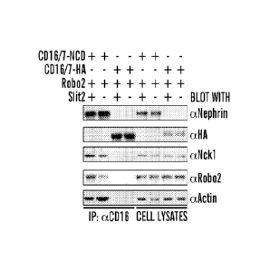
[0056] FIGS. 3A-3G demonstrate that Slit2-Robo2 signaling inhibits nephrin-
mediated actin
polymerization. (FIG. 3A) CD16/7-NCD is co-expressed with Robo2 in HEK cells,
which are treated
with anti-CD16 antibody and rhodamine-conjugated anti-IgG antibody in the
presence of Slit2
conditioned medium (Slit) or control conditioned medium (CTL). Cells are then
fixed and stained with
FITC-conjugated phalloidin to reveal F-actin. Scale bar, 5 iiri. NCD: nephrin
cytoplasmic domain.
(FIG. 3B) A similar assay as (FIG. 3A) except that CD16/7-NCD is replaced by
CD16/7-HA and is used
as a control assay. (FIG. 3C) The percentage of cells with F-actin tails over
total cells with CD16/7
clusters in each group is quantified. Data are represented as mean SEM, *p
<0.01, n=5. (FIG. 3D)
CD16/7-NCD in (FIG. 3A) is immunoprccipitated by anti-CD16 antibody after
Slit2 conditioned
medium stimulation (lanes1 and 3) or control conditioned medium (lanes 2 and
4). Note reduced F-actin
in lane 1. CD16/7-HA is used as a negative control. (FIG. 3E) Intensity
quantification of (FIG. 3D).
Data are represented as mean SEM; n=4, *p < 0.05 compared with the control,
paired student's t-test.
(FIG. 3F) Immunoprecipitation of nephrin from Robo2 knockout homozygous (Robo2-
/-),
heterozygous (Robo2+/-), and wild-type (Robo2+/+) mouse kidneys using the anti-
nephrin antibody.
Note increased F-actin in lane 3. (FIG. 3G) Intensity quantification of (FIG.
3F). Data are represented as
- 8 -
CA 02860558 2014-07-04
WO 2013/103811 PCT/US2013/020280
mean SEM; n=4, *p < 0.05 compared with the wild-type and heterozygous, ANOVA
analysis. See
also FIGS. 7A-7C.
[0057] FIGS. 4A-4W demonstrate podocyte structural phenotypes in the Robo2
homozygous null,
Robo2 podocyte specific knockout, and Robo2 and Nphsl double knockout mice.
(FIGS. 4A and 4B)
Representative images of newborn kidneys show podocyte bodies (arrowheads) and
Bowman's capsule
(arrows) in wild-type (FIG. 4A) and Robo2 homozygous null mice (FIG. 4B).
(FIGS. 4C and 4D) High
magnification images of (FIGS. 4A and4 B) show podocyte foot processes
(arrows) in the newborn
kidney. Scale bar, 1 m. (FIGS. 4E and 4F) Representative images of 3-week
kidneys at low
magnification show podocyte cell body (arrowheads) in a Robo2 homozygous null
mouse (FIG. 4F)
compared to an age-matched control (FIG. 4E). (FIGS. 4G and 4H) Higher
magnification images of
(FIGS. 4E and 4F) show disorganized shorter meandering foot processes (arrow)
in a 3-week Robo2
homozygous null mouse (FIG. 4H) compared to well-organized zip-like foot
processes in the
age-matched control (FIG. 4G). Scale bars: 2 m. (FIGS. 41 and 4.1)
Representative transmission
electron microscopy images (magnification at 5000x) depict the focal segmental
podocyte foot process
effacement (arrow in FIG. 4J) in a one month old Robo2 podocyte-specific
knockout mouse and the
normal phenotype in the control (FIG. 41). Abbreviations: gc: glomerular
capillary; us: urinary space.
(FIGS. 4K and 4L) Higher magnification transmission electron microscopy images
(40000x) show
broader podocyte foot processes (arrow in FIG. 4L) in a two months old Robo2
podocyte-specific
mutant mouse compared with the control (FIG. 4K). Abbreviations: fp, podocyte
foot process; GBM,
glomerular basement membrane. (FIG. 4M) Quantification of podocyte foot
process width in one
month old Robo2del50x;TgNplis2-Cre+podocyte specific knockout mice (Robo2 KO)
and the wild-type
littermate controls (WT). Data are represented as mean SEM, n=333, *p <
0.01. (FIG. 4N) ELISA
assay of spot urine shows an elevated albuminkreatinine ratio in Robo2delm10x;
Nphs2-Cre+ (KO) adult
mice compared with control wild-type (WT). Data are represented as mean SEM,
n=20, *p <0.01.
(FIG. 40) Western blot analysis shows the presence of albumin in urine; 1 1
urine was loaded on each
well, 0.2pg albumin was used as a positive control. WT, three wild-type
littermates; Robo2 KO, three
individual Robo2del5/flox;Nphs2-Cre+ mice. (FIGS. 4P and 4Q) Representative
scanning electron
microscope images show disrupted interdigitating podocyte foot processes that
resemble disorganized
cellular protrusions (arrows) in the Nphs1-/- single homozygous newborn mouse
kidney. Scale bars: 1
m. (FIGS. 4R and 4S) Glomeruli from Nphs1-/-Robo2-/- double homozygous newborn
mice exhibit
restored interdigitating foot processes (arrows), indicating alleviation of
nephrin null phenotype by
knocking out Robo2. (FIGS. 4T and 41T), Glomeruli from Robo2-/- single
homozygous newborn mice
display irregular and broader foot processes but extensive interdigitating
pattern formation (arrows).
(FIGS. 4V and 4W), Glomeruli from newborn wild type mice with normal regular
interdigitating
pattern of the foot process (arrows). See also FIGS. 8A-8Z and Tables 1-4.
[0058] FIGS. 5A-5M demonstrate that Robo2 is expressed in the developing
and adult glomeruli.
(FIGS. 5A and 5B) In situ hybridization analysis shows that Robo2 transcripts
are expressed in
- 9 -
CA 02860558 2014-07-04
WO 2013/103811 PCT/US2013/020280
developing glomeruli (arrows) at El 6.5. Magnification: 60X (FIG. 5A) and 200X
(FIG. 5B). (FIGS.
5C-5F) Immunohistochemistry (IHC) studies reveal that Robo2 is expressed
during developing
glomeruli from E14.5 to E17.5. Magnification: 600X. (FIG. 5G) IHC shows that
Robo2 is specifically
expressed in adult mouse glomeruli at 5 weeks of age (FIG. 5G). DAPI marks
cell nuclei in the kidney.
Magnification: 400X. (FIG. 5H) IHC co-localization stainings of 5w kidney show
Robo2 is
co-expressed in the glomerulus with podocyte marker Wtl. Magnification: 600X.
(FIGS. 5I-5K) Robo2
and WT1 are co-expressed in the mouse glomerulus at E16.5. Magnification:
600X. (FIGS. 5L and 5M)
IHC co-localization stainings of 5w kidney show Robo2 is co-expressed in the
glomerulus with
mesangial cell marker Pdgfrb (FIG. 5L), and endothelial cell marker Pecaml
(FIG. 5M). Magnification:
600X.
[0059] FIGS. 6A-6F' demonstrate that Robo2 interacts with Nck and forms a
complex with
nephrin, which is enhanced by Slit2 stimulation. (FIG. 6A) Co-IP of Robo2 and
nephrin with
endogenous Nck. Robo2, nephrin, and Fyn are expressed in HEK cells and
stimulated by Slit2. The
endogenous Nck is immunoprecipitated by an anti-Nck antibody. The mouse IgG is
used as a control.
The complex formation with nephrin is enhanced by Slit2 and Fyn expression.
(FIGS. 6B and 6C) Slit2
is expressed in the newborn mouse glomeruli by Immunoperoxidase staining (FIG.
6B) and is
co-expressed in the glomerulus with the podocyte marker Synaptopodin (FIG.
6C). Magnification:
600X. (FIGS. 6D and 6D') CDI 6/7-NCD is co-expressed with Robo2 in IIEK cells
in the presence of
Slit2, treated with anti-CD16 antibody and rhodamine-conjugated anti-IgG
antibody, then fixed and
stained with anti-Robo2 antibody. CD16/7-NCD clusters co-localize with Robo2
(FIG. 6D) but no
colocalization is observed in control CD16/7-HA clusters (FIG. 6D'). Scale
bar: 5 gm. NCD: nephrin
cytoplasmic domain. (FIGS. 6E and 6E') Deletion of Nck binding domain (NBD) in
Robo2 impairs its
co-localization with CD16/7-NCD in the presence of Slit2. CD16/7-NCD clusters
co-localize with
Robo2 (FIG. 6E) but no colocalization is observed in Robo2-ANBD clusters (FIG.
6E'). Scale bar: 5 gm.
(FIGS. 6F and 6F') Slit2 stimulation enhances CD16/7-NCD and Robo2 co-
localization in HEK cells.
CD16/7-NCD clusters co-localize with Robo2 in the presence of Slit2 (FIG. 6F)
but not with control
conditioned medium (FIG. 6F'). Scale bar: 5 gm.
[0060] FIGS. 7A-7C demonstrate deletion of Nck binding domain in Robo2
compromises
Slit2-Robo2 inhibition on nephrin-induced actin polymerization. (FIG. 7A)
CD16/7-NCD and Robo2
were co-expressed in HEK cells, clustered with anti-CD16 antibody and
rhodamine-conjugated
anti-IgG antibody in the presence of Slit2 conditioned medium (Slit2) or
control conditioned medium
(CTL). Cells were then fixed and stained with FITC-conjugated phalloidin to
reveal F-actin fibers.
Clusters of CD16/7-NCD and F-actin fibers were examined using confocal
microscopy. Scale bar, 5gm.
NCD, nephrin cytoplasmic domain. (FIG. 7B) CD16/7-NCD and Robo2-ANBD were co-
expressed in
IIEK cells. Scale bar, 5gm. NBD, Nck binding domain. (FIG. 7C) The percentage
of cells with F-actin
tails over total cells with CD16/7-NCD clusters in each group was quantified.
Data are represented as
mean SEM, *p = 1.436x10⁻⁵, **p = 6.32x10⁻⁵, n=5, ANOVA.
- 10 -
CA 02860558 2014-07-04
WO 2013/103811 PCT/US2013/020280
[0061] FIGS. 8A-8Z demonstrate glomerular phenotype in the Robo2 homozygous
null, Robo2
podocyte specific knockout, Robo2 and Nphsl double knockout mice, and a
proposed model of
Robo2-Nephrin signaling. (FIGS. 8A-8F) Transmission electron microscopy
analysis of glomerular
ultrastructure in newborn (NB) Robo2delsidels mutant mice kidney. (FIGS. 8A,
8C, 8E) Glomerular
ultrastructure from a newborn heterozygous Robo2 control mouse at low (FIG.
8A, 2200X), medium
(FIG. 8C, 15500X) and high (FIG. 8E, 52000X) magnifications. (FIGS. 8B, 8D,
8F) Glomerular
ultrastructure from a newborn homozygous Robo2 (-/-) (i.e., Robo2d'iskie15)
mutant mouse at low (FIG.
8B), medium (FIG. 8D) and high (FIG. 8F) magnifications. Arrows indicate focal
foot process
effacement. Abbreviations: gc: glomerular capillary; us: urinary space; GBM:
glomerular basement
membrane. (FIGS. 8G-8N) Abnormal podocyte foot process patterns in Robo2
podocytc-specific
knockout mice. (FIGS. 8G-8J) Representative scanning electron microscopy
images of glomeruli from
one-month old Robo2del50x;Nph,s2-Cre+ podocyte-specific knockout mice and aged
matched Robo2fl'4
control mice. Mild irregularities of the interdigitating podocyte foot
processes were found in a one
month old Robo2 podocyte-specific knockout mouse (FIGS. 8K and 8N). At seven
months old, Robo2
podocyte-specific knockout mice developed markedly irregular foot processes
(FIGS. 81, and 8N).
Scale bars: 10 gm (FIGS. 8G, 8H, 8K, 8L at 2000x magnification) and 2 gm
(FIGS. 81, 8J, 8M ,8N at
13000x magnification). (FIGS. 80-8T) Glomerular morphology in Robo2 podocyte-
specific knockout
mice. (FIGS. 80-8R) Periodic acid-Schiff (PAS) staining showed mesangial
matrix expansion in the
glomeruli from 2-month and 6-month old Robo2 podocyte-specific knockout mice
(FIGS. 8P, 8R)
compared to age-matched controls (FIGS. 80, 8Q). (FIG. 8S) Quantitative
analysis of glomeruli shows
mesangial matrix expansion in 12-month old Robo2 podocyte-specific knockout
mice (MU) compared
to age matched wild-type (WT) controls. Data are represented as mean SEM,
n=5, *p <0.01. (T)
Robo2 podocyte specific knockout does not affect podocyte numbers. Podocyte
cells were identified
using WT-1 staining. The number of podocytes per glomerular cross section was
counted in four
one-year old Robo2del5/fl0x;TgNphs2-Cre+
podocyte specific knockout mice (MU) compared to four
age-matched wild-type mice (WT). Data are represented as mean SEM, p =
0.645, t-test; mutant:
n=165 glomeruli; control: n=166 glomeruli. (FIGS. 8U-8Y) Glomerular phenotype
in Robo2 and Nphs 1
double knockout mice. (FIG. 8U) H&E staining shows glomeruli with
characteristic dilatations of the
Bowman's space (asterisks) in a Nph,s1-1- single homozygous newborn mouse,
400x. (FIG. 8V)
Glomeruli from a Robo2* single homozygous newborn mouse show absence of
Bowman's space
dilatations; 400x. (FIG. 8W) Normal looking glomeruli without significant
Bowman's space dilatations
(arrows) are shown in a Robo24-;Arphs1-/- double homozygous newborn mouse
indicating alleviation of
Nphs14- glomerular phenotype; 400x. (FIG. 8X) H&E staining of normal kidney
and glomeruli from an
age-matched wild-type newborn mouse control; 400x. (FIG. 8Y) Quantification of
glomeruli with
dilated Bowman's space in newborn mice show significant reduction of glomeruli
with the
characteristic dilatation phenotype of the Bowman's space in Robo24-;Nphs14-
double homozygous
compared to Nphs1-/- single homozygous (Robo2+/-;Nphs14). Data are represented
as mean SEM. *p <
- 11 -
CA 02860558 2014-07-04
WO 2013/103811 PCT/US2013/020280
0.01. (FIG. 8Z) A model of inhibitory effects of Slit2-Robo2 signaling on
nephrin to influence podocyte
foot process structure: Under physiological conditions (e.g., during foot
process development), nephrin
intracellular phosphorylated tyrosine domains (YDxV-p) recruit Nck through its
interaction with the
SII2 domain. Nck in turn recruits cytoskeleton regulators through its SII3
domains to promote actin
polymerization. Slit2 binds Robo2 to increase Robo2 intracellular domain
interaction with SH3
domains of Nck, which would prevent binding of Nck to cytoskeletal regulators
and result in an
inhibition of nephrin-induced actin polymerization. Balanced actin
polymerization is maintained during
podocte development for a normal foot process structure. In the absence of
Slit2-Robo2 signaling (e.g.,
when Robo2 is knocked out), the inhibitory effects of Robo2 on nephrin induced
polymerization is lost.
The SH3 domains of Nck are able to interact with downstream cytoskeletal
regulators to increase actin
polymerization, which may explain the altered podocyte foot process structure
in Robo2 mutant mice.
Abbreviations: Ig: Immunoglobulin domain; FN3: Fibronectin type 3 domain; SH2:
Src homolog 2
domain; SH3: Src homolog 3 domain; CCO, CC1, CC2, CC3: Cytoplasmic Conserved
region 0, 1, 2, 3.
DETAILED DESCRIPTION
[0062] Robo2 has been previously shown to be the cell surface receptor for
the repulsive guidance
cue Slit and to be involved in axon guidance and neuronal migration in the
nervous system. Nephrin is a
podocyte slit-diaphragm protein that functions in the kidney glomerular
filtration barrier. We
demonstrate herein that Robo2 is expressed at the basal surface of podocytes,
such as mouse podocytes,
and co-localizes with nephrin. Biochemical studies indicate that Robo2 forms a
complex with nephrin
in the kidney through adaptor protein Nck. In contrast to the role of nephrin
that promotes actin
polymerization, we show herein that Slit2-Robo2 signaling inhibits nephrin-
induced actin
polymerization. For example, the amount of F-actin associated with nephrin is
increased in Robo2
knockout mice that develop an altered podocyte foot process structure and
microalbuminuria. Genetic
interaction studies further reveal that loss of Robo2 alleviates the abnormal
podocyte phenotype in
nephrin null mice. The results provided herein show that Robo2 signaling acts
as a negative regulator on
nephrin to influence podocyte foot process architecture.
[0063] In addition, it has been shown that a patient having vesicoureteral
reflux (VUR) has a
chromosome translocation that disrupts the ROB02 gene and produces dominant
negative ROB02
fusion proteins that abrogate the SLIT2-ROB02 signaling pathway. Normally, VUR
is a disease
characterized by the retrograde flow of urine from the bladder into the
ureters and kidney and VUR
patients can present with reflux nephropathy, a condition that manifests with
severe proteinuria. It has
been shown that dominant negative ROB02 fusion proteins produced by a VUR
patient blocks the
SLIT2-ROB02 signaling pathway and protects the patient from reflux nephropathy
and proteinuria,
thus confirming and further supporting the inventors results in animal models
of the therapeutic value of
targeting the SLIT2-ROB02 signaling pathway for the treatment of chronic
kidney disease.
- 12 -
CA 02860558 2014-07-04
WO 2013/103811 PCT/US2013/020280
[0064] In the normal kidney, the trilaminar glomerular capillary wall,
composed of fenestrated
endothelial cells, basement membrane and podocytes, restricts the permeability
to plasma proteins.
Podocytes are specialized epithelial cells that extend primary and secondary
processes to cover the
outer surface of the glomerular basement membrane. The actin-rich
interdigitating secondary processes,
or foot processes, from neighboring podocytes create filtration slits bridged
by a semi-porous
slit-diaphragm that forms the final barrier to protein permeation. Whereas
genetic mutations of
podocyte slit-diaphragm proteins such as nephrin and others are associated
with hereditary forms of
proteinuric kidney disease (Tryggvason et al., 2006), it has become evident
that the proteins that make
up and associate with the slit-diaphragm are more than a simple structural
barrier. These proteins form a
balanced signaling network that can influence podocyte foot process structure
and function through
interaction with the F-actin cytoskeleton (Faul et al., 2007; Jones et al.,
2006; Verma et al., 2006).
[0065] Roundabout (Robo) family proteins, Robol. Robo2, Robo3 and Robo4 are
cell surface
receptors for the secreted ligand Slit (Dickson and Gilestro, 2006). Slit 1,
Slit2, and Slit3 were originally
found as repulsive guidance cues for axon pathfinding and migrating neurons
during nervous system
development (Guan and Rao, 2003). The transmembrane protein Robo contains five
Ig motifs and three
fibronectin type 111 (FNIII) repeats in its extracellular domain (Dickson and
Gilestro, 2006). While both
immunoglobulin (Ig) motifs 1 and 2 interact with Slit, the first Igl motif of
Robo is the primary binding
site for Slit (Dickson and Gilestro, 2006). The intracellular domain of Robo
has four cytoplasmic
conserved (CC) sequences named CCO, CC1. CC2, and CC3 (Bashaw et al., 2000;
Kidd et al., 1998;
Morlot et al., 2007; Zallen et al., 1998). CCO and CC1 contain tyrosine, while
CC2 and CC3 are
proline-rich stretches. The repulsive activity of Slit-Robo signaling inhibits
actin polymerization (Guan
and Rao, 2003) or induces F-actin depolymerization (Piper et al., 2006).
[0066] Slit-Robo signaling also plays crucial roles during early kidney
induction and ureteric bud
outgrowth. Mouse mutants that lack Slit2 or Robo2 develop supernumerary
ureteric buds, which lead to
a broad-spectrum of urinary tract phenotype including duplex kidneys, abnormal
ureterovesical
junctions and hydronephrosis (Grieshammer et al., 2004; Lu et al. 2007).
Disruption of ROB02 in
humans causes congenital anomalies of the kidneys and urinary tracts (CAKUT),
and point mutations
of ROB02 have been identified in patients with vesicoureteral reflux (VUR) (Lu
et al., 2007). Our
recent study demonstrates that Robo2 is crucial for the formation of a normal
ureteral orifice and for the
maintenance of an effective anti-reflux mechanism (Wang et al., 2011).
[0067] Herein we demonstrate that Robo2 is a novel podocyte protein
expressed at the basal
surface of glomerular podocytes in the kidney and is co-localized with nephrin
and podocin. Robo2
interacts directly with adaptor protein Nck SH3 domains and forms a complex
with nephrin. Whereas
Robo2 knockout mice develop altered podocyte foot processes, the loss of Robo2
alleviates the foot
process structural abnormalities that are seen in nephrin null mice. These
results described herein
indicate that Robo2 signaling acts as a negative regulator on nephrin
signaling to influence podocyte
foot process architecture. In addition, as demonstrated herein, it has been
discovered that the dominant
- 13 -
CA 02860558 2014-07-04
WO 2013/103811 PCT/US2013/020280
negative ROB02 fusion proteins produced by a patient blocks the SLIT2-ROB02
signaling pathway
and protects the patient from reflux nephropathy and proteinuria, thus
confirming and further
supporting the results described herein in animal models of the therapeutic
value of targeting the
SLIT2-ROB02 signaling pathway for the treatment of chronic kidney disease.
[0068] Accordingly, in some aspects, provided herein are methods for the
treatment of chronic
kidney disease in a subject in need thereof, such method comprising
administering to a subject having
or at risk for a chronic kidney disease a therapeutically effective amount of
a composition comprising a
SLIT2-ROB02 signaling pathway inhibitor.
[0069] Also provided herein, in some aspects, are methods for the reduction
of proteinuria in a
subject in need thereof, comprising administering to a subject having or at
risk for proteinuria a
therapeutically effective amount of a composition comprising a SLIT2-ROB02
signaling pathway
inhibitor.
[0070] In other aspects, provided herein are methods for preventing kidney
diseases or promoting
prophylaxis of kidney diseases in a subject in need thereof, comprising
administering to a subject a
therapeutically effective amount of a composition comprising a SLIT2-ROB02
signaling pathway
inhibitor so as to prevent or promote prophylaxis of kidney disease in the
subject.
[0071] Also provided herein, in some aspects, are methods for mitigating
the effects of kidney
disease, reducing the severity of kidney disease, reducing the likelihood of
developing kidney disease
and/or slowing the progression of kidney disease in a subject in need thereof.
[0072] As used herein, "ROBOT' refers to the polypeptide having the amino
acid sequence of:
MARRHERVIRRMWTWAPGLLMMTVVEWGHQGNGQGQGSRLRQEDEPPRIVEHPSDVIVSK
GEPTTLNCKAEGRPTPTIEWYKDGERVETDKDDPRSHRMLLPSGSLEFLRIVHGRRSKPDEGS
YVCVARNYLGEAVSRNASLEVALLRDDFRQNPTDVVVAAGEPAILECQPPRGHPEPTIYWKK
DKVRIDDKEERISIRGGKLMISNTRKSDAGMYTCVGTNMVGERDSDPAELTVFERPTFLRRPI
NQVVLEEEAVEFRCQVQGDPQPTVRWKKDDADLPRGRYDIKDDYTLRIKKTMSTDEGTYM
CIAENRVGKMEASATLTVRAPPQFVVRPRDQIVAQGRTVTFPCETKGNPQPAVEWOKEGSQ
NLLFPNQPQQPNSRCSVSPTGDLTITNIQRSDAGYYICQALTVAGSILAKAQLEVTDVLTDRPP
PIILQGPANQTLAVDGTALLKCKATGDPLPVISWLKEGFTFPGRDPRATIQEQGTLQIKNLRIS
DTGTYTCVATSSSGETSWSAVLDVTESGATISKNYDESDLPGPPSKPQVTDVTKNSVTLSWQ
PGIPGTLPASAYIIEAFSQSVSNSWQTVANHVKITLYTVRGLRPNTIYLEMVRAINPQGLSDPS
PMSDPVRTQDISPPAQGVDHRQVQKELGDVLVRLHNPVVLTPTTVQVTWTVDRQPQFIQGY
RVMYRQTSGIQATSSWQNIDAKVPTERSAVINNI,KKGVTYEIKVRPYFNEFQGMDSESKTV
RTILEAPSAPPQSVTVLTVGS YNSTSISVSWDPPPPDHQNGIIQEYKIWCLGNETREHINKT VD
AAIRSVIIGGLFPGIQYRVEVAASTSAGVGVKSEPQPIIIGRRNEVVITENNNSITEQITDVVKQP
AFIAGIGGACWVILMGESIWLYWRRKKRKGLSNYAVTFQRGDGGLMSNGSRPGLLNAGDPS
YPWLADSWPATSLPVNNSNSGPNEIGNEGRGDVLPPVPGQGDKTATMLSDGAIYSSIDETTKT
SYNSSSQITQATPYATTQILHSNSIHELAVDLPDPQWKSSIQQKTDLMGEGYSLPDQNKGNNG
- 14 -
CA 02860558 2014-07-04
WO 2013/103811 PCT/US2013/020280
GKGGKKKKNKNSSKPQKNNGSTWANVPI,PPPPVQPI,PGTEIEI IYAVEQQENGYDSDSWCPP
LPVQTYLHQGLEDELEEDDDRVPTPPVRGVASSPAISFGQQSTATLIPSPREEMQPMLQAHLD
ELTRAYQFDIAKQTWHIQSNNQPPQPPVPPLGYVSGALISDLETDVADDDADDEEEALEIPRP
LRALDQTPGSSMDNLDSSVTGKAFTSSQRPRPTSPFSTDSNTSAALSQSQRPRPTKKIIKGGRM
DQQPALPHRREGMTDEEALVPYSKPSEPSPGGHSSSGTASSKGSTGPRKTEVLRAGHQRNAS
DLLDIGYMGSNSQGQFTGEL (Homo sapiens roundabout homolog 2 isoform ROB02a; SEQ ID
NO: 1), as described by, e.g., NP_001122401.1 and encoded by NM_001128929.2
(SEQ ID NO: 2); or
MSLLMFTQLLLCGFLYVRVDGSRLRQEDEPPRIVEHPSDVIVSKGEPTTLNCKAEGRPTPTIE
WYKDGERVETDKDDPRSHRMLLPSGSLEFLRIVHGRRSKPDEGSYVCVARNYLGEAVSRNA
SLEVALLRDDFRQNPTDVVVAAGEPAILECQPPRGHPEPTIYWKKDKVRIDDKEERISIRGGK
LMISNTRKSDAGMYTCVGTNMVGERDSDPAELTVFERPTFLRRPINQVVLEEEAVEFRCQVQ
GDPQPTVRWKKDDADLPRGRYDIKDDYTLRIKKTMSTDEGTYMCIAENRVGKMEASATLTV
RAPPQEV VRPRDQ1VAQGR V'l EPCETKGNPQPAV LW QKEGS QNLLEPN QPQQPN SRC S V SP
TGDLTITNIQRSDAGYYICQALTVAGSILAKAQLEVTDVLTDRPPPIILQGPANQTLAVDGTAL
I,KCKATGDPI,PVISWI,KEGFTFPGRDPRATIQEQGTI,QIKNI,RISDTGTYTCVATSSSGETSWS
AVLDV ESGATISKNYDLSDLPGPPSKPQVTDVIKNSVILSWQPGTPGTLPASAYHEAFSQSV
SNSWQTVANHVKTTLYTVRGLRPNTIYLFMVRAINPQGLSDPSPMSDPVRTQDISPPAQGVD
IR QVQKEI ,GDVINRI TINPVVLTPTTVQVTWTVDRQPQFIQGYRVMYRQTSGLQATSSWQN
LDAKVPTERSAVLVNLKKGVIYEIKVRPYFNEFQGMDSESKTVRTTEEAPSAPPQSVTVLTV
GSYNSTSISVSWDPPPPDHQNGIIQEYKIWCLGNETREHINKTVDAAIRSVIIGGLFPGIQYRVE
VAASTSAGVGVKSEPQPIIIGRRNEVVITENNNSITEQITDVVKQPAFIAGIGGACWVILMGESI
WLYWRRKKRKGLSNYAVTFQRGDGGLMSNGSRPGLLNAGDPSYPWLADSWPATSLPVNNS
NSGPNEIGNEGRGDVLPPVPGQGDKTATMLSDGAIYSSIDETTKTSYNSSSQITQATPYATTQI
LHSNSIHELAVDLPDPQWKSSIQQKTDLMGEGYSLPDQNKGNNGGKGGKKKKNKNSSKPQK
NNGSTWANVPLPPPPVQPLPGTELEHYAVEQQENGYDSDSWCPPLPVQTYLHQGLEDELEED
DDRVPTPPVRGVASSPAISFGQQSTATLTPSPREEMQPMLQAHLDELTRAYQFDIAKQTWHIQ
SNNQPPQPPVPPLGYVSGALISDLETDVADDDADDEEEALEIPRPLRALDQTPGSSMDNLDSS
VTGKAFTSSQRPRPTSPFSTDSNTSAALSQSQRPRPTKKHKGGRMDQQPALPHRREGMTDEE
ALVPYSKPSEPSPGGHSSSGTASSKGSTGPRKTEVLRAGHQRNASDLLDIGYMGSNSQGQFTG
EL (Hoino sapiens roundabout homolog 2 isoform ROBO2b; SEQ ID NO: 3), as
described by, e.g.,
NP_002933.1 and encoded by NM_002942.4 (SEQ ID NO: 4), together with any
naturally occurring
allelic, splice variants, and processed forms thereof. Typically, ROB02 refers
to human ROB02. The
ROB02 gene is conserved in chimpanzee, Rhesus monkey, dog, cow, mouse, rat,
chicken, zebrafish,
fruit fly, mosquito, and C.elegans. Specific residues of ROB 02 can be
referred to as, for example,
"ROB02(30)."
[0073] Specific domains of ROB 02 can be referred to by such nomenclature
as well. The
N-terminal or "extracellular domain of ROB 02", comprising the five
immunoglobulin motifs and three
- 15 -
CA 02860558 2014-07-04
WO 2013/103811 PCT/US2013/020280
fibronectin type III (FNIII) repeats can be referred to as ROB02(46-848) of
SEQ ID NO: 1 or
ROB02(30-832) of SEQ ID NO: 3, for example. The immunoglobulin (Ig) motifs 1
and 2 that interact
with 51it2, or the "Ig SLIT binding domain" can be referred to as ROB02(46-
145) and
ROB02(151-237) respectively of SEQ ID NO: 1, and ROB02(30-129) and ROB02(135-
221)
respectively of SEQ ID NO: 3. Similarly, the "intracellular domain" comprising
the "Nck intracellular
binding domain," which comprises the four intracellular proline rich motifs,
described herein, can be
referred to as ROB02(881-1378) of SEQ ID NO: 3.
[0074] As used herein, the terms "ROB02 inhibitor," "ROB02 antagonist,"
"ROB02 inhibitor
agent," and "ROB02 antagonist agent" refer to a molecule or agent that
significantly blocks, inhibits,
reduces, or interferes with ROB02 (mammalian, such as human, ROB02) biological
activity in vitro, in
situ, and/or in vivo, including activity of downstream pathways mediated by
ROB02 signaling, such as,
for example, ROB02 interaction with the adaptor protein Nck and/or complex
formation with nephrin,
SLIT2-ROB0-2 mediated inhibition of nephrin-mediated actin polymerization,
and/or elicitation of a
cellular response to ROB 02. The term "agent" as used herein in reference to a
ROB02 inhibitor means
any compound or substance such as, but not limited to, a small molecule,
nucleic acid, polypeptide,
peptide, drug, ion, etc. An "agent" can be any chemical, entity, or moiety,
including, without limitation,
synthetic and naturally-occurring proteinaceous and non-proteinaceous
entities. In some embodiments
of the aspects described herein, an agent is a nucleic acid, a nucleic acid
analogue, a protein, an
antibody, a peptide, an aptamer, an oligomer of nucleic acids, an amino acid,
or a carbohydrate, and
includes, without limitation, proteins, oligonucleotides, ribozymes, DNAzymes,
glycoproteins,
antisense RNAs, siRNAs, lipoproteins, aptamers, and modifications and
combinations thereof etc.
Compounds for use in the therapeutic compositions and methods described herein
can be known to have
a desired activity and/or property, or can be selected from a library of
diverse compounds, using
screening methods known to one of ordinary skill in the art.
[0075] Exemplary ROB 02 inhibitors contemplated for use in the various
aspects and
embodiments described herein include, but are not limited to, anti-ROB02
antibodies or
antigen-binding fragments thereof that specifically bind to ROB 02; anti-sense
molecules directed to a
nucleic acid encoding ROB02 (e.g.., ROB02a or ROB02b or both); short
interfering RNA ("siRNA")
molecules directed to a nucleic acid encoding ROB02 (e.g.., R0B02a or R0BO2b
or both); RNA or
DNA aptamers that bind to ROB02, and inhibit/reduce/block ROB02 mediated
signaling; ROB02
structural analogs; and soluble ROB 02 proteins, inhibitory polypeptides,
e.g., dominant negative
polypeptides, or fusion polypeptides thereof. In some embodiments of these
aspects and all such aspects
described herein, a ROB02 inhibitor (e.g., an antibody or antigen-binding
fragment thereof) binds
(physically interacts with) ROB 02, targets downstream ROB 02 signaling,
and/or inhibits (reduces)
ROB02 synthesis, production or release. In some embodiments of these aspects
and all such aspects
described herein, a ROB02 inhibitor binds and prevents its binding a SLIT
protein ligand, such as
SLIT2. In some embodiments of these aspects and all such aspects described
herein, a ROB 02 inhibitor
- 16 -
CA 02860558 2014-07-04
WO 2013/103811 PCT/US2013/020280
specifically reduces or eliminates expression (i.e., transcription or
translation) of one or more ROB02
isoforms.
[0076] As used herein, a ROB 02 inhibitor or antagonist has the ability to
reduce the activity
and/or expression of ROB02 in a cell (e.g., podocytes) by at least 5%, at
least 10%, at least 15%, at least
20%, at least 25%, at least 30%, at least 35%, at least 40%, at least 45%, at
least 50%, at least 55%, at
least 60%, at least 65%, at least 70%, at least 75%, at least 80%, at least
85%, at least 90%, at least 95 %,
at least 98%, at least 99%, or more, relative to the activity or expression
level in the absence of the
ROB02 inhibitor.
[0077] Accordingly, in some embodiments of the compositions and methods
described herein, the
ROB 02 inhibitor inhibits ROB 02 mediated signal transduction. In some
embodiments of the
compositions and methods described herein, the ROB02 inhibitor targets ROB02
interaction with the
adaptor protein Nck and/or complex formation with nephrin, SLIT2-ROB0-2
mediated inhibition of
nephrin-mediated actin polymerization, and/or elicitation of a cellular
response to ROB02.
[0078] In some embodiments of the compositions and methods described
herein, the binding sites
of the ROB02 inhibitors, such as an antibody or antigen-binding fragment
thereof, are directed against
a ROB02 ligand interaction site, such as a SLIT2 ligand interaction site. In
some embodiments of the
compositions and methods described herein, the binding sites of the ROB 02
inhibitor, such as an
antibody or antigen-binding fragment thereof, are directed against a ROB02
adaptor interaction site
such as an Nck interaction site or the NCK intracellular binding domain
comprising the four
intracellular proline rich motifs of ROB 02. In some embodiments of the
compositions and methods
described herein, the binding sites of the ROB02 inhibitors are directed
against a site on a target in the
proximity of the ligand interaction site, in order to provide steric hindrance
for the interaction of the
receptor (e.g., ROB02) with its ligand (e.g., SLIT2). By binding to a ROB02
ligand interaction site, a
ROB02 inhibitor described herein can reduce or inhibit the activity or
expression of ROB02, and
downstream ROB 02 signaling consequences (e.g., ROB02 interaction with the
adaptor protein Nck
and/or complex formation with nephrin, SLIT2-ROB0-2 mediated inhibition of
nephrin-mediated actin
polymerization, and/or elicitation of a cellular response to ROB02). For
example, in some
embodiments of the compositions and methods described herein, the binding
sites of the ROB 02
inhibitors block or target at least the Igl, and preferably both the Igl and
Ig2 sites, on ROB 02, i.e.,
ROB02(46-145) and ROB02(151-237) respectively of SEQ ID NO: 1, and ROB02(30-
129) and
ROB02(135-221) respectively of SEQ ID NO: 3, for example. In some embodiments
of the
compositions and methods described herein, the binding sites of the ROB 02
inhibitors block or target
the ROB02 intracellular domain comprising the Net( intracellular binding
domain, i.e.,
ROB02(881-1378) of SEQ ID NO: 3. In some embodiments of the compositions and
methods
described herein, the binding sites of the ROB02 inhibitors block or target
the ROB02 Nck
intracellular binding domain comprising the four intracellular proline rich
motifs of ROB02. This can
- 17 -
CA 02860558 2014-07-04
WO 2013/103811 PCT/US2013/020280
be accomplished by a variety of means well known in the art, such as
antibodies and antigen-binding
fragments thereof, inhibitor RNAs, etc., and as described herein.
[0079] Accordingly, in some embodiments of the compositions and methods
described herein, the
ROB 02 inhibitor is an antibody or antigen-binding fragment thereof that
selectively binds or physically
interacts with ROB02. In some embodiments of the compositions and methods
described herein, the
ROB 02 inhibitor is an antibody or antigen-binding fragment thereof that binds
to ROB 02 and inhibits
and/or blocks and/or prevents interaction with Nck and/or complex formation
with nephrin. In some
embodiments of the compositions and methods described herein, the antibody or
antigen-binding
fragment thereof binds to the Ig SLIT binding domain of ROB02. In some
embodiments of the
compositions and methods described herein, the antibody or antigen-binding
fragment thereof binds to
the Ig1SLIT binding domain of ROB02 or both the Igl and Ig2 SLIT binding
domains of ROB02, i.e.,
ROB02(46-145) and ROB02(151-237) respectively of SEQ ID NO: 1, and ROB02(30-
129) and
ROB02(135-221) respectively of SEQ Ill NO: 3. In some embodiments of the
compositions and
methods described herein, the antibody or antigen-binding fragment thereof
binds to or blocks the
ROB02 intracellular domain, i.e., ROB02(881-1378) of SEQ ID NO: 3. In some
embodiments of the
compositions and methods described herein, the antibody or antigen-binding
fragment thereof binds to
or blocks the Nck intracellular binding domain comprising the four
intracellular proline rich motifs of
ROB02.
[0080] Antibodies specific for or that selectively bind ROB 02, suitable
for use in the
compositions and for practicing the methods described herein are preferably
monoclonal, and can
include, but are not limited to, human, humanized or chimeric antibodies,
comprising single chain
antibodies, Fab fragments, F(ab') fragments, fragments produced by a Fab
expression library, and/or
binding fragments of any of the above. Antibodies also refer to
inununoglobulin molecules and
immunologically active portions of immunoglobulin molecules, i.e., molecules
that contain antigen or
target binding sites or "antigen-binding fragments." The immunoglobulin
molecules described herein
can be of any type (e.g., IgG, IgE, IgM, IgD, IgA and TgY), class (e.g., IgG1
, IgG2, IgG3, IgG4, IgAl
and IgA2) or subclass of immunoglobulin molecule, as is understood by one of
skill in the art.
[0081] Accordingly, in some embodiments of the compositions and methods
described herein, a
ROB02 inhibitor as described herein is a monoclonal anti-ROB02 antibody or
antigen-binding
fragment.
[0082] In some embodiments of the compositions and methods described
herein, a ROB02
inhibitor as described herein is a ROB02 antibody fragment or antigen-binding
fragment. The terms
"antibody fragment," -antigen binding fragment," and "antibody derivative" as
used herein, refer to a
protein fragment that comprises only a portion of an intact antibody,
generally including an antigen
binding site of the intact antibody and thus retaining the ability to bind
antigen. Examples of antibody
fragments encompassed by the terms antibody fragment or antigen-binding
fragment include: (i) the
Fab fragment, having VL, CL, VH and CH1 domains; (ii) the Fab' fragment, which
is a Fab fragment
- 18 -
CA 02860558 2014-07-04
WO 2013/103811 PCT/US2013/020280
having one or more cysteine residues at the C-terminus of the CHI domain;
(iii) the Fd fragment having
VH and CH1 domains; (iv) the Fd' fragment having VH and CH1 domains and one or
more cysteine
residues at the C-terminus of the CH1 domain; (v) the Fv fragment having the
VL and VH domains of a
single arm of an antibody; (vi) a dAb fragment (Ward et al., Nature 341. 544-
546 (1989)) which
consists of a VH domain or a VT domain ; (vii) isolated CDR regions; (viii)
F(ab')2 fragments, a bivalent
fragment including two Fab' fragments linked by a disulphide bridge at the
hinge region; (ix) single
chain antibody molecules (e.g. single chain Fv; scFv) (Bird et al., Science
242:423-426 (1988); and
Huston et al., PNAS (USA) 85:5879-5883 (1988)); (x) "diabodies" with two
antigen binding sites,
comprising a heavy chain variable domain (VH) connected to a light chain
variable domain (VL) in the
same polypeptide chain (see, e.g., EP 404,097; WO 93/11161; and Hollinger et
al., Proc. Natl. Acad.
Sci. USA, 90:6444-6448 (1993)); (xi) ''linear antibodies" comprising a pair of
tandem Fd segments
(VH-CH1-VH-CH1) which, together with complementary light chain polypeptides,
form a pair of antigen
binding regions (Zapata et al. Protein Eng. 8(10):1057-1062 (1995); and U.S.
Pat, No. 5,641,870); and
modified versions of any of the foregoing (e.g., modified by the covalent
attachment of polyalkylene
glycol (e.g., polyethylene glycol, polypropylene glycol, polybutylene glycol)
or other suitable
polymer).
[0083] In some embodiments of the compositions and methods described
herein, a ROB 02
inhibitor or antagonist is a chimeric antibody derivative of a ROBO2
antagonist antibody or
antigen-binding fragment thereof.
[0084] The ROB02 inhibitor or antagonist antibodies and antigen-binding
fragments thereof
described herein can also be, in some embodiments, a humanized antibody
derivative.
[0085] In some embodiments, the ROB02 inhibitor or antagonist antibodies
and antigen-binding
fragments thereof described herein include derivatives that are modified,
i.e., by the covalent
attachment of any type of molecule to the antibody, provided that the covalent
attachment does not
prevent the antibody from binding to the target antigen, e.g., ROB02.
[0086] In sorne embodiments of the compositions and methods described
herein, completely
human antibodies are used, which are particularly desirable for the
therapeutic treatment of human
patients.
[0087] In sonic embodiments of the compositions and methods described
herein, the ROB 02
inhibitor comprises at least one antisense molecule capable of blocking or
decreasing the expression of
a particular functional ROB 02 by targeting nucleic acids encoding ROB 02,
e.g., SEQ ID NO: 2 or
SEQ ID NO: 4 or both, or relevant domains thereof. In some embodiments of the
compositions and
methods described herein, the at least one antisense molecule targets nucleic
acids encoding the Ig SLIT
binding domain of ROB02. In some embodiments of the compositions and methods
described herein,
the at least one antisense molecule targets nucleic acids encoding theIgl SLIT
binding domain of
ROB02 or both the Igl and Ig2 SLIT binding domains of ROB 02. In some
embodiments of the
compositions and methods described herein, the at least one antisense molecule
targets nucleic acids
- 19 -
CA 02860558 2014-07-04
WO 2013/103811 PCT/US2013/020280
encoding the ROB 02 intracellular domain. In some embodiments of the
compositions and methods
described herein, the at least one antisense molecule targets nucleic acids
encoding the Nck intracellular
binding domain comprising the four intracellular proline rich motifs of
ROB02.Methods are known to
those of ordinary skill in the art for the preparation of antisense
oligonucleotide molecules that will
specifically bind ROB02 mRNA without cross-reacting with other
polynucleotides. Exemplary sites of
targeting include, but are not limited to, the initiation codon, the 5'
regulatory regions, including
promoters or enhancers, the coding sequence, including any conserved consensus
regions, and the 3'
untranslated region. In some embodiment of these aspects and all such aspects
described herein, the
antisense oligonucleotides are about 10 to about 100 nucleotides in length,
about 15 to about 50
nucleotides in length, about 18 to about 25 nucleotides in length, or more. In
certain embodiments, the
antisense oligonucleotides further comprise chemical modifications to increase
nuclease resistance and
the like, such as, for example, phosphorothioate linkages and 2-0-sugar
modifications known to those
of ordinary skill in the art.
[0088] In some embodiments of the compositions and methods described
herein, the ROB 02
inhibitor comprises at least one short interfering RNA (siRNA) molecule
capable of blocking or
decreasing the expression of functional ROB02 by targeting nucleic acids
encoding or both isoforms of
ROB02, e.g., SEQ ID NO: 2 or SEQ ID NO: 4, or relevant domains thereof. In
some embodiments of
the compositions and methods described herein, the at least one siRNA molecule
targets nucleic acids
encoding the Ig SLIT binding domain of ROB02. In some embodiments of the
compositions and
methods described herein, the at least one siRNA molecule targets nucleic
acids encoding theIgl SLIT
binding domain of ROB02 or both the Igl and Ig2 SLIT binding domains of ROB02.
In some
embodiments of the compositions and methods described herein, the at least one
siRNA molecule
targets nucleic acids encoding the ROB02 intracellular domain. In some
embodiments of the
compositions and methods described herein, the at least one siRNA molecule
targets nucleic acids
encoding the Nck intracellular binding domain comprising the four
intracellular proline rich motifs of
ROB02. It is routine to prepare siRNA molecules that will specifically target
ROB02 mRNA without
cross-reacting with other polynucleotides. siRNA molecules for use in the
compositions and methods
described herein can be generated by methods known in the art, such as by
typical solid phase
oligonucleotide synthesis, and often will incorporate chemical modifications
to increase half-life and/or
efficacy of the siRNA agent, and/or to allow for a more robust delivery
formulation. Alternatively,
siRNA molecules are delivered using a vector encoding an expression cassette
for intracellular
transcription of siRNA.
[0089] In some embodiments of the compositions and methods described
herein, the ROB02
inhibitor is an RNA or DNA aptamer that binds to one or more isoforms of
ROB02. In some
embodiments of the compositions and methods described herein, a ROB 02
inhibitor or antagonist is an
RNA or DNA aptamer that binds or physically interacts with ROB 02, and blocks
interactions between
ROB02 and a ligand or adaptor molecule, for example, SLIT2 or Nck,
respectively. In some
- 20 -
CA 02860558 2014-07-04
WO 2013/103811 PCT/US2013/020280
embodiments of the compositions and methods described herein, a ROBO-2
inhibitor or antagonist is
an RNA or DNA aptamer that binds or physically interacts with ROB02, and
reduces, impedes, or
blocks downstream ROB 02 signaling, such as SLIT2-ROB0-2 mediated inhibition
of
nephrin-mediated actin polymerization, and/or elicitation of a cellular
response to ROB02. In some
embodiments of the compositions and methods described herein, the RNA or DNA
aptamer binds to or
physically interacts with the Ig SLIT binding domain of ROB02. In some
embodiments of the
compositions and methods described herein, the RNA or DNA aptamer binds to or
physically interacts
with the Ig1SLIT binding domain of ROB 02 or both the Igl and Ig2 SLIT binding
domains of ROB 02,
i.e., ROB02(46-145) and ROB02(151-237) respectively of SEQ ID NO: 1, and
ROB02(30-129) and
ROB02(135-221) respectively of SEQ ID NO: 3. In some embodiments of the
compositions and
methods described herein, the RNA or DNA aptamer binds to or physically
interacts with the ROB 02
intracellular domain, i.e., ROB02(881-1378) of SEQ ID NO: 3. In some
embodiments of the
compositions and methods described herein, the RNA or DNA aptamer binds to or
physically interacts
with or blocks the Nck intracellular binding domain comprising the four
intracellular proline rich motifs
of ROB02.
[0090] In some embodiments of the compositions and methods described
herein, the ROB02
inhibitor is a small molecule compound or agent that targets or binds to
ROB02, including, but not
limited to, small peptides or peptide-like molecules, soluble peptides, and
synthetic non-peptidyl
organic or inorganic compounds. As used herein, the term "small molecule"
refers to a chemical agent
which can include, but is not limited to, a peptide, a peptidomimetic, an
amino acid, an amino acid
analog, a polynucleotide, a polynucleotide analog, an aptamer, a nucleotide, a
nucleotide analog, an
organic or inorganic compound (e.g., including heterorganic and organometallic
compounds) having a
molecular weight less than about 10,000 grams per mole, organic or inorganic
compounds having a
molecular weight less than about 5,000 grams per mole, organic or inorganic
compounds having a
molecular weight less than about 1,000 grams per mole, organic or inorganic
compounds having a
molecular weight less than about 500 grams per mole, and salts, esters, and
other pharmaceutically
acceptable forms of such compounds. Exemplary sites of small molecule binding
include, but are not
limited to, the portion of ROB02 that binds to SLIT2 or to the adaptor Nck,
i.e., the Igl SLIT binding
domain of ROB02 or both the Igl and Ig2 SLIT binding domains of ROB 02, the
ROB 02 intracellular
domain or the Nck intracellular binding domain comprising the four
intracellular proline rich motifs of
ROB02.
[0091] In some embodiments of the compositions and methods described
herein, a ROB02
inhibitor or antagonist comprises a small molecule that binds to ROB 02 and
inhibits ROB02 biological
activity.
[0092] In some embodiments of the compositions and methods described
herein, the ROB 02
inhibitor or antagonist comprises at least one ROB 02 structural analog, such
as a dominant negative
ROB02 polypeptide. The term ROB02 structural analogs, as used herein, refers
to compounds that
- 21 -
CA 02860558 2014-07-04
WO 2013/103811 PCT/US2013/020280
have a similar three dimensional structure as part of that of R0B02 and which
bind to SLIT2 and/or to
Nck under physiological conditions in vitro or in vivo, wherein the binding at
least partially inhibits a
ROB02 biological activity, such as SLIT2-ROB02 mediated inhibition of nephrin-
mediated actin
polymerization, and/or elicitation of a cellular response to ROB02. Suitable
ROB02 structural analogs
can be designed and synthesized through molecular modeling of ROB02-SLIT2
binding, for example.
The ROB02 structural analogs can be monomers, dimers, or higher order
multimers in any desired
combination of the same or different structures to obtain improved affinities
and biological effects.
[0093] In some embodiments of the compositions and methods described
herein, a ROB02
inhibitor or antagonist comprises at least one soluble ROB02 receptor or
fusion polypeptide thereof,
such as, for example, a ROB02 inhibitory polypeptide. In some such
embodiments, the ROB02
inhibitory polypeptide is a dominant negative ROB02 fusion protein. In some
embodiments of the
compositions and methods described herein, the ROB02 inhibitory polypeptide
comprises the ROB02
extracellular domain, for example, the Ig 1 SLIT binding domain of ROB 02 or
both the Igl and Ig2
SLIT binding domains of ROB 02, with no intracellular ROB02 domains.
[0094] ROB02 inhibitors or antagonists for use in the compositions and
methods described herein
can be identified or characterized using methods known in the art, such as
protein-protein binding
assays, biochemical screening assays, immunoassays, and cell-based assays,
which are well known in
the art, including, but not limited to, those described herein in the
Examples.
[0095] For example, to identify a molecule that inhibits interaction
between ROB02 and its ligand,
e.g., SLIT2, binding assays can be used. For example, ROB 02 or SLIT is
immobilized on a microtiter
plate by covalent or non-covalent attachment. The assay is performed by adding
the non-immobilized
component (ligand or receptor), which can be labeled by a detectable label, to
the immobilized
component, in the presence or absence of a test agent. When the reaction is
complete, the non-reacted
components are removed and binding complexes are detected. If formation of
binding complexes is
inhibited by the presence of the test agent, the test agent can be deemed a
candidate antagonist that
inhibits binding between ROB02 and SLIT2, for example. Cell-based or membrane-
based assays can
also be used to identify ROB02 inhibitors. In other embodiments, by detecting
and/or measuring levels
of ROB02 gene expression, ROB02 inhibitor molecules that inhibit ROB02 gene
expression can be
tested. ROB02 gene expression can be detected and/or measured by a variety of
methods, such as real
time RT-PCR, enzyme-linked immunosorbent assay ("ELISA"), Northern blotting,
or flow cytometry,
and as known to one of ordinary skill in the art.
[0096] Such identified ROB02 inhibitors can further be tested using in vivo
animal models of
chronic kidney disease, such as glomerular and interstitial injury models
(e.g., animal models of lupus
nephritis, including mice of the NZB, (NZB x NZW) Fl hybrid (termed NZB/W),
and congenic
derivatives thereof, MRL/lpr and BXSB strains), animal models of aging (e.g.,
aged Sprague Dawley
rats and aged C57BL/6 mice); spontaneously hypertensive rats (SHR);
Buffalo/mna rats, which are a
model of human idiopathic nephrotic syndrome; Munich Wistar Fromter (MWF)
rats, which are a
- 22 -
CA 02860558 2014-07-04
WO 2013/103811 PCT/US2013/020280
genetic model related to a congenital deficit in nephron number being
predisposed to the development
of hypertension and salt sensitivity in adulthood; primary podocyte-specific
genetic FSGS models;
HIV-associated nephropathy (HIVAN) transgenic mouse models; animal models of
Alport syndrome,
which comprise mutations of the a3, a4, or a5 chains of type IV collagen
(COL4A3, COL4A4, and
COL4A5); immune-induced models, such as the Thy-1 nephritis model, which is an
experimental rat
model of mesangioproliferative glomerulonephritis (MsPGN), anti-glomerular
basement membrane
(GBM) models; and non-immune induced models.
[0097] As used herein, in regard to a ROB02 inhibitor, ''selectively binds'
or "specifically binds"
or "specific for" refers to the ability of a ROB02 inhibitor as described
herein, such as, for example, a
ROB02 antagonist antibody or ROB02 antigen-binding fragment thereof, to bind
to a target, i.e.,
ROB02, with a KD i0' M (10000 nM) or less, e. g. , 10-6M or less, 10-7M or
less, 10-8M or less, 10-9M
or less, 10-' M or less, 10-11 M or less. or 10-12M or less. For example, if a
ROB02 inhibitor/antagonist
described herein binds to ROB02 with a KD of 10-5 M or lower, but not to a
related molecule, such as,
for example, other ROBO family members, then the agent is said to specifically
bind ROB02. Specific
binding can be influenced by, for example, the affinity and avidity of, for
example, the ROB02
inhibitor/antagonist antibody or antigen-binding fragment thereof and the
concentration of polypeptide
agent. The person of ordinary skill in the art can determine appropriate
conditions under which the
polypeptide agents described herein selectively bind the targets using any
suitable methods, such as
titration of a polypeptide agent in a suitable cell binding assay.
[0098] In regard to the methods of treating chronic kidney disease by
inhibiting ROB 02 activity,
the term "chronic kidney disease" or CKD refers to renal diseases that slowly
and progressively worsen
over time due to the progressive loss of nephrons and consequent loss of renal
function. In the early
stages, there may be no symptoms. The loss of function usually takes months or
years to occur. It may
be so slow that symptoms do not appear until kidney function is less than one-
tenth of normal. The final
stage of chronic kidney disease is called end-stage renal disease (ESRD). At
this stage, the kidneys are
no longer able to remove enough wastes and excess fluids from the body. The
patient needs dialysis or
a kidney transplant. Diabetes, which leads to diabetic nephropathy, and high
blood pressure are the two
most common causes of chronic kidney disease and account for most cases. Other
diseases and
conditions that can damage the kidneys and lead to chronic kidney disease,
include: autoinunune
disorders (such as systemic lupus erythematosus and scleroderma); birth
defects of the kidneys (such as
polycystic kidney disease); certain toxic chemicals; glomerulonephritis;
injury or trauma; kidney stones
and infection; problems with the arteries leading to or inside the kidneys;
some pain medications and
other drugs (such as cancer drugs); reflux nephropathy (in which the kidneys
are damaged by the
backward flow of urine into the kidneys); etc. As used herein, "proteinuria"
refers to the presence of an
excess of serum proteins in the urine. Proteinuria can, in some embodiments,
be indicative of kidney
disease, but, by itself, is not conclusive.
-23 -
CA 02860558 2014-07-04
WO 2013/103811 PCT/US2013/020280
[0099] Accordingly, in some embodiments of these aspects and all such
aspects described herein,
the subject having or at risk for a chronic kidney disease has diabetic
nephropathy.
[00100] By "reduce' or "inhibit" in terms of the chronic kidney disease and
proteinuria treatment
methods described herein is meant the ability to cause an overall decrease
preferably of 20% or greater,
30% or greater, 40% or greater, 45% or greater, more preferably of 50% or
greater, of 55% or greater, of
60 % or greater, of 65% or greater, of 70% or greater, and most preferably of
75% or greater, 80% or
greater, 85% or greater, 90% or greater, or 95% or greater, for a given
parameter or symptom of a
chronic kidney disease. Reduce or inhibit can refer to, for example, symptoms
of the disorder being
treated, for example, high blood pressure, protein in the urine, etc.
[00101] High blood pressure is almost always present during all stages of
chronic kidney disease. A
nervous system exam may show signs of nerve damage. The health care provider
may hear abnormal
heart or lung sounds when listening with a stethoscope. The early symptoms of
chronic kidney disease
are also symptoms of other illnesses. These symptoms can be the only signs of
kidney disease until the
condition is more advanced. Symptoms of chronic kidney disease can include:
appetite loss; general ill
feeling and fatigue; headaches; itching (pruritus) and dry skin; nausea;
weight loss without trying to
lose weight; etc. Other symptoms that can develop, especially when kidney
function has gotten worse,
include: abnormally dark or light skin; bone pain; brain and nervous system
symptoms; drowsiness and
confusion; problems concentrating or thinking; numbness in the hands, feet, or
other areas; muscle
twitching or cramps; breath odor; easy bruising, bleeding, or blood in the
stool; excessive thirst;
frequent hiccups; low level of sexual interest and impotence; stopping of
menstrual periods
(amenorrhea); shortness of breath; sleep problems, such as insomnia, restless
leg syndrome, and
obstructive sleep apnea; swelling of the feet and hands (edema); vomiting,
typically in the morning.
[00102] Accordingly, in some embodiments of the methods described herein,
an effective amount
of a composition comprising a ROB02 inhibitor described herein is administered
to a subject in order to
alleviate a symptom of chronic kidney disease. As used herein, "alleviating a
symptom chronic kidney
disease" is ameliorating any condition or symptom associated with the chronic
kidney disease.
Alternatively, alleviating a symptom of a chronic kidney disease can involve
reducing one or more
symptoms of the chronic kidney disease in the subject relative to an untreated
control suffering from
chronic kidney disease or relative to the subject prior to the treatment. As
compared with an equivalent
untreated control, or the subject prior to the treatment with the ROB02
inhibitor, such reduction or
degree of prevention is at least 5%, at least 10%, at least 15%, at least 20%,
at least 25%, at least 30%, at
least 35%, at least 40%, at least 45%, at least 50%, at least 55%, at least
60%, at least 65%, at least 70%,
at least 75%, at least 80%, at least 85%, at least 90%, at least 95%, or more,
as measured by any standard
technique. Desirably, the chronic kidney disease is significantly reduced or
undetectable, as detected by
any standard method known in the art, in which case the chronic kidney disease
is considered to have
been treated. A patient who is being treated for a chronic kidney disease is
one who a medical
practitioner has diagnosed as having such a condition. Diagnosis can be by any
suitable means known to
- 24 -
CA 02860558 2014-07-04
WO 2013/103811 PCT/US2013/020280
one of ordinary skill in the art. Diagnosis and monitoring can involve, for
example, detecting the level
of specific proteins or molecules in a urine, blood, or serum sample, such as,
for example, albumin,
calcium, cholesterol, complete blood count (CBC), electrolytes, magnesium,
phosphorous, potassium,
sodium, or any combination thereof; assays to detect, for example, creatinine
clearance; creatinine
levels; BUN (blood urea nitrogen); through the use of specific techniques or
procedures, such as an
abdominal CT scan, abdominal MRI, abdominal ultrasound, kidney biopsy, kidney
scan, kidney
ultrasound; via detection of changes in results of assays or tests for
erythropoietin, Pill; bone density
test, or Vitamin D; or any combination of such detection methods and assays.
[00103] The terms "subject" and "individual" as used in regard to any of
the methods described
herein are used interchangeably herein, and refer to an animal, for example a
human, recipient of the
ROB02 inhibitors described herein. For treatment of disease states which are
specific for a specific
animal such as a human subject, the term "subject" refers to that specific
animal. The terms "non-human
animals" and "non-human mammals" are used interchangeably herein, and include
mammals such as
rats, mice, rabbits, sheep, cats, dogs, cows, pigs, and non-human primates.
The term "subject" also
encompasses any vertebrate including but not limited to mammals, reptiles,
amphibians and fish.
However, advantageously, the subject is a mammal such as a human, or other
mammals such as a
domesticated mammal, e.g. dog, cat, horse, and the like.
[00104] in some embodiments of these methods and all such methods described
herein, the method
further comprises administering to the subject an additional therapeutic
agent, in addition to the
ROB02 inhibitor. Such an additional therapeutic agent can be co-administered
with the ROB 02
inhibitor. As used herein, the phrase "co-administering" or to "co-administer"
means the administration
of a ROB02 inhibitor described herein and another compound, e.g., a
therapeutic agent, separately,
simultaneously, and/or sequentially over a period of time as determined by a
qualified care giver.
[00105] In some such embodiments, the additional therapeutic agent is an
angiotensin-converting
enzyme (ACE) inhibitor, an angiotensin II receptor blocker (ARB), or a
mineralocorticoid receptor
(MR) antagonist.
[00106] ACE inhibitors for use with the ROB02 inhibitors described herein
include, but are not
limited to, benazepril (marketed in the U.S. as LOTENSINTm), captopril
(marketed in the U.S. as
CAPOTENTm), enalapril/enalaprilat (marketed in the U.S. as VASOTECTm oral and
injectable),
fosinopril (marketed in the U.S. as MONOPRILTm), lisinopril (marketed in the
U.S. as ZhSTRILTm and
PRINIVILTm), moexipril (marketed in the U.S. as UNIVASCTm), perindopril
(marketed in the U.S. as
ACEONTm), quinapril (marketed in the U.S. as ACCUPRILTm), ramipril (marketed
in the U.S. as
ALTACE"), and trandolapril (marketed in the U.S. as MAVIKTm). ARBs for use
with the ROME
inhibitors described herein include candesartan (marketed in the U.S. as
ATACAND "), irbesartan
(marketed in the U.S. as AVAPROTm), olmesartan (marketed in the IJ.S. as
BENICARTm), losartan
(marketed in the U.S. as COZAARTm), valsartan (marketed in the U.S. as
DIOVANTm), telmisartan
(marketed in the U.S. as MICARDISTm), and eprosartan (marketed in the U.S. as
TEVETENTm).
- 25 -
CA 02860558 2014-07-04
WO 2013/103811 PCT/US2013/020280
[00107] In some embodiments of these methods and all such methods described
herein, the method
further comprises administering to the subject an effective amount of a
diuretic, in addition to the
ROB02 inhibitor. Diuretics include, but are not limited to, torsemide
(marketed in the U.S. as
DEMADEXTm), furosemide (marketed in the Ii.S. as LASIXTm), bumetanide
(marketed in the U.S. as
BUMEXTm), ethacrynic acid (marketed in the U.S. as EDECRINTm), torsemide
(marketed in the U.S. as
DEMADEX m), amiloride, (marketed in the U.S. as MIDAMOR"), acetazolamide
(marketed in the
11.S. as DIAMOXTm), pamabrom (marketed in the U.S. as AQUA-BAN), mannitol
(marketed in the
U.S. as ARIDOL" or OSMITROLTm), traimterene (marketed in the U.S. as
DYRENIUMTm),
spironolactone (marketed in the U.S. as ALDACTONETm), amiloride (marketed in
the U.S. as
MIDAMORTm), indapamide (marketed in the U.S. as LOZOLTm), hydrochlorothiazide
(marketed in the
U.S. as HYDRODIURILTm), metolazone (marketed in the U.S. as ZAROXOLYNTm or
MYKROXTm),
methylclothiazide (marketed in the U.S. as AQUATENSENTm or ENDURONTm),
hydrocholorthiazide
(marketed in the U.S. as AQUAZ1DE HTm or ESIDRDem or MICROZIDE"),
chlorothiazidc
(marketed in the U.S. as DIURILTm), bendroflumethiazide (marketed in the U.S.
as NATURETINTm),
polythiazide (marketed in the U.S. as RENESETm), hydroflumethiazide (marketed
in the U.S. as
SALURONTm), and chlorthalidone (marketed in the U.S. as THALITONElm). For a
complete listing
also see, e.g., Physician's Desk Reference, 2012 Edition, PDR Network (2011).
[00108] As used herein, in regard to any of the compositions and methods
comprising ROBO-2
inhibitors or combination treatments thereof described herein, the terms
"treat," "treatment," "treating,"
or "amelioration" refer to therapeutic treatments, wherein the object is to
reverse, alleviate, ameliorate,
inhibit, slow down or stop the progression or severity of a condition
associated with, a disease or
disorder. The term "treating" includes reducing or alleviating at least one
adverse effect or symptom of
a condition, disease or disorder associated with a chronic kidney disease,
such as, but not limited to,
diabetic nephropathy. Treatment is generally "effective" if one or more
symptoms or clinical markers
are reduced. Alternatively, treatment is "effective" if the progression of a
disease is reduced or halted.
That is, "treatment" includes not just the improvement of symptoms or markers,
but also a cessation of
at least slowing of progress or worsening of symptoms that would be expected
in absence of treatment.
Beneficial or desired clinical results include, but are not limited to,
alleviation of one or more
symptom(s), diminishment of extent of disease, stabilized (i.e., not
worsening) state of disease, delay or
slowing of disease progression, amelioration or palliation of the disease
state, and remission (whether
partial or total), whether detectable or undetectable. The term "treatment" of
a disease also includes
providing relief from the symptoms or side-effects of the disease (including
palliative treatment).
[00109] the term "effective amount" as used herein refers to the amount of
a ROBO-2 inhibitor
described herein, needed to alleviate at least one or more symptom of the
disease or disorder being
treated, and relates to a sufficient amount of pharmacological composition to
provide the desired effect.
The term "therapeutically effective amount'' therefore refers to an amount of
the ROB 0-2 inhibitor
described herein, using the methods as disclosed herein, that is sufficient to
provide a particular effect
- 26 -
CA 02860558 2014-07-04
WO 2013/103811 PCT/US2013/020280
when administered to a typical subject. An effective amount as used herein
would also include an
amount sufficient to delay the development of a symptom of the disease, alter
the course of a symptom
disease (for example but not limited to, slow the progression of a symptom of
the disease), or reverse a
symptom of the disease. Thus, it is not possible to specify the exact
"effective amount''. However, for
any given case, an appropriate "effective amount" can be determined by one of
ordinary skill in the art
using only routine experimentation.
[00110] Effective amounts, toxicity, and therapeutic efficacy can be
determined by standard
pharmaceutical procedures in cell cultures or experimental animals, e.g., for
determining the LD50 (the
dose lethal to 50% of the population) and the ED50 (the dose therapeutically
effective in 50% of the
population). The dosage can vary depending upon the dosage form employed and
the route of
administration utilized. The dose ratio between toxic and therapeutic effects
is the therapeutic index and
can be expressed as the ratio LD50/ED50. Compositions and methods that exhibit
large therapeutic
indices are preferred. A therapeutically effective dose can be estimated
initially from cell culture assays.
Also, a dose can be formulated in animal models to achieve a circulating
plasma concentration range
that includes the IC50 (i.e., the concentration of the ROB 0-2 inhibitor
described herein, which achieves
a half-maximal inhibition of measured function or activity) as determined in
cell culture, or in an
appropriate animal model. Levels in plasma can be measured, for example, by
high performance liquid
chromatography. The effects of any particular dosage can be monitored by a
suitable bioassay. The
dosage can be determined by a physician and adjusted, as necessary, to suit
observed effects of the
treatment. Depending on the type and severity of the chronic kidney disease,
about 1 ug/kg to 100
mg/kg (e.g., 0.1-20 mg/kg) of a ROB02 inhibitor described herein is an initial
candidate dosage range
for administration to the subject, whether, for example, by one or more
separate administrations, or by
continuous infusion.
Modes of Administration
[00111] The ROB02 inhibitors or combination treatments thereof described
herein can be
administered to a subject in need thereof by any appropriate route which
results in an effective treatment
in the subject. As used herein, the terms "administering," and "introducing"
are used interchangeably
and refer to the placement of a ROBO-2 inhibitor into a subject by a method or
route which results in at
least partial localization of such agents at a desired site, such that a
desired effect(s) is produced.
[00112] In some embodiments, the ROB02 inhibitor is administered to a
subject having a chronic
kidney disease by any mode of administration that delivers the agent
systemically or to a desired surface
or target, and can include, but is not limited to, injection, infusion,
instillation, and inhalation
administration. To the extent that polypeptide agents can be protected from
inactivation in the gut, oral
administration forms are also contemplated. "Injection" includes, without
limitation, intravenous,
intramuscular, intraarterial, intrathecal, intraventricular, intracapsular,
intraorbital, intracardiac,
intradermal, intraperitoneal, transtracheal, subcutaneous, subcuticular,
intraarticular, sub capsular,
subarachnoid, intraspinal, intracerebro spinal, and intrasternal injection and
infusion. In some
- 27 -
CA 02860558 2014-07-04
WO 2013/103811 PCT/US2013/020280
embodiments, the ROBO-2 inhibitors for use in the methods described herein are
administered by
intravenous infusion or injection.
[00113] The phrases "parenteral administration" and "administered
parenterally" as used herein,
refer to modes of administration other than enteral and topical
administration, usually by injection. The
phrases "systemic administration,'' "administered systemically". "peripheral
administration" and
"administered peripherally" as used herein refer to the administration of the
ROBO-2 inhibitor, other
than directly into a target site, tissue, or organ, such as a tumor site, such
that it enters the subject's
circulatory system and, thus, is subject to metabolism and other like
processes.
[00114] For the clinical use of the methods described herein,
administration of the ROBO-2
inhibitors described herein, can include formulation into pharmaceutical
compositions or
pharmaceutical formulations for parenteral administration, e.g., intravenous;
mucosal, e.g., intranasal;
ocular, or other mode of administration. In some embodiments, the ROB 0-2
inhibitors described herein
can be administered along with any pharmaceutically acceptable carrier
compound, material, or
composition which results in an effective treatment in the subject. Thus, a
pharmaceutical formulation
for use in the methods described herein can contain a ROB 0-2 inhibitor, as
described herein, in
combination with one or more pharmaceutically acceptable ingredients.
[00115] The phrase "pharmaceutically acceptable" refers to those compounds,
materials,
compositions, and/or dosage forms which are, within the scope of sound medical
judgment, suitable for
use in contact with the tissues of human beings and animals without excessive
toxicity, irritation,
allergic response, or other problem or complication, commensurate with a
reasonable benefit/risk ratio.
The phrase "pharmaceutically acceptable carrier" as used herein means a
pharmaceutically acceptable
material, composition or vehicle, such as a liquid or solid filler, diluent,
excipient, solvent, media,
encapsulating material, manufacturing aid (e.g., lubricant, talc magnesium,
calcium or zinc stearate, or
steric acid), or solvent encapsulating material, involved in maintaining the
stability, solubility, or
activity of, a ROBO-2 inhibitor. Each carrier must be "acceptable" in the
sense of being compatible with
the other ingredients of the formulation and not injurious to the patient.
Some examples of materials
which can serve as pharmaceutically-acceptable carriers include: (1) sugars,
such as lactose, glucose
and sucrose; (2) starches, such as corn starch and potato starch; (3)
cellulose, and its derivatives, such as
sodium carboxymethyl cellulose, methylcellulose, ethyl cellulose,
microcrystalline cellulose and
cellulose acetate; (4) powdered tragacanth; (5) malt; (6) gelatin; (7)
excipicnts, such as cocoa butter and
suppository waxes; (8) oils, such as peanut oil, cottonseed oil, safflower
oil, sesame oil, olive oil, corn
oil and soybean oil; (9) glycols, such as propylene glycol; (10) polyols, such
as glycerin, sorbitol,
mannitol and polyethylene glycol (PEG); (11) esters, such as ethyl oleate and
ethyl laurate; (12) agar;
(13) buffering agents, such as magnesium hydroxide and aluminum hydroxide;
(14) alginic acid; (15)
pyrogen-free water; (16) isotonic saline; (17) Ringer's solution; (19) pII
buffered solutions; (20)
polyesters, polycarbonates and/or polyanhydrides; (21) bulking agents, such as
polypeptides and amino
acids (22) serum components, such as serum albumin, HDL and LDL; (23) C2-C12
alcohols, such as
- 28 -
CA 02860558 2014-07-04
WO 2013/103811 PCT/US2013/020280
ethanol; and (24) other non-toxic compatible substances employed in
pharmaceutical formulations.
Release agents, coating agents, preservatives, and antioxidants can also be
present in the formulation.
The terms such as "excipient", "carrier", "pharmaceutically acceptable
carrier" or the like are used
interchangeably herein.
[00116] The ROBO-2 inhibitors described herein can be specially formulated
for administration of
the compound to a subject in solid, liquid or gel form, including those
adapted for the following: (1)
parenteral administration, for example, by subcutaneous, intramuscular,
intravenous or epidural
injection as, for example, a sterile solution or suspension, or sustained-
release formulation; (2) topical
application, for example, as a cream, ointment, or a controlled-release patch
or spray applied to the skin;
(3) intravaginally or intrarectally, for example, as a pessary, cream or foam;
(4) ocularly; (5)
transdermally; (6) transmucosally; or (79) nasally. Additionally, a ROB 0-2
inhibitor can be implanted
into a patient or injected using a drug delivery system. See, for example,
Urquhart, et ctl., Ann. Rev.
Pharmacol. Toxicol. 24: 199-236 (1984); Lewis, ed. "Controlled Release of
Pesticides and
Pharmaceuticals" (Plenum Press, New York, 1981); U.S. Pat. No. 3,773,919; and
U.S. Pat. No. 35
3,270,960.
[00117] Further embodiments of the formulations and modes of administration
of the compositions
comprising the ROB 0-2 inhibitors described herein, that can be used in the
methods described herein
are described below.
[00118] Parenteral Dosage Forms. Parenteral dosage forms of the ROBO-2
inhibitors can also be
administered to a subject with a chronic kidney condition by various routes,
including, but not limited to,
subcutaneous, intravenous (including bolus injection), intramuscular, and
intraarterial. Since
administration of parenteral dosage forms typically bypasses the patient's
natural defenses against
contaminants, parenteral dosage forms are preferably sterile or capable of
being sterilized prior to
administration to a patient. Examples of parenteral dosage forms include, but
are not limited to,
solutions ready for injection, dry products ready to be dissolved or suspended
in a pharmaceutically
acceptable vehicle for injection, suspensions ready for injection, controlled-
release parenteral dosage
forms, and emulsions.
[00119] Suitable vehicles that can be used to provide parenteral dosage
forms of the disclosure are
well known to those skilled in the art. Examples include, without limitation:
sterile water; water for
injection USP; saline solution; glucose solution; aqueous vehicles such as but
not limited to, sodium
chloride injection, Ringer's injection, dextrose Injection, dextrose and
sodium chloride injection, and
lactated Ringer's injection; water-miscible vehicles such as, but not limited
to, ethyl alcohol,
polyethylene glycol, and propylene glycol; and non-aqueous vehicles such as,
but not limited to, corn
oil, cottonseed oil, peanut oil, sesame oil, ethyl oleate, isopropyl
myristate, and benzyl benzoate.
[00120] In some embodiments, compositions comprising an effective amount of
a ROB 02 inhibitor
are formulated to be suitable for oral administration, for example as discrete
dosage forms, such as, but
not limited to, tablets (including without limitation scored or coated
tablets), pills, caplets, capsules,
- 29 -
CA 02860558 2014-07-04
WO 2013/103811 PCT/US2013/020280
chewable tablets, powder packets, cachets, troches, wafers, aerosol sprays, or
liquids, such as but not
limited to, syrups, elixirs, solutions or suspensions in an aqueous liquid, a
non-aqueous liquid, an
oil-in-water emulsion, or a water-in-oil emulsion. Such compositions contain a
predetermined amount
of the pharmaceutically acceptable salt of the disclosed compounds, and may be
prepared by methods of
pharmacy well known to those skilled in the art. See generally, Remington's
Pharmaceutical Sciences,
18th ed., Mack Publishing, Easton, Pa. (1990).
[00121] Due to their ease of administration, tablets and capsules represent
the most advantageous
solid oral dosage unit forms, in which case solid pharmaceutical excipients
are used. If desired, tablets
can be coated by standard aqueous or nonaqueous techniques. These dosage forms
can be prepared by
any of the methods of pharmacy. In general, pharmaceutical compositions and
dosage forms are
prepared by uniformly and intimately admixing the active ingredient(s) with
liquid carriers, finely
divided solid carriers, or both, and then shaping the product into the desired
presentation if necessary. In
some embodiments, oral dosage forms are not used for the antibiotic agent.
[00122] Typical oral dosage forms of the compositions comprising an
effective amount of a
R01102 inhibitor are prepared by combining the pharmaceutically acceptable
salt of the ROB02
inhibitor in an intimate admixture with at least one excipient according to
conventional pharmaceutical
compounding techniques. Excipients can take a wide variety of forms depending
on the form of the
composition desired for administration. For example, excipients suitable for
use in oral liquid or aerosol
dosage forms include, but are not limited to, water. glycols, oils, alcohols,
flavoring agents,
preservatives, and coloring agents. Examples of excipients suitable for use in
solid oral dosage forms
(e.g., powders, tablets, capsules, and caplets) include, but are not limited
to, starches, sugars,
microcrystalline cellulose, kaolin, diluents, granulating agents, lubricants,
binders, and disintegrating
agents.
[00123] Binders suitable for use in the pharmaceutical formulations
described herein include, but
are not limited to, corn starch, potato starch, or other starches, gelatin,
natural and synthetic gums such
as acacia, sodium alginate, alginic acid, other alginates, powdered
tragacanth, guar gum, cellulose and
its derivatives (e.g., ethyl cellulose, cellulose acetate, carboxymethyl
cellulose calcium, sodium
carboxymethyl cellulose), polyvinyl pyrrolidone, methyl cellulose, pre-
gelatinized starch,
hydroxypropyl methyl cellulose, (e.g., Nos. 2208, 2906, 2910),
microcrystalline cellulose, and mixtures
thereof.
[00124] Examples of fillers suitable for use in the pharmaceutical
formulations described herein
include, but are not limited to, talc, calcium carbonate (e.g., granules or
powder), microcrystalline
cellulose, powdered cellulose, dextrates, kaolin, mannitol, silicic acid,
sorbitol, starch, pre-gelatinized
starch, and mixtures thereof. The binder or filler in pharmaceutical
compositions described herein is
typically present in from about 50 to about 99 weight percent of the
pharmaceutical composition,
[00125] Disintegrants are used in the oral pharmaceutical formulations
described herein to provide
tablets that disintegrate when exposed to an aqueous environment. A sufficient
amount of disintegrant
- 30 -
=
that is neither too little nor too much to detrimentally alter the release of
the active ingredient(s) should
be used to form solid oral dosage forms of the ROB02 inhibitors described
herein. The amount of
disintegrant used varies based upon the type of formulation, and is readily
discernible to those of
ordinary skill in the art. Disintegrants that can be used to form oral
pharmaceutical formulations include,
but are not limited to, agar, alginic acid, calcium carbonate,
microcrystalline cellulose, croscannellose
sodium, crospovidone, polacrilin potassium, sodium starch glycolate, potato or
tapioca starch, other
starches, pre-gelatinized starch, clays, other algins, other celluloses, gums,
and mixtures thereof.
1001261 Lubricants that can be used to form oral pharmaceutical
formulations of the ROB02
inhibitors described herein, include, but are not limited to, calcium
stearate, magnesium stearate,
mineral oil, light mineral oil, glycerin. sorbitol, mannitol, polyethylene
glycol, other glycols, stearic
acid, sodium lauryl sulfate, talc, hydrogenated vegetable oil (e.g., peanut
oil, cottonseed oil, sunflower
oil, sesame oil, olive oil, corn oil, and soybean oil), zinc stearate, ethyl
oleate, ethyl laureate, agar, and
mixtures thereof. Additional lubricants include, for example, a syloid silica
gel (AEROSIL 200,
manufactured by W. R. Grace Co. of Baltimore, Md.), a coagulated aerosol of
synthetic silica (marketed
by Degussa Co. of Piano, Tex.), CAB-0-SIL (a pyrogenic silicon dioxide
product sold by Cabot Co. of
Boston, Mass.), and mixtures thereof. If used at all, lubricants are typically
used in an amount of less
than about 1 weight percent of the pharmaceutical compositions or dosage forms
into which they are
incorporated.
1001271 In other embodiments, lactose-free pharmaceutical formulations and
dosage forms are
provided, wherein such compositions preferably contain little, if any, lactose
or other mono- or
di-saccharides. As used herein, the term "lactose- free" means that the amount
of lactose present, if any,
is insufficient to substantially increase the degradation rate of an active
ingredient. Lactose-free
compositions of the disclosure can comprise excipients which are well known in
the art and are listed in
the USP (XXI )/NF (XVI).
100128] The oral formulations of the ROB02 inhibitors further encompass, in
some embodiments.
anhydrous pharmaceutical compositions and dosage forms comprising the ROB02
inhibitors described
herein as active ingredients, since water can facilitate the degradation of
some compounds. For example,
the addition of water (e.g., 5%) is widely accepted in the pharmaceutical arts
as a means of simulating
long-term storage in order to determine characteristics such as shelf life or
the stability of formulations
over time. See, e.g.. Jens T. Carstensen, Drug Stability: Principles &
Practice, 379-80 (2nd ed., Marcel
Dekker, NY, N.Y.: 1995). Anhydrous pharmaceutical compositions and dosage
forms described herein
can be prepared using anhydrous or low moisture containing ingredients and low
moisture or low
humidity conditions. Pharmaceutical compositions and dosage forms that
comprise lactose and at least
one active ingredient that comprises a primary or secondary amine are
preferably anhydrous if
substantial contact with moisture and/or humidity during manufacturing,
packaging, and/or storage is
expected. Anhydrous compositions are preferably packaged using materials known
to prevent exposure
to water such that they can be included in suitable formulary kits. Examples
of suitable packaging
- 31 -
CA 2860558 2019-05-22
include, but are not limited to, hermetically sealed foils, plastics, unit
dose containers (e.g., vials) with
or without desiccants, blister packs, and strip packs.
1001291 Aerosol formulations. A ROBO-2 inhibitor can be packaged in a
pressurized aerosol
container together with suitable propellants, for example, hydrocarbon
propellants like propane, butane,
or isobutane with conventional adjuvants. A ROBO-2 inhibitor can also be
administered in a
non-pressurized form such as in a nebulizer or atomizer. A ROBO-2 inhibitor
can also be administered
directly to the airways in the form of a dry powder, for example, by use of an
inhaler.
1001301 Suitable powder compositions include, by way of illustration,
powdered preparations of a
ROBO-2 inhibitor, thoroughly intermixed with lactose, or other inert powders
acceptable for
intrabronchial administration. The powder compositions can be administered via
an aerosol dispenser
or encased in a breakable capsule which can be inserted by the subject into a
device that punctures the
capsule and blows the powder out in a steady stream suitable for inhalation.
The compositions can
include propellants, surfactants, and co-solvents and can be filled into
conventional aerosol containers
that are closed by a suitable metering valve.
1001311 Aerosols for the delivery to the respiratory tract are known in the
art. See for example,
Adjei, A. and Garren, J. Pharm, Res., 1: 565-569 (1990); Zanen, P. and Lamm,
J.-W. J. Int. J. Pharm.,
114: 111-115(1995); Gonda, I. "Aerosols for delivery of therapeutic and
diagnostic agents to the
respiratory tract," in Critical Reviews in Therapeutic Drug Carrier Systems,
6:273-313 (1990);
Anderson et al., Am. Rev. Respir. Dis., 140: 1317-1324 (1989)) and have
potential for the systemic
delivery of peptides and proteins as well (Patton and Platz, Advanced Drug
Delivery Reviews,
8:179-196 (1992)); Timsina et. al., Int. J. Pharm., 101: 1-13 (1995); and
Tansey, I. P., Spray Technol.
Market, 4:26-29 (1994); French, D. L., Edwards, D. A. and Niven, R. W.,
Aerosol Sci., 27: 769-783
(1996); Visser, J., Powder Technology 58: 1-10 (1989)); Rudt, S. and R. H.
Muller, J. Controlled
Release, 22: 263-272 (1992); Tabata, Y, and Y. lkada, Biomed. Mater. Res., 22:
837-858 (1988); Wall,
D. A., Drug Delivery. 2: 10 1-20 1995); Patton, J. and Platz, R., Adv. Drug
Del. Rev., 8: 179-196
(1992); Bryon, P., Adv. Drug. Del. Rev., 5: 107-132 (1990); Patton, J. S., et
a/., Controlled Release, 28:
15 79-85 (1994); Damins, B. and Bains, W., Nature Biotechnology (1996); Niven,
R. W., et al., Pharm.
Res., 12(9); 1343-1349 (1995); and Kobayashi, S., et al., Pharm. Res., 13(1):
80-83 (1996).
1001321 The formulations of the ROBO-2 inhibitors described herein further
encompass anhydrous
pharmaceutical compositions and dosage forms comprising the disclosed
compounds as active
ingredients, since water can facilitate the degradation of some compounds. For
example, the addition of
water (e.g., 5%) is widely accepted in the pharmaceutical arts as a means of
simulating long-term
storage in order to determine characteristics such as shelf life or the
stability of formulations over time.
See. e.g.. Jens T. Carstensen, Drug Stability: Principles & Practice, 379-80
(2nd ed., Marcel Dekker,
NY, N.Y.: 1995). Anhydrous pharmaceutical compositions and dosage forms of the
disclosure can be
prepared using anhydrous or low moisture containing ingredients and low
moisture or low humidity
- 32 -
CA 2860558 2019-05-22
=
conditions. Pharmaceutical compositions and dosage forms that comprise lactose
and at least one active
ingredient that comprises a primary or secondary amine are preferably
anhydrous if substantial contact
with moisture and/or humidity during manufacturing, packaging, and/or storage
is expected.
Anhydrous compositions are preferably packaged using materials known to
prevent exposure to water
such that they can be included in suitable formulary kits. Examples of
suitable packaging include, but
are not limited to, hermetically sealed foils, plastics, unit dose containers
(e.g., vials) with or without
desiccants, blister packs, and strip packs.
1001331 Controlled and Delayed Release Dosage Forms. In some embodiments of
the aspects
described herein, a ROBO-2 inhibitor can be administered to a subject by
controlled- or delayed-release
means. Ideally, the use of an optimally designed controlled-release
preparation in medical treatment is
characterized by a minimum of drug substance being employed to cure or control
the condition in a
minimum amount of time. Advantages of controlled-release formulations include:
I) extended activity
of the drug; 2) reduced dosage frequency; 3) increased patient compliance; 4)
usage of less total drug; 5)
reduction in local or systemic side effects; 6) minimization of drug
accumulation; 7) reduction in blood
level fluctuations; 8) improvement in efficacy of treatment; 9) reduction of
potentiation or loss of drug
activity; and 10) improvement in speed of control of diseases or conditions.
(Kim, Cherng-ju,
Controlled Release Dosage Form Design, 2 (Technomic Publishing, Lancaster,
Pa.: 2000)).
Controlled-release formulations can be used to control a compound of formula
(I)'s onset of action,
duration of action, plasma levels within the therapeutic window, and peak
blood levels. In particular,
controlled- or extended-release dosage forms or formulations can be used to
ensure that the maximum
effectiveness of a compound of formula (I) is achieved while minimizing
potential adverse effects and
safety concerns, which can occur both from under-dosing a drug (i.e., going
below the minimum
therapeutic levels) as well as exceeding the toxicity level for the drug.
1001341 A variety of known controlled- or extended-release dosage forms,
formulations, and
devices can be adapted for use with the R0130-2 inhibitors described herein.
Examples include, but are
not limited to, those described in U.S. Pat. Nos.: 3,845,770; 3,916,899;
3,536,809; 3,598,123;
4,008,719; 5674,533; 5,059,595; 5,591 ,767; 5,120,548; 5,073,543; 5,639,476;
5,354,556; 5,733,566;
and 6,365,185 Bl. These dosage forms can be used to provide slow or controlled-
release of one or more
active ingredients using, for example, hydroxypropylmethyl cellulose, other
polymer matrices, gels,
permeable membranes, osmotic systems (such as OROS (Alza Corporation,
Mountain View, Calif.
USA)), multilayer coatings, microparticles, liposomes. or microsphercs or a
combination thereof to
provide the desired release profile in varying proportions. Additionally, ion
exchange materials can be
used to prepare immobilized, adsorbed salt forms of the disclosed compounds
and thus effect controlled
delivery of the drug. Examples of specific anion exchangers include, but are
not limited to, DUOLITE
A568 and DUOLITEO AP143 (Rohm&Haas, Spring House, Pa. USA).
-33-
CA 2860558 2019-05-22
CA 02860558 2014-07-04
WO 2013/103811 PCT/US2013/020280
[00135] In some embodiments of the methods described herein, a ROBO-2
inhibitor for use in the
methods described herein is administered to a subject by sustained release or
in pulses. Pulse therapy is
not a form of discontinuous administration of the same amount of a composition
over time, but
comprises administration of the same dose of the composition at a reduced
frequency or administration
of reduced doses. Sustained release or pulse administrations are particularly
preferred when the disorder
occurs continuously in the subject, for example where the subject has chronic
kidney disease. Each
pulse dose can be reduced and the total amount of a ROBO-2 inhibitor described
herein administered
over the course of treatment to the subject or patient is minimized.
[00136] The interval between pulses, when necessary, can be determined by
one of ordinary skill in
the art. Often, the interval between pulses can be calculated by administering
another dose of the
composition when the composition or the active component of the composition is
no longer detectable
in the subject prior to delivery of the next pulse. Intervals can also be
calculated from the in vivo
half-life of the composition. Intervals can be calculated as greater than the
in vivo half-life, or 2, 3, 4, 5
and even 10 times greater the composition half-life. Various methods and
apparatus for pulsing
compositions by infusion or other forms of delivery to the patient are
disclosed in U.S. Pat. Nos.
4,747,825; 4,723,958; 4,948,592; 4,965,251 and 5,403,590.
[00137] In some embodiments, sustained-release preparations comprising the
ROBO-2 inhibitor
can be prepared. Suitable examples of sustained-release preparations include
semipermeable matrices
of solid hydrophobic polymers containing the inhibitor, in which matrices are
in the form of shaped
articles, e.g., films, or microcapsule. Examples of sustained-release matrices
include polyesters,
hydrogels (for example, poly(2-hydroxyethyl-methacrylate), or
poly(vinylalcohol)), polylactides (U.S.
Pat. No. 3,773,919), copolymers of L-glutamic acid and y ethyl-L-glutamate,
non-degradable
ethylene-vinyl acetate, degradable lactic acid-glycolic acid copolymers such
as the LUPRON DEPOT
(injectable microspheres composed of lactic acid-glycolic acid copolymer and
leuprolide acetate), and
poly-D-(-)-3-hydroxybutyric acid.
[00138] The formulations comprising the ROBO-2 inhibitors to be used for in
vivo administration
are preferably sterile. This is readily accomplished by filtration through,
for example, sterile filtration
membranes, and other methods known to one of skill in the art.
[00139] Also provided herein, in some aspects, are assays, methods, and
systems for determining
whether an individual has a chronic kidney disease or a pre-disposition for a
chronic kidney disease or
proteinuria based on expression profiles or sequence information of ROB02 as a
biomarker indicative
of chronic kidney disease or a pre-disposition for a chronic kidney disease or
proteinuria. As
demonstrated herein, ROB02 is useful as a biomarker to identify a subject
having chronic kidney
disease or at high risk for chronic kidney disease or proteinuria or to
monitor the effects of treatment on
the progression of chronic kidney disease or proteinuria.
[00140] As used herein, a "biomarker" refers to an organic biomolecule
which is differentially
present in a sample taken from a subject of one phenotypic status (e.g.,
having a disease) as compared
- 34 -
CA 02860558 2014-07-04
WO 2013/103811 PCT/US2013/020280
with another phenotypic status (e.g., not having the disease). A biomarker is
differentially present
between different phenotypic statuses if the mean or median expression level
of the biomarker in the
different groups is calculated to be statistically significant. Common tests
for statistical significance
include, among others, t-test, ANOVA, Kruskal-Wallis, Wilcoxon, Mann-Whitney
and odds ratio.
Biomarkers, alone or in combination, provide measures of relative risk that a-
subject belongs to one
phenotypic status or another. As such, they are useful as markers for disease
(diagnostics), therapeutic
effectiveness of a drug (theranostics) and of drug toxicity.
[00141] ROB02 expression for use in the assays described herein can be
detected by any suitable
method, including detection or protein levels or detection of inRNA expression
levels. ROB02
polypeptidc can be detected in any form that may be found in a biological
sample obtained from a
subject, or in any form that may result from manipulation of the biological
sample (e.g., as a result of
sample processing). Modified forms of ROB02 can include modified proteins that
are a product of
allelic variants, splice variants, post-translational modification (e.g.,
glycosylation, proteolytic cleavage
(e.g., fragments of a parent protein), glycosylation, phosphorylation,
lipidation, oxidation, methylation,
cysteinylation, sulphonation, acetylation, and the like), oligomerization, de-
oligomerization (to
separate monomers from a multimeric form of the protein), denaturation, and
the like.
[00142] The assays described herein can be designed to detect all forms or
particular forms of
ROB02. Where desired, differentiation between different forms of ROB02, e.g.,
different isoforms,
can be accomplished by use of detection methods dependent upon physical
characteristics that differ
between the forms, e.g., different molecular weight, different molecular size,
presence/absence of
different epitopes, and the like.
[00143] Accordingly, provided herein, in some aspects, are assays for the
diagnosis of a subject at
having chronic kidney disease or at risk for chronic kidney disease or
proteinuria, the assay comprising:
measuring the level of ROB02 protein or nucleic acid in a biological sample
obtained from the subject,
wherein if the level of the ROB02 in the biological sample from the subject is
at the same level or
greater than (e.g., greater than by a statistically significant amount) a
threshold reference level for
ROB02, the subject likely is at risk for chronic kidney disease or proteinuria
or has chronic kidney
disease. For example, an increase in the level of ROB02 by more than about
10%, or more than about
20%, or more than about 30%, or more than about 40%, or more than about 50%,
or more than about
60%, or more, as compared to a reference threshold level of ROB 02. In some
embodiments, the
increase in the level of ROB02 is at least one standard deviation greater
than, or at least two standard
deviations, or more, greater than a median or mean ROB 02 reference threshold
level. Such median or
mean ROB02 reference levels can be obtained, for example, from five or more
samples obtained from
subjects not having chronic kidney disease or proteinuria, or from five or
more samples obtained from
the same subject at different timepoints.
[00144] In some embodiments of these assays, the amount of ROB02 measured
in a biological
sample is compared to a reference threshold level, or a reference biological
sample, such as biological
- 35 -
CA 02860558 2014-07-04
WO 2013/103811 PCT/US2013/020280
sample obtained from an age-matched normal control (e.g., an age-matched
subject not having a risk of
chronic kidney disease or proteinuria), or a healthy subject, e.g., a healthy
individual.
[00145] In some embodiments, the assays, systems and kits as disclosed
herein are also useful for
monitoring a course of treatment being administered to a subject. For example,
one can measure the
level of ROB 02 in a biological sample in the subject at a first timepoint
(e.g., ti) and compare with the
ROB02 biomarker reference threshold level, and if the measured level for ROB02
is the same or
higher than the reference threshold level, the subject can be administered an
appropriate therapeutic
treatment or regimen to delay or reduce the occurrence of chronic kidney
disease or proteinuria, e.g., for
example, increase exercise, reduce heart pressure, adjust diet etc. as
disclosed in the methods herein,
and then the level of the panel of ROB 02 biomarker protein can be measured at
a second (e.g., t2) and
subsequent timepoints (e.g., t3, t4, t5, t5 ... etc.), and compared to levels
of tROB02 at one or more
tunic points (e.g., at ti or any subsequent timepoint) or the reference
threshold levels of ROB02 to
determine if the therapeutic treatment or medical treatment or regimen for the
treatment to reduce the
risk of, delay, or reduce the occurrence of chronic kidney disease or
proteinuria is effective. In some
such embodiments, the assays, systems and kits as disclosed herein can be used
to monitor a therapeutic
treatment in a symptomatic subject (e.g., a subject known to have chronic
kidney disease or proteinuria),
where an effective treatment can be a decrease in ROB02 in the subject, or
alternatively the assays,
systems and kits as disclosed herein can be used to monitor the effect of
prophylactic treatment in an
asymptomatic subject (e.g., to prevent chronic kidney disease or proteinuria
occurring in a subject ), for
example, where the subject has been identified to be at risk of chronic kidney
disease or proteinuria
according to the methods as disclosed herein, or others known in the art, or
due to hereditary reasons,
for example.
[00146] The term "sample" as used herein generally refers to any material
containing nucleic acid,
either DNA or RNA, or amino acids. Generally, such material will be in the
form of a blood sample,
stool sample, tissue sample, cells, bacteria, histology section, or buccal
swab. Samples can be prepared,
for example samples can be fresh, fixed, frozen, or embedded in paraffin. The
term "biological sample"
as used herein refers to a cell or population of cells or a quantity of tissue
or fluid from a subject. Most
often, the sample has been removed from a subject, but the term "biological
sample" can also refer to
cells or tissue analyzed in vivo, i.e. without removal from the subject.
Often, a "biological sample" will
contain cells from the animal, but the term can also refer to non-cellular
biological material, such as
non-cellular fractions of blood, saliva, or urine, that can be used to measure
gene expression levels.
Biological samples include, but are not limited to, tissue biopsies, scrapes,
whole blood, plasma, serum,
urine, saliva, cell culture, or cerebrospinal fluid. Biological samples also
include tissue biopsies, cell
culture. A biological sample or tissue sample can refers to a sample of tissue
or fluid isolated from an
individual, including but not limited to, for example, urine, blood, plasma,
serum, kidney biopsy, stool,
sputum, spinal fluid, pleural fluid, nipple aspirates, lymph fluid, the
external sections of the skin,
respiratory, intestinal, and genitourinary tracts, tears, saliva, milk, cells
(including but not limited to
- 36 -
CA 02860558 2014-07-04
WO 2013/103811 PCT/US2013/020280
blood cells), tumors, organs, and also samples of an in vitro cell culture
constituent. In some
embodiments, where a urine sample is obtained, the urine sample is centrifuged
to pellet any kidney
cells, on which the assays and methods described herein can be performed. In
some embodiments, the
sample is from a kidney biopsy, such as a core needle biopsy of a kidney or
portion thereof, such as a
podocyte sample. In addition, fine needle aspirate samples are used. In some
embodiments, the
biological samples can be prepared, for example biological samples can be
fresh, fixed, frozen, or
embedded in paraffin. The sample can be obtained by removing a sample of cells
from a subject, but can
also be accomplished by using previously isolated cells (e.g. isolated by
another person), or by
performing the methods described herein in vivo.
[00147] The term "expression" as used herein refers to interchangeably to
the expression of a
polypeptide or protein or expression of a polynucleotide or expression of a
gene. Expression also refers
to the expression of pre-translational modified and post-translationally
modified proteins, as well as
expression of pre-mRNA molecules, alternatively spliced and mature mRNA
molecules. Expression of
a polynucleotide can be determined, for example, by measuring the production
of RNA transcript
molecules, for example messenger RNA (mRNA) transcript levels. Expression of a
protein or
polypeptide can be determined, for example, by immunoassay using an
antibody(ies) that bind with the
polypeptide. The term "encode" as it is applied to polynucleotides refers to a
polynucleotide which is
said to "encode" a polypeptide or protein if, in its native state or when
manipulated by methods well
known to those skilled in the art, it can be transcribed to produce the RNA
which can be translated into
an amino acid sequence to generate the polypeptide and/or a fragment thereof.
The antisense strand is
the complement of such a nucleic acid, and the encoding sequence can be
deduced therefrom. The term
"endogenously expressed" or "endogenous expression" refers to the expression
of a gene product at
normal levels and under normal regulation for that cell type.
[00148] Detection methods that can be used with the assays, methods, and
systems described herein
to measure levels of ROB02 protein or nucleic acid in a sample or biological
sample include optical
methods, electrochemical methods (voltametry and amperometry techniques),
atomic force microscopy,
and radio frequency methods, e.g., multipolar resonance spectroscopy. Optical
methods include
microscopy, both confocal and non-confocal, detection of fluorescence,
luminescence,
chemiluminescence, absorbance, reflectance, transmittance, and birefringence
or refractive index (e.g.,
surface plasmon resonance, ellipsometry, a resonant mirror method, a grating
coupler waveguide
method or interferometry).
[00149] In those embodiments of the assays, methods, and systems described
herein in which the
level of ROB 02 protein is determined, such as, for example, the level of a
protein of SEQ Ill NO: 1 or
SEQ ID NO: 3, one can use any proteomic approach commonly known to persons of
ordinary skill in
the art to measure the level of biomarker proteins in a biological sample. The
measurement can be either
quantitative or qualitative, so long as the measurement is capable of
determining or indicating whether
- 37 -
CA 02860558 2014-07-04
WO 2013/103811 PCT/US2013/020280
the level of ROB02 protein in the biological sample is the same as, or above
or below a reference
threshold value for ROB02 protein.
[00150] The measured level of ROB02 protein can, in some embodiments, be a
primary
measurement of the level of ROB 02 protein measuring the quantity of ROB 02
protein itself, such as by
detecting the number of ROB02 protein molecules in the sample) or it can be,
in some embodiments, a
secondary measurement of ROB 02 protein (a measurement from which the quantity
of ROB 02 protein
can be but not necessarily deduced, such as a measure of functional activity
or a measure of nucleic acid,
such as mRNA, encoding ROB02 protein). Qualitative data can also be derived or
obtained from
primary measurements.
[00151] In some embodiments of the assays and methods described herein,
ROB02 protein levels
can be measured using an affinity-based measurement technology. "Affinity" as
relates to an antibody
is a term well understood in the art and means the extent, or strength, of
binding of antibody to the
binding partner, such as a biomarker as described herein (or epitope thereof).
Affinity can be measured
and/or expressed in a number of ways known in the art, including, but not
limited to, equilibrium
dissociation constant (KD or Kd), apparent equilibrium dissociation constant
(Ka or Kd,), and IC50
(amount needed to effect 50% inhibition in a competition assay; used
interchangeably herein with
"30"). It is understood that, for purposes of this invention, an affinity is
an average affinity for a given
population of antibodies which bind to an epitope.
[00152] Affinity-based measurement technology utilizes a molecule that
specifically binds to the
biomarker protein being measured (an "affinity reagent," such as an antibody
or aptamer), although
other technologies, such as spectroscopy-based technologies (e.g., matrix-
assisted laser desorption
ionization-time of flight, MALDI-TOF spectroscopy) or assays measuring
bioactivity (e.g., assays
measuring mitogenicity of growth factors) can also be used. Affinity-based
technologies for use with
the assays and methods described herein can include antibody-based assays
(immunoassays) and assays
utilizing aptamers (nucleic acid molecules which specifically bind to other
molecules), such as ELONA.
Additionally, assays utilizing both antibodies and aptamers are also
contemplated (e.g., a sandwich
format assay utilizing an antibody for capture and an aptamer for detection).
A wide variety of
affinity-based assays are also known in the art.
[00153] Affinity-based assays typically utilize at least one epitope
derived from the biomarker
protein, i.e., ROB 02, and many affinity-based assay formats utilize more than
one epitope (e.g., two or
more epitopes are involved in "sandwich" format assays; at least one epitope
is used to capture the
biomarker protein, and at least one different epitope is used to detect the
marker).
[00154] Affinity-based assays can be in competition or direct reaction
formats, utilize
sandwich-type formats, and can further be heterogeneous (e.g., utilize solid
supports) or homogenous
(e.g., take place in a single phase) and/or utilize immunoprecipitation. Many
assays involve the use of
labeled affinity reagent (e.g., antibody, polypeptide, or aptamer); the labels
can be, for example,
enzymatic, fluorescent, chemilurninescent, radioactive, or dye molecules.
Assays which amplify the
- 38 -
CA 02860558 2014-07-04
WO 2013/103811 PCT/US2013/020280
signals from the probe are also known; examples of which are assays which
utilize biotin and avidin,
and enzyme-labeled and mediated immunoassays, such as ELISA and ELONA assays.
For example, the
biomarker concentrations from biological fluid samples may be measured by
LUMINEXO assay or
ELISA. Either of the biomarker or reagent specific for the biomarker can be
attached to a surface and
levels can be measured directly or indirectly.
[00155] In some embodiments of the assays, methods, and systems described
herein, ROB02
protein levels can be measured using an immunoassay affinity-based measurement
technology.
[00156] Immunoassay technologies can include any immunoassay technology
which can
quantitatively or qualitatively measure the level of ROB 02 protein in a
biological sample. Suitable
immunoassay technologies include, but are not limited to radioimmunoassay,
ELISA (enzyme-linked
immunosorbant assay), "sandwich" immunoassays, immunoradiometric assays,
immunodiffusion
assays, in situ immunoassays (using colloidal gold, enzyme or radioisotope
labels, for example),
western blot analysis, immunoprecipitations, immunofluorescence assays,
immunoelectrophoresis
assays, fluoroimmunoassay (FiA), immunoradiometric assay (IRMA),
immunoenzymometric assay
(IEMA), immunoluminescence assay and immunofluorescence assay (Madersbacher S.
Berger P.
Antibodies and immunoassays. Methods 2000;21:41-50), chemiluminescent assay,
immuno-PCR, and
western blot assay. Likewise, aptamer-based assays which can quantitatively or
qualitatively measure
the level of a biomarker in a biological sample can be used in the assays,
methods, and systems
described herein. Generally, aptamers can be substituted for antibodies in
nearly all formats of
immunoassay, although aptamers allow additional assay formats (such as
amplification of bound
aptamers using nucleic acid amplification technology such as PCR (U.S. Pat.
No. 4,683,202) or
isothermal amplification with composite primers (U.S. Pat. Nos. 6,251,639 and
6,692,918).
[00157] In some embodiments of the assays, methods, and systems described
herein, where
ROB02 protein levels are measured using an immunoassay affinity-based
measurement technology,
the immunoassay is performed by measuring the extent of the protein/antibody
interaction of the
biomarker/antibody interaction. Any known method of immunoassay can be used.
[00158] In some embodiments, a binding partner, e.g., an antibody or a
ligand binding to the
ROB02 protein in the binding assay, is preferably a labeled specific binding
partner, but not necessarily
an antibody. The binding partner is usually labeled itself, but alternatively
it can be detected by a
secondary reaction in which a signal is generated, e.g. from another labeled
substance.
[00159] Thus, the antibody which specifically binds to ROB02 protein can be
used in the assays,
methods, and systems described herein to determine the presence and/or amount
of ROB02 protein n a
biological sample. which can be used to detect the increased or decreased
concentration of ROB02
protein present in a diagnostic sample. Such antibodies can be raised by any
of the methods well known
in the immunodiagnostics field. The antibodies can be anti-ROB 02 protein
antibodies to any
biologically relevant state of the protein. Thus, for example, they could be
raised against the
- 39 -
CA 02860558 2014-07-04
WO 2013/103811 PCT/US2013/020280
unglycosylated form of ROB02 protein, which exists in the body in a
glycosylated form, or against a
peptide carrying a relevant epitope of ROB02 protein.
[00160] In some embodiments of these assays, method, and systems, an
amplified assay form can
be used, whereby an enhanced "signal" is produced from a relatively low level
of protein to be detected.
One particular form of an amplified immunoassay is enhanced chemiluminescent
assay. For example,
the antibody is labeled with horseradish peroxidase, which participates in a
chemiluminescent reaction
with luminol, a peroxide substrate and a compound which enhances the intensity
and duration of the
emitted light, typically 4-iodophenol or 4-hydroxycinnamic acid.
[00161] In some embodiments of these assays, method, and systems, an
amplified immunoassay
can be used comprising immuno-PCR. In this technique, the antibody is
covalcntly linked to a molecule
of arbitrary DNA comprising PCR primers, whereby the DNA with the antibody
attached to it is
amplified by the polymerase chain reaction. See E. R. Hendrickson et al.,
Nucleic Acids Research 23:
522-529 (1995).
[00162] Accordingly, in all embodiments of the assays, method, and systems
described herein, the
level of ROB02 protein can be determined using a protein-binding agent, also
referred to herein as
"protein-binding entity" or an "affinity reagent" can be used, in particular,
antibodies. For instance, the
affinity reagents, in particular, antibodies such as anti-biomarker antibodies
can be used in an
immunoassay, particularly in an ELISA (Enzyme Linked Immunosorbent Assay). In
embodiments
where the level of a biomarker protein can be measured in a biological sample
using methods
commonly known in the art, and including, for example but not limited to
isoform-specific chemical or
enzymatic cleavage of isoform proteins, immunobloting, immunohistochemical
analysis, ELISA, and
mass spectrometry.
[00163] In some embodiments of the assays, methods, and systems described
herein, ROB02
protein levels are measured using "Enzyme-Linked Immunosorbent Assay (ELISA)."
ELISA is a
technique for detecting and measuring the concentration of an antigen using a
labeled (e.g. enzyme
linked) form of the antibody. There are different forms of ELISA, which are
well known to those
skilled in the art. The standard techniques known in the art for ELISA are
described in "Methods in
Immunodiagnosis", 2nd Edition, Rose and Bigazzi, eds. John Wiley & Sons, 1980;
Campbell et al.,
"Methods and Immunology", W. A. Benjamin, Inc., 1964; and Oellerich, M. 1984,
J. Clin. Chem. Clin.
Biochem., 22:895-904.
[00164] In some embodiments of the assays, methods, and systems described
herein, ROB02
protein levels are measured using a sandwich assay ELISA. In a "sandwich
ELISA", an antibody (e.g.
anti-enzyme) is linked to a solid phase (i.e. a microtiter plate) and exposed
to a biological sample
containing antigen (e.g. enzyme). The solid phase is then washed to remove
unbound antigen. A labeled
antibody (e.g. enzyme linked) is then bound to the bound-antigen (if present)
forming an
antibody-antigen-antibody sandwich. Accordingly, using this method, a first
antibody to ROB02
protein is bound to the solid phase such as a well of a plastics microliter
plate, and incubated with the
-40 -
CA 02860558 2014-07-04
WO 2013/103811 PCT/US2013/020280
sample and with a labeled second antibody specific to ROB02 protein to be
assayed. Examples of
enzymes that can be linked to the antibody are alkaline phosphatase,
horseradish peroxidase, luciferase,
urease, and B-galactosidase. The enzyme linked antibody reacts with a
substrate to generate a colored
reaction product that can be measured.
[00165] In some embodiments of the assays, methods, and systems described
herein, ROB02
protein levels are measured using an antibody capture assay or competitive
ELISA. In a "competitive
ELISA", antibody is incubated with a sample containing antigen (i.e. enzyme).
The antigen-antibody
mixture is then contacted with a solid phase (e.g. a microtiter plate) that is
coated with antigen (i.e.,
enzyme). The more antigen present in the sample, the less free antibody that
will be available to bind to
the solid phase. A labeled (e.g., enzyme linked) secondary antibody is then
added to the solid phase to
determine the amount of primary antibody bound to the solid phase.
Accordingly, in some such
embodiments, a biological test sample is allowed to bind to a solid phase, and
the anti-ROB02 protein
antibody (e.g., antibodies that specifically bind ROB02 protein) can be added
and allowed to bind.
After washing away unbound material, the amount of antibody bound to the solid
phase is determined
using a labeled second antibody, anti- to the first.
[00166] In some embodiments of these assays, method, and systems, a label
is preferably an
enzyme. The substrate for the enzyme can be, for example, color-forming,
fluorescent or
chemiluminescent.
[00167] In some embodiments of the assays, methods, and systems described
herein, ROB02
protein levels are measured using immunohistochemistry. In an
"immunohistochemistry assay" a
section of tissue is tested for specific proteins by exposing the tissue to
antibodies that are specific for
the protein that is being assayed. The antibodies are then visualized by any
of a number of methods to
determine the presence and amount of the protein present. Examples of methods
used to visualize
antibodies are, for example, through enzymes linked to the antibodies (e.g.,
luciferase, alkaline
phosphatase, horseradish peroxidase, or beta-galactosidase), or chemical
methods (e.g., DAB/Substrate
chromagen). The sample is then analyzed microscopically, most preferably by
light microscopy of a
sample stained with a stain that is detected in the visible spectrum, using
any of a variety of such
staining methods and reagents known to those skilled in the art.
[00168] In sonic embodiments of the assays, methods, and systems described
herein, ROB02
protein levels are measured using radioimmunoassays. A radioimmunoassay is a
technique for
detecting and measuring the concentration of an antigen. i.e., ROB02, using a
labeled (e.g..
radioactively or fluorescently labeled) form of the antigen. Examples of
radioactive labels for antigens
include 3H, 14C, and 1251. The concentration of ROB02 in a biological sample
is measured by having
the ROB02 in the biological sample compete with the labeled (e.g.
radioactively) ROB02 for binding
to an antibody to ROB 02. To ensure competitive binding between the labeled
ROB 02 and the
unlabeled ROB02, the labeled ROB 02 is present in a concentration sufficient
to saturate the binding
- 41 -
sites of the antibody. The higher the concentration of ROB02 in the sample,
the lower the concentration
of labeled ROB02 that will bind to the antibody.
1001691 In some embodiments of the assays, methods, and systems described
herein, ROB02
protein levels are measured using an immunoradiometric assay (IRMA). IRMA is
an immunoassay in
which the antibody reagent is radioactively labeled. An IRMA requires the
production of a multivalent
antigen conjugate, by techniques such as conjugation to a protein e.g., rabbit
serum albumin (RSA). The
multivalent antigen conjugate must have at least 2 antigen residues per
molecule and the antigen
residues must be of sufficient distance apart to allow binding by at least two
antibodies to the antigen.
For example, in an IRMA the multivalent antigen conjugate can be attached to a
solid surface such as a
plastic sphere. Unlabeled "sample" antigen and antibody to antigen which is
radioactively labeled are
added to a test tube containing the multivalent antigen conjugate coated
sphere. The antigen in the
sample competes with the multivalent antigen conjugate for antigen antibody
binding sites. After an
appropriate incubation period, the unbound reactants are removed by washing
and the amount of
radioactivity on the solid phase is determined. The amount of bound
radioactive antibody is inversely
proportional to the concentration of antigen in the sample.
1001701 Other techniques can be used to detect the level of ROB02 protein
in a biological sample
can be performed according to a practitioner's preference, and based upon the
present disclosure and the
type of biological sample (i.e. plasma, urine, tissue sample etc.). One such
technique is Western
blotting (Towbin et at.. Proc. Nat. Acad. Sci. 76:4350 (1979)), wherein a
suitably treated sample is run
on an SDS-PAGE gel before being transferred to a solid support, such as a
nitrocellulose filter.
Detectably labeled anti-ROB02 antibodies or protein binding molecules can then
be used to assess the
level of ROB02 protein, where the intensity of the signal from the detectable
label corresponds to the
amount of ROB02 protein. Levels of the amount of ROB02 protein present can
also be quantified, for
example by densitometry.
1001711 In some embodiments of the assays, methods, and systems described
herein, ROB02
protein levels are measured using mass spectrometry such as MALDI/TOF (time-of-
flight),
SELDI/TOF, liquid chromatography-mass spectrometry (LC-MS), gas chromatography-
mass
spectrometry (GC-MS), high performance liquid chromatography-mass spectrometry
(HPLC-MS),
capillary electrophoresis-mass spectrometry, nuclear magnetic resonance
spectrometry, or tandem mass
spectrometry (e.g., MS/MS, MS/MS/MS, ESI-MS/MS, etc.). See for example, U.S.
Patent Application
Nos: 20030199001, 20030134304, 20030077616.
1001721 In some such embodiments, these methodologies can be combined with
the machines,
computer systems and media to produce an automated system for determining the
level of ROB02
protein in a biological sample and analysis to produce a printable report
which identifies, for example,
the level of ROB02 protein in a biological sample. In some instances, the
measurement of the level of
ROB02 is done remotely from the determination and comparison modules.
-42 -
CA 2860558 2019-05-22
1001731 Mass spectrometry methods are well known in the art and have been
used to quantify
and/or identify biomolecules, such as proteins (see, e.g., Li et al. (2000)
Tibtech 18:151-160; Rowley et
al. (2000) Methods 20: 383-397; and Kuster and Mann (1998) Curr. Opin.
Structural Biol. 8: 393-400).
Further, mass spectrometric techniques have been developed that permit at
least partial de novo
sequencing of isolated proteins. Chait et al., Science 262:89-92 (1993);
Keough et al.. Proc. Natl. Acad.
Sci. USA. 96:7131-6 (1999); reviewed in Bergman, EXS 88:133-44 (2000).
1001741 In certain embodiments, a gas phase ion spectrophotometer is used.
In other embodiments,
laser-desorption/ionization mass spectrometry is used to analyze the sample.
Modern laser
desorption/ionization mass spectrometry ("LDI-MS") can be practiced in two
main variations: matrix
assisted laser desorption/ionization ("MALD1'') mass spectrometry and surface-
enhanced laser
desorption/ionization ("SELDI"). In MALDI, the analyte is mixed with a
solution containing a matrix,
and a drop of the liquid is placed on the surface of a substrate. The matrix
solution then co-crystallizes
with the biological molecules. The substrate is inserted into the mass
spectrometer. Laser energy is
directed to the substrate surface where it desorbs and ionizes the biological
molecules without
significantly fragmenting them. See, e.g., U.S. Pat. No. 5,118.937 (Hillenkamp
etal.), and U.S. Pat. No.
5,045,694 (Beavis & Chait).
1001751 In SELD1, the substrate surface is modified so that it is an active
participant in the
desorption process. In one variant, the surface is derivatized with adsorbent
and/or capture reagents that
selectively bind the protein of interest. In another variant, the surface is
derivatized with energy
absorbing molecules that are not desorbed when struck with the laser. In
another variant, the surface is
derivatized with molecules that bind the protein of interest and that contain
a photolytic bond that is
broken upon application of the laser. In each of these methods, the
derivatizing agent generally is
localized to a specific location on the substrate surface where the sample is
applied. See, e.g., U.S. Pat.
No. 5,719,060 and WO 98/59361. The two methods can be combined by, for
example, using a SELDI
affinity surface to capture an analyte and adding matrix-containing liquid to
the captured analyte to
provide the energy absorbing material.
[001761 For additional information regarding mass spectrometers, see, e.g.,
Principles of
Instrumental Analysis, 3rd edition., Skoog, Saunders College Publishing,
Philadelphia, 1985; and
Kirk-Othmer Encyclopedia of Chemical Technology, 4th ed. Vol. 15 (John Wiley &
Sons, New York
1995), pp. 1071-1094.
1001771 Detection of the level of ROB02 protein will typically depend on
the detection of signal
intensity. This, in turn, can reflect the quantity and character of a
polypeptide bound to the substrate.
For example, in certain embodiments, the signal strength of peak values from
spectra of a first sample
and a second sample can be compared (e.g., visually, by computer analysis
etc.), to determine the
relative amounts of particular biomolecules. Software programs such as the
Biomarker Wizard
program (Ciphergen Biosystems, Inc., Fremont, Calif.) can be used to aid in
analyzing mass spectra.
The mass spectrometers and their techniques are well known to those of skill
in the art.
-43-
CA 2860558 2019-05-22
CA 02860558 2014-07-04
WO 2013/103811 PCT/US2013/020280
[00178] In some embodiments of the assays, methods, and systems described
herein, ROB02
protein levels are measured using gel electrophoresis techniques, in
particular SDS-PAGE (Sodium
Dodecylsulfate Polyacrylamide Gel Elektrophoresis), especially two dimensional
PAGE (2D-PAGE),
preferably two dimensional SDS-PAGE (2D-SDS-PAGE). According to a particular
example, the
assay is based on 2D-PAGE, in particular, using immobilized pH gradients
(IPGs) with a pH range
preferably over pH 4-9.
[00179] In some embodiments of the assays, methods, and systems described
herein, ROB02
protein levels are measured using gel electrophoresis techniques, in
particular, the above mentioned
techniques combined with other protein separation methods, particularly
methods known to those
skilled in the art, in particular, chromatography and/or size exclusion.
[00180] In some embodiments of the assays, methods, and systems described
herein, ROB02
protein levels are measured using resonance techniques, in particular, plasma
surface resonance.
[00181] In some embodiments of the assays, methods, and systems described
herein, ROB02
protein levels are measured using a protein biochip. A biochip generally
comprises a solid substrate
having a substantially planar surface, to which a capture reagent (e.g., an
adsorbent or affinity reagent)
is attached. Frequently, the surface of a biochip comprises a plurality of
addressable locations having
bound capture reagent bound. The biochip may also include bound capture
reagent that serves as a
control. Protein biochips are biochips adapted for the capture of
polypeptides. Many protein biochips
are described in the art. These include, for example, protein biochips
produced by Ciphergen
Biosystems, Inc. (Fremont, Calif.), Zyomyx (Hayward, Calif.), Invitrogen
(Carlsbad, Calif.), Biacore
(Uppsala, Sweden) and Procognia (Berkshire, UK). Examples of such protein
biochips are described in
the following patents or published patent applications: U.S. Pat. No.
6,225,047 (Hutchens &Yip); U.S.
Pat. No. 6,537,749 (Kuimelis and Wagner); U.S. Pat. No. 6,329,209 (Wagner et
al.); PCT International
Publication No. WO 00156934 (Englert et al.); PCT International Publication
No. WO 031048768
(Boutell et al.) and U.S. Pat. No. 5,242,828 (Bergstrom et al.).
[00182] The reference threshold levels or values of ROB02 protein levels
used for comparison with
the level of ROB 02 protein from a subject can vary, depending on the aspect
or embodiment described
herein being practiced, as will be understood throughout this specification,
and below. A reference
threshold value can be based on an individual sample value, such as for
example, a value obtained from
a biological sample from the subject being tested, but at an earlier point in
time (e.g., at a first timepoint
(t1), e.g., a first biomarker level measured, or at a second timepoint (t2),
e.g.,). A reference threshold
value can also be based on a pool of samples, for example, value(s) obtained
from samples from a pool
of subjects being tested. For example, in some embodiments, reference
threshold values for ROB02
protein are based on measured the 50% value (e.g., median) of ROB 02 protein
measured in subjects
known to have chronic kidney disease or proteinuria. For example, subjects in
the top 50% (e.g., at or
above the median level) for ROB 02 protein can be selected to be at risk of
having chronic kidney
disease or proteinuria. Reference value(s) can also be based on a pool of
samples including or excluding
-44 -
CA 02860558 2014-07-04
WO 2013/103811 PCT/US2013/020280
the sample(s) to be tested. The reference value can be based on a large number
of samples, such as from
population of healthy subjects of the chronological age-matched group, or from
subjects who do not
have or do not have a risk of chronic kidney disease or proteinuria. In some
embodiments, the reference
value can be at least one, more typically at least two, standard deviations
above the mean or median of
any of these assays or a predetermined mean or median.
[00183] For assessing the risk of a subject likely to experience or have
chronic kidney disease or
proteinuria by the assays, methods, and systems as disclosed herein, a
"reference threshold value" is
typically a predetermined reference threshold level, such as the median urine,
serum or blood ROB 02
protein obtained from a population of healthy subjects that are in the
chronological age group matched
with the chronological age of the tested subject. As indicated earlier, in
some situations, the reference
samples can also be gender matched, and/or matched based on ethnicity. In some
embodiments, the
reference threshold value for ROB 02 protein is the median level for that
biomarker in a type of
biological sample, e.g., urine, blood, serum, in a panel subjects for the same
ethnicity, e.g., Caucasian,
Black, Hispanic, Asian, and Asian-Indian, Pakistani, Middle Eastern and/or
Pacific Islander.
[00184] For assessing the risk of a subject likely to experience or have
chronic kidney disease or
proteinuria by the assays, methods, and systems as disclosed herein, the
reference threshold level for
ROB02 protein can be a predetermined level, such as an average or median of
levels obtained from a
population of healthy subjects that are in the chronological age group matched
with the chronological
age of the tested subject. Alternately, the reference threshold level for
ROB02 protein can be a
historical reference level for the particular subject that was obtained from a
sample derived from the
same subject, but at an earlier point in time, and/or when the subject did not
have a risk of chronic
kidney disease or proteinuria. In some instances, the reference threshold
level for ROB 02 protein can
be a historical reference level of ROB02 protein for a particular group of
subjects whom have all had
chronic kidney disease or proteinuria, due to, for example, diabetes.
[00185] In some embodiments, healthy subjects are selected as the control
subjects. In some
embodiments, controls are age-matched controls. Healthy subject can be used to
obtain a reference
threshold level ROB02 protein in, for example, a urine or serum sample. A
"healthy" subject or sample
from a "healthy" subject or individual as used herein is the same as those
commonly understood to one
skilled in the art. For example, one may use methods commonly known to
evaluate kidney function, as
described herein, to select control subjects as healthy subjects for diagnosis
and treatment methods
described herein. In some embodiments, subjects in good health with no signs
or symptom suggesting
chronic kidney disease can be recruited as healthy control subjects. The
subjects are evaluated based on
extensive evaluations consisted of medical history, family history, physical
and renal examinations by
clinicians, laboratory tests. Examples of analyses of chronic kidney disease
and/or proteinuria include,
but are not limited to detecting the level of specific proteins or molecules
in a urine, blood, or serum
sample, such as, for example, albumin, calcium, cholesterol, complete blood
count (CBC), electrolytes,
magnesium, phosphorous, potassium, sodium, or any combination thereof; assays
to detect, for
-45 -
CA 02860558 2014-07-04
WO 2013/103811 PCT/US2013/020280
example, creatinine clearance; creatinine levels; BIN (blood urea nitrogen);
through the use of specific
techniques or procedures, such as an abdominal CT scan, abdominal MRI,
abdominal ultrasound,
kidney biopsy, kidney scan, kidney ultrasound; via detection of changes in
results of assays or tests for
erythropoietin, Pill; bone density test. or Vitamin D; or any combination of
such detection methods
and assays.
[00186] Age-matched populations (from which reference values can be
obtained) are ideally the
same chronological age as the subject or individual being tested, but
approximately age-matched
populations are also acceptable. Approximately age-matched populations may be
within 1, 2, 3, 4, or 5
years of the chronological age of the individual tested, or can be groups of
different chronological ages
which encompass the chronological age of the individual being tested.
[00187] A subject that is compared to its "chronological age matched group"
is generally referring
to comparing the subject with a chronological age-matched within a range of 5
to 20 years.
Approximately age-matched populations can be in 2, 3, 4, 5, 6, 7, 8, 9, 10 or
15, or 20 year increments
(e.g. a "5 year increment" group can serve as the source for reference values
for a 62 year old subject
might include 58-62 year old individuals, 59-63 year old individuals, 60-64
year old individuals, 61-65
year old individuals, or 62-66 year old individuals). In a broader definition,
where there are larger gaps
between different chronological age groups, for example, when there are few
different chronological
age groups available for reference values, and the gaps between different
chronological age groups
exceed the 2, 3, 4, 5, 6, 7, 8, 9, 10 or 15, or 20 year increments described
herein, then the "chronological
age matched group" can refer to the age group that is in closer match to the
chronological age of the
subject (e.g. when references values available for an older age group (e.g.,
80-90 years) and a younger
age group (e.g., 20-30 years), a chronological age matched group for a 51 year
old can use the younger
age group (20-30 years), which is closer to the chronological age of the test
subject, as the reference
level.
[00188] Other factors to be considered while selecting control subjects
include, but not limited to,
species, gender, ethnicity, and so on. Hence, in some embodiments, a reference
level can be a
predetermined reference level, such as an average or median of levels obtained
from a population of
healthy control subjects that are gender-matched with the gender of the tested
subject. In some
embodiments, a reference level can be a predetermined reference level, such as
an average or median of
levels obtained from a population of healthy control subjects that are
ethnicity-matched with the
ethnicity of the tested subject ( e.g., Caucasian, Black, Hispanic, Asian, and
Asian-Indian, Pakistani,
Middle Eastern and Pacific Islander). In other embodiments, both chronological
age and gender of the
population of healthy subjects are matched with the chronological age and
gender of the tested subject,
respectively. In other embodiments, both chronological age and ethnicity of
the population of healthy
subjects are matched with the chronological age and ethnicity of the tested
subject. respectively. In
other embodiments, chronological age, gender, and ethnicity of the population
of healthy control
-46 -
CA 02860558 2014-07-04
WO 2013/103811 PCT/US2013/020280
subjects are all matched with the chronological age, gender, and ethnicity of
the tested subject,
respectively.
[00189] The process of comparing a level of ROB02 protein in a biological
sample from a subject
and a reference threshold level for ROB 02 protein can be carried out in any
convenient manner
appropriate and known to one of skill in the art. Generally, values of ROB 02
protein levels determined
using the assays, methods, and systems described herein can be quantitative
values (e.g., quantitative
values of concentration, such as milligrams of ROB02 protein per liter (e.g.,
mg/L) of sample, or an
absolute amount). Alternatively, values of ROB 02 protein levels can be
qualitative depending on the
measurement techniques, and thus the mode of comparing a value from a subject
and a reference value
can vary depending on the measurement technology employed. For example, the
comparison can be
made by inspecting the numerical data, by inspecting representations of the
data (e.g., inspecting
graphical representations such as bar or line graphs), and using standard
deviations of, for example, at
least one, or at least two standard deviations. In one example, when a
qualitative assay is used to
measure ROB 02 protein levels, the levels can be compared by visually
comparing the intensity of the
colored reaction product, or by comparing data from densitometric or
spectrometric measurements of
the colored reaction product (e.g., comparing numerical data or graphical
data, such as bar charts,
derived from the measuring device).
[00190] As described herein, ROB02 protein levels can be measured
quantitatively (absolute
values) or qualitatively (relative values). In some embodiments, quantitative
values of ROB02 protein
levels in the biological samples can indicate a given level (or grade) of risk
of chronic kidney disease or
proteinuria.
[00191] In some embodiments, the comparison is performed to determine the
magnitude of the
difference between the values from a subject and reference values (e.g.,
comparing the "fold" or
percentage difference between the measured ROB02 protein levels obtained from
a subject and the
reference threshold ROB02 protein value). A fold difference can be determined
by measuring the
absolute concentration of the ROB 02 protein levels, and comparing that to the
absolute value to the
reference threshold ROB 02 protein level, or a fold difference can be measured
by the relative
difference between a reference value and a sample value, where neither value
is a measure of absolute
concentration, and/or where both values are measured simultaneously. For
example, an ELISA
measures the absolute content or concentration of a protein from which a fold
change is determined in
comparison to the absolute concentration of the same protein in the reference.
As another example, an
antibody array measures the relative concentration from which a fold change is
determined.
Accordingly, the magnitude of the difference between the measured value and
the reference value that
suggests or indicates a particular diagnosis will depend on the method being
practiced.
[00192] As will be apparent to those of skill in the art, when replicate
measurements are taken for
measurement of ROB02 protein levels, the measured values from subjects can be
compared with the
reference threshold ROB 02 protein levels, and take into account the replicate
measurements. The
-47 -
CA 02860558 2014-07-04
WO 2013/103811 PCT/US2013/020280
replicate measurements can be taken into account by using either the mean or
median of the measured
values.
[00193] In some embodiments, the process of comparing can be manual or it
can, preferably, be
automated. For example, an assay device (such as a luminometer for measuring
chemiluminescent
signals) can include circuitry and software enabling it to compare a value
from a subject with a
reference value for ROB02 protein. Alternately, a separate device (e.g., a
digital computer) can be used
to compare the measured ROB 02 protein levels from subject(s) and the
reference threshold levels for
ROB02 protein. Automated devices for comparison can include stored reference
values for the ROB02
protein, or can compare the measured ROB02 protein levels from subject(s) with
reference threshold
levels for ROB02 protein that are derived from contemporaneously measured
reference samples
[00194] In some embodiments, a subject tested for ROB02 protein levels is
assigned into one of
two or more groups (statuses) based on the results of the assays, methods, and
systems described herein.
The diagnostic assays, methods, and systems described herein can be used to
classify between a number
of different states.
[00195] Accordingly, in some embodiments, determining whether a subject has
a high risk of
having chronic kidney disease or proteinuria (status: low-risk v. high risk)
is performed using the
diagnostic assays, methods, and systems described herein . Biomarker amounts
or patterns of ROB 02
protein determined as being characteristic of various risk states, e.g., high,
medium or low, are
identified. The risk of developing chronic kidney disease or proteinuria is
determined by measuring
ROB02 protein alone or in combination with other known biomarkers, and then
either submitting them
to a classification algorithm or comparing them with a reference amount (e.g.,
a cut off reference
amount as disclosed herein) that is associated with the particular risk level.
[00196] In some embodiments, provided herein are diagnostic assays,
methods, and systems for
determining the severity or stage or risk of having a chronic kidney disease
or proteinuria in a subject.
Each stage of chronic kidney disease, for example, has a characteristic amount
of ROB 02 protein or
relative amounts of ROB02 protein. The stage of a disease is determined by
measuring ROB02 protein,
alone or in combination with other biomarkers, and then either submitting them
to a classification
algorithm or comparing them with a reference amount and/or pattern of
biomarkers that is associated
with the particular stage, e.g., how soon the subject will likely develop
chronic kidney disease or
proteinuria. For example, one can classify between likely to have chronic
kidney disease or proteinuria
within a year (e.g., a poor prognosis) or a subject likely to have chronic
kidney disease or proteinuria in
the next 5 years.
[00197] Additional embodiments of the diagnostic assays, methods, and
systems relate to the
communication of results or diagnoses or both to technicians, physicians or
patients, for example. In
certain embodiments, computers are used to communicate assay results or
diagnoses or both to
interested parties, e.g., physicians and their patients. In some embodiments,
the assays are performed or
the assay results analyzed in a country or jurisdiction which differs from the
country or jurisdiction to
-48 -
CA 02860558 2014-07-04
WO 2013/103811 PCT[US2013/020280
which the results or diagnoses are communicated, for example. In some
embodiments, a risk of having
chronic kidney disease or proteinuria based on levels of ROB 02 protein in a
biological sample from the
subject is communicated to the subject after the levels or prognosis are
obtained. The prognosis or
diagnosis can be communicated to the subject by the subject's treating
physician. Alternatively, the
prognosis or diagnosis can be sent to the subject by email or communicated to
the subject by phone. A
computer can be used to communicate the prognosis or diagnosis by email or
phone, or via the internet
using a secure gateway patient log-in service. In certain embodiments, the
message containing results of
the prognosis or diagnostic test can be generated and delivered automatically
to the subject using a
combination of computer hardware and software which will be familiar to
artisans skilled in
telecommunications. In certain embodiments of the assays, methods, and systems
described herein, all
or some of the method steps, including the assaying of samples, diagnosing of
diseases, and
communicating of assay results or diagnoses, can be carried out in diverse
(e.g., foreign) jurisdictions.
[00198] In some embodiments of the diagnostic assays, methods, and systems
of qualifying or
assessing a risk of chronic kidney disease or proteinuria described herein,
the assays, methods, or
systems further comprise managing subject treatment based on the determination
of the risk of having a
chronic kidney disease or proteinuria. Such management includes the actions of
the physician or
clinician subsequent to determining the subjects risk of having chronic kidney
disease or proteinuria.
For example, if a physician makes a diagnosis of the subject at risk of
chronic kidney disease or
proteinuria, then a certain regimen of treatment can follow. A suitable
regimen of treatment can include,
without limitation, a supervised exercise program; control of blood pressure,
sugar intake, and/or lipid
levels; and drug therapies. In some embodiments, a diagnosis of a risk of
having chronic kidney disease
or proteinuria can be followed by further testing to determine whether a
patient is suffering from a
chronic kidney disease, or whether the patient is suffering from a related
disease. Also, if the diagnostic
test gives an inconclusive result on the risk of a major adverse event status,
further tests may be called
for. In some embodiments of the diagnostic assays, methods, and systems of
qualifying or assessing a
risk of chronic kidney disease or proteinuria described herein, if a physician
makes a diagnosis of the
subject not being at risk of chronic kidney disease or proteinuria, then no
treatment is provided.
[00199] The assay and ROB02 detection methods described herein can be
automated using robotics
and computer directed systems. A biological sample, such as a urine, plasma,
or blood sample, can be
injected into a system, such as a microfluidic device entirely run by a
robotic station from sample input
to output of the result.
[00200] Accordingly, also provided herein, in some aspects are systems (and
computer readable
medium for causing computer systems) to perform a method for determining
whether an individual has
a chronic kidney disease or proteinuria or a pre-disposition for a chronic
kidney disease or proteinuria
based on expression profiles or sequence information.
[00201] In some aspects, provided herein are systems for assessing if a
subject has or is at increased
risk for chronic kidney disease or proteinuria, the systems comprising: (a) a
determination module
-49 -
CA 02860558 2014-07-04
WO 2013/103811 PCT/US2013/020280
configured to receive a at least one biological sample and perform at least
one analysis on said
biological sample to measure a level of ROB02 in the biological sample or
determine the expression
ratio of ROB 02 relative to a pre-determined or threshold reference level and
to output said measured
level or expression ratio; (b) a storage device configured to store data
output information from the
determination module; (c) a comparison module adapted to receive input from
the storage device and
compare the data stored on the storage device with at least one reference
threshold ROB02 level,
wherein if the measured ROB02 protein level is at least the same or higher
than the reference threshold
level, the comparison module provides information to an output module that the
biological sample is
associated with a subject that deviates from the reference threshold biomarker
level; and (d) an output
module for displaying the information to the user.
[00202] In all aspects of the invention, methods to determine the levels of
ROB 02 protein can be
performed using an automated machine or system. Such machines and systems
generate a report, such
as displaying a report on a visible screen or a printable report which
indicates the levels of ROB 02
protein and/or report an increase or the same as a reference threshold level
for ROB02 protein, and/or if
the subject from which the sample was obtained is at risk of chronic kidney
disease or proteinuria.
[00203] Accordingly, some embodiments described herein also provide for a
machine, computer
systems and computer readable media for performing the steps of (i)
determining the levels of ROB02
protein, and (ii) indicating or reporting whether a subject is at risk of
having chronic kidney disease or
proteinuria.
[00204] Embodiments of these aspects are described through functional
modules, which are defined
by computer executable instructions recorded on computer readable media and
which cause a computer
to perform method steps when executed. The modules have been segregated by
function for the sake of
clarity. However, it should be understood that the modules need not correspond
to discreet blocks of
code and the described functions can be carried out by the execution of
various code portions stored on
various media and executed at various times. Furthermore, it should be
appreciated that the modules can
perform other functions, thus the modules are not limited to having any
particular functions or set of
functions.
[00205] The computer readable media can be any available tangible media
that can be accessed by a
computer. Computer readable media includes volatile and nonvolatile, removable
and non-removable
tangible media implemented in any method or technology for storage of
information such as computer
readable instructions, data structures, program modules or other data.
Computer readable media
includes, but is not limited to, RAM (random access memory), ROM (read only
memory), EPROM
(eraseable programmable read only memory), EEPROM (electrically eraseable
programmable read
only memory), flash memory or other memory technology, CD-ROM (compact disc
read only memory),
DVDs (digital versatile disks) or other optical storage media, magnetic
cassettes, magnetic tape,
magnetic disk storage or other magnetic storage media, other types of volatile
and non-volatile memory,
- 50 -
CA 02860558 2014-07-04
WO 2013/103811 PCT/US2013/020280
and any other tangible medium which can be used to store the desired
information and which can
accessed by a computer including and any suitable combination of the
foregoing.
[00206] Computer-readable data embodied on one or more computer-readable
media, or computer
readable medium, can define instructions, for example, as part of one or more
programs, that, as a result
of being executed by a computer, instruct the computer to perform one or more
of the functions
described herein (e.g., in relation to a system, or computer readable medium),
and/or various
embodiments, variations and combinations thereof. Such instructions can be
written in any of a
plurality of programming languages, for example, Java, J#, Visual Basic, C,
C#, C++, Fortran, Pascal,
Eiffel, Basic, COBOL assembly language, and the like, or any of a variety of
combinations thereof. The
computer-readable media on which such instructions are embodied can reside on
one or more of the
components of either of the system, or computer readable medium described
herein, can be distributed
across one or more of such components, and can be in transition there between.
[00207] The computer-readable media can be transportable such that the
instructions stored thereon
can be loaded onto any computer resource to implement the aspects of the
present invention discussed
herein. In addition, it should be appreciated that the instructions stored on
the computer readable media,
or the computer-readable medium, described above, are not limited to
instructions embodied as part of
an application program running on a host computer. Rather, the instructions
can be embodied as any
type of computer code (e.g., software or microcode) that can be employed to
program a computer to
implement aspects of the present invention. The computer executable
instructions can be written in a
suitable computer language or combination of several languages. Basic
computational biology methods
are known to those of ordinary skill in the art and are described in, for
example, Setubal and Meidanis et
al., Introduction to Computational Biology Methods (PWS Publishing Company,
Boston, 1997);
Salzberg, Searles, Kasif, (Ed.), Computational Methods in Molecular Biology,
(Elsevier, Amsterdam,
1998); Rashidi and Buehler, Bioinformatics Basics: Application in Biological
Science and Medicine
(CRC Press, London, 2000) and Ouelette and Bzevanis Bioinformatics: A
Practical Guide for Analysis
of Gene and Proteins (Wiley & Sons, Inc., 2nd ed., 2001).
[00208] The functional modules of certain embodiments of the aspects
described herein include a
determination module, a storage device, a comparison module and a display
module. The functional
modules can be executed on one, or multiple, computers, or by using one, or
multiple, computer
networks or computer systems.
[00209] In some aspects, provided herein are computer systems that can be
used to determine if a
subject is likely to have or be at risk of chronic kidney disease or
proteinuria. In some embodiments, a
computer system is connected to a determination module and is configured to
obtain output data from a
determination module regarding a biological sample, where the determination
module is configured to
detect the levels of ROB 02 protein in a biological sample obtained from the
subject; and where the
computer system comprises (a) a storage device configured to store data output
from the determination
module as well as reference data; where the storage device is connected to (b)
a comparison module
-51 -
CA 02860558 2014-07-04
WO 2013/103811 PCT/US2013/020280
which in some embodiments, is adapted to compare the output data stored on the
storage device with
stored reference data, and in alternative embodiments, adapted to compare the
output data with itself,
where the comparison module produces report data and is connected to (c) a
display module for
displaying a page of retrieved content (i.e. report data from the comparison
module) for the user on a
client computer, wherein the retrieved content can indicate the levels of
ROB02, and/or likelihood of
the subject experiencing chronic kidney disease or proteinuria in the future.
[00210] In some embodiments, the determination module has computer
executable instructions to
provide expression data, sequence information, information related to sequence
information in
computer readable form. As used herein, "sequence information" refers to any
nucleotide and/or amino
acid sequence, including but not limited to full-length nucleotide and/or
amino acid sequences, partial
nucleotide and/or amino acid sequences, or mutated sequences. Moreover,
information "related to" the
sequence information includes detection of the presence or absence of a
sequence (e.g., detection of a
mutation or deletion), determination of the concentration of a sequence in the
sample (e.g., amino acid
sequence expression levels, or nucleotide (RNA or DNA) expression levels), and
the like. The term
"sequence information" is intended to include the presence or absence of post-
translational
modifications (e.g. phosphorylation, glycosylation, summylation,
farnesylation, and the like).
[00211] As an example, determination modules for determining ROB 02
sequence or nucleic acid
expression information can include known systems for automated sequence
analysis including but not
limited to Hitachi FMBIO and Hitachi FMBIO II Fluorescent Scanners
(available from Hitachi
Genetic Systems, Alameda, California); SPECTRUMEDIX SCE 9610 Fully Automated
96-Capillary
Electrophoresis Genetic Analysis Systems (available from SpectruMedix LLC,
State College,
Pennsylvania ABI PRISM 377 DNA Sequencer, ABI 373 DNA Sequencer, ABI PRISM
310
Genetic Analyzer, ABI PRISM 3100 Genetic Analyzer, and ABI PRISM 3700 DNA
Analyzer
(available from Applied Biosystems, Foster City, California); Molecular
Dynamics
FLUORIMAGERTm 575, SI Fluorescent Scanners, and Molecular Dynamics
FLUORIMAGERTm 595
Fluorescent Scanners (available from Amersham Biosciences UK Limited, Little
Chalfont,
Buckinghamshire, England); GENOMYXSCTm DNA Sequencing System (available from
Genomyx
Corporation (Foster City, California); and PHARMACIA ALFTM DNA Sequencer and
Pharmacia
ALFEXPRESSTM (available from Amersham Biosciences UK Limited, Little Chalfont,
Buckinghamshire, England).
[00212] In some embodiments for determining sequence or protein
information, determination
modules include systems for protein and DNA analysis. For example, mass
spectrometry systems
including Matrix Assisted Laser Desorption Ionization ¨ Time of Flight (MALD1-
TOF) systems;
SELDI-TOF-MS ProteinChip array profiling systems, e.g. Machines with CIPHERGEN
PROTEIN
BIOLOGY SYSTEM IITM software; systems for analyzing gene expression data (see
for example U.S.
2003/0194711); systems for array based expression analysis, for example HT
array systems and
cartridge array systems available from Affymetrix (Santa Clara, CA 95051)
AutoLoader, COMPLETE
- 52 -
CA 02860558 2014-07-04
WO 2013/103811 PCT/US2013/020280
GENECHIPO Instrument System, Fluidics Station 450, Hybridization Oven 645, QC
Toolbox
Software Kit , Scanner 3000 7G, Scanner 3000 7G plus Targeted Genotyping
System, Scanner 3000 7G
Whole-Genome Association System, GENETITANTm Instrument , GeneChip Array
Station, HT
Array; an automated ELISA system (e.g. DSX or DS20 form Dynax, Chantilly, VA
or the
ENEASYSTEM Ill , TRITURUSO, THE MAGOO Plus); Densitometers (e.g. X-Rite-508-
Spectro
Densitometer0, The HYRYSTM 2 densitometer); automated Fluorescence in situ
hybridization systems
(see for example, United States Patent 6,136,540); 2D gel imaging systems
coupled with 2-D imaging
software; microplate readers; Fluorescence activated cell sorters (FACS) (e.g.
Flow Cytometer
FACSVantage SE, Becton Dickinson); radio isotope analyzers (e.g. scintillation
counters), or a
combination thereof.
[00213] In some embodiments of this aspect and all other aspects of the
present invention a variety
of software programs and formats can be used to store the biomarker protein
level information on the
storage device. Any number of data processor structuring formats (e.g., text
file or database) can be
employed to obtain or create a medium having recorded thereon the sequence
information or expression
level information.
[00214] The ROB02 expression information or information related to ROB02
expression
information determined in the determination module can be read by the storage
device. As used herein
the "storage device" is intended to include any suitable computing or
processing apparatus or other
device configured or adapted for storing data or information. Examples of
electronic apparatus suitable
for use with the present invention include stand-alone computing apparatus,
data telecommunications
networks, including local area networks (LAN), wide area networks (WAN), cloud
storage systems,
Internet, Intranet, and Extranet, and local and distributed computer
processing systems. Storage
devices also include, but are not limited to: magnetic storage media, such as
floppy discs, hard disc
storage media, magnetic tape, optical storage media such as CD-ROM, DVD,
electronic storage media
such as RAM, ROM, EPROM, EEPROM and the like, cloud storage systems, general
hard disks and
hybrids of these categories such as magnetic/optical storage media. The
storage device is adapted or
configured for having recorded thereon sequence information or expression
level information. Such
information may be provided in digital form that can be transmitted and read
electronically, e.g., via the
Internet, via a cloud system, on diskette, via USB (universal serial bus), or
via any other suitable mode
of communication.
[00215] As used herein, "expression level information'' refers to any
nucleotide and/or amino acid
expression level information, including but not limited to full-length
nucleotide and/or amino acid
sequences, partial nucleotide and/or amino acid sequences, or mutated
sequences. Moreover,
information "related to" the expression level information includes detection
of the presence or absence
of a sequence (e.g., presence or absence of an amino acid sequence, nucleotide
sequence, or post
translational modification), determination of the concentration of a sequence
in the sample (e.g., amino
-53 -
CA 02860558 2014-07-04
WO 2013/103811 PCT/US2013/020280
acid sequence levels, or nucleotide (RNA or DNA) expression levels, or level
of post translational
modification), and the like.
[00216] As used herein, ''stored" refers to a process for encoding
information on the storage device.
Those skilled in the art can readily adopt any of the presently known methods
for recording information
on known media to generate manufactures comprising the sequence information or
expression level
information.
[00217] A variety of software programs and formats can be used to store the
sequence information
or expression level information on the storage device. Any number of data
processor structuring
formats (e.g., text file or database) can be employed to obtain or create a
medium having recorded
thereon the sequence information or expression level information.
[00218] By providing sequence information or expression level information
in computer-readable
form, one can use the sequence information or expression level information in
readable form in the
comparison module to compare a specific sequence or expression profile with
the reference data within
the storage device. For example, search programs can be used to identify
fragments or regions of the
sequences that match a particular sequence (reference data, e.g., sequence
information obtained from a
control sample) or direct comparison of the determined expression level can be
compared to the
reference data expression level (e.g., expression level information obtained
from a control sample). The
comparison made in computer-readable form provides a computer readable
comparison result which
can be processed by a variety of means. Content based on the comparison result
can be retrieved from
the comparison module to indicate a specific disease or disorder, such as
chronic kidney disease or
proteinuria.
[00219] In some embodiments, the reference data stored in the storage
device to be read by the
comparison module is ROB02 sequence or expression information data obtained
from a control
biological sample of the same type as the biological sample to be tested.
Alternatively, the reference
data are a database, e.g., a part of the entire genome sequence of an
organism, or a protein family of
sequences, or an expression level profile (RNA, protein or peptide). In some
embodiments, the
reference data are sequence information or expression level profiles that are
indicative of a specific
disease or disorder, such as chronic kidney disease or proteinuria.
[00220] In some embodiments, the reference data are electronically or
digitally recorded and
annotated from databases including, but not limited to GcnB ank (NCB') protein
and DNA databases
such as genome, ESTs, SNPS, Traces, Celara, Ventor Reads, Watson reads, HGTS,
and the like; Swiss
Institute of Bioinformatics databases, such as ENZYME, PROSITE, SWISS-2DPAGE,
Swiss-Prot and
TrEMBL databases; the Melanie software package or the ExPASy WWW server, and
the like; the
SWISS-MODEL, Swiss-Shop and other network-based computational tools; the
Comprehensive
Microbial Resource database (available from The Institute of Genomic
Research). The resulting
information can be stored in a relational data base that may be employed to
determine homologies
between the reference data or genes or proteins within and among genomes.
- 54 -
=
1002211 By providing the levels of ROB02 in readable form in the comparison
module, it can be
used to compare with the reference threshold levels of ROB02 within the
storage device. The
comparison made in computer-readable form provides computer readable content
which can be
processed by a variety of means.
1002221 The "comparison module" can use a variety of available software
programs and formats for
the comparison operative to compare ROB02 sequence or expression level
information determined in
the determination module to reference ROB02 sequence or expression level data.
In some
embodiments, the comparison module is configured to use pattern recognition
techniques to compare
ROB02 sequence or expression level data from one or more entries to one or
more reference data
patterns. The comparison module can be configured using existing commercially-
available or
freely-available software for comparing patterns, and can be optimized for
particular data comparisons
that are conducted. The comparison module provides computer readable
information related to the
sequence information that can include, for example, detection of the presence
or absence of a sequence
(e.g., detection of a mutation or deletion (protein or DNA), information
regarding distinct alleles,
detection of post-translational modification, or omission or repetition of
sequences); determination of
the concentration of a sequence in the sample (e.g., amino acid
sequence/protein expression levels, or
nucleotide (RNA or DNA) expression levels, or levels of post-translational
modification), or
determination of an expression profile.
1002231 In some embodiments, the comparison module permits the comparison
of levels of ROB02
from the output data of the determination module with reference threshold
level data for each ROB02.
1002241 In some embodiments, the comparison module performs comparisons
with
mass-spectometry spectra, for example comparisons of peptide fragment sequence
information can be
carried out using spectra processed in MATLB with script called "Qcealign"
(see for example
W02007/022248) and "Qpeaks" (Spectrum Square Associates, Ithaca, NY), or
Ciphergen Peaks 2.1 TM
software. The processed spectra can then be aligned using alignment algorithms
that align sample data
to the control data using minimum entropy algorithm by taking baseline
corrected data (see for example
WIPO Publication W02007/022248). The comparison result can be further
processed by calculating
ratios. Protein expression profiles can be discerned.
1002251 Any available comparison software can be used, including but not
limited to, the Ciphergen
Express (CE) and Biomarker Patterns Software (BPS) package, Ciphergen
Biosystems, Inc., CA, USA.
Comparative analysis can be done with protein chip system software (e.g. The
Proteinchip suite for
Bio-Rad Laboratories).
1002261 The comparison module, or any other module described herein, can
include an operating
system (e.g., UNIX) on which runs a relational database management system, a
World Wide Web
application, and a World Wide Web server. World Wide Web application includes
the executable code
necessary for generation of database language statements (e.g., Structured
Query Language (SQL)
- 55 -
CA 2860558 2019-05-22
CA 02860558 2014-07-04
WO 2013/103811 PCT/US2013/020280
statements). Generally, the executables will include embedded SQL statements.
In addition, the World
Wide Web application can include a configuration file which contains pointers
and addresses to the
various software entities that comprise the server as well as the various
external and internal databases
which must be accessed to service user requests. The Configuration file also
directs requests for server
resources to the appropriate hardware--as can be necessary should the server
be distributed over two or
more separate computers. In some embodiments, the World Wide Web server
supports a TCP/IP
protocol. Local networks such as this are sometimes referred to as
"Intranets." An advantage of such
Intranets is that they allow easy communication with public domain databases
residing on the World
Wide Web (e.g., the GenBank or Swiss Pro World Wide Web site). Thus, in some
preferred
embodiments, users can directly access data (via Hypertext links for example)
residing on Internet
databases using a HTML interface provided by Web browsers and Web servers.
[00227] In some embodiments, the comparison module compares gene expression
profiles. For
example, detection of gene expression profiles can be determined using
Affymetrix Microarray Suite
software version 5.0 (MAS 5.0) (available from Affymetrix, Santa Clara,
California) to analyze the
relative abundance of a gene or genes on the basis of the intensity of the
signal from probe sets, and the
MAS 5.0 data files can be transferred into a database and analyzed with
Microsoft Excel and
GeneSpring 6.0 software (available from Agilent Technologies, Santa Clara,
California). The detection
algorithm of MAS 5.0 software can be used to obtain a comprehensive overview
of how many
transcripts are detected in given samples and allows a comparative analysis of
2 or more microarray
data sets.
[00228] In some embodiments, the comparison module compares protein
expression profiles. Any
available comparison software can be used, including but not limited to, the
Ciphergen Express (CE)
and Biornarker Patterns Software (BPS) package (available from Ciphergen
Biosystems, Inc., Freemont,
California). Comparative analysis can be done with protein chip system
software (e.g., The Proteinchip
Suite (available from Bio-Rad Laboratories, Hercules, California). Algorithms
for identifying
expression profiles can include the use of optimization algorithms such as the
mean variance algorithm
(e.g. JMP Genomics algorithm available from JMP Software Cary, North
Carolina).
[00229] The comparison module provides computer readable comparison result
that can be
processed in computer readable form by predefined criteria, or criteria
defined by a user, to provide a
content based in part on the comparison result that can be stored and output
as requested by a user using
a display module. The display module enables display of a content based in
part on the comparison
result for the user, wherein the content is a signal indicative of a chronic
kidney disease or proteinuria.
Such signal, can be for example, a display of content indicative of the
presence or absence of a chronic
kidney disease or proteinuria on a computer monitor, a printed page of content
indicating the presence
or absence of a chronic kidney disease or proteinuria from a printer, or a
light or sound indicative of the
presence or absence of a chronic kidney disease or proteinuria.
- 56 -
CA 02860558 2014-07-04
WO 2013/103811 PCT/US2013/020280
[00230] The content based on the comparison result can include an
expression profile of one or
more proteins, or an expression profile of one or more genes. In some
embodiments, the content based
on the comparison result is merely a signal indicative of the presence or
absence of a chronic kidney
disease or proteinuria based on ROB 02 protein levels.
[00231] In some embodiments, the content based on the comparison result is
displayed a on a
computer monitor. In one embodiment of the invention, the content based on the
comparison result is
displayed through printable media. In one embodiment of the invention, the
content based on the
comparison result is displayed as an indicator light or sound. The display
module can be any suitable
device configured to receive from a computer and display computer readable
information to a user.
Non-limiting examples include, for example, general-purpose computers such as
those based on Intel
PENTIUM-type processor, Apple computer and tablet devices, Motorola PowerPC,
Sun UltraSPARC,
Hewlett-Packard PA-RISC processors, any of a variety of processors available
from Advanced Micro
Devices (AMD) of Sunnyvale. California, or any other type of processor, visual
display devices such as
tablet devices, smartphone mobile devices, flat panel displays, cathode ray
tubes and the like, as well as
computer printers of various types.
[00232] In some embodiments, a World Wide Web browser is used for providing
a user interface
for display of the content based on the comparison result. It should be
understood that other modules of
the invention can be adapted to have a web browser interface. Through the Web
browser, a user may
construct requests for retrieving data from the comparison module. Thus, the
user will typically point
and click to user interface elements such as buttons, pull down menus, scroll
bars and the like
conventionally employed in graphical user interfaces. The requests so
formulated with the user's Web
browser are transmitted to a Web application which formats them to produce a
query that can be
employed to extract the pertinent information related to the sequence
information, e.g., display of an
indication of the presence or absence of mutation or deletion (DNA or
protein); display of expression
levels of an amino acid sequence (protein); display of nucleotide (RNA or DNA)
expression levels;
display of expression, SNP, or mutation profiles, or haplotypes, or display of
information based thereon.
In one embodiment, the sequence information of the reference sample data is
also displayed.
[00233] In some embodiments, the display module displays the comparison
result and whether the
comparison result is indicative of a disease, e.g., whether the expression
profile of ROB02 is indicative
of chronic kidney disease or proteinuria.
[00234] In some embodiments, the content based on the comparison result
that is displayed is a
signal (e.g. positive or negative signal) indicative of the presence or
absence of a chronic kidney disease
or proteinuria, thus only a positive or negative indication can be displayed.
[00235] Thus, provided herein are systems (and computer readable medium for
causing computer
systems) to perform assays and methods for determining whether an individual
has a chronic kidney
disease or proteinuria or a pre-disposition, for a chronic kidney disease or
proteinuria based on
expression profiles or sequence information.
- 57 -
CA 02860558 2014-07-04
WO 2013/103811 PCT/US2013/020280
[00236] Systems and computer readable medium, are merely an illustrative
embodiments of the
invention for performing assays and methods of determining whether an
individual has a specific
disease or disorder or a pre-disposition, for a specific disease or disorder
based on expression profiles or
sequence information, and is not intended to limit the scope of the invention.
Variations of systems, and
computer readable medium, are possible and are intended to fall within the
scope of the invention.
[00237] The modules of the system or used in the computer readable medium,
can assume
numerous configurations. For example, function can be provided on a single
machine or distributed
over multiple machines.
Robo2 is a Podocyte Protein Localized to the Basal Cell Surface of Mouse
Podocyte
[00238] During kidney development, Robo2 mRNA is expressed in the
metanephric mesenchyme
surrounding the arborizing ureteric bud and later in the proximal end of the S-
shaped body (Piper et al.,
2000), the location of primordial podocytes. To investigate whether Robo2 is
also involved in podocyte
maturation, in addition to its role in early kidney induction, we performed in
situ hybridization and
found Robo2 mRNA was expressed in the capillary loop stage of developing
glomeruli of mouse
embryos at embryonic day 16.5 (E16.5) (FIGS. 5A and 5B). Robo2 protein became
detectable by
immunofluorescence staining in the developing glomerulus around E14.5 and
reached peak expression
at E16.5 (FIGS. 5C-5E). Although the expression decreased after E17.5 during
development (FIG. 5F),
specific Robo2 expression was maintained in glomeruli after birth and was
detectable in adult mice at 5
weeks of age (FIGS. 5G, 5H, 5L-5M).
[00239] To determine the cellular localization of Robo2 in the developing
glomerulus, we
performed dual-label immunohistochemistry with glomerular cell type specific
markers. We found that
Robo2 protein was co-localized with nephrin (FIGS. 1A-1C) and podocin (FIGS.
1D-1F), two
podocyte slit-diaphragm associated proteins. Robo2 was also co-expressed in
the glomeruli with the
nephrin-interacting adaptor protein Nck (FIGS. 1G, 1I) and with WT1. a
constituent of podocyte nuclei
(FIGS. 5H-5K). Dual-labeling with antibodies against nidogen, a basement
membrane marker (FIGS.
1,J-1L and 1P) and Pecanal, an endothelial cell marker (FIGS. 1M-10, 5M)
showed that Robo2 was
localized adjacent to the external surface of the glomerular basement membrane
and absent from
endothelial cells. High-resolution confocal microscopy further demonstrated
that subcellular Robo2
was most abundant on the basal surface of podocytes (FIG. 1Q). Immunogold
electron microscopy of
postnatal mouse kidneys with an antibody against the cytoplasmic domain
established that Robo2 was
localized to podocyte foot processes close to the cytoplasmic face of the slit-
diaphragms (FIG. 1R).
These results demonstrate that Robo2 is a podocyte protein and its basal
subcellular localization in the
foot processes indicates that it plays a role in regulating podocyte foot
process structure.
Robo2 Intracellular Domain Interacts Directly with SH3 Domains of Adaptor
Protein Nck
[00240] Nephrin extracellular domain engagement leads to tyrosine
phosphorylation of its
intracellular domain by Src kinases and recruitment of the 5H2 domain of the
adaptor protein Nck,
which in turn induces actin polymerization (Jones et al., 2006; Verma et al.,
2006). Nck bears one SH2
- 58 -
CA 02860558 2014-07-04
WO 2013/103811 PCT/US2013/020280
domain in the C-terminus and three SII3 domains near the N-terminus. Actin
polymerization is
mediated by the SH3 domains of Nck (Rivera et al., 2004), which can recruit
various cytoskeleton
regulators including N-WASP and Pak (Jones et al., 2006). Previous studies
have shown that the SH3
domains of the Drosophila Nck homolog Dreadlock (Dock) also directly interact
with the intracellular
domain of Robo to inhibit actin polymerization (Fan et al., 2003; Yang and
Bashaw, 2006).
[00241] We tested whether mammalian Nck can also interact directly with
Robo2 in the podocyte to
regulate the F-actin cytoskeleton. To answer this question, we used a yeast
two-hybrid assay to examine
if Robo2 interacted with Nck. Since two mammalian Ncks (i.e. Nckl, Nck2) share
similar structure and
function in kidney development (Jones et al., 2006), we used Nckl in this
study and observed that the
intracellular domain of Robo2 directly interacted with Nckl (FIGS. 2A-2C).
Binding site mapping in
Robo2 for Nckl showed that the sequence from amino acid 1085 to 1301, which
contains 4 proline-rich
motifs, was crucial for the interaction (FIGS. 2A and 2C). Absence of this
proline-rich region
prevented its interaction with Nckl (FIG. 2A). Binding site mapping in Nckl
for Robo2 showed that
the first two 5H3 domains were required for its interaction with Robo2 because
deleting either or both
of them abrogated the interaction (FIG. 2B). Thus Robo2 and Nckl interaction
was mediated by two
well-characterized protein domains, the SH3 domains and proline-rich motifs
(FIG. 2C). CD2AP,
another podocyte adaptor protein, also bears three SH3 domains in its N-
terminus (Shih et al., 2001),
but we did not detect any interaction between CD2AP and Robo2 in either the
yeast two-hybrid or
co-precipitation assays. These observations indicate that the binding between
Robo2 and Nckl in the
podocyte is a specific interaction.
Full-Length Roho2 Forms a Complex with Nephrin through Nck
[00242] We confirmed the interaction between Robo2 and Nck by pull down and
co-precipitation
assays. His- and myc-tagged human full-length Robo2 (His-myc-Robo2) or his-
and myc-tagged
human Robo2 with a deletion of the Nckl binding domain (His-myc-Robo2-ANBD)
were expressed in
HEK cells. Transfected HEK cells were stimulated with Slit2 conditioned medium
(prepared from Slit2
stably transfected cells) to activate Robo2 and increase Nck binding (Fan et
al., 2003). Nil( was pulled
down with His-mycRobo2 from the HEK cell lysates using Ni-NTA beads but not
with
His-myc-Robo2-ANBD (FIG. 2D). Since the SH2 domain of Nck interacts with
phosphotyrosines in
the nephrin cytoplasmic domain (NCD) (Jones et al., 2006; Verma et al., 2006),
we examined whether
Robo2 formed a complex with nephrin through Nck using a co-precipitation
assay. To establish proof of
principle, we co-expressed Robo2 and nephrin in HEK cells with Fyn kinase to
increase nephrin
phosphorylation (Verma et al., 2006). Pull-down of His-myc-Robo2 from the HEK
cell lysates with
Ni-NTA beads co-precipitated Nck and nephrin when Fyn was expressed (FIG. 2E).
In the reverse
order, pulling down His-myc-nephrin co-precipitated Nck and Robo2 when Fyn
kinase was expressed
(FIG. 2F). Furthermore, the precipitates prepared with the anti-Nck antibody
contained both Robo2 and
nephrin when Fyn was over-expressed (FIG. 6A). These data indicate that
nephrin, Nck, and Robo2
form a complex in vitro. To validate these findings in vivo, we
immunoprecipitated Robo2 from
- 59 -
CA 02860558 2014-07-04
WO 2013/103811 PCT/US2013/020280
newborn mouse kidney lysates and found that Nck and nephrin were co-
precipitated (FIG. 2G).
Conversely, the precipitates prepared with the anti-nephrin antibody also
contained Nck and Robo2
(FIG. 2H). Since nephrin is uniquely expressed in podocytes, and Nck and Robo2
are also localized in
these cells in the kidney, these results indicate that nephrin, Nck, and Robo2
are able to form a complex
in podocytes.
[00243] To determine the role of 51it2 in the formation of the Robo2-Nck-
nephrin protein complex,
IIis-myc-Robo2, nephrin, and Fyn were co-expressed in IIEK cells that were
stimulated with Slit2
conditioned medium or control conditioned medium without Slit2 prior to co-
precipitation (FIG. 21).
We observed that Slit2 stimulation increased Robo2 binding to Nck and complex
formation with
nephrin. Both ratios of Nckl versus Robo2 and nephrin versus Robo2 were
increased after Slit2
stimulation (FIG. 2J). Consistent with this finding, we observed that 51it2
was strongly expressed in
newborn mouse glomeruli (FIGS. 6B, 6C).
Slit2-Robo2 Signaling Inhibits Nephrin-lnduced Actin Polymerization
[00244] Since Slit binds Robo to recruit Dock and srGAPs to inhibit actin
polymerization (Fan et al.,
2003), we wished to test whether Robo2 also recruits Nck to inhibit actin
polymerization in mammalian
cells, an opposite role to nephrin that promotes actin polymerization. To
address this question, we
studied actin polymerization by analyzing F-actin tails in cells expressing
the CD16/7-NCD chimeric
protein as previously described (Jones et al., 2006; Verma et al., 2006). This
model utilizes the
extracellular and transmembrane domains of the human immunoglobulin Fe
receptors CD16 and CD7
fused to the nephrin cytoplasmic domain (NCD). CD16/7-HA, in which NCD was
replaced by an HA
tag, was used as a negative control. These chimeric proteins were co-expressed
with Robo2 in HEK
cells and clustered by treatment with anti-CD16 antibody and a secondary
antibody conjugated to
rhodamine. We first examined if clustering of the nephrin cytoplasmic domain
could recruit Robo2. We
observed that engagement of CD16/7-NCD brought Robo2 into the clusters since
most of the Robo2
co-localized with the CD16/7-NCD clusters (FIGS. 6D-6F). However, no
colocalization of the Robo2
was observed either with the CD16/7-HA control (FIG. 6D') or with the Robo2-
ANBD construct (FIG.
6E'), in which the Robo2 Nck binding domain (NBD) was deleted. Interestingly,
in the absence of Slit2,
colocalization of CD16/7-NCD and Robo2 was significantly reduced (FIG. 6F').
These data provide
further evidence that the nephrin cytoplasmic domain is able to complex with
the Robo2 intracellular
domain in the presence of Slit2 and validates the model to determine if the
formation of a
Robo2-Nck-nephrin complex affects actin polymerization.
[00245] HEK cells expressing CD16/7-NCD and Robo2 were stimulated with
Slit2 or control
conditioned medium without Slit2 while clustered by the anti-CD16 antibody.
Actin polymerization
was evaluated by quantifying the number of HEK cells with visible F-actin
tails (Rivera et al., 2004).
We observed that ¨80% of the CD16/7-NCD clustered cells formed F-actin tails
that could be revealed
by phalloidin staining as previously reported (Jones et al., 2006; Verma et
al., 2036). Upon Slit2
stimulation, however, the number of cells with F-actin tails was significantly
reduced to approximately
- 60 -
CA 02860558 2014-07-04
WO 2013/103811 PCT/US2013/020280
40% (FIGS. 3A and 3C). Only a few cells were observed to contain shorter F-
actin tails when the
control CD16/7-HA proteins were clustered (FIGS. 3B and 3C). To further
investigate whether this
inhibition of actin polymerization required Nck, we repeated this assay using
Robo2 without Nck
binding domain (Robo2-ANBD) to determine if blocking of Nck binding to Robo2
would prevent Slit2-
Robo2 inhibition on nephrin-induced actin polymerization. CD16/7-NCD was co-
expressed with either
full-length Robo2 (FIG. 7A) or Robo2-ANBD (FIG. 7B) in HEK cells. We observed
that deletion of
Nck binding domain in Robo2 significantly compromised Slit2- Robo2 inhibition
on nephrin-induced
actin polymerization (FIG. 7C).
[00246] Previous study has shown that nephrin is linked to the F-actin
cytoskeleton (Yuan et al.,
2002). To determine if Slit2-Robo2 signaling could inhibit F-actin associated
to ncphrin, we
immunoprecipitated CD16/7-NCD and CD16/7-HA with anti-CD16 antibody and
examined the
amount of F-actin in the precipitates by Western blot. We observed that the
abundance of F-actin
associated with ncphrin was significantly reduced upon Slit2 stimulation
(FIGS. 3D and 3E).
Conversely, in vivo immunoprecipitation assay showed that F-actin associated
with nephrin
immunoprecipitated by an anti-nephrin antibody from Robo2 newborn null mouse
kidneys was
significantly increased compared with that from wild type or Robo2
heterozygous mouse kidneys
(FIGS. 3F and 3G). Taken together, these results indicate that Slit2-Robo2
signaling inhibits
nephrin-induced actin polymerization.
Loss of Robo2 in Podocytes Causes Altered Foot Process Structure in Mice
[00247] We and others have previously shown that almost all Robo2
homozygous null mice in
mixed genetic background die shortly after birth due to a severe CAKUT
phenotype (Grieshammer et
al., 2004; Lu et al., 2007; Wang et al., 2011). After breeding mice with a
Robo2del5 mutant allele for five
generations onto C57BL/6 genetic background, mating of Robo2cle154
heterozygous parents revealed
three Robo2del51del5homozygous null mice that survived to 3 weeks (among a
total of 160 mice analyzed
at weaning). To determine if Robo2 was required for podocyte foot process
formation during
development, we examined the ultrastructure of glomeruli in Robo2 null mice at
birth and 3 weeks of
age. Although the podocyte body, foot processes and slit-diaphragm were formed
at birth, transmission
electron microscopy showed focal foot process effacement in newborn
Robo2del5idel5 homozygous null
mice (FIGS. 8A-8F). By scanning electron microscopy, we observed irregular
interdigitating foot
processes in Robo2del5Idel5 homozygous null mice at birth and 3 weeks of age
(FIGS. 4A-4H). hese
findings indicate that Robo2 is required for normal podocyte foot process
patterning during kidney
development.
[00248] to examine the role of Robo2 in the maintenance of foot process
structure in mature
glomeruli, we generated podocyte specific Robo2 knockout mice by crossing
conditional Robo2tl0x/fl0x
mice with Robo2'; TgNplis2-Cie/+
heterozygous mice carrying a podocin-Cre transgene. Twenty
de-CreI+
podocyte specific Robo2 mutant mice with Robo2l5/fl0x; TgNphs2 genotype
and 20 littermate control
mice were analyzed up to one year of age. Podocyte specific Robo2 knockout
mice were viable and
-61 -
CA 02860558 2014-07-04
WO 2013/103811 PCT/US2013/020280
fertile. however, they displayed unusually broad podocyte foot processes and
focal segmental foot
process effacement at one month (FIGS. 4I-4M ). At 6 weeks of age the mutant
mice developed
significant microalbuminuria, which was detected by both ELISA and Western
blot analyses (FIGS.
4N and 40). In addition, scanning electron microscopy revealed foot process
patterning defects in
Robo2 podocyte specific knockout mice. Instead of orderly zipper-like
interdigitating secondary foot
processes in the wild-type, Robo2 podocyte specific knockout mice displayed
irregular and
disorganized foot process interdigitation patterning at one month (FIGS. 8G-
8J). These defects became
more obvious over time. At seven months of age, overtly disorganized, shorter,
and meandering foot
processes were observed in Robo2 podocyte specific knockout mice (FIGS. 8K-
8N), which were
similar to the phenotype of three-week old Robo2 null mice. Although Robo2
podocyte specific
knockout mice displayed normal podocyte number, matrix deposition was
significantly increased in
glomeruli (FIGS. 80-8T, Tables 1 and 2). These morphological changes indicate
that Robo2 plays a
role in regulating and maintain glomerular podocyte foot process structure.
Loss of Robo2 Alleviates the Podocyte Structural Defect in Nephrin Null Mice
[00249] Nephrin homozygous mice develop a characteristic phenotype with
dilation of the
Bowman's space, abnormally broad foot processes, absence of glomerular slit-
diaphragms, and
significant proteinuria (Done et al., 2008; Hamano et al.. 2002). Since Robo2
formed a complex with
nephrin, and Slit2-Robo2 signaling inhibited nephrin-induced actin
polymerization, we wondered if
loss of Robo2 would modify the podocyte phenotype in nephrin null mice. To
test this hypothesis of a
possible genetic interaction between Robo2 and nephrin, we generated both
germline Robo2-1-;Nphs1-1-
and podocyte-specific Robo2fi'da";TgNois2-ciel+;Nphsv-I-
i double Robo2-nephrin knockout mice. Like
Nphs1-/- single homozygote, both Robo24-;Nphs1-/- (4/4, 100%) and
Robo2floxffi0x;TgNph82-Crel+ ;Nphs14-
(3/3, 100%) double knockout mice died within 10 hours after birth.
Histological analysis, however,
revealed that the morphology of glomeruli in the Robo24-;Nphs1-1- double
homozygous mice appeared
relatively normal compared with the phenotype in 1Vphs14- single nephrin
homozygous mice, which had
a dilated Bowman's space (FIGS. 8U-8X). The number of glomeruli with dilated
Bowman's space was
significantly reduced in Robo2-1-;Nphs1-1- double homozygous mice (2/55, 3.6%)
compared with
nephrin single null mice (31/122. 25.4%) (FIGS. 8Y and Table 3). In addition,
analysis of glomerular
ultrastructure by scanning electron microscopy showed that the interdigitating
podocyte foot process
structure was observed in only 1 (6.67%) of 15 glomeruli from nephrin single
homozygous mice (FIGS.
4P and 4Q) compared with 100% in Robo2-/- single homozygotes (FIGS. 4T and 4U)
and wild-type
controls (FIG. 4V and 4W). Remarkably, the interdigitating pattern of the
podocyte foot processes was
restored in 12 (75%) of 16 glomeruli from Robo2-1-;Nphs1-1- double homozygous
newborn mouse
kidneys (FIGS. 4R and 4S, Table 4), indicating that a concomitant loss of
Robo2 and nephrin
alleviated the podocyte foot process structural phenotype in these mice. These
findings indicate that the
Robo2-Nck-nephrin physical interactions described above have a substantial
effect on podocyte foot
process morphology in vivo when the levels of expression of Robo2 and nephrin
are genetically altered.
- 62 -
CA 02860558 2014-07-04
WO 2013/103811 PCT/US2013/020280
[00250] Podocytes exhibit a remarkable degree of plasticity. During
development they differentiate
from simple cuboidal epithelial cells into the elaborate process-bearing cells
that we recognize as
mature podocytes (Reeves et al., 1978). This plasticity is retained after
maturation. It is seen most
graphically as reversible foot process effacement following experimental
surface charge neutralization
with protamine sulfate and restoration with heparin (Seiler et al., 1975) and
during relapse and
remission of proteinuria in children with minimal change disease (Nachman et
al., 2008). More subtle
changes in foot processes probably occur under physiological conditions in
response to positive and
negative signals in the form of hemodynamic, hormonal or paracrine stimuli.
Given the abundance of
F-actin in the foot processes, it is likely, without wishing to be bound or
limited by theory, that such
stimuli bring about those subtle changes in response to positive and negative
signals transduced to the
F-actin cytoskeleton. Too much and unbalanced positive signals may lead to
disease phenotype. Indeed,
although a physiological ligand has yet to be identified, it is clear that
clustering and phosphorylation of
nephrin induces actin polymerization by recruiting Nck, a mechanism that can
be involved in the
proteinuria induced in rats by a nephritogenic monoclonal antibody to the
extracellular domain of
nephrin (Topham et al., 1999) and in cases of congenital nephrotic syndrome
that develop anti-nephrin
alloantibodies after renal transplantation (Patrakka et al., 2002).
[00251] Our studies described herein reveal another level of negative
regulation of podocyte actin
polymerization in which Robo2, when bound by Slit, inhibits nephrin-induced
actin polymerization.
We propose, without wishing to be bound or limited by theory, that Slit-Robo2
signaling can inhibit
nephrin-induced actin polymerization to maintain normal podocyte foot process
structure as follows:
Under physiological conditions (e.g. during foot process development), nephrin
engagement leads to
phosphorylation of the intracellular Y1191/1208/1232 to which the Nck SH2
domain binds (Jones et al.,
2006; Verma et al., 2006). Nck in turn recruits cytoskeleton regulators such
as N-WASP through its
SH3 domains to promote actin polymerization for podocyte foot-process
extension or spreading (FIG.
8Z). Local secretion and binding of Slit increases the interaction of Robo2
with Nck through its proline
rich region and the first two SH3 domains of Nck. Sequestering the first two
SH3 domains of Nck by
Robo2 would inhibit nephrin-Nck mediated actin polymerization and decrease F-
actin associated with
nephrin to maintain a dynamic and balanced F-actin cytoskeleton and normal
podocyte foot process
structure (FIG. 8Z). In addition to direct inhibition of nephrin induced actin
polymerization through
Nck, Slit-Robo2 signaling can inactivate actin polymerization through other
pathways, such as
recruiting Ena, Abl, srGAPs to negatively regulate F-actin cytoskeleton as
previously reported (Bashaw
et al., 2000; Wong et al., 2001). In the absence of 51it2-Robo2 signaling
(e.g., when Roho2 is knocked
out), the inhibitory effects of Robo2 on nephrin induced polymerization is
lost. The SH3 domains of
Nck are able to interact with downstream cytoskeletal regulators to increase
actin polymerization (FIG.
8Z), which can explain the altered podocyte foot process structure identified
in Robo2 mutant mice. Our
results described herein thus support a mechanism whereby Slit-Robo signaling
can regulate podocyte
plasticity by negatively regulating F-actin cytoskeleton, which is similar to
the role of Slit-Robo
- 63 -
CA 02860558 2014-07-04
WO 2013/103811 PCT/US2013/020280
signaling in axon growth cone pathfinding (Guan and Rao, 2003). The
pathological finding of increased
matrix deposition in the Robo2 mutant mouse glomeruli likely represents a
secondary response.
[00252] Although it is clear from our studies described herein that Robo2
localizes to the basal
surface of podocytes and forms a complex with other established foot process
slit-diaphragm proteins
through its intracellular domain, it remains uncertain if it actually forms
part of the slit-diaphragm itself.
Interestingly, the extracellular domain of Robo2 resembles that of nephrin,
which has eight
immunoglobulin (Ig)-like motifs and one fibronectin domain whereas Robo2 has
five Ig-like motifs and
three fibronectin domains (FIG. 8Z) (Tryggvason et al., 2006). We have tested
the interaction between
the intracellular domain of Robo2 and the cytoplasmic domain of nephrin in the
yeast two-hybrid assay.
Our biochemical data (FIGS. 2E and 2F) also did not support a direct
interaction between these two
receptors in vitro. However, it is possible that the extracellular domain of
Robo2 can associate with the
extracellular domain of nephrin in vivo on the cell membranes of adjacent foot
processes through a
trans-interaction in the slit-diaphragm.
[00253] We found that Robo2 homozygous null and podocyte specific knockout
mice developed an
altered foot process interdigitating pattern, a phenotype that is different
from that of the nephrin null
mice (Hamano et al., 2002; Done, 2008). This is not surprising since nephrin
and Robo2 play opposite
roles in regulating podocyte F-actin cytoskeleton. While nephrin signaling
induces localized actin
polymerization, Slit2-Robo2 signaling acts as a negative regulator on actin
polymerization to maintain
podocyte foot process plasticity and dynamics. It is noteworthy that a similar
foot process organization
defect is observed in mice in which the actin-depolymerizing factor Cofilin-1,
another negative
regulator of the F-actin cytoskeleton in podocytes, is knocked out (Garg et
al., 2010). This indicates that
the absence of either an actin polymerization promoting factor such as nephrin
signaling or an
inhibitory factor such as Robo2 signaling will affect the normal structure of
podocytes. Thus the
balance between positive and negative F-actin cytoskeleton regulators in
podocytes is important to
maintain normal foot process structure. Regaining this balance by knocking out
both positive (nephrin)
and negative (Robo2) signals can explain the restoration of podocyte foot
process interdigitation and
milder glomerular phenotype in the Robo2-nephrin double knockout mice. Our
studies described herein
demonstrate the dual roles of nephrin as an essential component of the slit-
diaphragm to control
glomerular permselectivity on the one hand (Tryggvason et al., 2006) and as a
regulator of foot process
morphology through its interaction with the actin cytoskeleton (Jones et al.,
2006; Verma et al., 2006)
on the other. While Robo2 signaling clearly counters the positive signaling
effects of nephrin on the
foot processes, it remains to be determined if it also influences slit-
diaphragm integrity.
[00254] Accordingly, as described herein, we have identified Robo2 as a new
component of the
podocyte intercellular junction in the kidney. We have demonstrated
interactions between Robo2 and
nephrin using biochemical, functional, and genetic techniques and have shown
that Slit2-Robo2
signaling inhibits nephrin-induced actin dynamics. Our results indicate that
Robo2 signaling acts as a
negative regulator on nephrin to modulate podocyte foot process architecture.
This study extends our
- 64 -
understanding of the role of Slit-Robo signaling and identifies a novel
crosstalk mechanism by which
guidance cue receptor Robo might influence F-actin cytoskeleton dynamics.
1002551 Unless otherwise defined herein, scientific and technical terms
used in connection with the
present application shall have the meanings that are commonly understood by
those of ordinary skill in
the art to which this disclosure belongs. It should be understood that this
invention is not limited to the
particular methodology, protocols, and reagents, etc., described herein and as
such can vary. The
terminology used herein is for the purpose of describing particular
embodiments only, and is not
intended to limit the scope of the present invention, which is defined solely
by the claims. Definitions of
common terms in immunology, and molecular biology can be found in The Merck
Manual of Diagnosis
and Therapy, 18th Edition, published by Merck Research Laboratories, 2006
(ISBN 0-911910-18-2);
Robert S. Porter et al. (eds.), The Encyclopedia of Molecular Biology,
published by Blackwell Science
Ltd., 1994 (ISBN 0-632-02182-9); and Robert A. Meyers (ed.), Molecular Biology
and Biotechnology:
a Comprehensive Desk Reference, published by VCH Publishers, Inc., 1995 (ISBN
1-56081-569-8);
Immunology by Werner Luttmann, published by Elsevier, 2006. Definitions of
common terms in
molecular biology are found in Benjamin Lewin, Genes IX, published by Jones &
Bartlett Publishing,
2007 (ISBN-13: 9780763740634); Kendrew et al. (eds.), The Encyclopedia of
Molecular Biology,
published by Blackwell Science Ltd., 1994 (ISBN 0-632-02182-9); and Robert A.
Meyers (ed.),
Maniatis et al., Molecular Cloning: A Laboratory Manual, Cold Spring Harbor
Laboratory Press, Cold
Spring Harbor, N.Y.. USA (1982); Sambrook et al.. Molecular Cloning: A
Laboratory Manual (2 ed.),
Cold Spring Harbor Laboratory Press, Cold Spring harbor, N.Y., USA (1989);
Davis et al., Basic
Methods in Molecular Biology, Elsevier Science Publishing, Inc., New York. USA
(1986); or Methods
in Enzymology: Guide to Molecular Cloning Techniques Vol.152, S. L. Berger and
A. R. Kimmerl Eds.,
Academic Press Inc.. San Diego, USA (1987); Current Protocols in Molecular
Biology (CPMB) (Fred
M. Ausubel, et al. ed., John Wiley and Sons, Inc.), Current Protocols in
Protein Science (CPPS) (John E.
Coligan, et. al., ed., John Wiley and Sons, Inc.) and Current Protocols in
Immunology (CPI) (John E.
Coligan, et. al., ed. John Wiley and Sons, Inc.).
1002561 As used herein, the term "comprising- means that other elements can
also be present in
addition to the defined elements presented. The use of "comprising" indicates
inclusion rather than
1002571 As used herein the term "consisting essentially of' refers to those
elements required for a
given embodiment. The term permits the presence of additional elements that do
not materially affect
the basic and novel or functional characteristic(s) of that embodiment of the
invention.
1002581 The term ''consisting of' refers to compositions, methods, and
respective components
thereof as described herein, which are exclusive of any element not recited in
that description of the
embodiment.
- 65 -
CA 2860558 2019-05-22
1002591 Further, unless otherwise required by context, singular terms shall
include pluralities and
plural terms shall include the singular. As used in this specification and the
appended claims, the
singular forms "a," "an," and "the'' include plural references unless the
context clearly dictates
otherwise. Thus for example, references to 'the method'' includes one or more
methods, and/or steps of
the type described herein and/or which will become apparent to those persons
skilled in the art upon
reading this disclosure and so forth.
1002601 Other than in the operating examples, or where otherwise indicated,
all numbers expressing
quantities of ingredients or reaction conditions used herein should be
understood as modified in all
instances by the term "about." The term -about" when used in connection with
percentages can mean
1%.
1002611 It should be understood that this invention is not limited to the
particular methodology,
protocols, and reagents, etc., described herein and as such can vary. The
terminology used herein is for
the purpose of describing particular embodiments only, and is not intended to
limit the scope of the
present invention, which is defined solely by the claims.
1002621 All patents and other publications identified are for the purpose
of describing and
disclosing, for example, the methodologies described in such publications that
could be used in
connection with the present invention. These publications are provided solely
for their disclosure prior
to the filing date of the present application. Nothing in this regard should
be construed as an admission
that the inventors are not entitled to antedate such disclosure by virtue of
prior invention or for any other
reason. All statements as to the date or representation as to the contents of
these documents is based on
the information available to the applicants and does not constitute any
admission as to the correctness of
the dates or contents of these documents.
1002631 Embodiments of the various aspects described herein can be
illustrated by the following
numbered paragraphs:
2. A method for the treatment of chronic kidney disease in a subject in
need thereof, comprising
administering to a subject having or at risk for a chronic kidney disease a
therapeutically
effective amount of a composition comprising a ROB02 inhibitor.
3. A method for the reduction of proteinuria in a subject in need thereof,
comprising administering
to a subject having or at risk for proteinuria a therapeutically effective
amount of a composition
comprising a ROB02 inhibitor.
4. The method of any one of paragraphs lor 2, wherein the ROB02 inhibitor
is a blocking
antibody or antigen-binding fragment thereof specific for ROB02, an antisense
molecule
specific for ROB02, a short interfering RNA (siRNA) specific for ROB02, a
small molecule
inhibitor of ROB02. a ROB02 inhibitory polypeptide, or a ROB02 structural
analog.
5. The method of any one of paragraphs 1-3, wherein the ROB02 inhibitor
blocks or reduces
binding of R0802 to SLIT, to Nck, or to both.
6. The method of any one of paragraphs 1-4, wherein the ROB02 inhibitor is
specific for the Igl
-66-
CA 2860558 2019-05-22
CA 02860558 2014-07-04
WO 2013/103811 PCT/US2013/020280
SLIT binding domain, the ig 1 and Ig2 SLIT binding domains, the Nck
intracellular binding
domain, or any combination thereof.
7. The method of paragraph 3, wherein the ROB02 inhibitory polypeptide is a
dominant negative
ROB02 fusion protein, a polypeptide comprising a ROB02 extracellular domain
without the
intracellular domain, or a polypeptide comprising a ROB 02 intracellular
domain without the
extracellular domain.
8. The method of any one of paragraphs 1-6, wherein the subject having or
at risk for a chronic
kidney disease has diabetic nephropathy or high blood pressure.
9. The method of any one of paragraphs 1-7, further comprising
administering to the subject an
additional therapeutic agent.
10. The method of paragraph 8, wherein the additional therapeutic agent is an
angiotensin-converting enzyme (ACE) inhibitor or an angiotensin II receptor
blocker (ARB).
11. A method comprising:
a. assaying a biological test sample from a subject to determine an expression
level of
ROB02 polypeptide or an RNA encoding a ROB02 polypeptide;
b. determining whether the expression level of ROB 02 polypeptide or the
expression
level of the RNA encoding a ROB 02 polypeptide in the biological test sample
is above
a reference threshold level; and
c. diagnosing the subject as in need of treatment or therapy for chronic
kidney disease.
12. The method of paragraph 10, wherein assaying the expression level of ROB
02 polypeptide is
performed using an antibody or antigen-binding fragment thereof specific for
the ROB02
polypeptide.
13. The method of paragraph 10, wherein assaying the expression level of the
RNA encoding a
ROB 02 polypeptide is performed using PCR or a hybridization assay.
14. The method of any one of paragraphs 10-12, wherein the biological test
sample is a kidney
biopsy, urine, blood, serum sample, or cells pelleted from a urine sample.
15. The method of any one of paragraphs 10-13, wherein the expression level of
ROB02
polypeptide or the expression level of the RNA encoding a ROB 02 polypeptide
is at least 20%
above the reference threshold level.
16. The method of any one of paragraphs 10-13, wherein the expression level of
ROB02
polypeptide or the expression level of the RNA encoding a ROB 02 polypeptide
is at least two
standard deviations above the reference threshold level.
17. An assay comprising:
a. contacting a biological test sample isolated from a subject with a
reagent that detects
ROB 02 polypeptide or an RNA encoding a ROB 02 polypeptide; and
b. measuring the level of ROB 02 polypeptide or an RNA encoding a ROB 02
polypeptide,
-67 -
CA 02860558 2014-07-04
WO 2013/103811 PCT/US2013/020280
c. wherein an increased level of said ROB 02 polypeptide or said RNA
encoding a
ROB02 polypeptide, relative to a normal biological sample, identifies a
subject having
chronic kidney disease and/or progression of chronic kidney disease or
proteinuria.
18. The assay of paragraph 16, wherein detecting the expression level of ROB02
polypeptide is
performed using an antibody or antigen-binding fragment thereof specific for
the ROB 02
polypeptide.
19. The assay of paragraph 16, wherein detecting the expression level of the
RNA encoding a
ROB02 polypeptide is performed using PCR or a hybridization assay.
20. The assay of any one of paragraphs 16-18, wherein the biological test
sample is a kidney biopsy,
urine, blood, serum sample, or cells pelleted from a urine sample.
21. The assay of any one of paragraphs 16-19, wherein the expression level of
ROB02 polypeptide
or the expression level of the RNA encoding a ROB 02 polypeptide is at least
20% above the
reference threshold level.
22. The assay of any one of paragraphs 16-19, wherein the expression level of
ROB02 polypeptide
or the expression level of the RNA encoding a ROB02 polypeptide is at least
two standard
deviations above the reference threshold level.
23. The assay of any one of paragraphs 16-21, wherein the subject has been
diagnosed with
diabetes or high blood pressure.
24. A system for determining if a subject is at risk for chronic kidney
disease or proteinuria, or has
chronic kidney disease comprising:
a. a measuring module configured to determine the expression level of ROB02
polypeptide or the expression level of the RNA encoding a ROB 02 polypeptide
in a
biological sample obtained from a subject;
b. a comparison module configured to receive said expression level of ROB
02
polypeptide or the expression level of the RNA encoding a ROB 02 polypeptide
determined by the measuring module and perform at least one analysis to
determine
whether the expression level of ROB 02 polypeptide or the expression level of
the
RNA encoding a ROB02 polypeptide is greater than a pre-determined reference
level
or ratio, and to provide a retrieved content; and
c. a display module for displaying a content based the data output from
said comparison
module, wherein the content comprises a signal indicative that the expression
level or
ratio of ROB02 polypeptide or RNA is greater than the pre-determined reference
level
or ratio, or a signal indicative that the level or expression ratio of ROB 02
is not greater
than the reference level or pre-determined ratio.
25. The system of paragraph 23, wherein the content displayed on said display
module further
comprises a signal indicative of the subject being recommended to receive a
particular
treatment regimen.
- 68 -
CA 02860558 2014-07-04
WO 2013/103811 PCT/US2013/020280
26. A system for determining if a subject is at risk for chronic kidney
disease or proteinuria, or has
chronic kidney disease comprising:
a. a determination module configured to receive at least one test sample
obtained from a
subject and perform at least one analysis on said at least one test sample to
determine
the presence or absence of either of the following conditions:
i. an expression ratio of ROB02 greater than a pre-determined ratio, or
ii. an expression level of ROB02 greater than a pre-determined level
b. a storage device configured to store data output from said determination
module; and
c. a display module for displaying a content based on the data output from
said
determination module, wherein the content comprises a signal indicative that
the
expression ratio of ROB02 is greater than the pre-determined ratio or level of
ROB02
greater than a pre-determined level, or a signal indicative that the
expression ratio of
ROB 02 is not greater than the pre-determined ratio or not greater than a
pre-determined level.
27. The system of paragraph 25, wherein the content displayed on said display
module further
comprises a signal indicative of the subject being recommended to receive a
particular
treatment regimen.
28. A method for treating a human subject with a risk of chronic kidney
disease or proteinuria,
comprising administering a treatment or therapy to prevent the occurrence of
chronic kidney
disease or proteinuria to a human subject who is determined to have a level of
ROB02 protein
above a reference threshold level.
29. The method of paragraph 27, wherein the level of ROB02 protein is at least
20% above the
reference level.
30. The method of paragraph 27, wherein the level of ROB 02 protein is at
least two standard
deviations above the reference level.
31. The method of any one of paragraphs 27-29, wherein the treatment or
therapy to prevent the
occurrence of chronic kidney disease or protcinuria comprises a ROB02
inhibitor.
32. The method of paragraph 30, wherein the ROB02 inhibitor is a blocking
antibody or
antigen-binding fragment thereof specific for ROB 02, an antisense molecule
specific for
ROB02, a short interfering RNA (siRNA) specific for ROB02, a small molecule
inhibitor of
ROB02, a ROB02 inhibitory polypeptide, or a ROB02 structural analog.
33. The method of any one of paragraphs 30-31, wherein the ROB 02 inhibitor
blocks or reduces
binding of ROB 02 to SLIT, to Nck, or to both.
34. The method of any one of paragraphs 30-32, wherein the ROB 02 inhibitor is
specific for the
Igl SLIT binding domain, the Igl and Ig2 SLIT binding domains, the Nck
intracellular binding
domain, or any combination thereof.
35. The method of paragraph 31, wherein the ROB02 inhibitory polypeptide is a
dominant
- 69 -
CA 02860558 2014-07-04
WO 2013/103811 PCT/US2013/020280
negative ROB02 fusion protein, a polypeptide comprising a ROB02 extracellular
domain
without the intracellular domain, or a polypeptide comprising a ROB 02
intracellular domain
without the extracellular domain.
36. A ROB02 inhibitor for use in treating a chronic kidney disease.
37. A ROB02 inhibitor for use in treating proteinuria.
38. The use of any one of paragraphs 35 or 36, wherein the ROB 02 inhibitor is
a blocking antibody
or antigen-binding fragment thereof specific for ROB 02, an antisense molecule
specific for
ROB02, a short interfering RNA (siRNA) specific for ROB02, a small molecule
inhibitor of
ROB02, a ROB02 inhibitory polypeptide, or a ROB02 structural analog.
39. The use of any one of paragraphs 35-37, wherein the ROB02 inhibitor blocks
or reduces
binding of ROB 02 to SLIT, to Nck, or to both.
40. The use of any one of paragraphs 35-38, wherein the ROB 02 inhibitor is
specific for the Igl
SLIT binding domain. the Ig 1 and Ig2 SLIT binding domains, the Nck
intracellular binding
domain, or any combination thereof.
41. The use of paragraph 37, wherein the ROB02 inhibitory polypeptide is a
dominant negative
ROB02 fusion protein, a polypeptide comprising a ROB02 extracellular domain
without the
intracellular domain, or a polypeptide comprising a ROB 02 intracellular
domain without the
extracellular domain.
42. The use of any one of paragraphs 35-40, wherein the chronic kidney disease
or proteinuria is
caused by diabetic nephropathy or high blood pressure.
[00264] This invention is further illustrated by the following examples
which should not be
construed as limiting.
EXAMPLES
Tissue in Situ Hybridization, Immunohistochemistry, and Immunogold Electron
Microscopy
[00265] In situ hybridization analysis was performed with digoxigenin-
labeled Robo2 riboprobes as
previously described (Gricshammer et al., 2004). Immunohistochemistry was
performed on mouse
embryonic kidney tissues fixed in 4% paraformaldehyde and in adult mouse
kidney tissues fixed in
methanol. For immunogold electron microscopy, wild-type mouse kidneys were
dissected and fixed in
paraformaldehyde-lysine-periodate (PLP). Ultrathin sections of the mouse
kidney were prepared and
incubated with goat anti-Robo2 antibody (DAKO Corporation) and a secondary
antibody coupled to 10
nm colloidal gold (Ted Pella).
Yeast Two-Hybrid, Co-Precipitation, and Actin Polymerization Assays
[00266] The DUPLEX-A m yeast two-hybrid system (OriGene Tech) was used to
characterize
Robo2 and Nckl interaction according to manufacturer's instructions. Cell
culture, IIis-tagged protein
co-precipitation, and immunoprecipitaion were performed as previously reported
(Fan et al., 2003). For
endogenous immunoprecipitation, mouse newborn kidneys were utilized. CD16
antibody-mediated
-70 -
crosslinking of CD16/7 fusion proteins and the actin polymerization assay were
performed as
previously described (Jones et al., 2006; Rivera et al., 2004; Verma et al.,
2006).
Knockout Mouse study, Transmission and Scanning Electron Microscopy, and
Kidney Glomerular
Analysis
1002671 Mouse protocols were approved by the Institutional Animal Care and
Use Committee
(1ACUC) at Boston University Medical Center (!14388). The generation and
genotyping of Robot"
conditional allele, Robo2del5 (also called Robo2- interchangeably in this
paper) germline mutant allele,
and Robo2+ wild-type allele were described previously (Lti et al., 2007; Wang
et al., 2011). To generate
Robo2-nephrin double knockout mice, Robo2'- mice were crossed with Nphsl'i-
mice that were
generated previously (Haman et al., 2002). For transmission electron
microscopy, kidneys were fixed,
incubated in 2% glutaraldehyde in 0.15 M sodium cacodylate, dehydrated in
graded ethanol, embedded
in Upon, sectioned, and stained with uranyl acetate and lead citrate.
Ultrathin kidney sections were
examined using a JEM-1011 electron microscope. For scanning electron
microscopy, kidneys were
prepared following the standard protocol. For kidney pathological studies.
kidneys were fixed in 4%
paraformaldehyde, paraffin embedded, sectioned, and stained using standard
Periodic acid-Schiff
(PAS) or eosin hematoxylin (H&E) methods. For quantification of podocyte
number, WTI was used as
a podocyte nuclear marker and irnmunoperoxidase staining was performed on
kidney sections
following the standard protocol. WTI positive podocyte nuclei in each
glomerular cross section were
counted. For proteinuria analysis, "spot" urine specimens from 6 weeks old
mice were examined using
a murine albuminuria ELISA quantitation kit (Exocell) according to
manufacturer's instruction and
urine dipstick (Multistix, Bayer, IN) as a screening method.
Tissue In Situ Hybridization and Imniunohistochemistry
1002681 In situ hybridization analysis was performed with digoxigenin-
labeled Robo2 riboprobes as
previously described (Grieshammer et al., 2004). The Robo2 cDNA was linearized
with Notl and
probes were generated using the DIG DNA labeling and detection kit (Roche
Applied Science).
Hybridization was performed on 4% paraformaldehyde fixed OCT embedded mouse
embryonic kidney
frozen sections. Immunohistochemistry was performed on mouse embryonic kidney
tissues fixed in 4%
paraformaldehyde followed by 30% sucrose cryoprotection (Mugford et al., 2008)
and in adult mouse
kidney tissues fixed in methanol. Mouse kidneys embedded in OCT compound were
frozen and
sectioned using Cryostat at 8-10 p.m. Sections were stained with primary
antibodies and followed by an
appropriate FITC or Cy3 conjugated secondary antibodies. The primary
antibodies used in this study
include the ones against ROB02 (R&D System, Abnova, Santa Cruz Biotechnology),
nephrin (custom
synthesized) (Topham et al., 1999), Nck (Upstate/Millipore), podocin (Sigma),
nidogen (Santa Cruz
Biotechnology), Pecaml (BD Biosciences), WTI (Santa Cruz Biotechnology), 5LIT2
(Santa Cruz
Biotechnology). PDGFR beta (Cell Signaling), Synaptopodin (Santa Cruz
Biotechnology). Images
were obtained using a Perkin Elmer UltraView"' LCI multi-point spinning disc
laser-scanning confocal
-71 -
CA 2860558 2019-05-22
CA 02860558 2014-07-04
WO 2013/103811 PCT/US2013/020280
microscope and a Zeiss 1,SM 510 confocal laser scanning microscope with a 60x
oil immersion
objective.
Immunogold Electron Microscopy
[00269] Wild-type mouse kidneys were dissected and fixed in
paraformaldehyde-lysine-periodate
(PLP) at 4 C overnight. The tissue was washed in lx PBS and dehydrated in
graded ethanol and
embedded in LR White resin (Electron Microscopy Sciences). Ultrathin sections
of the mouse kidney
were prepared and transferred to Formvar-coated gold grids, and blocked with
1% bovine serum
albumin and 5% normal goat serum in lx PBS. The sections were then incubated
with goat anti-Robo2
antibody with a 1:50 dilution in DAKO (DAKO Corporation) at 4 C overnight. Non-
immune serum
was used as a control. After three washes with lx PBS, sections were incubated
with a IgG secondary
antibody coupled to 10 nm colloidal gold (Ted Pella) for 2 hours at room
temperature. Sections were
finally post-fixed with 1% glutaraldehyde and contrasted with uranyl acetate.
Sections were examined
with a JEM-1011 transmission electron microscope (JEOL, Tokyo, Japan) at 80kV,
and images were
acquired using an AMT digital imaging system (Advanced Microscopy Techniques,
Danvers, MA.)
and imported into Adobe Photoshop. Subcellular localization of Robo2 stained
with gold particles in
glomeruli was recognized on digital electron micrographs in comparison with
control micrographs
stained with non-immune serum.
Yeast Two-Hybrid Assay
[00270] The DUPLEX-Arm yeast two-hybrid system (OriGene Tech, Rockville,
MD) was used to
characterize Robo2 and Nckl interaction. The cDNAs encoding the intracellular
domain of human
Robo2 and its truncated forms were cloned into the pJG4-5 vector at EcoRIIXhol
sites, fusing them to
the transcription activation domain of B42. The cDNAs of human Nckl and its
truncated forms were
cloned into the pEG202 vector at EcoRIIXhol to fuse them to the DNA binding
domain of LexA. The
lacZ gene in the construct pSH18-34 and the LEU2 gene in the EGY48 strain
yeast genome were used
as reporter genes. The pEG202, pSH18-34, and pJG4-5 constructs were co-
transformed into yeast
EGY48 cells. The interaction was considered positive if the yeast cells turned
blue in the presence of
X-gal and grew in the absence of leucinc.
Cell Culture, DNA Constructs, Transfection, Co-Precipitation, and Western Blot
Analyses
[00271] HEK (293T) cells were transfected at 60% confluency using calcium
phosphate
transfection. 'lb make C-terminal his- and myc- tagged fusion proteins, full-
length human nephrin and
Robo2 were cloned into pSecTag B vector (Invitrogen) at Hind III/EcoR1 and
EcoR11Xhol restriction
sites respectively. Robo2-ANBD was obtained by deleting the Nck binding domain
(FIG. 2C) using
QUIKCHANGE site-directed mutagenesis kit (Strategene) according to
manufacturer's instructions.
Non-tagged Robo2 and Nckl were cloned into pCS2 vector (Addgene) at EcoR11Xhol
sites, nephrin at
Hind1111EcoR1 sites. human Fyn and myc- tagged Slit2 constructs have been
reported previously (Li et
al., 2008; Wong et al., 2001). CD16/7-NCD and CD I6/7-HA constructs were also
reported previously
(Verma et al., 2006). To detect Robo2 and Nckl interaction, C-terminal His-
and myc-tagged human
- 72 -
CA 02860558 2014-07-04
WO 2013/103811 PCT/US2013/020280
Robo2 or Robo2-ANBD was expressed in IIEK cells. Forty-eight hour post-
transfection, cells were
lysed in the lysis buffer (50 mM NaH2PO4, 300 mM NaC1, 10 mM Imidazole, 0.5%
TX100, lx protease
inhibitor [pH 8.0]). Cell lysates were centrifuged for 10 min at 4 C;
supernatants were incubated with
Ni-NTA resin (Qiagen) at 4 C for 2 hours to precipitate IIis-Robo2, NTA resin
without Ni was used as
a control. The resin was washed three times with washing buffer (50 mM
NaH2PO4, 300 mM NaCl, 20
mM Imidazole, 0.5% TX100 [pH 8.01) and heated at 95 C for 10 min. The
precipitates were resolved on
SDS-PAGE gels and blotted with rabbit anti-myc, rabbit monoclonal anti-Nckl
(Cell Signaling)
antibodies at a 1:1000 dilution. To examine the triple interaction among
Robo2, Nckl, and nephrin.
His-myc-Robo2 or His-rnyc-Robo2-ANBD were co-expressed in HEK cells with human
nephrin and
human Fyn. His-myc-Robo2 was precipitated with Ni-NTA beads as described
above. To confirm the
triple interaction, His-myc-nephrin was co-expressed with Robo2, and Fyn in
HEK cells and
His-myc-nephrin was pulled-down by Ni-NTA beads. Precipitates were blotted
with rabbit polyclonal
anti-myc, rabbit monoclonal anti-Nckl, rabbit polyclonal anti-nephrin, mouse
monoclonal anti-Robo2
(R&D systems), and rabbit polyclonal anti-Fyn (Santa Cruz Biotechnology)
antibodies at a 1:1000
dilution. For co-immunoprecipitation of endogenous proteins, kidneys from
newborn mice were
homogenized in the RIPA buffer (50 mA4 Iris [pH 7.41, 150 mM NaC1, 0.1% SDS,
1% NP-40, 0.5%
sodium deoxycholate, 1 mM Na3VO4, 1 mM NaF, lx protease inhibitor) on ice.
Samples were
centrifuged for 10 min at 4 C and the supernatant was incubated with 1 irg
mouse monoclonal
anti-Robo2 antibody (R&D Systems) for 1 hour at 4 C. The control goat IgG
(Santa Cruz
Biotechnology) was used as a control. Samples were then mixed with 30 il of
protein A/G Plus agarose
bead slurry (Santa Cruz Biotechnology) and further incubated for 12 hours at 4
C. Beads were then
washed three times in the RIPA buffer and proteins were eluted in lx protein
loading buffer by heating
at 95 C for 10 min. Precipitates were resolved on SDS-PAGE gels and blotted
with mouse anti-Robo2,
rabbit anti-nephrin, and rabbit anti-Nckl antibodies as described above. Actin
was blotted with
anti-beta-actin mouse antibody from Sigma. Intensity of the bands was measured
using ImageJ. For
proteinuria detection, mice spot urines were collected and diluted with lx
protein loading buffer at
1:100 dilution. Urine proteins were then resolved on SDS-PAGE gels and
purified albumin was used as
a control (MP Biomedicals). Gels were blotted with rabbit anti-albumin
polyclonal antibody (MP
Biomedicals).
CD16/7-NCD Crosslinking and Actin Polymerization Assay
[00272] CD16 antibody-mediated crosslinking of CD16/7 fusion proteins has
been described
previously (Jones et al., 2006; Rivera et al., 2004; Verma et al., 2006).
Briefly, CD16/7-NCD or
CD16/7-HA was co-expressed in HET( cells with Robo2. After 24 hours, cells
were transferred and
seeded on glass coverslips coated with polylysine for another 24 hours. Cells
were then incubated with
1 lug/m1 mouse monoclonal anti-CD16 (Beckman Coulter) for 30 mM at 37 C,
washed once with
DMEM, incubated with rhodamine-conjugated secondary antibody (Thermo
Scientific) diluted in Slit2
conditioned medium (Wong et al., 2001) or control conditioned medium for 30 mM
and fixed in 4%
-73 -
CA 02860558 2014-07-04
WO 2013/103811 PCT/US2013/020280
paraformaldehyde in lx PBS. F-actin was stained using FITC-conjugated
phalloidin (Invitrogen)
according to manufacturer's instruction. The newly formed F-actin bundles
stick to the clustered
nephrin (CD16/7-NCD) and look like comet tails (i.e. actin tails in the main
text) under fluorescence
microscope. In this experiment, we only analyzed the F-actin bundles formed by
clustering of
CD16/7-NCD and attached to the clusters. The cells with F-actin tails were
counted and compared to
the total CD16/7-NCD transfected cells. The quantification formula is:
Percentage % = (number of
transfected cells with F-actin tails / total number of cells transfected) x
100. Images were obtained using
a LSM510 confocal microscope with a 60x oil immersion objective.
Generation and Characterization of Robo2 Podocyte Specific Knockout Mice and
Robo2-Nephrin
Double Knockout Mice
[00273] The generation and genotyping of Robo2flox conditional allele,
Robo2del5 (also called
Robo2- interchangeably in this paper) gerinline mutant allele, and Robo2 +
wild-type allele were
described previously (Lu et al., 2007; Wang et al., 2011). Standard breeding
scheme was followed to
del-r
generate Robo2 podocyte specific Robo2 5/flox:TgNphs2Ce4 knockout mice, which
carry one Robo2d 15
allele and one Robo2f10' allele. In this compound mutant, podocyte specific
Cre recombinase driven by
podocin promoter deletes only the Robo2f10' allele to facilitate the
penetrance of a phenotype because
the other allele, Robo2deb, has been deleted ubiquitously from germline
expression. The authenticity of
Robo2d 150x;TgNPi1s2'crei+ mice was determined by tail DNA genotyping for the
presence of Robo2
and Robo2fic' alleles as well as TgNphs2 Cre
transgene. F2 littermates Robo2''" mice without Robo2d 15
allele and TgNPhs2-Cre transgene were used as controls. To generate Robo2-
nephrin double knockout mice,
Robo2 +/- heterozygous mice were crossed with Nphsr- heterozygous mice that
were generated
previously (Hamano et al., 2002). After the generation of Robo2+/-;Nphsr-
double heterozygous mice,
the cross of double heterozygous mice was performed to generate Robo2-/-;Nphs1-
/- double homozygous
mice as well as Nphs1-/- single homozygous, Robo2-/- single homozygous, and
Robo2+4;Nphsr+
wild-type controls. Mouse protocols were approved by the Institutional Animal
Care and Use
Committee (IACUC) at Boston University Medical Center (#14388).
[00274] Transmission and Scanning Electron Microscopy
[00275] For transmission electron microscopy, kidneys were dissected from
Robo2 homozygous
null mice and podocyte specific knockout mice, fixed in PLP at 4 C overnight,
and then incubated in
2% glutaraldehyde in 0.15 M sodium cacodylate for 6 hours. After washing in lx
PBS, fixed kidneys
were dehydrated in graded ethanol, embedded in Epon, sectioned, and stained
with uranyl acetate and
lead citrate. Ultrathin kidney sections were prepared and examined using a JEM-
1011 electron
microscope. Wild-type littermates were used as controls. For scanning electron
microscopy, kidney
samples from Robo2 homozygous null mice, podocyte specific knockout mice,
nephrin homozygous
null mice, and Robo2-nephrin double homozygous mice were prepared following
the protocol
described previously (Friedman and Ellisman, 1981) with minor modifications.
Briefly, the kidney was
perfused with 2.5% glutaraldehyde and 2% paraformaldehyde solution in 0.1M
cacodylate buffer
- 74 -
(Karnovsky's fixative, Electron Microscopy Sciences), and subsequently fixed
in the Karnovsky's
fixative for 24 hrs followed by postfixation in 2% osmium tetraoxide solution
(Electron Microscopy
Sciences). Kidney samples were cryofractured, dehydrated and dried using
hexamethyldisilazane
(Electron Microscopy Sciences). Kidney samples were imaged using an A inray
1000.A and Jeol 6340F
scanning electron microscopes. Three glomeruli from each animal were examined
to provide
representative images.
Mice Kidney Pathology Studies, Quantification of Podocyte Number, and
Proleinuria Analysis
1002761 For kidney pathological studies, kidneys were dissected and fixed
in 4% paraformaldehyde
overnight, and then treated with a graded ethanol series for paraffin
embedding. The kidney paraffin
blocks were sectioned at 4 um using a MT-920 microtome (MICROM) and stained
using standard
Periodic acid-Schiff(PAS) or eosin hematoxylin (H&E) methods. The glomeruli
were examined and
assessed for matrix deposition, segmental glomerulosclerosis, and dilatations
of the Bowman's space
using an Olympus BI-IT light microscope equipped with a SPOT digital camera
system. For
quantification of podocyte number, WTI was used as a podocyte nuclear marker
and
immunoperoxidase staining was performed on kidney sections following the
protocol described
previously (Sanden et al., 2003). Briefly, paraffin embedded kidney sections
from 4 one-year old
Robo2del""; TgNph52-Cre podocyte-specific knockout mice and 4 age-matched wild-
type control mice
were sectioned at 4 um and stained with WTI antibody (Santa Cruz
Biotechnology) after microwave
antigen retrieval. Biotinylated secondary antibody and Vectastain ABC kit
(Vector Laboratories) were
used to detect WT1 signal. WTI positive podocyte nuclei in each alomerular
cross section were counted
in total 165 glomeruli from four mutant mice and 166 glomeruli from four
control mice. For proteinuria
analysis, "spot" urine specimens from 6 weeks old mice were examined using a
sensitive murine
albuminuria ELISA quantitation kit (Exocell) according to manufacturer's
instruction and urine
dipstick (Multistixr" from Bayer, IN) as a screening method. Urine albumin was
normalized with
creatinine to provide an albumin/creatinine ratio. Creatinine in urine was
determined using the
creatininc detection kit (Sigma) according to manufacturer's instruction.
Urine albumin was also
examined by 12% SDS-PAGE and blotted with anti-albumin antibody (MP
Biomedicals). The data
from mutants and controls were analyzed using one-way ANOVA, Student t-test,
and Chi-square test.
Table 1. Quantitative Analysis of Glomeruli with Increased Matrix Expansion in
2 to 9 Months Old
Robo2 Podocyte Specific Knockout Mice (Mutant) Compared to Controls (Wild
type)
Genotype Mouse Age Total Glomeruli with % of Glomeruli with
ID# (months) glomeruli mesangial matrix mesangial matrix
counted expansion expansion*
Mutant 4048 2 96 13 12.35%
Mutant 1721 3 107 18 16.82%
Mutant 4005 6 103 17 16.51%
- 75 -
CA 2860558 2019-05-22
CA 02860558 2014-07-04
WO 2013/103811 PCT/US2013/020280
Mutant 1190 7 102 20 19.61%
Mutant 2396 9 80 14 17.50%
Mutant total 488 82 16.80%
Wild type 4058 2 90 4 4.44%
Wild type 4052 3 105 6 5.71%
Wild type 3919 6 107 4 3.74%
Wild type 1191 7 103 5 4.85%
Wild type 2385 9 106 5 4.72%
Wild type 511 24 4.70%
total
*p <0.01, n=5, 1-lest.
Table 2. Quantitative Analysis of Glomeruli with Increased Matrix Expansion in
12-months-old Robo2
Podocyte-Specific Knockout Mice (Mutant) Compared to Controls (Wild type)
Genotype Mouse Age Total Glomeruli with % of Glomeruli with
ID# (months) glomeruli mesangial matrix
mesangial matrix
counted expansion expansion*
Mutant 1844 12 136 17 12.50%
Mutant 1847 12 125 18 14.40%
Mutant 1877 12 127 11 8.66%
Mutant 1878 12 132 20 15.15%
Mutant 1948 12 142 28 19.72%
Mutant 662 94 14.20%
total
Wild type 1901 12 125 5 4.00%
Wild type 2429 12 179 8 4.47%
Wild type 2834 12 154 9 5.84%
Wild type 2836 12 159 7 4.40%
Wild type 2837 12 124 5 4.03%
Wild type 741 34 4.59%
total
*p <0,01, n=5, t-test.
Table 3. Morphology Analysis of Glomeruli with Dilated Bowman's Space in
Nphs14-
Single-Homozygous (Robo2+1-;Nphs1-1-) Compared to Robo2-1-;Nphs1-1- Double-
Homozygous Newborn
Mice by IIistology
-76 -
CA 02860558 2014-07-04
WO 2013/103811 PCT/US2013/020280
Genotype Total glomeruli Glomeruli with dilated % of Glomeruli
with
counted Bowman's space dilated Bowman's space
Robo2+I-;Nphs1i 122 31 25.4%
Robo2-1-;Nphs1-1- 55 2 3.6%
Robo2-1-;Nphs141- 158 3 1.9%
1?obo2+1+;Nphs1+1+ 271 1 0.4%
Note: The glomerulus was scored as positive with dilated Bowman's space if the
glomerulus displayed similar
phenotype as shown in Fig S4li was observed, and was scored as negative if
similar glomerulus as shown in FIG.
8V-8X was observed. Three mice from each genotype were analyzed. Robo2-1-
single homozygous
(Robo2-1-;Nphs1+1) and wild-type (Robo2+1+;Nphs1+1+) were used as controls.
Table 4. Morphology Analysis of Glomerular Podocyte Interdigitating Foot
Process (FP) Phenotype in
Nphs 1-I- Single-Homozygous (Robo2+1-;Nphs1-1-) Compared to Robo2-1-;Nphs1-1-
Double-Homozygous
Newborn Mice by Scanning Electron Microscopy
Genotype Total glomeruli Glomeruli with % of Glomeruli with
counted interdigitating FP interdigitating FP
structure structure
Robo2+1-;Nphsl-I- 15 1 6.67%
Robo2-1-;Nphs14- 16 12 75%
Robo2-I-;Nph5141- 13 13 100%
Robo2+1+;Nphs1+1+ 13 13 100%
REFERENCES
Bashaw, G. J., Kidd, T., Murray, D., Pawson, T., and Goodman, C. S. (2000).
Repulsive axon guidance:
Abelson and Enabled play opposing roles downstream of the roundabout receptor.
Cell 101, 703-715.
Dickson, B. J., and Gilestro, G. F. (2006). Regulation of commissural axon
pathfinding by slit and its
Robo receptors. Annu Rev Cell Dev Biol 22, 651-675.
Done, S. C., Takemoto, M., He, L., Sun, Y., Hultenby, K., Betsholtz, C., and
Tryggvason, K. (2008).
Nephrin is involved in podocyte maturation but not survival during glomerular
development. Kidney
Int 73, 697-704.
Lan, X., Labrador, J. P., Hing, H., and Bashaw, G. J. (2003). Slit stimulation
recruits Dock and Pak to
the roundabout receptor and increases Rae activity to regulate axon repulsion
at the CNS midline.
Neuron 40,113-127.
Faul, C., Asanuma, K., Yanagida-Asanuma, E., Kim, K., and Mundel, P. (2007).
Actin up: regulation of
podocyte structure and function by components of the actin cytoskeleton.
Trends Cell Biol 17, 428-437.
Furness, P. N., Hall, L. L., Shaw, J. A., and Pringle, J. H. (1999).
Glomerular expression of nephrin is
decreased in acquired human nephrotic syndrome. Nephrol Dial Transplant 14,
1234-1237.
Garg, P., Verma, R., Cook, L., Soofi, A., Venkatareddy, M., George, B.,
Mizuno, K., Gurniak, C.,
- 77 -
CA 02860558 2014-07-04
WO 2013/103811 PCT/US2013/020280
Witke, W., and Holzman, L. B. (2010). Actin-depolymerizing factor cofilin-1 is
necessary in
maintaining mature podocyte architecture. J Biol Chem 285, 22676-22688.
Grieshammer, U., Le, M., Plump, A. S., Wang, F., Tessier-Lavigne, M., and
Martin, G. R. (2004).
SLIT2-mediated ROB02 signaling restricts kidney induction to a single site.
Dev Cell 6, 709-717.
Guan, K. L., and Rao, Y. (2003). Signalling mechanisms mediating neuronal
responses to guidance
cues. Nat Rev Neurosci 4, 941-956.
Hamano, Y., Grunkemeyer, J. A., Sudhakar, A., Zeisberg, M., Cosgrove, D.,
Morello, R., Lee, B.,
Sugimoto, H., and Kalluri, R. (2002). Determinants of vascular permeability in
the kidney glomerulus.
J Biol Chem 277, 31154-31162.
Jones, N., Blasutig, I. M., Ercmina, V., Ruston, J. M., Bladt, F., Li, H.,
Huang, H., Larose, L., Li, S. S.,
Takano, T., et al. (2006). Nck adaptor proteins link nephrin to the actin
cytoskeleton of kidney
podocytes. Nature 440, 818-823.
Lu, W., van Ferde, A. M., Fan, X., Quintero-Rivera, F., Kulkarni, S.,
Ferguson, H. L., Kim, H., Fan, Y.,
Xi, Q., Li, Q. G., et al. (2007). Disruption of ROB 02 is associated with
urinary tract anomalies and
confers risk of vesicoureteral reflux. Am J Hum Genet 80, 616-632.
Nachman, P. H., Jennette, J. C., and Falk, R. J. (2008). Primary glomerular
disease. In Brenner &
Rector's The Kidney, B. M. Brenner, ed. (Pheladelphia, Saunders), pp. 987-
1066.
Patrakka, J., Ruotsalainen, V., Reponen, P., Qvist, E., Laine, J., Ifolmberg,
C., Tryggvason, K., and
Jalanko, H. (2002). Recurrence of nephrotic syndrome in kidney grafts of
patients with congenital
nephrotic syndrome of the Finnish type: role of nephrin. Transplantation 73,
394-403.
Piper, M., Anderson, R., Dwivedy, A., Weinl, C., van Horck, F., Leung, K. M.,
Cogill. E., and Holt, C.
(2006). Signaling mechanisms underlying Slit2-induced collapse of Xenopus
retinal growth cones.
Neuron 49, 215-228.
Piper, M., Georgas, K., Yamada, T., and Little, M. (2000). Expression of the
vertebrate Slit gene family
and their putative receptors, the Robo genes, in the developing murine kidney.
Mech Dev 94, 213-217.
Reeves, W., Caulfield, J. P., and Farquhar, M. G. (1978). Differentiation of
epithelial foot processes and
filtration slits: sequential appearance of occluding junctions, epithelial
polyanion, and slit membranes
in developing glomeruli. Lab Invest 39, 90-100.
Rivera, G. M., Briceno, C. A., Takeshima, F., Snapper, S. B., and Mayer, B. J.
(2004). Inducible
clustering of membrane-targeted SH3 domains of the adaptor protein Nck
triggers localized actin
polymerization. Curr Biol 14, 11-22.
Seiler, M. W., Venkatachalam, M. A., and Cotran, R. S. (1975). Glomerular
epithelium: structural
alterations induced by polycations. Science /89. 390-393.
Shih, N. Y., Li, J., Cotran, R., Mundel, P., Miner, J. H., and Shaw, A. S.
(2001). CD2AP localizes to the
slit diaphragm and binds to nephrin via a novel C-terminal domain. Am J Pathol
159, 2303-2308.
Topham, P. S., Kawachi. H., Haydar, S. A., Chugh, S., Addona, T. A., Charron,
K. B., Holzman, L. B.,
Shia, M., Shimizu, F., and Salant, D. J. (1999). Nephritogenic riciAb 5-1-6 is
directed at the extracellular
- 78 -
CA 02860558 2014-07-04
WO 2013/103811 PCT/US2013/020280
domain of rat nephrin. J Clin Invest 104, 1559-1566.
Tryggvason, K., Patrakka, J., and Wartiovaara, J. (2006). Hereditary
proteinuria syndromes and
mechanisms of proteinuria. N Engl J Med 354, 1387-1401.
Verma, R., Kovari, I., Soofi, A., Nihalani, D., Patrie, K., and Holzman, L. B.
(2006). Nephrin
ectodomain engagement results in Src kinase activation, nephrin
phosphorylation, Nek recruitment, and
actin polymerization. J Clin Invest 116, 1346-1359.
Wang, II., Li, Q., Liu, J., Mendelsohn, C., Salant, D. J., and Lu, W. (2011).
Noninvasive assessment of
antenatal hydronephrosis in mice reveals a critical role for Robo2 in
maintaining anti-reflux mechanism.
PLoS One 6, e24763.
Wong, K., Ren, X. R., Huang, Y. Z., Xic, Y., Liu, G., Saito, H., Tang, H.,
Wen, L., Brady-Kalnay, S. M.,
Mei, L., et al. (2001). Signal transduction in neuronal migration: roles of
GTPase activating proteins
and the small GTPase Cdc42 in the Slit-Robo pathway. Cell 107, 209-221.
Yuan, H., Takeuchi, E., and Salant, D. J. (2002). Podocyte slit-diaphragm
protein nephrin is linked to
the actin cytoskeleton. Am J Physiol Renal Physiol 282, F585-591.
Friedman, P. L., and Ellisman, M. H. (1981). Enhanced visualization of
peripheral nerve and sensory
receptors in the scanning electron microscope using
cryofracture and
osmium-thiocarbohydrazide-osmium impregnation. J Neurocytol 10, 111-131.
Li, X., Gao, X., Liu, G., Xiong, W., Wu, J., and Rao, Y. (2008). Netrin signal
transduction and the
guanine nucleotide exchange factor DOCK180 in attractive signaling. Nat
Neurosci 11, 28-35.
Mugford, J. W., Sipila, P., Kobayashi, A., Behringer, R. R., and McMahon, A.
P. (2008). Hoxdl 1
specifies a program of metanephric kidney development within the intermediate
mesoderm of the
mouse embryo. Dev Biol 319, 396-405.
Sanden, S. K., Wiggins, J. E., Goyal, M., Riggs, L. K., and Wiggins, R. C.
(2003). Evaluation of a thick
and thin section method for estimation of podocyte number, glomerular volume,
and glomerular
volume per podocyte in rat kidney with Wilms' tumor-1 protein used as a
podocyte nuclear marker. J
Am Soc Nephrol 14, 2484-2493.
-79 -