Note: Descriptions are shown in the official language in which they were submitted.
METHODS, SYSTEMS, AND DEVICES FOR
SURGICAL ACCESS AND INSERTION
Cross-Reference to Related Application(s)
[001] This application claims priority to Provisional Application No.
61/584,947, filed January 10, 2012; and Provisional Application No.
61/683,483, filed August 15,
2012.
Field of the Invention
[002] The various embodiments herein relate to systems, devices, and/or
methods relating
to surgical procedures, and more specifically for accessing an insufflated
cavity of a patient
and/or positioning surgical systems or devices into the cavity.
Background of the Invention
[003] Invasive surgical procedures are essential for addressing various
medical conditions.
When possible, minimally invasive procedures such as laparoscopy are
preferred.
[004] However, known minimally invasive technologies such as laparoscopy
are limited in
scope and complexity due in part to 1) mobility restrictions resulting from
using rigid tools
inserted through access ports, and 2) limited visual feedback. Further, the
technologies are also
limited due to difficulties relating to maintaining access to the surgical
cavity while also
maintaining insufflations of the cavity.
[005] There is a need in the art for improved surgical methods, systems,
and devices.
Brief Summary of the Invention
[006] Discussed herein are various surgical access and insertion devices
and methods.
[007] In Example 1, a surgical insertion device comprises a canister
defining a lumen, a top
cap coupled to a proximal end of the canister, and an incision port removably
coupled to a distal
end of the canister. The canister is sized to receive a surgical device in the
lumen. The top cap
comprises at least one lumen defined in the top cap, wherein the at least one
lumen is
configured to receive a support rod. The incision port comprises a fluidic
sealing component
configured to maintain a fluidic seal.
[008] Example 2 relates to the surgical insertion device according to
Example 1, wherein
the lumen is fluidically sealed in relation to ambient air.
[009] Example 3 relates to the surgical insertion device according to
Example 1, wherein
the canister comprises a flexible material or a substantially rigid material.
[010] Example 4 relates to the surgical insertion device according to
Example 1, wherein
the canister comprises a flexible portion and a substantially rigid portion.
[011] Example 5 relates to the surgical insertion device according to
Example 1, wherein
the canister has a cylindrical shape, a spherical shape, or a conical shape.
-1-
CA 2860754 2019-06-03
CA 02860754 2014-07-07
WO 2013/106569 PCMJS2013/021027
[012] Example 6 relates to the surgical insertion device according to
Example 1, wherein the
canister comprises at least one rib structure.
[013] Example 7 relates to the surgical insertion device according to
Example 1, wherein the
fluidic sealing component comprises a sealable sleeve device, a flexible seal
component, a removable lid
seal component, or a flap seal component.
[014] Example 8 relates to the surgical insertion device according to
Example 1, wherein the
top cap comprises at least one of a pressure relief valve, at least one
threaded lumen, a detachable cable
harness, and a clamp projection.
[015] Example 9 relates to the surgical insertion device according to
Example 1, further
comprising an outer handle set coupleable to the top cap.
[016] Example 10 relates to the surgical insertion device according to
Example 1, further
comprising at least one measurement mechanism coupled to the top cap or the
incision port.
[017] Example 11 relates to the surgical insertion device according to
Example 1, wherein the
canister comprises at least one access port, wherein the at least one access
port is a hand access port or
a side access port.
[018] In Example 12, a surgical insertion device comprises a flexible
canister defining a lumen,
a top cap coupled to a proximal end of the canister, an incision port
removably coupled to a distal end of
the canister, and a first measurement mechanism coupled with the top cap or
the incision port. The
canister is sized to receive a surgical device in the lumen. The top cap
comprises at least one lumen
defined in the top cap, wherein the at least lumen is configured to receive a
support rod. The incision port
comprising a fluidic sealing component is configured to maintain a fluidic
seal. The first measurement
mechanism is configured to measure the insertion depth of the surgical device.
[019] Example 13 relates to the surgical insertion device according to
Example 12, wherein the
first measurement mechanism comprises a sensor, a string measurement system, a
substantially rigid
structure system, or a camera.
[020] Example 14 relates to the surgical insertion device according to
Example 12, wherein the
fluidic sealing component comprises a sealable sleeve device, a flexible seal
component, a removable lid
seal component, or a flap seal component.
[021] Example 15 relates to the surgical insertion device according to
Example 12, wherein
wherein the top cap comprises at least one of a pressure relief valve, at
least one threaded lumen, a
detachable cable harness, and a clamp projection.
[022] Example 16 relates to the surgical insertion device according to
Example 12, further
comprising a second measurement mechanism coupled to the top cap or the
incision port, the second
measurement mechanism configured to measure any tilt of the flexible canister.
[023] In Example 17, a surgical insertion device comprises a canister
defining a lumen, a top
cap coupled to a proximal end of the canister, and an incision port removably
coupled to a distal end of
the canister. The canister is sized to receive a surgical device in the lumen,
wherein the surgical device
-2-
is a robotic surgical device comprising two arms. The top cap comprises a
pressure relief valve
and at least one lumen defined in the top cap, wherein the at least one lumen
is configured to
receive a support rod. The incision port comprises a fluidic sealing component
configured to
maintain a fluidic seal.
[024] Example 18 relates to the surgical insertion device according to
Example 17, wherein
the fluidic sealing component comprises a sealable sleeve device, a flexible
seal component, a
removable lid seal component, or a flap seal component.
[025] Example 19 relates to the surgical insertion device according to
Example 17, wherein
the top cap comprises at least one of at least one threaded lumen, a
detachable cable harness,
and a clamp projection.
[026] Example 20 relates to the surgical insertion device according to
Example 17, further
comprising at least one measurement mechanism coupled to the top cap or the
incision port.
[026a] In another aspect, the present invention resides in a surgical
insertion device
comprising: (a) a canister defining a lumen, wherein the canister is sized to
receive a surgical
device in the lumen; (b) a top cap coupled to a proximal end of the canister,
the top cap
comprising: (i) at least one lumen defined in the top cap, wherein the at
least one lumen is
configured to receive a support rod; and (ii) a detachable harness; and (c) an
incision port
removably coupled to a distal end of the canister, the incision port
comprising a fluidic sealing
component configured to maintain a fluidic seal.
[026b] In another aspect, the present invention resides in a surgical
insertion device
comprising: (a) a flexible canister defining a lumen, wherein the canister is
sized to receive a
surgical device in the lumen; (b) a top cap coupled to a proximal end of the
canister, the top cap
comprising: (i) at least one lumen defined in the top cap, wherein the at
least lumen is
configured to receive a support rod; and (ii) a detachable harness; (c) an
incision port removably
coupled to a distal end of the canister, the incision port comprising a
fluidic sealing component
configured to maintain a fluidic seal; and (d) a first measurement mechanism
coupled with the
top cap or the incision port, the first measurement mechanism configured to
measure the
insertion depth of the surgical device.
[026c] In another aspect, the present invention resides in a surgical
insertion device
comprising: (a) a canister defining a lumen, wherein the canister is sized to
receive a surgical
device in the lumen, wherein the surgical device is a robotic surgical device
comprising two
arms; (b) a top cap coupled to a proximal end of the canister, the top cap
comprising: (i) a
pressure relief valve; and (ii) at least one lumen defined in the top cap,
wherein the at least one
lumen is configured to receive a support rod coupled to the robotic surgical
device; (c) an
incision port removably coupled to a distal end of the canister, the incision
port comprising a
fluidic sealing component configured to maintain a fluidic seal; (d) a pre-
insertion configuration,
wherein the canister is at a first height and the two arms are in a parallel
configuration; and (e) a
post-insertion configuration, wherein the canister is at a second height and
the two arms are
rotated into a non-parallel configuration, and wherein the second height is
less than the first
height.
-3-
CA 2860754 2019-06-03
[027] While multiple embodiments are disclosed, still other embodiments of
the present
invention will become apparent to those skilled in the art from the following
detailed description,
which shows and describes illustrative embodiments of the invention. As will
be realized, the
invention is capable of modifications in various obvious aspects, all without
departing from the
spirit and scope of the present invention. Accordingly, the drawings and
detailed description are
to be regarded as illustrative in nature and not restrictive.
Brief Description of the Drawings
[028] FIG. 1A is a side view of an external pressurized system or
apparatus, according to
one embodiment.
[029] FIG. 1B is a perspective view of the external pressurized system or
apparatus of FIG.
1A with a surgical device positioned therein.
[030] FIG. 2A is an exploded side view of the external pressurized system
or apparatus of
FIG. 1A.
[031] FIG. 2B is an exploded perspective view of the external pressurized
system or
apparatus of FIG. 1A.
[032] FIG. 3A is an exploded side view of a top cap, according to one
embodiment.
[033] FIG. 3B is an exploded perspective view of the top cap of FIG. 3A.
[034] FIG. 4A is an exploded perspective view of a port, according to one
embodiment.
[035] FIG. 4B is an exploded side view of the port of FIG. 4A.
[036] FIG. 5A is an upper perspective view of a base ring and port ring,
according to one
embodiment.
[037] FIG. 5B is a lower perspective view of the base ring and port ring of
FIG. 5A.
[038] FIG. 6A is a top schematic view of a sealable sleeve device being
positioned in an
incision, according to one embodiment.
-3a-
CA 2860754 2019-06-03
CA 02860754 2014-07-07
WO 2013/106569 PCT/US2013/021027
[039] FIG. 6B is a top schematic view of the sealable sleeve device of FIG.
6A being positioned
in an incision, according to one embodiment.
[040] FIG. 6C is a top schematic view of the sealable sleeve device of FIG.
6A being positioned
in an incision, according to one embodiment.
[041] FIG. 6D is a top schematic view of the sealable sleeve device of FIG.
6A being positioned
in an incision, according to one embodiment.
[042] FIG. 7A is a side view of a fully assembled port, according to one
embodiment.
[043] FIG. 7B is a perspective view of the fully assembled port of FIG. 7A.
[044] FIG. 8A is a side view of the coupling of a canister and connector
ring, according to one
embodiment.
[045] FIG. 8B is a side view of the coupling of the canister and connector
ring of FIG. 8A.
[046] FIG. 9 is a side view of an external pressurized system or apparatus
with a surgical
device positioned therein, according to one embodiment.
[047] FIG. 10 is a perspective view of the external pressurized system or
apparatus of FIG. 9,
in which the surgical device has been urged out of the system or apparatus and
into the patient's cavity.
[048] FIG. 11 is a perspective view of the external pressurized system or
apparatus of FIG. 10,
in which the canister has been removed.
[049] FIG. 12 is a perspective view of an balloon seal insertion system or
apparatus, according
to one embodiment.
[050] FIG. 13A is a perspective view of an balloon seal insertion system or
apparatus,
according to one embodiment.
[051] FIG. 13B is an exploded perspective view of the balloon seal
insertion system or
apparatus of FIG. 13A.
[052] FIG. 14A is a perspective view of a port housing, according to one
embodiment.
[053] FIG. 14B is a cutaway perspective view of the port housing of FIG.
14A.
[054] FIG. 140 is a cutaway perspective view of the port housing of FIG.
14A.
[055] FIG. 15 is a perspective view of a standard sealable sleeve device,
according to one
embodiment.
[056] FIG. 16A is a cutaway side view of a balloon seal insertion system or
apparatus,
according to one embodiment.
[057] FIG. 16B is a cutaway perspective view of the balloon seal insertion
system or apparatus
of FIG. 16A.
[058] FIG. 17A is a cutaway perspective view of a balloon seal insertion
system or apparatus
with a first arm of a surgical device disposed therethrough, according to one
embodiment.
[059] FIG. 17B is a cutaway perspective view of the balloon seal insertion
system or apparatus
of FIG. 17A in which the first arm is positioned using a connection rod.
-4-
CA 02860754 2014-07-07
WO 2013/106569 PCT/US2013/021027
[060] FIG. 18 is a cutaway perspective view of a rubber seal
access/insertion device, according
to one embodiment.
[061] FIG. 19A is an exploded side view of a rubber seal access/insertion
device, according to
one embodiment.
[062] FIG. 19B is an exploded perspective view of the rubber seal
access/insertion device of
FIG. 19A.
[063] FIG. 20 is an exploded perspective view of the separate rubber seals
of a rubber seal
access/insertion device, according to one embodiment.
[064] FIG. 21 is a top view of a rubber seal accessfinsertion device,
according to one
embodiment.
[065] FIG. 22 is a base ring of a rubber seal access/insertion device,
according to one
embodiment.
[066] FIG. 23 is a side view of a rubber seal access/insertion device,
according to one
embodiment.
[067] FIG. 24A is a side view of an external pressurized system or
apparatus having one or
more additional access ports, according to one embodiment.
[068] FIG. 24B is another side view of the external pressurized system or
apparatus of FIG.
24A.
[069] FIG. 240 is a top view of the external pressurized system or
apparatus of FIG. 24A.
[070] FIG. 24D is a perspective view of the external pressurized system or
apparatus of FIG.
24A.
[071] FIG. 24E is another top view of the external pressurized system or
apparatus of FIG.
24A.
[072] FIG. 24F is a cutaway side view of the external pressurized system or
apparatus of FIG.
24A along the cross-section shown with the dotted line in FIG. 24E.
[073] FIG. 25 is a perspective view of an access port with a hand disposed
therethrough,
according to one embodiment.
[074] FIG. 26 is a top view of another access port, according to another
embodiment.
[075] FIG. 27A is a perspective view of a port adaptor ring coupling an
access port to a tube,
according to one embodiment.
[076] FIG. 27B is a perspective view of a device access port having a
device attachment
component, according to one embodiment.
[077] FIG. 28A is a perspective view of a glove port, according to one
embodiment.
[078] FIG. 28B is a perspective view of the glove port in FIG. 28A in use.
[079] FIG. 29A is a top schematic view of a sealable sleeve device being
positioned in an
incision, according to one embodiment.
-5-
CA 02860754 2014-07-07
WO 2013/106569 PCT/US2013/021027
[080] FIG. 29B is a top schematic view of the sealable sleeve device of
FIG. 29A being
positioned in an incision, according to one embodiment.
[081] FIG. 30 is a cutaway side view of an incision port, according to one
embodiment.
[082] FIG. 31A is a top view of a base ring of an incision port, according
to one embodiment.
[083] FIG. 31B is a perspective view of the base ring of FIG. 31A.
[084] FIG. 32 is a perspective view of a tube bracket, according to one
embodiment.
[085] FIG. 33 is a perspective view of a tube bracket coupling a main tube
to a base ring,
according to one embodiment.
[086] FIG. 34 is a perspective view of a sleeve clamp, according to one
embodiment.
[087] FIG. 35 is a cutaway side view of an incision port, according to one
embodiment.
[088] FIG. 36 is a perspective view of an incision port with an internal
coupling component,
according to one embodiment.
[089] FIG. 37A is a cutaway side view of an incision port coupled to a port
seal, according to
one embodiment.
[090] FIG. 37B is a cutaway perspective view of the incision port and the
port seal of FIG. 37A.
[091] FIG. 37C is a perspective view of the underside of a base seal ring,
according to one
embodiment.
[092] FIG. 38A is a cutaway side view of an incision port having a flap
seal component,
according to one embodiment.
[093] FIG. 38B is a cutaway side view of an incision port having a flap
seal component and
coupled to a port seal, according to one embodiment.
[094] FIG. 38C is a perspective top view of the incision port and a port
seal of FIG. 38B.
[095] FIG. 39A is a perspective side view of an external pressurized
device, according to
another embodiment.
[096] FIG. 39B is a perspective side view of the external pressurized
device of FIG. 39A.
[097] FIG. 40 is a side view of an external pressurized device having two
slots, according to a
further embodiment.
[098] FIG. 41A is a side view of a positioning tube, according to one
embodiment.
[099] FIG. 41B is a top view of the positioning tube of FIG. 41A.
[0100] FIG. 42 is a perspective view of a stacked incision port, according
to one embodiment.
[0101] FIG. 43 is a perspective view of an incision port having two seals,
according to one
embodiment.
[0102] FIG. 44 is a perspective view of an incision port having two seals,
according to another
embodiment.
[0103] FIG. 45A is a top view of an incision port, according to a further
embodiment.
[0104] FIG. 45B is a perspective view of the incision port of FIG. 45A.
-6-
CA 02860754 2014-07-07
WO 2013/106569 PCT/US2013/021027
[0105] FIG. 46A is a top view of an air barrier incision port system,
according to one
embodiment.
[0106] FIG. 46B is a top view of the air barrier port of the port system
of FIG. 46A.
[0107] FIG. 47 is a perspective side view of a rubber seal incision port,
according to one
embodiment.
[0108] FIG. 48A is a perspective side view of a dual brush incision port,
according to one
embodiment.
[0109] FIG. 48B is another perspective side view of the dual brush
incision port of FIG. 48A.
[0110] FIG. 49A is a perspective top view of a triple brush incision port,
according to one
embodiment.
[0111] FIG. 49B is a perspective side view of the triple brush incision
port of FIG. 49A.
[0112] FIG. 50A is a side view of an insertion device, according to one
embodiment.
[0113] FIG. 50B is another side view of the insertion device of FIG. 50A.
[0114] FIG. 500 is another side view of the insertion device of FIG. 50A.
[0115] FIG. 51A is a side view of an insertion device, according to
another embodiment.
[0116] FIG. 51B is a top view of the insertion device of FIG. 51A.
[0117] FIG. 52 is a side view of an insertion device, according to a
further embodiment.
[0118] FIG. 53 is a side view of a surgical device positioned in a
positioning rod, according to
one embodiment.
[0119] FIG. 54A is a side view of an internal pressurized bag device,
according to one
embodiment.
[0120] FIG. 54B is another side view of the internal pressurized bag
device of FIG. 54A.
[0121] FIG. 55 is a side view of another external pressurized system or
apparatus, according to
one embodiment.
[0122] FIG. 56A is a perspective side view of a top cap, according to one
embodiment.
[0123] FIG. 56B is another perspective side view of the top cap of FIG.
56A.
[0124] FIG. 57A is a perspective side view of a top cap and a canister,
according to one
embodiment.
[0125] FIG. 57B is another perspective side view of the top cap and
canister of FIG. 57A.
[0126] FIG. 58A is a perspective view of a top cap with a portion of a
device assembly
positioned therethrough, according to one embodiment.
[0127] FIG. 58B is a perspective view of the underside of the top cap of
FIG. 58A.
[0128] FIG. 59A is a perspective view of a top cap with a portion of a
device assembly
positioned therethrough, according to one embodiment.
[0129] FIG. 59B is a another perspective view of the top cap of FIG. 59A.
[0130] FIG. 60 is a cutaway perspective view of a top cap, according to
one embodiment.
-7-
CA 02860754 2014-07-07
WO 2013/106569 PCT/US2013/021027
[0131] FIG. 61A is a perspective side view of a top cap coupled to a
canister with a portion of a
device assembly positioned therethrough, according to one embodiment.
[0132] FIG. 61B is another perspective side view of the top cap of FIG.
61A.
[0133] FIG. 62A is a perspective side view of a base coupling component,
according to one
embodiment.
[0134] FIG. 62B is another perspective side view of the base coupling
component of FIG. 62A.
[0135] FIG. 63A is a perspective side view of a base coupling component
and an access port,
according to one embodiment.
[0136] FIG. 63B is another perspective side view of the base coupling
component and the
access port of FIG. 63A.
[0137] FIG. 63C is a perspective side view of a portion of the base
coupling component and the
access port of FIG. 63A.
[0138] FIG. 63D is another perspective side view of a portion of the base
coupling component
and the access port of FIG. 63A.
[0139] FIG. 63E is a cutaway side view of the base coupling component and
the access port of
FIG. 63A.
[0140] FIG. 64A is side view of an external pressurized system or
apparatus with a base
coupling component and access port, according to one embodiment.
[0141] FIG. 64B is a top view of the external pressurized system of FIG.
64A.
[0142] FIG. 65A is a side view of an external pressurized system or
apparatus with a base
coupling component and access port, according to one embodiment.
[0143] FIG. 65B is another side view of the external pressurized system or
apparatus of FIG.
65A.
[0144] FIG. 66A is a side view of an external pressurized system or
apparatus when the robotic
device is lowered through an opening created by an access port, according to
one embodiment.
[0145] FIG. 66B is another side view of the external pressurized system or
apparatus of FIG.
66A.
[0146] FIG. 67A is a side view of an external pressurized system or
apparatus in which the
forearms of the robotic device are positioned at an angle of or near 45 in
relation to the upper arms,
according to one embodiment.
[0147] FIG. 67B is another side view of the external pressurized system or
apparatus of FIG.
67A.
[0148] FIG. 68A is a side view of an external pressurized system or
apparatus in which the
forearms of the robotic device are positioned in a particular position,
according to one embodiment.
[0149] FIG. 68B is another side view of the external pressurized system or
apparatus of FIG.
67A.
-8-
CA 02860754 2014-07-07
WO 2013/106569 PCT/US2013/021027
[0150] FIG. 69A is a side view of an external pressurized system or
apparatus in which the
forearms of the robotic device are positioned in an appropriate starting
position for a procedure, according
to one embodiment.
[0151] FIG. 69B is another side view of the external pressurized system or
apparatus of FIG.
67A.
[0152] FIG. 70 is a side view of an external pressurized system or
apparatus having a flexible
container, according to another embodiment.
[0153] FIG. 71A is a perspective side view of a base coupling component,
according to one
embodiment.
[0154] FIG. 71B is another perspective side view of the base coupling
component of FIG. 71A.
[0155] FIG. 72A is a perspective side view of a port attachment having a
removable lid and an
access port, according to one embodiment.
[0156] FIG. 72B is another perspective side view of the port attachment
and access port of FIG.
72A.
[0157] FIG. 73A is a perspective side view of a port attachment having a
removable lid and an
access port, according to one embodiment.
[0158] FIG. 73B is another perspective side view of the port attachment
and access port of FIG.
73A.
[0159] FIG. 74A is a cutaway side view of a port attachment having a
removable lid and an
access port, according to one embodiment.
[0160] FIG. 74B is another cutaway side view of the port attachment and
access port of FIG.
74A.
[0161] FIG. 75A is a perspective side view of an external pressurized
insertion device having a
port attachment with a removable lid, according to one embodiment.
[0162] FIG. 75B is another perspective side view of the external
pressurized insertion device of
FIG. 75A.
[0163] FIG. 75C is another perspective side view of the external
pressurized insertion device of
FIG. 75A.
[0164] FIG. 76 is a perspective side view of a top cap having a pressure
relief valve, according
to one embodiment.
[0165] FIG. 77A is a perspective side view of a top cap having a pressure
relief valve and port
seal, according to one embodiment.
[0166] FIG. 77B is a perspective cutaway view of the top cap of FIG. 77A.
[0167] FIG. 78A is a side view of an insertion device having an actuator
and sensor package.
[0168] FIG. 78B is another side view of the insertion device of FIG. 78A.
[0169] FIG. 780 is another side view of the insertion device of FIG. 78A.
-9-
CA 02860754 2014-07-07
WO 2013/106569 PCT/US2013/021027
[0170] FIG. 79 is a side cutaway view of an insertion device having a
measurement mechanism
associated with the top cap, according to one embodiment.
[0171] FIG. 80 is a side cutaway view of an incision port of an insertion
device having a
measurement mechanism associated with the incision port, according to one
embodiment.
[0172] FIG. 81 is a top view of a top cap of an insertion device having a
string measurement
system, according to one embodiment.
[0173] FIG. 82A is a top view of a top cap of an insertion device having a
substantially rigid
structure measurement mechanism, according to one embodiment.
[0174] FIG. 82B is an underside view of the top cap of FIG. 82A.
[0175] FIG. 820 is an underside view of an incision port of the insertion
device of FIG. 82A.
[0176] FIG. 82D is a perspective view of the substantially rigid structure
having a pegged ball of
the insertion device of FIG. 82A.
[0177] FIG. 82E is a top view of the incision port of FIG. 82C.
[0178] FIG. 83 is a cutaway side view of an incision port having an
insufflations port, according
to one embodiment.
[0179] FIG. 84A is a cutaway side view of an insertion device having a
spherically shaped
canister, according to one embodiment.
[0180] FIG. 84B is a cutaway side view of an insertion device having a
conically shaped
canister, according to one embodiment.
[0181] FIG. 85A is a cutaway side view of an insertion device having a
canister with vertical rib
structures, according to one embodiment.
[0182] FIG. 85B is a cutaway side view of an insertion device having a
canister with horizontal
rib structures, according to one embodiment.
[0183] FIG. 850 is a cutaway side view of an insertion device having a
canister with spiral-
shaped rib structures, according to one embodiment.
[0184] FIG. 86A is a side view of a base coupler that can be releasably
coupled to a canister,
according to one embodiment.
[0185] FIG. 86B is another side view of the base coupler and canister of
FIG. 86A.
[0186] FIG. 860 is another side view of the base coupler and canister of
FIG. 86A.
[0187] FIG. 86D is another side view of the base coupler and canister of
FIG. 86A.
[0188] FIG. 87A is a perspective side view of a top cap and outer handle
set, according to one
embodiment.
[0189] FIG. 87B is a cutaway side view of the top cap and outer handle set
of FIG. 87A.
[0190] FIG. 870 is a perspective cutaway view of the top cap and outer
handle set of FIG. 87A.
[0191] FIG. 88A is a side view of an insertion device, according to one
embodiment.
[0192] FIG. 88B is a perspective view of a top cap of the insertion device
of FIG. 88A.
-10-
CA 02860754 2014-07-07
WO 2013/106569 PCT/US2013/021027
[0193] FIG. 880 is a perspective view of a mobile seal and outer handle
set of the insertion
device of FIG. 88A.
[0194] FIG. 88D is a perspective view of an incision port of the insertion
device of FIG. 88A.
[0195] FIG. 89 is a side view of an insertion device having a
substantially non-flexible canister
portion and a substantially flexible canister portion, according to one
embodiment.
Detailed Description
[0196] The various embodiments described herein relate to systems,
devices, and/or methods
for accessing an insufflated cavity of a patient and/or positioning surgical
systems or devices into the
cavity.
[0197] Certain embodiments provide for insertion of the surgical
systems/devices into the cavity
while maintaining sufficient insufflation of the cavity. Further embodiments
minimize the physical contact
of the surgeon or surgical users with the surgical devices/systems during the
insertion process. Other
implementations enhance the safety of the insertion process for the patient
and the systems/devices. For
example, some embodiments provide visualization of the system/device as it is
being inserted into the
patient's cavity to ensure that no damaging contact occurs between the
system/device and the patient. In
addition, certain embodiments allow for minimization of the incision
size/length. Further implementations
reduce the complexity of the access/insertion procedure and/or the steps
required for the procedure.
Other embodiments relate to devices that have minimal profiles, minimal size,
or are generally minimal in
function and appearance to enhance ease of handling and use.
[0198] It is understood that any of the various embodiments disclosed
herein could also be
automated or made into fully automatic devices/systems and thus could be used
by lightly-trained users,
such as on the battlefield or during a space mission or the like.
[0199] One embodiment relates to an external pressurized system or
apparatus. For example,
one implementation of an external pressurized system or apparatus 10 is
depicted in FIG. 1A. The
apparatus 10 has a canister 12 with a top cap 14 coupled to a top portion 16
of the canister 12. In this
embodiment, the canister 12 has a port 18 that is coupled to the canister 12
at a base portion 20 of the
canister 12. The port 18 is positioned in an incision in the skin 22 of the
patient, thereby providing access
to a cavity 24 of the patient. As shown in FIG. 1B, the apparatus 10 is
configured to receive a surgical
device 26 such that the device 26 can be inserted into the patient cavity 24
through the port 18 of the
apparatus 10.
[0200] In one implementation, the canister 12 is made of a hard plastic,
such as, for example,
poly(methyl methacrylate) ("PMMA"). Alternatively, the canister 12 can be made
of any known rigid
material that can be used in medical devices. It is understood that certain
embodiments of the canister
12 are transparent, such as those depicted in the figures provided. The
transparent canister 12 allows for
the user to see the surgical device 26 during insertion. Alternatively, the
canister 12 is not transparent
and the device 26 can be inserted without being able to view the device 26 in
the canister 12.
-11-
CA 02860754 2014-07-07
WO 2013/106569 PCT/US2013/021027
[0201] FIGS. 2A and 2B provide an exploded view of the external
pressurized apparatus 10
according to one embodiment. As discussed above, the top cap 14, also depicted
in FIGS. 3A and 3B, is
coupled to the top portion 16 of the canister 12. The top cap 14 has a seal 30
that is held in place with a
cover 32. According to one implementation, the cover is coupled to the top cap
14 with bolts, other
similar mechanical fasteners, or any other known mechanism, device, or method
for coupling two such
components together.
[0202] In one implementation as best shown in FIGS. 2B and 3B, the seal 30
has an orifice 34
defined in the seal 34. As best shown in FIG. 1B, the orifice 34 is configured
to receive a positioning rod
28, as described in further detail below. In one embodiment, the seal 30 is
made of some type of rubber.
Alternatively, the seal 30 can be made of any number of known materials that
can be used to provide a
fluid seal around a smooth rod, including a gel material or the like. In a
further alternative, the top cap 14
can have any known configuration that provides a seal having an orifice or
other type of access for a
positioning rod 28 or the like.
[0203] As best shown in FIGS. 2A, 2B, 4A, and 4B, the port 18 (also
referred to herein as an
"incision port"), in accordance with one implementation, has multiple
components. In this particular
embodiment, the port 18 has a connector ring 40, a base ring 42, a port ring
44, and a sealable sleeve
device 46. The sealable sleeve device 46 has an upper sleeve ring 46A and a
lower sleeve ring 46B,
both of which are coupled together by a flexible sleeve 46C. In certain
embodiments, the flexible sleeve
46C has elastic properties. As best shown in FIGS. 5A and 5B, the port ring 44
has multiple teeth or
protrusions 44A defined in a top portion of the ring 44 in a circular
configuration around a hole 50. In
addition, in one embodiment, the ring 44 has a lip 52 extending from the
bottom portion of the ring 44 and
defining an outer edge of the hole 50. As described below, this lip 52 can be
positioned within the
incision made in the patient, thereby defining the smallest circumference of
the incision. Further, the port
ring 44 has three guide projections 54 extending from the top portion of the
ring 44, which can aid in
keeping the base ring 42 positioned appropriately when it is placed on top of
the port ring 44 as described
below. In addition, according to one embodiment, the port ring 44 can also
have indentations 60 around
its circumference that allow a user to grasp the port ring 44 during use as
described below. Alternatively,
the port ring 44 can have any exterior feature or mechanism that a user can
use to better grasp the ring
44.
[0204] As also shown in FIGS. 5A and 5B, the base ring 42 has an underside
that has multiple
indentations 42B defined in the ring 42. In one embodiment, the indentations
42B correspond to the
protrusions 44A in the port ring 44 such that the base ring 42 and port ring
44 can be coupled and
rotational force can be transferred from one to the other, as described in
further detail below.
Alternatively, the features on the base ring 42 and the port ring 44 can be
ridges that can easily couple
together. In a further alternative, the features can be any known features or
physical components that
can be coupled together to allow for transmission of rotational force as
described herein. In addition, as
best shown in FIG. 5B, the underside of the base ring 42 has an exterior lip
or ridge 62, according to one
-12-
CA 02860754 2014-07-07
WO 2013/106569 PCT/US2013/021027
embodiment. When the base ring 42 is in contact with the port ring 44, the
ridge 62 is in slidable contact
with the port ring 44. In one implementation, the contact of the ridge 62 with
the port ring 44 can provide
a better seal that the ridges 42B, 44A provide alone. As such, this seal can
be a secondary seal that can
actually be strengthened as the sleeve device 46 is rotated and the two rings
42, 44 are urged together.
[0205] The connector ring 40 is configured to be coupleable with the
canister 12, as will be
described in further detail below. In addition, the connector ring 40 is
coupleable to the rest of the port 18
by being configured to be coupleable to the base ring 42. In one embodiment,
as best shown in FIG. 2B,
the connector ring 40 has multiple threaded holes 40A defined through the ring
40 that correspond to
multiple threaded holes 42A defined through the base ring 42, such that
screws, bolts, or the like can be
inserted into and through the threaded holes 40A, 42A of the two rings 40, 42,
thereby coupling the two
rings 40, 42 together. Alternatively, any known coupling components or methods
can be used to couple
the two rings 40, 42.
[0206] The base ring 42 is coupleable to the port ring 44. When the base
ring 42 is placed on
and in contact with the top of the port ring 44, the protrusions 44A are
positioned in the indentations 42B
and rotational friction is established such that any rotational force applied
to the base ring 42 will be
transmitted to the port ring 44 (or vice versa) without any slippage between
the two rings 42, 44. Further,
the base ring 42 and port ring 44 are coupled such that the holes 48, 50 in
each ring 42, 44 correspond
as well. Alternatively, any known coupling components or methods can be used
to couple the two rings
42, 44 in the same fashion.
[0207] In use, the external pressurized system 10 can be used to insert a
surgical device or
system into a cavity of a patient. One method of insertion will now be
described, but it is understood that
the embodiments disclosed herein are not limited to a single procedure and
instead can be used in any
procedure that falls within the spirit of the various implementations
contemplated herein.
[0208] In one embodiment, the port 18 is placed in an incision in the
following manner to create
a seal for the incision that fluidly seals the patient's cavity from the
ambient air outside the patient. First,
an incision is made in the patient that provides access to the patient's
target cavity. In one embodiment,
the cavity is the peritoneal cavity, but the target could be any known cavity.
Once the incision has been
made, the sealable sleeve device 46 is positioned in the incision, for example
as shown in FIGS. 6A, 6B,
6C, and 6D. In this embodiment, the device 46 is positioned through incision
58. The device 46 is
positioned in the incision by inserting the lower sleeve ring 46B (not shown
in FIGS. 6A-6D) through the
incision 58 such that the lower ring 46B is positioned within the patient and
the upper ring 46A is
positioned outside the patient, with the sleeve 46B extending through the
incision 58. According to one
embodiment, the lower sleeve ring 46B of the device 46 is a flexible ring 46B
that can be deformed such
that the ring 46B can be inserted through the incision 58.
[0209] In one embodiment, prior to positioning the sealable sleeve device
46 in the incision 58
as described above, the device 46 is first positioned in a similar fashion
through the hole 50 in the port
ring 44 and the hole 48 in the base ring 42. That is, the lower sleeve ring
46B is deformed and inserted
-13-
CA 02860754 2014-07-07
WO 2013/106569 PCT/US2013/021027
through the hole 50 and the hole 48, thereby resulting in the upper sleeve
ring 46A being positioned on
the top portion of the base ring 42 (which is positioned on the top portion of
the port ring 44) and the lower
sleeve ring 46B being positioned on the bottom portion of the port ring 44.
The lower sleeve ring 46B is
then inserted through the incision 58 in the patient as described above.
Alternatively, the sealable sleeve
device 46 can be positioned through the hole 50 in the port ring 44 and the
hole 48 in the base ring 42
after the device 46 has been positioned through the incision 58.
[0210] Once the lower ring 46B is inserted through the incision 58 as shown
in FIG. 6A and
further positioned in the hole 50 in the port ring 44, the upper ring 46A is
positioned over the incision 58
such that the incision 58 is centered within the ring 46A, as shown in FIG.
6B. For ease of understanding,
the port ring 44 is not depicted in these figures. The sealable sleeve 46 is
then tightened to create a seal
and position the lower ring 46B snugly to the underside of the incision 58 and
the upper ring 46A snugly
to the top portion of the base ring 42. This tightening occurs by rotating the
upper ring 46A. In one
embodiment, the upper ring 46A is less flexible (more rigid) than the lower
ring 46B, thereby allowing a
user to grasp it and rotate it. FIG. 6C depicts the sealable sleeve device 46
after the ring 46A has been
rotated, thereby causing the sleeve 460 to gather and begin to close the
opening in the sleeve 46C (or
"collapse on itself"). FIG. 6D shows the sleeve device 46 after the user has
successfully rotated the ring
46A to the point that a seal is formed in the sleeve 460 by closing the
opening therein.
[0211] It is understood that the base ring 42 and the port ring 44 are
intended to be generally
rotatable relative to each other during the process of positioning the port 18
and thereby sealing the
incision 58. That is, when the base ring 42 is initially positioned on the
port ring 44, the two rings 42, 44
are rotatable in relation to each other. This relative rotation of the two
rings 42, 44 allows for rotation of
the sleeve device 46, thereby resulting in the seal created by the sleeve
device 46 when it is sufficiently
constricted. However, when the sleeve device 46, the port ring 44, and the
base ring 42 are positioned in
the incision 58 and the sealable sleeve device 46 is tightened to close the
hole in the incision 58 as
described above, the elasticity of the sleeve 460 urges the base ring 42 and
port ring 44 together as
described above, causing the bottom surface of the base ring 42 and the top
surface of the port ring 44 to
come into contact such that the ridges 44A on the port ring 44 couple with the
ridges 42B on the base ring
42 as described above. The interfacing ridges 44A, 42B provide an interface or
coupling that will result in
rotational coupling of the rings 42, 44 when the rings are in contact, but
also is releasable when desired.
It is understood that the more force applied to urge the two rings 42, 44
together (the more that the sleeve
device 46 is rotated), the more secure the coupling of the ridges 44A, 44B
becomes.
[0212] Once the sleeve device 46, the port ring 44, and the base ring 42
are positioned in the
incision 58 as described above, the connector ring 40 is coupled to the base
ring 42. In one embodiment
as described above, the connector ring 40 is coupled to the base ring 42 via
nuts or bolts. Alternatively,
any standard coupling device or method can be used. Once the connector ring 40
is coupled to the base
ring 42, the port 18 is fully assembled, as shown in FIGS. 7A and 7B.
-14-
CA 02860754 2014-07-07
WO 2013/106569 PCT/US2013/021027
[0213] According to one embodiment, the coupling of the connector ring 40
to the base ring 42
as shown in FIG. 7A, in combination with the tightening of the sleeve device
46 as described above,
creates a fluid seal that seals the patient's cavity from the ambient air
outside the patient. More
specifically, at this point the sealable sleeve device 46 provides a seal as
best shown in FIG. 6D. One of
ordinary skill in the art understands that this fluidic seal is sufficient to
maintain the increased air pressure
of the insufflated cavity of the patient.
[0214] Once this seal is established, the canister 12 with the medical
device/system 26
positioned inside can be coupled to the connector ring 40 as best shown in
FIG. 1B such that the
device/system 26 can then be inserted into the insufflated cavity 24 of the
patient. Prior to that coupling,
the device/system 26 (coupled to a positioning rod 28) must be positioned in
the canister 12. While it is
understood that any number of known procedures within the spirit of the
embodiments contemplated
herein could be used to position the device/system 26 in the canister 12, one
implementation provides for
¨ prior to coupling the canister 12 to the port 18 ¨ inserting the
device/system 26 through the open end
(not shown) at the base portion 20 of the canister 12 (as best depicted in
FIG. 1A) and inserting the
positioning rod 28 through the orifice 34 defined in the seal 30 in the top
cap 14. It is understood that the
positioning rod 28, in accordance with some embodiments, can have one or more
lumens therein that can
contain one or more connection components (such as wires, cords, or the like)
that connect the
device/system 26 to an external controller of some kind, thereby allowing for
the controller to control the
device/system 26 via the connection component(s).
[0215] Once the device/system 26 is positioned in the canister 12 with the
positioning rod 28
extending out of the top cap 14 through the orifice 34 in the seal 30 as best
shown in FIGS. 1B, the
canister 12 can be coupled to the connector ring 40. In one embodiment as best
shown in FIGS. 8A and
8B, the base portion 20 of the canister 12 has at least 2 projections 12A
extending from the canister 12
that correspond to the slots 40B in the connector ring 40. More specifically,
in the implementation
depicted in FIGS. 8A and 8B, the canister 12 has 4 projections 12A (one of
which is not shown) that
correspond to 4 slots 40B in the connector ring 40. To couple the canister 12
to the ring 40, the four
projections 12A are inserted into the slots 40B and the canister 12 is rotated
in a counterclockwise
fashion to position the projections 12A in the fully coupled position in the
slots 40B as shown in FIG. 8B.
Alternatively, any known coupling mechanism, device, or procedure can be used
to couple the canister 12
to the ring 40.
[0216] Once the canister 12 is coupled to the port 18 as best shown in FIG.
9, a seal has been
achieved that fluidically separates and seals fluid within the canister 12
from fluid outside the canister 12.
At this point, the pressure inside the canister 12 is increased until it
matches the pressure of the
insufflated cavity 24. By equalizing the pressure in the canister 12 to the
pressure in the insufflated cavity
24, the device/system 26 positioned in the canister 12 can then be inserted
into the cavity 24 through the
seal created by the sealable sleeve device 46 without causing a loss of
pressure or loss of insufflation in
the cavity 24. According to one embodiment, the fluidic seal is maintained in
the canister 12 by the seal
-15-
CA 02860754 2014-07-07
WO 2013/106569 PCT/US2013/021027
created between the canister 12 and the port 18 and further by the seal
created between the positioning
rod 28 and the seal 30. More specifically with respect to the positioning rod
28 and the seal 30, it is
understood that the rod 28 is sized to contact the inner circumference of the
orifice 34 in the seal 30,
thereby resulting in an airtight fluidic seal between the rod 28 and the seal
30. It is understood that, at
this point, if a user wants to adjust the positioning of the device/system 26,
the user can do so using the
positioning rod 28.
[0217] Once the air pressure in the canister 12 is substantially the same
as the air pressure in
the insufflated cavity 24, the device/system 26 is moved out of the canister
12, through the port 18 and
the incision 58, and into the patient's cavity 24. According to one embodiment
as best shown in FIG. 1B,
the device/system 26 can be moved through the port 18 and into the cavity 24
using the positioning rod
28, which is coupled at its distal end to the device/system 26. That is, a
user can grasp a proximal end of
the rod 28 and move the rod 28 in a distal direction as desired to move the
device/system 26 distally out
of the canister 12 and into the cavity 24. In those implementations in which
the device/system is a robotic
device having operational arms, the device, including the arms, can be
advanced through the port 18 and
into the insufflated cavity 24. It is understood that the user can also turn
the rod 28 to turn the
device/system 26 as needed/desired as well. In this fashion, the user can
position the device/system 26
as desired within the patient's cavity 24 in order to perform a procedure.
[0218] In alternative embodiments, the positioning rod 28 can be a larger
rod than that depicted
in these figures such that the rod 28 can have multiple lumens defined within
the rod 28, including one or
more larger lumens that could be used for tool and/or camera insertion.
Insufflation after removal of the
canister 12 could also be accomplished through such a rod 28. In a further
alternative, instead of a rod, a
port such as a known SILS port could be used.
[0219] Once the device/system 26 has been inserted into, and is positioned
as desired in, the
patient's cavity 24, the fluidic seal is re-established between the
insufflated cavity 24 and the interior of
the canister 12 via the sealable sleeve device 46. As a result, the pressure
inside the canister 12 can be
lowered until it is substantially equal to the ambient pressure. At that
point, the canister 12 can be de-
coupled from the connector ring 40. That is, according to one embodiment, the
canister 12 is rotated in
the clockwise direction, thereby urging the projections 12A out of the slots
40B in the ring 40. Once the
canister 12 is removed, as best shown in FIG. 11, only the port 18 itself
remains with the fluidic seal
established by the combination of the port 18 components, including the
sealable sleeve device 46 as
described above. Thus, the user can freely position and operate the
device/system using the positioning
rod 28 (and, in some embodiments, the external controller (not shown)
connected to the device/system
via the connection component(s)). For example, the removal of the canister 12
can provide for additional
accessibility and freedom of movement for the rod 28. As such, the medical
procedure using the
system/device 26 is typically performed once the canister 12 is removed as
shown in FIG. 11.
[0220] Another access and insertion embodiment relates to a balloon seal
insertion method and
device for inserting a surgical device/system into a patient's cavity and
performing a surgical procedure
-16-
CA 02860754 2014-07-07
WO 2013/106569 PCT/US2013/021027
using a balloon seal insertion device that operates to maintain a fluidic seal
around the surgical device
such that the higher air pressure of the insufflated cavity is not lost during
the procedure. One example of
a balloon seal insertion device 100 being used to position and operate a
surgical device 102 in a patient's
insufflated cavity 106 is depicted in FIG. 12. As depicted, the insertion
device 100 is positioned on the
patient's skin (schematically depicted as 106) and through the incision in the
skin (not shown). The
connecting rod 104 coupled to the device 102 is positioned through the
insertion device 100, with the
surgical device 102 positioned within the patient's insufflated cavity 108.
[0221] As
best shown in FIGS. 12, 13A, and 13B, the insertion device 100 can maintain a
fluidic
seal during a surgical procedure because the device 100 has an expandable seal
114 (also referred to as
an "expandable balloon" or "balloon" herein) disposed through a hole 112
defined in the port housing 110
of the device 100. The balloon 114 provides a fluidic seal around any surgical
device positioned through
the hole 112 because the balloon 114 is flexible, expandable, and elastic. As
such, as the balloon 114 is
inflated, it provides "odd geometry molding," which means it can be expanded
around, come into contact
with, and conform to the shape of any object positioned through the hole 112,
thereby creating a fluidic
seal around that object, regardless of its shape.
[0222] As
best shown in FIG. 13B, the insertion device 100 comprises a port housing 110
that
defines a hole 112 as discussed above. As also discussed above, the balloon
114 is positioned within
the hole 112. The housing 110 further has two balloon inflation/deflation
ports 116A, 116B and a cavity
insufflation/deflation port 118. In addition, the housing 110 has two
attachment components 120
configured to allow for the attachment of the coupling components 122. The
coupling components 122
are used to couple the housing 110 to a standard sealable sleeve 46 as will be
discussed below.
[0223] The
ports 116A, 116B, 118 are configured to receive various types of standard
valves
and/or connections such as Luer locks, each of which is configured to provide
an interface for external
tubes, hoses, or the like for providing inflation or deflation as
desired/needed. In this specific
embodiment, two connections 124, 126 are Luer locks and one connection 128 is
a Schrader valve.
According to one implementation, a Schrader valve is used for connection 128
in port 116B to
accommodate connection to a standard air pump while also providing a release
valve to deflate the
balloon seal 114 when necessary. It is understood that any other known valves
or connections used with
medical devices ¨ such as, for example, any connections using standard UNF or
NPT size fittings ¨ can
be used in place of connections 124, 126, 128 with various implementations of
this device 100.
[0224] It is
understood that the various ports 116A, 116B, 118 are intended to couple to
external
hoses, tubes, or the like, one or more of which are in turn coupled to
external air pressure sources. It is
further understood that one or all of the external air pressure sources can be
an insufflation device or an
air pump typically used for inflation of a medical device. In one embodiment,
the external air pressure
source is a self-regulating device that self-regulates the level of the air
pressure. Alternatively, the
external air pressure source can be any known air pressure source that is used
with inflatable medical
devices.
-17-
CA 02860754 2014-07-07
WO 2013/106569 PCT/US2013/021027
[0225]
According to one embodiment, the balloon 114 has a top ring 140, a bottom ring
144, and
an expandable body 142 connecting the two rings 140, 144. It is understood
that these parts of the
balloon 114 can be part of a single integral piece that makes up the balloon
114. Alternatively, the
balloon 114 can be made up of separate components. The top ring 140 is
positioned on and coupled to
the top lip 130 on the top portion of the hole 112, while the bottom ring 144
is positioned on and coupled
to the bottom lip 132 on the bottom portion of the hole 112, as best shown in
FIGS. 14B and 140. In
accordance with one implementation, the rings 140, 144 can be coupled to the
lips 130, 132 chemically (a
glue or other type of adhesive) or mechanically (clamps, screws, or any other
known mechanical
attachment mechanisms). Alternatively, the expandable seal 114 can be any
known expandable device
or component that is used with medical devices and can provide a fluidic seal
via odd geometry molding.
In one embodiment, the balloon 114 is comprised of latex or some type of
rubber. Alternatively, the
balloon 114 can be made of any known material used in medical devices that is
expandable, elastic, and
can provide a fluidic seal via odd geometry molding.
[0226] In
one implementation, the thickness of the seal 114 can be modified to influence
how the
seal 114 operates. For example, various parts of the seal 114 can have
different thicknesses to influence
the way in which the seal 114 expands when it is inflated. Alternatively, the
seal 114 can have a single
thickness that can be varied to influence the resistance of the seal 114 when
an object is inserted through
it. Alternatively, the thickness can be varied for other reasons as well.
In a further alternative
embodiment, in addition to at least one expandable elastic material, an
additional material or materials
can be added to the seal 114. For example, a fabric or other type of material
that is less elastic and/or
less expandable can be included in the seal 114 to influence or control the
way the seal 114 expands
when it is inflated. For example, a fabric could be included in a top and
bottom portion of the seal 114 to
prevent the seal 114 from expanding vertically (up or down) and thereby
influence the seal 114 to expand
horizontally.
[0227] In
the embodiment as shown, the attachment components 120 are threaded holes
configured to receive screws or bolts or the like. Further, in this
implementation, the threaded holes 120
are positioned on opposite sides of the housing 110. Alternatively, any
appropriate known attachment
component 120 can be used to allow for attachment of the coupling components
122 to the housing 110.
Further, it is understood by one of ordinary skill that the number and
positioning of the attachment
components 120 on the housing can vary as desired to allow for different
configurations and different
types of coupling components 122.
[0228] FIGS.
14A, 14B, and 140 depict additional details about the configuration of the
port
housing 110, according to one embodiment. More specifically, as best shown in
FIG. 14B (which depicts
a cross-section of the housing 110), the port housing 110 has two balloon
inflation/deflation lumens 150A,
150B defined in the housing 110. The balloon inflation/deflation lumen 150A
provides a fluid connection
between the balloon inflation/deflation port 116A and the hole 112, thereby
allowing for inflation or
deflation of the expandable seal 114 via the port 116A. Similarly, the balloon
inflation/deflation lumen
-18-
CA 02860754 2014-07-07
WO 2013/106569 PCT/US2013/021027
150B provides a fluid connection between the balloon inflation/deflation port
116B and the hole 112,
thereby also allowing for inflation or deflation of the expandable seal 114
via the port 116B.
[0229] As best shown in FIG. 14C (which depicts a different cross-section
of the housing 110),
the port housing 110 also has a cavity insufflation/deflation lumen 152
defined in the housing 110 that
provides a fluid connection between the cavity insufflation/deflation port 118
and patient's cavity 108
which is in fluid communication with the underside of the housing 110 when the
housing is positioned on
the incision in the patient. This lumen 152 thus allows for insufflation or
deflation of the patient's cavity
108 via the port 118.
[0230] In use, the device 100 is positioned on the incision 160 in the
patient in combination with
a standard sealable sleeve device 162 as best shown in FIGS. 16A and 16B. The
standard sealable
sleeve device 162 is shown in FIG. 15. It has an upper ring 164 and a lower
ring 166 that are coupled
together by a flexible sleeve 168. According to one embodiment, the device 162
is substantially similar to
the sealable sleeve device described above with respect to FIGS. 2A, 2B, 6A,
6B, 6C, and 6D.
[0231] In one implementation, the sealable sleeve device 162 is first
positioned in the incision
160. It is understood that the sleeve device 162 can be inserted using steps
similar to those described
above. Alternatively, any known insertion steps can be used to insert the
device 162 into the incision
such that the upper ring 164 is positioned outside of the incision 160 and the
lower ring 166 is positioned
inside the patient's cavity, with the sleeve 168 disposed through the incision
160 itself, as best shown in
FIG. 16A.
[0232] Once the sleeve device 162 is positioned in the incision 160, the
housing 110 is coupled
to the sleeve device 162 as best shown in FIGS. 16A and 16B. More
specifically, according to one
implementation, the housing 110 is positioned over the upper ring 164 of the
sleeve device 162 such that
the upper ring 164 is positioned into the circular indentation or notch 170
defined in the bottom of the
housing 110. The configuration of the notch 170 corresponds to the
configuration of the upper ring 164
and thus is configured to receive the upper ring 164 such that the ring 164
fits snugly into the notch 170.
[0233] Once the ring 164 is positioned in the notch 170, the coupling
components 122 are
coupled to the attachment components 120 on the housing 110 and thereby firmly
couple the housing
110 to the sleeve device 162. The coupling components 122 in this embodiment
are components having
a vertical piece 122A and a horizontal piece 122B. The vertical pieces 122A
are coupled to the
attachment components 120 using a screw or bolt or similar mechanism. As best
shown in FIG. 16a,
when the vertical pieces 122A are coupled to the attachment components 120,
the horizontal pieces
122B are positioned under the housing 110 such that they are also positioned
under the upper ring 164
disposed in the notch 170. As such, the coupling components 122 operate to
retain or lock the upper ring
164 in the notch 170. As a result, the retention of the upper ring 164 into
the notch 170 can provide a
fluidic seal between the housing 110 and sleeve device 162. Alternatively, any
appropriate known
interface between the housing 110 and sleeve device 162 that provides a
fluidic seal can be used.
-19-
CA 02860754 2014-07-07
WO 2013/106569 PCT/US2013/021027
[0234] Once the housing 110 and sleeve device 162 are coupled, the balloon
114 can be
inflated using either port 116A or port 116B or both. When the balloon 114 has
been sufficiently inflated
such that the expandable body 142 of the balloon 114 contacts itself, a
fluidic seal is created between the
patient's cavity and the ambient air outside the patient's body. Once this
fluidic seal is established, the
patient's cavity 108 can be insufflated using port 118 to the desired pressure
inside the cavity 108 and the
appropriate devices and/or instruments can be inserted into the cavity 108
through the expanded balloon
114 seal with loss of pressure inside the cavity 108.
[0235] In one particular example as depicted in FIGS. 17A and 17B, a
device/system having two
robotic arms 180, 182 are positioned in the patient's cavity 108 through the
expanded balloon 114 seal.
More specifically, the first robotic arm 180 is inserted into the expanded
balloon 114 seal in FIG. 17A.
Due to the odd geometry formation of the expanded balloon 114, the fluidic
seal is maintained even as
the first arm 180 is being inserted through the balloon 114. Once the first
arm 180 is successfully
inserted into the cavity 108 and positioned as desired as shown in FIG. 17B
using a connection rod 184,
the second arm 182 is inserted into the balloon 114 seal. Again, the odd
geometry formation of the
balloon 114 allows this to occur without losing the fluidic seal and thus
without losing the higher pressure
of the insufflated cavity 108.
[0236] Returning to FIG. 12, this figure depicts a final position of the
robotic system having two
arms 180, 182. With the arms 180, 182 positioned as desired, the system can
now be operated by a user
or surgeon to perform the desired procedure.
[0237] It is contemplated that alternative embodiments of the balloon seal
devices could have
more than one balloon seal provided in a single device. Those two or more
balloon seals could be
provided in various configurations. For example, in one configuration, in
addition to the central seal
similar to that described above, a second seal could be provided off to one
side of the first seal and
positioned at an angle so that any device or object inserted through the
second seal would be inserted at
an angle. It is understood that these two or more balloon seals could be
pneumatically connected to the
same air pressure source(s), or, alternatively, each seal could be
pneumatically separate so that each
has its own pressure source and can be set at its own independent level of air
pressure.
[0238] Another access and insertion embodiment relates to a rubber seal
insertion method and
device for inserting a surgical device/system into a patient's cavity and
performing a surgical procedure
using a rubber seal access/insertion device that operates to maintain a
fluidic seal at the incision such
that the higher air pressure of the insufflated cavity is not lost during the
procedure. One example of a
rubber seal access/insertion device 200 is depicted in cross-sectional view in
FIG. 18. As depicted, the
access/insertion device 200 is positioned on the patient's skin (schematically
depicted as 202) over the
incision 206 in the skin 202 and is coupled to a standard sealable sleeve
device 204, which is disposed
through the incision 206.
[0239] As best shown in FIGS. 19A and 19B, the access/insertion device 200
has a base ring
210 that is coupleable to the sleeve device 204. The device 200 also has three
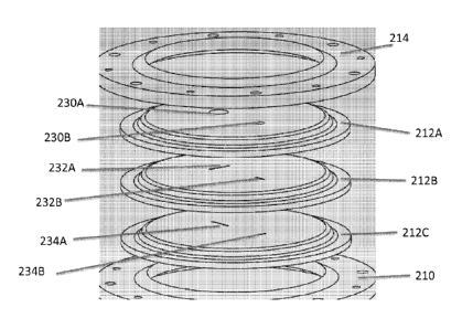
seals 212A, 212B, 2120
-20-
CA 02860754 2014-07-07
WO 2013/106569 PCT/US2013/021027
positioned between the base ring 210 and the first top ring 214. In some
embodiments, the device 200
has only the first set of seals (212A, 212B, 2120) and the first top ring 214.
In alternative embodiments
such as the implementation as shown, the device 200 also has a second set of
three seals 216A, 216B,
2160 positioned between the first top ring 214 and a second top ring 218. In
this implementation, the first
and second top rings 214, 126 are coupled to the base ring 210, thereby
maintaining the first set of seals
212A, 212B, 2120 and second set of seals 216A, 216B, 2160 in place such that
each of the sets of seals
212, 216 and the two top rings 214, 218 maintain a fluidic seal. According to
one embodiment, a set of
screws or bolts are positioned through the holes 210A, 214A, 218A defined in
the outer circumference of
each of the base ring 210, the first top ring 214, and the second top ring
218, respectively, and fastened
to fix the rings 210, 214, 218 in place. Alternatively, any known device or
mechanism for holding or fixing
the rings 210, 214, 218 (and thus the seals 212, 214) in place can be used.
[0240] According to one embodiment, the fluidic seal created by the set of
seals (212A, 212B,
2120, for example) is created by providing separate rubber seals having
different types of openings
defined in each such seal. For example, as best shown in FIG. 20, in this
implementation, the seals
212A, 212B, 2120 each have two different openings formed through them that are
different from the
corresponding openings in the other seals. Seal 212A has two substantially
circular holes 230A, 230B
formed through the seal 212A. The hole 230A is larger, is positioned more
centrally on the seal 212A,
and is intended to receive a surgical device or system such as a robotic
surgical device. The hole 230B
is smaller, is positioned closer to an edge of the seal 212A, and is intended
to receive a peripheral device
or component such as a trocar, a camera, or some other accessory tool. These
holes 230A, 230B are
intended to provide a fluidic seal around the perimeter of any object(s)
passed through them.
[0241] In contrast, seal 212B has two slits 232A, 232B formed through the
seal 212B. The slit
232A is larger and is positioned in a location that corresponds to hole 230A,
while slit 232B is smaller and
is positioned in a location that corresponds to hole 230B. Similarly, seal
2120 has a larger slit 234A
positioned in a location corresponding to hole 230A and slit 232A and further
has a smaller slit 234B
positioned in a location corresponding to hole 230B and slit 232B. In
addition, the slits 234A, 234B in
seal 2120 are positioned at a 90 degree angle with respect to the slits 232A,
232B in seal 212B.
According to one implementation, the combination of the slits 232A, 232B in
seal 212B with the slits
234A, 234B in seal 2120 results in a stronger fluid seal that can withstand
the increased pressure of the
insufflated cavity 208 of the patient without the slits opening and allowing
that increased pressure to be
lost.
[0242] By incorporating two sets of seals 212, 216 as shown in FIGS. 19A,
19B, the overall
fluidic seal created by the device 200, even when surgical devices are
inserted through the device 200, is
further strengthened. More specifically, as best shown in FIG. 19B, the first
top ring 214 defines a hole
214B at its center. When the first top ring 214 is positioned between the
first set of seals 212 and the
second set of seals 216, the hole 214B in the first top ring 214 creates a
cavity between the two sets of
seals 212, 214. As such, according to one embodiment, any loss of the fluidic
seal in one set of the seals
-21-
CA 02860754 2014-07-07
WO 2013/106569 PCT/US2013/021027
(either 212 or 214) will not cause a loss of the overall fluidic seal or leak
pressure directly from the
patient's cavity 208 into the ambient air outside the patient. Hence, the
cavity created by the first top ring
214 can minimize the overall pressure loss from any such leak.
[0243] In accordance with one implementation, each of the seals 212A,
212B, 212C, 216A,
216B, 216C is a relatively thin sheet of rubber. Alternatively, each of the
seals can be made of any
known flexible material that can serve as a seal in a medical device. In one
exemplary embodiment, each
of the seals is about 0.125 inches thick. Alternatively, the thickness of each
of the seals can vary
between about 0.0625 and about .25 inches thick. In a further alternative,
each set of three seals 212,
216 can be replaced with a single seal having a thickness ranging from about
0.1875 inches to about 0.75
inches. This thickness in a single seal, according to some embodiments, can
provide substantially the
same type of fluidic seal strength as the set of three thin seals.
[0244] As discussed above, according to certain embodiments, the device
200 has only one set
of seals 212A, 212B, 212C and only the first top ring 214. While such
embodiments do not have the
cavity created by the first top ring 214 as described above, the device 200
with a single set of seals 212
can still provide a sufficient fluidic seal. For example, such a device 200
would provide a sufficient fluidic
seal for insertion of any robotic device having sufficiently smooth external
features and surfaces. In
addition, a device 200 with a single set of seals 212 can reduce the size of
the overall device 200 and can
potentially reduce any trauma to the surgical device inserted through the
device 200 as a result of only
having to pass through a single set of seals 212.
[0245] FIG. 21, according to one implementation, depicts a top view of the
device 200. More
specifically, FIG. 21 shows the second top ring 218 positioned over the seal
216A. The holes 236A, 236B
in the seal 216A are visible as well.
[0246] In use, the rubber seal access/insertion device 200 can be
positioned for use in the
following manner. First, as described above with respect to other embodiments,
according to one
implementation, the sealable sleeve device 204 is first positioned in the
incision 206. It is understood that
the sleeve device 204 can be inserted using steps similar to those described
above. Alternatively, any
known insertion steps can be used to insert the device 204 into the incision
such that the upper ring 240
is positioned outside of the incision 206 and the lower ring 242 is positioned
inside the patient's cavity,
with the sleeve 244 disposed through the incision 206 itself, as best shown in
FIG. 18.
[0247] Once the sleeve device 204 is positioned in the incision 206, the
base ring 210 (and thus
the entire device 200) is coupled to the sleeve device 204 as best shown in
FIGS. 18 and 22. More
specifically, according to one implementation, the base ring 210 is positioned
over the upper ring 240 of
the sleeve device 204 such that the upper ring 240 is positioned into the
circular indentation or notch 250
defined in the bottom of the base ring 210. The configuration of the notch 250
corresponds to the
configuration of the upper ring 240 and thus is configured to receive the
upper ring 240 such that the ring
240 fits snugly into the notch 250.
-22-
CA 02860754 2014-07-07
WO 2013/106569 PCT/US2013/021027
[0248] Once the upper ring 240 is positioned in the notch 250, the coupling
components 220 are
coupled to the attachment components 252 on the base ring 210 and thereby
firmly couple the base ring
210 to the sleeve device 204. The coupling components 220 in this embodiment
are components having
a vertical piece 220A and a horizontal piece 220B as best shown in FIGS. 19A
or 22. The vertical pieces
220A are coupled to the attachment components 252 using a screw or bolt or
similar mechanism. As
best shown in FIG. 18, when the vertical pieces 220A are coupled to the
attachment components 252, the
horizontal pieces 220B are positioned under the base ring 210 such that they
are also positioned under
the upper ring 240 disposed in the notch 250. As such, the coupling components
220 operate to retain or
lock the upper ring 240 in the notch 250. As a result, the retention of the
upper ring 240 into the notch
250 can provide a fluidic seal between the base ring 210 and sleeve device
204. Alternatively, any
appropriate known interface between the base ring 210 and sleeve device 204
that provides a fluidic seal
can be used.
[0249] Once the device 200 and sleeve device 204 are coupled as best shown
in FIGS. 18 and
23, a fluidic seal has been established between the patient's cavity 208 and
the external air outside of the
patient. At this point, the patient's cavity can be insufflated to the desired
amount of air pressure.
Subsequently, one or more surgical devices can be inserted through the seals
212, 216 at the appropriate
holes/slits and into the patient's insufflated cavity 208. In one embodiment,
each arm of a robotic surgical
device can be separately and consecutively inserted through the larger hole
(and larger slits) of the seals
and into the cavity 208. Alternatively, any known devices can be inserted into
the cavity 208 so long as
they fit through the holes and slits as contemplated herein.
[0250] Another embodiment of an access/insertion device relates to another
external
pressurized system or apparatus similar to the system or apparatus depicted in
FIGS. 1-11 and described
in detail above. Like the device in FIGS. 1-11, the instant device is coupled
to a port that is positioned
over and/or in an incision in the skin of the patient, thereby providing
access to a cavity of the patient.
However, in the instant implementations as shown in FIGS. 24A-38 and discussed
below, the external
pressurized system/apparatus has a external body having one or more access
ports for the insertion of
not only surgical devices, but also additional equipment and/or the hands of
one or more users or medical
professionals, providing access to the interior of the pressurized
system/apparatus without loss of the
higher pressure inside the system/apparatus.
[0251] For example, one implementation of such an external pressurized
system or apparatus
300 is depicted in FIGS. 24A-24F. As best shown in FIGS. 240 (top view) and
24D (perspective view),
the device 300 has an external body 302 having a main tube (also referred to
as the "canister") 304, a left
hand tube 306 with a left hand access port 308, a right hand tube 310 with a
right hand access port 312,
and a side access tube 314 with a side access port 316. In addition, the main
tube 304 has a device port
318 coupled to a top portion of the tube 304.
[0252] The bottom portion of the main tube 304 is coupleable to an incision
port 320, as best
shown in FIGS. 24A and 24B. In turn, as best shown in FIG. 24F, the incision
port 320 is coupleable to a
-23-
CA 02860754 2014-07-07
WO 2013/106569 PCT/US2013/021027
standard sealable sleeve device 322, which can be positioned in the incision
324 made in the patient's
skin 326 to access a target cavity 328 of the patient. The incision port 320
and its coupling to both the
main tube 304 and the sealable sleeve device 322 are described in detail
below.
[0253] In the depicted implementation, the left and right hand access
ports 308, 312 can be
configured to allow a user or medical professional to insert her or his hands
through the ports 308, 312
and into the interior of the body 302. Further, the side access tube 314 with
access port 316 can be used
for storage of equipment and/or for assistance of another user by inserting
her or his hand through the
port 316. In addition, the device access port 318 can be configured such that
various medical
devices/systems can be inserted into the body 302 through the port 318.
Alternatively, any of the access
ports 308, 312, 316, 318 can be configured to allow for insertion of hands
and/or equipment/devices.
Further, in various alternative embodiments, it is understood that the body
302 could have a main tube
304 with one, two, or more than three additional tubes with access ports for
various uses, including any of
those discussed above. It is also understood that various embodiments
contemplated herein include
tubes and/or ports that are different sizes or shapes than those depicted. For
example, in some
implementations, the tubes and/or ports could be square or oval in shape.
[0254] In one implementation, the external body 302 (the main tube 304 and
the access tubes
306, 310, 314) is made of a hard plastic, such as, for example, poly(methyl
methacrylate) ("PMMA").
Alternatively, the body 302 can be made of any known rigid material that can
be used in medical devices.
It is understood that certain embodiments of the body 302 are transparent,
such as those depicted in the
figures provided. The transparent body 302 allows for the user to see the
interior of the tubes 304, 306,
310, 314 including any equipment or devices being inserted during the
procedure. Alternatively, the body
302 is not transparent and the equipment/devices can be inserted without being
able to view them in the
device 300.
[0255] According to one implementation, the sealable sleeve device 322, as
best shown in
FIGS. 24F, 29A, 29B, and 30, can be a standard, commercially available device
as described in the
various embodiments above. The device 322 has an upper ring 420 and a lower
ring 422 that are
coupled together by a flexible sleeve 424. According to one embodiment, the
device 322 is substantially
similar to the sealable sleeve device described above with respect to FIGS.
2A, 2B, 6A, 6B, 6C, and 6D.
[0256] According to one embodiment, the access ports 308, 312, 316, 318
are standard
commercially-available ports that allow various objects, including devices or
hands, to be inserted through
them and into a surgical space. One example of an access port 340 in use is
depicted in FIG. 25. As
shown in that figure, the port 340 allows for insertion of a hand through the
port 340. Another exemplary
access port 342 is depicted in FIG. 26. This port 342 is the GelSeal port
that is commercially available
from Applied Medical in Rancho Santa Margarita, CA. In this embodiment, the
port 342 has a body 344,
a rigid support ring 346, and a moveable clamp lever 348 that can be used to
tighten the port 342 and
thus secure the port 342 to any ringed object to which it is attached. More
specifically, the clamp lever
348 is depicted in three different positions. In position A, the lever 348 is
in the open position A and the
-24-
CA 02860754 2014-07-07
WO 2013/106569 PCT/US2013/021027
port 342 thus has its widest circumference. In position B, the lever 348 is
midway between the open
position A and the closed position C and the port 342 has a circumference that
is less than when it is in
the open position A. Finally, in position C, the lever 348 is positioned
against the port 342 in the closed
position C and the port 342 has its smallest circumference. In use, the lever
348 is typically in position A
when the port 342 is positioned and then the lever 348 is moved to position C
to clamp the port 342 in
place. In one embodiment, the body 344 is made of the soft, gel-like material
in the product as provided
by Applied Medical. Alternatively, the body 344 can be made of any material
that allows for objects
and/or hands to be inserted through the material such that the fluidic seal is
maintained so that the higher
pressure of the surgical cavity is not lost when an object is inserted through
the material.
[0257] In accordance with one implementation as shown in FIG. 27A, the
access ports 308, 312,
316, 318 are coupled to the tubes 304, 306, 310, 314 via a port adaptor ring
350. The port adaptor ring
350 has a first ring portion 352 that is sized to mate with any one of the
tubes 304, 306, 310, 314 of the
body 302. (In this particular depiction, the left hand access tube 306 is used
as an example.) The ring
350 also has a second ring portion 354 that is sized to mate with a port ¨ in
this case the left hand access
port 308.
[0258] According to one embodiment, the first ring portion 352 is coupled
to the tube 306 by
positioning the first ring portion 352 over the end of the tube 306 and
holding the first ring portion 352 in
place using thumb screws 356 that are inserted through threaded holes 358A in
the first ring portion 352
and into threaded holes 358B in the tube 306. Alternatively, any attachment
devices or mechanisms,
such as bolts, clamps, or the like, can be used to attach the first ring
portion 352 to the tube 306 (and, by
extension, to any of the tubes 304, 306, 310, 314). In one embodiment, a
gasket (not shown), such as a
foam or rubber gasket, is positioned between the tube 306 and the first ring
port 352 to ensure that a
fluidic seal is established between the two components.
[0259] The access port 308, in accordance with one implementation, is
coupled to the second
ring portion 354 in a fashion similar to that described above. That is, the
clamp lever 308A on the port
308 is placed in position A, and the port 308 is positioned over the second
ring portion 354. Then the
lever 308A is moved into the closed position ¨ position C ¨ such that the port
308 is clamped onto the
second ring portion 354. Alternatively, any known mechanism or method for
coupling a port similar to
port 308 to a device component can be used.
[0260] According to one embodiment as shown in FIG. 27B, the device access
port 318 can
have one or more additional structures to allow a user to easily stabilize or
position a surgical device
within the body 302 of the device 300 prior to or during use. More
specifically, the device access port 318
in certain implementations has one or more device attachment components 357
(also referred to as
"device clips") positioned along the inner lumen of the port 318. The device
clip 357 is configured to
retain a device such as a positioning rod 359 within the clip 357, thereby
providing a way to couple a
portion of the surgical device being used for the intended procedure to the
interior of the body 302. In
one embodiment, the attachment component 357 is an actual clip as shown in
FIG. 27B. Alternatively,
-25-
CA 02860754 2014-07-07
WO 2013/106569 PCT/US2013/021027
the component 357 can be a notch or other type of specifically configured
indentation 357 defined in the
inner lumen of the port 318 that is configured to receive a medical device
such as a positioning rod 359 or
the like. In a further alternative, the attachment component 357 can be any
mechanical or structural
mechanism or component that allows for coupling to a medical device. In
further embodiments, such
attachment components 357 can be positioned elsewhere in the body 302, such
as, for example, on an
interior port of another access port or elsewhere on an interior portion of
one of the tubes.
[0261] In various alternative embodiments, other types of access ports can
be used instead of
the ports described above and depicted in FIGS. 24-27B. For example, in one
specific alternative
implementation, one or more glove ports can be used such as the glove port 360
depicted in FIGS. 28A
and 28B. The glove port 360 has a glove component 362 coupled to a glove port
ring 364. In various
embodiments, the glove port 360 could be coupled at the glove port ring 362 to
one or more of the tubes
304, 306, 310, 314 on the body 302. In one embodiment, the glove port ring 362
is coupled to the tube
via a clamp lever similar to the clamp lever described with respect to FIG.
26. Alternatively, any known
coupling mechanism can be used. Unlike the access ports 308, 312, 316, 318,
the glove port 360 does
not require that a fluidic seal be established around the surgeon's arm or
whatever object is inserted
through it. As such, the glove port 360 can help to ensure that the pressure
differential between the
patient's cavity and the ambient air outside the patient will be maintained.
In one embodiment, the glove
port 360 has a pressure relief valve (not shown) that can be used to adjust
the volume, thereby
accounting for the volume change caused when a user inserts her or his hand
into the body 302 using the
glove component 362. FIG. 28B depicts the glove port 360 in use.
[0262] As mentioned above, the incision port 320 is configured to be
coupleable to both the
main tube 304 and to the sealable sleeve device 322, as shown in FIGS. 24F and
29. As best shown in
FIGS. 24A and 30, the incision port 320 has a base ring 370. The upper portion
of the base ring 370 can
be coupled to an internal coupling component 372, which can couple to the port
seal 450 as described in
further detail below. Further, the lower portion of the base ring 370 can be
coupled to external coupling
components 374 (also referred to in certain embodiments as "sleeve clamps"),
which couple the ring 370
to the sealable sleeve device 322. In addition, the base ring 370 can also be
coupled to coupling
components 376 (also referred to in certain embodiments as "tube brackets"),
which couple the ring 370
to the main tube 304 of the device 300.
[0263] FIGS. 31A and 31B depict the base ring 370, according to one
implementation. The ring
370 has a curved indentation or notch 378 configured to receive and couple
with the bottom portion of the
main tube 304. In addition, the ring 370 has three bracket receiving
components 380 configured to
receive the tube brackets 376. Further, as best shown in FIGS. 24F, 30, and
35, the bottom portion of the
ring 370 defines a circular indentation or lumen 381 that is configured to be
positioned over and receive
the upper ring 420 of the sleeve device 322. The ring 370 also has multiple
holes 384 defined in an
interior ring 382. The multiple holes 384 correlate to holes 436 in the base
plate 430 of the internal
coupling component 372, as described in detail below. Each of the bracket
receiving components 380
-26-
CA 02860754 2014-07-07
WO 2013/106569 PCT/US2013/021027
have a projection 386 and horizontal portion 388 on which the tube bracket 376
is positioned and a hole
390 that corresponds to the hole 394 in the tube bracket 376 as described in
detail below. In one
embodiment, a gasket (not shown), such as a silicon, foam or rubber gasket, is
provided between the
notch 378 and the bottom portion of the main tube 304 to strengthen the
fluidic seal between the two
components.
[0264] FIG. 32 depicts a tube bracket 376, according to one embodiment.
The tube bracket 376
has a base portion 392 having a hole 394 defined therein that corresponds to
the hole 390 in the bracket
receiving component 380 on the base ring 370. The bracket 376 also has a tube
contacting portion 396
having two holes 398 defined therein that correspond to the holes 404 in the
bottom portion of the main
tube 302, as described below.
[0265] According to one embodiment, the tube bracket 376 is used to couple
the main tube 302
to the base ring 370, as shown in FIG. 33. More specifically, the tube bracket
376 is positioned on the
bracket receiving components 380, with the base portion 392 of the bracket 376
positioned on the
horizontal portion 388 and the tube contacting portion 396 positioned on the
projection 386. In that
position, the bracket 376 is coupled to the base ring 370 by inserting a
threaded screw 400 through hole
394 in the bracket 376 and into hole 390 in the ring 370. Further, the bracket
376 is coupled to the main
tube 302 by inserting two threaded screws 402 through holes 398 in the bracket
376 and into holes 404 in
the tube 302. Thus, the tube 302 is attached in position against the incision
port 320 and specifically the
base ring 370 using the brackets 376. In the embodiments depicted in FIGS. 24A-
24F, there are three
tube brackets 376 ¨ spaced about 120 degrees from each other around the
circumference of the port 320
¨ that are used to couple the tube 302 to the port 320. Alternatively, two
brackets or more than three
brackets could be used in different positions around the port 320. In a
further alternative, any known type
of coupling mechanism could be used to keep the tube 302 coupled to the port
320.
[0266] As discussed above, the incision port 320 is coupled to the
sealable sleeve device 322
using the sleeve clamps 374. FIG. 34 depicts one embodiment of a sleeve clamp
374. The clamp 374
has a hole 406 defined in a top portion of the clamp 374, projections 408
configured to fit into the notches
410 defined under the bracket receiving components 380 on the base ring 370
(as best shown in FIG.
31B), and a projection 412 configured to help retain the upper ring 420 of the
sealable sleeve device 322
in position on the clamp 374, as discussed below. The hole 406 corresponds to
the hole 394 in the
bracket 376 and the hole 390 in the base ring 370 such that when the sleeve
clamp 374 is positioned
under the bracket receiving component 380 of the base ring 370 and the
threaded screw is inserted
through hole 394 and hole 390, it is also threaded into hole 406 such that the
sleeve clamp 374 is
coupled to the base ring 370.
[0267] As best shown in FIGS. 30 and 35, when the port 320 is positioned
over the sleeve
device 322 such that the upper ring 420 is positioned within the lumen 381 on
the bottom portion of the
base ring 370, the sleeve clamp 374 can be coupled to the base ring 370 as
described and the upper ring
420 of the sealable sleeve device 322 is contacted by the clamp 374 and
thereby retained in its desired
-27-
CA 02860754 2014-07-07
WO 2013/106569 PCT/US2013/021027
position as shown. Further, the notch 412 in the clamp 374 can further help to
retain the upper ring 420.
In one embodiment, a gasket (not shown), such as a foam, rubber, or silicone
gasket, is placed between
the upper ring 420 and the underside of the base ring 370, thereby providing a
stronger fluidic seal
between the two components.
[0268] As discussed above, according to one embodiment, the upper portion
of the base ring
370 can be coupled to an internal coupling component 372, as best shown in
FIGS. 24A, 30, and 36. The
internal coupling component 372 has a base plate 430 and a male component 432
projecting from the
base plate 430. The base plate 430 has multiple holes 436 defined in the plate
430. These holes 436
correspond to the holes 384 defined in the interior ring 382 of the base ring
370 such that screws 438 (or
bolts or any other known coupling mechanisms) can be used to couple the base
plate 430 to the interior
ring 382 of the base ring 370 as shown. In addition, the interior portion of
the male component 432 has
two device attachment components 440 (also referred to herein as "device
clips") (only one such clip 440
is shown in FIG. 36). Each device clip 440 is configured to be able to allow a
user to couple a positioning
rod (as described elsewhere herein) or some other device component to the clip
440 before or during a
surgical procedure, thereby stabilizing or maintaining the position of the
device.
[0269] As best shown in FIG. 36, the male component 432 has three notches
434 formed or
engineered on its outer circumference (one of which is fully depicted in FIG.
36). The notches 434 have a
vertical portion 434A and a horizontal portion 434B in communication with the
vertical portion 434A. Each
notch 434 is configured to received a corresponding projection formed on an
internal circumference of
any device intended to couple with the male component 432. As such, to couple
the device to the male
component 432, the device is positioned over the male component 432 with the
projections on the device
positioned over the corresponding notches 434 on the male component 432. The
device is then
positioned onto the male component 432 such that each projection moves along
the vertical portion 434A
of the notch 434 until it reaches the horizontal portion 434B. At that point,
the device can be rotated and
thereby move each projection circumferentially along the horizontal portion
434B of the notch 434,
thereby coupling the device to the male component 432 of the internal coupling
component 372.
[0270] In one implementation, as best shown in FIGS. 37A, 37B, and 37C, one
of the
components that can be coupled to the internal coupling component 372 is a
port seal 450. The port seal
450 has a seal clamp 452 coupled to a base seal ring 454. A seal component 456
is positioned between
the clamp 452 and the ring 454 so that the coupling of the clamp 452 to the
ring 454 fixes the seal
component 456 in place in the port seal 450. In one embodiment as shown, the
seal clamp 452 has
multiple holes 458 defined in the clamp 452 that correspond to holes (not
shown) in the base seal ring
454 such that threaded screws 460 (or bolts, or the like) can be inserted
through the holes 458 and into
the holes in the ring 454 to couple the two components together.
Alternatively, any other known
attachment mechanisms can be used. In one embodiment, a gasket (not shown),
such as a foam,
silicone, or rubber gasket, can be positioned between the male component 432
and the base seal ring
454 to strengthen the fluidic seal between the two components.
-28-
CA 02860754 2014-07-07
WO 2013/106569 PCT/US2013/021027
[0271] The seal clamp 452, in one embodiment, has multiple projections 464
extending from the
top surface of the clamp 424. These projections 464 can be easily grasped by a
user to place the port
seal 450 on the male component 432 or remove it therefrom. Further, as best
shown in FIG. 370, the
underside of the base seal ring 454 has three projections 462 disposed on the
inner circumference of the
ring 454. The three projections 462 correspond to the three notches 434
defined in the outer
circumference of the male component 432 such that the base seal ring 454 can
be coupled to the male
component 432 as described above.
[0272] According to one implementation, the seal component 456 (also
referred to herein as a
"flexible seal component" or an "elastic seal component") is a circular sheet
of flexible or elastic material
that is configured to allow a device or other equipment to be inserted through
the seal component 456 (or
to allow the seal component 456 to be positioned over such equipment, like a
positioning rod, as
described in further detail below). In one embodiment, the seal component 456
is a circular rubber sheet
having a small hole (not shown) in the sheet through which equipment can be
inserted. Alternatively, the
seal component 456 can be any known material configured to maintain a fluidic
seal when a device or
equipment is inserted through the seal component 456.
[0273] In accordance with one embodiment, a different type of seal
component can also be
incorporated into the device 300. As shown in FIGS. 38A, 38B, and 380, a flap
seal component 470 is
provided. The flap seal component 470 has two flaps ¨ a first flap 472 and a
second flap 474 ¨ that
contact each other at a midpoint in the component 470. Each of the flaps 472,
474 has ridges or teeth
472A, 474A on the surfaces that are in contact such that the ridges 472A on
flap 472 correspond to the
ridges 474A on flap 474 and thus interface or couple with each other. In one
implementation as shown,
the flap seal component 470 is positioned between the base ring 370 and the
internal coupling
component 372. According to one implementation, the configuration of the flaps
472, 474 extended
downward toward the patient's cavity and the coupled ridges 472A, 474A can
provide structural strength
to prevent a mechanical failure (also referred to as a "blowout") in which the
flaps 472, 474 are forced
outward by the higher air pressure until the flaps 472, 474 are extending
outward away from the patient's
cavity and the fluidic seal is lost.
[0274] In one embodiment as shown in FIG. 38A, the flap seal component 470
can be
incorporated into the incision port 320 and used when the port seal 450 is not
coupled to the port 320.
Alternatively, as shown in FIG. 38B, the flap seal component 470 can be
incorporated into the incision
port 320 and used when the port seal 450 is coupled to the port 320.
[0275] In use, the various embodiments disclosed or contemplated herein
relating to access and
insertion systems, devices, and methods that relate specifically to an
external device having one or more
ports for the insertion of not only medical devices, but also related
equipment and/or the hands of one or
more medical professionals to access the interior of the device during medical
procedures while being
able to maintain a higher air pressure within the device that is substantially
the same as the insufflated
cavity of the patient. According to one implementation, the high pressure is
around 18 mmHg above
-29-
CA 02860754 2014-07-07
WO 2013/106569 PCT/US2013/021027
atmospheric pressure, which is around the amount of pressure that is used to
insufflate a patient's
abdominal cavity during a laparoscopic procedure. Alternatively, any known
higher pressure amount that
is used during medical procedures can be used.
[0276] The method of using the device 300, according to one embodiment,
includes at least
some of the following steps. First, as described above with respect to other
embodiments, according to
one implementation, the sealable sleeve device 322 is first positioned in the
incision 324 (see FIGS. 24F,
29A, 29B, and 30). It is understood that the sleeve device 322 can be inserted
using steps similar to
those described above. Alternatively, any known insertion steps can be used to
insert the device 322 into
the incision 324 such that the upper ring 420 is positioned outside of the
incision 324 and the lower ring
422 is positioned inside the patient's cavity, with the sleeve 424 disposed
through the incision 324 itself,
as best shown in FIG. 30.
[0277] Next, the incision port 320 and the device 300 are coupled to the
sealable sleeve device
322. As best shown in FIGS. 30, 33, and 35, the base ring 370 of the incision
port 320 is positioned over
the upper ring 420 of the sleeve device 322 such that the upper ring 420 is
positioned in the lumen 381
on the bottom portion of the base ring 370. In addition, the bottom portion of
the main tube 304 of the
device body 302 can be positioned in the curved notch 378 on the base ring
370. At this point, both the
device 300 and the sleeve device are positioned as desired with respect to the
incision port 320 and must
be coupled to the port 320. To do so, the tube brackets 376 and the sleeve
clamps 374 are positioned on
the base ring 370 as described above and fixed in place using the threaded
screws 400. Then the
threaded screws 402 are placed as well. As such, the incision port 320 is
coupled to both the device 300
and the sleeve device 322 and a fluidic seal is created between the interior
of the body 302 and the
exterior.
[0278] According to one embodiment, at least one medical device or piece of
equipment that will
be used during the procedure can be placed in the body 302 prior to coupling
the body 302 to the incision
port 320. For example, in one embodiment, the device 480 disposed within the
body 302 as best shown
in FIGS. 24A, 24B, 24D, and 24F can be positioned within the body 302 and, in
some implementations,
secured to a device clip 357 (as shown in FIG. 27B). More specifically, in the
particular embodiment
depicted in FIGS. 24A, 24B, 24D, and 24F, the device 480 is made up of two
arms 482A, 482B that are
positioned within the body 302. Alternatively, any medical device that will be
used for the surgical
procedure could be positioned within the body 302 in the same or a similar
fashion.
[0279] It is understood, in accordance with one implementation, that the
port seal 450 is not
coupled to the internal coupling component 372 (which is coupled to the
incision port 320) at this point
during the placement of the device 300. As such, according to one embodiment,
the port seal 450 is
stored in the side access tube 314 while the body 302 is being coupled to the
port 320, as best shown in
FIGS. 24B, 240, and 24D. Alternatively, the port seal 450 can be uncoupled
from the internal coupling
component 372 and placed in the side access tube 314 prior to positioning the
medical device inside the
body 302 and coupling the body 302 to the incision port 320.
-30-
CA 02860754 2014-07-07
WO 2013/106569 PCT/US2013/021027
[0280] Once the device 300 is coupled to the incision port 320 and the
incision port 320 is
coupled to the sealable sleeve device 322, the fluidic seal within the device
300 has been established,
and the patient's cavity can be insufflated. This insufflation will result in
an increase in air pressure within
the patient's cavity and within the device 300 (because neither the port seal
450 nor the flap seal 470 is nt
coupled to the internal coupling component 372).
[0281] Once insufflation is achieved, the device 480 is positioned through
the incision port 320
and into the patient's cavity. More specifically, the user or medical
professional inserts her or his hands
into the left and right hand access ports 308, 312 and moves the medical
device through the incision port
320 and into position within the cavity. At this point, if the medical device
has a positioning rod 359, that
rod 359 can be coupled to a device clip 440 on the interior of the male
component 432 of the internal
coupling component 372 of the port 320, thereby establishing, maintaining, or
fixing the position of the
medical device within the patient's cavity. Alternatively, the device can be
positioned and maintained in
that position using any type of mechanism or method, including some type of
device or method
independent of the device 300.
[0282] Once the medical device is positioned as desired, the port seal 450
can be positioned in
place over the device (or the positioning rod 359 ¨ or rods ¨ of the device).
That is, the user reaches in
through the hand access ports 308, 312 and removes the seal 450 from the side
access tube 314 and
placed over the device/rod 359 so that the device and/or rod 359 is inserted
through the seal component
456 of the seal 450 and then coupled to the male component 432 of the internal
coupling component 372
as described above.
[0283] Once the port seal 450 is in place, the body 302 can be removed from
the incision port
320. More specifically, the user can remove the threaded screws 402 and then
remove the main tube
304 from the port 320. The fluidic seal between the patient's cavity and the
ambient air outside the
patient's body is maintained by the port seal 450.
[0284] The user/medical professional can then begin performing the medical
procedure.
[0285] An alternative external pressurized device embodiment is depicted in
FIGS. 39A and
39B. In this embodiment, the device 500 is a single tube 502 having a single
access port 504 disposed at
the top of the tube 502. The access port 504 serves to establish a fluidic
seal when a medical device or
a surgeon's hand is inserted through the port 504. The tube 502 also has two
camera ports 506
extending from a bottom portion of the tube 502. According to one
implementation, the tube 502 is
configured to couple to an incision port, including any incision port
disclosed elsewhere herein or any
known incision port.
[0286] A further embodiment depicted in FIG. 40 is another alternative
external pressurized
device 510. The device 510 has a tube 514 that is coupleable to an incision
port 516 and has two slots
511, 513 formed on opposite sides of the tube 514. These slots 511, 513
provide fluid communication
between the interior of the tube 514 and the exterior of the tube 514. In one
embodiment, the rod slots
512 are each configured to receive a positioning rod. The device 510 further
has two slot seals 512, with
-31-
CA 02860754 2014-07-07
WO 2013/106569 PCT/US2013/021027
one seal 512 positioned in each of the slots 511, 513. These slot seals 512
are configured to maintain a
fluidic seal at each of the slots 511, 513 such that an object can be
positioned through either or both slots
511, 513 and the fluidic seal is not lost. The tube 514 also has two sets of
device attachment
components 518A, 518B (also referred to as "rod clips"). Each set of rod clips
518A, 518B has two
device clips ¨ a horizontal clip 515A and an angled clip 515B.
[0287] In use, a device can be positioned within the tube 514 such that a
positioning rod coupled
to the device extends out of the tube 514 through one of the slots 511, 513.
The device can be fixed in
position in the tube 514 by coupling the positioning rod to the horizontal
clip 515A. The patient's cavity
can then be insufflated. When ready, the positioning rod can be moved down the
slot (511 or 513) such
that the device is being moved down the interior of the tube 514 and inserted
through the port 516 and
into the patient's cavity. At this point, the positioning rod is angled upward
and clipped to the angled clip
515B, thereby fixing the positioning of the device inside the patient's
cavity.
[0288] Another implementation relates to a positioning tube 520 as
depicted in FIGS. 41A and
41B. In this embodiment, the positioning tube 520 can also act as a large
positioning rod. The tube 520
has two guide slots 522 defined in or attached to an inner portion of the tube
520. The guide slots 522
are each configured to receive a positioning rod 524. In this implementation,
each device 526 (or device
arm) is coupled to an end of one of the positioning rods 524 and can be
inserted through the tube 520
and into the patient's cavity. Due to the size of the tube 520, the devices
526 must be inserted one at a
time. Alternatively, the tube 520 can be sized so that both devices 526 can be
inserted at the same time.
The tube 520 also has an air lock 528 disposed in the tube 520. The air lock
528 is configured to be
capable of fluidically dividing the tube 520 into two fluidically separate
compartments when the air lock
528 is closed.
[0289] In use, the positioning tube 520 (having a robotic arm 526 disposed
within the tube 520)
can be inserted through any of the various incision ports described elsewhere
herein. When the tube 520
is positioned so that the distal end of the tube 520 is extending into the
patient's cavity, a seal is created
at the top of the top by placing a seal cap (not shown) on the top of the tube
520. Once the inside of the
tube 520 is sealed, the positioning rod 524 can be urged distally and thereby
the arm 526 is urged out of
the tube 520 and into the patient's cavity. If a second arm 526 is going to be
inserted, the air lock 528 is
then closed. That is, the air lock 528 is closed to create a fluidic seal
between the top of the tube 520 and
the bottom of the tube 520. Once the air lock 528 is in place, the seal cap is
removed, and the second
arm 526 can be positioned in the tube 520. At this point, the seal cap can be
replaced, the air lock 528
can be released, and the second arm 526 can be inserted into the patient's
cavity.
[0290] Several additional embodiments relate to various types of incision
ports. For example,
FIG. 42 depicts a stacked incision port 540. The port 540 actually has two
access ports 542, 544 that are
coupled together, with a cavity 546 between the two access ports 542, 544. In
one embodiment, the
access ports 542, 544 are commercially available GelSeal ports. The cavity
546 between the two
access ports 542, 544 strengthens the overall fluidic seal of the port 540. In
other words, the cavity 546
-32-
CA 02860754 2014-07-07
WO 2013/106569 PCT/US2013/021027
reduces the amount of air pressure loss because any air pressure loss is lost
in the cavity and not lost to
the ambient air, thereby reducing the overall loss.
[0291] Another incision port embodiment is depicted in FIG. 43. This
incision port 550 actually
has two seals combined in the port: a rubber seal 552 and a flap seal 554. The
port 550 also has two
camera ports 556 extending out from the port 550. In one embodiment, the
rubber seal 552 has three
different rubber disks (not shown) similar to the different disks depicted in
FIG. 20 and described above.
The disks in this rubber seal 552 can have openings/incisions that differ for
each disk in the same fashion
as the disks shown in FIG. 20. Alternatively, the rubber seal 552 can be
similar to any rubber or flexible
seal described elsewhere herein. The flap seal 554, according to one
embodiment, is similar to the flap
seal depicted in FIGS. 38A-380.
[0292] FIG. 44 depicts another incision port embodiment. More specifically,
this port is a two-
seal port 560 having a first rubber seal 562 and a second rubber seal 564. The
port 560 also has a base
ring 570, a middle ring 568, and a top ring 566. The middle ring 568 creates a
cavity (not shown)
between the two seals 562, 564 that is configured to compartmentalize any lose
of pressure by either of
the seals 562, 564. The presence of the cavity makes this embodiment fairly
similar to the incision port
depicted in FIG. 42. According to one embodiment, each sheet of rubber 562,
564 is about 0.5 inches
thick and has a single slit (not shown) formed through the middle of it.
Alternatively, each sheet 562, 564
can have two openings (not shown) formed through the middle of it.
[0293] FIGS. 45A and 45B depict a further incision port embodiment. This
port is a three-sheet
rubber seal port 580 having a single ring 582 in which three sheets of rubber
(only the top sheet 584 is
shown). In one embodiment, each of the three sheets has an opening in it that
corresponds to the
openings in the other two sheets. In a further embodiment, the openings are
similar to those depicted in
FIG. 20 and described. Alternatively, each sheet can have two corresponding
openings.
[0294] FIGS. 46A and 46B depict a further incision port system embodiment.
This system is an
air barrier port system 590 having an air barrier port 592. This port 592 is
coupled to four air tubes 596A,
596B, 5960, 596D that are coupled to an air intake port 594. In operation,
high pressure air is provided
at the air intake port 594 and is forced through the four tubes 596A-D and
into the port 592. The four tube
connections 598A, 598B, 5980, 598D are positioned on the port 592 such that
the air is forced into a
channel (not shown) that encircles the hole 600 in the port 592. The air is
then forced through a circular
nozzle (not shown) in communication with the channel (not shown) that projects
the air out of the nozzle
and across the hole 600. The air flow projected across the hole 600, according
to one implementation, is
both directed and has a high velocity ¨ both of which have an impact on the
creation of an air barrier. As
a result, an air barrier is created in the hole 600 defined in the port 592.
That is, the high velocity air
movement across or within the hole 600 creates a fluidic seal that is
sufficient to maintain the insufflation
of a patient's cavity.
[0295] FIG. 47 depicts another incision port embodiment ¨ in this case, a
one-sheet rubber seal
port 610 having a single sheet of rubber 612 (other other flexible seal
material) positioned between a
-33-
CA 02860754 2014-07-07
WO 2013/106569 PCT/US2013/021027
base ring 614 and a top ring 616. In one embodiment, the sheet has slit (not
shown) formed in it through
which a surgical device or other equipment can be inserted. Alternatively, the
sheet can have two slits or
other types of openings.
[0296] Another incision port embodiment is shown in FIGS. 48A and 48B. This
port is a dual
brush port 620. This port 620 has a body 622 with a first brush holder 624 and
a second brush holder
626. The first brush 628 is positioned in the first brush holder 624 and the
second brush 630 is positioned
in the second brush holder 626. Further, the body 622 has an opening 632
formed in a bottom portion of
the body 622 that can provide access to the patient's cavity. The brush
bristles of the two brushes 628,
630 are mingled and meshed together at the brush seal 634 such that the mesh
of bristles creates a
fluidic seal that is sufficient to maintain a patient's insufflated cavity.
[0297] FIGS. 49A and 49B depict another brush port ¨ in this case, a triple
brush port 640. This
port 640 has a body 642 with first, second, and third brush holders 644, 646,
648. The first brush 650 is
positioned in the first brush holder 644, the second brush 652 is positioned
in the second brush holder
646, and the third brush 654 is positioned in the third brush holder 648.
Further, the body 642 has an
opening (not shown) formed in a bottom portion of the body 642 that can
provide access to the patient's
cavity. The brush bristles of the three brushes 650, 652, 654 are mingled and
meshed together at the
brush seal 656 such that the mesh of bristles creates a fluidic seal that is
sufficient to maintain a patient's
insufflated cavity.
[0298] According to another implementation, FIGS. 50A, 50B, and 50C depict
an insertion
device 670 that can be used to insert both arms of a robotic surgical device
into a patient's cavity. The
insertion device 670 has an insertion tube 672 through which an insertion rod
674 is slidably disposed. In
addition, the device has a first arm 676A and a second arm 676B, both of which
are coupled to the distal
end of the tube 672. The first arm 676A is coupled to an end bracket 680A
coupled to an end of the first
device body 682A, while the second arm 676B is coupled to an end bracket 680B
coupled to an end of
the second device body 682B. Further, the insertion rod 674 is coupled to two
center brackets (only
bracket 678A is visible in the figures) ¨ one center bracket 678A coupled to a
middle portion of the first
body 682A and a second center bracket (not shown) coupled to a middle portion
of the second body
682B.
[0299] In use, the insertion device 670 can be used to insert a two-armed
surgical device
through a hole (such as an incision, a port, or the like) and into a patient's
cavity prior to operating the
device within the cavity. To accomplish this insertion, the insertion device
670 initially maintains an
insertion configuration (as best shown in FIG. 50A) such that the surgical
device has its smallest
circumferential profile, thereby allowing it to pass through smaller holes.
Once the surgical device has
been inserted into the patient's cavity, the insertion device 670 can be moved
into its deployed
configuration (as best shown in FIG. 500) such that the surgical device is in
its operational configuration.
To accomplish this, a user or surgeon retracts the insertion rod 674 in a
proximal direction (away from the
surgical device. This retraction of the rod 674 urges the two center brackets
(with only center bracket
-34-
CA 02860754 2014-07-07
WO 2013/106569 PCT/US2013/021027
678A of body 682A depicted) in the same proximal direction. Because the two
end brackets 680A, 680B
are retained in substantially the same position by the two arms 676A, 676B,
the result is that the two
device bodies 682A, 682B move through a transition depicted in FIG. 50B and
into the operational
configuration depicted in FIG. 50C. At this point, the user or surgeon can use
the surgical device,
including its two arms 684A, 684B to perform the planned surgery or procedure.
[0300] There are numerous device access and insertion devices and methods
disclosed in the
instant application. All of the various devices and methods that allow for
access to a cavity and insertion
of devices having two arms can also generally be used with respect to devices
that can be uncoupled into
separate arms so as to allow each arm to be inserted individually. In one
embodiment, one advantage of
inserting each arm separately is that inserting a first arm and then a second
arm in a serial manner (and
possibly more arms) can likely be accomplished through a smaller incision when
compared to inserting
both arms simultaneously.
[0301] FIGS. 51A and 51B depict an alternative embodiment of an insertion
device 690 (in the
same spirit as the insertion device depicted in FIGS. 50A-C). While the above
embodiment in FIGS. 50A-
C depict an insertion device for use with a two-armed device, this insertion
device 690 is used with a
single arm 704 or with two arms that are inserted separately. That is, in this
embodiment, a single device
arm 704 is coupled to the insertion device 690. As shown, this device is
positioned through an insertion
tube 692 (which can also be a positioning or support rod). The device has two
moveable rods 694, 696
slidably disposed within the support rod 692. The first moveable rod 694 is
coupled at its distal end to a
first robotic arm 704 and at its proximal end to a control lever 698. The
second moveable rod 696 is
coupled at its distal end to a coupling link 700 (that is coupled to the arm
704) and at is proximal end to a
coupling link 702 (that is coupled to the lever 698).
[0302] In use, the lever 698 can be actuated to cause the first and second
rods 694, 696 to
move in relation to each other. This movement of the rods 694, 696 can be used
to move the arm 704
and thereby position the arm 704 as desired or needed inside the patient's
cavity.
[0303] As shown in FIG. 51B, which is a cross-section of the support rod
692, showing that the
support rod 692 can have two separate lumens 706, 708 or slots, one for each
of the moveable rods 694,
696. In one embodiment, the first moveable rod 694 is positioned in the first
lumen 706 and the second
moveable rod 696 is positioned in the second lumen 708.
[0304] In a further embodiment, it is understood that this support rod 692
could have two halves
¨ a right half 710 and a left half 712¨ that are coupleable at the mating
feature 714. Alternatively, the two
halves can be coupleable by any known mechanical means. The right half 710 is
configured to hold the
first and second rods 694, 696 relating to the first (or right) arm 704, while
the left half is configured to
hold the first and second rods 716, 718 relating to a second (or left) arm
(not shown). This embodiment
can thus be used with two arms, with each arm being inserted and positioned
separately.
[0305] FIG. 52 depicts another embodiment in which two separate arms can be
inserted and
positioned separately by using an overtube 722. In this device 720, the first
moveable rod 724 and
-35-
CA 02860754 2014-07-07
WO 2013/106569 PCT/US2013/021027
second moveable rod 726 are still positioned within a support rod 728.
However, in this embodiment, the
support rod 728 is positioned within an overtube 722. The overtube 722 can be
pass over the top of the
support rod 728 in order to couple the support rod 728 to a second support rod
(not shown) or another
half of a support rod. This embodiment is another way to couple the two
support rods or two halves of a
support rod just as the mating feature 714 accomplishes that task in the prior
embodiment.
[0306] Of course, as shown in FIG. 53, in any embodiment in which the
surgical device or
robotic arm has a motor 740 provided that can be positioned in the positioning
or support rod 744 and is
coupled to the robotic arm 742, there is no need for a separate insertion
device. Instead, the arm 742
can easily be positioned by actuating the motor 740 and transfer the motive
force through the beveled
gears 746 and to the arm 742.
[0307] FIGS. 54A and 54B depict a different type of access/insertion device
in comparison to the
devices described above. Unlike the above devices, which are generally
incision ports or devices
positioned outside the patient's cavity, the internal pressurized bag device
750 shown in these two figures
is initially positioned in the patient's cavity. The device 750 has a port
seal 752, an outer sleeve 754, and
an inner sleeve 756. The outer sleeve 754 is releasably sealed at the distal
end 758. That is, the outer
sleeve 754 has a releasable seal that can be intentionally broken or released
at a desired time during the
procedure, as described below.
[0308] In use, the entire device 750 can be positioned through an incision
port such that the
inner and outer sleeves 754, 756 are positioned inside the patient's cavity
with the port seal 752 coupled
to the incision port (thereby creating a fluidic seal). Once the device 750 is
positioned, the patient's cavity
can be insufflated, and the outer sleeve 754 can be pressurized to a pressure
that is greater than the
pressure of the insufflated cavity, thereby expanding the outer sleeve 754 to
its maximum expansion
(and, in some cases, making the outer sleeve 754 substantially rigid). At this
point, the surgical device
can be inserted through the incision port and into the outer sleeve 754 and
positioned as desired. At this
point, the outer sleeve 754 can be removed by releasing the releasable seal at
the distal end of the
sleeve 754. That is, the releasable seal could be a chemical seal such as an
adhesive that can be
deactivated by applying a different composition to it. Alternatively, the
releasable seal could be a
mechanical release such as a pull cord or something of the like. In a further
alternative, the releasable
seal could be any known mechanism or method for being able to release the
seal. Once the seal is
released, the outer sleeve 754 can be pulled out of the cavity over the inner
sleeve 756 and other
components as best shown in FIG. 54B.
[0309] FIG. 55 depicts another implementation of an external pressurized
system or apparatus
800. The apparatus 800 has a container 802 with a top cap 804 coupled to a top
portion of the container
802. In this embodiment, the container 802 has a port 806 that is coupled to
the container 802 at a base
portion of the container 802. The port 806 is configured to be position able
in an incision in the skin of the
patient, thereby providing access to a cavity of the patient. As shown in FIG.
55, the apparatus 800 is
-36-
CA 02860754 2014-07-07
WO 2013/106569 PCT/US2013/021027
configured to receive a surgical device 808 such that the device 808 can be
inserted into the patient
cavity through the port 806 of the apparatus 800.
[0310] According to one embodiment, in contrast to the canister 12
described above and
depicted in FIGS. 1A-10, the container 802 in this device 800 is made of a
flexible material such as, for
example, polyethylene plastic, latex, nylon, or silicone rubber. As such, the
container 802 can be
manipulated and configurable with respect to the shape of the container 802,
and more specifically can
be compressed longitudinally such that the height of the container 802 can be
reduced during insertion of
a robotic device into a patient's cavity. This will be described in further
detail herein.
[0311] The top cap 804 is depicted in further detail in FIGS. 56A-61B. As
shown in FIGS. 56A
and 56B, the top cap 804 has a cap body 810, a detachable cable harness 812,
an access lumen 814,
support rod lumens 816, threaded lumens 818, and a clamp projection 820. The
cap 804 has a notch
822 defined in the cap 804 that is configured to receive the harness 812. In
addition, the notch 822 has
five channels 824A defined or formed in the notch 822. The channels 824A match
with the channels
824B defined in the detachable harness 812 such that when the harness 812 is
positioned in the notch
822 and thus coupled with the cap body 810, the channels 824A and the channels
824B match up to form
lumens 824 as best shown in FIG. 56B. In one implementation, the lumens 824
can be formed in
different sizes and configured to receive various cables and/or
suction/irrigation tubes the extend from an
external controller through the top cap 804 to the surgical device 808.
[0312] In addition, the cap body 810 has a groove 826 formed or defined
around the outer edge
of the body 810, including the outer edge of the harness 812, such that when
the harness 812 is coupled
to the body 810, an 0-ring can be positioned around the outer edge of the body
810 in the groove 826.
[0313] FIGS. 57A and 57B depict the top cap 804 being coupled to the
canister 802. The
flexible canister 802 is positioned over the peripheral edge of the body 810
as best shown in FIG. 57B
and an elastic ring (also referred to as an "0-ring") 828 is positioned around
the canister 802 at the
groove 826 such that a portion of the canister 802 is positioned between the
body 810 and the ring 828 in
the groove 826 and the ring 828 urges the canister 802 into the groove 826,
thereby creating a fluidic seal
between the canister 802 and the top cap 804. Additionally, in one alternative
embodiment, silicone
sealant can be applied to the groove 826 to enhance the strength of the
fluidic seal. In accordance with
one implementation, the 0-ring 828 can also help to secure the cap body 810
and the harness 812
together. In a further alternative, the 0-ring 828 can be any elastic member
that can be used to maintain
a fluidically sealed coupling of the canister 802 and the top cap 804. In yet
another alternative, any
coupling mechanism can be used.
[0314] FIGS. 58A and 58B depict a portion of the device assembly 808 being
positioned through
the top cap 804. More specifically, the support rods 830 coupled to the device
808 are slidably positioned
through the lumens 816 in the cap body 810. Further, according to one
implementation, a portion of the
device 808 also couples to or mates with the top cap 804. More specifically, a
stabilization protrusion 832
on the device 808 is coupleable with a mating hole 834 defined or formed in an
underside of the body 810
-37-
CA 02860754 2014-07-07
WO 2013/106569 PCT/US2013/021027
as best shown in FIG. 58B. The positioning of the stabilization protrusion 832
in the mating hole 834
creates a pathway from lumen 814 into and through the stabilization protrusion
832, thereby allowing for
passage of additional tools or cameras through the device 800 without losing
pressure.
[0315] As shown in FIGS. 59A, 59B, and 60, the top cap 804 is coupled to
the support rods 830
with two threaded set screws 840. The set screws 840 are threaded through
lumens 818 as best
shown in FIG. 59B. More specifically, the set screws 840 can be screwed into
the threaded lumens 818
until the screws 840 contact the support rods 830. The set screws 840 are
configured to exert pressure
on the support rods 830, thereby creating frictional resistance that helps to
secure the support rods 830
and thus the device 808 to the top cap 804.
[0316] As best shown in FIG. 60, a connection cable 842 that is coupled at
its distal end to the
robotic device 808 is positioned through one of the lumens 824. It is
understood that other cables can be
positioned through the additional lumens 824 as well. In accordance with one
embodiment, the cables
are positioned in the channels 824A or 824B prior to coupling the harness 812
to the body 810.
Alternatively, one or more of the cables can be inserted through one of the
lumens 824 after the body 810
and harness 812 are coupled together.
[0317] FIGS. 61A and 61B show the container 802 coupled to the top cap 804.
[0318] FIGS. 62A and 62B depict the base coupling component (also referred
to as the "base
coupler") 850 that is coupled to a bottom portion of the container 802. The
base coupler 850 has an
upper groove 852, a lower groove 854, and three coupling protrusions (also
referred to as "coupling
notches") 856 that extend from a portion of the coupler 850 between the upper
and lower grooves 852,
854.
[0319] Like with the top cap 804 described above, the container 802 is
coupled to the base
coupler 850 using an 0-ring 858. More specifically, the container 802 is
positioned over the upper portion
of the coupler 850 such that the container 802 is positioned over the upper
groove 852 and adjacent to or
against the three protrusions 856. The 0-ring 858 is positioned over the
container 802 at the upper
groove 852 such that the 0-ring 858 urges a portion of the container 802 into
the groove 852, thereby
creating a fluidic seal between the container 802 and the base coupler 850.
[0320] FIGS. 63A, 63B, 630, 63D, and 63E depict the coupling of the base
coupler 850 to the
access port 806. The access port 806 has a top portion (or "top ring") 860, a
bottom portion (or "bottom
ring") 862, and a middle portion (or "neck") 864. The top ring 860 has three
coupling protrusions (also
referred to as "coupling tabs") 866 that extend from a portion of the top ring
860 and are configured to
mate with the coupling notches 856.
[0321] In one embodiment, the access port 806 is a known standard device
used in hand-
assisted laparoscopic surgery. As is understood in the art, the access port
806 provides a structured
open pathway through the cavity wall, such as the abdominal wall. at the
incision site. In one particular
example, the access port 806 is a commercially available retractor port 806
called the DEXTRUS
Retractor, which is available from Ethicon Endo-Surgery.
-38-
CA 02860754 2014-07-07
WO 2013/106569 PCT/US2013/021027
[0322] As best shown in FIGS. 63A and 63B, the base coupler 850 is coupled
to the access port
806 using an 0-ring 868. More specifically, the 0-ring 868 is positioned in
the lower groove 854 of the
coupler 850 and the top ring 860 is positioned over the lower portion of the
coupler 850 and the 0-ring
868 in the groove 854 such that the 0-ring 868 is compressed between the
coupler 850 and the top ring
860, thereby creating a fluidic seal between those two components.
[0323] As best shown in FIGS. 630 and 63D, as the top ring 860 is
positioned over the lower
portion of the coupler 850 and the 0-ring 868 as described above, the coupling
tabs 866 of the access
port 806 are coupled with the coupling notches 856 of the base coupler 850,
thereby enhancing the
stability of the coupling of the coupler 850 and the access port 806.
[0324] FIG. 63E depicts the entire coupling of the container 802 to the
access port 806 via the
coupler 850 as described above. Further, FIGS. 64A and 64B depict the external
pressurized insertion
device 800 in use, with the device 800 coupled to an access port 806 that is
positioned in an incision in a
patient's skin 870.
[0325] In use, according to one embodiment, the access port 806 and the
external pressurized
device 800 are positioned for a surgical procedure in the following manner. As
an initial matter, according
to one embodiment, the robotic device 808 is positioned inside the insertion
device 800 prior to placing
the port 806 and the device 800 in the appropriate surgical position. That is,
the robotic device 808 is
positioned inside the container 802, the support rods 830 coupled to the
device 808 are secured to the
top cap 804 with the set screws 840, any connection cables coupled to the
device 808 are positioned
through the lumens 824 in the top cap 804, and the flexible container 802 is
coupled and fluidically sealed
to the top cap 804 and the base coupler 850 via the 0-rings 828, 858.
Alternatively, the robotic device
808 is positioned inside the insertion device 800 after positioning the port
806 and device 800.
Regardless, as far as positioning the port 806 and device 800, the port 806 is
positioned first in certain
implementations. That is, in one embodiment, the bottom ring 862 is first
inserted through the incision
previously made in the patient's cavity wall. Once the ring 862 is positioned
through the incision and
inside the cavity, the ring 862 can help constrain the entire port 806 within
the incision by expanding to a
diameter that is greater than the diameter of the incision, as best shown in
FIG. 64A. In one embodiment,
the container 802 and the coupler 850 are coupled to the access port 806 prior
to positioning the port 806
in the incision. Alternatively, the port 806 is first positioned in the
incision, and then the coupler 850 and
the container are coupled to the port 806. Regardless, once the access port
806 and insertion device 800
are positioned, the patient's cavity can then be insufflated. Due to the
fluidic communication between the
cavity and the interior of the container 802 that is created by the access
port 806, the entire interior of the
insertion device 800 will be under the same pressure as the cavity.
[0326] In accordance with one implementation, once the access port 806 and
insertion device
800 are positioned correctly, the process of inserting the robotic device 808
into the patient's insufflated
cavity can take place in the following manner as best shown in FIGS. 65A-69B.
Initially, the robotic
device 808 begins with both arms parallel and vertical to the incision, as
best shown in FIGS. 65A and
-39-
CA 02860754 2014-07-07
WO 2013/106569 PCT/US2013/021027
65B. Then, the robot 808 is lowered through the opening created by the access
port 806 as shown in
FIGS. 66A and 66B. In accordance with one embodiment, as best shown by
comparing FIGS. 65A and
65B with FIGS. 66A and 66B, as the robot 808 is lowered, the flexible
container 802 shrinks in height by
allowing portions of the flexible material of the container 802 to "crumple"
or begin forming folds such that
the top cap 804 moves closer to the access port 806.
[0327] As best shown in FIGS. 67A and 67B, according to one embodiment,
once the "elbow
joints" of the arms of the robotic device 808 have cleared the cavity wall and
access port 806, the
forearms are rotated at the elbow joints until the forearms are positioned at
an angle of or near 45 in
relation to the upper arms (as best shown in FIG. 67A). Concurrently, the
"upper arms" are rotated at the
"shoulder joints" until the upper arms are positioned at an angle of or near
20 , as best shown in FIG.
67B. This rotation of the forearms and upper arms can help to ensure that the
device 808 will fit within
the patient's target cavity so that any contact of the robotic device 808 with
any internal tissues or organs
is minimized or eliminated. Alternatively, the forearms and upper arms can be
rotated to any angle that
minimizes the risk of contact with tissues or organs.
[0328] As best shown in FIGS. 68A and 68B, according to one embodiment, the
device 808 can
be inserted further into the patient's cavity by further positioning the arms
of the device 808 while the
container 802 continues to crumple, thereby resulting in further shrinkage of
the insertion device 800.
More specifically, the upper arms can be rotated further until they are
positioned at an angle of or near
45 , as best shown in FIG. 68B. This process of moving the device 808 further
into the cavity while
positioning the arms to avoid contact with organs or tissues and causing the
container 802 to crumple is
continued until the shoulder joints of the device 808 have cleared the cavity
wall and access port 806.
[0329] At this point, as best shown in FIGS. 69A and 69B, the forearms can
be rotated back to
center and the upper arms can be further rotated up, leaving the arms in an
appropriate starting position
for a surgical procedure. Once in the desired starting position, the device
808 can be locked or otherwise
stabilized in place using a known external clamping mechanism such as, for
example, an Iron Intern ,
which is commercially available from Automated Medical Products Corp.
[0330] FIG. 70 depicts another implementation of an external pressurized
system or apparatus
900. The apparatus 900 has a container 902 with a top cap 904 coupled to a top
portion of the container
902. In this embodiment, the container 902 has a port 906 that is coupled to
the container 902 at a base
portion of the container 902. The port 906 is configured to be position able
in an incision in the skin of the
patient, thereby providing access to a cavity of the patient. As shown in FIG.
70, the apparatus 900 is
configured to receive a surgical device 908 such that the device 908 can be
inserted into the patient
cavity through the port 906 of the apparatus 900.
[0331] According to one embodiment, like the container 802 described above
and depicted in
FIGS. 55-69B, the container 902 in this device 900 is made of a flexible
material such as, for example,
polyethylene plastic, latex, nylon, or silicone rubber.
-40-
CA 02860754 2014-07-07
WO 2013/106569 PCT/US2013/021027
[0332] In this embodiment, the top cap 904, the container 902, and the
robotic device 908 are
substantially similar to the top cap 804 and container 802 depicted and
described above. All the various
features and components described above apply to these top cap 904, container
902, and device 908
embodiments as well.
[0333] FIGS. 71A and 71B depict the base coupling component (also referred
to as the "base
coupler") 920 that is coupled to a bottom portion of the container 902. The
base coupler 920 has a
groove 922 and three coupling protrusions 924 that extend from the coupler
920. In accordance with one
implementation, each of the coupling protrusions 924 has a lumen 926
configured to receive a thumb
screw 928. The container 902 is coupled to the base coupler 920 using an 0-
ring 930. More specifically,
the container 902 is positioned over the upper portion of the coupler 920 such
that the container 902 is
positioned over the groove 922 and adjacent to or against the three
protrusions 924. The 0-ring 930 is
positioned over the container 902 at the groove 922 such that the 0-ring 930
urges a portion of the
container 902 into the groove 922, thereby creating a fluidic seal between the
container 902 and the base
coupler 920.
[0334] In this embodiment, the insertion device 900 has a port attachment
940 that is coupleable
to the base coupler 920 and the access port 906 such that the port attachment
940 is positioned between
the coupler 920 and the port 906. The port attachment 940 has a removable lid
944 that maintains a
fluidic seal when the lid 944 is in place on the port attachment 940, thereby
making it possible to maintain
insufflation of the patient's cavity even when the insertion device 900 is not
yet coupled to the access port
906.
[0335] FIGS. 72A and 72B depict the coupling of the port attachment 940 to
the access port
906. The port attachment 940 has three coupling notches 942 similar to the
coupling notches 856
described and depicted above. In addition, the port attachment 940 has a
removable lid 944 (also
referred to as a "removable seal component," "removable lid seal component,"
or "removable seal
component") that provides a fluidic seal when it is positioned in its closed
position in relation to the port
attachment 940. In the embodiment depicted in FIGS. 72A and 72B, the removable
lid 944 is a slidable
lid 944.
[0336] Like the access port 806 described and depicted above, this access
port 906 (as best
shown in FIG. 72A) has a top ring 946 that has three coupling protrusions
(also referred to as "coupling
tabs") 948 that extend from a portion of the top ring 946 and are configured
to mate with the coupling
notches 942 in the port attachment 940.
[0337] As best shown in FIG. 72A, the port attachment 940 has an 0-ring
950 that can be
positioned between the port attachment 940 and the access port 906 such that
the 0-ring 950 creates a
fluidic seal when the two components are coupled together.
[0338] In use, the port attachment 940 can be coupled to the access port
906 by positioning the
bottom portion of the port attachment 940 in the top portion of the top ring
946 with the 0-ring 950
-41-
CA 02860754 2014-07-07
WO 2013/106569 PCT/US2013/021027
positioned between the two components, with the coupling notches 942 on the
port attachment 940
mating with the coupling protrusions 948 on the top ring 946.
[0339] The port attachment 940 also has another 0-ring 952 that is
configured to be positioned
in the groove 954 formed in the top of the port attachment 940. In one
embodiment, the 0-ring 952 can
be placed in the groove 954 to help create an airtight seal when the port
attachment 940 is coupled to the
base coupler 920.
[0340] Further, the port attachment 940 also has three threaded lumens 956
in the top of the
attachment 940. In one embodiment, these lumens 956 are configured to receive
the thumb screws 928
that are positioned through the lumens 926 in the base coupler 920, thereby
allowing for coupling the
base coupler 920 to the port attachment 940 via the screws 928. Of course, it
is understood that other
coupling mechanisms besides thumb screws can be used. In various alternative
embodiments, any
known attachment or coupling mechanism or component can be used. Some non-
limiting examples
include magnets, quick clamps, quarter turn features, snap-in features, and
the like.
[0341] As best shown in FIGS. 73A and 73B, the slidable lid 944 can be
moved between a
closed position (as shown in FIG. 73B) and an open position (as shown in FIG.
73A). In this embodiment,
the slidable lid 944 is positioned in the port attachment 940 via a lid slot
958 in the port attachment 940.
In the open position, tools or robotic devices can be passed through the port
attachment 940 and the
access port 906. In the closed position, a fluid seal is established between
the lid 944 and the port
attachment 940, which makes it possible to insuff late the patient's cavity
prior to attaching the insertion
assembly 900. It is understood that while this embodiment of the removable lid
944 is a slidable lid 944,
any other known method or device for establishing a fluidic seal could be
used. Non-limiting examples
include a mechanical iris, leaf shutter, or any other known method of
providing a removable fluidic seal.
[0342] FIGS. 74A and 74B depict cross-sectional views of the entire lower
subassembly as
described above, including the base coupler 920, the port attachment 940, and
the access port 906.
More specifically, FIG. 74A shows the port attachment 940 coupled to the
access port 906, with the
slidable lid 944 fully inserted into the port attachment 940 in the closed
position, thereby creating a fluidic
seal. FIG. 74B shows all three components coupled together, including the base
coupler 920, the port
attachment 940, and the access port 906.
[0343] FIGS. 75A, 75B, and 750 depict the external pressurized insertion
device 900 in use,
according to one embodiment. Once the access port 906 is positioned in the
incision as discussed
above, the port attachment 940 can be coupled to the port 906, as best shown
in FIG. 75A. With the
slidable lid 944 in the closed position, a fluidic seal is established between
the port attachment 940 and
the port 906 such that the patient's cavity can be insufflated to the desired
Insufflation pressure. The
insertion device 900 can then be coupled to the port attachment 940 as best
shown in FIG. 75B. Once
the base coupler 920 is coupled to the port attachment 940 such that a fluidic
seal is established between
the two components, the slidable lid 944 can then be moved to its open
position (or fully remove) as best
shown in FIG. 750, thereby providing fluidic communication between the
patient's cavity and the interior
-42-
CA 02860754 2014-07-07
WO 2013/106569 PCT/US2013/021027
of the insertion device 900, resulting in equalized pressure in the device 900
and the cavity. The robotic
device 908 can be inserted via any of the same steps as described previously.
If the device 908
completes the desired surgical procedure and a different robotic device or
other type of tool needs to be
used, the robotic device 908 can be removed from the cavity, the slidable lid
944 can be replaced in the
closed position, and the base coupler 920 can be removed from port attachment
940. This allows
pressure to be maintained within the cavity, even during tool changes.
[0344] FIG. 76 depicts an alternative embodiment having a top cap 960 that
has a pressure
relief valve 962. During the process of lowering either of the robotic devices
808, 908 out of the insertion
device embodiments 800, 900 and into the cavity as described above with
respect to insertion devices
800 and 900, there is a pressure increase in the patient's cavity due to the
decreasing change in volume
of the container 802, 902. The pressure relief valve 962 can be configured to
release pressure if the
internal insufflation pressure increases above a typical value, thereby aiding
the process of inserting the
robotic device 808, 908 such that the attendant will not need to wait for the
pressure to equalize between
the cavity and the insertion device 800, 900.
[0345] Another implementation of a top cap 1000 having a pressure relief
valve 1002 is depicted
in FIGS. 77A and 77B. This cap 1000 also has a dual port seal component 1004
that can be configured
to receive one or more surgical instruments or devices such as a standard
laparoscopic tool.
Alternatively, it is contemplated that a top cap can have only one of the
pressure relief valve 1002 or the
dual seal component 1004.
[0346] As best shown in FIG. 77B, according to one implementation, the
pressure relief valve
1002 has an adjustment component (also referred to as an adjustment "door,"
"wall," or "button," or
"block") 1006 that is operably coupled to (or positioned against) one end of a
tension spring 1008 and has
two holes 1010A, 1010B that are configured to receive retention mechanisms
such as bolts, screws, or
other such standard devices or components configured to hold the adjustment
component 1006 in place.
The other end of the spring 1008 is coupled to a valve ball 1012 that is
positioned against a rim 1016 of
an opening 1014 on the underside of the top cap 1000. The spring 1008 is
configured to urge the ball
1012 toward the opening 1014 such that the ball 1012 (which has a larger outer
diameter than the inner
diameter of the rim 1016) contacts the rim 1016 of the opening 1014 and
thereby establishes a fluidic seal
between the ball 1012 and the rim 1016. In this embodiment, the adjustment
block 1006 is adjusted
using the retention mechanisms to move the block 1006 toward or away from the
ball 1012, thereby
increasing or decreasing, respectively, the force applied by the spring 1008
against the ball 1012 (and
thereby increasing or decreasing, respectively, the strength of the seal
between the ball 1012 and the rim
1016 of the opening 1014). Thus, the adjustment block 1006 can be used to
adjust the strength of the
seal based on the target maximum pressure threshold such that when the target
maximum pressure
threshold is reached (such as while lowering either of the robotic devices
808, 908 out of the insertion
device embodiments 800, 900 as described above), the ball 1012 is urged away
from the rim 1016 and
-43-
CA 02860754 2014-07-07
WO 2013/106569 PCT/US2013/021027
the seal between the rim 1016 and the ball 1012 is broken such that the
pressure is reduced by the gas
escaping through the valve 1002.
[0347] In an alternative embodiment, any known pressure relief valve for
use in medical devices
can be incorporated into the top cap 1000.
[0348] Continuing with FIG. 77B, the dual port seal component 1004 in this
embodiment has two
seal components: an elastic circular seal 1018 defining an opening 1020 and a
flap seal 1022 in fluid
communication with the circular seal 1018. The elastic circular seal 1018 is
configured to form a strong
seal around the smooth surfaces of a standard laparoscopic tool positioned
through the opening 1020. In
one implementation, the flap seal 1022 is a secondary seal that provides a
fluid seal when no tool is
positioned through the dual port seal component 1004. That is, when no tool is
positioned therethrough,
the two flaps 1024A, 1024B are urged into contact with each other by the
pressure in the patient's
insufflated cavity such that the two flaps 1024A, 1024B form a fluidic seal.
[0349] In an alternative embodiment, any known port seal component for use
in establishing a
fluidic seal with a laparoscopic tool positioned therethrough can be used.
[0350] According to various additional implementations, the insertion
devices disclosed or
contemplated herein can have one or more sensors or other types of measurement
mechanisms for
measuring the insertion depth of the surgical device being inserted into the
patient's cavity.
[0351] As an example, FIGS. 78A, 78B, and 78C depict an automatic
insertion device 1030
having a flexible container 1038 and an actuator and sensor package 1032. The
actuator can be any
known actuation device, including, for example, motor and gears, motor and
timing belts, linear screw,
pneumatics, hydraulics, or the like. The sensor could be any known sensing
device, including, for
example, a potentiometer, an encoder, optical sensors, or the like. When
actuated, the actuator and
sensor package 1032 lowers the surgical device 1034 through the incision. That
is, as shown in FIG.
78B, the top portion of the device 1030 is urged toward the bottom portion of
the device 1030 such that
the overall height of the device 1030 is reduced and the surgical device 1034
is moved distally out of the
bottom portion of the insertion device 1030. As the insertion occurs, the
sensor in the package 1032 is
configured to read the distance the surgical device 1034 has been inserted
into the patient's cavity.
Based on this distance, in one embodiment, the control program of the surgical
device 1034 can actuate
the motors of the surgical device 1034 to move the arms into desirable
positions so as to avoid making
contact with any organs or a cavity wall. The process can then be reversed to
remove the surgical device
1034 from the incision. In another implementation, an additional actuator 1036
could be used to grossly
position the surgical device 1034 during the insertion process or during the
surgery in order to access
multiple quadrants of the patient's cavity. This actuator 1034 rotates the
upper portion of the insertion
device 1030 relative to the access port. This rotation is possible because of
the flexible nature of the
container 1038.
[0352] FIG. 79 depicts another embodiment of an insertion device 1050
having one or more
measurement mechanisms 1054 for measuring the insertion depth of the surgical
device that is being
-44-
CA 02860754 2014-07-07
WO 2013/106569 PCT/US2013/021027
inserted into the patient's cavity using the insertion device 1050. In this
embodiment, the insertion depth
of the surgical device is determined by measuring the relative distance
between the top cap 1052 and the
port 1056. Further, in this embodiment, the measurement mechanism 1054 is a
sensor 1054 that is
coupled to, integrated into, or otherwise associated with the top cap 1052.
Alternatively, the top cap 1052
can have two or more sensors 1054. According to one embodiment, the sensor
1054 uses ultrasonic or
infrared energy and transmits the energy toward the port 1056. The energy is
reflected by the port 1056
back to the sensor 1054. In this embodiment, the sensor 1054 is a range finder
that can utilize the
energy reflected back from the port 1056 to determine the distance between the
top cap 1052 and the
port 1056. The distance between the top cap 1502 and the port 1056 can then be
used to calculate the
insertion depth of the surgical device.
[0353] In an alternative embodiment using a continuous sensor system, the
insertion device
1050 has not only the sensor 1054 associated with the top cap 1052, but also a
sensor (not shown)
associated with the port 1056. In this implementation, the sensor 1054 emits
energy that is received by
the sensor associated with the port 1056, which triggers the sensor associated
with the port 1056 to
transmit energy back to the sensor 1054 associated with the top cap 1052. The
sensor 1054 or a
separate controller can then calculate the distance between the top cap 1052
and the port 1056, which
can then be used to calculate the insertion depth of the surgical device.
[0354] In a further alternative, the measurement mechanism 1054 in the top
cap 1052 is a
camera 1054. The camera 1054 can utilize known image processing techniques on
known features of
the surgical device to determine the insertion depth of the device.
[0355] FIG. 80 depicts another embodiment relating to a port 1060 of an
insertion device having
one or more measurement mechanisms 1062 for measuring the insertion depth of a
surgical device. In
this implementation, as the surgical device (not shown) is urged through the
port 1060 and into the
patient's cavity, characteristics of the surgical device can be detected using
the measurement
mechanism(s) 1060 associated with the port 1060. And those characteristics can
be used to estimate or
determine the insertion depth of the surgical device. In one embodiment, the
measurement mechanism
1062 is a camera 1062 that can use image processing to capture and recognize
the portion of the
surgical device that is passing through the opening 1064 in the port 1060.
Alternatively, the surgical
device can be marked with some type of markers that are easily recognized by
the image processing
technology. Upon recognition of the device portion or the marker, the camera
1062 or a separate
processor or controller can calculate the insertion depth of the surgical
device based on that information.
[0356] In a further implementation, the measurement mechanism 1062 is an
RFID sensor 1062
that can sense one or more RFID markers (not shown) that are coupled to or
implanted in the surgical
device (not shown) passing through the port 1060. Alternatively, the RFID
markers in this embodiment
could also contain extra information that could be used in a two-way
communication system. That is, one
or more of the markers associated with the surgical device could be configured
to transmit information
through the same RF link to the sensor and/or a controller.
-45-
CA 02860754 2014-07-07
WO 2013/106569 PCT/US2013/021027
[0357] FIG. 81 depicts another embodiment of an insertion device having a
measurement
mechanism that measures the relative distance between the top cap and the port
to determine the
insertion depth of the surgical device. This embodiment relates to a top cap
1070 that has a string
measurement system 1072, which, in some embodiments, is a string potentiometer
system 1072. The
string measurement system 1072 is a system in which a string is extended from
the top cap 1070 to the
port (not shown) at the bottom of the insertion device (not shown) and the
amount of string that extends
from a rotatable drum is measured. In this embodiment, the system 1072 has a
rotatable sensor 1074, a
rotatable drum 1076, a spring-loaded string dispenser 1078, and string (not
shown) extending from the
dispenser and around the drum 1076. According to one embodiment, the sensor
1074 is a potentiometer
1074, and in some specific embodiments, the sensor 1074 is a multiple-turn
potentiometer 1074. The
rotatable sensor 1074 is coupled to the rotatable drum 1076 such that the
sensor 1074 rotates when the
drum 1076 rotates. In one embodiment as shown, the drum 1076 is a dual drum
1076 having a
measurement string drum half 1076A and a spring-loaded string drum half 1076B.
More specifically, the
string that extends down to the port (not shown) of the insertion device (not
shown) wraps around the
measurement string drum half 1076A, while a separate spring-loaded string (not
shown) that is coupled at
the other end to the spring-loaded string dispenser 1078 wraps around the
spring-loaded string drum half
1076B.
[0358] Alternatively, the system 1072 can have a single string (not
shown). For example, in one
embodiment, a string (not shown) is coupled directly to the rotatable sensor
1074. In a further
embodiment, the string measurement system 1072 can be used to measure the tilt
of the insertion device
(or the canister of the insertion device). According to one implementation,
the string measurement
system 1072 uses three strings to measure the tilt.
[0359] In use, the sensor 1074 can detect the distance between the top cap
1070 and the port
(not shown) by sensing the number of turns of the drum 1076, as the number of
turns is directly related to
the length of the string extending down to the port (not shown) and thus
directly related to the distance
between the top cap 1070 and the port (not shown). This information can be
used to calculate the
insertion depth of the surgical device.
[0360] In an alternative embodiment, more than one measurement mechanism
can be
incorporated into an insertion device. That is, a first measurement mechanism
can be incorporated into
the insertion device to measure the insertion depth of the surgical device
while a second measurement
mechanism can be incorporated to measure the amount of "tilt" in the insertion
device. It is understood
that this could be any combination of the measurement devices that are capable
of measuring depth
and/or tilt. It is further understood that any known device for measuring tilt
as described herein can be
used within the insertion devices contemplated herein. In this context, "tilt"
is intended to mean the angle
of the longitudinal axis of the canister in relation to the plane parallel to
the radius of the incision port.
Several embodiments of the canisters and insertion devices herein are
configured to allow for such tilt,
-46-
CA 02860754 2014-07-07
WO 2013/106569 PCT/US2013/021027
which can be utilized to better position the surgical device in the cavity
once it has exited the interior of
the canister prior to or during a procedure.
[0361] FIGS. 82A, 82B, 820, 82D, and 82E depict yet another implementation
of an insertion
device having a measurement mechanism that measures the relative distance
between the top cap and
the port to determine the insertion depth of the surgical device. This
embodiment relates to a top cap
1090 that has a substantially rigid structure measurement system 1092. The
measurement system 1092
is a system in which a substantially rigid structure 1094 extends from the top
cap 1090 to the port 1096 at
the bottom of the insertion device and the displacement of the structure 1094
is measured to determine
the distance between the top cap 1090 and the port 1096, which can be used to
calculate the insertion
depth of the surgical device.
[0362] In this embodiment, as shown in FIG. 82A, the substantially rigid
structure is a square bar
1094 that has a coupler 1098 at the top of the bar 1094. The bar 1094 extends
through a seal 1100 in
the top cap 1090 (as best shown in FIG. 82A), through a hole 1102 in the
underside of the top cap 1090
(as best shown in FIG. 82B), and through a hole 1104 in the port 1096 (as best
shown in FIG. 82E). In
one embodiment, the hole 1102 in the top cap 1090 is square and thus the
square bar 1094 cannot rotate
in relation to the top cap 1090 (and thus can't rotate in relation to the
insertion device). According to one
implementation, the seal 1100 in the top cap 1090 is an elastomeric seal 1100.
Alternatively, the seal
1100 is any seal that can maintain the pressure in the insertion device with
the bar 1094 disposed
therethough.
[0363] In one embodiment, the actual measurement of the displacement of
the square bar 1094
is accomplished using a string measurement system such as the system described
above with respect to
FIG. 81. The coupler 1098 on the top end of the square bar 1 094 is configured
to be coupleable to a
string (not shown) that is coupled in turn to the drum 1106 of the string
measurement system 1108. In
one embodiment the string measurement system 1108 operates in the same fashion
as the similar
system above.
[0364] As best shown in FIGS. 820, 82D, and 82E, the bottom of the square
bar 1094 is
constrained in the port 1096 via a pegged ball 1110 having four pegs that is
positioned in a cavity 1112
defined in the underside of the port 1096, wherein the cavity 1112 is in fluid
communication with the hole
1104 in the top side of the port 1096. The cavity 1112 is configured to match
the configuration of the
pegged ball 1110 as shown (with the four slots in the cavity 1112 matching
with the four pegs of the ball
1110) such that the ball 1110 can move within the cavity 1112 in a way that
allows angular offset but not
rotation about the longitudinal axis of the bar 1094. According to one
embodiment, the combination of
this constraint and the rotational constraint at the top cap 1090 allows the
surgical device to be
maneuvered into the body (that is, the insertion device can be tilted as
described elsewhere herein and
thereby maneuver and position the surgical device), but will maintain the
centerline of the robot lined up
with the insertion point.
-47-
CA 02860754 2014-07-07
WO 2013/106569 PCT/US2013/021027
[0365] In an
alternative embodiment, the substantially rigid structure is another shape
other than
square. In a further implementation, the structure can have any shape that can
match with a hole in the
top cap such that the structure cannot rotate in relation to the top cap.
Alternatively, the substantially rigid
structure can be made up of more than one bar. For example, in one alternative
embodiment, there can
be two substantially rigid structures extending from the top cap to the port.
In a further alternative, there
are three or more structures.
[0366]
Various other implementations of measurement mechanisms can be envisioned that
fall
within the scope and spirit of the embodiments disclosed herein. For
example, while various
embodiments discussed above relate to measurement of the relative distance
between the top cap and
the port, other alternative embodiments can measure the relative angular and
linear displacement
between the top and bottom of the insertion device. In addition, while various
embodiments discuss
above relate to sensors configured to emit and/or sense particular types of
energy (such as infrared or
ultrasonic energy), it is understood than any type of wireless technology that
would work with a sensor
can be used.
[0367] It is
understood that any of these measurement technologies can be incorporated into
any of the insertion device embodiments disclosed herein.
[0368] FIG.
83 depicts an alternative embodiment of an incision port 1120 that can be used
with
any of the insertion devices described above. In this implementation, the
incision port 1120 has a slidable
lid 1122 similar to the lid depicted in FIGS. 72A-75C. Further, the port 1120
also has an insufflation port
1124 that is in fluidic communication with the interior lumen or opening of
the incision port 1120. In this
embodiment, the insufflation port 1124 is a flow valve port 1124 that is
positioned on the port 1120 such
that it is below the slidable lid 1122. In one implementation, the
insufflation port 1124 is used to insuff late
the patient's cavity or to provide supplemental insufflation during a
procedure. In use, the lid 1122 is
positioned in the closed position to establish a fluid seal in the cavity (and
in the insertion device, as
described elsewhere above), and then gas is added to the patient's cavity via
the insufflation port 1124.
[0369] FIGS.
84A and 84B depict alternative insertion device embodiments that, unlike the
cylindrical canisters described above, have canisters with different shapes.
More specifically, FIG. 84A is
an insertion device 1130 with a flexible canister 1132 that is spherical in
shape. Further, FIG. 84B is an
insertion device 1140 with a flexible canister 1142 that is conical in shape.
According to one
embodiment, during compression, the spherical and conical canisters 1132, 1142
collapse or compress
or otherwise allow the top cap to be moved toward the incision port such that
the walls of the canisters
1132, 1142 expand or move outward. That is, the canisters 1132, 1142 do not
bend inward and thereby
interfere with the surgical device disposed within the canisters 1132, 1142
during collapse or compression
of the canisters 1132,1142.
[0370] FIGS.
85A, 85B, and 85C depict alternative insertion device embodiments that have
canisters that are reinforced with rib structures. More specifically, FIG. 85A
is an insertion device 1150
with a flexible canister 1152 having vertical rib structures 1154. FIG. 85B is
an insertion device 1160 with
-48-
CA 02860754 2014-07-07
WO 2013/106569 PCT/US2013/021027
a flexible canister 1162 having horizontal rib structures 1164. Further, FIG.
85C is an insertion device
1170 with a flexible canister 1172 having spiral-shaped rib structures 1174.
In accordance with one
embodiment, the rib structures in these exemplary embodiments create the
structure of each canister
while the flexible material in the canisters maintain the pressure therein.
Alternatively, any combination of
the rib structures can also be incorporated into a canister. In one
implementation, the rib structures
provide reinforcement for each canister such that the structures reduce the
amount of undesired bending
or collapsing of the canister during use.
[0371] FIGS. 86A, 86B, 86C, 86D depict an embodiment of a base coupler
1182 (of an incision
port 1180) that is releasably coupled to the canister 1184 of the incision
device. In this embodiment, the
surgical device (not shown) can be positioned in the canister 1184 prior to
the procedure and then
releasably coupled to the incision port 1180. The coupler 1182 has at least
one fixed support 1186 and at
least one releasable latch 1188. According to one embodiment, there are two
fixed supports 1186 (one is
not visible). The canister 1184 has a lip 1190 on the bottom of the canister
that can couple with the
coupler 1182. In use, the canister 1184 is positioned against the top of the
coupler 1182 in a tilted
position as shown in FIGS. 86B and 86C such that the lip 1190 is positioned
under the two fixed supports
1186. Then the entire bottom of the canister 1184 is placed into contact with
the coupler 1182, thereby
creating a seal between the lip 1190 and the coupler 1182. When the lip 1190
is positioned correctly, the
latch 1188 is moved into the latched position such that the lip 1190 is
retained in its position against the
coupler 1182 via the two fixed supports 1186 and the latch 1188 as best shown
in FIG. 86D.
[0372] FIGS. 87A, 87B, and 87C depict an embodiment of an insertion device
having top cap
1200 that is coupled to an outer handle set 1202 such that the top cap 1200
and handle set 1202 can be
moved relative to the flexible canister 1204. The outer handle set 1202 has an
outer ring 1206 that is
positioned around the outer circumference of the top cap 1200 such that there
is a fluid seal established
between the two components. In one embodiment, the fluidic seal is enhanced by
a rubber seal 1210
disposed between the top cap 1200 and outer ring 1206. Further, the set 1202
also has two handles
1208 coupled to the ring 1206 such that a user or medical professional can
easily grasp the set 1202.
More specifically, as best shown in FIG. 87B, the top cap 1200 and outer
handle set 1202 are moved
down over the walls of the flexible canister 1204 such that the canister 1204
walls are disposed between
the top cap 1200 and the handle set 1202. Thus, unlike certain embodiments
above, the top cap 1200 is
not fixed to the top of the canister 1204, but rather can be moved distally
toward the bottom of the
canister 1204 while pulling the walls of the canister 1204 through the seal of
the top cap 1200 and outer
handle set 1202 so as to reduce any bunching of the canister walls 1204 during
compression of the
device. In use, the top cap 1200 is free to slide within the flexible canister
1204 and is controlled via the
outer handle set 1202, which has handles 1208 that provide direct control of
the position and orientation
of the top cap 1200.
[0373] FIGS. 88A, 88B, 88C, and 88D depict an alternative embodiment of an
insertion device
1220 having top cap 1222 (as best shown in FIGS. 88A and 88B, a mobile seal
1224 (as best shown in
-49-
CA 02860754 2014-07-07
WO 2013/106569 PCT/US2013/021027
FIG. 88C, an outer handle set 1226 (as best shown in FIGS. 88A and 88C)
coupled to the mobile seal
1224, and an incision port 1228 (as best shown in FIGS. 88A and 88D). This
embodiment differs from
the previous embodiment in that the top cap 1222 in this device 1220 is not
mobile and instead is coupled
to the proximal end of the device 1220 as shown in FIG. 88A. Further, this
embodiment has a mobile
seal 1224 that is capable of moving along the length of the device 1220 in the
same fashion as the top
cap 1200 described above and depicted in FIGS. 87A-87C. Further, the outer
handle set 1226 is coupled
to the mobile seal 1224, instead of the top cap 1222.
[0374] According to one embodiment, the top cap 1222 in this device 1220 is
the primary seal of
the device 1220 such that it is not essential that the mobile seal 1224
maintains a fluidic seal as it is
moved along the length of the device 1220. As such, the top cap 1222 can have
all the sealing features
and components of any of the top cap embodiments described above, including
seals and access
openings for wires, suction, irrigation, and auxiliary tools . In accordance
with one implementation, the
mobile seal 1224 is used primarily, along with the outer handle set 1226, to
position the surgical device
into the patient's cavity. The mobile seal 1224 and the outer handle set 1226
are coupled together,
according to one embodiment, in a similar fashion and with similar components
as the outer handle set
1202 and the top cap 1200 described above. When the outer handle set 1226 is
moved, the mobile seal
1224 moves as well, and the handle set 1226 and seal 1224 can be moved
relative to the canister walls
in the same way as the top cap 1200 and handle set 1202 above.
[0375] According to one implementation, the external circumference of the
mobile seal 1224 is
non-circular such that coupling the seal 1224 to the outer handle set 1226
restrains the mobile seal 1224
from any axial movement in relation to the handle set 1226. As an example, the
outer circumference of
the seal 1224 can have the shape of a hexagon or an ellipse. Alternatively,
any mechanism or
component to restrain such axial movement can be used.
[0376] In one embodiment, the interface of the mobile seal 1224 and outer
handle set 1226 ¨
where the canister is positioned and must pass through ¨ need not provide a
fluidic seal. Further, in
certain implementations, the additional mechanisms or components such as ball
bearings or surfaces
conducive to movement can be incorporated into the interface, thereby
enhancing the ability of the
canister wall to pass through the interface easily. It is understood that
these mechanisms or components
can be incorporated into the seal 1224 or the handle set 1226 or both.
[0377] FIG. 89 depicts an alternative embodiment of an insertion device
1240 having a
substantially non-flexible canister portion 1242 that is coupled to a flexible
canister portion 1244, which in
turn is coupled to the incision port 1246. In this embodiment, the top cap
(not shown) can be coupled to
an outer handle set similar to that described above such that the top cap can
move along the non-flexible
canister portion 1242 with ease. The flexible canister portion 1244 provides a
flexible connection or
interface (which could also be described as a "ball joint like" interface)
that allows the movement of the
surgical device as needed. That is, the flexible canister portion 1244
enhances the ability to tilt the
insertion device 1240 as described above, thereby enhancing the ability to
move the surgical device
-50-
CA 02860754 2014-07-07
WO 2013/106569 PCT/US2013/021027
during insertion and during any procedure being performed. In one
implementation, the coupling of the
top cap and the outer handle set can be a magnetic connection so as to avoid
the necessary sealing.
Alternatively, different canister shapes and sizes can be envisioned. Further,
the flexible canister portion
can be located elsewhere on the device. In a further alternative, more than
one flexible canister portion
can be provided.
[0378] It is understood with respect to all of the various embodiments
described herein that the
medical devices being inserted into the patient are any known medical or
surgical devices for performing
procedures within a cavity of a patient. In certain embodiments, it is
understood that the medical devices
are robotic surgical devices having one or two arms. In various alternatives,
the robotic surgical devices
or systems can have or use three or more arms. In further alternatives, the
devices (or additional
devices) can be cameras or camera systems. Yet other alternatives, include the
use of "helper" tools that
can be inserted along with one or more medical devices or robotic devices.
[0379] Although the present invention has been described with reference to
preferred
embodiments, persons skilled in the art will recognize that changes may be
made in form and detail
without departing from the spirit and scope of the invention.
-51-