Note: Descriptions are shown in the official language in which they were submitted.
84148653
SYSTEM AND METHOD FOR THE RESECTION OF TISSUE
100011
BACKGROUND OF THE INVENTION
[00021 1. Field of the Invention. The
present invention relates systems and methods for
the resection and extraction of uterine fibroid 'tissue, polyps and other
abnormal uterine
tissue.
[0003j Uterine fibroids are non-cancerous tumors that develop in the wall of
uterus.
Such fibroids occur in a large percentage of the female population, with some
studies
indicating that up to 40 percent of all women have fibroids. Uterine fibroids
can grow
over time to be several centimeters in diameter and symptoms can include
menorrhagia,
reproductive dysfunction, pelvic pressure and pain.
100041 One current treatment of fibroids is hysteroscopic resection or
myomectomy
which involves transcervical access to the uterus with a hysteroscope together
with
insertion of a cutting instrument through a working channel in the
hysteroscope. The
cutting instrument may be a mechanical tissue cutter or an electrosurgical
resection device
such as a cutting loop. Mechanical cutting devices are disclosed in U.S, Pat,
No.
7,226,459; 6,032,673 and 5,730,752 and U.S. Published Patent App!.
2009/0270898. An
electrosurgical resecting device is disclosed in U.S. Pat. No. 5,906,615.
[0005] While hysteroscopi.c resection can be effective in removing uterine
fibroids and
polyps, one difficulty that may be encountered with resecting instruments is
control of the
instrument in the working channel of the hysteroscope. Typically, the
resecting
instrument is free to both rotate and axially translate within the working
channel. While
rotation of the instrument during use may be needed, it would be preferable to
to have the
.resecting instrument remain axially stationary relative to the hysteroscope
during use,
particularly with windowed tubular resection instruments. What is needed
therefore is a
system that can allow the resecting instrument to rotate freely while
inhibiting axial
1
Date Recue/Date Received 2021-04-15
CA 02861327 2014-07-15
WO 2013/110073 PCT/US2013/022559
displacement relative to the hysteroscope to provide for effective resection
and removal of
fibroid and polyp tissue through the hysteroscope.
SUMMARY OF THE INVENTION
100061 The present invention provides methods for resecting and removing
target tissue
from a patient's body, such as fibroids, polyps and abnormal tissuefrom a
uterus. The
tissue is resected and captured in a probe, catheter, or other tissue-removal
device, and
expelled from the capture device by vaporizing a fluid, typically a liquid,
adjacent to the
captured tissue in order to propel the tissue from the device, typically
through an
extraction or other lumen present in a body or shaft of the device. Exemplary
embodiments, the tissue removal device comprise a reciprocating blade or the
like, where
the blade may be advanced past a window on the device in order to resect a
tissue strip and
capture the strip within an interior volume or receptacle on the device. The
liquid or other
expandable fluid is also present in the device, and energy is applied to the
fluid in order to
cause rapid expansion, e.g., vaporization, in order to propel the severed
tissue strip
through the extraction lumen. In this way, the dimensions of the extraction
lumen can be
reduced, particularly in the distal regions of the device where size is of
critical importance.
[0007] In a first aspect of the present invention, an improved hysteroscopic
system
comprises a hysteroscope having a main body coupled to an extension portion.
The
extension portion, typically a shaft, is configured to extend transcervically
to a patient's
uterine cavity. First, second, and third channels extend from the main body to
a distal end
of the extension portion, typically being formed inside of a tubular wall or
structure of the
extension portion. A fluid source is coupleable to a proximal end of the first
channel, and a
pressure sensor is coupleable to a proximal end of the second channel. A
tissue resecting
probe is configured for introduction through the third channel. At least one
resistance
feature is included which is configured to provide a selected level of
resistance to axial
sliding of the probe through the third channel while permitting rotation of
the probe
within the third channel.
[0008] The resistance feature may comprise a non-linear third channel, i.e., a
third
channel having a non-linear centerline. Typically, the non-linear centerline
will be a
curved centerline, and the curved centerline extends over a length in the
range from 4 cm
to 8 cm. The curved centerline will usually have a radius in the range from
150 mm to 900
mm. In other aspects, the curved centerline has a proximal end which is offset
by a
2
CA 02861327 2014-07-15
WO 2013/110073 PCT/1JS2013/022559
distance in the range from 2 mm to 5 mm from a hypothetical centerline of the
third
channel if it were straight.
100091 Alternatively, the resistance feature may comprise detents formed in a
wall of the
shaft of the probe and detent-engaging elements within a component of the
endoscope.
100101 In other embodiments, the pressure sensor may be disposable. The second
channel may have a cross-sectional area of greater than 0.5 mm2, often greater
than 1.0
MM2 .
[0011] The system ofthe present invention may further comprise a controller
coupled to
the fluid source and adapted to selectively control flows to the uterine
cavity through the
first channel at a rate between 0 ml/min and 750 ml/min. The controller may be
coupled to
the pressure sensor and may be adapted to selectively control pressure in the
uterine cavity
at any level between 0 mmHg and 150 mmHg. The controller may be further
adapted to
selectively control flows from the uterine cavity through the probe in the
third channel at
any rate between 0 mllmin and 750 ml/min.
[0012] In a second aspect of the present invention, a system for accessing a
uterine
cavity comprises an elongated body extending longitudinally about a first axis
from a
handle end through a shaft portion to a distal end. First, second and third
channels extend
from the handle end to a distal region of the shaft portion. A positive
pressure fluid source
is in communication with the first channel, and a pressure sensor is
detachably coupled to
a proximal end of the second channel. The third channel has a curved
centerline and is
configured for fluid outflows therethrough.
100131 The system may further comprise a pressure relief valve in the handle
end, and
the third channel may be configured to receive an elongated tool.
[0014] In a third aspect of the present invention, a method for resecting
fibroids or
polyps in a uterus comprises transcervically introducing a distal end of an
extension
portion of a hysteroscope into the uterus. A resecting instrument is advanced
through a
curved channel of the hysteroscope so that a resecting end of the instrument
extends form
a distal end of the extension portion. The resecting end of the instrument is
engaged
against a fibroid or polyp while the instrument remains within the curved
channel. The
curve advantageously provides resistance to axial displacement of the
resecting instrument
shaft relative to the channel while the resecting end is engaged. The
resistance, however,
is such that the curve channel does not substantially inhibit rotation which
is desirable.
3
84148653
[0014a] According to one aspect of the present invention, there is provided
an improved
hysteroscopic system, comprising: a hysteroscope having a main body coupled to
an extension
portion, wherein the extension portion is configured to extend transcervically
to a patient's
uterine cavity; first, second, and third channels extending from the main body
to a distal end
of the extension portion; a fluid source coupleable to a proximal end of the
first channel; a
pressure sensor coupleable to a proximal end of the second channel; and a
tissue resecting
probe configured for introduction through the third channel; wherein the third
channel has a
straight channel portion that extends longitudinally through the extension
portion and a
proximal channel portion that extends through the main body; wherein the
proximal channel
portion includes at least one resistance feature configured to provide a
selected level of
resistance to axial sliding of the probe through the third channel while
permitting rotation of
the probe within the third channel.
3a
Date Recue/Date Received 2021-04-15
CA 02861327 2014-07-15
WO 2013/110073 PCT/US2013/022559
BRIEF DESCRIPTION OF DRAWINGS
[0015] FIG. 1 is a plan view of an assembly including a hysteroscope and a
tissue
resecting device corresponding to the invention that is inserted through a
working channel
of the hysteroscope.
100161 FIG. 2 is a schematic perspective view of a fluid management system
used for
distending the uterus and for assisting in electrosurgical tissue resection
and extraction.
[0017] FIG. 3 is a cross-sectional view of the shaft of the hysteroscope of
FIG. 1
showing various channels therein.
[0018] FIG. 4 is a schematic side view of the working end of the
electrosurgical tissue
resecting device of FIG. 1 showing an outer sleeve and a reciprocating inner
sleeve and an
electrode arrangement.
[0019] FIG. 5 is a schematic perspective view of the working end of the inner
sleeve of
FIG. 4 showing its electrode edge.
[0020] FIG. 6A is a schematic cut-away view of a portion of outer sleeve,
inner RF
resection sleeve and a tissue-receiving window of the outer sleeve.
100211 FIG. 6B is a schematic view of a distal end portion another embodiment
of inner
RF resection sleeve.
[0022] FIG. 7A is a cross sectional view of the inner RF resection sleeve of
FIG. 6B
taken along line 7A-7A of FIG. 6B.
100231 FIG. 7B is another cross sectional view of the inner RF resection
sleeve of FIG.
6B taken along line 7B-7B of FIG. 6B.
100241 FIG. 8 is a schematic view of a distal end portion of another
embodiment of inner
RF resection sleeve.
100251 FIG. 9A is a cross sectional view of the RF resection sleeve of FIG. 8
taken
along line 9A-9A of FIG. 8.
[0026] FIG. 9B is a cross sectional view of the RF resection sleeve of FIG. 8
taken along
line 9B-9B of FIG. 8.
[0027] FIG. 10A is a perspective view of the working end of the tissue
resecting device
of FIG. 1 with the reciprocating RF resection sleeve in a non-extended
position.
[0028] FIG. 10B is a perspective view of the tissue resecting device of FIG. 1
with the
reciprocating RF resection sleeve in a partially extended position.
[0029] FIG. 10C is a perspective view of the tissue resecting device of FIG. 1
with the
reciprocating RF resection sleeve in a fully extended position across the
tissue-receiving
window.
4
CA 02861327 2014-07-15
WO 2013/110073 PCT/US2013/022559
[0030] FIG. 11A is a sectional view of the working end of the tissue resecting
device of
FIG. 10A with the reciprocating RF resection sleeve in a non-extended
position.
100311 FIG. 11B is a sectional view of the working end of FIG. 10B with the
reciprocating RF resection sleeve in a partially extended position.
100321 FIG. 11C is a sectional view of the working end of FIG. 10C with the
reciprocating RF resection sleeve in a fully extended position.
[0033] FIG. 12A is an enlarged sectional view of the working end of tissue
resecting
device of FIG. 11B with the reciprocating RF resection sleeve in a partially
extended
position showing the RF field in a first RF mode and plasma resection of
tissue.
[0034] FIG. 12B is an enlarged sectional view of the working end of FIG. 11C
with the
reciprocating RF resection sleeve almost fully extended and showing the RF
fields
switching to a second RF mode from a first RF mode shown in FIG. 12.
[0035] FIG. 12C is an enlarged sectional view of the working end of FIG. 11C
with the
reciprocating RF resection sleeve again almost fully extended and showing the
explosive
vaporization of a captured liquid volume to expel resected tissue in the
proximal direction.
100361 FIG. 13 is an enlarged perspective view of a portion of the working end
of FIG.
12C showing an interior chamber and a fluted projecting element.
[0037] FIG. 14 is a sectional view of the working end of FIG. 12C showing an
interior
chamber and a variation of a projecting element.
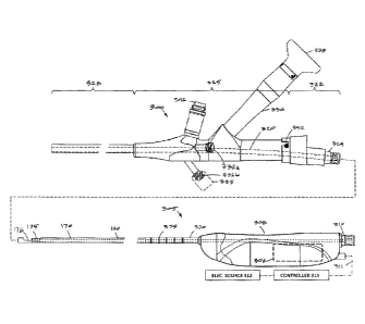
100381 FIG. 15 is a plan view of another fibroid removal system including an
endoscope
and an electrosurgical tissue resecting device that is inserted through a
curved working
channel of the hysteroscope.
[0039] FIG. 16 is a cut-away view of the hysteroscope of FIG. 15 showing a
disposable
adapter component carrying a seal assembly and further showing a working
channel with a
curved portion in the main body of the endoscope.
[0040] FIG. 17 is a sectional view of a handle portion of an endoscope having
an
expanded cross-section channel that provides a fluid reservoir and a solenoid-
relief valve
mechanism for rapid release of fluid from the system to reduce uterine cavity
pressure.
[0041] FIG. 18 is a cross-section of the handle portion of FIG. 17 taken along
line 18-18
of FIG. 17.
[0042] FIG. 19 is a sectional view of a handle portion of another endoscope
similar to
that of FIG. 17.
[0043] FIG. 20A is a schematic view of an annular flow channel and fluid
reservoir in
the endoscope handle portion of FIGS. 17-19.
CA 02861327 2014-07-15
WO 2013/110073 PCT/US2013/022559
[0044] FIG. 20B is a schematic view of an annular flow channel in an endoscope
handle
portion without the fluid reservoir as in the variation of FIGS. 17-19.
100451 FIG. 21 is a sectional view of a handle portion of another endoscope
similar to
that of FIGS. 17-18 with an optical sensor.
100461 FIG. 22 is a sectional view of a handle portion of another endoscope
similar to
that of FIGS. 17-18 with a passive pressure relief valve.
DETAILED DESCRIPTION OF THE INVENTION
[0047] FIG. 1 illustrates an assembly that comprises an endoscope 50 used for
hystcroscopy together with an electrosurgical tissue resecting device 100
extending
through a working channel 102 of the endoscope. The endoscope or hysteroscope
50 has a
handle 104 coupled to an elongated shaft 105 having a diameter of 5 mm to 7
mm. The
working channel 102 therein may be round, D-shaped or any other suitable
shape. The
endoscope shaft 105 is further configured with an optics channel 106 and one
or more
fluid inflow/outflow channels 108a, 108b (FIG. 3) that communicate with valve-
connectors 110a, 110b configured for coupling to a fluid inflow source 120
thereto, or
optionally a negative pressure source 125 (FIGS. 1-2). The fluid inflow source
120 is a
component of a fluid management system 126 as is known in the art (FIG. 2)
which
comprises a fluid container 128 and pump mechanism 130 which pumps fluid
through the
hysteroscope 50 into the uterine cavity. As can be seen in FIG. 2, the fluid
management
system 126 further includes the negative pressure source 125 (which can
comprise an
operating room wall suction source) coupled to the tissue resecting device
100. The
handle 104 of the endoscope includes the angled extension portion 132 with
optics to
which a videoscopic camera 135 can be operatively coupled. A light source 136
also is
coupled to light coupling 138 on the handle of the hysteroscope 50. The
working channel
102 of the hysteroscope is configured for insertion and manipulation of the
tissue resecting
and extracting device 100, for example to treat and remove fibroid tissue. In
one
embodiment, the hysteroscope shaft 105 has an axial length of 21 cm, and can
comprise a
0 scope, or 15 to 30 scope.
[0048] Still referring to FIG. 1, the tissue resecting device 100 has a
highly elongated
shaft assembly 140 configured to extend through the working channel 102 in the
hysteroscope. A handle 142 of the tissue resecting device 100 is adapted for
manipulating
the electrosurgical working end 145 of the device. In use, the handle 142 can
be
manipulated both rotationally and axially, for example, to orient the working
end 145 to
6
CA 02861327 2014-07-15
WO 2013/110073 PCT/US2013/022559
resect targeted fibroid or polyp tissue. The tissue resecting device 100 has
subsystems
coupled to its handle 142 to enable electrosurgical resection of targeted
tissue. A
radiofrequency generator or RF source 150 and controller 155 are coupled to at
least one
RF electrode carried by the working end 145 as will be described in detail
below. In one
embodiment shown in FIG. 1, an electrical cable 156 and negative pressure
source 125 are
operatively coupled to a connectors 158 and 159 in handle 142. The electrical
cable
couples the RF source 150 to the electrosurgical working end 145. The negative
pressure
source 125 communicates with a tissue-extraction channel 160 in the shaft
assembly 140
of the tissue extraction device 100 (FIG. 4).
[0049] FIG. 1 further illustrates a seal housing 162 that carries a
flexible seal 164 carried
by the hysteroscope handle 104 for sealing the shaft 140 of the tissue
resecting device 100
in the working channel 102 to prevent distending fluid from escaping from a
uterine
cavity.
[0050] In one embodiment as shown in FIG. 1, the handle 142 of tissue
resecting device
100 includes a motor drive 165 for reciprocating or otherwise moving a
resecting
component of the electrosurgical working end 145 as will be described below.
The handle
142 optionally includes one or more actuator buttons 166 for actuating the
device. In
another embodiment, a footswitch can be used to operate the device. In one
embodiment,
the system includes a switch or control mechanism to provide a plurality of
reciprocation
speeds, for example 1 Hz, 2 Hz, 3 Hz, 4 Hz and up to 8 Hz. Further, the system
can
include a mechanism for moving and locking the reciprocating sleeve in a non-
extended
position and in an extended position. Further, the system can include a
mechanism for
actuating a single reciprocating stroke.
[0051] Referring to FIGS. 1 and 4, an electrosurgical tissue resecting device
has an
elongate shaft assembly 140 extending about longitudinal axis 168 comprising
an exterior
or first outer sleeve 170 with passageway or lumen 172 therein that
accommodates a
second or inner sleeve 175 that can reciprocate (and optionally rotate or
oscillate) in lumen
172 to resect tissue as is known in that art of such tubular resection
devices. In one
embodiment, the tissue-receiving window 176 in the outer sleeve 170 has an
axial length
ranging between 10 mm and 30 mm and extends in a radial angle about outer
sleeve 170
from about 45 to 210 relative to axis 168 of the sleeve. The outer and inner
sleeves 170
and 175 can comprise a thin-wall stainless steel material and can function as
opposing
polarity electrodes as will be described in detail below. FIGS. 6A-8
illustrate insulating
layers carried by the outer and inner sleeves 170 and 175 to limit, control
and/or prevent
7
CA 02861327 2014-07-15
WO 2013/110073 PCT/US2013/022559
unwanted electrical current flows between certain portions of the sleeve. In
one
embodiment, a stainless steel outer sleeve 170 has an O.D. of 0.143" with an
I.D. of
0.133" and with an inner insulating layer (described below) the sleeve has a
nominal I.D.
of 0.125". In this embodiment, the stainless steel inner sleeve 175 has an
O.D. of 0.120"
with an I.D. of 0.112". The inner sleeve 175 with an outer insulating layer
has a nominal
O.D. of about 0.123" to 0.124" to reciprocate in lumen 172. In other
embodiments, outer
and or inner sleeves can be fabricated of metal, plastic, ceramic or a
combination thereof.
The cross-section of the sleeves can be round, oval or any other suitable
shape.
[0052] As can be seen in FIG. 4, the distal end 177 of inner sleeve 175
comprises a first
polarity electrode with distal resecting electrode edge 180 about which plasma
can be
generated. The electrode edge 180 also can be described as an active electrode
during
tissue resection since the electrode edge 180 then has a substantially smaller
surface area
than the opposing polarity or return electrode. In one embodiment in FIG 4,
the exposed
surfaces of outer sleeve 170 comprises the second polarity electrode 185,
which thus can
be described as the return electrode since during use such an electrode
surface has a
substantially larger surface area compared to the functionally exposed surface
area of the
active electrode edge 180.
[0053] In one aspect of the invention, the inner sleeve or resecting sleeve
175 has an
interior tissue extraction lumen 160 with first and second interior diameters
that are
adapted to electrosurgically resect tissue volumes rapidly¨and thereafter
consistently
extract the resected tissue strips through the highly elongated lumen 160
without clogging.
Now referring to FIGS. 5 and 6A, it can be seen that the inner sleeve 175 has
a first
diameter portion 190A that extends from the handle 142 (FIG. 1) to a distal
region 192 of
the sleeve 175 wherein the tissue extraction lumen transitions to a smaller
second diameter
lumen 190B with a reduced diameter indicated at B which is defined by the
electrode
sleeve element 195 that provides the electrode edge 180. The axial length C of
the
reduced cross-section lumen 190B can range from about 2 mm to 20 mm. In one
embodiment, the first diameter A is 0.112" and the second reduced diameter B
is 0.100".
As shown in FIG. 5, the inner sleeve 175 can be an electrically conductive
stainless steel
and the reduced diameter electrode portion also can comprise a stainless steel
electrode
sleeve element 195 that is welded in place by weld 196 (FIG. 6A). In another
alternative
embodiment, the electrode and reduced diameter electrode sleeve element 195
comprises a
tungsten tube that can be press fit into the distal end 198 of inner sleeve
175. FIGS. 5 and
6A further illustrates the interfacing insulation layers 202 and 204 carried
by the first and
8
CA 02861327 2014-07-15
WO 2013/110073 PCT/US2013/022559
second sleeves 170, 175, respectively. In FIG. 6A, the outer sleeve 170 is
lined with a
thin-wall insulating material 200, such as PFA, or another material described
below.
Similarly, the inner sleeve 175 has an exterior insulating layer 202. These
coating
materials can be lubricious as well as electrically insulating to reduce
friction during
reciprocation of the inner sleeve 175.
100541 The insulating layers 200 and 202 described above can comprise a
lubricious,
hydrophobic or hydrophilic polymeric material. For example, the material can
comprise a
bio-compatible material such as PFA, TEFLON , polytetrafluroethylene (PTFE),
FEP
(fluorinated ethylenepropylene), polyethylene, polyamide, ECTFE
(ethylenechlorotrifluoro-ethylenc), ETFE, PVDF, polyvinyl chloride or
silicone.
[0055] Now turning to FIG. 6B, another variation of inner sleeve 175 is
illustrated in a
schematic view together with a tissue volume being resected with the plasma
electrode
edge 180. In this embodiment, as in other embodiments in this disclosure, the
RF source
operates at selected operational parameters to create a plasma around the
electrode edge
180 of electrode sleeve 195 as is known in the art. Thus, the plasma generated
at electrode
edge 180 can resect and ablate a path P in the tissue 220, and is suited for
resecting fibroid
tissue and other abnormal uterine tissue. In FIG. 6B, the distal portion of
the inner sleeve
175 includes a ceramic collar 222 which is adjacent the distal edge 180 of the
electrode
sleeve 195. The ceramic 222 collar functions to confine plasma formation about
the distal
electrode edge 180 and functions further to prevent plasma from contacting and
damaging
the polymer insulating layer 202 on the inner sleeve 175 during operation. In
one aspect
of the invention, the path P in tissue 220 made with the plasma at electrode
edge 180
provides a path P having an ablated width indicated at W, wherein such path
width W is
substantially wide due to tissue vaporization. This removal and vaporization
of tissue in
path P is substantially different than the effect of cutting similar tissue
with a sharp blade
edge, as in various prior art devices. A sharp blade edge can divide tissue
(without
cauterization) but applies mechanical force to the tissue and may prevent a
large cross
section slug of tissue from being cut. In contrast, the plasma at the
electrode edge 180 can
vaporize a path P in tissue without applying any substantial force on the
tissue to thus
resect larger cross sections or slugs strips of tissue. Further, the plasma
ablation effect
reduces the cross section of tissue strip 225 received in the tissue-
extraction lumen 190B.
FIG. 6B depicts a tissue strip to 225 entering lumen 190B which has such a
smaller cross-
section than the lumen due to the vaporization of tissue. Further, the cross
section of
tissue 225 as it enters the larger cross-section lumen 190A results in even
greater free
9
CA 02861327 2014-07-15
WO 2013/110073 PCT/US2013/022559
space 196 around the tissue strip 225. Thus, the resection of tissue with the
plasma
electrode edge 180, together with the lumen transition from the smaller cross-
section
(190B) to the larger cross-section (190A) of the tissue-extraction lumen 160
can
significantly reduce or eliminate the potential for successive resected tissue
strips 225 to
clog the lumen. Prior art mechanical cutting devices with such small diameter
tissue-
extraction lumens typically have problems with tissue clogging.
[0056] In another aspect of the invention, the negative pressure source 225
coupled to
the proximal end of tissue-extraction lumen 160 (see FIGS. 1 and 4) also can
assist in
aspirating and moving tissue strips 225 in the extraction lumen 160 in the
proximal
direction to a collection reservoir (not shown) outside the handle 142 of the
device.
[0057] FIGS. 7A-7B illustrate the change in lumen diameter of resection sleeve
175 of
FIG. 6B. FIG. 8 illustrates the distal end of a variation of resection sleeve
175' which is
configured with an electrode resection element 195' that is partially tubular
in contrast to
the previously described tubular electrode element 195 (FIGS. 5 and 6A). FIGS.
9A-9B
again illustrate the change in cross-section of the tissue- extraction lumen
between reduced
cross-section region 190B' and the increased cross-section region 190A' of the
resection
sleeve 175' of FIG. 8. Thus, the functionality remains the same whether the
resection
electrode element 195' is tubular or partly tubular. In FIG. 8A, the ceramic
collar 222' is
shown, in one variation, as extending only partially around sleeve 175 to
cooperate with
the radial angle of resection electrode element 195'. Further, the variation
of FIG. 8
illustrates that the ceramic collar 222' has a larger outside diameter than
insulating layer
202. Thus, friction may be reduced since the short axial length of the ceramic
collar 222'
interfaces and slides against the interfacing insulating layer 200 about the
inner surface of
lumen 172 of outer sleeve 170.
[0058] In general, one aspect of the invention comprises a tissue resecting
and extracting
device (FIGS. 10A-11C) that includes first and second concentric sleeves
having an axis
and wherein the second (inner) sleeve 175 has an axially-extending tissue-
extraction
lumen therein, and wherein the second sleeve 175 is moveable between axially
non-
extended and extended positions relative to a tissue-receiving window 176 in
first sleeve
170 to resect tissue, and wherein the tissue extraction lumen 160 has first
and second
cross-sections. The second sleeve 175 has a distal end configured as a plasma
electrode
edge 180 to resect tissue disposed in tissue-receiving window 176 of the first
sleeve 170.
Further, the distal end of the second sleeve, and more particularly, the
electrode edge 180
is configured for plasma ablation of a substantially wide path in the tissue.
In general, the
CA 02861327 2014-07-15
WO 2013/110073 PCT/US2013/022559
tissue-extraction device is configured with a tissue extraction lumen 160
having a distal
end portion with a reduced cross-section that is smaller than a cross-section
of medial and
proximal portions of the lumen 160.
[0059] In one aspect of the invention, referring to FIGS. 7A-7B and 9A-9B, the
tissue-
extraction lumen 160 has a reduced cross-sectional area in lumen region 190A
proximate
the plasma tip or electrode edge 180 wherein said reduced cross section is
less that 95%,
90%, 85% or 80% than the cross sectional area of medial and proximal portions
190B of
the tissue-extraction lumen, and wherein the axial length of the tissue-
extraction lumen is
at least 10 cm, 20 cm, 30 cm or 40 cm. In one embodiment of tissue resecting
device 100
for hysteroscopic fibroid resection and extraction (FIG. 1), the shaft
assembly 140 of the
tissue resecting device is 35 cm in length.
100601 FIGS. 10A-10C illustrate the working end 145 of the tissue resecting
device 100
with the reciprocating resecting sleeve or inner sleeve 175 in three different
axial positions
relative to the tissue receiving window 176 in outer sleeve 170. In FIG. 10 A,
the
resecting sleeve 175 is shown in a retracted or non-extended position in which
the sleeve
175 is at it proximal limit of motion and is prepared to advance distally to
an extended
position to thereby electrosurgically resect tissue positioned in and/or
suctioned into in
window 176. FIG. 10B shows the inner sleeve 175 moved and advanced distally to
a
partially advanced or medial position relative to tissue receiving window 176.
FIG. 10C
illustrates the inner sleeve 175 fully advanced and extended to the distal
limit of its motion
wherein the plasma ablation electrode 180 has extended past the distal end 226
of tissue-
receiving window 176 at which moment the resected tissue strip 225 is excised
from tissue
volume 220 and captured in reduced cross-sectional lumen region 190A.
[0061] Now referring to FIGS. 10A-10C, FIGS. 11A-11C and FIGS. 12A-12C,
another
aspect of the invention comprises "tissue displacement" mechanisms provided by
multiple
elements and processes to "displace" and move tissue strips 225 (FIG. 12A) in
the
proximal direction in lumen 160 of inner sleeve 175 to thus ensure that tissue
does not
clog the lumen of the inner sleeve 175. As can seen in FIG. 10A and the
enlarged views
of FIGS. 11A-11C, one tissue displacement mechanism comprises a projecting
element
230 that extends proximally from distal tip 232 which is fixedly attached to
outer sleeve
170. The projecting element 230 extends proximally along central axis 168 in a
distal
chamber 240 defined by outer sleeve 170 and distal tip 232. In one embodiment
depicted
in FIG. 11A, the shaft-like projecting element 230, in a first functional
aspect, comprises a
mechanical pusher that functions to push a captured tissue strip 225
proximally from the
11
CA 02861327 2014-07-15
WO 2013/110073 PCT/US2013/022559
small cross-section lumen 190B of inner sleeve 175 (FIG. 12A) as the inner
sleeve 175
moves to its fully advanced or extended position.
100621 In a second functional aspect, the chamber 240 in the distal end of
sleeve 170 is
configured to capture a volume of saline distending fluid 244 (FIG. 12A) from
the
working space, and wherein the existing RF electrodes of the working end 145
are further
configured to explosively vaporize the captured fluid 244 to generate
proximally-directed
forces on tissue strips 225 resected and disposed in lumen 160 of the inner
sleeve 175
(FIGS. 12B and 12C). Both of these functional elements and processes (tissue
displacement mechanisms) can apply a substantial mechanical force on the
captured tissue
strips 225 by means of the explosive vaporization of liquid in chamber 240 and
can
function to move tissue strips 225 in the proximal direction in the tissue-
extraction lumen
160. It has been found that using the combination of multiple functional
elements and
processes can virtually eliminate the potential for tissue clogging the tissue
extraction
lumen 160.
[0063] More particularly, FIGS. 12A-12C illustrate the functional aspects of
the tissue
displacement mechanisms and the subsequent explosive vaporization of fluid
captured in
chamber 240. In FIG. 12A, the reciprocating inner sleeve 175 is shown in a
medial
position advancing distally wherein plasma at the resecting electrode edge 180
is resecting
a tissue strip 225 that is disposed within lumen 160 of the inner sleeve 175.
In FIG. 12A-
12C, it can be seen that the system operates in first and second
electrosurgical modes
corresponding to the reciprocation and axial range of motion of inner sleeve
175 relative
to the tissue-receiving window 176. As used herein, the term "electrosurgical
mode"
refers to which electrode of the two opposing polarity electrodes functions as
an "active
electrode" and which electrode functions as a "return electrode". The terms
"active
electrode" and -return electrode" are used in accordance with convention in
the art¨
wherein an active electrode has a smaller surface area than the return
electrode which thus
focuses RF energy density about such an active electrode. In the working end
145 of
FIGS. 10A-11C, the resecting electrode element 195 and its electrode edge 180
must
comprise the active electrode to focus energy about the electrode to generate
the plasma
for tissue resection. Such a high-intensity, energetic plasma at the electrode
edge 180 is
needed throughout stroke X indicated in FIG. 12A-12B to resect tissue. The
first mode
occurs over an axial length of travel of inner sleeve 175 as it crosses the
tissue-receiving
window 176, at which time the entire exterior surface of outer sleeve 170
comprises the
12
CA 02861327 2014-07-15
WO 2013/110073 PCT/US2013/022559
return electrode indicated at 185. The electrical fields EF of the first RF
mode are
indicated generally in FIG. 12A.
100641 FIG. 12 B illustrates the moment in time at which the distal
advancement or
extension of inner sleeve 175 entirely crosses the tissue-receiving window 176
(FIG.
12A). At this time, the electrode sleeve 195 and its electrode edge 180 are
confined within
the mostly insulated-wall chamber 240 defined by the outer sleeve 170 and
distal tip 232.
At this moment, the system is configured to switch to the second RF mode in
which the
electric fields EF switch from those described previously in the first RF
mode. As can be
seen in FIG. 12B, in this second mode, the limited interior surface area 250
(FIG. 12C) of
distal tip 232 that interfaces chamber 240 functions as an active electrode
and the distal
end portion of inner sleeve 175 exposed to chamber 240 acts as a return
electrode. In this
mode, very high energy densities occur about surface 250 and such a contained
electric
field EF can explosively and instantly vaporize the fluid 244 captured in
chamber 240.
The expansion of water vapor can be dramatic and can thus apply tremendous
mechanical
forces and fluid pressure on the tissue strip 225 to move the tissue strip in
the proximal
direction in the tissue extraction lumen 160. FIG. 12C illustrates such
explosive or
expansive vaporization of the distention fluid 244 captured in chamber 240 and
further
shows the tissue strip 225 being expelled in the proximal direction the lumen
160 of inner
sleeve 175.
100651 FIG. 14 shows the relative surface areas of the active and return
electrodes at the
extended range of motion of the inner sleeve 175, again illustrating that the
surface area of
the non-insulated distal end surface 250 is small compared to surface 255 of
electrode
sleeve which comprises the return electrode.
[0066] Still referring to FIGS. 12A-12C, it has been found that a single power
setting on
the RF source 150 and controller 155 can be configured both (i) to create
plasma at the
electrode edge 180 of electrode sleeve 195 to resect tissue in the first mode,
and (ii) to
explosively vaporize the captured distention fluid 244 in the second mode.
Further, it has
been found that the system can function with RF mode-switching automatically
at suitable
reciprocation rates ranging from 0.5 cycles per second to 8 or 10 cycles per
second. In
bench testing, it has been found that the tissue resecting device described
above can resect
and extract tissue at the rate of from 4 grams/min to 8 grams/min without any
potential for
tissue strips 225 clogging the tissue-extraction lumen 160. In these
embodiments, the
negative pressure source 125 also is coupled to the tissue-extraction lumen
160 to assist in
applying forces for tissue extraction.
13
CA 02861327 2014-07-15
WO 2013/110073 PCT/US2013/022559
[0067] Of particular interest, the fluid-capture chamber 240 defined by sleeve
170 and
distal tip 232 can be designed to have a selected volume, exposed electrode
surface area,
length and geometry to optimize the application of expelling forces to
resected tissue strips
225. In one embodiment, the diameter of the chamber is 3.175 mm and the length
is 5.0
mm which taking into account the projecting element 230, provided a captured
fluid
volume of approximately 0.040 mL. In other variations, the captured fluid
volume can
range from 0.004 mL to 0.080 mL.
[0068] In one example, a chamber 240 with a captured liquid volume of 0.040 mL
together with 100% conversion efficiency in and instantaneous vaporization
would require
103 Joules to heat the liquid from room temperature to water vapor. In
operation, since a
Joule is a W*s, and the system reciprocate at 3 Hz, the power required would
be on the
order of 311 W for full, instantaneous conversion to water vapor. A
corresponding
theoretical expansion of 1700x would occur in the phase transition, which
would results in
up to 25,000 psi instantaneously (14.7 psi x 1700), although due to losses in
efficiency and
non-instantaneous expansion, the actual pressures would be much less. In any
event, the
pressures are substantial and can apply significant expelling forces to the
captured tissue
strips 225.
[0069] Referring to FIG. 12A, the interior chamber 240 can have an axial
length from
about 0.5 mm to 10 mm to capture a liquid volume ranging from about 0.004 ml.
0.01 mL.
It can be understood in FIG. 12A, that the interior wall of chamber 240 has an
insulator
layer 200 which thus limits the electrode surface area 250 exposed to chamber
240. In one
embodiment, the distal tip 232 is stainless steel and is welded to outer
sleeve 170. The
post element 248 is welded to tip 232 or machined as a feature thereof. The
projecting
element 230 in this embodiment is a non-conductive ceramic.
[0070] FIG. 13 shows the cross-section of the ceramic projecting element 230
which
may be fluted, and which in one embodiment has three flute elements 260 and
three
corresponding axial grooves 262 in its surface. Any number of flutes, channels
or the like
is possible, for example from two to about 20. The fluted design increases the
available
cross-sectional area at the proximal end of the projecting element 230 to push
the tissue
strip 225, while at the same time the three grooves 262 permit the proximally-
directed
jetting of water vapor to impact the tissue exposed to the grooves 262. In one
embodiment, the axial length D (FIG. 12A) of the projecting element 230 is
configured to
push tissue entirely out of the reduced cross-sectional region 190B of the
electrode sleeve
element 195. In another embodiment, the volume of the chamber 240 is
configured to
14
CA 02861327 2014-07-15
WO 2013/110073 PCT/US2013/022559
capture liquid that when explosively vaporized provided a gas (water vapor)
volume
sufficient to expand into and occupy at least the volume defined by a 10% of
the total
length of extraction channel 160 in the device, usually at least 20% of the
extraction
channel 160, often at least 40% of the extraction channel 160, sometimes at
least 60% of
the extraction channel 160, other times at least 80% of the extraction channel
160, and
sometimes at least 100% of the extraction channel 160.
[0071] As can be understood from FIGS. 12A to 12C, the distending fluid 244 in
the
working space replenishes the captured fluid in chamber 240 as the inner
sleeve 175
moves in the proximal direction or towards its non-extended position. Thus,
when the
inner sleeve 175 again moves in the distal direction to resect tissue, the
interior chamber
240 is filled with fluid 244 which is then again contained and is then
available for
explosive vaporization as described above when the inner sleeve 175 closes the
tissue-
receiving window 176. In another embodiment, a one-way valve can be provided
in the
distal tip 232 to draw fluid directly into interior chamber 240 without the
need for fluid to
migrate through window 176.
100721 In another embodiment, the RF source 150 and controller 155 can be
programmed to modulate energy delivery parameters during stroke X and stroke Y
in
FIGS. 12A-12C to provide the optimal energy (i) for plasma resection with
electrode edge
180, and (ii) for explosively vaporizing the captured fluid in chamber 240.
100731 It should be appreciated that while an RF source is suitable for
causing explosive
vaporization of the captured fluid volume, any other energy source can be used
and falls
within the scope of the invention, such as an ultrasound transducer, HIFU, a
laser or light
energy source, a microwave or a resistive heat source.
[0074] FIG. 15 is a side view of a fibroid removal system similar to that of
FIG. 1 that
includes an endoscope 300 configured for use in hysteroscopy and an RF tissue
resecting
device 305 configured for introduction through the working channel in the
endoscope 300.
[0075] In FIG. 15, it can be seen that the resecting device has inner and
outer sleeves
170 and 175 with the inner sleeve 175 reciprocated axially relative to window
176 by a
motor 306 in handle 308. The tissue extraction channel 160 in the inner sleeve
175
extends through the handle 308 in communication with a quick-connect fitting
310. A
negative pressure source coupled to a flexible extraction tubing (not shown)
can be
connected to fitting 310 to thereby carry resected tissue and fluid to a
collection reservoir
(cf. FIG. 1). The motor 306 is coupled to an electrical cable 311 that extends
to an
electrical source 312 and controller 315.
84148653
100761 In FIGS. 15 and 16, it can be seen that the endoscope 300 is similar to
the
endoscope of FIGS. 1 and 3, except that endoscope 300 in FIGS. 15-16 differs
in that (i)
the endoscope has a different configuration of working channel 320 which is
curved to
provide a pre-determined resistance to sliding a resecting tool shaft in the
channel, and (ii)
the endoscope has a different type of disposable adapter component 322 that
carries a
quick-connect fitting 324 for purposes described below.
100771 More in particular, FIGS. 15-16 show that endoscope 300 has a handle or
main
body 325 of a metal that is coupled to an extension or shaft portion 328. The
elongated
shaft 328 can have a diameter ranging from 5 mm to 10 mm and in one embodiment
is 6,2
mm. The endoscope shaft 328 has an axial length of 15 to 35 cm and the
endoscope 300
can be a 00 scope, or 15 to 30" scope.
100781 The endoscope shaft 328 has an optics channel 106 and first and second
fluid
flow channels 108a and 108b as shown in the endoscope of FIG. 3. The flow
channels
108a and 108b (FIG. 3) communicate with Luer connectors 332a and 332b (see
FIGS. 15-
16). A fluid inflow source 120 (FIG. 2) is coupled to first connector 332a and
channel
108a. A pressure sensor 335 is coupled to second connector 332b and channel
108b. The
pressure sensor 335 is adapted to measure actual intracavity pressure (as
described further
below) and to send pressure signals continuously to controller 315.
100791 The main body 325 of the endoscope 300 includes the angled extension
portion
336 with optics and prism 337 which provides light path LP to thereby allow
viewing
through optics channel 106. A videoscopic camera can be coupled to the
proximal end
338 of the angled extension portion 336. A light source is coupled to light
connector 342
on the main body 325 of the endoscope.
100801 In FIGS. 15-16, it can be see that the endoscope 300 includes a
detachable and
disposable adapter component 322 that carries first and second seals 346 and
348 that are
configured to seal the working channel 320 when there is a resecting tool
shaft in the
channel or in the absence of a shaft in the channel 320. The more distal seal
348 can
comprise a duck-bill seal or its equivalent that seals the channel when there
is no tool shaft
in channel 320. The more proximal seal 346 comprises an elastomeric seal with
port 350
that can stretch and impinge on a tool shaft disposed in the channel 320. In
one variation
shown in FIG. 16, the disposable component 322 can molded of plastic and can
be
detachably coupled to main body 325 of the endoscope by a Mock 352. An o-ring
354
can be provided in an interface between the main body 325 and the disposable
component
322. Any suitable fitting can be used to couple the disposable component 322
to the main
16
CA 2861327 2019-02-08
84148653
body 325 such as threads, J-locks, etc. FIG. 16 further shows that the
disposable adapter
component 322 has an interior chamber 353 that has a substantial fluid volume
which can
optionally be configured with a manual or automated pressure relief valve as
will be
further described below in related embodiments.
100811 Referring again to FIGS. 15 and 16, it has been found that the curved
portion
355A of the working channel 320 functions to provide resistance to unwanted
axial sliding
of a resecting tool shall when in use, while at the same time not providing
any resistance
to rotation of the resecting device shaft. In use, the eleetrosurgical
resecting device 305 as
generally shown in FIGS. 1, 4, 10A-14 and 15 is manipulated to resect tissue
only by
pressing the working end window 176 into a targeted tissue site together with
slight
rotation of the working end while resecting tissue. During use, the working
end of the RF
resecting device of FIG. 15 should not be moved axially back and forth to
resect tissue
channel as is typical with commercially available RF resecting loops known in
the prior
art. For this reason, the configuration of curved working channel 355A shown
in FIGS.
15-16 provides a desired increase in resistance to axial sliding of the
resecting device shaft
in the endoscope which assists in preventing physicians from using the
combination of the
present invention (RF resecting device and endoscope) in the manner commonly
associated with prior art RF resecting loops. The shaft of the RF resecting
device 305 is
also configured to be suitably flexible to cooperate with the curved working
channel. It
has been found that a curved working channel as described herein does not
interfere with
the physician's rotation of the resecting device shaft in the working channel
320, which
also is advantageous.
100821 In FIGS. 15 and 16, an embodiment of endoscope 300 has a working
channel 320
that has a curved or non-straight portion 355A with curved axis 356A that
extends through
main body 325 and a straight channel portion 355B with straight axis 356B that
extends
longitudinally through the shaft portion 328 of the endoscope. The curved
channel portion
355A can extend over a length AA ranging from about 4 cm to 8 cm and in one
embodiment is about 5 cm. The curved channel portion 355A can have a radius R
ranging
from about 150 mm to 900 mm. In one embodiment, the central axis 356A of the
curved
channel portion 355A at the proximal face 360 of main body 325 is offset by a
distance
having dimension DD which can be about 2 mm to 5 mm (see FIG. 16) from the
hypothetical central axis 355B of the straight channel portion 355B if
extended to the
proximal face 360 of main body 325. In one embodiment, the offset dimension DD
is 2.0
mm. In an embodiment, the surface of a least the curved channel portion 355A
in the
17
CA 2861327 2019-02-08
84148653
metal main body 325 can have a coating of titanium nitride or gold which can
protect the
channel from damage over the working life of the endoscope.
100831 In another embodiment (not shown), the working channel 320 in an
endoscope
300 similar to that of FIG. 15 can be straight or curved and an alternative
mechanism can
be used to provide resistance to axial sliding of a tool shall. In one
variation, a
compression assembly known in the art can be used to squeeze an interference
clement
against the tool shaft in the working channel, such as radial inward
compression of an 0-
ring. FIG. 15 illustrates another mechanism that may be used to indicate or
resist axial
sliding of a tool shaft in the working channel. As can be seen in FIG. 15, the
RF resecting
device has a stiffener sleeve 370 disposed around the proximal end 372 of
outer sleeve
170. The stiffener sleeve 370 can have a length of 4 to 6 cm and is configured
with 5 to
50 annular grooves or detents 375 that cooperate with a spring element (not
shown) in the
adapter component 322 for engaging the detents 375 to provide tactile feedback
to the
physician relating to axial sliding of the tool shaft.
100841 In general, the cndoscope 300 comprises a main body 325 and extended
shaft
portion 328 that extends longitudinally to a distal end, a first channel
extending from the
handle end to the distal end coupleable to a fluid inflow source, a second
channel
extending from the handle end to the distal end configured for fluid outflows
and/or
receiving an RF resecting device, wherein the second channel has first
straight portion and
a second curved portion, and a disposable component carrying at least one seal
detachably
coupled to the endoscope main body and the second channel. in one variation,
the device
has first and second seals elements carried in the disposable component
configured to seal
the second channel with or without a tool shaft disposed therein. In one
variation, a third
channel is configured for coupling to a pressure sensor 335 (see FIGS. 15-16).
A fourth
channel is configured as an optics channel for viewing the uterine cavity A
fifth channel is
configured as a light guide extending from the main body of the endoscope to
the distal
end of the extended shaft portion 328. The endoscope can have a pressure
sensor 335 that
is configured to send pressure signals to a controller 315 to control fluid
inflows and fluid
outflows through the endoscope to thereby control fluid pressure in the
uterine cavity.
The controller can be operatively coupled to the fluid inflow and outflow
sources to
contemporaneously (i) control pressure within the uterine cavity by modulating
the
positive and negative pressure sources and (ii) control operating parameters
of the
ekctrosurgical rending device. The controller 315 can be adapted to
selectively control
flows to the uterine cavity through a flow channel at any rate between 0
ml/min and 750
18
CA 2861327 2019-02-08
CA 02861327 2014-07-15
WO 2013/110073 PCT/US2013/022559
ml/min. In another aspect of the invention, the controller 315 can be adapted
to selectively
control pressure in the uterine cavity at any level between 0 mmHg and 150
mmHg. The
controller 315 can be adapted to selectively control outflows from the uterine
cavity
through a channel in the system at any rate between 0 ml/min and 750 mlimin.
In one
variation, the pressure sensor 335 (FIG. 15) is disposable and is detachably
coupled to a
proximal end of a channel that has a cross-sectional area of greater than 0.1
mm2, greater
than 0.5 mm2 or greater than 1.0 mm2.
100851 FIGS. 17 and 18 illustrate another variation of endoscope 500 that is
configured
for use in hysteroscopy that includes mechanisms and systems for controlling
pressure in a
uterine cavity during a fibroid removal procedure. In one variation, the
endoscope 500
and system is adapted to automatically reduce intracavity pressure within a
predetermined
time interval after a set point of intracavity pressure has been reached. The
predetermined
set point can be 50 mm Hg, 60 mm Hg, 70 mm Hg, 80 mm Hg, 90 mm Hg, 100 mm Hg,
110 mm Hg, 120 mm Hg, 130 mm Hg, 140 mrn Hg, 150 mm Hg, 160 mm Hg, 170 mm
Hg or 180 mm Hg. In one variation, the predetermined pressure is 150 mm Hg.
The
predetermined interval can be in a range between 1 second and 10 seconds and
in one
variation is 5 seconds. In another variation, the system includes a pressure
relief valve for
releasing pressure at a predetermined maximum pressure which can in the range
of 150
mm Hg to 200 mm Hg and in one variation is 200 mm Hg. Of particular interest,
the
system is adapted to respond to a measurement of "actual" intracavity pressure
measured
by a pressure sensor in direct fluidic communication with the uterine cavity.
In prior the
art, fluid management systems that are adapted to release intracavity pressure
at a
predetermined set point use only an "estimated" intracavity pressure that is
estimated by a
software algorithm based on signals relating to fluid inflows communicated to
a flow
controller. Such prior art systems and algorithms are not capable of
accurately measuring
"actual" intracavity pressure.
100861 In FIGS. 17-18, it can be seen that endoscope 500 has standard features
including
a viewing channel 508, a light channel comprising optic fibers in shaft
portion 512, a
working channel 510 and one or more fluid inflow or outflow channels. The
shaft portion
512 of the endoscope extends about central longitudinal axis 515. The
endoscope body is
reusable and sterilizable as in known in the art. A handle or main body
portion 516 of the
endoscope body couples to the shaft 512 and carries an eyepiece 517 and luer
connectors
(not shown) communicating with first and second channels for fluid inflows and
outflows
as described previously. A light connector is indicated at 518.
19
CA 02861327 2014-07-15
WO 2013/110073 PCT/US2013/022559
[0087] As further can be seen in FIG. 17, a proximal endoscope or adapter
component
520 comprises a disposable adapter body which is attachable to the proximal
end of the
endoscope main body. The adapter component 520 can attached by either threads,
J-lock
or a snap fitting at interface 522 in a configuration that rotationally aligns
the channel or
lumen portion in component 520 with the channel in the endoscope main body
505.
100881 In one aspect of the invention, the proximal end 524 of the adapter
component
520 is configured as a mating portion of a quick-connect fitting 525. The
quick-connect
fitting 525 and 0-ring 528 can be used to couple an outflow tubing 530
directly to the
proximal end of the endoscope assembly 500 to allow the system to be used in a
diagnostic mode. A diagnostic mode consists of the physician performing a
diagnostic
procedure before using a resecting probe. Thus, when a resecting probe is not
inserted
through the endoscope the physician can connect the saline return flow tubing
directly to
the quick-connect fitting 525 and circulates distention fluid through an
inflow channel in
the endoscope device and outward through the working channel and outflow
tubing
coupled to the quick-connect 525 to distend the uterine cavity to thereby
allow viewing of
the cavity.
[0089] The adapter component 520 further carries seals 530a and 530b which
comprise
seals for (i) preventing fluid outflows through the working channel and
adapter when there
is no resecting tool disposed the endoscope and for (ii) providing a seal
around a resection
tool shaft when such a tool is disposed in the endoscope. These seals 530a and
530b can
be integrated into a one component or be spaced apart as shown in one
variation in FIG.
17.
[0090] In one aspect of the invention, as described above, the endoscope
assembly
includes a valve system configured to automatically reduce uterine cavity
pressure within
a predetermined time interval after a set point of intracavity pressure has
been reached. In
one variation, as stated above, the predetermined pressure is 150 mm Hg and
the
predetermined interval is 5 seconds. In one variation, a solenoid relief valve
540 is
operatively coupled to a controller 545 and is adapted to release at least a
predetermined
volume of distention fluid from the system (endoscope assembly) within a
predetermined
time interval to insure a very rapid release of pressure in the uterine
cavity. In one
variation, the predetermined volume is at least 0.1 cc, 0.5 cc, 1 cc, 2 cc, 3
cc, 5 cc or 10 cc
within 1 second to release intracavity pressure. The controller 545 receives
pressure
signals from a pressure sensor coupled directly to an outflow channel in the
endoscope as
described previously. The controller 545 also can be configured to close the
relief valve
CA 02861327 2014-07-15
WO 2013/110073 PCT/US2013/022559
540 after a predetermined time interval during which intracavity pressure is
below the set
point, which interval can be at least 1 second, 2 seconds, 5 seconds or 10
seconds.
100911 In one variation shown schematically in FIG. 17, the adapter component
520 is
configured to carry the solenoid or relief valve 540 which is coupled to a
system controller
545 through cable 546. The solenoid relief valve 540 also can include an
integrated
pressure sensor 548A coupled to the system controller 545 through cable 546
wherein a
pressure signal at the predetermined pressure will then actuate the solenoid
valve 540 to
release fluid from the interior channel to the environment to lower
intracavity pressure.
The pressure sensor 548A communicates with the uterine cavity through fluid in
the
working channel 510 (around a tool in channel 510) to directly sense pressure
in the
uterine cavity.
[0092] In another variation shown in FIG. 19, an independent pressure sensor
548B is
shown that communicates with an independent flow channel 552 in the endoscope
shaft
512 to allow direct measurement of uterine cavity pressure. The pressure
sensor 548B
again is operatively connected to controller 545.
100931 In another variation, a signal of a selected level of high pressure
from a pressure
sensor can terminate RF energy delivery or reciprocation/rotation of a
resecting device. In
another variation, a signal of a selected level of high pressure from a
pressure sensor can
trigger a change in inflows or outflows caused by a pump component of the
fluid
management system.
100941 In FIGS. 17 and 18, if can be seen that the interior of the adapter 520
and interior
of endoscope main body portion 516 are configured with a mating open space or
expanded
offset-axis channel portion 550 that enables optimal functioning of the
solenoid relief
valve 540. As can be seen in FIG. 19, a probe or tool shaft 555 of a resecting
device is
shown after having been introduced through the endoscope 500 and the shaft 555
has a
dimension that occupies a substantial cross-section of the tool-receiving
working channel
510. In the variation of FIGS. 17 and 19, the tool shaft 550 is introduced, in
order, (i)
through proximal end 524 of the adapter 520 and through channel 560 having
longitudinal
axis 565 in the proximal portion of the adaptor that has length AA, (ii)
through interior
expanded offset-axis channel portion 550 in the adapter 520 and proximal
portion of
handle 516 that has diameter D2, and (iii) through distal channel 510
(diameter D3) of the
endoscope shaft portion 512. As can be seen in FIGS, 17-18, the diameter D1 of
channel
560 is dimensioned to accommodate a stiffener sleeve 564 that extends around a
proximal
portion ofprobe shaft 555 adjacent the handle 566 of the reseeting probe 100
(see FIG. 1).
21
CA 02861327 2014-07-15
WO 2013/110073 PCT/US2013/022559
Referring to FIG. 17, it can be seen that channel 560 extends along axis 515
and the offset-
axis channel portion 550 extends along a central axes 570a, 570b and 570c, and
the distal
channel 510 extends along axis 575.
100951 FIG. 19 depicts tool shaft 555 disposed within the endoscope assembly
and it can
be seen that the volume of the offset-axis channel portion 550 enables optimal
functioning
of the solenoid relief valve 540 since the valve interfaces with a substantial
volume of a
fluid column that extends to the uterine cavity. As can be seen schematically
in FIGS.
20A and 20, the relief valve 540 interfaces with a large volume of fluid 576
in expanded
offset-axis channel 550 which communicates with the uterine cavity through a
smaller
volume of fluid in the annular space 577 around shaft 555 in elongated distal
channel 510
that extends through the assembly. As can be easily understood, the release of
fluid from
channel portion 550 responds to the pressure differential between interior
channel portion
550 and the external environment, which upon opening the relief valve 540, can
result in
very rapid release of fluid as described above. In one variation, the volume
of expanded
offset-axis channel 550 is at least 1 cc, 5cc or 10 and the fluid release rate
can be at least
0.1 cc, 0.5 cc, 1 cc, 2 cc, 3 cc, 5 cc or 10 cc within 1 second to release
pressure in the
uterine cavity. Thereafter, the pressure differential between the channel
portion 550 and
the uterine cavity will result an instantaneous reduction in pressure in the
uterine cavity.
100961 In another aspect of the invention, referring to FIGS. 20A-20B, the
fluid volume
576 in the expanded offset-axis channel 550 is needed to prevent transient
pressure spikes
on pressure sensor 548A which can be introduced by axial movement of probe
shaft 555 in
the assembly. It can be easily understood that if the tool shaft 555 is moved
axially in the
variation of FIG. 20B, there could be transient effects on any pressure sensor
having fluid
contact with the small annular space 577.
[0097] In another aspect of the invention, the small annular space 577 can be
transiently
impinged on by flexing the assembly during use or by mucous, blood, and/or
tissue debris
clogging the annular space 577. Thus, the fluid volume 576 in the expanded
offset-axis
channel 550 thus provides, in effect, a fluid reservoir in which mucous,
tissue debris, etc.
can settle or circulate and reduce the chance of debris impinging on the flow
path through
the relief valve 540. If a pressure sensor is positioned in channel 550, the
fluid volume
576 in offset-axis channel 550 further functions as a buffering reservoir
against transient
changes in the cross-section of annular space 577 due to flexing of the
device. It can be
understood from FIG. 20B that a sensor 540' in an annular space 577' (without
a buffering
22
CA 02861327 2014-07-15
WO 2013/110073 PCT/US2013/022559
reservoir volume 576 of FIG. 20A) can lead to a clogged sensor interface or
fluctuations in
pressure signals which would detract from system operation.
100981 Referring to FIG. 21, another embodiment has an optical sensor 580 in
expanded
offset-axis channel 550 that cooperates with a marking 585 on the probe shaft
555 to
determine the axial location of the shaft 555 relative to the sensor. In one
variation, the
position sensing system is operatively coupled to controller 545 to terminate
RF delivery
to the probe in the event the physician withdrew the probe working end into
the working
channel 510 with RF energy still activated. Contacting the plasma resecting
edge with the
endoscope could damage the endoscope.
[0099] In another variation, referring to FIG. 22, a passive pressure relief
valve 590 can
be disposed in the component 520 to release pressure at a predetermined
pressure, for
example, at least 150 mm Hg, 160 mm Hg, 170 mm Hg, 180 mm Hg, 190 mm Hg or 200
mm Hg. This passive relief valve can be used in combination with the
controller operated
solenoid.
[00100] In another variation, a temperature sensor can be disposed in the
component 520
to measure temperature of the fluid in channel 550 as an additional safety
mechanism.
[00101] It should be appreciated that a pressure sensor can be provided in any
embodiment of FIGS. 17-22 in communication with the expanded off-axis chamber
550,
in the location of the pressure relief valve shown in FIGS. 17-22.
[00102] Although particular embodiments of the present invention have been
described
above in detail, it will be understood that this description is merely for
purposes of
illustration and the above description of the invention is not exhaustive.
Specific features
of the invention are shown in some drawings and not in others, and this is for
convenience
only and any feature may be combined with another in accordance with the
invention. A
number of variations and alternatives will be apparent to one having ordinary
skills in the
art. Such alternatives and variations are intended to be included within the
scope of the
claims. Particular features that are presented in dependent claims can be
combined and
fall within the scope of the invention. The invention also encompasses
embodiments as if
dependent claims were alternatively written in a multiple dependent claim
format with
reference to other independent claims.
23