Note: Descriptions are shown in the official language in which they were submitted.
Accurate Analyte Measurements for Electrochemical Test Strip
Based on Multiple Discrete Measurements Defined by Sensed
Physical Characteristic(s) of the Sample Containing the Analyte
100011
BACKGROUND
100021 Electrochemical glucose biosensors, such as those used in the
OneTouch Ultra
whole blood testing kit, which is available from LifeScan, Inc., are designed
to measure the
concentration of glucose in a blood sample from patients with diabetes. The
measurement of
glucose can be based on the selective oxidation of glucose by the enzyme
glucose oxidase
(GO). The reactions that can occur in a glucose biosensor are summarized below
in
Equations 1 and 2.
Eq. 1 Glucose + GO(ox) 4 Gluconic Acid + GO(red)
Eq. 2 GO(red) + 2 Fe(CN)63- GO(0x) + 2 Fe(CN)64"
100031 As illustrated in Equation 1, glucose is oxidized to gluconic acid
by the oxidized
form of glucose oxidase (GO(ox)). It should be noted that GO(ox) may also be
referred to as
an "oxidized enzyme." During the reaction in Equation 1, the oxidized enzyme
G0(0.) is
converted to its reduced state, which is denoted as GO(red) (i.e., "reduced
enzyme"). Next,
the reduced enzyme GO(red) is re-oxidized back to GO(0x) by reaction with
Fe(CN)63-
1
CA 2861769 2019-12-02
CA 02861769 2014-06-26
WO 2013/098564 PCT/GB2012/053277
(referred to as either the oxidized mediator or ferri cyanide) as illustrated
in Equation 2.
During the re-generation of GO
(red) (red) back to its oxidized state GO(ox), Fe(CN)63- is reduced
to Fe(CN)64- (referred to as either reduced mediator or ferrocyanide).
[0004] When the reactions set forth above are conducted with a test signal
applied between
two electrodes, a test current can be created by the electrochemical re-
oxidation of the
reduced mediator at the electrode surface. Thus, since, in an ideal
environment, the
amount of ferrocyanide created during the chemical reaction described above is
directly
proportional to the amount of glucose in the sample positioned between the
electrodes, the
test current generated would be proportional to the glucose content of the
sample. A
mediator, such as ferricyanide, is a compound that accepts electrons from an
enzyme such
as glucose oxidase and then donates the electrons to an electrode. As the
concentration of
glucose in the sample increases, the amount of reduced mediator formed also
increases;
hence, there is a direct relationship between the test current, resulting from
the re-oxidation
of reduced mediator, and glucose concentration. In particular, the transfer of
electrons
across the electrical interface results in the flow of a test current (2 moles
of electrons for
every mole of glucose that is oxidized). The test current resulting from the
introduction of
glucose can, therefore, be referred to as a glucose current.
[0005] Electrochemical biosensors may be adversely affected by the presence
of certain
blood components that may undesirably affect the measurement and lead to
inaccuracies in
the detected signal. This inaccuracy may result in an inaccurate glucose
reading, leaving
the patient unaware of a potentially dangerous blood sugar level, for example.
As one
example, the blood hematocrit level (i.e. the percentage of the amount of
blood that is
occupied by red blood cells) can erroneously affect a resulting analyte
concentration
measurement.
[0006] Variations in a volume of red blood cells within blood can cause
variations in
glucose readings measured with disposable electrochemical biosensors.
Typically, a
negative bias (i.e., lower calculated analyte concentration) is observed at
high hematocrit,
while a positive bias (i.e., higher calculated analyte concentration) is
observed at low
hematocrit. At high hematocrit, for example, the red blood cells may impede
the reaction
of enzymes and electrochemical mediators, reduce the rate of chemistry
dissolution since
there is less plasma volume to solvate the chemical reactants, and slow
diffusion of the
2
mediator. These factors can result in a lower than expected glucose reading as
less current
is produced during the electrochemical process. Conversely, at low hematocrit,
fewer red
blood cells may affect the electrochemical reaction than expected, and a
higher measured
current can result. In addition, the blood sample resistance is also
hematocrit dependent,
which can affect voltage and/or current measurements.
100071 Several strategies have been used to reduce or avoid hematocrit
based variations on
blood glucose. For example, biosensors have been designed to incorporate
meshes to
remove red blood cells from the samples, or have included various compounds or
formulations designed to increase the viscosity of red blood cells and
attenuate the effect of
low hematocrit on concentration determinations. Other test strips have
included lysis agents
and systems configured to determine hemoglobin concentration in an attempt to
correct for
the effects of hematocrit. Further, biosensors have been configured to measure
hematocrit
by measuring an electrical response of the fluid sample via alternating
current signals or
change in optical variations after irradiating the blood sample with light, or
measuring
hematocrit based on a function of sample chamber fill time. A common technique
of the
strategies involving detection of hematocrit is to use the measured hematocrit
value to
correct or change the measured analyte concentration, which technique is
generally shown
and described in the following respective US Patent Application Publication
Nos.
2010/0283488; 2010/0206749; 2009/0236237; 2010/0276303; 2010/0206749;
2009/0223834; 2008/0083618; 2004/0079652; 2010/0283488; 2010/0206749;
2009/0194432; or US Patent Nos., 7,972,861 and 7,258,769.
SUMMARY OF THE DISCLOSURE
100081 Applicant has provided various embodiments of a technique to allow
for improved
glucose measurement using a relationship between sampling time point and
hematocrit to
derive or calculate a specific sampling time point that can be used to
calculate a more
accurate analyte concentration from an electrochemical biosensor. This newly
provided
technique does not rely on correction(s) or modification(s) to be made to an
analyte
measurement, thereby reducing test time while at the same time improving
accuracy.
3
CA 2861769 2019-12-02
CA 02861769 2014-06-26
WO 2013/098564 PCT/GB2012/053277
[0009] In a first aspect, a method of determining an analyte concentration
from a
physiological sample with a biosensor is provided. The biosensor has at least
two
electrodes and a reagent disposed on at least one electrode of the electrodes.
The method
can be achieved by: depositing a physiological sample on any one of the at
least two
electrodes to start an analyte test sequence; applying a first signal to the
sample to derive a
physical characteristic of the sample; driving a second signal to the sample
for a first
sampling time duration that overlaps with the test sequence to obtain a first
transient signal
output from the sample, the first transient signal correlated to both time and
magnitude
during the first sampling time duration; extracting a specific sampling time
during the test
sequence in the first sampling time duration based on the physical
characteristic of the
sample; defining a second sampling time duration based on the specific
sampling time such
that the second sampling time duration overlaps the first sampling time
duration; obtaining
from the first transient signal a second transient signal referenced with
respect to the
second sampling time duration; dividing the second transient signal into
discrete intervals
with respect to the second sampling time duration; deriving respective
magnitudes of the
second transient signal at discrete selected intervals in the second sampling
time duration;
and determining an analyte concentration based on respective magnitudes of the
second
transient signal at the discrete selected time intervals.
[0010] In a second aspect, a method of determining an analyte concentration
from a
physiological sample with a biosensor is provided. The biosensor has at least
two
electrodes and a reagent disposed on at least one electrode of the electrodes.
The method
can be achieved by: depositing a physiological sample on any one of the at
least two
electrodes to start an analyte test sequence; applying a first signal to the
sample to derive a
physical characteristic of the sample; driving a second signal to the sample
for a first
sampling time duration that overlaps with the test sequence to obtain a first
transient signal
output from the sample, the first transient signal correlated to both time and
magnitude
during the first sampling time duration; extracting a specific sampling time
during the test
sequence in the first sampling time duration based on the physical
characteristic of the
sample; obtaining from the first transient signal a second transient signal
over a second
sampling time duration; deriving respective magnitudes of the second transient
signal at
selected intervals in the second sampling time duration; and determining an
analyte
4
CA 02861769 2014-06-26
WO 2013/098564
PCT/GB2012/053277
concentration based on respective magnitudes of the second transient signal at
the
selected time intervals.
[0011] In a third aspect, a method of determining an analyte concentration
from a
physiological sample with a biosensor is provided. The biosensor has at least
two
electrodes and a reagent disposed on at least one electrode of the electrodes.
The method
can be achieved by: depositing a physiological sample on any one of the at
least two
electrodes to start an analyte test sequence; applying a first signal to the
sample to derive a
physical characteristic of the sample; extracting a specific sampling time in
a first sampling
time duration; driving a second signal into the sample for the first sampling
time duration;
measuring or sampling a first transient signal output from the sample for the
duration of
the first sampling time duration; defining a specific range of time that
includes the specific
sampling time in the first sampling time duration; obtaining plural magnitudes
of the first
transient signal at respective discrete intervals within the specific range of
time, and
determining the analyte concentration based on the magnitudes of the first
transient signal
from the obtaining step.
[0012] In a fourth aspect, a method of determining an analyte concentration
from a
physiological sample with a biosensor is provided. The biosensor has at least
two
electrodes and a reagent disposed on at least one electrode of the electrodes.
The method
can be achieved by: depositing a physiological sample on any one of the at
least two
electrodes to start an analyte test sequence; applying a first signal to the
sample to derive a
physical characteristic of the sample; extracting a specific sampling time in
a first sampling
time duration; driving a second signal into the sample for the first sampling
time duration;
measuring or sampling a first transient signal output from the sample for the
duration of
the first sampling time duration; obtaining
plural magnitudes of the first transient
signal output at time intervals other than at about the specific sampling
time; and deterring
the analyte concentration based on the plural magnitudes of the first
transient signal from
the obtaining step.
[0013] In a fifth aspect, a method of determining an analyte concentration
from a
physiological sample with a biosensor is provided. The biosensor has at least
two
electrodes and a reagent disposed on at least one electrode of the electrodes.
The method
can be achieved by: depositing a physiological sample on any one of the at
least two
CA 02861769 2014-06-26
WO 2013/098564 PCT/GB2012/053277
electrodes to start an analyte test sequence for each of a plurality of the
biosensors;
applying a first signal to the sample to derive a physical characteristic of
the sample for
each of a plurality of the biosensors; extracting a specific sampling time in
a first sampling
time duration for each of a plurality of the biosensors; driving a second
signal into the
sample for the first sampling time duration for each of a plurality of the
biosensors;
measuring or sampling a first transient signal output from the sample for the
duration of
the first sampling time duration for each of a plurality of the biosensors;
defining a specific
range of time that includes the specific sampling time in the first sampling
time duration
for each of a plurality of the biosensors; obtaining plural magnitudes of the
first transient
signal at respective discrete intervals within the specific range of time for
each of a
plurality of the biosensors, and determining the analyte concentration for
each of the
plurality of the biosensors based on the magnitudes of the first transient
signal from the
obtaining step such that an error between a plurality of analyte
concentrations determined
by the determining step for each of the plurality of the biosensors is less
than +15% as
compared to referential value at each of 30%, 42%, and 55% hematocrits.
[0014] For these aspects, the following features may also be utilized in
various
combinations. For example, the specific range of time may include magnitudes
of first
transient signal measured before the specific sampling time; the step of
extracting the
specific sampling time may include calculating a defined specific sampling
time in the first
sampling time duration based on the physical characteristic of the sample; the
calculating
step for the defined specific sampling time may include utilizing an equation
of the form:
SpecificSamplingTiine = xj-Ix2 + x3
where
"SpecificSamplingTime" is designated as a time point from the start of the
test sequence at which to sample the output signal of the biosensor,
11 represents physical characteristic of the sample;
x7 is about 4.3e5, or is equal to 4.3e5, or is equal to 4.3e5 +/- 10%, 5% or
1% of the numerical value provided hereof;
x2 is about (¨)3.9, or is equal to -3.9, or is equal to -3.9 +1- 10%, 5% or
1% of the numerical value provided hereof; and
6
CA 02861769 2014-06-26
WO 2013/098564 PCT/GB2012/053277
.v3 is about 4.8, or is equal to 4.8, or is equal to 4.8 +/- 10%, 5% or 1% of
the numerical value provided herein.
[0015] With reference to these aspects, the following features may also be
utilized in
various combinations with these aspects. For example, the step of defining the
second
sampling time duration may include obtaining an absolute value of a difference
between
the defined specific sampling time and a predetermined time point to define a
start time
(T1) and an end time (T2) approximately equal to the specific sampling time
point, and the
first sampling time duration may include about 10 seconds or less from the
step of
depositing the sample; the step of obtaining further may include defining a
second
sampling time duration that overlaps the first sampling time duration and
includes a
portion of the first transient signal and its magnitudes with respect to time
of the second
sampling time duration, wherein the portion is designated as a second
transient signal; the
step of obtaining the second transient signal may include extracting from the
first transient
signal a portion of the first transient signal that is designated as a second
transient signal
that is within the second sampling time duration; the deriving of respective
magnitudes of
the second transient signal at discrete selected time intervals may include
calculating a
magnitude of the second transient signal during each selected time intervals;
the dividing
may include dividing the second transient signal into at least 22 intervals in
sequence
starting from interval one at about the start time to interval twenty-two at
about the end
time.
[0016] As with other features, the following features may also be utilized
in combination
with these aforementioned aspects. For example, the determination of analyte
concentration may be obtained by utilizing an equation of the form:
(1i3j)X1X(1/211-X41151¨X51/111, 5 1i) ,X2
1 / ¨
1141) 1121+x41151
where:
7
CA 02861769 2014-06-26
WO 2013/098564 PCT/GB2012/053277
G is representative of analyte concentration;
lj magnitude of second transient signal at interval 17, or I/ =
magnitude of second
transient signal at interval 17, or /7 = magnitude of second transient signal
at
interval 17, +/- 10%, 5% or 1%;
/2 magnitude of second transient signal at interval 13, or /2 =
magnitude of second
transient signal at interval 13, or /2 = magnitude of second transient signal
at
interval 13, +1- 10%, 5% or 1%;
/3 magnitude of second transient signal at interval 5, or /3 =
magnitude of second
transient signal at interval 5, or /3 = magnitude of second transient signal
at interval
5, +7- 10%, 5% or 1%;
14 magnitude of second transient signal at interval 3, 14 = magnitude
of second
transient signal at interval 3, or 14 = magnitude of second transient signal
at interval
3, +7- 10%, 5% or 1%;
/5 magnitude of second transient signal at interval 22; 15 =
magnitude of second
transient signal at interval 22, or /5 = magnitude of second transient signal
at
interval 22, +7- 10%, 5% or 1%
x/=0.75, or )0=0.75 +/- 10%, 5% or 1%;
x2,--t337.27, x2=337.27, or x2=337.27 +/- 10%, 5% or 1%;
(¨) 16.81, x3= (¨) 16.81, or x3= (¨) 16.81 +/- 10%, 5% or 1%;
x4;--:1 .41, x4=1.41, or x4=1.41 +/- 10%, 5% or 1%; and
X52.67, x5=2.67, or x5=2.67 +/- 10%, 5% or 1%;
or the determination of analyte concentration may be obtained by utilizing an
equation of the form:
3 x )
i. (
(I I) x2-11 I
2 -X4
X5
where:
G is representative of analyte concentration;
h magnitude of second transient signal at interval 11, h = magnitude
of second
8
CA 02861769 2014-06-26
WO 2013/098564 PCT/GB2012/053277
transient signal at interval 11, or Ii = magnitude of second transient signal
at interval 11, +1- 10%,
5% or 1%;
magnitude of second transient signal at interval 7, /2 = magnitude of second
transient signal at interval 7, or /2 = magnitude of second transient signal
at interval
7, +/- 10%, 5% or 1%;
x10.59, x1=0.59, or x/=0.59 +/- 10%, 5% or 1%;
x2z--2.51, x2=2.51, or x2=2.51 +1- 10%, 5% or 1%;
2.74, x3=(¨)12.74, or x3=(¨)12.74 +1- 10%, 5% or 1%,
(¨) 188.31, x4= (¨) 188.31, or x4= (¨) 188.31 +/- 10%, 5% or 1%; and
x5=9.2, or x5=9.2 +/- 10%, 5% or 1%;
or the determination of analyte concentration may be obtained by utilizing an
equation of the form:
X In(I/ 11)X3
x2121 1131x4-x5
X6
where
G is representative of analyte concentration;
h magnitude of second transient signal at interval 20, h =
magnitude of second
transient signal at interval 20, or h = magnitude of second transient signal
at
interval 20, +/- 10%, 5% or 1%;
/2 magnitude of second transient signal at interval 22, /2 =
magnitude of second
transient signal at interval 22, or /2 = magnitude of second transient signal
at
interval 22, +/- 10%, 5% or 1%;
/3 magnitude of second transient signal at interval 19, /3 =
magnitude of second
transient signal at interval 19, or /3 = magnitude of second transient signal
at
interval 19, +/- 10%, 5% or 1%;
9
CA 02861769 2014-06-26
WO 2013/098564 PCT/GB2012/053277
xj=20.15, or xi=20.15 +1-10%, 5% or 1%;
xy-----1.0446, x2=1.0446, or x2=1.0446 +/- 10%, 5% or 1%;
x3=0.95, x3=0.95, or x3=0.95 +1- 10%, 5% or 1%;
x4==1.39, x4=1.39, or x4=1.39 +1- 10%, 5% or 1%;
x5=(¨)0.71, x5=(¨)0.71, or x5=(¨)0.71 +1- 10%, 5% or 1%; and
x60.11, x6=0.11, or x6=0.11 +/- 10%, 5% or 1%;
or the determination of analyte concentration may be obtained by utilizing an
equation of the form:
( /3 )
/1 r1 ¨x2 rt
X3 X I/5 I ¨X5
G= 2
x4
where:
G is representative of analyte concentration;
magnitude of second transient signal at interval 5, I = magnitude of second
transient signal at interval 5, or h = magnitude of second transient signal at
interval
5, +1- 10%, 5% or 1%;
/2
magnitude of second transient signal at interval 1, = magnitude of second
transient signal at interval 1, or /2 = magnitude of second transient signal
at interval
1, +1- 10%, 5% or 1%;
h
magnitude of second transient signal at interval 2, /3 = magnitude of second
transient signal at interval 2, or /3 = magnitude of second transient signal
at interval
2, +,/- 10%, 5% or 1%;
/4
magnitude of second transient signal at interval 10, /4 = magnitude of second
transient signal at interval 10, or /4 = magnitude of second transient signal
at
interval 10, +1- 10%, 5% or 1%;
15
magnitude of second transient signal at interval 22, /5 = magnitude of second
transient signal at interval 22, /5 = magnitude of second transient signal at
interval
22, +/- 10%, 5% or 1%;
CA 02861769 2014-06-26
WO 2013/098564 PCT/GB2012/053277
x/0.70, xi=0.70, or xi=0.70 +/- 10%, 5% or 1%,
x2z0.49, x2=0.49, or x2=0.49 +1- 10%, 5% or 1%,
X328.59, x3=28.59, or x3=28.59 +/- 10%, 5% or 1%,
X40.7, x4=0.7, or x4=0.7 +/- 10%, 5% or 1%, and
x5z15.51, x5=15.51, or x5=15.51 +/- 10%, 5% or 1%;
or the determination of analyte concentration may be obtained by utilizing an
equation of the form:
X 1
X X2 113 12-1-X31/31-FX4
/2 X5 1/4 14-X6 X7
.X8
where:
G is representative of analyte concentration;
I magnitude of second transient signal at interval 19, h =
magnitude of second
transient signal at interval 19, or h = magnitude of second transient signal
at
interval 19, +1- 10%, 5% or 1%;
/2 magnitude of second transient signal at interval 16, /2 =
magnitude of second
transient signal at interval 16, /2 = magnitude of second transient signal at
interval
16, +/- 10%, 5% or 1%;
/3 magnitude of second transient signal at interval 11, /3 =
magnitude of second
transient signal at interval 11, or /3 = magnitude of second transient signal
at
interval 11, +/- 10%, 5% or 1%;
/4 magnitude of second transient signal at interval 5, /4 =
magnitude of second
transient signal at interval 5, or 14 = magnitude of second transient signal
at interval
5, +I- 10%, 5% or 1%;
x(-2--'( _____ )1.68, xi=( )1.68, or x/=( )1.68 +1- 10%, 5% or 1%;
x20.95, x2=0.95, or x2=0.95 +/- 10%, 5% or 1%;
x3(¨)4.97, x3=(¨)4.97, or x3=(¨)4.97 +1- 10%, 5% or 1%;
11
CA 02861769 2014-06-26
WO 2013/098564 PCT/GB2012/053277
x4z6.29, x4=6.29, or x4=6.29 +/- 10%, 5% or 1%;
X53.08,X5=3.08, or x3=3.08 +1-10%, 5% or 1%;
x6:---( ______ )5.84, x6=( ________ )5.84, or x6-=( )5.84 +1- 10%, 5% or
1%;
x( ___________ )0.47, x7=( ________ )0.47, or x7=( )0.47 +1- 10%, 5% or 1%;
x0.01, x8=0.01, or x8=0.01 +1- 10%, 5% or 1%;
or the determination of analyte concentration may be obtained by utilizing an
equation of the form:
(111 I Xi X2 113 13 + X3 113 12 + x41131 + X5
112 I X X6 114 12 X7 114 I X8 )
X9
G = __________________________________________________________
x10
where:
G is representative of analyte concentration;
I magnitude of second transient signal at interval 16, h =
magnitude of second
transient signal at interval 16, or h = magnitude of second transient signal
at
interval 16, +1- 10%, 5% or 1%;
/2 magnitude of second transient signal at interval 5, = magnitude
of second
transient signal at interval 5, or /2 = magnitude of second transient signal
at interval
5, +/- 10%, 5% or 1%;
/3 magnitude of second transient signal at interval 12, 13 =
magnitude of second
transient signal at interval 12, or /3 = magnitude of second transient signal
at
interval 12, +1- 10%, 5% or 1%;
14 magnitude of second transient signal at interval 14, 14 =
magnitude of second
transient signal at interval 14, or 14 = magnitude of second transient signal
at
interval 14, +1- 10%, 5% or 1%;
x11.18, x1=1.18, or x1=1.18 +1- 10%, 5% or 1%;
x2=0.97, x2=0.97, or x2=0.97 +1- 10%, 5% or 1%;
x37(¨)11.32, x3=(¨)11.32, or x3=(¨)11.32 +1- 10%, 5% or 1%;
x438.76, x4=38.76, or x4=38.76 +1- 10%, 5% or 1%;
12
CA 02861769 2014-06-26
WO 2013/098564 PCT/GB2012/053277
x5=(¨)39.32, or x5=(¨)39.32 +/- 10%, 5% or 1%;
x6-=0.0928, x6=0.0928, or x6=0.0928 +/- 10%, 5% or 1%;
xf-t( ________ )0.85, x7=( ________ )0.85, or x7=( )0.85 +1- 10%, 5% or 1%;
x1.75, x8=1.75, or x8=1.75 +/-10%, 5% or 1%;
x9=(¨)9.38, or x9=(¨)9.38 +1- 10%, 5% or 1%; and
x10"-----0.25, x10=0.25, or x/0=0.25 +/- 10%, 5% or 1%.
[0017] In any of these features, the magnitude of the second transient
signal at each of the
plurality of discrete intervals may include an average magnitude of the signal
sampled
throughout each interval; the applying of the first signal and the driving of
the second
signal may be in sequential order; the applying of the first signal may
overlap with the
driving of the second signal; the applying of the first signal may include
directing an
alternating signal to the sample so that a physical characteristic of the
sample is determined
from an output of the alternating signal; the applying of the first signal may
include
directing an optical signal to the sample so that a physical characteristic of
the sample is
determined from an output of the optical signal; the physical characteristic
may include
hematocrit and the analyte may include glucose; the physical characteristic
may include at
least one of viscosity, hematocrit, temperature, or density of the sample; the
directing may
include driving first and second alternating signal at different respective
frequencies in
which a first frequency may include a frequency than the second frequency; the
first
frequency may be at least one order of magnitude lower than the second
frequency; the
first frequency may include any frequency in the range of about 10kHz to about
250kHz,
or about 10kHz to about 90kHz; the obtaining may include extracting from the
first
transient signal a second transient signal referenced with respect to the
second sampling
time duration; the obtaining may include removing signals from the first
transient signals
that are outside of the second sampling time duration to leave the second
transient signal
within the second sampling time duration; the deriving may include storing
magnitudes of
the second transient signal for each discrete intervals in the second sampling
time duration.
[0018] In a fifth aspect, an analyte measurement system is provided that
includes a
biosensor and an analyte meter. The biosensor includes a substrate, a
plurality of
electrodes connected to respective electrode connectors. The analyte meter
includes a
13
CA 02861769 2014-06-26
WO 2013/098564 PCT/GB2012/053277
housing, a biosensor port connector configured to connect to the respective
electrode
connectors of the biosensor. The meter also includes a microprocessor in
electrical
communication with the biosensor port connector to apply electrical signals or
sense
electrical signals from the plurality of electrodes during a test sequence.
The
microprocessor is configured to: (a) apply a first signal to the plurality of
electrodes so that
a physical characteristic of the sample is derived to provide a specific
sampling time, (b)
apply a second signal to the plurality of electrodes, (c) measure a first
transient output
signal from the plurality of electrodes; (d) extract a second transient output
signal from the
first output signal; (e) determine a magnitude of the second transient output
signal over a
plurality of discrete time intervals; and (f) calculate the analyte
concentration from the
magnitudes of the second transient output signal at selected intervals of the
plurality of
discrete time intervals.
[0019] In a sixth aspect, an analyte measurement system is provided that
includes a test
strip and an analyte meter. The test strip includes a substrate, a plurality
of electrodes
disposed on the substrate and connected to respective electrode connectors The
analyte
meter includes a housing, a test strip port connector configured to connect to
the respective
electrode connectors of the test strip. The meter also includes a
microprocessor in
electrical communication with the test strip port connector to apply
electrical signals or
sense electrical signals from the plurality of electrodes during a test
sequence. The
microprocessor in electrical communication with the test strip port connector
to apply
electrical signals or sense electrical signals from the plurality of
electrodes during a test
sequence, the microprocessor is configured to: (a) apply a first signal to the
plurality of
electrodes so that a physical characteristic of the sample is derived to
provide a specific
sampling time, (b) apply a second signal to the plurality of electrodes, (c)
measure a first
transient output signal from the plurality of electrodes; (d) extract a second
transient output
signal from the first output signal; (e) determine a magnitude of the second
transient output
signal over a plurality of discrete time intervals; and (f) calculate the
analyte concentration
from the magnitudes of the second transient output signal at selected
intervals of the
plurality of discrete time intervals to annunciate the analyte concentration
within about 10
seconds of a start of the test sequence.
14
CA 02861769 2014-06-26
WO 2013/098564
PCT/GB2012/053277
[0020] In a seventh aspect, an analyte meter is provided that includes a
housing and a test strip
port connector configured to connect to respective electrode connectors of a
test strip. The
meter also includes a microprocessor in electrical communication with the test
strip port
connector to apply electrical signals or sense electrical signals from a
plurality of
electrodes of the test strip during a test sequence. The microprocessor is
configured to: (a)
apply a first signal to the plurality of electrodes so that a physical
characteristic of the
sample is derived to provide a specific sampling time, (b) apply a second
signal to the
plurality of electrodes, (c) measure a first transient output signal from the
plurality of
electrodes; (d) extract a second transient output signal from the first output
signal; (e)
determine a magnitude of the second transient output signal over a plurality
of discrete
time intervals; and (f) calculate the analyte concentration from the
magnitudes of the
second transient output signal at selected intervals of the plurality of
discrete time
intervals.
[0021] In any of the fifth, sixth and seventh aspects, the following
features can also be
utilized in combination with the aforementioned aspects. For example, the
plurality of
electrodes may include at least two electrodes to measure the physical
characteristic and at
least two other electrodes to measure the analyte concentration; the at least
two electrodes
and the at least two other electrodes may be disposed in the same chamber
provided on the
substrate; the at least two electrodes and the at least two other electrodes
may be disposed
in different chambers provided on the substrate; the at least two electrodes
may comprise
two electrodes to measure the physical characteristic and the analyte
concentration, the
plurality of electrodes may include two electrodes to measure the physical
characteristic
and the analyte concentration; all of the electrodes may be disposed on the
same plane
defined by the substrate; a reagent may be disposed proximate the at least two
other
electrodes and no reagent disposed on the at least two electrodes; the
plurality of discrete
time intervals may comprise at least 22 discrete time intervals, the specific
sampling time
may be calculated using an equation of the form:
,S'pecific,S'amplingTime = xillx2 + x,
where
"SpecificSamplingTime" is designated as a time point from the start of the
test sequence at which to sample the output signal of the biosensor,
CA 02861769 2014-06-26
WO 2013/098564 PCT/GB2012/053277
Hrepresents physical characteristic of the sample;
xl represents about 4.3e5, or is equal to 4.3e5, or is equal to 4.3e5 +1-
10%, 5% or 1% of the numerical value provided hereof;
X2 represents about ( ____ )3.9, or is equal to -3.9, or is equal to -3.9 +/-
10%,
5% or 1% of the numerical value provided hereof; and
x3 represents about 4.8, or is equal to -3.9, or is equal to -3.9 +/- 10%, 5%
or 1% of the numerical value provided hereof.
[0022] As indicated earlier, other features can also be used with the
fifth, sixth and seventh
aspects. For example, the microprocessor may calculate the analyte
concentration with an
equation of the form:
(1/31)X1 X(1/21 X41i5I¨X51/1 I I I, 511) -x2
1141) I/21+x41/51 _____ 1/
where:
G is representative of analyte concentration;
Ij magnitude of second transient signal at interval 17, or Ii =
magnitude of second
transient signal at interval 17, or h = magnitude of second transient signal
at
interval 17, +1- 10%, 5% or 1%;
/2 magnitude of second transient signal at interval 13, or /9=
magnitude of second
transient signal at interval 13, or 12 = magnitude of second transient signal
at
interval 13, +/- 10%, 5% or 1%;
/3 magnitude of second transient signal at interval 5, or 13 =
magnitude of second
transient signal at interval 5, or 13 = magnitude of second transient signal
at interval
5, +/- 10%, 5% or 1%;
14 magnitude of second transient signal at interval 3, 14 =
magnitude of second
transient signal at interval 3, or 14 = magnitude of second transient signal
at interval
3, +/- 10%, 5% or 1%;
/5 magnitude of second transient signal at interval 22; /5 =
magnitude of second
16
CA 02861769 2014-06-26
WO 2013/098564 PCT/GB2012/053277
transient signal at interval 22, or /5= magnitude of second transient signal
at interval 22, +1- 10%,
5% or 1%
x10.75, x10.75, or x/-0.75 +/- 10%, 5% or 1%;
x2::----337.27, x2=337.27, or x2=337.27 +/- 10%, 5% or 1%;
(¨) 16.81, x3= (¨) 16.81, or x3= (¨) 16.81 +/-10%, 5% or 1%;
x4z1.41, x4=1.41, or x4=1.41 +/- 10%, 5% or 1%; and
xs,,---2.67, x5=2.67, or x5=2.67 +1- 10%, 5% or 1%;
[0023] As another example, the microprocessor may also calculate the analyte
concentration with
an equation of the form:
X3
-xi(Iii I)(X2 11 1)-
2 X4
X5
where:
G is representative of analyte concentration;
ii magnitude of second transient signal at interval 11, ii =
magnitude of second
transient signal at interval 11, or IT = magnitude of second transient signal
at
interval 11, +/- 10%, 5% or 1%;
/2 magnitude of second transient signal at interval 7, 12 =
magnitude of second
transient signal at interval 7, or 12 = magnitude of second transient signal
at
interval 7, +/- 10%, 5% or 1%;
x0.59, x=0.59, or )0=0.59 +1- 10%, 5% or 1%;
x2=---2.51, x2=2.51, or x2=2.51 +1- 10%, 5% or 1%;
x3;---( ______ )12.74, x3¨( _________ )12.74, or x3=( )12.74 +1- 10%, 5% or
1%;
x4z-: ( ______ ) 188.31, x4= ( __________ ) 188.31, or x4= ( ) 188.31 +/-
10%, 5% or 1%; and
x5z9.2, x5=9.2, or x5=9.2 +/- 10%, 5% or 1%
[0024] In an alternative example, the microprocessor may calculate the analyte
concentration
with an equation of the form:
17
CA 02861769 2014-06-26
WO 2013/098564 PCT/GB2012/053277
X in(x2-1I11\X3 1131x4-x
1121)
X6
where G is representative of analyte concentration;
h magnitude of second transient signal at interval 20, Ii =
magnitude of second
transient signal at interval 20, or h = magnitude of second transient signal
at
interval 20, +/- 10%, 5% or 1%;
magnitude of second transient signal at interval 22, /2 = magnitude of second
transient signal at interval 22, or 12 = magnitude of second transient signal
at
interval 22, +1- 10%, 5% or 1%;
/3 magnitude of second transient signal at interval 19, /3 =
magnitude of second
transient signal at interval 19, or /3 = magnitude of second transient signal
at
interval 19, +/- 10%, 5% or 1%;
x1:2=20.15, xi=20.15, or xi=20.15 +7- 10%, 5% or 1%;
x2z--,1 .0446, x2=1.0446, or x2=1.0446 +/- 10%, 5% or 1%;
x3--:--0.95, x3=0.95, or x3=0.95 +/- 10%, 5% or 1%;
x4=1.39, or x4=1.39 +7- 10%, 5% or 1%;
X5(¨)0.71, x5=(¨)0.71, or x5=(¨)0.71 +/- 10%, 5% or 1%; and
x6,---=0.11, x6=0.11, or x6=0.11 +/- 10%, 5% or 1%;
[0025] Alternatively, the microprocessor may calculate the analyte
concentration with an equation
of the form:
/3
Xi¨X2
X3V1( 4 ) X I/51¨X5
2
G=
X4
18
CA 02861769 2014-06-26
WO 2013/098564 PCT/GB2012/053277
where:
G is representative of analyte concentration
magnitude of second transient signal at interval 5, Ii = magnitude of second
transient signal at interval 5, or Ii = magnitude of second transient signal
at interval
5, +/- 10%, 5% or 1%;
12 magnitude of second transient signal at interval 1, 12 =
magnitude of second
transient signal at interval 1, or /2 = magnitude of second transient signal
at interval
1, +/- 10%, 5% or 1%;
/3 = magnitude of second transient signal at interval 2, /3 = magnitude of
second
transient signal at interval 2, or /3 = magnitude of second transient signal
at interval
2, +,/- 10%, 5% or 1%;
14,, magnitude of second transient signal at interval 10, 14= magnitude of
second
transient signal at interval 10, or 14 = magnitude of second transient signal
at
interval 10, +/- 10%, 5% or 1%;
/5 = magnitude of second transient signal at interval 22, Is = magnitude of
second
transient signal at interval 22, 15= magnitude of second transient signal at
interval
22, +/- 10%, 5% or 1%;
x1-0.70, x7=0.70, or x7=0.70 +/- 10%, 5% or 1%,
x2=0.49, x2=0.49, or x2=0 49 +/- 10%, 5% or 1%,
x3=28.59, x3=28.59, or x3=28.59 +/- 10%, 5% or 1%,
X40.7, x4=0.7, or x4=0.7 +/- 10%, 5% or 1%, and
x5=15.51, x5=15.51, or x5=15.51 +/- 10%, 5% or 1%;
or the microprocessor calculates the analyte concentration with an equation of
the
form:
( /1 X1 XX21/3121-X31/31-FX4,)
________________________________________________________ x7
_ ______________________________________________________
12 X511414-X6
.X8
19
CA 02861769 2014-06-26
WO 2013/098564 PCT/GB2012/053277
where:
G is representative of analyte concentration;
h magnitude of second transient signal at interval 19, h =
magnitude of second
transient signal at interval 19, or h = magnitude of second transient signal
at
interval 19, +1- 10%, 5% or 1%;
/2 magnitude of second transient signal at interval 16, /2 =
magnitude of second
transient signal at interval 16, /2 = magnitude of second transient signal at
interval
16, +/- 10%, 5% or 1%;
/3 magnitude of second transient signal at interval 11, /3 =
magnitude of second
transient signal at interval 11, or /3 = magnitude of second transient signal
at
interval 11, +/- 10%, 5% or 1%;
,=-2, magnitude of second transient signal at interval 5, 14 = magnitude of
second
transient signal at interval 5, or /4 = magnitude of second transient signal
at interval
5, +/- 10%, 5% or 1%;
xi=(¨)1.68, or x/=(¨)1.68 +1- 10%, 5% or 1%;
x2=0.95, x2=0.95, or x2=0.95 +/- 10%, 5% or 1%;
x3z(¨)4.97, x3=(¨)4.97, or x3=(¨)4.97 +1- 10%, 5% or 1%;
x4-6.29, x4=6.29, or x4=6.29 +1- 10%, 5% or 1%;
x53.08, x5=3.08, or x5=3.08 +/- 10%, 5% or 1%;
xoz(¨)5.84, xo=(¨)5.84, or x6=(¨)5.84 +1- 10%, 5% or 1%;
x7=-(¨)0.47, x7=(¨)0.47, or x7=(¨)0.47 +/- 10%, 5% or 1%;
x8=0.01, or x8=0.01 +1- 10%, 5% or 1%;
or the microprocessor calculates the analyte concentration with an equation of
the
form:
( IX' X X211313 + X311312 + X41131 + X5
G )_X9
\, X6 114 12 + X71/41 + x8
=
x10
where:
CA 02861769 2014-06-26
WO 2013/098564 PCT/GB2012/053277
G is representative of analyte concentration;
lj magnitude of second transient signal at interval 16, 4/ =
magnitude of second
transient signal at interval 16, or /7 = magnitude of second transient signal
at
interval 16, +/- 10%, 5% or 1%;
/2 magnitude of second transient signal at interval 5, = magnitude
of second
transient signal at interval 5, or /2 = magnitude of second transient signal
at interval
5, +/- 10%, 5% or 1%;
/3 magnitude of second transient signal at interval 12, 1:5 =
magnitude of second
transient signal at interval 12, or /3 = magnitude of second transient signal
at
interval 12, +/- 10%, 5% or 1%;
/4 ,2-2, magnitude of second transient signal at interval 14, /4 = magnitude
of second
transient signal at interval 14, or /4 = magnitude of second transient signal
at
interval 14, +1- 10%, 5% or 1%;
xf-,=1.18, x/-1.18, or xi=1.18 +1- 10%, 5% or 1%;
x20.97, x2=0.97, or x2=0 97 +1- 10%, 5% or 1%;
x3z(¨)11.32, x3=(¨)11.32, or x3=(¨)11.32 +/- 10%, 5% or 1%;
x4--,38.76, x4=38.76, or x4=38.76 +1- 10%, 5% or 1%;
)39.32, x5¨( ___________ )39.32, or x5¨( )39.32 +1- 10%, 5% or 1%;
x6=0.0928, x6=0.0928, or x6=0.0928 +/- 10%, 5% or 1%;
X7(¨)0.85, x7=(¨)0.85, or x7=(¨)0.85 +1- 10%, 5% or 1%;
x1.75, x8=1.75, or x8=1.75 +/- 10%, 5% or 1%;
x9=(¨)9.38, or x9=(¨)9.38 +1- 10%, 5% or 1%; and
x10z0.25, x10=0.25, or x/0=0.25 +/- 10%, 5% or 1%.
[0026] Additional features can also be utilized with the fifth, sixth and
seventh aspects.
For example, the magnitude of the second transient signal at each of the
plurality of
discrete intervals may include an average magnitude of the signal sampled
throughout each
interval; an error between a plurality of analyte concentrations calculated by
the
microprocessor may be less than 15% as compared to referential value at 30%
hematocrits; an error between a plurality of analyte concentrations calculated
by the
microprocessor may be less than 15% as compared to referential value at 42%
21
CA 02861769 2014-06-26
WO 2013/098564 PCT/GB2012/053277
hematocrits; an error between a plurality of analyte concentrations calculated
by the
microprocessor may be less than 15% as compared to referential value at 55%
hematocrits.
[0027] These and other embodiments, features and advantages will become
apparent to
those skilled in the art when taken with reference to the following more
detailed
description of the exemplary embodiments of the disclosure in conjunction with
the
accompanying drawings that are first briefly described.
[0028] In any of the above aspects, the fluid/physiological sample may be
blood. In any of the
above aspects, the analyte may be glucose. In any of the above aspects, the
physical
characteristic may include at least one of viscosity, hematocrit, or density
of the sample, or
the physical characteristic may be hematocrit, wherein, optionally, the
hematocrit level is
between 30% and 55%. In any of the above aspects, the first and/or second
signal may be
an electrical signal. In particular, the alternating signal may be an
alternating electrical
signal. In any of the above aspects, where H represents, or is, the physical
characteristic of
the sample, it may be in the form of hematocrit. In any of the above aspects,
the physical
characteristic may be determined from a measured characteristic, such as the
impedance or
phase angle difference or offset between the input signal and the output
signal from the
sample.
[0029] In the aforementioned aspects of the disclosure, the steps of
extracting, defining,
obtaining, dividing, deriving, determining, calculating and/or storing
(possibly in
conjunction with an equation) may be performed be an electronic circuit or a
processor.
These steps may also be implemented as executable instructions stored on a
computer
readable medium; the instructions, when executed by a computer may perform the
steps of
any one of the aforementioned methods.
[0030] In additional aspects of the disclosure, there are computer readable
media, each
medium comprising executable instructions, which, when executed by a computer,
perform
the steps of any one of the aforementioned methods.
[0031] In additional aspects of the disclosure, there are devices, such as
test meters or
analyte testing devices, each device or meter comprising an electronic circuit
or processor
configured to perform the steps of any one of the aforementioned methods.
22
CA 02861769 2014-06-26
WO 2013/098564 PCT/GB2012/053277
BRIEF DESCRIPTION OF THE DRAWINGS
[0032] The accompanying drawings, which are incorporated herein and
constitute part of
this specification, illustrate presently preferred embodiments of the
disclosure, and,
together with the general description given above and the detailed description
given below,
serve to explain features of the disclosure (wherein like numerals represent
like elements),
in which:
[0033] Figure 1 illustrates an analyte measurement system.
[0034] Figure 2A illustrates in simplified schematic form the components of
the meter
200.
[0035] Figure 2B illustrates in schematic form the components of yet
another variation of
the components of the meter 200.
[0036] Figure 3A(1) illustrates the biosensor 100 of the system of Figure 1
in which there
are two physical characteristic sensing electrodes upstream of the measurement
electrodes.
[0037] Figure 3A(2) illustrates a variation of the test strip of Figure
3A(1) in which a
shielding or grounding electrode is provided for proximate the entrance of the
test
chamber;
[0038] Figure 3A(3) illustrates a variation of the test strip of Figure
3A(2) in which a
reagent area has been extended upstream to cover at least one of the physical
characteristic
sensing electrodes;
[0039] Figure 3A(4) illustrates a variation of test strip 100 of Figures
3A(1), 3A(2) and
3A(3) in which certain components of the test strip have been integrated
together into a
single unit;
[0040] Figure 3A(5) illustrates a plan view of the biosensor.
[0041] Figure 3A(6) illustrates a close-up plan view of the electrodes in
the biosensor.
[0042] Figure 3B illustrates a variation of the biosensor of Figures 3A(1-
6) in which one
physical characteristic sensing electrode is disposed proximate the entrance
and the other
physical characteristic sensing electrode is at the terminal end of the test
cell with the
measurement electrodes disposed between the pair of physical characteristic
sensing
electrodes.
23
CA 02861769 2014-06-26
WO 2013/098564 PCT/GB2012/053277
[0043] Figures 3C and 3D illustrate variations of Figures 3A(1 -6) in
which the physical
characteristic sensing electrodes are disposed next to each other at the
terminal end of the
test chamber with the measurement electrodes upstream of the physical
characteristic
sensing electrodes.
[0044] Figures 3E and 3F illustrates a physical characteristic sensing
electrodes
arrangement similar to that of Figures 3A(1-6) in which the pair of physical
characteristic
sensing electrodes are proximate the entrance of the test chamber.
[0045] Figure 3G is a simplified, perspective, exploded view of an
analytical biosensor
according to an embodiment of the present disclosure;
[0046] Figure 3H is a simplified top view of the analytical biosensor of
Figure 3G;
[0047] Figure 31 is a simplified cross-sectional side view of the
analytical biosensor of
Figure 3H taken along line A-A of Figure 3H;
[0048] Figure 3J is a simplified cross-sectional end view of the analytical
biosensor of
Figure 3H taken along line B-B of Figure 3H; and
[0049] Figure 3K is a simplified, perspective exploded view of an
analytical test strip
according to an embodiment of the present disclosure;
[0050] Figure 3L is a simplified top view of the electrically-insulating
substrate and a
portion of a first patterned conductor layer of an analytical biosensor of
Figure 3K;
[0051] Figure 3M is a simplified top view of the first patterned spacer
layer of the
analytical biosensor of Figure 3K;
[0052] Figure 3N is a simplified top view of the second patterned spacer
layer of the
analytical biosensor of Figure 3K;
[0053] Figure 30 is a simplified cross-sectional side view of the
analytical biosensor of
Figure 3K taken along line A-A of Figures 2A;
[0054] Figure 3P is a simplified, perspective exploded view of an
analytical test strip
according to another embodiment of the present disclosure;
[0055] Figure 3Q is a simplified top view of the electrically insulating
substrate and first
patterned conductor layer of the analytical biosensor of Figure 3P;
[0056] Figure 3R is a simplified top view of a portion of a second
patterned spacer layer
and second patterned conductor layer of the analytical biosensor of Figure 3P;
24
CA 02861769 2014-06-26
WO 2013/098564 PCT/GB2012/053277
[0057] Figure 3S is a simplified top view of a third patterned spacer
layer of the
analytical biosensor of Figure 3P;
[0058] Figure 3T is a simplified cross-sectional side view of the
analytical biosensor of
Figure 3P taken along line B-B of Figures 3Q.
[0059] Figure 4A illustrates a graph of time over applied potential to the
biosensor of
Figure 1.
[0060] Figure 4B illustrates a graph of time over output current from the
biosensor of
Figure 1.
[0061] Figure 5 illustrates a waveform applied to the test chamber and a
waveform as
measured from the test chamber to show a time delay between the waveforms.
[0062] Figure 6A illustrates a logic diagram of an exemplary method to
achieve a more
accurate analyte determination.
[0063] Figure 6B illustrates a variation on the logical process of Figure
6A.
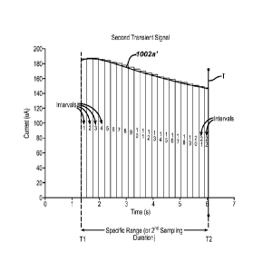
[0064] Figure 7A illustrates output signal transients that are sampled
during a test
sequence duration for respective high, medium, and low glucose concentrations
for each
range of hematocrits at 30%, 42% and 55%.
[0065] Figure 7B illustrates the relationship between hematocrits and the
time at which a
magnitude of the transient signal is measured.
[0066] Figure 7C illustrates one transient signal output, i.e., a "first
transient signal" from
the transient signals of Figure 7B.
[0067] Figure 7D illustrates the extraction of a portion of the one
transient signal output in
Figure 7C and the exemplary timing intervals for measuring the magnitudes of
this portion,
characterized here as a "second transient signal."
[0068] Figure 7E illustrates the extracted signals of Figure 7B and shifted
to the left so that
the start time for each of the second transient signals is about zero.
[0069] Figure 8A illustrates data from test measurements conducted with the
known
technique which shows relatively high bias along with substantial variations
in the bias
with respect to upper and lower hematocrit values.
[0070] Figures 8B, 8C, 8D, 8E, 8F, and 8G illustrate data from test
measurements
conducted with variations of the exemplary technique herein such that the data
show the
CA 02861769 2014-06-26
WO 2013/098564 PCT/GB2012/053277
bias of less than +15% for the hematocrit range of about 30% to about 55%
while
attainting relatively little variations in bias for hematocrits at extreme
values.
MODES OF CARRYING OUT THE INVENTION
[0071] The following detailed description should be read with reference to
the drawings, in
which like elements in different drawings are identically numbered. The
drawings, which
are not necessarily to scale, depict selected embodiments and are not intended
to limit the
scope of the invention. The detailed description illustrates by way of
example, not by way
of limitation, the principles of the invention. This description will clearly
enable one
skilled in the art to make and use the invention, and describes several
embodiments,
adaptations, variations, alternatives and uses of the invention, including
what is presently
believed to be the best mode of carrying out the invention.
[0072] As used herein, the terms "about" or "approximately" for any
numerical values or
ranges indicate a suitable dimensional tolerance that allows the part or
collection of
components to function for its intended purpose as described herein. More
specifically,
"about" or "approximately" may refer to the range of values +10% of the
recited value, e.g.
"about 90%" may refer to the range of values from 81% to 99%. As used herein,
"an
absolute value" of a difference refers to the magnitude of the difference,
i.e. it is always
positive. In addition, as used herein, the terms "patient," "host," "user,"
and "subject" refer
to any human or animal subject and are not intended to limit the systems or
methods to
human use, although use of the subject invention in a human patient represents
a preferred
embodiment. As used herein, "oscillating signal" includes voltage signal(s) or
current
signal(s) that, respectively, change polarity or alternate direction of
current or are multi-
directional. Also used herein, the phrase "electrical signal" or "signal" is
intended to
include direct current signal, alternating signal or any signal within the
electromagnetic
spectrum. The terms "processor"; "microprocessor"; or "microcontroller" are
intended to
have the same meaning and are intended to be used interchangeably.
[0073] Figure 1 illustrates a test meter 200, for testing analyte (e.g.,
glucose) levels in the
blood of an individual with a biosensor produced by the methods and techniques
illustrated
and described herein. Test meter 200 may include user interface inputs (206,
210, 214),
which can be in the form of buttons, for entry of data, navigation of menus,
and execution
26
CA 02861769 2014-06-26
WO 2013/098564 PCT/GB2012/053277
of commands. Data can include values representative of analyte concentration,
and/or
information that are related to the everyday lifestyle of an individual.
Information, which
is related to the everyday lifestyle, can include food intake, medication use,
the occurrence
of health check-ups, general health condition and exercise levels of an
individual. Test
meter 200 can also include a display 204 that can be used to report measured
glucose
levels, and to facilitate entry of lifestyle related information.
[0074] Test meter 200 may include a first user interface input 206, a
second user interface
input 210, and a third user interface input 214. User interface inputs 206,
210, and 214
facilitate entry and analysis of data stored in the testing device, enabling a
user to navigate
through the user interface displayed on display 204. User interface inputs
206, 210, and
214 include a first marking 208, a second marking 212, and a third marking
216, which
help in correlating user interface inputs to characters on display 204.
[0075] Test meter 200 can be turned on by inserting a biosensor 100 (or its
variants 400,
500, or 600) into a strip port connector 220, by pressing and briefly holding
first user
interface input 206, or by the detection of data traffic across a data port
218. Test meter
200 can be switched off by removing biosensor 100 (or its variants 400, 500,
or 600),
pressing and briefly holding first user interface input 206, navigating to and
selecting a
meter off option from a main menu screen, or by not pressing any buttons for a
predetermined time. Display 104 can optionally include a backlight.
[0076] In one embodiment, test meter 200 can be configured to not receive a
calibration
input for example, from any external source, when switching from a first test
strip batch to
a second test strip batch. Thus, in one exemplary embodiment, the meter is
configured to
not receive a calibration input from external sources, such as a user
interface (such as
inputs 206, 210, 214), an inserted test strip, a separate code key or a code
strip, data port
218. Such a calibration input is not necessary when all of the test strip
batches have a
substantially uniform calibration characteristic. The calibration input can be
a set of values
ascribed to a particular test strip batch. For example, the calibration input
can include a
batch slope and a batch intercept value for a particular test strip batch. The
calibrations
input, such as batch slope and intercept values, may be preset within the
meter as will be
described below.
27
CA 02861769 2014-06-26
WO 2013/098564 PCT/GB2012/053277
[0077] Referring to Figure 2A, an exemplary internal layout of test meter
200 is shown.
Test meter 200 may include a processor 300, which in some embodiments
described and
illustrated herein is a 32-bit RISC microcontroller. In the preferred
embodiments
described and illustrated herein, processor 300 is preferably selected from
the MSP 430
family of ultra-low power microcontrollers manufactured by Texas Instruments
of Dallas,
Texas. The processor can be bi-directionally connected via I/O ports 314 to a
memory 302,
which in some embodiments described and illustrated herein is an EEPROM. Also
connected to processor 300 vial/0 ports 214 are the data port 218, the user
interface inputs
206, 210, and 214, and a display driver 320. Data port 218 can be connected to
processor
300, thereby enabling transfer of data between memory 302 and an external
device, such as
a personal computer. User interface inputs 206, 210, and 214 are directly
connected to
processor 300. Processor 300 controls display 204 via display driver 320.
Memory 302
may be pre-loaded with calibration information, such as batch slope and batch
intercept
values, during production of test meter 200. This pre-loaded calibration
information can
be accessed and used by processor 300 upon receiving a suitable signal (such
as current)
from the strip via strip port connector 220 so as to calculate a corresponding
analyte level
(such as blood glucose concentration) using the signal and the calibration
information
without receiving calibration input from any external source.
[0078] In embodiments described and illustrated herein, test meter 200 may
include an
Application Specific Integrated Circuit (ASIC) 304, so as to provide
electronic circuitry
used in measurements of glucose level in blood that has been applied to a
biosensor 100
(or its variants 400, 500, or 600) inserted into strip port connector 220.
Analog voltages
can pass to and from ASIC 304 by way of an analog interface 306. Analog
signals from
analog interface 306 can be converted to digital signals by an AiD converter
316.
Processor 300 further includes a core 308, a ROM 310 (containing computer
code), a
RAM 312, and a clock 318. In one embodiment, the processor 300 is configured
(or
programmed) to disable all of the user interface inputs except for a single
input upon a
display of an analyte value by the display unit such as, for example, during a
time period
after an analyte measurement. In an alternative embodiment, the processor 300
is
configured (or programmed) to ignore any input from all of the user interface
inputs except
for a single input upon a display of an analyte value by the display unit.
Detailed
28
descriptions and illustrations of the meter 200 are shown and described in
International
Patent Application Publication No. W02006070200.
100791 Figure 3A(1) is an exemplary exploded perspective view of a test strip
100, which may
include seven layers disposed on a substrate 5. The seven layers disposed on
substrate 5 can
be a first conductive layer 50 (which can also be referred to as electrode
layer 50), an
insulation layer 16, two overlapping reagent layers 22a and 22b, an adhesive
layer 60 which
includes adhesive portions 24, 26, and 28, a hydrophilic layer 70, and a top
layer 80 which
forms a cover 94 for the test strip 100. Test strip 100 may be manufactured in
a series of
steps where the conductive layer 50, insulation layer 16, reagent layers 22,
and adhesive
layer 60 are sequentially deposited on substrate 5 using, for example, a
screen-printing
process. Note that the electrodes 10, 12, and 14 are disposed for contact with
the reagent
layer 22a and 22b whereas the physical characteristic sensing electrodes 19a
and 20a are
spaced apart and not in contact with the reagent layer 22. Hydrophilic layer
70 and top layer
80 can be disposed from a roll stock and laminated onto substrate 5 as either
an integrated
laminate or as separate layers. Test strip 100 has a distal portion 3 and a
proximal portion 4
as shown in Figure 3A(1).
100801 Test strip 100 may include a sample-receiving chamber 92 through which
a physiological
fluid sample 95 may be drawn through or deposited (Fig. 3A(2)). The
physiological fluid
sample discussed herein may be blood. Sample-receiving chamber 92 can include
an inlet at
a proximal end and an outlet at the side edges of test strip 100, as
illustrated in Figure 3A(1).
A fluid sample 95 can be applied to the inlet along axis L-L (Fig. 3A(2)) to
fill a sample-
receiving chamber 92 so that analyte can be measured from the sample. The side
edges of a
first adhesive pad 24 and a second adhesive pad 26 located adjacent to reagent
layer 22 each
define a wall of sample-receiving chamber 92, as illustrated in Figure 3A(1).
A bottom
portion or "floor" of sample-receiving chamber 92 may include a portion of
substrate 5,
conductive layer 50, and insulation layer 16, as illustrated in Figure 3A(1).
A top portion or
"roof' of sample-receiving chamber 92 may include distal hydrophilic portion
32, as
illustrated in Figure 3A(1). For test strip 100, as illustrated in Figure
3A(1), substrate Scan
be used as a foundation for helping support subsequently applied layers.
Substrate 5 can be in the form of a polyester sheet such as a polyethylene
tetraphthalate
29
CA 2861769 2019-12-02
CA 02861769 2014-06-26
WO 2013/098564 PCT/GB2012/053277
(PET) material (Hostaphan PET supplied by Mitsubishi). Substrate 5 can be in a
roll
format, nominally 350 microns thick by 370 millimeters wide and approximately
60 meters
in length.
[0081] A conductive layer is required for forming electrodes that can be used
for the
electrochemical measurement of glucose. First conductive layer 50 can be made
from a
carbon ink that is screen-printed onto substrate 5. In a screen-printing
process, carbon ink
is loaded onto a screen and then transferred through the screen using a
squeegee. The
printed carbon ink can be dried using hot air at about 140 C. The carbon ink
can include
VAGH resin, carbon black, graphite (KS15), and one or more solvents for the
resin, carbon
and graphite mixture. More particularly, the carbon ink may incorporate a
ratio of carbon
black: VAGH resin of about 2.90:1 and a ratio of graphite: carbon black of
about 2.62:1 in
the carbon ink.
[0082] For test strip 100, as illustrated in Figure 3A(1), first conductive
layer 50 may include a
reference electrode 10, a first working electrode 12, a second working
electrode 14, third
and fourth physical characteristic sensing electrodes 19a and 19b, a first
contact pad 13, a
second contact pad 15, a reference contact pad 11, a first working electrode
track 8, a
second working electrode track 9, a reference electrode track 7, and a strip
detection bar
17. The physical characteristic sensing electrodes 19a and 20a are provided
with
respective electrode tracks 19b and 20b. The conductive layer may be formed
from carbon
ink. First contact pad 13, second contact pad 15, and reference contact pad 11
may be
adapted to electrically connect to a test meter. First working electrode track
8 provides an
electrically continuous pathway from first working electrode 12 to first
contact pad 13.
Similarly, second working electrode track 9 provides an electrically
continuous pathway
from second working electrode 14 to second contact pad 15. Similarly,
reference electrode
track 7 provides an electrically continuous pathway from reference electrode
10 to
reference contact pad 11. Strip detection bar 17 is electrically connected to
reference
contact pad 11. Third and fourth electrode tracks 19b and 20b connect to the
respective
electrodes 19a and 20a. A test meter can detect that test strip 100 has been
properly
inserted by measuring a continuity between reference contact pad 11 and strip
detection
bar 17, as illustrated in Figure 3A(1).
CA 02861769 2014-06-26
WO 2013/098564 PCT/GB2012/053277
[0083] In the embodiment of Figure 3A(2) which is a variation of the test
strip of Figure
3A(1), an additional electrode 10a is provided as an extension of any of the
plurality of
electrodes 19a, 20a, 14, 12, and 10. It must be noted that the built-in
shielding or
grounding electrode 10a is used to reduce or eliminate any capacitance
coupling between
the finger or body of the user and the characteristic measurement electrodes
19a and 20a.
The grounding electrode 10a allows for any capacitance to be directed away
from the
sensing electrodes 19a and 20a. To do this, the grounding electrode 10a can be
connected
any one of the other five electrodes or to its own separate contact pad (and
track) for
connection to ground on the meter instead of one or more of contact pads 15,
17, 13 via
respective tracks 7, 8, and 9. In a preferred embodiment, the grounding
electrode 10a is
connected to one of the three electrodes that has reagent 22 disposed thereon.
In a most
preferred embodiment, the grounding electrode 10a is connected to electrode
10. Being
the grounding electrode, it is advantageous to connect the grounding electrode
to the
reference electrode (10) so not to contribute any additional current to the
working electrode
measurements which may come from background interfering compounds in the
sample.
Further by connecting the shield or grounding electrode 10a to electrode 10,
this is
believed to effectively increase the size of the counter electrode 10 which
can become
limiting especially at high signals. In the embodiment of Figure 3A(2), the
reagent are
arranged so that they are not in contact with the measurement electrodes 19a
and 20a.
Alternatively, in the embodiment of Figure 3A(3), the reagent 22 is arranged
so that the
reagent 22 contacts at least one of the sensing electrodes 19a and 20a.
[0084] In alternate version of test strip 100, shown here in Figure 3A(4), the
top layer 38,
hydrophilic film layer 34 and spacer 29 have been combined together to form an
integrated
assembly for mounting to the substrate 5 with reagent layer 22' disposed
proximate
insulation layer 16'.
[0085] In Figure 3A(5), it can be seen in the plan view that the first two
electrodes 19a and 20a are
nearest to the entrance of the blood receiving channel 18. The tracks of the
electrodes are
configured to mate with five respective contact surfaces of the strip
receiving port. As
shown in Figure 3A(6), which is a close-up of sample receiving end of the
strip 100, the
first electrode track 19a is spaced at a distance Li from the second electrode
track 20a.
The second electrode track 20a is spaced at a distance L2 from electrode 10,
which
31
CA 02861769 2014-06-26
WO 2013/098564 PCT/GB2012/053277
distance L2 may be from about 1 to about 1/2 of Li. The thickness hl of the
electrode 19a
can be the same or different in size as compared to thickness h2 of the second
electrode
20a. For electrode 10, the thickness h3 can be about 6 to about 7 times that
of thickness hl
whereas respective thicknesses h4 and h5 can be about 2 to about 4 times that
of hl or h2.
In the preferred embodiment, the distance Li may be about 1.2 millimeters and
the
thickness hl may be about 0.2 millimeters.
[0086] Variations of the biosensor 100 (Figures 3A(1-6)) are shown in
Figures 3B-3T.
Briefly, with regard to variations of biosensor 100 (illustrated exemplarily
in Figures 3B
through 3T), these biosensors include an enzymatic reagent layer disposed on
the working
electrode, a patterned spacer layer disposed over the first patterned
conductive layer and
configured to define a sample chamber within the analytical biosensor, and a
second
patterned conductive layer disposed above the first patterned conductive
layer. The second
patterned conductive layer includes a first phase-shift measurement electrode
and a second
phase-shift measurement electrode. Moreover, the first and second phase-shift
measurement electrodes are disposed in the sample chamber and are configured
to
measure, along with the hand-held test meter, a phase shift of an electrical
signal forced
through a bodily fluid sample introduced into the sample chamber during use of
the
analytical biosensor. Such phase-shift measurement electrodes are also
referred to herein
as bodily fluid phase-shift measurement electrodes. Analytical biosensors of
various
embodiments described herein are believed to be advantageous in that, for
example, the
first and second phase-shift measurement electrodes are disposed above the
working and
reference electrodes, thus enabling a sample chamber of advantageously low
volume. This
is in contrast to a configuration wherein the first and second phase-shift
measurement
electrodes are disposed in a co-planar relationship with the working and
reference
electrodes thus requiring a larger bodily fluid sample volume and sample
chamber to
enable the bodily fluid sample to cover the first and second phase-shift
measurement
electrodes as well as the working and reference electrodes.
[0087] In the embodiment of Figure 3B, the analyte measurement electrodes
10, 12, and
14 are disposed in generally the same configuration as in Figs. 3A(1, 2, 3, 4,
5, or 6). The
electrodes 19a and 20a to sense hematocrit level, however, are disposed in a
spaced apart
configuration in which one electrode 19a is proximate an entrance 92a to the
test chamber
32
CA 02861769 2014-06-26
WO 2013/098564 PCT/GB2012/053277
92 and another electrode 20a is at the opposite end of the test chamber 92. At
least one of
the electrodes on the biosensor is disposed to be in contact with a reagent
layer 22.
[0088] In Figures 3C, 3D, 3E and 3F, the hematocrit sensing electrodes 19a
and 20a are
disposed adjacent each other and may be placed at the opposite end 92b of the
entrance 92a
to the test chamber 92 (Figs. 3C and 3D) or adjacent the entrance 92a (Figs.
3E and 3F).
In all of these embodiments, the physical characteristic sensing electrodes
are spaced apart
from the reagent layer 22 so that these physical characteristic sensing
electrodes are not
impacted by the electrochemical reaction of the reagent in the presence of a
fluid sample
(e.g., blood or interstitial fluid) containing glucose.
[0089] Referring to Figures 3G through 3J, electrochemical-based analytical
biosensor 400
includes an electrically-insulating substrate layer 402, a first patterned
conductive layer
404 disposed on the electrically-insulating substrate layer, an enzymatic
reagent layer 406
(for clarity depicted in Figure 3G only), a patterned spacer layer 408, a
second patterned
conductive layer 410 disposed above first patterned conductive layer 404, and
an
electrically-insulating top layer 412. Patterned spacer layer 408 is
configured such that
electrochemical-based analytical biosensor 400 also includes a sample chamber
414
formed therein with patterned spacer layer 408 defining outer walls of sample
chamber
414.
[0090] First patterned conductive layer 404 includes three electrodes, a
counter electrode
404a (also referred to as a reference electrode), a first working electrode
404b and a second
working electrode 404c (see Figure 3G).
[0091] Second patterned conductive layer 410 includes a first phase-shift
measurement
electrode 411 and a second phase shift measurement electrode 413. Second
patterned
conductive layer 410 also includes a first phase-shift probe contact 416 and a
second
phase-shift probe contact 418.
[0092] During use of electrochemical-based analytical biosensor 400 to
determine an
analyte in a bodily fluid sample (e.g., blood glucose concentration in a whole
blood
sample), electrodes 404a, 404b and 404c are employed by an associated meter
(not shown)
to monitor an electrochemical response of the electrochemical-based analytical
biosensor.
The electrochemical response can be, for example, an electrochemical reaction
induced
current of interest. The magnitude of such a current can then be correlated,
taking into
33
CA 02861769 2014-06-26
WO 2013/098564 PCT/GB2012/053277
consideration the physical characteristic (e.g., hematocrit) of the bodily
fluid sample as
determined by the bodily fluid sample's phase shift, with the amount of
analyte present in
the bodily fluid sample under investigation. During such use, a bodily fluid
sample is
applied to electrochemical-based analytical biosensor 400 and, thereby,
received in sample
chamber 414.
[0093] Electrically-insulating substrate layer 402 can be any suitable
electrically-insulating
substrate known to one skilled in the art including, for example, a nylon
substrate,
polycarbonate substrate, a polyimide substrate, a polyvinyl chloride
substrate, a
polyethylene substrate, a polypropylene substrate, a glycolated polyester
(PETG) substrate,
a polystyrene substrate, a silicon substrate, ceramic substrate, glass
substrate or a polyester
substrate (e.g., a 7 millimeters thick polyester substrate). The electrically-
insulating
substrate can have any suitable dimensions including, for example, a width
dimension of
about 5 mm, a length dimension of about 27 mm and a thickness dimension of
about 0.5
mm.
[0094] First patterned conductive layer 404 can be formed of any suitable
electrically
conductive material such as, for example, gold, palladium, carbon, silver,
platinum, tin
oxide, iridium, indium, or combinations thereof (e.g., indium doped tin
oxide). Moreover,
any suitable technique or combination of techniques can be employed to form
first
patterned conductive layer 404 including, for example, sputtering,
evaporation, electro-less
plating, screen-printing, contact printing, laser ablation or gravure
printing. A typical but
non-limiting thickness for the patterned conductive layer is in the range of
5nanometers to
400nanometers.
[0095] As is known, conventional electrochemical-based analyte biosensors
(e.g. test
strips) employ a working electrode along with an associated counter/reference
electrode
and enzymatic reagent layer to facilitate an electrochemical reaction with an
analyte of
interest and, thereby, determine the presence and/or concentration of that
analyte. For
example, an electrochemical-based analyte biosensor for the determination of
glucose
concentration in a blood sample can employ an enzymatic reagent that includes
the enzyme
glucose oxidase and the mediator ferricyanide (which is reduced to the
mediator
ferrocyanide during the electrochemical reaction). Such conventional analyte
test strips
34
and enzymatic reagent layers are described in, for example, U.S. Patents
5,708,247;
5,951,836; 6,241,862; and 6,284,125. In this regard, the reagent layer
employed in various
embodiments provided herein can include any suitable sample-soluble enzymatic
reagents,
with the selection of enzymatic reagents being dependent on the analyte to be
determined
and the bodily fluid sample. For example, if glucose is to be determined in a
blood sample,
enzymatic reagent layer 406 can include glucose oxidase or glucose
dehydrogenase along
with other components necessary for functional operation.
100961 In general, enzymatic reagent layer 406 includes at least an enzyme
and a mediator.
Examples of suitable mediators include, for example, ruthenium, Hexaammine
Ruthenium
(III) Chloride, ferricyanide, ferrocene, ferrocene derivatives, osmium
bipyridyl complexes,
and quinone derivatives. Examples of suitable enzymes include glucose oxidase,
glucose
dehydrogenase (GDH) using a pyrroloquinoline quinone (PQQ) co-factor, GDH
using a
nicotinamide adenine dinucleotide (NAD) co-factor, and GDH using a flavin
adenine
dinucleotide (FAD) co-factor. Enzymatic reagent layer 406 can be applied
during
manufacturing using any suitable technique including, for example, screen
printing.
100971 Applicant notes that enzymatic reagent layer 406 may also contain
suitable buffers
(such as, for example, Tris HCl, Citraconate, Citrate and Phosphate),
hydroxyethylcelulose
[HEC], carboxymethylcellulose, ethycellulose and alginate, enzyme stabilizers
and other
additives as are known in the field.
100981 Further details regarding the use of electrodes and enzymatic
reagent layers for the
determination of the concentrations of analytes in a bodily fluid sample,
albeit in the absence
of the phase-shift measurement electrodes, analytical test strips and related
methods
described herein, are in U.S. Patent No. 6,733,655.
100991 Patterned spacer layer 408 can be formed of any suitable material
including, for
example, a 95micrometers thick, double-sided pressure sensitive adhesive
layer, a heat
activated adhesive layer, or a thermo-setting adhesive plastic layer.
Patterned spacer layer
408 can have, for example, a thickness in the range of from about 1 micron to
about 500
CA 2861769 2019-12-02
microns, preferably between about 10 microns and about 400 microns, and more
preferably
between about 40 microns and about 200 microns.
1001001 Second patterned conductive layer 410 can be formed of any suitable
conductive
material including, for example, copper, silver, palladium, gold and
conductive carbon
materials. Second patterned conductive layer 410 can be, for example, disposed
on a lower
surface of electrically-insulating top layer 412 (as depicted in Figures 3G-
3J) or embedded in
the lower surface of electrically-insulating top layer 412. Second patterned
conductive layer
410 can have any suitable thickness including, for example, a thickness in the
range of 20
microns to 400 microns.
1001011 First phase-shift measurement electrode 411 and second phase shift
measurement
electrode 413 of second patterned conductive layer 410 are separated within
sample chamber
414 by a gap (in the horizontal direction of Figure 3J) that is suitable for
phase-shift
measurement. Such a gap can be, for example, in the range of 20 microns to
1,400 microns
with a typical gap being 500 microns. Moreover, the surface area of first
phase-shift
measurement electrode 111 and second phase shift measurement electrode 113
that is
exposed to a bodily fluid sample within sample chamber 414 is typically about
0.5 mm2 but
can range, for example, from about 0.1 mm2 to about 2.0 mm2.
1001021 Electrochemical-based analytical biosensor 400 can be manufactured,
for example,
by the sequential aligned formation of first patterned conductive layer 404,
enzymatic
reagent layer 406, patterned spacer layer 408, second patterned conductive
layer 410 and
electrically insulating top layer 412 onto electrically-insulating substrate
layer 402. Any
suitable techniques known to one skilled in the art can be used to accomplish
such sequential
aligned formation, including, for example, screen printing, photolithography,
photogravure,
chemical vapour deposition, sputtering, tape lamination techniques and
combinations
thereof.
1001031 Analytical biosensors according to embodiments provided herein
can be
configured, for example, for operable electrical connection (via, for example,
first and
second phase shift probe contacts 416 and 418) and use with the analytical
test strip sample
cell interface of a hand-held test meter as described in co-pending patent
application
13/250,525 (Publication No. US 2013-0084589 Al)
36
CA 2861769 2019-12-02
1001041 It has
been determined that a relationship exists between the reactance of a
whole blood sample and the physical characteristic (e.g., hematocrit) of that
sample.
Electrical modeling of a bodily fluid sample (e.g., a whole blood sample) as
parallel
capacitive and resistive components indicates that when an alternating current
(AC) signal is
forced through the bodily fluid sample, the phase shift of the alternating
signal will be
dependent on both the frequency of the alternating signal voltage and the
physical
characteristic (e.g., hematocrit) of the sample. Therefore, the physical
characteristic (e.g.,
hematocrit) of a bodily fluid sample can be measured by, for example, driving
alternating
signals of a known frequency (or known frequencies) through the bodily fluid
sample and
detecting their phase shift. The phase-shift measurement electrodes of
analytical biosensors
of various embodiments described herein are particularly suitable for use in
such phase-shift
measurements since the first and second phase shift measurement electrodes are
in direct
contact with a bodily fluid sample present in the sample chamber.
1001051
Applicant notes that for various embodiments of analytical biosensors (e.g.,
an electrochemical-based analytical test strip) described here for use with a
hand-held test
meter in the determination of an analyte (such as glucose) in a bodily fluid
sample (for
example, a whole blood sample) may include an electrically insulating
substrate, a first
patterned conductor layer disposed on the electrically insulating substrate
and having a
working electrode and a reference electrode. The analytical biosensor may also
include an
enzymatic reagent layer disposed on the working electrode, a first patterned
spacer layer
a phase-shift-based hematocrit measurement block that includes:
channel and an analyte determination sample chamber within the analytical
biosensor, and a
second patterned spacer layer disposed over the first patterned spacer layer
and defining at
least a second sample-receiving channel. In addition, the analytical biosensor
further
includes a bodily fluid phase-shift sample chamber in fluidic communication
with the second
sample-receiving channel. Moreover, the first sample-receiving channel and
analyte
determination sample chamber of the analytical biosensor are isolated from the
second
sample-receiving channel and bodily fluid phase-shift sample chamber of the
analytical
biosensor.
37
CA 2861769 2019-12-02
CA 02861769 2014-06-26
WO 2013/098564 PCT/GB2012/053277
[00106] Analytical biosensors of various embodiments described herein
are
believed by applicant to be beneficial in that, for example, the isolation
(fluidic and
electrical) between the analyte determination sample chamber and the bodily
fluid phase-
shift sample chamber prevents potential interference between the determination
of the
analyte in the bodily fluid sample and a phase-shift measurement of the bodily
fluid.
Applicant notes that certain advantages are obtained by having the first
sample-receiving
channel and analyte determination chamber are separated from the second sample-
receiving channel and bodily fluid phase-shift sample chamber by portions of
the first
and/or second patterned spacer layers that can be thinner, thus providing for
an analytical
biosensor with a small, yet mechanically stable, cross-section.
[00107] Referring to Figures 3K-30, electrochemical-based analytical
biosensor 500
includes an electrically-insulating substrate 502, a first patterned conductor
layer 504
disposed on the electrically-insulating substrate layer, an enzymatic reagent
layer 506 (for
clarity depicted in Figure 3K only), a first patterned spacer layer 508, a
second patterned
spacer layer 510, and a top cover 511. In the embodiment of Figure 3K, first
pattered
spacer layer 508 and second patterned spacer layer 510 are depicted as bi-
layer structures.
However, the first and second patterned spacer layers employed in various
embodiments
provided herein can be unitary layers or any other suitably formed layer.
[00108] First patterned spacer layer 508 is configured such that
electrochemical-based
analytical biosensor 500 also includes a first sample-receiving channel 512
and an analyte
determination sample chamber 514. First patterned spacer layer 508 is also
configured to
define a bodily fluid phase-shift sample chamber 516 and an analyte
determination sample
chamber vent 518 (for clarity not depicted in Figure 3K).
[00109] Second patterned spacer layer 510 is configured to define a second
sample-
receiving channel 520 and a bodily fluid phase-shift chamber vent 522 (for
clarity not
depicted in Figure 3K).
[00110] First patterned conductor layer 504 includes a first phase-shift
measurement
electrode 524, a second phase-shift measurement electrode 526, two working
electrodes
528a and 528b and a reference electrode 530. For clarity, Figure 3L depicts
only first
phase-shift measurement electrode 524 and second phase-shift measurement
electrode 526
and not the entirety of first patterned conductor layer 504.
38
CA 02861769 2014-06-26
WO 2013/098564 PCT/GB2012/053277
[00111] First sample-receiving channel 512 and analyte determination sample
chamber
514 are isolated, both fluidically and electrically, from second sample-
receiving channel
520 and bodily fluid phase-shift sample chamber 516 (see Figure 30 in
particular wherein
the first and second patterned conductor layers are omitted for clarity).
Moreover, in the
embodiment of Figure 30, the bodily fluid phase-shift sample chamber is
disposed in a
side-by-side configuration with the analyte determination sample chamber.
[00112] During use of electrochemical-based analytical biosensor 500 to
determine an
analyte in a bodily fluid sample (e.g., blood glucose concentration in a whole
blood
sample), working and reference electrodes are employed by an associated meter
(not
shown) to monitor an electrochemical response of the electrochemical-based
analytical
biosensor. The electrochemical response can be, for example, an
electrochemical reaction
induced current of interest. The magnitude of such a current can then be
correlated, taking
into consideration the haematocrit of the bodily fluid sample as determined by
the bodily
fluid sample's phase shift, with the amount of analyte present in the bodily
fluid sample
under investigation. During such use, a bodily fluid sample is applied to
electrochemical-
based analytical biosensor 500 and, thereby, received in both analyte
determination sample
chamber 514 and bodily fluid phase-shift sample chamber 516.
[00113] Electrically-insulating substrate 502 can be any suitable
electrically-insulating
substrate known to one skilled in the art including, for example, a nylon
substrate,
polycarbonate substrate, a polyimide substrate, a polyvinyl chloride
substrate, a
polyethylene substrate, a polypropylene substrate, a glycolated polyester
(PETG) substrate,
a polystyrene substrate, a silicon substrate, ceramic substrate, glass
substrate or a polyester
substrate (e.g., a 7 millimeters thick polyester substrate). The electrically-
insulating
substrate can have any suitable dimensions including, for example, a width
dimension of
about 5 mm, a length dimension of about 27 mm and a thickness dimension of
about 0.5
mm.
[00114] First patterned conductor layer 504 can be formed of any suitable
electrically
conductive material such as, for example, gold, palladium, carbon, silver,
platinum, tin
oxide, iridium, indium, or combinations thereof (e.g., indium doped tin
oxide). Moreover,
any suitable technique or combination of techniques can be employed to form
first
patterned conductor layer 504 including, for example, sputtering, evaporation,
electro-less
39
plating. screen-printing, contact printing, laser ablation or gravure
printing. A typical but
non-limiting thickness for the patterned conductor layer is in the range of
5nanometers to
500nanometers.
1001151 Applicant notes that conventional electrochemical-based analyte
biosensors employ
a working electrode along with an associated counter/reference electrode and
enzymatic
reagent layer to facilitate an electrochemical reaction with an analyte of
interest and, thereby,
determine the presence and/or concentration of that analyte. For example, an
electrochemical-based analyte biosensor for the determination of glucose
concentration in a
blood sample can employ an enzymatic reagent that includes the enzyme glucose
oxidase
and the mediator ferricyanide (which is reduced to the mediator ferrocyanide
during the
electrochemical reaction). Such conventional analyte test strips and enzymatic
reagent
layers are described in, for example, U.S. Patents 5,708,247; 5,951,836;
6,241,862; and
6,284,125. In this regard, the reagent layer employed in various embodiments
provided
herein can include any suitable sample-soluble enzymatic reagents, with the
selection of
enzymatic reagents being dependent on the analyte to be determined and the
bodily fluid
sample. For example, if glucose is to be determined in a blood sample,
enzymatic reagent
layer 506 can include glucose oxidase or glucose dehydrogenase along with
other
components necessary for functional operation.
[001161 In general, enzymatic reagent layer 506 includes at least an
enzyme and a mediator.
Examples of suitable mediators include, for example, ferricyanide, ferrocene,
ferrocene
derivatives, osmium bipyridyl complexes, and quinone derivatives. Examples of
suitable
enzymes include glucose oxidase. glucose dehydrogenase (GDH) using a
pyrroloquinoline
quinone (PQQ) co-factor, GDH using a nicotinamide adenine dinucleotide (NAD)
co-factor,
and GDH using a flavin adenine dinucleotide (FAD) co-factor. Enzymatic reagent
layer 506
can be applied during manufacturing using any suitable technique including,
for example,
screen printing.
1001171 Applicant notes that enzymatic reagent layer 506 may also contain
suitable buffers
(such as, for example, Tris HC1, Citraconate, Citrate and Phosphate),
hydroxyethylcelulose
CA 2861769 2019-12-02
[HEC], carboxymethylcellulose, ethycellulose and alginate, enzyme stabilizers
and other
additives as are known in the field.
1001181 Further details regarding the use of electrodes and enzymatic
reagent layers for the
determination of the concentrations of analytes in a bodily fluid sample,
albeit in the absence
of the phase-shift measurement electrodes, bodily-fluid phase-shift sample
chambers and
second sample receiving channels analytical test strips and related methods
described herein,
are in U.S. Patent No. 6,733,655.
1001191 First and second patterned spacer layers 508 and 510 respectively
can be formed of
any suitable material including, for example, a 95micrometers thick, double-
sided pressure
sensitive adhesive layer, a heat activated adhesive layer, or a thermo-setting
adhesive plastic
layer. First patterned spacer layer 508 can have, for example, a thickness in
the range of
from about 1 micron to about 500 microns, preferably between about 10 microns
and about
400 microns, and more preferably between about 40 microns and about 600
microns.
1001201 Electrochemical-based analytical biosensor 500 can be manufactured,
for example,
by the sequential aligned formation of first patterned conductor layer 504,
enzymatic reagent
layer 506, first patterned spacer layer 508, and second patterned spacer layer
510 onto
electrically-insulating substrate 502. Any suitable techniques known to one
skilled in the art
can be used to accomplish such sequential aligned formation, including, for
example, screen
printing, photolithography, photogravure, chemical vapour deposition,
sputtering, tape
lamination techniques and combinations thereof.
1001211 Analytical biosensors according to embodiments can be
configured, for
example, for operable electrical connection and use with the analytical
biosensor sample cell
interface of a hand-held test meter as described in co-pending patent
application 13/250,525
(Publication No. US 2013-0084589 Al) .
1001221 It has been determined that a relationship exists between the
reactance of a
whole blood sample and the physical characteristic (e.g., hematocrit) of that
sample.
Electrical modeling of a bodily fluid sample (e.g., a whole blood sample) as
parallel
41
CA 2861769 2019-12-02
CA 02861769 2014-06-26
WO 2013/098564 PCT/GB2012/053277
capacitive and resistive components indicates that when an alternating signal
such as, for
example, alternating-current (AC) signal is forced through the bodily fluid
sample, the
phase shift of the alternating signal will be dependent on both the frequency
of the
alternating signal voltage and the physical characteristic (e.g., hematocrit,
viscosity,
temperature) of the sample. Therefore, the physical characteristic (e.g.,
hematocrit,
viscosity, temperature) of a bodily fluid sample can be measured by, for
example, driving
alternating signals of known frequencies through the bodily fluid sample and
detecting
their phase shift. The phase-shift measurement electrodes of analytical test
strips of
various embodiments described herein are particularly suitable for use in such
phase-shift
measurements since the first and second phase shift measurement electrodes are
in direct
contact with a bodily fluid sample present in the sample chamber.
[00123] Referring to Figures 3P-3T, electrochemical-based analytical
test strip 600
includes an electrically-insulating substrate 602, a first patterned conductor
layer 604
disposed on the electrically-insulating substrate layer, an enzymatic reagent
layer 606 (for
clarity depicted in Figure 3P only), a first patterned spacer layer 608, a
second patterned
conductor layer 609, a second patterned spacer layer 610, and a top cover 611.
In the
embodiment of Figure 3P, first pattered spacer layer 608 and second patterned
spacer layer
610 are depicted as bi-layer structures. However, the first and second
patterned spacer
layers employed in various embodiments provided herein can be unitary layers
or any
other suitably formatted layer.
[00124] First patterned spacer layer 608 is configured such that
electrochemical-
based analytical biosensor 600 also includes a first sample-receiving channel
612, an
analyte determination sample chamber 614 and an analyte determination sample
chamber
vent 618 (not depicted in Figure 3P but depicted with dashed lines in Figure
3R). Analyte
determination sample chamber vent 618 is configured to aid in the introduction
of a bodily
fluid sample into analyte determination sample chamber 614 via first sample-
receiving
channel 612.
[00125] Second patterned spacer layer 610 is configured to define a
second sample-
receiving channel 620, a bodily fluid phase-shift sample chamber 616 and a
bodily fluid
phase-shift chamber vent 622 (not depicted in Figure 3P but depicted with
dashed lines in
Figure 3S). Bodily fluid phase-shift chamber vent 622 is configured to aid in
the
42
CA 02861769 2014-06-26
WO 2013/098564 PCT/GB2012/053277
introduction of a bodily fluid sample into bodily fluid phase-shift sample
chamber 616 via
second sample-receiving channel 620.
[00126] First patterned conductor layer 604 two working electrodes 628a and
628b
(depicted in Figures 3P and 3Q) and a reference electrode 630 (also depicted
in Figures 3P
and 3Q). Second patterned conductor layer 609 includes a first phase-shift
measurement
electrode 624 and a second phase-shift measurement electrode 626 and is
disposed above
first patterned spacer layer 608 and embedded in the bi-layer structure of
second pattered
spacer layer 610.
[00127] First sample-receiving channel 612 and analyte determination
sample
chamber 614 are isolated, both fluidically and electrically, from second
sample-receiving
channel 620 and bodily fluid phase-shift sample chamber 616 (see Figure 3T in
particular
wherein the first and second patterned conductor layers are not depicted for
clarity).
[00128] In the various embodiments of the test strip, there are two
measurements that are
made to a blood sample deposited on the test strip. One measurement is that of
the glucose
in the blood sample while the other is that of physical characteristic (e.g.,
hematocrit) in
the same sample. Both measurements (glucose and hematocrit) can be performed
in
sequence, simultaneously or overlapping in duration. For example, the glucose
measurement can be performed first then the physical characteristic (e.g.,
hematocrit); the
physical characteristic (e.g., hematocrit) measurement first then the glucose
measurement;
both measurements at the same time; or a duration of one measurement may
overlap a
duration of the other measurement. Each measurement is discussed in detail as
follow with
respect to Figures 4A, 4B and 5.
[00129] Figure 4A is an exemplary chart of a test signal applied to test
strip 100 and its
variations shown here in Figures 3A-3T. Before a fluid sample is applied to
test strip 100
(or its variants 400, 500, or 600), test meter 200 is in a fluid detection
mode in which a first
test signal of about 400 millivolts is applied between second working
electrode and
reference electrode. A second test signal of about 400 millivolts is
preferably applied
simultaneously between first working electrode (e.g., electrode 12 of strip
100) and
reference electrode (e.g., electrode 10 of strip 100). Alternatively, the
second test signal
may also be applied contemporaneously such that a time interval of the
application of the
43
CA 02861769 2014-06-26
WO 2013/098564
PCT/GB2012/053277
first test signal overlaps with a time interval in the application of the
second test voltage.
The test meter may be in a fluid detection mode during fluid detection time
interval TFD
prior to the detection of physiological fluid at starting time at zero. In the
fluid detection
mode, test meter 200 determines when a fluid is applied to test strip 100 (or
its variants
400, 500, or 600) such that the fluid wets second working electrode 14 and
reference
electrode 10. Once test meter 200 recognizes that the physiological fluid has
been applied
because of, for example, a sufficient increase in the measured test current at
second
working electrode 14, test meter 200 assigns a zero second marker at zero time
"0" and
starts the test time interval Ti. Test meter 200 may sample the current
transient output at a
suitable sampling rate, such as, for example, every 1 milliseconds to every
100
milliseconds. Upon the completion of the test time interval T,, the test
signal is removed.
For simplicity, Figure 4A only shows the first test signal applied to test
strip 100 (or its
variants 400, 500, or 600).
[00130] Hereafter,
a description of how analyte (e.g., glucose) concentration is
determined from the known current transients (e.g., the measured electrical
current
response in microamperes as a function of time) that are measured when the
test voltages
of Figure 4A are applied to the test strip 100 (or its variants 400, 500, or
600).
[00131] In Figure 4A, the first and second test voltages applied to test
strip 100 (or its
variants described herein) are generally from about +100 millivolts to about
+600
millivolts. In one embodiment in which the electrodes include carbon ink and
the mediator
includes ferricyanide, the test signal is about +400 millivolts. Other
mediator and
electrode material combinations will require different test voltages, as is
known to those
skilled in the art. The duration of the test voltages is generally from about
1 to about 5
seconds after a reaction period and is typically about 3 seconds after a
reaction period.
Typically, test sequence time Ts is measured relative to time to. As the
voltage 401 is
maintained in Figure 4A for the duration of Ts, output signals are generated,
shown here in
Figure 4B with the current transient 702 for the first working electrode 12
being generated
starting at zero time and likewise the current transient 704 for the second
working
electrode 14 is also generated with respect to the zero time. It is noted that
while the signal
transients 702 and 704 have been placed on the same referential zero point for
purposes of
explaining the process, in physical term, there is a slight time differential
between the two
44
CA 02861769 2014-06-26
WO 2013/098564 PCT/GB2012/053277
signals due to fluid flow in the chamber towards each of the working
electrodes 12 and 14
along axis L-L. However, the current transients are sampled and configured in
the
microcontroller to have the same start time. In Figure 4B, the current
transients build up to
a peak proximate peak time Tp at which time, the current slowly drops off
until
approximately one of 2.5 seconds or 5 seconds after zero time. At the point
706,
approximately at 5 seconds, the output signal for each of the working
electrodes 12 and 14
may be measured and added together. Alternatively, the signal from only one of
the
working electrodes 12 and 14 can be doubled.
[00132] Referring back to Fig. 2B, the system drives a signal to measure or
sample the
output signals /E from at least one the working electrodes (12 and 14) at any
one of a
plurality of time points or positions Tl, T2, T3, .... TN. As can be seen in
Fig. 4B, the
time position can be any time point or interval in the test sequence Ts. For
example, the
time position at which the output signal is measured can be a single time
point T1.5 at 1.5
seconds or an interval 708 (e.g., interval-10 milliseconds or more depending
on the
sampling rate of the system) overlapping the time point T2.8 proximate 2.8
seconds..
[00133] From knowledge of the batch calibration code offset and batch slope
for the
particular test strip 100 and its variations in Figures 3B-3T, the analyte
(e.g., glucose)
concentration can be calculated.
[00134] It is noted that "Intercept" and "Slope" are the values
obtained by
measuring calibration data from a batch of test strips. Typically around 1500
strips (or
more in some instances) are selected at random from the lot or batch.
Physiological fluid
(e.g., blood samples) from donors is spiked to various analyte levels,
typically six different
glucose concentrations. Typically, blood from 12 different donors is spiked to
each of the
six levels. Eight strips are given blood from identical donors and levels so
that a total of
12 x 6 x 8 576 tests are conducted for that lot. These are benchmarked against
actual
analyte level (e.g., blood glucose concentration) by measuring these using a
standard
laboratory analyzer such as Yellow Springs Instrument (YSI). A graph of
measured
glucose concentration is plotted against actual glucose concentration (or
measured current
versus YSI current) and a formula y = mx+c least squares fitted to the graph
to give a value
for batch slope m and batch intercept c for the remaining strips from the lot
or batch.
CA 02861769 2014-06-26
WO 2013/098564
PCT/GB2012/053277
[00135] It is
worthwhile here to note that the various components, systems and
procedures described earlier allow for applicant to provide for analyte
measurement system
that heretofore was not available in the art. In particular, this system
includes a test strip
that has a substrate and a plurality of electrodes disposed on the substrate
and connected to
respective electrode connectors. The system further includes an analyte meter
that has a
housing, a test strip port connector configured to connect to the respective
electrode
connectors of the test strip, and a microprocessor 300. The microprocessor 300
is in
electrical communication with the test strip port connector 220 to apply
electrical signals
or sense electrical signals from the plurality of electrodes.
[00136] Referring to Figure 2B, details of a preferred implementation of
meter 200 where
the same numeral in respective Figures 2A and 2B have a common description. In
Figure
2B, a strip port connector 220 is connected to the analogue interface 306 by
five lines
including an impedance sensing line EIC to receive signals from physical
characteristic
sensing electrode(s), alternating signal line AC driving signals to the
physical
characteristic sensing electrode(s), reference line Ref for a reference
electrode, and current
sensing lines from respective working electrode 1 and working electrode 2
(i.e., Iwei and
4e2). A strip detection line 221 can also be provided for the connector 220 to
indicate
insertion of a test strip. The analog interface 306 provides four inputs to
the processor 300:
(1) real impedance Z'; (2) imaginary impedance Z"; (3) current sampled or
measured from
working electrode 1 of the biosensor or I wei; (4) current sampled or measured
from
working electrode 2 of the biosensor or 'we/. There is one output from the
processor 300
to the interface 306 to drive an oscillating signal AC (of any value from
about 25kHz to
250kHz or higher) to the physical characteristic sensing electrodes. A phase
differential P
(in degrees) can be determined from the real impedance Z' and imaginary
impedance Z"
where:
P=tan1{Z"/Z'} Eq. 3.1
[00137] and magnitude M (in ohms and conventionally written as I Z ) from
line Z' and
Z" of the interface 306 can be determined where
46
CA 02861769 2014-06-26
WO 2013/098564 PCT/GB2012/053277
[00138] m 4i(z ,)2 (z,,)2
Eq. 3.2
[00139] In this system, the microprocessor is configured to: (a) apply a
first signal to the
plurality of electrodes so that a specific sampling time point is determined
from a physical
characteristic of a physiological fluid sample is derived, (b) apply a second
signal to the
plurality of electrodes, and (c) measure a current output from one of the
plurality of
electrodes at the defined specific time point so that an analyte concentration
is determined.
The "specific time point" may also be referred to herein as a "specified time
point". For
this system, the plurality of electrodes of the test strip or biosensor
includes at least two
electrodes to measure the physical characteristic and at least two other
electrodes to
measure the analyte concentration. For example, the at least two electrodes
and the at least
two other electrodes are disposed in the same chamber provided on the
substrate.
Alternatively, the at least two electrodes and the at least two other
electrodes are disposed
in different chambers provided on the substrate. It is noted that for some
embodiments, all
of the electrodes are disposed on the same plane defined by the substrate. In
particular, in
some of the embodiments described herein, a reagent is disposed proximate the
at least two
other electrodes and no reagent is disposed on the at least two electrodes.
One feature of
note in this system is the ability to provide for an accurate analyte
measurement within
about 10 seconds of deposition of a physiological sample onto the biosensor as
part of the
test sequence.
[00140] A description of applicant's technique to determine the
physical
characteristic (e.g., hematocrit) of the blood sample is provided in relation
to Figure 5. In
Figure 5, the system 200 (Fig. 2) applies a first oscillating input signal 800
at a first
frequency (e.g., of about 25ki10-Hertz to 250 kHz or higher) to a pair of
electrodes. The
system is also set up to measure or detect a first oscillating output signal
802 from the third
and fourth electrodes, which in particular involve measuring a first time
differential Ati
between the first input and output oscillating signals. At the same time or
during
overlapping time durations, the system may also apply a second oscillating
input signal
(not shown for brevity) at a second frequency (e.g., about 100ki10-Hertz to
about
1MegaHertz or more, and preferably about 250 kilo Hertz) to a pair of
electrodes and then
measure or detect a second oscillating output signal from the third and fourth
electrodes,
47
which may involve measuring a second time differential At2(not shown) between
the first
input and output oscillating signals. From these signals, the system estimates
a physical
characteristic (e.g., hematocrit) of the blood sample based on the first and
second time
differentials At and At2. Thereafter, the system is able to derive a glucose
concentration.
The estimate of the physical characteristic (e.g., hematocrit) can be done by
applying an
equation of the form
(Ci At' ¨ C2 At2 ¨ C3 )
HCTEsT = ______________________________
ml Eq. 3.3
where
each of CI, C2, and C3 is an operational constant for the test strip,
mi represent a parameter from regressions data.
1001411
1001421 Another technique to determine physical characteristic (e.g.,
hematocrit) can
be by two independent measurements of physical characteristic (e.g.,
hematocrit). This can
be obtained by determining: (a) the impedance of the blood sample at a first
frequency and
(b) the phase angle of the blood sample at a second frequency substantially
higher than the
first frequency. In this technique, the blood sample is modeled as a circuit
having unknown
reactance and unknown resistance. With this model, an impedance (as signified
by notation
" I Z I ") for measurement (a) can be determined from the applied voltage, the
voltage across
a known resistor (e.g., the intrinsic strip resistance), and the voltage
across the unknown
impedance Vz; and similarly, for measurement (b) the phase angle can be
measured from a
time difference between the input and output signals by those skilled in the
art. Other
suitable techniques for determining the physical characteristic (e.g.,
hematocrit, viscosity, or
density) of the physiological fluid sample can also be utilized such as, for
example, US
Patent No. 4,919,770 or "Electric Cell¨Substrate Impedance Sensing (ECIS) as a
Noninvasive Means to Monitor the Kinetics of Cell Spreading to Artificial
Surfaces" by
Joachim Wegener, Charles R. Keese, and Ivar Giaever and published by
Experimental Cell
Research 259, 158-166 (2000) doi:10.1006/excr.2000.4919, available online at
http://www.idealibrary.coml; "Utilization of AC Impedance Measurements for
Electrochemical Glucose Sensing Using Glucose Oxidase to Improve Detection
Selectivity"
48
CA 2861769 2019-12-02
by Takuya Kohma, Ilidefumi Hasegawa, Daisuke Oyamatsu, and Susumu Kuwabata and
published by Bull. Chem. Soc. Jpn. Vol. 80, No. 1, 158-165 (2007).
1001431 Another technique to determine the physical characteristic (e.g.,
hematorcrits,
density, or temperature) can be obtained by knowing the phase difference
(e.g., phase angle)
and magnitude of the impedance of the sample. In one example, the following
relationship is
provided for the estimate of the physical characteristic or impedance
characteristic of the
sample ("IC"):
IC= M2 * yi + M* y2 +3 +P *4 +P * y5
Eq. 3.4
where: M (from Equation 3.2) represents a magnitude I Z I of a
measured
impedance (in ohms);
P (from Equation 3.1) represents a phase difference between the
input and output signals (in degrees))
yi is about -3.2e-08 and + 10%, 5% or 1% of the numerical value
provided hereof;
yz is about 4.1e-03 and 10%, 5% or 1% of the numerical value
provided hereof;
y3 is about -2.5e+01 and 10%, 5% or 1% of the numerical value
provided hereof);
y4 is about 1.5e-01 and 100%, 5% or 1% of the numerical value
provided hereof; and
49
CA 2861769 2019-12-02
CA 02861769 2014-06-26
WO 2013/098564 PCT/GB2012/053277
y5 is about 5.0 and 10%, 5% or 1% of the numerical value provided hereof
[00144] It is noted here that where the frequency of the input AC signal is
high (e.g., greater
than 75kHz) then the parametric terms yi and y2 relating to the magnitude of
impedance M
may be +200% of the exemplary values given hereinsuch that each of the
parametric terms
may include zero or even a negative value. On the other hand, where the
frequency of the
AC signal is low (e.g., less than 75 kHz), the parametric terms y4 and y5
relating to the
phase angle P may be +200% of the exemplary values given hereinsuch that each
of the
parametric terms may include zero or even a negative value. It is noted here
that a
magnitude of H, as used herein, is generally equal to the magnitude of IC. In
one
exemplary implementation, the term H or HCT is equal to IC as the term H or
HCT is used
herein this application.
[00145] In another alternative implementation, Equation 3.5 is provided.
Equation 3.5 is
the exact derivation of the quadratic relationship, without using phase angles
as in
Equation 3.4.
2
- y2 + Vy2 - (43(y1 - /0)
jc = ___________________________________
2y1 Eq. 3.5
where:
IC is the Impedance Characteristic [%];
M is the magnitude of impedance [Ohm];
y iis about 1.2292e1 and 10%, 5% or 1% of the numerical value provided
hereof;
y2 is about ¨4.3431e2 and + 10%, 5% or 1% of the numerical value
provided hereof;
y3 is about 3.5260e4 and 10%, 5% or 1% of the numerical value provided
hereof
CA 02861769 2014-06-26
WO 2013/098564
PCT/GB2012/053277
[00146] By virtue of the various components, systems and insights provided
herein, at
least a method of determining an analyte concentration from a physiological
sample, which
may, for example, be blood (and variations of such method) is achieved by
applicant.
Briefly, applicant's techniques involve obtaining information or data on at
least one
physical characteristic of a physiological fluid sample (such as, for example,
hematocrit or
viscosity), deriving a specific sampling time in a test sequence sampling time
duration,
driving a predetermined signal into the sample, measuring or sampling a first
transient
signal output from the sample for the duration of the test sequence sampling
time duration;
defining a specific range of time that includes the specific sampling time in
the test
sequence sampling time duration, extracting magnitudes of the first transient
signal at
respective discrete intervals within the specific range of time, and
determining the analyte
concentration based on the extracted magnitudes of the first transient signal
contained
within the specific range of time.
[00147] With reference to Figure 6A, the method involves depositing a
physiological
sample on a biosensor at step 904 (e.g., in the form of a test strip 100 as
shown in Figures
3A(1-6)-3T and preferably Figures 3A(1-6) that has been inserted into a meter
(step 902).
Once the meter 200 is turned on, a voltage is applied to the strip 100 (or its
variants 400,
500, or 600) and when the sample is deposited onto the test chamber, the
applied voltage
physically transforms the analyte in the sample into a different form due to
the enzymatic
reaction of the analyte with the reagent in the test chamber. As the sample
flows into the
capillary channel of the test cell, at least one physical characteristic of
the sample is
obtained (step 908). In particular, the step of obtaining or measuring the
physical
characteristic (step 908) may include applying a first signal to the sample to
derive a
physical characteristic of the sample, while the step 906 of initiating an
enzymatic reaction
(e.g., by applying electrical signals to the sample and reagent) may involve
driving a
second signal to the sample for a duration that may coincide with the test
sequence ("first
sampling time duration"). The driving of a second signal into the sample (via
electrodes)
in step 910 allows for a measurement of output signals from the sample (via
the electrodes)
over a time period, which can be the same as the first sampling time duration.
The output
signal can also be characterized here as a first-transient-signal (e.g.,
transient curves 1002,
1004, and 1006 in Fig. 7A that relate to time and magnitudes) that is
referenced with
51
CA 02861769 2014-06-26
WO 2013/098564 PCT/GB2012/053277
respect to both magnitudes (e.g., microamps) and time (e.g, milliseconds). At
step 912,
an extraction or determination of a specific sampling time T is made based on
the values of
the physical characteristic of the sample. A discussion of how specific
sampling time T is
extracted from the physical characteristics will be provided at a later point
in this
application. Referring back to Figure 6A, at step 914, the first transient
signal output is
measured or sampled (and represented in Fig. 7A, in which the first transient
signal is
correlated to both time and magnitude, giving a plot of magnitude (e.g.
current) against
time) over a test sequence sampling time duration from about 0 seconds to
about 10
seconds. At step 916, a specific range of time (from Ti to T2) that would
include specific
sampling time T on the first sampling time duration is defined to be a second
sampling
time duration. At step 918, magnitudes of the first transient signal (e.g.,
1002a) that are
found within the specific range of time (or second sampling time duration) are
measured or
sampled by the system processor. Although all of the magnitudes are measured
at step
918, only selected magnitudes occurring at different intervals within the
second sampling
time duration (or specific time range) are utilized by the processor to
convert these
magnitudes into an analyte concentration value in step 920.
[00148] The process of extracting magnitudes of the first transient
signal to provide
for the second transient signal can be understood with reference to Figures 7C
and 7D. In
Figure 7C, the first transient signal 1002a is illustrated with reference to
magnitude (in
micro-amps from about 20 to about 180 microamps) and time (first sampling time
duration
from about 0 to about 7 seconds). In order to extract selected magnitudes of
the first
transient signal 1002a, the system must first define the specific time range
T1-T2,
characterized here as "second sampling time duration." This is done by
determining the
specific sampling time T.
[00149] Once specific sampling time T is determined, the start time TI
of this
specific range can be determined by taking a difference of specific sampling
time T (in
seconds) and a predetermined time A (also in seconds). The end time T2 is set
to be equal
to about specific sampling time T. Once range T1-T2 is defined, the system
removes all
transient signals outside of this specific time range, which is seen in Figure
7D. To allow
for processing, the remaining transient signal (now defined as a second
transient signal
1002a') can be divided into intervals (which is preferably equal intervals but
may be of
52
CA 02861769 2014-06-26
WO 2013/098564 PCT/GB2012/053277
unequal intervals) and designated in Fig. 7D as numerals "I" to "22" for each
interval of
the second transient 1002a'. The system may determine as close a value of the
magnitude
for each interval as possible. However, it is preferable, for ease of
processing to utilize an
average of the sampled magnitudes within each interval as the magnitude
representative of
that specific interval. It is noted that the second transient signal 1002a'
can be offset to
reduce confusion in computing the selected magnitudes so that the start time
Ti would be
set to start at zero seconds, shown here in Fig. 7E, along with other
transient signals
extracted from first transient signals of Fig. 7A.
[00150] Now that an overview has been provided of applicant's
technique, details
will now be given of particular techniques used in some of the steps in Figure
6A or 6B.
In particular, the step of applying of the first signal involves directing an
alternating signal
provided by an appropriate power source (e.g., the meter 200) to the sample so
that a
physical characteristic of the sample is determined from an output of the
alternating signal.
The physical characteristic being detected may be one or more of viscosity,
hematocrit or
density. This may include driving first and second alternating signal at
different respective
frequencies in which a first frequency is lower than the second frequency.
Preferably, the
first frequency is at least one order of magnitude lower than the second
frequency. As an
example, the first frequency may be any frequency in the range of about 10 kHz
to about
100 kHz and the second frequency may be from about 250 kHz to about 1 MHz or
more.
As used herein, the phrase "alternating signal" can have some portions of the
signal
alternating in polarity or all alternating current signal or an alternating
current with a direct
current offset or even a multi-directional signal combined with a direct-
current signal.
[00151] Once the physical characteristic of the sample has been determined
or obtained
from a suitable technique, the physical characteristic can be used to define a
specific
sampling time T at which point during the test sequence the output signal of
the test
chamber is used for further refinement of measured transient output signals to
provide for
an output of the analyte concentration in the sample. Specifically, applicant
has found a
relationship between the physical characteristic (e.g., hematocrit) and the
analyte
concentration, as shown here in Figure 7A, where hematocrit is related to the
analyte
concentration (shown by current magnitudes in microamps). This relationship
has been
further explored such that the inventor was able to derive a direct
relationship between the
53
CA 02861769 2014-06-26
WO 2013/098564 PCT/GB2012/053277
specific sampling time of the sample and the physical characteristic of the
sample (e.g.,
hematocrit), shown here in Figure 7B as line 708. As a consequence, by knowing
the
physical characteristic of the sample (e.g., hematocrit) from Equation 4
above, the
relationship 708 in Figure 7B can be exploited to allow the specific sampling
time to be
specified to accommodate the different levels of physical characteristic
(e.g., hematocrit)
so as to achieve much more accurate glucose concentration measurements.
[00152] In Figure 7A, it can be seen that as the analyte concentration
(proportional
to the current output) increases, the peak of the high glucose concentration
(denoted by
1002a, 1004a, and 1006a) is shifted to the right as compared to the medium
glucose
concentration (denoted by 1002b, 1004b, and 1006b). Similarly, the peak of the
medium
glucose concentration is further to the right of Fig. 7A as compared to low
glucose
concentration (denoted by 1002c, 1004c, and 1006c). It can also be seen here
that the
steady-state of the low glucose concentrations (1002c, 1004c, and 1006c) is
reached earlier
than the medium glucose concentrations (1002b, 1004b, and 1006b). This pattern
is
repeated for high glucose concentration (1002a, 1004a, and 1006b) as compared
to
medium glucose concentrations.
[00153] From data in Figure 7A, the inventor was able to derive a
second degree
relationship between the sensed physical characteristic and the sampling time,
shown here
as line 708 in Figure 7B. In Figure 7B, a curve 708 is fitted to hematocrit
values at about
30%, 42% and about 55% and glucose values for these ranges of hematocrits
(from Fig.
7A). This fitted curve is found by the inventor to be an equation of the form:
Specific SamplingTime = xlHX2 + x3 Eq. 4
where (for convenience),
"SpecificSamplingTime" is designated as an approximate time point from
the start of the test sequence at which to sample the output signal of the
test strip,
H represents physical characteristic of the sample (e.g. in the form of
hematocrit);
x/ is about 4.3e5;
x, is about -3.9; and
x3 is about 4.8.
54
CA 02861769 2014-06-26
WO 2013/098564
PCT/GB2012/053277
[00154] Although
the method may indicate only one sampling time point, the
method may include sampling as many time points as required, such as, for
example,
sampling the current output over multiple discrete time points or continuously
(e.g., at
specified sampling time such as, every 10 milliseconds to 100 milliseconds or
constantly
over a duration) from the start of the test sequence until at least about 10
seconds or less
after the start and the results stored for processing near the end of the test
sequence.
Applicant notes that the appropriate sampling time is measured from the start
of the test
sequence but any appropriate datum may be utilized in order to determine when
to sample
the output current. As a practical matter, the system can be programmed to
sample the
output current at an appropriate time sampling interval during the entire test
sequence such
as for example, one sampling every 100 milliseconds or even as little as about
every 1
milliseconds. In this variation, the specific sampling time is the value used
to further
determine a specific time range of the first sampling time duration.
[00155] Instead of calculating from Equation 4 for the specific
sampling time in the
test sequence from about 0 to about 7 seconds, a look-up table, represented
exemplarily
here with reference to Table 1 can also be utilized in place of Equation 4 or
in addition to
Equation 4 to specify an appropriate sampling time point. In Table 1, the
value of the
physical characteristic is used by the processor of the system to look up the
appropriate
time at which the signal output of the biosensor is sampled or measured to
determine the
analyte concentration. For example, once the physical characteristic has been
determined,
in this case 33% hematocrit, the time at which the signal output of the
biosensor 100 is
utilized in determining the analyte concentration can be gleaned from Table 1,
which
shows that specific sampling time is at approximately 5.32 seconds after the
start of the
test sequence.
Table 1
Physical Characteristic (e.g., Hematocrit %) Specific Time T (seconds)
30 5.56
31 5.46
32 5.38
CA 02861769 2014-06-26
WO 2013/098564 PCT/GB2012/053277
Physical Characteristic (e.g., Hematocrit %) Specific Time T (seconds)
33 5.32
34 5.26
35 5.2
36 5.16
37 5.12
38 5.08
39 5.06
40 5.02
41 5
42 5
43 4.98
44 4.96
45 4.96
46 4.94
47 4.92
48 4.92
49 4.9
50 4.9
51 4.9
52 4.88
53 4.88
54 4.88
55 4.86
[00156] It should be noted that the step of applying the first signal
and the driving of
the second signal is in sequential order in that the order may be the first
signal then the
second signal or both signals overlapping in sequence; alternatively, the
second signal first
then the first signal or both signals overlapping in sequence. Alternatively,
the applying of
the first signal and the driving of the second signal may take place
simultaneously.
56
CA 02861769 2014-06-26
WO 2013/098564 PCT/GB2012/053277
[00157] It is noted that in the preferred embodiments, the measurement
of a current
output for the glucose concentration is performed prior to the estimation of
the physical
characteristic (e.g., hematocrit). Alternatively, the physical characteristic
(e.g., hematocrit)
level can be estimated, measured, or obtained prior to the measurement of the
glucose
concentration.
[00158] With reference to Figure 6B, a refinement of the method of
Figure 6A is
discussed. Steps 900-910 are the same as discussed with reference to Figure 6A
and
therefore are not repeated for brevity. At step 912', a specific sampling time
T in the first
sampling time duration is defined based on the physical characteristic of the
sample. A
second sampling duration time is defined based on the specific sampling time T
in step
914'. A second transient signal (1002a' in Fig. 7D) that is obtained by
deleting
magnitudes of the first transient signal 1002a (Fig. 7C) that are outside of
the specific time
range Ti -T2 in Fig. 7D. By this process, a second transient signal (1002a' in
Fig. 7D) is
obtained from the first transient signal (1002a in Fig. 7C). As shown in
Figure 7D, the
specific time range Ti to T2 includes specific sampling time T. In particular,
Ti is about
equal to the difference between the specific sampling time T and a
predetermined time A
and T2 is about equal to specific sampling time T. In another embodiment, Ti
is about
equal to an absolute value of the difference of specific sampling time T and
A, and where
T2 is about equal to T. In the preferred embodiments, A is approximately 4.2
seconds.
With reference to step 920 in Figures 6A or 6B, analyte concentration may be
determined
in step 920 by application of certain selected magnitudes of the second
transient signal
(e.g., 1002a') in various mathematical algorithms derived by applicant based
on a large
amount of known analyte concentrations, as actually measured, as compared to
laboratory
referential analyte concentrations which are referred to herein as referential
or datum
values for determining accuracy of the known analyte concentration. In
particular, a first
algorithm may utilize five different magnitudes of the second transient to
arrive at the
analyte concentration (G). The magnitudes of second transient signal are
typically quoted
in nA, thus the intercept is typically quoted in nA, and the slope is
typically quoted in
nA/(mg/dL), giving analyte concentration in mg/dL. The first analyte
concentration
algorithm is represented here as Equation 5:
57
CA 02861769 2014-06-26
WO 2013/098564 PCT/GB2012/053277
(1/3 Xi
() X (I/21 +x41/51¨xs j /1 I1/5) ¨x2
\j/41 1/211-X41/51
,X3
[00159]
Eq. 5
where:
h= magnitude of signal at interval 17 (approximately 3.3seconds from Ti);
/2 = magnitude of signal at interval 13 (approximately 2.5seconds from start
time
Ti);
/3 = magnitude of signal at interval 5 (approximately 0.9seconds from start
time
Ti);
14= magnitude of signal at interval 3 (approximately 0.5 seconds from start
time
T1);
/5 = magnitude of signal at interval 22 (approximately 4.3 seconds from start
time
Ti);
x7=0.7503, x2=337.27, x3=(-)16.811, x4=1.4128, x5=2.6707,
wherein, as noted above, the magnitudes of second transient signal may be
quoted
in nA, x2 may be quoted in nA, and x3 may be quoted in nAi(mg/dL).
[00160] In a second variation of the algorithm, only two magnitudes of
the extracted
second transient signal may be used to determine the analyte concentration
(G), which in
this case is glucose. The second algorithm is represented by Eq. 6:
X3 )
(X -
x1(1111) 2II 214
X5
Eq. 6
where:
II= magnitude of signal at interval 11 (approximately 2.1seconds from start
time
Ti);
/2 = magnitude of signal at interval 7 (approximately 1.3seconds from start
time
Ti);
58
CA 02861769 2014-06-26
WO 2013/098564 PCT/GB2012/053277
x/=0.5865, x2=2.5099, x3=(-)12.738, x4=(-)188.31, x5=9.1996,
wherein, as noted above, the magnitudes of second transient signal may be
quoted
in nA, x4 may be quoted in nA, and x5 may be quoted in nA/(mg/dL).
[00161] In a
third variation of the algorithm, only three magnitudes of the second
transient
signal may be used to determine the analyte concentration (G), which in this
case is
glucose. The third algorithm is represented by Eq. 7:
1.1.11)X3 y
xiln(X2- 1/3 1-4 -X5
1121
X6
Eq. 7
where:
ii = magnitude of signal at interval 20 (approximately 3.9seconds from start
time
Ti);
/2 = magnitude of signal at interval 22 (approximately 4.3 seconds from start
time
Ti);
/3 = magnitude of signal at interval 19 (approximately 3.7seconds from start
time
Ti);
x1=20.154, x2=1.0446, x3=0.9546, x4=1.3894, x5=00.7141, x6=0.1163,
wherein, as noted above, the magnitudes of second transient signal may be
quoted
in nA, x5 may be quoted in nA, and x6 may be quoted in nAi(mg/dL).
[00162] In a
fourth variation of the algorithm, five magnitudes of the second transient
signal
may be used to determine the analyte concentration (G), which in this case is
glucose. The
fourth algorithm is represented by Eq. 8:
(x ¨x
13
X31111\- 1 2114 X
¨ I/5 I ¨X5
G= /2
x4
[00163] Eq. 8
where:
Ij = magnitude of signal at interval 5 (approximately 0.9seconds from start
time
59
CA 02861769 2014-06-26
WO 2013/098564 PCT/GB2012/053277
TI);
12 = magnitude of signal at interval 1 (approximately 0.1seconds from start
time
Ti);
/3 = magnitude of signal at interval 2 (approximately 0.3seconds from start
time
Ti);
14= magnitude of signal at interval 10 (approximately 1.9seconds from start
time
Ti);
/5 = magnitude of signal at interval 22 (approximately 4.3seconds from start
time
Ti);
x1=0.7060, x2=0.4864, x3=28.5946, x4=0.6979, x5=15.5099,
wherein, as noted above, the magnitudes of second transient signal may be
quoted
in nA, x5 may be quoted in nA, and x4 may be quoted in nA/(mg/dL).
[00164] In a fifth variation of the algorithm, four magnitudes of the
second transient signal
may be used to determine the analyte concentration (G), which in this case is
glucose. The
fifth algorithm is represented by Eq. 9:
ii 1X1 XX21/312+X31/31+X4)
X7
_ \I-21
x51141+x6 __________________________________________
X8
Eq. 9
where:
Ij = magnitude of signal at interval 19 (approximately 3.7seconds from start
time
T1);
/2 = magnitude of signal at interval 16 (approximately 3.1seconds from start
time
Ti);
/3 = magnitude of signal at interval 11 (approximately 2.1seconds from start
time
Ti);
14= magnitude of signal at interval 5 (approximately 0.9seconds from start
time
Ti);
xi=(-)1.6842, x2=0.9527, x3=(-)4.9724, x4=6.2936, x5=3.0770, x6=(-)5.8427,
x7=(-)0.4714, x8=0.0079,
CA 02861769 2014-06-26
WO 2013/098564 PCT/GB2012/053277
wherein, as noted above, the magnitudes of second transient signal may be
quoted in nA, x7 may
be quoted in nA, and x8 may be quoted in nA/(mg/dL).
[00165] In a
sixth variation of the algorithm, four magnitudes of the second transient
signal
may be used to determine the analyte concentration (G), which in this case is
glucose. The
sixth algorithm is represented by Eq. 10:
( /1 I x1 X X2 113 13 + X3 113 12 + X4113 1 + X5
G )_X9
\ 12 1 X6 I 14 I 2 X7I/41 X8
=
x10
[00166] Eq.
10
where:
= magnitude of signal at interval 16 (approximately 3.1seconds from start time
Ti);
/2 = magnitude of signal at interval 5 (approximately 0.9seconds from start
time
Ti);
/3 = magnitude of signal at interval 12 (approximately 2.3seconds from start
time
Ti);
/4 = magnitude of signal at interval 14 (approximately 2.7seconds from start
time
Ti);
xi=1.1842, x2=0.9740, x3=(-)11.316, x4=38.763, x5=(-)39.319, x6=0.0928,
x(-)0.8503, x8=1.7545, x9=09.3804, x/0=0.2465,
wherein, as noted above, the magnitudes of second transient signal may be
quoted
in nA, x9 may be quoted in nA, and xio may be quoted in nAi(mg/dL).
[00167] It is
noted that each of the current outputs (e.g., Ii, 12, 13, 14, 15) in Equations
5-10
being measured can be a current output from one working electrode in a
biosensor that has
one working electrode or where there is more than one working electrode, a sum
of current
outputs from the plurality of working electrodes in a biosensor with plural
working
electrodes. In the exemplary embodiments, each of the current outputs at the
specified
sampling time points (e.g., Ii, 12, 13, 14, 15) is a total of or a sum of the
current outputs from
working electrodes 12 and 14 of exemplary biosensor 100. For example, in
Equation 10, if
the current output for first working electrode at the sixteenth interval (at ¨
3.1 secs) is 120
61
CA 02861769 2014-06-26
WO 2013/098564 PCT/GB2012/053277
nanoamperes and the current output at the second working electrode is 150
nanoamperes
at the same interval (-3.1 secs), the magnitude of II is the sum of both
values and therefore
270 nanoamperes. Similarly, the current output of I2 is the sum of the current
output from
first working electrode 12 at the fifth interval (-0.9sec) and the current
output from second
working electrode 14 at the fifth interval. The remainder of the currents are
obtained in the
same manner for Equation 10.
[00168] Instead
of a total current summed from each working electrode for each sampling
time, an average of the current from each working electrode at each sampling
time can be
used in the Equations 5-10 described herein, and of course, with appropriate
modification
to the operational coefficients (as known to those skilled in the art) to
account for a lower
measured current at each sampling time than as compared to an embodiment where
the
measured currents at each sampling time point are added together.
Alternatively, the
average of the measured currents at each sampling time required by Equations 5-
10 can be
multiplied by two and used without the necessity of deriving the operational
coefficients as
in the prior example.
[00169] Thus,
as another benefit of the teaching provided herein, an increased accuracy of
an analyte test measurement is heretofore is achieved as compared to the known
technique
which provides for a higher bias or error of 20% for hematocrits of 30%, 42%
and 55%,
shown here in Figure 8A in the known test strips. Specifically, a method is
provided in
which a batch of test strips is provided, typically in a batch of about 845
samples (and in
some cases up to 1 million samples (or test strips) per batch), introducing a
referential
sample containing a referential concentration of an analyte to each test strip
of the batch to
initiate a test sequence. The method involves reacting the analyte to cause a
physical
transformation of the analyte with the reagent between the two electrodes,
determining a
physical characteristic of the referential sample, selecting specific multiple
sampling time
points that are generally unaffected by the physical characteristic and
determining an
analyte concentration based on the multiple specific sampling time points such
that at least
95% of the analyte concentration values of the batch of test strips are within
15% of the
referential analyte concentration for the range of hematocrit from about 30%
to about 55%
hematocrit (e.g. about 42% hematocrit), shown here in Figures 8B, 8C, 8D, 8E,
8F, and
8G.
62
CA 02861769 2014-06-26
WO 2013/098564 PCT/GB2012/053277
[00170] In each of Figures 8A-8G, experiments were performed with a batch
of strips (in
this case about 845 strip samples) to quantify the improvement in the glucose
measurements from the methods described herein. The quantification of the
improvement
can be shown by the "bias" at different levels of hematocrit. The bias, which
is an estimate
of the relative error in the glucose measurement, was calculated for each
glucose
concentration determined with the methods described herein. The bias for each
glucose
concentration was determined with equations of the form:
BiaSabs'-'1 Gcalculated Greference for Greference less than 100 mg/dL glucose
and
G
c alcula te d referenceG
Bias% = for Gõfeõ,õ, greater than or equal to 100 mg/dL
Greference
glucose
where Bias abs is absolute bias,
Bias% is percent bias,
Gealeulated is the glucose concentration determined by the
method herein and
Greference is the reference glucose concentration.
[00171] In Figure 8A, when the results are plotted for error or bias in the
known test strips,
the glucose concentrations at low hematocrits (30%) show a substantial bias of
greater than
20% for glucose concentration at 100 mg/dL or greater concentrations. At the
other range
of hematocrit (55%), the bias again is substantially high for glucose
concentrations of 100
mg/dL or greater.
[00172] In sharp contrast, when the techniques of the present invention are
applied, it can be
seen in Figures 8B, 8C, 8D, 8E, 8F, and 8G that glucose concentrations at
extremes of
hematocrits (30% or 55%) are now within the bias of +15% and -15% regardless
of
whether the glucose concentration is 100 mg/dL or higher.
[00173] Plotting the centroids of the glucose data against hematocrits, it
can be seen that the
centroids of the data define a line 1100 extending between the centroids for
glucose
concentrations at 30%, 42% and 55% hematocrit. Line 1100 shows a negative
slope
thereby indicating the variations in bias of the results at low hematocrit
(30%) to high
hematocrit (55%). Surprisingly, for the embodiments provided herein, it can be
seen in
63
CA 02861769 2014-06-26
WO 2013/098564
PCT/GB2012/053277
these figures 8B, 8C, 8D, 8E, 8F and 8G that the centroids of the glucose
concentration
data are generally flat at zero bias regardless of the hematocrit parameters
of 30%, 42% or
55%. Specifically, with respect to Figure 8B, which uses Equation 5 as part of
inventor's
first new technique, line 1102 connecting the centroids of glucose data for
low, medium
and high hematocrits is virtually horizontal or flat. With respect to Figure
SC, which uses
Equation 6 as part of the inventor's second new technique, line 1104
connecting the
centroids of the data at the three hematocrit parameters is not quite as flat
as line 1102.
Nevertheless, the slope of line 1104 is almost insignificant when compared to
line 1100 of
the known technique in Figure 8A. With respect to Figure 8D, which uses
Equation 7 as
part of the inventor's third technique to determine the glucose
concentrations, line 1106
connecting the centroids of the data is again not quite as flat as line 1102
of Fig. 8B.
Nevertheless, the slope of line 1106 (Fig. 8D) is almost insignificant when
compared to
line 1100 of the known technique (Fig. 8A). With respect to Figure 8E, which
uses
Equation 8 as part of the inventor's fourth new technique to determine the
glucose
concentrations, line 1108 connecting the centroids of the data is virtually
flat, indicating
that variations in bias between extremes of hematocrit are virtually
insignificant. With
respect to Figures 8F and 8G, which use respective Equations 9 and 10 as part
of the
inventor's respective fifth and sixth new techniques, the line (1110 or 1112)
connecting the
centroids of the glucose concentration data (for each of the Figures 8F and
8G) is also
virtually flat for each of these figures.
[00174] Applicant notes that the equations presented above which
result in
generation of glucose results G1-G6 (in respective Figs. 8B - 8G) were
generated using test
strip 100 (as shown generally in Figures 3A(1), 3A(5) and 3A(6)). If a test
strip is used
with differing sizes of the various electrodes (including the working
electrodes), the
division parameter (e.g. x10 in equation 10) must be adjusted by measuring the
current
outputs specific to the respective sizes of the strips and conducting
regression analysis of
the current outputs for adjustment of the division parameters.
[00175] Applicant
further notes that while all six equations are equivalent in terms of
returning an accurate glucose concentration result, they have they strong and
weak points.
A combination of these equations may be used to cover optimal performance
across
different ranges. For example, Equation 10 may be used for low glucose
concentration and
64
Equation 5 for high glucose concentration. Alternatively, some or all of the
equations may
be utilized together in various permutations to allow for a derivation of a
glucose
concentration that account for large variations in glucose values depending on
the operating
parameters.
1001761 Although the techniques described herein have been directed to
determination of
glucose, the techniques can also applied to other analytes (with appropriate
modifications by
those skilled in the art) that are affected by physical characteristic(s) of
the fluid sample in
which the analyte(s) is disposed in the fluid sample. For example, the
physical characteristic
(e.g., hematocrit, viscosity, temperature or density) of a blood sample could
be accounted for
in determination of ketone or cholesterol in the blood sample. Other biosensor
configurations can also be utilized. For example, the biosensors shown and
described in the
following US Patents can be utilized with the various embodiments described
herein: US
Patent Nos. 6179979; 6193873; 6284125; 6413410; 6475372; 6716577; 6749887;
6863801;
6890421; 7045046; 7291256; 7498132.
1001771 As is known, the detection of the physical characteristic does not
have to be done by
alternating signals but can be done with other techniques. For example, a
suitable sensor can
be utilized (e.g.. US Patent Application Publication No. 20100005865 or
EP1804048 B1) to
determine the viscosity or other physical characteristics. Alternatively, the
viscosity can be
determined and used to derive for hematocrits based on the known relationship
between
hematocrits and viscosity as described in "Blood Rheology and Hemodynamics" by
Oguz K.
Baskurt, M.D., Ph.D.,1 and Herbert J. Meiselman, Sc.D., Seminars in Thrombosis
and
Hemostasis, volume 29, number 5, 2003.
1001781 As described earlier, the microcontroller or an equivalent
microprocessor (and
associated components that allow the microcontroller to function for its
intended purpose in
the intended environment such as, for example, the processor 300 in Figure 2B)
can be
utilized with computer codes or software instructions to carry out the methods
and
techniques described herein. Applicant notes that the exemplary
microcontroller 300
(along with suitable components for functional operation of the processor 300)
in Figure
2B is embedded with firmware or loaded with computer software representative
of the logic
diagrams in Figures 6A or 6B and the microcontroller 300, along with
associated
CA 2861769 2019-12-02
CA 02861769 2014-06-26
WO 2013/098564 PCT/GB2012/053277
connector 220 and interface 306 and equivalents thereof, are the means for:
(a)
determining a specified sampling time based on a sensed or estimated physical
characteristic of a sample deposited on a plurality of electrodes of the test
strip, the
specified sampling time being at least one time point or interval referenced
from a start of a
test sequence upon deposition of a sample on the test strip; (b) applying a
second signal to
the plurality of electrodes to measure a first transient output signal from
the plurality of
electrodes due to application of the second signal to the plurality of
electrodes; (c)
extracting a second transient output signal from the first output signal; (d)
determining a
magnitude of the second transient output signal over a plurality of discrete
time intervals;
and (e) calculating the analyte concentration from the magnitudes of the
second transient
output signal at selected intervals of the plurality of discrete time
intervals.
[00179] The means for calculating may include a microprocessor programmed
to calculate
the analyte concentration with any one of Equations 5-10, along with their
respective
parameters, as described earlier.
[00180] Moreover, while the invention has been described in terms of
particular variations
and illustrative figures, those of ordinary skill in the art will recognize
that the invention is
not limited to the variations or figures described. In addition, where methods
and steps
described above indicate certain events occurring in certain order, it is
intended that certain
steps do not have to be performed in the order described but in any order as
long as the
steps allow the embodiments to function for their intended purposes.
Therefore, to the
extent there are variations of the invention, which are within the spirit of
the disclosure or
equivalent to the invention found in the claims, it is the intent that this
patent will cover
those variations as well.
66
CA 02861769 2014-06-26
WO 2013/098564
PCT/GB2012/053277
EMBODIMENTS
The following embodiments may or may not be claimed:
I. A method
of determining an analyte concentration from a physiological sample with a
biosensor having at least two electrodes and a reagent disposed on at least
one electrode of the
electrodes, the method comprising:
depositing a physiological sample on any one of the at least two electrodes to
start an analyte test
sequence;
applying a first signal to the sample to derive a physical characteristic of
the sample;
driving a second signal to the sample for a first sampling time duration that
overlaps with the test
sequence to obtain a first transient signal output from the sample, the first
transient signal
correlated to both time and magnitude during the first sampling time duration;
extracting a specific sampling time during the test sequence in the first
sampling time duration
based on the physical characteristic of the sample;
defining a second sampling time duration based on the specific sampling time
such that the second
sampling time duration overlaps the first sampling time duration;
obtaining from the first transient signal a second transient signal referenced
with respect to the
second sampling time duration;
dividing the second transient signal into discrete intervals with respect to
the second sampling
time duration;
deriving respective magnitudes of the second transient signal at discrete
selected intervals in the
second sampling time duration; and
determining an analyte concentration based on respective magnitudes of the
second transient
signal at the discrete selected time intervals.
2. A method
of determining an analyte concentration from a physiological sample with a
biosensor having at least two electrodes and a reagent disposed on at least
one electrode of the
electrodes, the method comprising:
depositing a physiological sample on any one of the at least two electrodes to
start an analyte test
sequence;
applying a first signal to the sample to derive a physical characteristic of
the sample;
67
CA 02861769 2014-06-26
WO 2013/098564 PCT/GB2012/053277
driving a second signal to the sample for a first sampling time duration that
overlaps with the test
sequence to obtain a first transient signal output from the sample, the first
transient signal
correlated to both time and magnitude during the first sampling time duration;
extracting a specific sampling time during the test sequence in the first
sampling time duration
based on the physical characteristic of the sample;
obtaining from the first transient signal a second transient signal over a
second sampling time
duration;
deriving respective magnitudes of the second transient signal at selected
intervals in the second
sampling time duration; and
determining an analyte concentration based on respective magnitudes of the
second transient
signal at the selected time intervals.
3. A method of determining an analyte concentration from a physiological
sample with a
biosensor having at least two electrodes and a reagent disposed on at least
one electrode of the
electrodes, the method comprising:
depositing a physiological sample on any one of the at least two electrodes to
start an analyte test
sequence;
applying a first signal to the sample to derive a physical characteristic of
the sample;
extracting a specific sampling time in a first sampling time duration;
driving a second signal into the sample for the first sampling time duration,
measuring or sampling
a first transient signal output from the sample for the duration of the first
sampling time duration;
defining a specific range of time that includes the specific sampling time in
the first sampling time
duration;
obtaining plural magnitudes of the first transient signal at respective
discrete intervals within the
specific range of time, and
determining the analyte concentration based on the magnitudes of the first
transient signal from
the obtaining step.
4. A method of determining an analyte concentration from a physiological
sample with a
biosensor having at least two electrodes and a reagent disposed on at least
one electrode of the
electrodes, the method comprising:
68
CA 02861769 2014-06-26
WO 2013/098564 PCT/GB2012/053277
depositing a physiological sample on any one of the at least two electrodes to
start an analyte test
sequence;
applying a first signal to the sample to derive a physical characteristic of
the sample;
extracting a specific sampling time in a first sampling time duration;
driving a second signal into the sample for the first sampling time duration,
measuring or sampling
a first transient signal output from the sample for the duration of the first
sampling time duration;
obtaining plural magnitudes of the first transient signal output at time
intervals other than at about
the specific sampling time; and
determining the analyte concentration based on the plural magnitudes of the
first transient signal
from the obtaining step.
5. A method of demonstrating the accuracy of an analyte concentration from
a physiological
sample with a biosensor having at least two electrodes and a reagent disposed
on at least one
electrode of the electrodes, the method comprising:
depositing a physiological sample on any one of the at least two electrodes to
start an analyte test
sequence for each of a plurality of the biosensors;
applying a first signal to the sample to derive a physical characteristic of
the sample for each of the
plurality of the biosensors;
extracting a specific sampling time in a first sampling time duration for each
of the plurality of the
biosensors;
driving a second signal into the sample for the first sampling time duration
for each of a plurality
of the biosensors;
measuring or sampling a first transient signal output from the sample for the
duration of the first
sampling time duration for each of the plurality of the biosensors;
defining a specific range of time that includes the specific sampling time in
the first sampling time
duration for each of the plurality of the biosensors;
obtaining plural magnitudes of the first transient signal at respective
discrete intervals within the
specific range of time for each of the plurality of the biosensors; and
determining the analyte concentration based on the magnitudes of the first
transient signal from
the obtaining step for each of the plurality of the biosensors such that an
error between a plurality
of analyte concentrations determined by the determining step for the plurality
of the biosensors is
69
CA 02861769 2014-06-26
WO 2013/098564 PCT/GB2012/053277
less than 15% as compared to referential value at each of 30%, 42%, and 55%
hematocrits
6. The method of embodiment 3 or embodiment 5, in which the specific range
of time
includes magnitudes of first transient signal measured before the specific
sampling time.
7. The method of one of embodiments 1, 2, 3, 4, or 5, in which the step of
extracting the
specific sampling time comprises calculating a defined specific sampling time
in the first sampling
time duration based on the physical characteristic of the sample.
8. The method of embodiment 7, in which the calculating step for the
defined specific
sampling time comprises utilizing an equation of the form:
SpecificSamplingTime = x1fi12 + x,
where
"SpecOcSamplingTime" is designated as a time point from the start of the
test sequence at which to sample the output signal of the biosensor,
H represents physical characteristic of the sample;
x/ is about 4.3e5;
x2 is about (¨)3.9; and
x3 is about 4.8.
9. The method of embodiment 1, in which the step of defining the second
sampling time
duration comprises obtaining an absolute value of a difference between the
defined specific
sampling time and a predetermined time point to define a start time (Ti) and
an end time (T2)
approximately equal to the specific sampling time point, and the first
sampling time duration
comprises about 10 seconds or less from the step of depositing the sample.
10. The method of embodiment 2, in which the step of obtaining further
comprises defining a
second sampling time duration that overlaps the first sampling time duration
and includes a
portion of the first transient signal and its magnitudes with respect to time
of the second sampling
time duration, wherein the portion is designated as a second transient signal.
CA 02861769 2014-06-26
WO 2013/098564 PCT/GB2012/053277
11. The method of embodiment 9, in which the step of obtaining the second
transient signal
comprises extracting from the first transient signal a portion of the first
transient signal that is
designated as a second transient signal that is within the second sampling
time duration.
12. The method of embodiment 11, in which the deriving of respective
magnitudes of the
second transient signal at discrete selected time intervals comprises
calculating a magnitude of the
second transient signal during each selected time interval.
13. The method of embodiment 12, in which the dividing comprises dividing
the second
transient signal into at least 22 intervals in sequence starting from interval
one at about the start
time to interval twenty-two at about the end time.
14. The method of embodiment 13, in which the determination of analyte
concentration is
obtained by utilizing an equation of the form:
(1/31)X1 x (1/2I+X41/51¨X5 1/11
qi4j) 1/21 X41151 1/51)¨X2
x3
where:
G is representative of analyte concentration;
Ij magnitude of second transient signal at interval 17;
1-2 magnitude of second transient signal at interval 13;
/3 magnitude of second transient signal at interval 5;
14 magnitude of second transient signal at interval 3;
/5 magnitude of second transient signal at interval 22;
xr=0.75;
x2;----337.27;
(¨)16.81;
x41.41; and
x 5,=2.67 .
71
CA 02861769 2014-06-26
WO 2013/098564
PCT/GB2012/053277
15. The method of embodiment 13, in which the determination of analyte
concentration is
obtained by utilizing an equation of the form:
X3 \
.X1 /1
a 1)(X2 -111 ,
2 I -X4
X5
where:
G is representative of analyte concentration;
lj magnitude of second transient signal at interval 11;
2 magnitude of second transient signal at interval 7;
xiz--0.59;
x2:=--2.51;
_____________ )12.74;
(¨) 188.31; and
,c5z9.2.
16. The method of embodiment 13, in which the determination of analyte
concentration is
obtained by utilizing an equation of the form:
x1ln(x2Ii1i)x3 1131x4 -x5
112i)
x6
where
G is representative of analyte concentration;
Ii magnitude of second transient signal at interval 20;
/2 magnitude of second transient signal at interval 22;
1.3 magnitude of second transient signal at interval 19;
72
CA 02861769 2014-06-26
WO 2013/098564 PCT/GB2012/053277
xi=20.15;
x '7=1.0446;
x3=0.95;
x4=1.39;
x5=(¨)0.71; and
x6=0.11.
17. The method of embodiment 13, in which the determination of analyte
concentration is
obtained by utilizing an equation of the form:
(xl¨x
X3 I 211 4 ) X I is I ¨X5
12
x4
where:
G is representative of analyte concentration;
= magnitude of second transient signal at interval 5;
/2 = magnitude of second transient signal at interval 1;
13= magnitude of second transient signal at interval 2;
/4 = magnitude of second transient signal at interval 10;
/5 = magnitude of second transient signal at interval 22;
xi=0.70;
x2=0.49;
x3c---28. 59;
x4=0.7; and
x5=15.51.
18. The method of embodiment 13, in which the determination of analyte
concentration is
obtained by utilizing an equation of the form:
73
CA 02861769 2014-06-26
WO 2013/098564 PCT/GB2012/053277
( /1 1X1 1/3 12-i-X31/31+X4)
X 7
1
_ 2 x51141+.,x6
X8
where:
G is representative of analyte concentration;
zt magnitude of second transient signal at interval 19;
/2 magnitude of second transient signal at interval 16;
/3 magnitude of second transient signal at interval 11;
14 magnitude of second transient signal at interval 5;
xf--(¨)1.68;
x2=-0.95;
xi-( _________ )4.97;
x4-=-6.29;
x5z3 .08;
.84;
x7(¨)0.47; and
x =0 .01.
19. The method of embodiment 13, in which the determination of analyte
concentration is
obtained by utilizing an equation of the form:
(14 I xl X X2 113 13 + x3 113 12 +
X4 113 1 + X5
G )
As, X6 /4 I 2 X7 I /4 I X8 ¨x9
=
xto
where:
G is representative of analyte concentration;
lj magnitude of second transient signal at interval 16;
magnitude of second transient signal at interval 5;
magnitude of second transient signal at interval 12;
74
CA 02861769 2014-06-26
WO 2013/098564 PCT/GB2012/053277
magnitude of second transient signal at interval 14;
xi-=1.18;
x2=0.97;
xiz( _________ )11.32;
x4z38. 76;
x5(¨)39.32;
x6=0.0928;
x8;---1.75;
x9=(¨)9.38; and
x/0=0.25.
20. The method of any one of embodiments 14-19, in which the magnitude of
the second
transient signal at each of the plurality of discrete intervals comprises an
average magnitude of
measured magnitudes at each discrete interval.
21. The method of any one of embodiment 1, embodiment 2, embodiment 3,
embodiment 4 or
embodiment 5, in which the applying of the first signal and the driving of the
second signal is in
sequential order.
22. The method of any one of embodiment 1, embodiment 2, embodiment 3,
embodiment 4, or
embodiment 5 in which the applying of the first signal overlaps with the
driving of the second
signal.
23. The method of any one of embodiment 1, embodiment 2, embodiment, 3,
embodiment 4,
or embodiment 5 in which the applying of the first signal comprises directing
an alternating signal
to the sample so that a physical characteristic of the sample is determined
from an output of the
alternating signal.
24. The method of any one of embodiment 1, embodiment 2, or embodiment, 3,
embodiment
4, or embodiment 5 in which the applying of the first signal comprises
directing an optical signal
CA 02861769 2014-06-26
WO 2013/098564 PCT/GB2012/053277
to the sample so that a physical characteristic of the sample is determined
from an output of the
optical signal.
25. The method of embodiment 24, in which the physical characteristic
comprises hematocrit
and the analyte comprises glucose.
26. The method of any one of embodiment 1, embodiment 2, or embodiment, 3,
embodiment
4, or embodiment 5 in which the physical characteristic comprises at least one
of viscosity,
hematocrit, temperature or density of the sample.
27. The method of embodiment 23, in which the directing comprises driving
first and second
alternating signals at different respective frequencies in which a first
frequency comprises a
frequency than the second frequency.
28. The method of embodiment 27, in which the first frequency is at least
one order of
magnitude lower than the second frequency.
29. The method of embodiment 28, in which the first frequency comprises any
frequency in
the range of about 10kHz to about 250kHz.
30. The method of embodiment 2, in which the obtaining comprises extracting
from the first
transient signal a second transient signal referenced with respect to the
second sampling time
duration.
31. The method of embodiment 1 or embodiment 2, in which the obtaining
comprises
removing signals from the first transient signals that are outside of the
second sampling time
duration to leave the second transient signal within the second sampling time
duration.
32. The method of one of embodiment 30 or 31, in which the deriving
comprises storing
magnitudes of the second transient signal for each discrete interval in the
second sampling time
duration.
76
CA 02861769 2014-06-26
WO 2013/098564 PCT/GB2012/053277
33. An analyte measurement system comprising:
a test strip including:
a substrate;
a plurality of electrodes disposed on the substrate and connected to
respective
electrode connectors; and
an analyte meter including:
a housing;
a test strip port connector configured to connect to the respective electrode
connectors of the test strip; and
a microprocessor in electrical communication with the test strip port
connector to
apply electrical signals or sense electrical signals from the plurality of
electrodes
during a test sequence,
the microprocessor is configured to:
(a) apply a first signal to the plurality of electrodes so that a physical
characteristic
of the sample is derived to provide a specific sampling time,
(b) apply a second signal to the plurality of electrodes,
(c) measure a first transient output signal from the plurality of electrodes;
(d) extract a second transient output signal from the first output signal;
(e) determine a magnitude of the second transient output signal over a
plurality of
discrete time intervals; and
(f) calculate the analyte concentration from the magnitudes of the second
transient
output signal at selected intervals of the plurality of discrete time
intervals.
34. An analyte measurement system comprising:
a test strip including:
a substrate;
a plurality of electrodes disposed on the substrate and connected to
respective
electrode connectors; and
an analyte meter including:
a housing;
77
CA 02861769 2014-06-26
WO 2013/098564 PCT/GB2012/053277
a test strip port connector configured to connect to the respective electrode
connectors of the test
strip; and
a microprocessor in electrical communication with the test strip port
connector to
apply electrical signals or sense electrical signals from the plurality of
electrodes
during a test sequence,
the microprocessor is configured to:
(a) apply a first signal to the plurality of electrodes so that a physical
characteristic
of the sample is derived to provide a specific sampling time,
(b) apply a second signal to the plurality of electrodes,
(c) measure a first transient output signal from the plurality of electrodes;
(d) extract a second transient output signal from the first output signal;
(e) determine a magnitude of the second transient output signal over a
plurality of
discrete time intervals; and
(f) calculate the analyte concentration from the magnitudes of the second
transient
output signal at selected intervals of the plurality of discrete time
intervals to
annunciate the analyte concentration within about 10 seconds of a start of the
test
sequence.
35. The system of embodiment 33 or embodiment 34, in which the plurality of
electrodes
comprises at least two electrodes to measure the physical characteristic and
at least two other
electrodes to measure the analyte concentration.
36. The system of embodiment 35, in which the at least two electrodes and
the at least two
other electrodes are disposed in the same chamber provided on the substrate.
37. The system of embodiment 35, in which the at least two electrodes and
the at least two
other electrodes are disposed in different chambers provided on the substrate.
38. The system of embodiment 37, in which the different chambers are
disposed adjacent to
each other on an edge of the substrate.
78
CA 02861769 2014-06-26
WO 2013/098564 PCT/GB2012/053277
39. The system of embodiment 35, in which the at least two electrodes and
the at least two
other electrodes are disposed in a common chamber that receives a fluid
sample.
40. The system of embodiment 33 or embodiment 34, in which the plurality of
electrodes
comprises two electrodes to measure the physical characteristic and the
analyte concentration.
41. The system of one of embodiments 33-40, in which all of the electrodes
are disposed on
the same plane defined by the substrate.
42. The system of one of embodiments 35-39, in which a reagent is disposed
proximate the at
least two other electrodes and no reagent is disposed on the at least two
electrodes.
43. The system of embodiment 33 or embodiment 34, in which the specific
sampling time is
calculated using an equation of the form:
SpecificSamplingTiine = xiHx2 + x,
where
"SpecificSamplingTime" is designated as a time point from the start of the
test sequence at which to sample the output signal of the test strip,
H represents physical characteristic of the sample;
x7 represents about 4.3e5;
x2 represents about (¨)3.9; and
.T3 represents about 4.8.
44. The system of any one of embodiments 33, 34, or 41, in which the
plurality of discrete
time intervals comprises at least 22 discrete time intervals.
45. The system of embodiment 44, in which the microprocessor calculates the
analyte
concentration with an equation of the form:
79
CA 02861769 2014-06-26
WO 2013/098564
PCT/GB2012/053277
(1/31)X1 X (1/21 X41/51¨X51/111 , )
\.1/41) 1/21 X41/51 ______ 5 I ¨.2C2
x3
where:
G is representative of analyte concentration;
magnitude of second transient signal at interval 17;
magnitude of second transient signal at interval 13;
13 magnitude of second transient signal at interval 5;
/4 magnitude of second transient signal at interval 3;
/5 magnitude of second transient signal at interval 22;
xp-z0.75;
xjz-137.27;
(¨)16.81;
x4:-.4.41; and
46. The system of embodiment 44, in which the microprocessor calculates the
analyte
concentration with an equation of the form:
x3
xi(I il)(x2 ¨
2 1) ¨X4
Xs
where:
G is representative of analyte concentration;
magnitude of second transient signal at interval 11;
magnitude of second transient signal at interval 7;
xr=0.59,
x2=--2.51;
________ )12.74;
CA 02861769 2014-06-26
WO 2013/098564 PCT/GB2012/053277
x4(¨) 188.31; and
X59.2.
47. The system of embodiment 44, in which the microprocessor calculates the
analyte
concentration with an equation of the form:
I/ iiX3
xiln(x2i ,,, ) 113 IX4
2,,
x6
where
G is representative of analyte concentration;
Ij magnitude of second transient signal at interval 20;
/2= magnitude of second transient signal at interval 22;
/3= magnitude of second transient signal at interval 19;
x/=20.15;
x2=,1.0446;
x3=0.95;
x4=1.39;
x5=(¨)0.71; and
x6=0.11.
48. The system of embodiment 44, in which the microprocessor calculates the
analyte
concentration with an equation of the form:
13 \
,õ (X1-X2114
A, 3 1-, X 1/5 1-X5
G= 12
,X4
81
CA 02861769 2014-06-26
WO 2013/098564 PCT/GB2012/053277
where:
G is representative of analyte concentration;
I magnitude of second transient signal at interval 5;
/2 magnitude of second transient signal at interval 1;
I magnitude of second transient signal at interval 2;
/4 magnitude of second transient signal at interval 10;
/5 magnitude of second transient signal at interval 22;
xp--=0.70,
x 0.49,
x4==0.7, and
x5,=i15.51.
49. The system of embodiment 44, in which the microprocessor calculates the
analyte
concentration with an equation of the form:
( X1 x21/312+-3
X 1/3I-FX4
X _______________________________________________ X7
/2 X51/41+X6
X8
where:
G is representative of analyte concentration;
I magnitude of second transient signal at interval 19;
magnitude of second transient signal at interval 16;
/3 magnitude of second transient signal at interval 11;
14z magnitude of second transient signal at interval 5;
xf---:( __ )1.68;
xf---0.95;
x(¨)4. 97;
x4,--z6.29;
82
CA 02861769 2014-06-26
WO 2013/098564 PCT/GB2012/053277
x5R-1.08;
x6(¨)5.84;
xf---( __ )0.47; and
x8=0.01.
50. The system of embodiment 44, in which the microprocessor calculates the
analyte
concentration with an equation of the form:
(I IX' X2 1/3 13 + X311312 + X41131 + X5)
G = I 12 I X X611412 X71141 X8 _________ ) X9
x10
where:
G is glucose concentration;
magnitude of second transient signal at interval 16;
magnitude of second transient signal at interval 5;
13 magnitude of second transient signal at interval 12;
14 magnitude of second transient signal at interval 14;
x '2-1=0.97;
x(¨)1 1.32;
x4,--z38.76;
x(¨)3 9.32;
xef0.0928;
x(¨)0. 85;
________ )9.38; and
83
CA 02861769 2014-06-26
WO 2013/098564 PCT/GB2012/053277
51. The system of any one of embodiments 33-50, in which the magnitude of
the
second transient signal at each of the plurality of discrete time intervals
comprises an average
magnitude of the signal sampled throughout each interval.
52. The system of any one of embodiments 43-50, in which an error between a
plurality of analyte concentrations calculated by the microprocessor is less
than 15% as
compared to referential value at 30% hematocrits.
53. The system of any one of embodiments 43-50, in which an error between
the
plurality of analyte concentrations calculated by the microprocessor is less
than 15% as
compared to referential value at 42% hematocrits.
54. The system of any one of embodiments 43-50, in which an error between a
plurality of analyte concentrations calculated by the microprocessor is less
than 15% as
compared to referential value at 55% hematocrits.
55. The method of embodiment 2, further comprising the step of dividing the
second
transient signal into discrete intervals with respect to the second sampling
time duration.
56. The method of embodiment 3, further comprising the step of dividing the
first
transient signal into discrete intervals with respect to the specific range of
time.
57. The method of embodiment 5, further comprising dividing the first
transient signal
into discrete intervals with respect to the specific range of time.
58. The method or system of any one of the embodiments from 1-57, in which the
physical
characteristic represented by H is generally equal to an impedance
characteristic determined by an
equation of the form:
A **
= H2 * yi + y2 + y3 +P 2 * y4 + rD y5
84
CA 02861769 2014-06-26
WO 2013/098564 PCT/GB2012/053277
where: IC represents an impedance characteristic;
M represents a magnitude I Z I of a measured impedance in ohms);
P represents a phase difference between the input and output signals
(in degrees)
yi is about -3.2e-08;
Y2 is about 4.1e-03;
y3 is about -2.5e+01;
y4 is about 1.5e-01; and
y5 is about 5Ø
59. The method or system of any one of the embodiments from 1-57, in which
the physical
characteristic represented by H is generally equal to an impedance
characteristic determined by an
equation of the form:
- Y2 VY22 (4'3(Y1 ¨
=
2 yi
where:
IC represents the Impedance Characteristic [%]
M represents the magnitude of impedance [Ohm]
y' is about 1.2292e1
Y2 is about ¨4.3431e2
y3 is about 3.5260e4.
ADDITIONAL ASPECTS OF THE DISCLOSURE
Section "A"
The following aspects, form part of the present disclosure:
1. A method of determining an analyte concentration from a physiological
sample with a
biosensor having at least two electrodes and a reagent disposed on at least
one of the electrodes,
the method comprising:
depositing a physiological sample on the at least two electrodes to start an
analyte test
sequence;
applying a first electrical signal to the sample to measure a physical
characteristic of the
sample;
deriving a batch slope for the reagent based on the measured physical
characteristic from
an equation of the form:
x = aH2 + bH + c
where x represents a derived batch slope;
H is measured or estimated hematocrit;
a represents about! .4e-6,
b represents about-3.8e-4,
c represents about3.6e-2;
driving a second electrical signal to the sample; and
measuring an output current from at least one of the at least two electrodes;
calculating an analyte concentration based on the measured output current and
derived
batch slope with an equation of the form:
¨ intercept'
- _________________
where
86
CA 2861769 2019-12-02
CA 02861769 2014-06-26
WO 2013/098564 PCT/GB2012/053277
Go represents an analyte concentration
IE represents a current (proportional to analyte concentration) determined
from
the sum of the end currents measured at a predetermined time;
Intercept represents calibration parameter for a batch of
biosensors;
x represents a derived batch slope from the deriving step.
2. A method of determining an analyte concentration from a physiological
sample with a
biosensor having at least two electrodes and a reagent disposed on at least
one of the electrodes,
the method comprising:
depositing a physiological sample on the at least two electrodes to start an
analyte test
sequence;
applying a first electrical signal to the sample to measure a physical
characteristic of the
sample;
deriving a batch slope for the reagent based on the measured physical
characteristic;
driving a second electrical signal to the sample; and
measuring an output current from at least one of the at least two electrodes;
calculating an analyte concentration based on the measured output current and
derived batch slope
from the measured physical characteristic of the sample.
3. The method of aspect Al or aspect A2, in which the applying of the first
signal and the
driving of the second signal is in sequential order.
4. The method of aspect Al or aspect A2, in which the applying of the first
signal overlaps
with the driving of the second signal.
5. The method of aspect Al or aspect A2, in which the applying of the first
signal comprises
directing an alternating signal to the sample so that a physical
characteristic of the sample is
determined from an output of the alternating signal.
6. The method of aspect Al or aspect A2, in which the applying of the first
signal comprises
directing an optical signal to the sample so that a physical characteristic of
the sample is
determined from an output of the optical signal.
87
CA 02861769 2014-06-26
WO 2013/098564 PCT/GB2012/053277
7. The method of one of aspect AS or aspect A6, in which the physical
characteristic
comprises hematocrit and the analyte comprises glucose.
8. The method of one of aspect AS or aspect A6, in which the physical
characteristic
comprises at least one of viscosity, hematocrit, and density of the sample.
9. The method of aspect AS, in which the directing comprises driving first
and second
alternating signal at different respective frequencies in which a first
frequency is lower than the
second frequency.
10. The method of aspect A9, in which the first frequency is at least one
order of magnitude
lower than the second frequency.
11. The method of aspect A10, in which the first frequency comprises any
frequency in the
range of about 10kHz to about 90kHz.
12. The method of aspect A2, in which the deriving comprises calculating a
batch slope from
an equation of the form:
x = aH2 + bH + e
where x represents a derived batch slope from the
deriving step;
H is measured or estimated hematocrit; a
represents about1.4e-6, b represents about-3.8e-4,
c represents about3.6e-2.
13. The method of aspect Al2, in which the calculating of the analyte
concentration comprises
utilizing an equation of the form:
G ¨I ¨ Intercept
0 ¨
where
88
CA 02861769 2014-06-26
WO 2013/098564 PCT/GB2012/053277
Go represents an analyte concentration
k represents a current (proportional to analyte
concentration) determined from the sum of the end currents
measured at a predetermined time of about 5 seconds after a start
of the test sequence;
Intercept represents calibration parameter for a batch of
biosensors;
x represents a derived batch slope from the deriving step.
14. An analyte measurement system comprising:
a test strip including:
a substrate;
a plurality of electrodes connected to respective electrode connectors; and
an analyte meter including:
a housing;
a test strip port connector configured to connect to the respective electrode
connectors of the test strip; and
a microprocessor in electrical communication with the test strip port
connector to
apply electrical signals or sense electrical signals from the plurality of
electrodes
during a test sequence, the microprocessor is configured to, during the test
sequence,:
(a) apply a first electrical signal to the plurality of electrodes so that
batch slope
defined by a physical characteristic of a physiological fluid sample is
derived and (b)
apply a second electrical signal to the plurality of electrodes so that an
analyte
concentration is determined based on the derived batch slope.
15. The system of aspect A14, in which the plurality of electrodes
comprises at least two
electrodes to measure the physical characteristic and at least two other
electrodes to measure the
analyte concentration.
16. The system of aspect A14, in which the at least two electrodes and the
at least two other
electrodes are disposed in the same chamber provided on the substrate.
17. The system of aspect A 1 4, in which the at least two electrodes and
the at least two other
89
CA 02861769 2014-06-26
WO 2013/098564 PCT/GB2012/053277
electrodes are disposed in different chambers provided on the substrate.
18. The system of aspect A14, in which the at least two electrodes comprise
two electrodes to
measure the physical characteristic and the analyte concentration.
19. The system of one of aspects A16, A17, or A18, in which all of the
electrodes are disposed
on the same plane defined by the substrate.
20. The system of one of aspect Al 7 or aspect A18, in which a reagent is
disposed proximate
on the at least two other electrodes and no reagent is disposed on the at
least two electrodes.
21. The system of aspect A14, in which the batch slope is calculated from
an equation of the
form:
x = aH 2 + bH + c
where x represents a derived batch slope from the
deriving step;
H represents measured or estimated hematocrit; a
represents about1.4e-6, b represents about-3.8e-4,
c represents about3.6e-2.
22. The system of aspect A21, in which the analyte concentration is
determined from an
equation of the form:
G
I ¨ Intercept
- 0¨
¨ _________________________
where
Go represents an analyte concentration
k represents a current (proportional to analyte
concentration) determined from the sum of the end currents
measured at a predetermined time;
Intercept represents calibration parameter for a batch of test
strips;
x represents a derived batch slope from the deriving step.
CA 02861769 2014-06-26
WO 2013/098564 PCT/GB2012/053277
23. An analyte measurement system comprising:
a test strip including:
a substrate;
a plurality of electrodes connected to respective electrode connectors; and
an analyte meter including:
a housing;
a test strip port connector configured to connect to the respective electrode
connectors of the test strip; and
a microprocessor in electrical communication with the test strip port
connector to apply
electrical signals or sense electrical signals from the plurality of
electrodes, the
microprocessor is configured to, during a test sequence: (a) apply a first
electrical
signal to the plurality of electrodes so that batch slope defined by a
physical
characteristic of a physiological fluid sample is derived and (b) apply a
second
electrical signal to the plurality of electrodes so that an analyte
concentration is
determined based on the derived batch slope obtained from the physical
characteristic
of the sample within about 10 seconds of a start of the test sequence.
24. The system of aspect A23, in which the plurality of electrodes
comprises at least two
electrodes to measure the physical characteristic and at least two other
electrodes to measure the
analyte concentration.
25. The system of aspect A23, in which the at least two electrodes and the
at least two other
electrodes are disposed in the same chamber provided on the substrate.
26. The system of aspect A23, in which the at least two electrodes and the
at least two other
electrodes are disposed in different chambers provided on the substrate.
27. The system of aspect A23, in which the at least two electrodes comprise
two electrodes to
measure the physical characteristic and the analyte concentration.
91
CA 02861769 2014-06-26
WO 2013/098564 PCT/GB2012/053277
28. The system of one of aspects A24, A25, or A26, in which all of the
electrodes are
disposed on the same plane defined by the substrate.
29. The system of one of aspect A23 or aspect A24, in which a reagent is
disposed proximate
on the at least two other electrodes and no reagent is disposed on the at
least two electrodes.
30. The system of aspect A23, in which the batch slope is calculated from
an equation of the
form:
x = aH2 + bH + c
where x represents a derived batch slope from the
deriving step;
H represents measured or estimated hematocrit; a
represents about1.4e-6, b represents about-3.8e-4,
c represents about3.6e-2.
31. The system of aspect A30, in which analyte concentration is calculated
from an equation of
the form:
/ ¨Intercept
G
0
where
Go represents an analyte concentration
IE represents a current (proportional to analyte
concentration) determined from the sum of the end currents
measured at a predetermined time;
Intercept represents calibration parameter for a batch of test
strips;
x represents a derived batch slope from the deriving step.
32. A method of obtaining increased accuracy of a test strip, the method
comprising:
providing for a batch of test strips;
introducing a referential sample containing a referential concentration of an
analyte
to each of the batch of test strips to initiate a test sequence;
92
CA 02861769 2014-06-26
WO 2013/098564 PCT/GB2012/053277
reacting the analyte with a reagent on the test strip to cause a physical
transformation of
the analyte between the two electrodes;
determining a physical characteristic of the referential sample,
deriving a batch slope for the batch of test strips based on the determined
physical
characteristics of the referential sample;
sampling an electrical output of the referential sample at a predetermined
time point
during the test sequence;
calculating an analyte concentration based on the defined batch slope and
sampled
electrical output to provide for a final analyte concentration value for each
of the batch of
test strips such that at least 95% of the final analyte concentration values
of the batch of
test strips are within +15% of the referential analyte concentration.
33. The method of aspect A32, in which the applying of the first signal and
the driving of the
second signal is in sequential order.
34. The method of aspect A32, in which the applying of the first signal
overlaps with the
driving of the second signal.
35. The method of aspect A32, in which the applying of the first signal
comprises directing an
alternating signal to the sample so that a physical characteristic of the
sample is determined from
an output of the alternating signal.
36. The method of aspect A32, in which the applying of the first signal
comprises directing an
optical signal to the sample so that a physical characteristic of the sample
is determined from an
output of the optical signal.
37. The method of one of aspect A35 or aspect A36 in which the physical
characteristic
comprises hematocrit and the analyte comprises glucose.
93
CA 02861769 2014-06-26
WO 2013/098564 PCT/GB2012/053277
38. The method of one of aspect A35 or aspect A36 in which the physical
characteristic
comprises at least one of viscosity, hematocrit, and density.
39. The method of aspect A34, in which the directing comprises driving
first and second
alternating signal at different respective frequencies in which a first
frequency is lower than the
second frequency.
40. The method of aspect A39, in which the first frequency is at least one
order of magnitude
lower than the second frequency.
41. The method of aspect A40, in which the first frequency comprises any
frequency in the
range of about 10kHz to about 90kHz.
42. The method of aspect A32, in which the deriving comprises calculating a
batch slope from
an equation of the form:
x = aH 2 + bH + c
where x represents a derived batch slope from the
deriving step;
H represents measured or estimated hematocrit; a
represents about1.4e-6, b represents about-3.8e-4,
c represents about3.6e-2.
43. The method of aspect A42, in which the calculating of the analyte
concentration comprises
utilizing an equation of the form:
G _rI ¨ Intercept
G ¨ _______________________
0
where
Go represents an analyte concentration
1E represents a current (proportional to analyte
concentration) determined from the sum of the end currents
measured at a predetermined time;
94
CA 02861769 2014-06-26
WO 2013/098564 PCT/GB2012/053277
Intercept represents calibration parameter for a batch of test strips;
x represents a derived batch slope from the deriving step.
44. A method of determining an analyte concentration from a physiological
sample, the
method comprising:
depositing a physiological sample on a biosensor;
applying electrical signals to the sample to transform the analyte into a
different material;
measuring a physical characteristic of the sample;
evaluating signal output from the sample;
deriving a parameter of the biosensor from the measured physical
characteristic; and
determining an analyte concentration based on the derived parameter of the
biosensor and
the signal output of the sample.
45. The method of aspect A44, in which the measuring comprises applying a
first electrical
signal to the sample to measure a physical characteristic of the sample.
46. The method of aspect A44, in which the evaluating comprises driving a
second electrical
signal to the sample.
47. The method of aspect A46, in which the applying of the first signal and
the driving of the
second signal is in sequential order.
48. The method of aspect A46, in which the applying of the first signal
overlaps with the
driving of the second signal.
49. The method of aspect A46, in which the applying of the first signal
comprises directing an
alternating signal to the sample so that a physical characteristic of the
sample is determined from
an output of the alternating signal.
50. The method of aspect A44, in which the applying of the first signal
comprises directing an
optical signal to the sample so that a physical characteristic of the sample
is determined from an
CA 02861769 2014-06-26
WO 2013/098564 PCT/GB2012/053277
output of the optical signal.
51. The method of one of aspect A49 or aspect A50 in which the physical
characteristic
comprises hematocrit and the analyte comprises glucose.
52. The method of one of aspect A49 or aspect A50 in which the physical
characteristic
comprises at least one of viscosity, hematocrit, and density.
53. The method of aspect A49, in which the directing comprises driving
first and second
alternating signal at different respective frequencies in which a first
frequency is lower than the
second frequency.
54. The method of aspect A53, in which the first frequency is at least one
order of magnitude
lower than the second frequency.
55. The method of aspect A54, in which the first frequency comprises any
frequency in the
range of about 10kHz to about 90kHz.
56. The method of aspect A44, in which the deriving comprises calculating a
batch slope from
an equation of the form:
x = aH 2 + bH + c
where x represents a derived batch slope from the
deriving step;
H represents measured or estimated hematocrit; a
represents about 1.4e-6, b represents about -3.8e-
4, c represents about 3.6e-2.
57. The method of aspect A56, in which the calculating of the analyte
concentration comprises
utilizing an equation of the form:
/ ¨Intercept
G ¨ _______________________
0 ¨
96
where
Go represents an analyte concentration
IE represents a current (proportional to analyte
concentration) determined from the sum of the end currents
measured at a predetermined time;
Intercept represents calibration parameter for a batch of test
strips;
x represents a derived batch slope from the deriving step.
Section "B"
The following aspectsform part of the present disclosure:
1. A method of determining an analyte concentration from a physiological
sample with a
biosensor having at least two electrodes and a reagent disposed on at least
one electrode of the
electrodes, the method comprising:
depositing a physiological sample on the at least two electrodes to start an
analyte test
sequence;
applying a first electrical signal to the sample to derive a physical
characteristic of the
sample;
obtaining a physical characteristic of the sample;
specifying a sampling time based on the obtained physical characteristic;
driving a second electrical signal to the sample; and
measuring an output current at the specified sampling time from at least one
electrode of
the at least two electrodes;
calculating an analyte concentration based on the measured output current.
2. The method of aspect Bl, in which the applying of the first signal and
the driving of the
second signal is in sequential order.
97
CA 2861769 2019-12-02
CA 02861769 2014-06-26
WO 2013/098564 PCT/GB2012/053277
3. The method of aspect BI, in which the applying of the first signal
overlaps with the
driving of the second signal.
4. The method of aspect Bl, in which the applying of the first signal
comprises directing an
alternating signal to the sample so that a physical characteristic of the
sample is determined from
an output of the alternating signal.
5. The method of aspect BI, in which the applying of the first signal
comprises directing an
optical signal to the sample so that a physical characteristic of the sample
is determined from an
output of the optical signal.
6. The method of one of aspect B4 or aspect B5 in which the physical
characteristic
comprises hematocrit and the analyte comprises glucose.
7. The method of aspect Bl, in which the physical characteristic comprises
at least one of
viscosity, hematocrit, and density of the sample.
8. The method of aspect B4, in which the directing comprises driving first
and second
alternating signal at different respective frequencies in which a first
frequency is lower than the
second frequency.
9. The method of aspect B8, in which the first frequency is at least one
order of magnitude
lower than the second frequency.
10. The method of aspect B9, in which the first frequency comprises any
frequency in the
range of about 10kHz to about 90kHz.
11. The method of aspect Bl, in which the specified sampling time is
calculated using an
equation of the form:
SpecifiedSamplingTime = x1Hx2 + X3
98
CA 02861769 2014-06-26
WO 2013/098564 PCT/GB2012/053277
where "SpecifiedSamplingTime" is designated as a time point from the start of
the test
sequence at which to sample the output signal of the
test strip,
H represents physical characteristic of the sample in
the form of hematocrit;
x1 is about 4.3e5;
x2 is about -3.9; and
X3 is about 4.8.
12. The method of aspect B11, in which the calculating of the analyte
concentration is
JE ¨Intercept
computed with an equation of the form: GO ¨ Slope
where
Go represents an analyte concentration
k represents a current (proportional to analyte
concentration) determined from the sum of the end currents
measured at the SpecifiedSamplingTime;
Slope represents the value obtained from calibration testing
of a batch of test strip of which this particular strip comes from;
and
Intercept represents the value obtained from calibration testing of a
batch of test strip of which this particular strip comes from.
13. An analyte measurement system comprising:
a test strip including:
a substrate;
a plurality of electrodes connected to respective electrode connectors; and
an analyte meter including:
a housing;
a test strip port connector configured to connect to the respective electrode
connectors of the test strip; and
a microprocessor in electrical communication with the test strip port
connector to
99
CA 02861769 2014-06-26
WO 2013/098564 PCT/GB2012/053277
apply electrical signals or sense electrical signals from the plurality of
electrodes during a test
sequence, the microprocessor is configured to, during the test sequence,: (a)
apply a
first electrical signal to the plurality of electrodes so that a specific
sampling time point
is determined from a physical characteristic of a physiological fluid sample
is derived,
(b) apply a second electrical signal to the plurality of electrodes, and (c)
measure a
current output from one of the plurality of electrodes at the specified
sampling time
point so that an analyte concentration is determined.
14. The system of aspect B13, in which the plurality of electrodes
comprises at least two
electrodes to measure the physical characteristic and at least two other
electrodes to measure the
analyte concentration.
15. The system of aspect B14, in which the at least two electrodes and the
at least two other
electrodes are disposed in the same chamber provided on the substrate.
16. The system of aspect B14, in which the at least two electrodes and the
at least two other
electrodes are disposed in different chambers provided on the substrate.
17. The system of aspect B14, in which the at least two electrodes comprise
two electrodes to
measure the physical characteristic and the analyte concentration.
18. The system of one of aspects B15, B16, or B17, in which all of the
electrodes are disposed
on the same plane defined by the substrate.
19. The system of one of aspect B16 or aspect B17, in which a reagent is
disposed proximate
on the at least two other electrodes and no reagent is disposed on the at
least two electrodes.
20. The system of aspect B13, in which the specified sampling time is
calculated using an
equation of the form:
Speci.fiedSamplingTime = x1Hx2 + x3
100
CA 02861769 2014-06-26
WO 2013/098564 PCT/GB2012/053277
where "SpecifiedSamplingTime" is designated as a time point from the start of
the test
sequence at which to sample the output signal of the
test strip,
H represents physical characteristic of the sample in
the form of hematocrit;
Xi represents about 4.3e5;
12 represents about -3.9; and
X3 represents about 4.8.
21. The system of aspect B20, in which the analyte concentration is
determined from an
I ¨Intercept
G _ E
equation of the form: 0 ¨ Slope
where
Go represents an analyte concentration
k represents a current (proportional to analyte
concentration) determined from the sum of the end currents
measured at the ,S'pecifiedSamplingTime;
Slope represents the value obtained from calibration testing
of a batch of test strip of which this particular strip comes from;
and
Intercept represents the value obtained from calibration testing of a
batch of test strip of which this particular strip comes from.
22. An analyte measurement system comprising:
a test strip including:
a substrate;
a plurality of electrodes connected to respective electrode connectors; and
an analyte meter including:
a housing;
a test strip port connector configured to connect to the respective electrode
connectors of the test strip; and
a microprocessor in electrical communication with the test strip port
connector to apply
electrical signals or sense electrical signals from the plurality of
electrodes, the
101
CA 02861769 2014-06-26
WO 2013/098564 PCT/GB2012/053277
microprocessor is configured to, during a test sequence: (a) apply a first
electrical signal to the
plurality of electrodes so that a specific sampling time point is determined
from a
physical characteristic of a physiological fluid sample is derived, (b) apply
a second
electrical signal to the plurality of electrodes, and (c) measure a current
output from
one of the plurality of electrodes at the specified sampling time point so
that an analyte
concentration of the sample is determined based on the specific sampling time
point
within about 10 seconds of a start of the test sequence.
23. The system of aspect B22, in which the plurality of electrodes
comprises at least two
electrodes to measure the physical characteristic and at least two other
electrodes to measure the
analyte concentration.
24. The system of aspect B23, in which the at least two electrodes and the
at least two other
electrodes are disposed in the same chamber provided on the substrate.
25. The system of aspect B23, in which the at least two electrodes and the
at least two other
electrodes are disposed in different chambers provided on the substrate.
26. The system of aspect B23, in which the at least two electrodes comprise
two electrodes to
measure the physical characteristic and the analyte concentration.
27. The system of one of aspects B23, B24, B25, or B26, in which all of the
electrodes are
disposed on the same plane defined by the substrate.
28. The system of one of aspect B22 or aspect B23, in which a reagent is
disposed proximate
on the at least two other electrodes and no reagent is disposed on the at
least two electrodes.
29. The system of aspect B22, in which the specified sampling time is
calculated using an
equation of the form:
SpecifiedSamplingTime = x1llx2 + X3
102
CA 02861769 2014-06-26
WO 2013/098564 PCT/GB2012/053277
where "SpecifiedSamplingTime" is designated as a time point from the start of
the test
sequence at which to sample the output signal of the
test strip,
H represents physical characteristic of the sample in
the form of hematocrit;
Xi represents about 4.3e5;
12 represents about -3.9; and
X3 represents about 4.8.
30. The system of aspect B29, in which analyte concentration is calculated
from an equation of
_[
IE ¨Intercept
G
the form: 0 ¨ Slope
where
Go represents an analyte concentration
k represents a current (proportional to analyte
concentration) determined from the sum of the end currents
measured at the ,S'pecifiedSamplingTime;
Slope represents the value obtained from calibration testing
of a batch of test strip of which this particular strip comes from;
and
Intercept represents the value obtained from calibration testing of a
batch of test strip of which this particular strip comes from.
31. A method of determining an analyte concentration from a physiological
sample, the
method comprising:
depositing a physiological sample on a biosensor having a reagent deposited
thereon;
applying electrical signals to the sample and the reagent to transform the
analyte
into a different material;
obtaining a physical characteristic of the sample;
specifying a time point for sampling of current output based on the obtained
physical characteristic;
measuring signal output at the specified sampling time point; and
103
CA 02861769 2014-06-26
WO 2013/098564 PCT/GB2012/053277
determining an analyte concentration based the measured signal output of the
sample.
32. The method of aspect B31, in which the obtaining comprises driving a
second electrical
signal to the sample to derive a physical characteristic of the sample.
33. The method of aspect B44, in which the applying comprises applying a
first electrical
signal to the sample to derive a physical characteristic of the sample, and
the applying of the first
signal and the driving of the second signal is in sequential order.
34. The method of aspect B33, in which the applying of the first signal
overlaps with the
driving of the second signal.
35. The method of aspect B33, in which the applying of the first signal
comprises directing an
alternating signal to the sample so that a physical characteristic of the
sample is determined from
an output of the alternating signal.
36. The method of aspect B33, in which the applying of the first signal
comprises directing an
optical signal to the sample so that a physical characteristic of the sample
is determined from an
output of the optical signal.
37. The method of one of aspect B35 or aspect B36 in which the physical
characteristic
comprises hematocrit and the analyte comprises glucose.
38. The method of one of aspect B36 or aspect B37 in which the physical
characteristic
comprises at least one of viscosity, hematocrit, and density.
39. The method of aspect B36, in which the directing comprises driving
first and second
alternating signal at different respective frequencies in which a first
frequency is lower than the
second frequency.
40. The method of aspect B39, in which the first frequency is at least one
order of magnitude
104
CA 02861769 2014-06-26
WO 2013/098564 PCT/GB2012/053277
lower than the second frequency.
41. The method of aspect B40, in which the first frequency comprises any
frequency in the
range of about 10kHz to about 90kHz.
42. The method of aspect B31, in which the specified sampling time is
calculated using an
equation of the form:
SpecifiedSamplingTitne = xil-Ix2 + x3
where "SpecUiedSamplingTime" is designated as a
time point from the start of the test sequence at
which to sample the output signal of the test strip,
H represents physical characteristic of the sample in
the form of hematocrit;
xi represents about 4.3e5;
12 represents about -3.9; and
13 represents about 4.8.
43. The method of aspect B42, in which the calculating of the analyte
concentration
¨Intercept
G ¨ E
comprises utilizing an equation of the form: 0 ¨ Slope
where
Go represents an analyte concentration
IE represents a current (proportional to analyte
concentration) determined from the sum of the end currents
measured at the SpecifiedSainplingTiine;
Slope represents the value obtained from calibration testing
of a batch of test strip of which this particular strip comes from;
and
Intercept represents the value obtained from calibration testing of a
batch of test strip of which this particular strip comes from.
105
Section "C"
The following aspects, form part of the present disclosure:
1. A method of determining an analyte concentration from a physiological
sample with a
biosensor having at least two electrodes and a reagent disposed on at least
one electrode of the
electrodes, the method comprising:
depositing a physiological sample on the at least two electrodes to start an
analyte test
sequence;
applying a first electrical signal to the sample to derive a physical
characteristic of the
sample;
obtaining a physical characteristic of the sample;
specifying a sampling time based on the physical characteristic from the
obtaining step;
deriving a batch slope for the reagent based on the physical characteristic
from the obtaining
step;
driving a second electrical signal to the sample; and
measuring an output signal at the specified sampling time from at least one
electrode of
the at least two electrodes;
calculating an analyte concentration based on the measured output signal at
the specified
sampling time and the derived batch slope.
2. The method of aspect Cl, in which the applying of the first signal and
the driving of the
second signal is in sequential order.
3. The method of aspect Cl, in which the applying of the first signal
overlaps with the
driving of the second signal.
4. The method of aspect Cl, in which the applying of the first signal
comprises directing an
alternating signal to the sample so that a physical characteristic of the
sample is determined from
106
CA 2861769 2019-12-02
CA 02861769 2014-06-26
WO 2013/098564 PCT/GB2012/053277
an output of the alternating signal.
5. The method of aspect CI, in which the applying of the first signal
comprises directing an
optical signal to the sample so that a physical characteristic of the sample
is determined from an
output of the optical signal.
6. The method of one of aspect C4 or aspect C5 in which the physical
characteristic
comprises hematocrit and the analyte comprises glucose.
7. The method of aspect Cl, in which the physical characteristic comprises
at least one of
viscosity, hematocrit, and density of the sample.
8. The method of aspect C4, in which the directing comprises driving first
and second
alternating signal at different respective frequencies in which a first
frequency is lower than the
second frequency.
9. The method of aspect C8, in which the first frequency is at least one
order of magnitude
lower than the second frequency.
10. The method of aspect C9, in which the first frequency comprises any
frequency in the
range of about 10kHz to about 90kHz.
11. The method of aspect Cl, in which the specified sampling time is
calculated using an
equation of the form: SpecifiedSarnplingTime = + x3
where "SpecifiedSamplingTime" is designated as a
time point from the start of the test sequence at
which to sample the output signal of the test strip,
H represents physical characteristic of the sample in
the form of hematocrit;
.v1 is about 4.3e5;
107
CA 02861769 2014-06-26
WO 2013/098564 PCT/GB2012/053277
X2 is about -3.9; and
x3 is about 4.8.
12. The method of aspect C11, in which the derived slope is determined from
an equation of
the form:
NeTt)Slope = aH 2 bH + c
where 11 is measured or estimated physical characteristic (e.g., hematocrit);
a is about 1.35e-6,
b is about -3.79e-4,
c is about 3.56e-2.
13. The method of aspect C12, in which the calculating of the analyte
concentration is
G LIE ¨Intercept
computed with an equation of the form: 0 _ ¨ ATewSiope
where
Go represents an analyte concentration
1E represents a signal (proportional to analyte
concentration) determined from the sum of the end signals
measured at the SpectfiedSamplingTime;
NewSlope represents the value derived from the measured
physical characteristic; and
Intercept represents the value obtained from calibration testing of a
batch of test strip of which this particular strip comes from.
14. An analyte measurement system comprising:
a test strip including:
a substrate;
a plurality of electrodes connected to respective electrode connectors; and
an analyte meter including:
a housing;
a test strip port connector configured to connect to the respective electrode
108
CA 02861769 2014-06-26
WO 2013/098564 PCT/GB2012/053277
connectors of the test strip; and
a microprocessor in electrical communication with the test strip port
connector to
apply electrical signals or sense electrical signals from the plurality of
electrodes
during a test sequence, the microprocessor is configured to, during the test
sequence,:
(a) apply a first electrical signal to the plurality of electrodes so that a
specific sampling
time point and a batch slope are determined from a physical characteristic of
a
physiological fluid sample are derived, (b) apply a second electrical signal
to the
plurality of electrodes, and (c) measure a signal output from one of the
plurality of
electrodes at the specified sampling time point so that an analyte
concentration is
determined based on the measured signal at the specified time point and the
batch
slope.
15. The system of aspect C14, in which the plurality of electrodes
comprises at least two
electrodes to measure the physical characteristic and at least two other
electrodes to measure the
analyte concentration.
16. The system of aspect C15, in which the at least two electrodes and the
at least two other
electrodes are disposed in the same chamber provided on the substrate.
17. The system of aspect C15, in which the at least two electrodes and the
at least two other
electrodes are disposed in different chambers provided on the substrate.
18. The system of aspect C15, in which the at least two electrodes comprise
two electrodes to
measure the physical characteristic and the analyte concentration.
19. The system of one of aspects C16, C17, or C18, in which all of the
electrodes are disposed
on the same plane defined by the substrate.
20. The system of one of aspect C17 or aspect C18, in which a reagent is
disposed proximate
on the at least two other electrodes and no reagent is disposed on the at
least two electrodes.
109
CA 02861769 2014-06-26
WO 2013/098564 PCT/GB2012/053277
21. The system of aspect Cl 4, in which the specified sampling time is
calculated using an
equation of the form: SpecifieciSamplingTime = xillx2 + x3
where "SpecifiedSamplingTime" is designated as a
time point from the start of the test sequence at
which to sample the output signal of the test strip,
H represents physical characteristic of the sample in
the form of hematocrit;
x1 represents about 4.3e5;
x2 represents about -3.9; and
X3 represents about 4.8.
22. The method of aspect C21, in which the derived slope is determined from
an equation of
the form:
NeviSlope = aH 2 bH + c
where H is measured or estimated physical characteristic (e.g., hematocri I);
a is about I.35e-6,
b is about -3.79e-4,
c is about 3.56e-2.
23. The method of aspect C22, in which the calculating of the analyte
concentration is
IE ¨ Mtercept
G ¨
computed with an equation of the form: 0 ¨ NewSlape
where
Go represents an analyte concentration
k represents a signal (proportional to analyte
concentration) determined from the sum of the end signals
measured at the SpectfiedSamplinglime;
NewSlope represents the value derived from the measured
physical characteristic; and
Intercept represents the value obtained from calibration testing of a
batch of test strip of which this particular strip comes from.
110
CA 02861769 2014-06-26
WO 2013/098564 PCT/GB2012/053277
24. An analyte measurement system comprising:
a test strip including:
a substrate;
a plurality of electrodes connected to respective electrode connectors; and
an analyte meter including:
a housing;
a test strip port connector configured to connect to the respective electrode
connectors of the test strip; and
a microprocessor in electrical communication with the test strip port
connector to apply
electrical signals or sense electrical signals from the plurality of
electrodes, the
microprocessor is configured to, during a test sequence:
(a) apply a first electrical signal to the plurality of electrodes so that a
specific
sampling time point and a batch slope of the test strip are determined from a
physical characteristic of a physiological fluid sample is derived,
(b) apply a second electrical signal to the plurality of electrodes, and
(c) measure a signal output from one of the plurality of electrodes at the
specified sampling time point so that an analyte concentration of the sample
is
determined based on the specific sampling time point and batch slope within
about
seconds of a start of the test sequence.
25. The system of aspect C24, in which the plurality of electrodes
comprises at least two
electrodes to measure the physical characteristic and at least two other
electrodes to measure the
analyte concentration.
26. The system of aspect C24, in which the at least two electrodes and the
at least two other
electrodes are disposed in the same chamber provided on the substrate.
27. The system of aspect C24, in which the at least two electrodes and the
at least two other
electrodes are disposed in different chambers provided on the substrate.
28. The system of aspect C24, in which the at least two electrodes comprise
two electrodes to
111
CA 02861769 2014-06-26
WO 2013/098564 PCT/GB2012/053277
measure the physical characteristic and the analyte concentration.
29. The system of one of aspects C24, C25, C26, or C27, in which all of the
electrodes are
disposed on the same plane defined by the substrate.
30. The system of one of aspect C23 or aspect C24, in which a reagent is
disposed proximate
on the at least two other electrodes and no reagent is disposed on the at
least two electrodes.
31. The system of aspect C24, in which the specified sampling time is
calculated using an
equation of the form: , , = ¨ _ r-rx2
NoecTecoampangi ime ¨ xin +
where "SpecifiedSamplinglime" is designated as a
time point from the start of the test sequence at
which to sample the output signal of the test strip,
H represents physical characteristic of the sample in
the form of hematocrit;
X] represents about 4.3e5;
x2 represents about -3.9; and
X3 represents about 4.8.
32. The system of aspect C31, in which the derived slope is determined from
an equation of
the form:
NewSlope = aH 2 bH + c
where NewSlope represents the derived slope;
H is measured or estimated physical characteristic (e.g., heinatocrit);
a is about 1.35e-6,
b is about -3.79e-4,
c is about 3.56e-2.
33. The method of aspect C32, in which the calculating of the analyte
concentration is
112
CA 02861769 2014-06-26
WO 2013/098564 PCT/GB2012/053277
[
IE ¨Intercept
computed with an equation of the form: GO ¨ NewSlope
where
Go represents an analyte concentration
k represents a signal (proportional to analyte
concentration) determined from the sum of the end signals
measured at the SpecOedSamplingTiine;
NewSlope represents the value derived from the measured
physical characteristic; and
Intercept represents the value obtained from calibration testing of a
batch of test strip of which this particular strip comes from.
34. A method of obtaining increased accuracy of a test strip, the method
comprising:
providing for a batch of test strips;
introducing a referential sample containing a referential concentration of an
analyte
to each of the batch of test strips to initiate a test sequence;
reacting the analyte to cause a physical transformation of the analyte between
the
two electrodes;
determining a physical characteristic of the referential sample;
deriving a batch slope of the batch of test strips based on the determined
physical
characteristic;
sampling an electrical output of the referential sample at a specified time
point
during the test sequence defined by the measured physical characteristic;
calculating an analyte concentration based on the specified time point and the
derived batch slope to provide for a final analyte concentration value for
each of the batch
of test strips such that at least 95% of the final analyte concentration
values of the batch of
test strips are within 15% of the referential analyte concentration.
35. The method of aspect C34, in which the reacting comprises driving a
second electrical
signal to the sample and the determining comprises applying a first electrical
signal to the sample
113
CA 02861769 2014-06-26
WO 2013/098564 PCT/GB2012/053277
to derive a physical characteristic of the sample, and the applying of the
first signal and the
driving of the second signal is in sequential order.
36. The method of aspect C35, in which the applying of the first signal
overlaps with the
driving of the second signal.
37. The method of aspect C34, in which the applying of the first signal
comprises directing an
alternating signal to the sample so that a physical characteristic of the
sample is determined from
an output of the alternating signal.
38. The method of aspect C34, in which the applying of the first signal
comprises directing an
optical signal to the sample so that a physical characteristic of the sample
is determined from an
output of the optical signal.
39. The method of one of aspect C37 or aspect C38 in which the physical
characteristic
comprises hematocrit and the analyte comprises glucose.
40. The method of one of aspect C37 or aspect C38 in which the physical
characteristic
comprises at least one of viscosity, hematocrit, and density.
41. The method of aspect C37, in which the directing comprises driving
first and second
alternating signal at different respective frequencies in which a first
frequency is lower than the
second frequency.
42. The method of aspect C41, in which the first frequency is at least one
order of magnitude
lower than the second frequency.
43. The method of aspect C41, in which the first frequency comprises any
frequency in the
range of about 10kHz to about 90kHz.
44. The method of aspect C34, in which the specified sampling time is
calculated using an
114
CA 02861769 2014-06-26
WO 2013/098564 PCT/GB2012/053277
equation of the form: SpecifiedSamplingTime = xiHX2 + x3
where "SpecifieciSamplingTime" is designated as a
time point from the start of the test sequence at
which to sample the output signal of the test strip,
H represents physical characteristic of the sample in
the form of hematocrit;
Xj represents about 4.3e5;
x2 represents about -3.9; and
x3 represents about 4.8.
45. The method of aspect C44, in which the derived slope is determined from
an equation of
the form:
NewSlope = aH 2 bH + c
where H is measured or estimated physical characteristic (e.g., hematocrit);
a is about 1.35e-6,
b is about -3.79e-4,
c is about 3.56e-2.
46. The method of aspect C45, in which the calculating of the analyte
concentration is
/E ¨Intercept
G _
computed with an equation of the form: 0 ¨ L NewSlope
where
Go represents an analyte concentration
k represents a signal (proportional to analyte
concentration) determined from the sum of the end signals
measured at the SpecifiedS'amplingTime;
NewSlope represents the value derived from the measured
physical characteristic; and
Intercept represents the value obtained from calibration testing of a
batch of test strip of which this particular strip comes from.
115
CA 02861769 2014-06-26
WO 2013/098564 PCT/GB2012/053277
47. A method of determining an analyte concentration from a physiological
sample, the
method comprising:
depositing a physiological sample on a biosensor having a reagent deposited
thereon;
applying electrical signals to the sample and the reagent to transform the
analyte
into a different material;
obtaining a physical characteristic of the sample;
specifying a time point for sampling of signal output based on the physical
characteristic from the specifying step;
deriving a batch slope of the biosensor;
measuring signal output at the specified sampling time point; and
determining an analyte concentration based on the measured signal output of
the
sample at the specified sampling time point and the derived batch slope.
48. The method of aspect C47, in which the obtaining comprises driving a
second electrical
signal to the sample to derive a physical characteristic of the sample.
49. The method of aspect C48, in which the applying comprises applying a
first electrical
signal to the sample to derive a physical characteristic of the sample, and
the applying of the first
signal and the driving of the second signal is in sequential order.
50. The method of aspect C49, in which the applying of the first signal
overlaps with the
driving of the second signal.
51. The method of aspect C50, in which the applying of the first signal
comprises directing an
alternating signal to the sample so that a physical characteristic of the
sample is determined from
an output of the alternating signal.
52. The method of aspect C50, in which the applying of the first signal
comprises directing an
optical signal to the sample so that a physical characteristic of the sample
is determined from an
output of the optical signal.
116
CA 02861769 2014-06-26
WO 2013/098564 PCT/GB2012/053277
53. The method of one of aspect C51 or aspect C52 in which the physical
characteristic
comprises hematocrit and the analyte comprises glucose.
54. The method of one of aspect C52 or aspect C53 in which the physical
characteristic
comprises at least one of viscosity, hematocrit, and density.
55. The method of aspect C53, in which the directing comprises driving
first and second
alternating signal at different respective frequencies in which a first
frequency is lower than the
second frequency.
56. The method of aspect C55, in which the first frequency is at least one
order of magnitude
lower than the second frequency.
57. The method of aspect C56, in which the first frequency comprises any
frequency in the
range of about 10kHz to about 90kHz.
58. The method of aspect C47, in which the specified sampling time is
calculated using an
equation of the form:
p' ecified5amplingTime = x1Hx2 + x3
where "SpecifiedSamplingTime" is designated as a
time point from the start of the test sequence at
which to sample the output signal of the test strip,
H represents physical characteristic of the sample in
the form of hematocrit;
.x/ represents about 4.3e5;
x2 represents about -3.9; and
X3 represents about 4.8.
59. The method of aspect C58, in which the derived slope is determined from
an equation of
the form:
117
New Slope = aH2 + bH + c
where H is measured or estimated physical characteristic (e.g., hematocrit);
a is about 1.35e-6,
h is about -3.79e-4,
c is about 3.56e-2.
60. The method of aspect C59, in which the calculating of the analyte
concentration is
computed with an equation of the form:
[4; ¨ Intercepri
0 Nor:slope I
where
Go represents an analyte concentration
IE represents a signal (proportional to analyte
concentration) determined from the sum of the end signals
measured at the SpecifiedSamplingTime;
NewSlope represents the value derived from the measured
physical characteristic; and
Intercept represents the value obtained from calibration
testing of batch of test strip of which this particular strip comes
from.
61. The method or system of respective one of aspects C12, C22, C32, C44,
or C59, which a
is about -1.98e-6; b is about -2.87e-5; and c is about 2.67e-2.
Section "D"
The following aspects, form part of the present disclosure:
1. A method of determining an analyte concentration from a physiological
sample with a
biosensor having at least two electrodes and a reagent disposed on at least
one of the electrodes,
the method comprising:
118
CA 2861769 2019-12-02
CA 02861769 2014-06-26
WO 2013/098564 PCT/GB2012/053277
depositing a physiological sample on the at least two electrodes to start an
analyte test
sequence;
applying a first electrical signal to the sample to measure a physical
characteristic
of the sample;
driving a second electrical signal to the sample to cause an enzymatic
reaction of
the analyte and the reagent;
estimating an analyte concentration based on a predetermined sampling time
point
from the start of the test sequence;
selecting a sampling time point from a look-up table that includes a matrix in
which
different qualitative categories of the estimated analyte are set forth in the
leftmost column
of the matrix and different qualitative categories of the measured physical
characteristic are
set forth in the topmost row of the matrix and the sampling times are provided
in the
remaining cells of the matrix;
measuring signal output from the sample at the selected sampling time point
from
the look-up table;
calculating an analyte concentration from measured output signal sampled at
said
selected sampling time point in accordance with an equation of the form:
G =[ ¨ Intercept]
IT
0 Slope
where Go represents an analyte concentration;
IT represents a signal (proportional to analyte concentration)
determined from the sum of the end signals measured at a specified
sampling time T;
Slope represents the value obtained from calibration testing of a
batch of test strip of which this particular strip comes from; and
Intercept represents the value obtained from calibration testing of a
batch of test strip of which this particular strip comes from.
119
CA 02861769 2014-06-26
WO 2013/098564 PCT/GB2012/053277
2. A method of determining an analyte concentration from a physiological
sample with a
biosensor having at least two electrodes and a reagent disposed on at least
one of the
electrodes, the method comprising:
depositing a physiological sample on the at least two electrodes to start an
analyte
test sequence;
applying a first electrical signal to the sample to measure a physical
characteristic
of the sample;
driving a second electrical signal to the sample to cause an enzymatic
reaction of
the analyte and the reagent;
estimating an analyte concentration based on a predetermined sampling time
point
from the start of the test sequence;
selecting a sampling time point based on both the measured physical
characteristic
and the estimated analyte concentration;
measuring signal output from the sample at the selected sampling time point;
calculating an analyte concentration from measured output signal sampled at
said
selected sampling time point.
3. The method of aspect DI or aspect D2, in which the applying of the first
signal and the
driving of the second signal is sequential.
4. The method of aspect DI or aspect D2, in which the applying of the first
signal overlaps
with the driving of the second signal.
5. The method of aspect DI or aspect D2, in which the applying of the first
signal comprises
directing an alternating signal to the sample so that a physical
characteristic of the sample is
determined from an output of the alternating signal.
6. The method of aspect DS in which the physical characteristic comprises
hematocrit and the
analyte comprises glucose.
7. The method of one of aspect DS or aspect D6 in which the physical
characteristic
120
CA 02861769 2014-06-26
WO 2013/098564 PCT/GB2012/053277
comprises at least one of viscosity, hematocrit, and density.
8. The method of aspect D5, in which the directing comprises driving first
and second
alternating signal at different respective frequencies in which a first
frequency is lower than the
second frequency.
9. The method of aspect D8, in which the first frequency is at least one
order of magnitude
lower than the second frequency.
10. The method of aspect D8, in which the first frequency comprises any
frequency in the
range of about 10kHz to about 90kHz.
11. The method of aspect D1 or aspect D2, in which the measuring comprises
sampling the
signal output continuously at the start of the test sequence until at least
about 10 seconds after the
start.
12. The method of aspect D2, further comprising estimating an analyte
concentration based on
a measurement of the output signal at a predetermined time.
13. The method of aspect D12, in which the predetermined time comprises
about 5 seconds
from the start of the test sequence.
14. The method of aspect D12, in which the estimating comprises comparing
the estimated
analyte concentration and the measured physical characteristic against a look-
up table having
different respective ranges of analyte concentration and physical
characteristic of the sample
indexed against different sample measurement times so that the point in time
for measurement of
the output from the sample of the second signal is obtained for the
calculating step.
15. The method of aspect D2, in which the calculating step comprises
utilizing an equation of
the form:
121
CA 02861769 2014-06-26
WO 2013/098564 PCT/GB2012/053277
[IT ¨Intercept
Go ____ ¨
Slope
where Go represents an analyte concentration;
IT represents a signal (proportional to analyte concentration)
determined from the sum of the end signals measured at a specified
sampling time T;
Slope represents the value obtained from calibration testing of a
batch of test strip of which this particular strip comes from; and
Intercept represents the value obtained from calibration testing of a
batch of test strip of which this particular strip comes from
16. An analyte measurement system comprising:
a test strip including:
a substrate;
a plurality of electrodes connected to respective electrode connectors; and
an analyte meter including:
a housing;
a test strip port connector configured to connect to the respective electrode
connectors of the test strip; and
a microprocessor in electrical communication with the test strip port
connector to
apply electrical signals or sense electrical signals from the plurality of
electrodes, the
microprocessor is configured to: (a) apply a first electrical signal to the
plurality of
electrodes so that a physical characteristic of a physiological fluid sample
is
determined; (b) estimating an analyte concentration based on a predetermined
sampling
time point during a test sequence; and (c) apply a second electrical signal to
the
plurality of electrodes at a sampling time point during the test sequence
dictated by the
determined physical characteristic so that an analyte concentration is
calculated from
the second electrical signal.
17. The system of aspect D16, in which the plurality of electrodes
comprises at least two
122
CA 02861769 2014-06-26
WO 2013/098564 PCT/GB2012/053277
electrodes to measure the physical characteristic and at least two other
electrodes to measure the
analyte concentration.
18. The system of aspect D17, in which the at least two electrodes and the
at least two other
electrodes are disposed in the same chamber provided on the substrate.
19. The system of aspect D17, in which the at least two electrodes and the
at least two other
electrodes are disposed in different chambers provided on the substrate.
20. The system of one of aspect D18 or aspect D19, in which all of the
electrodes are disposed
on the same plane defined by the substrate.
21. The system of one of aspect D18 or aspect D19, in which a reagent is
disposed proximate
on the at least two other electrodes and no reagent is disposed on the at
least two electrodes.
22. An analyte measurement system comprising:
a test strip including:
a substrate;
a plurality of electrodes connected to respective electrode connectors; and
an analyte meter including:
a housing;
a test strip port connector configured to connect to the respective electrode
connectors of the test strip; and
a microprocessor in electrical communication with the test strip port
connector to apply
electrical signals or sense electrical signals from the plurality of
electrodes, the
microprocessor is configured to: (a) apply a first electrical signal to the
plurality of
electrodes so that a physical characteristic of a physiological fluid sample
is determined
during a test sequence; (b) estimating an analyte concentration based on a
predetermined
sampling time point during a test sequence; and (c) apply a second electrical
signal to the
plurality of electrodes at a sampling time point during the test sequence
dictated by the
123
CA 02861769 2014-06-26
WO 2013/098564 PCT/GB2012/053277
determined physical characteristic so that so that an analyte concentration is
determined from the
second electrical signal within about 10 seconds of a start of the test
sequence.
23. The system of aspect D23, in which the plurality of electrodes
comprises at least two
electrodes to measure the physical characteristic and at least two other
electrodes to measure the
analyte concentration.
24. The system of aspect D23, in which the at least two electrodes and the
at least two other
electrodes are disposed in the same chamber provided on the substrate.
25. The system of aspect D23, in which the at least two electrodes and the
at least two other
electrodes are disposed in different chambers provided on the substrate.
26. The system of one of aspect D24 or aspect D25, in which all of the
electrodes are disposed
on the same plane defined by the substrate.
27. The system of one of aspect D24 or aspect D25, in which a reagent is
disposed proximate
on the at least two other electrodes and no reagent is disposed on the at
least two electrodes.
28. A method of obtaining increased accuracy of a test strip, the method
comprising:
providing for a batch of test strips;
introducing a referential sample containing a referential concentration of an
analyte
to each of the batch of test strips to start a test sequence;
reacting the analyte with reagent disposed on each of the test strips to cause
a
physical transformation of the analyte between the two electrodes;
estimating an analyte concentration based on measured signal output of the
sample
at a predetermined time point from the start of the test sequence;
determining a physical characteristic of the referential sample;
124
CA 02861769 2014-06-26
WO 2013/098564 PCT/GB2012/053277
sampling an electrical output of the referential sample at a dictated time
point during the
test sequence defined by the measured physical characteristic and the
estimated analyte
concentration;
calculating an analyte concentration based on the dictated time point to
provide for
a final analyte concentration value for each of the batch of test strips such
that at least 95%
of the final analyte concentration values of the batch of test strips are
within 10% of the
referential analyte concentration for a range of hematocrit of the sample from
about 30% to
about 55%.
29. The method of aspect D28, in which the applying of the first signal and
the driving of the
second signal is sequential.
30. The method of aspect D28, in which the applying of the first signal
overlaps with the
driving of the second signal.
31. The method of aspect D28, in which the applying of the first signal
comprises directing an
alternating signal to the sample so that a physical characteristic of the
sample is determined from
an output of the alternating signal.
32. The method of aspect D28, in which the applying of the first signal
comprises directing an
electromagnetic signal to the sample so that a physical characteristic of the
sample is determined
from an output of the electromagnetic signal.
33. The method of one of aspect D31 or aspect D32, in which the physical
characteristic
comprises hematocrit and the analyte comprises glucose.
34. The method of one of aspect D31 or aspect D32, in which the physical
characteristic
comprises at least one of viscosity, hematocrit, and density.
35. The method of aspect D30, in which the directing comprises driving
first and second
125
CA 02861769 2014-06-26
WO 2013/098564 PCT/GB2012/053277
alternating signal at different respective frequencies in which a first
frequency is lower than the
second frequency.
36. The method of aspect D35, in which the first frequency is at least one
order of magnitude
lower than the second frequency.
37. The method of aspect D36, in which the first frequency comprises any
frequency in the
range of about 10kHz to about 90kHz.
38. The method of aspect D29, in which the measuring comprises sampling the
signal output
continuously at the start of the test sequence until at least about 10 seconds
after the start.
39. The method of aspect D29, further comprising estimating an analyte
concentration based
on a measurement of the output signal at a predetermined time.
40. The method of aspect D39, in which the estimating comprises comparing
the estimated
analyte concentration and the measured physical characteristic against a look-
up table having
different respective ranges of analyte concentration and physical
characteristic of the sample
indexed against different sample measurement times so that the point in time
for measurement of
the output from the sample of the second signal is obtained for the
calculating step.
41. A method of determining an analyte concentration from a physiological
sample, the
method comprising:
depositing a physiological sample on a biosensor to start a test sequence;
causing the analyte in the sample to undergo an enzymatic reaction;
estimating an analyte concentration in the sample;
measuring at least one physical characteristic of the sample;
defining a time point from the start of the test sequence to sample output
signals of
the biosensor based on the estimated analyte concentration and at least one
physical
characteristic from the measuring step;
sampling output signals of the biosensor at the defined time point;
126
CA 02861769 2014-06-26
WO 2013/098564 PCT/GB2012/053277
determining an analyte concentration from sampled signals at the defined time
point.
42. The method of aspect D41, in which the measuring comprises applying a
first electrical
signal to the sample to measure a physical characteristic of the sample; the
causing step comprises
driving a second electrical signal to the sample; the measuring comprises
evaluating an output
signal from the at least two electrodes at a point in time after the start of
the test sequence, in
which the point in time is set as a function of at least the measured physical
characteristic; and
the determining step comprises calculating an analyte concentration from the
measured output
signal at said point in time.
43. The method of aspect D41, in which the applying of the first signal and
the driving of the
second signal is sequential.
44. The method of aspect D41, in which the applying of the first signal
overlaps with the
driving of the second signal.
45. The method of aspect D41, in which the applying of the first signal
comprises directing an
alternating signal to the sample so that a physical characteristic of the
sample is determined from
an output of the alternating signal.
46. The method of aspect D41, further comprising estimating an analyte
concentration based
on a predetermined sampling time point from the start of the test sequence.
47. The method of aspect D46, in which the defining comprises selecting a
defined time point
based on both the measured physical characteristic and the estimated analyte
concentration.
48. The method of one of aspect D45 or aspect D46 in which the physical
characteristic
comprises hematocrit and the analyte comprises glucose.
49. The method of one of aspect D44 or aspect D45 in which the physical
characteristic
comprises at least one of viscosity, hematocrit, and density.
127
CA 02861769 2014-06-26
WO 2013/098564 PCT/GB2012/053277
50. The method of aspect D46, in which the directing comprises driving
first and second
alternating signal at different respective frequencies in which a first
frequency is lower than the
second frequency.
51. The method of aspect D50, in which the first frequency is at least one
order of magnitude
lower than the second frequency.
52. The method of aspect D51, in which the first frequency comprises any
frequency in the
range of about 10kHz to about 90kHz.
53. The method of aspect D41, in which the measuring comprises sampling the
signal output
continuously at the start of the test sequence until at least about 10 seconds
after the start.
54. The method of aspect D53, further comprising estimating an analyte
concentration based
on a measurement of the output signal at a predetermined time.
55. The method of aspect D54, in which the estimating comprises comparing
the estimated
analyte concentration and the measured physical characteristic against a look-
up table having
different respective ranges of analyte concentration and physical
characteristic of the sample
indexed against different sample measurement times so that the point in time
for measurement of
the output from the sample of the second signal is obtained for the
calculating step.
56. The method or system of any one of aspects D1 to D55, in which the
sampling time point
is selected from a look-up table that includes a matrix in which different
qualitative categories of
the estimated analyte are set forth in the leftmost column of the matrix and
different qualitative
categories of the measured physical characteristic are set forth in the
topmost row of the matrix
and the sampling times are provided in the remaining cells of the matrix.
128
Section "E"
The following aspects form part of the present disclosure:
1. A method of determining an analyte concentration from a physiological
sample with a
biosensor having at least two electrodes and a reagent disposed on at least
one electrode of the
electrodes, the method comprising:
depositing a physiological sample on the at least two electrodes to start an
analyte test
sequence;
applying a first electrical signal to the sample to derive a physical
characteristic of the
sample;
driving a second electrical signal to the sample for a first sampling time
duration that
overlaps with the test sequence to obtain a first transient signal output from
the sample, the first
transient signal correlated to both time and magnitude during the first
sampling time duration;
extracting a specific sampling time during the test sequence in the first
sampling time
duration based on the physical characteristic of the sample;
defining a second sampling time duration based on the specific sampling time
such that
the second sampling time duration overlaps the first sampling time duration;
obtaining from the first transient signal a second transient signal referenced
with respect
to the second sampling time duration;
dividing the second transient signal into discrete intervals with respect to
the second
sampling time duration;
deriving respective magnitudes of the second transient signal at discrete
selected intervals
in the second sampling time duration; and
determining an analyte concentration based on respective magnitudes of the
second
transient signal at the discrete selected time intervals.
129
CA 2861769 2019-12-02
CA 02861769 2014-06-26
WO 2013/098564
PCT/GB2012/053277
2. A method of determining an analyte concentration from a physiological
sample with a
biosensor having at least two electrodes and a reagent disposed on at least
one electrode of the
electrodes, the method comprising:
depositing a physiological sample on the at least two electrodes to start an
analyte test
sequence;
applying a first electrical signal to the sample to derive a physical
characteristic of the
sample;
driving a second electrical signal to the sample for a first sampling time
duration that
overlaps with the test sequence to obtain a first transient signal output from
the sample, the first
transient signal correlated to both time and magnitude during the first
sampling time duration;
extracting a specific sampling time during the test sequence in the first
sampling time
duration based on the physical characteristic of the sample;
obtaining from the first transient signal a second transient signal over a
second sampling
time duration;
deriving respective magnitudes of the second transient signal at selected
intervals in the
second sampling time duration; and
determining an analyte concentration based on respective magnitudes of the
second
transient signal at the selected time intervals.
3. A method
of determining an analyte concentration from a physiological sample with a
biosensor having at least two electrodes and a reagent disposed on at least
one electrode of the
electrodes, the method comprising:
depositing a physiological sample on the at least two electrodes to start an
analyte test
sequence;
applying a first electrical signal to the sample to derive a physical
characteristic of the
sample;
extracting a specific sampling time in a first sampling time duration;
applying or driving a second signal into the sample for the first sampling
time duration,
measuring or sampling a first transient signal output from the sample for the
duration of
the first sampling time duration;
130
CA 02861769 2014-06-26
WO 2013/098564 PCT/GB2012/053277
defining a specific range of time that includes the specific sampling time in
the first
sampling time duration;
obtaining plural magnitudes of the first transient signal at respective
discrete intervals
within the specific range of time, and
determining the analyte concentration based on the magnitudes of the first
transient signal
from the obtaining step.
4. A method of determining an analyte concentration from a physiological
sample with a
biosensor having at least two electrodes and a reagent disposed on at least
one electrode of the
electrodes, the method comprising:
depositing a physiological sample on the at least two electrodes to start an
analyte test
sequence;
applying a first electrical signal to the sample to derive a physical
characteristic of the
sample;
extracting a specific sampling time in a first sampling time duration;
applying or driving a second signal into the sample for the first sampling
time duration,
measuring or sampling a first transient signal output from the sample for the
duration of the first
sampling time duration;
obtaining plural magnitudes of the first transient signal output at time
intervals other than
at about the specific sampling time; and
deterring the analyte concentration based on the plural magnitudes of the
first transient
signal from the obtaining step.
5. A method of determining an analyte concentration from a physiological
sample with a
biosensor having at least two electrodes and a reagent disposed on at least
one electrode of the
electrodes, the method comprising:
depositing a physiological sample on the at least two electrodes to start an
analyte test
sequence for each of a plurality of the biosensors;
applying a first electrical signal to the sample to derive a physical
characteristic of the
sample for each of the plurality of the biosensors;
extracting a specific sampling time in a first sampling time duration for each
of the
131
CA 02861769 2014-06-26
WO 2013/098564 PCT/GB2012/053277
plurality of the biosensors;
applying or driving a second signal into the sample for the first sampling
time duration for
each of a plurality of the biosensors;
measuring or sampling a first transient signal output from the sample for the
duration of
the first sampling time duration for each of the plurality of the biosensors;
defining a specific range of time that includes the specific sampling time in
the first
sampling time duration for each of the plurality of the biosensors;
obtaining plural magnitudes of the first transient signal at respective
discrete intervals
within the specific range of time for each of the plurality of the biosensors;
and
determining the analyte concentration based on the magnitudes of the first
transient signal
from the obtaining step for each of the plurality of the biosensors such that
an error between a
plurality of analyte concentrations determined by the determining step for the
plurality of the
biosensors is less than 15% as compared to referential value at each of 30%,
42%, and 55%
hematocrits.
6. The method of one of aspects El, E2, or E3, in which the specific range
of time include
magnitudes of first transient signal measured before the specific sampling
time.
7. The method of one of aspects El, E2, E3, E4, or E5, in which the step of
extracting the
specific sampling time comprises calculating a defined specific sampling time
in the first sampling
time duration based on the physical characteristic of the sample.
8. The method of aspect E6, in which the calculating step for the defined
specific sampling
time comprises utilizing an equation of the form:
Specified,SamplingTime = xiHX2 + x3
where "SpecifiedS'amplinglime" is designated as a
time point from the start of the test sequence at
which to sample the output signal of the biosensor,
H represents physical characteristic of the sample in
the form of hematocrit;
132
CA 02861769 2014-06-26
WO 2013/098564 PCT/GB2012/053277
Xi is about 4.3e5;
x2 is about (¨)3.9; and
.ic3 is about 4.8.
9. The method of aspect E8, in which the step of defining the second
sampling time duration
comprises obtaining an absolute value of a difference between the defined
specific sampling time
and a predetermined time point to define a start time (Ti) and an end time
(T2) approximately
equal to the specified sampling time point, and the first sampling time
duration comprises about
seconds or less from the step of depositing the sample.
10. The method of aspect E8, in which the step of obtaining further
comprises defining a
second sampling time duration that overlaps the first sampling time duration
and includes a
portion of the first transient signal and its magnitudes with respect to time
of the second sampling
time duration, wherein the portion is designated as a second transient signal.
11. The method of aspect E9, in which the step of obtaining the second
transient signal
comprises extracting from the first transient signal a portion of the first
transient signal that is
designated as a second transient signal that is within the second sampling
time duration.
12. The method of aspect Ell, in which the deriving of respective
magnitudes of the second
transient signal at discrete selected time intervals comprises calculating a
magnitude of the second
transient signal during each selected time intervals.
13. The method of aspect E12, in which the dividing comprises dividing the
second transient
signal into at least 22 intervals in sequence starting from interval one at
about the start time to
interval twenty-two at about the end time.
14. The method of aspect E13, in which the determination of analyte
concentration is obtained
by utilizing an equation of the form:
133
CA 02861769 2014-06-26
WO 2013/098564 PCT/GB2012/053277
(1/31)X1 H-
x(II2X41/51¨X51/111
k.1/41) 1/211-X4i/51 li51)¨x2
X3
where:
G comprises analyte concentration; I/ -= magnitude of second transient signal
at interval 17;12
magnitude of second transient signal at interval 13; /3 = magnitude of second
transient signal at
interval 5; /4 = magnitude of second transient signal at interval 3; h
magnitude of second
transient signal at interval 22; x/0.75; x2=337.27; x3,=-== (¨)16.81; x4z1.41;
and x5=2.67.
15. The method of aspect El 0, in which the determination of analyte
concentration is obtained
by utilizing an equation of the form:
x,(iiii)(x2-,)
12 I ,X4
X5
where:
G comprises analyte concentration; h magnitude of second transient signal
at interval 11; /2
magnitude of second transient signal at interval 7; xj=-0.59; x3=(¨)12.74;
X4 = (¨) 188.31; and
16. The method of aspect E13, in which the determination of analyte
concentration is obtained
by utilizing an equation of the form:
X3
xiln(x2I7 t) 1/3 IX4 -X5
2i
X6
134
CA 02861769 2014-06-26
WO 2013/098564 PCT/GB2012/053277
where G comprises analyte concentration; Ii magnitude of second transient
signal at interval
20; 12 magnitude of second transient signal at interval 22; /3 magnitude of
second transient
signal at interval 19; xi=20.15; x x5'z( __ )0.71; x60.11.
17. The method of aspect E13, in which the determination of analyte
concentration is obtained
by utilizing an equation of the form:
13
r ¨ii(x 1x 2 1
X 4
3 , X1/5 I ¨X5
12 I
X4
where:
magnitude of second transient signal at interval 5; /2 magnitude of second
transient signal at
interval 1; /3 magnitude of second transient signal at interval 2; /4
magnitude of second
transient signal at interval 10; /5 magnitude of second transient signal at
interval 22; xf-,41.70;
x2z0.49, x3z28.59, x4z0.7, and x5z15.51.
18. The method of aspect El 0, in which the determination of analyte
concentration is obtained
by utilizing an equation of the form:
( 1 )< X2 , 3+X 12- 1/31+X4 1/11X3
X7
/2 X51/41+X6
.X8
where:
G comprises glucose concentration; I] magnitude of second transient signal at
interval 19; /2
magnitude of second transient signal at interval 16; /3 zt magnitude of second
transient signal at
interval 11; /4 magnitude of second transient signal at interval 5; xi(¨)1.68;
x3z(¨)4.97; x5R--3.08; xez(¨)5.84;
135
CA 02861769 2014-06-26
WO 2013/098564 PCT/GB2012/053277
19. The method of aspect El 0, in which the determination of analyte
concentration is
obtained by utilizing an equation of the form:
(I 11 I X1
1d121 X X2 113 13 + X3 113 12 + x41131 + X5
G
X6 114 I 2 + X7I/41 + X8 )¨x9
=
xi
where:
G comprises glucose concentration; 111 magnitude of second transient signal at
interval 16; /2
magnitude of second transient signal at interval 5;f3 magnitude of second
transient signal at
interval 12; 14z magnitude of second transient signal at interval 14; x/1.18;
x2z0.97;
x3z(¨)11.32; x4z38.76; x5z(¨)39.32; x6z0.0928; x7c-(¨)0.85;
x9'Rt(¨)9.38; and
x.inz0.25.
20. The method of any one of aspects E14-E19, in which the magnitude of the
second transient
signal at each of the plurality of discrete intervals comprises an average
magnitude of measured
magnitudes at each discrete interval.
21. The method of any one of aspect El, aspect E2, or aspect E3, in which
the applying of the
first signal and the driving of the second signal is in sequential order.
22. The method of any one of aspect El, aspect E2, or aspect E3, in which
the applying of the
first signal overlaps with the driving of the second signal.
23. The method of any one of aspect El, aspect E2, or aspect E3, in which
the applying of the
first signal comprises directing an alternating signal to the sample so that a
physical characteristic
of the sample is determined from an output of the alternating signal.
24. The method of any one of aspect El, aspect E2, or aspect E3, in which
the applying of the
first signal comprises directing an optical signal to the sample so that a
physical characteristic of
the sample is determined from an output of the optical signal.
136
CA 02861769 2014-06-26
WO 2013/098564 PCT/GB2012/053277
25. The method of aspect E24, in which the physical characteristic
comprises hematocrit and
the analyte comprises glucose.
26. The method of any one of aspect El, aspect E2, or aspect E3, in which
the physical
characteristic comprises at least one of viscosity, hematocrit, or density of
the sample.
27. The method of aspect E24, in which the directing comprises driving
first and second
alternating signal at different respective frequencies in which a first
frequency comprises a
frequency than the second frequency.
28. The method of aspect E25, in which the first frequency is at least one
order of magnitude
lower than the second frequency.
29. The method of aspect E26, in which the first frequency comprises any
frequency in the
range of about 10kHz to about 90kHz.
30. The method of any one of aspect El, aspect E2, or aspect E3, in which
the obtaining
comprises extracting from the first transient signal a second transient signal
referenced with
respect to the second sampling time duration
31. The method of any one of aspect El, aspect E2, or aspect E3, in which
the obtaining
comprises removing signals from the first transient signals that are outside
of the second sampling
time duration to leave the second transient signal within the second sampling
time duration.
32. The method of one of aspect E30 or aspect E31, in which the deriving
comprises storing
magnitudes of the second transient signal for each discrete intervals in the
second sampling time
duration.
33. An analyte measurement system comprising:
a test strip including:
137
CA 02861769 2014-06-26
WO 2013/098564 PCT/GB2012/053277
a substrate;
a plurality of electrodes disposed on the substrate and connected to
respective
electrode connectors; and
an analyte meter including:
a housing;
a test strip port connector configured to connect to the respective electrode
connectors of the test strip; and
a microprocessor in electrical communication with the test strip port
connector to
apply electrical signals or sense electrical signals from the plurality of
electrodes
during a test sequence, the microprocessor is configured to: (a) apply a first
electrical
signal to the plurality of electrodes so that a physical characteristic of the
sample is
derived to provide a specific sampling time, (b) apply a second electrical
signal to the
plurality of electrodes, (c) measure a first transient output signal from the
plurality of
electrodes; (d) extract a second transient output signal from the first output
signal; (e)
determine a magnitude of the second transient output signal over at least 22
discrete
time intervals; and (0 calculate the analyte concentration from the magnitudes
of the
second transient output signal at selected intervals of the at least 22
discrete time
intervals.
34. An analyte measurement system comprising:
a test strip including:
a substrate;
a plurality of electrodes disposed on the substrate and connected to
respective
electrode connectors; and
an analyte meter including:
a housing;
a test strip port connector configured to connect to the respective electrode
connectors of the test strip; and
a microprocessor in electrical communication with the test strip port
connector to apply
electrical signals or sense electrical signals from the plurality of
electrodes during a test sequence,
the microprocessor is configured to: (a) apply a first electrical signal to
the plurality of electrodes
138
CA 02861769 2014-06-26
WO 2013/098564 PCT/GB2012/053277
so that a physical characteristic of the sample is derived to provide a
specific sampling time, (b)
apply a second electrical signal to the plurality of electrodes, (c) measure a
first transient output
signal from the plurality of electrodes; (d) extract a second transient output
signal from the first
output signal; (e) determine a magnitude of the second transient output signal
over at least 22
discrete time intervals; and (f) calculate the analyte concentration from the
magnitudes of the
second transient output signal at selected intervals of the at least 22
discrete time intervals to
annunciate the analyte concentration within about 10 seconds of a start of the
test sequence
35. The system of one of aspect E33 or aspect E34, in which the plurality
of electrodes
comprises at least two electrodes to measure the physical characteristic and
at least two other
electrodes to measure the analyte concentration.
36. The system of aspect E35, in which the at least two electrodes and the
at least two other
electrodes are disposed in the same chamber provided on the substrate.
37. The system of aspect E35, in which the at least two electrodes and the
at least two other
electrodes are disposed in different chambers provided on the substrate.
38. The system of aspect E37, in which the different chambers are disposed
adjacent to each
other on an edge of the substrate.
39. The system of aspect E35, in which the at least two electrodes and the
other at least two
electrodes are dispose in a common chamber that receives a fluid sample.
40. The system of aspect E35, in which the at least two electrodes comprise
two electrodes to
measure the physical characteristic and the analyte concentration.
41. The system of one of aspects E33-40, in which all of the electrodes are
disposed on the
same plane defined by the substrate.
42. The system of one of aspects E33-40, in which a reagent is disposed
proximate on the at
139
CA 02861769 2014-06-26
WO 2013/098564 PCT/GB2012/053277
least two other electrodes and no reagent is disposed on the at least two
electrodes.
43. The
system of aspect E33 or aspect E34, in which the specified sampling time is
calculated
using an equation of the form:
SpecifiedSampiingTime = xiFix2 + xs
where "SpecifiedSamplingTime" is designated as a
time point from the start of the test sequence at
which to sample the output signal of the test strip,
H represents physical characteristic of the sample in
the form of hematocrit;
xi represents about 4.3e5;
x2 represents about (--)3.9; and
x3 represents about 4.8.
44. The
system of any one of aspects E33, E34, or E41, in which the microprocessor
calculates
the analyte concentration with an equation of the form:
(1/31)X1 X(1/21 X41/51¨X5 T I)
51 -x2
\1141) 11211-x41151 11
.X3
where:
G comprises analyte concentration; f1 ,c--f magnitude of second transient
signal at interval 17; /2
magnitude of second transient signal at interval 13; Ii
magnitude of second transient signal at
interval 5; 14
magnitude of second transient signal at interval 3; /15 magnitude of second
transient signal at interval 22; x1z0.75; x233 7.27; x (¨)16.81; x47----.1.41;
and x5f,2.67.
45. The
system of any one of aspects E33, E34, or E44, in which the microprocessor
calculates
the analyte concentration with an equation of the form:
140
CA 02861769 2014-06-26
WO 2013/098564 PCT/GB2012/053277
( X2 - ,.1)
_x1(1111) I/ 2 I - x4
X5
where:
G comprises analyte concentration; // magnitude of second transient signal
at interval 11; /2 "--=
magnitude of second transient signal at interval 7; xf---0.59; xj----
;(¨)12.74;
x.iz(¨) 188.31; and x5,---=9.2.
46. The system of any one of aspects E33, E34, or E41, in which the
microprocessor calculates
the analyte concentration with an equation of the form:
Xi in(x2liii)X3
r, II lx4-x
3 5
1121
X6
where G comprises analyte concentration; Ii magnitude of second transient
signal at interval 20;
magnitude of second transient signal at interval 22; /3 magnitude of second
transient signal
at interval 19 ;x/z20.15; x21.0446; x30.95; x4z1.39; x60.11.
47. The system of any one of aspects E33, E34, or E41, in which the
microprocessor calculates
the analyte concentration with an equation of the form:
/11( l3\
xl¨x2
X3 l`F X lis I ¨X5
G = 2
X4
where:
141
CA 02861769 2014-06-26
WO 2013/098564 PCT/GB2012/053277
Ii magnitude of second transient signal at interval 5; 12 magnitude of
second transient signal
at interval 1; 13 magnitude of second transient signal at interval 2; 14
magnitude of second
transient signal at interval 10; /5 magnitude of second transient signal at
interval 22; xr=0.70,
X20.49, x3z28.59, x40.7, and X515.51.
48. The system of any one of aspects E33, E34, or E41, in which the
microprocessor calculates
the analyte concentration with an equation of the form:
11/1IX1 xX211312+X31/31+X4)
_
121 X5 II4 I +X6 ________ X7
X8
where:
G comprises glucose concentration; II magnitude of second transient signal at
interval 19;12
magnitude of second transient signal at interval 16; /3 magnitude of second
transient signal at
interval 11; /4 magnitude of second transient signal at interval 5; xr,---
(¨)1.68; x20.95;
x3;---(¨)4.97; x5,--=3.08; x6=z(¨)5.84; .y7z(¨)0.47;
49. The system of any one of aspects E33, E34, or E41, in which the
microprocessor calculates
the analyte concentration with an equation of the form:
I(I X1 x2 113 1 3 2
X3 113 1 + x41131 + X5
G X9
i /2 i X6 I /4 I 2 + X7 114 I + X8
= X
0
where:
G comprises glucose concentration; Ij magnitude of second transient signal at
interval 16; /2
magnitude of second transient signal at interval 5; 13 magnitude of second
transient signal at
interval 12; /4 magnitude of second transient signal at interval 14; xiz-1.18;
x3z,-(¨)11.32; x5z(¨)39.32; x6"----0.0928; xf(¨)0.85; xe-4.75; x(¨)9.38;
and
142
xioz0.25.
50. The system of any one of aspects E33, E34, or E41, in which the
magnitude of the second
transient signal at each of the plurality of discrete intervals comprises an
average magnitude
of the signal sampled throughout each interval.
51. The system of any one of aspects E33, E34, or E41, in which an error
between a plurality of
analyte concentrations calculated by the microprocessor is less than 15% as
compared to
referential value at 30% hematocrits.
52. The system of any one of aspects E33, E34, or E41, in which an error
between the plurality
of analyte concentrations calculated by the microprocessor is less than 15% as
compared to
referential value at 42% hematocrits.
53. The system of any one of aspects E33, E34, or E41, in which an error
between a plurality of
analyte concentrations calculated by the microprocessor is less than 15% as
compared to
referential value at 55% hematocrits
Section "F"
The following aspects, form part of the present disclosure:
1. A hand-held test meter for use with an analytical test strip in the
determination of
an analyte in a bodily fluid sample, the hand-held test meter comprising:
a housing;
a microcontroller block disposed in the housing; and
a phase-shift-based hematocrit measurement block that includes:
143
CA 2861769 2019-12-02
CA 02861769 2014-06-26
WO 2013/098564 PCT/GB2012/053277
a signal generation sub-block;
a low pass filter sub-block;
an analytical test strip sample cell interface sub-block;
a transimpedance amplifier sub-block; and
a phase detector sub-block,
wherein the phase-shift-based hematocrit measurement block and microcontroller
block are configured to measure the phase shift of a bodily fluid sample in a
sample cell of an
analytical test strip inserted in the hand-held test meter, and
wherein the microcontroller block is configured to compute the hematocrit of
the
bodily fluid based on the measured phase shift.
2. The hand-held test meter of aspect Fl wherein the phase-shift-based
hematocrit
measurement block and microcontroller block are configured to measure the
phase shift using a
signal of a first frequency and a second signal of a second frequency.
3. The hand-held test meter of aspect F2 wherein the bodily fluid sample is
a whole
blood sample and wherein the first frequency is in the range of 10kHz to 25kHz
and the second
frequency is in the range of 250kHz to 500kHz.
4. The hand-held test meter of aspect Fl wherein the phase detector sub-
block is
configured as a rising edge capture phase detector.
5. The hand-held test meter of aspect Fl wherein the phase detector sub-
block is
configured as a dual edge capture phase detector.
6. The hand-held test meter of aspect Fl wherein the phase detector sub-
block is
configured as an XOR phase detector.
7. The hand-held test meter of aspect Fl wherein the phase detector sub-
block is
configured as a synchronous modulation phase detector.
144
CA 02861769 2014-06-26
WO 2013/098564 PCT/GB2012/053277
8. The hand-held test meter of aspect Fl further including a calibration
load sub-
block configured in parallel with the analytical test strip sample cell
interface sub-block.
9. The hand-held test meter of aspect Fl wherein the signal generation sub-
block is
configured to generate at least a first electrical signal of a first frequency
and a second electrical
signal of a second frequency.
10. The hand-held test meter of aspect Fl wherein the phase-shift-based
hematocrit
measurement block and microcontroller block are configured to measure the
phase shift of a
bodily fluid sample in a sample cell of an analytical test strip inserted in
the hand-held test meter
by forcing a signal of known frequency through the bodily fluid sample and
measuring the phase-
shift of the signal.
11. The hand-held test meter of aspect F9 wherein the first frequency is in
the range of
10kHz to 25kHz and the second frequency is in the range of 250kHz to 500kHz,
and
wherein the phase-shift-based hematocrit measurement block and microcontroller
block are
configured such that the signal of the first frequency is employed as a
reference signal during the
measurement of the phase shift of a bodily fluid sample.
12. The hand-held test meter of aspect F9 wherein the signal generation
block is
integrated with the microcontroller block.
1 3 . The hand-held test meter of aspect Fl wherein the analytical test
strip sample cell
interface block is configured to operatively interface with the sample cell of
the analytical test
strip via a first electrode and as second electrode of the analytical test
strip disposed in the sample
cell.
14. The hand-held test meter of aspect F1 wherein the analytical test strip
is an
electrochemical-based analytical test strip configured for the determination
of glucose in a whole
blood sample.
145
CA 02861769 2014-06-26
WO 2013/098564 PCT/GB2012/053277
15. The hand-held test meter of aspect Fl wherein the phase detector sub-
block is
configured as a Quadratur DEMUX phase detector.
16. A method for employing a hand-held test meter and analytical test
strip, the method
comprising:
introducing a whole blood sample into a sample cell of an analytical test
strip;
measuring a phase shift of the bodily fluid sample in the sample cell using a
phase-shift-based measurement block and a microcontroller block of a hand-held
test meter; and
computing the hematocrit of whole blood sample based on the measured
phase shift using the microcontroller block.
17. The method of aspect F16 further including:
determining an analyte in the introduced bodily fluid sample using the
analytical test strip, hand-
held test meter and computed hematocrit.
18. The method of aspect F17 wherein the analytical test strip is an
electrochemical-
based analytical test strip and the analyte is glucose.
19. The method of aspect F16 wherein the measuring step includes measuring
the
phase shift with a phase-shift based measurement circuit block that includes:
a signal generation sub-block;
a low pass filter sub-block;
an analytical test strip sample cell interface sub-block;
a transimpedance amplifier sub-block; and
a phase detector sub-block.
20. The method of aspect F19 wherein the phase detector sub-block is
configured as a
rising edge capture phase detector.
146
CA 02861769 2014-06-26
WO 2013/098564 PCT/GB2012/053277
21. The method of aspect F19 wherein the phase detector sub-block is
configured as a
dual edge capture phase detector.
22. The method of aspect F19 wherein the phase detector sub-block is
configured as an
XOR phase detector.
23. The method of aspect F19 wherein the phase detector sub-block is
configured as a
synchronous modulation phase detector.
24. The method of aspect F19 wherein the phase detector sub-block is
configured as a
Quadratur DEMUX phase detector.
25. The method of aspect F16 wherein the phase-shift-based hematocrit
measurement
block and microcontroller block are configured to measure the phase shift
using a signal of a first
frequency and a second signal of a second frequency.
26. The method of aspect F25 wherein the bodily fluid sample is a whole
blood sample
and wherein the first frequency is in the range of 10kHz to 25kHz and the
second frequency is in
the range of 250kHz to 500kHz.
147
APPENDIX
The following appendix, forms part of the present disclosure:
The disclosure below relates, in general, to medical devices and, in
particular, to
test meters and related methods.
The determination (e.g., detection and/or concentration measurement) of an
analyte
in a fluid sample is of particular interest in the medical field. For example,
it can be desirable to
determine glucose, ketone bodies, cholesterol, lipoproteins, triglycerides,
acetaminophen and/or
HbAlc concentrations in a sample of a bodily fluid such as urine, blood,
plasma or interstitial fluid.
Such determinations can be achieved using a hand-held test meter in
combination with analytical
test strips (e.g., electrochemical-based analytical test strips).
The novel features of the disclosure are set forth with particularity in
aspects F. A
better understanding of the features and advantages of the present disclosure
will be obtained by
reference to the following detailed description that sets forth illustrative
embodiments, in which the
principles of the disclosure are utilized, and the accompanying drawings, in
which like numerals
indicate like elements, of which:
FIG. 9 is a simplified depiction of a hand-held test meter according to an
embodiment of the present disclosure;
FIG. 10 is a simplified block diagram of various blocks of the hand-held test
meter of
FIG. 9;
148
CA 2861769 2019-12-02
CA 02861769 2014-06-26
WO 2013/098564 PCT/GB2012/053277
FIG. 11 is a simplified block diagram of a phase-shift-based hematocrit
measurement block as can be employed in embodiments according to the present
disclosure;
FIG. 12 is a simplified annotated schematic diagram of a dual low pass filter
sub-
block as can be employed in embodiments of the present disclosure;
FIG. 13 is a simplified annotated schematic diagram of a transimpedance
amplifier
(TIA) sub-block as can be employed in embodiments of the present disclosure;
FIG. 14 is a simplified annotated schematic block diagram depicting a dual low
pass filter sub-block, a calibration load sub-block, an analytical test strip
sample cell interface sub-
block, a transimpedance amplifier sub-block, an XOR phase shift measurement
sub-block and a
Quadratur DEMUX phase-shift measurement sub-block as can be employed in a
phase-shift-based
hematocrit measurement block of embodiments of the present disclosure; and
FIG. 15 is a flow diagram depicting stages in a method for employing a hand-
held
test meter according to an embodiment of the present disclosure.
The following detailed description should be read with reference to the
drawings, in
which like elements in different drawings are identically numbered. The
drawings, which are not
necessarily to scale, depict exemplary embodiments for the purpose of
explanation only and are
not intended to limit the scope of the disclosure. The detailed description
illustrates by way of
example, not by way of limitation, the principles of the disclosure. This
description will clearly
enable one skilled in the art to make and use the disclosure, and describes
several embodiments,
adaptations, variations, alternatives and uses of the disclosure, including
what is presently believed
to be the best mode of carrying out the disclosure.
As used herein, the terms "about" or "approximately" for any numerical values
or
ranges indicate a suitable dimensional tolerance that allows the part or
collection of components to
function for its intended purpose as described herein.
In general, hand-held test meters for use with an analytical test strip in the
determination of an analyte (such as glucose) in a bodily fluid sample (i.e.,
a whole blood sample)
according to embodiments of the present disclosure include a housing, a
microcontroller block
disposed in the housing, and a phase-shift-based hematocrit measurement block
(also referred to as
149
a phase-shift-based hematocrit circuit). In such hand-held test meters, the
phase-shift-based
hematocrit measurement block includes a signal generation sub-block, a low
pass filter sub-block,
an analytical test strip sample cell interface sub-block, a transimpedance
amplifier sub-block, and a
phase detector sub-block. In addition, the phase-shift-based hematocrit
measurement block and
microcontroller block are configured to measure the phase shift of a bodily
fluid sample in a sample
cell of an analytical test strip inserted in the hand-held test meter and the
microcontroller block is
also configured to compute the hematocrit of the bodily fluid sample based on
the measured phase
shift.
Hand-held test meters according to embodiments of the present disclosure are
beneficial in that they provide improved accuracy of analyte determination
(such as glucose
determination) in whole blood samples by measuring the hematocrit of the whole
blood sample and
then employing the measured hematocrit during analyte determination.
Once one skilled in the art is apprised of the present disclosure, he or she
will
recognize that an example of a hand-held test meter that can be readily
modified as a hand-hand test
meter according to the present disclosure is the commercially available
OneTouch Ultra 2
glucose meter from LifeScan Inc. (Milpitas, California). Additional examples
of hand-held test
meters that can also be modified are found in U.S. Patent Application
Publications No's.
2007/0084734 (published on April 19, 2007) and 2007/0087397 (published on
April 19, 2007) and
in International Publication Number W02010/049669 (published on May 6, 2010.
FIG. 9 is a simplified depiction of a hand-held test meter 100 according to an
embodiment of the present disclosure. FIG. 10 is a simplified block diagram of
various blocks of
hand-held test meter 100. FIG. 11 is a simplified combined block diagram of a
phase-shift-based
hematocrit measurement block of hand-held test meter 100. FIG. 12 is a
simplified annotated
schematic diagram of a dual low pass filter sub-block of hand-held test meter
100. FIG. 13 is a
simplified annotated schematic diagram of a transimpedance amplifier sub-block
of hand-held test
meter 100. FIG. 14 is a simplified annotated schematic block diagram of
portions of a phase-shift-
based hematocrit measurement block of hand-held test meter 100.
150
CA 2861769 2019-12-02
CA 02861769 2014-06-26
WO 2013/098564 PCT/GB2012/053277
Referring to FIGs. 9 through 14, hand-held test meter 100 includes a display
102, a
plurality of user interface buttons 104, a strip port connector 106, a USB
interface 108, and a
housing 110 (see FIG. 9). Referring to FIG. 10 in particular, hand-held test
meter 100 also
includes a microcontroller block 112, a phase-shift-based hematocrit
measurement block 114, a
display control block 116, a memory block 118 and other electronic components
(not shown) for
applying a test voltage to analytical test strip (labeled TS in FIG. 9), and
also for measuring an
electrochemical response (e.g., plurality of test current values) and
determining an analyte based
on the electrochemical response. To simplify the current descriptions, the
figures do not depict all
such electronic circuitry.
Display 102 can be, for example, a liquid crystal display or a bi-stable
display
configured to show a screen image. An example of a screen image may include a
glucose
concentration, a date and time, an error message, and a user interface for
instructing an end user
how to perform a test.
Strip port connector 106 is configured to operatively interface with an
analytical
test strip TS, such as an electrochemical-based analytical test strip
configured for the
determination of glucose in a whole blood sample. Therefore, the analytical
test strip is
configured for operative insertion into strip port connector 106 and to
operatively interface with
phase-shift-based hematocrit measurement block 114 via, for example, suitable
electrical contacts.
USB Interface 108 can be any suitable interface known to one skilled in the
art.
USB Interface 108 is essentially a passive component that is configured to
power and provide a
data line to hand-held test meter 100.
Once an analytical test strip is interfaced with hand-held test meter 100, or
prior
thereto, a bodily fluid sample (e.g., a whole blood sample) is introduced into
a sample chamber of
the analytical test strip. The analytical test strip can include enzymatic
reagents that selectively
and quantitatively transform an analyte into another predetermined chemical
form. For example,
151
CA 02861769 2014-06-26
WO 2013/098564 PCT/GB2012/053277
the analytical test strip can include an enzymatic reagent with ferricyanide
and glucose oxidase
so that glucose can be physically transformed into an oxidized form.
Memory block 118 of hand-held test meter 100 includes a suitable algorithm and
can be configured, along with microcontroller block 112 to determine an
analyte based on the
electrochemical response of analytical test strip and the hematocrit of the
introduced sample. For
example, in the determination of the analyte blood glucose, the hematocrit can
be used to
compensate for the effect of hematocrit on electrochemically determined blood
glucose
concentrations.
Microcontroller block 112 is disposed within housing 110 and can include any
suitable microcontroller and/or micro-processer known to those of skill in the
art. One such
suitable microcontroller is a microcontroller commercially available from
Texas Instruments,
Dallas, TX USA and part number M5P430F5138. This microcontroller can generate
a square
wave of 25 to 250kHz and a 90 degree phase-shifted wave of the same frequency
and, thereby,
function as a signal generation s-block described further below. MSP430F5138
also has Analog-
to-Digital (AID) processing capabilities suitable for measuring voltages
generated by phase shift
based hematocrit measurement blocks employed in embodiments of the present
disclosure.
Referring in particular to FIG. 11, phase-shift-based hematocrit measurement
block
114 includes a signal generation sub-block 120, a low pass filter sub-block
122, an analytical test
strip sample cell interface sub-block 124, an optional calibration load block
126 (within the dashed
lines of FIG. 11), a transimpedance amplifier sub-block 128, and a phase
detector sub-block 130.
As described further below, phase-shift-based hematocrit measurement block 114
and microcontroller block 112 are configured to measure the phase shift of a
bodily fluid sample
in a sample cell of an analytical test strip inserted in the hand-held test
meter by, for example,
measuring the phase shift of one or more high frequency electrical signals
driven through the
bodily fluid sample. In addition, microcontroller block 112 is configured to
compute the
hematocrit of the bodily fluid based on the measured phase shift.
Microcontroller 112 can
compute the hematocrit by, for example, employing an A/D converter to measure
voltages
152
CA 02861769 2014-06-26
WO 2013/098564 PCT/GB2012/053277
received from a phase-detector sub-block, convert the voltages into a phase-
shift and then
employing a suitable algorithm or look-up table to convert the phase-shift
into a hematocrit value.
Once apprised of the present disclosure, one skilled in the art will recognize
that such an algorithm
and/or look-up table will be configured to take into account various factors
such as strip geometry
(including electrode area and sample chamber volume) and signal frequency.
It has been determined that a relationship exists between the reactance of a
whole
blood sample and the hematocrit of that sample. Electrical modeling of a
bodily fluid sample (i.e.,
a whole blood sample) as parallel capacitive and resistive components
indicates that when an
alternating current (AC) signal is forced through the bodily fluid sample, the
phase shift of the AC
signal will be dependent on both the frequency of the AC voltage and the
hematocrit of the
sample. Moreover, modeling indicates that hematocrit has a relatively minor
effect on the phase
shift when the frequency of the signal is in the range of approximately 10kHz
to 25kHz and a
maximum effect on the phase shift when the frequency of the signal is in the
range of
approximately 250 kHz to 500KHz. Therefore, the hematocrit of a bodily fluid
sample can be
measured by, for example, driving AC signals of known frequency through the
bodily fluid
sample and detecting their phase shift. For example, the phase-shift of a
signal with a frequency
in the range of 10kHz to 25kHz can be used as a reference reading in such a
hematocrit
measurement while the phase shift of a signal with a frequency in the range of
250 kHz to 500kHz
can be used as the primary measurement.
Referring to FIGs. 11 through 14 in particular, signal generation sub-block
120 can
be any suitable signal generation block and is configured to generate a square
wave (OV to Vref)
of a desired frequency. Such a signal generation sub-block can, if desired, be
integrated into
microcontroller block 112.
The signal generated by signal generation sub-block 120 is communicated to
dual
low pass filter sub-block 122, which is configured to convert the square wave
signal to a sine
wave signal of a predetermined frequency. The dual LPF of FIG. 12 is
configured to provide
both a signal of a first frequency (such as a frequency in the range of 10kHz
to 25kHz) and a
153
RECTIFIED SHEET (RULE 91) ISA/EP
CA 02861769 2014-06-26
WO 2013/098564 PCT/GB2012/053277
signal of a second frequency (such as a frequency in the range of 250 kHz to
500kHz) to the
analytical test strip sample cell interface sub-block and an analytical test
strips' sample chamber
(also referred to as the HCT measurement cell). Selection of the first and
second frequency is
accomplished using switch IC7 of FIG. 12. The dual LPF of FIG. 12 includes
employs two
suitable operational amplifiers (IC4 and IC5) such as the operational
amplifier available from
Texas Instruments, Dallas, Texas, USA as high-speed, voltage feedback, CMOS
operational
amplifier part number OPA354.
Referring to FIG. 12, F-DRV represents a square wave input of either a low or
high
frequency (e.g., 251d-lz or 250 kHz) and is connected to both IC4 and ICS.
Signal Fi-HIGH/LOW
(from the microcontroller) selects the output of dual low pass filter sub-
block 122 via switch IC7.
C5 in FIG. 12 is configured to block the operating voltage of dual low pass
filter sub-block 122
from the HCT measurement cell.
Although a specific dual LPF is depicted in FIG. 12, dual low pass filter sub-
block
122 can be any suitable low pass filter sub-block known to one skilled in the
art including, for
example, any suitable multiple feedback low pass filter, or a Sallen and Key
low pass filter.
The sine wave produced by low pass filter sub-block 122 is communicated to
analytical test strip sample cell interface sub-block 124 where it is driven
across the sample cell of
the analytical test strip (also referred to as an HCT measurement cell).
Analytical test strip sample
cell interface block 124 can be any suitable sample cell interface block
including, for example, an
interface block configured to operatively interface with the sample cell of
the analytical test strip
via first electrode and second electrodes of the analytical test strip
disposed in the sample cell. In
such a configuration, the signal can be driven into the sample cell (from the
low pass filter sub-
block) via the first electrode and picked-up from the sample cell (by the
transimpedance amplifier
sub-block) via the second electrode as depicted in FIG. 14.
The current produced by driving the signal across the sample cell is picked-up
by
transimpedance amplifier sub-block 128 and converted into a voltage signal for
communication to
phase detector sub-block 130.
154
CA 02861769 2014-06-26
WO 2013/098564 PCT/GB2012/053277
Transimpedance sub-block 128 can be any suitable transimpedance sub-block
known to one skilled in the art. FIG. 13 is a simplified annotated schematic
block diagram of one
such transimpedance amplifier sub-block (based on two 0PA354 operational
amplifiers, IC3 and
IC9). The first stage of TIA sub-block 128 operates at, for example, 400mV,
which limits the AC
amplitude to +/-400mV. The second stage of TIA sub-block 128 operates at
Vref/2, a
configuration which enables the generation of an output of the full span of
the microcontroller
AiD inputs. C9 of TIA sub-block 128 serves as a blocking component that only
allows an AC sine
wave signal to pass.
Phase detector sub-block 130 can be any suitable phase detector sub-block that
produces either a digital frequency that can be read back by microcontroller
block 112 using a
capture function, or an analog voltage that can be read back by
microcontroller block 112 using an
analog to digital converter. FIG. 14 depicts a schematic that includes two
such phase detector sub-
blocks, namely an XOR phase detector (in the upper half of FIG. 14 and
including IC22 and IC23)
and a Quadrature DEMUX phase detector (in the lower half of FIG. 14 and
including IC12 and
IC13).
FIG. 14 also depicts a calibration load sub-block 126 that includes a switch
(IC16)
and a dummy load R7 and C6. Calibration load sub-block 126 is configured for
the dynamic
measurement of a phase offset for the known phase shift of zero degrees
produced by resistor R7,
thus providing a phase offset for use in calibration. C6 is configured to
force a predetermined
slight phase shift, e.g. to compensate for phase delays caused by parasitic
capacities in the signal
traces to the sample cell, or for phase delays in the electrical circuits (LPF
and TIA).
The Quadrature DEMUX phase detector circuit of FIG. 14 includes two portions,
one portion for a resistive part of the incoming AC signal and one portion for
the reactive portion
of the incoming AC signal. Use of such two portions enables the simultaneous
measurement of
both the resistive and reactive portion of the AC signal and a measurement
range that covers 0
degrees to 360 degrees. The Quadrature DEMUX circuit of FIG. 14 generates two
separate output
voltages. One of these output voltages represents the "in phase measurement"
and is proportional
155
CA 02861769 2014-06-26
WO 2013/098564 PCT/GB2012/053277
to the "resistive" part of the AC signal, the other output voltage represents
the "Quadrature
Measurement" and is proportional to the "reactive part of the signal. The
phase shift is calculated
as:
cl = tan- (v QuAD-PHASE VIN-PHASE)
Such a Quadrature DEMUX phase detector circuit can also be employed to
measure the impedance of a bodily fluid sample in the sample cell. It is
hypothesized, without
being bound, that the impedance could be employed along with the phase-shift,
or independently
thereof, to determine the hematocrit of the bodily sample. The amplitude of a
signal forced
through the sample cell can be calculated using the two voltage outputs of the
Quadrature
DEMUX circuit as follows:
Amplitude = SQR ((VQuAD-pHAsE)2 + (VIN-pHAsE)2)
This amplitude can then be compared to an amplitude measured for the known
resistor of calibration load block 126 to determine the impedance.
The XOR phase detector portion has a measurement range of 0 to 180 , or
alternatively a measurement range of -90 to +90 , depending whether the
"Square wave input
from I.iC" is in phase to the sine wave or is set to a 90 phase shift. The
XOR phase detector
produces an output frequency that is always double the input frequency,
however the duty cycle
varies. If both inputs are perfectly in phase, the output is LOW, if both
inputs are 180 shifted the
output is always HIGH. By integrating the output signal (e.g. via a simple RC
element) a voltage
can be generated that is directly proportional to the phase shift between both
inputs.
Once apprised of the present disclosure, one skilled in the art will recognize
that
phase detector sub-blocks employed in embodiments of the present disclosure
can take any
suitable form and include, for example, forms that employ rising edge capture
techniques, dual
edge capture techniques, XOR techniques and synchronous demodulation
techniques.
156
CA 02861769 2014-06-26
WO 2013/098564 PCT/GB2012/053277
Since low pass filter sub-block 122, transimpedance amplifier sub-block 128
and
phase detector sub-block 130 can introduce a residual phase shift into phase-
shift-based hematocrit
measurement block 114, calibration load block 126 can be optionally included
in the phase-shift-
based hematocrit measurement block. Calibration load block 126 is configured
to be essentially
resistive in nature (for example a 33k-ohm load) and, therefore, induces no
phase shift between
excitation voltage and generated current. Calibration load block 126 is
configured to be switched
in across the circuit to give a "zero" calibration reading. Once calibrated,
the hand-held test meter
can measure the phase shift of a bodily fluid sample, subtract the "zero"
reading to compute a
corrected phase shift and subsequently compute the bodily sample hematocrit
based on the
corrected phase shift.
FIG. 15 is a flow diagram depicting stages in a method 200 for employing a
hand-
held test meter and analytical test strip (e.g., an electrochemical-based
analytical test strip).
Method 200, at step 210, includes introducing a whole blood sample into a
sample cell of the
analytical test strip.
At step 220, a phase shift of the whole blood sample in the sample cell is
measured
using a phase-shift-based measurement block and a microcontroller block of a
hand-held test
meter. Method 200 further includes computing the hematocrit of whole blood
sample based on
the measured phase shift using the microcontroller block (see step 230 of FIG.
15).
Once apprised of the present disclosure, one skilled in the art will recognize
that
methods according to embodiments of the present disclosure, including method
200, can be
readily modified to incorporate any of the techniques, benefits and
characteristics of hand-held test
meters according to embodiments of the present disclosure and described
herein. For example, if
desired, an analyte in the introduced bodily fluid sample using the analytical
test strip, hand-held
test meter and computed hematocrit.
157