Note: Descriptions are shown in the official language in which they were submitted.
WO 2013/110157 CA 02862060 2014-07-21
PCT/CA2012/001168
1
CRY0 SENSITIZING AGENTS FOR THE ENHANCEMENT OF
CRYOTHE RAM(
FIELD OF THE INVENTION =
The present invention relates to a method and system for enhancing the effects
of cryoablation, such as increasing lesion size and reducing damage to non-
target
tissue.
BACKGROI:ND OF THE INVENTION
Ablation therapy is a technique that uses temperature extremes to destroy or
alter body tissue. for example cryoablation (which uses Freezing temperatures)
and
radiofrequency ablation (-REA,- which uses heat). Such undesirable tissue may
be a
tumor. cardiac tissue associated with arrhythmia, or diseased tissue. Ablation
catheters are typically used to perform these techniques. and may generally
include a
power source. an energy and/or coolant source, and one or more ablation
elements
(such as a Peltier cooler, a balloon through which coolant circulates, or RE
electrodes).
Even though ablation may be effective for treating some conditions,
techniques such as cryoablation are not always the preferred mode of treatment
for
some diseases. However. adoption of ablation therapy by the medical community
would be enhanced by improving the visualization of the -kill zone- (for
example, the
treatment region within the imaged iceball edge), increasing the size of the
kill zone.
and/or minimizing, the incursion of collateral damage to non-target
surrounding tissue.
The effectiveness of ablation therapy is largely dependent on the ability of
the
physician to predict the critical isotherm (temperature at which complete cell
destruction occurs) based on the imaging feedback (for example. of the edge of
the
iceball), and thus the outcome of ablation can vary greatly. Further, it can
be difficult
to destroy target tissue at the periphery of the treatment area (such as the
iceball)
while avoiding damage to non-target cells.
In an exemplary cryoablation procedure. the cryoablation elements are placed
in contact with living body tissue to be ablated. and the temperature of the
device at
the cryoablation element is reduced to a temperature well below 0 C. After
cooling
of the cryoablation element begins. the temperature of the tissue in contact
with the
cryoablation element reaches the phase transition temperature and begins to
freeze.
CA 02862060 2014-07-21
WO 2013/110157
PCT/CA2012/001168
As rnore heat is extracted, the temperature of the device continues to drop
and the
freezing interface (iceball) begins to propagate outward from the surface of
the
cryoablation element farther into the tissue, and this may result in a
variable
temperature distribution in both the frozen and unfrozen regions of the
tissue.
The freezing interface continues to penetrate into the tissue until either the
temperature of the cryoablation clement rises (for example. when the flow of
coolant
within the deice stops) or until the heat of the living tissue surrounding the
frozen
lesion reaches a steady state condition (that is, the heat becomes equal to
the amount
of heat removable by the cryoablation element). At this point, the frozen
tissue has a
temperature distribution that ranges from a low cryogenic temperature at the
tissue/cryoablation element interface to the phase transition temperature on
the outer
edge of the frozen lesion. The temperatures in the unfrozen tissue range from
the
phase transition temperature at the margin of the frozen lesion to the normal
body
temperature. In typical cryoablation protocols. the cooling system keeps the
tissue
frozen for a desired period of time, and then the tissue is allowed to
passively heat and
thaw. Depending on the procedure, the tissue may again be frozen after
thawing. The
application of multiple freeze-thaw (FT) cycles has been shown to beneficially
impact
lesion size. However, multiple FT cycles also increases treatment time and may
increase the likelihood of damaging non-target tissue.
10 Not only do temperature variations occur at and around the treatment
site. but
a variety of post-freezing effects occur in tissue that must be accounted for
when
optimizing the effects of cryoablation. When using a cryoablation device such
as a
cryoprobe at sub-zero temperatures to ablate an area of tissue, the thermal
effects on
each cell vary depending on its distance from the cryoprobe (closer cells
experiencing
lower temperatures and faster freezing rates). Complete tissue destruction may
occur
at temperatures below approximately -40 'C. and temperatures at the edge of
the
iceball may be around -0.5 C. Uneven cell death rates may occur between -40
C
and -0.5 C.
Damage to cells from cryoablation may be by several mechanisms, including
cellular. vascular. and immunological. At higher cooling rates near the
cryoprobe.
direct cell damage occurs due to the presence of ice crystals both within the
cell and
in the extracellular space within the tissue, up to a temperature of -0.5 'C.
At low
CA 02862060 2014-07-21
WO 2013/110157
PCT/CA2012/001168
3
cooling rates. the presence of extracellular ice causes solutes concentration
outside the
cell to rise, which in turn causes an osmotic imbalance of the cell membrane
and
dehydration of the cell. Vascular mechanisms of destruction may involve the
shutdown of microvasculature after freezing and resultant ischemia. direct
endothelial
injury, thrombosis, free-radical formation, and inflammation. Immunological
mechanisms of injury. such as when treating a tumor. may include the release
of
proteins into the blood stream. These proteins function as antigens, which may
induce an immune reaction against the remaining tumor by stimulating immune
cells
to produce antibodies against tumor cells. Cryoablation may also increase the
level of
serum cytokines and induce maturation of dendritic cells, which then stimulate
T-cells
against the antigen.
Similarly. REA destroys tissue instantaneously at temperatures greater than 60
C, with mechanisms of cell death including protein denaturation and
destruction of
blood vessels. Like cryoablation, the outcome of treatment is difficult to
predict.
which effectiveness being a function of treatment time. treatment temperature,
and
distance of tissue from the treatment element.
Certain chemicals have been shown to increase tissue sensitivity to
temperature extremes. For example. the application of temperature-sensitizing
adjuvants ("TSAs-) may increase the likelihood that cells within the periphery
of the
iceball that would otherwise remain viable will be destroyed by ablation
treatment.
These adjuvants (also referred to as -agents-) may include thermophysical
adjuvants.
chemotherapeutic adjuvants. cytokines or vascular-based adjuvants. and
itnmunomodulators. Additionally, the application of low-current energy as an
adjuvant may enhance the effects of cryoablation by increasing salt ion
movement
through the cell membrane. thereby increasing the salt imbalance occurring
during
freezing.
Sensitizing an area of target tissue before or during cryotherapy is therefore
desired because an increase amount of damage may be incurred by the target
tissue at
higher tetnperatures, thus minimizing the energy requirements of the treatment
device.
Further. collateral damage may be mitigated. For example, cryotreatment of the
heart
may have unintended adverse consequences on the lungs, phrenic nerve. and
other
CA 02862060 2014-07-21
WO 2013/110157
PCT/CA2012/001168
4
parts of the body because of the intense cold required to treat areas of the
heart such
as the pulmonary veins.
However, a convenient method of applying these adjuvants to target tissue in
vivo is needed. For example, although adjuvants such as antifreeze proteins
increase
tissue sensitivity to cold. such results have been obtained after soaking
excised tissue
in the adjuvant. not through precise adjuvant application on living target
tissue during
a cryoprocedure.
SUMMARY OF THE INVENTION
The present invention advantageously provides a method. system. and device
for treating tissue with temperature-sensitizing adjuvants to enhance the
effects of
ablation therapy. The method may comprise identifying tissue to receive
ablation
therapy. treating the tissue with a temperature-sensitizing agent, and
activating an
ablation therapy. device proximate the treated tissue. The temperature-
sensitizing
agent may be applied to the tissue by an applicator. the applicator being at
least one
of: an applicator integrated with the ablation therapy device. and an
applicator
integrated with a second device that is not the ablation therapy device. The
ablation
therapy may be at least one of: cryoablation and the ablation therapy device
is a
cryoablation device: radiofrequeney ablation and the ablation therapy device
is a
radiofrequency ablation device; and combination thereof. The temperature-
sensitizing agent may be a temperature-sensitizing adjuvant selected from the
group
consisting of thermophysical adjuvants, chemotherapeutic adjuvants. vascular
adjuvants. immunomodulator adjuvants. aquaporin inhibitors and combinations
thereof.
The applicator may be integrated with the ablation therapy device. the
applicator being at least one of an ablation element having, an outer surface,
the outer
surface being coated with a layer of temperature-sensitizing adjuvant. a
distal end of
the ablation therapy device, the distal end being coated with a layer of
temperature-
sensitizing adjuvant. a cannula slidably disposed within a lumen of the
ablation
therapy device and being in fluid communication with a reservoir for
containing the
temperature-sensitizing adjuvant; and a spray nozzle at the distal end of the
ablation
therapy device and being in fluid communication with a reservoir for
containing the
temperature-sensitizing adjuvant. Additionally or alternatively. the
applicator may be
CA 02862060 2014-07-21
WO 2013/110157
PCT/CA2012/001168
integrated with the second device, the applicator being at least one of a
distal end of
the second device, the distal end being coated with a layer of temperature-
sensitizing
adjuvant, a distal end of the second device. the distal end being in fluid
communication with a reservoir for containing the temperature-sensitizing
adjuvant. a
5 hypodermic needle and syringe for containing the temperature-sensitizing
adjuvant:
and a spray nozzle. the nozzle being in fluid communication with a reservoir
for
containing the temperature-sensitizing adjuvant.
The ablation element may be an expandable element, and the distal end of the
device may' include a plurality of indentations each sized to contain a volume
of
temperature-sensitizing adjuvant. Further, the ablation element may be coated
with a
layer of temperature-sensitizing adjuvant further includes a layer of
temperature-
sensitive substrate material between the ablation element and layer of
temperature-
sensitizing adjuvant. the layer of substrate material readily separating from
the
ablation element when substrate material is within a certain temperature
range.
Further. the distal end of the ablation therapy device may be coated with a
layer of
temperature-sensitizing adjuvant further includes a layer of temperature-
sensitive
substrate material between the ablation therapy device and layer of
temperature-
sensitizing adjuvant, the layer of substrate material readily separating from
the
ablation therapy device when the substrate material is within a certain
temperature
range.
The temperature-sensitizing agent may be delivered either before or after the
application of ablation therapy- to the tissue. The temperature-sensitizing
agent is an
electrode, and the electrode may be operable to emit a low current eneruy of
between
approximately 100 millivolt (mV) to approximately' 500 mV for less than 1
millisecond (ms).
In a further embodiment, the method may comprise identifying tissue to be
ablated: treating the tissue with a cryo-sensitizing formulation; and
activating a
cryoablation device proximate the treated tissue. at least a portion of the
cryoablation
device being coated with a layer of the cryo-sensitizing formulation used to
treat the
tissue. the cryo-sensitizing formulation being selected from the group
consisting of
thermophysical adjuvants, chemotherapeutic adjuvants. vascular adjuvants,
immunomodulator adjuvants, aquaporin inhibitors and combinations thereof'.
CA 02862060 2014-07-21
WO 2013/110157
PCT/CA2012/001168
6
The device may comprise a cryo-sensitizing adjuvant operable in association
with a cryotherapy device, the cryo-sensitizing adjuvant enhancing the
effectiveness
of tissue destruction upon application of temperatures below 0 C. The cryo-
sensitizing adjuvant may be applied to the tissue by an applicator, the
applicator being
at least one of integrated with the cryotherapy device. integrated with a
second device
that is not the cryotherapy device. The cryo-sensitizing adjuvant may be a
cryo-
sensitizing adjuvant selected from the group consisting of: thermophysical
adjuvants,
chemotherapeutic adjuvants, vascular adjuvants, immunomodulator adjuvants,
aquaporin inhibitors and combinations thereof. Alternatively. the cryo-
sensitizing
adjuvant may be an electrode that emits a low current energy of between
approximately 100 mV to approximately 500 mV for less than 1 ms.
The applicator is integrated with the cryotherapy device. the applicator being
at least one of a treatment element having an outer surface, the outer surface
being
coated NNith a layer of cryo-sensitizing adjuvant, a distal end or the
cryotherapy
device. the distal end being coated with a layer ofcryo-sensitizing adjuvant.
a cannula
slidably disposed within the cryotherapy device and being in fluid
communication
with a reservoir for containing the cryo-sensitizing adjuvant: and a spray
nozzle
located at a distal end of the cryotherapy device and being in fluid
communication
with a reservoir for containing the cryo-sensitizing adjuvant. Alternatively,
the
applicator may be integrated with the second device. the applicator being at
least one
of a distal end ()Utile second device. the distal end being coated with a
layer of cryo-
sensitizing adjuvant, a distal end of the second device. the distal end being
in fluid
communication with a reservoir for containing the cryo-sensitizing adjuvant, a
hypodermic needle and syringe for containing the cryo-sensitizing adjuvant.
and a
spray nozzle, the nozzle being in fluid communication with a reservoir for
containing
the cryo-sensitizing adjuvant.
BRIEF DESCRIPTION OF THE DRAWINGS
A more complete understanding of the present invention. and the attendant
advantages and features thereof, will be more readily understood by reference
to the
following detailed description when considered in conjunction with the
accompanying
drawings wherein:
CA 02862060 2014-07-21
WO 2013/110157
PCT/CA2012/001168
7
FIGS. 1 A ¨ 1C show a method and exemplary results of ablating non-treated
tissue. as known in the prior art;
FIGS. 2A -- 2D show a method and exemplary results of ablating tissue treated
with thermo-sensitizing adjuvant:
FIG. 3 shows an exemplary ablation system;
FIG. 4A shows a cross-sectional view of the distal end of a device. the device
including a balloon coated with a layer of temperature-sensitizing adjuvant:
FIG. 4B shows the cross-sectional view of the distal end of a device. the
device including a balloon coated with a substrate layer and layer of
temperature-
sensitizing adjuvant;
FIG 4C shows the cross-sectional view of the distal end of a device. the
device
including a balloon with a layer of porou:s material containing temperature-
sensitizing
adjuvant;
FIG. 5 shows the distal end of an ablation device, the distal tip coated with
a
layer of temperature-sensitizing adjuvant:
FIG. 6 shows the distal end of an ablation device. the distal tip having a
plurality of depressions and being coated with a layer of temperature-
sensitizing
adjuvant:
FIG. 7 shows a cross-sectional view of the distal end of an ablation device.
the
distal tip having a spray nozzle for the application of temperature-
sensitizing adjuvant
to tissue:
FIG. 8 shows the distal end of an ablation device, the device having a cannula
for the application of temperature-sensitizing adjuvant to tissue;
FIG. 9 shows the distal end of an ablation device, the device having an
electrode;
FIG. 10A shows a cross-sectional view of the distal end of an ablation device.
the device having both a balloon and one or more electrodes:
FIG. 10B shows a side view of the distal end of an ablation device. the device
having both a balloon and one or more electrodes:
FIG. 11A shows a first exemplary embodiment of an ablation device used in
association with a second device for the application of temperature-
sensitizing
adjuvant to tissue;
CA 02862060 2014-07-21
WO 2013/110157
PCT/CA2012/001168
8
FIG. 11B shows a second exemplary embodiment of an ablation device used
in association with a second device for the application of temperature-
sensitizing
adjuvant to tissue; and
FIG. 12 shows the distal end of an ablation device, the device having a
guidewire lutnen for the application of temperature-sensitizing adjuvant to
tissue.
It should be noted that the drawitlits arc not drawn to scale.
DETAILED DESCRIPTION OF THE INVENTION
As used herein. the term "enhancing the effects of ablation- refers to
augmenting the vascular. immunologic, and/or direct cellular effects of
cryoinjury,
increasing the accuracy in predicting lesion dimensions. increasing the
likelihood that
cells within a viability' zone will be destroyed by the ablation therapy.
and/or reducing
collateral damage to non-target tissue.
As used herein. the term "'ablation zone- refers to the area of tissue that is
thermally affected by the ablation therapy. The ablation zone includes a
"destruction
zone- (area in which substantially all cells are irreversibly damaged or
destroyed) and
a "viability/ zone- (area in which fewer than substantially' all cells are
destroyed, with
more cells remaining viable than destroyed). The ablation zone may correspond
to an
iceball created during cryoablation or the area of tissue thermally affected
by RFA.
with the destruction zone having a temperature of approximately -40 C and
below,
and the viability zone having a temperature of between approximately -40 C.
and
approximately 0 'C. Likewise. the destruction zone of an REA zone. the zone at
which tissue coagulation may,- occur. has a temperature of between
approximately 60
C and approximately 100 C.
As used herein, the term "distal end- refers to the distal region of an
ablation
device and includes one or more ablation elements (such as electrodes or
balloons)
and adjuvant applicator elements (such as adjuvant coatings, spray nozzles.
and
applicator tubes). Additionally. the term "distal end- refers to the distal
region of a
second device and includes adjuvant applicator elements such as hypodermic
needles,
swabs, adjuvant coatings. spray nozzles. and applicator tubes). The term
"distalmost
tip- refers to the tip Ian ablation or second device (for example. a tip of a
balloon
catheter that extends beyond the distal end of the balloon. as shown in FIG.
10B).
CA 02862060 2014-07-21
WO 2013/110157
PCT/CA2012/001168
9
The distalmost tip includes a smaller area than the distal end of an ablation
or second
device.
Referring now to FIGS. IA - 1C. a method and exemplary results of ablating
non-treated tissue are shown, as is known in the prior art. Cryoablation is
shown in
FIGS. IA - 1C. with FIG. IA depicting target tissue 10 identified for ablation
(the
larger outer area being non-target tissue). When the cryoablation element 12
(such as
an electrode. as shown in FIG. 1A) of an ablation device 14 is placed in
contact with
target tissue 10 and activated, an iceball 16 forms. An iceball 16
substantially
corresponds to the ablation zone 18 and includes two temperature zones: a
destruction
zone 20 closer to the cryoablation element (approximately -40 C and below)
and a
viability zone 22 closer to the iceball 16 edge (approximately -40 C. to
approximately
0 C). Therefore. the lesion (the area of tissue destroyed, corresponding to
the
destruction zone 20) is smaller than the ablation zone 18 (as shown in FIG.
IC. with
the ablation zone 18 being depicted with dashed lines), which makes it
difficult to
accurately predict the size and/or shape of the lesion created. Additional FT
cycles
may be used to increase the size of the iceball 16. but this not only makes
the
procedure longer. but also increases the likelihood of damage to non-target
tissue.
For example. the border between target and non-target tissue may lie beneath
the
imaged iceball 16. making it difficult to impossible to determine whether non-
target
tissue is being ablated. For simplicity, the area of the ablation zone 18 and
the iceball
16 area are depicted as being the same in FIG. 1B. As shown in FIG. 1B. the
ablation
device 14 is an ablation catheter having a fixed diameter. but could also be
an ablation
catheter having an expandable element such as a balloon (as shown in FIGS. 3,
4A,
4B, 10A. and 10B).
Referring now to FIGS. 2A - 2D. a method and exemplary results of ablating
tissue treated with thermo-sensitizing adjuvant are shown. Cryoablation is
used as a
non-limiting embodiment in FIGS. 2A - 2D, and similar results may be effected
by
other ablation techniques (such as RFA). In FIG. 2A, the tissue that will
receive
ablation therapy ("target tissue-) 10 is identified. The target tissue 10 is
then treated
with a temperature-sensitizing agent 26 (as shown in FIG. 2B) using an
applicator 28.
and the ablation therapy device 14 (such as a fixed-diameter ablation device
as shown
in FIG. 2C) is activated and applied to the treated target tissue 10. When
cryoablation
CA 02862060 2014-07-21
WO 2013/110157
PCT/CA2012/001168
is used, an iceball 16 will form (as shown in FIG. 2C), which substantially
corresponds to the ablation zone 18 and includes a destruction zone 20 and
viability
zone 22. For simplicity-, the area of the ablation zone 18 and the iceball 16
area are
depicted as beirw, the same in FIG. 2C. The applicator 28 may be a fixed-
diameter
5 applicator. as shown in FIG. 213: however. the applicator could be of a
different type,
for example, as shown and described in FIGS. 3, 4A, and 4B.
Continuing to refer to FIGS. 2A -- 2D, the temperature-sensitizing agent 26
may be applied both before and after ablation therapy, or temperature-
sensitizing
agent 26 may be applied only before or only after ablation therapy. Whether
before or
10 after ablation therapy, the temperature-sensitizing agent 26 may be
applied to an area
30 that substantially corresponds to the target tissue 10. although the
application area
30 may be larger than the area of target tissue 10. However. the effects of
ablation
therapy may only be enhanced within the ablation zone 18 (that is. tissue
thermally
affected by the ablation therapy). For example. the lesion may substantially
correspond to the ablation zone 18. even though the application area 30
extended
beyond the ablation zone 18. Further. if the TSA 26 is considered toxic to non-
target
tissue. the TSA 26 is carefully applied onto to target tissue 10 using the
applicator 28.
As shown in FIG. 21). the lesion (depicted as the destruction zone 20) may
substantially correspond to the entire ablation zone 18. effectively reducing
the
viability zone 22. Additionally, the depth of the destruction zone 20 may be
increased, depending on the absorption characteristics of the -ESA 26 and the
tissue 10
to which the TSA 26 is applied.
The temperature-sensitizing agent 26 may have any of a variety of modes of
action, and may be used with both cryoablation and RFA therapies. For example,
the
temperature-sensitizing agent 26 may be a therrnophysical adjuvant. a
chemotherapeutic adjuvant, a vascular adjuvant, an aquaporin inhibitor. or an
immunomodulator adjuvant. However. some adjuvants may have multiple modes of
action (such as .INF-a, which may be classified as both a vascular adjuvant
and an
immunomodulator adjuvant. Additionally. the temperature-sensitizing agent 26
may
include one adjuvant, or may include a mixture of adjuvants having different
modes
of action. When used with cryoablation, a TSA (referred to as. in this case. a
eryo-
sensitizing adjuvant) may increase cell destruction within the viability zone
22 (such
CA 02862060 2014-07-21
WO 2013/110157
PCT/CA2012/001168
11
as at temperatures of between approximately -40 C and approximately 0 C),
effectively increasing the destruction zone 20. The controlled application of
temperature-sensitizing agents as described herein may reduce any toxic
effects to
non-target tissue.
Thermophysical adjuvants used as cryo-sensitizing adjuvants may include
antifreeze proteins (AR's), salts, amino acids, nucleic acids, peptides
(including
proteins and other polypeptides). although other thermophysical adjuvants may
be
used. Thermophysical cryo-sensitizing adjuvants may modify the crystalline ice
phase during freezing, thereby increasing the amount of direct cell injury due
to the
presence of' ice crystals. For example. AFTs may modify ice crystals to a
spicular
shape. which is effective to mechanically disrupt cell membranes and tissue
connective structures. Salt solutions (such as NaC1 and KC1) and amino acids
(such
as glycine) may induce secondary ice fonnation, which can enhance cell injury
between -21 'C. and -5 "C. Additionally. thermophysical adjuvants may be
effective
when applied only a few minutes before cry oablation.
Chemotherapeutic adjuvants used as cryo-sensitizing adjuvants may include
adriamycin, peplomycin. 5-fluorouracil. cisplatin, bleomycin, and etoposide,
although
other chemotherapeutic cryo-sensitizing adjuvants may be used. The use of
chemotherapeutic cryo-sensitizing adjuvants with cryoablation may' enhance
cell
destruction at temperatures between, for example, -15 C and -5 'C. Some
chemotherapeutic cryo-sensitizing adjuvants may be toxic to non-target cells
(such as
non-tumor. normal cells). and the controlled application of these adjuvants to
target
tissue (such as shown and described in FIGS 3-10) may reduce toxicity to non-
target
cells.
Vascular-based adjuvants used as cryo-sensitizing adjuvants may include
cytokines such as TNF-a, although other vascular cryo-sensitizing adjuvants
may be
used. Vascular cryo-sensitizing adjuvants may increase susceptibility of the
microvasculature to the vascular mode ofcryoinjury. Effects may include blood
coagulation. vasoconstriction, inflammation, and free-radical formation. Like
chemotherapeutic cryo-sensitizing adjuvants, the controlled application of
vascular
cryo-sensitizing adjuvants to target tissue (such as shown and described in
FIGS. 3-
10) may reduce toxicity to non-target cells.
CA 02862060 2014-07-21
WO 2013/110157
PCT/CA2012/001168
12
Aquaporins are, generally, small integral membrane proteins that function as
molecular water channels within the cellular membrane. Aquaporin inhibitors
may be
used to prevent water egress from within cells during freeze duration. Such
trapping
of water within the cell in a localized fashion would result in greater
accumulation of
intracellular ice in the targeted region. Intracellular ice damages organelles
and
membranes. causing irreversible damage that results in cell death. A small
difference
in solute concentration results in a very large osmotic pressure gradient
across the cell
membrane: however, animal cell membranes cannot withstand any appreciable
pressure gradient. Water movement may eliminate differences in osmolality
across
the cell membrane, but not lithe water is trapped inside the cell or impeded
by
aquaporin inhibitors. Human hearts express mRNA for AQP-1, -3. -4, -5, -7. -9.
-10.
and -1 1. but only express AQP-1 and possible AQP-3 protein. In addition,
endothelial
aquaporins, which move water either into or out of the interstitial space or
capillaries.
depending on the direction of the osmotic gradient. would likewise be
inhibited in
blood vessels within the ablation target treated with aquaporin inhibiting
agents. This
will cause further tissue destruction from the effects of coagulation
necrosis.
Aquaporin inhibitors may be based on metallic (for example, mercury. silver,
or gold)
reactive compounds. as well as new small-molecule or peptide aquaporin
blockers.
Immunomodulator adjuvants used as cryo-sensitizing adjuvants may enhance
immunological cell injury by stimulating the cells of the immune system
through the
production of cytokines such as TNF-a and [FN-y.
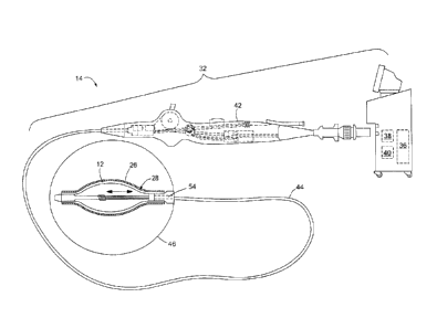
Referring now to FIG. 3. an exemplary ablation system 32 is shown. The
system 32 generally includes a console 34 that houses various controls and an
ablation
device 14 for treating tissue. The system 32 may be adapted for cryoablation.
RFA.
or both. The console 34 may include one or more of a coolant reservoir 36, an
RE'
generator 38. a TSA reservoir 40. and may further include various displays.
screens,
user input controls. keyboards. buttons, valves, conduits. connectors, power
sources,
and computers for adjusting and monitoring system parameters.
Continuing to refer to FIG. 3. the ablation device 14 may generally include a
handle 42, an elongate body 44 having a distal end 46 and an ablation element
12.
The handle 42 may include µarious knobs. levers, user control devices, input
ports,
outlet ports. connectors, lumens, and N\ ires. The distal end 46 of the
elongate body 44
CA 02862060 2014-07-21
WO 2013/110157
PCT/CA2012/001168
13
may include one or more ablation elements 12. The one or more ablation
elements 12
may be a balloon (as shown in FIG. 3), electrodes (as shown in FIG. 2C), a
combination thereof, or any other type of ablation element 12. In some
embodiments.
the ablation element 12 may also be the TSA applicator 28, for example, a
cryoablation balloon coated with a layer of TSA 26 (as shown in FIG. 3). The
elongate body 44 may further include a lumen 54 in fluid communication with
the
coolant reservoir 36 if the device 14 is used for cryoablation. lithe device
14 is used
for RFA, the elongate body 44 may include a lumen 54 in communication with an
RF
generator 38 and/or a power source. Alternatively. the device 14 may be used
for
both cryoablation and RFA. in which case the device 14 may include several
lumens
in communication with the one or more ablation elements 12.
Referring now to FIGS. 4 ¨ 12, embodiments of TSA applicators 28 are
shown. Generally. the applicator 28 may- be either integrated with the
ablation device
14 (as shown in FIGS. 4,A and 4B). or integrated with a second device 56
having a
distal end 58 (as shown in FIGS. 11A). or both (as shown in FIG. 11B).
Further, as
shown and described in FIG. 3. for example, the ablation element 12 of the
ablation
device 14 may be the applicator 28 (that is, the ablation element 12 may be
coated
with a layer of TSA 26), or the applicator 28 may be incorporated into another
area of
the device 14 (for example, the distalmost tip of a balloon catheter may be
coated with
a layer of TSA 26, whereas the balloon is not coated). The distal end 46 of
the
ablation device 1 4 may be suited for cryoablation. RFA. or both. and may be
coated
with a layer of TSA 26 (as shown in FIGS. 4A. I OA. and 10B) or a substrate
layer 60
and TSA layer 26 (as shown in FIG. 4B). Further. the ablation device 14 may be
a
fixed-diameter device (as shown in FIGS. 11A and 11B) or the ablation device
14
may have an expandable ablation element 12. such as a balloon (as shown in
FIGS.
4A. 4B. 10A, and 10B). Although not shown in FIGS. 4 10. the ablation element
12
would be placed in contact with target tissue 10 during an ablation procedure,
with the
applicator 28 (either as part of the ablation device 14 or second device 56)
being
proximate or in contact with the tissue I() to apply' TSA 26.
Referring now to FIGS. 4A-C. cross-sectional views of the distal end 46 of an
ablation device 14 are shown. the ablation device 14 including an ablation
element 12
(such as a balloon, as shown in FIGS. 4A-C) coated with a layer of TSA 26. The
CA 02862060 2014-07-21
WO 2013/110157
PCT/CA2012/001168
14
balloon ablation element 12 may be suited for either cryoablation, RFA, or
both (or
neither, if the balloon functions as an applicator 28 that is part of a non-
ablating
second device 56). and is coated at least in part with a layer of TSA 26.
Additionally
or alternatively, the balloon ablation element 12 may be coated with a layer
of nano-
or micro-porous material 50, with small amounts of TSA 26 being contained
within
the nano- or micro-pores 52. When the balloon ablation element 12 is pressed
against
the target tissue 10. the TSA 26 may be released from the pores 52 to the
tissue 10.
For example, the porous material 50 may be spongelike in that it contains a
plurality
of throughpores. Additionally or alternatively. the porous material may
contain a
plurality or surface indentations (as shown in FIG. 4C). The layer of TSA 26
may be
between approximately' 0.01 microns to approximately 200 microns (as shown in
FIG.
4A). The ablation element 12 may be additionally' coated with a substrate
layer 60.
which may be located between the ablation element 12 and TSA layer 26 (as
shown in
FIG. 4B). The substrate layer 60 may include one or more temperature sensitive
compounds that readily separate from the ablation element 12 when a certain
threshold temperature is reached (for example. 0 C or 60 ''C). This substrate
layer 60
thus facilitates movement of the TSA 26 from the distal end 46 of the ablation
device
14 to the target tissue 10. Additionally or alternatively. the substrate layer
60 may be
separated froin the ablation element 12 by mechanical stress. for example. as
when
created as a balloon ablation element 12 is inflated.
Referring now to FIGS. 5 and 6. the distal end 46 of an ablation device 14 is
shown, the distal end 46 being coated with a layer of ISA 26. The coated area
of the
distal end 56 may include an ablation element 12 (as shown in FIG. 5) suited
for
either cryoablation. RFA. or both (such as a focal catheter). or the coated
area of the
distal end 56 may, not include an ablation element 12 (as shown in FIG. 6).
and is
coated at least in part with a layer of TSA 26. The layer of TSA 26 may be
between
approximately 0.01 microns to approximately' 200 microns. The distal end 46
may be
additionally coated with a temperature-sensitive substrate layer 60, which may
be
located between the distal end 46 and TSA layer 26 (as shown and described in
FIG.
4B). Additionally, as shown in FIG. 6, the surface of the distal end 46 of the
device
14 may include a plurality of indentations or depressions 62 sized to contain
a volume
of TSA 26. For example, each indentation 62 may contain as little as 0.1 ul.
and as
CA 02862060 2014-07-21
WO 2013/110157
PCT/CA2012/001168
much as 1 p.L. The indentations 62 may either supplement or replace the TSA
layer
26.
Referring now to FIG. 7, a cross-sectional view of the distal end 46 of an
ablation device 14 is shown, the distal end 46 having a spray nozzle 64 for
the
5 application of TSA 26 to tissue 10. The :spray nozzle 64 includes a
plurality of
apertures 65 in the distal end 46 of the device 14 through which pressurized
.TSA 26
may pass and be atomized or broken into small droplets. Each droplet may be,
for
example. between approximately 0.5 1,1m and approximately 0.5 mm. The spray
nozzle 64 may be in fluid communication with the device lumen 54 and TSA
10 reservoir 40.
Referring now to FIG. 8. the distal end 46 of an ablation device 14 is shown,
the ablation device 14 having a cannula or other element 66 for the
application of
TSA 26 to tissue 10. The cannula 66 may be slidably movable within the device
lumen 54. and may be advanced beyond the distal end 46 of the device 14 to
bring the
15 outlet 68 of the cannula 66 in contact with or near the tissue 10.
Temperature-
sensitizing adjuvant 26 is then either sprayed (as shown in FIG. 7). dripped
(as shown
in FIG. 8). or otherwise applied from the outlet 68 to the tissue 10. The
cannula 66
may further include a lumen 70. in fluid communication with the outlet 68 and
the
TSA reservoir 40. Alternatively. the device 14 may be as shown in FIG. 12. W
herein
the device 14 is an over-the-wire catheter having a guidewire lumen 54 with an
outlet
71. Temperature-sensitizing adjuvant 26 is then either dripped. squirted. or
otherwise
applied from the outlet 71 of the guidewire lumen 54. Further. the device 14
may
include an expandable ablation element 1 2.
Referring now to FIG. 9. the distal end 46 of an ablation device 14 is shown.
the distal end 46 including an ablation element 12 (such as an electrode, as
shown in
FIG. 9). The ablation element 12 may be suited for RF ablation. and is capable
of
emitting at least low-current energy (for example, 100 mV to approximately 500
mV).
and may also be capable of emittin,2 RFA-level energy. The application of low-
current energy to target tissue 1() facilitates the creation of a salt-
concentration
gradient (such as the salt-concentration gradient that develops during slow
freezing of
tissue) and enhances water permeability of cell membranes. Cells of the tissue
10
respond to an increase in salt concentration by.' releasing water, resulting
in cell
WO 2013/110157 CA 02862060 2014-07-21
PCT/CA2012/001168
16
dehydration and eventually death. The ablation element 12 may apply low-
current
energy to the tissue l 0 either before, during. or after ablation.
Alternatively, the same
ablation element 12 may first apply low-current energy to the tissue 10
("gradient
generating mode-) and then apply RFA-level energy to the tissue 10 ("ablation
mode"). Further, multiple cycles of gradient generating mode/ablation mode may
be
applied.
Referring now to FIGS. 10A and 1013. the distal end 46 of an ablation device
14 is shown, the device 14 having more than one treatment elements 12. As
shown in
FIGS. 10A and 1013, the ablation device 14 includes both a balloon 72 and one
or
more electrodes 74. The electrodes 74 may be located on the distalmost point
of the
distal end 46. on the balloon 72. or both. For example. one electrode 74 at
the
distalmost point of the distal end 46 may be used in gradient generating mode,
while
other electrodes 74 on the balloon 72 may be used in ablation mode. The one or
more
electrodes 74 may be in any configuration. for example. discrete electrodes or
bands
that at least partially circumscribe the balloon 72 (as shown in FIG. 10B).
Alternatively, the balloon 72 may be a cryoablation de x ice. with a tip
and/or balloon
electrodes 74 beim./ used in gradient generating mode. Still further. the
balloon 72.
electrodes 74. and/or the ablation device 14 may be coated with a layer of TSA
26. as
shown and described in FIGS. 4A and 4B.
Referring now to FIGS. 11A and 11B. a first exemplary embodiment of an
ablation device 14 used in association with a second device 56 for the
application of
TSA to tissue 10 is shown. As shown in FIG. I IA. the ablation device 14 may
have a
distal end 46 including one or more ablation elements 12. The second device 56
may
be any device capable of applying TSA 26 to the target tissue 10. For example,
the
second device 56 may be a catheter-type device or swab having a distal end 58
coated
with a laver of TSA 26 (as shown in FIG. 11A), a device having a spray nozzle
(as
shown in FIGS. 7 and I 1B) or dropper apparatus in fluid communication with a
TSA
source (such as the TSA reservoir 40). or a hypodermic needle with a syringe
containing a volume of TSA 26. As shown in FIG. 11B, both the ablation device
14
and the second device 56 may function as applicators 28. 28a. The ablation
device 14
may serve as an applicator 28 when. for example. an ablation balloon or distal
end 46
is coated with TSA.
CA 02862060 2014-07-21
WO 2013/110157
PCT/CA2012/001168
17
It will be understood that any of the applicators 28 as described herein may
be
incorporated into either an ablation device 14 or a second device 56. For
example. an
ablation device 14 may have a spray nozzle 64 at the distalmost end and a
balloon
ablation element 12. Additionally. any number of second devices 56 may be
used.
Further, the ablation device 14 may include any number of ablation elements
12, and
may be suited for any type of ablation therapy.
It will be appreciated by persons skilled in the art that the present
invention is
not limited to what has been particularly shown and described herein above. In
addition, unless mention was made above to the contrary', it should be noted
that all of
the accompanying drawings are not to scale. A variety of modifications and
variations are possible in light of the above teachings without departing from
the
scope and spirit of the invention. which is limited only by the following
claims.