Note: Descriptions are shown in the official language in which they were submitted.
CA 02862061 2014-07-18
WO 2012/098226
PCT/EP2012/050863
Visualization of lipid metabolism
The present invention relates to the field of in vivo determination of enzyme
activity. It
allows visualization of organisms, organs, tissues and cells. In particular,
the present
Invention provides a method of in vivo visualization and a composition
suitable for in
vivo determination and/or visualization of enzyme activity by methods such as
Magnetic Resonance Imaging, also called Magnetic Resonance Tomography (MRI or
MB.]), or Magnetic Particle Imaging (MPI). In particular, the activity of the
enzyme
lipoprotein lipase affects the signals received and allows conclusions on the
lipid
metabolism of an organism, an organ system, an organ, a tissue and a cell of
interest.
This method can be employed, e.g., for diagnosis of cardiac disorders, of
tumor
prognosis and of disorders of the lipid metabolism. The composition used
comprises
superparamagnetic iron oxide nanocrystals (SPIO) incorporated in the core of
lipid
micelles designated nanosomes.
In contrast to glucose, lipids such as triacylglycerol (TAG) or
cholesterolesters (CB) are
not soluble in the blood and are transported in the form of triglyceride-rich
lipoproteins
(TRL). These micelles comprise an amphiphillic monolayer of phospholipids and
free
cholesterol in which apolipoproteins are embedded, e.g., apoB. In the
hydrophobic core,
TAG and CB are found. In the intestine, lipids are packaged into lipoproteins
as
chylomicrons, and are transported to peripheral tissues such as adipose
tissue, heart and
muscle. In the bloodstream, lipoprotein lipase (LPL) mediates the release of
fatty acids
from TAG. While the fatty acids are taken up by underlying tissues, the
remaining
rather cholesterol-rich chylomicron remnant particles are cleared by the liver
(Williams
2008), The liver can generate endogenous lipoproteins, Very-Low-Density
lipoproteins,
VLDL, when uptake of lipids from food is low. These can be taken up
analogously to
the chylomicrons by the lipid-consuming tissues. Further lipoprotein
fractions, high-
density lipoproteins (HDL) and low-density lipoproteins (LDL) also carry a
small
fraction of TAG.
The enzyme lipoprotein lipase, LPL, is localized at the endothelium of cells
taking up
lipids, in particular, heart and skeletal muscle as well as white and brown
adipose tissue.
CA 02862061 2014-07-18
WO 2012/098226
PCT/EP2012/050863
2
LPL catalyses the reaction of triacylglyc,erol + H20 <=> diacylglycerol + a
carboxylate.
Its main role lies in hydrolys of triacylglycerols in chylonnicrons and very
low-density
lipoproteins (VLDL). It also hydrolyzes diacylglycerol. LPL is the gatekeeper
enzyme
in lipid metabolism, as it catalyses the most time critical step of lipolysis,
and an
inhibition of LPL is thus sufficient for blocking lipid uptake in tissue. LPL
is the central
enzyme in vascular TAG and fatty acid metabolism (Merkel 2002; Olivacrona
2010). A
defect in its gene leads to a severe hypertriglyceridemia with pancreatitis as
clinical
consequence in humans. LPL activity is crucial for heart function as most of
energy
consumed by the heart is produced by oxidation of lipoprotein-derived fatty
acids.
Mice which are deficient for LPL in the heart develop cardiac dysfunction
despite an
increased glucose oxidation (Augustus 2006; Yamashita 2008). Therefore,
changes in
LPL expression in type 2 diabetics might be one reason for the heart failure
which
frequently occurs in these patients (Park 2007). Overexpression of the enzyme
also
leads to cardiomyopathy. Thus, controlled activity of LPL is essential for
physiological
heart function.
Growing tumors secrete pro-inflammatory cytokines like 1L-6 and TNFalpha.
These
cytokines down-regulate the expression and activity of LPL in peripheral
tissues. As
LPL is crucial for lipid uptake, decreasing its activity results in a marked
caloric deficit
in adipose tissue, muscle and heart. The consequence is a massive loss of
muscle and fat
mass which ultimately leads to cachexia. On the other hand, reports found a
link
between high expression of LPL by certain cancer tumor cells such as non-small
cell
lung and a shorter patient survival (Trost 2009). The same correlation of high
LPL
expression and poor clinical outcome was found in chronic lymphocytic leukemia
(Heintel 2005; Oppezzo 2005). Taken together, these studies suggest an
important role
for LPL activity in tumor development and associated cachexia, as it delivers
energy for
tumor growth while it steals energy from peripheral tissues.
Magnetic resonance imaging (MRI), nuclear magnetic resonance imaging (NM), or
magnetic resonance tomography (MRT) is a medical imaging technique used to
visualize detailed internal structures in vivo. The terms are used
interchangeably in the
context of this application. The good contrast MR1 provides between the
different soft
tissues of the body makes it especially useful in brain, muscles, heart, and
cancer.
CA 02862061 2014-07-18
WO 2012/098226
PCT/EP2012/050863
3
MRI has a growing importance in diagnosing heart function. It can be used to
visualize
cardiac anatomy with high resolution. So called Cine sequences, which
visualize the
heart cycle, allow determination of the ejection volume and movement of the
heart
muscle. Use of contrast agent also allows visualization of perfusion of the
heart. A so-
called "Late-enhancement" in MRI imaging with contrast agent can show scars
e.g.
caused by infarction.
MRI uses a magnetic field to align the magnetization of some atoms in the
body, then
=
uses radio frequency fields to systematically alter the alignment of this
magnetization.
This causes the nuclei to produce a rotating magnetic field detectable by the
scanner,
which is recorded to visualize the scanned area of the body.
MRI contrast agents may be injected intravenously to enhance the visibility of
internal
body structures, e.gõ of blood vessels or tumors. MRI contrast agents alter
the
relaxation times of tissues and body cavities where they are present.
Depending on the
image weighting, this can give a higher or !wilier signal.
The most commonly used compounds for contrast enhancement are gadolinium-
based.
LDL- and HDL-like micelles enriched with hydrophobic gadolinium chelates have
been
used as contrast agent for the detection of tumors and atherosclerotic plaques
(Prias et
al., 2004; Corbin et al., 2006; Glickson et al., 2008). However, these agents
have some
disadvantages.
Patients with renal disorders or insufficiency have shown Serious side effects
upon use
of gadolinium-based contrast agents, some fatal. Due to these problems, these
contrast
agents should not any more be used in such patients. Renal disorders are most
prevalent
either in older people or in patients suffering from diabetes and high blood
pressure. As
this is exactly the group of patients for which MRT visualization of, e.g.,
the heart is of
the highest interest alternative MRT contrast agents are needed.
An alternative, which does not show serious side effects and can be used in
all patients,
Is provided by iron oxide based MRI contrast agents. Two types iron oxide
contrast
agents are well known in the state of the art: Superparamagnetio Iron Oxide
(SPIO) and
Ultrasmall Superparamagnetic Iron Oxide (USPIO). These contrast agents
typically are
suspended colloids of iron oxide nanoparticles. When injected during imaging,
they
CA 02862061 2014-07-18
WO 2012/098226
PCT/EP2012/050863
4
reduce the T2 signals of absorbing tissues. SPIO and USPIO contrast agents
have been
used successfully, e.g., for liver tumor enhancement.
Due to their excellent suitability for electron microscopy as well as
fluorescence and
magnetic-resonance-imaging, quantum dots, gold and superparamagnetic iron
oxide
(SPI0s) nanocrystals have been extensively applied as labels for biomedical
imaging
(Michalet 2005; Alivisatos 2004). Furthermore, several sensors based on
nanocrystals
for different applications have been developed over the last years (Perez
2002; Medintz
2005; Koh 2008; Lee 2008; Snee 2008; McLaurin 2009). But none of these sensors
has
been applied in vivo so far.
=
MRI or the related method of Magnetic Particle Imaging (MPI) can be used for
visualizing physical function, e.g., of the heart as well as perfusion.
However,
biochemical parameters such as the energy metabolism of specific areas cannot
yet be
visualized in vivo with acceptable resolution. This could be of particular
benefit, e.g.,
because it would allow for diagnosis of a future potential loss of function of
tissue, e.g.,
by showing change from lipid to glucose metabolism for energy generation or
excessive
use of lipids. This would allow for therapeutic intervention and could prevent
chronic
loss of function.
Previously, the inventors (Bruns et al., Nature Nanotechnology, 2009, which is
fully
incorporated herein by reference) disclosed a new method to visualize
lipoproteins,
using superparamagnetic iron oxide nanocrystals embedded into the core of
lipoproteins. They showed that it is possible to image and quantify the
kinetics of
lipoprotein metabolism in vivo using dynamic MRL The lipoproteins were taken
up by
liver cells in wild-type mice, and displayed defective clearance in knock-out
mice
lacking a lipoprotein receptor or its ligand, indicating that the nanocrystals
did not
influence the specificity of the metabolic process. Using this strategy, it is
possible to
study the clearance of lipoproteins in metabolic disorders and to improve the
contrast in
clinical imaging. However, no dependence of enzymatic activity was observed in
the
published experiments.
In light of this, the inventors now solved the problem of providing an in vivo
method of
determining and visualizing a compartment in a subject dependent on enzymatic
activity
CA 02862061 2014-07-18
WO 2012/098226
PCT/EP2012/050863
in said compartment, in particular, lipoprotein lipase (LPL) activity. Thus,
LPL activity
can be determined and visualized in vivo. The invention is further described
below and
in the appended claims.
The invention provides a method of in vivo visualization, comprising
a) administering a composition comprising superparamagnetic iron oxide
nanocrystals
(SPIO) incorporated in the core of nanosomes to a subject, and
b) determining and visualizing lipoprotein lipase activity in a compartment of
the
subject
The invention also provides a method of in vivo determination and/or
visualization of
lipoprotein lipase (LPL) activity, comprising
a) administering a composition comprising superparamagnetic iron oxide
nanocrystals
(SPIO) incorporated in the core of nanosomes to a subject, and
b) determining and visualizing presence of SPIO in a compartment of the
subject,
wherein the presence of SPIO indicates lipoprotein lipase (LPL) activity in
the
compartment or associated with the compartment
The invention also provides a composition comprising superpararnagnetic iron
oxide
nanocrystals (SPIO) incorporated in the core of nanosomes for use in in vivo
visualizing
lipoprotein lipase (LPL) activity, comprising
a) administering the composition to a subject, and
b) determining and visualizing presence of SPIO in a compartment of the
subject,
wherein the presence of SPIO indicates lipoprotein lipase (LPL) activity in
the
compartment.
A composition comprising superparamagnetic iron oxide nanocrystals (SPIO)
incorporated in the core of nanosomes (SPIO nanosomes) can be prepared, e.g.,
as
disclosed below or according to methods disclosed by Bruns et al., Nature
Nanotechnology 2009, or according to Tromsdorf et al, Nano Letters 2007.
The nanosomes may be prepared from biological samples, e.g., from the subject
which
is to be examined. For example, lipids used for the preparation of the
nanosomes can be
extracted from lipoproteins isolated by standard centrifugation protocols from
plasma,
CA 02862061 2014-07-18
WO 2012/098226
PCT/EP2012/050863
6
The nanosomes may also be artificially prepared, e.g., assembled from the
components
according to methods known in the state of the art.
In one embodiment, the nanosomes are based on TEL such as chylomicrons or
VLDL.
It is also possible that the nanosomes are based on the composition of, e.g.
LDL or
HDL.
Chylomicrons are synthesized in the postprandial phase by enterocytes within
the
Intestine and have a diameter between 75 ¨ 1200 nm depending on the
composition of
the meal. The size of SPIO-nanosomes is dependent on the lipid mixture used,
e.g., for
lipids extracted from human plasma TRL, it is approximately 250 nm and
therefore is
within the size of physiological postprandial lipoproteins.
After assembling within intestinal cells, chylomicrons enter the blood stream
via the
thoracic duct, which is the largest lymphatic vessel in the body draining into
the
systemic circulation via the left subclavian vein into the heart. Similar to
chylomicrons,
intravenously injected nanosomes reach the systemic circulation via the heart,
In
addition, it is important to note that nascent chylomicrons do not contain any
apolipoprotein E (apoB) or lipoprotein lipase (LPL). Consequently, nanosomes
do not
need to contain exogenously added apoE and LPL when serving as a model
particle for
chylomiorons.
Therefore, SPIO-Nanosomes can be prepared with or without the addition of
apolipoproteins. If the apolipoproteins are not added to nanosomes before
administration to the subject, e.g., i.v., the nanosomes will acquire
apolipoproteins after
the Injection into the circulation.
In one embodiment, the nanosomes are prepared comprising apolipoproteins such
as
ApoB. Apolipoproteins may be of human or other origin, e.g., mouse, rat, ape
or swine.
Preferably, they are of the same species origin as the subject The subject
may, e.g., be
human, mouse, rat, ape or swine. The composition of the nanosomes may vary,
depending on the intended organ or tissue that is to be analysed. In
particular,
apolipoproteins or a particular lipid composition may be chosen to target the
nanosomes
to specific organs/tissues.
CA 02862061 2014-07-18
WO 2012/098226
PCT/EP2012/050863
7
Nanosomes are micelles made of, e.g., lipids extracted from lipoproteins, and
lipophilic
nanocrystals may be embedded in the core.
In one embodiment, SPIO nanosomes may comprise at least about 40 %
triglycerides.
They may comprise about 0.25% to about 20% phospholipids, about 0 % - about 20
%
cholesterin and/or cholesterin ester, about 0 % - about 10 % cholate and 0 % -
about 50
% dryweight of nanocrystals, preferably, 3 % to about 30 % dryweight of
nanocrystals.
The nanosomes preferably have a size between 30 I1M -2 um. % in the context of
the
application relates to weight/weight, if not explicitely mentioned otherwise.
The composition comprising nanosomes preferably comprises about 0.1% - about
30 %
lipids in total in an aqueous buffer. Preferably, the composition is non-toxic
and suitable
for administration to a human, e.g., for 1.v. injection.
Preferably, nanosomes which may be used in the context of the invention carry
lipophilic nanocrystals (also designated nanoparticles) which may cause a
detectable
signal in imaging modality used for humans, e.g. SPIO nanocrystals of about 2-
30 nm
size, specifically, about 6nm or about 10 nm for MRI, and they are a substrate
for LPL.
The SPIO preferably comprise nanocrystals having a size of about 2 to about 30
nm, or
about 5 to about 20 nm, e.g., about 6 to about 10 nm. The SPIO preferably
comprise
Fe304 and/or Fe203 nanocrystals (e.g, having a size of about 6-10 urn), but
they may
also or additionally comprise MnFe204 nanocrystals. Alternatively or
additionally, other
kinds of superparamagnetic nanoparticles or superparamagnetic materials with a
size
less than 50 nm can be used in the nanosomes of the invention. These materials
could
Include superparamagnetic iron nanoparticles with a gold shell (iron-gold core-
shell
nanoparticles), superparamagnetic iron nanoparticles with an iron oxide shell,
superparamagnetic iron platin nanoparticles, superparamagnetic iron oxide
nanocrystals
with another composition than Fe304 or Fe203. Any material which can be used
to cause
a contrast in MRI pictures may be used.
=
All kinds of iipophilic nanocrystals, like quantum dots, SPIO or gold
nanoparticles,
preferably with particle sizes between 2 and 30 nm, can be embedded into the
nanosomes used in the invention. The nanosomes comprising the nanocrystals
allow
multimocial visualization as well as quantification of lipoprotein metabolism,
in
CA 02862061 2014-07-18
WO 2012/098226
PCT/EP2012/050863
8
particular, LPL activity, in real-time by non-invasive imaging in vivo. The
MRI contrast
agent based on nanocrystals used in the present invention may consist of
nanosomes
with multiple SPIOs inside, These SPIO form an ensemble in which their
magnetic
moments interacts with each other, This interaction leads to a maximized and
constant
r24 relaxivity which can be described by the static dephasing regime (SDR). It
allows
quantifying lipoprotein metabolism by real-time MR imaging (Bruns 2009).
In the method of the invention, visualization of the SPIO nanosomes takes
place in vivo,
i.e. in a compartment of an organism (a subject). The compartment may be an
organ, a
tissue or a cell or an area thereof. The LPL enzyme activity may be in a
compartment or
associated with the compartment. For example, the LPL enzyme activity is
usually
associated with the endothelium of a tissue/organ. This is enzyme activity is
not
considered to be associated with the circulation, but with the tissue/organ
bordering the
circulation, i.e., the compartment into which the SPIO are taken up.
For the purposes of this application, the circulation is thus not considered a
compartment or organ. LPL activity in the tissues/compartments leads to
diminished
presence of SPIO nanosomes in the circulation, as these can be taken up from
the
circulation into organs/tissues dependent on LPL activity.
In general, in the context of the invention, "a" or "the" does not only
designate "one",
but also includes a plurality. For example "a compartment" may also be more
than one
compartment. The method of the invention allows a high spatial resolution of
the
presence of the SPIO.
In the context of the invention, a tissue may be selected from the group
comprising
tumor, atherosclerotic plaque, sites of inflammation and adipose tissue.
Adipose tissue
may be white adipose tissue or brown adipose tissue. The organ or tissue
analyzed by
the method of the invention may express lipoprotein lipase and exhibit
lipoprotein lipase
activity under physiological or non-physiological conditions.
The organ is preferably selected from the group comprising heart, skeletal
muscle, brain
and tumors as well as sites of inflammation Liver and spleen are under
physiological
conditions not among these organs, as uptake into these organs is not LPL
dependent. In
CA 02862061 2014-07-18
WO 2012/098226
PCT/EP2012/050863
9
conditions of tumors in the liver or spleen with an increased abnormal
expression of
LPL, the invention might be applied to measure LPL activity.
Administration of the SPIO nanosomes may be by oral or Intravenous
administration, in
particular, intravenous administration. Oral administration of nanosomes
comprising
contrast agents such as SPIO may be used to analyse, determine and/or localize
sites of
lipase activity in the gastrointestinal tract, in particular, pancreatic
lipase activity.
In the context of the invention, the composition may be administered in an
effective
amount, i.e., an amount that allows visualization. This can be determined,
e.g., by the
clinician. In one embodiment about 0.05 mg iron within nanosomes / kg body
weight ¨
2 mg/kg body weight are to be administered intravenously. It may be favourable
to use
0.1 mg iron within nanosomes / kg body weight ¨ 1 mg/kg body weight to be
administered intravenously. Preferentially, 0.3 mg iron within nanosomes / kg
body
weight 0.5 mg/kg body weight are to be administered intravenously. The amount
can
be adapted depending on, e.g., age, sex, the aim of the analysis and/or the
condition of
the subject.
The determination and/or visualization may be performed by 1VIRI. Dynamic MRI,
may
also be employed. Suitable protocols are described herein or in Bruns et al.,
Nature
Nanotechnology 2009. One significant advantage of the method of the invention
is that
it may be performed without invasive methods. Another advantage is that no
administration or radioactive compounds to the subject are required.
In addition, the invention may be used to determine LPL activity with other
non-
invasive imaging techniques in which lipophilic nanoparticles or nanocrystals
can be
used as a contrast agent. In particular, SPIO-nanosomes may be applied to
contrast
compartments and/or visualize and/or measure LPL activity by magnetic particle
imaging (MN). For example, the heart might be contrasted, and/or LPL activity
may be
visualized in the heart by magnetic particle imaging (WI),
The method of the invention may also be employed for visualization of the
organ, tissue
or cell. The SPIO nanosomes may be used similarly to a conventional
contrasting agent.
For example, as the agent is mostly taken up in perfused areas of a tissue or
an organ,
perfusion can be visualized.
CA 02862061 2014-07-18
WO 2012/098226
PCT/EP2012/050863
In one embodiment of the method of the invention, the tissue that is
visualized Is tumor
tissue, and the LPL activity is predictive of progression of the tumor. In
particular, a
high LPL activity is predictive of fast progression of the tumor.
In one embodiment, the invention provides a method of diagnosing a tumor. The
method may provide a prognosis of clinical outcome. In this context, a high
lipase
activity has been shown to correlate with a bad prognosis, i.e. fast
progression of the
tumor, and a low lipase activity correlates with slow progression of the tumor
and/or
regression. A higher lipase activity thus corrolates with a worse prognosis.
In one embodiment, the invention provides a method of diagnosing a disorder of
the
lipid metabolism, comprising performing the method of the invention. Disorders
of the
lipid metabolism may include type II diabetes and cachexia, Also, genetic
deficiency for
LPL, Apolipoprotein CII, Apolipoprotein AV and/or glycosylphosphatidylinositol-
anchored high-density lipoprotein-binding protein I may be diagnosed by
performing
the method of the invention.
The invention also provides a method of diagnosing a cardiac disorder,
comprising
performing the method of the invention. The cardiac disorder may be selected
from the
group comprising coronary heart disease, coronary artery disease,
cardiomyopathy,
alcoholic cardiomyopathy, congenital heart disease, ischemic cardiomyopathy,
hypertensive cardiomyopathy, nutritional diseases affecting the heart,
valvular
cardiomyopathy, inflammatory cardiomyopathy, cardiomyopathy secondary to a
systemic metabolic disease or myocardiodystrophy. The following cardiac
disorders
may also be diagnosed: dilated cardiomyopathy, hypertrophic cardiomyopathy,
arrhythmogenic right ventricular cardiomyopathy, restrictive cardiomyopathy or
noncompaction cardiomyopathy. The method of the invention is especially
advantageous for localizing disorders in specific areas of the heart and/or
quantifying
the area of an organ such as the heart which is afflicted by the disorder.
In one embodiment of the method of the invention, the organ that is visualized
is heart.
For example, viability and/or perfusion of an organ such as heart may be
detected, e.g.,
scarring due to infarction. The method .of the invention may also be used for
diagnosing
disorders of the lipid metabolism in the heart. For example, the organ that is
visualized
CA 02862061 2014-07-18
WO 2012/098226
PCT/EP2012/050863
11
may be heart, and disorders of the lipid metabolism in the heart may be
detected and/or
localized to specific areas of the heart. Exemplary disorders are mentioned
above.
In a method of diagnosis of the invention, one step may be comparing the
results of the
analysis of the subject with a reference, such as results from a healthy
subject and/or a
from a group of healthy subjects (i.e., average results from such a group)
and/or from a
subject or subjects previously diagnosed with the disorder or tumor in
question. E.g., a
result significantly varying from a healthy reference may indicate that the
subject has a
disorder.
The invention also relates to a diagnostic and/or prognostic composition
comprising
superpammagnetic Iron oxide nanocrystals (RIO) incorporated in the core of
nanosomes for use in in vivo determining and/or visualizing lipoprotein lipase
(LPL)
activity. The composition is also for use in diagnosing disorders of the lipid
metabolism,
for use in diagnosing cardiac disorders or for use in prognosis of a tumor, as
described
in detail above.
The inventors have demonstrated that it is possible to visualize enzyme
activity in the
heart and other tissues with the method of the invention. They provide a
method for
imaging of the metabolism with a high resolution, e.g., in the heart or in
tumors. The
method's results are comparable with PET-CT (Positron emission tomography
combined with computer tomography) analysis with radioactive 18-Fluor-
desoxyglucose (FDG), however, there is no requirement for use of radioactive
isotopes
(as in PET and SPECT) or ionising radiation (as in CT and X-rays).
Furthermore, the
metabolism is directly visualized with high-resolution MRI significantly
improving
spatial resolution compared to approaches based on PET or SPECT. In comparison
to
new development of systems like PET-MRT, the method of the invention still has
the
advantage of higher resolution combined with the option of not using
radioactive
tracers. Furthermore, apparatus requirements are lower (MRT only instead of
PET-CT
or PET-MRT).
Visualization, in the context of the invention, is meant to comprise steps of
determining
a result e.g., a measure of LPL activity, and of generating an Image based on
this result,
wherein the activity is linked to an area in which it has been detected, and
graphically
CA 02862061 2014-07-18
WO 2012/098226
PCT/EP2012/050863
12
represented. MR1 or MN are typical methods comprising visualization of a
result in the
context of the invention.
It is noted that the combined time and spatial resolution of the method of the
invention
are of excellent quality and could not be achieved with any other technique,
neither by
non-invasive nor invasive approaches. MRI, used with the method of the
invention, can
provide a spatial resolution of below I. mm, preferably, of below 0,1 mm.
Temporal
resolution preferably is below 0,1 sec.
=
In the examples below, it is shown in a mouse model that in vivo imaging of
uptake of
SPIO nanosomes is feasible, and that Increased LPL activity can be detected
via an
increased uptake of SPIO nanosomes. Surprisingly, the uptake was directly
dependent
on LPL activity, as inhibition of LPL via THL, Tetrahydrolipstatin,
specifically
inhibited uptake. Quantitative analysis of the organ distribution with
radioactive
labelling of the SPIO in the nanosomes confirmed these result. The use of
radioactive
SPIO is however not required in the method of the invention.
The examples shown below are meant to illustrate, but not to limit the
invention. Other
embodiments can be envisaged by the skilled person taking the description of
the
invention into account.
Figure legends
Fig. 1. Cold exposure modulates fasting and postprandial triglyceride-rich
lipoprotein levels
(a) Triglyceride and (b) cholesterol FPLC profiles of pooled plasma from
fasted FVB
mice after 4 h and 24 h cold exposure at 4 C. (c) Plasma triglycerides during
an oral fat
tolerance test in control and cold-exposed FVB mice. (d) FPLC lipoprotein
profiling in
control and cold-exposed FVB mice two hours after an oral fat load. (e) Organ
distribution of triolein-derived 311-radioactivity in control and cold-exposed
FVB mice
two hours after gavage. Mean values s.e.m. with n ¨ 12 in (c) and n = 4 in
(e).*P <
0.05; 8`P <0.01; SP <0.001.
Fig. 2. Activated BAT is a central target organ for TRL uptake
CA 02862061 2014-07-18
WO 2012/098226
PCT/EP2012/050863
13
(a) Plasma clearance of 59Fe-SPIO and (b)3H-triolein-labeled TRL in control
and cold-
exposed C57BL/6 mice. (c) Organ distribution of59Fe-SPIO and (d) triolein-
derived 3H-
radioactivity 15 min after intravenous injection. Mean values s.e.m. with n
5. (e)
Representative transversal magnetic resonance (MR) images of a control and
mild-
exposed wild-type FVB mouse before and approximately 10 min after injection of
SP10-labeled TRL. Arrows in upper panel point to BAT whereas arrows in lower
panel
Indicate the liver. Bar: 1 cm. (I) Coronary MR images of a representative cold
mouse
before, 10 min and (g) 1 week after injection of SPIO-TRL with identical MR
settings.
(h) Representative intravital confocal microscopy images of dissected BAT in a
live=
cold-exposed FVB mouse 2 min (left) and 30 min (right) after QD-TRL (green)
injection (arrows indicate QD-TRL). FITC-dextran (red) to stain blood vessels
and
DAPI (blue) to label nuclei. Bar: 25 gm (1) Representative transmission
electron
microscopy pictures of high-pressure frozen BAT samples from a SPIO-TRL
injected,
cold-exposed FVB mouse. L: lipid droplet, M: mitochondrium, C: capillary.
Upper
panel left, bar: 5 gm; upper panel middle, bar: 1 gm; upper panel right, bar:
0.05 gm;
lower panel left, bar: 1 gm; lower panel middle, bar: 0.05 gm; lower panel
right, bar:
0.02 gm. *P< 0.05; eµP < 0.01; $P <0.001.
Fig. 3. LPL and 0)36 drive TRL clearance Into BAT
(a) Organ distribution of 59Fe-SPIO and (b) triolein-derived 3H-radioactivity
15 min
after intravenous injection in cold FVB mice that were pre-injected with
teirahydrolipstatin (TI-IL) to inhibit LPL activity or with heparin to release
LPL into
circulation, respectively. Mean values s.e.m. with n 5. (c) Oral fat
tolerance test in
cold FVB mice pre-treated with TIM. Mean values s.e.m. with n =-- 5. (d)
Relative
mRNA expression levels in C57BL/6.1 BAT of master genes regulating
thermogenesis
(Pparg, Ppargcla, Ucpl, D1o2), lipoprotein binding (141r, Lrpl, Apoe, Gpthbpl,
Cd36), lipolysis of lipoproteins (Lpl, Gpihbpl, Angpt14) as well as fatty acid
uptake
(Fatpl, Fatp3, Fatp4 and Cd36). (e) Determination of Cd36 and other fatty acid
transporters mRNA copy numbers by Tat:Man. (1) Consecutive FPLC analysis of
TRL-
3H-triolein and albumin-3H-oleate in cold-exposed wild-type and Cd364-
littermates. (g)
Organ distribution of 59Fe-SPIO and (h) triolein-derived 3H-radioactivity 15
min after
CA 02862061 2014-07-18
WO 2012/098226
PCT/EP2012/050863
14
intravenous Injection of radiolabeled TRL into Cd364- and wild-type
littermates. Mean
values s.e.m. with n > 6. *P <0.05; 413 < 0.01; $P <0.O01.
Fig. 4. BAT activation corrects hyperlipidemia and is not impaired in insulin
resistance
(a) Triglyceride levels in hyperlipidemic Apoart- mice during cold exposure
and (b)
pictures of plasma after 24 h cold exposure. (c) Triglyceride and cholesterol
FPLC
profiling of pooled plasma from Apoar" mice after 4 h and 24 h cold exposure.
(d)
Environmental scanning electron microscopy studies of brown adipose tissue
from lean
and obese control and cold-exposed mice. (bar: 25 um) (e) pictures of
interscapular
BAT in control and cold-exposed obese mice (bar: 0.5 cm). Combined oral
glucose and
fat tolerance test in lean and obese control and cold-exposed mice using f,
14c.
deoxyglucose and (g) 3H-triolein tracers. (h) Turnover kinetics and (i) organ
uptake of
3H-triolein-TRL in lean and obese control and cold-exposed mice. Mean values
s.e.m.
with fr--- 6. *P <0.05;"? <0.01;$P <0.001.
Fig. 5 SPIO-Nanosomes were injected via the tail vein into a fasted mouse. Due
to the
Increased LPL expression in heart (myocard) upon fasting, there is strong
uptake of
SPIO-Nanosomes into the myocard. The myocard is enhanced due to the negative
contrast caused by SPIO. (a) Transversal T2*-weighted MR1 picture before
injection of
SPIO-Nanosomes. (b) Transversal T2*-weighted MR1 picture after injection of
SPIO-
Nanosomes. (c) Difference of (a) and (b). The myocard is visualized as an
enhanced
ring. In a second, equally fasted littermate mouse, 1 min before the SPIO-
Nanosomes
were injected, LPL was specifically inhibited by injection of a lipase-
specific inhibitor
(Tetrahydrolipstatin (THL) (200 I with 1.25 mg/ml THL In PBS with 10% DMSO)).
(d) Transversal T2*-weighted MRI-picture before injection of SPIO-Nanosomes.
(0)
Transversal T2*-weighted MRI-picture after injection of SPIO-Nanosomes. (f)
Difference of (e) and (f). The myocard is not enhanced. The blood within the
lumen of
the heart is enhanced as the SPIO-Nanosomes remain in the circulation because
LPL-
mediated uptake is blocked.
Fig. 6 SPIO-Nanosomes were injected via the tall vein Into a cold-exposed
mouse. Due
to the increased LPL expression in brown adipose tissue (BAT) upon cold
exposure,
CA 02862061 2014-07-18
WO 2012/098226 PCT/EP2012/050863
there is a strong uptake of SPIO-Nanosomes into the BAT. The BAT is enhanced
due to
the negative contrast caused by SPIO. (a) Transversal T2*-weighted MRI picture
before
injection of SPIO-Nanosomes. (b) Transversal T2*-weighted MRI picture after
injection
of SPIO-Nanosomes. (c) Difference of (a) and (13). The BAT is visualized as
five
enhanced structures. In a second, equally cold-exposed littermate mouse, 1 min
before
the SPIO-Nanosomes were injected, LPL was specifically inhibited by injection
of a
lipase-specific inhibitor (Tetrahydrolipstatin (THL) (200 I with 1.25 mg/ml
THL in
PBS with 10% DMSO)). (d) Transversal T2*-weighted MR.1 picture before
injection of
SPIO-Nanosomes. (e) Transversal T2*-weighted MR1 picture after injection of
SPIO-
Nanosomes. (f) Difference of (e) and (f). The BAT is not enhanced.
Fig. 7 SPIO-Nanosomes were injected via the tail vein into a cold-exposed
mouse. The
uptake into the BAT is measured by dynamic MRI. At tr=0 sec, SPIO-Nanosomes
were
injected. Due to the increased LPL expression in brown adipose tissue (BAT)
upon cold
exposure, there is strong uptake of SPIO-Nanosomes into the BAT. In a second,
equally
cold-exposed mouse, 1 min before the SPIO-Nanosomes were injected, LPL was
specifically inhibited by injection of a lipase-specific inhibitor
(Tetrahydrolipstatin
(THL) (200 1 with 1.25 mg/ml THL in PBS with 10% DMS0). In a third, equally
cold-
exposed mouse, 1 min before the SPIO-Nanosomes were injected, human
chylomicrons
were injected in a tenfold concentration to saturate specific chylomicron
binding sites
on the endothelium (100 1 with concentration 100 mg/ml triglycerides). (a)
Comparison between cold control mouse (continuous line) and THL-injected mouse
(dashed line). (b) Comparison between cold control mouse (continuous line) and
chylomicron injected mouse (dashed line).
Examples
Example 1: Brown adipose tissue activity controls triglyceride clearance
METHODS
Animals and diets. All experimental procedures were performed with approval
from
the animal care committees responsible for the University Medical Center
Hamburg-
Eppendorf. Animals were housed at 22 C with ad libitum access to standard
laboratory
chow diet. We used male age-matched (16-22 weeks) Ld1r , Apocd and
CA 02862061 2014-07-18
WO 2012/098226
PCT/EP2012/050863
16
respective FVB wild-type mice as well as LRP1-N2 knockin, Cd364- and C57B1/6J
wild-type mice which were fasted 4 h prior to the experiment. Control (22 C)
and cold
exposure (4 C) was performed in single cages for 24 h unless indicated
otherwise. To
induce insulin resistance and obesity, male C57BL/6J mice were single-caged
and were
at 4 weeks of age fed a diabetogenic high-fat diet" ad libitum for 16 weeks.
Turnover studies and organ distribution. For turnover studies, anesthetized
mice
were tail vein injected with 200 itiL radiolabeled TRL. Lipoprotein turnover
was
determined from 10 ul plasma 0.5, 1, 2, 5 and 15 min after injection. After 15
min,
blood was removed by cardiac puncture, the right atrium was opened, and the
carcass
was perfitsed through the left ventricle with PBS containing 50 U heparin.
Then,
organs were harvested and weighed. For measurement of radioactivity, organs
were
solubilized in Solvable (PerkinElmer, Boston, USA, 0,1 mL per 10 mg organ),
200 L
were counted in scintillation fluid and TRL uptake was calculated as c.p.m.
per mg
organ. Oral fat tolerance tests were performed by gavage of 100 uL olive oil
with [9,10-
31-1(N)]-triolein (370 KBq per mouse). To measure lipoprotein production
triton WR-
1339 (Tyloxapol from Sigma; 0.5 mg per g body weight as 10% solution in PBS)
was
injected into the tail vein. Plasma was collected at indicated time points.
For the
measurement of hepatic production 1(3)-3H glycerol (125 KBq per mouse) was
injected
prior to triton WR-1339. Lipids were extracted from plasma samples 35 and 3H-
glycerol
incorporated into triglycerides was measured as described above. For
chylomicron
production mice received 3H-triolein in olive oil by gavage as described above
directly
after triton WR-1339 injection. Intestine-derived radioactivity in plasma was
measured
as described above. For manipulation of LPL function, THL (Roche, 12.5 mg mri
DMSO) was diluted to 1.25 mg mri in 10% DMSO in PBS. Mice received 200 I., of
either 0.25 mg THL, 50 U heparin (ratiopharm) or 10% DMSO in PBS (mock). After
1
min 59Fe-SPIO- and 3H-triolein-labeled TRL were injected and plasma clearance
and
organ uptake were determined as described above. For postprandial studies mice
were
i.p. injected with 0.25 mg THL or mock solution prior to gavage of 200 L olive
oil.
Blood was collected at indicated time points and plasma triglyceride levels
were
determined.
CA 02862061 2014-07-18
WO 2012/098226
PCT/EP2012/050863
17
In vivo imaging studies. MR1 was performed as described before. Briefly, all
static
and dynamic MRI measurements were performed with a clinical 3 Testa MR scanner
(Philips Medical Systems, Netherlands) equipped with a custom-made small
animal
solenoid coil. The dynamic measurements were based on a gradient-echo sequence
(Supplementary table 3). The applied sequence is highly sensitive to
susceptibility
effects caused by local magnetic field inhomogeneities caused by SPIO-TRL.
D1COM
data were processed with Image (http://rsbwellnih.gov/iji). For cryo electron
microscopy, SP1O-TRL were intravenously injected into control or cold wild-
type FVB
mice. After 30 min mice were sacrificed, BAT biopsies were taken and processed
for
transmission electron microscopy (TEM) as described. Micrographs were obtained
with a FBI Eagle 4k CCD camera and a Te,chnal 20 T'EM operated at 200 kV. For
environmental scanning electron microscopy studies of BAT anaesthetized mice
were
perfused with PBS-Heparin as above and organs were fixed with 2.5%
glutaraldehyde
in PBS, washed, and postfixed for 30 min with 1% 0s04 in PBS. For intravital
microscopy interscapular BAT was dissected in anaesthetized mice and
visualized by a
confocal microscope equipped with a resonant scanner (Nikon AIR). QD-labeled
TEL
und fluorescent probes were injected via a tail vein catheter and 15 or 30
confocal
images per second were recorded. The acquired data sets were aligned to reduce
object
movements due to mouse breathing and denoised with a Savitzky-Golay filter in
Nikon
NIS Elements AR 3.10. Labeling, animations and quicktime-export were done with
Adobe After Effects C$4.
Statistics. To assess statistical significance two-tailed, unpaired Student's
Mest or two-
way ANOVA followed by post-hoc Bonferroni's test was performed. P < 0.05 was
considered significant.
Preparation and labeling of in, plasma parameters, RNA extraction and real-
time
quantitative PCR, endothelial permeability testing and Western blotting were
performed
as known in the art. Preparation of TRL comprising SPIO was performed
essentially as
disclosed In Bruns et al. Nature Nanotechnology 4, 2008:193-201 and the
supplement to
said publication(14), with the modification that nanocrystals with a size of
about 10 nm
were used. The increased size led to a better signal.
RESULTS
CA 02862061 2014-07-18
WO 2012/098226
PCT/EP2012/050863
18
Brown adipose tissue (BAT) burns fatty acids for heat production in order to
defend the
body against cold and has recently been shown to be present in humans".
Triglyceride-rich lipoproteins (TRL) transport lipids in the bloodstream,
where fatty
acids are liberated by the action of lipoprotein lipase (LPL)6. Patty acids
are taken up by
peripheral organs such as muscle and adipose tissue, whereas remaining
cholesterol-rich
remnant particles are cleared by the liver6. Elevated triglycerides and
prolonged
circulation of remnants, especially in diabetic dyslipidemia, are risk factors
for
cardiovascular diseasen I. However, the precise biological importance of BAT
for TRL
clearance remains unclear. Here, the inventors show that increased BAT
activity
induced by short-term cold exposure controls TRL metabolism in mice. Cold
exposure
drastically accelerated plasma clearance of triglycerides as a result of
increased uptake
into BAT, a process crucially dependent on local LPL activity and
transmembrane
receptor CD36. In pathophysiological settings, cold exposure corrected
hyperlipidemia
and improved deleterious effects of insulin resistance. In conclusion, BAT
activity
controls vascular lipoprotein homeostasis by inducing a metabolic program that
boosts
TRL turnover and channels lipids into BAT. Activation of BAT might be a
therapeutic
approach to reduce elevated triglyceride levels and combat obesity in humans.
To determine whether cold exposure alters the lipoprotein profile, plasma from
FVB
wild-type mice kept at 22 ("C (control mice) or at 4 C in a cold room (cold
mice) was
analyzed by fast performance liquid chromatography (FPLC). TRL-triglycerldes
were
markedly reduced after 4 h and 24 h (Fig. la) demonstrating that cold exposure
lowers
triglyceride levels efficiently. HDL-cholesterol, however, is slightly
increased (Fig, lb)
which is= probably explained by an increase of TRL-derived HDL precursors12.
After a
fatty meal, triglyceride-rich chylomicrons transport dietary lipids, but it is
unclear
whether BAT is involved in their processing. Therefore an oral fat tolerance
test was
performed with olive oil mixed with 31-1-triolein in wild-type FVB mice (Fig.
1c). In
control mice, triglyceride levels rose with a peak after 2 h and decline as
expected6'13. In
cold mice, triglyceride levels remained persistently low during the
postprandial phase.
The lipoprotein profile (Fig. Id and Supplementary Fig. 1) confirmed the
presence of
chylomicrons in control mice whereas they were absent in cold mice. The
corresponding 3H-radioactivity profile resembled the curve for triglycerides
in control
mice, while in cold mice plasma 3H-radioactivity steadily rose (Supplementary
Fig. 2).
CA 02862061 2014-07-18
WO 2012/098226
PCT/EP2012/050863
19
The latter can be explained by the occurrence of small molecule fatty acid
degradation
products in the blood (Supplementary Fig. 3). Organ distribution of 3H-
triolein-derived
radioactivity 2 h after gavage (Fig. le and Supplementary Fig. 4) revealed a
selective
increase in organ uptake of fatty acids into BAT. The total contribution of
BAT was
higher than for muscle which also participates In heat production In response
to acute
cold by shivering therrnogenesisl. As the production rates of hepatic
(Supplementary
Fig. 5) as well as of intestinal TRL (Supplementary Fig: 6) were unaltered by
cold
exposure, the increase in specific uptake of TRL-lipids into different BAT
depots
suggests an accelerated clearance of postprandial TRL in cold mice. To
investigate the
clearance and kinetics in more detail, both 3H-triolein and hydrophobic "Fe-
superparamagnetic iron oxide (SPIO) nanocrystals were embedded into the core
of TRL
particles. These particles allowed to following lipoprotein and fatty acid
uptake
simultaneously, because upon LPL-mediated lipolysis 3H-oleate was released,
while
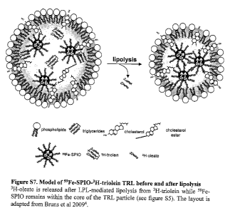
hydrophobic 59Fe-SPIO nanocrystals remained within the TRL core (Supplementary
Figs. 7 and 8). Clearance of both TRL-derived 59Fe-SPIO (Fig. 2a) and 3H-
triolein (Fig.
2b) was significantly faster in cold compared to control mice. The organ
distributions of
3H-triolein and 59Fe-SPIO indicated that the accelerated clearance was
mediated by an
approximately 10-fold increase in specific uptake into BAT (Fig. 2c,4 and
Supplementary Fig. 9). Total amounts of 3H-triolein and 59Fe-SPIO uptake in
BAT were
comparable to total liver uptake while the contribution of other tissues was
small. We
confirmed these findings using non-hydrolysable 3H-cholesterol ethers, a
conventional
TRL core label (Supplementary Figs. 10 and 11). The concomitant reduced
hepatic TRL
uptake indicated that cold exposure shifted the clearance of lipoproteins from
liver to
BAT. Notably, uptake into subcutaneous white adipose tissue was also increased
which
can be explained by the presence and activation of brown adipocytes after cold
exposure
(Supplementary Fig. 12). Recently, hydrophobic SPIO nanocrystals that
accelerate spin-
spin relaxations were embedded into the TRL core to follow lipoprotein uptake
into the
liver by dynamic magnetic resonance imaging (MRI)14. Irrespectively of BAT
activity,
we observed uptake into the liver of control and cold-exposed mice (Pig. 2e).
However,
cold exposure markedly increased the negative contrast of several BAT depots,
indicative for increased TRL presence (Fig. 2e,f and Supplementary Fig. 13).
We
observed a pronounced negative contrast in BAT even one week after injection
(Fig. 2g)
suggesting uptake of the entire SPIO-labeled lipoprotein particle. Intravital
microscopy
CA 02862061 2014-07-18
WO 2012/098226
PCT/EP2012/050863
enables to study physiologic processes in vivo on a cellular level. We
visualized the
vascular circulation and structure of interscapular BAT in real time. In cold-
exposed
mice, BAT-mediated processing of TRL labeled with hydrophobic fluorescent
nanocrystals (QD-TRL) revealed a rapid attachment to the endothelium which was
followed by QD-TRL internalization (Fig. 2h). Cryo electron microscopy studies
showed that in cold mice, SPIOs were detected underneath capillaries of BAT 30
min
after injection indicating TRL particle internalization (Fig. 2i). It has been
shown that
TRL lipolysis products cause a decrease In endothelial barrier function". By
injection
of Evans Blue or 125I-labeled albumin with or without inhibiting lipolysis; we
found that
endothelial permeability in BAT is increased upon cold exposure and that this
process
was dependent on simultaneous lipolysis (Supplementary Fig. 14). These
findings
indicate that cold exposure-induced increase in lipoprotein turnover remodels
endothelial permeability, thereby allowing an increased internalization of TRL
into
BAT. Taken together, activated BAT accelerates plasma TRL turnover and is a
major
target organ for TRL uptake.
To gain further mechanistic insight into BAT-mediated TRL processing, we
studied
turnover and organ uptake of radiolabeled TRL in mouse models that display
defective
function of proteins important for lipolysis (apoAV)I3'16 and particle uptake
(apoE, LDL
receptor, LRP1)1741, but none of them displayed a reduced uptake into BAT
(Supplementary Fig. 15); moreover, uptake was increased in apoE- and apoAV-
deficient mice probably due to impaired liver uptake.
To assess whether the canonical LPL pathway is involved in uptake of TRL Into
BAT,
we Inhibited LPL activity by injecting tetTahydrolipstatin (THL), a specific
inhibitor22.
Local LPL activity in BAT is required for the uptake of TRL, as THL pre-
treatment
abolished uptake of both 59Fe-SPIO and 3H-triolein into BAT of cold mice (Fig.
3a,b).
Uptake into the heart was also inhibited. The results show that uptake of the
TRL (the
nanosomes) is dependent on LPL activity.
In addition, the inventors showed that release of LPL from the endothelium by
heparin
pre-treatment also blocked uptake of 311-triolein and s9Fe-SPIO into BAT. It
is
noteworthy that heparin leads to transient maximized LPL activity in the blood
stream23,
however, the amount of fatty acids internalized into BAT under these
conditions was
CA 02862061 2014-07-18
WO 2012/098226
PCT/EP2012/050863
21
very low compared to mock-treated mice. These results indicate that local LPL
activity
In BAT drives lipolysis and is required for fatty acid as well as for TRL
particle uptake
into BAT (Fig. 3a,b). In line with Fig. lc, mock-treated cold mice showed no
increase in
plasma triglycerides after lipid savage. In contrast, THL-treated cold animals
displayed
a significant postprandial triglyeeride response supporting a role of LPL for
triglyceride
clearance in cold mice (Fig. 3c). Intravital confocal imaging depicted that
after initial
TRL binding to the vascular wail, fluorescent-labeled TRL can be released by
heparin.
However, time-delayed heparin injection had no influence on already
internalized TRL
particles, but blocked binding of a second bolus of TRL. Taken together,
uptake of TRL
into BAT comprises heparin-sensible initial binding to the vessel wall and
subsequent
internalization of particles in a LPL-dependent manner.
To find candidates that could influence TRL or fatty acid uptake, we analyzed
the gene
expression profile of BAT from C57BL/65 mice after cold exposure using real-
time
PCR (Fig. 3d). Among regulated genes some are known factors for thermogenesis
(Ppargcla., Ucpl, Dio2)1, some are involved in lipoprotein metabolism (Apoe,
Lrpl,
Lpl, Gpihbpl, Angpt14, Ldlr, Cd36)6'24"27 and some are important for fatty
acid uptake
(Fatpl, Fatp3, Fatp4 and Cd36)28'29. The expression of the gene coding for the
adipocyte
master transcription factor peroxisome proliferator-activated receptor y
(Pparg) was not
influenced. VEGF¨B has recently been described to facilitate endothelial fatty
acid
uptake by a specific stimulatory effect on Fatp3 and Fatp4 expression28. The
observation that expression levels of Cd36, a gene coding for a transmembrane
lipid and
lipoprotein receptor, were significantly increased and the highest absolute
whereas other
fatty acid transporters and Vegfb had a rather decreased expression (Fig, 3e),
prompted
us to analyze TRL metabolism after cold exposure in Cd364- mice. The crucial
importance of CD36 was conceivable, because approximately 60 % of the Cd364-
mice
died during the 24 h cold exposure. Therefore, the exposure time was reduced
to 12 h
leading to a drastically reduced body temperature in Cd364- mice
(Supplementary Fig.
16) associated with low locomotor activity and noticeable shivering
(Supplementary
movies 6 and 7). After 12 h recovery at room temperature, Cd364- mice were
indistinguishable from wild-type. The increase in free fatty acid levels in
plasma of
Cd364- mice which was even more pronounced after cold exposure underlines the
importance of this receptor for lipid uptake (Supplementary table I). FPLC
analyses
=
CA 02862061 2014-07-18
WO 2012/098226
PCT/EP2012/050863
22
demonstrated that this phenotype correlated not only with a slower turnover of
3H-
triolein-TRL but also clearance of 31-1-oleate bound to albumin was delayed
compared to
cold wild-types (Fig. 3f and Supplementary Fig. 17). Consequently, we observed
a
significant reduction in 59Fe-SP10- as well as 3H-triolein-TRL (Fig. 3g,h)
uptake into
BAT, demonstrating that CD36 is important for both fatty acid and lipoprotein
particle
uptake in cold mice. We conclude that CD36 is an important regulator of TEL
metabolism and TRL-derived fatty acid uptake into BAT.
Given the high impact of BAT on TEL turnover, we investigated whether BAT
activation is also able to lower plasma triglycerides in Apoari- mice. This
model of
severe hyperlipidemia displays an impaired lipolytic TRL processing13'16. In
these mice
cold exposure corrected plasma lipids within hours and TRL-triglyceride,s as
well as
TRL-cholesterol (Fig. 4a-c) levels declined to values comparable to fasted
wild-type
mice. Thus, we conclude that modulation of BAT activity can correct
hyperlipidemia.
To further delineate the biological importance of BAT in a pathophysiological
state we
analyzed TEL metabolism in a well-established model of diet-induced obesity
and
insulin resistance (Supplementary table 2)39. Brown adipocytes of obese mice
appeared
hypertrophic compared to lean controls as determined by environmental scanning
electron microscopy (Fig. 4d). Notably, after cold-induced lipolysis, lipid
droplets in
brown adipocytes from lean and obese mice shrank to a similar extent which was
also
emphasized by the brownish reappearance of interscapular BAT (Fig, 4e). The
expression profile of cold-modulated genes was similar in lean and obese mice
(Supplementary Fig. 18) and consequently, there was no significant correlation
between
body weight and weight loss (Supplementary Fig. 19). Next we investigated
whether
glucose and TRL metabolism are influenced by BAT in this model of obesity.
This is of
special Interest, as it was suggested that, in humans, body fat mass inversely
correlates
with BAT activity as determined by PET-CT using radioactive glucose
tracers5.31=32.
However, it so flir remained unclear whether glucose and/or lipid uptake into
BAT is
influenced by insulin or insulin resistance3.33'34. As expected, compared to
lean controls
a combined oral glucose and fat tolerance test displayed an impaired glucose
tolerance
in control obese mice which was normalized upon cold exposure (Supplementary
Fig.
20). Correspondingly, the uptake of 14C-deoxyglucose (Fig. 4f and
Supplementary Fig.
21) and 3H-triolein (Fig, 4g and Supplementary Fig. 22) were significantly
increased
CA 02862061 2014-07-18
WO 2012/098226
PCT/EP2012/050863
23
into BAT of both lean and obese mice. The stimulated glucose uptake might be
explained by increased levels of glucose transporters GLUTI and GLUT4 in BAT
and
heart (Supplementary Fig. 23). In obese mice glucose uptake into BAT and heart
was
higher than in lean mice which might be explained by improved local insulin
sensitivity
in cold mice" (Supplementary Fig. 24). Furthermore, TRL clearance was
accelerated in
obese mice compared to lean controls (Fig. 4h) even when corrected for body
weight
(Supplementary Fig. 25). Accordingly, we observed a similar uptake of TRL into
BAT
before and. after activation in lean and obese mice when corrected for weights
of
dissected organs (Fig. 4i and Supplementary Fig. 25) confirming that uptake of
TRL
Into BAT is independent of insulin levels and insulin resistance. Differences
in heart
uptake of TRL appear to be mouse strain-specific between C57BL/6 and FVB
(compare
Fig. 2c).
In summary, we show that after short-term cold exposure, BAT is quantitatively
important for lipoprotein metabolism. Fatty acids are efficiently channeled
into BAT
due to a metabolic program that boosts TRL uptake into BAT. This process is
associated with increased endothelial permeability for lipoproteins and Is
crucially
dependent on LPL and CD36. BAT activation is able to correct hyperlipidemia
and
improves deleterious effects of obesity despite insulin resistance. Moreover,
we provide
a non-invasive method to measure BAT activity using nanocrystals embedded into
the
lipoprotein core (nanosomes) via MRI. Given the low toxicity of iron-based
nanocrystals, this technology can be used in a clinical setting and provides a
key tool to
assess, e.g., activity of human brown adipose tissue, the future target for
therapeutic
intervention of obesity and elevated blood lipids.
Example 2: A method to sense LPL activity by non-invasive magnetic resonance
imaging under physiological and pathophysiological conditions in a very high
resolution using SPIO-nanosomes
METHODS
Animals and diets. All experimental procedures were performed with approval
from
the animal care committees responsible for the University Medical Center
Hamburg-
Eppendorf. Animals were housed at 22 C with ad libitum access to standard
laboratory
CA 02862061 2014-07-18
WO 2012/098226
PCT/EP2012/050863
24
chow diet. We used male and female age-matched (16-22 weeks) FVB wildtype mice
which were fasted 24 h or 4 h prior to the experiment. Fasting (22 C) and
cold
exposure (4 C) was performed in single cages for 24 h unless indicated
otherwise.
For manipulation of LPL function, THL (Roche, 12.5 mg m1-I DMSO) was diluted
to
1.25 mg mr1 in 10% DMSO in PBS. Mice received 200 L of either 0.25 mg THL, 50
U heparin (ratiopharm) or PBS (mock). After 1 min, SPIO-nanosomes were
injected,
and plasma clearance and organ uptake were determined by dynamic MRI.
In vivo imaging studies. MRI was performed as described before". Briefly, all
static
and dynamic MRI measurements were performed with a clinical 3 Tesla MR scanner
(Philips Medical Systems, Netherlands) equipped with a custom-made small
animal
solenoid coil. The dynamic measurements were based on a gradient-echo sequence
(Supplementary table 3). The applied sequence is highly sensitive to
susceptibility
effects caused by local magnetic field inhomogeneities caused by SPIO-TRL.
DICOM
data were processed with ImageJ (http://rsbweb.nih.gov/ij/). SPIO-nanosomes
were
injected via a tail vein catheter,
Preparation of TEL comprising SPIO was performed essentially as disclosed in
Bruns et
al. Nature Nanotechnology 4, 2008:193-201 and the supplement to said
publication('),
with the modification that nanoparticles with a size of about 10 nm were used.
RESULTS
To investigate the LPL as well as lipoprotein clearance and kinetics in more
detail,
hydrophobic superparamagnetic iron oxide (SPIO) 10 nm sized nanocrystals were
embedded into the core of TEL particles. Therefore, 0.1 mg iron in the form of
10 nm
SPIO and 5 mg lipids extracted from human TRL lipoproteins were mixed in
chloroform. The chloroform was evaporated and 1 ml PBS was added. This mixture
was, as described in Bruns et al. Nature Nanotechnology 4, 2008:193-201,
sonicated for
minutes and filtered through a syringe filter. 300 tl of these nanosomes were
injected to follow lipoprotein uptake by dynamic magnetic resonance imaging
(MRI)14.
Irrespectively of BAT or heart activity, we observed uptake into the liver of
control, 24
h fasted and cold-exposed mice. However, 24h fasting markedly increased the
negative
CA 02862061 2014-07-18
WO 2012/098226
PCT/EP2012/050863
contrast of the myocardium and cold exposure markedly increased the negative
contrast
of several BAT depots, indicative for increased LPL activity.
To assess whether the canonical LPL pathway is involved in uptake of SPIO-
nanosomes
into BAT, we inhibited LPL activity by injecting tetrahydrolipstatin (THL), a
specific
Inhibitor. Local LPL activity in BAT and heart is required for the uptake of
SPIO-
nanosomes, as THL pre-treatment abolished uptake of SPIO into the heart of
fasted
mice and BAT of cold mice (Fig. 5 and 6).
In addition, the inventors showed that release of LPL from the endothelium by
heparin
pre-treatment also blocked uptake of SPIO into BAT. It is noteworthy that
heparin leads
to transient maximized LPL activity in the blood stream. These results
indicate that
local LPL activity in BAT or the heart drives lipolysis and is required for
SPIO-
nanosomes uptake into BAT and the heart (Pig. 5, 6 and 7). The uptake of SPIO-
nanosomes into BAT can be blocked by the injection of native chylomicrons.
This
indicates that the nanosomes used in the method of the invention are
recognized by the
same machinery (e.g. LPL) as TRL.
Taken together, the experiment shows that uptake of the nanosomes of the
invention
into BAT and the heart comprises heparin-sensible initial binding to the
vessel wall and
subsequent internalization of particles in a LPL-dependent manner.
In summary, we demonstrate that it is possible to measure LPL-activity by non-
invasive
MR1 in a very high temporal and spatial resolution using SPIO-nanosomes.
Example 3: Preparation of Nanosomes comprising SPIO
3A: Extraction of human / patient specific lipid mixtures from TRL
SPIO nanosomes suitable for use in the invention have been prepared by
addition of 1
mg dry weight of 6nm or 10 nm SPIO nanocrystals (comprising 0.33 mg iron) to
20 mg
human lipid extracted according to methods known in the state of the art,
e.g., from the
patient.
CA 02862061 2014-07-18
WO 2012/098226
PCT/EP2012/050863
26
3B Assembly of nanosomes
40 mg lipids consisting of 78.4 % l,2,34ri-(cis,cis-9,12-
octadecadienoyl)glycerol,
19.6% 1,2-diacyl-snglycero-3-phosphocholine, 2 % 1-acyl-sn-glycero-3-phospho-
choline were mixed and 2 mg dry weight of 6 nm MnFe204SPIO nanocrystals (0.22
mg
iron) added. Micelles were formed according to methods known in the art.
3C Use of Intralipid for preparation of nanosomes. in particular. SPIO
nanosomes
The invention further provides a method for preparing nanosomes, in particular
SPIO
nanosomes and the nanosomes prepared with this method based on an Intravenous
lipid
supplement accepted for use in humans, e.g., Intra1ipid0 =
IntralipidO, e.g., Intralipis0 20%, is a 20% intravenous fat emulsion. It is a
sterile,
non-pyrogenic fat emulsion prepared for intravenous administration as a source
of
calories and essential fatty acids. It comprises about 20% soybean oil, 1.2%
egg yolk
phospholipids, 2.25% glycerine and water for injection. Sodium hydroxide has
been
added to adjust the pH to 6 to 8,9, in particular, 8. The soybean oil may be a
refined
natural product consisting of a mixture of neutral triglycerides of
predominantly
unsaturated fatty acids. The major component fatty acids may be linoleic (44-
62%),
oleic (19-30%), palmitic (7-14%), linolenic (4-11%) and stead (1.4-5.5%).
Methods
for lipid extraction and micelle formation are known in the state of the art.
Nanosomes can be prepared from the lipid extracted from this or a similar
intravenous
lipid supplement accepted for use in humans by addition of 0.25 mg ¨ 10 mg
(preferably, 0.5 mg ¨ 5 mg or 1 mg ¨ 3 mg) dry weight of SPIO having a size of
2-30
am (preferably, 4fim - 16 am; more preferably 6 ¨ 10 am) SPIO nanocrystals to
20 mg
human lipid. The nanocrystals preferably comprise about 0.33 mg iron.
This has the advantage that the nanosomes are easily available without using
human
material, which avoids questions of infection risk and lowers costs.
References
All references cited herein are frilly incorporated by reference.
1. Cannon, B. & Nedergaard, J. Brown adipose tissue: function and
physiological
significance. Physic.' Rev. 84, 277-359 (2004).
CA 02862061 2014-07-18
WO 2012/098226
PCT/EP2012/050863
27
2. Enerback, S. Human brown adipose tissue. Cell Metab 11, 248-252(2010).
3. Cypess, A.M. et al. Identification and importance of brown adipose
tissue in adult
humans. N. Engl. I. Med 360, 1509-1517 (2009).
4. Virtanen, K.A. et aL Functional brown adipose tissue in healthy adults.
N. Engl. J.
Med. 360, 1518-1525 (2009).
5. van Marken Lichtenbelt, W.D. et al. Cold-activated brown adipose tissue
in
healthy men. N. Engl. J. Med. 360, 1500-1508 (2009).
6. Williams, K.J. Molecular processes that handle -- and mishandle --
dietary lipids.
J. Clin. Invest 118, 3247-3259 (2008).
7. Holcanson, J.E. & Austin, M.A. Plasma trig,lyceride level is a risk
factor for
cardiovascular disease independent of high-density lipoprotein cholesterol
level: a
meta-analysis of population-based prospective studies, ./. Cardiovasc. Risk 3,
213-
219 (1996).
8. Austin, M.A. et al. Cardiovascular disease mortality in familial forms
of
hypeit-iglyceridemia: A 20-year prospective study. Circulation 101, 2777-2782
(2000).
9. Cullen, P. Evidence that triglycerides are an independent coronary heart
disease
risk factor. Am. J. Cardiol. 86,943-949 (2000).
10. Mooradian, A.D. Dyslipidemia in type 2 diabetes mellitus. Nat. Clin.
Pract.
Endocrinol, Metab 5, 150-159 (2009).
11. Ginsberg, H.N. Insulin resistance and cardiovascular disease. .1: Clin.
Invest 106,
453-458 (2000).
12. von, E.A., Hersberger, M. & Rohrer, L. Current understanding of the
metabolism
and biological actions of HDL. Curr. Opin. Clin. Nutr. Metab Care 8, 147-152
(2005).
13. Merkel, M. et al. Apolipoprotein AV accelerates plasma hydrolysis of
triglyceride-rich lipoproteins by Interaction with proteoglycan-bound
lipoprotein
lipase../. Biol. Chem. 280, 21553-21560 (2005).
14. Bruns, O.T. et al. Real-time magnetic resonance imaging and quantification
of
Lipoprotein metabolism in vivo using nanomstals. Nat. Nanotechnol. 4, 193-201
(2009).
15. Eiselein, L., Wilson, D.W., Lame, M.W. & Rutledge, J.C. Lipolysis
products from
triglyceride-rich lipoproteins increase endothelial permeability, perturb
zonula
occludens-1 and F-actin, and induce apoptosis. Am. .1. Physiol Heart Circ.
Physiol
292,112745-H2753 (2007).
16. Pennacchio, L.A. et at. An apolipoprotein influencing triglycerides in
humans and
mice revealed by comparative sequencing. Science 294, 169-173 (2001).
17. Zhang, S.H., Reddick, Piedrahita,
LA. & Maeda, N. Spontaneous
hypercholesterolemia and arterial lesions in mice lacking apolipoprotein E.
Science 258, 468-471 (1992).
18. Brown, M.S. & Goldstein, J.L. A receptor-mediated pathway for cholesterol
homeostasis. Science 232, 34-47 (1986).
19, Rohlmann, A.,
Gotthardt, M., Hammer, R.E. & Herz, J. Inducible Inactivation of
hepatic LRP gene by cre-mediated recombination confirms role of LRP in
clearance of chylomicron remnants. J. Clin. Invest 101, 689-695 (1998).
20. Beisiegel, U., Weber,
W., Ihrke, G., Herz, J. & Stanley, K.K. The LDL-receptor-
related protein, LRP, is an apolipoprotein E-binding protein. Nature 341, 162-
164
(1989).
CA 02862061 2014-07-18
WO 2012/098226
PCT/EP2012/050863
28
21. Gordts, P.L. et al. Inactivation of the LRP1 intracellular NPxYxxl..
motif in
LDLR-deficient mice enhances postprandial dyslipldemia and atherosclerosis.
Arterioscler. Throat& Vasa Biol. 29, 1258-1264 (2009).
22. Augustus, A.S., Kako, Y., Yagyu, H. & Goldberg, I.J. Routes of FA
delivery to
cardiac muscle: modulation of lipoprotein lipolysis alters uptake of TO-
derived
FA. Am. J. Physiol Endocrinol Metab 284, E331-E339 (2003).
23. Neuger, L. et al. Effects of heparin on the uptake of lipoprotein
lipase in rat liver.
BMC Physiol 4, 13 (2004).
24. Sukonina, V., Lookene, A., Olivecrona, T. & Olivecrona, 0. Angiopoietin-
like
protein 4 converts lipoprotein lipase to inactive monomers and modulates
lipase
activity in adipose tissue. Proc. Natl. Acad. Sc!. U. S. A 103, 17450-17455
(2006).
25. Moore, KJ. et al. Loss of receptor-mediated lipid uptake via scavenger
receptor A
or CD36 pathways does not ameliorate atherosclerosis in hyperlipidemic mice.
J.
Clin. Invest 115, 2192-2201 (2005).
26. Goudriaan, J.R. et al CD36 deficiency in mice impairs lipoprotein lipase-
mediated triglyceride clearance. J. Lipid Res. 46, 2175-2181(2005).
27. Carneheim, C., Nedergaard, J. & Cannon, B. Beta-adrenergic stimulation of
lipoprotein lipase in rat brown adipose tissue during acclimation to cold. Am.
õI
Physiol 246, E327-13333 (1984).
28. Hagberg, C.E. et al. Vascular endothelial growth factor B controls
endothelial
fatty acid uptake. Nature 464, 917-921 (2010).
29. Febbraio, M. et al. A null mutation in murine CD36 reveals an important
role in
fatty acid and lipoprotein metabolism. J. Biol. Chem. 274, 19055-19062(1999).
30. Surwit, R.S. et al. Differential effects of fat and sucrose on the
development of
obesity and diabetes in C57BL/61 and A/J mice. Metabolism 44, 645-651 (1995).
31. Saito, M. at al. High incidence of metabolically active brown adipose
tissue in
healthy adult humans: effects of cold exposure and adiposity. Diabetes 58,
1526-
1531 (2009).
32. Zingaretti, M.C. et al. The presence of IJCP1 demonstrates that
metabolically
active adipose tissue in the neck of adult humans truly represents brown
adipose
tissue. FASEB J. 23, 3113-3120 (2009).
33. Vallerand, A.L., Perusse, F. & Bukowiecki, Li. Cold exposure potentiates
the
effect of insulin on in vivo glucose uptake. Am. J. Physiol 253, E179-E186
(1987).
34. Skarulis, M.C. et al. Thyroid hormone induced brown adipose tissue and
amelioration of diabetes in a patient with extreme insulin resistance. .1.
Clin.
Endocrinol. Metab 95,256-262 (2010).
35. Dole, V.P. A relation between non-esterified fatty acids in plasma and the
metabolism of glucose. J Clin. Invest 35, 150-154 (1956).
36. Hohenberg, H., Tobler, M. & Muller, M. High-pressure freezing of tissue
obtained by fine-needle biopsy. J. lificrosc. 183, 133-139 (1996).
37. Alivisatos P. The use of nanocrystals in biological detection. Nat
Biotechnol.
2004 Jan;22(1):47-52.
38. Augustus AS, Buchanan J, Park TS, Hirata K, Nob HL, Sun J, Homma S,
D'armiento J, Abel ED, Goldberg U. Loss of lipoprotein lipase-derived fatty
acids
leads to increased cardiac glucose metabolism and heart dysfunction. J Biol
Chem. 2006 Mar 31;281(13):8716-23.
39. Bruns OT, 'Mich H, Peldschus K, Kaul MG, Tromsdorf UI, Lauterwasser J,
Nikolic MS, Mollwitz B, Merkel M, Bigall NC, Supra S, Reimer R, Hohenberg
= Weller H, Eychmtiller A, Adam 0, Beisiegel U, Heeren J. Real-time
magnetic
CA 02862061 2014-07-18
WO 2012/098226
PCT/EP2012/050863
29
resonance imaging and quantification of lipoprotein metabolism in vivo using
nanocrystals. Nat Nanotechnol. 2009 Mar;4(3):193-201.
40. Heintel D, Kienle D, Shehata M, Krbber A, Kroemer E, Schwarzinger I,
Mitteregger D, Le T, Gleiss A, Mannhalter C, Chott A, Schwarzmeier 3, Fonatsch
C, Gaiger A, Dohner H, Stilgenbauer S, Jager U; CLL Study Group. High
expression of lipoprotein lipase in poor risk B-cell chronic lymphocytic
leukemia.
Leukemia. 2005 Jul;19(7):1216-23.
41. Hong R, Cima MJ, Weissleder R, Josephson L. Magnetic microparticle
aggregation for viscosity determination by MR Magn Reson Med. 2008
Mar;59(3):515-20.
42. Koh 1, Hong R, Weissleder R, Josephson L. Sensitive NMR sensors detect
antibodies to influenza. Angew Chem Int Ed Engl. 2008;47(22):4119-21.
43. Lee H, Sun E, Ham D, Weissleder R. Chip-NMR biosensor for detection and
molecular analysis of cells. Nat Med. 2008 Aug;14(8):869-74.
44. McLaurin El, Greytak AB, Bawendi MG, Nocera DO. Two-photon absorbing
nanoaystal sensors for ratiornetric detection of oxygen. 3 Am Chem Soc. 2009
Sep 16;131(36):12994-3001.
45. Merkel M, Eckel RH, Goldberg 13. Lipoprotein lipase: genetics, lipid
uptake, and
regulation. I Lipid Res. 2002 Dec;43(12):1997-2006,
46. Medintz IL, Uyeda HT, Goldman ER, Mattoussi H. Quantum dot bioconjugates
for imaging, labelling and sensing. Nat Mater. 2005 Jun;4(6):435-46.
47. Michalet X, Pinaud Bentolila LA, Tsay 3M, Doose S, Li JJ,
Sundaresan G,
Wu AM, Gambhir SS, Weiss S. Quantum dots for live cells, in vivo imaging, and
diagnostics. Science. 2005 Jan 28;307(5709):538-44.
48. Murray CB, Norris DJ, Bawendi MG. Synthesis and characterization of
nearly
monodisperse CdE (E sulfur,
selenium, tellurium) semiconductor
nanocrystallites J. Am. Chem. Soc., 1993, 115 (19), pp 8706-8715
49. Olivecrona 0, Olivecrona Triglyceride lipases and atherosclerosis. Curt
Opin
Lipidol. 2010 Jul 30. [Epub ahead of print]
50. Oppezzo P, Vasconcelos Y, Settegrana C, Jeanne! D, Vuillier F, Legarff-
Tavernier M, Kimura BY, Bechet S, Dumas 0, Brissard M, Merle-Bdral H,
Yamamoto M, Dighlero G, Davi F; French Cooperative Group on CLL. The
LPL/ADAM29 expression ratio is a novel prognosis indicator in chronic
lymphocytic leukemia. Blood. 2005 Jul 15;106(2):650-7.
51. Park TS, Yamashita H, Blaner WS, Goldberg U. Lipids in the heart: a
source of
fret and a source of toxins. Curr Opin Lipidol. 2007 Jun;18(3):277-82.
52. Perez 3M, Josephson L, O'Loughlin T, Hogemann D, Weissleder R. Magnetic
relaxation switches capable of sensing molecular interactions. Nat Biotechnol.
2002 Aug;20(8):816-20.
53. Snee PT, Somers RC, Nair G, Zimmer JP, Bawendi MG, Nocera Da A
ratiometric CdSe/ZnS nanocrystal pH sensor. 3 Am Chem Soc. 2006 Oct
18;128(41):13320-1.
54. Trost Z, Sok M, Marc 3, Ceme D. Increased lipoprotein lipase activity
in non-
small cell lung cancer tissue predicts shorter patient survival. Arch Med Res.
2009 Jul;40(5):364-8.
55. Williams 1(3. Molecular processes that handle - and mishandle dietary
lipids. J
Clin Invest. 2008 Oct;118(10):3247-59.
CA 02862061 2014-07-18
WO 2012/098226
PCT/EP2012/050863
56, Yamashita H, Bharadwaj KG, Ikeda S, Park IS, Goldberg IJ. Cardiac
metabolic
compensation to hypertension requires lipoprotein lipase. Am J Physiol
Endocrinol Metab. 2008 Sep;295(3):E705-13.
57. Frias, J. C., Williams, K. J., Fisher, E. A. & Fayad, Z. A. Recombinant
HDL-like
natioparticles: a specific contrast agent for MRI of atherosclerotic plaques.
J. Am.
Chem. Soc. 126, 16316-16517 (2004).
58. Corbin, I. IL et al. Low-density lipoprotein nanoparticles as magnetic
resonance
imaging contrast agents. Neoplasia 8,488-498 (2006).
59. Glickson, J. D. et al. Lipoprotein nanoplatform for targeted delivery
of diagnostic
and therapeutic agents. Mol. Imaging 7, 101-110 (2008).
60, Tromsdorf et al., 2007, Nano Letters 7, 2422-2427.
=