Note: Descriptions are shown in the official language in which they were submitted.
CA 02862108 2014-07-21
WO 2013/162445
PCT/SE2013/050392
1
HUMAN CARBONIC ANHYDRASE II WITH INCREASED PHYSICAL
STABILITY
TECHNICAL FIELD OF THE INVENTION
The present invention relates to an engineered variant of the enzyme
human carbonic anhydrase II with increased physical stability as defined by
increased thermodynamic, thermal and kinetic stability as compared to the
wild type enzyme. The present invention also relates to a method of
increasing the physical stability of carbonic anhydrases. Furthermore, the
invention relates to the use of said enzyme in any technical application used
for CO2 extraction from a medium. Furthermore, the present invention also
relates to isolated polynucleotides encoding the polypeptide as well as
isolated polypeptides. The invention also relates to nucleic acid constructs
and vectors comprising the polynucleotides.
BACKGROUND ART
Carbonic anhydrases (CA, EC 4.2.2.1) is a group of enzymes that
catalyzes the reversible reaction of carbon dioxide and water into bicarbonate
and proton according to:
CO2 + H20 HCO3- + H+
Carbonic anhydrases are widely distributed throughout nature and are
categorized in five distinct classes, the a-, I3-, y-, 6-, and The a-
class carbonic anhydrases can be found in vertebrates, bacteria, algae and
green plants whereas J3-class carbonic anhydrases are found in bacteria,
algae and chloroplasts. One of each 6 and -class carbonic anhydrases have
been isolated from eukaryotic marine diatoms. The only y-class carbonic
anhydrase (Cam) isolated so far has been isolated from the thermophilic
Archaeon Methanosarcina thermophilai21. However, since the five classes
have evolved through convergent evolution they differ significantly from each
other with regard to amino acid sequence, structure and activity.
The a-class carbonic anhydrases belongs to a superfamily of homologous
proteins i.e. their genes have evolved from a common ancestral gene.
CA 02862108 2014-07-21
WO 2013/162445
PCT/SE2013/050392
2
Among the most effective carbonic anhydrases are the a-carbonic
anhydrases from vertebrates with a turn over number (kcat) of up to
1.4 = 106 s-1, which is 107 times faster than the spontaneous reaction.
Furthermore, the catalytic efficiency (kcat/Km) for e.g. human carbonic
anhydrase 11 is 1.5 = 108M-1 s-1' which is close to a diffusion controlled
reaction. Since the natural function of the enzyme is e.g. to facilitate the
removal of CO2 from the blood (human carbonic anhydrase 11) it has been
suggested that carbonic anhydrases can be used as biological catalysts in
bioreactors designed for capturing CO2 from various gas streams. At this time
there is a consensus view that the concentration of carbon dioxide in the
atmosphere is the major contributor to increasing global warming, which has
also been concluded by the Intergovernmental Panel on Climate Change
(IPCC)131. Thus, several chemical methods have been suggested and tested
for carbon capture and sequestration (CCS). However, most of these operate
at extreme pressure or temperature and use harmful chemical compounds
and still consume high amounts of energy at low efficiency. lf, instead, an
enzyme based bioreactor utilizing carbonic anhydrase as a catalyst could be
used, this could solve the energy and environmental problem with chemical
reactors. Several such bioreactors and processes have been suggested in
e.g. W02006/089423, U.S. Pat. no. 6,524, 842, W02004/007058, WO
2004/028667, U.S. 2004/0029257, U.S. Pat. no. 7,132, 090, WO
2005/114417, U.S. Pat. no. 6,143,556, WO 2004/104160, US 2005/214936
and US 7,892,814. The aforementioned processes generally operate by
bringing carbonic anhydrase, either free in solution or immobilized, in
contact
with CO2 dissolved in the solution. However, since the operational conditions
such as temperature, pH and chemical composition of the solution etc can
vary widely depending on application, neither of these processes is of any
value if the necessary carbonic anhydrase catalyst is not stable enough to
function at the operational conditions or have long enough life time to be
economically viable.
Unfortunately, since there are no organisms living under the conditions
that can prevail in a CO2-capturing bioreactor, nature has not provided us
with
a carbonic anhydrase with the desired stability or efficiency. Mammalian,
CA 02862108 2014-07-21
WO 2013/162445
PCT/SE2013/050392
3
plant and prokaryotic carbonic anhydrases have through natural evolution
been selected to be stable at the physiological condition of the respective
organism. Thus, a- and 13 class carbonic anhydrases are generally only stable
at physiological conditions, i.e. approximately 37 C or lower. The only heat-
stable carbonic anhydrase has been found in Methanosarcina thermophila,
which has an optimal growth at 55 C and produces a y-carbonic anhydrase
(Cam) with a heat denaturation temperature (melting point, Tm) of about 70
C. However, this enzyme has a catalytic turn over that is approximately a 10-
fold slower than that of e.g. human carbonic anhydrase II (kcat of approx. 1.2
=
105 s-1 as compared to 1.4 = 106 s-1). Furthermore, the catalytic efficiency
is
approximately 20-fold lower (7.5 = 106 M-1 = s-1) as compared to the 1.5 = 108
M-1 = s-1 for human carbonic anhydrase 1114 51. Other features of y-carbonic
anhydrase from Methanosarcina thermophila that makes it less interesting as
a catalyst for a bioreactor is that it is a homotrimeric protein, i.e. an
enzyme
built up from three identical polypeptide chains. Each of the polypeptides
contains 213 amino acids and has a molecular weight of approx. 23 kD, i.e. a
total of 639 amino acids and a molecular weight of 69.15 kD. This can be
compared to HCA II which is a monomeric protein of 259 amino acids and a
molecular weight of 29.3 kDi61. Thus, an advantage of HCA II, as compared to
Cam, is that it will not be inactivated by dissocation of polypeptides.
Another
problem associated with the use of y-carbonic anhydrase from
Methanosarcina thermophila is that to obtain the most active form of the
enzyme (Fe2+-Cam) it needs to be produced anaerobically and to be
protected from air during purification and use. If these prerequisites are not
met, the naturally occurring Fe2+ in the active site is oxidized to Fe3+ and
subsequently exchanged by Zn2+, which lowers the activity an additional 3-
foldi6'71.
The conversion rate and efficiency is of course of great importance for
the technical and economical feasibility of using carbonic anhydrases in any
CO2-capturing process. Thus, if it would be possible to use human carbonic
anhydrase II, a bioreactor would require 10 - 20 times less enzyme
(alternatively be 10 - 20 times smaller with the same amount of enzyme) than
CA 02862108 2014-07-21
WO 2013/162445
PCT/SE2013/050392
4
a corresponding reactor using e.g. y-carbonic anhydrase from
Methanosarcina thermophila.
Enzymes are macromolecular protein biomolecules that are able to
function as highly effective, high-performing biological catalysts and are
fundamental for all biological life. They are substances that accelerate the
chemical reactions of life without being consumed themselves in the reaction.
Isolated enzymes are important in many industrial processes for treating
biological substrates. Thus, enzymes for industrial and environmental
applications have a large and increasing economical and ecological value.
One bottleneck in the application of enzymes in industrial processes is
that in order to be active, enzymes and other proteins must keep a highly
ordered and folded structure. However, the highly ordered structure of
proteins is only maintained if the proteins are stable at the prevailing
conditions, i.e. pH, ionic strength, temperature, etc., within certain limits
that
are specific for each type of protein. In terms of natural selection of
proteins
during evolution, this notion stresses the fact that a protein molecule only
makes structural sense when it exists under conditions similar to those for
which it was selected, in its so called native state. Protein stability can
fundamentally be divided in chemical stability and physical stability.
Chemical
stability relates to changes in activity of the enzyme in response to various
chemical alterations, e.g. deamidination of aspargine to aspartate and
oxidation of methionine. Changes in activity can be due to changes of the
amino acids involved in the enzymatic process or due to that the chemically
modified enzyme looses its structure and hence activity. Physical stability
relates to the intrinsic ability of the protein to find and maintain its
structure
(and hence activity). Physical stability can be measured in several ways, e.g.
as the thermodynamic stability, the thermal stability and the kinetic
stability
which are all a function of the sum of interactions within the protein and
between the protein and its surroundings.
Therefore, in the quest to design more stable proteins, it is important to
understand the differences and benefits, as well as the underlying
mechanisms, of each type of stability to be able to attain proteins with the
desired increased stability.
CA 02862108 2014-07-21
WO 2013/162445
PCT/SE2013/050392
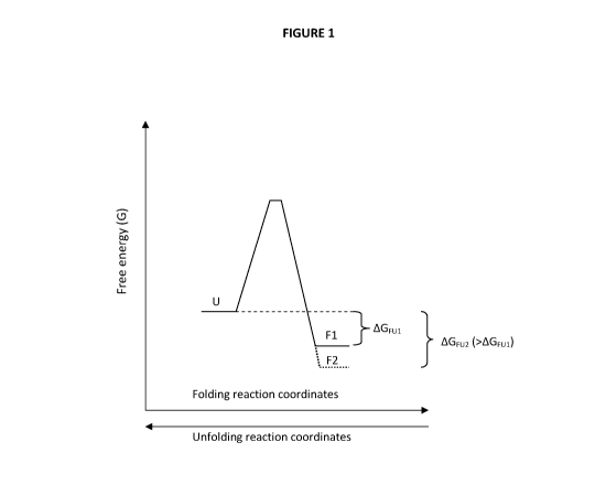
Thermodynamic stability is a measure of the difference in free energy
(AG) between the inactive unfolded (U) states and the folded state (F) in
which the enzyme is active. Thermodynamic stability can be determined at
equilibrium conditions if the protein is free to unfold and re-fold. This two-
state
5 model can be written as:
F U
Thus, in this case the stability is simply the difference in free energy
between
the U and the F states (AG = GUnfolded GFolded) and the stability is defined
as
AGFu, where
AGFu = -RTInK.
K represents the equilibrium constant between the unfolded and the folded
state (K=[U]/[F]) and, therefore, the more thermodynamically stable the
protein is the larger the difference in free energy (AG) is. This can also be
graphically represented by plotting the difference in free energy between the
unfolded and native state. (See Fig. 1).
Thus, simplified, the thermodynamic stability can be increased by
either destabilizing the unfolded state (higher free energy of U) or
stabilizing
the native state (lower free energy of F) so as to maximize the difference in
free energy (AGFu) between the two states. The change in free energy needs
to be lower than zero (AG < 0) for the folding reaction to be efficient, that
is,
favoring the native state of the protein. Since the difference in free energy
is
determined by its enthalpy (AH, interactions) and entropy (AS, disorder)
according to AG = AH - TLS a favorable AG can be accomplished by
strengthening the interactions of the folded state, leading to lowered
enthalpy
(e.g. hydrogen bonds, ion bonds, better packing of the protein interior etc.).
The same, i.e. a larger difference in free energy between the unfolded and
folded state, can be accomplished by destabilizing the unfolded state.
Furthermore, for the unfolded state, which can be assumed to be a random
coil, the same can be accomplished by restraining the freedom of the
CA 02862108 2014-07-21
WO 2013/162445
PCT/SE2013/050392
6
unfolded state leading to lowered entropy of the unfolded states and thereby
a higher level of free energy for the unfolded state.
The melting point (Tm) of a protein, i.e. the midpoint temperature of
unfolding, is a measure of a proteins thermal stability. In industrial
processes
it is often desirable to use enzymes with a high melting point since it is in
many cases beneficial if the reaction can take place at an elevated
temperature (higher rates of reaction, lower viscosity, less microbial growth,
less fouling etc). For this reason, what is often focused on for proteins that
have a potential use in industrial, enzyme based, processes is that the
protein
has a high thermal stability (i.e. a high melting point).
It is, however, important to recognize that at standard temperature (25
C) the GFu values for a thermolabile protein are not necessarily lower than
for a thermostable protein, i.e. a high thermal stability is not the same as a
high thermodynamic stability at all temperaturesm. Thus, it is not possible to
deduce the melting point of a protein by simply determine its thermodynamic
stability at ambient temperature or vice versa. The melting temperature (Tm)
is the temperature at which U and F are at equilibrium and are equally
populated and is determined by the GFu(T) function, and will occur when the
denaturing pressure (temperature) is so high that GFu = 0. When GFu is
plotted as a function of temperature, the GFu(T) function displays a skewed
parabola that intersects the x-axis twice (i.e. both heat- and cold
denaturation
occurs) (see Fig. 2).
Figure 2 illustrates how the thermostability of a hypothetic protein thus
can be increased by other means than increasing the thermodynamic stability
(AGuF) of the protein at standard temperatures.
Thus, thermal stability is related, but not equivalent, to thermodynamic
stability. That is, at ambient temperatures a protein can have a relatively
low
thermodynamic stability and still prove to have a relatively high melting
point.
Kinetic stability is a measure of at what rate a protein unfolds (ku). This
is especially important for proteins or conditions that denature proteins
irreversibly to unfolded states. A protein can denature irreversibly if the
protein in the unfolded state rapidly undergoes some permanent change such
CA 02862108 2014-07-21
WO 2013/162445 PCT/SE2013/050392
7
as proteolytic degradation or aggregation (which often is the case with
thermally denatured proteins).
ku
F 1.1 Irreversibly inactivated
In these cases it is not the difference in free energy between the folded and
unfolded state that is important. That will only affect the equilibrium and
this is
not a true equilibrium process. Instead, for kinetic stability, the important
thing
is the difference in free energy between the folded state (F) and the
transition
state (ts4) on the unfolding pathway which determines the activation energy
for unfolding (EA, unfolding). Hence, EA, unfolding determines the rate
constant of
unfolding (ku) and thereby at what rate an irreversible inactivation of the
unfolded state can take place (See Fig. 3).
Thus, this is in no way related to the thermodynamic stability (AGFu) or
the thermal stability (Tni) and other means are necessary to increase the
kinetic stability as compared to AGFu and Tn.,. In order to change the free
energy of the transition state the folding/unfolding mechanism of the protein
needs to be affected. Simplified, when an ensemble of proteins fold they will
mainly follow the fastest route that produces folding intermediates and
transition states of lowest possible energy levels. However, if this route is
no
longer accessible, they will be forced to fold via an alternative route that
has
folding intermediates and transition states of higher energy. This will in
effect
lead to a route that places the transition state at a higher level of free
energy.
In this case, since the folded state has the same energy level as before
(still
needs to be in its highly ordered native fold to be active) the height of EA,
unfolding will have increased and thus provide a barrier to unfolding leading
to a
slower unfolding rate constant (ku).
Thus, for a protein to be valuable for any application it needs to have a
large negative AGFu at the temperature of operation so that the protein
operates well below its melting point (Tn.). Equally important is that it
needs a
high kinetic stability so that the protein is maintained in the natively
folded
state and the protein does not sample the unfolded state which will render it
irreversibly inactive. Hence, a high kinetic stability will lead to slow
unfolding
CA 02862108 2014-07-21
WO 2013/162445
PCT/SE2013/050392
8
and a long lifetime of the protein. This is true for all conditions and will
for
example increase shelf life of the protein at ambient temperatures, but the
activation energy for unfolding (EA, unfolding) will also provide a barrier
for
unfolding also if the protein operates close to or even above its unfolding
point (thermal or other) and thus keeping the unfolding rate constant (ku) low
and the lifetime high also at conditions that induce unfolding.
There are numerous ways of stabilizing proteinsi91, either by stabilizing
the folded state or by destabilizing the unfolded state by different means.
However, most methods to stabilize the folded state rely on strengthening
local interactions that are only formed once the protein is folded and few
will
substantially affect the folding route and hence the kinetic stability.
Furthermore, because of the often hundreds of amino acids to vary and the
thousands of interactions within the protein and between the protein and the
surroundings, it is very difficult to simply examine the structure and
pinpoint
what to change in order to increase the stability. This is also the reason why
combinatory methods like directed evolution has been developed. Since
these methods produce thousands of variants of the protein by chance",
which are subsequently tested for activity at different conditions, it
circumvents the need for detailed knowledge of the protein structure, or
understanding of protein stability. However, for those well acquainted with
the
art of protein stability and stabilization it is possible to design more
stable
proteins by knowledge-based protein engineering. One attractive way to
stabilize a specific protein by knowledge-based protein engineering is to
graft
structural motifs that is known to be stabilizing from one protein homolog to
the protein homolog that is to be stabilized, of which there are numerous
examples in the literature 110'111. Two proteins are considered to be
homologous if they have identical amino acid residues in a significant number
of sequential positions along the polypeptide chain. However, as is text book
knowledge in protein chemistry, the three dimensional structure is much more
conserved than sequence and it is often found that proteins with very low
sequence identity still have similar function and similar three-dimensional
structuresi121. Thus, members of such families are also considered to be
homologous even though polypeptide sequence identities are not statistically
CA 02862108 2014-07-21
WO 2013/162445
PCT/SE2013/050392
9
significant, only structurally or functionally significant. Furthermore,
homologous proteins always contain a core region (structurally conserved
regions) where the general folds of the peptide chains are very similar. That
is, the scaffold of even distantly related homologous proteins with low
sequence identity have similar structure. It is these relationships that make
it
possible to transfer stabilizing amino acid combinations or motifs between
structurally homologous proteins if there is three dimensional structural data
available. Structural data can originate from X-ray crystallography, nuclear
magnetic resonance spectroscopy or model building. If two such structures of
homologous proteins are superimposed, one with stabilizing interactions of
interest (the template) and the other to be stabilized (the target), the three
dimensionally structurally equivalent position of stabilizing amino acids to
be
changed can be identified in the target structure.
One way of reducing the freedom (i.e. entropy) of the unfolded state
and thus place the unfolded state on a higher energy level is to introduce
covalent links between parts of the protein. This can be done by changing the
original amino acids to cysteins which are able to form covalent disulfide
bridges (S-S) if the thiol groups of the two amino acid side chains are
correctly placed in space. To design such bridges is however not trivial since
the geometry of an unstrained -CH2-S-S-CH2- bridge in proteins is limited to
rather narrow conformational constraints, and deviations from the geometrical
constraints will introduce strains into the folded structure. However, because
of the geometrical constraints, identification of disulfide bridges are
particularly amenable for homology modeling to identify amino acid positions
to alter to cysteines in order to introduce disulfide bridges in homologous
proteins, of which there are numerous examples of in the literaturei13:141
Although this method has a limited rate of success since the
replacement of the wild type amino acid and the introduction of a disulfide
bridge will often lead to loss of favorable interactions or strain in the
folded
state, it will lead to a larger thermodynamic stability (AGFu) if the folded
state
is unaffected (See Fig. 4).
Further, if the introduced disulfide bridge brings together parts of the
protein that normally are in close contact during early stages of the folding
CA 02862108 2014-07-21
WO 2013/162445
PCT/SE2013/050392
event, it will not affect the folding pathway and will thus only increase the
thermodynamic stability and possibly the rate of folding (under the
prerequisite that the energy level of the folded state is unaffected). If
however
the introduced disulfide bridge brings parts of the protein together, that
during
5 normal folding does not interact early in the folding event, this will
lead to that
the protein likely needs to fold via an alternative route that has a
transition
state of higher free energy. Under the prerequisite that the energy level of
the
folded state is unaffected, this will lead to that the activation energy for
unfolding (EA, unfolding) will become higher and thus the unfolding rate will
be
10 slower and the lifetime of the protein will be increased. If this can be
accomplished, an ideal protein, with both a high thermodynamic stability (and
possibly increased melting temperature) and a high kinetic stability, is
constructed (See Fig. 5).
Besides being potentially able to increase both the thermodynamic and
the kinetic stability of proteins, the stabilization is of entropic origin by
restricting the freedom of the unfolded state by incorporation of a covalent
bond (disulfide bridge). Thus, enthalpic stabilizing interactions by
introducing
disulfide bridges will not display a strong temperature dependence, which can
otherwise weaken or strengthen e.g. hydrogen bonds, salt bridges, ionic
bonds or hydrophobic effects. In addition, this also means that the
stabilization will be less influenced also by other characteristics of the
surrounding media, such as polarity and ionic strength etc, and the relative
increase in stability will be maintained also in media other than buffered
aqueous solutions.
From the above it can be presumed that to increase the physical
stability of a protein even more, one simply adds more disulfide bridges.
However, this is not uncomplicated for several reasons. Firstly, the
introduction of even a single stabilizing disulfide bond is challenging, since
often what is gained in energy difference by decreased entropy of the
unfolded state is often also lost in enthalpic energy in the folded state,
because of lost non-covalent interactions, or strain introduced into the
structure so that the GFu of the engineered protein is the same or even less
than that of the wild type protein (i.e. thermodynamically destabilized).
Thus,
CA 02862108 2014-07-21
WO 2013/162445
PCT/SE2013/050392
11
introducing two or more disulfide bridges might increase or decrease the
stability of the protein. Secondly, with two disulfide bridges present, the
folding pathway of the protein could be blocked, so that the protein is no
longer able to fold into its native active form. Thirdly, when more than two
cysteines are introduced in a protein there is a high risk that the cysteines
make disulfide bonds with the wrong partner during synthesis or folding. This
will always lead to an inactive protein as it will not be able to find its
folded
active conformation. This is also especially important during production of
heterologous (e.g. mammalian) proteins with multiple disulfide bonds in
recombinant systems (e.g. bacteria) as the formation of correct or native
disulfide bonds in such systems is very inefficient, often leading to low
yield of
production of functional enzymes.
Summary of the Invention
Since there are no naturally occuring carbonic anhydrases meeting the
requirements that need to be met to be used in an enzyme based bioreactor
to capture CO2, there exists a need in the art for development of engineered
carbonic anhydrases that meet the expected requirements and which are
simple and economical to produce, have a high catalytic activity, have a high
physical stability and a long life time under various conditions.
The aim of the present invention is therefore to solve the problems and
disadvantages described above by providing a carbonic anhydrase which is
simple and economical to produce, has a high catalytic activity, a high
physical stability as determined by thermodynamic, thermal and kinetic
stability and a long life time under various conditions.
This is achieved according to the present invention by means of an
isolated polypeptide having carbonic anhydrase activity, the sequence of
which corresponds to modified human carbonic anhydrase 11, wherein the
polypeptide comprises the mutations A23C, 599C, L202C, C2055 and
V241C, has increased physical stability compared to wild type carbonic
anhydrase 11 and further comprises disulfide bridges between C23 and C202
and/or between C99 and C241.
CA 02862108 2014-07-21
WO 2013/162445
PCT/SE2013/050392
12
According to one embodiment the isolated polypeptide having carbonic
anhydrase activity has a thermodynamic stability increased by 23.5 kJ/mol
compared to wild type carbonic anhydrase II.
According to another embodiment the isolated polypeptide having
carbonic anhydrase activity has a melting point increased by 18.5 C
compared to wild type carbonic anhydrase II.
In a further embodiment the isolated polypeptide having carbonic
anhydrase activity has an activation energy of unfolding increased by 25
kJ/mol compared to wild type carbonic anhydrase II.
In one embodiment the isolated polypeptide having carbonic
anhydrase activity has a rate of unfolding in water at 21 C that is about
22.000 times slower compared to wild type human carbonic anhydrase II.
According to one embodiment the isolated polypeptide having carbonic
anhydrase activity has a half-life of 86 days at 60 C, 8 days at 65 C and
1.6
days at 70 C.
According to another embodiment the isolated polypeptide having
carbonic anhydrase activity maintains its increased physical stability
compared to wild type carbonic anhydrase II in aqueous solutions of ethanol
amines, comprising methyldietanolamine (MDEA), monoethanolamine (MEA),
diethanolamine (DEA), and aminoethoxyethanol.
According to a further embodiment the isolated polypeptide having
carbonic anhydrase activity has the sequence according to SEQ ID NO: 8.
The aim of the present invention is further achieved by a method of
increasing the physical stability of carbonic anhydrases (EC 4.2.2.1) selected
from the superfamily of naturally occuring or modified a-carbonic anhydrases,
comprising insertion of a combination of two stabilizing disulfide bridges at
the
three dimensionally equivalent or sequentially homologous positions to C23,
C99, C202 and C241 in SEQ ID NO: 8, equivalent to positions A23, S99,
L202 and V241 in human carbonic anhydrase II.
The present invention also relates to a construct comprising a
polypeptide according to the present invention, operably linked to one or more
control sequences that direct the production of the polypeptide in an
expression host.
CA 02862108 2014-07-21
WO 2013/162445
PCT/SE2013/050392
13
In one embodiment the present invention relates to a recombinant
expression vector comprising the construct according to the invention.
The aim of the present invention is further achieved by means of a
recombinant host cell comprising the construct according to the invention or
the recombinant expression vector according to the invention.
The aim of the present invention is further achieved by use of an
isolated polypeptide having carbonic anhydrase activity according to the
present invention for extraction of carbon dioxide from a carbon dioxide
containing medium.
According to another embodiment the carbon dioxide containing
medium is a gas.
In one embodiment the gas is a flue gas, biogas, vent gas, or natural
gas.
In another embodiment the carbon dioxide containing medium is a
liquid.
In a further embodiment the carbon dioxide containing medium is a
multiphase mixture.
According to one embodiment the extraction of carbon dioxide from a
carbon dioxide containing medium takes place in a bioreactor.
The present invention further relates to a method of preparing an
isolated polypeptide of SEQ ID NO: 8, comprising acceleration of the forma-
tion of disulfide bridges by incubation of the polypeptide at elevated tempera-
tures of 25-60 C in the presence of an oxidizing agent at a pH of 7-10.
Further, the present invention relates to an isolated polynucleotide
having a sequence which encodes for a polypeptide according to the present
invention.
According to one embodiment the isolated polypeptide has at least 75
% remaining CO2 hydration activity compared to pseudo-wild-type HCA II.
In one embodiment the isolated polypeptide has a thermodynamic
stability of 54 kJ/mole.
In another embodiment the isolated polypeptide has a melting point of
77.5 C after incubation for 15 min.
CA 02862108 2014-07-21
WO 2013/162445
PCT/SE2013/050392
14
In a further embodiment the isolated polypeptide has a remaining CO2
hydration activity of 100 % after incubation for 15 min at 70 C.
In another embodiment the isolated polypeptide has a remaining CO2
hydration activity of at least 20 % after incubation for 15 min at 70-95 C.
According to another embodiment the isolated polypeptide has
a remaining CO2 hydration activity of 100 % after incubation for 2 h at 65 C.
According to a further embodiment the isolated polypeptide has
an activation energy of unfolding of 121 kJ/mole.
In a further embodiment the isolated polypeptide has a rate of
unfolding in water at 21 C of 4.2 x 10⁻⁹ min-1.
According to a further embodiment the isolated polypeptide has at least
95 % identity with the amino acid sequence of SEQ ID NO: 2 or SEQ ID NO:
4 or SEQ ID NO: 6 or SEQ ID NO:8.
According to another embodiment the isolated polypeptide has at least
98 % identity with the amino acid sequence of SEQ ID NO: 2 or SEQ ID NO:
4 or SEQ ID NO: 6 or SEQ ID NO: 8.
Brief Description of the Drawings
Fig. 1 is a graph illustrating the definition of difference in free energy,
between the unfolded state (U) and the native folded state (F) of a protein
(AGFui). The graph further illustrates how the thermodynamic stability can be
increased by stabilizing the folded state (AGFu2).
Fig. 2 illustrates the relationship between thermodynamic and thermal
stability and that knowledge about the thermodynamic stability at a single
temperature does not give any information about the melting temperature (T,)
of a protein. The GFu (T) function of a hypothetical thermolabile protein (¨)
with its melting temperature (T,) and the possible increase in T, by up
shifting (¨. ¨ ), right shifting (- - -) and flattening ( .............. ) of
the GFu (T) function
(adapted from ref. 8).
Fig. 3 is a graph illustrating the definition of activation energy of
unfolding (EA, unfolding) of a protein determined by the difference in free
energy
between the folded state (F) and the transition state (ts4) on the unfolding
pathway.
CA 02862108 2014-07-21
WO 2013/162445
PCT/SE2013/050392
Fig. 4 is a graph illustrating how the thermodynamic stability (AGFu) for
a protein is increased by restricting the freedom of the unfolded state by
incorporation of a disulfide bridge (Us_s), thus placing the unfolded state on
a
higher energy level.
5 Fig. 5 is a graph illustrating the resulting increase in both thermo-
dynamic stability (AGFu, s_s) and activation energy for unfolding (EA,s_s),
for a
protein with a disulfide bridge inserted at positions that affect both the
free-
dom of the unfolded state as well as the folding pathway and thereby the
transition state (.....). Comparison is made with an unmodified reference wild
10 type (wt) protein (¨).
Fig. 6 is a graph illustrating the enzyme variants resistance to unfolding
in a denaturing agent as fraction of unfolded protein as a function of Gu-HCI
concentration for SEQ ID NO: 2 (0), 4 (N) , 6 (0) and 8 incubated over night
( ) and SEQ ID NO: 8 incubated 2-5 days (*).
15 Fig. 7 A-C illustrates the life times at 60 C (Fig. 7A), 65 C (Fig.
7B)
and 70 C (Fig. 7C) for SEQ ID NO:2 (0),4 (N),6 (0) and 8 (*). Note that
SEQ ID NO 2 is only measured at 60 C as it is instantly inactivated already
at this temperature (Fig. 7A) and that SEQ ID NO: 4 is only measured at 60
and 65 C (Fig. 7A and 7B). The only variant having an appreciable life time
at all temperatures is the polypeptide of SEQ ID NO: 8.
Detailed Description of Preferred Embodiments of the Invention
One aspect of the present invention is to provide an enzyme that has a
high enough physical stability to make bioreactors, that are designed and
capable of extracting CO2 from a CO2-containing medium, practical and
economically feasible.
The present disclosure provides an engineered, highly efficient, human
carbonic anhydrase II variant that has an increased physical stability as
determined by thermodynamic, thermal and kinetic stability as well as
prolonged life time.
The stabilized human carbonic anhydrase II according to the present
invention has a thermodynamic stability increased by 23.5 kJ/mol.
The present invention further provides an engineered human carbonic
anhydrase 11 that is heat-stable and is able to catalyze the hydration of CO2
at
CA 02862108 2014-07-21
WO 2013/162445
PCT/SE2013/050392
16
normal and elevated temperatures over long periods of time. The heat
stability of the present invention provides a carbonic anhydrase that has a
melting point of 77.5 C and maintains 100 % CO2 hydration activity for at
least 15 min at 70 C and more than 20 % residual CO2 hydration activity at
95 C for at least 15 min.
The present invention also provides an engineered kinetically stabilized
human carbonic anhydrase II that has an activation energy for unfolding (EA,
unfolding) increased by 25 kJ/mol and a rate of unfolding (ku) at ambient
temperature that is about 22 000 times slower than the wild type enzyme.
The present disclosure further provides an engineered human carbonic
anhydrase II that maintains its relative stabilization properties in relation
to the
wild-type enzyme also in solutions other than buffered aqueous solutions e.g.
ethanolamine solutions.
The present invention also provides a method to economically and
effectively produce the engineered human carbonic anhydrase II according to
the present invention.
The present invention further provides polynucleotides encoding the
wild-type and the engineered human carbonic anhydrase II according to the
invention.
The present invention relates to a genetically engineered variant of the
enzyme human carbonic anhydrase II having the amino acid sequence
according to SEQ ID NO: 8, having substantially increased physical stability,
as defined by increased thermal, thermodynamic and kinetic stability, and as
compared to those of its parent enzymes having the amino acid sequence of
SEQ ID NO: 2, 4 and 6. The nucleotide sequences corresponding to SEQ ID
NO: 2, 4, 6 and 8 are shown in SEQ ID NO: 1, 3, 5 and 7, respectively. The
increased physical stability provides the enzyme properties that allows the
enzyme to be used, with an increased life-time, at elevated temperatures (i.e.
higher than 37 C) and in media other than buffered aqueouos solutions (e.g.
in methyldiethanolamine solutions).
Furthermore, the combination of SEQ ID NO: 2, 4 and 6 leads to the
properties of SEQ ID NO: 8 that allows it to be produced in an economically
viable way. One aspect of the invention is the use of stable carbonic
CA 02862108 2014-07-21
WO 2013/162445
PCT/SE2013/050392
17
anhydrases as catalysts in bioreactors for capture and sequestration of CO2
from CO2-containing gases, liquids or multiphase mixtures. The present
invention is of particular importance when a prolonged life-time is desired
and/or when the temperature of the CO2-containing medium is above the
melting point of naturally occurring or commercially available carbonic
anhydrases. The present invention is additionally useful both for sequestra-
tion (hydration) of CO2 and subsequent recovery of bicarbonate (dehydration)
of the previously sequestered CO2.
DEFINITIONS
"Carbonic anhydrase" and the abbreviation "CA" is used interchange-
ably to refer to a polypeptide having enzymatic E.0 4.2.1.1 activity and that
is
capable of catalyzing the inter-conversion of carbon dioxide and water to
bicarbonate and a proton.
"Human carbonic anhydrase II" and "HCA II" is used interchangeably to
denote the iso-form 2 variant of human carbonic anhydrase II.
"Wild-type" or "naturally occurring" refers to the form of polypeptide or
polynucleotide sequence that can be found in nature and has not been
intentionally modified by human manipulation.
"Pseudo-wild-type human carbonic anhydrase II" ("HCA Ilpwt") refers to
a variant of human carbonic anhydrase II with characteristics indistinguishab-
le from the wild type human carbonic anhydrase II with the naturally occurring
cysteine in position 205 exchanged by genetic manipulation to instead code
for the amino acid serine (C205S). Conventional denotation of human carbo-
nic anhydrase iso-form sequences sometimes refers to positions relative to
the positions in human carbonic anhydrase I and numbering can thus differ
between different publications. However, unless otherwise stated all positions
defined in this text refers to the sequences and positions as defined in SEQ
ID NO: 1-8.
"Modified" polypeptides according to the invention involves
polypeptides having more mutations, truncated variants of the polypeptides,
and polypeptides having one or more amino acids added at the N- or C-
terminal part of the polypeptide.
CA 02862108 2014-07-21
WO 2013/162445
PCT/SE2013/050392
18
EXAMPLES
Example 1
Selection of mutation positions
The positions chosen for mutation and introduction of cysteines were
based on the findings of two earlier variants of HCA Ilpwt. Although not a
valid
measure of physical stability1151, for one variant (SEQ ID NO: 4) the midpoint
of denaturation in increasing concentrations of a chemical denaturant
(guanidine hydrochloride) was increasedi161. In another variant (SEQ ID NO:
6) the thermodynamic stability was increased at ambient temperature (23
C)1171. In these two individually engineered disulfide bridge variants of HCA
Ilp,t, cysteine in position 99 makes a disulfide bridge with cysteine in
position
241 in one variant (SEQ ID NO: 4) and in the other variant (SEQ ID NO: 6)
cysteine in position 23 makes a disulfide bridge with cysteine in position
202.
However, all other important parameters concerning stability for these
variants were unknown. Since the following information cannot simply be
deduced from knowing the midpoint concentration of unfolding for one
component (SEQ ID NO: 2) or the thermodynamic stability at ambient
temperatures of the other component (SEQ ID NO: 6), the thermodynamic
stability, the melting point, the stability in 30 % ethanol amine solution,
the
kinetic stability, the unfolding rates and the lifetime at elevated
temperatures
of both the individual variants (SEQ ID NO: 4 and 6) were determined
according to the following examples. From the collective information gained
for the individual variants (SEQ ID NO: 4 and 6) in example 6 ¨ 10 in this
document, it is understood that both variants individually possess properties
that are beneficial for carbonic anhydrases to be used in an industrial
process
designed to capture CO2. Thus, a combination of the two variants could
tentatively lead to an enzyme variant with several of the necessary properties
enhanced. However, as can be understood from the background art, this
cannot be acclaimed without the necessary design of a combined variant and
the characterization thereof.
Furthermore, a combination of the two disulfide bridges could very well
also lead to that the protein can no longer fold or the cysteines make
disuldfide bonds with the wrong partner and thereby fold to a non-native
state.
CA 02862108 2014-07-21
WO 2013/162445
PCT/SE2013/050392
19
For one of the variants (SEQ ID NO: 6) it was also earlier found that an out
of
the ordinary chemical method was needed to form the disulfide bridge under
an acceptable time scale, which would hamper the large scale production of
the enzymei161. For the efficient large-scale production of the enzyme the
earlier proposed methods would be hard to implement to an economically
feasible industrial production process of the enzyme. Thus, based on the
experimental findings of the two single-disulfide variants in this document, a
novel double-disulfide variant (SEQ ID NO: 8) was designed (example 2),
produced (example 3-5) and characterized with regards to important
properties such as activity, physical stability and lifetime (example 6-11).
Example 2
Site-directed mutagenesis of HCAII2m.
All variants were produced by the same methods. As a template for
further modifications, a nucleotide (SEQ ID NO: 1) coding for a well known
variant of HCA II with the only cysteine in the polypeptide sequence at
position 205 (SEQ ID NO: 2) replaced with a serine, was usedi181. The use of
this variant prevents faulty disulfide bridges from being formed between any
introduced new cysteine and the otherwise single naturally occurring cysteine
in position 205. This variant of HCA II has further properties that are
indistinguishable from the wild type HCA II and is therefore identified as a
pseudo-wild-type human carbonic anhydrase II (HCA Ilp,t). The nucleotide
sequence coding for HCA Ilpwt was cloned into the plasmid pACA, a vector for
T7-RNA polymerase-directed expression. The production of T7 RNA
polymerase is in turn under control by a lac promotor, thus production of the
cloned HCA II protein can be activated by addition of lactose or analogs such
as IPTG. The plasmid was maintained in a laboratory expression strain of E.
coli (BL21/DE3). Plasmids were prepared by using the Qiagen plasmid
preparation kit according to the manufacturer's instructions. Mutagenesis
oligo-nucleotides were designed and ordered to specification from DNA
technology AS (Denmark). The HCA Ilpwt nucleotide sequence, contained in
the purified plasmids, was thereafter subject to site-directed mutagenesis
using the aforementioned DNA oligomers and the QuickChange site-directed
mutagenesis kit from Stratagene. After purification of the treated plasmids,
CA 02862108 2014-07-21
WO 2013/162445
PCT/SE2013/050392
aliquots of the plasmids was sent for sequencing (GATC Gmbh, Germany) for
verification of correct desired sequence and mutations. After verification the
plasmids was used to transform a new set of BL21/DE3 cells which were
grown to a cell density of approx. OD 1 at A660 in 20 ml 2 x LB medium. The
5 cells were transferred in aliquots of 500 pL to Eppendorf tubes and mixed
with
500 pL 50 % glycerol and frozen in liquid nitrogen. The E. coli stocks were
thereafter stored at - 70 C.
Example 3
Protein production
10 All variants were produced by the same methods. 2 x 15 mL of over-
night cultures of 50 mL of transformed BL21/DE3, containing plasm ids
carrying the mutated HCA Ilp,t, and grown in LB medium at 37 C, was
transferred and used to inoculate 2 x 1.5 L of LB medium in shake bottles.
The cells were allowed to grow at 37 C to a cell density of approx. OD 0.8 at
15 A660 and were then supplemented with IPTG and ZnSO4 to a final concentra-
tion of 1 mM, respectively and the cells were left to produce the protein over
night. The cells of the culture broths were sedimented by centrifugation at
3.000 x g and the supernatant was discarded. The cells were resuspended in
40 mL of 10mM tris-H2SO4, pH 9Ø The cell suspension was thereafter sub-
20 jected to ultrasonication to break the cell walls and release the cell
content.
The cell suspension was thereafter centrifuged at 10.000 x g for 30 min and
the supernatant containing the produced mutated HCA Ilpwt was collected.
The pH of the supernatant was adjusted to an approx. pH of 9 with tris base.
The supernatant was mixed with approx. 10 mL of an affinity gel for HCA II
(BioRad CM agarose with a sulfonamide coupled to the matrix) and allowed
to stand for 30 min before being applied to a chromatography column. The gel
was washed with several bed volumes of 10 mM tris-H2SO4, pH 9.0 under
monitoring of the A280. When no more change in A280 could be detected the
protein was eluted with 10 mM tris-H2504, pH 7.0 and 0.5 M azide. The
eluate was collected and transferred to dialysis tubes with a molecular weight
cut-off of 10 kDa (Millipore) and then dialyzed against 5 x 10 L of dialysis
buffer (10 mM tris-H2504, pH 7.5) with at least 8 h between each change of
CA 02862108 2014-07-21
WO 2013/162445
PCT/SE2013/050392
21
buffer. The dialyzed protein solutions were then collected and concentrated in
centrifugation tubes with a molecular cut-off of 10 kDa.
Concentration of the protein sample was determined by A280 measure-
ment using an extinction coefficient of 6280 = 55 400 M-1 cm-1. The protein
sample was further analyzed for purity by overloading of protein sample (10
pg per well) onto a SDS-PAGE. After the SDS-PAGE run the proteins in the
gel were stained with commassie brilliant blue. For each produced protein
sample it was found that no other protein band could be visually detected.
Thus, since the proteins were considered to be pure the mutated variants of
HCA II could be subject to further analysis.
Example 4
Detection of free cysteines
All variants containing cysteines were analyzed by the same methods.
Free cysteines, i.e. non-productive cysteines that had not formed a cystine
residue with its expected partner and thus had not formed a stabilizing
disulfide bridge, was detected by 7-chloro-4-nitrobenzofurazan (NBD-CI).
Protein, tris-H2504pH 7.5 and guanidine hydrochloride (Gu-HCI) were mixed
to a final concentration of 17.1 pM, 0.1 M and 5 M, respectively. Free
cysteines were detected with a time scan of 30 min at 420 nm using a
spectrophotometer (Hitachi U-2001) after addition of a tenfold excess of NBD-
CI (171 pM). As a reference, a sample of HCA Ilp,t (that has no cysteine
amino acid residue) was run. If there are free cysteines, the NBD-CI will
react
with the thiol group and form a cysteine-NBD moiety that absorbs light in the
visual wavelength (turns yellow). With an extinction coefficient of 420 = 13
000
M-1 cm-1 for the cysteine-NBD moiety, one free cysteine per protein will give
an absorbance A420 of 0.22 at the used concentration of protein after the
reaction, and four free cysteines will thus give an absorbance A420 of 0.88.
The only disulfide variant that did not show increase of absorbance at 420 nm
after the reaction was the single disulfide bridge variant SEQ ID NO: 4 which
thus had no free cysteines and a single disulfide bond fully formed. The other
single-disulfide variant (SEQ ID NO: 6) was, as earlier found, not able to
spontaneously form its disulfide bridge116' 171. More importantly, it was
subsequently found that the novel double-disulfide variant (SEQ ID NO: 8)
CA 02862108 2014-07-21
WO 2013/162445
PCT/SE2013/050392
22
also had about 50 % of free cysteins (2 out of 4 cysteines not forming a
disulfide bridge). Most likely, this indicates that one disulfide bridge had
formed spontaneously, whereas the other disulfide bridge was not formed
during production of the enzyme of SEQ ID NO: 8. Thus, a method to form
the missing disulfide bridge needed to be developed.
Example 5
Formation of disulfide bridges of SEQ ID NO: 6 and 8
Due to low resistance of the reduced form towards unfolding in
guanidine hydrochloride (Cm, Fu of 0.7 M Gu-HCI)1171, the disulfide bridge of
SEQ ID NO: 6 was formed by a chemical method as has previously been
described in the literaturei161, resulting in a protein with both cysteines
reacted
in a correct disulfide bridge and with a retained native and active
conformation. However, the double-disulfide bridge variant of SEQ ID NO: 8
had only one out of two disulfide bridges formed. Most likely, it was the
disulfide bridge of SEQ ID NO: 4 that had formed and the disulfide bridge of
SEQ ID NO: 6 that had not formed, analogously to the behavior of the indivi-
dual disulfide bridge variants. Nevertheless, regardless of which of the two
disulfide bridges that had formed, each will individually lead to a higher
thermal stability of the protein (see example 9). Thus, instead of using the
earlier described chemical method to increase the structural flexibility to
facilitate for the cysteines to find each other, the formation of the second
disulfide bridge in SEQ ID NO: 8 could be accomplished by allowing the
reaction to take place at elevated temperatures.
Therefore, an alternative scheme to the chemical method used to form
the disulfide bridge of SEQ ID NO: 6 was developed for the double-disulfide
bridge variant of SEQ ID NO: 8. Since the melting point of the least
stabilized
variant (SEQ ID NO: 4) with a formed disulfide bridge is increased by 7.5 C
and is unaffected by incubation at temperatures < 55 C (see example 9), the
double disulfide variant of SEQ ID NO: 8 could effectively be incubated at 50
C to induce formation of the second disulfide bridge donated from SEQ ID
NO: 6. For the purpose of verifying this approach an experimental assay was
designed. Two stock solutions containing 85.5 pM of protein (SEQ ID NO: 8
with only one disulfide bridge formed) in 50 mM tris-H2504 pH 8.5
CA 02862108 2014-07-21
WO 2013/162445
PCT/SE2013/050392
23
supplemented with a 100 fold concentration of oxidized dithiotreitol (DTT) was
prepared. One solution was incubated at room temperature whereas the other
was incubated in a heated cabinet at 50 C. At certain time points aliquots of
the stock solutions were withdrawn and measured for free cysteines as
described in example 4. It was found that by incubating the sample at 50 C
this method yielded 100 % disulfide bridge formation of SEQ ID NO: 8 within
24 hours. At this time the sample incubated at room temperature had only
formed approx. 20 % of the disulfide bridges. The samples were further
analyzed by SDS-PAGE which revealed that no dimers had been formed
during the thermal process, indicating that correct disulfide bridges had been
formed. In terms of applicability of the enzyme of SEQ ID NO: 8 this is a very
important result as it makes the large-scale production of the variant
feasible.
Partly because, as compared to the earlier described chemical method, less
amount of costly chemicals is needed since no addition of Gu-HCI is
necessary in the process.
Furthermore, the completed enzyme product does not need down-
stream processing to be cleaned from the denaturing agent Gu-HCI. Yet
more, the reaction rate with SEQ ID NO: 8 and the described "thermal"
method is faster (24 h for 100 % disulfide bridge formation) than the chemical
method as it takes place at elevated temperatures. This can be compared to
the rate of disulfide bridge formation in SEQ ID NO: 6 using the earlier
described chemical method (100 h for 100 % disulfide bridge formation).
Example 6
Stability against unfolding by denaturing agents in ageous solution and in 30
% methyldiethanolamine
Aliquotes of 0.85 pM solutions of each of the described HCA II variants
of SEQ ID NO: 2, 4, 6 and 8 were incubated in room temperature over night
(approx 18 hours) in increasing amount of the denaturant Gu-HCI (0 - 6 M) in
buffered solutions (0.1 M tris-H2504 pH 7.5). For the methyldiethanolamine
(MDEA) measurements the solution also contained MDEA at a final concent-
ration of 30 %. Fluorescence spectra were recorded for each variant at all Gu-
HCI concentrations choosen in a spectrofluorometer (Jobin-Yvon Fluoromax
4). Excitation wavelength was 295 nm and three accumulative emission
CA 02862108 2014-07-21
WO 2013/162445
PCT/SE2013/050392
24
spectra were recorded for each sample between 310 - 400 nm. From the
spectra the wavelength shift was determined. The data was normalized and
the fractional change as a function of Gu-HCI concentration was determined.
From this it was determined at what Gu-HCI concentration the midpoint of
unfolding (Cm, Fu) occurred for each variant in both media (table 1). For
samples incubated in buffered aqueous solution the values for HCA I lp,,t (SEQ
ID NO: 2) and the two single disulfide bridge variants (SEQ ID NO: 4 and 6)
reached earlier found values (Cm, Fu of 1.0, 1.40 and 1.85 M Gu-HCI,
respectively). Unexpectedly, the subsequently produced novel SEQ ID NO: 8
variant reached an apparent very high Cm, Fu of 2.6 M Gu-HCI, which is higher
than the sum of each individual stabilizing disulfide bridge (1.0 + 0.4 + 0.85
=
2.25 M GuHCI). This could mean one of two things. Either there were some
synergistic effect in the stability making the double disulfide bridge enzyme
of
SEQ ID NO: 8 in fact more stable against denaturation by Gu-HCI than the
sum of stabilization of the two contributing single disulfide bridge variants.
Alternatively, the kinetic stability of SEQ ID NO: 8 was increased so that the
rate of unfolding was slower and thus the protein sample of SEQ ID NO: 8 did
not reach equilibrium in 18 h. Therefore, the very same samples were
incubated for an additional 24 h (total of 42 h) before data was collected
again. For SEQ ID NO: 8 it was found that the curve had shifted to lower
concentrations and stopped at a Cm, Fu of 2.25 M Gu-HCI. Thus, although the
SEQ ID NO: 8 enzyme was not as resistant to denaturants as the initial result
indicated, this is still a considerable increase in stability as determined by
Cm,
FU and compared to SEQ ID NO: 2, 4 and 6. Furthermore, this also proves
that the combination of disulfide bridge variants of SEQ ID NO: 4 and 6 is
achievable since the protein can still fold and the stability reaches the sum
of
each individual stabilizing disulfide bridge (2.25 M GuHCI) and thus no
stabilizing effects regarding resistance to denaturing agents are lost from
combining the two.
The slow equilibration that was found for SEQ ID NO: 8 is a behavior
that to our knowledge has not earlier been demonstrated for any earlier
variants of HCA II. The behavior implies that the double disulfide variant of
SEQ ID NO: 8 has a high kinetic barrier to unfolding. This would then lead to
CA 02862108 2014-07-21
WO 2013/162445
PCT/SE2013/050392
that the unfolding rate of SEQ ID NO: 8 is slower than for each of the
individual disulfide bridge variants of SEQ ID NO: 4 and 6, and thus that the
equilibrium between the folded and unfolded state takes longer time to reach
than for each individual disulfide bridge variant (SEQ ID NO: 4 and 6).
5 Table 1.
Midpoint concentration of unfolding in increasing concentration of Gu-HCI
SEQ ID SEQ ID SEQ ID SEQ ID NO 8 SEQ ID
NO 8
NO2 N04 N06 (INCUBATION (INCUBATION
3-5 DAYS) OVER
NIGHT)
cLkA-120 1.0 1.4 1.85 2.25 2.6
(M Gu-HCI)
pc:x> 0.3 0.75 1.2 1.4
MDEA)
(M Gu-HCI)
The stability against denaturing agents in 30 % MDEA follows the
same trend, i.e. SEQ ID NO: 2 has the lowest Cm, Fu followed by SEQ ID NO:
4, 6 and 8 respectively (see table 1). Thus, this result confirms that
although
10 MDEA generally destabilizes the proteins, the relative increase in
stability
against denaturation in Gu-HCI from introduced disulfide bridges is almost
unaffected by the properties of the surrounding media, as earlier described.
Therefore, also in 30 % MDEA, the protein acording to SEQ ID NO: 8 has a
considerably higher stability than the HCA Ilp,t variant (SEQ ID NO: 2) has.
15 Thus, the protein variants of SEQ ID NO: 6 and 8 are more stable even in
30
% MDEA than SEQ ID NO: 2 is even in buffered aqueous solution.
Example 7
Thermodynamic stability in aqueous solution
The equilibrium constant data (K) in the transition region, for each
20 enzyme variant, obtained in example 6 was used to calculate the
thermodynamic stability of the respective enzyme variant in purely buffered
aqueous solution according to the relationship AG = -RTIn(K) by the linear
extrapolation methodi191.
CA 02862108 2014-07-21
WO 2013/162445 PCT/SE2013/050392
26
Table 2.
Thermodynamic stability in buffered aqueous solution at ambient temperature
(21 C).
Thermodynamic SEQ ID NO 2 SEQ ID NO 4 SEQ ID NO 6 SEQ ID
stability NO 8
(inc. 3-5
days)
AG (H20) 30.5 39 46 54
(kJ/mol)
LAG (H20) 8.5 12.5 23.5
(kJ/mol) relative
to SEQ ID NO 2
Clearly, the increased resistance of SEQ ID NO: 8 to denaturation by
Gu-HCI as judged by Cm, Fu values is an effect of a significantly increased
thermodynamic stability. Furthermore, the increase in thermodynamic stability
of SEQ ID NO: 8 is slightly larger than the sum of increased stability of SEQ
ID NO: 4 and 6 (8.5 + 12.5 < 23.5), indicating a small synergistic effect.
Example 8
Activity assays of carbonic anhydrases
Activity assays were used in order to measure the change in enzyme
activity in response to changes in conditions (denaturing agents and
temperature) to reveal melting temperatures (Tm), unfolding rates, kinetic
stability and life time at elevated temperatures. Activity assays are also
important to establish the absolute activity of the protein variants of SEQ ID
NO: 4, 6 and 8, in relation to the pseudo-wild-type enzyme, since what is
desired is an as high as possible catalytic activity and efficiency also in
the
engineered variants.
Several variants of colorometric CO2-hydration activity assays have
earlier been described in literature[20, 21, 22] which are all based on the
enzymatic reaction which leads to the production of bicarbonate and protons
from carbon dioxide and water. Thus, enzymatic activity of carbonic
anhydrase will give a faster decrease in pH than the spontaneous reaction
CA 02862108 2014-07-21
WO 2013/162445
PCT/SE2013/050392
27
and can be monitored if the reaction takes place in a buffer containing the pH
indicator bromothymol blue (BTB).
An aqueous stock solution saturated with CO2was prepared by
bubbling ice cooled deionized water with CO2 through a gas diffuser for at
least 1 hour prior to use. To monitor the CO2-hydration activity per mg of
enzyme, 2 ml of 25 mM veronal-H2SO4, pH 8.2, containing 20 mg/L of BTB
was mixed with 1 ml deionized water and 30 1_ of protein (8.5 M) in a small
beaker placed in an ice-bath on top of a magnetic stirrer. All solutions were
kept on ice prior to use. The reaction was started by the addition of 2 ml of
the
CO2 saturated solution to the stirred buffer solution. Simultaneously with the
addition of CO2 saturated solution a stop watch was started and the time to
reach pH 6.5 was determined by comparison of color to a reference sample
containing 2 ml 0.2 M Na-phosphate buffer pH 6.5, 2 ml 25 mM veronal-
H2SO4, pH 8.2, containing 20 mg/L of BTB and 1 ml deionized water. The
time to reach pH 6.5 was measured for the catalyzed reactions (tc) and for the
un-catalyzed blank reactions (tb) and the following equation was used to
determine activity units (A.U.) per mg enzyme:
(4) ¨
Tlimg ________________________
In all CO2-hydration experiments the amount of enzyme in the activity
assay was 7.5 g. The CO2-hydration activity of the three disulfide variants
(SEQ ID NO: 4, 6 and 8) at the conditions of measurement (0 C) was found
to be 105, 82 and 75 percent respectively of the activity of the HCA Ilpwt
variant. Thus, all disulfide bridge variants remain highly active with regards
to
CO2 hydration.
Example 9
Thermal stability assay
For each enzyme variant (SEQ ID NO: 2, 4, 6 and the subsequently
produced SEQ ID NO: 8) stock solutions of 8.5 M enzyme in 10 mM tris-
H2504 pH 7.0 were prepared. Aliqoutes of 70 1_ enzyme solutions was
placed in thin-walled PCR tubes. In order to prevent increase in enzyme
CA 02862108 2014-07-21
WO 2013/162445
PCT/SE2013/050392
28
concentration in the enzyme solutions after incubation, due to evaporation
and condensation of liquid in the PCR tube, a PCR thermocycler with a
heated top was used (GeneAmp PCR-system 9600, Applied Biosystems). For
each enzyme variant and target temperature a sample was placed in the
thermocycler which was programmed for constant ramping to the set
temperature (55 to 95 C) and incubated for 15 min or 2 hours. After
incubation the samples were allowed to cool to room temperature for 10 min
before measurements of the residual enzymatic activity according to example
8. All experiments were performed in duplicates. The resulting residual
activity after thermal treatment for 15 minutes and 2 hours is presented in
table 3 and 4, respectively. Clearly, all engineered variants have higher
thermostability than the pseudo-wildtype enzyme (SEQ ID NO: 2).
Furthermore, the variant with the highest thermostability is the constructed
double disulfide bridge variant (SEQ ID NO: 8). To calculate the approximate
T, (i.e. the temperature at which 50 % residual activity remain) of each
variant, data from the 15 min incubation was fitted to a sigmoidal function
(Table-Curve, Jandel Scientific) and is presented in Table 5.
However, it is important to note that the T, values obtained are only
apparent melting points. Nevertheless, the increase in T, (AT,) of modified
variants (SEQ ID NO: 4, 6 and 8), as compared to HCA Ilpwt (SEQ ID NO: 2),
represents accurate values. The reason for this is that thermal denaturation
of
the enzymes is not an equilibrium process but is an example of irreversible
inactivation where the enzymes aggregate after unfolding at temperatures
close to or above their respective melting points. Thus, what is actually
monitored in the activity assay is how large population of enzyme molecules
that have yet not unfolded and aggregated at the respective temperature.
Consequently, in this case of thermal stability, the kinetic stability is as
important as the thermodynamic stability in deciding the behavior at elevated
temperatures. Thus, in comparison to the other variants, SEQ ID NO: 8 has
two striking characteristics. Firstly, it has an exceptionally high melting
point
of approximately 77.5 C which is an increase of 18.5 C compared to the
pseudo-wild-type variant, and even higher than the approx. 70 C of the y-
carbonic anhydrase from Methanosarcina thermophila. Secondly, the enzyme
CA 02862108 2014-07-21
WO 2013/162445
PCT/SE2013/050392
29
of SEQ ID NO: 8 has an apparent residual activity of above 20 % at
temperatures far beyond its melting point of 77.5 C. This is almost certainly
a
result of a remarkably high kinetic stability, resulting in that not even
incubation for 15 min at 95 C is enough to completely inactivate all enzyme
molecules. Clearly, this is a valuable feature if the enzyme is to be used in
e.g. a temperature phased process were the temperature is continuously
altered between low and high temperatures since the high kinetic stability of
SEQ ID NO: 8 allows the enzyme to survive short bursts of temperatures far
beyond its melting temperature.
Table 3
Percent remaining CO2-hydration activity after 15 min incubation
TEMPERATURE ( C)
ENZYME VARIANT 55 60 65 70 75 80 85 90 95
SEQ ID NO 2 100 22 2 0 0 ND ND ND ND
SEQ ID NO 4 100 100 77 4 0 0 ND ND ND
SEQ ID NO 6 100 100 100 80 2 1 ND ND ND
SEQ ID NO 8 100 100 100 100 76 26 24 23 22
Table 4
Percent remaining CO2-hydration activity after 2 h incubation
TEMPERATURE ( C)
ENZYME VARIANT 55 60 65 70 75 80 85 90 95
SEQ ID NO 2 100 2 1 0 0 ND ND
ND ND
SEQ ID NO 4 100 95 50 1 0 0 ND ND ND
SEQ ID NO 6 100 100 100 39 0 0 ND ND ND
SEQ ID NO 8 100 100 100 92 38 7 7 ND ND
Table 5
Melting points and increase in melting point of enzyme variants
T, AT,
Enzyme variant
SEQ ID NO: 2 59
SEQ ID NO: 4 66.5 7.5
SEQ ID NO: 6 71.5 12.5
SEQ ID NO: 8 77.5 18.5
CA 02862108 2014-07-21
WO 2013/162445
PCT/SE2013/050392
Example 10
Kinetic stability of engineered HCA II variants
In order to determine the unfolding rates (ku) and activation energy for
unfolding (EA, unfolding) of the respective enzyme variant, chemical
denaturation
5 was employed. For the unfolding assay each enzyme variant was subjected
to increasing concentrations of Gu-HCI, starting from a concentration of 0.2 -
0.3 M above their respective midpoint concentration of unfolding (Cm, Fu, see
table 1, example 6) in steps of 0.1 M. For example HCA Ilpwt, with a Cm, Fu of
1.0 M Gu-HCI, was denatured in Gu-HCI concentrations of 1.2, 1.3, 1.4, and
10 1.5 M Gu-HCI. Stock solutions of Gu-HCI, to reach the final assay
concentration, were prepared and protein stock solutions of 0.5 mg/ml were
prepared for each enzyme variant. 2.5 I of enzyme was mixed with 47.5 I of
Gu-HCI to reach the targeted Gu-HCI concentration and a protein
concentration of 0.025 mg/ml (8.5 M). Each protein variant and each Gu-HCI
15 concentration samples were prepared at room temperature (21 C), from
stock solutions, to monitor residual activity after 10 and 30 sec, and 1, 2,
5,
30, 45, and 60 min. Activity measurements were done as described in
example 8 (with 30 I of sample), with the difference that the buffered BTB
solution was supplemented with 0.5 mM of the metal chelator EDTA to
20 prevent refolding of enzymes in the assay. Refolding could otherwise
occur
as both protein and the denaturing agent (Gu-HCI) is diluted in the activity
assay. EDTA binds Zn2+ which is released from unfolded enzymes and
present in solution, and which is necessary for the activity HCA II. Thus, the
addition of EDTA to the assay "freezes" the state of the sample so that
25 residual CO2-hydration activity can be measured.
When the residual activity is plotted as a function of time for each Gu-HCI
concentration the unfolding rate (ku) at that very Gu-HCI concentration can be
calculated by fitting the data to a single exponential term according to y= a
x
e-kx. To calculate the unfolding rate constant in aqueous solution the natural
30 logarithm of the measured rate constants for the respective enzyme
variant is
plotted against Gu-HCI concentration and the linearized data is extrapolated
to 0 M Gu-HCI (giving the In ku at 0 M Gu-HCI). The free energy of activation
(AG4), that is, in this case, the free energy of activation for unfolding (EA,
CA 02862108 2014-07-21
WO 2013/162445
PCT/SE2013/050392
31
unfolding), can be calculated using the Arrhenius equation, ,AG4=RT[In(kB/h)-
In(ku/T)], where R is the gas constant, 8.314 J.mo1-1.K-1, kB/h is the
constant
2.08358.1010, T is the absolute temperature in Kelvin and ku is the rate
constant for unfolding. The results from the measurements of kinetic stability
are presented in table 6.
Table 6
Unfolding kinetics data of enzyme variants at 21 C
SEQ ID NO 2 SEQ ID NO 4 SEQ ID NO 6 SEQ ID NO
8
Lnku, H20 -9.30 -14.1 -12.2 -19.3
ku, H20 (min-1) 9.110-5 7.5*10-' 5,110-5 4.2*1 0-
9
TIMES SLOWER 122 18 22000
UNFOLDING AS
COMPARED TO
SEQ ID NO 2
EA, unfolding 96 kJ/mol 108 kJ/mol 103 kJ/mol 121
kJ/mol
AEA, unfolding 12 kJ/mol 7 kJ/mol 25
kJ/mol
(INCREASE AS
COMPARED TO
SEQ ID NO 2)
For all stabilized disulfide bridge variants (SEQ ID NO: 4, 6 and 8)
there is an obvious decrease in unfolding rate (ku (H20)) which culminates in
the very slow unfolding of the constructed SEQ ID NO: 8 that unfolds 22.000
times slower than the HCA Ilpwt variant (SEQ ID NO: 2) in aqueous media at
21 C.
What is important is that the enzyme of SEQ ID NO: 8 behaves as a
completely new variant of HCA II. SEQ ID NO: 4 was found to confer a high
increase in kinetic stability (AEA, unfolding of 12 kJ/mol) and a lower
increase in
thermodynamic stability (LAGFu of 8.5 kJ/mol), and behaves thus as an
enzyme with an engineered disulfide bridge with an altered folding pathway
and thereby a transition state at a higher energy level. Contrary to SEQ ID
NO: 4, the enzyme of SEQ ID NO: 6 was found to have a lower increase in
kinetic stability (AEA, unfolding of 7 kJ/mol) but possesses a high
thermodynamic
stabilization (LAGFu of12.5 kJ/mol). Thus, this variant has an unfolded state
CA 02862108 2014-07-21
WO 2013/162445
PCT/SE2013/050392
32
placed on an even higher level of free energy. On the other hand it has a
folding pathway that is only slightly altered and therefore a transition state
with only a slightly higher energy level than for the enzyme of SEQ ID NO: 2.
However, for the enzyme of SEQ ID NO: 8 there is both a very high kinetic
stabilization (AEA, unfolding of 25 kJ/mol) and a very high increase in
thermodynamic stability (AAGFu of 23.5 kJ/mol). For the thermodynamic
stability the increase is close, although not identical, to the sum of
increased
stability of SEQ ID NO: 4 and 6. However, the very large increase in kinetic
stability is an unpredictable effect that stems from the successful
introduction
of two disulfide bridges in the enzyme, at positions that forces the protein
to
fold via an unexplored pathway, which differs from the enzymes of SEQ ID
NO: 2, 4 and 6, while at the same time the folding ability and enzymatic
activity is retained.
Example 11
Life-time at elevated temperatures assayed by esterase activity
measurements
The increased thermal, thermodynamic and kinetic stability of the
double disulfide bridge variant of SEQ ID NO: 8 should render it a high life
time at elevated temperatures. For practical reasons this was monitored by
the esterase activity of the enzyme. Stock solutions of 2.5 mg/ml of each
enzyme variant were prepared in 10 mM tris-H2504 pH 7.0 in tubes with
screw cap and sealing to prevent evaporation. These were placed in a heated
cabinet at the desired temperature (60, 65 or 70 C) and aliqoutes were
withdrawn at different time points for measurement of residual esterase
activity. Esterase activity was assayed by adding 6 1_11_ of protein sample to
1.44 m L of reaction buffer (50 mM tris-H2504, pH 8.5 with an ionic strength
of
0.1 M adjusted with Na2504) in a cuvette. The sample was supplemented
with reagent, 60 pL of 30 mM para-nitrophenyl acetate (pNPA) in ice cold
acetone, and briefly mixed before esterase activity was measured at 348 nm
in a spectrophotometer. The increase in absorbance of the catalyzed reaction
was monitored for 60 seconds and the increase in absorbance of a blank
reaction (no enzyme added) was then subtracted. The apparent second-order
CA 02862108 2014-07-21
WO 2013/162445 PCT/SE2013/050392
33
rate constant (k") was calculated according to earlier described
methodologyi231.
As a time zero reference the esterase activity of each enzyme stock
solution was determined before heat treatment. The esterase activity of the
three disulfide variants (SEQ ID NO: 4, 6 and 8) at the conditions of
measurement (approx. 21 C) was found to be 99, 86 and 78 percent
respectively, compared to the activity of the HCA Ilp,t variant. Thus, all
disulfide bridge variants remain highly active also with regards to the
esterase
activity and to the approximate same degree as CO2 hydration activity
(example 8). The residual activity of each variant at each temperature was
plotted against time and fitted to a single exponential term (y=a.e-kx) to
obtain
the rate constant for unfolding for each enzyme variant at the three
temperatures. Since the inactivation is a first-order rate process, the half-
life
(ti,) of each enzyme variant at each temperature can be calculated by
ty2=In2/k. The results of the life time experiments are presented in table 7-9
and Fig. 7.
Table 7.
Percent remaining esterase activity after 15 min incubation
TEMPERATURE ( C)
ENZYME VARIANT 60 65 70
SEQ ID NO 2 1 0 0
SEQ ID NO 4 102 61 1
SEQ ID NO 6 104 83 82
SEQ ID NO 8 99 96 91
Table 8.
Percent remaining esterase activity after 2 h incubation
TEMPERATURE ( C)
ENZYME VARIANT 60 65 70
SEQ ID NO 2 0 ND ND
SEQ ID NO 4 103 50 ND
SEQ ID NO 6 104 72 39
SEQ ID NO 8 106 89 80
CA 02862108 2014-07-21
WO 2013/162445 PCT/SE2013/050392
34
Table 9.
Inactivation rate constants (ku), inactivation half time (t%) and ti/10 of
enzyme
variants at 60 ¨ 70 C.
TEMPERATURE
ENZYME 60 C 65 C 70 C
VARIANT
ku (h-1) ty2(h) tin() (h) ku(h1) ty2(h) t1/10
(h) ku(h1) ty2(h) t1/10 (h)
SEQ ID
NO 2
SEQ ID 0,0205 35 112 0,346 2 6,6
NO 4
SEQ ID 0,00255 272 903 0,0475 15 48 0,434
2 5,3
NO 6
SEQ ID 0,000337 2057 6832 0,00381 182 604 0,0180
38 127
NO 8 (86 (285 (8 (25 (1.6 (5.3
days) days) days) days) days)
days)
The increased physical stability, engineered into the double disulfide bridge
variant of SEQ ID NO: 8, results in a much slower inactivation rate and thus a
much longer life time at increased temperatures than the other variants. At 60
C the half time of activity inactivation is 86 days for the double disulfide
bridge variant of SEQ ID NO: 8 which can be compared to HCAllpwt (SEQ ID
NO: 2) which is instantly unfolded and inactivated at the same temperature.
Furthermore, the time for the activity to fall down to 1/10 for SEQ ID NO: 8
is
approximately 285 days at 60 C. That is, if two different reactors used the
same amount of the engineered HCA11 of SEQ ID NO: 8 and an enzyme with
1/10 of activity (as for example Cam), respectively, the reactor with SEQ ID
NO: 8 would need 285 days to even fall to the low starting value of the Cam
reactor. Thus, it is not only the physical stability per se that is important
for an
enzyme's practicability, but also the activity and efficiency of the protein.
Even at 70 C, where all other variants are quickly unfolded and inactivated,
the enzyme of SEQ ID NO: 8 has an appreciable slow inactivation and
increased life time with a half life of 1.6 days. The ability to withstand
temperatures in the range of 60 - 70 C with a long life time is an extremely
important feature of SEQ ID NO: 8. At for example modern incinerator plants
the flue gas cleaning consists of so many steps that the flue gas is cooled
CA 02862108 2014-07-21
WO 2013/162445
PCT/SE2013/050392
down to approx. 60 - 70 C before it reaches the smokestack [241. Thus, any
enzyme that is to be used in a CO2-capturing bioreactor at an incinerator
plant should preferably also be stable and active in the temperature range of
60 - 70 C, which is thus fulfilled by the enzyme of SEQ ID NO: 8.
5 Furthermore, the combination of disulfide bridges of SEQ ID NO: 8 has
been
engineered into an enzyme (HCAllp,t) that belongs to the structurally
conserved superfamily of a-carbonic anhydrases. To find structurally related
proteins, a database search of the three dimensional structure of HCA II (PDB
ID 2cba) was executed against the Conserved Domain Database (CDD)1251,
10 which includes alignments of conserved protein domains to known 3-
dimensional protein structures in the Molecular Modeling Database (MMDB).
The search resulted in 4977 protein sequences with related conserved
domains and 438 related solved structures from the a-carbonic anhydrase
superfamily. Thus, the combination of the structural motifs of the disulfide
15 bridges between position C23- C202 and C99- C241 in the SEQ ID NO:8
variant of HCA Ilpwt can be identified and most likely be grafted also into
other
members of the a-carbonic anhydrase superfamily by homology modeling as
earlier described. Indeed, the significantly increased stability of SEQ ID NO:
6
was originally accomplished by homology modeling between HCA II and the
20 distantly related homologous a-carbonic anhydrase from Neisseria
gonorrhoeae (NGCA, 38.5 % sequence identity) which has a naturally
occurring disulfide bridge. By homology modeling it was found that the
positions for the disulfide bridge in NGCA (sequence positions of C28 and
C181) had their three dimensionally structurally equivalent positions in HCA
II
25 at the positions A23 and L202. Thus, by homology modeling against a
distantly related homologous enzyme a geometrically correct disulfide bridge
could be grafted from NGCA into the correct positions in HCAIII171.
CA 02862108 2014-07-21
WO 2013/162445
PCT/SE2013/050392
36
REFERENCES
1) Zimmerman, S.A. and Ferry, J.G. (2008). The 0- and y-classes of
carbonic anhydrase. Current pharmaceutical design 14, 716-721.
2) Alber, B.E., Ferry, J.G. (1994). A carbonic anhydrase from the
archaeon Methanosarcina thermophila. Proceedings of the National
Academy of Science 91, 6909-6913.
3) IPCC, 2007: Climate Change 2007: Synthesis Report. Contribution of
Working Groups I, II and III to the Fourth Assessment Report of the
Intergovernmental Panel on Climate Change [Core Writing Team,
Pachauri, R.K and Reisinger, A. (eds.)]. IPCC, Geneva, Switzerland.
4) Zimmerman, S.A., Tomb, J.-F., Ferry, J.G. (2010). Characterization of
CamH from Methanosarcina thermophila, founding member of a
subclass of the y class of carbonic anhydrases. Journal of
Bacteriology, 192, 1353-1360.
5) Silverman, D.N. and Lindskog, S. (1988). The catalytic mechanism of
carbonic anhydrase: Implications of a rate-limiting protolysis of water.
Accounts of chemical research, 21, 30 - 36.
6) Ferry, J.G. (2010). The y class of carbonic anhydrase. Biochimica et
Biophysica Acta, 1804, 374-381.
7) MacAuley, S. R., Zimmerman, S. A., Apolinario, E. E., Eyilia, C., Hou,
Y.-M., Ferry, J. G. and Sowers, K. R. (2009). The archetype y-class
carbonic anhydrase (Cam) contains iron when synthesized in vivo.
Biochemistry, 48, 817-819.
8) Sterner, R. and Liebl, W. (2001). Thermophilic adaptation of proteins.
Critical Reviews in Biochemistry and Molecular Biology 36, 39 -106.
9) Eijsink, V.G.H., Bjork, A., G6seidnes, S., Sirev6g, R., Synstad, B, van
den Burg, B. and Vriend, G. (2004). Rational engineering of enzyme
stability. Journal of Biotechnology 113, 105-120.
10) Wang, T.W., Zhu, H., Ma, X.Y., Ma, Y.S., Wei, D.Z. (2006).
Structure-based stabilization of an enzyme: The case of penicillin
acylase from Alcaligenes faecalis. Protein and Peptide Letters 13,
177-183.
CA 02862108 2014-07-21
WO 2013/162445
PCT/SE2013/050392
37
11) Qi, X., Guo, Q., Wei, Y., Xu, H. Huang, R. (2012). Enhancement of
pH stability and activity of glycerol dehydratase from Klebsiella
pneumoniae by rational design. Biotechnology Letters 34, 339-346.
12) Brandon, C. and Tooze, J. In Introduction to Protein Structure. 2nd Ed.
ISBN 0-8153-2305-0. Garland publishing, NY.
13) Lin, C., Chao, Y., Xiangshan, Z., Yuanxing, Z. (2009). Rational
introduction of disulfide bond to enhance optimal temperature of
Lipomyces starkeyi alpha-dextranase expressed in Pichia pastoris.
Journal of Microbiology and Biotechnology 19, 1 506-1 51 3
14) Pellequer, J.-L., Chen, S.-W., W. (2006). Multi-template approach to
modeling engineered disulfide bonds. Proteins-Structure Function
and Bioinformatics 65, 192-202.
15) Shortle, D. (1996). The denatured state (the other half of the folding
equation) and its role in protein stability FASEB J. 10, 27-34.
16) Karlsson, M., Martensson, L.-G., Karlsson, C and Carlsson, U. (2005).
Denaturant-assisted formation of a stabilizing disulfide bridge from
engineered cysteines in nonideal conformations. Biochemistry 44,
3487-3493.
17) Martensson, L.-G., Karlsson, M. and Carlsson, U. (2002). Dramatic
stabilization of the native state of human carbonic anhydrase II by an
engineered disulfide bond. Biochemistry 41, 15867-15875.
18) Freskgard, P.-0., Carlsson, U., Martensson, L.-G., Jonsson, B.-H.
(1991). Folding around the C-terminus of human carbonic anhydrase II
- Kinetic characterization by use of a chemically reactive SH-group
introduced by protein engineering. FEBS letters 289, 117 -122.
19) Pace, C.N. and Shaw, K.L. (2000). Linear extrapolation method of
analyzing solvent denaturation curves. Proteins 41, 1-7.
20) Wilbur, K.M. and Anderson, N.G. (1948). Electrometric and
colorometric determination of carbonic anhydrase. Journal of biological
chemistry 176, 147 - 154.
21) Rickli, E.E., Ghazanfar, S.A.S., Gibbons, B.H. and Edsall, J.T. (1964),
Carbonic anhydrase from human erythrocytes. Preparation and
CA 02862108 2014-07-21
WO 2013/162445 PCT/SE2013/050392
38
properties of two enzymes. Journal of biological chemistry, 239, 1066-
1078.
22) Khalifah R.G. (1971). The carbon dioxide hydration activity of carbonic
anhydrase. Journal of biological chemistry 246, 2561-2573.
23) Armstrong J.M., Wyers, D.W., Verpoorte, J.A. and Edsall, J.T. (1966).
Purification and properties of human erythrocyte carbonic anhydrases.
Journal of biological chemistry 241, 5137 - 5149.
24) Cimini, S., Prisciandaro, M. and Barba, D. (2005). Simulation of a
waste incineration process with flue-gas cleaning and heat recovery
sections using Aspen plus. Waste Management 25, 171 - 175.
25) National Center for Biotechnology Information. URL:
http://www.ncbi.nlm.nih.gov/