Note: Descriptions are shown in the official language in which they were submitted.
CA 02862928 2014-07-25
WO 2013/116310
PCT/US2013/023806
1
TREATMENT OF PELVIC ORGAN PROLAPSE
BACKGROUND OF THE INVENTION
[0001] The present invention relates to the diagnosis and treatment of pelvic
organ prolapse and related conditions. The diagnosis and treatment may involve
the use
of a multiple sensor-enabled device for vaginal insertion capable of providing
real-time
data regarding the patient's physiology, the position and movement of the
urethra, and
the muscular strength of the patient's vagina and pelvic floor.
[0002] Pelvic organ prolapse (POP) generally relates to a condition where the
muscles and ligaments supporting a woman's pelvic organs weaken thereby
causing the
pelvic organs to slip out of place (prolapse). There are different types of
POP, including
vaginal vault prolapse, bladder prolapse, rectal prolapse, uterine prolapse,
and small
bowel prolapse. Some women develop vaginal prolapse, usually after menopause,
childbirth or a hysterectomy.
[0003] In certain cases, POP occurs due to the damage of the tissues that
support the intra-abdominal contents causing the contents of the abdominal
cavity to
spill through the weakest support points and extrude through the vaginal
walls. This
weakness can be at the bladder area, the uterine area or the rectal/enterocele
area. The
condition can worsen over time, and the patient may need corrective surgery.
[0004] Information regarding the anatomical areas of weakness suspected as
contributing to the condition as well as the primary area of weakness can
facilitate
appropriate corrective surgery at an early stage and in a targeted fashion to
repair the
herniated abdominal contents through the pelvic floor area. In addition,
specially
designed patches, for example, could be used to prevent further prolapse.
[0005] Presently, there is no available test that can accurately diagnose POP
by
localizing and evaluating the herniated areas suspected of giving rise to a
patient's POP.
The types of diagnostic tests commonly relied upon today include the cotton
swab test
(where the health care provider inserts a small, cotton-tipped applicator
lubricated with
anesthetic gel into the patient's urethra, the patient is asked to strain, and
the applicator
may indicate a loss of support to the urethra); the bladder function test (to
measure the
ability of the patient's bladder to store and empty urine, which might aid the
health care
CA 02862928 2014-07-25
WO 2013/116310
PCT/US2013/023806
2
provider to determine the most appropriate type of surgery for bladder or
urethral
prolapse); pelvic floor strength tests (where the health care provider relies
upon personal
experience to approximate the strength of the patient's pelvic floor and
sphincter
muscles, and possibly, the strength of muscles and ligaments that support the
patient's
vaginal walls, uterus, rectum, urethra and bladder); and imaging tests (which
include
magnetic resonance imaging (MRI) to obtain a three- dimensional image of the
pelvis;
ultrasound to visualize the patient's kidneys, bladder or the muscles around
the
patient's anus; cystoscopy to evaluate symptoms of urinary urgency, frequency,
bladder pain or blood in the urine by insertion of a thin tube with a light
and camera on
the tip (cytoscope) into the patient's urethra to view the urethra and
bladder. None of
these techniques, however, alone or collectively, can provide the positional
and
pressure data to yield as detailed and accurate POP diagnosis as possible
through the
instant invention.
[0006] Furthermore, there is evidence that pelvic floor training can
strengthen
the pelvic floor muscles to remedy or otherwise alleviate urinary incontinence
(UI)
and POP, and thereby avoid surgery. Present methods for pelvic floor training,
however, do not offer a way for the health care provider or the patient to
measure
improvement, confirm that such exercises are being performed correctly, or to
accurately monitor the amount of time the patient is doing the exercises and
amount
of exertion the patient is using in order to improve or prevent UI or POP.
[0007] The multiple sensor-enabled device disclosed here can assist the health
care provider and the patient to assess whether the patient is properly
performing
Kegel exercises and otherwise achieving the therapeutic goals.
[0008] Physical therapists today employ certain electronic devices to help the
patient perform Kegel exercises. In these cases, a vaginal insert with sensors
may be
viewed as electrical impulses on a screen. But these devices cannot reflect
what
muscles the patient is contracting, indicate whether the patient is
contracting the
appropriate muscles, or monitor the patient's progress. Essentially, the only
information readout is a tracing that reflects the discharge of electrical
stimuli, but
which offers no assurance to the health care provider or patient that the
needed
CA 02862928 2014-07-25
WO 2013/116310
PCT/US2013/023806
3
strengthening of the pelvic floor muscles is occurring. Electrical stimulation
might
provide temporary relief of UI if the electrical impulses happen to be engaged
and
placed correctly. However, because it is difficult, if not impossible, to know
the
amount of electrical discharge needed and the correct positioning, these
methods do
not work effectively or long-term. The electrical stimulation might allow the
patient
to recognize their own muscles, but falls short of facilitating the
strengthening of the
patient's muscles to result in an improvement, because the patient must also
contract
the particular muscles properly.
[0009] The multiple sensor-enabled device of the instant invention would allow
the health care provider and the patient to visualize whether the patient is
actually doing
the pelvic floor exercises correctly. Moreover, educating the patient on the
correct way
of using the device would allow the patient to take the device with her, and
in the
privacy of her home, visualize her exercise regimen through a convenient
display, such as
a computer or smart phone application. The patient may also benefit from
inserting,
removing and cleaning the device at her convenience. Furthermore, the patient
can
monitor and record her progress and send her information back to the health
care
provider to assure her compliance. The convenience and privacy of home
training and
progress monitoring can enhance patient compliance with the therapeutic
regimen, and
facilitate a more efficient achievement of therapeutic goals.
BRIEF SUMMARY OF THE INVENTION
[0010] The present invention relates to the diagnosis and treatment of pelvic
organ prolapse (POP). In an embodiment of the invention, this diagnosis and
treatment
involves the use of a multiple sensor-enabled device for vaginal insertion
capable of
providing real-time data regarding the patient's physiology, the position and
movement
of the urethra, and the muscular strength of the patient's vagina and pelvic
floor. In one
embodiment, the device may be inflatable.
[0011] The multiple sensor-enabled device may include at least one sensor
capable of providing real-time data of one or more types selected from the
group
consisting of position, movement, pressure, and flow. In this regard, a sensor
may have a
single measurement and reporting capability, or may have multiple measurement
and
reporting capabilities.
CA 02862928 2014-07-25
WO 2013/116310
PCT/US2013/023806
4
[0012] The present invention also includes a method for the diagnosis or
treatment of urinary incontinence (UI) or POP comprising providing a multiple
sensor-
enabled device in a patient and determining the anatomical state of the
patient capable of
relieving the incontinence. The device can indicate the position of the
patient's urethra
and vagina, and allow the health care provider or patient to visualize the
relative
movement of these anatomical organs, and thus, show the patient whether her
efforts at
performing Kegel exercises are being performed correctly.
[0013] Often, the proper performance of Kegel exercises is difficult to
explain
and difficult for the patient to understand how to achieve. If the patient
misunderstands
how to perform such exercises, she can perform them wrong, usually by
performing a
valsalva maneuver and consequently causing more damage to the pelvic floor by
causing the abdominal contents to be pushed down through the pelvic floor.
[0014] The multiple sensor-enabled device of the present invention would also
enable the health care provider and patient to view quantitatively what
vaginal
pressure is being exerted by the patient at any time, to recognize the vaginal
muscular
strength, and to facilitate the patient's performance of muscular exercises in
a precise
manner. The position and pressure of the posterior vaginal wall as well as
that of the
lower intestines and rectal area can be determined using the device.
[0015] In an embodiment of the present invention, where the device includes
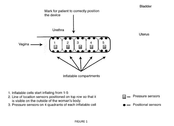
Inflatable components as shown in Figure 1 as an example, POP could also be
alleviated or prevented by placing the device into the vagina and inflating
each section
from farthest to the most proximal. The most proximal inflatable section may
be
inflated to prevent spillage of the vaginal contents. In this mode of
operation, the
device offers advantages over devices, such as pessaries, used today. Every
month,
rather than the patient having to return to the health care provider to
extract the
pessary and clean it for reinsertion, the patient would be able to withdraw
the instant
device and clean it for reinsertion in the convenience and privacy of her own
home,
which may include any location outside the health care provider's office or
facility.
In this regard, the device disclosed here may be used to improve a woman's
vaginal
muscular strength by performing vaginal strengthening exercises (VSE) to
achieve
CA 02862928 2014-07-25
WO 2013/116310
PCT/US2013/023806
her desired sexual health as well as to address any UI or POP conditions.
[0016] The present invention contemplates the real-time position and
movement tracking described in International Patent Application
PCT/US2010/053712, and the multiple sensor-enable device described in U.S.
provisional patent application Serial No. 61/563,889, which are hereby
incorporated
in their entirety by reference. In this regard, the real-time position and
movement
tracking may include sensing the position of the anatomical organ of interest
to an
anatomical reference point, such as the patient's pubic bone, the coccyx or
the vagina,
or to an external reference point, such as a target on a patient's garment or
in the
patient's surroundings. The method may be performed in real-time, for example,
during a medical examination, procedure, or surgery. In another embodiment,
the
method may be performed at multiple time intervals. The multiple time
intervals may
occur, for example, pre- and post-event, wherein the event may be pregnancy or
menopause.
[0017] The multiple sensor-enabled device may also provide pressure data,
which reflects muscular strength, and provide a health care provider a
detailed map of
where the weakest anatomical points are for purposes of POP diagnosis and
treatment.
Where vaginal strengthening exercises are inadequate to prevent or relieve UI
or POP, a
surgeon would be able to use this information to target corrective procedures
appropriately.
BRIEF DESCRIPTION OF THE DRAWINGS
[0018] Figure 1 depicts a lateral view of an embodiment of the present
invention.
DETAILED DESCRIPTION OF THE INVENTION
[0019] When used in the claims, the terms "a" and "an" and "the" and similar
references in the context of describing the invention (especially in the
context of the
following claims) are to be construed to cover both the singular and the
plural, unless
otherwise indicated herein or clearly contradicted by context. Also when used
in the
claims, the terms "comprising," "having," "including," and "containing" are to
be
construed as open- ended terms (i.e., meaning "including, but not limited
to,") unless
CA 02862928 2014-07-25
WO 2013/116310
PCT/US2013/023806
6
otherwise noted. To the extent used, the recitation of ranges of values herein
are merely
intended to serve as a shorthand method of referring individually to each
separate value
falling within the range, unless otherwise indicated herein, and each separate
value is
incorporated into the specification as if it were individually recited herein.
All methods
described herein can be performed in any suitable order unless otherwise
indicated
herein or otherwise clearly contradicted by context. The use of any and all
examples, or
exemplary language (e.g., "such as") provided herein, is intended merely to
better
illuminate the invention and does not pose a limitation on the scope of the
invention
unless otherwise claimed. No language in the specification should be construed
as
indicating any non-claimed element as essential to the practice of the
invention.
Variations of the embodiments may become apparent to those of ordinary skill
in the art
upon reading the description. Accordingly, this invention includes all
modifications and
equivalents of the subject matter recited in the claims appended hereto as
permitted by
applicable law. Moreover, any combination of the described elements in all
possible
variations thereof is encompassed by the invention unless otherwise indicated
herein or
otherwise clearly contradicted by context.
[0020] For purposes of the present invention, the term "urethra" may be
defined as
the canal leading from the bladder, discharging the urine externally. See
STEDMAN's
MEDICAL DICTIONARY, at page 2072 (28th ed). In females, the urethra is a canal
about 4 centimeters long passing from the bladder, in close relation with the
anterior wall
of the vagina and having a long axis that parallels that of the vagina opening
in the
vestibule of the vagina posterior to the clitoris and anterior to the vaginal
orifice. Id. The
term "urinary bladder" refers to a musculomembranous elastic bag serving as a
storage
place for the urine, filled via the ureters and drained via the urethra. Id.
at page 226. The
term "bladder neck" is defined as the smooth muscle of the bladder neck is
histologically,
histochemically and pharmacologically distinct from the detrusor muscle proper
and so the
bladder neck should be considered as a separate functional unit. See GRAY's
ANATOMY,
at page 1290 (39th ed.). The arrangement of smooth muscle in this region is
quite different
in males and females, and therefore each sex is described separately. In
females, the
bladder neck consists of morphologically distinct smooth muscle. The large
diameter
fasciculi characteristic of the detrusor is replaced in the region of the
bladder neck by
small diameter fasciculi which extend obliquely or longitudinally into the
urethral wall.
CA 02862928 2014-07-25
WO 2013/116310
PCT/US2013/023806
7
Id. In the normal female the bladder neck which above the pelvic floor
supported
predominantly by the pubovesical ligaments, the endopelvic fascia of the
pelvic floor and
levator ani. These support the urethra at rest; with elevated intra-abdominal
pressure the
levators contract increasing urethral closure pressure to maintain continence.
This
anatomical arrangement commonly alters after parturition and with increasing
age, such
that the bladder neck lies beneath the pelvic floor, particularly when the
intra-abdominal
pressure rises. The mechanism described above may fail to maintain continence
(incontinence as a result of urethral hypermobility).
[0021] As commonly understood, the term "vagina" refers to an elastic muscular
canal that extends from the cervix to the vulva. Although there is wide
anatomical
variation, the length ofthe unaroused vagina of a woman of child-bearing age
is
approximately 6 to 7.5 cm (2.5 to 3 inches) across the anterior wall (front),
and 9 cm (3.5
inches) long across the posterior wall (rear). The vagina connects the
superficial vulva to
the cervix of the deep uterus. In a typical woman standing upright, the
vaginal tube points
in an upward-backward direction and forms an angle of slightly more than 45
degrees
with the uterus. The vaginal opening is at the caudal end of the vulva, behind
the opening
of the urethra. The upper one- fourth of the vagina is separated from the
rectum by the
recto-uterine pouch.
[0022] In the present invention, for example, a device for vaginal insertion
may be
equipped with at least one sensor capable of providing real-time data of one
or more types
selected from the group consisting of position, movement, pressure, and flow.
In this
regard, a sensor may have a single measurement and reporting capability, or
may have
multiple measurement and reporting capabilities. The data obtained by the
multiple
sensor-enabled device may be reported in any number of ways known in the art,
including
the transmission to, and visualization on, a graphical user interface
wirelessly.
[0023] The device would be inserted into the vagina until the patient feels
her
cervix. The distal section of the device, in an inflatable embodiment, would
be filled with
air, gel, liquid, or other appropriate material suitable for inflation and
deflation of a
compartment, to fit the patient's vagina. In an embodiment with multiple
inflatable
sections, the rest of the compartments could be filled from distal to proximal
(vaginal
opening). In this way, a patient with POP not only would strengthen her
vaginal muscles
but could also use the device as a pessary that can be easily removed at home
and would
CA 02862928 2014-07-25
WO 2013/116310
PCT/US2013/023806
8
not have the complications currently associated with pessaries, such as
pressure point
problems and vaginal infections.
[0024] When the device is properly inserted and inflated, the health care
provider
or patient can visualize the device on a display screen. When the patient is
asked to
perform Kegel movements, the vaginal pressure or strength of the vaginal
musculature
will also be visualizable on the screen. The health care provider could then
go through the
exercises with the patient to ensure that she is performing the exercises
optimally and has
understood how to interpret the information and otherwise use the equipment
properly.
[0025] The multiple sensor-enabled device would be invaluable as a study or
rehabilitation tool for the health care provider as well as the patient who is
considering a
pregnancy. The health care provider may be able to provide the patient with an
exercise
regimen that could strengthen her vagina and urinary musculature at home
before she had
her baby, helping her prevent urinary incontinence in the future and
strengthening her
pelvic floor, before the possible damage may occur during pregnancy and
delivery.
[0026] The multiple sensor-enabled device could aid various diagnoses that
rely
upon data concerning the position, strength and pressures of the vaginal
space. By
combining pressure sensors along the multiple sensor-enabled inflatable
vaginal insert
along with the positional sensors, objective measurements relating to vaginal
pressure and
positional location can be evaluated and correlated to aid in the diagnosis
and treatment of
UI or POP and the rehabilitation of the vaginal muscles and pelvic floor.
[0027] In yet another embodiment of the present invention, the multiple sensor-
enabled device can provide data, which is transmitted and recorded in a manner
to create
and maintain historical patient information for medical and/or fitness
purposes, such as a
pelvic floor muscle strengthening exercise calendar.
[0028] Another use for a multiple sensor-enabled device would be to correct
fecal
incontinence, which is often another sequela of pregnancy and childbirth. For
example, if
a rectocele or enterocele is diagnosed, a multiple sensor-enabled device could
be inserted
into the rectum. With this information the health care provider would be able
to properly
diagnose the etiology of the fecal incontinence whether that is due to muscle
weakness of
the pelvic floor, a rectal sphincter deficiency, or a combination of the two.
The health
care provider could target the surgical repair, in real-time if preferred, to
correct the fecal
incontinence.
CA 02862928 2014-07-25
WO 2013/116310
PCT/US2013/023806
9
[0029] The multiple sensor-enabled device may incorporate at least one sensor
capable of measuring and/or reporting data of various types including
position,
movement, pressure and flow. A multiple sensor-enabled device with more than
one
individual sensor may be arrayed as depicted in Figure 1. However, a multiple
sensor-
enabled device may incorporate a single sensor capable of multiple measurement
and
reporting capabilities.
[0030] The position and movement data may be of the sort measured and/or
reported by any number of sensor devices, including an accelerometer,
gyroscope,
inductive non-contact position sensor, string potentiometer, linear variable
differential
transformer, potentiometer, capacitive transducer, Eddy-current sensor, Hall
effect sensor,
optical proximity sensor, piezo-electric transducer and photodiode array. The
position and
movement data may also include magnetic, electromagnetic,
microelectromechanical,
radio frequency, ultrasound and video.
[0031] The pressure and flow data may be of the sort measured and/or reported
by
any number of sensor devices, including force collector types, such as piezo-
resistive,
capacitive, electromagnetic, piezo-electric, optical, potentiometric, or other
types, such as
resonant, thermal, ionization, ultrasonic, and density (mass and index of
refraction). In
addition, sensor technology that recognizes movement and touch may be
incorporated,
which includes the types such as resistive, surface acoustic wave, capacitive
(surface
capacitance, projected capacitance, mutual capacitance, and self-capacitance),
infrared,
optical imaging, dispersive signal technology, and acoustic pulse recognition.
[0032] Figure 1 depicts a multiple sensor-enable device for vaginal insertion
with
inflatable compartments. The number and precise placement of an individual
sensor may
vary depending on the type of positional, movement, pressure or flow
measurement
and/or reporting system employed. An individual sensor may have a single
function or be
multifunction (such as positional tracking combined with pressure and flow
sensing). The
multiple sensor-enabled device may also embody a video observation and/or
recording
device as well as an illumination source to facilitate such video capture. The
precise
placement of the sensor(s) and video capture component(s) need not be pre-
defined, and
may be configured according to the requirements of the desired application.
CA 02862928 2014-07-25
WO 2013/116310
PCT/US2013/023806
SPECIFIC EXAMPLES
[0033] As described earlier, the devices of the present invention may embody
at
least one sensor capable of measuring and reporting at least one data type,
including
position, movement, pressure, and flow. These include, but are not limited to,
magnetic,
electromagnetic, microelectromechanical, radio frequency, ultrasound and
video. One
example of a multiple sensor-enabled device contains various
microelectromechanical
(MEMS) sensors: a 3-axis accelerometer, a roll/pitch gyroscope and a yaw rate
gyroscope, and a pressure and flow transducer. The sensors may be mounted on a
small
flexible printed circuit board (PCB) and then attached to, or incorporated
within, the
device. The 3-axis accelerometer tracks translation of the device in three
directions. The
gyroscopes are utilized to account for gravitational rotation, allowing real-
time
movement to be tracked.
[0034] A PCB is prepared with MEMS sensors mounted thereon. Soft leads can
trail the MEMS sensors to supporting components, including, for example, a
data
acquisition card which may be used for transforming analog signals to digital
signals. The
PCB is set within the wall of the device. The location of the device may be
determined by
the output signals of the MEMS sensors.
[0035] In an embodiment where the multiple sensor-enabled device contains
inflatable compartments, the device may be inserted in the length of the
vagina at which
point the compartment nearest the cervix is inflated to obtain a stationary
and/or
comfortable fit within the vagina. Any additional inflatable compartments may
be inflated
together or in sequence from distal to proximal to the vaginal opening.
[0036] The patient may be asked to perform a Kegel movement, while the health
care provider and/or the patient observes the display output to confirm that
the patient is
performing the exercise optimally. The pressure and muscular strength of the
vagina as
measured by the multiple sensor-enable device would be displayed to reflect
the
effectiveness of the therapy. The position of the urethra and bladder neck may
also be
displayed in real time on a graphical user interface and/or recorded.
[0037] Following the examination using the multiple sensor-enabled device, the
health care provider may conclude that rehabilitation is an efficacious option
for the
patient. In this regard, the measurements provided by the multiple sensor-
enabled device
CA 02862928 2014-07-25
WO 2013/116310
PCT/US2013/023806
11
may be recorded to facilitate appropriate patient instructions on performing
Kegel
exercises in an optimal manner using the visual (on-screen) information
provided by the
device in real-time. Once engaging the proper musculature has been
successfully
communicated to the patient during the medical office visit, the patient may
be sent home
with the instructions to perform Kegel exercises five to six times daily, for
example. Four
to six weeks later the patient may return for another examination using the
multiple
sensor-enabled device to evaluate rehabilitative treatment effectiveness,
which may allow
the health care provider to advise the patient about the prospects for
restoring complete
continence with a continued rehabilitation regime and/or a surgical procedure.
[0038] Detailed embodiments of the present invention are disclosed herein;
however, it is to be understood that the disclosed embodiments are merely
exemplary of
the invention that may be embodied in various forms. It will be appreciated
that many
modifications and other variations that will be appreciated by those skilled
in the art are
within the intended scope of this invention as claimed below without departing
from the
teachings, spirit and intended scope of the invention.