Note: Descriptions are shown in the official language in which they were submitted.
CA 02863066 2014-07-28
WO 2013/116287
PCT/US2013/023774
ANTI-IG-E M1' ANTIBODIES AND METHODS USING SAME
CROSS-REFERENCE TO RELATED PATENT APPLICATIONS
This Application claims the priority benefit of U.S. provisional application
serial no. 61/593,282,
filed January 31, 2012, U.S. provisional application serial no. 61/613,434,
filed March 20, 2012,
U.S. provisional application serial no. 61/621,453, filed April 6, 2012, and
U.S. provisional
application serial no. 61/635,253, filed April 18, 2012, all of which are
incorporated herein by
reference in their entirety.
FIELD OF THE INVENTION
This invention relates to anti-IgE antibodies that bind to the M1' segment of
a human IgE and
their use in treating and preventing IgE-mediated disorders.
BACKGROUND OF THE INVENTION
Allergy refers to certain diseases in which immune responses to environmental
antigens cause
tissue inflammation and organ dysfunction. The clinical features of each
allergic disease reflect
the immunologically induced inflammatory response in the organ or tissue
involved. These
features are generally independent of the chemical or physical properties of
the antigen. The
diversity of allergic responses arises from the involvement of different
immunological effector
pathways, each of which generates a unique pattern of inflammation.
Allergy is common throughout the world. The predilection for specific
diseases, however, varies
among different age groups, sexes and races. The prevalence of sensitivity to
specific allergens
is determined both by genetic predilection and by the geographic and cultural
factors that are
responsible for exposure to the allergen. A clinical state of allergy affects
only some individuals
who encounter each allergen. The occurrence of allergic disease on exposure to
an allergen
requires not only prior "sensitization" but also other factors that determine
the localization of the
reaction to a particular organ.
A biological process that precedes the disease of allergy upon allergen
exposure is an immune
response known as "sensitization" or the sensitization phase. Once
sensitization occurs, an
individual does not become symptomatic until there is a subsequent exposure to
the allergen.
The effect of sensitization is also known as immune memory.
CA 02863066 2014-07-28
WO 2013/116287
PCT/US2013/023774
Elevated EE levels are associated with allergic diseases iitcluditig allergic-
asthma. IgE plays a
central role in allergies by virtue of their role as allergen receptors on the
surface of mast cells
and basophils. IgE antibodies are fixed to the surface of mast cells and
basophils at the Fc
portion of the molecule to a high affinity cell surface receptor, called
FcERI. The allergic
reaction is initiated when the polyvalent allergen molecule binds to
antibodies that are occupying
these receptors. The result is a bridging of the FcERI, which in turn signals
intracellularly
causing the release and activation of mediators of inflammation: histamine,
leukotrienes,
chemotactic factors, platelet-activating factor, and proteinases. These
activated mediators act
locally and cause increased vascular permeability, vasodilation, smooth muscle
contraction and
mucous gland secretion. Such events are termed clinically the immediate or
early phase, and
occur within the first 15-30 minutes following allergen exposure. Over the
succeeding 12 hours
there is progressive tissue infiltration of inflammatory cells, proceeding
from neutrophils to
eosinophils to mononuclear cells in response to other chemical mediators not
quite fully
understood. This period of time 6-12 hours after allergen exposure is
designated the late phase
and is characterized by clinical manifestations of cellular inflammation.
Given that late phase
reactions, especially in the lung, occur in the absence of early phase
reactions, it is still not
entirely understood if the late phase reaction is necessarily IgE mediated.
This mechanism is
primarily responsible for the anaphylaxis, urticarial and the atopic diseases
such as allergic
rhinitis, allergic asthma, atopic dermatitis and allergic gastroenteropathy.
IgE exists in a membrane bound form and in a secreted form. These distinct
forms appear to be
splice variants. Previous approaches to achieve therapeutic effect by down
regulating IgE
targeted primarily the secreted form (e.g., XOLAIR omalizumab), so as to
prevent or disarm
further "arming" of the immune system. The secreted form of IgE is a shorter
form, essentially
the Fc region ends at the CH4 domain, whereas the longer form includes
additional C-terminal
residues including the peptides encoded by the exons known as Ml/M1' and M2.
Conventional
therapy with anti-IgE antibodies, which bind to the secreted form of IgE,
results in reduction of
secreted serum IgE (total IgE not complexed to Xolair). Casale et at., J.
Allergy Clin. Immunol.
100(1): 110-121 (1997).
Membrane bound IgE which includes the M1' section is present in human IgE-
switched B cells,
IgE memory B cell, and IgE plasmablasts. U.S. Patent No. 8,071,097 (also
described in
W02008/116149, the disclosures both of which are incorporated by reference in
their entirety)
2
CA 02863066 2014-07-28
WO 2013/116287
PCT/US2013/023774
discloses antibodies that target the M1' segment of membrane bound IgE. These
antibodies may
deplete M1' expressing B cells via apoptosis and/or antibody-dependent cell-
mediated
cytotoxicity mechanism.
All references cited herein, including patent applications and publications,
are hereby
incorporated by reference in their entirety.
SUMMARY OF THE INVENTION
Provided herein is a method of treating or preventing an IgE-mediated disorder
comprising
administering to a human patient an effective amount of an anti-IgE antibody
that binds the M1'
segment of a human IgE, wherein an interval between administrations of the
antibody is about
one month or longer. In some embodiments, the interval between administrations
is about two
months, about three months, about four months, about five months, about six
months, or longer.
In some embodiments, the interval between administrations is about three
months with an
additional administration at week 4 after the first administration. In some
embodiments, the
antibody is administered at a dosage from about 150 mg to about 450 mg per
dose. In some
embodiments, the antibody is administered at a dosage of about 150 mg per
dose, about 300 mg
per dose, or about 450 mg per dose. In some embodiments, the antibody is
administered
subcutaneously or intravenously. In some embodiments, the serum total IgE in
the human
patient is reduced relative to baseline after the antibody treatment. In some
embodiments, the
allergen-specific IgE in the human patient is reduced relative to baseline
after the antibody
treatment. In some embodiments, an allergen-induced increase in serum total
IgE in the human
patient is prevented or reduced after the antibody treatment. In some
embodiments, an allergen-
induced increase in allergen-specific IgE in the human patient is prevented or
reduced after the
antibody treatment. In some embodiment, the production of new IgE is prevented
or reduced
after antibody treatment.
Also provided herein is a method of treating or preventing an IgE-mediated
disorder comprising
administering to a human patient an effective amount of an anti-IgE antibody
that binds the M1'
segment of a human IgE, wherein the antibody is administered at a dose of
about 150 mg to
about 450 mg per dose. In some embodiments, the antibody is administered at a
dosage about
150 mg/per dose, about 300 mg/per dose, or about 450 mg per dose. In some
embodiments, the
antibody is administered subcutaneously or intravenously.
3
CA 02863066 2014-07-28
WO 2013/116287
PCT/US2013/023774
Also provided herein is a method of reducing serum total IgE and/or allergen-
specific IgE in a
human relative to baseline comprising administering to a human patient an
effective amount of
an anti-IgE antibody that binds the M1' segment of a human IgE, wherein an
interval between
administrations of the antibody is about one month or longer. In some
embodiments, the serum
total IgE is reduced by at least about 20% from the baseline level. In some
embodiments, the
serum total IgE is reduced by at least about 25% from the baseline level. In
some embodiments,
the reduction of the serum total IgE is sustained for at least one month, at
least two months, at
least three months, at least four months, at least five months, or at least
six months after the last
administration of the antibody. Also provided herein is a method of preventing
the production of
new IgE comprising administering to a human patient an effective amount of an
anti-IgE
antibody that binds the M1' segment of a human IgE.
Also provided herein is a method of preventing or reducing an allergen-induced
increase in
serum total IgE and/or in allergen-specific IgE in a human patient comprising
administering to a
human patient an effective amount of an anti-IgE antibody that binds to the
M1' segment of a
human IgE. In some embodiments, an interval between administrations of the
antibody is about
one month or longer. In some embodiments, an interval between administrations
of the antibody
is about two months. In some embodiments, an interval between administrations
of the antibody
is about three months. In some embodiments, an interval between
administrations of the
antibody is about four months. In some embodiments, an interval between
administrations of the
antibody is about five months. In some embodiments, an interval between
administrations of the
antibody is about six months. In some embodiments, the antibody is
administered at a dose of
about 150 to about 450 mg per dose. In some embodiments, the allergen-induced
increase in
allergen-specific IgE is prevented or reduced. In some embodiments, the
prevention or reduction
of allergen-allergen induced increase in serum total IgE and/or allergen-
specific IgE is sustained
for at least one month, at least two months, at least three months, or at
least six months after the
last administration of antibody.
In some embodiments of the methods described herein, the antibody is
administered for treating
an IgE-mediated disorder selected from the group consisting of: allergic
rhinitis, allergic asthma,
non-allergic asthma, atopic dermatitis, allergic gastroenteropathy,
anaphylaxis, urticaria, food
allergies, allergic bronchopulmonary aspergillosis, parasitic diseases,
interstitial cystitis, hyper-
IgE syndrome, ataxia-telangiectasia, Wiskott-Aldrich syndrome, athymic
lymphoplasia, IgE
4
CA 02863066 2014-07-28
WO 2013/116287
PCT/US2013/023774
myeloma, graft-versus-host reaction and allergic purpura. In some embodiments,
the IgE-
mediated disorder is allergic rhinitis, allergic asthma, or non-allergic
asthma. In some
embodiments, the method described herein is for treating a human patient
having allergic asthma
that is inadequately controlled by standard of care, e.g., a high-dose inhaled
or oral
corticosteroids in combination with a second controller. In some embodiments,
the method
described herein is for treating a human patient having severe, moderate, or
mild asthma. In
some embodiments, the method described herein is for treating a human patient
with allergic
asthma inadequately controlled despite high dose inhaled corticosteroids (ICS)
(?400 lug/day
total daily dose of fluticasone propionate (FP) or equivalent) and a second
controller ( e.g., after
as least 12 weeks or at least 36 weeks of treatment). In some embodiments, the
second
controller is a bronchodilator or an anti-leukotriene agent.
In some embodiments, the method described herein further comprises
administering to the
human patient a second drug in conjunction with the antibody for treating or
preventing an IgE-
mediated disorder, wherein the second drug is selected from the group
consisting of: an anti-IgE
antibody, an antihistamine, a bronchodilator, a glucocorticoid, an NSAID, a
decongestant, a
cough suppressant, an analgesic, a TNF-antagonist, an integrin antagonist, an
immunosuppressive agent, an IL-4 antagonist, an IL-13 antagonist, a dual IL-
4/IL-13 antagonist,
a DMARD, an antibody that binds to a B-cell surface marker, and a BAFF
antagonist. In some
embodiments of the methods described herein, the antibody is administered to
the human patient
in conjunction with a second method treatment for an IgE-mediated disorder. In
some
embodiments, the second method of treatment comprises a treatment regimen of
allergen
desensitization.
In the methods described herein, any of the anti-IgE antibodies described
herein may be
administered to the human patient. In some embodiments, the anti-IgE antibody
is a chimeric, a
humanized, or a human antibody. In some embodiments, the antibody specifically
binds an
epitope in the M1' segment of a human IgE shown in Figure 14. In some
embodiments, the anti-
IgE antibody specifically binds to the same epitope as one bound by an
antibody selected from
the group consisting of: 47H4, 7A6, 26A11, 47H4v5, 7A6v1 and 26A11v6. In some
embodiments, the epitope corresponds to a peptide having the amino acid
sequence selected
from the group consisting of: SEQ ID NO:4, SEQ ID NO:5, SEQ ID NO:6, and SEQ
ID NO:7.
In some embodiments, the antibody specifically binds an epitope in the M1'
segment of IgE
5
CA 02863066 2014-07-28
WO 2013/116287
PCT/US2013/023774
defined by the residues 317 to 352 of SEQ ID NO:l. In some embodiments, the
antibody
specifically binds an epitope in the M1' segment of IgE defined by the
residues 317 to 352 of
SEQ ID NO:1 and has a Scatchard binding affinity that is equivalent to that of
the murine anti-
IgE antibody 47H4. In some embodiments, the affinity is between 0.30 and 0.83
nM. In some
embodiments, the antibody specifically binds an epitope in the M1' segment of
IgE defined by
the residues 317 to 352 of SEQ ID NO:1 and has a Scatchard binding affinity
that is equivalent
to that of anti-IgE antibody 47H4v5. In some embodiments, the affinity is
about 1.5 nM. In
some embodiments, the antibody comprises the heavy chain and light chain HVRs
of an
antibody or antigen-binding fragment thereof selected from the group
consisting of: 26A11,
26A11 v.1-16, 7A6, 7A6v1, 47H4, and 47H4v1-6. In some embodiments, the
antibody
comprises heavy and light variable regions of the heavy and light chains of
the antibody or
antigen-binding fragment thereof selected from the group consisting of: 26A11,
26A 11 v.1-16,
7A6, 7A6v1, 47H4, 47H4v1-6.
In some embodiments, the anti-IgE antibody comprises a heavy chain and a light
chain variable
region, wherein the heavy chain variable region comprises the amino acid
sequence of SEQ ID
NO:29 and the light chain variable region comprises the amino acid sequence of
SEQ ID NO:19.
In some embodiments, the antibody comprises a heavy chain and a light chain
variable region,
wherein the heavy chain variable region comprises an HVR-H1, HVR-H2 and HVR-
H3, and the
light chain variable region comprises HVR-L1, HVR-L2 and HVR-L3, and wherein
(a) the
HVR-H1 comprises residues 26-35 of SEQ ID NO:29, (b) the HVR-H2 comprises
residues 49-
66 of SEQ ID NO:29, (c) the HVR-H3 comprises residues 97-106 of SEQ ID NO:29,
(d) the
HVR-L1 comprises residues 24-39 of SEQ ID NO:19, (e) the HVR-L2 comprises
residues 55-61
of SEQ ID NO:19, and (0 the HVR-L3 comprises residues 94-102 of SEQ ID NO:19.
The In
some embodiments, the antibody further comprises a human consensus framework.
In some
embodiments, the heavy chain variable region of the antibody comprises a
subgroup III
consensus framework. In some embodiments, the light chain variable region
comprises a kappa
subgroup I consensus framework. In some embodiments, the anti-IgE antibody
administered to a
human patient comprises a heavy chain and a light chain variable region,
wherein the heavy
chain variable region comprises the amino acid sequence of SEQ ID NO:39 and
the light chain
variable region comprises the amino acid sequence of SEQ ID NO:40. In some
embodiments, an
antigen-binding fragment of an anti-IgE antibody is administered to a human
patient, wherein the
6
CA 02863066 2014-07-28
WO 2013/116287
PCT/US2013/023774
anti-IgE antibody comprises a heavy chain and a light chain variable region,
wherein the heavy
chain variable region comprises the amino acid sequence of SEQ ID NO:39 and
the light chain
variable region comprises the amino acid sequence of SEQ ID NO:40.
In some embodiments, the anti-IgE antibody has ADCC activity. In some
embodiments, the
anti-IgE antibody is afucosylated. In some embodiments, the anti-IgE antibody
depletes IgE-
switched B-cells. In some embodiments, the anti-IgE antibody depletes IgE
memory B-cells. In
some embodiments, the anti-IgE antibody depletes IgE plasmablast. In some
embodiments, the
anti-IgE antibody is in pharmaceutical composition comprising the antibody and
a
pharmaceutically acceptable carrier.
Also provided herein is a kit comprising an anti-IgE antibody that binds the
M1' segment of a
human IgE and a package insert indicating that the antibody is administered to
a human patient
for treating an IgE-mediated disorder, wherein an interval between
administrations of the
antibody to the human patient is about one month or longer.
Also provided here is a kit comprising an anti-IgE antibody that binds the M1'
segment of a
human IgE and a package insert indicating that the antibody is administered to
a human patient
for treating an IgE-mediated disorder, wherein the antibody is administered at
a dosage from
about 150 mg to about 450 mg per dose.
In some embodiments, the package insert in the kit further indicates that the
treatment is
effective in reducing serum total IgE relative to baseline in a human patient.
In some
embodiments, the package insert in the kit further indicates that the
treatment is effective in
reducing allergen-specific IgE relative to baseline in a human patient. In
some embodiments, the
package insert further indicates that the treatment is effective in preventing
or reducing an
allergen-induced increase in serum total IgE in a human patient. In some
embodiments, the
package insert further indicates that the treatment is effective in preventing
or reducing an
allergen-induced increase in allergen-specific IgE in a human patient. In some
embodiments, the
human patient has severe, moderate, or mild asthma. In some embodiments, the
antibody in the
kit is in a vial. In some embodiments, the antibody in the kit is in a pre-
filled syringe. In some
embodiment, the kit further comprises an injection device (such as an auto-
injector).
In some embodiments, the kit further comprises a second drug selected from the
group consisting
of: an anti-IgE antibody, an antihistamine, a bronchodilator, a
glucocorticoid, an NSAID, a
decongestant, a cough suppressant, an analgesic, a TNF-antagonist, an integrin
antagonist, an
7
CA 02863066 2014-07-28
WO 2013/116287
PCT/US2013/023774
immunosuppressive agent, an IL-4 antagonist, an IL-13 antagonist, a dual IL-
4/IL-13 antagonist,
a DMARD, an antibody that binds to a B-cell surface marker, and a BAFF
antagonist, and a
package insert indicating administration of the antibody to a human patient at
monthly or
quarterly intervals in conjunction with the second drug for treating an IgE-
mediated disorder. In
some embodiments, the second drug is anti-IgE antibody rhuMAbE25.
In some embodiment, the invention provides for any of the prior described anti-
IgE-M1'
antibodies for use in any of the prior described methods, wherein the
antibodies are administered
by any of the prior described dosing internval, amounts or regimens.
In some embodiment, the invention provides for any of the prior described anti-
IgE-M1'
antibodies for use in any of the prior described methods, wherein the
antibodies are prepared to
be administered by any of the prior described dosing internval, amounts or
regimens.
In some embodiment, the invention provides for the use of any of the prior
described anti-IgE-
M1' antibodies any of the prior described methods, wherein the antibodies are
administered is
any of the prior described dosing intervals, amounts or regimens.
It is to be understood that one, some, or all of the properties of the various
embodiments
described herein may be combined to form other embodiments of the present
invention. These
and other aspects of the invention will become apparent to one of skill in the
art.
BRIEF DESCRIPTION OF THE DRAWINGS
Figure 1 is a diagram of the Phase la single ascending-dose study in healthy
volunteers.
Figure 2 is a diagram of the Phase lb multiple ascending dose study in
patients with mild
asthma.
Figure 3 is a graph demonstrating mean serum concentration over time in all
cohorts in the
Phase la study.
Figure 4 is a graph demonstrating mean serum concentration over time in all
cohorts in the
Phase lb study.
Figure 5 is graph demonstrating serum total IgE at 85 and 168 days after
treatment with
MEMP1972A. Data are presented for study Days 85 (A) and 168 (B), with study
Day 1 defined
as the day of the single-dose administration. Data are expressed as % change
from the baseline,
where baseline is defined as the average of pre-dose visits; mean SD, n=3-4
patients in 0.003,
0.03 and 0.3 mg/kg IV groups, n = 5 in 1, 3, 5 mg/kg IV and 3 mg/kg SC group,
and n = 14 in
placebo group.
8
CA 02863066 2014-07-28
WO 2013/116287
PCT/US2013/023774
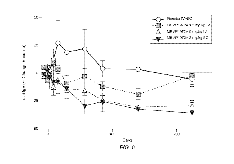
Figure 6 is a graph demonstrating serum total IgE in patients with allergic
rhinitis after treatment
with MEMP1972A. Data are expressed as % change from baseline, where baseline
is defined as
the average of pre-dose visits; mean SD, n = 8 patients in MEMP1972A groups
and n = 12 in
placebo group (IV and SC combined).
Figure 7 is a diagram of the Phase 2a proof-of-activity allergen challenge
study in patients with
mild asthma.
Figure 8 is a graph demonstrating that the anti-M1 prime antibody (MEMP1972A)
reduced both
early asthmatic reactions (EAR) and late asthmatic reactions (LAR) as measured
by percent
decline in forced expiratory volume in 1 second (FEVi) over time following
allergen inhalation
in a Phase 2a study. A) shows the mean percent FEVi as compared to baseline
(pre-challenge)
over time following the allergen inhalation in patients at screening before
placebo or drug
administration. B) shows mean percent FEVi as compared to baseline (pre-
challenge) over time
following the allergen inhalation at Day 86 in patients with placebo or the
drug (MEMP1972A).
Figure 9 is a graph demonstrating that the anti-M1 prime antibody prevented
allergen-induced
increase in allergen-specific IgE and reduced total IgE in patients with mild
asthma in a Phase 2a
study. A) shows allergen-specific IgE (to challenge allergens) in patients
treated with placebo or
Anti-M1 prime (MEMP1972A). *p<0.01;tp<0.05. B) shows allergen-specific IgE (to
irrelevant
allergens (i.e., non-challenge allergens)) in patients subjected to whole lung
allergen challenge
and treated with placebo or Anti-M1 prime (MEMP1972A). C) shows total IgE in
patients
treated with placebo or Anti-M1 prime in interim results. D) shows total IgE
in patients treated
with placebo or Anti-M1 prime. IgE levels before start of study set as
baseline of 100%
(MEMP1972A). *p<0.01. Data shown as mean SEM. For A)-C), not all subjects
included in
late time points; n = 4-5/treatment group on Day 197.
Figure 10 is a graph showing that MEMP1972A reduced allergen challenge-induced
increases in
eosinophils. A) shows sputum eosinophil levels at screening. B) shows sputum
eosinophil
levels at week 12 of the study. Data are presented as mean standard error.
Figure 11 is a graph showing reduction of peripheral blood eosinophils in
patients treated with
MEMP1972A. A) shows percentage of baseline (screening) in eosinophils (%).
*p<0.10;
t p<0.15. B) shows percentage of baseline (screening) in eosinophils (absolute
count). *p<0.10;
t p<0.15. Data are presented as mean standard error.
9
CA 02863066 2014-07-28
WO 2013/116287
PCT/US2013/023774
Figure 12 is a graph showing MEMP1972A blocks increases in allergen-induced
CCL17 levels.
A) shows CCL17 levels as a percentage of baseline over the course of the
study. B) shows
CCL17 levels as a percentage of baselineat screening and Day 86. Data are
presented as mean
standard error.
Figure 13 is a diagram of the Phase 2b study in patients with asthma. Patients
in the placebo
group receive a total of nine placebo doses at monthly intervals (weeks 0, 4,
8, 12, 16, 20, 24, 28,
and 32). Patients in the 300 mg anti-M1 prime antibody arm receive a total of
nine doses of the
antibody at monthly intervals (weeks 0, 4, 8, 12, 16, 20, 24, 28, and 32).
Patients in the 150 mg
and 450 mg anti-M1 prime antibody arms receive a total of four active doses of
the antibody
including three doese at quarterly intervals (weeks 0, 12, and 24) as well as
an extra dose at week
4 and the remaining five doses are placebo.
Figure 14 is an alignment of selected constant chain regions of IgE of the
human (SEQ ID
NO:1), rhesus monkey (SEQ ID NO:2) and cynomolgous monkey (SEQ ID NO:3). Shown
are
the approximate locations of the CH2, CH3, CH4, M1', transmembrane and
intracellular
domains.
Figures 15A-F show the variable light and heavy chain sequences of murine
antibody 26A11,
7A6 and 47H4 and various humanized variants thereof Positions are numbered
according to
Kabat and hypervariable regions that were grafted to the variable consenus
network (Kappa I for
light, subgroup III for heavy chain) are boxed. A) shows, relative to human
kappa I light chain
(SEQ ID NO:8), the variable light chain of 26Al1 (SEQ ID NO:9) and humanized
variants 1,4
(SEQ ID NO:10), variants 2,5 (SEQ ID NO:11), variants 3,6 (SEQ ID NO:12),
variants 13,15
(SEQ ID NO:13) and variants 14,16 (SEQ ID NO:14). B) shows, relative to human
kappa I light
chain (SEQ ID NO:8), the variable light chain of 7A6 (SEQ ID NO:15) and
humanized variant 1
(SEQ ID NO:16). C) shows, relative to human kappa I light chain (SEQ ID NO:8),
the variable
light chain of 47H4 (SEQ ID NO:17) and humanized variants 1,3 (SEQ ID NO:18)
and variants
2, 4-6 (SEQ ID NO:19). D) shows, relative to human III heavy chain (SEQ ID
NO:20), the
variable heavy chain of 26Al1 (SEQ ID NO:21) and humanized variants 1-3, 13,
14 (SEQ ID
NO:22) and variants 4-6, 15, 16 (SEQ ID NO:23). E) shows, relative to human
heavy chain
(SEQ ID NO:20), the variable heavy chain of 7A6 (SEQ ID NO:24) and humanized
variant 1
(SEQ ID NO:25). F) shows, relative to human III heavy chain (SEQ ID NO:20),
the variable
CA 02863066 2014-07-28
WO 2013/116287
PCT/US2013/023774
heavy chain of 47H4 (SEQ ID NO:26), and humanized variants 1,2 (SEQ ID NO:27),
variants 3-
4 (SEQ ID NO:28), variant 5 (SEQ ID NO:29) and variant 6 (SEQ ID NO:30).
Figure 16 is a graph demonstrating that the anti-M1 prime antibody reduced
challenge and non-
challenge specific IgE in patients with mild asthma in a Phase 2a study. A)
shows allergen-
specific IgE to challenge allergens in patients treated with placebo or Anti-
M1 prime
(MEMP1972A). B) shows allergen-specific IgE to non-challenge allergens in
patients treated
with placebo or Anti-M1 prime (MEMP1972A). IgE levels are shown as percent of
baseline, and
IgE levels before start of study is the baseline (100%). Data shown as mean or
median.
DETAILED DESCRIPTION OF THE INVENTION
The present application provides methods of treating or preventing an IgE-
mediated disorder
using an anti-IgE antibody that binds to the M1' segment of an IgE. The
inventors have shown
in clinical studies that a humanized anti-M1' antibody was effective in
reducing serum total IgE
and allergen specific IgE in healthy subjects, and patients with allergic
rhinitis or allergic asthma,
and such reduction of total IgE was sustained for at least three months after
the last dose in both
the single and multiple dose studies. In addition, in a separate study, the
inventors have shown
that an allergen-induced increase in serum total IgE and in allergen-specific
IgE was prevented or
reduced in patients after the anti-IgE antibody treatment.
I. General techniques
The techniques and procedures described or referenced herein are generally
well understood and
commonly employed using conventional methodology by those skilled in the art,
such as, for
example, the widely utilized methodologies described in Sambrook et al.,
Molecular Cloning: A
Laboratory Manual 3d edition (2001) Cold Spring Harbor Laboratory Press, Cold
Spring
Harbor, N.Y.; Current Protocols in Molecular Biology (F.M. Ausubel, et al.
eds., (2003)); the
series Methods in Enzymology (Academic Press, Inc.): PCR 2: A Practical
Approach (M.J.
MacPherson, B.D. Hames and G.R. Taylor eds. (1995)), Harlow and Lane, eds.
(1988)
Antibodies, A Laboratory Manual, and Animal Cell Culture (R.I. Freshney, ed.
(1987));
Oligonucleotide Synthesis (M.J. Gait, ed., 1984); Methods in Molecular
Biology, Humana Press;
Cell Biology: A Laboratory Notebook (J.E. Cellis, ed., 1998) Academic Press;
Animal Cell
Culture (R.I. Freshney), ed., 1987); Introduction to Cell and Tissue Culture
(J.P. Mather and
P.E. Roberts, 1998) Plenum Press; Cell and Tissue Culture: Laboratory
Procedures (A. Doyle,
J.B. Griffiths, and D.G. Newell, eds., 1993-8) J. Wiley and Sons; Handbook of
Experimental
11
CA 02863066 2014-07-28
WO 2013/116287 PCT/US2013/023774
Immunology (D.M. Weir and C.C. Blackwell, eds.); Gene Transfer Vectors for
Mammalian Cells
(J.M. Miller and M.P. Cabs, eds., 1987); PCR: The Polymerase Chain Reaction,
(Mullis et al.,
eds., 1994); Current Protocols in Immunology (J.E. Coligan et al., eds.,
1991); Short Protocols
in Molecular Biology (Wiley and Sons, 1999); Immunobiology (C.A. Janeway and
P. Travers,
1997); Antibodies (P. Finch, 1997); Antibodies: A Practical Approach (D.
Catty., ed., IRL Press,
1988-1989); Monoclonal Antibodies: A Practical Approach (P. Shepherd and C.
Dean, eds.,
Oxford University Press, 2000); Using Antibodies: A Laboratory Manual (E.
Harlow and D.
Lane (Cold Spring Harbor Laboratory Press, 1999); The Antibodies (M. Zanetti
and J. D. Capra,
eds., Harwood Academic Publishers, 1995); and Cancer: Principles and Practice
of Oncology
(V.T. DeVita et al., eds., J.B. Lippincott Company, 1993).
II. Definitions
An "allergen" or "immunogen' is any molecule that can trigger an immune
response. As used
herein, the term covers either the antigenic molecule itself, or its source,
such as pollen grain,
animal dander, insect venom or food product. This is contrasted with the term
antigen, which
refers to a molecule can be specifically recognized by an immunoglobulin or T-
cell receptor.
Any foreign substance capable of inducing an immune response is a potential
allergen. Many
different chemicals of both natural and synthetic origin are known to be
allergenic. Complex
natural organic chemicals, especially proteins, are likely to cause antibody-
mediated allergy,
whereas simple organic compounds, inorganic chemicals, and metals more
preferentially cause
T-cell mediated allergy. In some cases, the same allergen may be responsible
for more than one
type of allergy. Exposure to the allergen may be through inhalation,
injection, injection, or skin
contact.
The term "antibody" includes monoclonal antibodies (including full length
antibodies which
have an immunoglobulin Fc region), antibody compositions with polyepitopic
specificity,
multispecific antibodies (e.g., bispecific antibodies, diabodies, and single-
chain molecules, as
well as antibody fragments (e.g., Fab, F(ab')2, and Fv). The term
"immunoglobulin" (Ig) is used
interchangeably with "antibody" herein.
The basic 4-chain antibody unit is a heterotetrameric glycoprotein composed of
two identical
light (L) chains and two identical heavy (H) chains. An IgM antibody consists
of 5 of the basic
heterotetramer units along with an additional polypeptide called a J chain,
and contains 10
antigen binding sites, while IgA antibodies comprise from 2-5 of the basic 4-
chain units which
12
CA 02863066 2014-07-28
WO 2013/116287
PCT/US2013/023774
can polymerize to form polyvalent assemblages in combination with the J chain.
In the case of
IgGs, the 4-chain unit is generally about 150,000 daltons. Each L chain is
linked to an H chain
by one covalent disulfide bond, while the two H chains are linked to each
other by one or more
disulfide bonds depending on the H chain isotype. Each H and L chain also has
regularly spaced
intrachain disulfide bridges. Each H chain has at the N-terminus, a variable
domain (VII)
followed by three constant domains (CH) for each of the a and y chains and
four CH domains for
i.1 and 8 isotypes. Each L chain has at the N-terminus, a variable domain (VL)
followed by a
constant domain at its other end. The VL is aligned with the VH and the CL is
aligned with the
first constant domain of the heavy chain (CH1). Particular amino acid residues
are believed to
form an interface between the light chain and heavy chain variable domains.
The pairing of a VH
and VL together forms a single antigen-binding site. For the structure and
properties of the
different classes of antibodies, see e.g., Basic and Clinical Immunology, 8th
Edition, Daniel P.
Sties, Abba I. Ten and Tristram G. Parsolw (eds), Appleton & Lange, Norwalk,
CT, 1994, page
71 and Chapter 6.
The L chain from any vertebrate species can be assigned to one of two clearly
distinct types,
called kappa and lambda, based on the amino acid sequences of their constant
domains.
Depending on the amino acid sequence of the constant domain of their heavy
chains (CH),
immunoglobulins can be assigned to different classes or isotypes. There are
five classes of
immunoglobulins: IgA, IgD, IgE, IgG and IgM, having heavy chains designated a,
6, 8, y and
respectively. The y and a classes are further divided into subclasses on the
basis of relatively
minor differences in the CH sequence and function, e.g., humans express the
following
subclasses: IgGl, IgG2, IgG3, IgG4, IgAl and IgA2.
An "isolated" antibody is one that has been identified, separated and/or
recovered from a
component of its production environment (E.g., naturally or recombinantly).
Preferably, the
isolated polypeptide is free of association with all other components from its
production
environment. Contaminant components of its production environment, such as
that resulting
from recombinant transfected cells, are materials that would typically
interfere with research,
diagnostic or therapeutic uses for the antibody, and may include enzymes,
hormones, and other
proteinaceous or non-proteinaceous solutes. In preferred embodiments, the
polypeptide will be
purified: (1) to greater than 95% by weight of antibody as determined by, for
example, the Lowry
method, and in some embodiments, to greater than 99% by weight; (1) to a
degree sufficient to
13
CA 02863066 2014-07-28
WO 2013/116287
PCT/US2013/023774
obtain at least 15 residues of N-terminal or internal amino acid sequence by
use of a spinning
cup sequenator, or (3) to homogeneity by SDS-PAGE under non-reducing or
reducing conditions
using Coomassie blue or, preferably, silver stain. Isolated antibody includes
the antibody in situ
within recombinant cells since at least one component of the antibody's
natural environment will
not be present. Ordinarily, however, an isolated polypeptide or antibody will
be prepared by at
least one purification step.
The "variable region" or "variable domain" of an antibody refers to the amino-
terminal domains
of the heavy or light chain of the antibody. The variable domains of the heavy
chain and light
chain may be referred to as "VH" and "VL", respectively. These domains are
generally the most
variable parts of the antibody (relative to other antibodies of the same
class) and contain the
antigen binding sites.
The term "variable" refers to the fact that certain segments of the variable
domains differ
extensively in sequence among antibodies. The V domain mediates antigen
binding and defines
the specificity of a particular antibody for its particular antigen. However,
the variability is not
evenly distributed across the entire span of the variable domains. Instead, it
is concentrated in
three segments called hypervariable regions (HVRs) both in the light-chain and
the heavy chain
variable domains. The more highly conserved portions of variable domains are
called the
framework regions (FR). The variable domains of native heavy and light chains
each comprise
four FR regions, largely adopting a beta-sheet configuration, connected by
three HVRs, which
form loops connecting, and in some cases forming part of, the beta-sheet
structure. The HVRs in
each chain are held together in close proximity by the FR regions and, with
the HVRs from the
other chain, contribute to the formation of the antigen binding site of
antibodies (see Kabat et al.,
Sequences of Immunological Interest, Fifth Edition, National Institute of
Health, Bethesda, MD
(1991)). The constant domains are not involved directly in the binding of
antibody to an antigen,
but exhibit various effector functions, such as participation of the antibody
in antibody-
dependent cellular toxicity.
The term "monoclonal antibody" as used herein refers to an antibody obtained
from a population
of substantially homogeneous antibodies, i.e., the individual antibodies
comprising the
population are identical except for possible naturally occurring mutations
and/or post-translation
modifications (e.g., isomerizations, amidations) that may be present in minor
amounts.
Monoclonal antibodies are highly specific, being directed against a single
antigenic site. In
14
CA 02863066 2014-07-28
WO 2013/116287
PCT/US2013/023774
contrast to polyclonal antibody preparations which typically include different
antibodies directed
against different determinants (epitopes), each monoclonal antibody is
directed against a single
determinant on the antigen. In addition to their specificity, the monoclonal
antibodies are
advantageous in that they are synthesized by the hybridoma culture,
uncontaminated by other
immunoglobulins. The modifier "monoclonal" indicates the character of the
antibody as being
obtained from a substantially homogeneous population of antibodies, and is not
to be construed
as requiring production of the antibody by any particular method. For example,
the monoclonal
antibodies to be used in accordance with the present invention may be made by
a variety of
techniques, including, for example, the hybridoma method (e.g., Kohler and
Milstein., Nature,
256:495-97 (1975); Hongo et at., Hybridoma, 14 (3): 253-260 (1995), Harlow et
at., Antibodies:
A Laboratory Manual, (Cold Spring Harbor Laboratory Press, 2" ed. 1988);
Hammerling et at.,
in: Monoclonal Antibodies and T-Cell Hybridomas 563-681 (Elsevier, N.Y.,
1981)),
recombinant DNA methods (see, e.g.,U U.S. Patent No. 4,816,567), phage-display
technologies
(see, e.g., Clackson et at., Nature, 352: 624-628 (1991); Marks et at., J.
Mot. Biol. 222: 581-597
(1992); Sidhu et at., J. Mot. Biol. 338(2): 299-310 (2004); Lee et at., J.
Mot. Biol. 340(5): 1073-
1093 (2004); Fellouse, Proc. Natl. Acad. Sci. USA 101(34): 12467-12472 (2004);
and Lee et at.,
J. Immunol. Methods 284(1-2): 119-132 (2004), and technologies for producing
human or
human-like antibodies in animals that have parts or all of the human
immunoglobulin loci or
genes encoding human immunoglobulin sequences (see, e.g., WO 1998/24893; WO
1996/34096;
WO 1996/33735; WO 1991/10741; Jakobovits et at., Proc. Natl. Acad. Sci. USA
90: 2551
(1993); Jakobovits et at., Nature 362: 255-258 (1993); Bruggemann et at., Year
in Immunol.
7:33 (1993); U.S. Patent Nos. 5,545,807; 5,545,806; 5,569,825; 5,625,126;
5,633,425; and
5,661,016; Marks et at., Rio/Technology 10: 779-783 (1992); Lonberg et at.,
Nature 368: 856-
859 (1994); Morrison, Nature 368: 812-813 (1994); Fishwild et at., Nature
Biotechnol. 14: 845-
851 (1996); Neuberger, Nature Biotechnol. 14: 826 (1996); and Lonberg and
Huszar, Intern.
Rev. Immunol. 13: 65-93 (1995).
The term "naked antibody" refers to an antibody that is not conjugated to a
cytotoxic moiety or
radiolabel.
The terms "full-length antibody," "intact antibody" or "whole antibody" are
used
interchangeably to refer to an antibody in its substantially intact form, as
opposed to an antibody
fragment. Specifically whole antibodies include those with heavy and light
chains including an
CA 02863066 2014-07-28
WO 2013/116287
PCT/US2013/023774
Fe region. The constant domains may be native sequence constant domains (e.g.,
human native
sequence constant domains) or amino acid sequence variants thereof. In some
cases, the intact
antibody may have one or more effector functions.
An "antibody that binds to the same epitope" as a reference antibody refers to
an antibody that
blocks binding of the reference antibody to its antigen in a competition assay
by 50% or more,
and conversely, the reference antibody blocks binding of the antibody to its
antigen in a
competition assay by 50% or more. Competition assays are known in the art.
In an exemplary competition assay, immobilized the M1' segment of IgE is
incubated in a
solution comprising a first labeled antibody that binds to the M1' segment
(e.g., antibody 47H4,
47H4 vi, v2, v3, v4, v5 or v6) and a second unlabeled antibody that is being
tested for its ability
to compete with the first antibody for binding to the M1' segment. The second
antibody may be
present in a hybridoma supernatant. As a control, immobilized M1' segment is
incubated in a
solution comprising the first labeled antibody but not the second unlabeled
antibody. After
incubation under conditions permissive for binding of the first antibody to
M1' segment, excess
unbound antibody is removed, and the amount of label associated with
immobilized M1'
segment is measured. If the amount of label associated with immobilized M1'
segment is
substantially reduced in the test sample relative to the control sample, then
that indicates that the
second antibody is competing with the first antibody for binding to M1'
segment. See Harlow
and Lane (1988) Antibodies: A Laboratory Manual ch.14 (Cold Spring Harbor
Laboratory, Cold
Spring Harbor, NY).
An "antibody fragment" comprises a portion of an intact antibody, preferably
the antigen
binding and/or the variable region of the intact antibody. Examples of
antibody fragments
include Fab, Fab', F(ab')2 and Fv fragments; diabodies; linear antibodies (see
U.S. Patent
5,641,870, Example 2; Zapata et al., Protein Eng. 8(10): 1057-1062 [1995]);
single-chain
antibody molecules and multispecific antibodies formed from antibody
fragments.
Papain digestion of antibodies produced two identical antigen-binding
fragments, called "Fab"
fragments, and a residual "Fe" fragment, a designation reflecting the ability
to crystallize readily.
The Fab fragment consists of an entire L chain along with the variable region
domain of the H
chain (VH), and the first constant domain of one heavy chain (CH1). Each Fab
fragment is
monovalent with respect to antigen binding, i.e., it has a single antigen-
binding site. Pepsin
treatment of an antibody yields a single large F(ab')2 fragment which roughly
corresponds to two
16
CA 02863066 2014-07-28
WO 2013/116287
PCT/US2013/023774
disulfide linked Fab fragments having different antigen-binding activity and
is still capable of
cross-linking antigen. Fab' fragments differ from Fab fragments by having a
few additional
residues at the carboxy terminus of the CH1 domain including one or more
cysteines from the
antibody hinge region. Fab'-SH is the designation herein for Fab' in which the
cysteine
The Fc fragment comprises the carboxy-terminal portions of both H chains held
together by
disulfides. The effector functions of antibodies are determined by sequences
in the Fc region,
"Single-chain Fv" also abbreviated as "sFv" or "scFv" are antibody fragments
that comprise the
17
CA 02863066 2014-07-28
WO 2013/116287
PCT/US2013/023774
domains such that inter-chain but not intra-chain pairing of the V domains is
achieved, thereby
resulting in a bivalent fragment, i.e., a fragment having two antigen-binding
sites. Bispecific
diabodies are heterodimers of two "crossover" sFAT fragments in which the VH
and VL domains of
the two antibodies are present on different polypeptide chains. Diabodies are
described in
greater detail in, for example, EP 404,097; WO 93/11161; Hollinger et al.,
Proc. Natl. Acad. Sci.
USA 90: 6444-6448 (1993).
The monoclonal antibodies herein specifically include "chimeric" antibodies
(immunoglobulins)
in which a portion of the heavy and/or light chain is identical with or
homologous to
corresponding sequences in antibodies derived from a particular species or
belonging to a
particular antibody class or subclass, while the remainder of the chain(s)
is(are) identical with or
homologous to corresponding sequences in antibodies derived from another
species or belonging
to another antibody class or subclass, as well as fragments of such
antibodies, so long as they
exhibit the desired biological activity (U.S. Patent No. 4,816,567; Morrison
et at., Proc. Natl.
Acad. Sci. USA, 81:6851-6855 (1984)). Chimeric antibodies of interest herein
include
PRIMATIZED antibodies wherein the antigen-binding region of the antibody is
derived from
an antibody produced by, e.g., immunizing macaque monkeys with an antigen of
interest. As
used herein, "humanized antibody" is used a subset of "chimeric antibodies."
"Humanized" forms of non-human (e.g., murine) antibodies are chimeric
antibodies that contain
minimal sequence derived from non-human immunoglobulin. In one embodiment, a
humanized
antibody is a human immunoglobulin (recipient antibody) in which residues from
an HVR of the
recipient are replaced by residues from an HVR of a non-human species (donor
antibody) such as
mouse, rat, rabbit or non-human primate having the desired specificity,
affinity, and/or capacity.
In some instances, FR residues of the human immunoglobulin are replaced by
corresponding
non-human residues. Furthermore, humanized antibodies may comprise residues
that are not
found in the recipient antibody or in the donor antibody. These modifications
may be made to
further refine antibody performance, such as binding affinity. In general, a
humanized antibody
will comprise substantially all of at least one, and typically two, variable
domains, in which all or
substantially all of the hypervariable loops correspond to those of a non-
human immunoglobulin
sequence, and all or substantially all of the FR regions are those of a human
immunoglobulin
sequence, although the FR regions may include one or more individual FR
residue substitutions
that improve antibody performance, such as binding affinity, isomerization,
immunogenicity, etc.
18
CA 02863066 2014-07-28
WO 2013/116287
PCT/US2013/023774
The number of these amino acid substitutions in the FR are typically no more
than 6 in the H
chain, and in the L chain, no more than 3. The humanized antibody optionally
will also
comprise at least a portion of an immunoglobulin constant region (Fc),
typically that of a human
immunoglobulin. For further details, see, e.g., Jones et at., Nature 321:522-
525 (1986);
Riechmann et at., Nature 332:323-329 (1988); and Presta, Curr. Op. Struct.
Biol. 2:593-596
(1992). See also, for example, Vaswani and Hamilton, Ann. Allergy, Asthma &
Immunol. 1:105-
115 (1998); Harris, Biochem. Soc. Transactions 23:1035-1038 (1995); Hurle and
Gross, Curr.
Op. Biotech. 5:428-433 (1994); and U.S. Pat. Nos. 6,982,321 and 7,087,409.
A "human antibody" is one that possesses an amino-acid sequence corresponding
to that of an
antibody produced by a human and/or has been made using any of the techniques
for making
human antibodies as disclosed herein. This definition of a human antibody
specifically excludes
a humanized antibody comprising non-human antigen-binding residues. Human
antibodies can
be produced using various techniques known in the art, including phage-display
libraries.
Hoogenboom and Winter, J. Mot. Biol., 227:381 (1991); Marks et at., J. Mot.
Biol., 222:581
(1991). Also available for the preparation of human monoclonal antibodies are
methods
described in Cole et at., Monoclonal Antibodies and Cancer Therapy, Alan R.
Liss, p. 77 (1985);
Boerner et at., J. Immunol., 147(1):86-95 (1991). See also van Dijk and van de
Winkel, Curr.
Opin. Pharmacol., 5: 368-74 (2001). Human antibodies can be prepared by
administering the
antigen to a transgenic animal that has been modified to produce such
antibodies in response to
antigenic challenge, but whose endogenous loci have been disabled, e.g.,
immunized xenomice
(see, e.g., U.S. Pat. Nos. 6,075,181 and 6,150,584 regarding XENOMOUSETm
technology). See
also, for example, Li et at., Proc. Natl. Acad. Sci. USA, 103:3557-3562 (2006)
regarding human
antibodies generated via a human B-cell hybridoma technology.
The term "hypervariable region," "HVR," or "HV," when used herein refers to
the regions of an
antibody-variable domain that are hypervariable in sequence and/or form
structurally defined
loops. Generally, antibodies comprise six HVRs; three in the VH (H1, H2, H3),
and three in the
VL (L1, L2, L3). In native antibodies, H3 and L3 display the most diversity of
the six HVRs,
and H3 in particular is believed to play a unique role in conferring fine
specificity to antibodies.
See, e.g.,Xu et at. Immunity 13:37-45 (2000); Johnson and Wu in Methods in
Molecular Biology
248:1-25 (Lo, ed., Human Press, Totowa, NJ, 2003)). Indeed, naturally
occurring camelid
antibodies consisting of a heavy chain only are functional and stable in the
absence of light
19
CA 02863066 2014-07-28
WO 2013/116287
PCT/US2013/023774
chain. See, e.g., Hamers-Casterman et at., Nature 363:446-448 (1993) and
Sheriff et at., Nature
Struct. Biol. 3:733-736 (1996).
A number of HVR delineations are in use and are encompassed herein. The HVRs
that are
Kabat complementarity-determining regions (CDRs) are based on sequence
variability and are
the most commonly used (Kabat et at., supra). Chothia refers instead to the
location of the
structural loops (Chothia and Lesk J. Mot. Biol. 196:901-917 (1987)). The AbM
HVRs
represent a compromise between the Kabat CDRs and Chothia structural loops,
and are used by
Oxford Molecular's AbM antibody-modeling software. The "contact" HVRs are
based on an
analysis of the available complex crystal structures. The residues from each
of these HVRs are
noted below.
Loop Kabat AbM Chothia Contact
Li L24-L34 L24-L34 L26-L34 L30-L36
L2 L50-L56 L50-L56 L50-L56 L46-L55
L3 L89-L97 L89-L97 L91-L96 L89-L96
H1 H31-H35B H26-H35B H26-H32 H30-H35B (Kabat Numbering)
H1 H31-H35 H26-H35 H26-H32 H30-H35 (Chothia
Numbering)
H2 H50-H65 H50-H58 H53-H56 H47-H58
H3 H95-H102 H95-H102 H95-H102 H93-H101
HVRs may comprise "extended HVRs" as follows: 24-36 or 24-34 (L1), 46-56 or 50-
56 (L2),
and 89-97 or 89-96 (L3) in the VL, and 26-35 (H1), 50-65 or 47-65 (a preferred
embodiment)
(H2), and 93-102 (H3) in the VH. The variable-domain residues are numbered
according to
Kabat et at., supra, for each of these extended-HVR definitions.
"Framework" or "FR" residues are those variable-domain residues other than the
HVR residues
as herein defined.
The expression "variable-domain residue-numbering as in Kabat" or "amino-acid-
position
numbering as in Kabat," and variations thereof, refers to the numbering system
used for heavy-
chain variable domains or light-chain variable domains of the compilation of
antibodies in Kabat
et at., supra. Using this numbering system, the actual linear amino acid
sequence may contain
fewer or additional amino acids corresponding to a shortening of, or insertion
into, a FR or HVR
of the variable domain. For example, a heavy-chain variable domain may include
a single amino
acid insert (residue 52a according to Kabat) after residue 52 of H2 and
inserted residues (e.g.
residues 82a, 82b, and 82c, etc. according to Kabat) after heavy-chain FR
residue 82. The Kabat
CA 02863066 2014-07-28
WO 2013/116287
PCT/US2013/023774
numbering of residues may be determined for a given antibody by alignment at
regions of
homology of the sequence of the antibody with a "standard" Kabat numbered
sequence.
An "acceptor human framework" for the purposes herein is a framework
comprising the amino
acid sequence of a VL or VH framework derived from a human immunoglobulin
framework or a
human consensus framework. An acceptor human framework "derived from" a human
immunoglobulin framework or a human consensus framework may comprise the same
amino
acid sequence thereof, or it may contain pre-existing amino acid sequence
changes. In some
embodiments, the number of pre-existing amino acid changes are 10 or less, 9
or less, 8 or less, 7
or less, 6 or less, 5 or less, 4 or less, 3 or less, or 2 or less.
A "human consensus framework" is a framework that represents the most commonly
occurring
amino acid residues in a selection of human immunoglobulin VL or VH framework
sequences.
Generally, the selection of human immunoglobulin VL or VH sequences is from a
subgroup of
variable domain sequences. Generally, the subgroup of sequences is a subgroup
as in Kabat et
at., Sequences of Proteins of Immunological Interest, 5th Ed. Public Health
Service, National
Institutes of Health, Bethesda, MD (1991). In one embodiment, for the VL, the
subgroup is
subgroup kappa I as in Kabat et at., supra. In one embodiment, for the VH, the
subgroup is
subgroup III as in Kabat et at., supra.
A "VH subgroup III consensus framework" comprises the consensus sequence
obtained from the
amino acid sequences in variable heavy subgroup III of Kabat et at., supra. In
one embodiment,
the VH subgroup III consensus framework amino acid sequence comprises at least
a portion or
all of each of the following sequences:
EVQLVESGGGLVQPGGSLRLSCAAS (SEQ ID NO:31) (H1),
WVRQAPGKGLEWVA (SEQ ID NO:32) (H2),
RFTISRDDSKNTLYLQMNSLRAEDTAVYYCAR (SEQ ID NO:33) (H3),
WGQGTLVTVSS (SEQ ID NO:34) (H4).
A "VL subgroup I consensus framework" comprises the consensus sequence
obtained from the
amino acid sequences in variable light kappa subgroup I of Kabat et at.,
supra. In one
embodiment, the VH subgroup I consensus framework amino acid sequence
comprises at least a
portion or all of each of the following sequences:
DIQMTQSPSSLSASVGDRVTITC (SEQ ID NO:35) (L1),
WYQQKPGKAPKLLIY (SEQ ID NO:36) (L2),
21
CA 02863066 2014-07-28
WO 2013/116287
PCT/US2013/023774
GVPSRFSGSGSGTDFTLTISSLQPEDFATYYC (SEQ ID NO:37) (L3),
FGQGTKVEIKR (SEQ ID NO:38) (L4).
An "amino-acid modification" at a specified position, e.g. of the Fe region,
refers to the
substitution or deletion of the specified residue, or the insertion of at
least one amino acid
residue adjacent the specified residue. Insertion "adjacent" to a specified
residue means
insertion within one to two residues thereof. The insertion may be N-terminal
or C-terminal to
the specified residue. The preferred amino acid modification herein is a
substitution.
An "affinity-matured" antibody is one with one or more alterations in one or
more HVRs thereof
that result in an improvement in the affinity of the antibody for antigen,
compared to a parent
antibody that does not possess those alteration(s). In one embodiment, an
affinity-matured
antibody has nanomolar or even picomolar affinities for the target antigen.
Affinity-matured
antibodies are produced by procedures known in the art. For example, Marks et
at.,
Rio/Technology 10:779-783 (1992) describes affinity maturation by VH- and VL-
domain
shuffling. Random mutagenesis of HVR and/or framework residues is described
by, for
example: Barbas et at., Proc Nat. Acad. Sci. USA 91:3809-3813 (1994); Schier
et at., Gene
169:147-155 (1995); Yelton et at., J. Immunol. 155:1994-2004 (1995); Jackson
et at., J.
Immunol. 154(7):3310-9 (1995); and Hawkins et at., J. Mot. Biol. 226:889-896
(1992).
An antibody that "specifically binds to" or is "specific for" a particular
polypeptide or an epitope
on a particular polypeptide is one that binds to that particular polypeptide
or epitope on a
particular polypeptide without substantially binding to any other polypeptide
or polypeptide
epitope. For example, the M1' specific antibodies of the present invention are
specific to the
M1' extracellular segment of IgE found on membrane IgE on B-cells, but which
is not present on
secreted IgE. In some embodiments, the antibody that binds to the M1' segment
of IgE has a
dissociation constant (Kd) of < liAM, < 100 nM, < 10 nM, < 1 nM, < 0.1 nM, <
0.01 nM, or
< 0.001 nM (e.g. 10-8M or less, e.g. from 10-8M to 10-13M, e.g., from 10-9M to
10-13 M).
Antibodies that "induce apoptosis" or are "apoptotic" are those that induce
programmed cell
death as determined by standard apoptosis assays, such as binding of annexin
V, fragmentation
of DNA, cell shrinkage, dilation of endoplamic reticulum, cell fragmentation,
and/or formation
of membrane vesicles (called apoptotic bodies). For example, the apoptotic
activity of the anti-
IgE antibodies of the present invention can be showed by staining cells with
surface bound IgE
with annexin V.
22
CA 02863066 2014-07-28
WO 2013/116287
PCT/US2013/023774
The term "total serum IgE" or "serum total IgE" refers to a total amount of
IgE present in a
serum sample.
The term "allergen-specific IgE" refers to IgE that is specific to a
particular antigen, resulting
from an initial exposure to allergen in a process known as allergy
sensitization, and which binds
the surface of mast cells and basophils and which can result in the activation
of mast cells and
basophils upon subsequent exposure to the same allergen. Several mitogenic
factors in viruses
(e.g., Cytomegalovirus - CMV), bacteria (e.g., Staphylococcus), helminths
(e.g., Ascaris,
Schistosoma) and adjuvant factors in air pollution (e.g., cigarette smoke, and
diesel exhaust)
stimulate the production of IgE molecules without initiating any allergen
specific IgE-
sensitization. Thus, because IgE levels may elevate in a manner that does not
necessarily
predispose the host to become more susceptible to an IgE-mediated disorder,
allergen-specific
IgE levels are sometimes used in clinical evaluations.
As used herein, a "baseline" level (such as baseline level for serum total
IgE, and allergen-
specific IgE) in a human refers to the level before an administration of an
anti-IgE antibody
described herein to the human.
The term "deplete IgE-M1 prime expressing B-cells" means the ability to
deplete one or more of
IgE-switched B-cells, IgE plasmablasts, or IgE memory B cells, consequently
reducing the
population of or effectiveness of B-cells that specifically secrete IgE (i.e.,
plasma cells), but does
not significantly affect the population or effectiveness of B-cells that
secrete other
immunoglobulins, such as IgGl, IgG2, IgG3, IgG4, IgA and IgM.
The term "solid phase" describes a non-aqueous matrix to which the antibody of
the present
invention can adhere. Examples of solid phases encompassed herein include
those formed
partially or entirely of glass (e.g., controlled pore glass), polysaccharides
(e.g., agarose),
polyacrylamides, polystyrene, polyvinyl alcohol and silicones. In certain
embodiments,
depending on the context, the solid phase can comprise the well of an assay
plate; in others it is a
purification column (e.g., an affinity chromatography column). This term also
includes a
discontinuous solid phase of discrete particles, such as those described in
U.S. Patent No.
4,275,149.
Antibody "effector functions" refer to those biological activities
attributable to the Fc region (a
native sequence Fc region or amino acid sequence variant Fc region) of an
antibody, and vary
with the antibody isotype. Examples of antibody effector functions include:
Clq binding and
23
CA 02863066 2014-07-28
WO 2013/116287
PCT/US2013/023774
complement dependent cytotoxicity; Fc receptor binding; antibody ¨ dependent
cell-mediated
cytotoxicity (ADCC); phagocytosis; down regulation of cell surface receptors
(e.g., B cell
receptors); and B cell activation.
"Antibody-dependent cell-mediated cytotoxicity" or ADCC refers to a form of
cytotoxicity in
which secreted Ig bound onto Fc receptors (FcRs) present on certain cytotoxic
cells (e.g., natural
killer (NK) cells, neutrophils and macrophages) enable these cytotoxic
effector cells to bind
specifically to an antigen-bearing target cell and subsequently kill the
target cell with cytotoxins.
The antibodies "arm" the cytotoxic cells and are required for killing of the
target cell by this
mechanism. The primary cells for mediating ADCC, NK cells, express FcyRIII
only, whereas
monocytes express FcyRI, FcyRII and FcyRIII. Fc expression on hematopoietic
cells is
summarized in Table 3 on page 464 of Ravetch and Kinet, Annu. Rev. Immunol. 9:
457-92
(1991). To assess ADCC activity of a molecule of interest, an in vitro ADCC
assay, such as that
described in U.S. Patent No. 5,500,362 or 5,821,337 may be performed. Useful
effector cells for
such assays include peripheral blood mononuclear cells (PBMC) and natural
killer (NK) cells.
Alternatively, or additionally, ADCC activity of the molecule of interest may
be assessed in vivo,
e.g., in an animal model such as that disclosed in Clynes et al., PNAS USA
95:652-656 (1998).
Unless indicated otherwise herein, the numbering of the residues in an
immunoglobulin heavy
chain is that of the EU index as in Kabat et al., supra. The "EU index as in
Kabat" refers to the
residue numbering of the human IgG1 EU antibody.
The term "Fe region" herein is used to define a C-terminal region of an
immunoglobulin heavy
chain, including native-sequence Fc regions and variant Fc regions. Although
the boundaries of
the Fc region of an immunoglobulin heavy chain might vary, the human IgG heavy-
chain Fc
region is usually defined to stretch from an amino acid residue at position
Cys226, or from
Pro230, to the carboxyl-terminus thereof. The C-terminal lysine (residue 447
according to the
EU numbering system) of the Fc region may be removed, for example, during
production or
purification of the antibody, or by recombinantly engineering the nucleic acid
encoding a heavy
chain of the antibody. Accordingly, a composition of intact antibodies may
comprise antibody
populations with all K447 residues removed, antibody populations with no K447
residues
removed, and antibody populations having a mixture of antibodies with and
without the K447
residue. Suitable native-sequence Fc regions for use in the antibodies of the
invention include
human IgGl, IgG2, IgG3 and IgG4.
24
CA 02863066 2014-07-28
WO 2013/116287
PCT/US2013/023774
"Fe receptor" or "FcR" describes a receptor that binds to the Fc region of an
antibody. The
preferred FcR is a native sequence human FcR. Moreover, a preferred FcR is one
which binds
an IgG antibody (a gamma receptor) and includes receptors of the FcyRI,
FcyRII, and FcyRIII
subclasses, including allelic variants and alternatively spliced forms of
these receptors, FcyRII
The term "Fe receptor" or "FcR" also includes the neonatal receptor, FcRn,
which is responsible
CA 02863066 2014-07-28
WO 2013/116287
PCT/US2013/023774
"Complement dependent cytotoxicity" or "CDC" refers to the lysis of a target
cell in the presence
of complement. Activation of the classical complement pathway is initiated by
the binding of
the first component of the complement system (Cl q) to antibodies (of the
appropriate subclass)
which are bound to their cognate antigen. To assess complement activation, a
CDC assay, e.g.,
as described in Gazzano-Santoro et al., J. Immunol. Methods 202: 163 (1996),
may be
performed.
Polypeptide variants with altered Fc region amino acid sequences and increased
or decreased
Clq binding capability are described in US patent No. 6,194,551B1 and
W099/51642. The
contents of those patent publications are specifically incorporated herein by
reference. See, also,
Idusogie et al. J. Immunol. 164: 4178-4184 (2000).
The carbohydrate attached to the Fc region may be altered. Native antibodies
produced by
mammalian cells typically comprise a branched, biantennary oligosaccharide
that is generally
attached by an N-linkage to Asn297 of the CH2 domain of the Fc region. See,
e.g., Wright et al.
(1997) TIBTECH 15:26-32. The oligosaccharide may include various
carbohydrates, e.g.,
mannose, N-acetyl glucosamine (GIcNAc), galactose, and sialic acid, as well as
a fucose
attached to a GIcNAc in the "stem" of the biantennary oligosaccharide
structure. In some
embodiments, modifications of the oligosaccharide in an IgG may be made in
order to create
IgGs with certain additionally improved properties.
For example, antibody modifications are provided having a carbohydrate
structure that lacks
fucose attached (directly or indirectly) to an Fc region. Such modifications
may have improved
ADCC function. See, e.g., US Patent Publication Nos. US 2003/0157108 (Presta,
L.); US
2004/0093621 (Kyowa Hakko Kogyo Co., Ltd). Examples of publications related to
"defucosylated" or "fucose-deficient" antibody modifications include: US
2003/0157108; WO
2000/61739; WO 2001/29246; US 2003/0115614; US 2002/0164328; US 2004/0093621;
US
2004/0132140; US 2004/0110704; US 2004/0110282; US 2004/0109865; WO
2003/085119;
WO 2003/084570; WO 2005/035586; WO 2005/035778; W02005/053742; W02002/031140;
Okazaki et al. J. MoL Biol. 336: 1239-1249 (2004); Yamane-Ohnuki et al.
Biotech. Bioeng. 87:
614 (2004). Examples of cell lines capable of producing defucosylated
antibodies include Lee 13
CHO cells deficient in protein fucosylation (Ripka et al. Arch. Biochem.
Biophys. 249:533-545
(1986); US Pat. Appl. Pub. No. 2003/0157108 Al, Presta, L; and WO 2004/056312
Al, Adams et
al., especially at Example 11), and knockout cell lines, such as alpha- 1,6-
fucosyltransferase
26
CA 02863066 2014-07-28
WO 2013/116287
PCT/US2013/023774
gene, FUT8, knockout CHO cells (see, e.g., Yamane-Ohnuki et al., Biotech.
Bioeng. 87: 614
(2004); Kanda, Y. et al, Biotechnol. Bioeng., 94(4):680-688 (2006); and
W02003/085107).
"Binding affinity" generally refers to the strength of the sum total of non-
covalent interactions
between a single binding site of a molecule (e.g., an antibody) and its
binding partner (e.g., an
antigen). Unless indicated otherwise, as used herein, "binding affinity"
refers to intrinsic
binding affinity that reflects a 1:1 interaction between members of a binding
pair (e.g., antibody
and antigen). The affinity of a molecule X for its partner Y can generally be
represented by the
dissociation constant (Kid). Affinity can be measured by common methods known
in the art,
including those described herein. Low-affinity antibodies generally bind
antigen slowly and tend
to dissociate readily, whereas high-affinity antibodies generally bind antigen
faster and tend to
remain bound longer. A variety of methods of measuring binding affinity are
known in the art,
any of which can be used for purposes of the present invention. Specific
illustrative and
exemplary embodiments for measuring binding affinity are described in the
following.
In one embodiment, the "Kd" or "Kd value" according to this invention is
measured by a
radiolabeled antigen-binding assay (RIA) performed with the Fab version of an
antibody of
interest and its antigen as described by the following assay. Solution-binding
affinity of Fabs for
antigen is measured by equilibrating Fab with a minimal concentration of
(125I)-labeled antigen
in the presence of a titration series of unlabeled antigen, then capturing
bound antigen with an
anti-Fab antibody-coated plate (see, e.g., Chen et al., J. Mol. Biol. 293:865-
881 (1999)). To
establish conditions for the assay, microtiter plates (DYNEX Technologies,
Inc.) are coated
overnight with 5 [tg/ml of a capturing anti-Fab antibody (Cappel Labs) in 50
mM sodium
carbonate (pH 9.6), and subsequently blocked with 2% (w/v) bovine serum
albumin in PBS for
two to five hours at room temperature (approximately 23 C). In a non-adsorbent
plate (Nunc
#269620), 100 pM or 26 pM [12511-antigen are mixed with serial dilutions of a
Fab of interest
(e.g., consistent with assessment of the anti-VEGF antibody, Fab-12, in Presta
et al., Cancer Res.
57:4593-4599 (1997)). The Fab of interest is then incubated overnight;
however, the incubation
may continue for a longer period (e.g., about 65 hours) to ensure that
equilibrium is reached.
Thereafter, the mixtures are transferred to the capture plate for incubation
at room temperature
(e.g., for one hour). The solution is then removed and the plate washed eight
times with 0.1%
TWEEN-20Tm surfactant in PBS. When the plates have dried, 150 pl/well of
scintillant
(MICROSCINT-20Tm; Packard) is added, and the plates are counted on a
TOPCOUNTTm
27
CA 02863066 2014-07-28
WO 2013/116287
PCT/US2013/023774
gamma counter (Packard) for ten minutes. Concentrations of each Fab that give
less than or
equal to 20% of maximal binding are chosen for use in competitive binding
assays.
According to another embodiment, the Kd is measured by using surface-plasmon
resonance
assays using a BIACORE8-2000 or a BIACORE8-3000 instrument (BIAcore, Inc.,
Piscataway,
NJ) at 25 C with immobilized antigen CM5 chips at ¨10 response units (RU).
Briefly,
carboxymethylated dextran biosensor chips (CMS, BIAcore Inc.) are activated
with N-ethyl-N'-
(3-dimethylaminopropy1)-carbodiimide hydrochloride (EDC) and N-
hydroxysuccinimide (NHS)
according to the supplier's instructions. Antigen is diluted with 10 mM sodium
acetate, pH 4.8,
to 5 [tg/ml (-0.2 [tM) before injection at a flow rate of 5 pi/minute to
achieve approximately 10
response units (RU) of coupled protein. Following the injection of antigen, 1
M ethanolamine is
injected to block unreacted groups. For kinetics measurements, two-fold serial
dilutions of Fab
(0.78 nM to 500 nM) are injected in PBS with 0.05% TWEEN 20TM surfactant
(PBST) at 25 C
at a flow rate of approximately 25 pl/min. Association rates (1(011) and
dissociation rates (koff) are
calculated using a simple one-to-one Langmuir binding model (BIAcore8
Evaluation Software
version 3.2) by simultaneously fitting the association and dissociation
sensorgrams. The
equilibrium dissociation constant (Kd) is calculated as the ratio koff/kon.
See, e.g., Chen et at., J.
Mot. Biol. 293:865-881 (1999). If the on-rate exceeds 106 M-1 s-1 by the
surface-plasmon
resonance assay above, then the on-rate can be determined by using a
fluorescent quenching
technique that measures the increase or decrease in fluorescence-emission
intensity (excitation =
295 nm; emission = 340 nm, 16 nm band-pass) at 25 C of a 20 nM anti-antigen
antibody (Fab
form) in PBS, pH 7.2, in the presence of increasing concentrations of antigen
as measured in a
spectrometer, such as a stop-flow-equipped spectrophotometer (Aviv
Instruments) or a 8000-
series SLM-AMINCOTm spectrophotometer (ThermoSpectronic) with a stirred
cuvette.
An "on-rate," "rate of association," "association rate," or "k0õ" according to
this invention can
also be determined as described above using a BIACORE8-2000 or a BIACORE8-3000
system
(BIAcore, Inc., Piscataway, NJ).
The phrase "substantially reduced," or "substantially different," as used
herein, denotes a
sufficiently high degree of difference between two numeric values (generally
one associated with
a molecule and the other associated with a reference/comparator molecule) such
that one of skill
in the art would consider the difference between the two values to be of
statistical significance
within the context of the biological characteristic measured by said values
(e.g., Kd values). The
28
CA 02863066 2014-07-28
WO 2013/116287
PCT/US2013/023774
difference between said two values is, for example, greater than about 10%,
greater than about
20%, greater than about 30%, greater than about 40%, and/or greater than about
50% as a
function of the value for the reference/comparator molecule.
The term "substantially similar" or "substantially the same," as used herein,
denotes a
sufficiently high degree of similarity between two numeric values (for
example, one associated
with an antibody of the invention and the other associated with a
reference/comparator antibody),
such that one of skill in the art would consider the difference between the
two values to be of
little or no biological and/or statistical significance within the context of
the biological
characteristic measured by said values (e.g., Kd values). The difference
between said two values
is, for example, less than about 50%, less than about 40%, less than about
30%, less than about
20%, and/or less than about 10% as a function of the reference/comparator
value.
"Percent (%) amino acid sequence identity" and "homology" with respect to a
peptide,
polyp eptide or antibody sequence are defined as the percentage of amino acid
residues in a
candidate sequence that are identical with the amino acid residues in the
specific peptide or
polypeptide sequence, after aligning the sequences and introducing gaps, if
necessary, to achieve
the maximum percent sequence identity, and not considering any conservative
substitutions as
part of the sequence identity. Alignment for purposes of determining percent
amino acid
sequence identity can be achieved in various ways that are within the skill in
the art, for instance,
using publicly available computer software such as BLAST, BLAST-2, ALIGN or
MEGALIGNTM (DNASTAR) software. Those skilled in the art can determine
appropriate
parameters for measuring alignment, including any algorithms needed to achieve
maximal
alignment over the full length of the sequences being compared. For purposes
herein, however,
% amino acid sequence identity values are generated using the sequence
comparison computer
program ALIGN-2, authored by Genentech, Inc. The source code of ALIGN-2 has
been filed
with user documentation in the U.S. Copyright Office, Washington D.C., 20559,
where it is
registered under U.S. Copyright Registration No. TXU510087. The ALIGN-2
program is
publicly available through Genentech, Inc., South San Francisco, California.
The ALIGN-2
program should be compiled for use on a UNIX operating system, preferably
digital UNIX
V4.0D. All sequence comparison parameters are set by the ALIGN-2 program and
do not vary.
In situations where ALIGN-2 is employed for amino acid sequence comparisons,
the % amino
acid sequence identity of a given amino acid sequence A to, with, or against a
given amino acid
29
CA 02863066 2014-07-28
WO 2013/116287
PCT/US2013/023774
sequence B (which can alternatively be phrased as a given amino acid sequence
A that has or
comprises a certain % amino acid sequence identity to, with, or against a
given amino acid
sequence B) is calculated as follows:
100 times the fraction X/Y
where X is the number of amino acid residues scored as identical matches by
the sequence
alignment program ALIGN-2 in that program's alignment of A and B, and where Y
is the total
number of amino acid residues in B. It will be appreciated that where the
length of amino acid
sequence A is not equal to the length of amino acid sequence B, the % amino
acid sequence
identity of A to B will not equal the % amino acid sequence identity of B to
A.
Unless specifically stated otherwise, all % amino acid sequence identity
values used herein are
obtained as described in the immediately preceding paragraph using the ALIGN-2
computer
program.
An "isolated" nucleic acid molecule encoding the antibodies herein is a
nucleic acid molecule
that is identified and separated from at least one contaminant nucleic acid
molecule with which it
is ordinarily associated in the environment in which it was produced.
Preferably, the isolated
nucleic acid is free of association with all components associated with the
production
environment. The isolated nucleic acid molecules encoding the polypeptides and
antibodies
herein is in a form other than in the form or setting in which it is found in
nature. Isolated
nucleic acid molecules therefore are distinguished from nucleic acid encoding
the polypeptides
and antibodies herein existing naturally in cells.
The term "control sequences" refers to DNA sequences necessary for the
expression of an
operably linked coding sequence in a particular host organism. The control
sequences that are
suitable for prokaryotes, for example, include a promoter, optionally an
operator sequence, and a
ribosome binding site. Eukaryotic cells are known to utilize promoters,
polyadenylation signals,
and enhancers.
Nucleic acid is "operably linked" when it is placed into a functional
relationship with another
nucleic acid sequence. For example, DNA for a presequence or secretory leader
is operably
linked to DNA for a polypeptide if it is expressed as a preprotein that
participates in the secretion
of the polypeptide; a promoter or enhancer is operably linked to a coding
sequence if it affects
the transcription of the sequence; or a ribosome binding site is operably
linked to a coding
sequence if it is positioned so as to facilitate translation. Generally,
"operably linked" means
CA 02863066 2014-07-28
WO 2013/116287
PCT/US2013/023774
that the DNA sequences being linked are contiguous, and, in the case of a
secretory leader,
contiguous and in reading phase. However, enhancers do not have to be
contiguous. Linking is
accomplished by ligation at convenient restriction sites. If such sites do not
exist, the synthetic
oligonucleotide adaptors or linkers are used in accordance with conventional
practice.
A "stable" formulation is one in which the protein therein essentially retains
its physical and
chemical stability and integrity upon storage. Various analytical techniques
for measuring
protein stability are available in the art and are reviewed in Peptide and
Protein Drug Delivery,
247-301, Vincent Lee Ed., Marcel Dekker, Inc., New York, New York, Pubs.
(1991) and Jones,
A. Adv. Drug Delivery Rev. 10: 29-90 (1993). Stability can be measured at a
selected
temperature for a selected time period. For rapid screening, the formulation
may be kept at 40 C
for 2 weeks to 1 month, at which time stability is measured. Where the
formulation is to be
stored at 2-8 C, generally the formulation should be stable at 30 C or 40 C
for at least 1 month
and/or stable at 2-8 C for at least 2 years. Where the formulation is to be
stored at 30 C,
generally the formulation should be stable for at least 2 years at 30 C and/or
stable at 40 C for at
least 6 months. For example, the extent of aggregation during storage can be
used as an
indicator of protein stability. Thus, a "stable" formulation may be one
wherein less than about
10% and preferably less than about 5% of the protein are present as an
aggregate in the
formulation. In other embodiments, any increase in aggregate formation during
storage of the
formulation can be determined.
A "reconstituted" formulation is one which has been prepared by dissolving a
lyophilized protein
or antibody formulation in a diluent such that the protein is dispersed
throughout. The
reconstituted formulation is suitable for administration (e.g. parenteral
administration) to a
patient to be treated with the protein of interest and, in certain embodiments
of the invention,
may be one which is suitable for subcutaneous administration.
An "isotonic" formulation is one which has essentially the same osmotic
pressure as human
blood. Isotonic formulations will generally have an osmotic pressure from
about 250 to 350
mOsm. The term "hypotonic" describes a formulation with an osmotic pressure
below that of
human blood. Correspondingly, the term "hypertonic" is used to describe a
formulation with an
osmotic pressure above that of human blood. Isotonicity can be measured using
a vapor pressure
or ice-freezing type osmometer, for example. The formulations of the present
invention are
hypertonic as a result of the addition of salt and/or buffer.
31
CA 02863066 2014-07-28
WO 2013/116287
PCT/US2013/023774
"Carriers" as used herein include pharmaceutically acceptable carriers,
excipients, or stabilizers
that are nontoxic to the cell or mammal being exposed thereto at the dosages
and concentrations
employed. Often the physiologically acceptable carrier is an aqueous pH
buffered solution.
Examples of physiologically acceptable carriers include buffers such as
phosphate, citrate, and
other organic acids; antioxidants including ascorbic acid; low molecular
weight (less than about
residues) polypeptide; proteins, such as serum albumin, gelatin, or
immunoglobulins;
hydrophilic polymers such as polyvinylpyrrolidone; amino acids such as
glycine, glutamine,
asparagine, arginine or lysine; monosaccharides, disaccharides, and other
carbohydrates
including glucose, mannose, or dextrins; chelating agents such as EDTA; sugar
alcohols such as
10 mannitol or sorbitol; salt-forming counterions such as sodium; and/or
nonionic surfactants such
as TWEENTm, polyethylene glycol (PEG), and PLURONICSTM.
A "package insert" refers to instructions customarily included in commercial
packages of
medicaments that contain information about the indications customarily
included in commercial
packages of medicaments that contain information about the indications, usage,
dosage,
administration, contraindications, other medicaments to be combined with the
packaged product,
and/or warnings concerning the use of such medicaments, etc.
A "pharmaceutically acceptable acid" includes inorganic and organic acids
which are non toxic
at the concentration and manner in which they are formulated. For example,
suitable inorganic
acids include hydrochloric, perchloric, hydrobromic, hydroiodic, nitric,
sulfuric, sulfonic,
sulfinic, sulfanilic, phosphoric, carbonic, etc. Suitable organic acids
include straight and
branched-chain alkyl, aromatic, cyclic, cycloaliphatic, arylaliphatic,
heterocyclic, saturated,
unsaturated, mono, di- and tri-carboxylic, including for example, formic,
acetic, 2-hydroxyacetic,
trifluoroacetic, phenylacetic, trimethylacetic, t-butyl acetic, anthranilic,
propanoic, 2-
hydroxypropanoic, 2-oxopropanoic, propandioic, cyclopentanepropionic,
cyclopentane
propionic, 3-phenylpropionic, butanoic, butandioic, benzoic, 3-(4-
hydroxybenzoyl)benzoic, 2-
acetoxy-benzoic, ascorbic, cinnamic, lauryl sulfuric, stearic, muconic,
mandelic, succinic,
embonic, fumaric, malic, maleic, hydroxymaleic, malonic, lactic, citric,
tartaric, glycolic,
glyconic, gluconic, pyruvic, glyoxalic, oxalic, mesylic, succinic, salicylic,
phthalic, palmoic,
palmeic, thiocyanic, methanesulphonic, ethanesulphonic, 1,2-ethanedisulfonic,
2-
hydroxyethanesulfonic, benzenesulphonic, 4-chorobenzenesulfonic, napthalene-2-
sulphonic, p-
32
CA 02863066 2014-07-28
WO 2013/116287
PCT/US2013/023774
toluenesulphonic, camphorsulphonic, 4-methylbicyclo[2.2.2]-oct-2-ene-1-
carboxylic,
glucoheptonic, 4,4'-methylenebis-3-(hydroxy-2-ene-1-carboxylic acid),
hydroxynapthoic.
"Pharmaceutically-acceptable bases" include inorganic and organic bases which
are non-toxic at
the concentration and manner in which they are formulated. For example,
suitable bases include
those formed from inorganic base forming metals such as lithium, sodium,
potassium,
magnesium, calcium, ammonium, iron, zinc, copper, manganese, aluminum, N-
methylglucamine, morpholine, piperidine and organic nontoxic bases including,
primary,
secondary and tertiary amines, substituted amines, cyclic amines and basic ion
exchange resins,
[e.g.,N(R)4 (where R' is independently H or C1_4 alkyl, e.g., ammonium,
Tris)], for example,
isopropylamine, trimethylamine, diethylamine, triethylamine, tripropylamine,
ethanolamine, 2-
diethylaminoethanol, trimethamine, dicyclohexylamine, lysine, arginine,
histidine, caffeine,
procaine, hydrabamine, choline, betaine, ethylenediamine, glucosamine,
methylglucamine,
theobromine, purines, piperazine, piperidine, N-ethylpiperidine, polyamine
resins and the like.
Particularly preferred organic non-toxic bases are isopropylamine,
diethylamine, ethanolamine,
trimethamine, dicyclohexylamine, choline, and caffeine.
Additional pharmaceutically acceptable acids and bases useable with the
present invention
include those which are derived from the amino acids, for example, histidine,
glycine,
phenylalanine, aspartic acid, glutamic acid, lysine and asparagine.
"Pharmaceutically acceptable" buffers and salts include those derived from
both acid and base
addition salts of the above indicated acids and bases. Specific buffers and/
or salts include
histidine, succinate and acetate.
A "pharmaceutically acceptable sugar" is a molecule which, when combined with
a protein of
interest, significantly prevents or reduces chemical and/or physical
instability of the protein upon
storage. When the formulation is intended to be lyophilized and then
reconstituted,
"pharmaceutically acceptable sugars" may also be known as a "lyoprotectant".
Exemplary
sugars and their corresponding sugar alcohols include: an amino acid such as
monosodium
glutamate or histidine; a methylamine such as betaine; a lyotropic salt such
as magnesium
sulfate; a polyol such as trihydric or higher molecular weight sugar alcohols,
e.g. glycerin,
dextran, erythritol, glycerol, arabitol, xylitol, sorbitol, and mannitol;
propylene glycol;
polyethylene glycol; PLURONICS ; and combinations thereof. Additional
exemplary
lyoprotectants include glycerin and gelatin, and the sugars mellibiose,
melezitose, raffinose,
33
CA 02863066 2014-07-28
WO 2013/116287
PCT/US2013/023774
mannotriose and stachyose. Examples of reducing sugars include glucose,
maltose, lactose,
maltulose, iso-maltulose and lactulose. Examples of non-reducing sugars
include non-reducing
glycosides of polyhydroxy compounds selected from sugar alcohols and other
straight chain
polyalcohols. Preferred sugar alcohols are monoglycosides, especially those
compounds
obtained by reduction of disaccharides such as lactose, maltose, lactulose and
maltulose. The
glycosidic side group can be either glucosidic or galactosidic. Additional
examples of sugar
alcohols are glucitol, maltitol, lactitol and iso-maltulose. The preferred
pharmaceutically-
acceptable sugars are the non-reducing sugars trehalose or sucrose.
Pharmaceutically acceptable
sugars are added to the formulation in a "protecting amount" (e.g. pre-
lyophilization) which
means that the protein essentially retains its physical and chemical stability
and integrity during
storage (e.g., after reconstitution and storage).
The "diluent" of interest herein is one which is pharmaceutically acceptable
(safe and non-toxic
for administration to a human) and is useful for the preparation of a liquid
formulation, such as a
formulation reconstituted after lyophilization. Exemplary diluents include
sterile water,
bacteriostatic water for injection (BWFI), a pH buffered solution (e.g.
phosphate-buffered
saline), sterile saline solution, Ringer's solution or dextrose solution. In
an alternative
embodiment, diluents can include aqueous solutions of salts and/or buffers.
A "preservative" is a compound which can be added to the formulations herein
to reduce
bacterial activity. The addition of a preservative may, for example,
facilitate the production of a
multi-use (multiple-dose) formulation. Examples of potential preservatives
include
octadecyldimethylbenzyl ammonium chloride, hexamethonium chloride,
benzalkonium chloride
(a mixture of alkylbenzyldimethylammonium chlorides in which the alkyl groups
are long-chain
compounds), and benzethonium chloride. Other types of preservatives include
aromatic alcohols
such as phenol, butyl and benzyl alcohol, alkyl parabens such as methyl or
propyl paraben,
catechol, resorcinol, cyclohexanol, 3-pentanol, and m-cresol. The most
preferred preservative
herein is benzyl alcohol.
As used herein, the term "treatment" refers to clinical intervention designed
to alter the natural
course of the individual or cell being treated during the course of clinical
pathology. Desirable
effects of treatment include decreasing the rate of disease progression,
ameliorating or palliating
the disease state, and remission or improved prognosis. In some embodiments,
the treatment
improves asthma control, reduces asthma exacerbations, improves lung function,
and/or
34
CA 02863066 2014-07-28
WO 2013/116287
PCT/US2013/023774
improves patient reported symptoms. An individual is successfully "treated",
for example, if
one or more symptoms associated with an IgE-mediated disorder are mitigated or
eliminated.
As used herein, "in conjunction with" refers to administration of one
treatment modality in
addition to another treatment modality. As such, "in conjunction with" refers
to administration
of one treatment modality before, during or after administration of the other
treatment modality
to the individual.
As used herein, the term "prevention" includes providing prophylaxis with
respect to occurrence
or recurrence of a disease in an individual. An individual may be predisposed
to, susceptible to
an IgE-mediated disorder, or at risk of developing an IgE-mediated disorder,
but has not yet been
diagnosed with the disorder. In some embodiments, anti-IgE antibodies
described herein are
used to delay development of an IgE-mediated disorder. In some embodiments,
the anti-IgE
antibodies described herein prevents asthma exacerbations and/or decline in
lung function or
asthma states. In some embodiments, the anti-IgE antibodies described herein
prevent IgE-
mediated immune response.
As used herein, an individual "at risk" of developing an IgE-mediated disorder
may or may not
have detectable disease or symptoms of disease, and may or may not have
displayed detectable
disease or symptoms of disease prior to the treatment methods described
herein. "At risk"
denotes that an individual has one or more risk factors, which are measurable
parameters that
correlate with development of the IgE-medicated disorder, as known in the art.
An individual
having one or more of these risk factors has a higher probability of
developing the disorder than
an individual without one or more of these risk factors.
An "effective amount" refers to at least an amount effective, at dosages and
for periods of time
necessary, to achieve the desired or indicated effect, including a therapeutic
or prophylactic
result. An effective amount can be provided in one or more administrations.
A "therapeutically effective amount" is at least the minimum concentration
required to effect a
measurable improvement of a particular disorder. A therapeutically effective
amount herein may
vary according to factors such as the disease state, age, sex, and weight of
the patient, and the
ability of the antibody to elicit a desired response in the individual. A
therapeutically effective
amount is also one in which any toxic or detrimental effects of the antibody
are outweighed by
the therapeutically beneficial effects. A "prophylactically effective amount"
refers to an amount
effective, at the dosages and for periods of time necessary, to achieve the
desired prophylactic
CA 02863066 2014-07-28
WO 2013/116287
PCT/US2013/023774
result. Typically but not necessarily, since a prophylactic dose is used in
subjects prior to or at
the earlier stage of disease, the prophylactically effective amount can be
less than the
therapeutically effective amount.
"Chronic" administration refers to administration of the medicament(s) in a
continuous as
opposed to acute mode, so as to main the initial therapeutic effect (activity)
for an extended
period of time. "Intermittent" administration is treatment that is not
consecutively done without
interruption, but rather is cyclic in nature.
As used herein, an "individual" or a "subject" is a mammal. A "mammal" for
purposes of
treatment refers to any animal classified as a mammal, including humans,
domestic and farm
animals, and zoo, sports, or pet animals, such as dogs, horses, rabbits,
cattle, pigs, hamsters,
gerbils, mice, ferrets, rats, cats, etc. Preferably, the mammal is human.
The term "pharmaceutical formulation" refers to a preparation that is in such
form as to permit
the biological activity of the active ingredient to be effective, and that
contains no additional
components that are unacceptably toxic to a subject to which the formulation
would be
administered. Such formulations are sterile.
A "sterile" formulation is aseptic or free from all living microorganisms and
their spores.
An "antihistamine" as used herein is an agent that antagonizes the
physiological effect of
histamine. The binding of histamine to its receptors, H1 and H2 results in the
characteristic
allergic symptoms and effects or itching, redness, swelling etc. Many
antihistamines act by
blocking the binding of histamine to its receptors, H1, H2; however others are
believed to operate
by inhibiting the release of histamine. Examples of antihistamines are
chlorpheniramine,
diphenhydramine, promethazine, cromolyn sodium, astemizole, azatadine maleate,
bropheniramine maleate, carbinoxamine maleate, cetirizine hydrochloride,
clemastine fumarate,
cyproheptadine hydrochloride, dexbrompheniramine maleate, dexchlorpheniramine
maleate,
dimenhydrinate, diphenhydramine hydrochloride, doxylamine succinate,
fexofendadine
hydrochloride, terphenadine hydrochloride, hydroxyzine hydrochloride,
loratidine, meclizine
hydrochloride, tripelannamine citrate, tripelennamine hydrochloride,
triprolidine hydrochloride.
A "bronchodilator" as used herein, describes agents that antagonize or reverse
bronchoconstriction, a physiological event that occurs typically in early
phase asthmatic
reactions resulting in decreased lung capacity and shortness of breath.
Example bronchodilators
include epinephrine, a broad acting alpha and beta-adrenergic, and the beta-
adrenergics,
36
CA 02863066 2014-07-28
WO 2013/116287
PCT/US2013/023774
albuterol, pirbuterol, metaproterenol, salmeterol, and isoetharine.
Bronchodilation can also be
achieved through administration of xanthines, including aminophylline and
theophylline.
A "non-steroidal anti-inflammatory drug" or "NSAID" , as used herein describes
agents having
anti-inflammatory activity that are not steroidal based. Example NSAID's
include acematacin,
acetaminophen, aspirin, azapropazone, benorylate, bromfenac sodium,
cyclooxygenase (COX)-2
inhibitors such as GR 253035, MK966, celecoxib (CELEBREX ; 4-(5-(4-
methylpheny1)-3-
(trifluoromethyl)-1H-pyrazol-1-y1) benzene- sulfonamide) and valdecoxib
(BEXTRA8),
diclofenac, diclofenac retard, diclofenac sodium, diflunisal, etodolac,
fenbufen, fenoprofen
calcium, flurbiprofen, ibuprofen, ibuprofen retard, indomethacin, ketoprofen,
meclofenamate
sodium, mefenamic acid, meloxicam (MOBIC ), nabumetone, naproxen, naproxen
sodium,
oxyphenbutazone, phenylbutzone, piroxicam, sulindac, tenoxicam, tiaprofenic
acid, tolmetin,
tolmetin sodium, including salts and derivatives thereof, etc.
The term "IgE-mediated disorders" are disorders associated with excess IgE
levels or activity in
which atypical symptoms may manifest due to levels of IgE locally and/or
systemically in the
body. Such disorders include, asthma, atopic dermatitis, allergic rhinitis,
fibrosis (e.g.,
pulmonary fibrosis, such as IPF). IgE-mediated disorders include atopic
disorders, which are
characterized by a general inherited propensity to respond immunologically to
many common
naturally occurring inhaled and ingested antigens and the continual production
of IgE antibodies.
Specific atopic disorders include allergic asthma, allergic rhinitis
(conjunctivitis), atopic
dermatitis, food allergy, anaphylaxis, contact dermatitis, allergic
gastroenteropathy, allergic
bronchopulmonary aspergillosis and allergic purpura (Henoch-Schonlein). Atopic
patients often
have multiple allergies, meaning that they have IgE antibodies to, and
symptoms from, many
environmental allergens, including seasonal, perennial and occupational
allergens. Example
seasonal allergens include pollens (e.g., grass, tree, rye, timothy, ragweed),
while example
perennial allergens include fungi (e.g., molds, mold spores), feathers, animal
(e.g., pet or other
animal dander) and insect (e.g., dust mite) debris. Example occupational
allergens also include
animal (e.g. mice) and plant antigens as well as drugs, detergents, metals and
immunoenhancers
such as isocyanates. Non-antigen specific stimuli that can result in an IgE-
mediated reaction
include infection, irritants such as smoke, combustion fumes, diesel exhaust
particles and
sulphur dioxide, exercise, cold and emotional stress. Specific
hypersensitivity reactions in atopic
and nonatopic individuals with a certain genetic background may result from
exposure to
37
CA 02863066 2014-07-28
WO 2013/116287
PCT/US2013/023774
proteins in foods (e.g., legumes, peanuts), venom (e.g., insect, snake),
vaccines, hormones,
antiserum, enzymes, latex, antibiotics, muscle relaxants, vitamins,
cytotoxins, opiates, and
polysaccharides such as dextrin, iron dextran and polygeline.
Other disorders associated with elevated IgE levels, that appear to be IgE-
mediated and are
treatable with the formulations of this present invention include: ataxia-
telangiectasia, Churg-
Strauss Syndrome, eczema, enteritis, gastroenteropathy, graft-versus-host
reaction, hyper-IgE
(Job's) syndrome, hypersensitivity (e.g., anaphylactic hypersensitivity,
candidiasis, vasculitis),
IgE myeloma, inflammatory bowel disease (e.g., Crohn's disease, ulcerative
colitis,
indeterminate colitis and infectious colitis), mucositis (e.g., oral
mucositis, gastrointestinal
mucositis, nasal mucositis and proctitis), necrotizing enterocolitis and
esophagitis, parasitic
diseases (e.g., trypanosomiasis), hypersensitivity vasculitis, urticaria and
Wiskott-Aldrich
syndrome.
Additionally, disorders that may be treatable by lowering IgE levels,
regardless of whether the
disorders themselves are associated with elevated IgE, and thus should be
considered within the
scope of "IgE-mediated disorder" include: Addison's disease (chronic
adrenocortical
insufficiency), alopecia, hereditary angioedema, anigioedema (Bannister's
disease,
angioneurotic edema), ankylosing spondylitis, aplastic anemia, arteritis,
amyloidosis, immune
disorders, such as autoimmune hemolytic anemia, autoimmune oophoritis,
autoimmune orchitis,
autoimmune polyendocrine failure, autoimmune hemolytic anemia,
autoimmunocytopenia,
autoimmune glomerulonephritis, Behcet's disease, bronchitis, Buerger's
disease, bullous
pemphigoid, Caplan's syndrome (rheumatoid pneumoconiosis), carditis, celiac
sprue, Chediak-
Higashi syndrome, chronic obstructive lung Disease (COPD), Cogan-Reese
syndrome
(iridocorneal endothelial syndrome), CREST syndrome, dermatitis herpetiformis
(Duhring's
disease), diabetes mellitus, eosinophilic fasciitis, eosinophilic nephritis,
episcleritis, extrinsic
allergic alveolitis, familial paroxysmal polyserositis, Felty's syndrome,
fibrosing alveolitis,
glomerulonephritis, Goodpasture's syndrome, granulocytopenia, granuloma,
granulomatosis,
granuloma myositis, Graves' disease, Guillain-Barre syndrome (polyneuritis),
Hashimoto '5
thyroiditis (lymphadenoid goiter), hemochromatosis, histocytosis,
hypereosinophilic syndrome,
irritable bowel syndrome, juvenile arthritis, keratitis, leprosy, lupus
erythematosus, Lyell's
disease, Lyme disease, mixed connective tissue disease, mononeuritis,
mononeuritis multiplex,
Muckle-Wells syndrome, mucocutaneous lymphoid syndrome (Kawasaki's disease),
38
CA 02863066 2014-07-28
WO 2013/116287
PCT/US2013/023774
multicentric reticulohistiocystosis, multiple sclerosis, myasthenia gravis,
mycosis fungoides,
panninculitis, pemphigoid, pemphigus, pericarditis, polyneuritis,
polyarteritis nodoas, psoriasis,
psoriatic arthritis, pulmonary arthritis, pulmonary adenomatosis, pulmonary
fibrosis, relapsing
polychondritis, rheumatic fever, rheumatoid arthritis, rhinosinusitis
(sinusitis), sarcoidosis,
scleritis, sclerosing cholangitis, serum sickness, Sezary syndrome, Sjogren's
syndrome,
Stevens-Johnson syndrome, systemic mastocytosis, transplant rejection,
thrombocytopenic
purpura, thymic alymphoplasia, uveitis, vitiligo, Wegener's granulomatosis.
The term "asthma" refers to a complex disorder characterized by variable and
recurring
symptoms, reversible airflow obstruction (e.g., by bronchodilator) and
bronchial
hyperresponsiveness which may or may not be associated with underlying
inflammation.
Examples of asthma include aspirin sensitive/exacerbated asthma, atopic
asthma, severe
asthma, mild asthma, moderate to severe asthma, corticosteroid naïve asthma,
chronic asthma,
corticosteroid resistant asthma, corticosteroid refractory asthma, newly
diagnosed and untreated
asthma, asthma due to smoking, asthma uncontrolled on corticosteroids and
other asthmas as
mentioned in J Allergy Clin Immunol (2010) 126(5):926-938.
Asthma-Like Symptom includes a symptom selected from the group consisting of
shortness of
breath, cough (changes in sputum production and/or sputum quality and/or cough
frequency),
wheezing, chest tightness, bronchioconstriction and nocturnal awakenings
ascribed to one of the
symptoms above or a combination of these symptoms (Juniper et al (2000) Am. J.
Respir. Crit.
Care Med., 162(4), 1330-1334.).
The term "mild asthma" refers to a patient generally experiencing symptoms or
exacerbations
less than two times a week, nocturnal symptoms less than two times a month,
and is
asymptomatic between exacerbations. Mild, intermittent asthma is often treated
as needed with
the following: inhaled bronchodilators (short-acting inhaled beta2- agonists);
avoidance of
known triggers; annual influenza vaccination; pneumococcal vaccination every 6
to 10 years,
and in some cases, an inhaled beta2-agonist, cromolyn, or nedocromil prior to
exposure to
identified triggers. If the patient has an increasing need for short-acting
beta2-agonist (e.g., uses
short-acting beta2-agonist more than three to four times in 1 day for an acute
exacerbation or
uses more than one canister a month for symptoms), the patient may require a
stepup in therapy.
The term "moderate asthma" generally refers to asthma in which the patient
experiences
exacerbations more than two times a week and the exacerbations affect sleep
and activity; the
39
CA 02863066 2014-07-28
WO 2013/116287
PCT/US2013/023774
patient has nighttime awakenings due to asthma more than two times a month;
the patient has
chronic asthma symptoms that require short-acting inhaled beta2-agonist daily
or every other
day; and the patient's pretreatment baseline PEF or FEV1 is 60 to 80 percent
predicted and PEF
variability is 20 to 30 percent.
The term "severe asthma" generally refers to asthma in which the patient has
almost continuous
symptoms, frequent exacerbations, frequent nighttime awakenings due to the
asthma, limited
activities, PEF or FEV1 baseline less than 60 percent predicted, and PEF
variability of 20 to 30
percent.
The term "corticosteroid" includes glucocorticoids and mineralocorticoids. For
example,
corticosteroid includes, but is not limited to fluticasone (including
fluticasone propionate (FP)),
beclometasone, budesonide, ciclesonide, mometasone, flunisolide,
betamethasone,
hydrocortisone, prednisone, prednisolone, methylprednisolone, and
triamcinolone. "Inhalable
corticosteroid" means a corticosteroid that is suitable for delivery by
inhalation. Exemplary
inhalable corticosteroids are fluticasone, beclomethasone dipropionate,
budenoside,
mometasone furoate, ciclesonide, flunisolide, triamcinolone acetonide and any
other
corticosteroid currently available or becoming available in the future.
Examples of
corticosteroids that can be inhaled and are combined with a long-acting beta2-
agonist include,
but are not limited to: budesonide/formoterol and fluticasone/salmeterol.
A "glucocorticoid" as used herein describes steroidal based agents having anti-
inflammatory
activity. Glucocorticoids are commonly used to attenuate late phase asthmatic
reaction and to
treat asthma exacerbations. Example glucocorticoids include prednisone,
beclomethasone
dipropionate, triamcinolone acetonide, flunisolide, betamethasone, budesonide,
dexamethasone,
desamehasone tramcinolone, fludrocortisone acetate, flunisolide, fluticasone
propionate,
hydrocortisone, prednisolone [including methylprednisolone (e.g., SOLU-MEDROL
methlprednisolone sodium succinate)], and triamcinolone.
The term "FEV1" refers to the volume of air exhaled in the first second of a
forced expiration. It
is a measure of airway obstruction. Provocative concentration of methacholine
required to
induce a 20% decline in FEV1 (PC20) is a measure of airway hyper-
responsiveness. FEV1 may
be noted in other similar ways, e.g., FEV1, and it should be understood that
all such similar
variations have the same meaning.
CA 02863066 2014-07-28
WO 2013/116287
PCT/US2013/023774
The term "relative change in FEVI" = (FEV1 at week 12 of treatment ¨ FEV1
prior to start of
treatment) divided by FEV1.
An "autoimmune disorder" herein is a disease or disorder arising from and
directed against an
individual's own tissues or organs or a co-segregation or manifestation
thereof or resulting
condition therefrom. In many of these autoimmune and inflammatory disorders, a
number of
clinical and laboratory markers may exist, including, but not limited to,
hypergammaglobulinemia, high levels of autoantibodies, antigen-antibody
complex deposits in
tissues, benefit from corticosteroid or immunosuppressive treatments, and
lymphoid cell
aggregates in affected tissues. Without being limited to any one theory
regarding B-cell
mediated autoimmune disorder, it is believed that B cells demonstrate a
pathogenic effect in
human autoimmune diseases through a multitude of mechanistic pathways,
including
autoantibody production, immune complex formation, dendritic and T-cell
activation, cytokine
synthesis, direct chemokine release, and providing a nidus for ectopic neo-
lymphogenesis. Each
of these pathways may participate to different degrees in the pathology of
autoimmune diseases.
"Autoimmune disease" can be an organ-specific disease (i.e., the immune
response is
specifically directed against an organ system such as the endocrine system,
the hematopoietic
system, the skin, the cardiopulmonary system, the gastrointestinal and liver
systems, the renal
system, the thyroid, the ears, the neuromuscular system, the central nervous
system, etc.) or a
systemic disease that can affect multiple organ systems (for example, systemic
lupus
erythematosus (SLE), rheumatoid arthritis (RA), polymyositis, etc.). Preferred
such diseases
include autoimmune rheumatologic disorders (such as, for example, RA,
Sjogren's syndrome,
scleroderma, lupus such as SLE and lupus nephritis, polymyositis-
dermatomyositis,
cryoglobulinemia, anti-phospholipid antibody syndrome, and psoriatic
arthritis), autoimmune
gastrointestinal and liver disorders (such as, for example, inflammatory bowel
diseases (e.g.,
ulcerative colitis and Crohn's disease), autoimmune gastritis and pernicious
anemia,
autoimmune hepatitis, primary biliary cirrhosis, primary sclerosing
cholangitis, and celiac
disease), vasculitis (such as, for example, ANCA-negative vasculitis and ANCA-
associated
vasculitis, including Churg-Strauss vasculitis, Wegener's granulomatosis, and
microscopic
polyangiitis), autoimmune neurological disorders (such as, for example,
multiple sclerosis,
opsoclonus myoclonus syndrome, myasthenia gravis, neuromyelitis optica,
Parkinson's disease,
Alzheimer's disease, and autoimmune polyneuropathies), renal disorders (such
as, for example,
41
CA 02863066 2014-07-28
WO 2013/116287
PCT/US2013/023774
glomerulonephritis, Goodpasture's syndrome, and Berger's disease), autoimmune
dermatologic
disorders (such as, for example, psoriasis, urticaria, hives, pemphigus
vulgaris, bullous
pemphigoid, and cutaneous lupus erythematosus), hematologic disorders (such
as, for example,
thrombocytopenic purpura, thrombotic thrombocytopenic purpura, post-
transfusion purpura,
and autoimmune hemolytic anemia), atherosclerosis, uveitis, autoimmune hearing
diseases
(such as, for example, inner ear disease and hearing loss), Behcet's disease,
Raynaud's
syndrome, organ transplant, and autoimmune endocrine disorders (such as, for
example,
diabetic-related autoimmune diseases such as insulin-dependent diabetes
mellitus (IDDM),
Addison's disease, and autoimmune thyroid disease (e.g., Graves' disease and
thyroiditis)).
More preferred such diseases include, for example, RA, ulcerative colitis,
ANCA-associated
vasculitis, lupus, multiple sclerosis, Sjogren's syndrome, Graves' disease,
IDDM, pernicious
anemia, thyroiditis, and glomerulonephritis.
The term "cytotoxic agent" as used herein refers to a substance that inhibits
or prevents the
function of cells and/or causes destruction of cells. The term includes
radioactive isotopes (e.g.
At211, 11315 11255 y905 Re1865 Re1885 sm1535 Bi2125 ,-.32
r and radioactive isotopes of Lu),
and toxins
such as small-molecule toxins or enzymatically active toxins of bacterial,
fungal, plant or
animal origin, or fragments thereof.
The term "cytokine" is a generic term for proteins released by one cell
population that act on
another cell as intercellular mediators. Examples of such cytokines are
lymphokines,
monokines; interleukins (ILs) such as IL-1, IL-la, IL-2, IL-3, IL-4, IL-5, IL-
6, IL-7, IL-8, IL-9,
IL-1 1, IL-12, IL-13, IL-1 5, including PROLEUKIN rIL-2; a tumor-necrosis
factor such as
TNF-a or TNF-13; and other polypeptide factors including LIF and kit ligand
(I(L). As used
herein, the term cytokine includes proteins from natural sources or from
recombinant cell
culture and biologically active equivalents of the native-sequence cytokines,
including
synthetically produced small-molecule entities and pharmaceutically acceptable
derivatives and
salts thereof
The term "hormone" refers to polypeptide hormones, which are generally
secreted by glandular
organs with ducts. Included among the hormones are, for example, growth
hormone such as
human growth hormone, N-methionyl human growth hormone, and bovine growth
hormone;
parathyroid hormone; thyroxine; insulin; proinsulin; relaxin; estradiol;
hormone-replacement
therapy; androgens such as calusterone, dromostanolone propionate,
epitiostanol, mepitiostane,
42
CA 02863066 2014-07-28
WO 2013/116287
PCT/US2013/023774
or testolactone; prorelaxin; glycoprotein hormones such as follicle
stimulating hormone (FSH),
thyroid stimulating hormone (TSH), and luteinizing hormone (LH); prolactin,
placental
lactogen, mouse gonadotropin-associated peptide, gonadotropin-releasing
hormone; inhibin;
activin; mullerian-inhibiting substance; and thrombopoietin. As used herein,
the term hormone
includes proteins from natural sources or from recombinant cell culture and
biologically active
equivalents of the native-sequence hormone, including synthetically produced
small-molecule
entities and pharmaceutically acceptable derivatives and salts thereof
The term "growth factor" refers to proteins that promote growth, and include,
for example,
hepatic growth factor; fibroblast growth factor; vascular endothelial growth
factor; nerve
growth factors such as NGF-13; platelet-derived growth factor; transforming
growth factors
(TGFs) such as TGF-a and TGF-13; insulin-like growth factor-I and -II;
erythropoietin (EPO);
osteoinductive factors; interferons such as interferon-a, -13, and -y; and
colony stimulating
factors (CSFs) such as macrophage-CSF (M-CSF); granulocyte-macrophage-CSF (GM-
CSF);
and granulocyte-CSF (G-CSF). As used herein, the term growth factor includes
proteins from
natural sources or from recombinant cell culture and biologically active
equivalents of the
native-sequence growth factor, including synthetically produced small-molecule
entities and
pharmaceutically acceptable derivatives and salts thereof
The term "integrin" refers to a receptor protein that allows cells both to
bind to and to respond
to the extracellular matrix and is involved in a variety of cellular functions
such as wound
healing, cell differentiation, homing of tumor cells and apoptosis. They are
part of a large
family of cell adhesion receptors that are involved in cell-extracellular
matrix and cell-cell
interactions. Functional integrins consist of two transmembrane glycoprotein
subunits, called
alpha and beta, that are non-covalently bound. The alpha subunits all share
some homology to
each other, as do the beta subunits. The receptors always contain one alpha
chain and one beta
chain. Examples include Alpha6betal, Alpha3betal, Alpha7betal, LFA-1 etc. As
used herein,
the term "integrin" includes proteins from natural sources or from recombinant
cell culture and
biologically active equivalents of the native-sequence integrin, including
synthetically produced
small-molecule entities and pharmaceutically acceptable derivatives and salts
thereof
A "TNF antagonist" is defined herein as a molecule that decreases, blocks,
inhibits, abrogates,
or interferes with TNFa activity in vitro, in situ, and/or preferably in vivo.
A suitable TNF
antagonist can also decrease block, abrogate, interfere, prevent and/or
inhibit TNF RNA, DNA
43
CA 02863066 2014-07-28
WO 2013/116287
PCT/US2013/023774
or protein synthesis, TNFa release, TNFa receptor signaling, membrane TNFa
cleavage, TNFa
activity, TNFa production and/or synthesis. Such TNF antagonists include, but
are not limited
to, anti-TNFa antibodies, antigen-binding fragments thereof, specified mutants
or domains
thereof that bind specifically to TNFa that, upon binding to TNFa, destroy or
deplete cells
expressing the TNFa in a mammal and/or interferes with one or more functions
of those cells, a
soluble TNF receptor (e.g., p55, p70 or p85) or fragment, fusion polypeptides
thereof, a small-
molecule TNF antagonist, e.g., TNF binding protein I or II (TBP-I or TBP-II),
nerelimonmab,
CDP-571, infliximab, enteracept (ENBRELTm), adalimulab (HUMIRATm), CDP-571,
CDP-
870, afelimomab, lenercept, and the like), antigen-binding fragments thereof,
and receptor
molecules that bind specifically to TNFa; compounds that prevent and/or
inhibit TNFa
synthesis, TNFa release or its action on target cells, such as thalidomide,
tenidap,
phosphodiesterase inhibitors (e.g, pentoxifylline and rolipram), A2b adenosine
receptor agonists
and A2b adenosine receptor enhancers; compounds that prevent and/or inhibit
TNFa receptor
signalling, such as mitogen activated protein (MAP) kinase inhibitors;
compounds that block
and/or inhibit membrane TNFa cleavage, such as metalloproteinase inhibitors;
compounds that
block and/or inhibit TNFa activity, such as angiotensin converting enzyme
(ACE) inhibitors
(e.g., captopril); and compounds that block and/or inhibit TNFa production
and/or synthesis,
such as MAP kinase inhibitors. The preferred antagonist comprises an antibody.
"Tumor necrosis factor-alpha", TNF-alpha", or "TNFa" refers to a human TNFa
molecule
comprising the amino acid sequence of Pennica et al., Nature, 312:721 (1984)
or Aggarwal et
al., JBC, 260:2345 (1985). A "TNFa inhibitor" herein is an agent that
inhibits, to some extent,
a biological function of TNFa, generally through binding to TNFa and
neutralizing its activity.
Examples of TNFa inhibitors herein include etanercept (ENBREL8), infliximab
(REMICADE8), and adalimumab (HUMIRATm).
Examples of "integrin antagonists or antibodies" herein include an LFA-1
antibody, such as
efalizumab (RAPTIVA ) commercially available from Genentech, or an alpha 4
integrin
antibody such as natalizumab (AINTEGREN ) available from Biogen, or
diazacyclic
phenylalanine derivatives (WO 2003/89410), phenylalanine derivatives (WO
2003/70709, WO
2002/28830, WO 2002/16329 and WO 2003/53926), phenylpropionic acid derivatives
(WO
2003/10135), enamine derivatives (WO 2001/79173), propanoic acid derivatives
(WO
2000/37444), alkanoic acid derivatives (WO 2000/32575), substituted phenyl
derivatives (US
44
CA 02863066 2014-07-28
WO 2013/116287
PCT/US2013/023774
Pat. Nos. 6,677,339 and 6,348,463), aromatic amine derivatives (US Pat. No.
6,369,229),
ADAM disintegrin domain polypeptides (U52002/0042368), antibodies to
alphavbeta3 integrin
(EP 633945), aza-bridged bicyclic amino acid derivatives (WO 2002/02556), etc.
The term "immunosuppressive agent" refers to a substance that acts to suppress
or mask the
immune system of the subject being treated herein. This would include
substances that
suppress cytokine production, down-regulate or suppress self-antigen
expression, or mask the
MHC antigens. Examples of such agents include 2-amino-6-aryl-5 -substituted
pyrimidines (see
U.S. 4,665,077); non-steroidal anti-inflammatory drugs (NSAIDs); ganciclovir,
tacrolimus,
glucocorticoids such as cortisol or aldosterone, anti-inflammatory agents such
as a
cyclooxygenase inhibitor, a 5-lipoxygenase inhibitor, or a leukotriene
receptor antagonist;
purine antagonists such as azathioprine or mycophenolate mofetil (MMF);
trocade (Ro32-355);
a peripheral sigma receptor antagonist such as ISR-31747; alkylating agents
such as
cyclophosphamide; bromocryptine; danazol; dapsone; glutaraldehyde (which masks
the MHC
antigens, as described in U.S. 4,120,649); anti-idiotypic antibodies for MHC
antigens and MHC
fragments; cyclosporin A; steroids such as corticosteroids or
glucocorticosteroids or
glucocorticoid analogs, e.g., prednisone, methylprednisolone, including SOLU-
MEDROL
methylprednisolone sodium succinate, rimexolone, and dexamethasone;
dihydrofolate reductase
inhibitors such as methotrexate (oral or subcutaneous); anti-malarial agents
such as chloroquine
and hydroxychloroquine; sulfasalazine; leflunomide; cytokine release
inhibitors such as SB-
210396 and SB-217969 monoclonal antibodies and a MHC II antagonist such as
ZD2315; a
PG1 receptor antagonist such as ZD4953; a VLA4 adhesion blocker such as
ZD7349; anti-
cytokine or anti-cytokine receptor antibodies including anti-interferon-alpha,
-beta, or -gamma
antibodies, anti-TNF-a antibodies (infliximab (REMICADE8) or adalimumab), anti-
TNF-a
immunoadhesin (etanercept), anti-TNF-beta antibodies, interleukin-1 (IL-1)
blockers such as
recombinant HuIL-1Ra and IL-1B inhibitor, anti-interleukin-2 (IL-2) antibodies
and anti-IL-2
receptor antibodies; IL-2 fusion toxin; anti-L3T4 antibodies; leflunomide;
heterologous anti-
lymphocyte globulin; OPC-14597; NISV (immune response modifier); an essential
fatty acid
such as gammalinolenic acid or eicosapentaenoic acid; CD-4 blockers, pan-T
antibodies,
preferably anti-CD3 or anti-CD4/CD4a antibodies; co-stimulatory modifier
(e.g., CTLA4-Fc
fusion, also known as ABATACEPTTm; anti-interleukin-6 (IL-6) receptor
antibodies and
antagonists; anti-LFA-1 antibodies, including anti-CD11 a and anti-CD18
antibodies; soluble
CA 02863066 2014-07-28
WO 2013/116287
PCT/US2013/023774
peptide containing a LFA-3 binding domain (WO 1990/08187); streptokinase; IL-
10; anti-IL-4
antagonists, anti-IL-13 antagonists and bispecific anti-IL-4/IL-13 antagonist
antibodies,
transforming growth factor-beta (TGF-beta); streptodornase; RNA or DNA from
the host;
FK506; RS-61443; enlimomab; CDP-855; PNP inhibitor; CH-3298; GW353430;
4162W94,
chlorambucil; deoxyspergualin; rapamycin; T-cell receptor (US 5,114,721); T-
cell receptor
fragments (Offner et at., Science, 251: 430-2 (1991); WO 1990/11294; Janeway,
Nature, 341:
482-483 (1989); and WO 1991/01133); BAFF antagonists such as BAFF antibodies
and BR3
antibodies; zTNF4 antagonists (Mackay and Mackay, Trends Immunol., 23:113-5
(2002));
biologic agents that interfere with T-cell helper signals, such as anti-CD40
receptor or anti-
CD40 ligand (CD154), including blocking antibodies to CD4O-CD40 ligand (e.g.,
Dune et at.,
Science, 261: 1328-30 (1993); Mohan et al., J. Immunol., 154: 1470-80 (1995))
and CTLA4-Ig
(Finck et at., Science, 265: 1225-7 (1994)); and T-cell receptor antibodies
(EP 340,109) such as
T10B9. Some preferred immunosuppressive agents herein include
cyclophosphamide,
chlorambucil, azathioprine, leflunomide, MMF, or methotrexate (MTX).
"Disease-modifting anti-rheumatic drugs" or "DMARDs" include, e.g.,
chloroquine,
hydroxycloroquine, myocrisin, auranofin, sulfasalazine, methotrexate,
leflunomide, etanercept,
infliximab (and oral and subcutaneous MTX), azathioprine, D-penicilamine, gold
salts (oral),
gold salts (intramuscular), minocycline, cyclosporine, e.g., cyclosporine A
and topical
cyclosporine, staphylococcal protein A (Goodyear and Silverman, J. Exp. Med.,
197:1125-39
(2003)), including salts and derivatives thereof, etc.
A "B cell" is a lymphocyte that matures within the bone marrow, and includes a
naive B cell,
memory B cell, or effector B cell (plasma cells). The B cell herein may be
normal or non-
malignant.
A "B-cell surface marker" or "B-cell surface antigen" herein is an antigen
expressed on the
surface of a B cell that can be targeted with an antagonist that binds
thereto. Exemplary B-cell
surface markers include the CD10, CD19, CD20, CD21, CD22, CD23, CD24, CD37,
CD40,
CD53, CD72, CD73, CD74, CDw75, CDw76, CD77, CDw78, CD79a, CD79b, CD80, CD81,
CD82, CD83, CDw84, CD85 and CD86 leukocyte surface markers (for descriptions,
see The
Leukocyte Antigen Facts Book, 2" Edition. 1997, ed. Barclay et at. Academic
Press, Harcourt
Brace & Co., New York). Other B-cell surface markers include RP105, FcRH2, B-
cell CR2,
CCR6, P2X5, HLA-DOB, CXCR5, FCER2, BR3, Btig, NAG14, SLGC16270, FcRH1, IRTA2,
46
CA 02863066 2014-07-28
WO 2013/116287
PCT/US2013/023774
ATWD578, FcRH3, IRTA1, FcRH6, BCMA, and 239287. The preferred B-cell surface
marker
is preferentially expressed on B cells compared to other non-B-cell tissues of
a mammal and
may be expressed on both precursor and mature B cells. The most preferred such
markers are
CD20 and CD22.
The "CD20" antigen, or "CD20," is an about 35-kDa, non-glycosylated
phosphoprotein found
on the surface of greater than 90% of B cells from peripheral blood or
lymphoid organs. CD20
is present on both normal B cells as well as malignant B cells, but is not
expressed on stem
cells. Other names for CD20 in the literature include "B-lymphocyte-restricted
antigen" and
"Bp35". The CD20 antigen is described in Clark et at., Proc. Natl. Acad. Sci.
(USA) 82:1766
(1985), for example.
The "CD22" antigen, or "CD22," also known as BL-CAM or Lyb8, is a type 1
integral
membrane glycoprotein with molecular weight of about 130 (reduced) to 140kD
(unreduced). It
is expressed in both the cytoplasm and cell membrane of B-lymphocytes. CD22
antigen appears
early in B-cell lymphocyte differentiation at approximately the same stage as
the CD19 antigen.
Unlike other B-cell markers, CD22 membrane expression is limited to the late
differentiation
stages comprised between mature B cells (CD22+) and plasma cells (CD22-). The
CD22
antigen is described, for example, in Wilson et at., J. Exp. Med. 173:137
(1991) and Wilson et
at., J. Immunol. 150:5013 (1993).
An "antibody that binds to a B-cell surface marker" is a molecule that, upon
binding to a B-cell
surface marker, destroys or depletes B cells in a mammal and/or interferes
with one or more B-
cell functions, e.g. by reducing or preventing a humoral response elicited by
the B cell. The
antibody preferably is able to deplete B cells (i.e. reduce circulating B-cell
levels) in a mammal
treated therewith. Such depletion may be achieved via various mechanisms such
antibody-
dependent cell-mediated cytotoxicity (ADCC) and/or complement-dependent
cytotoxicity
(CDC), inhibition of B-cell proliferation and/or induction of B-cell death
(e.g. via apoptosis).
Examples of CD20 antibodies include: "C2B8," which is now called "rituximab"
("RITUXAN ") (US Patent No. 5,736,137); the yttrium-[90]-labelled 2B8 murine
antibody
designated "Y2B8" or "Ibritumomab Tiuxetan" (ZEVAL1IN ) commercially available
from
IDEC Pharmaceuticals, Inc. (US Patent No. 5,736,137; 2B8 deposited with ATCC
under
accession no. HB11388 on June 22, 1993); murine IgG2a "Bl," also called
"Tositumomab,"
optionally labelled with 1311 to generate the "131I-B1" or "iodine 1131
tositumomab" antibody
47
CA 02863066 2014-07-28
WO 2013/116287
PCT/US2013/023774
(BEXXARTM) commercially available from Corixa (see, also, US Patent No.
5,595,721);
murine monoclonal antibody "1F5" (Press et at. Blood 69(2):584-591 (1987) and
variants
thereof including "framework patched" or humanized 1F5 (WO 2003/002607, Leung,
S.;
ATCC deposit HB-96450); murine 2H7 and chimeric 2H7 antibody (US Patent No.
5,677,180);
a humanized 2H7 (WO 2004/056312 (Lowman et al.) and as set forth below); HUMAX-
CD20Tm fully human, high-affinity antibody targeted at the CD20 molecule in
the cell
membrane of B-cells (Genmab, Denmark; see, for example, Glennie and van de
Winkel, Drug
Discovery Today 8: 503-510 (2003) and Cragg et at., Blood 101: 1045-1052
(2003)); the human
monoclonal antibodies set forth in W004/035607 (Teeling et al.); AME-133 TM
antibodies
(Applied Molecular Evolution); A20 antibody or variants thereof such as
chimeric or
humanized A20 antibody (cA20, hA20, respectively) (US 2003/0219433,
Immunomedics); and
monoclonal antibodies L27, G28-2, 93-1B3, B-Cl or NU-B2 available from the
International
Leukocyte Typing Workshop (Valentine et at., In: Leukocyte Typing III
(McMichael, Ed., p.
440, Oxford University Press (1987)). The preferred CD20 antibodies herein are
chimeric,
humanized, or human CD20 antibodies, more preferably rituximab, a humanized
2H7, chimeric
or humanized A20 antibody (Immunomedics), and HUMAX-CD20 TM human CD20
antibody
(Genmab).
The terms "rituximab" or "RITUXAAP" herein refer to the genetically engineered
chimeric
murine/human monoclonal antibody directed against the CD20 antigen and
designated "C2B8"
in US Patent No. 5,736,137, including fragments thereof that retain the
ability to bind CD20.
Purely for the purposes herein and unless indicated otherwise, a "humanized
2H7" refers to a
humanized CD20 antibody, or an antigen-binding fragment thereof, wherein the
antibody is
effective to deplete primate B cells in vivo. The antibody includes those set
forth in US
2006/0062787 and the figures thereof, and including version 114, the sequences
of which are
provided in US 2006/0188495. See also US 2006/0034835 and US 2006/0024300. In
a
summary of various preferred embodiments of the invention, the V region of
variants based on
2H7 version 16 as disclosed in US 2006/0062787 will have the amino acid
sequences of v16
except at the positions of amino acid substitutions that are indicated in the
table below. Unless
otherwise indicated, the 2H7 variants will have the same L chain as that of
v16.
48
CA 02863066 2014-07-28
WO 2013/116287
PCT/US2013/023774
2H7 Heavy chain Light chain Fc changes
version (VH) changes (VI) changes
16
31 S298A, E333A, K334A
73 N100A M32L
75 N100A M32L S298A, E333A, K334A
96 D56A, N100A S92A
114 D56A, N100A M32L, S92A S298A, E333A, K334A
115 D56A, N100A M32L, S92A S298A, E333A, K334A, E356D, M358L
116 D56A, N100A M32L, S92A S298A, K334A, K322A
138 D56A, N100A M32L, S92A S298A, E333A, K334A, K326A
477 D56A, N100A M32L, S92A S298A, E333A, K334A, K326A, N434W
375 - K334L
One preferred humanized 2H7 is an intact antibody or antibody fragment having
the sequence
of version 16. Another preferred humanized 2H7 has the sequences of version
114.
"BAFF antagonists" are any molecules that block the activity of BAFF or BR3.
They include
immunoadhesins comprising a portion of BR3, TACI or BCMA that binds BAFF, or
variants
thereof that bind BAFF. In other aspects, the BAFF antagonist is a BAFF
antibody. A "BAFF
antibody" is an antibody that binds BAFF, and preferably binds BAFF within a
region of human
BAFF comprising residues 162-275 of human BAFF. In another aspect, the BAFF
antagonist is
a BR3 antibody. A "BR3 antibody" is an antibody that binds BR3, and preferably
binds BR3
within a region of human BR3 comprising residues 23-38 of human BR3. The
sequences of
human BAFF and human BR3 are found, e.g., in US 2006/0062787. Other examples
of BAFF-
binding polypeptides or BAFF antibodies can be found in, e.g., WO 2002/092620,
WO
2003/014294, Gordon et at., Biochemistry 42(20):5977-83 (2003), Kelley et at.,
J. Biol.Chem.
279:16727-35 (2004), WO 1998/18921, WO 2001/12812, WO 2000/68378 and WO
2000/40716.
"Anti-IgE antibody" includes any antibody that binds specifically to IgE in a
manner so as to not
induce cross-linking when IgE is bound to the high affinity receptor on mast
cells and basophils.
Exemplary antibodies include the antibodies of the invention as well as
rhuMabE25 (E25,
XOLAIRc)), E26, E27, as well as CGP-5101 (Hu-901) and the HA antibody. The
amino acid
sequences of the heavy and light chain variable domains of the humanized anti-
IgE antibodies
E25, E26 and E27 are disclosed, for example in U.S.P. 6,172,213 and
W099/01556. The CGP-
5101 (Hu-901) antibody is described in Come et at., (1997) J. Clin. Invest.
99(5): 879-887, WO
49
CA 02863066 2014-07-28
WO 2013/116287
PCT/US2013/023774
92/17207 and ATCC Dep. Nos. BRL-10706, BRL-11130, BRL-11131, BRL-11132 and BRL-
11133. The HA antibody is described in USSN 60/444,229, W02004/070011 and
W02004/070010.
The term "about" as used herein refers to the usual error range for the
respective value readily
known to the skilled person in this technical field. Reference to "about" a
value or parameter
herein includes (ad describes) embodiments that are directed to that value or
parameter per se.
As used herein and in the appended claims, the singular forms "a," "an," and
"the" include
plural reference unless the context clearly indicates otherwise. For example,
reference to an
"antibody" is a reference to from one to many antibodies, such as molar
amounts, and includes
equivalents thereof known to those skilled in the art, and so forth.
It is understood that aspect and embodiments of the invention described herein
include
"comprising," "consisting," and "consisting essentially of" aspects and
embodiments.
III. Compositions and Methods of the Invention
Provided herein are anti-IgE antibodies that bind to the M1' segment of an IgE
and method of
using the anti-IgE antibodies for treating or preventing an IgE-mediated
disorder.
With respect to all methods described herein, reference to an anti-IgE/M'
antibody also includes
compositions comprising one or more of those agents. Such compositions may
further
comprise suitable excipients, such as pharmaceutically acceptable excipients
(carriers)
including buffers, acids, bases, sugars, diluents, preservatives, and the
like, which are well
known in the art and are described herein. The present methods can be used
alone or in
combination with other conventional methods of treatment.
A. Anti-IgE Antibodies
The antibodies that may be used in the methods described herein include anti-
IgE antibodies
that bind the M1' segment of IgE. The anti-IgE antibodies described herein has
one or more of
the following characteristics: (a) specifically binds to the M1' segment of
IgE (such as a human
IgE); (b) induces apoptosis of IgE-expressing B cells in vitro and/or in vivo;
(c) depletes IgE-
M1' expressing cells (such as IgE-switched B cells, IgE plasmablast, and IgE
memory B cells)
via apoptosis and/or antibody-dependent cell-mediated cytotoxicity in vitro;
(d) depletes IgE-
M1' expressing cells in a mammal; (e) reduces serum total IgE in a mammal; (f)
reduces
allergen-specific IgE in a mammal; (g) prevents or reduces an allergen-induced
increase in
CA 02863066 2014-07-28
WO 2013/116287
PCT/US2013/023774
serum total IgE or allergen-specific IgE in a mammal; (h) induces calcium flux
in IgE-
expressing B cells in vitro and/or in vivo; and (i) treats and/or prevents an
IgE-mediated
disorder (e.g., allergic rhinitis, and allergic asthma).
Methods for measuring the depletion of IgE-M1' expressing cells (such as IgE-
switched B cells,
IgE plasmablast, and IgE memory B cells) are known in the art and described in
U.S. Patent No.
8,071,097. M1' expressing B cells can be detected by quantitative PCR in human
peripheral
blood. In brief, RNA is extracted from whole blood collected in PaxGene
collection tubes.
RNA is converted to cDNA and reverse (GTGGCAGAGCACCCTATCC) (SEQ ID NO:41)
and forward (CAGCGAGCGGTGTCTGT) (SEQ ID NO:42) primers, and fluorescent probe
(CCAGCCCGGGATTT) (SEQ ID NO:43) are using to amplify M1' mRNA by TaqMan. M1'
expressing B cells can also be detected by flow cytometry in human peripheral
blood. Briefly, B
cells are enriched from approximately 40-50 ml of whole blood using
RossetteSep kit. The
enriched B cells are subsequently stained for surface markers of memory B
cells and M1'.
Methods (such as ELISA) known in the art may be used to measure the level of
serum total IgE
and allergen-specific IgE. Standard clinical assays for total and allergen
specific IgE are
Siemens Immulite 2000 assays (Siemens Medical Solutions Diagnostics, Los
Angeles CA) and
Phadia ImmunoCAP assays (Phadia Inc.). See Li, et at., Ann Clin Lab Sci.,
34(1):67-74 (2004)
and Libeer, et at., Clin Chem Lab Med., 45(3):413-415.
Methods for measuring calcium flux in IgE-expressing B cells induced by anti-
IgE antibodies
are known in the art. See, e.g., U.S. Patent No. 8,071,097, Example 7.
In some embodiments, the anti-IgE antibody binds to any epitope within the M1'
segment of
human IgE, rhesus IgE, and/or Cyno IgE shown in Figure 14. In some
embodiments, the
antibody specifically binds to the same epitope as the one bound by an
antibody selected from
the group consisting of: 47H4, 7A6, 26A11, 47H4v1, 47H4v2, 47H4v3, 47H4v4,
47H4v5,
47H4v6, 7A6v1, and 26A11v6. These antibodies are described in U.S. Patent No.
8,071,097.
The heavy and light chain variable amino acid sequences of these antibodies
are shown in
Figures 15A-15F. In some embodiments, the antibody binds to an epitope
corresponding to a
peptide selected from the group consisting of: SAQSQRAPDRVLCHS (SEQ ID NO:4),
RAPDRVLCHSGQQQG (SEQ ID NO:5), GQQQGLPRAAGGSVP (SEQ ID NO:6) or
PRAAGGSVPHPRCH (SEQ ID NO:7). In some embodiments, the antibody is an antigen-
51
CA 02863066 2014-07-28
WO 2013/116287
PCT/US2013/023774
binding fragment. In some embodiments, the antibody is a humanized antibody, a
human
antibody, or a chimeric antibody. In some embodiments, the antibody is
afucosylated.
In some embodiments, the anti-IgE antibody comprises the heavy chain and light
chain HVRs
(such as one, two, three, four, five or six HVRs) of the antibody shown in
Figures 15A-15F. In
some embodiments, the anti-IgE antibody comprises a light chain comprising
HVR1, HVR2
and HVR3 (such as the three Kabat CDRs, Chothia CDRs, or contact CDRs) of the
light chain
of the antibody shown in Figures 15A-15C, and/or a heavy chain comprising
HVR1, HVR2 and
HVR3 (such as the three Kabat CDRs, Chothia CDRs, or contact CDRs) of the
heavy chain of
the antibody shown in Figures 15D-15F. In some embodiments, the antibody
comprises the
__ heavy and light chain variable region amino acid sequences of the
antibodies shown in Figures
15A-15F. In some embodiments, the antibody comprises the heavy and light chain
HVRs of
antibody 47H4v5. In some embodiments, the antibody comprises the heavy and
light chain
amino acid sequences of antibody 47H4v5. In some embodiments, the antibody is
an antibody
selected from the consisting of 26A11 v1-16, 7A6v1, and 47H4v1-6. In some
embodiments,
__ the antibody is afucosylated.
In some embodiments, the antibody is antibody 47H4v5 or an antigen-binding
fragment thereof.
Antibody 47H4v5 (MEMP1972A) have the heavy chain amino acid sequence of SEQ ID
NO:39
and the light chain amino acid sequence of SEQ ID NO:40. In some embodiments,
the antibody
is afucosylated.
47H4v5 heavy chain
EVQLVESGGGLVQPGGSLRLSCAASGFTFSDYGIAWVRQAPGKGLEWVAFISDLAYTIY
YADTVTGRFTISRDNSKNTLYLQMNSLRAEDTAVYYCARDNWDAMDYWGQGTLVTV
SSASTKGPSVFPLAPSSKSTSGGTAALGCLVKDYFPEPVTVSWNSGALTSGVHTFPAVLQ
SSGLYSLSSVVTVPSSSLGTQTYICNVNHKPSNTKVDKKVEPKSCDKTHTCPPCPAPELL
GGPSVFLFPPKPKDTLMISRTPEVTCVVVDVSHEDPEVKFNWYVDGVEVHNAKTKPREE
QYNSTYRVVSVLTVLHQDWLNGKEYKCKVSNKALPAPIEKTISKAKGQPREPQVYTLPP
SREEMTKNQVSLTCLVKGFYPSDIAVEWESNGQPENNYKTTPPVLDSDGSFFLYSKLTV
DKSRWQQGNVFSCSVMHEALHNHYTQKSLSLSPGK (SEQ ID NO:39)
52
CA 02863066 2014-07-28
WO 2013/116287
PCT/US2013/023774
47H4v5 light chain
DIQMTQSPSSLSASVGDRVTITCRSSQSLVHNNANTYLHWYQQKPGKAPKLLIYKVSNR
FSGVPSRFSGSGSGTDFTLTISSLQPEDFATYYCSQNTLVPWTFGQGTKVEIKRTVAAPSV
FIFPPSDEQLKSGTASVVCLLNNFYPREAKVQWKVDNALQSGNSQESVTEQDSKDSTYS
LSSTLTLSKADYEKHKVYACEVTHQGLSSPVTKSFNRGEC (SEQ ID NO:40)
In some embodiments, the anti-IgE antibodies described herein bind to an M1'
segment of
human IgE with a Scatchard binding affinity to human IgE that is equivalent to
that of the
murine anti-IgE antibody 47H4 or a humanized variant thereof (such as 47H4v1-
6). The
Scatchard binding affinity is measure as described in Example 2A of U.S.
Patent No. 8,071,097.
In some embodiments, the affinity is equivalent to the binding affinity of
47H4. In some
embodiments, the affinity is between 0.30 and 0.83 nM. In yet another specific
aspect, the
affinity is equivalent to the binding affinity of 47H4v5. In a further
specific aspect, the affinity
is about 1.5 nM.
B. Antibody Preparation
The antibodies useful in the present invention can encompass monoclonal
antibodies,
polyclonal antibodies, antibody fragments (e.g., Fab, Fab'-SH, Fv, scFv, and
F(ab')2), chimeric
antibodies, bispecific antibodies, multivalent antibodies, heteroconjugate
antibodies, fusion
proteins comprising an antibody portion, humanized antibodies, and any other
modified
configuration of the immunoglobulin molecule that comprises an antigen
recognition site of the
required specificity, including glycosylation variants of antibodies, amino
acid sequence
variants of antibodies, and covalently modified antibodies. The antibodies may
be murine, rat,
human, or of any other origin (including chimeric or humanized antibodies).
1) Polyclonal antibodies
Polyclonal antibodies are generally raised in animals by multiple subcutaneous
(sc) or
intraperitoneal (ip) injections of the relevant antigen and an adjuvant. It
may be useful to
conjugate the relevant antigen to a protein that is immunogenic in the species
to be immunized,
e.g., keyhole limpet hemocyanin (KLH), serum albumin, bovine thyroglobulin, or
soybean
trypsin inhibitor, using a bifunctional or derivatizing agent, e.g.,
maleimidobenzoyl
53
CA 02863066 2014-07-28
WO 2013/116287
PCT/US2013/023774
sulfosuccinimide ester (conjugation through cysteine residues), N-
hydroxysuccinimide (through
lysien residues), glutaraldehyde, succinic anhydride, SOC12, or RiN=C=NR,
where R and R1 are
independently lower alkyl groups. Examples of adjuvants which may be employed
include
Freund's complete adjuvant and MPL-TDM adjuvant (monophosphoryl Lipid A,
synthetic
trehalose dicorynomycolate). The immunization protocol may be selected by one
skilled in the
art without undue experimentation.
The animals are immunized against the antigen, immunogenic conjugates, or
derivatives by
combining, e.g., 100 ilg or 5 ilg or the protein or conjugate (for rabbits or
mice, respectively)
with 3 volumes of Freund's complete adjuvant and injecting the solution
intradermally at
multiple sites. One month later, the animals are boosted with 1/5 to 1/10 the
original amount of
peptide or conjugate in Freund's complete adjuvant by subcutaneous injection
at multiple sites.
Seven to fourteen days later, the animals are bled and the serum is assayed
for antibody titer.
Animals are boosted until the titer plateaus. Conjugates also can be made in
recombinant cell
culture as protein fusions. Also, aggregating agents such as alum are suitable
to enhance the
immune response.
2) Monoclonal antibodies
Monoclonal antibodies are obtained from a population of substantially
homogeneous
antibodies, i.e., the individual antibodies comprising the population are
identical except for
possible naturally occurring mutations and/or post-translational modifications
(e.g.,
isomerizations, amidations) that may be present in minor amounts. Thus, the
modifier
"monoclonal" indicates the character of the antibody as not being a mixture of
discrete
antibodies.
For example, the monoclonal antibodies may be made using the hybridoma method
first
described by Kohler et at., Nature, 256:495 (1975), or may be made by
recombinant DNA
methods (U.S. Patent No. 4,816,567).
In the hybridoma method, a mouse or other appropriate host animal, such as a
hamster, is
immunized as hereinabove described to elicit lymphocytes that produce or are
capable of
producing antibodies that will specifically bind to the protein used for
immunization.
Alternatively, lymphocytes may be immunized in vitro. Lymphocytes then are
fused with
myeloma cells using a suitable fusing agent, such as polyethylene glycol, to
form a hybridoma
54
CA 02863066 2014-07-28
WO 2013/116287
PCT/US2013/023774
cell (Goding, Monoclonal Antibodies: Principles and Practice, pp.59-103
(Academic Press,
1986).
The immunizing agent will typically include the antigenic protein or a fusion
variant thereof
Generally either peripheral blood lymphocytes ("PBLs") are used if cells of
human origin are
desired, or spleen cells or lymph node cells are used if non-human mammalian
sources are
desired. The lymphoctyes are then fused with an immortalized cell line using a
suitable fusing
agent, such as polyethylene glycol, to form a hybridoma cell. Goding,
Monoclonal Antibodies:
Principles and Practice, Academic Press (1986), pp. 59-103.
Immortalized cell lines are usually transformed mammalian cells, particularly
myeloma cells of
rodent, bovine and human origin. Usually, rat or mouse myeloma cell lines are
employed. The
hybridoma cells thus prepared are seeded and grown in a suitable culture
medium that
preferably contains one or more substances that inhibit the growth or survival
of the unfused,
parental myeloma cells. For example, if the parental myeloma cells lack the
enzyme
hypoxanthine guanine phosphoribosyl transferase (HGPRT or HPRT), the culture
medium for
the hybridomas typically will include hypoxanthine, aminopterin, and thymidine
(HAT
medium), which are substances that prevent the growth of HGPRT-deficient
cells.
Preferred immortalized myeloma cells are those that fuse efficiently, support
stable high-level
production of antibody by the selected antibody-producing cells, and are
sensitive to a medium
such as HAT medium. Among these, preferred are murine myeloma lines, such as
those
derived from MOPC-21 and MPC-11 mouse tumors available from the Salk Institute
Cell
Distribution Center, San Diego, California USA, and SP-2 cells (and
derivatives thereof, e.g.,
X63-Ag8-653) available from the American Type Culture Collection, Manassas,
Virginia USA.
Human myeloma and mouse-human heteromyeloma cell lines also have been
described for the
production of human monoclonal antibodies (Kozbor, J. Immunol., 133:3001
(1984); Brodeur et
al., Monoclonal Antibody Production Techniques and Applications, pp. 51-63
(Marcel Dekker,
Inc., New York, 1987)).
Culture medium in which hybridoma cells are growing is assayed for production
of monoclonal
antibodies directed against the antigen. Preferably, the binding specificity
of monoclonal
antibodies produced by hybridoma cells is determined by immunoprecipitation or
by an in vitro
binding assay, such as radioimmunoassay (RIA) or enzyme-linked immunosorbent
assay
(ELISA).
CA 02863066 2014-07-28
WO 2013/116287
PCT/US2013/023774
The culture medium in which the hybridoma cells are cultured can be assayed
for the presence
of monoclonal antibodies directed against the desired antigen. Preferably, the
binding affinity
and specificity of the monoclonal antibody can be determined by
immunoprecipitation or by an
in vitro binding assay, such as radioimmunoassay (RIA) or enzyme-linked assay
(ELISA). Such
techniques and assays are known in the in art. For example, binding affinity
may be determined
by the Scatchard analysis of Munson et at., Anal. Biochem., 107:220 (1980).
After hybridoma cells are identified that produce antibodies of the desired
specificity, affinity,
and/or activity, the clones may be subcloned by limiting dilution procedures
and grown by
standard methods (Goding, supra). Suitable culture media for this purpose
include, for
example, D-MEM or RPMI-1640 medium. In addition, the hybridoma cells may be
grown in
vivo as tumors in a mammal.
The monoclonal antibodies secreted by the subclones are suitably separated
from the culture
medium, ascites fluid, or serum by conventional immunoglobulin purification
procedures such
as, for example, protein A-Sepharose, hydroxylapatite chromatography, gel
electrophoresis,
dialysis, or affinity chromatography.
Monoclonal antibodies may also be made by recombinant DNA methods, such as
those
described in U.S. Patent No. 4,816,567, and as described above. DNA encoding
the
monoclonal antibodies is readily isolated and sequenced using conventional
procedures (e.g., by
using oligonucleotide probes that are capable of binding specifically to genes
encoding the
heavy and light chains of murine antibodies). The hybridoma cells serve as a
preferred source
of such DNA. Once isolated, the DNA may be placed into expression vectors,
which are then
transfected into host cells such as E. coli cells, simian COS cells, Chinese
hamster ovary (CHO)
cells, or myeloma cells that do not otherwise produce immunoglobulin protein,
in order to
synthesize monoclonal antibodies in such recombinant host cells. Review
articles on
recombinant expression in bacteria of DNA encoding the antibody include Skerra
et at., Curr.
Opinion in Immunol., 5:256-262 (1993) and Pliickthun, Immunol. Revs. 130:151-
188 (1992).
In a further embodiment, antibodies can be isolated from antibody phage
libraries generated
using the techniques described in McCafferty et at., Nature, 348:552-554
(1990). Clackson et
at., Nature, 352:624-628 (1991) and Marks et at., J. Mot. Biol., 222:581-597
(1991) describe
the isolation of murine and human antibodies, respectively, using phage
libraries. Subsequent
publications describe the production of high affinity (nM range) human
antibodies by chain
56
CA 02863066 2014-07-28
WO 2013/116287
PCT/US2013/023774
shuffling (Marks et at., Rio/Technology, 10:779-783 (1992)), as well as
combinatorial infection
and in vivo recombination as a strategy for constructing very large phage
libraries (Waterhouse
et at., Nucl. Acids Res., 21:2265-2266 (1993)). Thus, these techniques are
viable alternatives to
traditional monoclonal antibody hybridoma techniques for isolation of
monoclonal antibodies.
The DNA also may be modified, for example, by substituting the coding sequence
for human
heavy- and light-chain constant domains in place of the homologous murine
sequences (U.S.
Patent No. 4,816,567; Morrison, et al., Proc. Natl Acad. Sci. USA, 81:6851
(1984)), or by
covalently joining to the immunoglobulin coding sequence all or part of the
coding sequence for
a non-immunoglobulin polypeptide. Typically such non-immunoglobulin
polypeptides are
substituted for the constant domains of an antibody, or they are substituted
for the variable
domains of one antigen-combining site of an antibody to create a chimeric
bivalent antibody
comprising one antigen-combining site having specificity for an antigen and
another antigen-
combining site having specificity for a different antigen.
The monoclonal antibodies described herein may by monovalent, the preparation
of which is
well known in the art. For example, one method involves recombinant expression
of
immunoglobulin light chain and a modified heavy chain. The heavy chain is
truncated
generally at any point in the Fc region so as to prevent heavy chain
crosslinking. Alternatively,
the relevant cysteine residues may be substituted with another amino acid
residue or are deleted
so as to prevent crosslinking. In vitro methods are also suitable for
preparing monovalent
antibodies. Digestion of antibodies to produce fragments thereof, particularly
Fab fragments,
can be accomplished using routine techniques known in the art.
Chimeric or hybrid antibodies also may be prepared in vitro using known
methods in synthetic
protein chemistry, including those involving crosslinking agents. For example,
immunotoxins
may be constructed using a disulfide-exchange reaction or by forming a
thioether bond.
Examples of suitable reagents for this purpose include iminothiolate and
methy1-4-
mercaptobutyrimidate.
3) Humanized antibodies.
The antibodies of the invention may further comprise humanized or human
antibodies.
Humanized forms of non-human (e.g., murine) antibodies are chimeric
immunoglobulins,
immunoglobulin chains or fragments thereof (such as Fv, Fab, Fab', F(ab')2 or
other antigen-
57
CA 02863066 2014-07-28
WO 2013/116287
PCT/US2013/023774
binding subsequences of antibodies) which contain minimal sequence derived
from non-human
immunoglobulin. Humanized antibodies include human immunoglobulins (recipient
antibody)
in which residues from a complementarity determining region (CDR) of the
recipient are
replaced by residues from a CDR of a non-human species (donor antibody) such
as mouse, rat
or rabbit having the desired specificity, affinity and capacity. In some
instances, Fv framework
residues of the human immunoglobulin are replaced by corresponding non-human
residues.
Humanized antibodies may also comprise residues which are found neither in the
recipient
antibody nor in the imported CDR or framework sequences. In general, the
humanized
antibody will comprise substantially all of at least one, and typically two,
variable domain, in
which all or substantially all of the CDR regions correspond to those of a non-
human
immunoglobulin and all or substantially all of the FR regions are those of a
human
immunoglobulin consensus sequence. The humanized antibody optimally also will
comprise at
least a portion of an immunoglobulin constant region (Fc), typically that of a
human
immunoglobulin. Jones et at., Nature 321: 522-525 (1986); Riechmann et at.,
Nature 332: 323-
329 (1988) and Presta, Curr. Opin. Struct. Biol. 2: 593-596 (1992).
Methods for humanizing non-human antibodies are well known in the art.
Generally, a
humanized antibody has one or more amino acid residues introduced into it from
a source
which is non-human. These non-human amino acid residues are often referred to
as "import"
residues, which are typically taken from an "import" variable domain.
Humanization can be
essentially performed following the method of Winter and co-workers, Jones et
at., Nature
321:522-525 (1986); Riechmann et at., Nature 332:323-327 (1988); Verhoeyen et
at., Science
239:1534-1536 (1988), or through substituting rodent CDRs or CDR sequences for
the
corresponding sequences of a human antibody. Accordingly, such "humanized"
antibodies are
chimeric antibodies (U.S. Patent No. 4,816,567), wherein substantially less
than an intact
human variable domain has been substituted by the corresponding sequence from
a non-human
species. In practice, humanized antibodies are typically human antibodies in
which some CDR
residues and possibly some FR residues are substituted by residues from
analogous sites in
rodent antibodies.
The choice of human variable domains, both light and heavy, to be used in
making the
humanized antibodies is very important to reduce antigenicity. According to
the so-called
"best-fit" method, the sequence of the variable domain of a rodent antibody is
screened against
58
CA 02863066 2014-07-28
WO 2013/116287
PCT/US2013/023774
the entire library of known human variable-domain sequences. The human
sequence which is
closest to that of the rodent is then accepted as the human framework (FR) for
the humanized
antibody. Sims et at., J. Immunol., 151:2296 (1993); Chothia et at., J. Mot.
Biol., 196:901
(1987). Another method uses a particular framework derived from the consensus
sequence of
all human antibodies of a particular subgroup of light or heavy chains. The
same framework
may be used for several different humanized antibodies. Carter et at., Proc.
Natl. Acad. Sci.
USA, 89:4285 (1992); Presta et at., J. Immunol., 151:2623 (1993).
It is further important that antibodies be humanized with retention of high
affinity for the
antigen and other favorable biological properties. To achieve this goal,
according to a preferred
method, humanized antibodies are prepared by a process of analysis of the
parental sequences
and various conceptual humanized products using three-dimensional models of
the parental and
humanized sequences. Three-dimensional immunoglobulin models are commonly
available
and are familiar to those skilled in the art. Computer programs are available
which illustrate
and display probable three-dimensional conformational structures of selected
candidate
immunoglobulin sequences. Inspection of these displays permits analysis of the
likely role of
the residues in the functioning of the candidate immunoglobulin sequence,
i.e., the analysis of
residues that influence the ability of the candidate immunoglobulin to bind
its antigen. In this
way, FR residues can be selected and combined from the recipient and import
sequences so that
the desired antibody characteristic, such as increased affinity for the target
antigen(s), is
achieved. In general, the CDR residues are directly and most substantially
involved in
influencing antigen binding.
Various forms of the humanized antibody are contemplated. For example, the
humanized
antibody may be an antibody fragment, such as an Fab, which is optionally
conjugated with one
or more cytotoxic agent(s) in order to generate an immunoconjugate.
Alternatively, the
humanized antibody may be an intact antibody, such as an intact IgG1 antibody.
4) Human antibodies
As an alternative to humanization, human antibodies can be generated. For
example, it is now
possible to produce transgenic animals (e.g., mice) that are capable, upon
immunization, of
producing a full repertoire of human antibodies in the absence of endogenous
immunoglobulin
production. For example, it has been described that the homozygous deletion of
the antibody
59
CA 02863066 2014-07-28
WO 2013/116287
PCT/US2013/023774
heavy-chain joining region (JH) gene in chimeric and germ-line mutant mice
results in complete
inhibition of endogenous antibody production. Transfer of the human germ-line
immunoglobulin gene array in such germ-line mutant mice will result in the
production of
human antibodies upon antigen challenge. See, e.g., Jakobovits et at., Proc.
Natl. Acad. Sci.
USA, 90:2551 (1993); Jakobovits et al., Nature, 362:255-258 (1993);
Bruggermann et al., Year
in Immuno., 7:33 (1993); U.S. Patent Nos. 5,591,669 and WO 97/17852.
Alternatively, phage display technology can be used to produce human
antibodies and antibody
fragments in vitro, from immunoglobulin variable (V) domain gene repertoires
from
unimmunized donors. McCafferty et at., Nature 348:552-553 (1990); Hoogenboom
and
Winter, J. Mot. Biol. 227: 381 (1991). According to this technique, antibody V
domain genes
are cloned in-frame into either a major or minor coat protein gene of a
filamentous
bacteriophage, such as M13 or fd, and displayed as functional antibody
fragments on the surface
of the phage particle. Because the filamentous particle contains a single-
stranded DNA copy of
the phage genome, selections based on the functional properties of the
antibody also result in
selection of the gene encoding the antibody exhibiting those properties. Thus,
the phage
mimics some of the properties of the B-cell. Phage display can be performed in
a variety of
formats, reviewed in, e.g., Johnson, Kevin S. and Chiswell, David J., Curr.
Opin Struct. Biol.
3:564-571 (1993). Several sources of V-gene segments can be used for phage
display.
Clackson et al., Nature 352:624-628 (1991) isolated a diverse array of anti-
oxazolone
antibodies from a small random combinatorial library of V genes derived from
the spleens of
immunized mice. A repertoire of V genes from unimmunized human donors can be
constructed
and antibodies to a diverse array of antigens (including self-antigens) can be
isolated essentially
following the techniques described by Marks et at., J. Mot. Biol. 222:581-597
(1991), or
Griffith et at., EMBO J. 12:725-734 (1993). See also, U.S. Patent. Nos.
5,565,332 and
5,573,905.
The techniques of Cole et at., and Boerner et at., are also available for the
preparation of human
monoclonal antibodies (Cole et at., Monoclonal Antibodies and Cancer Therapy,
Alan R. Liss,
p. 77 (1985) and Boerner et at., J. Immunol. 147(1): 86-95 (1991). Similarly,
human antibodies
can be made by introducing human immunoglobulin loci into transgenic animals,
e.g., mice in
which the endogenous immunoglobulin genes have been partially or completely
inactivated.
Upon challenge, human antibody production is observed, which closely resembles
that seen in
CA 02863066 2014-07-28
WO 2013/116287
PCT/US2013/023774
humans in all respects, including gene rearrangement, assembly and antibody
repertoire. This
approach is described, for example, in U.S. Patent Nos. 5,545,807; 5,545,806,
5,569,825,
5,625,126, 5,633,425, 5,661,016 and in the following scientific publications:
Marks et at.,
Rio/Technology 10: 779-783 (1992); Lonberg et at., Nature 368: 856-859 (1994);
Morrison,
Nature 368: 812-13 (1994), Fishwild et at., Nature Biotechnology 14: 845-51
(1996),
Neuberger, Nature Biotechnology 14: 826 (1996) and Lonberg and Huszar, Intern.
Rev.
Immunol. 13: 65-93 (1995).
Finally, human antibodies may also be generated in vitro by activated B cells
(see U.S. Patent
Nos 5,567,610 and 5,229,275).
5) Antibody Fragments
In certain circumstances there are advantages to using antibody fragments,
rather than whole
antibodies. Smaller fragment sizes allow for rapid clearance, and may lead to
improved access
to solid tumors.
Various techniques have been developed for the production of antibody
fragments.
Traditionally, these fragments were derived via proteolytic digestion of
intact antibodies (see,
e.g., Morimoto et at., J Biochem Biophys. Method. 24:107-117 (1992); and
Brennan et at.,
Science 229:81 (1985)). However, these fragments can now be produced directly
by
recombinant host cells. Fab, Fv and scFv antibody fragments can all be
expressed in and
secreted from E. coli, thus allowing the facile production of large amounts of
these fragments.
Antibody fragments can be isolated from the antibody phage libraries discussed
above.
Alternatively, Fab'-SH fragments can be directly recovered from E. coli and
chemically coupled
to form F(ab')2 fragments (Carter et at., Rio/Technology 10:163-167 (1992)).
According to
another approach, F(ab')2 fragments can be isolated directly from recombinant
host cell culture.
Fab and F(ab')2 with increase in vivo half-life is described in U.S. Patent
No. 5,869,046. In
other embodiments, the antibody of choice is a single chain Fv fragment
(scFv). See WO
93/16185; U.S. Patent No. 5,571,894 and U.S. Patent No. 5,587,458. The
antibody fragment
may also be a "linear antibody", e.g., as described in U.S. Patent 5,641,870.
Such linear
antibody fragments may be monospecific or bispecific.
61
CA 02863066 2014-07-28
WO 2013/116287
PCT/US2013/023774
6) Antibody Dependent Enzyme-Mediated Prodrug Therapy (ADEPT)
The antibodies of the present invention may also be used in ADEPT by
conjugating the
antibody to a prodrug-activating enzyme which converts a prodrug (e.g. a
peptidyl
chemotherapeutic agent, see WO 81/01145) to an active anti-cancer drug. See,
for example,
WO 88/07378 and U.S. Patent No. 4,975,278.
The enzyme component of the immunoconjugate useful for ADEPT includes any
enzyme
capable of acting on a prodrug in such a way so as to convert it into its more
active, cytotoxic
form.
Enzymes that are useful in the method of this invention include, but are not
limited to,
glycosidase, glucose oxidase, human lysozyme, human glucuronidase, alkaline
phosphatase
useful for converting phosphate-containing prodrugs into free drugs;
arylsulfatase useful for
converting sulfate-containing prodrugs into free drugs; cytosine deaminase
useful for converting
non-toxic 5-fluorocytosine into the anti-cancer drug 5-fluorouracil;
proteases, such as serratia
protease, thermolysin, subtilisin, carboxypeptidases (e.g., carboxypeptidase
G2 and
carboxypeptidase A) and cathepsins (such as cathepsins B and L), that are
useful for converting
peptide-containing prodrugs into free drugs; D-alanylcarboxypeptidases, useful
for converting
prodrugs that contain D-amino acid substituents; carbohydrate-cleaving enzymes
such
as 13¨galactosidase and neuraminidase useful for converting glycosylated
prodrugs into free
drugs; 13¨lactamase useful for converting drugs derivatized with 13¨lactams
into free drugs; and
penicillin amidases, such as penicillin Vamidase or penicillin G amidase,
useful for converting
drugs derivatized at their amine nitrogens with phenoxyacetyl or phenylacetyl
groups,
respectively, into free drugs. Alternatively, antibodies with enzymatic
activity, also known in
the art as "abzymes" can be used to convert the prodrugs of the invention into
free active drugs
(see, e.g., Massey, Nature 328: 457-458 (1987)). Antibody-abzyme conjugates
can be prepared
as described herein for delivery of the abzyme to a tumor cell population.
The above enzymes can be covalently bound to the polypeptide or antibodies
described herein
by techniques well known in the art such as the use of the heterobifunctional
cross-linking
agents discussed above. Alternatively, fusion proteins comprising at least the
antigen binding
region of the antibody of the invention linked to at least a functionally
active portion of an
enzyme of the invention can be constructed using recombinant DNA techniques
well known in
the art (see, e.g. Neuberger et at., Nature 312: 604-608 (1984)).
62
CA 02863066 2014-07-28
WO 2013/116287
PCT/US2013/023774
7) Bispecific and polyspecific antibodies
Bispecific antibodies (BsAbs) are antibodies that have binding specificities
for at least two
different epitopes, including those on the same or another protein.
Alternatively, one arm can
be armed to bind to the target antigen, and another arm can be combined with
an arm that binds
to a triggering molecule on a leukocyte such as a T-cell receptor molecule
(e.g., CD3), or Fc
receptors for IgG (FcyR) such as FcyR1 (CD64), FcyRII (CD32) and FcyRIII
(CD16), so as to
focus and localize cellular defense mechanisms to the target antigen-
expressing cell. Such
antibodies can be derived from full length antibodies or antibody fragments
(e.g. F(a02
bispecific antibodies).
Bispecific antibodies may also be used to localize cytotoxic agents to cells
which express the
target antigen. Such antibodies possess one arm that binds the desired antigen
and another arm
that binds the cytotoxic agent (e.g., saporin, anti-interferon-a, vinca
alkoloid, ricin A chain,
methotrexate or radioactive isotope hapten). Examples of known bispecific
antibodies include
anti-ErbB2/anti-FcgRIII (WO 96/16673), anti-ErbB2/anti-FcgRI (U.S .P.
5,837,234), anti-
ErbB2/anti-CD3 (U.S.P. 5,821,337).
Methods for making bispecific antibodies are known in the art. Traditional
production of full
length bispecific antibodies is based on the coexpression of two
immunoglobulin heavy-
chain/light chain pairs, where the two chains have different specificities.
Millstein et at.,
Nature, 305:537-539 (1983). Because of the random assortment of immunoglobulin
heavy and
light chains, these hybridomas (quadromas) produce a potential mixture of 10
different antibody
molecules, of which only one has the correct bispecific structure.
Purification of the correct
molecule, which is usually done by affinity chromatography steps, is rather
cumbersome, and
the product yields are low. Similar procedures are disclosed in WO 93/08829
and in
Traunecker et at., EMBO J., 10:3655-3659 (1991).
According to a different approach, antibody variable domains with the desired
binding
specificities (antibody-antigen combining sites) are fused to immunoglobulin
constant domain
sequences. The fusion preferably is with an immunoglobulin heavy chain
constant domain,
comprising at least part of the hinge, CH2, and CH3 regions. It is preferred
to have the first
heavy-chain constant region (CH1) containing the site necessary for light
chain binding, present
in at least one of the fusions. DNAs encoding the immunoglobulin heavy chain
fusions and, if
desired, the immunoglobulin light chain, are inserted into separate expression
vectors, and are
63
CA 02863066 2014-07-28
WO 2013/116287
PCT/US2013/023774
co-transfected into a suitable host organism. This provides for great
flexibility in adjusting the
mutual proportions of the three polypeptide fragments in embodiments when
unequal ratios of
the three polypeptide chains used in the construction provide the optimum
yields. It is,
however, possible to insert the coding sequences for two or all three
polypeptide chains in one
expression vector when the expression of at least two polypeptide chains in
equal ratios results
in high yields or when the ratios are of no particular significance.
In a preferred embodiment of this approach, the bispecific antibodies are
composed of a hybrid
immunoglobulin heavy chain with a first binding specificity in one arm, and a
hybrid
immunoglobulin heavy chain-light chain pair (providing a second binding
specificity) in the
other arm. It was found that this asymmetric structure facilitates the
separation of the desired
bispecific compound from unwanted immunoglobulin chain combinations, as the
presence of an
immunoglobulin light chain in only one half of the bispecific molecules
provides for an easy
way of separation. This approach is disclosed in WO 94/04690. For further
details of
generating bispecific antibodies, see, for example, Suresh et at., Methods in
Enzymology 121:
210 (1986).
According to another approach described in WO 96/27011 or U.S.P. 5,731,168,
the interface
between a pair of antibody molecules can be engineered to maximize the
percentage of
heterodimers which are recovered from recombinant cell culture. The preferred
interface
comprises at least a part of the CH3 region of an antibody constant domain. In
this method, one
or more small amino acid side chains from the interface of the first antibody
molecule are
replaced with larger side chains (e.g., tyrosine or tryptophan). Compensatory
"cavities" of
identical or similar size to the large side chains(s) are created on the
interface of the second
antibody molecule by replacing large amino acid side chains with smaller ones
(e.g., alanine or
threonine). This provides a mechanism for increasing the yield of the
heterodimer over other
unwanted end-products such as homodimers.
Techniques for generating bispecific antibodies from antibody fragments have
been described in
the literature. For example, bispecific antibodies can be prepared using
chemical linkage.
Brennan et at., Science 229: 81(1985) describe a procedure wherein intact
antibodies are
proteolytically cleaved to generate F(ab')2 fragments. These fragments are
reduced in the
presence of the dithiol complexing agent sodium arsenite to stabilize vicinal
dithiols and
prevent intermolecular disulfide formation. The Fab' fragments generated are
then converted to
64
CA 02863066 2014-07-28
WO 2013/116287
PCT/US2013/023774
thionitrobenzoate (TNB) derivatives. One of the Fab'-TNB derivatives is then
reconverted to
the Fab'-TNB derivative to form the bispecific antibody. The bispecific
antibodies produced
can be used as agents for the selective immobilization of enzymes.
Fab' fragments may be directly recovered from E. coli and chemically coupled
to form
bispecific antibodies. Shalaby et at., J. Exp. Med. 175: 217-225 (1992)
describes the production
of fully humanized bispecific antibody F(a1302 molecules. Each Fab' fragment
was separately
secreted from E. coli and subjected to directed chemical coupling in vitro to
form the bispecific
antibody. The bispecific antibody thus formed was able to bind to cells
overexpressing the
ErbB2 receptor and normal human T cells, as well as trigger the lytic activity
of human
cytotoxic lymphocytes against human breast tumor targets.
Various techniques for making and isolating bivalent antibody fragments
directly from
recombinant cell culture have also been described. For example, bivalent
heterodimers have
been produced using leucine zippers. Kostelny et at., J. Immunol., 148(5):1547-
1553 (1992).
The leucine zipper peptides from the Fos and Jun proteins were linked to the
Fab' portions of
two different antibodies by gene fusion. The antibody homodimers were reduced
at the hinge
region to form monomers and then re-oxidized to form the antibody
heterodimers. The
"diabody" technology described by Hollinger et at., Proc. Natl. Acad. Sci.
USA, 90: 6444-6448
(1993) has provided an alternative mechanism for making bispecific/bivalent
antibody
fragments. The fragments comprise a heavy-chain variable domain (VH) connected
to a light-
chain variable domain (VL) by a linker which is too short to allow pairing
between the two
domains on the same chain. Accordingly, the VH and VL domains of one fragment
are forced to
pair with the complementary VL and VH domains of another fragment, thereby
forming two
antigen-binding sites. Another strategy for making bispecific/bivalent
antibody fragments by the
use of single-chain Fv (sFv) dimers has also been reported. See Gruber et at.,
J. Immunol.,
152:5368 (1994).
Antibodies with more than two valencies are contemplated. For example,
trispecific antibodies
can be prepared. Tutt et at., J. Immunol. 147: 60 (1991).
Exemplary bispecific antibodies may bind to two different epitopes on a given
molecule.
Alternatively, an anti-protein arm may be combined with an arm which binds to
a triggering
molecule on a leukocyte such as a T-cell receptor molecule (e.g., CD2, CD3,
CD28 or B7), or
Fc receptors for IgG (FcyR), such as FcyRI (CD64), FcyRII (CD32) and FcyRIII
(CD16) so as to
CA 02863066 2014-07-28
WO 2013/116287
PCT/US2013/023774
focus cellular defense mechanisms to the cell expressing the particular
protein. Bispecific
antibodies may also be used to localize cytotoxic agents to cells which
express a particular
protein. Such antibodies possess a protein-binding arm and an arm which binds
a cytotoxic
agent or a radionuclide chelator, such as EOTUBE, DPTA, DOTA or TETA. Another
bispecific antibody of interest binds the protein of interest and further
binds tissue factor (TF).
8) Multivalent Antibodies
A multivalent antibody may be internalized (and/or catabolized) faster than a
bivalent antibody
by a cell expressing an antigen to which the antibodies bind. The antibodies
of the present
invention can be multivalent antibodies (which are other than of the IgM
class) with three or
more antigen binding sites (e.g. tetravalent antibodies), which can be readily
produced by
recombinant expression of nucleic acid encoding the polypeptide chains of the
antibody. The
multivalent antibody can comprise a dimerization domain and three or more
antigen binding
sites. The preferred dimerization domain comprises (or consists of) an Fc
region or a hinge
region. In this scenario, the antibody will comprise an Fc region and three or
more antigen
binding sites amino-terminal to the Fc region. The preferred multivalent
antibody herein
comprises (or consists of) three to about eight, but preferably four, antigen
binding sites. The
multivalent antibody comprises at least one polypeptide chain (and preferably
two polypeptide
chains), wherein the polypeptide chain(s) comprise two or more variable
domains. For
instance, the polypeptide chain(s) may comprise VD1-(X1).-VD2-(X2).-Fc,
wherein VD1 is a
first variable domain, VD2 is a second variable domain, Fc is one polypeptide
chain of an Fc
region, X1 and X2 represent an amino acid or polypeptide, and n is 0 or 1. For
instance, the
polypeptide chain(s) may comprise: VH-CH1-flexible linker-VH-CH1-Fc region
chain; or VH-
CH1-VH-CH1-Fc region chain. The multivalent antibody herein preferably further
comprises at
least two (and preferably four) light chain variable domain polypeptides. The
multivalent
antibody herein may, for instance, comprise from about two to about eight
light chain variable
domain polypeptides. The light chain variable domain polypeptides contemplated
here
comprise a light chain variable domain and, optionally, further comprise a CL
domain.
66
CA 02863066 2014-07-28
WO 2013/116287
PCT/US2013/023774
9) Heteroconjugate Antibodies
Heteroconjugate antibodies are also within the scope of the present invention.
Heteroconjugate
antibodies are composed of two covalently joined antibodies. For example, one
of the
antibodies in the heteroconjugate can be coupled to avidin, the other to
biotin. Such antibodies
have, for example, been proposed to target immune system cells to unwanted
cells, U.S.P.
4,676,980, and for treatment of HIV infection. WO 91/00360, WO 92/200373 and
EP
0308936. It is contemplated that the antibodies may be prepared in vitro using
known methods
in synthetic protein chemistry, including those involving crosslinking agents.
For example,
immunotoxins may be constructed using a disulfide exchange reaction or by
forming a thioether
bond. Examples of suitable reagents for this purpose include iminothiolate and
methy1-4-
mercaptobutyrimidate and those disclosed, for example, in U.S. Patent No.
4,676,980.
Heteroconjugate antibodies may be made using any convenient cross-linking
methods. Suitable
cross-linking agents are well known in the art, and are disclosed in US Patent
No. 4,676,980,
along with a number of cross-linking techniques.
10) Effector Function Engineering
It may be desirable to modify the antibody of the invention with respect to
effector function,
e.g., so as to enhance antigen-dependent cell-mediated cyotoxicity (ADCC)
and/or complement
dependent cytotoxicity (CDC) of the antibody. This may be achieved by
introducing one or
more amino acid substitutions in an Fc region of the antibody. Alternatively
or additionally,
cysteine residue(s) may be introduced in the Fc region, thereby allowing
interchain disulfide
bond formation in this region. The homodimeric antibody thus generated may
have improved
internalization capability and/or increased complement-mediated cell killing
and antibody-
dependent cellular cytotoxicity (ADCC). See Caron et at., J. Exp Med. 176:1191-
1195 (1992)
and Shopes, B. J. Immunol. 148:2918-2922 (1992). Homodimeric antibodies with
enhanced
anti-tumor activity may also be prepared using heterobifunctional cross-
linkers as described in
Wolff et at., Cancer Research 53:2560-2565 (1993). Alternatively, an antibody
can be
engineered which has dual Fc regions and may thereby have enhanced complement
lysis and
ADCC capabilities. See Stevenson et at., Anti-Cancer Drug Design 3:219-230
(1989).
To increase the serum half life of the antibody, one may incorporate a salvage
receptor binding
epitope into the antibody (especially an antibody fragment) as described in
U.S. Patent
67
CA 02863066 2014-07-28
WO 2013/116287
PCT/US2013/023774
5,739,277, for example. As used herein, the term "salvage receptor binding
epitope" refers to
an epitope of the Fc region of an IgG molecule (e.g., IgGi, IgG2, IgG3, or
Igat) that is
responsible for increasing the in vivo serum half-life of the IgG molecule.
11) Immunoconjugates
The invention also pertains to immunoconjugates or antibody-drug conjugates
(ADC),
comprising an antibody conjugated to a cytotoxic agent such as a
chemotherapeutic agent, toxin
(e.g., an enzymatically active toxin of bacterial, fungal, plant, or animal
origin, or fragments
thereof), or a radioactive isotope (i.e., a radioconjugate). Such ADC must
show an acceptable
safety profile.
The use of ADCs for the local delivery of cytotoxic or cytostatic agents,
e.g., drugs to kill or
inhibit tumor cells in the treatment of cancer [Syrigos and Epenetos,
Anticancer Research 19:
605-14 (1999); Niculeascu-Duvaz and Springer, Adv. Drug Del. Rev. 26: 151-72
(1997); US
4,975,278] theoretically allows targeted delivery of the drug moiety to
tumors, and intracellular
accumulation therein, where systemic administration of these unconjugated drug
agents may
result in unacceptable levels of toxicity to normal cells as well as the tumor
cells sought to be
eliminated (Baldwin et al., Lancet, 603-05 (1986); Thorpe, (1985) Antibody
Carriers Of
Cytotoxic Agents In Cancer Therapy: A Review, in Monoclonal Antibodies '84:
Biological And
Clinical Applications, A. Pinchera et al. (eds), pp. 475-506). Maximal
efficacy with minimal
toxicity is sought thereby. Both polyclonal antibodies and monoclonal
antibodies have been
reported as useful in these strategies (Rowland et al., Cancer Immunol.
Immunother. 21:183-87
(1986)). Drugs used in these methods include daunomycin, doxorubicin,
methotrexate, and
vindesine. Toxins used in antibody-toxin conjugates include bacterial toxins
such as diphtheria
toxin, plant toxins such as ricin, small molecule toxins such as geldanamycin
(Mandler et al., J.
Nat. Cancer Inst. 92(19):1573-81 (2000); Mandler et al., Bioorganic & Med.
Chem. Letters
10:1025-28 (2000); Mandler et al., Bioconjugate Chem. 13: 786-91 (2002)),
maytansinoids (EP
1391213; Liu et al., Proc. Natl. Acad. Sci. USA 93: 8618-23 (1996)), and
calicheamicin (Lode
et al., Cancer Res. 58:2928 (1998); and Hinman et al., Cancer Res. 53:3336-42
(1993)). The
toxins may exert their cytotoxic and cytostatic effects by mechanisms
including tubulin binding,
DNA binding, or topoisomerase inhibition. Some cytotoxic drugs tend to be
inactive or less
active when conjugated to large antibodies or protein receptor ligands.
68
CA 02863066 2014-07-28
WO 2013/116287
PCT/US2013/023774
Chemotherapeutic agents useful in the generation of such immunoconjugates have
been
described above, and include BCNU, streptozoicin, vincristine, vinblastine,
adriamycin and 5-
fluorouracil.
Enzymatically active toxins and fragments thereof that can be used include
diphtheria A chain,
nonbinding active fragments of diphtheria toxin, exotoxin A chain (from
Pseudomonas
aeruginosa), ricin A chain, abrin A chain, modeccin A chain, alpha-sarcin,
Aleurites fordii
proteins, dianthin proteins, Phytolaca americana proteins (PAPI, PAPII, and
PAP-S),
momordica charantia inhibitor, curcin, crotin, sapaonaria officinalis
inhibitor, gelonin,
mitogellin, restrictocin, phenomycin, enomycin, and the tricothecenes. A
variety of
radionuclides are available for the production of radioconjugated antibodies.
Examples include
212 = 131j, 1n, 90
Bl, 1, In, Y, and 186Re. Conjugates of the antibody and cytotoxic
agent are made using
a variety of bifunctional protein-coupling agents such as N-succinimidy1-3-(2-
pyridyldithiol)
propionate (SPDP), iminothiolane (IT), bifunctional derivatives of imidoesters
(such as
dimethyl adipimidate HC1), active esters (such as disuccinimidyl suberate),
aldehydes (such as
glutaraldehyde), bis-azido compounds (such as bis (p-azidobenzoyl)
hexanediamine), bis-
diazonium derivatives (such as bis-(p-diazoniumbenzoy1)-ethylenediamine),
diisocyanates (such
as toluene 2,6-diisocyanate), and bis-active fluorine compounds (such as 1,5-
difluoro-2,4-
dinitrobenzene). For example, a ricin immunotoxin can be prepared as described
in Vitetta et
at., Science, 238: 1098 (1987). Carbon-14-labeled 1-isothiocyanatobenzy1-3-
methyldiethylene
triamine-pentaacetic acid (MX-DTPA) is an exemplary chelating agent for
conjugation of
radionucleotide to the antibody. See, for example, WO 1994/11026.
Conjugates of the antibody and cytotoxic agent are made using a variety of
bifunctional protein-
coupling agents such as N-succinimidy1-3-(2-pyridyldithiol) propionate (SPDP),
iminothiolane
(IT), bifunctional derivatives of imidoesters (such as dimethyl adipimidate
HCL), active esters
(such as disuccinimidyl suberate), aldehydes (such as glutareldehyde), bis-
azido compounds
(such as bis (p-azidobenzoyl) hexanediamine), bis-diazonium derivatives (such
as bis-(p-
diazoniumbenzoy1)-ethylenediamine), diisocyanates (such as tolyene 2,6-
diisocyanate), and bis-
active fluorine compounds (such as 1,5-difluoro-2,4-dinitrobenzene). For
example, a ricin
immunotoxin can be prepared as described in Vitetta et at., Science, 238: 1098
(1987). Carbon-
14-labeled 1-isothiocyanatobenzy1-3-methyldiethylene triaminepentaacetic acid
(MX-DTPA) is
an exemplary chelating agent for conjugation of radionucleotide to the
antibody. See
69
CA 02863066 2014-07-28
WO 2013/116287
PCT/US2013/023774
W094/11026. The linker may be a "cleavable linker" facilitating release of the
cytotoxic drug
in the cell. For example, an acid-labile linker, peptidase-sensitive linker,
dimethyl linker or
disulfide-containing linker (Chari et at., Cancer Res. 52:127-131 (1992)) may
be used.
Additionally, the small molecule toxins such as calicheamicin, maytansine
(U.S.P. 5,208,020),
trichothene and CC1065 are also contemplated as conjugatable toxins for use
with the inventive
formulation. In one embodiment, the full length antibody or antigen binding
fragments thereof
can be conjugated to one or more maytansinoid molecules (e.g., about 1 to
about 10
maytansinoid molecules per antibody molecule). Maytansinoids are mitotic
inhibitors which act
by inhibiting tubulin polymerization. Maytansinoids, isolated from natural
sources or prepared
synthetically, including maytansine, maytansinal and derivatives and analogues
thereof have
been described, see e.g., U.S. Patent No. 5,208,020 and references cited
therein (see col. 2, line
53 to col. 3, line 10) and U.S. Patents 3,896,111 and 4,151,042. Methods of
preparing
antibody-maytansinoid conjugates are also described in U.S. Pat. No.
5,208,020. In a preferred
embodiment, a maytansinoid is linked to the antibody via a disulfide or other
sulfur-containing
linker group. Maytansine may, for example, be converted to May-SS-Me, which
may be
reduced to May-SH3 and reacted with modified antibody to generate a
maytansinoid-antibody
immunoconjugate. Chari et at., Cancer Res. 52: 127-131 (1992). The antibody
can be
modified by known methods and the antibody containing free or protected thiol
groups is then
reacted with a disulfide containing maytansinoid to produce the conjugate. The
cytotoxicity of
the antibody-maytansinoid conjugate can be measured in vitro or in vivo by
known methods and
the IC50 determined.
Calicheamicin is another immunoconjugate of interest. The calicheamicin family
of antibiotics
are capable of producing double-stranded DNA breaks at sub-picomolar
concentrations.
Structural analogues of calicheamicin which may be used include, but are not
limited to, yi1,
a21, a31, N-aceya-y11, PSAG and 01 1 (Hinman et at., Cancer Res. 53:3336-3342
(1993) and
Lode et at., Cancer Res. 58:2925-2928 (1998)). Other anti-tumor drugs that the
antibody can be
conjugated to include QFA which is an antifolate. Both calicheamicin and QFA
have
intracellular sites of actions and do not readily cross the plasma membrane.
Therefore, cellular
uptake of these agents through antibody mediated internalization greatly
enhances their
cytotoxic effects.
CA 02863066 2014-07-28
WO 2013/116287 PCT/US2013/023774
Immunoconjugates formed between an antibody and a compound with nucleolytic
activity (e.g.,
a ribonuclease or DNA endonuclease such as deoxyribonuclease, DNase) are also
contemplated.
In the ADCs of the invention, an antibody (Ab) is conjugated to one or more
drug moieties (D),
e.g. about 1 to about 20 drug moieties per antibody, through a linker (L). The
ADC of Formula
I may be prepared by several routes, employing organic chemistry reactions,
conditions, and
reagents known to those skilled in the art, including: (1) reaction of a
nucleophilic group of an
antibody with a bivalent linker reagent, to form Ab-L, via a covalent bond,
followed by reaction
with a drug moiety D; and (2) reaction of a nucleophilic group of a drug
moiety with a bivalent
linker reagent, to form D-L, via a covalent bond, followed by reaction with
the nucleophilic
group of an antibody.
Ab¨(L¨D)p Formula I
Nucleophilic groups on antibodies include, but are not limited to: (i) N-
terminal amine groups,
(ii) side-chain amine groups, e.g. lysine, (iii) side-chain thiol groups, e.g.
cysteine, and (iv)
sugar hydroxyl or amino groups where the antibody is glycosylated. Amine,
thiol, and hydroxyl
groups are nucleophilic and capable of reacting to form covalent bonds with
electrophilic
groups on linker moieties and linker reagents including: (i) active esters
such as NHS esters,
HOBt esters, haloformates, and acid halides; (ii) alkyl and benzyl halides
such as
haloacetamides; and (iii) aldehydes, ketones, carboxyl, and maleimide groups.
Certain
antibodies have reducible interchain disulfides, i.e. cysteine bridges.
Antibodies may be made
reactive for conjugation with linker reagents by treatment with a reducing
agent such as DTT
(dithiothreitol). Each cysteine bridge will thus form, theoretically, two
reactive thiol
nucleophiles. Additional nucleophilic groups can be introduced into antibodies
through the
reaction of lysines with 2-iminothiolane (Traut's reagent) resulting in
conversion of an amine
into a thiol.
ADCs of the invention may also be produced by modification of the antibody to
introduce
electrophilic moieties, which can react with nucleophilic substituents on the
linker reagent or
drug. The sugars of glycosylated antibodies may be oxidized, e.g. with
periodate oxidizing
reagents, to form aldehyde or ketone groups that may react with the amine
group of linker
reagents or drug moieties. The resulting imine Schiff base groups may form a
stable linkage, or
may be reduced, e.g. by borohydride reagents to form stable amine linkages. In
one
embodiment, reaction of the carbohydrate portion of a glycosylated antibody
with either
71
CA 02863066 2014-07-28
WO 2013/116287
PCT/US2013/023774
galactose oxidase or sodium meta-periodate may yield carbonyl (aldehyde and
ketone) groups in
the protein that can react with appropriate groups on the drug (Domen et at.,
J. Chromatog.,
510: 293-302 (1990)). In another embodiment, proteins containing N-terminal
serine or
threonine residues can react with sodium meta-periodate, resulting in
production of an aldehyde
in place of the first amino acid (Geoghegan and Stroh, Bioconjugate Chem.
3:138-46 (1992);
US 5,362,852). Such aldehyde can be reacted with a drug moiety or linker
nucleophile.
Likewise, nucleophilic groups on a drug moiety include, but are not limited
to: amine, thiol,
hydroxyl, hydrazide, oxime, hydrazine, thiosemicarbazone, hydrazine
carboxylate, and
arylhydrazide groups capable of reacting to form covalent bonds with
electrophilic groups on
linker moieties and linker reagents including: (i) active esters such as NHS
esters, HOBt esters,
haloformates, and acid halides; (ii) alkyl and benzyl halides such as
haloacetamides; and (iii)
aldehydes, ketones, carboxyl, and maleimide groups.
Alternatively, a fusion protein comprising the antibody and cytotoxic agent
may be made, e.g.,
by recombinant techniques or peptide synthesis. The length of DNA may comprise
respective
regions encoding the two portions of the conjugate either adjacent one another
or separated by a
region encoding a linker peptide that does not destroy the desired properties
of the conjugate.
In yet another embodiment, the antibody may be conjugated to a "receptor"
(such streptavidin)
for utilization in tumor pre-targeting wherein the antibody-receptor conjugate
is administered to
the patient, followed by removal of unbound conjugate from the circulation
using a clearing
agent and then administration of a "ligand" (e.g., avidin) that is conjugated
to a cytotoxic agent
(e.g., a radionucleotide).
The ADCs herein are optionally prepared with cross-linker reagents: BMPS,
EMCS, GMBS,
HBVS, LC-SMCC, MBS, MPBH, SBAP, SIA, SIAB, SMCC, SMPB, SMPH, sulfo-EMCS,
sulfo-GMBS, sulfo-KMUS, sulfo-MBS, sulfo-SIAB, sulfo-SMCC, and sulfo-SMPB, and
SVSB
(succinimidy1-(4-vinylsulfone)benzoate), which are commercially available
(e.g., Pierce
Biotechnology, Inc., Rockford, IL).
The antibody may also be conjugated to a highly radioactive atom. A variety of
radionuclides
are available for the production of radioconjugated antibodies. Examples
include At211, Bi2125
11315 Inn% y905 Reim, Rein, sm1535 p32 and Pb 212
and radioactive isotopes of Lu. When the
conjugate is used for diagnosis, it may comprise a radioactive atom for
scintigraphic studies, for
example Tc99 or 1123, or a spin label for nuclear magnetic resonance (nmr)
imaging (also known
72
CA 02863066 2014-07-28
WO 2013/116287
PCT/US2013/023774
as magnetic resonance imaging, mri), such as iodine-123, iodine-131, indium-
111, fluorine-19,
carbon-13, nitrogen-15, oxygen-17, gadolinium, manganese or iron.
The radio- or other labels may be incorporated in the conjugate in known ways.
For example,
the peptide may be biosynthesized or may be synthesized by chemical amino acid
synthesis
using suitable amino acid precursors involving, for example, fluorine-19 in
place of hydrogen.
Labels such Tc99 or 1123, Re186, Re188
and In 11 can be attached via a cysteine residue in the
peptide. Yttrium-90 can be attached via a lysine residue. The IODOGEN method
can be used
to incorporate iodine-123, Fraker et at., Biohem. Biophys. Res. Commun. 80:49-
57 (1978).
Other methods of conjugating radionuclides are described in "Monoclonal
Antibodies in
Immunoscintigraphy," (Chatal, CRC Press 1989).
Alternatively, a fusion protein comprising the antibody and the cytotoxic
agent may be made by
recombinant techniques or peptide synthesis. The length of DNA may comprise
respective
regions encoding the two portions of the conjugate either adjacent to one
another or separated
by a region encoding a linker peptide which does not destroy the desired
properties of the
conjugate.
In another embodiment, the antibody may be conjugated to a "receptor" (such as
streptavidin)
for utilization in tumor pretargeting wherein the antibody-receptor conjugate
is administered to
the patient, followed by removal of unbound conjugate from the circulation
using a clearing
agent and then administration of a "ligand" (e.g., avidin) that is conjugated
to a cytotoxic agent
(e.g., a radionucleotide).
12) Other Amino Acid Sequence Modifications
Amino acid sequence modification(s) of the antibodies described herein are
contemplated. For
example, it may be desirable to improve the binding affinity and/or other
biological properties
of the antibody. Amino acid sequence variants of the antibody are prepared by
introducing
appropriate nucleotide changes into the antibody nucleic acid, or by peptide
synthesis. Such
modifications include, for example, deletions from, and/or insertions into
and/or substitutions
of, residues within the amino acid sequences of the antibody. Any combination
of deletion,
insertion, and substitution is made to arrive at the final construct, provided
that the final
construct possesses the desired characteristics. The amino acid changes also
may alter post-
73
CA 02863066 2014-07-28
WO 2013/116287
PCT/US2013/023774
translational processes of the antibody, such as changing the number or
position of
glycosylation sites.
A useful method for identification of certain residues or regions of the
antibody that are
preferred locations for mutagenesis is called "alanine scanning mutagenesis"
as described by
Cunningham and Wells in Science, 244:1081-1085 (1989). Here, a residue or
group of target
residues are identified (e.g., charged residues such as arg, asp, his, lys,
and glu) and replaced by
a neutral or negatively charged amino acid (most preferably alanine or
polyalanine) to affect the
interaction of the amino acids antigen. Those amino acid locations
demonstrating functional
sensitivity to the substitutions then are refined by introducing further or
other variants at, or for,
the sites of substitution. Thus, while the site for introducing an amino acid
sequence variation
is predetermined, the nature of the mutation per se need not be predetermined.
For example, to
analyze the performance of a mutation at a given site, ala scanning or random
mutagenesis is
conducted at the target codon or region and the expressed antibody variants
are screened for the
desired activity.
Amino acid sequence insertions include amino- and/or carboxyl-terminal fusions
ranging in
length from one residue to polypeptides containing a hundred or more residues,
as well as
intrasequence insertions of single or multiple amino acid residues. Examples
of terminal
insertions include an antibody with an N-terminal methionyl residue or the
antibody fused to a
cytotoxic polypeptide. Other insertional variants of the antibody molecule
include the fusion to
the N- or C-terminus of the antibody to an enzyme (e.g. for ADEPT) or a
polypeptide which
increases the serum half-life of the antibody.
Another type of variant is an amino acid substitution variant. These variants
have at least one
amino acid residue in the antibody molecule replaced by a different residue.
The sites of
greatest interest for substitutional mutagenesis include the hypervariable
regions, but FR
alterations are also contemplated. Conservative substitutions are shown in the
Table A below
under the heading of "preferred substitutions". If such substitutions result
in a change in
biological activity, then more substantial changes, denominated "exemplary
substitutions" in
Table A, or as further described below in reference to amino acid classes, may
be introduced
and the products screened.
74
CA 02863066 2014-07-28
WO 2013/116287 PCT/US2013/023774
TABLE A
Amino Acid Substitutions
Original Residue Exemplary Preferred
Substitutions Substitutions
Ala (A) val; leu; ile val
Arg (R) lys; gln; asn lys
Asn (N) gln; his; asp, lys; arg gln
Asp (D) glu; asn glu
Cys (C) ser; ala ser
Gln (Q) asn; glu asn
Glu (E) asp; gln asp
Gly (G) ala ala
His (H) asn; gln; lys; arg arg
Ile (I) leu; val; met; ala; phe; norleucine leu
Leu (L) norleucine; ile; val; met; ala; phe ile
Lys (K) arg; gln; asn arg
Met (M) leu; phe; ile leu
Phe (F) leu; val; ile; ala; tyr tyr
Pro (P) Ala ala
Ser (S) Thr thr
Thr (T) S er ser
TT (W) tyr; phe tyr
Tyr (Y) tip; phe; thr; ser phe
Val (V) ile; leu; met; phe; ala; norleucine leu
Substantial modifications in the biological properties of the antibody are
accomplished by
selecting substitutions that differ significantly in their effect on
maintaining (a) the structure of
the polypeptide backbone in the area of the substitution, for example, as a
sheet or helical
conformation, (b) the charge or hydrophobicity of the molecule at the target
site, or (c) the bulk
of the side chain. Naturally occurring residues are divided into groups based
on common side-
chain properties:
CA 02863066 2014-07-28
WO 2013/116287
PCT/US2013/023774
(1) hydrophobic: norleucine, met, ala, val, leu, ile;
(2) neutral hydrophilic: cys, ser, thr;
(3) acidic: asp, glu;
(4) basic: asn, gln, his, lys, arg;
(5) residues that influence chain orientation: gly, pro; and
(6) aromatic: tip, tyr, phe.
Non-conservative substitutions will entail exchanging a member of one of these
classes for
another class.
Any cysteine residue not involved in maintaining the proper conformation of
the antibody also
may be substituted, generally with serine, to improve the oxidative stability
of the molecule and
prevent aberrant crosslinking. Conversely, cysteine bond(s) may be added to
the antibody to
improve its stability (particularly where the antibody is an antibody fragment
such as an Fv
fragment).
A particularly preferred type of substitutional variant involves substituting
one or more
hypervariable region residues of a parent antibody (e.g. a humanized or human
antibody).
Generally, the resulting variant(s) selected for further development will have
improved
biological properties relative to the parent antibody from which they are
generated. A
convenient way for generating such substitutional variants involves affinity
maturation using
phage display. Briefly, several hypervariable region sites (e.g. 6-7 sites)
are mutated to generate
all possible amino substitutions at each site. The antibody variants thus
generated are displayed
in a monovalent fashion from filamentous phage particles as fusions to the
gene III product of
M13 packaged within each particle. The phage-displayed variants are then
screened for their
biological activity (e.g. binding affinity) as herein disclosed. In order to
identify candidate
hypervariable region sites for modification, alanine scanning mutagenesis can
be performed to
identify hypervariable region residues contributing significantly to antigen
binding.
Alternatively, or additionally, it may be beneficial to analyze a crystal
structure of the antigen-
antibody complex to identify contact points between the antibody and IgE. Such
contact
residues and neighboring residues are candidates for substitution according to
the techniques
elaborated herein. Once such variants are generated, the panel of variants is
subjected to
screening as described herein and antibodies with superior properties in one
or more relevant
assays may be selected for further development.
76
CA 02863066 2014-07-28
WO 2013/116287
PCT/US2013/023774
Another type of amino acid variant of the antibody alters the original
glycosylation pattern of
the antibody. By altering is meant deleting one or more carbohydrate moieties
found in the
antibody, and/or adding one or more glycosylation sites that are not present
in the antibody.
Glycosylation of antibodies is typically either N-linked or 0-linked. N-linked
refers to the
attachment of the carbohydrate moiety to the side chain of an asp aragine
residue. The tripeptide
sequences asparagine-X-serine and asparagine-X-threonine, where X is any amino
acid except
proline, are the recognition sequences for enzymatic attachment of the
carbohydrate moiety to
the asparagine side chain. Thus, the presence of either of these tripeptide
sequences in a
polypeptide creates a potential glycosylation site. 0-linked glycosylation
refers to the
attachment of one of the sugars N-aceylgalactosamine, galactose, or xylose to
a hydroxyamino
acid, most commonly serine or threonine, although 5-hydroxyproline or 5-
hydroxylysine may
also be used.
Addition of glycosylation sites to the antibody is conveniently accomplished
by altering the
amino acid sequence such that it contains one or more of the above-described
tripeptide
sequences (for N-linked glycosylation sites). The alteration may also be made
by the addition
of, or substitution by, one or more serine or threonine residues to the
sequence of the original
antibody (for 0-linked glycosylation sites).
Nucleic acid molecules encoding amino acid sequence variants of the anti-IgE
antibody are
prepared by a variety of methods known in the art. These methods include, but
are not limited
to, isolation from a natural source (in the case of naturally occurring amino
acid sequence
variants) or preparation by oligonucleotide-mediated (or site-directed)
mutagenesis, PCR
mutagenesis, and cassette mutagenesis of an earlier prepared variant or a non-
variant version of
the anti-IgE antibody.
13) Other Antibody Modifications
The antibodies of the present invention can be further modified to contain
additional
nonproteinaceous moieties that are known in the art and readily available.
Preferably, the
moieties suitable for derivatization of the antibody are water-soluble
polymers. Non-limiting
examples of water-soluble polymers include, but are not limited to,
polyethylene glycol (PEG),
copolymers of ethylene glycol/propylene glycol, carboxymethylcellulose,
dextran, polyvinyl
alcohol, polyvinyl pyrrolidone, poly-1, 3-dioxolane, poly-1,3,6-trioxane,
ethylene/maleic
77
CA 02863066 2014-07-28
WO 2013/116287
PCT/US2013/023774
anhydride copolymer, polyaminoacids (either homopolymers or random
copolymers), and
dextran or poly(n-vinyl pyrrolidone)polyethylene glycol, polypropylene glycol
homopolymers,
polypropylene oxide/ethylene oxide co-polymers, polyoxyethylated polyols
(e.g., glycerol),
polyvinyl alcohol, and mixtures thereof. Polyethylene glycol propionaldehyde
may have
advantages in manufacturing due to its stability in water. The polymer may be
of any molecular
weight, and may be branched or unbranched. The number of polymers attached to
the antibody
may vary, and if more than one polymer is attached, they can be the same or
different
molecules. In general, the number and/or type of polymers used for
derivatization can be
determined based on considerations including, but not limited to, the
particular properties or
functions of the antibody to be improved, whether the antibody derivative will
be used in a
therapy under defined conditions, etc. Such techniques and other suitable
formulations are
disclosed in Remington: The Science and Practice of Pharmacy, 20th Ed.,
Alfonso Gennaro,
Ed., Philadelphia College of Pharmacy and Science (2000).
C. Recombinant Preparation of Anti-IgE Antibodies
The invention also provides an isolated nucleic acid encoding apoptotic anti-
IgE antibodies,
vectors and host cells comprising such nucleic acid, and recombinant
techniques for the
production of the antibody.
For recombinant production of the antibody, the nucleic acid encoding it is
isolated and inserted
into a replicable vector for further cloning (amplification of the DNA) or for
expression. DNA
encoding the monoclonal antibody is readily isolated and sequenced using
conventional
procedures (e.g., by using oligonucleotide proves that are capable of binding
specifically to
genes encoding the heavy and light chains of the antibody). Many vectors are
available. The
vector components generally include, but are not limited to, one or more of
the following, a
signal sequence, an origin of replication, one or more marker genes, and
enhancer element, a
promoter, and a transcription termination sequence.
(1) Signal sequence component
The anti-IgE antibodies of this invention may be produced recombinantly not
only directly, but
also as a fusion polypeptide with a heterologous polypeptide, which is
preferably a signal
sequence or other polypeptide having a specific cleavage site at the N-
terminus of the mature
protein or polypeptide. The heterologous signal sequence selected preferably
is one that is
78
CA 02863066 2014-07-28
WO 2013/116287
PCT/US2013/023774
recognized and processed (i.e., cleaved by a signal peptidase) by the host
cell. For prokaryotic
host cells that do not recognize and process the native mammalian signal
sequence, the signal
sequence is substituted by a prokaryotic signal sequence selected, for
example, from the group
of the alkaline phosphatase, penicillinase, lpp, or heat-stable enterotoxin II
leaders. For yeast
secretion the native signal sequence may be substituted by, e.g., the yeast
invertase leader,
factor leader (including Saccharomyces and Kluyveromyces -factor leaders), or
acid
phosphatase leader, the C. albicans glucoamylase leader, or the signal
described in WO
90/13646. In mammalian cell expression, mammalian signal sequences as well as
viral
secretory leaders, for example, the herpes simplex gD signal, are available.
The DNA for such precursor region is ligated in reading frame to DNA encoding
the IgE
binding antibody.
(2) Origin of replication
Both expression and cloning vectors contain a nucleic acid sequence that
enables the vector to
replicate in one or more selected host cells. Generally, in cloning vectors
this sequence is one
that enables the vector to replicate independently of the host chromosomal
DNA, and includes
origins of replication or autonomously replicating sequences. Such sequences
are well known
for a variety of bacteria, yeast, and viruses. The origin of replication from
the plasmid pBR322
is suitable for most Gram-negative bacteria, the 2 plasmid origin is suitable
for yeast, and
various viral origins (5V40, polyoma, adenovirus, VSV or BPV) are useful for
cloning vectors
in mammalian cells. Generally, the origin of replication component is not
needed for
mammalian expression vectors (the 5V40 origin may typically be used only
because it contains
the early promoter).
(3) Selection gene component
Expression and cloning vectors may contain a selection gene, also termed a
selectable marker.
Typical selection genes encode proteins that (a) confer resistance to
antibiotics or other toxins,
e.g., ampicillin, neomycin, methotrexate, or tetracycline, (b) complement
auxotrophic
deficiencies, or (c) supply critical nutrients not available from complex
media, e.g., the gene
encoding D-alanine racemase for Bacilli.
One example of a selection scheme utilizes a drug to arrest growth of a host
cell. Those cells
that are successfully transformed with a heterologous gene produce a protein
conferring drug
79
CA 02863066 2014-07-28
WO 2013/116287
PCT/US2013/023774
resistance and thus survive the selection regimen. Examples of such dominant
selection use the
drugs neomycin, mycophenolic acid and hygromycin.
Another example of suitable selectable markers for mammalian cells are those
that enable the
identification of cells competent to take up the IgE binding antibody nucleic
acid, such as
DHFR, thymidine kinase, metallothionein-I and -II, preferably primate
metallothionein genes,
adenosine deaminase, ornithine decarboxylase, etc.
For example, cells transformed with the DHFR selection gene are first
identified by culturing all
of the transformants in a culture medium that contains methotrexate (Mtx), a
competitive
antagonist of DHFR. An appropriate host cell when wild-type DHFR is employed
is the
Chinese hamster ovary (CHO) cell line deficient in DHFR activity (e.g., ATCC
CRL-9096).
Alternatively, host cells (particularly wild-type hosts that contain
endogenous DHFR)
transformed or co-transformed with DNA sequences encoding IgE binding
antibody, wild-type
DHFR protein, and another selectable marker such as aminoglycoside 3'-
phosphotransferase
(APH) can be selected by cell growth in medium containing a selection agent
for the selectable
marker such as an aminoglycosidic antibiotic, e.g., kanamycin, neomycin, or
G418. See U.S.
Patent No. 4,965,199.
A suitable selection gene for use in yeast is the trpl gene present in the
yeast plasmid YRp7
(Stinchcomb et at., Nature, 282:39 (1979)). The trpl gene provides a selection
marker for a
mutant strain of yeast lacking the ability to grow in tryptophan, for example,
ATCC No. 44076
or PEP4-1. Jones, Genetics, 85:12 (1977). The presence of the trpl lesion in
the yeast host cell
genome then provides an effective environment for detecting transformation by
growth in the
absence of tryptophan. Similarly, Leu2-deficient yeast strains (ATCC 20,622 or
38,626) are
complemented by known plasmids bearing the Leu2 gene.
In addition, vectors derived from the 1.6 gm circular plasmid pKD1 can be used
for
transformation of Kluyveromyces yeasts. Alternatively, an expression system
for large-scale
production of recombinant calf chymosin was reported for K. lactis. Van den
Berg,
Rio/Technology, 8:135 (1990). Stable multi-copy expression vectors for
secretion of mature
recombinant human serum albumin by industrial strains of Kluyveromyces have
also been
disclosed. Fleer et at., Rio/Technology, 9:968-975 (1991).
80
CA 02863066 2014-07-28
WO 2013/116287
PCT/US2013/023774
(4) Promoter component
Expression and cloning vectors usually contain a promoter that is recognized
by the host
organism and is operably linked to the nucleic acid encoding the IgE binding
antibody.
Promoters suitable for use with prokaryotic hosts include the phoA promoter, -
lactamase and
lactose promoter systems, alkaline phosphatase promoter, a tryptophan (trp)
promoter system,
and hybrid promoters such as the tac promoter. However, other known bacterial
promoters are
suitable. Promoters for use in bacterial systems also will contain a Shine-
Dalgarno (S.D.)
sequence operably linked to the DNA encoding the IgE binding antibody.
Promoter sequences are known for eukaryotes. Virtually all eukaryotic genes
have an AT-rich
region located approximately 25 to 30 bases upstream from the site where
transcription is
initiated. Another sequence found 70 to 80 bases upstream from the start of
transcription of
many genes is a CNCAAT region where N may be any nucleotide. At the 3' end of
most
eukaryotic genes is an AATAAA sequence that may be the signal for addition of
the poly A tail
to the 3' end of the coding sequence. All of these sequences are suitably
inserted into eukaryotic
expression vectors.
Examples of suitable promoter sequences for use with yeast hosts include the
promoters for 3-
phosphoglycerate kinase or other glycolytic enzymes, such as enolase,
glyceraldehyde-3-
phos-phate dehydrogenase, hexokinase, pyruvate decarboxylase, phospho-
fructokinase, glucose-
6-phosphate isomerase, 3-phosphoglycerate mutase, pyruvate kinase,
triosephosphate
isomerase, phosphoglucose isomerase, and glucokinase.
Other yeast promoters, which are inducible promoters having the additional
advantage of
transcription controlled by growth conditions, are the promoter regions for
alcohol
dehydrogenase 2, isocytochrome C, acid phosphatase, degradative enzymes
associated with
nitrogen metabolism, metallothionein, glyceraldehyde-3-phosphate
dehydrogenase, and
enzymes responsible for maltose and galactose utilization. Suitable vectors
and promoters for
use in yeast expression are further described in EP 73,657. Yeast enhancers
also are
advantageously used with yeast promoters.
IgE binding antibody transcription from vectors in mammalian host cells is
controlled, for
example, by promoters obtained from the genomes of viruses such as polyoma
virus, fowlpox
virus, adenovirus (such as Adenovirus 2), bovine papilloma virus, avian
sarcoma virus,
cytomegalovirus, a retrovirus, hepatitis-B virus and most preferably Simian
Virus 40 (5V40),
81
CA 02863066 2014-07-28
WO 2013/116287
PCT/US2013/023774
from heterologous mammalian promoters, e.g., the actin promoter or an
immunoglobulin
promoter, from heat-shock promoters, provided such promoters are compatible
with the host
cell systems.
The early and late promoters of the SV40 virus are conveniently obtained as an
SV40 restriction
fragment that also contains the SV40 viral origin of replication. The
immediate early promoter
of the human cytomegalovirus is conveniently obtained as a HindIII E
restriction fragment. A
system for expressing DNA in mammalian hosts using the bovine papilloma virus
as a vector is
disclosed in U.S. Patent No. 4,419,446. A modification of this system is
described in U.S.
Patent No. 4,601,978. See also Reyes et at., Nature 297:598-601 (1982) on
expression of
human -interferon cDNA in mouse cells under the control of a thymidine kinase
promoter from
herpes simplex virus. Alternatively, the Rous Sarcoma Virus long terminal
repeat can be used
as the promoter.
(5) Enhancer element component
Transcription of a DNA encoding the IgE binding antibody of this invention by
higher
eukaryotes is often increased by inserting an enhancer sequence into the
vector. Many enhancer
sequences are now known from mammalian genes (globin, elastase, albumin, -
fetoprotein, and
insulin). Typically, however, one will use an enhancer from a eukaryotic cell
virus. Examples
include the 5V40 enhancer on the late side of the replication origin (bp 100-
270), the
cytomegalovirus early promoter enhancer, the polyoma enhancer on the late side
of the
replication origin, and adenovirus enhancers. See also Yaniv, Nature 297:17-18
(1982) on
enhancing elements for activation of eukaryotic promoters. The enhancer may be
spliced into
the vector at a position 5' or 3' to the IgE binding antibody-encoding
sequence, but is preferably
located at a site 5' from the promoter.
(6) Transcription termination component
Expression vectors used in eukaryotic host cells (yeast, fungi, insect, plant,
animal, human, or
nucleated cells from other multicellular organisms) will also contain
sequences necessary for
the termination of transcription and for stabilizing the mRNA. Such sequences
are commonly
available from the 5' and, occasionally 3', untranslated regions of eukaryotic
or viral DNAs or
cDNAs. These regions contain nucleotide segments transcribed as polyadenylated
fragments in
the untranslated portion of the mRNA encoding IgE binding antibody. One useful
transcription
82
CA 02863066 2014-07-28
WO 2013/116287
PCT/US2013/023774
termination component is the bovine growth hormone polyadenylation region. See
W094/11026 and the expression vector disclosed therein.
(7) Selection and transformation of host cells
Suitable host cells for cloning or expressing the DNA in the vectors herein
are the prokaryote,
yeast, or higher eukaryote cells described above. Suitable prokaryotes for
this purpose include
eubacteria, such as Gram-negative or Gram-positive organisms, for example,
Enterobacteriaceae such as Escherichia, e.g., E. coli, Enterobacter, Erwinia,
Klebsiella,
Proteus, Salmonella, e.g., Salmonella typhimurium, Serratia, e.g., Serratia
marcescans, and
Shigella, as well as Bacilli such as B. subtilis and B. licheniformis (e.g.,
B. licheniformis 41P
disclosed in DD 266,710 published 12 April 1989), Pseudomonas such as P.
aeruginosa, and
Streptomyces . One preferred E. coli cloning host is E. coli 294 (ATCC
31,446), although other
strains such as E. coli B, E. coli X1776 (ATCC 31,537), and E. coli W3110
(ATCC 27,325) are
suitable. These examples are illustrative rather than limiting.
Full length antibody, antibody fragments, and antibody fusion proteins can be
produced in
bacteria, in particular when glycosylation and Fc effector function are not
needed, such as when
the therapeutic antibody is conjugated to a cytotoxic agent (e.g., a toxin)
and the
immunoconjugate by itself shows effectiveness in tumor cell destruction. Full
length antibodies
have greater half life in circulation. Production in E. coli is faster and
more cost efficient. For
expression of antibody fragments and polypeptides in bacteria, see, e.g., U.S.
5,648,237 (Carter
et al.), U.S. 5,789,199 (Joly et al.), and U.S. 5,840,523 (Simmons et al.)
which describes
translation initiation region (TIR) and signal sequences for optimizing
expression and secretion.
After expression, the antibody is isolated from the E. coli cell paste in a
soluble fraction and can
be purified through, e.g., a protein A or G column depending on the isotype.
Final purification
can be carried out similar to the process for purifying antibody expressed,
e.g., in CHO cells.
In addition to prokaryotes, eukaryotic microbes such as filamentous fungi or
yeast are suitable
cloning or expression hosts for IgE binding antibody-encoding vectors.
Saccharomyces
cerevisiae, or common baker's yeast, is the most commonly used among lower
eukaryotic host
microorganisms. However, a number of other genera, species, and strains are
commonly
available and useful herein, such as Schizosaccharomyces pombe; Kluyveromyces
hosts such as,
e.g., K. lactis, K. fragilis (ATCC 12,424), K. bulgaricus (ATCC 16,045), K.
wickeramii (ATCC
24,178), K. waltii (ATCC 56,500), K. drosophilarum (ATCC 36,906), K.
thermotolerans, and
83
CA 02863066 2014-07-28
WO 2013/116287
PCT/US2013/023774
K. marxianus; yarrowia (EP 402,226); Pichia pastoris (EP 183,070); Candida;
Trichoderma
reesia (EP 244,234); Neurospora crassa; Schwanniomyces such as Schwanniomyces
occidentalis; and filamentous fungi such as, e.g., Neurospora, Penicillium,
Tolypocladium, and
Aspergillus hosts such as A. nidulans and A. niger.
Suitable host cells for the expression of glycosylated IgE binding antibody
are derived from
multicellular organisms. Examples of invertebrate cells include plant and
insect cells.
Numerous baculoviral strains and variants and corresponding permissive insect
host cells from
hosts such as Spodoptera frugiperda (caterpillar), Aedes aegypti (mosquito),
Aedes albopictus
(mosquito), Drosophila melanogaster (fruitfly), and Bombyx mori have been
identified. A
variety of viral strains for transfection are publicly available, e.g., the L-
1 variant of Autographa
californica NPV and the Bm-5 strain of Bombyx mori NPV, and such viruses may
be used as
the virus herein according to the present invention, particularly for
transfection of Spodoptera
frugiperda cells.
Plant cell cultures of cotton, corn, potato, soybean, petunia, tomato, and
tobacco can also be
utilized as hosts.
However, interest has been greatest in vertebrate cells, and propagation of
vertebrate cells in
culture (tissue culture) has become a routine procedure. Examples of useful
mammalian host
cell lines are monkey kidney CV1 line transformed by 5V40 (COS-7, ATCC CRL
1651);
human embryonic kidney line (293 or 293 cells subcloned for growth in
suspension culture,
Graham et al., J. Gen Virol. 36:59 (1977)); baby hamster kidney cells (BHK,
ATCC CCL 10);
Chinese hamster ovary cells/-DHFR (CHO, Urlaub et al., Proc. Natl. Acad. Sci.
USA 77:4216
(1980)); mouse sertoli cells (TM4, Mather, Biol. Reprod. 23:243-251 (1980) );
monkey kidney
cells (CV1 ATCC CCL 70); African green monkey kidney cells (VERO-76, ATCC CRL-
1587);
human cervical carcinoma cells (HELA, ATCC CCL 2); canine kidney cells (MDCK,
ATCC
CCL 34); buffalo rat liver cells (BRL 3A, ATCC CRL 1442); human lung cells
(W138, ATCC
CCL 75); human liver cells (Hep G2, HB 8065); mouse mammary tumor (MMT 060562,
ATCC CCL51); TRI cells (Mather et al., Annals N.Y. Acad. Sci. 383:44-68
(1982)); MRC 5
cells; F54 cells; and a human hepatoma line (Hep G2).
Host cells are transformed with the above-described expression or cloning
vectors for IgE
binding antibody production and cultured in conventional nutrient media
modified as
84
CA 02863066 2014-07-28
WO 2013/116287
PCT/US2013/023774
appropriate for inducing promoters, selecting transformants, or amplifying the
genes encoding
the desired sequences.
(8) Culturing the host cells
The host cells used to produce the IgE binding antibody of this invention may
be cultured in a
variety of media. Commercially available media such as Ham's F10 (Sigma),
Minimal Essential
Medium ((MEM), (Sigma), RPMI-1640 (Sigma), and Dulbecco's Modified Eagle's
Medium
((DMEM), Sigma) are suitable for culturing the host cells. In addition, any of
the media
described in Ham et at., Meth. Enz. 58:44 (1979), Barnes et at., Anal.
Biochem.102:255 (1980),
U.S. Pat. Nos. 4,767,704; 4,657,866; 4,927,762; 4,560,655; or 5,122,469; WO
90/03430; WO
87/00195; or U.S. Patent Re. 30,985 may be used as culture media for the host
cells. Any of
these media may be supplemented as necessary with hormones and/or other growth
factors
(such as insulin, transferrin, or epidermal growth factor), salts (such as
sodium chloride,
calcium, magnesium, and phosphate), buffers (such as HEPES), nucleotides (such
as adenosine
and thymidine), antibiotics (such as GENTAMYCINTm drug), trace elements
(defined as
inorganic compounds usually present at final concentrations in the micromolar
range), and
glucose or an equivalent energy source. Any other necessary supplements may
also be included
at appropriate concentrations that would be known to those skilled in the art.
The culture
conditions, such as temperature, pH, and the like, are those previously used
with the host cell
selected for expression, and will be apparent to the ordinarily skilled
artisan.
(9) Purification of antibody
When using recombinant techniques, the antibody can be produced
intracellularly, in the
periplasmic space, or directly secreted into the medium. If the antibody is
produced
intracellularly, as a first step, the particulate debris, either host cells or
lysed fragments, are
removed, for example, by centrifugation or ultrafiltration. Carter et at.,
Rio/Technology 10:163-
167 (1992) describe a procedure for isolating antibodies which are secreted to
the periplasmic
space of E. coli. Briefly, cell paste is thawed in the presence of sodium
acetate (pH 3.5),
EDTA, and phenylmethylsulfonylfluoride (PMSF) over about 30 min. Cell debris
can be
removed by centrifugation. Where the antibody is secreted into the medium,
supernatants from
such expression systems are generally first concentrated using a commercially
available protein
concentration filter, for example, an Amicon or Millipore Pellicon
ultrafiltration unit. A
CA 02863066 2014-07-28
WO 2013/116287
PCT/US2013/023774
protease inhibitor such as PMSF may be included in any of the foregoing steps
to inhibit
proteolysis and antibiotics may be included to prevent the growth of
adventitious contaminants.
The antibody composition prepared from the cells can be purified using, for
example,
hydroxylapatite chromatography, gel electrophoresis, dialysis, and affinity
chromatography,
with affinity chromatography being the preferred purification technique. The
suitability of
protein A as an affinity ligand depends on the species and isotype of any
immunoglobulin Fc
domain that is present in the antibody. Protein A can be used to purify
antibodies that are based
on human 1, 2, or 4 heavy chains (Lindmark et at., J. Immunol. Meth. 62:1-13
(1983)). Protein
G is recommended for all mouse isotypes and for human 3 (Guss et at., EMBO J.
5:15671575
(1986)). The matrix to which the affinity ligand is attached is most often
agarose, but other
matrices are available. Mechanically stable matrices such as controlled pore
glass or
poly(styrene-divinyl)benzene allow for faster flow rates and shorter
processing times than can
be achieved with agarose. Where the antibody comprises a CH3 domain, the
Bakerbond
ABXTmresin (J. T. Baker, Phillipsburg, NJ) is useful for purification. Other
techniques for
protein purification such as fractionation on an ion-exchange column, ethanol
precipitation,
Reverse Phase HPLC, chromatography on silica, chromatography on heparin
SEPHAROSETM
chromatography on an anion or cation exchange resin (such as a polyaspartic
acid column),
chromatofocusing, SDS-PAGE, and ammonium sulfate precipitation are also
available
depending on the antibody to be recovered.
Following any preliminary purification step(s), the mixture comprising the
antibody of interest
and contaminants may be subjected to low pH hydrophobic interaction
chromatography using
an elution buffer at a pH between about 2.5-4.5, preferably performed at low
salt concentrations
(e.g., from about 0-0.25M salt).
D. Pharmaceutical Formulations
Therapeutic formulations are prepared for storage by mixing the active
ingredient having the
desired degree of purity with optional pharmaceutically acceptable carriers,
excipients or
stabilizers (Remington: The Science and Practice of Pharmacy, 20th Ed.,
Lippincott Williams
& Wiklins, Pub., Gennaro Ed., Philadelphia, PA 2000). Acceptable carriers,
excipients, or
stabilizers are nontoxic to recipients at the dosages and concentrations
employed, and include
buffers, antioxidants including ascorbic acid, methionine, Vitamin E, sodium
metabisulfite;
86
CA 02863066 2014-07-28
WO 2013/116287
PCT/US2013/023774
preservatives, isotonicifiers, stabilizers, metal complexes (e.g. Zn-protein
complexes); chelating
agents such as EDTA and/or non-ionic surfactants.
When the therapeutic agent is an antibody fragment, the smallest inhibitory
fragment which
specifically binds to the binding domain of the target protein is preferred.
For example, based
upon the variable region sequences of an antibody, antibody fragments or even
peptide
molecules can be designed which retain the ability to bind the target protein
sequence. Such
peptides can be synthesized chemically and/or produced by recombinant DNA
technology (see,
e.g., Marasco et at., Proc. Natl. Acad. Sci. USA 90: 7889-7893 [1993]).
Buffers are used to control the pH in a range which optimizes the therapeutic
effectiveness,
especially if stability is pH dependent. Buffers are preferably present at
concentrations ranging
from about 50 mM to about 250 mM. Suitable buffering agents for use with the
present
invention include both organic and inorganic acids and salts thereof. For
example, citrate,
phosphate, succinate, tartrate, fumarate, gluconate, oxalate, lactate,
acetate. Additionally,
buffers may be comprised of histidine and trimethylamine salts such as Tris.
Preservatives are added to retard microbial growth, and are typically present
in a range from
0.2% - 1.0% (w/v). Suitable preservatives for use with the present invention
include
octadecyldimethylbenzyl ammonium chloride; hexamethonium chloride;
benzalkonium halides
(e.g., chloride, bromide, iodide), benzethonium chloride; thimerosal, phenol,
butyl or benzyl
alcohol; alkyl parabens such as methyl or propyl paraben; catechol;
resorcinol; cyclohexanol, 3-
pentanol, and m-cresol.
Tonicity agents, sometimes known as "stabilizers" are present to adjust or
maintain the tonicity
of liquid in a composition. When used with large, charged biomolecules such as
proteins and
antibodies, they are often termed "stabilizers" because they can interact with
the charged groups
of the amino acid side chains, thereby lessening the potential for inter and
intra-molecular
interactions. Tonicity agents can be present in any amount between 0.1% to 25%
by weight,
preferably 1 to 5%, taking into account the relative amounts of the other
ingredients. Preferred
tonicity agents include polyhydric sugar alcohols, preferably trihydric or
higher sugar alcohols,
such as glycerin, erythritol, arabitol, xylitol, sorbitol and mannitol.
Additional excipients include agents which can serve as one or more of the
following: (1)
bulking agents, (2) solubility enhancers, (3) stabilizers and (4) and agents
preventing
denaturation or adherence to the container wall. Such excipients include:
polyhydric sugar
87
CA 02863066 2014-07-28
WO 2013/116287
PCT/US2013/023774
alcohols (enumerated above); amino acids such as alanine, glycine, glutamine,
asparagine,
histidine, arginine, lysine, ornithine, leucine, 2-phenylalanine, glutamic
acid, threonine, etc.;
organic sugars or sugar alcohols such as sucrose, lactose, lactitol,
trehalose, stachyose,
mannose, sorbose, xylose, ribose, ribitol, myoinisitose, myoinisitol,
galactose, galactitol,
glycerol, cyclitols (e.g., inositol), polyethylene glycol; sulfur containing
reducing agents, such
as urea, glutathione, thioctic acid, sodium thioglycolate, thioglycerol, a-
monothioglycerol and
sodium thio sulfate; low molecular weight proteins such as human serum
albumin, bovine
serum albumin, gelatin or other immunoglobulins; hydrophilic polymers such as
polyvinylpyrrolidone; monosaccharides (e.g., xylose, mannose, fructose,
glucose; disaccharides
(e.g., lactose, maltose, sucrose); trisaccharides such as raffinose; and
polysaccharides such as
dextrin or dextran.
Non-ionic surfactants or detergents (also known as "wetting agents") are
present to help
solubilize the therapeutic agent as well as to protect the therapeutic protein
against agitation-
induced aggregation, which also permits the formulation to be exposed to shear
surface stress
without causing denaturation of the active therapeutic protein or antibody.
Non-ionic
surfactants are present in a range of about 0.05 mg/ml to about 1.0 mg/ml,
preferably about 0.07
mg/ml to about 0.2 mg/ml.
Suitable non-ionic surfactants include polysorbates (20, 40, 60, 65, 80,
etc.), polyoxamers (184,
188, etc.), PLURONIC polyols, TRITON , polyoxyethylene sorbitan monoethers
(TWEEN -
20, TWEEN -80, etc.), lauromacrogol 400, polyoxyl 40 stearate, polyoxyethylene
hydrogenated castor oil 10, 50 and 60, glycerol monostearate, sucrose fatty
acid ester, methyl
celluose and carboxymethyl cellulose. Anionic detergents that can be used
include sodium
lauryl sulfate, dioctyle sodium sulfosuccinate and dioctyl sodium sulfonate.
Cationic detergents
include benzalkonium chloride or benzethonium chloride.
In order for the formulations to be used for in vivo administration, they must
be sterile. The
formulation may be rendered sterile by filtration through sterile filtration
membranes. The
therapeutic compositions herein generally are placed into a container having a
sterile access
port, for example, an intravenous solution bag or vial having a stopper
pierceable by a
hypodermic injection needle.
The route of administration is in accordance with known and accepted methods,
such as by
single or multiple bolus or infusion over a long period of time in a suitable
manner, e.g.,
88
CA 02863066 2014-07-28
WO 2013/116287
PCT/US2013/023774
injection or infusion by subcutaneous, intravenous, intraperitoneal,
intramuscular, intraarterial,
intralesional or intraarticular routes, topical administration, inhalation or
by sustained release or
extended-release means.
The formulation herein may also contain more than one active compound as
necessary for the
particular indication being treated, preferably those with complementary
activities that do not
adversely affect each other. Alternatively, or in addition, the composition
may comprise a
cytotoxic agent, cytokine or growth inhibitory agent. Such molecules are
suitably present in
combination in amounts that are effective for the purpose intended.
The active ingredients may also be entrapped in microcapsules prepared, for
example, by
coascervation techniques or by interfacial polymerization, for example,
hydroxymethylcellulose
or gelatin-microcapsules and poly-(methylmethacylate) microcapsules,
respectively, in colloidal
drug delivery systems (for example, liposomes, albumin microspheres,
microemulsions, nano-
particles and nanocapsules) or in macroemulsions. Such techniques are
disclosed in
Remington '1s Pharmaceutical Sciences 18th edition, supra.
Stability of the proteins and antibodies described herein may be enhanced
through the use of
non-toxic "water-soluble polyvalent metal salts". Examples include Ca2', Mg2',
Zn2', Fe2',
Fe3', Cu2', Sn2', Sn4', Al2 and A13. Example anions that can form water
soluble salts with the
above polyvalent metal cations include those formed from inorganic acids
and/or organic acids.
Such water-soluble salts have a solubility in water (at 20 C) of at least
about 20 mg/ml,
alternatively at least about 100 mg/ml, alternatively at least about 200
mg/ml.
Suitable inorganic acids that can be used to form the "water soluble
polyvalent metal salts"
include hydrochloric, acetic, sulfuric, nitric, thiocyanic and phosphoric
acid. Suitable organic
acids that can be used include aliphatic carboxylic acid and aromatic acids.
Aliphatic acids
within this definition may be defined as saturated or unsaturated C2_9
carboxylic acids (e.g.,
aliphatic mono-, di- and tri-carboxylic acids). For example, exemplary
monocarboxylic acids
within this definition include the saturated C29 monocarboxylic acids acetic,
proprionic, butyric,
valeric, caproic, enanthic, caprylic pelargonic and capryonic, and the
unsaturated C2_9
monocarboxylic acids acrylic, propriolic methacrylic, crotonic and isocrotonic
acids.
Exemplary dicarboxylic acids include the saturated C2_9 dicarboxylic acids
malonic, succinic,
glutaric, adipic and pimelic, while unsaturated C2_9 dicarboxylic acids
include maleic, fumaric,
citraconic and mesaconic acids. Exemplary tricarboxylic acids include the
saturated C2_9
89
CA 02863066 2014-07-28
WO 2013/116287
PCT/US2013/023774
tricarboxylic acids tricarballylic and 1,2,3-butanetricarboxylic acid.
Additionally, the
carboxylic acids of this definition may also contain one or two hydroxyl
groups to form hydroxy
carboxylic acids. Exemplary hydroxy carboxylic acids include glycolic, lactic,
glyceric,
tartronic, malic, tartaric and citric acid. Aromatic acids within this
definition include benzoic
and salicylic acid.
Commonly employed water soluble polyvalent metal salts which may be used to
help stabilize
the encapsulated polypeptides of this invention include, for example: (1) the
inorganic acid
metal salts of halides (e.g., zinc chloride, calcium chloride), sulfates,
nitrates, phosphates and
thiocyanates; (2) the aliphatic carboxylic acid metal salts (e.g., calcium
acetate, zinc acetate,
calcium proprionate, zinc glycolate, calcium lactate, zinc lactate and zinc
tartrate); and (3) the
aromatic carboxylic acid metal salts of benzoates (e.g., zinc benzoate) and
salicylates.
In some embodiments, the anti-IgE antibody is in a formulation comprising 100
mg/mL
antibody, 30 mM histidine/histidine hydrochloride, 140 mM arginine
hydrochloride, 0.04%
(w/v) polysorbate 20, pH 5.5.
E. Methods of Treatment
Provided herein are methods for treating or preventing an IgE-mediated
disorder comprising
administering to a human patient an effective amount an anti-IgE antibody that
binds the M1'
segment of an IgE (such as a human IgE). In some embodiments, the human
patient has been
diagnosed with the IgE-medicated disorder or is at risk of developing the IgE-
medicated
disorder.
Provided herein are methods of reducing serum total IgE and/or allergen-
specific IgE relative to
baseline in a human comprising administering to a human patient an effective
amount of an
anti-IgE antibody that binds the M1' segment of an IgE (such as a human IgE).
Serum total IgE
refers to a total amount of IgE present in a serum sample. Serum total IgE
includes all the
allergen-specific IgEs, and includes free or unbound, as well as IgE that is
complexed with a
binding partner (e.g., anti-IgE antibody, IgE-bearing B cells).
Provided herein are methods of preventing or reducing an allergen-induced
increase in total
serum IgE and/or allergen-specific IgE comprising administering to a human
patient an
effective amount of an anti-IgE antibody that binds the M1' segment of an IgE
(such as a human
IgE). In some embodiments, the prevention or reduction of the allergen-induced
increase is
CA 02863066 2014-07-28
WO 2013/116287
PCT/US2013/023774
measured by comparing to the allergen-induced increase after treatment of the
antibody to the
allergen-induced increase before the treatment of the antibody. In some
embodiments, the
prevention or reduction of the allergen-induced increase is measured by
comparing to the
allergen-induced increase after treatment of the antibody to the allergen-
induced increase in
another patient or average increase in human patients without the antibody
treatment. In some
embodiments, the production of new IgE is prevented.
Provided herein are methods of preventing production of new IgE comprising
administering to a
human patient an effective amount of an anti-IgE antibody that binds the M1'
segment of an IgE
(such as a human IgE).
In some embodiments of the methods described herein, an interval between
administrations of
the antibody is about one month or longer. In some embodiments, the interval
between
administrations is about two months, about three months, about four months,
about five months,
about six months or longer. As used herein, an interval between
administrations refers to the
time period between one administration of the antibody and the next
administration of the
antibody. As used herein, an interval of about one month includes four weeks.
Accordingly, in
some embodiments, the interval between administrations is about four weeks,
about eight
weeks, about twelve weeks, about sixteen weeks, about twenty weeks, about
twenty four weeks,
or longer. In some embodiments, the treatment includes multiple
administrations of the
antibody, wherein the interval between administrations may vary. For example,
the interval
between the first administration and the second administration is about one
month, and the
intervals between the subsequent administrations are about three months. In
some
embodiments, the interval between the first administration and the second
administration is
about one month, the interval between the second administration and the third
administration is
about two months, and the intervals between the subsequent administrations are
about three
months.
In some embodiments, the anti-IgE antibody described herein is administered at
a flat dose. In
some embodiment, the anti-IgE antibody described herein is administered to a
human patient at
a dosage from about 150 to about 450 mg per dose. In some embodiment, the anti-
IgE antibody
is administered to a human patient at a dosage of about any of 150 mg, 200 mg,
250 mg, 300
mg, 350 mg, 400 mg, and 450 mg per dose. Any of the dosing frequency described
above may
be used.
91
CA 02863066 2014-07-28
WO 2013/116287
PCT/US2013/023774
In some embodiments, the administration of the antibody to the human patient
reduces the
serum total IgE and/or allergen-specific IgE in the human patient. In some
embodiments, the
serum total IgE is reduced by at least about 20% from the baseline. In some
embodiments, the
serum total IgE is reduced by at least about 25% from the baseline. In some
embodiments, the
reduction of serum total IgE is sustained for at least one month, at least two
months, at least
three months, at least four months, at least five month, at least six months,
or longer after the
last administration of the antibody. In some embodiments, the allergen-
specific IgE is reduced
from baseline. In some embodiments, the administration of the antibody to the
human patient
prevents or reduces an allergen-induced increase in serum total IgE and/or
allergen-specific IgE.
In some embodiments, the prevention or reduction of allergen-induced increase
in serum total
IgE and/or allergen-specific IgE is sustained for at least one month, at least
two months, at least
three months, at least four months, at least five months, at least six months
or longer after the
last administration of the antibody. Methods known in the art and described
herein may be used
for measuring serum total IgE and allergen-specific IgE levels.
In some embodiments, the administration of the antibody has at least one of
the following
effects: 1) reduces exacerbation rate by? 50% as compared to placebo, and at
least one of the
following: improves FEV1 by? 5%, reduces symptom frequency or severity
compared to
placebo within 12 weeks of first dose and after 36 weeks of active dosing, and
increases well
controlled weeks compared to placebo over 24 -36 weeks of active dosing; 2)
reduces
exacerbation rate by? 50% as compared to placebo, improves FEV1 by < 5%, no
reduction in
symptom frequency or severity compared to placebo within 12 weeks of first
dose and after 36
weeks of active dosing, and no change in well controlled weeks compared to
placebo over 24 -
36 weeks of active dosing; 3) reduces exacerbation rate by 40-49% as compared
to placebo,
improves FEV1 by < 5%, and at least one of the following: reduces symptom
frequency or
severity compared to placebo within 12 weeks of first dose and after 36 weeks
of active dosing,
and increases well controlled weeks compared to placebo over 24 -36 weeks of
active dosing; 4)
reduces exacerbation rate by 40-49% as compared to placebo, and at least one
of the following:
improves FEV1 by? 5%, reduces symptom frequency or severity compared to
placebo within
12 weeks of first dose and after 36 weeks of active dosing, and increases well
controlled weeks
compared to placebo over 24 -36 weeks of active dosing; and 5) reduces
exacerbation rate by
<40% as compared to placebo, and at least two of the following: improves FEV1
by? 5%,
92
CA 02863066 2014-07-28
WO 2013/116287
PCT/US2013/023774
reduces symptom frequency or severity compared to placebo within 12 weeks of
first dose and
after 36 weeks of active dosing, and increases well controlled weeks compared
to placebo over
24 -36 weeks of active dosing.
As used herein, a baseline level (such as baseline level for serum total IgE,
and allergen-specific
IgE) in a human refers to the level before an administration of an anti-IgE
antibody described
herein to the human.
For the prevention or treatment of disease, the appropriate dosage of an
active agent, will
depend on the type of disease to be treated, as defined above, the severity
and course of the
disease, whether the agent is administered for preventive or therapeutic
purposes, previous
therapy, the patient's clinical history and response to the agent, and the
discretion of the
attending physician. The agent is suitably administered to the patient at one
time or over a
series of treatments.
A preferred method of treatment is the treatment of IgE-mediated disorders.
IgE mediated
disorders includes atopic disorders, which are characterized by an inherited
propensity to
respond immunologically to many common naturally occurring inhaled and
ingested antigens
and the continual production of IgE antibodies. Specific atopic disorders
include allergic
asthma, allergic rhinitis, atopic dermatitis and allergic gastroenteropathy.
Atopic patients often
have multiple allergies, meaning that they have IgE antibodies to, and
symptoms from, many
environmental allergens, including pollens, fungi (e.g., molds), animal and
insect debris and
certain foods.
However disorders associated with elevated IgE levels are not limited to those
with an inherited
(atopic) etiology. Other disorders associated with elevated IgE levels, that
appear to be IgE-
mediated and are treatable with the formulations of this present invention
include
hypersensitivity (e.g., anaphylactic hypersensitivity), eczema, urticaria,
allergic
bronchopulmonary aspergillosis, parasitic diseases, hyper-IgE syndrome, ataxia-
telangiectasia,
Wiskott-Aldrich syndrome, thymic alymphoplasia, IgE myeloma and graft-versus-
host reaction.
Allergic rhinitis, also known as allergic rhinoconjunctivitis or hay fever, is
the most common
manifestation of an atopic reaction to inhaled allergens, the severity and
duration of which is
often correlative with the intensity and length of exposure to the allergen.
It is a chronic
disease, which may first appear at any age, but the onset is usually during
childhood or
adolescence. A typical attack consists of profuse watery rhinorrhea,
paroxysmal sneezing, nasal
93
CA 02863066 2014-07-28
WO 2013/116287
PCT/US2013/023774
obstruction and itching of the nose and palate. Postnasal mucus drainage also
causes sore
throat, throat clearing and cough. There can also be symptoms of allergic
blepharoconjunctivitis, with intense itching of the conjunctivae and eyelids,
redness, tearing,
and photophobia. Severe attacks are often accompanied by systemic malaise,
weakness,
fatigue, and sometimes, muscle soreness after intense periods of sneezing.
Asthma, also known as reversible obstructive airway disease, is characterized
by
hyperresponsiveness of the tracheobronchial tree to respiratory irritants and
bronchoconstrictor
chemicals, producing attacks of wheezing, dyspnea, chest tightness, and cough
that are
reversible spontaneously or with treatment. It is a chronic disease involving
the entire airway,
but varies in severity from occasional mild transient episodes to severe,
chronic, life-threatening
bronchial obstruction. Asthma and atopy may coexist, but only about half of
asthmatics are also
atopic, and an even smaller percentage of atopic patients also have asthma.
However, atopy and
asthma are not entirely independent in that asthma occurs more frequently
among atopic than
amongst nonatopic individuals, especially during childhood. Asthma has further
been
historically broken down into two subgroups, extrinsic asthma and intrinsic
asthma.
Extrinsic asthma, also known as allergic, atopic or immunologic asthma, is
descriptive of
patients that generally develop asthma early in life, usually during infancy
or childhood. Other
manifestations of atopy, including eczema or allergic rhinitis often coexist.
Asthmatic attacks
can occur during pollen seasons, in the presence of animals, or on exposure to
house dust,
feather pillows, or other allergens. Skin tests show positive wheal-and-flare
reactions to the
causative allergens. Interestingly, serum total IgE concentrations are
frequently elevated, but
are sometimes normal.
Intrinsic asthma, also known as nonallergic or idopathic asthma, typically
first occurs during
adult life, after an apparent respiratory infection. Symptoms include chronic
or recurrent
bronchial obstruction unrelated to pollen seasons or exposure to other
allergens. Skin tests are
negative to the usual atopic allergens, serum IgE concentration is normal.
Additional symptoms
include sputum blood and eosinophilia. Other schemes for classifying asthma
into subgroups,
like aspirin-sensitive, exercise-induced, infectious and psychologic merely
define external
triggering factors that affect certain patients more so than others.
Finally, it is important to note that while some classifications have
historically associated only
allergic asthma with IgE dependency, there is now strong statistically
significant data showing a
94
CA 02863066 2014-07-28
WO 2013/116287
PCT/US2013/023774
correlation between IgE and asthma (both allergic and non-allergic). Chapter
27, "The Atopic
Diseases", A.I. Terr in Medical Immunology, 9th Ed., Simon and Schuster,
Stites et al, Ed.
(1997). As a result, the term "IgE-mediated disorders", for purposes of this
patent application,
includes both allergic and non-allergic asthma.
Physical signs of an asthma attack include tachypnea, audible wheezing, and
use of the
accessory muscles of respiration. Rapid pulse and elevated blood pressure are
also typically
present, as are elevated levels of eosinophils in the peripheral blood and
nasal secretions.
Pulmonary functions show a decrease in flow rates and 1 second forced
expiratory volume
(FE-Vi). The total lung capacity and functional residual capacity are
typically normal or slightly
increased, but may be decreased with extreme bronchospasm.
The pathology of asthma can be distinguished by early phase and late phase
reactions. The
early phase is characterized by smooth muscle contraction, edema and
hypersecretion, while the
late phase reactions are characterized by cellular inflammation. Asthma can be
induced by
various non-specific triggers including infections (e.g., viral respiratory
infections), physiologic
factors (e.g., exercise, hyperventilation, deep breathing, psychologic
factors), atmospheric
factors (e.g., sulfur dioxide, ammonia, cold air, ozone, distilled water
vapor), ingestants (e.g.,
propranolol, aspirin, nonsteroidal anti-inflammatory drugs), experimental
inhalants (e.g.,
hypertonic solutions, citric acid, histamine, methacholine, prostaglandin F2)
and occupational
inhalants (e.g., isocyanates). Various additional occupational or
environmental allergens that
cause allergic asthma can include animal products, insect dusts, sea
creatures, plant products,
fruits, seeds, leaves and pollens, organic dyes and inks, microbial agents,
enzymes, therapeutic
agents, sterilizing agents, and inorganic and organic chemicals.
Atopic dermatitis, also known as eczema, neurodermatitis, atopic eczema or
Besnier's prurigo,
is common chronic skin disorder specific to a subset of patients with the
familial and
immunologic features of atopy. The essential feature is a pruritic dermal
inflammatory
response, which induces a characteristic symmetrically distributed skin
eruption with
predilection for certain sites. There is also frequent overproduction of IgE
by plasma cells.
While atopic dermatitis is classified as a cutaneous form of atopy because it
is associated with
allergic rhinitis and asthma and high IgE levels, the severity of the
dermatitis, however, does not
always correlate with exposure to allergens on skin testing, and
desensitization (unlike other
allergic diseases) is not effective treatment. While high serum IgE is
confirmatory of a
CA 02863066 2014-07-28
WO 2013/116287
PCT/US2013/023774
diagnosis of allergic asthma, normal levels do not preclude it. Onset of the
disease can occur at
any age, and lesions begin acutely with erythematous edematous papule or
plaque with scaling.
Itching leads to weeping and crusting, then to chronic lichenification. On the
cellular level,
acute lesion is edemous and the dermis is infiltrated with mononuclear cells,
CD4 lymphocytes.
Neutrophils, eosinophils, plasma cells and basophils are rare, but
degranulated mast cells are
present. Chronic lesions feature epidermal hyperplasia, hyperkeratosis and
parakeratosis, and
the dermis is infiltrated with mononuclear cells, Langerhans' cells and mast
cells. There may
also be focal areas of fibrosis, including involvement of the perineurium of
small nerves.
Allergic gastroenteropathy, also known as eosinophilic gastroenteropathy, is
an unusual atopic
manifestation in which multiple IgE food sensitivities are associated with a
local
gastrointestinal tract mucosal reaction. It is rare in adults, but more
common, but transient, in
infants. The condition results when ingested food allergens react with local
IgE antibodies in
the jejunal mucosa liberate mast cell mediators, resulting in gastrointestinal
symptoms shortly
after the meal. Continued exposure produced chronic inflammation, resulting in
gastrointestinal
protein loss and hypoproteinemic edema. Blood loss through the inflamed
intestinal mucosa
may be significant enough to cause iron deficiency anemia. The allergic
reaction occurs locally
in the upper gastrointestinal mucosa following allergen exposure, but resolves
with allergen
avoidance.
Anaphylaxis and urticaria are clearly IgE-mediated, but they lack genetic
determinants, and
have no predilection for atopic individuals. Anaphylaxis is an acute,
generalized allergic
reaction with simultaneous involvement of several organ systems, usually
cardiovascular,
respiratory, cutaneous and gastrointestinal. The reaction is immunologically
mediated, and it
occurs on exposure to an allergen to which the subject has been previously
sensitized. Urticaria
and angioedema refers to the physical swelling, erythema and itching resulting
from histamine
stimulated receptor in superficial cutaneous blood vessels, and is the
hallmark cutaneous feature
of systemic anaphylaxis. Systemic anaphylaxis is the occurrence of an IgE-
mediated reaction
simultaneously in multiple organs resulting from drug, insect venom or food.
It is caused
suddenly by allergen induced, mast cell loaded IgE, resulting in profound and
life-threatening
alteration in the functioning of various vital organs. Vascular collapse,
acute airway
obstruction, cutaneous vasodilation and edema, and gastrointestinal and
genitourinary muscle
spasm occur almost simultaneously, although not always to the same degree.
96
CA 02863066 2014-07-28
WO 2013/116287
PCT/US2013/023774
The pathology of anaphylaxis includes angioedema and hyperinflated lungs, with
mucous
plugging of airways and focal atelectasis. On a cellular level, the lungs
appear similarly as
during an acute asthma attack, with hypersecretion of bronchial submucosal
glands, mucosal
and submucosal edema, peribronchial vascular congestion and eosinophilia in
the bronchial
walls. Pulmonary edema and hemorrhage may be present. Bronchial muscle spasm,
hyperinflation, and even rupture of alveoli may also be present. Important
features of human
anaphylaxis include edema, vascular congestion, and eosinophilia in the lamina
propria of the
larynx, trachea, epiglottis and hypopharynx.
Exposure to the allergen may be through ingestion, injection, inhalation or
contact with skin or
mucous membrane. The reaction begins within seconds or minutes after exposure
to the
allergen. There may be an initial fright or sense of impending doom, followed
rapidly by
symptoms in one or more target organ systems: cardiovascular, respiratory,
cutaneous or
gastrointestinal.
The allergens responsible for anaphylaxis differ from those commonly
associated with atopy.
Foods, drugs, insect venoms or latex are the common sources. Food allergens
includes those
found in crustaceans, mollusks (e.g., lobster, shrimp, crab), fish, legumes
(e.g., peanuts, peas,
beans, licorice), seeds (e.g. sesame, cottonseed, caraway, mustard, flaxseed,
sunflower), nuts,
berries, egg whites, buckwheat and milk. Drug allergens include those found in
heterologous
proteins and polypeptides, polysaccharides and haptenic drugs. Insect
allergens include
Hymenoptera insects, including the honeybee, yellow jacket, hornet, wasp and
fire ant.
While epinephrine is the typical treatment for anaphylaxis, antihistamine or
other histamine
blockers are typically prescribed for less severe urticaria or angioedemic
reaction.
F. Combination Therapies
The method of the invention can be combined with known methods of treatment
for IgE-
mediated disorder, either as combined or additional treatment steps or as
additional components
of a therapeutic formulation.
For example, antihistamines, especially non-sedating antihistamines may be
administered
before, prior to, or commensurate with the anti-IgE antibodies of the
invention. Suitable
antihistamines include those of the alkylamine (e.g., chlorpheniramine),
ethanolamine (e.g.,
diphenhydramine) and phenothiazine (e.g., promethazine). While many
antihistamines
97
CA 02863066 2014-07-28
WO 2013/116287
PCT/US2013/023774
antagonize the pharmacological effects of histamine by blocking its receptor
sites on the
effector cells, other common antihistamine drugs operate by blocking histamine
release from
mast cells that have been sensitized and armed with allergen-specific IgE
(e.g., cromolyn
sodium). Example antihistamines include astemizole, azatadine maleate,
bropheniramine
maleate, carbinoxamine maleate, cetirizine hydrochloride, clemastine fumarate,
cyproheptadine
hydrochloride, dexbrompheniramine maleate, dexchlorpheniramine maleate,
dimenhydrinate,
diphenhydramine hydrochloride, doxylamine succinate, fexofendadine
hydrochloride,
terphenadine hydrochloride, hydroxyzine hydrochloride, loratidine, meclizine
hydrochloride,
tripelannamine citrate, tripelennamine hydrochloride, triprolidine
hydrochloride.
Particular symptoms of IgE-mediated disorders (e.g., early phase reactions)
can be ameliorated
with sympathomimetics or drugs having bronchodialator effect. Epinephrine is a
broad acting
alpha and beta-adrenergic often administered subcutaneously in a dose of 0.2 -
0.5 mL of 1:100
aqueous solution. A longer acting form of epinephrine (i.e., terbutaline) in
1:200 suspension is
also used when a longer duration effect is desired. Suitable additional beta-
adrenergics include
albuterol, pirbuterol, metaproterenol, salmeterol, isoetharine and formeterol
for administration
nasally (e.g., hand-held nebulizer, intermittent positive-pressure breathing
device, or metered-
dose pressurized inhalers) or orally.
Bronchodilation can also be achieved through administration of xanthines,
especially when they
are administered in combination with the above sympathomimetic drugs. Example
xanthines
include aminophylline (iv. 250-500 mg) and theophylline (oral, 10-20 ug/m1
serum
concentration).
Other symptoms from various IgE-mediated disorders (e.g., late phase
reactions) can be
attenuated by treatment with glucocorticoids or other drugs having anti-
inflammatory effects.
Prednisone (30-60 mg daily) is administered systemically for severe attacks,
while
beclomethasone dipropionate, triamcinolone acetonide and flunisolide are
administered in
aerosolized form as long-term maintenance therapy. Additional corticosteroids
that have anti-
inflammatory effects include: betamethasone, budesonide, dexamethasone,
fludrocortisone
acetate, flunisolide, fluticasone propionate, hydrocortisone,
methylprednisolone, prednisolone,
prednisone, triamcinolone.
Non-steroidal anti-inflammatory drugs that may also be used in combination
with the
therapeutic methods of the invention include, acetaminophen, aspirin,
bromfenac sodium,
98
CA 02863066 2014-07-28
WO 2013/116287
PCT/US2013/023774
diclofenac sodium, diflunisal, etodolac, fenoprofen calcium, flurbiprofen,
ibuprofen,
indomethacin, ketoprofen, meclofenamate sodium, mefenamic acid, nabumetone,
naproxen,
naproxen sodium, oxyphenbutazone, phenylbutzone, piroxicam, sulindac, tolmetin
sodium.
Additionally, the maximum therapeutic benefit may also be achieved with the
administration of
decongestants (e.g., phenylephrine, phenylpropanolamine, pseudoephadrin),
cough suppressants
(e.g., dextromethorphan, codeine, or hydrocodone) or analgesic (e.g.,
acetaminophen, aspirin).
Allergen desensitization is a treatment form in which allergens are injected
into the patient for
the purpose or reducing or eliminating the allergic response. It is also known
as allergen
immunotherapy, hyposensitization or allergy injection therapy. It is often
used in combination
with other allergy treatments, but not often as a primary treatment. It has
been successfully
employed when allergen avoidance is impossible. A typical allergen
desensitization treatment
incorporates subcutaneous injection of sterile allergen in increasing doses
once or twice a week
until a dose is achieved that produces a transient small local area of
inflammation at the
injection site. The dose is then given on a maintenance schedule once every 2-
4 weeks.
Allergic desensitization is most often used in the treatment of allergic
asthma and allergic
rhinitis, although it has had success in treating anaphylaxis. Desensitization
has also been
effectively used through the use of adjuvants, such as incomplete Freund's
adjuvant, which is
an emulsion of aqueous antigen in mineral oil. The physiological effect
creates an insoluble
liquid depot from which droplets of allergen are gradually released. Another
form of allergen
desensitization is to polymerize monomeric allergens with glutaraldehyde to
create a molecule
with relatively low allergenicity (i.e., causes allergic response), while
retaining an effective
degree of immunogenicity.
G. Pharmaceutical Dosages and Administration
Dosages and desired drug concentration of pharmaceutical compositions of the
present
invention may vary depending on the particular use envisioned. In some
embodiments, the
dosage for the anti-IgE antibody is from about 150 mg to about 450 mg per dose
for a human
patient. In some embodiments, the dosage is about any of 150 mg, 200 mg, 250
mg, 300 mg,
350 mg, 400 mg, or 450 mg per dose for a human patient. In some embodiments,
the antibody
is administered subcutaneously or intravenously. In some embodiments, the
antibody is
administered at monthly intervals or greater than monthly intervals. In some
embodiments, the
99
CA 02863066 2014-07-28
WO 2013/116287
PCT/US2013/023774
antibody is administered at quarterly intervals. The progress of the therapy
may be monitored
during the treatment by conventional techniques and assays, such as measuring
serum total IgE
levels, allergen-specific IgE levels, allergen-induced increase in serum total
IgE and allergen-
specific IgE, etc.
In some embodiments, the anti-IgE antibodies described herein are administered
to the mammal
by subcutaneous (i.e. beneath the skin) administration. For such purposes, the
formulation may
be injected using a syringe. However, other devices for administration of the
formulation are
available such as injection devices (e.g. the INJECT-EASETm and
GENJECTTmdevices);
injector pens (such as the GENPENTm); auto-injector devices, needleless
devices (e.g.
MEDIJECTORTm and BIOJECTORTm); and subcutaneous patch delivery systems.
H. Articles of Manufacture and Kits
In another aspect, an article of manufacture or kit is provided which
comprises an anti-IgE
antibody described herein. The article of manufacture or kit may further
comprise instructions
for use of the antibody in the methods of the invention. Thus, in certain
embodiments, the
article of manufacture or kit comprises instructions for the use of an anti-
IgE antibody in
methods for treating or preventing an IgE-mediated disorder in an individual
comprising
administering to the individual an effective amount of an anti-IgE antibody.
In certain
embodiments, the individual is a human. In some embodiments, the individual
has severe,
moderate, or mild asthma.
The article of manufacture or kit may further comprise a container. Suitable
containers include,
for example, bottles, vials (e.g., dual chamber vials), syringes (such as
single or dual chamber
syringes) and test tubes. The container may be formed from a variety of
materials such as glass
or plastic. The container holds the formulation. The article of manufacture or
kit may further
comprise a label or a package insert, which is on or associated with the
container, may indicate
directions for reconstitution and/or use of the formulation. The label or
package insert may
further indicate that the formulation is useful or intended for subcutaneous,
intravenous, or
other modes of administration for treating or preventing an IgE-mediated
disorder in an
individual. The container holding the formulation may be a single-use vial or
a multi-use vial,
which allows for repeat administrations (e.g. from 2-6 administrations) of the
reconstituted
formulation. The article of manufacture or kit may further comprise a second
container
100
CA 02863066 2014-07-28
WO 2013/116287
PCT/US2013/023774
comprising a suitable diluent (e.g., BWFI). Upon mixing the diluent and the
lyophilized
formulation, the final protein, polypeptide, or small molecule concentration
in the reconstituted
formulation will generally be at least 50 mg/ml. The article of manufacture or
kit may further
include other materials desirable from a commercial, therapeutic, and user
standpoint, including
other buffers, diluents, filters, needles, syringes, and package inserts with
instructions for use.
In a specific embodiment, the present invention provides kits for a single
dose-administration
unit. Such kits comprise a container of an aqueous formulation of therapeutic
protein or
antibody, including both single or multi-chambered pre-filled syringes.
Exemplary pre-filled
syringes are available from Vetter GmbH, Ravensburg, Germany.
The article of manufacture or kit herein optionally further comprises a
container comprising a
second medicament, wherein the anti-IgE antibody is a first medicament, and
which article or
kit further comprises instructions on the label or package insert for treating
the subject with the
second medicament, in an effective amount. The second medicament may be any of
those set
forth above, with an exemplary second medicament being an anti-IgE antibody,
an
antihistamine, a bronchodilator, a glucocorticoid, an NSAID, a decongestant, a
cough
suppressant, an analgesic, a TNF-antagonist, an integrin antagonist, an
immunosuppressive
agent, an IL-4 antagonist, an IL-13 antagonist, a dual IL-4/IL-13 antagonist,
a DMARD, an
antibody that binds to a B-cell surface marker, and a BAFF antagonist.
In another embodiment, provided herein is an article of manufacture or kit
comprising the
formulations described herein for administration in an auto-injector device.
An auto-injector
can be described as an injection device that upon activation, will deliver its
contents without
additional necessary action from the patient or administrator. They are
particularly suited for
self-medication of therapeutic formulations when the delivery rate must be
constant and the
time of delivery is greater than a few moments.
The invention will be more fully understood by reference to the following
examples. They
should not, however, be construed as limiting the scope of the invention. All
citations
throughout the disclosure are hereby expressly incorporated by reference.
101
CA 02863066 2014-07-28
WO 2013/116287
PCT/US2013/023774
EXAMPLES
Example 1: Anti-M1 prime monoclonal antibody, MEMP1972A, treatment reduced
serum
IgE in healthy volunteers and subjects with allergic rhinitis in a Phase la/b
study.
Elevated IgE levels are associated with allergic disease including allergic
asthma. Membrane
IgE includes a sequence termed `Ml prime', which is present in human IgE-
switched B-cells,
IgE memory B-cells, and IgE plasmablasts. MEMP1972A is a humanized monoclonal
antibody
targeting M1 prime, which depletes Ml-prime expressing cells via apoptosis
and/or antibody-
dependent cell-mediated cytotoxicity mechanisms in vitro, therefore reducing
IgE levels without
affecting other immunoglobulin isotypes. Development of MEMP1972A for the
treatment of
allergic asthma was initiated with Phase 1 trials to test safety in healthy
volunteers and allergic
subjects.
Methods
Two Phase 1, randomized, blinded, placebo-controlled studies investigated the
safety,
tolerability, pharmacokinetics and pharmacodynamics of MEMP1972A in (1)
healthy adult
volunteers (n=31 MEMP1972A, n=14 placebo) and (2) adult subjects with allergic
rhinitis
(n=24 MEMP1972A, n=12 placebo [NCT01160861]).
Phase JA Study. A total of 45 healthy volunteers were enrolled into seven
predefined single
ascending-dose cohorts of 7 subjects each (Cohorts A-G) (Table 1). Subjects
were screened to
assess their eligibility to enter the study within 35 days before Day 1 of
treatment and eligible
subjects were required to check into the clinic the day before treatment (Day -
1). Each eligible
subject was randomly assigned to receive a single intravenous (IV) or
subcutaneous (SC) dose
of MEMP1972A or matching placebo according to a single ascending dose
escalation schema
(Fig. 1).
Table 1. Study Cohorts for Phase lA study
Number of Number of
Dose Total Doses
CohortRoute Subjects
Subjects
(mg/kg) Administered
(active drug) (placebo)
A 0.003 1 IV 3 2
0.03 1 IV 3 2
0.3 1 IV 5 2
102
CA 02863066 2014-07-28
WO 2013/116287
PCT/US2013/023774
D 1.0 1 IV 5
2
E 3.0 1 IV 5
2
F 5.0 1 IV 5 2
G 3.0 1 SC 5
2
MEMP1972A was produced using Chinese hamster ovary (CHO) cells, purified and
formulated
as 100 mg/mL MEMP1972A in 30 mM histidine/histidine hydrochloride, 140 mM
arginine
hydrochloride, and 0.04% (w/v) polysorbate 20 at pH 5.5 with water for
injection. The
MEMP1972A drug product was supplied as a sterile preservative-free liquid
solution for IV and
SC administration in a single-use, 2-mL clear glass vial that was stoppered
with a 13-mm
fluoro-resin laminated stopper and capped with an aluminum cap with a flip-off
plastic seal.
Each vial contained 150 mg of active pharmaceutical ingredient (API). Diluent
and matching
placebo for MEMP1972A contained the same excipients as the drug product,
without API.
Placebo was supplied in a vial configuration identical to the drug product.
Diluent was supplied
in a 50-mL clear glass vial that is stoppered with a 20-mm fluoro-resin
laminated stopper and
capped with an aluminum cap with a flip-off plastic seal containing 25 mL of
diluent.
MEMP1972A was administered as a single dose ranging from 0.003 to 5 mg/kg IV
or 3 mg/kg
SC on Day 1. MEMP1972A, placebo, and diluent vials were refrigerated at 2 C-8
C until use.
Dose preparations for IV infusions of Cohorts A-D were done by diluting
MEMP1972A or
placebo with diluent into an empty sterile vial. Once diluted using diluent,
MEMP1972A or
placebo solutions were stored at refrigerated or room temperatures of 2 C-25 C
for up to 8
hours prior to use. No dilution was required for IV Cohort E, F and SC Cohort
G. For subjects
receiving IV doses, MEMP1972A or placebo was delivered using a syringe pump
with a
microbore extension set for a duration of 1 hour. For subjects receiving SC
doses,
MEMP1972A or placebo was administered by SC injection, using syringes (BD
insulin
syringe). For the administration of the 3 mg/kg dose (Cohort G), up to 1.0 mL
was delivered
SC per injection site using a 1.0 cc syringe with a 25 or 27 gauge needle that
is 5/8 of inch long.
SC injections were administered in the abdomen.
After start of study on Day 1, subjects were required to return to the clinic
on Day 5 for the first
follow up assessment. For the seven cohorts (A-G), physical assessments and
pharmacokinetic
(PK) samples were obtained on Day 1, 30 minutes predose, 0-60 minutes post
study drug
administration and 24 hours post study drug administration. Additional
assessments and PK
103
CA 02863066 2014-07-28
WO 2013/116287
PCT/US2013/023774
samples were obtained on Days 5, 14, 29, 85, and 168. Blood serum samples were
assessed for
total serum levels of MEMP1972A by quantitative immunoassays, for the presence
of anti-
therapeutic antibodies (ATA) using a bridge ELISA, and measurement of total
and allergen-
specific IgE using a standard clinical assay, Immulite 2000 (Siemens Medical
Solutions
Diagnostics, Los Angeles CA). See Li, et at., Ann Clin Lab Sci., 34(1):67-74
(2004).
Methods known in the art may be used to detect presence of anti-therapeutic
antibodies in a
serum sample. For example, for detection of antibodies to MEMP1972A in human
serum,
serum samples were diluted 1/50 and subjected to a bridge ELISA. A 70 iut
sample of the
diluted serum was loaded per well in a 96-well polypropylene microplate
(Corning Inc., Lowell,
MA) together with 70 iut of Master Mix containing 2.0 g/mL biotinylated
MEMP1972A and
2.0 g/mL digoxigenin-conjugated MEMP1972A to capture antibodies directed
against
MEMP1972A. The microplate was incubated overnight for 16 to 24 hours at room
temperature
with agitation. The samples from the polypropylene microplate were then
transferred to a
streptavidin-coated 96-well Reacti-Bind High Bind microplate (Pierce,
Rockford, IL) and
incubated for 2 hours at room temperature with agitation to capture bridged
complexes. After
washing the wells, a mouse anti-digoxin antibody conjugated with horseradish
peroxidase
(HRP) was added and the samples were incubated for 1 hour at room temperature.
A
peroxidase substrate, tetramethyl benzidine, was subsequently added for color
development, and
the reaction was stopped by adding 1M phosphoric acid. The plates were read at
450 nm for
detection absorbance and read at 630 nm for reference absorbance using a
E1x800 reader
(BioTEK, Winooski, VT). Antibody titers were determined by a log titer data
reduction
program, Watson LIMS version 7.2Ø04 (Thermo Electron Corp., Louisville, CO).
Blood RNA samples were used to measure M1 prime mRNA expression by
quantitative
polymerase chain reactions (qPCR). Briefly, RNA was purified from the whole
blood samples
collected from patients using the PAXgene Blood RNA Kit (Qiagen Inc.). After
purification,
250 ng of total RNA was reverse transcribed to cDNA by using the SuperScript
VILO cDNA
Synthesis Kit (11754-050, Invitrogen), according to manfufacturer's
instructions on a BioRad
C1000 Thermal Cycler (BioRad, Hercules CA). For qPCR, cDNA was amplified with
Forward
Primer (5'-CAGCGAGCGGTGTCTGT- 3') (SEQ ID NO:42), Reverse Primer (5'-
GTGGCAGAGCACCCTATCC - 3') (SEQ ID NO:41) and 6 FAM¨MGB Probe (5'-
104
CA 02863066 2014-07-28
WO 2013/116287
PCT/US2013/023774
CCAGCCCGGGATTT - 3') (SEQ ID NO:43 on an ABI7900HT Fast Real-Time PCR machine
(Qiagen Inc.) using 5D52.3 software (Qiagen Inc.).
Inclusion criteria for eligible subjects were: Age 18-55 years; body mass
index (BMI) between
18 and 32 kg/m2; weight 40-120 kg; in good health as determined by no
clinically significant
findings from medical history, 12-lead ECG, and vital signs including oral
body temperature at
35-37.5 C, systolic blood pressure at 90-140 mm Hg, and diastolic blood
pressure at 50 ¨90
mm Hg; neutrophil counts at screening and Day -1 visit > 1,600 cells/pL;
platelet counts at
screening and Day -1 visit > 140,000 cells/1AL; males or females who were
surgically sterilized,
post-menopausal for the previous year, or were using two acceptable methods of
contraception
against pregnancy through at least 6 months (>5 anticipated half-lives of
MEMP1972A) after
the dose of study drug; non-smokers, as well as light or occasional smokers
able to pass the
laboratory screenings and refrain from smoking in the designated confinement
period; and
deemed able to comply with requirements of the study, including the follow-up
period.
Exclusion criteria included: active diagnosis of asthma or allergy with
evidence of recent
history of symptoms and/or treatment within the previous 5 years; history of
anaphylaxis, and
hypersensitivity or drug allergies. Additional exclusion criteria can be found
with the identifier
NCT01160861 at the world wide web at clinicaltrials.gov.
Phase 1B Study. A total of 36 subjects with seasonal or perennial allergic
rhinitis (SAR or
PAR) were randomized based on a treatment allocation of approximately 2:1
MEMP1972A:placebo, within 3 planned multiple ascending dose cohorts (X, Y, Z)
with 12
subjects per cohort (8 active and 4 placebo) (Table 2). Subjects were screened
to assess their
eligibility to enter the study within 35 days before Day 1 of treatment. Each
eligible subject was
randomly assigned to receive a single intravenous (IV) or subcutaneous (SC)
dose of
MEMP1972A or matching placebo according to a dose initiation and escalation
schema (Fig.
2). The dose levels were 1.5 mg/kg IV in the first cohort (X), followed by 5
mg/kg IV in the
second cohort (Y), and then 3 mg/kg administered SC in the third cohort (Z).
Each subject
received three doses administered every 4 weeks. Dosing in the first cohort
(Cohort X: 1.5
mg/kg IV) was based upon review of clinical and laboratory data from the A-E
cohorts in the
Phase lA study. The initiation of dosing for the second cohort (Cohort Y: 5
mg/kg IV) was
based upon review of data from the F cohort in the Phase lA study and 14 days
of post-dosing
follow-up data from the 1.5 mg/kg multi-dose IV Cohort (X). The initiation of
dosing for the
105
CA 02863066 2014-07-28
WO 2013/116287
PCT/US2013/023774
third cohort (Cohort Z: 3 mg/kg SC) was based upon review of 14 days of post-
dosing follow-
up data from the 3 mg/kg single-dose SC Cohort (G) in the Phase lA study, as
well as 14 days
of post-first dose follow-up from all subjects in the 1.5 mg/kg multi-dose IV
Cohort (X).
Table 2. Study Cohorts for Phase 1B study
Number of Number of
Dose Total Doses
CohortRoute Subjects
Subjects
(mg/kg) Administered
(active drug) (placebo)
X 1.5 3 IV 8 4
5.0 3 IV 8 4
3.0 3 SC 8 4
MEMP1972A was administered as a dose of 1.5 mg/kg IV, 5.0 mg/kg IV or 3.0
mg/kg SC
according to the cohort study on Days 1, 29, and 57. Dose preparations for IV
infusions of
Cohort X (1.5 mg/kg IV) were done by diluting MEMP1972A or placebo with
diluent into an
empty sterile vial. No dilution was required for Cohort Y (5.0 mg/kg IV) and
Cohort Z (3.0
mg/kg SC). For subjects receiving IV doses, MEMP1972A or placebo was delivered
using a
syringe pump with a microbore extension set for a duration of 1 hour for the
first two doses
(Day 1 and Day 29) and for a duration of 30 minutes for the third dose (Day
57). For subjects
receiving SC doses, MEMP1972A or placebo was administered by SC injection,
using syringes
(BD insulin syringe). For the administration of the 3 mg/kg dose (Cohort Z),
up to 1.0 mL was
delivered SC per injection site using a 1.0 cc syringe with a 25 or 27 gauge
needle that is 5/8 of
inch long. SC injections were administered in the abdomen.
For Cohorts X and Y, physical assessments and pharmacokinetic (PK) samples
were obtained
0-60 minutes after the end of the study drug infusion of Day 1, as well as 24
hours after dosing.
For the second dose (Day 29), PK samples were obtained before dosing and 0-60
minutes after
the end of the infusion. For the third and final dose (Day 57), samples were
obtained before
dosing, as well as 0-60 minutes after the end of the infusion. Additional PK
samples were
obtained on Days 8, 15, 85, 140, and 224. For Cohort Z, PK samples were
obtained 0-60
minutes after the study drug injection on Day 1, as well as 24 hours after
dosing. For the second
and third dose (Days 29 and 57), PK samples were obtained pre-dose only.
Additional PK
106
CA 02863066 2014-07-28
WO 2013/116287
PCT/US2013/023774
samples were obtained on Days 8, 15, 36, 64, 85, 140, and 224. Blood serum
samples were
assessed for total serum levels of MEMP1972A by quantitative immunoassays, for
the presence
of anti-therapeutic antibodies (ATA) using a bridge ELISA (see assay described
in phase lA
study), and measurement of total and allergen-specific IgE using a standard
clinical assay,
Immulite 2000 (Siemens Medical Solutions Diagnostics, Los Angeles CA). See Li,
et at., Ann
Clin Lab Sci., 34(1):67-74 (2004).
Blood RNA samples were used to measure M1 prime mRNA expression by
quantitative
polymerase chain reactions (qPCR). Briefly, RNA was purified from the whole
blood samples
collected from patients using the PAXgene Blood RNA Kit (Qiagen Inc.). After
purification,
250 ng of total RNA was reverse transcribed to cDNA by using the SuperScript
VILO cDNA
Synthesis Kit (11754-050, Invitrogen), according to manfufacturer's
instructions on a BioRad
C1000 Thermal Cycler (BioRad, Hercules CA). For qPCR, cDNA was amplified with
Forward
Primer (5'-CAGCGAGCGGTGTCTGT- 3') (SEQ ID NO:42), Reverse Primer (5'-
GTGGCAGAGCACCCTATCC - 3') (SEQ ID NO:41) and 6 FAM¨MGB Probe (5'-
CCAGCCCGGGATTT - 3') (SEQ ID NO:43) on an ABI7900HT Fast Real-Time PCR machine
(Qiagen Inc.) using 5D52.3 software (Qiagen Inc.).
Inclusion criteria for eligible subjects were: Age 18-55 years; diagnosis of
seasonal or
perennial allergic rhinitis; body mass index (BMI) between 18 and 32 kg/m2;
weight 40-120
kg; total IgE serum level >10 IU/ml or >10 IU/ml and at least one allergen-
specific IgE >0.1
kIU/L; in good health as determined by no clinically significant findings from
medical history,
12-lead ECG, and vital signs including oral body temperature at 35-37.5 C,
systolic blood
pressure at 90-140 mm Hg, and diastolic blood pressure at 50 ¨90 mm Hg; males
or females
who were surgically sterilized, post-menopausal for the previous year, or were
using two
acceptable methods of contraception against pregnancy through at least 6
months (>5
anticipated half-lives of MEMP1972A) after the dose of study drug; and deemed
able to comply
with requirements of the study, including the follow-up period. Exclusion
criteria included:
history of anaphylaxis, hypersensitivity or drug allergies; history of an
asthma diagnosis
requiring use of a daily controller medication or rescue use of a short-acting
bronchodilator
within the last 3 years; and forced expiratory volume in 1 second (FEVi) < 80%
of predicted at
screening. Additional exclusion criteria can be found with the identifier
NCT01160861 at the
world wide web at clinicaltrials.gov.
107
CA 02863066 2014-07-28
WO 2013/116287
PCT/US2013/023774
Results
MEMP1972A was well tolerated in both Phase la/b studies. Assessment of
pharmacokinetic
characteristics following single or multiple IV administration demonstrated
dose-proportional
exposures. Based on non-compartmental analyses, the mean terminal half-life
was in the range
of 20-21 days and the mean clearance was 2.2-2.7 mL/day/kg across the two
studies.
In the Phase la study, after IV administration, MEMP1972A had a slow mean
clearance (2.31-
2.74 mL/day/kg), and a long terminal half-life (-21 days), in line with data
for an IgG1
monoclonal antibody. The volume of distribution ranged from 66.6 to 81.6
ml/kg. Mean serum
concentrations were dose proportional (Fig. 3). The total exposure (AUCo-inf)
and C. showed
dose proportional increases with increasing dose levels across the IV cohorts
from 0.3 to 5.0
mg/kg (Table 3). The relative SC bioavailability was 66.4%, which was
estimated as the ratio
of the mean AUC0If for the IV and SC cohorts of the same dose level. In the
Phase lb study,
MEMP1972A pharmacokinetic concentrations were dose proportional following
repeated IV
administration at 1.5 or 5.0 mg/kg (Table 4; Fig. 4). The relative SC
bioavailability was
estimated as the ratio of the dose-normalized AUC011f for the SC dose to those
of the IV
cohorts. The mean relative SC bioavailability was approximately 55.1%.
The population PK analysis of the two Phase I studies of MEMP1972A
demonstrated a mean
terminal half-life of 19.6 days, clearance (CL) of 216 mL/day and central
volume of distribution
(V,) of 3.5 L following IV administration, and bioavailability of 68.8% after
SC administration.
Body weight was found to be a significant covariate for CL and V.
Treatment with MEMP1972A led to a dose-dependent reduction in serum total IgE.
In healthy
volunteers, a single dose of MEMP1972A at 3 and 5 mg/kg IV resulted in a serum
total IgE
reduction of 28% and 23%, respectively, at Day 85 relative to baseline,
whereas no reduction
was observed in the placebo, lower IV and 3 mg/kg Sc cohorts (Fig. 5A). In
subjects with
allergic rhinitis, three monthly doses of MEMP1972A at 5 mg/kg IV and 3 mg/kg
SC resulted
in a mean serum total IgE reduction of 24% and 26%, respectively, at Day 85
relative to
baseline, whereas no significant reduction was observed in the placebo and 1.5
mg/kg IV
cohorts (Fig. 6). Serum total IgE reduction was sustained 6 months after
dosing in both studies
(Fig. 5B and Fig. 6). At day 224, allergen-specific IgE was significantly
reduced by 40% from
baseline in the 3 mg/kg SC cohort, whereareas 9%, 14% and 33% reductions from
baseline
were observed in the placebo, 1.5 mg/kg IV and 5 mg/kg IV patients,
respectively. The IgE
108
CA 02863066 2014-07-28
WO 2013/116287
PCT/US2013/023774
responses of both Phase I studies, as characterized by a PK-PD model that
described the
dynamics of M1 prime-expressing B cells/plasmablasts, IgE-producing plasma
cells and serum
IgE levels, demonstrated an IC50 of 2.7 iug/mL for the effect of MEMP1972A on
B cell
depletion and half-life of IgE-producing plasma cells of approximately130
days. Taken
together, these data demonstrate that targeting M1 prime-expressing B cells
using
MEMP1972A leads to a reduction in serum total IgE in human subjects with or
without allergic
disease. Furthermore, treatment with MEMP1972A led to a dose-dependent
reduction in serum
total IgE levels, with reductions in IgE sustained for six months after the
last dose. In both
Phase 1 studies, adverse event (AE) profiles were similar between the
MEMP1972A and
placebo treatment groups and the majority of events were mild or moderate. In
the Phase la
single ascending dose study, commonly reported AEs included headache, fatigue,
vessel
puncture site hematoma, and nasopharyngitis. The proportion of subjects with?
1 treatment-
emergent AE (TEAE) was comparable in the placebo-treated group (11/14; 78.6%)
and the
MEMP1972A group (21/31; 67.7%). Most AEs were not considered to be related to
the study
drug. Headache was the most frequently reported study drug-related AE (n = 5
[16.1%]) in the
MEMP1972A-treated subjects and n = 1 [8.3%] in the IV placebo-treated group).
In the Phase
lb multiple ascending dose study, the most common AEs were headache, nasal
congestion,
back pain, dizziness and fatigue. TEAEs were experienced by 83% (20/24) of
MEMP1972A-
treated subjects and 75% (9/12) of placebo-treated subjects, the majority of
which were mild. A
single severe TEAE was reported during the study by a subject receiving the
placebo. The most
frequently reported TEAEs in subjects receiving MEMP1972A were upper
respiratory tract
infection (n = 7 [29.2%] in the MEMP1972A-treated subjects, n = 2 [25%] in the
IV placebo-
treated group) and headache (n = 6 [25.0%], n = 1 IV and n = 1 SC [12.5%] of
the respective
placebo groups). No serious adverse events were reported in either study. Anti-
therapeutic
antibody levels remained negative throughout the two Phase I studies for all
subjects who
received MEMP1972A.
109
CA 02863066 2014-07-28
WO 2013/116287 PCT/US2013/023774
Table 3. Pharmacokinetic parameters of MEMP1972A in healthy volunteers.
Dose AUCO-mf Cmax t1/2 CL or CL/F
V, or Vz/F
Cohort (mg/kg) Route Statistic (jlg day/mL) (Kg/mL) (day)
(mL/day/kg) (mL/kg)
C 0.3 IV N 4 5 4 4
4
Mean 120 6.21 21.2 2.55
77.2
SD 17.4 0.630 2.33 0.361
5.92
D 1.0 IV N 5 5 5
5 5
Mean 367 21.0 20.7 2.74
81.6
SD 34.2 4.29 2.22 0.249
10.8
E 3.0 IV N 5 5 5
5 5
Mean 1140 57.1 20.1 2.72
77.9
SD 229 14.2 2.07 0.521
11.5
F 5.0 IV N 5 5 5 5
5
Mean 2170 110 20.0 2.31
66.6
SD 129 16.5 2.71 0.141
8.95
G 3.0 SC N 5 5 5
5 5
Mean 757 21.5 15.5 4.18
91.9
SD 215 7.27 2.63 0.986
21.0
AUCo-inf = area under the concentration-time curve from time 0 to infinity; C.
= maximum
observed concentration; t1/2 = elimination half-life; CL = total serum
clearance for IV cohorts (C - F);
CL/F = apparent total serum clearance after SC administration (cohort G); V, =
volume of distribution
for IV cohorts (C - F); Vz/F = the apparent volume of distribution after SC
administration (cohort G).
Insufficient concentration data were obtained from the cohorts A (0.003 mg/kg
IV) and B (0.03) mg/kg
to estimate PK parameters.
110
CA 02863066 2014-07-28
WO 2013/116287
PCT/US2013/023774
Table 4. Pharmacokinetic parameters of MEMP1972A in patients with allergic
rhinitis.
Dose AUCO-mf Cmax,last t1/2 CL
or CL/F Vz or Vz/F
Cohort (mg/kg) Route Statistic
(Kg day/mL) (Kg/mL) (day) (mL/day/kg) (mL/kg)
X 1.5 IV n 8 8 8 8
8
Mean 2180 40.8 20.9 2.16
62.7
SD 418 7.38 5.55 0.586
12.4
5.0 IV n 8 8 8 8 8
Mean 7060 130 21.4 2.17
66.7
SD 1090 35.8 3.09 0.330
12.3
3.0 SC n 7 7 7 7 7
Mean 2370 32.2 20.7 3.97
117
SD 520 12.9 3.39 0.893
25.3
AUCo-inf = area under the concentration-time curve from time 0 to infinity;
Cmax,last = maximum
observed concentration post last dose on Day 57; t1/2 = elimination half-life;
CL = total serum
clearance for IV cohorts (X and Y); CL/F = apparent total serum clearance
after SC administration
(cohort Z); Vz = volume of distribution for IV cohorts (X and Y); Vz/F =
apparent volume of distribution
after SC administration (cohort Z).
Conclusions
In both Phase I studies, MEMP1972A was well tolerated up to 5 mg/kg IV and at
3 mg/kg SC
in healthy volunteers and in patients with allergic rhinitis. AE profiles were
similar between the
MEMP1972A and placebo treatment groups and the majority of events were mild or
moderate.
Treatment with MEMP1972A led to a reduction in serum total IgE in healthy
volunteers and in
patients with allergic rhinitis, in the 3 mg/kg SC and 5 mg/kg IV cohorts,
which was sustained
approximately 6 months after the last dose.
Example 2: Effect of an anti-M1 prime monoclonal antibody, MEMP1972A, in a
Phase 2a
proof-of-activity allergen challenge study in subjects with mild asthma
MEMP1972A was evaluated in a Phase 2a study (NCT01196039) to test proof-of-
activity
following allergen inhalation challenge (AIC).
Methods
This randomized, double-blind, placebo-controlled, multicenter study evaluated
the activity,
safety and tolerability of MEMP1972A in subjects with mild asthma following
AIC.
MEMP1972A was produced using Chinese hamster ovary (CHO) cells, purified and
formulated
as 100 mg/mL MEMP1972A in 30 mM histidine/histidine hydrochloride, 140 mM
arginine
hydrochloride, and 0.04% (w/v) polysorbate 20 at pH 5.5 with water for
injection. The
MEMP1972A drug product was supplied as a sterile preservative-free liquid
solution for IV and
SC administration in a single-use, 2-mL clear glass vial that was stoppered
with a 13-mm
111
CA 02863066 2014-07-28
WO 2013/116287
PCT/US2013/023774
fluoro-resin laminated stopper and capped with an aluminum cap with a flip-off
plastic seal.
Each vial contained 150 mg of active pharmaceutical ingredient (API). Matching
placebo for
MEMP1972A contained the same excipients as the drug product, without API.
Placebo was
supplied in a vial configuration identical to the drug product. Patients with
stable, mild allergic
asthma were screened (Days -35 to 1) to obtain a cohort of patients with
documented EAR
(early asthmatic response) and LAR (late asthmatic response) to inhaled
incremental challenge.
Twenty-nine adult subjects were randomized (1:1) to receive placebo or
MEMP1972A, which
was administered intravenously at 5 mg/kg every 4 weeks for a total of 12
weeks (i.e., Days 1,
29 and 57) and followed with a five month safety follow-up (i.e., Days 57-197)
(Fig. 7).
Baseline and demographic characteristics were similar between the treatment
groups (Table 5).
Screening and blood samples were taken from subjects pre-treatment. Allergen
challenge was
provided pre-treatment at Day-1 and a post-treatment allergen challenge was
provided on Day
86 (plus or minus 3 days), about twenty-nine days after the third and last
infusion dose. For
allergen challenge, ten different specific IgEs were measured in all patients
(Cat-hair, cat-
dander, horse, HDMf, HDMp, June Grass, Ragweed, Red Top, Sweet Vernal,
Timothy) and
every patient was challenged with one of those 10 allergens, depending on
their skin reactivity.
This relevant challenge allergen was different for each patient. Detectable
allergen-specific IgE
due to allergens that the subject was not challenged with were captured as
irrelevant or non-
challenge specific IgE levels. Irrelevant or non-challenge allergens that were
highly related to
the relevant challenge allergen (e.g., cat dander vs cat hair) were excluded
from the irrelevant or
non-challenge specific IgE evaluations. Methacholine challenges were performed
24 hours
before and 24 hours following allergen challenges. Methacholine chloride stock
(methacholine
chloride powder for inhalation) was prepared at a concentration of 128 mg/mL
in 0.9% sodium
chloride (normal saline). Methacholine was further diluted in normal saline to
achieve working
concentrations from 0.031 mg/mL to 128 mg/mL. Methacholine inhalation was
performed using
the method of Cockcroft (Cockcroft et al. 1987). Briefly, subjects inhaled
methacholine from a
Hans Rudolf valve connected to a Wright nebulizer with an output of 0.13
mL/min. Subjects
were instructed to wear nose clips and to breathe normally from the mouthpiece
during the 2-
minute inhalation period. Subjects inhaled normal saline, followed by doubling
concentrations
of methacholine for 2 minutes. FEVi was measured 30 and 90 seconds after each
inhalation.
The test ended when a 20% decline in FEVi of a subject's baseline value
occurred and the
112
CA 02863066 2014-07-28
WO 2013/116287
PCT/US2013/023774
methacholine PC20 was calculated. A sputum induction procedure was performed
according to
the method described by Pizzichini et al. 1996 (Eur. Respir. J. 9:1174-1180)
24 hours before the
allergen challenge, and 7 hours and 24 hours following each allergen
challenge. Sputum
samples can be measured for protein levels and mRNA expression for analysis of
total IgE and
M1 prime. The methacholine challenge preceded the sputum induction.
Eosinophils in sputum
and blood were also measured as exploratory biomarkers. Levels of eosinophils
in the
peripheral blood were measured using a standard complete blood count (CBC)
assay.
Eosinophils in the sputum were identified based on cell morphology and
staining, and
subsequently counted under a microscope for determination of eosinophil
levels. No
modification of the MEMP1972A dose levels were allowed during the study. The
primary
outcome measure was the area under the curve (AUC) of the allergen-induced
late asthmatic
response (LAR) between 3 and 7 hours after allergen challenge at week 12 (Day
86) after dose
1. Secondary outcome measures included the early asthmatic response (EAR) AUC
between 0
and 3 hours or 0 and 2 hours post-treatment allergen challenge to obtain the
area of the percent
decline in FEVi over time and included the change in methacholine challenge
PC20 on Day 87
(24 hours after the post-dosing allergen challenge) relative to the pre-
allergen challenge PC20
calculated on Day 85. Serum total IgE and allergen-specific IgE were assessed
to demonstrate
mechanistic activity of MEMP1972A using a standard clinical assay, Phadia
ImmunoCAP
(Phadia Inc.). Levels of CCL17 in the serum were measured using a Human
CCL17/TARC
Quantikine ELISA kit (R&D Systems, Minneapolis, MN; catalog no. DDN100).
Table 5. Baseline and demographic characteristics
Baseline characteristic MEMP1972A Placebo All
subjects
(n=15) (n=14) (n=29)
Mean age, years (SD) 30.9 (10.6) 29.4 (10.3) 30.2
(10.3)
Sex, female (%) 7 (46.7) 10 (71.4) 17 (58.6)
Race, white (%) 13 (86.7) 13 (92.9) 26(89.7)
Mean weight, kg (SD) 79.13 (18.25) 76.49 (15.90)
77.86 (16.91)
Mean FEVi, % predicted (SD) 90.99 (13.67) 89.66 (12.13)
90.35 (12.73)
Mean LAR AUC, %/hr (SD) 17.1 (6.2) 17.8 (4.1) 17.5
(5.2)
Mean total IgE, IU/mL (SD) 72.2 (59.7) 89.8 (81.2) 80.7
(70.2)
FEVi = forced expiratory volume in 1 second; LAR = late asthmatic response;
AUC = area
under the curve; SD=standard deviation
113
CA 02863066 2014-07-28
WO 2013/116287
PCT/US2013/023774
Inclusion criteria for eligible subjects were: age 18 to 65 years; weight
between 50 and 125 kg;
mild, stable allergic asthma; history of episodic wheeze and shortness of
breath; FEVi at
baseline? 70% of the predicted value; males or females who were surgically
sterilized, post-
menopausal for the previous year, or were using two acceptable methods of
contraception
against pregnancy through at least 5 months following the last administration
of study drug;
documented PC20 value for prediction of the starting allergen concentration at
screening;
positive skin prick test to common standard aeroallergens extracts; and
positive allergen-
induced early and late airway response. The positive allergen-induced early
airway response
was defined as a >20% decline in FEVi from the highest pre-allergen value
measured 0-2 hours
or 0-3 hours post-allergen challenge. The late airway response was defined as
a >15% decline
in FEVi from the highest pre-allergen value measured 3-7 hours post-allergen
challenge.
Exclusion criteria included: A worsening of asthma within 6 weeks preceding
Visit 1; acute
respiratory infection within 6 weeks preceding Visit 2 or any ongoing chronic
infection; history
of recurrent bacterial infection as an adult or history or presence of any
chronic infectious
condition; lung disease other than mild allergic asthma; chronic use of any
other medication for
treatment of allergic lung disease other than short-acting 132-agonists or
ipratropium bromide;
use of cromoglycate, nedocromil, leukotriene receptor antagonists, and
inhibitors of 5-
lipoxygenase are not permitted within 4 weeks prior to Visit 2; allergen or
peptide
immunotherapy within 6 months prior to study treatment; and treatment with a
monoclonal
antibody or chimeric biomolecule within the previous 5 months, including
omalizumab, at the
time of Visit 2. Additional exclusion criteria can be found with the
identifier NCT01196039 at
the world wide web at clinicaltrials.gov.
Results
A total of 28 subjects were included in the primary endpoint analysis (n=15
MEMP1972A,
n=13 placebo); one subject from the placebo group withdrew from the study due
to increased
asthma symptoms. At screening, the two treatment groups had similar percentage
declines in
forced expiratory volume in 1 second (FEVi) assessments. At the second visit,
one month after
the third dose of study drug, improvements in both the EAR and LAR were
observed compared
with placebo. MEMP1972A treatment was well tolerated after 12 weeks in
subjects with mild
asthma. At Week 12, the AUC of the LAR in MEMP1972A-treated subjects was
reduced by a
mean of 36% vs. placebo (90% CI: -14, 69%, p=0.21) (Fig. 8A and B). In
addition, the AUC of
114
CA 02863066 2014-07-28
WO 2013/116287
PCT/US2013/023774
the EAR was significantly decreased by a mean of 26% vs. placebo (90% CI: 6,
43%, p=0.046),
whereas there was no effect on airway hyperresponsiveness to methacholine. The
AUC of the
EAR was calculated between 0 and 2 hours after allergen challenge at Week 12.
The maximum
LAR FEVi decline was 13% (90% CI: -33%, 44; p=0.58) vs. placebo (Fig. 8). The
maximum
EAR FEVi decline was 11% (90% CI: -7%, 26; p=0.27) vs. placebo (Fig. 8).
Subgroup
analyses demonstrated that subjects with >2 consecutive >15% drops (subgroup
A) or >3
consecutive 10% drops in pre-challenge LAR FEVi (subgroup B) showed a greater
response to
MEMP1972A treatment, with LAR AUC reductions of 43% (90% CI: -5%, 77%) and 62%
(90% CI: 29%, 88%), respectively compared with placebo (Table 6). Subjects who
showed
LAR AUC >10%/hour at screening (subgroup C) also showed an improved response,
with a
LAR AUC reduction of 54% (90% CI 10%, 84%) (Table 6).
Baseline serum total IgE levels were low (ranging 7-254 IU/ml) but balanced
between placebo-
(48.5 IU/ml) and MEMP1972A- (57 IU/ml) treated patients. In the placebo group,
whole-lung
allergen challenge induced an increase in both total and allergen-specific IgE
(Fig. 9A and C).
By day 29, allergen challenges administered during screening increased
baseline total and
allergen-specific IgE by 117% and 188%, respectively. By day 113, the second
allergen
challenge, administered on day 86, resulted in further increases of baseline
total and allergen
specific IgE of 141% and 308%, respectively. MEMP1972A treatment completely
prevented
this allergen-induced increase in serum total and allergen-specific IgE.
Allergen inhalation
challenge at screening and week 12 in the treatment group induced an
approximate 2-fold
increase in serum allergen-specific IgE, which was completely blocked by
MEMP1972A
treatment (p<0.01 vs. placebo) (Fig. 9A). No increase in allergen-specific IgE
was observed at
any time in MEMP1972A-treated patients. Additionally, irrelevant or non-
challenge specific
IgE evaluations demonstrated that there was no increase in the irrelevant or
non-challenge
allergen-specific IgE levels following challenge in placebo and MEMP1972A-
treated patients
(Fig. 9B). At Day 85 (pre-challenge), the median percent reduction in IgE
levels was
approximately 20% from baseline for both total IgE and specific (challenge
relevant allergen or
non-challenge allergens) IgE levels in the MEMP1972A treated subjects (Fig.
16A and B). At
Day 197, the median percent reduction in IgE levels was approximately 20% from
baseline for
both total IgE and specific challenge relevant IgE levels and more than 20%
from baseline for
both total IgE and specific non-challenge IgE levels in the MEMP1972A treated
subjects (Fig.
115
CA 02863066 2014-07-28
WO 2013/116287
PCT/US2013/023774
16A and B). Similarly to the increase in serum allergen-specific IgE, AIC at
screening and
week 12 induced more than an approximate 10-fold increase in sputum
eosinophils, which was
reduced by MEMP1972A treatment at week 12 (Fig. 10). On day 86, 7 hours after
allergen
challenge, sputum eosinophils in placebo treated patients were 16% 4.5%
(mean standard
error). In MEMP1972A-treated patients, sputum eosinophil levels 7 hours after
allergen
challenge on day 86 were 8.0% 2.2%. Additionally, MEMP1972A treatment
reduced serum
total IgE by approximately 20% from baseline at 8 weeks after initiation of
treatment (p<0.01
vs. placebo) (Fig. 9A and C) and reduced blood eosinophils by approximately
45% at week 28
(Fig. 11). Further analysis with data from additional patients showed that
MEMP1972A
treatment lead up to a 25% from baseline reduction in serum total IgE at day
197 (Fig. 9D).
Blood eosinophils decreased in patients treated with MEMP1972A over time,
showing mean
reductions in absolute counts of 20% and 28% of baseline levels at week 20 and
week 38,
respectively. In contrast, blood eosinophil levels in placebo-treated patients
were increased in
relation to baseline and remained above baseline for the duration of the
study. Following
allergen challenge, serum levels of the Th2 chemoattractant, CCL17, were
significantly
increased in the placebo-treated, but not the MEMP1972A-treated patients (Fig.
12). In
placebo-treated patients, allergen challenge caused a 116 pg/ml increase in
serum CCL17 levels
from Day 85 (24 hours prior to allergen challenge) to Day 87 (24 hours after
allergen
challenge). In patients treated with MEMP1972A, serum CCL17 levels increased
by 26 pg/ml
following allergen challenge from Day 85 to Day 87. Attenuation of allergen-
induced EAR,
LAR, serum IgE, serum CCL17 as well as sputum and blood eosinophils following
AIC was
consistent with the mechanism of action of MEMP1972A. Results from this study
suggest that
depletion of the M1 prime-expressing B-cell lineage is an effective
therapeutic strategy for the
treatment of allergic asthma. There was no significant increase of adverse
events or serious
adverse events in the treatment group as compared to the placebo group.
MEMP1972A was
well tolerated, with no treatment-emergent adverse events (AEs) in the active
group, no cluster
of AEs indicative of a toxic effect seen for any organ system, and no serious
AEs, severe AEs,
or AEs leading to discontinuation of study drug. The most frequent AE
preferred term was
headache, occurring in five patients; three in the placebo arm and two in the
MEMP1972A arm.
Other AEs reported in at least two patients in either arm were nasopharyngitis
(more frequent in
116
CA 02863066 2014-07-28
WO 2013/116287 PCT/US2013/023774
the MEMP1972A group), chest discomfort (two patients in the MEMP1972A group),
dizziness
and oropharyngeal pain, with two placebo patients each, respectively (Table
B).
Table B. Common adverse events occurring in at? 2 patients per group
Placebo (n=14) MEMP1972A (n=15)
Adverse Event
n(%) n(%)
Any adverse event 8 (57) 10
(67)
Headache 3 (21) 2*
(13)
Nasopharyngitis 1 (7) 4*
(27)
Chest discomfort 0 2*
(13)
Asthma 2 (14) 0
Dizziness 2 (14) 0
Hypersensitivity 2 (14) 0
Oropharyngeal pain 2 (14) 0
* All were mild, Grade 1 events
Table 6. Pre-challenge LAR FEVi subgroup analysis
Mean LAR Mean LAR Mean EAR Mean EAR
MEMP1972 Placeb AUC Max AUC Max
Group A o reduction reduction vs. reduction
reduction
N N vs. placebo placebo vs. placebo
vs. placebo
(90% CI) (90% CI) (90% CI)
(90% CI)
Subgroup A:
(>2
consecutive
8 7 43% (-5,77) 33% (-5, 63) 20% (-10,
43) 2% (-23, 24)
>15% drops in
pre-challenge
LAR FEVI)
Subgroup B:
(?3
consecutive
6 62% (29, 88) 44% (15, 65) 35% (5, 55) 20% (-4, 39)
>10% drops in
pre-challenge
LAR FEVI)
Subgroups A
11 8 56% (21, 83) 41% (14, 62) 31% (6, 49)
13% (-9, 32)
or B
Subgroup C:
LAR AUC
11 9 54% (10, 84) 35% (-2, 60)
31% (7, 49) 17% (-3, 34)
screening
>10%/hr
117
CA 02863066 2014-07-28
WO 2013/116287
PCT/US2013/023774
All subjects 15 13 36% (-14'
13% (-33,44) 26% (6, 43) 11% (-7, 26)
69)
AUC = area under the curve; CI = confidence interval; EAR = early asthmatic
response; FEVi =
forced expiratory volume in 1 second; LAR = late asthmatic response
Conclusions
After administration of three monthly doses, MEMP1972A treatment reduced the
overall level
of asthmatic response compared with placebo. No increases in allergen-specific
IgE were
observed at any time in MEMP1972A-treated subjects demonstrating persistence
of effect post-
treatment. The observed reduction in total IgE following MEMP1972A treatment
is consistent
with data from Phase I studies in healthy volunteers and subjects with
allergic rhinitis.
MEMP197A was well tolerated, with no significant differences in AEs observed
between the
treatment groups. These data support continued investigation of MEMP1972A as a
treatment
for asthma and a Phase IIb study of MEMP1972A in subjects with poorly
controlled asthma,
despite ICS and LABA treatment, was planned.
Example 3: Effect of an anti-M1 prime monoclonal antibody, MEMP1972A, in a
Phase 2b
proof-of-concept study for a quarterly dosage regimen in subjects with asthma
MEMP1972A is evaluated in a Phase 2b proof-of-concept study in patients with
allergic asthma
inadequately controlled on inhaled steroids and a second controller. This
randomized, double-
blind, placebo-controlled, 36-week study is performed to evaluate the
efficacy, safety and
tolerability of a MEMP1972A dosing regimen in subjects with allergic asthma
who remain
inadequately controlled on chronic therapy with high dose inhaled
corticosteroids and a second
controller medication. About 560 subjects from an 18 to 75 year old target
population are
randomized (1:1:1:1) into four cohorts (A-D) to receive placebo or MEMP1972A
(Fig. 13).
The 140 subjects in Cohort A are given a subcutaneous (SC) dose 300 mg of the
study drug
every four weeks over the course of 36 weeks (Weeks 0, 4, 8, 12, 16, 20, 24,
28, and 32). The
140 subjects in Cohort B are given a total of four 450 mg SC doses of the
study drug with active
doses at quarterly intervals as well as an extra dose at Week 4 (Weeks 0, 4,
12, and 24). The
140 subjects in Cohort C are given a total of four 150 mg SC doses of the
study drug with active
doses at quarterly intervals as well as an extra dose at Week 4 (Weeks 0, 4,
12, and 24). The
140 subjects in Cohort D are given a placebo dose every four weeks over the
course of the 36
118
CA 02863066 2014-07-28
WO 2013/116287
PCT/US2013/023774
week study (Weeks 0, 4, 8, 12, 16, 20, 24, 28, and 32). The anticipated time
on study treatment
is 36 weeks, with a 48-week follow-up.
The primary endpoint for the quarterly dosing regimen is the rate of protocol-
defined asthma
exacerbations resulting in use of systemic steroids over 36 weeks. The rate is
estimated by the
total number of protocol-defined exacerbations observed in the group over the
treatment period
divided by total patient-weeks at risk for the group. For each individual
patient, weeks at risk is
computed as the number of days between the treatment completion or treatment
period
discontinuation date and the date of first study drug administration, divided
by 7 days. Poisson
regression with over dispersion is used in the analysis to assess the
treatment effect on the rate
of protocol-defined asthma exacerbations. The model adjusts for serum
periostin level (< 50
ng/mL, > 50 ng/mL), number of exacerbations requiring use of systemic
corticosteroids in the
prior 18 months (1, >1), IgE level ( < 75 IU/mL, 75¨ 200 IU/mL, > 200 IU/mL),
and country.
Secondary endpoints are relative change from baseline to Weeks 12 and 36 in
pre-
bronchodilator FEVi, change from baseline to Weeks 12 and 36 in the asthma
symptoms score
and percentage of weeks from Weeks 24 to 36 that asthma is well controlled.
"Well-controlled"
means no nighttime awakenings due to asthma symptoms and < 2 days of short
acting beta
agonist (SABA) use per week, as documented by patient diary.
Exploratory endpoints include the rate of exacerbations beyond Week 36,
response at Weeks 36
and 84, FEVi and other spirometric measures of lung function change in asthma
control, change
in asthma symptom scores, and exploratory biomarkers. Exploratory analyses for
all outcome
measures are performed within the diagnostic subsets (periostin and other pre-
specified
biomarker candidates).
Safety outcome measure includes incidence of adverse events in 48 weeks and
incidence of
anti-therapeutic antibodies (ATAs) in 84 weeks. Pharmacokinetics outcome
measure includes
area under the concentration-time curve (AUC) measured pre- and post-dose of
MEMP1972 at
weeks 0, 4, 12, 24, and 36.
The primary endpoint for demonstrating the clinical efficacy of the quarterly
dosing regimen in
the Phase 2b study is > 55% exacerbation rate reduction as compared to
placebo. The
secondary endpoint is? 5% improvement in FEVi, or reduction in asthma symptom
frequency
or severity as compared to placebo within 12 weeks of first dose and after 36
weeks of active
dosing, or increased percent of well controlled weeks compared to placebo over
24 weeks to 36
119
CA 02863066 2014-07-28
WO 2013/116287
PCT/US2013/023774
weeks of active dosing as demonstrated by a difference of >1 well-controlled
weeks with
control measured by short acting beta agonist (SABA) use and night time
awakenings. Clinical
efficacy determination of the quarterly dosing regimen is further evaluated in
subjects
demonstrating primary and secondary endpoints outside of the ranges described
above (see
Table 7).
Table 7. Clinical efficacy criteria for quarterly dosing regimen.
Efficacy Effective Evaluate Evaluate
Evaluate
Endpoints
Primary Exacerbation rate
> 50% > 50% 40-49%
40-49%
Endpoint reduction
compared to
AND AND AND AND
placebo
Secondary
Endpoint Improvement in > 5% <5% <5% > 5%
FEVI
OR OR AND AND
OR
Reduction in Symptom No symptom Symptom
Symptom
symptom frequency Reduction Reduction Reduction
Reduction
or severity compared
to placebo within 12
weeks of first dose
and after 36 weeks
of active dosing
OR OR AND OR
OR
Increased percent of Change in No change in Change in
Change in
well controlled proportion of proportion of
proportion of proportion of
weeks compared to well controlled well controlled well
controlled well controlled
placebo over 24 weeks weeks weeks weeks
weeks to 36 weeks
of active dosing
For this study, the asthma exacerbation event that is used to assess the
primary endpoint is
defined as new or increased asthma symptoms that lead to hospitalization or to
treatment with
systemic corticosteroids. The new or increased asthma symptoms include at
least one of the
following new or increased (if pre-existing) symptoms: wheezing, cough
(including changes in
sputum production or quality as well as cough frequency), chest tightness,
shortness of breath,
or nocturnal awakening ascribed to one of the symptoms above. As a result of
any of these
asthma symptoms, one of the following must be documented: Hospitalization for
asthma
120
CA 02863066 2014-07-28
WO 2013/116287
PCT/US2013/023774
treatment or treatment with systemic corticosteroids. Treatment with systemic
corticosteroids is
defined as: treatment with oral, intravenous (IV), or intramuscular (IM)
corticosteroids for at
least 3 days, or an emergency department visit with at least one dose of IV or
IM
corticosteroids. The onset or start of an asthma exacerbation is defined by
the date of
hospitalization or the date treatment with systemic steroids (oral, IM, or IV)
began, whichever
occurs first. The end of an exacerbation is defined by discontinuation of
systemic steroids. Any
protocol-defined asthma exacerbation episode that occurs within 7 days of the
last dose of
systemic corticosteroid (oral, IM, IV) treatment for a prior protocol-defined
exacerbation is
considered a continuation of the previous exacerbation.
Clinical safety criteria are: 1) acceptable anaphylactic reactions as
demonstrated by < or equal to
2 subjects with serious anaphylactic reactions related to Anti-M1 prime; 2) no
Anti-M1 prime
subjects with Grade 4 injection site reactions; 3) no persistent Grade 4 (<500
cells/mcl)
neutropenia or thrombocytopenia (<20,000 cells/mcl) in Anti-M1 prime subjects;
and 3)
minimal increase in infection rates as demonstrated by less than or equal to
15% Anti-M1 prime
subjects over placebo with infections and less than 6% of Anti-MI prime
subjects over placebo
with Grade 4 infections.
Inclusion criteria for eligible subjects include: age 18 to 75 years; weight?
40 kg; diagnosis of
asthma for at least 12 months; evidence of documented bronchodilator
reversibility defined by
either 12% or greater fl-agonist reversibility using up to 4 puffs albuterol
(at screening or with
last two years), or PC20 FEVi methacholine (provocative concentration of
methacholine
producing a 20% fall in FEVi) 8 mg/ml or less (within last 2 years);
prebronchodilator FEVi >
40% and < 80% predicted at Visit 1; required daily use of ICS (?400 jig/day
total daily dose of
fluticasone propionate (FP) or equivalent (Table 8 below) and second
controller for a minimum
of 3 consecutive months prior to Visit 1; history of at least one asthma
exacerbation requiring
systemic corticosteroid treatment for at least 3 days (or administered in
formulation to provide
therapeutic dose? 72 hours) and no more than 30 days in the 18 months prior to
Visit 1;
inadequately-controlled asthma at Visits 1 and 2 as documented by asthma
control
questionnaire score (ACQ) score? 1.50 at each visit; inadequately controlled
asthma despite
compliance with asthma controller therapy documented by daily diary during the
run-in period
as defined by, with complete diary data entry for at least 10 of 14 days prior
to randomization,?
1 nighttime awakening/week and rescue medication use (at least one "puff' of
short acting beta
121
CA 02863066 2014-07-28
WO 2013/116287
PCT/US2013/023774
agonist [SABA] or nebulized SABA at least 2 days/week); either serum IgE
"positive" to at
least one "clinically relevant" aeroallergen or a serum total IgE of at least
75 IU/mL; ECG at
screening within normal limits.
Table 8. Equivalent doses of ICS: relative to fluticasone propionate 400
g/day dose
400 lag/d
Alternative ICS FP
FP MDI 10 puffs/d
44 g/puff
FP MDI 4 puffs/d
110 g/puff
FP MDI 2 puffs/d
220 g/puff
FP DPI 8 doses/d
50 g/dose
FP DPI 4 puffs/d
100 g/dose
FP DPI 2 doses/d
250 g/dose
Budesonide DPI 2 doses/d
200 g/dose
Budesonide CFC 2 doses/d
200 g/dose
Beclomethasone CFC 10 puffs/d
42 g/puff
Beclomethasone CFC 5 puffs/d
84 g/puff
Beclomethasone HFA 10 puffs/d
40 g/puff
Beclomethasone HFA 5 puffs/d
80 g/puff
Flunisolide 2 puffs/d
250 g/puff
Mometasone DPI 2 doses/d
220 g/dose
Ciclesonide HFA 5 puffs/d
80 g/puff
Ciclesonide HFA 2-3 puffs/d
160 g/puff
Triamcinolone MDI 4 puffs/d
100 g/puff
Note: Dose equivalents derived based on number of micrograms of steroid per
actuation without
adjustment for potency. CFC = chlorofluorocarbons; DPI = dry powder inhaler;
FP = fluticason propionate,
HFA = hydrofluoroalkane; ICS = inhaled corticosteroid; MDI = metered dose
inhale d = day.
122
CA 02863066 2014-07-28
WO 2013/116287
PCT/US2013/023774
Subjects with the following criteria are excluded from study entry: Asthma
exacerbation
requiring systemic steroids in the 30 days prior to Visit 1 or between Visit 1
and Visit 2; > 20%
relative change in FEV1 between Visit 1 and Visit 2; failed screening for this
study more than
once (patients may rescreen one time if the reason for screen failure is
transient); have pre-
existing active lung disease other than asthma; any infection including
chronic or latent
infections or infections requiring treatment during screening, this includes
respiratory tract
infections (including, but not limited, to sinus disease and bronchitis);
clinically significant
medical disease that is uncontrolled despite treatment or is likely to require
a change in therapy
during the study or is of unknown etiology (e.g., chronic liver disease)
including chronic
diseases that may exacerbate (e.g., inflammatory bowel disease or rheumatoid
arthritis), a
known malignancy (newly diagnosed or inadequately treated) or current
evaluation for a
potential malignancy, known immunodeficiency, including but not limited to HIV
infection,
regardless of treatment status; elevated IgE levels for reasons other than
allergy (including, but
not limited to, parasitic infections, hyperimmunoglobulin E syndrome,
bronchopulmonary
asp ergillosis and Wiskott-Aldrich syndrome); any condition that
contraindicates the use of an
investigational drug or that may affect the interpretation of the results or
the patient's
ability to participate including past or current substance abuse, lack of
compliance, ongoing or
recent participation in another investigational trial (within 30 days prior to
Visit 1 or 5 half-lives
of the investigational drug, whichever is longer); history of significant
exposure to water-borne
parasites (within 6 weeks prior to Visit 1) and/or have recent diarrheal
illness of indeterminate
etiology (within 3 months prior to Visit 1), an exception is made if a stool
test is obtained and
the results are negative and documented; former smoker with >10 pack year
history or current
smoker (former smoker must have stopped smoking more than 12 months prior to
Visit 1);
history of anaphylaxis (from any trigger) or allergic reaction during the use
(or possible use if
blinded study) of a monoclonal antibody; use of any excluded concomitant
therapies (see Table
9); men and women who are not willing to use a highly effective method of
contraception
through at least Week 60 of the study; women who are not pregnant or lactating
at the time of
Visit 1; and absolute neutrophil count < 1.5 x 10⁹/L during screening.
123
CA 02863066 2014-07-28
WO 2013/116287
PCT/US2013/023774
Table 9. Concomitant Medications and Windows
Medication Prohibited Period (1 month = 30
days)
ICS No change to ICS dose within 30
days prior to
Visit 1. No anticipated changes in ICS
throughout the study
Extended release (depot injection) steroids
Within 6 months prior to Visit 1 and throughout
the study
Maintenance oral steroids (daily or Q0D)a Within 6 months prior to Visit 1
and continuing
throughout the study
Any oral, IV or IM steroids' Within 30 days prior to Visit 1 and
continuing
throughout the study
Allergen immunotherapy No
change to immunotherapy or new
immunotherapy within 3 months prior to Visit
1.
No anticipated changes in immunotherapy
throughout the study.
Leukotriene modifiers No change in dose, including
initiation of
therapy, 30 days prior to Visit 1.
No anticipated changes in leukotriene dose
throughout the study.
Any immunomodulatory agents (including but
Within 3 months or 5 drug half-lives
not limited to methotrexate, troleandomycin, (whichever is longer) prior to
Visit 1 and
oral gold, cyclosporin, anti-TNF therapy, and continuing throughout the
study
mycophenolate) with the exception of
corticosteroids.c
Terbutaline or ipratropium Between Visits 1 and 20
Monoclonal antibody therapy
Within 5 months prior to Visit lb and
continuing throughout the study
Omalizumab Within 5 months prior to Visit 1
and continuing
throughout the study
Investigational drug (including investigational
Within 30 days or 5 half-lives (whichever is
use of a formulation of an approved drug)
longer) prior to Visit 1 and continuing
throughout the study
ICS=inhaled corticosteroids; IM=intramuscular; IV=intravenous; Q0D=every other
day
Note: Patients who take any extended release (depot injection) steroids,
immunomodulatory agents, or investigational drugs must be discontinued from
study drug,
but should remain in the study for continued observation and assessments.
Oral, IV or IM
(nondepot preparation) steroids, terbutali or ipratropium may be used to treat
an asthma
exacerbation while the patient continues to receive study drug, however the
duration of
treatment must be less than 30 days or study drug must be discontinued.
Steroids used to
treat indications other than asthma are prohibited. If necessary for treatment
of another
124
CA 02863066 2014-07-28
WO 2013/116287
PCT/US2013/023774
indication, the duration of treatment must remain less than 30 days or study
drug must be
discontinued. The doses of ICS, allergen immunotherapy, and leukotriene
modifiers
should remain stable throughout the study. However, if the doses of these
medications are
changed for a brief period (less than 30 days) during the treatment period,
the patient may
be allowed to continue study drug. If the dose is changed for a period lasting
30 days or
more, study drug must be discontinued. The patient should remain in the study
for
continued observation and assessments. The Medical Monitor should be consulted
to
ensure there are no safety risks associated with continuing study drug in
addition to
concomitant medications.
a A standard short course(from 3 to 30 days) of oral or intravenous steroid
agents for the
treatment of an asthma exacerbation is appropriate and permissible anytime >
30 days prior
to Visit 1 and after Visit 2. Between Visits 1 and 2 oral or IV or IM steroid
use or change
in ICS dose renders the patient ineligible.
b
Patients participating in a trial of an investigational monoclonal antibody
which has not been
unblinded, should be assumed to have received the active agent.
c Vaccinations are permitted before or during the study. To more clearly
interpret any reaction
that occurs subsequent to an injection (of study drug or vaccine), it is
recommended that the
vaccination not occur on the same day as study drug administration.
If both must be
administered on the same day, the injection site for the vaccination should be
remote (different
limb) from the study drug administration site(s).
125