Note: Descriptions are shown in the official language in which they were submitted.
HEMODYNAMIC ASSIST DEVICE
CROSS-REFERENCE
The present application relies on U.S. Patent Provisional Application No.
61/595,953 filed
on February 7, 2012.
FIELD
The present specification relates generally to cardiovascular flow assist
devices. More
particularly, the present specification relates to an intravascular,
collapsible pumping device that
is implanted and removed in a minimally invasive manner and which acts to
increase blood flow
in hemodynamically compromised patients.
BACKGROUND
Heart failure is defined as a condition in which a person's heart is no longer
capable of
supplying adequate blood flow to meet the needs of the body. Congestive heart
failure (CHF)
refers to a condition wherein the heart does not transfer blood to end organs
efficiently or it has to
do so with increased filling pressures. CHF, rather than being its own
disease, occurs as a result
of any one, or combination, of a number of conditions which affect the heart,
including, but not
limited to, myocardial infarction, dilated cardiomyopathy, valvular heart
disease, hypertension,
obesity, diabetes, and cigarette smoking. All of these conditions can lead to
CHF by overloading
or causing damage to the heart muscle.
It has been estimated that nearly 5 million Americans have CHF. Increasing
prevalence,
hospitalizations, and deaths have made CHF a major chronic condition in the
United States and
throughout the world. After the diagnosis of CHF, the death rate is 50% within
5 years. Each year,
there are more than 400,000 new cases in the United States alone. The
prevalence of CHF is
increasing as the population ages.
Therapies for patients suffering from CHF include medical, surgical, and
biopharmaceutical (for example, growth factors, cytokines, myoblasts, and stem
cells).
Improvement in prognosis through medical therapy has reached a ceiling. There
is widespread
thought that current medical therapies cannot be effectively expanded upon.
Heart transplant is an
effective surgical remedy for patients with CHF. However, the demand far
outstrips the
1
Date Recue/Date Received 2020-04-22
CA 02863234 2014-07-29
WO 2013/119752 PCT/US2013/025056
availability of donor hearts. Therefore, a mechanical solution is sorely
needed to treat heart
failure.
Typically for mechanical treatment of CHF, a pump such as a ventricular assist
device
(VAD) is implanted in a patient awaiting a heart transplant. The VAD is
implanted as a "bridge
to transplant" or "destination therapy" for those weakened hearts that arc
expected to become
unable to pump enough blood to sustain life. A VAD is typically attached to
the left ventricle and
draws blood from the left ventricle and sends the blood to the aorta.
A number of other devices have been proposed for assisting the diseased heart
and
supporting decompensated hemodynamics.
For example, United States Patent Number
5,911,685, assigned to Impella Cardiosystems AG, describes "An intravascular
microaxial flow
pump, comprising: a cylindrical drive unit of preselected outer diameter
having an electric motor
disposed therein driving a shaft distally extending therefrom wherein such
shaft is supported
solely by two bearings, one located at the extreme proximal end of said drive
unit and another at
the extreme distal end of said drive unit; a cylindrical intravascular
microaxial flow pump
housing rigidly attached to said drive unit having essentially the same
preselected outer diameter
and oriented to be coaxially and distally disposed with respect to said drive
unit; and an impeller
disposed within said pump housing, rigidly affixed to said shaft, and located
immediately
adjacent said distal bearing, operative to draw fluid into and through said
housing and over said
drive unit."
In addition, United States Patent Number 7,125,376, assigned to Thoratec
Corporation,
describes "An intravascular extracardiac pumping system for supplementing
blood circulation
through a patient experiencing congestive heart failure without any component
thereof being
connected to the patient's heart, the extracardiac system comprising: a pump
configured to pump
blood through the patient at subcardiac volumetric rates, said pump having an
average flow rate
that, during normal operation thereof, is substantially below that of the
patient's heart when
healthy, the pump configured to be positioned within the vasculature of a
patient; an inflow
conduit fluidly coupled to the pump to direct blood to the pump, the inflow
conduit configured to
be positioned within the vasculature of the patient; and an outflow conduit
fluidly coupled to the
pump to direct blood away from the pump, the outflow conduit configured to be
positioned
within the vasculature of the patient; whereby the pump and the inflow and
outflow conduits are
2
CA 02863234 2014-07-29
WO 2013/119752 PCT/US2013/025056
configured so as to be inserted subcutaneously into the vasculature in an
minimally-invasive
procedure; and wherein the pump comprises an impeller."
A cardiac recovery is possible for patients who suffer from CHF, especially
through
treatment with biopharmaceuticals. The likelihood of cardiac recovery is
believed to be
increased by reducing the stress on the heart from the decompensated state.
However, the
existence of a VAD surgically inserted into the heart reduces the likelihood
of cardiac recovery
from CHF. The gold standard for treatment of advanced heart failure is a heart
transplant but the
scarcity of transplantable hearts makes this impossible for the vast majority
of patients.
Therefore, there exists a need for a hemodynamic assist device that can be
implanted and
retrieved in a minimally invasive manner, without damaging the heart and
preventing cardiac
recovery.
SUMMARY
The present specification is directed toward an intravascular, hemodynamic
flow assist
device, comprising: a miniature helical screw pump with at least one
collapsible blade; a
collapsible cage structure surrounding said pump; and, a motor to drive said
pump; wherein said
device transforms from a first, collapsed configuration to a second expanded
configuration,
wherein the diameter of the first configuration is smaller than the diameter
of the second
configuration, and further wherein said device is converted into said first
configuration during
implantation and retrieval and converted into said second configuration for
deployment and
operation.
In one embodiment, the intravascular hemodynamic flow assist device comprises
a first
shaft having a lumen, a proximal end, and a distal end; a second shaft having
a proximal end and
a distal end, wherein a portion of said proximal end of said second shaft is
disposed within, and
configured to telescope into and out of, a portion said lumen of said first
shaft at the distal end of
said first shaft; at least one set of pump blades adapted to expand to an
expanded configuration
from a first collapsed configuration and collapse from the expanded
configuration back to said
first collapsed configuration, wherein said at least one set of pump blades is
attached to said first
shaft and arranged such that said first shaft has the form of a helical screw
pump; a motor
attached to said proximal end of said first shaft for coaxially rotating said
first shaft and said
blades about said second shaft to pump blood through the device; a housing
encircling and
3
CA 02863234 2014-07-29
WO 2013/119752 PCT/US2013/025056
containing said motor; a cap attached to said distal end of said second shaft;
a plurality of arms
each having a proximal end and a distal end, wherein said proximal end of each
of said plurality
of arms is attached to said housing and wherein said distal end of each of
said plurality of arms is
attached to said cap; and a battery contained within said housing providing
power to said motor,
wherein said device is transformable between the first collapsed configuration
and the expanded
configuration, wherein the diameter of the first collapsed configuration is
smaller than the
diameter of the second expanded configuration, wherein said blades and said
arms are
compressed against said first shaft when the device is in the first collapsed
configuration, and
wherein said blades expand away from said first shaft and said arms expand
away from said first
shaft to form a cage surrounding said blades when the device is in said
expanded configuration.
Optionally, the hemodynamic flow assist device further comprises a wire
attached to said
motor, wherein said wire provides power and/or control from a power and/or
control device
external to a patient. The blades and portions of said arms comprise a shape
memory metal. The
shape memory metal is Nitinol. The hemodynamic flow assist device further
comprises a
coupling positioned between said proximal end of said first shaft and said
motor for transferring
rotation to said first shaft.
Optionally, the hemodynamic flow assist device further comprises at least one
sensor for
sensing a functional parameter of said device and/or a physiological parameter
of a patient,
wherein data from said sensor is transmitted to a controller and wherein said
controller uses said
data to control said device. The hemodynamic flow assist device further
comprises at least one
camera. The hemodynamic flow assist device further comprises a mechanism for
changing a
size of said cage based on the size of a patient's aorta. The first shaft
further comprises a
plurality of compression rings to allow for deformation of the first shaft
during placement.
Optionally, the hemodynamic flow assist device further comprises a
compressible tubular
cylinder having a lumen for directing blood flow into said device, wherein
said cylinder is
positioned within said cage and is attached to said second shaft by at least
one strut, further
wherein said cylinder is compressed against said first shaft when said device
is in said first
collapsed configuration. The hemodynamic flow assist device further comprises
a self-charging
battery or inverter, wherein said self-charging battery is charged by the
unassisted flow of blood
turning said blades when a patient having said device implanted is in the
prone position.
4
CA 02863234 2014-07-29
WO 2013/119752 PCT/US2013/025056
Optionally, the hemodynamic flow assist device further comprises an
accelerometer,
wherein said accelerometer detects a position of a patient and generates data
indicative of said
position, and wherein a controller receives said data and causes a rotational
speed of the device
to adjust accordingly. The cage has a cone shape configured to resist
dislodgement within a
patient's aorta.
In another embodiment, an intravascular hemodynamic flow assist device
comprising: a
first shaft having a lumen, a proximal end, and a distal end; a second shaft
having a proximal end
and a distal end, wherein a portion of said proximal end of said second shaft
is disposed within,
and configured to telescope into and out of, a portion said lumen of said
first shaft at the distal
end of said first shaft; at least one set of collapsible pump blades attached
to said first shaft, said
blades arranged such that said first shaft forms a helical screw pump; a motor
attached to said
proximal end of said first shaft for coaxially rotating said first shaft and
said at least one set of
collapsible blades about said second shaft to pump blood through the device; a
housing
encircling and containing said motor; a cap attached to said distal end of
said second shaft; an
elongate, collapsible tubular cylinder having a lumen, a proximal end, and a
distal end, wherein
said cylinder is attached to said second shaft by a plurality of struts; and,
a battery contained
within said housing providing power to said motor.
In another embodiment, an intravascular hemodynamic flow assist device
comprises a
first shaft having a lumen, a proximal end, and a distal end; a second shaft
having a proximal end
and a distal end, wherein a portion of said proximal end of said second shaft
is disposed within,
and configured to, telescope into and out of, a portion said lumen of said
first shaft at the distal
end of said first shaft; a first bearing coupled to and coaxially rotatable
about said proximal end
of said first shaft; a second bearing coupled to and coaxially rotatable about
said distal end of
said second shaft; at least one set of collapsible pump blades attached at a
first end to said first
bearing and at a second end to said second bearing, said blades arranged such
that said first shaft
and second shafts form a helical screw pump; a housing attached to said
proximal end of said
first shaft; a cap attached to said distal end of said second shaft; and, a
plurality of arms each
having a proximal end and a distal end, wherein said proximal end of each of
said plurality of
arms is attached to said housing and wherein said distal end of each of said
plurality of aims is
attached to said cap; wherein portions of said arms are magnetically charged
and cause said
blades to spin via magnetic coupling; wherein said device is transformable
between a first,
5
CA 02863234 2014-07-29
WO 2013/119752 PCT/US2013/025056
collapsed configuration and a second expanded configuration, wherein the
diameter of the first
configuration is smaller than the diameter of the second configuration,
further wherein said
device is converted into said first configuration during implantation and
retrieval and converted
into said second configuration for deployment and operation, further wherein
said second shaft
partially telescopes distally out of said first shaft and said blades and said
arms are compressed
against said first shaft when the device is in said first configuration,
further wherein said second
shaft partially telescopes proximally into said first shaft, said blades
expand away from said first
shaft, and said arms expand away from said first shaft to form a cage
surrounding said blades
when the device is in said second configuration, still further wherein said
arms contact an inner
wall of an aorta to hold the device in place.
In another embodiment, the present specification discloses a method of
implanting the
hemodynamic flow assist devices disclosed above, where the method comprises:
providing a
tubular sheath having a lumen, a proximal end, a distal end, and a guide wire
disposed within
said lumen; creating an access point into an artery of a patient; inserting
said sheath and wire into
said artery and advancing it such that said distal end of said sheath is
positioned within said
patient's descending aorta; inserting said flow assist device, in said first
configuration, into said
sheath and advancing it along said guide wire to said distal end of said
sheath; providing a
positioning device comprising an elongate flexible shaft having a proximal end
and a distal end,
wherein said distal end is coupled to said housing of said flow assist device
and said proximal
end is manipulated by a physician; using said positioning device to advance
said flow assist
device beyond said distal end of said sheath and to position said flow assist
device within said
patient's aorta, wherein said flow assist device passively expands from said
first configuration to
said second configuration once it is beyond said distal end of said sheath;
uncoupling said
positioning device from said flow assist device and removing said positioning
device and said
sheath from said aorta via said artery; and, closing said access point in said
artery.
Optionally, the artery is any one of a femoral, external iliac, common iliac,
subclavian,
brachial, and axillary artery. The flow assist device is positioned within
said descending aorta
between a left brachiocephalic trunk and a point distal a renal artery.
In another embodiment, the specification discloses a blood vessel closure
device
comprising: an elongate tubular sheath having a sheath lumen, a proximal end,
and a distal end;
an elongate tamper tool disposed within said sheath lumen and having a tool
lumen, a proximal
6
CA 02863234 2014-07-29
WO 2013/119752 PCT/US2013/025056
end, and a distal end wherein said distal end of said tool is positioned
proximate and within said
distal end of said sheath and said proximal end of said tool extends beyond
said proximal end of
said sheath, further wherein said tool includes a handle at said proximal end;
and a pair of
compressible discs positioned within said distal end of said sheath distal to
and in contact with
said distal end of said tool, said discs connected by a center member and
transformable between
a first configuration and a second configuration, wherein said discs are
compressed and have a
tubular shape when in said first configuration and are expanded and have an
umbrella shape
when in said second configuration, further wherein said discs are deployable
beyond said distal
end of said sheath by pushing on said handle of said tool such that said tool
moves distally into
said sheath and pushes out said discs; further wherein said discs are in said
first configuration
when disposed within said sheath and are in said second configuration when
advanced beyond
said distal end of said sheath; wherein, when said discs are deployed in said
second
configuration, a first distal disc is positioned within a blood vessel and a
second proximal disc is
positioned outside the blood vessel with the center member occluding an
opening in a wall of
said blood vessel.
in another embodiment, the present specification discloses a method of closing
an
opening in a blood vessel wall using the closure device disclosed above, where
the method
comprises the steps of: providing a guide wire having a proximal end and a
distal end; inserting a
said distal end of said guide wire into said blood vessel through said
opening; inserting said
proximal end of said guide wire into said tool lumen and advancing said
closure device along
said guide wire; positioning said distal end of said sheath in the interior of
said blood vessel;
pushing on said handle of said tool of said closure device to advance a distal
disc beyond said
distal end of said sheath, said distal disc passively expanding into said
second configuration
within said blood vessel; pulling back on said closure device to position said
distal disc against
an inner wall of said blood vessel; pushing on said handle of said tool of
said closure device to
advance a proximal disc beyond said distal end of said sheath, said proximal
disc passively
expanding into said second configuration outside of said blood vessel and
resting against an
outer wall of said blood vessel such that the distal and proximal discs and
center member act to
occlude said opening in said blood vessel; and, removing said sheath with said
tool and said
guidewire.
7
There is provided an intravascular hemodynamic flow assist device comprising:
a first shaft
having a lumen, a proximal end, and a distal end, said first shaft comprising
a first blade
attachment ring segment, wherein the first blade attachment ring segment
comprises a first
cylindrical ring portion for attachment to the first shaft and at least one
curved first blade unit
connected to the first cylindrical ring portion and extending outwardly from
the first cylindrical
ring portion; and a second blade attachment ring segment, physically separate
from the first
blade attachment ring segment, positioned in-line and distal to said first
blade attachment ring
segment, wherein the second blade attachment ring segment comprises a second
cylindrical ring
portion for attachment to the first shaft and at least one curved second blade
unit connected to
the second cylindrical ring portion and extending outwardly from the second
cylindrical ring
portion, wherein, when said device is deployed, said at least one curved first
blade unit and at
least one curved second blade unit form, in combination, a helical screw pump;
and a second
shaft having a proximal end and a distal end, wherein a portion of said
proximal end of said
second shaft is disposed within, and configured to telescope into and out of,
a portion said lumen
of said first shaft at the distal end of said first shaft; a motor positioned
at said proximal end of
said first shaft for coaxially rotating said first shaft and said blades about
said second shaft to
pump blood through the device; a housing containing said motor; a cap attached
to said distal
end of said second shaft; a plurality of arms each having a proximal end and a
distal end,
wherein said proximal end of each of said plurality of arms is attached to
said housing and
wherein said distal end of each of said plurality of arms is attached to said
cap; and, a battery
contained within said housing providing power to said motor, wherein said
device has a first
diameter in an undeployed configuration and a second diameter in a deployed
configuration,
wherein the second diameter is greater than the first diameter, wherein said
blades and said arms
are positioned against said first shaft when the device is in the undeployed
configuration, and
wherein said blades expand away from said first shaft and said arms expand
away from said
first shaft to form a cage surrounding said blades when the device is in said
deployed
configuration.
7a
Date Recue/Date Received 2021-01-29
CA 02863234 2014-07-29
WO 2013/119752 PCT/US2013/025056
The aforementioned and other embodiments of the present invention shall be
described in
greater depth in the drawings and detailed description provided below.
BRIEF DESCRIPTION OF THE DRAWINGS
These and other features and advantages of the present invention will be
further
appreciated, as they become better understood by reference to the detailed
description when
considered in connection with the accompanying drawings:
FIG. 1 is an oblique front view illustration of one embodiment of the
cardiovascular flow
assist device in the expanded, deployed configuration;
FIG. 2 is an oblique, cross-sectional illustration of an aorta depicting one
embodiment of
the cardiovascular flow assist device in the expanded, deployed configuration
positioned therein;
FIG. 3 is an oblique front view illustration of one embodiment of the
cardiovascular flow
assist device in the collapsed, deliverable configuration;
FIG. 4A is an oblique, front view illustration depicting one embodiment of a
cardiovascular flow assist device in the expanded, deployed configuration side
by side with
another cardiovascular flow assist device in the collapsed, deliverable
configuration;
FIG. 4B is a side view illustration depicting the same embodiment of a
cardiovascular
flow assist device in the expanded, deployed configuration side by side with
another
cardiovascular flow assist device in the collapsed, deliverable configuration,
of FIG. 4A;
FIG. 5A is a side view illustration of one embodiment of the cardiovascular
flow assist
device in the expanded, deployed configuration, depicting two cage support
members removed
.. from either side of the helical screw pump;
FIG. 5B is an oblique, side view illustration of one embodiment of an outer
shaft portion
blade attachment segment, with one attached blade, of the cardiovascular flow
assist device;
FIG. 6A is an oblique, front view illustration of one embodiment of the
helical screw
pump of the cardiovascular flow assist device, depicting two sets of helical
blades in the
expanded configuration;
FIG. 6B is a side view illustration of the same embodiment of the helical
screw pump of
the cardiovascular flow assist device, depicting two sets of helical blades in
the expanded
configuration, of FIG. 6A;
8
CA 02863234 2014-07-29
WO 2013/119752 PCT/US2013/025056
FIG. 7A is an oblique, front view illustration of one embodiment of the
helical screw
pump of the cardiovascular flow assist device in the expanded configuration,
depicting one set of
helical blades;
FIG. 7B is a side view illustration of the same embodiment of the helical
screw pump of
the cardiovascular flow assist device, depicting one set of helical blades in
the expanded
configuration, of FIG. 7A;
FIG. 7C is a front-on view illustration of the same embodiment of the helical
screw pump
of the cardiovascular flow assist device, depicting one set of helical blades
in the expanded
configuration, of FIG. 7A;
FIG. 8A is an oblique, front view illustration of one embodiment of the
helical screw
pump of the cardiovascular flow assist device, depicting one set of helical
blades in the collapsed
configuration;
FIG. 8B is a side view illustration of the same embodiment of the helical
screw pump of
the cardiovascular flow assist device, depicting one set of helical blades in
the collapsed
configuration, of FIG. 8A;
FIG. 8C is a front-on view illustration of the same embodiment of the helical
screw pump
of the cardiovascular flow assist device, depicting one set of helical blades
in the collapsed
configuration, of FIG. 8A;
FIG. 9A is an oblique, front view illustration of one embodiment of two cage
support
members formed together into a singular cage arm in the expanded
configuration;
FIG. 9B is a side view illustration of the same embodiment of two cage support
members
formed together into a singular cage arm in the expanded configuration of FIG.
9A;
FIG. 9C is a top-down view illustration of the same embodiment of two cage
support
members formed together into a singular cage arm in the expanded configuration
of FIG. 9A;
FIG. 9D is a front-on view illustration of the same embodiment of two cage
support
members formed together into a singular cage arm in the expanded configuration
of FIG. 9A;
FIG. 10A is an oblique, front view illustration of one embodiment of four cage
arms
combined together to form a complete basket-like cage in the expanded
configuration;
FIG. 10B is a side view illustration of the same embodiment of four cage arms
combined
together to form a complete basket-like cage in the expanded configuration of
FIG. 10A;
9
CA 02863234 2014-07-29
WO 2013/119752 PCT/US2013/025056
FIG. 10C is a front-on view illustration of the same embodiment of four cage
arms
combined together to form a complete basket-like cage in the expanded
configuration of FIG.
10A;
FIG 11A is an oblique, front view illustration of one embodiment of four cage
arms
combined together to form a complete basket-like cage in the collapsed
configuration;
FIG. 11B is a side view illustration of the same embodiment of four cage arms
combined
together to form a complete basket-like cage in the collapsed configuration of
FIG. 11A;
FIG. 11C is a front-on view illustration of the same embodiment of four cage
arms
combined together to form a complete basket-like cage in the collapsed
configuration of FIG.
11A;
FIG. 12A is a side view illustration on one embodiment of an arterial closure
device,
depicting the arterial closure discs of the device positioned within a
delivery sheath;
FIG. 12B is a side view illustration of the same embodiment of the arterial
closure device
of FIG. 12A, depicting the distal arterial closure disc expanded and deployed
from the distal end
of the delivery sheath;
FIG. 12C is a side view illustration of the same embodiment of the arterial
closure device
of FIG. 12A, depicting both the distal and proximal arterial closure discs
expanded and deployed
from the distal end of the delivery sheath;
FIG. 12D is a side view illustration of one embodiment of the arterial closure
discs fully
deployed with the delivery sheath removed;
FIG. 12E is an illustration of one embodiment of the arterial closure discs,
depicting the
struts used to expand the discs to their deployed configuration;
FIG. 13 is a flowchart illustrating the steps involved in implanting the
hemodynamic flow
assist device in the descending aorta of a patient, in accordance with one
embodiment of the
present specification; and,
FIG. 14 is a flowchart illustrating the steps involved in closing an arterial
access point
using the arterial closure device, in accordance with one embodiment of the
present
specification.
DETAILED DESCRIPTION
CA 02863234 2014-07-29
WO 2013/119752 PCT/US2013/025056
The present specification is directed toward an intravascular, collapsible
pumping device
that is implanted and removed in a minimally invasive manner and which acts to
increase blood
flow in hemodynamically compromised patients. The device is positioned within
the aorta,
downstream of the aortic arch, and offloads the diseased heart by increasing
systemic blood flow.
In one embodiment, the device is an elongate, cylindrically shaped device with
a proximal end
and a distal end, comprising a miniature pump, a basket-like cage enclosing
said pump, and a
motor to drive the pump. In one embodiment, power for the motor is supplied by
an internal
battery. In another embodiment, at least one wire extends from the proximal
end of the device
and provides power to the device.
Optionally, in one embodiment, the wire also provides control for the device.
In one
embodiment, the device includes a cap at its distal end. In one embodiment,
the pump is a
helical screw pump, such as an Archimedes' pump, comprising a rotating shaft
with at least one
set of collapsible pump blades attached thereto. In one embodiment, the
rotating shaft comprises
an inner portion and an outer portion, wherein the inner portion is capable of
slidable movement
partially into and out of the outer portion. In one embodiment, preloaded
compression separating
rings on the shaft provide fluid tight seals and allow for any axial
displacement introduced by
flexible coupling and pressure on the pump blades. The cage is comprised of a
multitude of
support members and provides support to the pump and anchors the pump within
the descending
aorta. The pump blades and portions of the cage support members are composed
of a shape
memory metal that allows the device to change from a first, deliverable and
collapsed
configuration into a second, deployed and expanded configuration. In one
embodiment, the
pump blades and portions of each support member are composed of Nitinol. In
one embodiment,
as the device is collapsed, the inner portion of the rotating shaft extends
partially from the outer
portion, causing the device to become elongated when in the collapsed
configuration. At the
same time, the pump blades and cage support members collapse in toward the
center of the
device, resulting in the total diameter of the device being decreased while in
the collapsible
configuration.
In one embodiment, the intravascular, collapsible pumping device of the
present
specification includes at least one sensor. In one embodiment, the sensor is a
full 3D space
profile pressure quad-sensor. In another embodiment, the sensor is an inflow
quad-sensor. In
another embodiment, the sensor is a temperature and outflow quad-sensor. The
sensor is used to
11
CA 02863234 2014-07-29
WO 2013/119752 PCT/US2013/025056
relay information regarding the initial positioning and initial aortic wall
proximity, based on
differentials of comparable sensor pairs at any stage of the device. In one
embodiment, the
sensor provides the health care professional with vital device functionality
information. In
another embodiment, the device includes two or more sensors positioned at
different locations
along the length of the device. In one embodiment, a first sensor is
positioned proximate the
distal end of the device and a second sensor is positioned proximate the
proximal end of the
device. Differences in values measured between the first sensor and the second
sensor are used
to determine rates of flow and functionality of the device. In one embodiment,
the intravascular,
collapsible pumping device of the present specification includes at least one
camera. In one
embodiment, the camera is positioned proximate the distal end of the device.
In one
embodiment, the camera is an infra-red (IR) charged coupled device (CCD)
camera.
In one embodiment, the device is implanted percutaneously through a patient's
artery. In
one embodiment, the device is introduced via the femoral artery. In another
embodiment, the
device is introduced via the external iliac artery. In another embodiment, the
device is
introduced via the common iliac artery. In yet another embodiment, the device
is introduced via
the subclavian artery. In one embodiment, a puncture is made in the patient's
thigh area and a
sheath is introduced into the femoral artery and its distal end is positioned
in the aorta. The
device is mechanically inserted into the sheath. The sheath has a diameter
that is smaller than
the diameter of the device in its expanded configuration and is larger than
the diameter of the
device in its collapsed configuration. In one embodiment, the act of inserting
the device into the
sheath causes the device to compress into its collapsed configuration. The
sheath and collapsed
device are advanced into the patient's aorta to the desired deployment
location. In one
embodiment, the device is deployed in the descending aorta just downstream
from the left
brachiocephalic trunk. In another embodiment, the device is deployed in the
descending aorta
just downstream from the renal arteries. In various other embodiments, the
device is deployed
anywhere along the descending aorta between the left brachiocephalic trunk and
just downstream
from the renal arteries, with care taken not to occlude any branches contained
therewithin. In
various other embodiments, access is obtained through the subclavian, axillary
or brachial
arteries.
Once the sheath and device have reached the desired deployment location, the
sheath is
refracted while the device is held in place by an attached positioning shaft.
The positioning shaft
12
is an elongate, flexible, solid shaft having a proximal end and a distal end.
The distal end of the
shaft attaches to the proximal end of the device with either a screw or clip
and the shaft traverses
the entire length of the sheath. The proximal end of the shaft exits from the
proximal end of the
sheath and includes a proximal knob that can be manipulated outside the
sheath. The shaft is
detached from the device via an unlock mechanism after the device is
positioned appropriately. In
one embodiment, once the sheath has cleared the device, the pump blades and
cage support
members expand and the inner portion of the rotating shaft telescopes partly
into the outer portion
of said shaft. In another embodiment, a distal portion of the rotating shaft
extends partially into
the distal cap when in the expanded configuration. In this embodiment, the
distal cap comprises a
fluid filled cavity to accommodate a distal portion of the rotating shaft. The
fluid is eliminated
when the cage expands. As the device changes into its deployed, expanded
configuration, its length
shortens and diameter increases. The cage support members come to rest upon
the walls of the
aorta and the rotating shaft with attached pump blades is free to spin within
the cage. The
positioning shaft is disengaged from the proximal end of the device and
removed from the sheath.
The sheath is then removed from the patient. In an embodiment in which the
device has an internal
battery, the puncture site is sutured close. In an alternate embodiment, in
which the device includes
a power and/or control wire, said wire extends from the puncture site and is
secured at the patient's
skin. In one embodiment, the wire extends to a battery and/or control pack
which sits in a belt or
vest at the belt level.
The present specification is also directed toward a retrieval device used to
remove the
pumping device from the patient's aorta. In one embodiment, the retrieval
device is similar to the
one described in United States Patent Number 7,878,967, entitled "Heart
Failure/Hemodynamic
Device" and assigned to the applicant of the present invention. In one
embodiment, when the
pumping device is ready to be removed, a sheath is once again introduced
percutaneously into the
femoral artery using the power and/or control wire, if remaining. In another
embodiment, the
control and the power wires comprise at least two separate wires coming from
diagonally opposite
ends of the proximal portion of the device. The removal device is then
inserted into the sheath and
both are advanced through the vasculature into the descending aorta and up to
the pumping device.
The retrieval device is then advanced further beyond the end of the sheath.
The distal end of the
retrieval device interfaces with the proximal end of the pumping device such
that the pumping
device
13
Date Recue/Date Received 2020-04-22
CA 02863234 2014-07-29
WO 2013/119752 PCT/US2013/025056
becomes connected to the retrieval device. This connection can be a mechanical
locking
mechanism or magnetically assisted. The retrieval device is then retracted
back into the sheath,
bringing the pumping device with it. The attached proximal wires and enclosing
wires jacket
that, in one embodiment, is reinforced for added strength can be used to pull
the device into the
sheath. As the cage comes into contact with the sheath, the support members
are compressed
back toward the center of the pumping device. Compression of the cage causes
the inner portion
of the rotating shaft to partially extend out from the outer portion of said
rotating shaft. In
another embodiment, wherein the distal cap comprises a cavity to house a
distal portion of the
shaft, the distal cap extends away from the shaft and said cavity fills with
blood during retrieval.
In one embodiment, as the cage gradually collapses, the shaft with helical
pump blades rotates
reversely. The initial blade shape and the fully expanded blade shape are
developed with a blade
profile such that when rotated reversely allow the blades to be deformed and
take a similar shape
as in the insertion stage. In one embodiment, the inner construction and
details of the enclosed
cage will provide further support and guidance to the pump blades to assist in
their deformation
and effectively place them in the inside space of the compressed cage.
Compression of the cage
support members and extension of the rotating shaft inner portion result in
collapse of the helical
pump blades. Pulling of the pumping device into the sheath via the attached
retrieval device
causes the pumping device to revert back to its collapsed, retrievable
configuration. Once fully
withdrawn into the sheath, the pumping device, along with the attached
retrieval device and
sheath, is removed through the femoral artery and the access site is sutured
closed. In one
embodiment, a sieve-like filter is attached to the distal end of the retrieval
device. This circular
filter is deployed when the retrieval device is extended beyond the distal end
of the sheath. The
filter traps any debris that is dislodged in the process of retrieving the
pumping device. The filter
then also collapses into the sheath along with the device after the device is
retracted into the
sheath.
In one embodiment, retrieval of the device employs two wires attached to the
proximal
end of the pumping device. A retrieval device is inserted into the access
vessel using the two
wires as rails to guide the retrieval device to the pumping device. In one
embodiment, the wires
can be used to elongate the shaft of the pumping device when put on tension,
thereby collapsing
the device prior to retrieval.
14
CA 02863234 2014-07-29
WO 2013/119752 PCT/US2013/025056
In another embodiment, wherein the pumping device includes an internal battery
and no
wires extend from the body of the patient, retrieval of the device employs
magnetism. The
proximal end of the pumping device and the distal end of the retrieval device
are magnetized
with opposite polarities so that the two will connect when the retrieval
device is advanced to the
deployed pumping device.
Optionally, in one embodiment, the rotating shaft of the pump is comprised of
a
stretchable material rather than inner and outer portions. When the device is
collapsed, the shaft
stretches, increasing the length of the device. Once released from the
insertion sheath, the shaft
contracts to its default shape. In this embodiment, the at least one set of
blades is attached only
at the proximal and distal ends of the shaft. As the shaft is stretched, the
blades and cage support
members stretch and compress toward the center of said shaft. As the shaft
contracts to its
default shape, the blades return to their operable, expanded configuration.
Optionally, in one embodiment, the at least one set of blades is attached only
to bearings
positioned at the proximal and distal ends of the shaft. In this embodiment,
only the blades
rotate with the bearings. In one embodiment, the blades are rotated via
magnetic coupling. The
shaft does not rotate, resulting in fewer moving parts and lower power
consumption. This
embodiment can be utilized on a telescoping shaft or a stretchable shaft as
described above.
Optionally, in one embodiment, the basket-like cage acts as a stator and
rotates the blades
such that the entire helical blade set(s) and cage are magnetically active and
become a rotor of
the coreless motor, eliminating the need for an electric motor at the proximal
end of the device.
In this embodiment, the blades are composed of a magnetic field material and
the cage
components possess the ability to electrically induce a polarized magnetic
field.
Optionally, in one embodiment, the device includes a collapsible, continuous
cylinder
positioned just within the cage. The cylinder is open at its distal and
proximal ends to allow for
the passage of blood. The space between the blades of the pump and the
cylinder is minimal,
improving the efficiency of the device by decreasing the amount of leakage
around the blades.
In one embodiment, the blades and the cylinder arc like charged so that the
cylinder would be
magnetically levitated and not come into contact with the blades.
Optionally, in one embodiment, the device includes a collapsible, continuous
cylinder in
place of the basket-like cage. The cylinder is open at its distal and proximal
ends to allow for the
passage of blood. The outside circumference of the cylinder rests upon the
inner wall of the
CA 02863234 2014-07-29
WO 2013/119752 PCT/US2013/025056
aorta. In one embodiment, the cylinder is attached to the device via
collapsible struts. The space
between the blades of the pump and the cylinder is minimal, improving the
efficiency of the
device by decreasing the amount of leakage around the blades.
Optionally, in one embodiment, the device includes a mechanism to adjust the
diameter
of the device in the deployed configuration dependent upon the size of the
patient's aorta. In one
embodiment, the power/control wire leading from the proximal end of the device
enables the
physician to dial in the cage diameter by extending or retracting the inner
shaft portion within the
outer shaft portion.
Optionally, in one embodiment, the device is designed in a manner such that
when in the
expanded configuration, said device takes on a slightly elliptical shape in
which the proximal end
is slightly smaller in diameter than the distal end. Such a design provides at
least two benefits.
First, the device sits in the aorta like a cone, resisting migration caused by
the constant blood
flow and forward pressure experienced by the device. Second, the device is
easier to retrieve as
it fits more easily back into the sheath.
Optionally, in one embodiment, magnetic coupling is used between the motor and
the
pump with the motor parts being hermetically sealed so that no fluid seepage
can occur.
Optionally, in one embodiment, the device includes a self-charging battery in
its proximal
end. In this embodiment, the device includes an inverter. While the patient is
at rest and the
device is not in use, inertia and momentum caused by the blood flow generated
by the heart
continues to rotate the blades and is stored as energy for use when the device
is in operation.
Optionally, in one embodiment, the device includes an accelerometer to detect
increased
movement by the patient, signifying increased physical activity. Based on the
heightened
physical activity, the device increases blood flow to meet demands.
Conversely, if the
accelerometer detects decreased physical activity, the device will decrease
blood flow. In
another embodiment, the device includes a flow meter. The flow meter will
detect increased
blood flow from the heart during heightened physical activity and the device
will in turn increase
speed and therefore blood flow. In one embodiment, the flow meter sends data
to the patient via
the cable attached to the proximal end of the device. The patient can then
increase or decrease
blood flow provided by the device based upon values obtained from the flow
meter.
Optionally, in one embodiment, the distal end cap includes a mechanism that
assists in
the transformation of the device from the collapsed configuration into the
deployed
16
CA 02863234 2014-07-29
WO 2013/119752 PCT/US2013/025056
configuration. The distal cap is hollow and contains a biocompatible fluid
that is used to provide
hydraulics to change the device between collapsed and expanded shapes.
The device of the present specification increases blood flow to the body parts
located
downstream of said device, thereby decreasing strain upon the diseased heart.
As demand on the
heart is lessened, the heart muscle is able to rest and, over time, partially
repair itself. In one
embodiment, the helical screw pump of the device spins at a variable rate that
is fully controlled
in a closed loop via a monitoring and controlling computer. In one embodiment,
the helical
screw pump of the device spins at a rate within a range of 100 to 1000 rpms.
The lower speed
allows for greater energy efficiency and decreased red blood cell destruction
caused by the
pump. In one embodiment, at least an additional 2.5 L/min of blood flow is
provided by the
device of the present specification. In various embodiments, additional blood
flow greater than
the amount of 2.5L/min, and greater than that provided by a normally
functioning heart at rest
(about 5L/min) are provided by the device of the present specification.
Without assistance, the
compromised heart would not be able to sustain adequate blood flow to the
body, resulting in
continual worsening of heart failure, eventually leading to death of the
patient.
The present specification is also directed toward an arterial closure device
used to close
the access point in the artery following implantation or removal of the
pumping device. In one
embodiment, the arterial closure device comprises a sheath having a lumen, a
proximal end, and
a distal end. Disposed within the distal end of the sheath is a pair of
arterial closure discs
connected by a center member. When in the sheath, the discs are compressed
into a tubular
configuration. A tamper tool having a proximal end and a distal end extends
within the lumen of
the sheath. The proximal end of the tamper tool includes a handle and the
distal end abuts the
proximal disc of the pair of arterial closure discs. A physician places the
distal end of the sheath
in the artery through the access site. The physician then pushes on the handle
of the tamper tool
which causes the distal disc to extend beyond the distal end of the sheath and
into the artery. As
the distal disc extends, it expands into an umbrella shape. The physician then
pulls back on the
device such that the distal disc abuts the inner wall of the artery. Pushing
again on the handle
extends the proximal disc beyond the distal end of the sheath. The proximal
disc also expands
into an umbrella shape and comes to rest on the outer wall of the artery,
effectively closing the
arterial access site.
17
CA 02863234 2014-07-29
WO 2013/119752 PCT/US2013/025056
The present invention is directed toward multiple embodiments. The following
disclosure
is provided in order to enable a person having ordinary skill in the art to
practice the invention.
Language used in this specification should not be interpreted as a general
disavowal of any one
specific embodiment or used to limit the claims beyond the meaning of the
terms used therein.
The general principles defined herein may be applied to other embodiments and
applications
without departing from the spirit and scope of the invention. Also, the
terminology and
phraseology used is for the purpose of describing exemplary embodiments and
should not be
considered limiting. Thus, the present invention is to be accorded the widest
scope encompassing
numerous alternatives, modifications and equivalents consistent with the
principles and features
disclosed. For purpose of clarity, details relating to technical material that
is known in the
technical fields related to the invention have not been described in detail so
as not to
unnecessarily obscure the present invention.
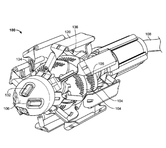
FIG. 1 is an oblique front view illustration of one embodiment of the
cardiovascular flow
assist device 100 in the expanded, deployed configuration. In the pictured
embodiment, the
device 100 includes two sets of helical pump blades 104. Each blade 104
includes a multitude of
small fenestrations 105. These fenestrations 105 impart increased flexibility
to each blade 104
so that the blades will be easier to compress without sacrificing efficiency
of the pump. Each
blade 104 is connected to a portion of the rotating pump shaft, which is not
easily visualized in
this figure but is further discussed with reference to FIG.'s 5A ¨ 6B. A cable
108 extends from
the proximal end of the device 100 and, in various embodiments, carries power
supply and/or
control wires from outside the body to the device 100. The device 100 includes
eight cage
support members 120 encircling the pump. In one embodiment, two support
members 120 are
manufactured together in one piece to assist in assembly of the device, as
will be further
discussed with reference to FIG. 's 9A ¨ 9D. In one embodiment, the device 100
includes a cap
106 at its distal end. In one embodiment, the distal end cap 106 is cone
shaped. In the pictured
embodiment, the distal end cap 106 includes four sensors 132 positioned
equidistant from one
another proximate the distal tip of said end cap 106. Four additional sensors
134 are positioned
proximate the distal end of every second cage support member 120, such that
every support
member 120 containing a sensor is adjacent to a support member 120 without a
sensor. Each
support member 120 containing a distal end sensor 134 also has an additional
sensor 136
proximate its proximal end, resulting in a total of twelve sensors on the
device 100. Although
18
CA 02863234 2014-07-29
WO 2013/119752 PCT/US2013/025056
twelve sensors are depicted in the pictured embodiment, any number of sensors
may be used to
provide the health care professional information regarding the functionality
of the device 100.
Data gathered by the sensors is transferred to a processor outside the patient
via cable 108.
FIG. 2 is an oblique, cross-sectional illustration of an aorta 240 depicting
one
embodiment of the cardiovascular flow assist device 200 in the expanded,
deployed
configuration positioned therein. When deployed, the diameter of the cage of
the device 200 is
slightly larger than the internal diameter of the aorta, such that the cage
support members 220
contact the inner wall of the aorta. Each cage support member 220 becomes
fixes in place
against the aortic wall, securing the device 200 within the aorta 240. The
pump is then free to
rotate within the support cage, increasing blood flow downstream from the
device 200.
FIG. 3 is an oblique front view illustration of one embodiment of the
cardiovascular flow
assist device 300 in the collapsed, deliverable configuration. In the pictured
embodiment, the
device 300 includes a distal end cap 306 with sensors 332 and a power/control
cable 308
emanating from its proximal end. The cage support members 320 are collapsed in
toward the
center of the device 300, forming an elongate, streamlined cylindrical shape.
This collapsed
configuration enables the physician to implant the device in a percutaneous
fashion, avoiding a
more invasive surgical procedure and resulting in less stress and discomfort
to the patient.
FIG.'s 4A and 4B are oblique front and side illustrations respectively,
depicting one
embodiment of a cardiovascular flow assist device 400 in the expanded,
deployed configuration
side by side with another cardiovascular flow assist device 401 in the
collapsed, deliverable
configuration. The cage support members 420 of the deployed device 400 are
seen in their fully
expanded state, exposing the pump and helical pump blades 404. The cage
support members
421 of the collapsed device 401 are seen in their fully compressed state,
collapsed toward the
center of the device and coming to rest in contact with one another. As can be
seen in FIG.' s 4A
and 4B, the diameter of the device 400 when in the expanded configuration,
particularly the
diameter of the cage, is larger than the diameter of the device 401 when in
the collapsed
configuration. In one embodiment, the diameter of the device 400 in the
expanded configuration
is in the range of 15 ¨ 30 mm. In one embodiment, the diameter of the device
400 in the
expanded configuration is 25 mm. In one embodiment, the diameter of the device
401 in the
collapsed configuration is in the range of 3 ¨ 8 mm. In one embodiment, the
diameter of the
device 401 in the collapsed configuration is 6 mm. As can also be seen in
FIG.'s 4A and 4B, the
19
CA 02863234 2014-07-29
WO 2013/119752 PCT/US2013/025056
length of the device 400 when in the expanded configuration is shorter than
the length of the
device 401 when in the collapsed configuration. In one embodiment, the length
of the device
400 when in the expanded configuration is in the range of 20 ¨ 90 mm. In one
embodiment, the
length of the device 401 when in the collapsed configuration is in the range
of 30 ¨ 100 mm.
FIG. 5A is a side view illustration of one embodiment of the cardiovascular
flow assist
device 500 in the expanded, deployed configuration, depicting two cage support
members 520
removed from either side of the helical screw pump 503. The end cap (not
shown) has also been
removed from the device 500 pictured in FIG. 5A. These components have been
removed to
provide enhanced visualization of the helical screw pump 503 of the device
500. In one
embodiment, the helical screw pump 503 comprises an elongate, cylindrical
inner shaft portion
511, a distal outer shaft portion segment 512, four outer shaft portion blade
attachment segments
514, five outer shaft portion spacer segments 513, and a proximal outer shaft
portion segment
516. The pump 503 is connected at its proximal end, via a coupling 507, to a
motor 509. In one
embodiment, the coupling 507 is a low friction flexible coupling which
transfers rotation from
the motor 509 to the shaft. The coupling 507 acts to keep the motor 509 and
shaft in alignment
and prevents binding and stoppage of the motor 509. In the pictured
embodiment, the pump 503
includes two sets of helical blades 504. Each outer blade attachment segment
514 of the pump
503 shaft includes two attached blades 504 positioned 180 degrees apart on
either side of said
segment 514. Each of the two blade sets comprises four separate blades 504. In
various
embodiments, the pitch of each blade in the deployed configuration is within
the range of 20 to
70 degrees. In one embodiment, the pitch of each blade in the deployed
configuration is 45
degrees. The blades 504 in each set join to form a continuous helical screw
spiraling around
either side of the pump 503. Having two sets of blades 504 improves
performance of the pump
by increasing pumping efficiency and by balancing the pump 503. In addition,
having the pump
blades formed in segments eases collapsibility and allows for intended
deformation to create the
smallest outside profile for minimally invasive intravascular insertion. In
one embodiment, each
blade 504 includes a multitude of fenestrations 505 to increase flexibility of
the blades for
compression and expansion. In one embodiment, the blades 504 are coated in
silicon to prevent
blood flow through the fenestrations 505.
In one embodiment, the inner shaft portion 511 of the pump extends through to
the
coupling 507 and is slidably movable within the pump's outer shaft portion
segments 512, 514,
CA 02863234 2014-07-29
WO 2013/119752 PCT/US2013/025056
513, 516. This allows the device to lengthen and shorten during compression
and expansion
respectively. In one embodiment, the distal end of the inner shaft portion 511
of the pump 503
and the distal ends of the cage support members 520 attach to the distal end
cap (not shown). To
lend linear stability to the device 500, in one embodiment, the inner shaft
portion 511, distal
outer shaft portion segment 512, outer shaft portion blade attachment segments
514, and
proximal outer shaft portion segment 516 are composed of stainless steel. In
one embodiment,
the outer shaft portion spacer segments 513 are composed of silicon to absorb
pressure during
compression and expansion of the device 500. As mentioned earlier, the blades
504 are
composed of a shape memory metal to allow for compression and expansion of
said blades 504.
In one embodiment, the shape memory metal is Nitinol. In one embodiment, the
device 500
includes a Teflon motor seal.
FIG. 5B is an oblique, side view illustration of one embodiment of an outer
shaft portion
blade attachment segment 514, with one attached blade 504, of the
cardiovascular flow assist
device. In one embodiment, each blade 504 is laser welded to each segment 514
at two points
517 along the outer circumference of the segment 514, with a gap 518 in
between the two weld
points 517. The gap 518, along with the fenestrations 505 in the blade 504,
lends greater
flexibility to the blade 504 to ease blade compression and expansion.
FIG. 6A is an oblique, front view illustration and FIG. 6B is a side view
illustration of
one embodiment of the helical screw pump 603 of the cardiovascular flow assist
device,
depicting two sets of helical blades 604 in the expanded configuration. The
distal end cap and
cage have been completely removed to enhance pump 603 visualization. In the
embodiment
depicted in FIG. 's 6A and 6B, the pump 603 does not include a coupling and
the entirety of the
motor 609 can be seen. Also visible are the inner shaft portion 611, distal
outer shaft portion
segment 612, outer shaft portion blade attachment segments 614, outer shaft
portion spacer
segments 613, and proximal outer shaft portion segment 616.
FIG. 's 7A, B, and C are oblique front, side, and front-on view illustrations
respectively,
of one embodiment of the helical screw pump 703 of the cardiovascular flow
assist device,
depicting one set of helical blades 704 in the expanded configuration. The
distal end cap and
cage have been completely removed to enhance pump 703 visualization. Referring
simultaneously to FIG.'s 7A and 7B, the pictured embodiment of the pump 703
does not include
a coupling and the entirety of the motor 709 can be seen. Also visible are the
inner shaft portion
21
CA 02863234 2014-07-29
WO 2013/119752 PCT/US2013/025056
711, distal outer shaft portion segment 712, outer shaft portion blade
attachment segments 714,
outer shaft portion spacer segments 713, and proximal outer shaft portion
segment 716. FIG. 7C
illustrates how each blade 704 meets the other to form a virtually seamless
helical screw.
FIG.'s 8A, 8B, and 8C are oblique front, side, and front-on view illustrations
respectively, of one embodiment of the helical screw pump of the
cardiovascular flow assist
device, depicting one set of helical blades in the collapsed configuration.
The distal end cap,
cage, coupling, and motor have been completely removed to enhance pump 803
visualization.
Referring simultaneously to FIG.'s 8A and 8B, the inner shaft portion 811,
distal outer shaft
portion segment 812, outer shaft portion blade attachment segments 814, outer
shaft portion
spacer segments 813, and proximal outer shaft portion segment 816 are all
visible. FIG. 8C
illustrates how each blade 804 compresses in toward the body of the pump shaft
while in the
collapsed configuration.
FIG. 9A is an oblique, front view illustration and FIG. 9B is a side view
illustration of
one embodiment of two cage support members 920 formed together into a singular
cage arm 950
in the expanded configuration. Referring simultaneously to FIG.'s 9A and 9B,
while in the
expanded configuration, the two cage support members 920 of each cage arm 950
are expanded
outward from the pump (not shown) and from one another. At the distal end of
each cage arm
950, the two support members come together in the form of a distal quarter-
circle 951. At the
proximal end of each cage arm 950, the two support members come together in
the form of a
proximal quarter circle 958 with attached elongate linear member 959. In one
embodiment, four
cage arms 950 are circularly arranged around the helical screw pump (not
shown) of the device
to form the basket-like cage support structure. The four distal quarter-
circles 951 are attached to
the distal end cap (not shown) and inner shaft portion (not shown) of the pump
at the distal end
of the device. The four proximal quarter-circles 958, with attached elongate
linear members 959,
are attached to a housing (not shown) supporting the motor (not shown) at the
proximal end of
the device.
FIG. 9C is a top-down view illustration of the same embodiment of two cage
support
members 920 formed together into a singular cage arm 950 in the expanded
configuration of
FIG. 9A. In one embodiment, the central, thin rectangular shaped portion 921
of each cage
support member 920 is composed of stainless steel. In this embodiment, the
rigidity of this
portion 921 lends stability to the device. In another embodiment, the central,
thin rectangular
22
CA 02863234 2014-07-29
WO 2013/119752 PCT/US2013/025056
shaped portion 921 of each cage support member 920 is composed of a shape
memory metal. In
one embodiment, the shape memory metal is Nitinol. In this embodiment, the
flexibility of this
portion 921 allows the cage to fit more snugly within the aorta. This portion
921 comes to rest
against the inner wall of the aorta when the device is deployed. Distal and
proximal to each
central portion 921 are two hinge portions 922 and 924 respectively. Each
hinge portion 922,
924 is composed of a shape memory metal and allows for compression and
expansion of each
cage support member 920. In one embodiment, the shape memory metal is Nitinol.
In one
embodiment, the distal end 923 and proximal end 925 of each support member 920
are
composed of stainless steel. This again lends overall stability to the device
and allows for
attachment of the support members 920 to the other components of the device.
In one
embodiment, each elongate linear member 959 is composed of stainless steel.
In one embodiment, each hinge portion 922, 924 includes at least one slit 926
to enhance
flexibility and for the passage of a wire leading from a sensor positioned
distally on the device.
Additionally, in one embodiment, each hinge portion 922, 924 includes an
elongate tubular
member 927 along its external edge for the guiding of sensor and/or camera
wires. In one
embodiment, each central rectangular portion 921 includes an elongate tubular
member along
one side for the guiding of sensor and/or camera wires.
FIG. 9D is a front-on view illustration of the same embodiment of two cage
support
members 920 formed together into a singular cage arm 950 in the expanded
configuration of
FIG. 9A. Visible in FIG. 9D are the slits 926 and elongate tubular members
927, 928 for the
passage of sensor and/or camera wires.
Figures 10A, 10B, and 10C are oblique front, side, and front-on view
illustrations
respectively, of one embodiment of four cage arms 1050 combined together to
form a complete
basket-like cage 1060 in the expanded configuration. In various other
embodiments, the cage
includes fewer or more than four arms and takes on a variety of other shapes,
including, but not
limited to, an ellipse. Referring simultaneously to FIG.'s 10A and 10B, each
cage arm 1050
comprises two cage support members 1020 and one elongate linear member 1059.
The complete
cage 1060 comprises four cage arms 1050 arranged together such that the distal
ends of each
cage arm 1050 come together to form a circle 1062 at the distal end of the
device. The distal end
of the cage 1060 is attached to the distal end cap (not shown) and inner shaft
portion at the circle
1062. The four elongate linear members 1059 enclose a housing at the proximal
end of the
23
CA 02863234 2014-07-29
WO 2013/119752 PCT/US2013/025056
device and are spaced apart from one another in 90 degree increments. In one
embodiment, the
housing contains the motor to drive the device and a battery to power the
motor. In addition, in
one embodiment, the housing includes a locking mechanism to couple with the
positioning shaft.
In the expanded configuration, the eight central rectangular portions 1021 of
each cage support
member 1020 are expanded out away from the center of the device and from one
another. FIG.
10C illustrates the circle 1062 formed at the distal end of the cage 1060 by
the combination of
four cage arms 1050.
FIG.'s 11A, 11B, and 11C are oblique front, side, and front-on view
illustrations
respectively, of one embodiment of four cage arms 1150 combined together to
form a complete
basket-like cage 1160 in the collapsed configuration. Referring simultaneously
to FIG.'s 11A
and 11B, each cage arm 1150 comprises two cage support members 1120 and one
elongate linear
member 1159. The complete cage 1160 comprises four cage arms 1150 arranged
together such
that the distal ends of each cage arm 1150 come together to form a circle 1162
at the distal end of
the device. The distal end of the cage 1160 is attached to the distal end cap
(not shown) and
inner shaft portion (not shown) at the circle 1162. The four elongate linear
members 1159
enclose a housing (not shown) at the proximal end of the device and are spaced
apart from one
another in 90 degree increments. In the collapsed configuration, the eight
central rectangular
portions 1121 of each cage support member 1120 are compressed in toward the
center of the
device and are in contact with one another. FIG. 11C illustrates the circle
1162 formed at the
distal end of the cage 1160 by the combination of four cage arms 1150.
FIG. 12A is a side view illustration on one embodiment of an arterial closure
device
1200, depicting the arterial closure discs 1205, 1210 of the device 1200
positioned within a
delivery sheath 1220. The arterial closure device is used to seal the
arteriotomy site after
insertion or retrieval of the pumping device of the present specification. In
one embodiment, the
closure device 1200 includes a pair of opposing umbrella shaped discs 1205,
1210. The distal
disc 1205 includes a concave-convex deployed shape wherein its inner concave
surface contacts
the inner wall of the artery and the proximal disc 1210 includes a concave-
convex deployed
shape wherein its inner concave surface contacts the outer wall of the artery.
The discs 1205,
1210 are connected at their centers by a diaphragm 1207 having a lumen. Both
discs 1205, 1210
are initially constrained inside an elongate delivery sheath 1220, having a
lumen and proximal
24
CA 02863234 2014-07-29
WO 2013/119752 PCT/US2013/025056
and distal ends, and are deployed and expanded by extending distally from the
distal tip of the
delivery sheath 1220.
The delivery sheath 1220 includes a delivery sheath head 1222 at its proximal
end and
handles 1227 along its length. The delivery sheath head 1222 includes a distal
end that attaches
to the proximal end of the sheath and a proximal end. The delivery sheath head
1222 and
handles 1227 are used by the physician to manipulate the closure device 1200
during placement.
The delivery sheath 1220 includes an elongate blood return tube 1224 having a
proximal end and
a distal end. The distal end of the blood return tube 1224 is positioned at
the distal end of the
delivery sheath 1220 and the proximal end of the blood return tube 1224 exits
at a point between
the distal and proximal ends of the delivery sheath 1220. The closure device
1200 includes a
tamper tool 1230 for extending the discs 1205, 1210 beyond the distal end of
the delivery sheath
1220. The tamper tool 1230 comprises an elongate shaft having a tamper tool
lumen, a proximal
end, and a distal end and extends within the lumen of the delivery sheath
1220. The distal end of
the tamper tool 1230 abuts the proximal end of the proximal disc 1210. At its
proximal end, the
tamper tool 1230 includes a handle 1232 that extends beyond the proximal end
of the delivery
sheath head 1222. Positioned on the tamper tool 1230 distal to the handle 1232
are a distal rivet
1235 and a proximal rivet 1237. During placement of the discs 1205, 1210 the
distal rivet 1235
and proximal rivet 1237 sequentially engage a groove 1229 positioned within
the proximal end
of the delivery sheath head 1222. A string 1209 is attached to the distal disc
1205 and extends
through the lumen of the diaphragm 1207, through the center of the proximal
disc 1210, and
proximally through the tamper tool 1230 lumen.
During placement of the arterial closure discs 1205, 1210, the entire sheath
system is
advanced over the wire 1240 extending from the distal end of the pumping
device. The wire
1240 extends through the tamper tool lumen and guides the closure device 1200.
If arterial
closure is being performed after removal of the pumping device, a separate
guide wire is first
introduced into the artery. For arterial closure after the insertion of the
pumping device, the
delivery sheath 1220 of the closure device 1200 is delivered through the
existing arterial sheath
used to insert the pumping device.
FIG. 12B is a side view illustration of the same embodiment of the arterial
closure device
1200 of FIG. 12A, depicting the distal arterial closure disc 1205 expanded and
deployed from the
distal end of the delivery sheath 1220. Once the distal end of the delivery
sheath 1220 is
CA 02863234 2014-07-29
WO 2013/119752 PCT/US2013/025056
positioned inside the artery 1250, as confirmed by the presence of blood 1255
at the proximal
end of the blood return tube 1224, the distal disc 1205 of the closure device
1200 is pushed out
with the tamper tool 1230. Once beyond the distal end of the delivery sheath
1220, the distal disc
1205 expands to its umbrella shape. The tamper handle 1232 is pushed distally
into the delivery
sheath head 1222 until the distal rivet 1235 engages the groove 1229,
effectively locking the
tamper tool 1230 in place. The entire closure device 1200 is then pulled back
so that the umbrella
shaped distal disc 1205 opposes the hole in the artery from the inside. The
existing arterial sheath
for inserting the pumping device is then removed and the delivery sheath 1220
is left just outside
the artery 1250.
FIG. 12C is a side view illustration of the same embodiment of the arterial
closure device
1200 of FIG. 12A, depicting both the distal 1205 and proximal 1210 arterial
closure discs
expanded and deployed from the distal end of the delivery sheath 1220. The
reverse umbrella
shaped proximal disc 1210 of the closure device 1200 is delivered to the
outside of the artery
1250 by simultaneously pulling back the delivery sheath 1220 and pushing in
the tamper tool
handle 1232 until the delivery sheath head 1222 and tamper tool handle 1232
are fully apposed.
In this position, the proximal rivet 1237 of the tamper tool 1230 engages the
groove 1229 of the
delivery sheath head 1222, effectively locking the tamper tool 1230 in place.
In one
embodiment, the delivery sheath head 1222 is pushed distally along the
delivery sheath 1220,
wherein the delivery sheath 1220 includes a rivet 1225 that engages a second
groove 1221 within
the delivery sheath head 1222, effectively locking the delivery sheath 1220 in
place within the
delivery sheath head 1222. The delivery sheath 1220, with the delivery sheath
head 1222, and
the tamper tool 1230 are then removed.
FIG. 12D is a side view illustration of one embodiment of the arterial closure
discs 1205,
1210 fully deployed with the delivery sheath removed. The distal disc 1205 is
depicted within
the artery 1250 and the proximal disc 1210 is depicted outside the artery
1250. The two
apposing discs 1205, 1210 with center diaphragm 1207 seal the arteriotomy
site. Once both
discs 1205, 1210 are in place, the sheath and tamper are pulled out, exposing
only the string 1209
attached to the distal disc 1205. Once arteriotomy closure is confirmed, the
string 1209 is cut
below the skin. The guide wire in the center can be removed if necessary from
the center
diaphragm 1207 keeping the artery 1250 sealed.
26
CA 02863234 2014-07-29
WO 2013/119752 PCT/US2013/025056
FIG. 12E is an illustration of one embodiment of the arterial closure discs
1205, 1210,
depicting the struts 1204 used to expand the discs 1205, 1210 to their
deployed configuration.
The discs 1205, 1210 are constricted and tubular shaped, as depicted in FIG.
12A, when
constrained in the restraining sheath and expand into umbrella shaped discs
1205, 1210 outside
the sheath, as depicted in FIG. 12E. The proximal surface 1203 of the proximal
disc 1210
indicates the point where the tamper tool pushes on the discs to deploy them
from the sheath. In
one embodiment, the discs are made out of an expandable and biocompatible
material. The discs
1205, 1210 include a diaphragm 1207 interconnecting them with a lumen 1208 in
the center to
accommodate the string and guide wire exiting the artery. Each disc 1205, 1210
also includes a
hole at its center for accommodation of the string and guide wire. The
radiating struts 1204 act
to expand the discs into their umbrella shape upon deployment.
FIG. 13 is a flowchart illustrating the steps involved in implanting the
hemodynamic flow
assist device in the descending aorta of a patient, in accordance with one
embodiment of the
present specification. At step 1302, a physician creates an access point in
the femoral artery of a
patient. A sheath with a guide wire is inserted into the femoral artery and
advanced such that the
distal end of the sheath is positioned within the descending aorta at step
1304. Then, at step
1306, the hemodynamic flow assist device, in the collapsed configuration, is
inserted into the
sheath and is advanced along the guide wire. At step 1308, a positioning
device attached to the
proximal end of the flow assist device is used to advance the flow assist
device beyond the distal
end of the sheath, causing the flow assist device to passively transform from
its collapsed
configuration into its expanded configuration. At step 1310, the positioning
device is used to
position the flow assist device within the descending aorta. The positioning
device is then
uncoupled from the flow assist device at step 1312. At step 1314, the
positioning device and
sheath with guide wire are removed from the patient. The access point is then
closed at step
1316.
FIG. 14 is a flowchart illustrating the steps involved in closing an arterial
access point
using the arterial closure device, in accordance with one embodiment of the
present
specification. At step 1402, a physician inserts a guide wire into the artery
through the access
point. The proximal end of the guide wire is inserted into the lumen of the
tamper tool and the
closure device is advanced along the guide wire at step 1404. The distal end
of the closure
device is positioned in the interior of the artery at step 1406. Then, at step
1408, the handle of
27
CA 02863234 2014-07-29
WO 2013/119752 PCT/US2013/025056
the tamper tool is pushed to advance the distal disc beyond the distal end of
the sheath, causing
the distal disc to passively transform from its compressed configuration into
its expanded
configuration within the artery. At step 1410, the closure device is pulled
back to position the
distal disc against the inner wall of the artery. Then, at step 1412, the
handle of the tamper tool
is pushed to advance the proximal disc beyond the distal end of the sheath,
causing the proximal
disc to passively transform from its compressed configuration into its
expanded configuration
outside the artery. The closure device and guide wire are then removed from
the patient at step
1414.
The above examples are merely illustrative of the many applications of the
system of the
present invention. Although only a few embodiments of the present invention
have been
described herein, it should be understood that the present invention might be
embodied in many
other specific forms without departing from the spirit or scope of the
invention. Therefore, the
present examples and embodiments are to be considered as illustrative and not
restrictive, and
the invention may be modified within the scope of the appended claims.
28