Note: Descriptions are shown in the official language in which they were submitted.
INNATE IMMUNE PROTEINS AS BIOMARKERS FOR CNS INJURY
CROSS-REFERENCE TO RELATED APPLICATIONS
[0001]
FIELD OF THE INVENTION
[0002] The present invention relates generally to the fields of neurology,
immunology, and
diagnostics. In particular, the present invention relates to the
identification of biomarkers in
biological samples which can predict the severity of neuronal injury, such as
spinal cord and
traumatic brain injury, in patients. The identified biomarkers may also be
used in determining
prognosis, directing therapeutic and rehabilitation efforts, and monitoring
response to treatment
for patients with a central nervous system injury.
BACKGROUND OF THE INVENTION
[0003] Nucleotide-binding oligomerization domain (NOD)-containing protein-like
receptors
(NLRs) are a recently discovered class of innate immune receptors that play a
crucial role in
initiating inflammatory responses following tissue injury in the central
nervous system (CNS)
(Abulafia et al., 2009, Silverman etal., 2009). Previous work shows that NLRP1
(also known as
NAcht leucine-rich-repeat protein 1 (NALP-1)) forms an inflammasome complex
comprising
NLRP1, the adaptor protein apoptosis-associated speck-like protein containing
a caspase
recruitment domain (ASC) and the caspase-1 enzyme that orchestrate the early
inflammatory
processes after spinal cord injury (SCI) and traumatic brain injury (TB!) via
IL-113 activation (de
Rivero Vaccari et al., 2008; 2009). The formation of inflammasomes is induced
by physical
damage to the plasma membrane, and by certain endogenous ligands referred to
as danger
associated molecular patterns (DAMPs) or exogenous ligands known as pathogen
associated
molecular patterns (PAMPs) (Bianchi, 2007, Wakefield etal., 2010). However,
the full IL-113
response also depends on the activation of Toll-like receptors (TLRs) and/or
purinergic ATP-
gated receptors, which induce the transcription of pro- IL-! 3.
1
CA 2863417 2019-08-12
CA 02863417 2014-07-30
WO 2013/119673 PCT/US2013/024941
[0004] Hyperinflammatory responses associated with tissue damage can promote
pathogenesis
of SCI and TBI via overproduction of IL-113 and other potentially neurotoxic
products.
Inflammasome-mediated 1L-113 overproduction is involved in the pathogenesis of
type 2
diabetes, liver damage and muscular dystrophy (Kufer and Sansonetti, 2011).
Moreover,
increasing genetic evidence suggests that inflammasome activation could also
drive adaptive
immunity in types of dermatitis, skin related allergies and asthma (Kufer and
Sansonetti, 2011).
In addition, inflammasome components may be secreted into the extracellular
milieu via a
mechanism involving the exosome pathway (Bianchi, 2007). The inflammasome
therefore has a
complex connection with the control of adaptive immune responses that has
become the subject
of intense investigation. Whether inflammasomes are associated with tissue
destructive
inflammatory processes after SCI and TBI in humans has not been investigated.
[0005] TBI affects an estimated 1.5 million people each year and causes one-
third of injury-
related deaths. Approximately 5.3 million Americans are living today with a
permanent TBI-
related disability. Predicting the severity and outcome of TBI and well as SCI
is difficult, given
the lack of objective, laboratory-based biomarkers. Currently, the Glasgow
Coma Scale (GCS)
score (Teasdale et al., 1974) is the best available clinical predictor of
injury severity; however,
its value is limited in patients undergoing pharmacological paralysis for
intubation, as a motor
score cannot be obtained (Brain Trauma Foundation, American Association of
Neurological
Surgeons, 2000). Predicting outcome is further complicated by the
heterogeneity of pathology in
patients with a similar GCS score. Therefore, the identification of diagnostic
and prognostic
biomarkers that directly reflect injury to CNS cells is imperative. Such
biomarkers of TBI and
SCI will enable clinicians to assess the degree of damage to the brain or
spinal cord, relay
prognostic information to the patient's family members, and target acute and
chronic treatments
to specific CNS damage mechanisms. Therefore, an early, accurate diagnostic
test designed to
target neuroprotective strategies would be a most desirable prognostic tool.
[0006] Although, significant progress has been made regarding the verification
and testing of
various biomarkers after stroke and TBI, limited data are available regarding
what biomarkers
are appropriate for SCI. The biomarkers 5-10013, neuron-specific enolase,
neurofilament light
chain and glial fibrillary acidic protein are significantly increased in cases
of SCI in experimental
animals studies (Skouen etal., 1999, Ma et al., 2001, Nagy et al., 2002,
Cornefjord et al., 2004,
Loy etal., 2005, Cao et al., 2008, Pouw etal., 2009). Although some biomarkers
show
CA 02863417 2014-07-30
WO 2013/119673 PCT/US2013/024941
promising results, these do not yet provide a sensitive prognostic tool.
Quantitative standards for
determining the extent of SCI and TBI during the acute phase must be developed
and validated.
[0007] A new approach for evaluating the primary cord and brain damage in the
acute phase is
the assessment of biomarkers in the cerebrospinal fluid (CSF). Since CSF
surrounds the spinal
cord and brain, damage to the cord or brain may lead to the release of
proteins and molecules
from central nervous system cells into the CSF that may serve as biomarkers
for SCI and TBI in
the CSF. Several studies have been conducted concerning S-10013, neuron-
specific enolase,
neurofilament light chain, and glial fibrillary acidic protein (GFAP) in CSF
and serum of animal
models of SCI (Pouw et al., 2009). However, only one study has investigated
neurofilament
protein and GFAP in CSF after SCI in humans (Guez etal., 2003). Thus, there is
a need in the art
to identify biomarkers of neuronal damage following central nervous system
injury in humans
that can be used to ascertain the severity of the injury and facilitate the
selection of an
appropriate therapeutic strategy to maximize recovery.
SUMMARY OF THE INVENTION
[0008] The present invention is based, in part, on the discovery that NLRP I
(NALP-1)
inflammasome components are secreted into the cerebrospinal fluid (CSF)
acutely after SCI and
traumatic brain injury (TBI) in humans. Elevated inflammasome protein levels
in the CSF
following central nervous system (CNS) injury represent the degree of
neuroinflammation in
CNS tissue and reflect the extent of inflammatory-induced damage. The CSF
levels of
inflammasome protein following injury correlate with the degree of functional
recovery in
patients and thus, can be used as acute biomarkers to predict patient
prognosis and direct
therapeutic interventions. Accordingly, the present invention provides a
method of assessing the
severity of a CNS injury in a patient.
[0009] In one embodiment, the invention provides a method of evaluating a
patient suspected of
having a CNS injury comprising providing a biological sample from a patient
presenting with
clinical symptoms consistent with a CNS injury, measuring the level of at
least one
inflammasome protein in the biological sample, determining the presence or
absence of a protein
signature associated with a CNS injury or a more severe CNS injury, wherein
the protein
signature comprises an elevated level of said at least one inflammasome
protein, and selecting
patients exhibiting the presence of the protein signature as having a CNS
injury or a more severe
3
CA 02863417 2014-07-30
WO 2013/119673 PCT/US2013/024941
CNS injury. In certain embodiments, said one or more inflammasome proteins are
NLRP1
(NALP-1), ASC, or caspase-1. The diagnostic methods of the invention may
further comprise
administering a neuroprotective treatment to the patient based on the measured
level of one or
more inflammasome proteins, and following changes in the level of one or more
inflammasome
proteins as a mechanism to monitor response to treatment.
[0010] In some embodiments, the levels of one or more inflammasome proteins in
the patient's
sample can be used to prepare an inflammasome protein profile associated with
CNS injury. The
levels of inflammasome proteins in the profile may be determined relative to
levels of the
proteins in control samples or pre-determined reference values or ranges of
reference values.
The inflammasome protein profiles are, in some embodiments, indicative of the
presence or
severity of CNS injury in a patient. When such protein profiles are prepared
from samples
obtained from patients following administration of a neuroprotective
treatment, the
inflammasome protein profiles are indicative of therapeutic efficacy of the
neuroprotective
treatment in the patient.
[0011] The present invention also provides a method of determining a prognosis
for a patient
with a central nervous system injury. In one embodiment, the method comprises
providing a
biological sample, such as cerebrospinal fluid, obtained from the patient
shortly after injury (e.g.,
within a week of injury), and measuring the level of at least one inflammasome
protein in the
biological sample to prepare an inflammasome protein profile, wherein the
inflammasome
protein profile is indicative of the prognosis of the patient. In particular
embodiments, an
elevated level of at least one inflammasome protein relative to a pre-
determined reference value
or range of reference values is indicative of a poorer prognosis or
unfavorable patient outcome.
For, example elevated inflammasome protein levels arc predictive of the
patient having a
Glasgow Outcome Scale (GOS) score of 1 to 3 upon follow-up assessment. In
other
embodiments, a reduced level of at least one inflammasome protein relative to
a pre-determined
reference value or range of reference values is predictive of a favorable
patient outcome (e.g.
GOS score of 4 or 5 upon follow-up assessment). In certain embodiments, the
method provides
a prognosis for a patient with a spinal cord or traumatic brain injury.
[0012] The present invention also includes kits for preparing an inflammasome
protein profile
associated with CNS injury. In one embodiment, the kit comprises a labeled-
binding partner,
such as labeled-antibody or fragment thereof, that specifically binds to one
or more
4
CA 02863417 2014-07-30
WO 2013/119673 PCT/US2013/024941
inflammasome proteins, wherein said one or more inflammasome proteins are
selected from the
group consisting of NLRP1, ASC, caspase-1, and combinations thereof
BRIEF DESCRIPTION OF DRAWINGS
[0013] Figure 1. NLRP1, ASC, and Caspase-1 are biomarkers that predict outcome
after
SCI. CSF samples were immunoblotted with antibodies against NLRP1, caspase-1
and ASC.
CSF samples from uninjured patients were used as controls. Immunoblot analysis
of 6 different
cases of patients with SCI indicates that patients (2, 3 and 4) who express
low levels of caspase-1
acutely after SC1 have a better prognosis than subjects (1, 5 and 6) who have
elevated levels of
this protein in CSF.
[0014] Figure 2. NLRP1 inflammasome proteins are expressed in cells of the
CNS. Spinal
cord sections were obtained from decedents that had injury to the spinal cord.
Immunohistochemical analysis combined with light microscopy indicates that
NLRP1 is
expressed in neurons of the ventral horn (black arrows) myelinated axons
(black arrow heads)
and oligodendrocytes (yellow arrows) (top panel). Caspase-1 is expressed in
swollen axons
(spheroids, blue arrows), motor neurons (black arrows) and in oligodendrocytes
(yellow arrows)
(central panel). ASC is present in neurons in the ventral horn (black arrows),
white matter
oligodendrocytes (yellow arrow) and macrophagesimicroglia (blue arrow heads)
(bottom panel).
[0015] Figure 3. Scatter plots of expression of inflammasome proteins in
controls and
patients with TB!. Samples were immunoblotted for ASC (A), caspase-1 (B), and
NALP-1 (C).
The p values in the upper left corner represent results of a Mann-Whitney U-
test. Densitometric
analysis revealed a significant increase in expression of ASC, caspase-1
(p20), and NALP-1 in
the CSF of patients with TB1 compared with nontrauma controls. Solid lines
denote mean values
for each group. Different shapes correspond to patient outcomes at 5 months
postinjury.
Representative immunoblots are shown. Samples were run on the same gel but
were
noncontiguous. N = the number of TBI samples analyzed; * = GOS Score 5; = =
GOS Score 4;
0 = GOS Score 3; V = GOS Score 1.
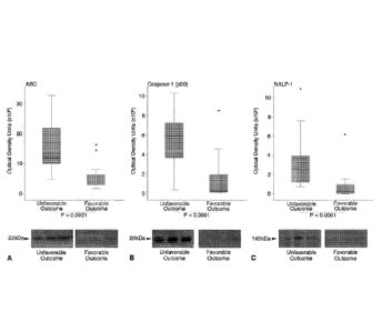
[0016] Figure 4. Box plots of expression of inflammasome proteins sorted by
outcome
category. The ends of the whiskers represent the lowest datum within 1.5
interquartile range of
the lower quartile and the highest datum within 1.5 interquartile range of the
upper quartile. The
asterisks represent the outliers. Mann-Whitney U-tests indicate higher
expression of ASC (A),
CA 02863417 2014-07-30
WO 2013/119673 PCT/US2013/024941
caspase-1 (p20) (B), and NALP-1 (C) are significantly associated with an
unfavorable outcome 5
months after injury (p < 0.0001). Representative immunoblots for each protein
are shown.
Samples were run on the same gel but were noncontiguous.
[0017] Figure 5. Scatter plots and estimated linear regression of ASC (A),
caspase-1 (p20) (B),
and NALP-1 (C) expression in the CSF with GOS score. Probability values of the
linear
regression are shown in the top left of each graph. Expression of each protein
correlated
significantly with COS score at 5 months post-injury. The p values on the x
axis represent post
hoc comparisons of a Kruskal-Wallis test. Representative immunoblots are
shown. Samples were
run on the same gel but were noncontiguous.
[0018] Figure 6. Caspase-1 levels in CSF one, two, and three days following
TBI in pediatric
patients receiving hypothermia treatment or no treatment (normothermia).
DETAILED DESCRIPTION OF THE INVENTION
[0019] The present invention is based, in part, on the discovery that NLRP1
inflammasomes play
an important role in inflammatory responses after SCI and TBI in humans. In
particular, the
present inventors have surprisingly found that nucleotide-binding leucine-rich
repeat pyrin
domain containing protein 1 (NLRP1), the adaptor protein apoptosis-associated
speck-like
protein containing a caspase recruitment domain (ASC), and caspase-1 are
secreted into the
cerebrospinal fluid (CSF) of human patients following SCI and TBI. Thus, these
inflammasome
proteins represent sensitive biomarkers of the severity of central nervous
system injury in human
patients. Accordingly, the present invention provides a method of assessing
the severity of a
central nervous system injury in a patient by measuring the level of at least
one inflammasome
protein in a biological sample obtained from the patient, wherein the measured
level of said at
least one inflammasome protein is indicative of the severity of the central
nervous system injury
in the patient.
[0020] As used herein, the term "inflammasome" refers to a multi-protein
complex that activates
caspase-1 activity, which in turn regulates IL-1 p, IL-18 and IL-33 processing
and activation. See
Arend et al. 2008; Li et al. 2008; and Martinon et al. 2002, each of which is
incorporated by
reference in their entireties. An "inflammasome protein" is a protein
component of
inflammasome complexes and can include, but is not limited to, an nucleotide
binding domain,
leucine-rich repeat containing (NLR) family member (e.g. NLRP1), ASC, caspase-
1, caspase-11,
6
CA 02863417 2014-07-30
WO 2013/119673 PCT/US2013/024941
X-linked inhibitor of apoptosis protein (XIAP), and pannexin-1. NLRP1 is also
known as NAcht
leucine-rich-repeat protein 1 (NALP-1). Thus, the terms "NLRP1" and "NALP-1
are used
interchangeably throughout the disclosure. In certain embodiments, the method
comprises
measuring an inflammasome protein selected from the group consisting of NLRP1
(NALP-1),
ASC, caspase-1, or combinations thereof. In one embodiment, the p20 subunit of
active caspase-
1 is measured.
[0021] The terms "patient" or "subject" are used interchangeably herein, and
is meant a
mammalian subject to be treated, with human patients being preferred. In
certain embodiments,
the patient is a pediatric patient. Pediatric patients include newborns (birth
to 1 month of age),
infants (1 month to 2 years of age), children (2 to 12 years of age), and
adolescents (12-21 years
of age). In some cases, the methods of the invention find use in experimental
animals, in
veterinary application, and in the development of animal models for disease,
including, but not
limited to, rodents including mice, rats, and hamsters, and primates.
[0022] In certain embodiments, the present invention provides a method of
evaluating a patient
suspected of having a central nervous system (CNS) injury. In one embodiment,
the method
comprises providing a biological sample from a patient presenting with
clinical symptoms
consistent with a CNS injury; measuring the level of at least one inflammasome
protein in the
biological sample; determining the presence or absence of a protein signature
associated with a
CNS injury or a more severe CNS injury, wherein the protein signature
comprises an elevated
level of said at least one inflammasome protein; and selecting patients
exhibiting the presence of
the protein signature as having a CNS injury or a more severe CNS injury.
[0023] A patient may be suspected of having a CNS injury on the basis of
neurologic symptoms
(motor, sensory, cognitive) and/or radiological evaluation (MRI, CT scan, X-
ray) consistent with
a CNS injury, e.g., after a physician's exam. In some embodiments, a patient
suspected of having
a CNS injury, particularly a spinal cord injury, may having a rating of A or B
on the American
Spinal Cord Injury Association (ASIA) Impairment Scale. The ASIA Impairment
Scale is a
standard diagnostic tool that assess a patient's motor and sensory function.
The classification
ratings and accompanying descriptions of the ASIA Impairment Scale are as
follows:
7
CA 02863417 2014-07-30
WO 2013/119673 PCT/US2013/024941
Classification/Rating Description
A Complete: no motor or sensory function is preserved below
the level of
injury, including the sacral segments S4-S5
Incomplete: sensory, but not motor, function is preserved below the
neurologic level and some sensation in the sacral segments S4-S5
Incomplete: motor function is preserved below the neurologic level,
however, more than half of key muscles below the neurologic level have
a muscle grade less than 3 (i.e., not strong enough to move against
gravity)
Incomplete: motor function is preserved below the neurologic level, and
at least half of key muscles below the neurologic level have a muscle
grade of 3 or more (i.e., joints can be moved against gravity)
Normal: motor and sensory functions are normal
Thus, a patient presenting with a classification rating of A or B on the ASIA
Impairment Scale
has no motor function below the level of the injury.
[0024] In other embodiments, a patient suspected of having a CNS injury may
have a score of <
12 (e.g. 3 to 12) on the Glasgow Coma Scale (GCS). In still other embodiments,
the patient may
have a GCS score of < 8 (e.g. 3 to 8). The GCS is a neurological scale
commonly used to assess
the level of consciousness of patients after injury or trauma. The scale is
composed of three tests
(eye, verbal and motor responses), each of which is assigned a value on a
scale up to 6. The three
values separately as well as their sum are considered. The lowest possible GCS
score (the sum)
is 3 (deep coma or death), while the highest is 15 (fully awake person). A GCS
score < 9 is
indicative of severe brain injury whereas a GCS score? 13 is indicative of
minor brain injury. A
GCS score between 9-12 is generally indicative of a moderate brain injury.
[0025] A patient suspected of having a CNS injury may have one or more signs
and symptoms of
CNS injury, such as temporary loss of consciousness, confusion,
disorientation, memory or
concentration problems, headache, dizziness, loss of balance, nausea or
vomiting, sensory
disruptions (e.g. blurred vision, ringing in the ears, bad taste in the mouth,
loss of sensation in
limbs), loss of motor function, sensitivity to light or sound, mood changes or
mood swings,
depression or anxiety, fatigue, drowsiness, and sleep disturbances.
[0026] In some embodiments, the level, concentration, or abundance of one or
more
inflammasome proteins is measured in a biological sample obtained from a
patient (e.g. a patient
suspected of having or suffering from a CNS injury). In particular
embodiments, the levels,
concentrations, or abundance of one or more inflammasome proteins is
indicative of the severity
8
CA 02863417 2014-07-30
WO 2013/119673 PCT/US2013/024941
of CNS injury in the patient. A CNS injury includes, but is not limited to, a
traumatic brain
injury, a stroke- related injury, a cerebral aneurism-related injury, a spinal
cord injury (e.g.
contusions, compressions, lacerations), concussion-related injury (including
post-concussion
syndrome), cerebral ischemia, injury resulting from neurodegenerative diseases
(including
Parkinson's disease, Dementia Pugilistica, Huntington's disease, Alzheimer's
disease,
Creutzfeldt-Jakob disease), seizure-related injuries, multiple sclerosis,
amyotrophic lateral
sclerosis, and other CNS traumas. In certain embodiments, the levels,
concentrations, or
abundance of one or more inflammasome proteins is indicative of the severity
of traumatic brain
injury or spinal cord injury in the patient.
[0027] As used herein, "biological sample" refers to any bodily fluid or
tissue obtained from a
patient or subject. A biological sample can include, but is not limited to,
whole blood, red blood
cells, plasma, serum, peripheral blood mononuclear cells (PBMCs), urine,
saliva, tears, buccal
swabs, CSF, CNS microdialysate, and nerve tissue. In one embodiment, the
biological sample is
CSF, saliva, serum, plasma, or urine. In certain embodiments, the biological
sample is CSF.
[0028] In some embodiments, the measured level, concentration, or abundance of
one or more
inflammasome proteins in the biological sample is used to prepare an
inflammasome protein
profile, wherein the profile is indicative of the severity of a CNS injury in
the patient or the
patient's prognosis or recovery potential from a CNS injury. The inflammasome
protein profile
may comprise the level, abundance, or concentration of one or more
inflammasome proteins
measured in the patient's sample optionally in relation to a pre-determined
value or range of
reference values as described herein. In certain embodiments, the inflammasome
proteins in the
profile include NLRP1 (NALP-1), ASC, and/or caspase- I (e.g. p20 subunit of
caspase-1). In one
particular embodiment, the inflammasomc protein profile comprises the level,
abundance, or
concentration of each of NLRP1 (NALP-1), ASC, and caspasc- 1 (e.g. p20 subunit
of caspasc-1).
[0029] In one aspect of the invention, the method of evaluating a patient
suspected of having a
CNS injury comprises determining the presence or absence of a protein
signature associated with
a CNS injury or a more severe CNS injury based on the measured level,
abundance, or
concentration of one or more inflammasome proteins in the patient sample or on
the
inflammasome protein profile prepared from the patient's sample. In certain
embodiments, the
protein signature comprises an elevated level of at least one inflammasome
protein. The level of
said at least one inflammasome protein in the protein signature may be
enhanced relative to the
9
CA 02863417 2014-07-30
WO 2013/119673 PCT/US2013/024941
level of the protein in a control sample or relative to a pre-determined
reference value or range of
reference values as further described herein. The protein signature may, in
certain embodiments,
comprise an elevated level for each of caspase-1 (e.g. p20 subunit of caspase-
1), NLRP1, and
ASC. Patients who exhibit the protein signature may be selected or identified
as having a CNS
injury or a more severe CNS injury.
[0030] The level or concentration of at least one inflammasome protein can be
assessed at a
single time point (e.g. after a potential CNS injury) and compared to a pre-
determined reference
value or range of reference values or can be assessed at multiple time points
(e.g. two, three,
four, five or more) after a potential CNS injury and compared to a pre-
determined reference
value or to previously assessed values. For instance, a biological sample for
measuring levels or
concentrations of inflammasome proteins can be obtained from a patient within
one hour of a
potential CNS injury to two weeks following a potential CNS injury. In some
embodiments, the
biological sample is obtained within one day, two days, three days, four days,
five days, six days,
seven days, ten days, or twelve days of a CNS injury or potential injury.
[0031] As used herein, "pre-determined reference value" refers to a pre-
determined value of the
level or concentration of an inflammasome protein ascertained from a known
sample. For
instance, the pre-determined reference value can reflect the level or
concentration of an
inflammasome protein in a sample obtained from a control subject (i.e., an
uninjured, healthy
subject). The control subject may, in some embodiments, be age-matched to the
patients being
evaluated. Thus, in particular embodiments, the measured level or
concentration of at least one
inflammasome protein is compared or determined relative to the level or
concentration of said at
least one inflammasome protein in a control sample (i.e. obtained from an
uninjured subject).
[0032] In other embodiments, the pre-determined reference value or range of
reference values
can reflect the level or concentration of an inflammasome protein in a sample
obtained from a
patient with a known severity of CNS injury as assessed by clinical measures
or post mortem
analysis. A pre-determined reference value can also be a known amount or
concentration of an
inflammasome protein. Such a known amount or concentration of an inflammasome
protein may
correlate with an average level or concentration of the inflammasome protein
from a population
of control subjects or a population of patients with known levels of injury.
In another
embodiment, the pre-determined reference value can be a range of values,
which, for instance,
can represent a mean plus or minus a standard deviation or confidence
interval. A range of
CA 02863417 2014-07-30
WO 2013/119673 PCT/US2013/024941
reference values can also refer to individual reference values for a
particular inflammasome
protein across various levels of CNS injury severity. In certain embodiments,
an increase in the
level of one or more inflammasome proteins (e.g., NLRP1 (NALP-1), ASC, or
caspase-1)
relative to a pre-determined reference value or range of reference values is
indicative of a more
severe central nervous system injury.
[0033] In some embodiments, the method of assessing the severity of a CNS
injury further
comprises measuring the level or concentration of one or more proteins
described in U.S. Patent
Publication No. 2011/0177974, which is hereby incorporated by reference in its
entirety, in
addition to measuring the level or concentration of one or more inflammasome
proteins. For
instance, in certain embodiments, the method further comprises measuring the
level or
concentration of one or more proteins selected from ubiquitin C-terminal
hydrolase Li; vesicular
membrane protein p-24; synuclein; microtubule-associated protein;
synaptophysin; Vimentin;
Synaptotagmin; Synaptojanin-2; Synapsin2; CRMP1, 2; Amphiphysin-1; PSD95; PSD-
93;
Calmodulin dependent protein kinase II (CAMPK)-alpha, beta, gamma; Myelin
basic protein
(MBP); Myelin proteolipid protein (PLP); Myelin Oligodendrocyte specific
protein (MOSP);
Myelin Oligodendrocyte glycoprotein (MOG); myelin associated protein (MAG); NF-
H; NF-L;
NF-M; BIII-tubulin-1 or combinations thereof in the biological sample obtained
from the patient
in addition to measuring the level or concentration of one or more
inflammasome proteins.
Thus, the protein signature may comprise an elevated level of one or more of
these proteins in
addition to the elevated level of one or more inflammasome proteins. In other
embodiments, the
method further comprises measuring the level or concentration of one or more
proteins selected
from S-10013, neuron-specific enolase, neurofilament light chain, glial
fibrillary acidic protein
(GFAP) or combinations thereof in the biological sample obtained from the
patient in addition to
measuring the level or concentration of one or more inflammasome proteins. In
one
embodiment, the protein signature associated with a CNS injury or a more
severe CNS injury
comprises an elevated level of one or more proteins selected from S-10013,
neuron-specific
enolase, neurofilament light chain, glial fibrillary acidic protein (GFAP) in
addition to an
elevated level of one or more inflammasome proteins (e.g. NLRP1 (NALP-1), ASC,
or caspase-
1).
[0034] In other embodiments of the invention, the methods of assessing the
severity of a CNS
injury in a patient or evaluating a patient suspected of having a CNS injury
further comprise
11
CA 02863417 2014-07-30
WO 2013/119673 PCT/US2013/024941
administering a neuroprotective treatment to the patient based on the measured
level of said at
least one inflammasome protein or when a protein signature associated with a
CNS injury or a
more severe CNS injury is identified. Such neuroprotective treatments include
drugs that reduce
excitotoxicity, oxidative stress, and inflammation. Thus, suitable
neuroprotective treatments
include, but are not limited to, methylprednisolone, 17a-estradiol, 1713-
estradiol, ginsenoside,
progesterone, simvastatin, deprenyl, minocycline, resveratrol, and other
glutamate receptor
antagonists (e.g. NMDA receptor antagonists) and antioxidants. In some
embodiments,
neuroprotective treatments are neutralizing antibodies against an inflammasome
protein or
binding fragments thereof, such as those described in U.S. Patent Publication
No. 2009/0104200,
which is hereby incorporated by reference in its entirety. For instance, in
one embodiment, the
neuroprotective treatment is an anti-ASC antibody or fragment thereof Anti-ASC
antibodies
include antibodies that specifically bind to amino acid residues 178-193 of
rat ASC (accession
number BAC43754), e.g., amino acid sequence ALRQTQPYLVTDLEQS (SEQ ID NO:1), or
antibodies that specifically bind to the amino acid sequence RESQSYLVEDLERS
(SEQ ID
NO:2) of human ASC. In another embodiment, the neuroprotective treatment is an
anti-NLRP1
antibody or fragment thereof. Suitable neutralizing anti-NLRP1 antibodies or
fragments thereof
include antibodies that specifically bind to the amino acid sequence
CEYYTEIREREREKSEKGR (SEQ ID NO:3) of human NLRP1 or the amino acid sequence
MEESQSKEESNTEG (SEQ ID NO: 4) of rat NLRP1. The neutralizing antibodies or
antibody
fragments may be polyclonal antibodies, monoclonal antibodies, chimeric
antibodies, humanized
antibodies, single-chain variable fragments (scFvs) and the like. Aptamers
that specifically bind
to an inflammasome protein or epitope thereof (e.g., SEQ ID NOs: 1-4) may also
be suitable
neuroprotective treatments. Neuroprotective treatments also encompass
therapeutic regimens or
rehabilitative procedures, such as hypothermia treatment.
[0035] The success of, or response to, treatment can also be monitored by
measuring the levels
of at least one inflammasome protein. Accordingly, in some embodiments, the
methods of
evaluating a patient further comprise measuring the level of at least one
inflammasome protein in
a biological sample obtained from the patient following neuroprotective
treatment, preparing a
treatment protein signature associated with a positive response to the
neuroprotective treatment,
wherein the treatment protein signature comprises a reduced level of at least
one inflammasome
protein, and identifying patients exhibiting the presence of the treatment
protein signature as
12
CA 02863417 2014-07-30
WO 2013/119673 PCT/US2013/024941
responding positively to the neuroprotective treatment. A reduction in the
level, abundance, or
concentration of one or more inflammasome proteins (e.g. NLRP1, ASC, and
caspase-1) is
indicative of the efficacy of the neuroprotective treatment in the patient.
The one or more
inflammasome proteins measured in the sample obtained following treatment may
be the same as
or different than the inflammasome proteins measured in the sample obtained
prior to treatment.
The inflammasome protein levels may also be used to adjust dosage or frequency
of a
neuroprotective treatment.
[0036] The present invention also provides a method of determining a prognosis
for a patient
with a central nervous system injury. In one embodiment, the method comprises
providing a
biological sample obtained from the patient within a week of injury, and
measuring the level of
at least one inflammasome protein in the biological sample to prepare an
inflammasome protein
profile as described above, wherein the inflammasome protein profile is
indicative of the
prognosis of the patient. In certain preferred embodiments, the biological
sample is obtained
from the patient within one week, within five days, or within three days of
injury. In some
embodiments, an increase in the level of one or more inflammasome proteins
(e.g., NLRP1,
ASC, caspase-1, or combinations thereof) relative to a pre-determined
reference value or range
of reference values is indicative of a poorer prognosis. For instance, an
increase of about 20% to
about 300% in the level of one or more inflammasome proteins relative to a pre-
determined
reference value or range of reference values is indicative of a poorer
prognosis. In one
embodiment, increased levels of caspase-1, particularly the p20 subunit of
active caspase-1,
relative to a pre-determined reference value or range of reference values
acutely after injury (i.e.
within a week of injury) is indicative of a poorer prognosis.
[0037] In particular embodiments, an elevated level of at least one
inflammasome protein
relative to a pre-determined reference value or range of reference values is
predictive of the
patient's recovery potential or long-term outcome as assessed by the Glasgow
Outcome Scale
(GOS). The GOS is a scale that allows for the objective assessment of a
patient's recovery
following brain injury. The scale is comprised of scores ranging from 1 to 5
with the following
descriptions:
13
CA 02863417 2014-07-30
WO 2013/119673 PCT/US2013/024941
Score/Category Description
1 -Death Severe injury or death without recovery of
consciousness
2-Persistent Vegetative State Severe damage with prolonged state of
unresponsiveness and a lack of higher mental
functions
3-Severe Disability Severe injury with permanent need for help with
daily living
4-Moderate Disability No need for assistance in everyday life, employment
is possible but may require special equipment
5-Low Disability Light damage with minor neurological and
psychological deficits.
In one embodiment, an elevated level of at least one inflammasome protein
relative to a pre-
determined reference value or range of reference values is predictive of the
patient having a GOS
score of 1 to 3 upon follow-up assessment (i.e. the patient having an
unfavorable outcome, such
as death or severe disability). In another embodiment, a reduced level of at
least one
inflammasome protein relative to a pre-determined reference value or range of
reference values
is predictive of the patient having a GOS score of 4 or 5 upon follow-up
assessment (i.e. the
patient having a favorable outcome, such as moderate to low disability). The
inventors have
found that the CSF levels of one or more inflammasome proteins within three
days following a
CNS injury are useful for predicting the long-term outcome or recovery
potential of the patient.
Elevated inflammasome proteins levels correlate with unfavorable outcomes for
the patient,
whereas reduced or low inflammasome protein levels correlate with favorable
outcomes for the
patient (Example 3).
[0038] The inflammasome proteins of the invention and other marker proteins
can be measured
in a biological sample by various methods known to those skilled in the art.
For instance,
proteins can be measured by methods including, but not limited to, liquid
chromatography, gas
chromatography, mass spectrometry, radioimmunoassays, immunofluorescent
assays, FRET-
based assays, immunoblot, ELISAs, or liquid chromatography followed by mass
spectrometry
(e.g., MALDI MS). One of skill in the art can ascertain other suitable methods
for measuring
and quantitating any particular biomarker protein of the invention.
[0039] The present invention also includes kits for preparing an inflammasome
protein profile
associated with CNS injury, such as spinal cord injury or traumatic brain
injury. The kits may
include a reagent for measuring at least one inflammasome protein and
instructions for
14
CA 02863417 2014-07-30
WO 2013/119673 PCT/US2013/024941
measuring said at least one inflammasome protein for assessing the severity of
a central nervous
system injury in a patient. As used herein, a "reagent" refers to the
components necessary for
detecting or quantitating one or more proteins by any one of the methods
described herein. For
instance, in some embodiments, kits for measuring one or more inflammasome
proteins can
include reagents for performing liquid or gas chromatography, mass
spectrometry,
immunoassays, immunoblots, or electrophoresis to detect one or more
inflammasome proteins as
described herein. In some embodiments, the kit includes reagents for measuring
one or more
inflammasome proteins selected from NLRP1, ASC, caspase-1, or combinations
thereof
[0040] In one embodiment, the kit comprises a labeled-binding partner that
specifically binds to
one or more inflammasome proteins, wherein said one or more inflammasome
proteins are
selected from the group consisting of NLRP1, ASC, caspase-1, and combinations
thereof
Suitable binding partners for specifically binding to inflammasome proteins
include, but are not
limited to, antibodies and fragments thereof, aptamers, peptides, and the
like. In certain
embodiments, the binding partners for detecting NLPR1 are antibodies or
fragments thereof,
aptamers, or peptides that specifically bind to the amino acid sequence of SEQ
ID NO: 3 or SEQ
ID NO: 4 of human NLRP1 and rat NLRP1, respectively. In other embodiments, the
binding
partners for detecting ASC are antibodies or fragments thereof, aptamers, or
peptides that
specifically bind to the amino acid sequence of SEQ ID NO: 1 or SEQ ID NO: 2
of rat ASC and
human ASC, respectively. Labels that can be conjugated to the binding partner
include metal
nanoparticles (e.g., gold, silver, copper, platinum, cadmium, and composite
nanoparticles),
fluorescent labels (e.g., fluorescein, Texas-Red, green fluorescent protein,
yellow fluorescent
protein, cyan fluorescent protein, Alexa dye molecules, etc.), and enzyme
labels (e.g., alkaline
phosphatase, horseradish peroxidase, beta-galactosidase, beta-lactamase,
galactose oxidase,
lactoperoxidase, luciferase, myeloperoxidasc, and amylase).
[0041] In some embodiments, the kit can include reagents for measuring one or
more
inflammasome proteins in CSF samples. In other embodiments, the kits can
include reagents for
measuring one or more inflammasome proteins in other patient samples including
nerve tissue,
CNS microdialysate, blood, saliva, serum, plasma, or urine. In still other
embodiments, the kits
further comprise a set of reference values to which the measured level of one
or more
inflammasome proteins can be compared.
CA 02863417 2014-07-30
WO 2013/119673 PCT/US2013/024941
[0042] This invention is further illustrated by the following additional
examples that should not
be construed as limiting. Those of skill in the art should, in light of the
present disclosure,
appreciate that many changes can be made to the specific embodiments which are
disclosed and
still obtain a like or similar result without departing from the spirit and
scope of the invention.
EXAMPLES
Example I. Inflammasome Proteins are Secreted into Cerebrospinal Fluid after
Spinal
Cord Injury
[0043] To determine whether NLRP1 inflammasome proteins were present in
cerebrospinal fluid
(CSF) following spinal cord injury (SC), CSF samples from seven patients with
SC1 or control
patients were analyzed for levels of nucleotide-binding leucine-rich repeat
pyrin domain
containing protein 1 (NLRP1; also known as NAcht leucine-rich-repeat protein 1
(NALP-1)),
apoptosis-associated speck-like protein containing a caspase recruitment
domain (ASC), and
caspase-1. The American Spinal Cord Injury Association (ASIA) scale of the SCI
patients at
admission to the emergency department ranged from AIS A to B. Information
regarding the
diagnosis, procedures and outcomes of the patients is shown in Table 1. None
of the patients had
any complications. CSF from uninjured individuals was obtained as a control
from three males
and two females ranging from 67 to 91 years old.
[0044] For detection of inflammasome proteins, CSF samples were prepared with
Laemali
buffer. Immunoblot analysis was carried with the Criterion system (Bio-Rad) as
described
previously (de Rivero Vaccari et al., 2008) using antibodies (1:1000 dilution)
to NLRP1 (Bethyl
Laboratories), Caspase-1 (Imgenex) and ASC (Santa Cruz). Proteins were
resolved in 14-20%
TGX Criterion precasted gels (Bio-Rad), transferred to polyvinylidenc
difluoridc (PVDF)
transfer membranes (Tropifluor ¨ Applied Biosystems) and placed in blocking
buffer (PBS,
0.1% Tween-20, 0.4% I-Block (Applied Biosystems) and then incubated for one
hour with
primary antibodies. Membranes were then incubated for one hour with anti-
mouse, anti-rat or
anti-rabbit horseradish peroxidase (HRP)-linked antibodies. Signal
visualization was performed
by enhanced chemiluminescence.
[0045] Immunoblot analysis of control CSF samples (n=5) revealed very low
levels of NLRP1
inflammasome proteins (Figure 1). In contrast, immunoblot analysis of samples
from 6 different
SCI patients showed an increase in the levels of NLRP1, caspase-1 and ASC in
the CSF when
16
CA 02863417 2014-07-30
WO 2013/119673 PCT/US2013/024941
compared to CSF from control subjects. It should be noted that patients 2, 3
and 4 (Figure 1) did
not show increased levels of caspase-1 acutely (day 0 through day 2) after
SCI. Interestingly,
these patients demonstrated stark motor improvement at 2 days after SCI.
Patients 1, 5, and 6
showed increased levels of caspase-1, ASC and NLRP1 inflammasome proteins
acutely after
SCI and these individuals had a poor prognosis and did not show motor
improvements. Thus, it
appears that individuals that present with low levels of caspase-1 in CSF
acutely after SCI may
have a better prognosis than those individuals who show increased levels of
this biomarker.
[0046] The results from these experiments show that protein levels of NLRP1,
ASC, and
caspase-1 in CSF are increased following injury to the central nervous system
and suggest that
levels of these inflammasomc proteins can serve as biomarkers of the severity
of neuronal
damage following injury thereby directing treatment and rehabilitation
efforts, monitoring
response to treatment, and aiding in the determination of prognosis of
recovery in injured
patients.
Example 2. Immunohistochemical Expression of NLRP1 Inflammasome Proteins in
Spinal
Cords After Injury
[0047] Spinal cord sections were obtained from nine decedents (8 males and 1
female with ages
ranging from 20 to 77 years) who had injury to the spinal cord due to
vertebral fractures. The
spinal cord injury was assessed microscopically, using bright field optics, by
examining one
H&E or H&E/DAB-stained section from the lesion center of each case or from
cervical, thoracic
and lumbar sections from control cases. The spinal cord injuries were
classified on the basis of
their histological appearance as "contusionlcyst," massive compression, or
laceration (Fleming et
al., 2006). Confusional injuries were characterized by an intact pia and
relative preservation of
the anatomical relations of various elements of the spinal cord, and variable
degrees of injury
ranging from involvement of the entire cross-sectional area to large usually
asymmetric areas of
tissue damage. Massive compression injuries were characterized by disruption
of the pia and
severe distortion and disruption of spinal cord parenchyma. Laceration
injuries, which by
definition were perforating or penetrating injuries caused by weapons or
projectiles, were
associated with breaching of the pia and linear tearing of the cord tissue.
[0048] All tissue samples had been removed within 24 h of death and fixed in
neutral buffered
formalin. Blocks from the spinal cords were dehydrated, embedded in paraffin
wax, cut into 6
17
CA 02863417 2014-07-30
WO 2013/119673 PCT/US2013/024941
1.tm thick sections and placed on positively charged glass slides. One set of
sections was stained
with hematoxylin-eosin (H&E) and the remaining sets were used for
immunohistochemistry.
Paraffin-embedded sections were stained with anti-NLRP1 (Bethyl Laboratories
as described in
de Rivero Vaccari et al. 2008), anti-caspase-1 (Upstate), and anti-ASC
(Chemicon) using
diaminobenzidine (DAB) as the chromophore and hematoxylin. Negative controls
included
sections in which the primary antibody was omitted and sections incubated with
isotype-matched
antibodies (1:100-1:10,000 IgG). These positive and negative controls were
processed with
every batch of immunohistochemical slides.
[0049] In all cases, tissue samples from the center of injury and at various
distances above and
below the injury were obtained. The data from tissue from the center of the
lesion were used to
compare the inflammatory responses between cases whereas those from the
remote, uninjured
segments of the spinal cord served as within-case controls. Between-case
comparison of the
remote samples was not possible because, for different cases, the distance of
these samples from
the lesion center was variable.
[0050] Immunohistochemical analysis combined with light microscopy indicated
that NLRP1 is
expressed in neurons of the ventral horn (black arrows), myelinated axons
(arrow heads) and
oligodendrocytes (yellow arrows) in injured spinal cords (Figure 2). Moreover,
NLRP1
immunoreactivity in areas of the penumbra (C7) was higher than in areas
distant to the epicenter
(L2).
[0051] DAB immunoreactivity for caspase-1 was detected in swollen axons
(spheroids, blue
arrows) (Figure 2), and arterioles (not shown). At areas of the penumbra,
caspase-1 staining is
present in motor neurons (black arrows) of the ventral horn, and in the white
matter in
oligodendrocytes (yellow arrows). Caspase-1 immunoreactivity in
oligodendrocytes (yellow
arrows) was the same at all levels of the spinal cords examined, regardless of
proximity to the
epicenter. At areas distant to the epicenter (T12), caspase-1 was also present
in motor neurons
(black arrows) but with decreased immunoreactivity than the penumbra (C7).
This finding
indicates that caspase-1 immunoreactivity in neurons decreases as the distance
to the epicenter
increases, similarly to NLRP1.
[0052] At areas of the penumbra (C7) and distant to the epicenter (L2),
neurons in the ventral
horn (black arrows) and white matter oligodendrocytes (yellow arrow) showed
ASC
immunoreactivity. In addition, ASC was also present in macrophages/microglia
at the epicenter
18
CA 02863417 2014-07-30
WO 2013/119673 PCT/US2013/024941
(blue arrow heads). Moreover, ASC immunoreactivity was also detected in the
substantia
gelatinosa (dorsal horn) at C7 and L2 (not shown).
[0053] Neuroinflammation has been considered to play a critical role in the
pathogenesis of SCI
and TBI, but the role of the innate immune response has not been examined
directly. The innate
immune system senses microbial and viral pathogen-associated molecular
patterns and danger
signals released from damaged or stressed cells to trigger conserved
intracellular signaling
pathways that drive proinflammatory responses that are critical for productive
innate and
adaptive immunity. Excessive inflammatory responses become deleterious leading
to tissue
destruction. The results of this experiment provide evidence demonstrating
that the NLRP1
inflammasome signaling system is activated in the innate immune response in
damaged human
spinal cord and brain tissue after trauma. These findings support the idea
that activation of the
NLRP1 inflammasome signaling system is an early event in spinal cord and brain
pathology and
that these proteins may serve as biomarkers for SCI and TBI in humans.
Example 3. Inflammasome Proteins in Cerebrospinal Fluid of Brain-Injured
Patients are
Biomarkers of Functional Outcome
[0054] To determine whether inflammasome proteins may serve as biomarkers for
other types of
central nervous system injury, a total of 45 CSF samples were collected from
23 traumatic brain
injury (TBI) patients on the day of injury and up to three days after the
injury and analyzed by
immunoblot for levels of NALP-1 (also known as NLRP1), ASC, and caspase-1.
Each of the
patients presented with the following inclusion criteria: severe or moderate
head trauma
(Glasgow Coma Scale (GCS) score < 12), age 1 month to 65 years, and
ventriculostomy.
Twenty-two of the patients suffered severe brain trauma (GCS score < 8) and 1
suffered
moderate brain trauma (moderate TBI GCS score range 9-12). Nine patients (5
men and 4
women) with a mean age of 66.3 years (range 29-91 years) served as controls.
Control patients
required a ventriculostomy for nontraumatic pathology. Patients with acute
meningitis, cerebral
vasculitis, or other recent CNS infection were excluded. Information regarding
patient
demographics, intracranial pathology, GCS score at presentation, and Glasgow
Outcome Scale
(GOS) score at 5 months post-injury is shown in Table 2.
[0055] Cerebrospinal fluid samples were collected within 12 hours of injury
and up to 72 hours
after injury. Samples were centrifuged at 2000g for 10 minutes at 4 C to
pellet cellular bodies
19
CA 02863417 2014-07-30
WO 2013/119673 PCT/US2013/024941
and debris. Supernatants were resolved by gel electrophoresis and
immunoblotted as previously
described (de Rivero Vaccari et al., 2008). Quantification of band density was
performed with
UNSCAN-IT gel digitizing software (Silk Scientific). Due to the low volume of
sample
available, NALP-1 was analyzed in 6 of the 9 controls, caspase-1 was analyzed
in 43 of the 45
TBI samples, and NALP-1 was analyzed in 42 of the 45 TBI samples. Immunoblot
analysis
shows that the inflammasome proteins ASC, caspase-1 (p20), and NALP-1 are
present in the
CSF of patients with TBI and nontrauma controls. Quantitative data from a
densitometric
analysis are shown in Figure 3. Expression of the 22-1(D isoform of ASC (Fig.
3A), the p20
subunit of cleaved caspasc-1 (Fig. 3B), and NALP-1 (Fig. 3C) is significantly
elevated in the
CSF of TBI patients compared with nontrauma controls (p < 0.0001, p = 0.0029,
and p = 0.0202,
respectively).
[0056] To determine if the levels of inflammasome components correlate with
outcome, we
grouped study participants by outcome category (GOS Scores 1 and 3,
unfavorable outcome;
GOS Scores 4 and 5, favorable outcome). At 5 months postinjury, 3 patients had
a GOS score of
1 (death), 11 patients had a GOS score of 3 (severe disability), 6 patients
had a GOS score of 4
(moderate disability), and 3 patients had a GOS score of 5 (good recovery).
Within the sample of
patients with TBI, no patient remained with a GOS score of 2 (persistent
vegetative state).We
detected significantly higher levels of ASC (Fig. 4A), caspase-1 (p20) (Fig.
4B), and NALP-1
(Fig. 4C) in the CSF of TBI patients with unfavorable outcomes, including
death and severe
disability with complete dependence on others for activities of daily living
(p <0.0001).
[0057] To further understand the relationship between outcome and inflammasome
proteins, we
constructed modified scatter plots of expression levels of ASC, caspase-1
(p20), and NALP-1
and GOS (Figure 5). A calculated linear regression line is shown for each
plot. Linear regression
analysis shows that expression of ASC (Fig. 5A; p < 0.05), caspasc-1 (p20)
(Fig. 5B; p < 0.01),
and NALP-1 (Fig. 5C; p < 0.05) correlate significantly with outcome at 5
months. Post hoc, the
Dunn multiple comparison tests following a Kruskal-Wallis test showed that the
levels of ASC
are significantly higher in patients with severe disability (GOS Score 3)
compared with patients
with moderate disability (GOS Score 4) (p < 0.001) and patients with mild to
no disability (GOS
Score 5) (p < 0.05). Similarly, expression levels of caspase-1 (p20) and NALP-
1 are significantly
higher in patients with severe disability (GOS Score 3) than in those with
moderate disability
(GOS Score 4) (p < 0.001).
CA 02863417 2014-07-30
WO 2013/119673 PCT/US2013/024941
[0058] The results of this study show that inflammasome proteins (e.g. ASC,
NALP-1, and
caspase-1) are acutely elevated (e.g. within 72 hours) in the CSF of patients
with TBI as
compared with nontrauma controls. Elevation of these proteins likely reflects
the extent of
neuroinflamrnation, suggesting that inflammasome proteins can serve as acute
biomarkers of
CNS injury. These findings are clinically relevant, as CSF biomarkers are more
specific
indicators of neuropathology than serum biomarkers. Cerebrospinal fluid
directly bathes the
brain, closely reflecting the extracellular milieu and biochemical changes
that are specific to the
CNS. Sampling the CSF eliminates influences of multiorgan trauma or other
systemic pathology
represented in the scrum, which is significant as patients with TBI often
present with trauma to
other organ systems.
[0059] The results also demonstrate that levels of inflammasome proteins are
significantly higher
in the CSF of patients who have died and those with severe disability than in
patients with
moderate to no disability, suggesting that inflammasome activation produces
chronic
neuroinflammation, contributing to secondary injury and poor outcome 5 months
after TBI. The
extent of acute elevation of these proteins can predict an unfavorable versus
favorable outcome.
Such markers could also direct treatment and rehabilitation efforts. The
clinician would target
therapies to patients identified as having a greater risk of inflammation-
mediated secondary
injury.
[0060] Response to treatment could be monitored by following the levels of
ASC, active
caspase-1, and NALP-1 in the CSF. One such treatment, therapeutic hypothermia,
attenuates the
endogenous inflammatory response of the CNS to 'TBI by decreasing cytokine
production and
reducing activation of astrocytes and microglia (Aibiki et at., 1999; Goss et
at., 1995; Kumar et
at., 1997; Truettner et at., 2005), and cortical neurons exposed to moderate
hypothermia in
culture show a decrease in activation of the inflammasome (Tomura et at., in
press). Thus, ASC,
active caspase-1, and NALP-1 can serve as objective, biochemical indicators of
treatment
efficacy for patients with CNS injury.
Example 4. Hypothermia Decreases Caspase-1 Activation after Traumatic Brain
Injury in
Pediatric Patients
[0061] To evaluate whether inflammasome proteins, such as caspase-1, can also
be used to
monitor treatment efficacy in TBI patients, CSF caspase-1 levels obtained from
pediatric patients
21
who received hypothermia treatment following TBI were compared to those
obtained from
pediatric patients who did not receive treatment following TBI. Cerebrospinal
fluid of pediatric
patients (ages 0.1 to 16 years) was obtained at different times after
traumatic brain injury (day 1,
2 and 3). Patients were divided into those that received hypothermia treatment
and those who did
not (normothermia). As shown in Figure 6, the data indicate that within 24
hours after injury the
levels of caspase-1 were lower in the hypothermia group when compared to the
normothermia
group. Thus post-traumatic therapeutic hypothermia lowers the levels of
caspase-1 activation
after brain injury, consistent with findings that those patients that receive
hypothermia treatment
present better outcomes when compared to those patients who do not get the
hypothermia
treatment.
100621 It is understood that the disclosed invention is not limited to the
particular methodology,
protocols and materials described as these can vary. It is also understood
that the terminology
used herein is for the purposes of describing particular embodiments only and
is not intended to
limit the scope of the present invention which will be limited only by the
appended claims.
100631 Those skilled in the art will recognize, or be able to ascertain using
no more than routine
experimentation, many equivalents to the specific embodiments of the invention
described
herein. Such equivalents are intended to be encompassed by the following
claims.
22
CA 2863417 2019-08-12
CA 02863417 2014-07-30
WO 2013/119673 PCT/US2013/024941
Tule 1. Spinal Cord Injury Subjects
Patient Age Gender Race Mechanism Spinal AIS Level Surgery
Hypothermia Other Exam
of Injury Injury Grade injuries
at
Rehab
DIC
43 M Black Auto vs. C741 A C7 C4-12 Yes one
C7
Pedestrian bilateral Laminectomy ASIA
A
jumped linstrumented
facets fusion
2 38 M Hispanic Fall from T3 & T4 B 13 T1-6 o
one T3
height Burst Laminectomy ASIA
D
fractures wlinstrumented
fusion
3 21 M White Driving C516 A C5 C5/6 Anterior
Yes None C5
accident Fracture disectomy & ASIA
C
dislocation fusion
4 19 M Black Motor C4/5 B C4 C4/5 Anterior \o
None Cl
vehicle Bilateral disectomy & ASIA
D
accident jumped fusion
facets
22 M White Rugby C516 A C5 C5/6 Anterior Yes
None C6
accident Bi ateral disectomy & ASIA
A
jumped fusion
facets
6 4 M Hispanic Motorcycle 11/2 A 13 C5-12 o
Degloving T3
vs. Fracture Laminectomy
injury and ASIA B
Pedestrian dislocation wlinstrumented fractures
fusion of face
23
SUBSTITUTE SHEET (RULE 26)
CA 02863417 2014-07-30
WO 2013/119673
PCT/US2013/024941
Table 2, Summary of demographic data in patients with1BI"
Case No, Age Race Mechanism GCS GOS
Intracranial Pathology
(yrs), of Injury Score+ Score:
Sex
1 26, M White MVA 3 4 bilat temporal cortical contusions, SAH,
SDH
2 22, M Hispanic motorcycle 3 4 SDH, diffuse SAH
accident
3 19, F Hispanic MVA 7 3 front . & temporoparietal & basal ganglia
hemorrhagic contusions, frontoparietal SAN
4 30, F Hispanic MVA 4 scattered SAH, diffuse cerebral ecema
21, M Hispanic motorcycle 5 3 frontal SDH & SAN
accident
6 17, F Hispanic MVA 4 3 diffuse extraaxial & intraparenchymal
hemorrhage
7 26, M Hispanic MVA 8 3 diffuse SAH, IVH, & parenchymal
hemorrhagic
contusions
'3 36, M White ATV accident 3 5 SDH, SAH, & diffuse cerebral
edema
9 16, VI Hispanic MVA 7 5 frontotemporal SDH, frontal lobe
contusion, cerebral
edema
22, M Hispanic MVA 3 3 SDH
11 36, M Black Gunshot 3 1 SDH, bullet fragments in frontal
lobe
vvround
12 54, M Hispanic Fall 3 3 frontal hemorrhagic contusions,
parietooccipital
24
SUBSTITUTE SHEET (RULE 26)
CA 02863417 2014-07-30
WO 2013/119673
PCT/US2013/024941
Case No, Age Race Mechanism GCS GOS Intracranial
Pathology
(yrs), of Injury Score+ Score:
Sex
hemorrhagic contusion, SDH, brain edema
13 62, M Black MVA 8 3 diffuse frontoparietal hemorrhage,
frontal & parietal
lobe hemorrhagic contusions
14 49, F Rite Assault 3 temporal & frontal lobe hemorrhagic
contusions,
frontotemporal SAH, frontal dural hematoma
15 20, M Hispanic Motorcycle 6 4 bone fragments in frontoparietal
brain parenchyma,
accident scattered frontoparietal SAH, hemorrhagic
contusions
16 28, M Hispanic Motorcycle 4 3 SDH
accident
17 21, VI Black MVA 5 4 mild cerebral edema
1 45, M Hispanic Gunshot 8 3 bullet fragments in occipita lobe,
occipital SDH,
wound minimal parietal & occipital pneumocephalus
19 21, M White MVA 3 3 diffuse axonal injury, scattered SAH,
parietal SDH,
mild hydrocephalus
20 17, M Hispanic Sports injury 4 4 SDH, SAH, diffuse cerebral edema
21 19, F Hispanic MVA 11 5 bone fragments in frontal lobe
parenchyma, frontal
lobe contusion, edema, pneumocephalus
22 65, M White MVA 7 1 parietal SDH, SAH, parieta
lemorrhagic contusion,
uncal herniation
2.5
SUBSTITUTE SHEET (RULE 26)
CA 02863417 2014-07-30
WO 2013/119673
PCT/US2013/024941
Case No, Age Race Mechanism GCS GOS Intracranial
Pathology
(yrs), of Injury Score+ Score:
Sex
23 18, M White MVA 3 1 diffuse axonal injury, scattered
hemorrhagic
contusions in frontal, temporal, parietal lobes &
corpus callosum, IVF_
* IVH = intraventrcular hemorrhage; SAH = subarachnoid hemorrhage; SDH =
subdural hematoma. MVA = motor vehic e
accident; ATV = al terrain vehicle
t Obtained on admission.
Assessed at 5 months postinjury.
26
SUBSTITUTE SHEET (RULE 26)
REFERENCES
Abulafia DP, de Rivero Vaccari JP, Lozano JD, Lotocki G, Keane RW, Dietrich
WD. Inhibition
of the inflammasome complex reduces the inflammatory response after
thromboembolic stroke
in mice. J Cereb Blood Flow Metab 29:534-544, 2009.
Aibiki M, Maekawa S, Ogura S, Kinoshita Y, Kawai N, Yokono S: Effect of
moderate
hypothermia on systemic and internal jugular plasma IL-6 levels after
traumatic brain injury
in humans. J Neurotrauma 16:225-232, 1999.
Arend WP, Palmer G, Gabay C. IL-1, IL-18, and IL-33 families of cytokines.
Immunol Rev
223:20-38, 2008.
Bianchi ME. DAMPs, PAMPs and alarmins: all we need to know about danger. J
Leukoc Biol
81:1-5, 2007.
Brain Trauma Foundation, American Association of Neurological Surgeons, Joint
Section on
Neurotrauma and Critical Care: Initial management. J Neurotrauma 17:463-469,
2000.
Cao F, Yang XF, Liu WG, Hu WW, Li G, Zheng XJ, Shen F, Zhao XQ, Lv ST.
Elevation of
neuron-specific enolase and 5-100beta protein level in experimental acute
spinal cord injury. J
Clin Neurosci 15:541-544, 2008.
Cornefjord M, Nyberg F, Rosengren L, Brisby H. Cerebrospinal fluid biomarkers
in
experimental spinal nerve root injury. Spine (Phila Pa 1976) 29:1862-1868,
2004.
de Rivero Vaccari JP, Lotocki G, Alonso OF, Bramlett HM, Dietrich WD, Keane
RW.
Therapeutic neutralization of the NLRP1 inflammasome reduces the innate immune
response
and improves histopathology after traumatic brain injury. J Cereb Blood Flow
Metab 29:1251-
1261, 2009.
26A
CA 2863417 2019-08-12
CA 02863417 2014-07-30
WO 2013/119673 PCT/US2013/024941
de Rivero Vaccari JP, Lotocki G, Marcillo AE, Dietrich WD, Keane RW. A
molecular platform
in neurons regulates inflammation after spinal cord injury. J Neurosci 28:3404-
3414, 2008.
Goss JR, Styren SD, Miller PD, Kochanek PM, Palmer AM, Marion DW, et al:
Hypothermia
attenuates the normal increase in interleukin 1 beta RNA and nerve growth
factor following
traumatic brain injury in the rat. J Neurotrauma 12: 159-167, 1995.
Guez M, Hildingsson C, Rosengren L, Karlsson K, Toolanen G. Nervous tissue
damage markers
in cerebrospinal fluid after cervical spine injuries and whiplash trauma. J
Neurotrauma 20:853-
858, 2003.
Kufer TA, Sansonetti PJ. NLR functions beyond pathogen recognition. Nat
Immunol 12:121-
128, 2011.
Kumar K, Wu X, Evans AT: GFAP-immunoreactivity following hypothermic forebrain
ischemia. Metab Brain Dis 12:21-27, 1997.
Li H, Willingham SB, Ting JP, Re F. Cutting edge: inflammasome activation by
alum and alum's
adjuvant effect are mediated by NLRP3. J Immunol 181:17-21, 2008.
Loy DN, Sroufe AE, Pelt JL, Burke DA, Cao QL, Talbott SF, Whittemore SR. Serum
biomarkers
for experimental acute spinal cord injury: rapid elevation of neuron-specific
enolase and S-
100beta. Neurosurgery 56:391-397; discussion 391-397, 2005.
Ma J, Novikov LN, Karlsson K, Kellerth JO, Wiberg M. Plexus avulsion and
spinal cord injury
increase the serum concentration of S-100 protein: an experimental study in
rats. Scand J Plast
Reconstr Surg Hand Surg 35:355-359, 2001.
Martinon F, Burns K, Tschopp J. The inflammasome: a molecular platform
triggering activation
of inflammatory caspases and processing of proIL-beta. Mol Cell 10:417-426,
2002.
27
CA 02863417 2014-07-30
WO 2013/119673 PCT/US2013/024941
Nagy G, Dzsinich C, Selmeci L, Sepa G, Dzsinich M, Kekesi V, Juhasz-Nagy A.
Biochemical
alterations in cerebrospinal fluid during thoracoabdominal aortic cross-
clamping in dogs. Ann
Vase Surg 16:436-441, 2002.
Pouw MH, Hosman AJ, van Middendorp JJ, Verbeek MM, Vos PE, van de Meent H.
Biomarkers in spinal cord injury. Spinal Cord 47:519-525, 2009.
Qu Y, Franchi L, Nunez G, Dubyak GR. Nonclassical IL-1 beta secretion
stimulated by P2X7
receptors is dependent on inflammasome activation and correlated with exosome
release in
murine macrophages. J Immunol 179:1913-1925, 2007.
Silverman WR, de Rivero Vaccari JP, Locovei S. Qiu F, Carlsson SK, Scemes E,
Keane RW,
Dahl G. The pannexin 1 channel activates the inflammasome in neurons and
astrocytes. J Biol
Chem 284:18143-18151, 2009.
Skouen JS, Brisby H, Otani K, Olmarker K, Rosengren L, Rydevik B. Protein
markers in
cerebrospinal fluid in experimental nerve root injury. A study of slow-onset
chronic compression
effects or the biochemical effects of nucleus pulposus on sacral nerve roots.
Spine (Phila Pa
1976) 24:2195-2200, 1999.
Teasdale G, Jennett B. Assessment of coma and impaired consciousness. A
practical scale.
Lancet 2:81-84, 1974.
Tomura S, de Rivcro Vaccari JP, Keane RW, Bramlett HM, Dietrich WD: Effects of
therapeutic
hypothermia on inflammasome signaling after traumatic brain injury. J Cereb
Blood Flow Metab
[in press], 2012.
Truettner JS, Suzuki T, Dietrich WD: The effect of therapeutic hypothermia on
the expression of
inflammatory response genes following moderate traumatic brain injury in the
rat. Brain Res Mol
Brain Res 138:124-134, 2005.
28
CA 02863417 2014-07-30
WO 2013/119673 PCT/US2013/024941
Wakefield D, Gray P, Chang J, Di Girolamo N, McCluskey P. The role of PAMPs
and DAMPs
in the pathogenesis of acute and recurrent anterior uveitis. Br J Ophthalmol
94:271-274, 2010.
29