Note: Descriptions are shown in the official language in which they were submitted.
CA 02863501 2014-07-31
WO 2013/121414
PCT/IL2013/050090
1
A METHOD AND SYSTEM FOR ESTIMATING MOMENTARY
CARDIOVASCULAR PERFORMANCE RESERVE
Field of the Invention
The present invention relates to the field of medical diagnostic. More
particularly, the invention relates to a method utilizing either invasive
measurements or non invasive vital signs to estimate quantitatively
cardiovascular performance reserve, to indicate (through the
cardiovascular reserve measure) the cardiovascular status in general and
to predict the diagnosis (e.g. shock or heart failure) and evaluate its
course.
Background of the invention
At present two major cardiovascular related morbidities, namely (a) heart
failure and (b) shock (of all kinds) are lacking measurable indicator,
diagnostic test, monitoring and follow-up capabilities (see a report of the
American College of Cardiology/American Heart Association Task Force,
Task Force on Practice Guidelines by Hunt SA, Abraham WT, Chin MH et
al. "ACC/AHA 2005 Guideline Update for the Diagnosis and Management
of Chronic Heart Failure in the Adult", and the publication by Antonelli
M, Levy M, Andrews PJD et al. "Hemodynamic monitoring in shock and
implications for management", International Concensus Conference,
Paris, France, 27-28 April 2006, Intensive Care Medicine, 2006;4:575-590)
wherein:
- Heart failure refers herein to a global term for the physiological state
(either an acute event or chronic course) in which cardiac output is
insufficient in meeting the needs of the body (manifested as
intolerability to perform different levels of physical activity). It is
usually due to cardiac dysfunction (low cardiac output heart failure)
but may also occur when the body's requirements for oxygen and
nutrients are increased and the demand outstrips what the circulation
can provide (e.g. severe anemia, Gram negative septicemia, beriberi,
CA 02863501 2014-07-31
WO 2013/121414
PCT/IL2013/050090
2
thyrotoxicosis, Paget's disease, arteriovenous fistulae, etc. (termed
"high output cardiac failure"); and
- Shock (also known as circulatory shock) refers herein to a life
threatening condition of acute circulatory failure characterized by
inadequate or inappropriately distributed tissue perfusion, which
results in generalized cellular hypoxia. There are several shock types
characterized by the underlying mechanisms (cardiogenic,
hypovolemic, obstructive and distributive, etc.). However, regardless
the underlying cause, all types of shock share identical manifestation
of tissue level perfusion insufficiency. The mortality rate is very high
and reaches 50%. All types of shock lack a satisfactory single diagnostic
test or quantitative measure to evaluate the proceedings leading to
shock and recognized pending or pre shock condition. In shock, either
Cardiac Output (CO) or Systemic Vascular Resistance SVR (also
known as total peripheral resistance) or both are severely decreased
(see Serwin R, Audwin JG, Meena M. "Caring for critically ill patient in
the emergency department", Emergency Medicine Reports, 2011;
32:193-207).
Existing diagnostic methods
Early diagnosis is essential in order to intervene before irreversible
consequences occur. Diagnosis is clinical and no specific test is available
(see Serwin R, Audwin JG, Meena M. "Caring for Critically Ill Patient in
the Emergency Department", Emergency Medicine Reports, 2011; 32:193-
207). Low blood pressure is not synonym to shock nor tachycardia. Shock
Index (SI) which is the quotient of Systolic Blood Pressure (SBP) by Heart
Rate (HR): SI=SBP/HR, was first introduced in 1967 (by Allgower M,
Burn i C., "The "Shock Index", Dtsch Med Wochenschr 1967; 92:1947-
1950) but was not implemented as a standard of evaluation and is still
controversial (see Olerud S. Allgower M., "Evaluation and management of
the polytraumatized patient in various centers", World J. Surg. 1983;
7:143-148).
CA 02863501 2014-07-31
WO 2013/121414
PCT/1L2013/050090
3
Invasive hemodynamic measurements are carried out in order to provide a
diagnostic basis of the cardiovascular performance (see for example,
Williams SG, Cooke GA, Wright DJ, Parsons WJ, Riley RL, Marshall P,
Tan LB., "Peak exercise cardiac power output; a direct indicator of cardiac
function strongly predictive of prognosis in chronic heart failure", Eur
Heart J. 2001; 22: 1496-1503) but are complicated costly and risky.
Furthermore, even when invasive measurements were taken the insight
was neither satisfactory nor conclusive in cases of heart failure or shock
(see Hunt SA, Abraham WT, Chin MH et al., "ACC/AHA 2005 Guideline
Update for the Diagnosis and Management of Chronic Heart Failure in
the Adult: a report of the American College of Cardiology/American Heart
Association Task Force, Task Force on Practice Guidelines", and Antonelli
M, Levy M, Andrews PJD et-al., "Hemodynamic monitoring in shock and
implications for management", International Concensus Conference,
Paris, France, 27-28 April 2006, Intensive care Medicine, 2006; 4:575-590).
Even cardiac output which is considered the most significant
cardiovascular measure fails to predict accurately shock and heart failure
(Antonelli M, Levy M, Andrews PJD et-al., "Hemodynamic monitoring in
shock and implications for management", International Concensus
Conference, Paris, France, 27-28 April 2006, Intensive care Medicine,
2006; 4:575-590).
In order to avoid invasive hemodynamic measurements on one hand and
in order to provide hemodynamic information on the other hand, several
indirect methods were suggested, for example, such as those disclosed in
US Patent applications No. 2011/0152651 and 2005/0090753A1, US patent
No. 4,798,211, US Patent No. 5,178,151, and US Patent No. 7,054,679
among which, suggestions included measurements of heart rate variability
through ECG, impedance cardiography, movement and acceleration
measurements and analysis of the pulse pressure shape through dedicated
CA 02863501 2014-07-31
WO 2013/121414
PCT/IL2013/050090
4
equipment. However, as for today none of such methods became
significant in the clinical practice. As of the impedance cardiography
method for example (Packer M, Abraham WT, Mehra MR et-al., "Utility of
impedance cardiography for the identification of short-term risk of clinical
decompensation in stable patient with chronic heart failure", Journal of
the American college of Cardiology, 2006; 47:2245-2252) it remains in the
research arena. As for heart rate variability (HRV) for example (Malik M
et-al., "Heart rate variability, standards of measurement, physiological
interpretation, and clinical use", Task Force of the European Society of
Cardiology, The North American Society of Pacing Electrocardiography)
only seldom it is still used to predict myocardial infarction prognosis.
Acute phase monitoring systems of the severely ill patient (such as in
intensive care or intermediate) are based on vital signs which induce an
alarm which can be schematically classified into four main categories: 1.
out of range of a single vital sign, 2. trend evaluation of a single vital
sign,
3. wave related analysis (e.g. ECG, blood pressure or respiration), and 4.
complex algorithms that involve multiple vital signs formulas predicting
specific or non specific deterioration or negative outcome (Tarassenko L,
Hann A, Young D. "Integrated monitoring and analysis for early warning
of patient deterioration", British Journal of Anaesthesia. 2006;97:64-8).
Several publications complaint that though alarin algorithm may
accurately predict deterioration, it lacks in providing intelligence (Bloom
J, Tremper KK, "Alarm in the intensive care unit: too much of a good
thing is dangerous: is it time to add some intelligence to alarms?" Crit.
Care Med., 2010; 38:702-703). Hence, alarm should include two
characteristics: first, being accurate alarm validly predicting or detecting
deterioration or negative outcome, the second (nevertheless important) is
providing intelligence or insight either pointing toward a specific
impairment or directing the staff towards the appropriate response. Most
of the comprehensive alarms were proven accurate in prediction of
deterioration, but lacked in pointing toward the underlying impairment
CA 02863501 2014-07-31
WO 2013/121414
PCT/IL2013/050090
hence left the staff unknowing where the impairment were exactly located.
Unfortunately, this result sometimes in turning the alarm off by the
frustrated staff (Bloom J, Tremper KK, "Alarm in the intensive care unit:
too much of a good thing is dangerous: is it time to add some intelligence
to alarms?" Crit. Care Med., 2010; 38:702-703, and Imhoff M, Kuhls S,
"Alarm algorithms in critical care monitoring", Anesth. Analg. 2006;
102:1525-37).
Therefore, it is an object of the present invention to provide a system
which is capable to estimate the cardiovascular performance reserve
(which is defined latter) through either invasive measurements or non
invasive vital signs, and by which to indicate the cardiovascular status of
a patient.
It is another object of the present invention to provide a single diagnostic
test to quantitatively diagnose heart failure, to quantify its severity and to
monitor severity dynamic in the short term and to follow changes of the
long term.
It is yet another object of the present invention to provide a single
diagnostic test to quantitatively diagnose shock and to quantify its
severity and to monitor severity dynamic.
It is still another object of the present invention to provide an alarm
system which is capable to estimate the cardiovascular performance
reserve, through invasive measurement or non-invasive vital signs, and by
which to indicates the cardiovascular status of a patient and as derived by
this status to alarm while detecting cardiovascular deterioration,
indication or prediction.
Other objects and advantages of the invention will become apparent as the
description proceeds.
6
Summary of the Invention
The invention relates to a method for determining a cardiovascular
performance reserve for each individual patient, comprising the steps of:
a. receiving input physiological data from the patient for
obtaining a parameter Z which is or approximates the
product of the Stroke Volume (SV) by the Systemic Vascular
Resistance (SVR);
b. providing a value representing the Respiratory Rate (RR) of
said patient, wherein the Respiratory Rate (RR) value is
provided by measurements using dedicated device(s),
calculations from the input physiological data or manually by
using best estimate;
c. providing anthropometric data of said patient for calculating
the Body Surface Area (BSA) of said individual, wherein the
anthropometric data includes at least body dimensions (such
as height and weight) of said patient;
d. calculating the Cardiovascular Reserve (CVR) by using said Z
parameter and said RR according to following formula:
CVR = (Z/RR);
e. calculating a Cardiovascular Reserve Index (CVRI) by
standardizing said CVR (by said BSA) and normalizing it to a
scale of 1 according to the following formula:
CVRI = CVR/(BSA*4); and
f. outputting said Cardiovascular Reserve Index.
In one embodiment, there is provided a method for determining a
cardiovascular performance reserve for an individual using a medical
system that includes at least one data source and a computerized analysis
unit in communication with the at least one data source, the computerized
analysis unit employing a processor and a memory, comprising the steps
of: a. receiving, by the analysis unit from the at least one data source,
input physiological data from the individual for obtaining a parameter Z
CA 2863501 2018-08-20
6a
which is or approximates the product of the Stroke Volume (SV) by the
Systemic Vascular Resistance (SVR); b. providing a value, to the analysis
unit from the at least one data source, representing the Respiratory Rate
(RR) of the individual; c. providing anthropometric data, to the analysis
unit from the at least one data source, of the individual for calculating the
Body Surface Area (BSA) of the individual; d. processing, by the analysis
unit, the input physiological data signals to provide a normalized
Cardiovascular Reserve Index (CVRI) by using the Z parameter, the RR
and the BSA, according to the following formula: ((Z/RR)/(BSA*4)); and e.
outputting the CVRI for estimating a momentary cardiovascular
performance reserve of the individual and/or for prioritizing medical
assistance or triage for the individual over other individuals awaiting
medical assistance or triage.
According to an embodiment of the present invention, the input
physiological data are measurable hemodynamic-related data of the
patient which yield the actual SV and SVR of said patient (i.e.,
Z=SV*SVR).
According to an embodiment of the present invention, Z is approximated
by the formula Z=80*(MABP-CVP)/HR, wherein the input physiological
CA 2863501 2018-08-20
CA 02863501 2014-07-31
WO 2013/121414
PCT/1L2013/050090
7
data are measurable either from non invasive vital signs measurements
or, if available, from an invasive measurements through an arterial
catheter and wherein said measurable data is used for obtaining the Mean
Arterial Blood Pressure (MABP), the Heart Rate (HR), and if available,
the Central Venous Pressure (CVP) of said patient. According to an
embodiment of the invention, the cardiovascular reserve index can be
calculated by using the difference (MABP-CVP) or the difference best
estimate if CVP is not available.
According to embodiments of the invention, the method further comprises
providing indication on cardiovascular status at a specific time point for
diagnostic purposes determining whether medical decision making is
required for the individual based on the outputted index and the said
indication. The method may further comprise providing indication on
cardiovascular status by trend over time for cardiovascular dynamics
indication, determining whether medical attention is required for the
individual based on the outputted index and the said indication (e.g., for
decision making). Wherein for both cases, the method may further used for
prioritizing medical assistance for the individual based on the
cardiovascular reserve index and indication as compared to indices and
indications of other individuals awaiting medical assistance or triage.
According to embodiments of the invention, outputting the cardiovascular
reserve index includes displaying the index for at least one individual, and
creating a graph consisting of the current index and a plurality of past
indexes for said individual with or without indication on the trend over
time (such as "stable" "deterioration" or "improvement", etc).
In another aspect the invention relates to a system for estimating
momentary cardiovascular reserve, comprising:
a) at least one data source capable of being connected to at least
one individual for obtaining physiological data from said
8
individual and for obtaining anthropometric data related to each
individual; and
b) an analysis unit in communication with said data source for
possessing the data received from said data source, in order to
determine an index representing said momentary cardiovascular
reserve index.
According to an embodiment of the present invention, the data source
includes a vital sign monitor (or sensor(s)), wherein said vital sign monitor
will be in communication with the individual where in communication
includes having said vital sign monitor affixed, attached, implanted,
coupled, abutting the individual's tissue, resident in clothing or equipment
worn by said individual, and/or proximate to said individual. According to
some embodiments of the invention, the data source is connected to a
transmitter (and/or receiver) that allows physiological data and
anthropometric data to be communicated to the analysis unit, thereby
allowing remote monitoring of the individual or monitoring during a
medical event such as triage, transport, treatment or telemedicine
decision.
In one embodiment, there is provided a system for estimating momentary
cardiovascular reserve, comprising: a) at least one data source capable of
being connected to an individual for obtaining physiological data from the
individual, for obtaining anthropometric data related to the individual and
for obtaining a value representing the Respiratory Rate (RR) of the
individual, wherein the physiological data is used for obtaining a
parameter Z which is or approximates the product of the Stroke Volume
(SV) by the Systemic Vascular Resistance (SVR), and wherein the
anthropometric data is used for a calculation of the Body Surface Area
(BSA) of the individual; and b) an analysis unit, employing a processor
and a memory, in communication with the at least one data source
adapted for processing the data received from the at least one data source,
CA 2863501 2018-08-20
8a
in order to determine an index representing the momentary
cardiovascular reserve by calculating a normalized Cardiovascular
Reserve Index (CVRI) by using the Z parameter, the RR and the BSA,
according to the following formula: ((Z/RR)/(BSA*4)), wherein the analysis
unit is configured to output the CVRI for estimating a momentary
cardiovascular performance reserve of the individual and/or for
prioritizing medical assistance or triage for the individual over other
individuals awaiting medical assistance or triage.
According to an embodiment of the present invention, the analysis unit is
in communication with the data source through a wired connection and/or
wireless connection. Optionally, the analysis unit can be a separate
component not present on the individual on whom the data source is
present or in communication with.
Brief Description of the Drawings
In the drawings:
- Fig. 1
describes the conceptual (hypothetical) cardiovascular reserve
dependency by physical activity intensity and by heart failure
severity;
CA 2863501 2018-08-20
CA 02863501 2014-07-31
WO 2013/121414
PCT/IL2013/050090
9
- Fig. 2 describes Cardiac Output (CO) dependency by physical
activity intensity and by heart failure severity putting the
respective CO averages of each condition. As evident CO presents
non-monotonously dependency, hence, CO cannot represent
cardiovascular performance reserve;
- Fig. 3 describes Ejection Fraction (EF) dependency by physical
activity intensity and by heart failure severity putting the
respective EF averages of each condition. As evident EF presents
non-monotonously dependency hence EF cannot represent
cardiovascular performance reserve;
- Fig. 4 describes the dependency of different conditions on SV (as Y
axis) and SVR (as X axis), and demonstrates various hyperbolic iso-
product curves (each hyperbolic line represent a constant product of
SVxSVR);
- Fig. 5 describes intermediate variable Z (which is the product of SV
by SVR) dependency by physical activity intensity and by heart
failure severity, putting the respective SV and SVR averages and
calculating Z for each condition. As evident Z presents
monotonously dependence, hence, Z can represent cardiovascular
reserve;
- Fig. 6 describes the actual Cardiovascular Reserve Index (CVRI)
dependency by physical activity intensity and by heart failure
severity, according to an embodiment of the present invention
putting the respective average values of each condition for each of
the invention formula variables;
- Fig. 7 describes the momentary cardiovascular reserve dependency
by physical activity intensity and by heart failure severity, putting
the respective average values in the CVRI formula of the invention
for each of the invention formula variables;
- Fig. 8 describes the invention method results estimating
momentary cardiovascular reserve dependency by different levels of
hypovolemia, according to an embodiment of the present invention
CA 02863501 2014-07-31
WO 2013/121414
PCT/1L2013/050090
putting the respective average values for each of the invention
formula variables;
- Fig. 9 is a ROC curve for shock prediction by CVRI and by SI;
- Fig. 10 is a ROC curve for heart failure prediction by CVRI;
- Fig. 11 schematically illustrates a conceptual design of a system for
estimating momentary cardiovascular reserve, according to some
embodiments of the present invention;
- Fig. 12 schematically illustrates an extended automatic non
invasive blood pressure device with manual data entry interface,
according to an embodiment of the present invention;
- Fig. 13 schematically illustrates the device of Fig. 12 provided with
a respiratory rate detection unit;
- Fig. 14 schematically illustrates the device of Fig. 13 including a
central processing unit (such as FDA, notepad etc.); and
- Fig. 15 schematically illustrates an example for implementing the
system of the present invention as an extended sport pulse rate
device such as "pulse watch" with manual data entry interface and
respiratory detection device.
Detailed Description of the Invention
The present invention relates to a method and system for quantitatively
estimating the cardiovascular performance reserve of a patient, a method
to measure it, according which it may indicate the cardiovascular
performance status and predict the cardiovascular performance related
diagnosis.
The invention in at least one exemplary embodiment includes a device and
method capable of calculating in real time a Cardio-Vascular Reserve
Index (CVRI) which indicates quantitatively how much cardio ¨ vascular
performance reserve is left to an individual subject at the exact moment
and condition of measurement - either at rest or under enhance physical
activity or under any provocative intervention or during a disease or other
CA 02863501 2014-07-31
WO 2013/121414
PCT/IL2013/050090
11
medical condition. This allows gaining more timely information indicating
the patient's cardiovascular performance reserve condition. The CVRI can
also provide a trend indication whether an individual is improved,
deteriorated or even approaching a cardiovascular collapse (shock).
The present invention is adaptable for use by medical emergency
personnel or medics in any setting, such as road accident, disaster sites,
combat zones, caregiver office, sport medicine or hospitals.
The systems and methods of the present invention allow better, simpler,
immediate and more accurate evaluation and diagnosis of any of the above
mentioned settings.
The systems and methods of the present invention also enable decision
making support by health care providers confronting mass casualty event,
regarding triage, namely which patient to treat or to evacuate earlier than
the others.
An additional advantage provided by the invention is the real time
displaying and documenting the CVRI of a patient or of a plurality of
patients. This is important in many cases inasmuch as there is a plurality
of injured patients, rendering it difficult for the medical crew to determine
which patient they should treated earlier.
The figures and the following description relate to embodiments of the
present invention by way of illustration only. It should be noted that from
the following discussion, alternative embodiments of the structures and
methods disclosed herein will be readily recognized as viable alternatives
that may be employed without departing from the principles of the
claimed invention.
CA 02863501 2014-07-31
WO 2013/121414
PCT/1L2013/050090
12
Cardiovascular reserve is a term frequently used but the meaning was
inconclusive. The embodiments of the present invention provides a novel
cardiovascular paradigm according which healthy subjects, heart failure
patients of diverse severities and shock of different types represent
different placing along the cardiovascular performance reserve scale.
The method of the present invention is based on our conceptual insight of
what cardiovascular performance related morbidities are. The underlying
assumption is that the cardiovascular reserve at rest of a healthy subject
is maximal. Heart failure patient may have reduced cardiovascular
reserve at rest (proportional to the heart failure severity). Each subject at
rest can perform physical activity and may increase it until reaching
cardiovascular exhaustion. Exhaustion is, according our paradigm, a
reversible debilitating condition that disabled further increase or even
maintaining the present physical activity level.
Healthy subject reaches that exhaustion level only following intensive
physical activity while heart failure patient will reach exhaustion level at
milder efforts which we may refer to as premature cardiovascular
exhaustion. In order to determine the best course of therapy, physicians
often assess the stage of heart failure according to the New York Heart
Association (NYHA) functional classification system (The criteria
committee of the New York Heart Association, "Nomenclature and criteria
for diagnosis of disease of the heart and great vessels", 9th edition, Boston,
Mass: Little, Brown & Co; 1994:253-256). This classification relates
symptoms to the patient capability to perform everyday activities (i.e.
based on the patient anamnesis). We expect heart failure patient NYHA
Class I to be capable of performing considerable effort very closed to
healthy subject before reaching exhaustion, while NYHA Class IV patient
is expected to be capable to perform only mild exercise before reaching
exhaustion. In general we refer to "heart failure" as "reduced
CA 02863501 2014-07-31
WO 2013/121414
PCT/1L2013/050090
13
cardiovascular performance reserve" proportional to the severity of heart
failure which reaches exhaustion earlier (premature exhaustion).
Under the same conceptual assumption the term "shock" with respect to
the cardiovascular performance reserve is further deterioration on the
cardiovascular reserve scale which had reached cardiovascular
insufficiency. Shock is an unsustainable condition, non reversible
spontaneously, i.e. unless intervene to correct would undergo a
devastating chain of events until death.
We assumed that each of these conditions can be placed in ordinal order
on the cardiovascular performance reserve scale. Fig. 1 presents
graphically the dependence of the expected cardiovascular performance
reserve on heart failure severity and exercise intensity according our
conceptual hypothesis. The graphical expression of the conceptual
hypothesis of cardiovascular performance reserve resembles low frequency
heart rate variability (LF-HRV) power decrease on physical activity and
with morbidity (heart failure) [Malpas SC, Neural influences on
cardiovascular variability: possibilities and pitfalls in Am J Physiol Heart
Circ Physiol. 2002;282:H6-H201. The overall principle can be summarized
as the severer the morbidity the lower the cardiovascular reserve and the
intensified the physical activity the lower the cardiovascular reserve left.
We assume that given our conceptual hypothesis is true, then there must
be an underlying measurable hemodynamic characteristic or parameter
which its respective values place these conditions accordingly on the
cardiovascular performance reserve scale as the conceptual hypothesis
had predicted.
CA 02863501 2014-07-31
WO 2013/121414
PCT/1L2013/050090
14
Before further describing our work we recall some relevant hydrodynamic
variables, their definitions and relationships which are already known:
CO [cm3/min] - Cardiac Output
SV [cm3] - Stroke Volume
HR [beat/min] - Heart Rate
RR [respirations/min] - Respiratory Rate
SVR [dynes.sec-Lcm-5] - Systemic Vascular Resistance (also known as TPR
Total Peripheral Resistance)
SBP [mmHg] ¨ Systolic Blood Pressure
DBP [mmHg] ¨ Diastolic Blood Pressure
RAP [mmHg] - Right Atrial Pressure
CVP [mmHg] - Central Vein Pressure (which is considered as
approximation of RAP]
MABP [mmHg] - Mean Arterial Blood Pressure
MABP should be calculated by:
ofTp(t)dt/T
in which p(t) is the instantaneous actual arterial blood pressure as
measured in invasive blood pressure measurements, dynamically ranges
between SBP and DBP, and T is the time span.
Simpler estimates of MABP may be used as regard with non-invasive
blood pressure measurements. It is common to assume that MABP is
approximated by the following formula:
(1) MABP DBP + (SBP ¨ DBP)/3 (see Cardiovascular Physiology
Concept. Editor Klabunde RE, Second Edition, Lippincott Williams &
Wilkins, 2011)
It should be noted that MABP approximation depends on the pulse
pressure curve shape and on heart rate (see Murray WB, Gorven AM,
CA 02863501 2014-07-31
WO 2013/121414
PCT/1L2013/050090
"Invasive v. non-invasive blood pressure measurements - the influence of
the pressure contour", S. Mr. Med. J. 1991; 79: 134-9) so the
approximation of MABP in formula (1) may be deviated.
Some relationships between the hemodynamic parameters are already
known based on physical principles. By simplifying Darcy's Law (Darcy H.
Les "Fontaine publiques de la ville de Dijon", Dalmont, Paris. 1856), we
get the equation:
Flow = Pressure difference/Resistance
When applied to the circulatory system, we get:
(2) CO = 80 x (MABP ¨ RAP)/SVR
CO can be also given by:
CO = SV*HR
=>
(3) SV= CO/HR
BSA [m2] - Body Surface Area
There are several approximate expressions of BSA, for example Mosteller
formula (Mosteller RD, "Simplified calculation of body surface area", N.
Engl. J. Med. 1987; 317:1098):
(4) BSA = (weight(kg)*Height(cm)/3600) .5
It is a common practice to normalize some of the hemodynamic
parameters by BSA.
In order to identify the above mention underlying parameter according to
our conceptual hypothesis we had allocated the specific hemodynamic
parameter representative/average value for a diversity of conditions (such
CA 02863501 2014-07-31
WO 2013/121414
PCT/1L2013/050090
16
as healthy subject at rest, heart failure patients of different severity
levels, different levels of exercise of healthy subjects and heart failure
patients, as well as different types of shock).
We evaluated each of the hemodynamic parameters' capability to
discriminate and organized the conditions on ordinal order (by morbidity
level and physical activity intensity as predicted) by which, at rest,
healthy subject is placed on one end and shock on the other hand (as shock
patient can be considered incapable of exercise). As for exercise, healthy
subject is placed on one end and the severer heart failure on the other end.
Moreover we expect ordinal decrease by exercise intensity.
We evaluated each hemodynamic parameter in order to realize whether it
can solely places the above mention conditions on the cardiovascular
reserve scale according our conceptual hypothesis. Some of which are for
example CO and EF which frequently considered predictive to
cardiovascular performance:
Cardiac Output (CO):
Fig. 2 presents Cardiac Output (CO) dependency by physical activity
intensity and by heart failure severity. As can be clearly evident CO failed
to play the role of cardiovascular performance reserve measure since it
failed to discriminate and place the different conditions in ordinal order as
expected by the conceptual hypothesis.
Ejection Fraction (EF):
Fig. 3 presents Ejection Fraction (EF) dependency by physical activity
intensity and by heart failure severity. As can be clearly evident EF failed
to play the role of cardiovascular reserve measure since it failed to
discriminate and place the different conditions in ordinal order.
CA 02863501 2014-07-31
WO 2013/121414
PCT/1L2013/050090
17
As neither of the individual hemodynamic parameters complied with our
cardiovascular reserve hypothesis we analyzed the combination of
hemodynamic parameters.
Reference will now be made to several embodiments of the present
invention, examples of which are illustrated in the accompanying figures.
Wherever practicable similar or like reference numbers may be used in the
figures and may indicate similar or like functionality. The figures depict
embodiments of the present invention or show relevant graphs for
purposes of illustration only. One skilled in the art will readily recognize
from the following description that alternative embodiments of the
structures and methods illustrated herein may be employed without
departing from the principles of the invention described herein.
While plotting the different conditions according their representative SV
values on Y axis and their representative SVR values on X axis, we had
realized that the different types of shock were located differently though
not randomly, but rather draw a hyperbolic like curve (as shown in Fig. 4).
Taking the insight of the hyperbolic curves further we realized that the
product SV*SVR defined various hyperbolic iso-product (i.e. SV*SVR)
curves. At rest a healthy subject is on the highest iso-product curve and all
types of shock are on different locations on the lowest. While exercising a
healthy subject moves from right to left and accordingly from a higher iso-
product curve to a lower one. A heart failure patient at rest is already on a
lower iso-product curve (lower than the healthy one) and he moves further
to a lower iso-product curve while exercising. The intensified the exercise,
the lower the placing on a SV*SVR iso-product curve until reaching an
exhaustion's curve. A heart failure patient that is anyhow on a lower iso-
product curve at rest reaches exhaustion curve earlier (premature
exhaustion) following milder physical activity intensity (which is
reciprocal to his heart failure severity). However the exhaustion curve is
identical to all conditions.
CA 02863501 2014-07-31
WO 2013/121414
PCT/1L2013/050090
18
Hence, we had concluded that the cardiovascular reserve measure may be
proportional to the product of both SV x SVR. Interestingly this product
(SV x SVR) is proportional to the "open loop gain" of the baro-receptor
control loop model (Dvir H, Bobrovsky BZ, Gabbay U. "A novel heart rate
control model provides insights linking LF-HRV behavior to the open loop
gain components". Accepted for publication by IJC). The decisive role of
the "open loop gain" on the mechanism and behavior of the low frequency
heart rate variability (LF-HRV) was also pointed out there, showing that
high open loop gain results in high LF-HRV power. Since LF-HRV power
is believed to be associated with favored prognosis and vise versa, lack of
LF-HRV at rest predicts bad prognosis (Kleiger RE, Miller JP, Bigger JT,
Moss AJ, "Decreased heart rate variability and its association with
increased mortality after acute myocardial infarction" Am. J. Cardiol.
1987; 59:256-62). The importance of the open loop gain in the
cardiovascular performance was further discussed in (Gabbay U,
Bobrovsky BZ, "Hypothesis: Low frequency heart rate variability (LF-
HRV) is an input for undisclosed yet biological adaptive control, governing
the cardiovascular regulations to assure optimal functioning", Medical
Hypotheses. 2012;78:211-12).
An intermediate parameter Z which is the product of SV by SVR
(Z=SV*SVR), is presented in Fig. 5 presenting Z dependency by physical
activity intensity and by heart failure severity. Z may play the role of
cardiovascular performance reserve measure since it discriminate and
place the different conditions in ordinal order as had been predicted by the
conceptual hypothesis in Fig. 1. At rest normal subject is on one end and
shock on the other end. In exercise of whatever intensity a healthy subject
is on one end and heart failure on the other end. Moreover the changes
with exercise intensity are according to the expected.
CA 02863501 2014-07-31
WO 2013/121414
PCT/1L2013/050090
19
Following empirical and statistical experiments we found that the
cardiovascular performance reserve is best discriminated when Z (the
product of SV and SVR) was also divided by respiratory rate (RR).
Cardiovascular Reserve (none standardized) CVR, is given by:
(5) CVR = Z/RR = (SV*SVR)/RR
While evaluating heterogeneous population, even better discrimination is
gained by Standardized CVR by dividing CVR by BSA and in order to
normalize the measure to a scale of 1 by further dividing by 4 (empirically)
to obtain CVRI ¨ cardiovascular reserve index as given by:
(6) CVRI = CVR/(BSA*4) = Z/(RR*BSA*4) = SV*SVR/(RR*BSA*4)
If the measurement of SV and SVR are known, then formula (6) may be
the bottom-line formula.
In most cases both SV and SVR measurements are unfeasible. However,
even though each of the parameters (SV and SVR) is very difficult to be
measured, we found that the product (SV*SVR) can be calculated by using
alternative parameters, thus the product SV*SVR (i.e., the intermediate
parameter Z) can be obtained and replaced by the formula [Z=80*(MABP-
CVP)/HR], as will be further explained hereinafter.
All the above will be better understood through the following illustrative
and non-limitative description and examples. For the sake of brevity,
however, the CVRI calculations that were found to yield the best results
and examples will be described hereinafter.
The following is an exemplary method for determining an index for a
patient according to an embodiment of the present invention. The method
begins by receiving (or recording depending upon the implementation) the
CA 02863501 2014-07-31
WO 2013/121414
PCT/IL2013/050090
data from an individual that are required to obtain the following
parameters: the Mean Arterial Blood Pressure (MABP), HR, RR and BSA.
The data from the individual can be measured or obtained by different
types of existing healthcare medical devices, or alternatively by a
dedicated device configured to measure such data and accordingly to
calculate CRVI as described in further details hereinafter.
Substitute SV in formula (5) using formula (3) and substitute SVR in
formula (5) using formula (6), we get:
(7) CVRI = 80*(MABP-CVF')/(RR * HR*BSA*4)
= 20*(MABP-CVP)/(RR * HR*BSA)
In case of intensive care patient or any patient with both arterial line and
CVP line which measure directly arterial pressure and central vein
pressure respectively, then (7) may be the bottom line formula.
The CVRI calculations employ the CVP measurements yielded the best
results and, therefore, this is one preferred method to carry out our
invention, although of course less precise results can be obtained using
alternative calculations, all of which are encompassed by the invention.
However, given CVP is not routinely measured and given its value is
generally small in comparison with MABP, the difference (MABP-CVP)
may be estimated in several ways such as fraction (e.g. 0.95 x MABP)
yielding CVRI estimate as indicated by the following formula:
(8) CVRI (20*MABP*0.95)/(HR * RR * BSA).
CVP may be entirely neglected yielding CVRI estimate as indicated by:
(9) CVRI (20*MABP)/(HR * RR * BSA),
CA 02863501 2014-07-31
WO 2013/121414
PCT/1L2013/050090
21
or CVP may be estimated as constant (e.g. 4mmHg) yielding CVRI
estimate as indicated by:
(10) CVRI (20*(MABP-4))/(HR * RR * BSA)
Note that in order to estimate the index despite lacking respiratory rate,
RR may be estimated through HR e.g. RR=HR/5 at rest, revealing CVRI
estimate:
(11) CVRI (20*(MABP-CVP))/(HR * (HR/5) * BSA)
= (100*(MABP-CVP))/(HR2*BSA)
The cardiovascular index for the individual is calculated according to one
of the above formulas (6 or 7 or 8 or 9 or 10 or 11) to obtain a number
representing the cardio vascular performance reserve which carries
diagnostic and severity estimation capabilities. The cardiovascular index
is an indication of how much cardio ¨ vascular performance reserve is
preserved at the exact moment of measurement - either at rest or under
enhance physical activity or under any provocative intervention or during
a disease or other medical condition.
The quantitative index provides a momentary diagnostic prediction (at
different conditions such as rest and different physical activity intensities)
either of being entirely preserved (healthy subject) or indicating reduced
cardiovascular performance (heart failure and its severity) or
cardiovascular insufficiency (shock) which sometimes called circulatory
insufficiency, cardiovascular collapse or circulatory collapse.
The quantitative index enables monitoring cardiovascular dynamics in the
short term such as severely ill sepsis patient, myocardial infarction or
acute heart failure patient in which severity dynamic evaluation is
essential. In these patients monotonous decrease may represent a
CA 02863501 2014-07-31
WO 2013/121414
PCT/1L2013/050090
22
deterioration which may be the beginnings of an approaching shock even
before such deterioration is manifested. It may however indicate
circulatory improvement among shock patients, steady state or
deterioration.
The quantitative index enables long term cardiovascular performance
follow-up indicating improvement, deterioration, steady state or
fluctuations over time such as in chronic heart failure patient for example.
In these patients identification of the overall trend may enable
intervention such as replacing the existing medication or adjust dosing,
which may be considered as a step forward towards personalized
medication.
Unless otherwise indicated, the CVRI calculation as described herein may
be performed by executable code and instructions stored in computer
readable medium and running on one or more processor-based systems as
described in further details hereinafter. However, state machines, and/or
hardware electronic circuits can also be utilized.
Similarly, while certain examples may refer to a health care monitoring
systems or data health care devices, electronic medical record as well as
other computer or electronic systems can be used, such as, without
limitation, a network-enabled personal digital assistant (PDA), a smart
phone (e.g., with an operating system and on which a user can install
applications) and so on.
Example 1- SV and SYR measurements are available
The following is an exemplary method for determining an index for a
patient based on SV, SVR, RR and BSA as of formula (6) according to an
embodiment of the present invention. The method begins by receiving (or
recording depending upon the implementation) the data from an
individual that are required to obtain the following parameters: the SV,
CA 02863501 2014-07-31
WO 2013/121414
PCT/1L2013/050090
23
SVR, RR and BSA. The data from the individual can be measured or
obtained by different types of existing healthcare medical devices, or
alternatively by a dedicated device configured to measure such data either
directly or through indirect estimation and accordingly to calculate the
CVRI. The index for the individual is calculated by taking the product
SV*SVR divided by RR, BSA and 4 to obtain CVRI a number representing
the cardio vascular performance diagnostic and severity estimation
capabilities.
Example 2- SV and SVR are unavailable but vital signs are
available
The following is an exemplary method for determining an index for a
patient based on MABP, CVP, HR, RR and BSA, as of formulas 7, 8, 9, 10,
11 according to an embodiment of the present invention. The method
begins by receiving (or recording depending upon the implementation) the
data from an individual that are required to obtain the following
parameters: the Arterial Blood Pressure, HR, RR and BSA. The data from
the individual can be measured or obtained by different types of existing
healthcare medical devices, or alternatively by a dedicated device
configured to measure such data and accordingly to calculate the CVRI.
The index for the individual is calculated by taking the difference (MABP-
CVP) or its estimate as in formulas 7, 8, 9, 10, 11 multiply by 20, divided
by the HR, RR and BSA to obtain CVRI a number representing the cardio
vascular performance reserve which carries diagnostic and severity
estimation capabilities.
The cardiovascular reserve index suggested by the method of the present
invention as described by the above examples hereinabove is universally,
normalized (regardless of the individual age, body built, health status or
gender), quantitative, and can be computed on the basis of easy to
measure, available medical measurements in any setting this evaluation
CA 02863501 2014-07-31
WO 2013/121414
PCT/IL2013/050090
24
is needed (medical office, intensive care facility, hospitals, sport arena,
street or battle field triage or self assessment).
An isolated CVRI measurement reveals cardio vascular performance
reserve which carry diagnostic and severity prediction. Repeated CVRI
measurements over time reveal cardio vascular performance dynamics
(which indicates stability, deterioration or improvement of the
cardiovascular performance reserve). CVRI may be implemented in
continuous monitoring as for patient in shock, severely ill patient, or the
patient in risk to deteriorate (e.g., acute heart failure in intensive care
unit). CVRI provides long term cardio-vascular performance evaluation as
for chronic heart patients on cardiologic follow-up. CVRI provides home
monitoring solution (with sampling intervals according the severity) for
heart failure patients under tele-medicine care, self assessment, etc.
Fig. 6 and 7 (bar diagram) describe the actual Cardiovascular Reserve
Index (CVRI) dependency by physical activity intensity and by heart
failure severity, according to an embodiment of the present invention
putting the respective average values of each condition for each of the
invention formula variables.
Fig. 8 describes CVRI dependency by different levels of hypovolemia,
according to an embodiment of the present invention putting the
respective average values for each of the invention formula variables.
Receiver Operating Characteristics ROC is an acceptable method to
evaluate diagnostic prediction ("Receiver Operating Characteristics curves
and related decision measures: a tutorial", Chemometrics and Intelligent
Laboratory Systems, 2006; 80:24-38). As was evident in our cases study
(based on case reports published in the literature) CVRI revealed excellent
ROC curve for shock prediction which was superior to SI (Fig 9). We found
CA 02863501 2014-07-31
WO 2013/121414
PCT/1L2013/050090
in our study that CVRI revealed excellent ROC curve for heart failure
prediction (Fig. 10).
Conceptual Embodiments of the Invention
Throughout this description the term "medical system" is used to indicate
an essentially medical data device/system adapted to analysis
physiological measurement data. This term does not imply any particular
medical field, construction material or geometry, and the invention is
applicable to all suitable medical systems in any field such as intensive
care unit, medical office, sport medicine, operation and intervention
facilities, mass casualty arena, medical rescue team, remote evaluation,
evaluation during training self assessment, inspected assessment or
remote inspection, etc. As will be appreciated by the skilled person the
medical system can be implemented as a dedicated standalone device or it
can be embedded within common devices, such as an ambulatory
electrocardiography device.
The below mentioned devices are examples of existing devices which may
be adapted to measure CVRI. These devices measure, collect, archive or
display all or some of the physiological parameters and vital signs which
are utilized in CVRI formula. Utilizing the existing data while
accomplishing the missing parameters essential to compute CVRI may
indeed enable calculating CVRI. The missing parameters can be
accomplished by diverse methods either through adding measuring unit
(for example respirometer to measure respiration rate to an automated
blood pressure device), keypad interface to input missing measurements
(weight and height to compute BSA for example), analyzing existing data
to reveal the missing parameter (such as for example analyzing existing
ECG data to reveal the respiratory rate) etc.
CA 02863501 2014-07-31
WO 2013/121414
PCT/1L2013/050090
26
Holier
A combination of two holter types: existing ECG bolter and existing blood
pressure bolter produce HR and BP but lacks RR, weight and height. RR
may be derived by external respiration detector or through ECG analyzing
algorithm to detect respiration out of the ECG. Height and weight may be
input through an input interface to a processing unit.
Cardio Pulmonary Stress Test
This existing test system is a combination of ECG stress test (ergometry)
with existing pulmonary functional test which together produce HR, RR
but lacks BP, weight and height. BP may derived by external automatic
blood pressure device that export measurements to a processing unit.
Height and weight may be input through an input interface to a
processing unit.
Ergometry
This existing ECG stress test (ergometry) produces HR, but lacks RR, BP,
weight and height. RR may be derived by external respiration detector or
through ECG analyzing algorithm to detect respiration out of the ECG. BP
may be derived by external automatic blood pressure device that export
measurements to a processing unit. Height and weight may be input
through an input interface to a processing unit.
Automated Blood Pressure Device
This existing automated BP device produces HR, SBP and DBP but lacks
MABP, RR, weight and height. RR may be derived by external respiration
detector or best estimated through HR (for example RR --=,'HR/5 at rest).
MABP may be calculated by SBP and DBP. Height and weight may be
input through an input interface to a processing unit.
CA 02863501 2014-07-31
WO 2013/121414
PCT/IL2013/050090
27
Monitor (invasive measurements)
This existing monitoring device detects HR, RR (by different methods,
such as (i) impedance, (ii) inspirium/experium detections or
measurements, e.g., CO2 measurements through the nose, temperature
differences, etc.), MABP (e.g., through arterial line), and CVP (e.g.,
through central vein line). It may lack anthropometric data such as weight
and height which may be input through an input interface to the monitor
processing unit.
Monitor (non-invasive measurements)
This existing monitoring device detects HR, RR, NIBP (non invasive blood
pressure) which compute MABP through SBP and DBP. It may lack
anthropometric data such as weight and height which may be input
through an input interface to the monitor processing unit.
Multi-parametric tests
Multi-parametric tests, such as Polysomnography (PSG), can also be
adapted to calculate the CVRI, if required, by adding complementary
measurements such as for example RR.
Fig. 11 schematically illustrates a conceptual design of a medical system
that can be used in conjunction with the invention for performing the
methods discussed above. The illustrated medical system 10 includes a
vital sign source 11 and an analysis unit 12 in communication with the
vital sign source 11. The system 10 although illustrated with one vital sign
source 11 may be expanded to include a plurality of vital sign sources
connected to one individual and/or multiple individuals. In at least one
exemplary embodiment, an individual would have multiple vital sign
sources connected to monitor different vital signs for the system 10.
According to some embodiments of the present invention, the system can
be designed or configured to handle monitoring of multiple individuals.
CA 02863501 2014-07-31
WO 2013/121414
PCT/IL2013/050090
28
The method of the present invention provides a simple quantitative
cardiovascular measure which is unique as it can be easily computed
either through invasive measurement routinely performed in intensive
care facilities or through routine non-invasive vital signs. This measure
indicates cardiovascular performance status and may utilized in CVRI
derived predictive test as for example for shock prediction and for heart
failure prediction.
As will be appreciated by the skilled person the arrangement described in
Fig. 11 results in an enhanced medical device, such that the
implementation of at least part of the above calculations makes it possible
to effectively analyze the patient condition or to provide indication of its
cardiovascular status. Exemplary vital sign sources (such as the vital sign
source 11 of Fig. 11) may include a vital sign monitor (or sensor) or similar
devices as described below:
Implementation within a monitor which displays CVRI numerically
(which is physiologically meaningful), and possibly with explicit
diagnostic prediction (such as text of "shock" or "heart failure" or "normal"
etc.) and trends with or without graphical presentation of CVRI versus
time, with explicit text notification (such as "deterioration" or
"improvement" over time). Cardiovascular performance quantification and
diagnostic prediction are unique and no other method had ever succeeded
with.
Figs. 12-15 show variations of an ambulatory device that can be used in
conjunction with the invention (e.g. for medical offices or self assessment
at home). The device illustrated in these figures is particularly convenient
because it can be adapted or modified to provide the CVRI without the
need to carry out major (or any) alterations in the structure. The device
generally indicated by numeral 14 in the figures 12-13 can be a traditional
automatic noninvasive blood pressure/pulse measuring device, which
CA 02863501 2014-07-31
WO 2013/121414
PCT/IL2013/050090
29
comprises a common blood pressure cuff 18 (Fig. 12) and a data entry
interface (e.g., a keypad 15, or a touch sensitive screen 17, etc. as shown in
Fig. 12) to enter hemodynamic and/or anthropometric related data (such
as height, weight) and the respiratory rate (RR), which through
embodiment of the invention output CVRI and indicates the
cardiovascular status. The device 14 may further comprise a display unit
17, a control panel 16 (which alternatively may be included in the touch
sensitive element 17) or other common operating means as shown in Fig.
12.
Referring now to Fig. 13, the above device 14 is adapted to communicate
also with a respiration rate detection unit counter (e.g., a respirometer
19), either via wired (Fig. 13-A) or wireless (Fig. 13-B) communication
link, in order to automatically feeds the respiration rate into the device 14.
The wired connection as in Fig. 13-A can also supply power to the
respirometer 19.
In Fig. 14, a traditional automatic noninvasive blood pressure/pulse
measuring device 20 is shown, which interface into a portable computing
device 21 (e.g., a PDA, smart-phone, etc). The portable computing device
21 enables: data entry of anthropometric variables manually, additional
data entry such as but not limited to the patient and setting identification,
medical history etc. or other relevant data (e.g., via I/O data port 22). The
control function may be similar as element 16 in Fig. 12 either directly by
the PDA 21 or through 20 or both. In this figure, the portable computing
device 21 is in communication with the respirometer 19.
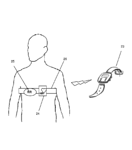
Fig 15 schematically illustrates an implementation of the CVRI for self
assessment during sporting, according to an embodiment of the present
invention. In this embodiment, an extended pulse rate sport device 23
which enables input (e.g manually) of height and weight and the initial
blood pressure, in which the respiration rate RR is given either through an
CA 02863501 2014-07-31
WO 2013/121414
PCT/IL2013/050090
existing ECG unit 24 through dedicated analysis or through a dedicated
respiratory sensor 25 (e.g. strain gage) embedded in the elastic band of the
existing chest strap 26.
Each of the above may include memory, output transmission to a control
center, external computer either directly or through a network, archive or
printer. Flash memory enables manual transmission and direct viewing
through a self operating viewer.
The vital sign monitor will be in communication with an individual where
in communication includes having the monitor affixed, attached,
implanted, coupled, abutting the individual's tissue, resident in clothing or
equipment worn by the individual, and proximate to the individual.
The analysis unit 12 is in communication with the vital sign source 11
through a wired connection or wireless connection such as infrared, radio,
Bluetooth, Wi-Fi, etc. where the connection can be continual, intermittent
(or on a predetermined schedule), as needed or as permitted by the
circumstances. The analysis unit 12 may be a separate component not
present on the individual on whom the vital sign source 11 is present or in
communication with, for example, to allow remote monitoring of the
individual or monitoring during a medical event such as triage, transport,
or treatment. In this implementation, the vital sign source 11 is connected
to a transmitter (and/or receiver) 13 that allows vital sign data to be
communicated to the analysis unit 12 as illustrated in the figure.
Alternatively, the analysis unit 12 may be located on (or proximate to) the
individual whom the vital sign source 11 is in communication, and in this
implementation an exemplary system for the analysis unit 12 to be
configured as part of a given monitoring system that is capable of
communicating with a remote user. If the analysis unit 12 is located on
CA 02863501 2014-07-31
WO 2013/121414
PCT/1L2013/050090
31
the individual, then in at least one exemplary embodiment the analysis
unit 12 is connected to a corresponding transmitter (and/or receiver).
The analysis unit 12 processes received vital sign data from the vital sign
source 11 and enable anthropometric data entry either directly or through
an intermediate component. It may or may not enable identification of the
patient and the settings. Depending upon the implementation, the set of
vital sign data includes heart rate, respiratory rate and blood pressure to
be able to determine the CVRI.
The term blood pressure refers to any measuring method of blood pressure
that enable output of MABP, either invasive which compute MABP
directly or non invasive which estimate MABP through SBP and DBP.
The analysis unit 12 can be implemented as software on a variety of
hardware computing devices including computers and PDAs. The software
includes the ability to process the received vital signs signals to provide as
an output the desired indicators relating to cardiovascular status and to
adjust cut-point level for certain predictive test aims. The software when
used to implement the method of the present invention, may include
notification/alarm unit to provide notification to the operator/user with an
audio notification, a mechanical notification such as vibration, a visual
notification including activation of a light(s) or via a display, either as
signal, number or text (indicate the exact status such as normal, heart
failure (which may or may not indicates its severity) and shock. signal to
another entity or device, or any combination of these if predetermined
conditions occur or predetermined thresholds are exceed by a vital sign or
the indicator. The analysis unit 12 in at least one exemplary embodiment
is connected to a storage unit (e.g., a buffer, RAM and disk storage, etc.)
for storing data associated with its operation. It may be also transmitted
through wired or wireless communication to remote location or from
CA 02863501 2014-07-31
WO 2013/121414
PCT/IL2013/050090
32
remote location either for telemedicine, central monitoring control or
remote archiving.
The invention can take the form of an entirely hardware embodiment, an
entirely software embodiment or an embodiment containing both
hardware and software elements. In at least one exemplary embodiment,
the invention is implemented in software, which includes but is not
limited to firmware, resident software, microcode, etc. Furthermore, the
invention can take the form of a computer program product accessible
from a computer-usable or computer-readable medium providing program
code for use by or in connection with a computer or any instruction
execution system. For the purposes of this description, a computer-usable
or computer readable medium can be any apparatus that can contain,
store, communicate, propagate, or transport the program for use by or in
connection with the instruction execution system, apparatus, or device.
The present invention provides a comprehensive alarm system which
carries physiological insight and hence it meets the need of intensive care
units regarding the alarm: "to be accurate" and that "it carries
intelligence" as such need is well described in the article "Alarms in the
intensive care unit: too much of a good thing is dangerous: is it time to add
some intelligence to alarms?" by Blum JM et. al., Crit Care Med. 2010 Feb;
38(2):451-6. According to the embodiments described hereinabove the
system of the present invention provides a quantified cardiovascular
performance reserve measure and methods of how to measure it.
Moreover, the system of the present invention carries capability of
diagnosis prediction (such as normal, heart failure and its severity and
shock). Cardiovascular performance quantification and diagnostic
prediction are unique and no other method had ever succeeded with.
The terms, "for example", "e.g.", "optionally", as used herein, are intended
to be used to introduce non-limiting examples. While certain references
CA 02863501 2014-07-31
WO 2013/121414
PCT/IL2013/050090
33
are made to certain example system components or services, other
components and services can be used as well and/or the example
components can be combined into fewer components and/or divided into
further components. Moreover, the appearance and terminology as
depicted and described herein, are intended to be illustrative and
exemplary, and in no way limit the scope of the invention as claimed.
While some embodiments of the invention have been described by way of
illustration, it will be apparent that the invention can be carried into
practice with many modifications, variations and adaptations, and with
the use of numerous equivalents or alternative solutions that are within
the scope of persons skilled in the art, without departing from the spirit of
the invention or exceeding the scope of the claims.