Note: Descriptions are shown in the official language in which they were submitted.
WO 2013/117746 PCT/EP2013/052619
SIGNATURES AND DETERMINANTS FOR DIAGNOSING INFECTIONS
AND METHODS OF USE THEREOF
RELATED APPLICATIONS
10001] This application claims the benefit of U.S.S.N. 61/596,950, filed
February 9, 2012, and
U.S.S.N. 61/652,631, filed May 29,2012.
FIELD OF THE INVENTION
100021 The present invention, in some embodiments thereof, relates
generally to the
identification of biological signatures and determinants associated with
bacterial and viral
infections and methods of using such biological signatures in the screening
diagnosis, therapy,
and monitoring of infection.
BACKGROUND OF THE INVENTION
10003] Antibiotics (Abx) are the world's most prescribed class of drugs
with a 25-30 billion SUS
global market. Abx are also the world's most misused drug with a significant
fraction of all drugs (40-
70%) being wrongly prescribed (Linder, J.A. and R.S. Stafford 2001; Scott, J.
G. and D. Cohen, et al.
2001; Davey, P. and E. Brown, et at 2006; Cadieux, G. and R. Tamblyn, et al.
2007; Pulcini, C. and E.
Cua, et al. 2007)'("CDC - Get Smart: Fast Facts About Antibiotic Resistance"
2011).
[0004] One type of Abx misuse is when the drug is administered in case of a
non-bacterial disease,
such as a viral infection, for which Abx is ineffective. For example,
according to the USA center for
disease control and prevention CDC, over 60 Million wrong Abx prescriptions
are given annually to treat
flu in the US. The health-care and economic consequences of the Abx over-
prescription include: (i) the
cost of antibiotics that are unnecessarily prescribed globally, estimated at
>$10 billion annually; (ii) side
effects resulting from unnecessary Abx treatment are reducing quality of
healthcare, causing
complications and prolonged hospitalization (e.g. allergic reactions, Abx
associated diarrhea, intestinal
yeast etc.) and (iii) the emergence of resistant strains of bacteria as a
result of the overuse (the CDC has
declared the rise in antibiotic resistance of bacteria as "one of the world's
most pressing health problems
in the 21 century" (Arias, C.A. and B.E. Murray 2009; "CDC - About
Antimicrobial Resistance" 2011)).
1
CA 2863819 2018-02-14
CA 02863819 2014-08-06
WO 2013/117746 PCT/EP2013/052619
[0005] Antibiotics under-prescription is not uncommon either. For example
up to 15% of adult
bacterial pneumonia hospitalized patients in the US receive delayed or no Abx
treatment, even though in
these instances early treatment can save lives and reduce complications(Houck,
P.M. and D. W. Bratzler,
et al 2002),
[0006] Technologies for infectious disease diagnostics have the potential
to reduce the associated
health and financial burden associated with Abx misuse. Ideally, such a
technology should: (i) accurately
differentiate between a bacterial and viral infections; (ii) be rapid (within
minutes); (iii) be able to
differentiate between pathogenic and non-pathogenic bacteria that are part of
the body's natural flora; (iv)
differentiate between mixed co-infections and pure viral infections and (v) be
applicable in cases where
the pathogen is inaccessible (e.g. sinusisits, pneumonia, otitis-media,
bronchitis, etc).
[0007] Current solutions (such as culture, PCR and immunoassays) do not
fulfill all these
requirements: (i) Some of the assays yield poor diagnostic accuracy (e.g. low
sensitivity or
specificity)(Uyeki et al. 2009), and are restricted to a limited set of
bacterial or viral strains; (ii) they
often require hours to days; (iii) they do not distinguish between pathogenic
and non-pathogenic bacteria
(Del Mar, C 1992), thus leading to false positives; (iv) they often fail to
distinguish between a mixed and
a pure viral infections and (v) they require direct sampling of the infection
site in which traces of the
disease causing agent are searched for, thus prohibiting the diagnosis in
cases where the pathogen resides
in an inaccessible tissue, which is often the case.
[0008] Consequentially, there still a diagnostic gap, which in turn often
leads physicians to
either over-prescribe Abx (the "Just-in-case-approach"), or under-prescribe
Abx (the "Wait-and-
see-approach") (Little, P.S. and I. Williamson 1994; Little, P. 2005; Spiro,
D. M. and K. Y. Tay,
et al 2006), both of which have far reaching health and financial
consequences.
[0009] Accordingly, a need exists for a rapid method that accurately
differentiates between
bacterial, viral, mixed and non-infectious disease patients that addresses
these challenges.
SUMMARY OF THE INVENTION
[00010] The present invention, in some embodiments thereof, is based on the
identification of
signatures and determinants associated with bacterial, viral and mixed (i.e.,
bacterial and viral
co-infections) infections, patients with a non-infectious disease and healthy
subjects. The
methods of the invention allow for the identification of type of infection a
subject is suffering
from, which in turn allows for the selection of an appropriate treatment
regimen. Various
CA 02863819 2014-08-06
WO 2013/117746 PCT/EP2013/052619
embodiments of the invention address limitations of current diagnostic
solutions by: (i) allowing
accurate diagnostics on a broad range of pathogens; (ii) enabling rapid
diagnosis (within
minutes); (iii) insensitivity to the presence of non-pathogenic bacteria and
viruses (thus reducing
the problem of false-positive); (iv) providing means for distinguishing
between mixed from pure
viral infections, and (v) eliminating the need for direct sampling of the
pathogen, thus enabling
diagnosis of inaccessible infections. Thus, some methods of the invention
allow for the selection
of subjects for whom antibiotic treatment is desired and prevent unnecessary
antibiotic treatment
of subjects having only a viral infection or a non-infectious disease. Some
methods of the
invention also allow for the selection of subjects for whom anti-viral
treatment is advantageous.
To develop and validate various aspects of the invention, the inventors
conducted a large
prospective multi-center clinical trial enrolling 655 hospital patients with
different types of
infections as well as controls (patients with a non-infectious disease and
healthy individuals).
The inventors then performed meticulous molecular and biochemical
experimentation and
measured the levels of over 570 polypeptides and other physiological
determinants in these
patients using quantitative assays. They found that most determinants were not
indicative of the
underlying infection type (e.g. bacterial, viral mixed and non-infectious
disease). Moreover, even
determinants with a well-established immunological role in the host response
to infection failed
to robustly distinguish between patients with different underlying infection
types. Diverging
from this norm were a few unique determinants, which the inventors were able
to identify, that
were able to differentiate between various types of infections.
[00011] In various aspects the invention provides methods of ruling out a
bacterial infection in
a subject by measuring the polypeptide concentration of TRAIL in a subject
derived sample; and
ruling out a bacterial infection for the subject if the polypeptide
concentration of TRAIL
determined is higher than a pre-determined first threshold value. Optionally,
the method further
includes ruling in a viral infection in the subject if the polypeptide
concentration of TRAIL is
higher than a pre-determined second threshold value.
[00012] In another aspect the invention provides a method of ruling out a
viral infection in a
subject measuring the polypeptide concentration of TRAIL in a subject derived
sample; and
ruling out a viral infection for the subject if the polypeptide concentration
of TRAIL determined
is lower than a pre-determined first threshold value. Optionally, the method
further includes
3
CA 02863819 2014-08-06
WO 2013/117746 PCT/EP2013/052619
ruling in a bacterial infection in the subject if the polypeptide
concentration of TRAIL
determined in step (a) is lower than a pre-determined second threshold value.
[00013] In a further aspect the invention provides a method of ruling in a
bacterial infection in
a subject by measuring the polypeptide concentration of TRAIL in a subject
derived sample
ruling in a bacterial infection for the subject if the polypeptide
concentration of TRAIL is lower
than a pre-determined first threshold value.
[00014] In another aspects the invention provides a method of ruling in a
viral infection in a
subject by measuring the polypeptide concentration of TRAIL in a subject
derived sample; and
ruling in a viral infection for the subject if the polypeptide concentration
of TRAIL is higher than
a pre-determined first threshold value.
[00015] In various aspects the invention includes a method of
distinguishing between a
bacterial infection and a viral infection in a subject by measuring the
polypeptide concentration
of TRAIL and CRP in a subject derived sample, applying a pre-determined
mathematical
function on the concentrations of TRAIL and CRP to compute a score and
comparing the score
to a predetermined reference value.
[00016] In another aspect, the invention provides a method of
distinguishing between a
bacterial or mixed infection, and a viral infection in a subject by measuring
the polypeptide
concentration of TRAIL and CRP in a subject derived sample, applying a pre-
determined
mathematical function on the concentrations of TRAIL and CRP to compute a
score and
comparing the score to a predetermined reference value.
[00017] In various embodiments any of the above described methods further
includes
measuring the polypeptide concentration of one or more polypeptide selected
from the group
consisting of SAA, PCT, B2M Mac-2BP, IL1RA and IP10, applying a pre-determined
mathematical function on the concentrations of the polypeptide concentration
measure to
compute a score, comparing the score to a predetermined reference value.
Specifically in some
embodiments TRAIL, CRP and SAA are measured; TRAIL, CRP and IP10 are measured;
[00018] TRAIL, CRP and PCT are measured; TRAIL, CRP and IL1RA are measured;
TRAIL, CRP and B2M are measured; TRAIL, CRP and Mac-2BP are measured; TRAIL,
CRP,
SAA and PCT are measured; TRAIL, CRP, Mac-2BP and SAA are measured; TRAIL,
CRP,
SAA and IP10 are measured; TRAIL, CRP, SAA and ILI RA are measured; TRAIL,
CRP, SAA,
4
CA 02863819 2014-08-06
WO 2013/117746 PCT/EP2013/052619
PCT and IPIO are measured; TRAIL, CRP, SAA, PCT and ILI RA are measured; or
TRAIL,
CRP, SAA, IPIO and IL IRA are measured.
[00019] In a further aspect the invention includes method of providing a
treatment
recommendation i.e., selecting a treatment regimen for a subject by measuring
the polypeptide
concentration of TRAIL in a subject derived sample; and recommending that the
subject receives
an antibiotic treatment if polypeptide concentration of TRAIL is lower than a
pre-determined
threshold value; recommending that the patient does not receive an antibiotic
treatment if the
polypeptide concentration of TRAIL is higher than a pre-determined threshold
value; or
recommending that the patient receive an anti-viral treatment if the
polypeptide concentration of
TRAIL determined in step (a) is higher than a pre-determined threshold value.
[00020] In another aspect the invention includes a method of providing a
treatment
recommendation for a subject by identifying the type infection (i.e.,
bacterial, viral, mixed
infection or no infection) in the subject according to the method of any of
the disclosed methods
and recommending that the subject receive an antibiotic treatment if the
subject is identified as
having bacterial infcction or a mixed infection; or an anti- viral treatment
is if the subject is
identified as having a viral infection.
[00021] In yet another aspect the invention provides a method of providing
a diagnostic test
recommendation for a subject by measuring the polypeptide concentration of
TRAIL in a subject
derived sample; and recommending testing the sample for a bacteria if the
polypeptide
concentration of TRAIL is lower than a pre-determined threshold value; or
recommending
testing the sample for a virus if the polypeptide concentration of TRAIL is
higher than a pre-
determined threshold value.
[00022] In a further aspect the invention includes method of providing a
diagnostic test
recommendation for a subject by identifying the infection type (i.e.,
bacterial, viral, mixed
infection or no infection) in the subject according to any of the disclosed
methods and
[00023] Recommending a test to determine the source of the bacterial
infection if the subject
is identified as having a bacterial infection or a mixed infection; or a test
to determine the source
of the viral infection if the subject is identified as having a viral
infection.
[00024] In various aspects any of the above methods further includes
measuring one or more
of the following DETERMINANTS IL1RA, 1P10, Mac-2BP, B2M, BCA-1, CHI3L1,
Eotaxin,
CA 02863819 2014-08-06
WO 2013/117746 PCT/EP2013/052619
ILI a, MCP, CD62L, VEGFR2, CHP, CMPK2, CORO1C, EIF2AK2, ISG15, RPL22L1, RTN3,
CD112, CD134, CD182, CD231, CD235A, CD335, CD337, CD45, CD49D, CD66A/C/D/E,
CD73, CD84, EGFR, GPR162, HLA-A/B/C, ITGAM, NRG1, RAP1B, SELL SPINT2, SSEA1,
IgG non-specific bound molecules, IL1 , I-TAC; IFITM3, IFIT3, EIF4B, IFIT I,
L0C26010,
MBOAT2, MX1, OAS2, RSAD2, ADIPOR1, CD15, CD8A, IFITM1, IL7;
[00025] CRP, SAA, TREM-1, PCT, IL-8, TREM-1 and IL6; Age, absolute
neutrophil count
(ANC), absolute lymphocyte count (ALC), neutrophil % (Neu(%)), lymphocyte %
(Lym (%)),
monocyte % (Mono (%)), Maximal temperature, Time from symptoms, Creatinine
(Cr),
Potassium (K), Pulse and Urea.
[00026] In another aspect the invention provide a method of distinguishing
between a subject
having an infectious disease and one having a non-infectious disease. For
example, in one
embodiment the an infectious disease is ruled out in a subject measuring the
polypeptide
concentration of one or more polypeptides including TRAIL, IP10, IL1Ra or Mac-
2BP in a
subject derived sample;, applying a pre-determined mathematical function on
the concentrations
of the polypeptidcs measured to compute a score, comparing the score to a
predetermined
reference value. Optionally, the polypeptide concentration of one or more
polypeptides
including SAA, CRP, IL6, IL8, and PCT, TREM-1 are measured
1000271 In various aspects the method distinguishes a virally infected
subject from either a
subject with non-infectious disease or a healthy subject; a bacterially
infected subject, from
either a subject with non-infectious disease or a healthy subject; a subject
with an infectious
disease from either a subject with an non-infectious disease or a healthy
subject; a bacterially
infected subject from a virally infected subject; a mixed infected subject
from a virally infected
subject; a mixed infected subject from a bacterially infected subject and a
bacterially or mixed
infected and subject from a virally infected subject.
[00028] These methods include measuring the levels of a first DETERMINANT
including
TRAIL, IL1RA, IP10, Mac-2BP, B2M, BCA-1, CHI3L1, Eotaxin, ILla, MCP, CD62L,
VEGFR2, CHP, CMPK2, CORO1C, ElF2AK2, ISG15, RPL22L1, RTN3, CD112, CD134,
CD182, CD231, CD235A, CD335, CD337, CD45, CD49D, CD66A/C/D/E, CD73, CD84,
EGFR, GPR162, HLA-A/B/C, 1TGAM, NRG1, RAP1B, SEL1, SP1NT2, SSEA1, IgG non-
specific bound molecules, IL1 , 1-TAC and TNFR1 in a sample from the subject
and
6
CA 02863819 2014-08-06
WO 2013/117746 PCT/EP2013/052619
measuring the levels of a second DETERMINANT including TRAIL, IL1RA, IP10, Mac-
2BP,
B2M, BCA-1, CHI3L1, Eotaxin, ILla, MCP, CD62L, VEGFR2, CHP, CMPK2, CORO1C,
EIF2AK2, ISG15, RPL22L1, RTN3, CD112, CD134, CD182, CD231, CD235A, CD335,
CD337, CD45, CD49D, CD66A/C/D/E, CD73, CD84, EGFR, GPR162, HLA-A/B/C, ITGAM,
NRG1, RAP1B, SELI, SPINT2, SSEA1, IgG non-specific bound molecules, ILL I-TAC
TNER1; IFITM3, IFIT3, ElF4B, IFIT1, L0C26010, MBOAT2, MX1, OAS2, RSAD2,
ADIPORI, CD15, CD8A, IFITM1, IL7; CRP, SAA, TREM-1, PCT, IL-8, TREM-1 and IL6;
Age, absolute neutrophil count (ANC), absolute lymphocyte count (ALC),
neutrophil %
(Neu(%)), lymphocyte % (Lym (%)), monocyte (Mono (%)), Maximal temperature,
Time
from symptoms, Creatinine (Cr), Potassium (K), Pulse and Urea and comparing
the levels of the
first and second DETERMINANTS to a reference value thereby identifying the
type of infection
in the subject wherein the measurement of the second DETERMINANT increases the
accuracy
of the identification of the type of infection over the measurement of the
first DETERMINANT.
Optionally, further includes measuring the level of a one or more additional
DETERMINANTS
including: TRAIL, MIRA, IP10, Mac-2BP, B2M, BCA-1, CHI3L1, Eotaxin, ILla, MCPõ
CD62L, VEGFR2, CHP, CMPK2, CORO1C, EIF2AK2, ISG15, RPL22L1, RTN3, CD! 12,
CD134, CD182, CD231, CD235A, CD335, CD337, CD45, CD49D, CD66A/C/D/E, CD73,
CD84, EGFR, GPR162, HLA-A/B/C, ITGAM, NRG1, RAP 1B, SELI, SPINT2, SSEA1, IgG
non-specific bound molecules, ILL I-TAC TNER1; IFITM3, IFIT3, EIF4B, IFIT1,
L0C26010,
MBOAT2, MX1, OAS2, RSAD2, ADIPOR1, CD15, CD8A, IFITM1, IL7; CRP, SAA, TREM-
1, PCT, IL-8, TREM-1 and IL6; Age , absolute neutrophil count (ANC), absolute
lymphocyte
count (ALC), neutrophil % (Neu(%)), lymphocyte % (Lym (%)), monocyte % (Mono
(%)),
Maximal temperature, Time from symptoms, Creatinine (Cr), Potassium (K), Pulse
and Urea;
wherein the measurement of the additional DETERMINANTS increases the accuracy
of the
identification of the type of infection over the measurement of the first and
second
DETERMINANTS. In one aspect the method distinguishes a bacterially infected
subject from a
virally infected subject by measuring one or more DETERMINANTS selected from
B2M, BCA-
1, CHI3L1, Eotaxin, IL1RA, IP10, MCP, Mac-2BP, TRAIL, CD62L and VEGFR2 are
measured
and one or more DETERMINANTS selected from the group consisting of CRP, TREM-
1, SAA,
PCT, IL-8, IL6, ANC, ALC, Neu (%), Lym (%), Mono (%), Maximal temperature,
Time from
7
CA 02863819 2014-08-06
WO 2013/117746 PCT/EP2013/052619
symptoms, Age, Creatinine (Cr), Potassium (K), Pulse and Urea. For example,
CRP and TRAIL
are measured; CRP and TRAIL and SAA are measured; CRP and TRAIL and Mac-2BP
are
measured; CRP and TRAIL and PCT and are measured; CRP and TRAIL and SAA and
Mac-
2BP are measured; PCT and TRAIL are measured; or SAA and TRAIL are measured.In
a
another aspect the method distinguishes between a mixed infected subject and a
virally infected
subject by measuring wherein one or more DETERMINANTS selected from TRAIL,
IP10,
IL1RA, CHI3L1, CMPK2 and MCP-2 are measured and optionally one or more
DETERMINANTS selected from the group consisting of CRP, SAA, ANC, ATP6V0B,
CES1,
CORO1A, HERC5, IFITML LIPT1, L0C26010, LRDD, Lym (%), MCP-2, MX1, Neu (%),
OAS2, PARP9, RSAD2, SART3, WBC, PCT, IL-8, IL6 and TREM-1..
In another aspect the method distinguishes between a bacterial or mixed
infected subject
and a virally infected subject by measuring wherein one or more DETERMINANTS
selected
from TRAIL, ILI RA, IP10, ARG1, CD337, CD73, CD84, CHI3L1, CHP, CMPK2, COROIC,
E1F2AK2, Eotaxin, GPR162, HLA-A/B/C, ISG15, ITGAM, Mac-2BP, NRG1, RAP1B,
RPL22L1, SSEA1, RSAD2, RTN3, SELIõ VEGFR2, CD62L and VEGFR2 arc measured and
optionally one or more DETERMINANTS selected from the group consisting of CRP,
SAA,
PCT, IL6, IL8, ADIPORI. ANC, Age, B2M, Bili total, CD15, Cr, EIF4B, IFIT1,
IFIT3, IFITMI,
IL7R, K (potassium), KIAA0082, L0C26010, Lym (%), MBOAT2, MCP-2, MXI, Na, Neu
(%), OAS2, PARP9, PTEN, Pulse, Urea, WBC, ZBP1, mIgG1 and TREM-1.
In another aspect the method distinguishes between a subject with an
infectious disease and
a subject with a non-infectious disease or a healthy subject by measuring one
or more
DETERMINANTS selected from IP10, IL1RA, TRAIL, BCA-1, CCL19-MIP3b, CES1 and
CMPK2. Optionally, one or more DETERMINANTS selected from CRP, SAA, PCT, IL6,
IL8,
ARPC2, ATP6V0B, Cr, Eos (%), HERC5, IFI6, IFIT3, KIAA0082, LIPT1, LOC26010,
LRDD,
MBOAT2, MX1, Maximal temperature, OAS2, PARP9, Pulse, QARS, RAB13, RPL34,
RSAD2, SART3, RIM22, UBE2N, )(AFL IL11, I-TAC and TNFR1 are measured.
Any of the above described methods can be used to further select a treatment
regimen for
the subject. For example, if a subject identified as having a viral infection
the subject is selected
to receive an anti-viral treatment regimen. When a subject is identified as
having a non-viral
disease the subject is selected not to receive an anti-viral treatment
regimen. When a subject is
8
CA 02863819 2014-08-06
WO 2013/117746 PCT/EP2013/052619
identified as having a bacterial or a mixed infection the subject is selected
to receive an antibiotic
treatment regimen. When a subject identified as having a viral infection, a
non-infectious
disease or healthy the subject is not selected to receive an antibiotic
treatment regimen.
[00029] In a further aspect the invention provides for monitoring the
effectiveness of treatment for an
infection by detecting the level of one or more polypeptide-DETERMINANTS
selected from the group
consisting of TRAIL, IL1RA, IP10, B2M, Mac-2BP, BCA-1, CHI3L1, Eotaxin, MCP,
Mac-2BP, TRAIL,
CD62L, VEGFR2, CHP, CMPK2, CORO1C, EIF2AK2, ISG15, RPL22L1, RTN3, CD112,
CD134,
CD182, CD231, CD235A, CD335, CD337, CD45, CD49D, CD66A/C/D/E, CD73, CD84,
EGFR,
GPR162, IILA-A/B/C, ITGAM, NRG1, RAP1B, SELI, SPINT2, SSEA1, IL11, ILla, I-TAC
and TNER1
in a first sample from the subject at a first period of time; detecting the
level of one or more polypeptide-
DETERMINANTS selected from the group consisting of TRAIL, IL1RA, IP10, B2M,
Mac-2BP, BCA-1,
CHI3L1, EotaxinMCP, Mac-2BP, TRAIL, CD62L, VEGFR2, CHP, CMPK2, CORO1C,
EIF2AK2,
ISG15, RPL22L1, RTN3, CD112, CD134, CD182, CD231, CD235A, CD335, CD337, CD45,
CD49D,
CD66A/C/D/E, CD73, CD84, EGFR, GPR162, HLA-A/B/C, ITGAM, NRG1, RAP1B, SELI,
SPINT2,
SSEA1, 1111, ILla, I-TAC and TNFR1 in a second sample from the subject at a
second period of time;
and comparing the level of the one or more polypeptide detected in the first
sample to the level detected
the second sample, or to a reference value, The effectiveness of treatment is
monitored by a change in the
level of one or more polypeptides. Optionally, the method further includes
detecting one or more
polypeptide-DETERMINANTS selected from CRP, SAA, TREM-1, PCT, IL-8 and IL6 in
the first and
second samples.
The subject has previously been treated for the infection. Alternatively the
subject has not been
previously treated for the infection. In some aspects the first sample is
taken from the subject
prior to being treated for the infection and the second sample is taken from
the subject after
being treated for the infection. In some aspects, the second sample is taken
from the subject after
recurrence of the infection or prior to recurrence of the infection.
[00030] The sample is for example, whole blood or a fraction thereof A
blood fraction
sample contains cells that include lymphocytes, monocytes and granulocytes.
The expression
level of the polypeptide is determined by electrophoretieally, or
immunochemically. The
immunochemical detection is for example, by flow cytometry, radioimmunoassay,
immunofluorescence assay or by an enzyme-linked immunosorbent assay.
9
CA 02863819 2014-08-06
WO 2013/117746 PCT/EP2013/052619
1000311 A clinically significant alteration in the level of the one or more
polypeptides in
the sample indicates an infection in the subject. In some aspects the level of
the one or more
DETERMINANTS is compared to a reference value, such as an index value. In some
aspects
the reference value or index value are determined after performing age
dependent normalization
or stratification. In any of the above methods the DETERMINANTS are preferably
selected such
that their MCC is >= 0.4 or the AUC is >= 0.7. In other aspects DETERMINANTS
are
preferably selected such that their Wilcoxon rank sum p-values are less than
10-6 or less than 104
or less than 10-3.
[00032] In any of the above methods the concentration of TRAIL is measured
within about 24 hours
after sample is obtained or is measured in a sample that was stored at 12 C or
lower, wherein the storage
begins less than 24 hours after the sample is obtained.
[000331 The infection further includes an infection reference expression
profile, having a pattern of
levels of two or more polypeptides selected from the group consisting of
TRAIL, IL1RA, -11310, B2M,
BCA-1, CHI3L1, Eotaxin, MCP, Mac-2BP, CD62L, VEGFR2, CHP, CMPK2, CORO1C,
EIF2AK2,
ISG15, RPL22L1, RTN3, CD112, CD134, CD182, CD231, CD235A, CD335, CD337, CD45,
CD49D,
CD66A/C/D/E, CD73, CD84, EGFR, GPR162, HLA-A/B/C, ITGAM, NRG1, RAP1B, SELI,
SPINT2,
SSEA1 , IL11, IL la , I-TAC and TNFR1, and optionally further having a pattern
of levels of one or more
polypeptides selected from the group consisting of CRP, SAA, TREM-1, PCT, IL-8
and 1L6. Also
include in the invention is a machine readable media containing one or more
infection reference
expression profiles according to the invention
[00034] In another aspect the invention includes a kit having a plurality
of polypeptide detection
reagents that detect the corresponding polypeptides including TRAIL, 1L1 RA,
IP10, 112M, BCA-1,
CHI3L1, Eotaxin, ILla, MCP, Mac-2BP, CD62L, VEGFR2, CHP, CMPK2, CORO1C,
EIF2AK2,
1SG15, RPL22L1, RTN3, CD112, CD134, CD182, CD231, CD235A, CD335, CD337, CD45,
CD49D,
CD66A/C/D/E, CD73, CD84, EGFR, GPR162, HLA-A/B/C, ITGAM, NRG1, RAP1B, SELI,
SPINT2,
SSEA1, IL11, I-TAC and TNFR1, and optionally further plurality of polypeptide
detection reagents that
detect the corresponding polypeptide including CRP, SAA, TREM-1, PCT, IL-8 and
IL6. The detection
reagent is comprises one or more antibodies or fragments thereof.
[00035]
[00036] Unless otherwise defined, all technical and scientific terms used
herein have the same
meaning as commonly understood by one of ordinary skill in the art to which
this invention
pertains. Although methods and materials similar or equivalent to those
described herein can be
WO 2013/117746 PCT/EP2013/052619
used in the practice of the present invention, suitable methods and materials
are described below.
In cases of conflict, the present
specification, including definitions, will control. In addition, the
materials, methods, and
examples described herein arc illustrative only and arc not intended to be
limiting.
[00037] Other features and advantages of the invention will be apparent
from and
encompassed by the following detailed description and claims.
BRIEF DESCRIPTION OF THE DRAWINGS
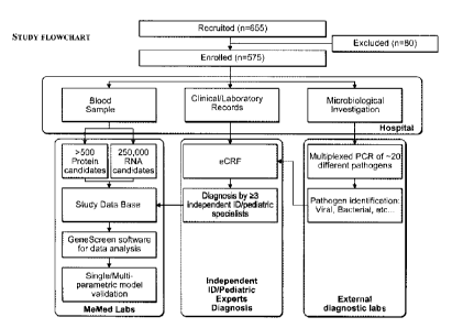
[00038] Figure 1: Clinical study workflow.
[00039] Figure 2: Characterization of the 575 patients enrolled in the
clinical study.
[00040] Figure 3: Summary of patient cohorts.
[00041] Figure 4: Age distribution of the entire study population (A)
(N=575) and pediatric
patients (B) (N=350).
[00042] Figure 5: Distribution of isolated pathogens by pathogenic
subgroups (A) and by
strains (B) (stains isolated from >1% of patients are presented).
[00043] Figure 6: Distribution of involved physiologic systems in
infectious disease patients.
(N= 484).
[01:044] Figure 7: Distribution of major clinical syndromes (A) and
specific clinical
syndromes (B) of the patients enrolled in the clinical study (all enrolled
patients, N = 575).
[00045] Figure 8: Distribution of maximal body temperatures (all enrolled
patients, N = 575).
[0(8)46] Figure 9: Distribution of time from initiation of symptoms (all
enrolled patients, N =
575).
W0471 Figure 10: Distribution of eomorbidities of the patient population
(A) and distribution
of chronic medications (B) of the patients enrolled in the clinical study (all
chronically ill
patients, N = 170).
[00048] Figure 11: Distribution of recruitment sites (all enrolled
patients, N =575).
[00049] Figure 12: Calibration curves for TRAIL (A), Mac-2BP (B) and SAA
(C).
[00050] Figure 13: Intra-assay variability for TRAIL (A), Mac-2-BP (B) and
SAA (C).
[00051] Figure 14: Inter-assay variability for TRAIL (A), Mac-2-BP (B) and
SAA (C).
11
CA 2863819 2018-02-14
1000521 Figure 15: Measurements of plasma vs. serum concentrations of
TRAIL (A), Mac-2-
BP (B) and SAA (C).
1000531 Figure 16: The analytes decay rates at 25 C for TRAIL (A), Mac-2-
BP (B) and SAA
(C).
1000541 Figure 17: Correlation of TRAIL levels measured using ELISA and
Luminex.
1000551 Figures I8A-H: Polypeptides with an immunological role do not
necessarily show a
differential response.
1000561 Figure 19: In-vitro differentially expressed polypeptides do not
necessarily show in-
vivo differential expression.
1000571 Figures 20A-T: Examples of DETERMINANTS that differentiate
between bacterial
versus viral infected subjects.
1000581 Figures 21A1-Al2, 21B1-B4 and 21C1-C3: Examples of DETERMINANTS that
differentiate between mixed versus viral infected subjects (A), infectious
versus non-infectious
subjects (B) and infectious versus healthy subjects (C).
1000591 Figure 22: Colonization of non-infectious and healthy subjects.
1000601 Figures 23A-B: Examples of scatter graphs showing the diagnosis
of bacterial ('+'
marks) versus viral ('O' marks) infected patients using a combination of two
statistically
significant DETERMINANTS. Patient classification was performed using a linear
SVM trained
on 90% of the data, where white and gray regions indicate the space of
DETERMINANT
combinations that were classified as viral and bacterial respectively. Each
plot corresponds to a
different combination of two DETERMINANTS.
1000611 Figure 24: Examples of scatter graphs showing the diagnosis of
Mixed ('+' marks)
versus viral ('O' marks) infected patients using a combination of two
statistically significant
DETERMINANTS.
1000621 Figure 25: The TCM-signature accuracy in diagnosing bacterial vs.
viral infections in
patients whose diagnosis was clear. The analysis was performed using the
'Clear (bacterial,
viral)' cohort; N = 170.
1000631 Figure 26: The TCM-signature accuracy in diagnosing bacterial vs.
viral infections in
patients whose diagnosis was determined by a consensus of experts. The
analysis was performed
using the 'Consensus (bacterial, viral)' cohort.
12
CA 2863819 2020-03-26
[00064] Figure 27: the TCM-signature accuracy in diagnosing bacterial vs.
viral patients in
patients whose diagnosis was determined by majority of an expert panel. The
analysis was
performed using the 'Majority (bacterial, viral)' cohort.
1000651 Figure 28: the TCM-signature accuracy in distinguishing mixed co-
infections from
pure viral infections in patients whose diagnosis was determined by majority
of an expert panel.
The analysis was performed using the 'Majority (viral, mixed)' cohort.
1000661 Figures 29A-B: The TCM-signature accuracy in diagnosing bacterial
vs. viral patients
in the 'Consensus (bacterial, viral)' cohort and the 'Majority (bacterial,
viral) cohort before and
after inclusion of patients who were initially excluded from the study.
[00067] Figure 30: Accuracy of the TCM-signature as a function of time from
symptom onset.
Error bars represent 95% CI.
1000681 Figure 31: Accuracy of the TCM-signature as a function of maximal
fever measured.
Error bars represent 95% CI.
[00069] Figure 32: DETERMINANT levels in different infections as a function
of Age.
1000701 Figure 33: Prevalence of select bacterial and viral strains in
patients with non-
infectious (A) and infectious diseases (B) in the 'Majority (bacterial, viral,
mixed, non-
infectious)' cohort.
1000711 Figure 34: TCM signature performance in patients with (+) and
without (-)
colonization by select bacterial and viral strains. Error bars represent 95%
CI.
[00072] Figure 35: Scatter plots (left panel), box plots (middle panel) and
the approximation
of the log normal distributions (right panel) of the levels of TRAIL in
bacterial and viral patients.
The analysis was performed using the 'Consensus (bacterial, viral)' cohort, N
= 434.
1000731 Figure 36: ROC curve for the analyte TRAIL. The analysis was
performed using the
'Consensus [bacterial, viral]' cohort, N = 343.
[00074] Figure 37: The balance between the number of patients diagnosed and
the accuracy of
the TRAIL assay.
1000751 Figures 38A1-A3 and 38B: Examples of DETERMINANTS whose mRNA levels
have been found to be differentially expressed in viral compared to bacterial
infections, but their
polypeptide levels in bacterial versus viral infected patients show no
significant differential
response. (A) The protein levels of IF144, IF144L and IF127 in bacterial
(diamonds) and viral
13
CA 2863819 2019-08-01
(squares) infections. (B) The mRNA expression levels of the IF144, IFI44L, and
1E127 genes in
bacterial (diamonds) and viral (squares) infections. Median value is indicated
with a solid line.
[00076] Figure 39: TCM-signature sensitivity and specificity increase as
the cutoffs used for
filtering out patients with marginal responses become more stringent. The
analysis was
performed using the 'Consensus (bacterial, viral) cohort. Every point
corresponds to the
sensitivity and specificity attained at the cutoff in which the two measures
were kept equal.
1000771 Figure 40: TCM-signature sensitivity and specificity increase as
the cutoffs used for
filtering out patients with marginal responses become more stringent. The
analysis was
performed using the 'Majority (bacterial, viral)' cohort. Every point
corresponds to the sensitivity
and specificity attained at the cutoff in which the two measures were kept
equal.
[00078] Figure 41: The levels of TRAIL increase during the acute phase of a
viral infection
and then gradually decrease to baseline levels (A, B). In patients with an
acute bacterial infection
its levels decrease and then increase back to baseline levels during
convalescence (C).
1000791 Figure 42: Comparison of the genetic sequence of TRAIL across
organisms.
DETAILED DESCRIPTION OF THE INVENTION
1000801 The present invention, in some embodiments thereof, relates to the
identification of
signatures and determinants associated with bacterial, viral and mixed (i.e.,
bacterial and viral co-
infections) infections. More specifically we discovered that certain
polypeptide-DETERMINANTS are
differentially expressed in a statistically significant manner in subjects
with bacteria, viral or mixed (i.e.,
bacterial and viral co-infections) as well as non-infectious disease and
healthy subjects. These
polypeptide-DETERMINANTS include TRAIL, 'LIRA, IP10, Mac-2BP, B2M, BCA-1,
CHI3L1,
Eotaxin, ILI a, MCP, CD62L, VEGFR2, CHP, CMPI(2, CORO1C, EIF2A1(2, ISG15,
RPL22L1,
RTN3, CD112, CD134, CD182, CD231, CD235A, CD335, CD337, CD45, CD49D,
CD66A/C/D/E, CD73, CD84, EGFR, GPR162, HLA-A/B/C, ITGAM, NRG1, RAPIB, SELI,
SP1NT2, SSEA1, IgG non-specific bound molecules, ILE I-TAC, TNER1, IFITM3,
IFIT3,
EIF4B, IFIT1, L0C26010, MBOAT2, MX1, OAS2, RSAD2, ADIPOR1, CD15, CD8A,
IFITM1, IL7, CRP, SAA, TREM-1, PCT, IL-8, TREM-1, IL6, ARG1, ARPC2, ATP6V0B,
BCA-1, BRI3BP, CCL19-MIP3b, CES1, CORO1A, HERC5, 1E16, IFIT3, KIAA0082, LIPT1,
14
CA 2863819 2019-08-01
CA 02863819 2014-08-06
WO 2013/117746 PCT/EP2013/052619
LRDD, MCP-2, PARP9, PTEN, QARS, RAB13, RPL34, SART3, TRIM22, UBE2N, XAF1 and
ZBP I .
[00081] In some embodiments the polypeptide-DETERMINANTS are soluble-
polypeptides
that include B2M, BCA-1, CHI3L1, Eotaxin, ILla, IP10, MCP, Mac-2BP, TRAIL.
CD62L,
VEGFR2, IL11, IL1RA, I-TAC and TNFR1.
[00082] In other embodiments the polypeptide-DETERMINANTS are intracellular-
polypeptides that include CHP, CMPK2, CORO1C, ElF2AK2, ISG15, RPL22L1 and
RTN3.
[00083] In other embodiments the polypeptide-DETERMINANTS are membrane
polypeptides that include CD112, CD134, CD182, CD231, CD235A, CD335, CD337,
CD45,
CD49D, CD66A/C/D/E, CD73, CD84, EGFR, GPR162, HLA-A/B/C, ITGAM, NRG1, RAP1B,
SELI, SPINT2 and SSEAL
[00084] In other embodiments the polypeptide-DETERMINANTS further include
polypeptides selected from the group consisting of EIF4B, IFIT1, IFIT3,
L0C26010, MBOAT2,
MX1, OAS2, RSAD2, ADIPOR1, CD15, CD8A, IFITM1, IFITM3, IL7R, CRP, SAA, sTREM,
PCT, IL-8 and IL6.
[00085] In other embodiments the DETERMINANTS further include clinical-
DETERMINANTS selected from the group consisting of: ANC, ALC, Neu (%), Lym
(%), Mono
(%), Maximal temperature, Time from symptoms, Age, Creatinine (Cr), Potassium
(K), Pulse
and Urea.
[00086] In some embodiments, the DETERMINANTS further comprise measurements of
one
or more polypeptides or clinical-DETERMINANTS selected from the group
consisting of:
ARG1, ARPC2, ATP6V0Bõ BILI (BILIRUBIN), BRI3BP, CCL19-MIP3B, CES1, CORO1A,
E0S(%), HERC5, IF16, IFIT3, KIAA0082, LIPT1, LRDD, MCP-2, NA (Sodium), PARP9,
PTEN, QARS, RAB13, RPL34, SART3, TRIM22, UBE2N, WBC (Whole Blood Count),
XAF1 and ZBP1 .
[00087] Different infectious agents have unique molecular patterns that can
be identified and
targeted by the immune system. Pathogen-associated molecular patterns (PAMPs)
are an
example of such molecules that are associated with different groups of
pathogens and may be
recognized by cells of the innate immune system using Toll-like receptors
(TLRs) and other
CA 02863819 2014-08-06
WO 2013/117746 PCT/EP2013/052619
pattern recognition receptors (e.g. NOD proteins) (Akira, S. and S. Uematsu,
et at 2006; Murphy,
K. and P. Travers, et al 2007). These patterns may vary considerably between
different classes of
pathogens and thus elicit different immune responses. For example, TLR-4 can
recognize
lipopolysaccharide, a constituent of gram negative bacteria, as well as
lipoteichoic acids,
constituent of gram positive bacteria, hence promoting an anti-microbial
response of the immune
system (Akira, S. and S. Uematsu, et al 2006; Murphy, K. and P. Travers, et al
2007). TLR-3 can
recognize single stranded RNA (often indicative of a viral infection) and thus
prompt the
appropriate anti-viral response(Akira, S. and S. Uematsu, et al 2006; Murphy,
K. and P. Travers,
et at 2007). By distinguishing between different classes of pathogens (e.g
bacterial versus viral)
the immune system can mount the appropriate defense.
[00088] In the past few decades, several host markers have been identified
that can be used for
differential diagnosis of infection source in various indications. One example
is Procalcitonin
(PCT), a precursor of the hormone calcitonin produced by the C-cells of the
thyroid gland. PCT
levels in the blood stream of healthy individuals is hardly detectable (in the
pg/ml range) but it
might increase dramatically, as a result of a severe infection with levels
rising up to 100 ng/ml.
PCT is heavily used to diagnose patients with systemic infection, sepsis, with
sensitivity of 76%
and specificity of 70%(Jones, A. E. and J.F. Fiechtl, et al 2007). However,
studies that tested the
diagnostic value of PCT in other non-systemic infection such as pneumonia or
upper respiratory
tract infections found it to be limited(Brunkhorst, F. M. and B. Al-Nawas, et
al 2002; Tang M. P.
and Eslick GD 2007), especially when used in isolation.
[00089] Another widely used marker is the acute phase protein, C-reactive
protein (CRP).
CRP levels in the blood often rise in response to inflammation. Therefore,
when used as an
adjunct biomarker in the right clinical context, CRP may prove useful for
improving detection
accuracy of infections (Povoa P. 2002). However, in some indications such as
sepsis its
specificity and sensitivity were found to be considerably lower than PCT
(Hatherill, M. and S.
M. Tibby, et al 1999). Additionally, its clinical utility as a stand-alone
marker for Abx
prescription decision making has been criticized (Brian Clyne and Jonathan S
Olshaker 1999).
One reason for CRP's limited accuracy in the context of infectious disease
stems from the fact
that CRP may rise in indications other than bacterial infection. For example
some viral infections
including adenoviruscs (Appenzeller C et at. 2002; A. Putto, 0. Mcurman, and
0. Ruuskancn
16
CA 02863819 2014-08-06
WO 2013/117746 PCT/EP2013/052619
1986) are known to cause a significant increase in the levels of CRP that
mimics a bacterial
response, thus limiting CRP's accuracy as a single marker for differentiating
between viral and
bacterial infections. CRP may also rise in non-infectious disease such as
trauma. Other proposed
markers for detection of different sources of infection and sepsis include
CD64 (Rudensky, B.
and G. Sirota, et al 2008), and HNL (riaertoft, G. and T. Foucard, et al.
2005). The reliability
and evidence supporting the usage of these markers for the purpose of
diagnostics of viral versus
bacterial infections in a broad setting are limited.
[00090] The present invention, in some embodiments thereof, seeks to
overcome the above
mentioned diagnostic challenges by: (i) enabling accurate differentiation
between a broad range
of bacterial versus viral infections; (ii) enabling rapid diagnostics (within
minutes); (iii) avoiding
the "false positive" identification of non-pathogenic bacteria that are part
of the body's natural
flora, (iv) allowing accurate differentiation between mixed and pure viral
infections and (v)
allowing diagnosis in cases where the pathogen is inaccessible.
[00091] To this end the inventors sought to identify and test a novel set
of biomarkers whose
levels arc differentially expressed in viral, bacterial and mixed infected
patients, and in patients
with a non-infectious disease and to use the combined measurements of these
biomarkers
coupled with pattern recognition algorithms to accurately identify the source
of infection with
the aim of assisting physicians to accurately prescribe the correct treatment.
[00092] To facilitate a solution that is generally applicable, the
inventors performed a large
clinical trial in which they enrolled a heterogeneous cohort of 655 patients
including different
ages, medical backgrounds, ethnicities, pathogen types, clinical syndromes and
time from
appearance of symptoms, fever, co-morbidities (see Figures 4-10). The
inventors then measured
the levels of over 570 different polypeptides using quantitative assays, and
were able to screen a
small subset of polypeptides that was robustly differentially expressed in
different types of
infections. They used the combined signature of these selected polypeptides to
develop and test
various aspects of the present solution.
[00093] To address the challenge of rapid diagnosis, some aspects of the
invention focus on
biomarkers that can be rapidly measured, such as proteins, rather than
biomarkers whose
measurement may require hours to days, such as nucleic-acid based biomarkers.
Note that high-
throughput quantitative measurements of nucleic-acids for the purpose of
biomarker discovery
17
CA 02863819 2014-08-06
WO 2013/117746 PCT/EP2013/052619
have become feasible in recent years using technologies such as microarrays
and deep
sequencing. However, performing such quantitative high-throughput measurements
on the
proteome level remains a challenge. Thus, some aspects of the present
invention focus on the
proteome level.
[00094] To address the clinical challenge of mixed infection diagnosis and
treatment, some
aspects of the present invention include a method for differentiating between
mixed infections
(which require Abx treatment despite the presence of a virus) and pure viral
infections (which do
not require Abx treatment).
[00095] Some aspects of the present invention also address the challenge of
"false-positive"
diagnostics due to non-pathogenic strains of bacteria that are part of the
body's natural flora. This
is achieved by measuring biomarkers derived from the host rather than the
pathogen.
[00096] Another aspect of the present invention enables the diagnosis of
different infections,
which is invariant to the presence or absence of colonizers (e.g. bacteria and
viruses that are part
of the natural flora). This addresses one of the major challenges in
infectious disease diagnostics
today: "false-positives" due to colonizers.
[00097] Importantly, some aspects of the current invention do not require
direct access to the
pathogen, because the immune system circulates in the entire body, thereby
facilitating diagnosis
in cases in which the pathogen is inaccessible.
[00098] Another aspect of the present invention is the fraction in which
the biomarkers are
measured, which affects the ease by which the assay can be performed in the
clinical settings,
and especially the point-of-care. For example, it is easier to measure
proteins in the serum or
plasma fraction compared to nucleic acids or intra-cellular proteins in the
leukocytes fraction
(the latter requires an additional experimental step in which leukocytes are
isolated from the
whole blood sample, washed and lysed). Accordingly, some aspects of the
present invention also
describe serum and plasma based protein signatures that are easily measurable
using various
immunoassays available in clinical settings.
[00099] Other aspects of the invention provide methods for identifying
subjects who have an
infection by the detection of DETERMINANTS associated with an infection,
including those
subjects who are asymptomatic for the infection. These signatures and
DETERMINANTS are
also useful for monitoring subjects undergoing treatments and therapies for
infection, and for
18
CA 02863819 2014-08-06
WO 2013/117746 PCT/EP2013/052619
selecting or modifying diagnostics, therapies and treatments that would be
efficacious in subjects
having an infection.
[0001001 Exemplary polypeptide-DETERMINANT measured in the present invention
The polypeptide-DETERMINANT names presented herein are given by way of
example. Many
alternative names, aliases, modifications, isoforms and variations will be
apparent to those skilled
in the art. Accordingly, it is intended to embrace all the alternative protein
names, aliases,
modifications isoforms and variations.
B2M: additional alias of B2M include without limitation beta-2-microglobulin
and
CDABP0092. B2M is a component of MHC class I molecules, which are present on
all
nucleated cells. The protein encoded by this gene also encodes an isoform
present in the serum.
The protein has a predominantly beta-pleated sheet structure that can form
amyloid fibrils in
some pathological conditions.
[0001011 BCAl: BCA1 is a B lymphocyte chemoattractant, independently cloned
and named
Angie, is a CXC chemokine strongly expressed in the follicles of the spleen,
lymph nodes, and
Pcycr's patches. It preferentially promotes the migration of B lymphocytes
(compared to T cells
and macrophages), apparently by stimulating calcium influx into, and
chemotaxis of, cells
expressing Burkitt's lymphoma receptor 1 (BLR-1). It may therefore function in
the homing of B
lymphocytes to follicles(provided by RefSeq).
[0001021 CHI3L1: chitinase 3-like 1 (cartilage glycoprotein-39); additional
aliases of CHI3L1
include without limitation ASRT7, CGP-39, GP-39, GP39, HC-gp39, HCGP-3P, YKL-
40,
YKL40, YYL-40 and hCGP-39. Chitinases catalyze the hydrolysis of chitin, which
is an
abundant glycopolymer found in insect exoskeletons and fungal cell walls. The
glycoside
hydrolase 18 family of chitinases includes eight human family members. This
gene encodes a
glycoprotein member of the glyeosyl hydrolase 18 family that lacks chitinase
activity can be
secreted by activated macrophages, chondrocytes, neutrophils and synovial
cells. CHI3L1
inhibits oxidant-induced lung injury, augments adaptive Th2 immunity,
regulates apoptosis,
stimulates alternative macrophage activation, and contributes to fibrosis and
wound healing.
[0001031 Eotaxin: This gene is one of several Cys-Cys (CC) cytokine genes
clustered on the q-
arm of chromosome 17. Cytokincs are a family of secreted proteins involved in
immunorcgulatory and inflammatory processes. The CC cytokincs are proteins
characterized by
19
CA 02863819 2014-08-06
WO 2013/117746 PCT/EP2013/052619
two adjacent cysteines. The cytokine encoded by this gene displays chemotactic
activity for
eosinophils, but not mononuclear cells or neutrophils. This eosinophil
specific chemokine
assumed to be involved in eosinophilic inflammatory diseases such as atopic
dermatitis, allergic
rhinitis, asthma and parasitic infections (provided by RefSeq). In response to
the presence of
allergens, this protein directly promotes the accumulation of eosinophils, a
prominent feature of
allergic inflammatory reactions.
[000104] IL1A: The protein encoded by this gene is a member of the interleukin
1 cytokine
family. This cytokine is a pleiotropic cytokine involved in various immune
responses,
inflammatory processes, and hematopoiesis. This cytokine can be produced by
monocytes and
macrophages as a proprotein, which is proteolytically processed and released
in response to cell
injury, and thus induces apoptosis. This gene and eight other interleukin 1
family genes form a
cytokine gene cluster on chromosome 2. IL-1 proteins are involved in the
inflammatory
response, being identified as endogenous pyrogens, and are reported to
stimulate the release of
prostaglandin and collagenase from synovial cells.
[000105] MCP: Thc protein encoded by this gene is a type 1 mcmbranc protein
and is a
regulatory part of the complement system. The encoded protein has cofactor
activity for
inactivation of complement components C3b and C4b by serum factor I, which
protects the host
cell from damage by complement. In addition, the encoded protein can act as a
receptor for the
[000106] Edmonston strain of measles virus, human herpesvirus-6, and type IV
pili of
pathogenic Neisseria. The protein encoded by this gene may be involved in the
fusion of the
spermatozoa with the oocyte during fertilization. Mutations at this locus have
been associated
with susceptibility to hemolytic uremic syndrome. Alternatively spliced
transcript variants
encoding different isoforms have been described (provided by RefSeq).
[000107] MAC-2-BP: Additional aliases of MAC-2-BP include without limitation
LGALS3BP, 90K, serum protein 90K, BTBD17B, M2BP and lectin, galactoside-
binding,
soluble, 3 binding protein. The galectins are a family of beta-galactoside-
binding proteins
implicated in modulating cell-cell and cell-matrix interactions. The levels of
MAC-2-BP were
found to be elevated in the serum of cancer patients. It appears to be
implicated in immune
response associated with natural killer (NK) and lymphokine-activated killer
(LAK) cell
CA 02863819 2014-08-06
WO 2013/117746 PCT/EP2013/052619
cytotoxicity. The native protein can bind specifically to a human macrophage-
associated lectin
known as Mac-2 as well as galectin 1.
[000108] CD62L: This gene encodes a cell surface adhesion molecule that
belongs to a family
of adhesion/homing receptors. The encoded protein contains a C-type lectin-
like domain, a
calcium-binding epidermal growth factor-like domain, and two short complement-
like repeats.
The gene product is required for binding and subsequent rolling of leucocytes
on endothelial
cells, facilitating their migration into secondary lymphoid organs and
inflammation sites. Single-
nucleotide polymorphisms in this gene have been associated with various
diseases including
immunoglobulin A nephropathy. Alternatively spliced transcript variants have
been found for
this gene (provided by RefSeq). The protein encoded by this gene has a soluble
form denoted
sCD62L.
[000109] VEGFR2: Vascular endothelial growth factor (VEGF) is a major growth
factor for
endothelial cells. This gene encodes one of the two receptors of the VEGF.
This receptor, known
as kinase insert domain receptor, is a type III receptor tyrosine kinase. It
functions as the main
mediator of VEGF-induccd endothelial proliferation, survival, migration,
tubular morphogcncsis
and sprouting. The signaling and trafficking of this receptor are regulated by
multiple factors,
including Rab GTPase, P2Y purine nucleotide receptor, integrin alphaVbeta3, T-
cell protein
tyrosine phosphatase, etc.. Mutations of this gene are implicated in infantile
capillary
hemangiomas (provided by RefSeq). The protein encoded by this gene has a
soluble form
denoted sVEGFR2.
[000110] TRAIL: The protein encoded by this gene is a cytokine that belongs to
the tumor
necrosis factor (TNF) ligand family. Additional names of the gene include
without limitations
APO2L, TNF-related apoptosis-inducing ligand, TNFSF10 and CD253. TRAIL exists
in a
membrane bound form and a soluble form, both of which can induce apoptosis in
different cells,
such as transformed tumor cells. This protein binds to several members of the
TNF receptor
superfamily such as TNFRSF10A/TRAILR1, NFRSF10B/TRAILR2, NFRSF10C/TRAILR3,
TNFRSF10D/TRAILR4, and possibly also to NFRSF11B/OPG. The activity of this
protein may
be modulated by binding to the decoy receptors such as NFRSF10C/TRAILR3,
TNFRSF10D/TRAILR4, and NFRSF11B/OPG that cannot induce apoptosis. The binding
of this
protein to its receptors has been shown to trigger the activation of MAPK8ANK,
caspasc 8, and
21
CA 02863819 2014-08-06
WO 2013/117746 PCT/EP2013/052619
caspase 3. Alternatively spliced transcript variants encoding different
isoforms have been found
for this gene. TRAIL can be proteolytically cleaved from the cell surface to
produce a soluble
form that has a homotrimeric structure.
[000111] CHP: This gene encodes a phosphoprotein that binds to the Na+/H+
exchanger
NHEl. This protein serves as an essential cofactor which supports the
physiological activity of
NHE family members and may play a role in the mitogenic regulation of NHEl.
The protein
shares similarity with calcineurin B and calmodulin and it is also known to be
an endogenous
inhibitor of calcineurin activity (provided by RefSeq).
[000112] CMPK2: This gene encodes a protein that may participate in dUTP and
dCTP
synthesis in mitochondria. Is able to phosphorylate dUMP, dCMP, CMP, UMP and
monophosphates of the pyrimidine nucleoside analogs ddC, dFdC, araC, BVDU and
FdUrd with
ATP as phosphate donor. Efficacy is highest for dUMP followed by dCMP; CMP and
UMP are
poor substrates. May be involved in mtDNA depletion caused by long term
treatment with ddC
or other pyrimidine analogs.
[000113] CORO1C: This gcnc encodes a member of thc WD repeat protcin family.
WD
repeats are minimally conserved regions of approximately 40 amino acids
typically bracketed by
gly-his and trp-asp (GH-WD), which may facilitate formation of heterotrimeric
or multiprotein
complexes. Members of this family are involved in a variety of cellular
processes, including cell
cycle progression, signal transduction, apoptosis, and gene regulation.
[000114] EIF2AK2: EIF2AK2 is a protein serine/threonine kinase that acquires
enzymatic
activity following autophosphorylation, a process mediated by double-stranded
RNA (dsRNA).
Additional aliases include without limitation: PKR, PRKR, EIF2AK1, protein
kinase, interferon-
inducible double stranded RNA dependent, p68 kinase, etc. Activation of
EIF2AK2 allows the
kinase to phosphorylate its natural substrate, the alpha subunit of eukaryotic
protein synthesis
initiation factor-2 (EIF2-alpha; MIM 603907), leading to the inhibition of
protein synthesis..
[000115] ISG15 ubiquitin-like modifier; additional aliases of ISG15
include without
limitation G P2, IF115, IPI 7, UCRP and hUCRP. This ubiquitin-like protein is
conjugated to
intracellular target proteins after IFN-alpha or IFN-beta stimulation. Its
enzymatic pathway is
partially distinct from that of ubiquitin, differing in substrate specificity
and interaction with
ligating enzymes. ISG15 conjugation pathway uses a dedicated El enzyme, but
seems to
22
CA 02863819 2014-08-06
WO 2013/117746 PCT/EP2013/052619
converge with the Ub conjugation pathway at the level of a specific E2 enzyme.
Targets include
STAT1, SERPINA3G/SPI2A, JAK1, MAPK3/ERK1, PLCG1, EIF2AK2/PKR, MXI/MxA, and
RIG-1.. Shows specific chemotactic activity towards neutrophils and activates
them to induce
release of eosinophil chemotactic factors. May serve as a trans-acting binding
factor directing the
association of ligated target proteins to intermediate filaments. May also be
involved in
autocrine, paracrine and endocrine mechanisms, as in cell-to-cell signaling,
possibly partly by
inducing IFN-gamma secretion by monocytes and macrophages.
[000116] RTN3: May be involved in membrane trafficking in the early secretory
pathway.
Inhibits BACE1 activity and amyloid precursor protein processing. May induce
caspase-8
cascade and apoptosis. May favor BCL2 translocation to the mitochondria upon
endoplasmic
reticulum stress. In case of enteroviruses infection, RTN3 may be involved in
the viral
replication or pathogenesis.
[000117] CD112: This gene encodes a single-pass type I membrane glycoprotein
with two Ig-
like C2-type domains and an Ig-like V-type domain. This protein is one of the
plasma membrane
components of adhcrcns junctions. It also serves as an entry for certain
mutant strains of herpes
simplex virus and pseudorabies virus, and it is involved in cell to cell
spreading of these viruses.
Variations in this gene have been associated with differences in the severity
of multiple sclerosis.
Alternate transcriptional splice variants, encoding different isoforms, have
been characterized.
(provided by RefS eq).
[000118] CD134: The protein encoded by this gene is a member of the TNF-
receptor
superfamily. This receptor has been shown to activate NF-kappaB through its
interaction with
adaptor proteins TRAF2 and TRAF5. Knockout studies in mice suggested that this
receptor
promotes the expression of apoptosis inhibitors BCL2 and BCL21L11ECL2-XL, and
thus
suppresses apoptosis. The knockout studies also suggested the roles of this
receptor in CD4+ T
cell response, as well as in T cell-dependent B cell proliferation and
differentiation (provided by
RefSeq).
[000119] CD182: The protein encoded by this gene is a member of the G-protein-
coupled
receptor family. This protein is a receptor for interleukin 8 (TLS). It binds
to IL8 with high
affinity, and transduccs the signal through a G-protein activated second
messenger system. This
receptor also binds to chemokine (C-X-C motif) ligand 1 (CXCL1/MGSA), a
protein with
23
CA 02863819 2014-08-06
WO 2013/117746 PCT/EP2013/052619
melanoma growth stimulating activity, and has been shown to be a major
component required for
serum-dependent melanoma cell growth. This receptor mediates neutrophil
migration to sites of
inflammation. The angiogenic effects of IL8 in intestinal microvascular
endothelial cells are
found to be mediated by this receptor. Knockout studies in mice suggested that
this receptor
controls the positioning of oligodendrocyte precursors in developing spinal
cord by arresting
their migration. This gene, IL8RA, a gene encoding another high affinity IL8
receptor, as well as
IL8RBP, a pseudogene of IL8RB, form a gene cluster in a region mapped to
chromosome 2q33-
q36. Alternatively spliced variants, encoding the same protein, have been
identified (provided by
RefSeq).
[000120] CD231: The protein encoded by this gene is a member of the
transmembrane 4
superfamily, also known as the tetraspanin family. Most of these members are
cell-surface
proteins that are characterized by the presence of four hydrophobic domains.
The proteins
mediate signal transduction events that play a role in the regulation of cell
development,
activation, growth and motility. This encoded protein is a cell surface
glycoprotein and may have
a role in the control of neurite outgrowth. It is known to complex with
intcgrins. This gene is
associated with X-linked mental retardation and neuropsychiatric diseases such
as Huntington's
chorea, fragile X syndrome and myotonic dystrophy (provided by RefSeq).
[0001211 CD235a: CD235a is the major intrinsic membrane protein of the
erythrocyte. The N-
terminal glycosylated segment, which lies outside the erythrocyte membrane,
has MN blood
group receptors. Appears to be important for the function of SLC4A1 and is
required for high
activity of SLC4A1. May be involved in translocation of SLC4A1 to the plasma
membrane. Is a
receptor for influenza virus. Is a receptor for Plasmodium falciparum
erythrocyte-binding antigen
175 (EBA-175); binding of EBA-175 is dependent on sialic acid residues of the
0-linked
glycans. Appears to be a receptor for Hepatitis A virus (HAV).
[000122] CD335: Cytotoxicity-activating receptor that may contribute to the
increased
efficiency of activated natural killer(NK) cells to mediate tumor cell lysis.
[0001231 CD337: The protein encoded by this gene is a natural cytotoxicity
receptor (NCR)
that may aid NK cells in the lysis of tumor cells. The encoded protein
interacts with CD3-zeta
(CD247), a T-cell receptor. A single nucleotide polymorphism in the 5'
untranslated region of
24
CA 02863819 2014-08-06
WO 2013/117746 PCT/EP2013/052619
this gene has been associated with mild malaria suceptibility. Three
transcript variants encoding
different isoforms have been found for this gene.
[0001241 CD45: The protein encoded by this gene is a member of the protein
tyrosine
phosphatase (PTP) family. PTPs are known to be signaling molecules that
regulate a variety of
cellular processes including cell growth, differentiation, mitotic cycle, and
oncogenic
transformation. This PTP contains an extracellular domain, a single
transmembrane segment and
two tandem intracytoplasmic catalytic domains, and thus belongs to receptor
type PTP. This
gene is specifically expressed in hematopoietic cells. This PTP has been shown
to be an essential
regulator of T- and B-cell antigen receptor signaling. It functions through
either direct interaction
with components of the antigen receptor complexes, or by activating various
Src family kinases
required for the antigen receptor signaling. This PTP also suppresses JAK
kinases, and thus
functions as a regulator of cytokine receptor signaling. Several alternatively
spliced transcripts
variants of this gene, which encode distinct isoforms, have been reported.
[0001251 CD49d: The product of this gene belongs to the integrin alpha chain
family of
proteins. Intcgrins arc hctcrodimcric integral membrane proteins composed of
an alpha chain and
a beta chain. This gene encodes an alpha 4 chain. Unlike other integrin alpha
chains, alpha 4
neither contains an I-domain, nor undergoes disulfide-linked cleavage. Alpha 4
chain associates
with either beta 1 chain or beta 7 chain (provided by RefSeq).
[0001261 CD66a: This gene encodes a member of the carcinoembryonic antigen
(CEA) gene
family, which belongs to the immunoglobulin superfamily. Two subgroups of the
CEA family,
the CEA cell adhesion molecules and the pregnancy-specific glycoproteins, are
located within a
1.2 Mb cluster on the long arm of chromosome 19. Eleven pseudogenes of the CEA
cell
adhesion molecule subgroup are also found in the cluster. The encoded protein
was originally
described in bile ducts of liver as biliary glycoprotein. Subsequently, it was
found to be a cell-
cell adhesion molecule detected on leukocytes, epithelia, and endothelia. The
encoded protein
mediates cell adhesion via homophilic as well as heterophilic binding to other
proteins of the
subgroup. Multiple cellular activities have been attributed to the encoded
protein, including roles
in the differentiation and arrangement of tissue three-dimensional structure,
angiogenesis,
apoptosis, tumor suppression, metastasis, and the modulation of innate and
adaptive immune
responses. Multiple transcript variants encoding different isoforms have been
reported.
CA 02863819 2014-08-06
WO 2013/117746 PCT/EP2013/052619
[0001271 CD66c: Carcinoembryonic antigen (CEA; MIM 114890) is one of the most
widely
used tumor markers in serum immunoassay determinations of carcinoma. An
apparent lack of
absolute cancer specificity for CEA probably results in part from the presence
in normal and
neoplastic tissues of antigens that share antigenic determinants with the 180-
kD form of CEA
(Barnett et al., 1988 (PubMed 3220478)). For background information on the CEA
family of
genes, see CEACAM1 (MIM 109770) (supplied by OMIM).
[0001281 CD66d: This gene encodes a member of the family of carcinoembryonic
antigen-
related cell adhesion molecules (CEACAMs), which are used by several bacterial
pathogens to
bind and invade host cells. The encoded transmembrane protein directs
phagocytosis of several
bacterial species that is dependent on the small GTPase Rac. It is thought to
serve an important
role in controlling human-specific pathogens by the innate immune system.
Alternatively spliced
transcript variants have been described, but their biological validity has not
been determined
(provided by RefSeq).
[0001291 CD66e: CD66c, a member of the CEACAM subfamily, serves as a surface
glycoprotcin that plays a role in cell adhesion and in intracellular
signaling. CD66c also serves a
receptor for E.coli Dr adhesins.
[0001301 CD84: CD84 plays a role as adhesion receptor functioning by
homophilic
interactions and by clustering. Recruits SH2 domain-containing proteins
SH2D1A/SAP.
Increases proliferative responses of activated T-cells and SH2D1A/SAP does not
seen be
required for this process. Homophilic interactions enhance interferon
gamma/IFNG secretion in
lymphocytes and induce platelet stimulation via a SH2D1A/SAP-dependent
pathway. CD84 may
also serve as a marker for hematopoietic progenitor cells
[0001311 EGFR: The protein encoded by this gene is a transmembrane
glycoprotein that is a
member of the protein kinase superfamily. This protein is a receptor for
members of the
epidermal growth factor family. EGFR is a cell surface protein that binds to
epidermal growth
factor. Binding of the protein to a ligand induces receptor dimerization and
tyrosine
autophosphorylation and leads to cell proliferation. Mutations in this gene
are associated with
lung cancer. Multiple alternatively spliced transcript variants that encode
different protein
isoforms have been found for this gene (provided by RefScq).
26
CA 02863819 2014-08-06
WO 2013/117746 PCT/EP2013/052619
[0001321 CPR162: This gene was identified upon genomic analysis of a gene-
dense region at
human chromosome 12p13. It appears to be mainly expressed in the brain;
however, its function
is not known. Alternatively spliced transcript variants encoding different
isoforms have been
identified (provided by RefS eq).
[0001331 HLA-A: HLA-A belongs to the HLA class I heavy chain paralogues. This
class I
molecule is a heterodimer consisting of a heavy chain and a light chain (beta-
2 microglobulin).
The heavy chain is anchored in the membrane. Class I molecules play a central
role in the
immune system by presenting peptides derived from the endoplasmie reticulum
lumen. They are
expressed in nearly all cells. The heavy chain is approximately 45 kDa and its
gene contains 8
exons. Exon 1 encodes the leader peptide, exons 2 and 3 encode the alphal and
a1pha2 domains,
which both bind the peptide, exon 4 encodes the alpha3 domain, exon 5 encodes
the
transmembrane region, and exons 6 and 7 encode the cytoplasmic tail.
Polymorphisms within
exon 2 and exon 3 are responsible for the peptide binding specificity of each
class one molecule.
Typing for these polymorphisms is routinely done for bone marrow and kidney
transplantation.
Hundreds of IILA-A alleles have been described (provided by RefScq).
[0001341 HLA-B: HLA-B belongs to the HLA class I heavy chain paralogues. This
class I
molecule is a heterodimer consisting of a heavy chain and a light chain (beta-
2 microglobulin).
The heavy chain is anchored in the membrane. Class I molecules play a central
role in the
immune system by presenting peptides derived from the endoplasmie reticulum
lumen. They are
expressed in nearly all cells. The heavy chain is approximately 45 kDa and its
gene contains 8
exons. Exon 1 encodes the leader peptide, exon 2 and 3 encode the alphal and
a1pha2 domains,
which both bind the peptide, exon 4 encodes the alpha3 domain, exon 5 encodes
the
transmembrane region and exons 6 and 7 encode the cytoplasmic tail.
Polymorphisms within
exon 2 and exon 3 are responsible for the peptide binding specificity of each
class one molecule.
Typing for these polymorphisms is routinely done for bone marrow and kidney
transplantation.
Hundreds of HLA-B alleles have been described (provided by RefSeq).
[0001351 HLA-C: HLA-C belongs to the HLA class I heavy chain paralogues. This
class I
molecule is a heterodimer consisting of a heavy chain and a light chain (beta-
2 microglobulin).
The heavy chain is anchored in the membrane. Class 1 molecules play a central
role in the
immune system by presenting peptides derived from cndoplasmic reticulum lumen.
They are
27
CA 02863819 2014-08-06
WO 2013/117746 PCT/EP2013/052619
expressed in nearly all cells. The heavy chain is approximately 45 kDa and its
gene contains 8
exons. Exon one encodes the leader peptide, exons 2 and 3 encode the alphal
and alpha2
domain, which both bind the peptide, exon 4 encodes the alpha3 domain, exon 5
encodes the
transmembrane region, and exons 6 and 7 encode the cytoplasmic tail.
Polymorphisms within
exon 2 and exon 3 are responsible for the peptide binding specificity of each
class one molecule.
Typing for these polymorphisms is routinely done for bone marrow and kidney
transplantation.
Over one hundred HLA-C alleles have been described (provided by RefSeq).
[0001361 ITGAM: This gene encodes the integrin alpha M chain. Integrins are
heterodimeric
integral membrane proteins composed of an alpha chain and a beta chain. This I-
domain
containing alpha integrin combines with the beta 2 chain (ITGB2) to form a
leukocyte-specific
integrin referred to as macrophage receptor 1 ('Mac-1'), or inactivated-C3b
(iC3b) receptor 3
('CR3'). The alpha M beta 2 integrin is important in the adherence of
neutrophils and monocytes
to stimulated endothelium, and also in the phagocytosis of complement coated
particles. Multiple
transcript variants encoding different isoforms have been found for this gene
(provided by
RcfSeq).
[0001371 NRG1: The protein encoded by this gene was originally identified as a
44-kD
glycoprotein that interacts with the NEU/ERBB2 receptor tyrosine kinase to
increase its
phosphorvlation on tyrosine residues. This protein is a signaling protein that
mediates cell-cell
interactions and plays critical roles in the growth and development of
multiple organ systems. It
is known that an extraordinary variety of different isoforms are produced from
this gene through
alternative promoter usage and splicing. These isoforms are tissue-
specifically expressed and
differ significantly in their structure, and thereby these isoforms are
classified into types I, II, III,
IV, V and VI. The gene dysregulation has been linked to diseases such as
cancer, schizophrenia
and bipolar disorder (BPD)(provided by RefSeq).
[0001381 RAP1B: GTP-binding protein that possesses intrinsic GTPase activity.
Contributes to
the polarizing activity of KRIT1 and CDH5 in the establishment and maintenance
of correct
endothelial cell polarity and vascular lumen. Required for the localization of
phosphorylated
PRKCZ, PARD3 and TIAM1 to the cell junction.
[0001391 SELL: This gene encodes a selenoprotein, which contains a
selenocysteine (Sec)
residue at its active site. The sclenocysteine is encoded by the VGA codon
that normally signals
28
CA 02863819 2014-08-06
WO 2013/117746 PCT/EP2013/052619
translation termination. The 3' UTR of selenoprotein genes have a common stem-
loop structure,
the sec insertion sequence (SECTS), that is necessary for the recognition of
UGA as a Sec codon
rather than as a stop signal (provided by RefSeq) .
[000140] SPINT2: This gene encodes a transmembrane protein with two
extracellular Kunitz
domains that inhibits a variety of serine proteases. The protein inhibits HGF
activator which
prevents the formation of active hepatocyte growth factor. This gene is a
putative tumor
suppressor, and mutations in this gene result in congenital sodium diarrhea.
Multiple transcript
variants encoding different isoforms have been found for this gene (provided
by RefSeq).
[000141] EIF4B: Required for the binding of mRNA to ribosomes. Functions in
close
association with EIF4-F and EIF4-A. It binds near the 5'-terminal cap of mRNA
in the presence
of EIF-4F and ATP. It promotes the ATPase activity and the ATP-dependent RNA
unwinding
activity of both EIF4-A and EIF4-F.
[000142] IFIT1: Interferon-induced protein with tetratricopeptide repeats.
[000143] IFITM3/IFITM2: IFN-induced antiviral protein that mediates cellular
innate
immunity to at least three major human pathogens, namely influenza A Ill Ni
virus, West Nile
virus (WNV), and dengue virus (WNV), by inhibiting the early step(s) of
replication.
[000144] RSAD2: Radical S-adenosyl methionine domain containing 2; additional
aliases of
RSAD2 include without limitation 2510004L01Rik, cig33, cig5 and vigl. RSAD2
can impair
virus budding by disrupting lipid rafts at the plasma membrane, a feature
which is essential for
the budding process of many viruses. Acts through binding with and
inactivating FPPS, an
enzyme involved in synthesis of cholesterol, farnesylated and geranylated
proteins, ubiquinone
dolichol and heme.
[000145] ADIPORI: ADIPOR1 is a receptor for globular and full-length
adiponectin (APM1),
an essential hormone secreted by adipocytes that acts as an antidiabetic. It
is probably involved
in metabolic pathways that regulate lipid metabolism such as fatty acid
oxidation. It mediates
increased AMPK, PPARA ligand activity, fatty acid oxidation and glucose uptake
by
adiponectin. ADIPOR1 has some high-affinity receptors for globular adiponectin
and low-
affinity receptors for full-length adiponectin.
29
CA 02863819 2014-08-06
WO 2013/117746 PCT/EP2013/052619
[000146] CD15 (FUT4): The product of this gene transfers fucose to N-
acetyllactosamine
polysaccharides to generate fucosylated carbohydrate structures. It catalyzes
the synthesis of the
non-sialylated antigen, Lewis x (CD15).
[000147] CD73: The protein encoded by this gene is a plasma membrane protein
that catalyzes
the conversion of extracellular nucleotides to membrane-permeable nucleosides.
The encoded
protein is used as a determinant of lymphocyte differentiation. Defects in
this gene can lead to
the calcification of joints and arteries. Two transcript variants encoding
different isoforms have
been found for this gene.
[000148] CD8A: The CD8 antigen is a cell surface glycoprotein found on most
cytotoxic T
lymphocytes that mediates efficient cell-cell interactions within the immune
system. The CD8
antigen acts as a corepressor with the T-cell receptor on the T lymphocyte to
recognize antigens
displayed by an antigen presenting cell (APC) in the context of class I MHC
molecules. The
coreceptor functions as either a homodimer composed of two alpha chains, or as
a heterodimer
composed of one alpha and one beta chain. Both alpha and beta chains share
significant
homology to immunoglobulin variable light chains. This gene encodes the CD8
alpha chain
isoforms. Multiple transcript variants encoding different isoforms have been
found for this gene
(provided by RefSeq).
[000149] IFITM1: IFN-induced antiviral protein that mediate cellular innate
immunity to at
least three major human pathogens, namely influenza A H1N1 virus, West Nile
virus, and
dengue virus by inhibiting the early step(s) of replication. Plays a key role
in the antiproliferative
action of IFN-gamma either by inhibiting the ERK activition or by arresting
cell growth in G1
phase in a p53-dependent manner. Implicated in the control of cell growth.
Component of a
multimeric complex involved in the transduction of antiproliferative and
homotypic adhesion
signals.
[000150] IFITM3: IFN-induced antiviral protein that mediates cellular innate
immunity to at
least three major human pathogens, namely influenza A H1N1 virus, West Nile
virus (WNV),
and dengue virus (WNV), by inhibiting the early step(s) of replication.
[000151] IL7R: The protein encoded by this gene is a receptor for interleukine
7 (1L7). The
function of this receptor requires the interleukin 2 receptor, gamma chain
(1L2RG), which is a
common gamma chain shared by the receptors of various cytokincs, including
interleukin 2, 4, 7,
CA 02863819 2014-08-06
WO 2013/117746 PCT/EP2013/052619
9, and 15. This protein has been shown to play a critical role in the V(D)J
recombination during
lymphocyte development. This protein is also found to control the
accessibility of the TCR
gamma locus by STAT5 and histone acetylation. Knockout studies in mice
suggested that
blocking apoptosis is an essential function of this protein during
differentiation and activation of
T lymphocytes. The functional defects in this protein may be associated with
the pathogenesis of
the severe combined immunodeficiency (SCID).
[0001521 CRP: C-reactive protein; additional aliases of CRP include without
limitation RP11-
419N10.4 and PTX1. The protein encoded by this gene belongs to the pentaxin
family. It is
involved in several host defense related functions based on its ability to
recognize foreign
pathogens and damaged cells of the host and to initiate their elimination by
interacting with
humoral and cellular effector systems in the blood. Consequently, the level of
this protein in
plasma increases greatly during acute phase response to tissue injury,
infection, or other
inflammatory stimuli. CRP displays several functions associated with host
defense: it promotes
agglutination, bacterial capsular swelling, phagocytosis and complement
fixation through its
calcium-dcpcndent binding to phosphorylcholinc.
[0001531 TREM1: Triggering receptor expressed on myeloid cells 1; additional
aliases of
TREM1 are CD354 and TREM-1.This gene encodes a receptor belonging to the Ig
superfamily
that is expressed on myeloid cells. This protein amplifies neutrophil and
monocyte-mediated
inflammatory responses triggered by bacterial and fungal infections by
stimulating release of
pro-inflammatory chemokines and cytokines, as well as increased surface
expression of cell
activation markers. Alternatively spliced transcript variants encoding
different isoforms have
been noted for this gene. The protein encoded by this gene has a soluble form
which is denoted
by sTREM1.
[0001541 PCT: Procalcitonin (PCT) is a peptide precursor of the hormone
calcitonin, the latter being
involved with calcium homeostasis. The levels of procalcitomn rise in a
response to a proinflammatory
stimulus.
[0001551 SAA: encodes a member of the serum amyloid A family of
apolipoproteins. The
encoded protein is a major acute phase protein that is highly expressed in
response to
inflammation and tissue injury. This protein also plays an important role in
HDL metabolism and
cholesterol homeostasis. High levels of this protein are associated with
chronic inflammatory
31
CA 02863819 2014-08-06
WO 2013/117746 PCT/EP2013/052619
diseases including atherosclerosis, rheumatoid arthritis, Alzheimer's disease
and Crohn s disease.
This protein may also be a potential biomarker for certain tumors. Alternate
splicing results in
multiple transcript variants that encode the same protein.
[000156] IL6: This gene encodes a cytokine that functions in inflammation and
the maturation
of B cells. In addition, the encoded protein has been shown to be an
endogenous pyrogen capable
of inducing fever in people with autoimmune diseases or infections. The
protein is primarily
produced at sites of acute and chronic inflammation, where it is secreted into
the serum and
induces a transcriptional inflammatory response through interleukin 6
receptor, alpha. The
functioning of this gene is implicated in a wide variety of inflammation-
associated disease states,
including suspectibility to diabetes mellitus and systemic juvenile rheumatoid
arthritis (provided
by RefSeq).
[000157] ARG1: Arginase catalyzes the hydrolysis of arginine to ornithine and
urea. At least
two isoforms of mammalian arginase exist (types I and II) which differ in
their tissue
distribution, subcellular localization, immunologic crossreactivity and
physiologic function. The
type I isoform cncodcd by this gcnc, is a cytosolic cnzymc and expressed
predominantly in the
liver as a component of the urea cycle. Inherited deficiency of this enzyme
results in
argininemia, an autosomal recessive disorder characterized by hyperammonemia
(provided by
RefSeq).
[000158] ARPC2: This gene encodes one of seven subunits of the human Arp2/3
protein
complex. The Arp2/3 protein complex has been implicated in the control of
actin polymerization
in cells and has been conserved through evolution. The exact role of the
protein encoded by this
gene, the p34 subunit, has yet to be determined. Two alternatively spliced
variants have been
characterized to date. Additional alternatively spliced variants have been
described but their full
length nature has not been determined (provided by RefSeq).
[000159] ATP6VOB: H'-ATPase (vacuolar ATPase, V-ATPase) is an enzyme
transporter that
functions to acidify intracellular compartments in eukaryotic cells. It is
ubiquitously expressed
and is present in endomembrane organelles such as vacuoles, lysosomes,
endosomes, the Golgi
apparatus, chromaffin granules and coated vesicles, as well as in the plasma
membrane. H -
ATPase is a multi-subunit complex composed of two domains. The V1 domain is
responsible for
ATP hydrolysis and the VO domain is responsible for protein translocation.
There are two main
32
CA 02863819 2014-08-06
WO 2013/117746 PCT/EP2013/052619
mechanisms of regulating H'-ATPase activity; recycling of H l-ATPase-
containing vesicles to
and from the plasma membrane and glucose-sensitive assembly/disassembly of the
holo-enzyme
complex. These transporters play an important role in processes such as
receptor-mediated
endocytosis, protein degradation and coupled transport. They have a function
in bone
reabsorption and mutations in the A3 gene cause recessive osteopetrosis.
Furthermore, Ht
ATPases have been implicated in tumor metastasis and regulation of sperm
motility and
maturation.
[0001601 BRI3BP: Involved in tumorigenesis and may function by stabilizing
p53/TP53.
[0001611 CCL19: This gene is one of several CC cytokine genes clustered on the
p-arm of
chromosome 9. Cytokines are a family of secreted proteins involved in
immunoregulatory and
inflammatory processes. The CC cytokines are proteins characterized by two
adjacent cysteines.
The cytokine encoded by this gene may play a role in normal lymphocyte
recirculation and
homing. It also plays an important role in trafficking of T cells in thymus,
and in T cell and B
cell migration to secondary lymphoid organs. It specifically binds to
chemokinc receptor CCR7
(provided by RcfScq).
[0001621 CES1: Involved in the detoxification of xenobiotics and in the
activation of ester and
amide prodrugs. Hydrolyzes aromatic and aliphatic esters, but has no catalytic
activity toward
amides or a fatty acyl-CoA ester. Hydrolyzes the methyl ester group of cocaine
to form
benzoylecgonine. Catalyzes the transesterification of cocaine to form
cocaethylene. Displays
fatty acid ethyl ester synthase activity, catalyzing the ethyl esterification
of oleic acid to
ethyloleate.
[0001631 CORO1A : May be a crucial component of the cytoskeleton of highly
motile cells,
functioning both in the invagination of large pieces of plasma membrane, as
well as in forming
protrusions of the plasma membrane involved in cell locomotion. In
mycobacteria-infected cells,
its retention on the phagosomal membrane prevents fusion between phagosomes
and lysosomes,
[0001641 HERC5: Major E3 ligase for ISG15 conjugation. Acts as a positive
regulator of
innate antiviral response in cells induced by interferon. Makes part of the
ISGylation machinery
that recognizes target proteins in a broad and relatively non-specific manner.
Catalyzes
ISGylation of IRF3 which results in sustained activation. It attenuates IRF3-
PIN1 interaction,
which antagonizes 1RF3 ubiquitination and degradation, and boosts the
antiviral response.
33
CA 02863819 2014-08-06
WO 2013/117746 PCT/EP2013/052619
Catalyzes ISGylation of influenza A viral NS1 which attenuates virulence;
ISGylated NS1 fails
to form homodimers and thus to interact with its RNA targets. It catalyzes
ISGylation of
papillomavirus type 16 Li protein which results in dominant-negative effect on
virus infectivity.
Physically associated with polyribosomes, broadly modifies newly synthesized
proteins in a co-
translational manner. In an interferon-stimulated cell, newly translated viral
proteins are primary
targets of ISG15.
[0001651 IFI6: This gene was first identified as one of the many genes induced
by interferon.
The encoded protein may play a critical role in the regulation of apoptosis. A
mini satellite that
consists of 26 repeats of a 12 nucleotide repeating element resembling the
mammalian splice
donor consensus sequence begins near the end of the second exon. Alternatively
spliced
transcript variants that encode different isoforms by using the two downstream
repeat units as
splice donor sites have been described.
[0001661 IFIT3: Additional aliases of the protein include without limitation:
interferon-
induced protein with tetratricopeptide repeats 3, IF160, ISG60 and Interferon-
induced 60 kDa
protein.
[0001671 MBOAT2: Acyltransferase which mediates the conversion of
lysophosphatidyl-
ethanolamine (1-acyl-sn-glycero-3-phosphoethanolamine or LPE) into
phosphatidyl-
ethanolamine (1,2-diacyl-sn-glycero-3-phosphoethanolamine or PE) (LPEAT
activity). Catalyzes
also the acylation of lysophosphatidic acid (LPA) into phosphatidic acid (PA)
(LPAAT activity).
Has also a very weak lysophosphatidyl-choline acyltransferase (LPCAT
activity). Prefers oleoyl-
CoA as the acyl donor. Lysophospholipid acyltransferases (LPLATs) catalyze the
reacylation
step of the phospholipid remodeling pathway also known as the Lands cycle.
[0001681 MX1/1VIXA: myxovirus (influenza virus) resistance 1; additional
aliases of MX1
include without limitation IFI-78K, IFI78, MX and MxA. In mouse, the
interferon-inducible Mx
protein is responsible for a specific antiviral state against influenza virus
infection. The protein
encoded by this gene is similar to the mouse protein as determined by its
antigenic relatedness,
induction conditions, physicochemical properties, and amino acid analysis.
This cytoplasmic
protein is a member of both the dynamin family and the family of large
GTPases.
[0001691 OAS2: This gene encodes a member of the 2-5A synthetase family,
essential proteins
involved in the innate immune response to viral infection. The encoded protein
is induced by
34
CA 02863819 2014-08-06
WO 2013/117746 PCT/EP2013/052619
interferons and uses adenosine triphosphate in 2'-specific nucleotidyl
transfer reactions to
synthesize 2',5'-oligoadenylates (2-5As). These molecules activate latent
RNase L, which results
in viral RNA degradation and the inhibition of viral replication. The three
known members of
this gene family are located in a cluster on chromosome 12. Alternatively
spliced transcript
variants encoding different isoforms have been described.
[000170] KIAA0082 (FTSJD2): S-adenosyl-L-methionine-dependent
methyltransferase that
mediates mRNA capl 2'-0-ribose methylation to the 5'-cap structure of mRNAs.
Methylates the
ribose of the first nucleotide of a m(7)GpppG-capped mRNA to produce
m(7)GpppNmp (cap 1).
Capl modification is linked to higher levels of translation. May be involved
in the interferon
[000171] LIPT1: The process of transferring lipoic acid to proteins is a two-
step process. The
first step is the activation of lipoic acid by lipoate-activating enzyme to
form lipoyl-AMP. For
the second step, the protein encoded by this gene transfers the lipoyl moiety
to apoproteins.
Alternative splicing in the 5' UTR of this gene results in five transcript
variants that encode the
same protein. (provided by RefSeq)
[000172] LRDD: The protein encoded by this gene contains a lcucinc-rich repeat
and a death
domain. This protein has been shown to interact with other death domain
proteins, such as Fas
(TNFRSF6)-associated via death domain (FADD) and MAP-kinase activating death
domain-
containing protein (MADD), and thus may function as an adaptor protein in cell
death-related
signaling processes. The expression of the mouse counterpart of this gene has
been found to be
positively regulated by the tumor suppressor p53 and to induce cell apoptosis
in response to
DNA damage, which suggests a role for this gene as an effector of p53-
dependent apoptosis.
Alternative splicing results in multiple transcript variants.
[000173] MCP-2: This gene is one of several cytokine genes clustered on the q-
arm of
chromosome 17. Cytokines are a family of secreted proteins involved in
immunoregulatory and
inflammatory processes. The protein encoded by this gene is structurally
related to the CXC
subfamily of cytokines. Members of this subfamily are characterized by two
cysteines separated
by a single amino acid. This cytokine displays chemotactic activity for
monocytes, lymphocytes,
basophils and eosinophils. By recruiting leukocytes to sites of inflammation
this cytokine may
contribute to tumor-associated leukocyte infiltration and to the antiviral
state against HIV
infection (provided by RefSeq).
CA 02863819 2014-08-06
WO 2013/117746 PCT/EP2013/052619
[000174] PARP9: Poly (ADP-ribose) polymerase (PARP) catalyzes the post-
translational
modification of proteins by the addition of multiple ADP-ribose moieties. PARP
transfers ADP-
ribose from nicotinamide dinucleotide (NAD) to glu/asp residues on the
substrate protein, and
also polymerizes ADP-ribose to form long/branched chain polymers. PARP
inhibitors are being
developed for use in a number of pathologies including cancer, diabetes,
stroke and
cardiovascular diseases.
[000175] PTEN: Tumor suppressor. Acts as a dual-specificity protein
phosphatase,
ephosphorylating tyrosine-, serine- and threonine-phosphorylated proteins.
Also acts as a lipid
phosphatase, removing the phosphate in the D3 position of the inositol ring
from
phosphatidylinositol (PI) 3,4,5-trisphosphate, PI 3,4-diphosphate, PI 3-
phosphate and inositol
1,3,4,5-tetralisphosphate with order of substrate preference in vitro
PtdIns(3,4,5)P3 >
PtdIns(3,4)P2 > Ptdins3P > Ins(1,3,4,5)P4. The lipid phosphatase activity is
critical for its tumor
suppressor function. Antagonizes the PI3K-AKT/PKB signaling pathway by
dephosphorylating
phosphoinositidcs and thereby modulating cell cycle progression and cell
survival. The un-
phosphorylatcd form cooperates with AlP1 to suppress AKT1 activation.
Dcphosphorylatcs
tyrosine-phosphorylated focal adhesion kinase and inhibits cell migration and
integrin-mediated
cell spreading and focal adhesion formation. Plays a role as a key modulator
of the AKT-mTOR
signaling pathway controlling the tempo of the process of newborn neurons
integration during
adult neurogenesis, including correct neuron positioning, dendritic
development and synapse
formation. May be a negative regulator of insulin signaling and glucose
metabolism in adipose
tissue. The nuclear monoubiquitinated form possesses greater apoptotic
potential, whereas the
cytoplasmic nonubiquitinated form induces less tumor suppressive ability.
[000176] OARS: Aminoacyl-tRNA synthetases catalyze the aminoacylation of tRNA
by their
cognate amino acid. Because of their central role in linking amino acids with
nucleotide triplets
contained in tRNAs, aminoacyl-tRNA synthetases are thought to be among the
first proteins that
appeared in evolution. In metazoans, 9 aminoacyl-tRNA synthetases specific for
glutamine (gin),
glutamic acid (glu), and 7 other amino acids are associated within a
multienzyme complex.
Although present in eukaryotes, glutaminyl-tRNA synthetase (OARS) is absent
from many
prokaryotes, mitochondria, and chloroplasts, in which Gln-tRNA(G1n) is formed
by
36
CA 02863819 2014-08-06
WO 2013/117746 PCT/EP2013/052619
transamidation of the misacylated Glu-tRNA(G1n). Glutaminyl-tRNA synthetase
belongs to the
class-I aminoacyl-tRNA synthetase family.
10001771 RAB13: could participate in polarized transport, in the assembly
and/or the activity
of tight junctions.
10001781 RPL34: Ribosomes, the organelles that catalyze protein synthesis,
consist of a small
40S subunit and a large 60S subunit. Together these subunits are composed of 4
RNA species
and approximately 80 structurally distinct proteins. This gene encodes a
ribosomal protein that is
a component of the 60S subunit. The protein belongs to the L34E family of
ribosomal proteins. It
is located in the cytoplasm. This gene originally was thought to be located at
17q21, but it has
been mapped to 4q. Transcript variants derived from alternative splicing,
alternative transcription
initiation sites, and/or alternative polyadenylation exist; these variants
encode the same protein.
As is typical for genes encoding ribosomal proteins, there are multiple
processed pseudogenes of
this gene dispersed through the genome.
10001791 SART3: The protein encoded by this gene is an RNA-binding nuclear
protein that is
a tumor-rejection antigen. This antigen possesses tumor cpitopcs capable of
inducing IlLA-A24-
restricted and tumor-specific cytotoxic T lymphocytes in cancer patients and
may be useful for
specific immunotherapy. This gene product is found to be an important cellular
factor for HIV-1
gene expression and viral replication. It also associates transiently with U6
and U47116 snRNPs
during the recycling phase of the spliceosome cycle. This encoded protein is
thought to be
involved in the regulation of mRNA splicing.
[0001801 TRIM22: Interferon-induced antiviral protein involved in cell innate
immunity. The
antiviral activity could in part be mediated by TRIM22-dependent
ubiquitination of viral
proteins. Plays a role in restricting the replication of HIV-1,
encephalomyocarditis virus (EMCV)
and hepatitis B virus (HBV). Acts as a transcriptional repressor of HBV core
promoter. May
have E3 ubiquitin-protein ligase activity.
10001811 UBE2N: The UBE2V1-UBE2N and UBE2V2-UBE2N heterodimers catalyze the
synthesis of non-canonical 'Lys-63'-linked polyubiquitin chains. This type of
polyubiquitination
does not lead to protein degradation by the proteasome. It mediates
transcriptional activation of
target genes. It plays a role in the control of progress through the cell
cycle and differentiation.
Plays a role in the error-free DNA repair pathway and contributes to the
survival of cells after
37
CA 02863819 2014-08-06
WO 2013/117746 PCT/EP2013/052619
DNA damage. Acts together with the E3 ligases, HLTF and SHPRH, in the 'Lys-63'-
linked poly
ubiquitination of PCNA upon genotoxic stress, which is required for DNA
repair. It appears to
act together with E3 ligase RNF5 in the 'Lys-63'-linked polyubiquitination of
JKAMP thereby
regulating JKAMP function by decreasing its association with components of the
proteasome
and ERAD.
[0001821 XAF1: Seems to function as a negative regulator of members of the IAP
(inhibitor of
apoptosis protein) family. Inhibits anti-caspase activity of BIRC4. Induces
cleavage and
inactivation of BIRC4 independent of caspase activation. Mediates TNF-alpha-
induced apoptosis
and is involved in apoptosis in trophoblast cells. May inhibit BIRC4
indirectly by activating the
mitochondrial apoptosis pathway. After translocation to mitochondra, promotes
translocation of
BAX to mitochondria and cytochrome c release from mitochondria. Seems to
promote the
redistribution of BIRC4 from the cytoplasm to the nucleus, probably
independent of BIRC4
inactivation which seems to occur in the cytoplasm. The BIRC4-XAF1 complex
mediates down-
regulation of BIRC5/survivin; the process requires the E3 ligasc activity of
BIRC4. Seems to be
involved in cellular sensitivity to the proapoptotic actions of TRAIL. May be
a tumor suppressor
by mediating apoptosis resistance of cancer cells.
[0001831 ZBP1: DLMI encodes a Z-DNA binding protein. Z-DNA formation is a
dynamic
process, largely controlled by the amount of supercoiling. May play a role in
host defense against
tumors and pathogens. Binds Z-DNA (By similarity).
[0001841 IL11: The protein encoded by this gene is a member of the gp130
family of
cytokines. These cytokines drive the assembly of multisubunit receptor
complexes, all of which
contain at least one molecule of the transmembrane signaling receptor IL6ST
(gp130). This
cytokine is shown to stimulate the T-cell-dependent development of
immunoglobulin-producing
B cells. It is also found to support the proliferation of hematopoietic stem
cells and
megakaryocyte progenitor cells.
[0001851 IL1RA: The protein encoded by this gene is a cytokine receptor that
belongs to the
interleukin 1 receptor family. This protein is a receptor for interleukin
alpha (IL1 A), interleukin
beta (IL1B), and interleukin 1 receptor, type I (IL1R1/IL1 RA). It is an
important mediator
involved in many cytokinc induced immune and inflammatory responses.
Additional names of
38
CA 02863819 2014-08-06
WO 2013/117746 PCT/EP2013/052619
the gene include without limitations: CD121A, IL-1RT1, p80, CD121a antigen,
CD121A, IL1R
and ILI ra.
10001861 IP10: This gene encodes a chemokine of the CXC subfamily and ligand
for the
receptor CXCR3. Binding of this protein to CXCR3 results in pleiotropic
effects, including
stimulation of monocytes, natural killer and T-cell migration, and modulation
of adhesion
molecule expression. Additional names of the gene include without limitations:
CXCL10,
Gamma-IP10, INP10 and chemokine (C-X-C motif) ligand 10.
[0001871 I-TAC: Chemotactic for interleukin-activated T-cells but not
unstimulated T-cells,
neutrophils or monocytes. Induces calcium release in activated T-cells. Binds
to CXCR3. May
play an important role in CNS diseases which involve T-cell recruitment.
Additional names of
the gene include without limitations: SCYB11, SCYB9B and CXCL11.
[0001881 TNFR1: Receptor for TNFSF2/TNF-alpha and homotrimeric
TNFSF1/1ymphotoxin-
alpha. The adapter molecule FADD recruits caspase-8 to the activated receptor.
The resulting
death-inducing signaling complex (DISC) performs caspase-8 protcolytic
activation which
initiates the subsequent cascade of caspascs (aspartatc-spccific cystcinc
protcascs) mediating
apoptosis. Additional names of the gene include without limitations: TNFRSF1A,
TNFAR, p55,
p60, CD120a antigen and CD120a antigen.
[0001891 IL-8: The protein encoded by this gene is a member of the CXC
chemokine family.
Additional aliases of IL-8 include without limitation: Interleukin 8, K60,
CXCL8, SCYB8, GCP-1, TSG-
1, MDNCF, b-ENAP, MONAP, alveolar macrophage chemotactic factor I, NAP-1, beta
endothelial cell-
derived neutrophil activating peptide, GCP1, beta-thromboglobulin-like
protein, LECT, chemokine (C-X-
C motif) ligand 8, LUCT, emoctakin, LYNAP, interleukin-8, NAF, lung giant cell
carcinoma-derived
chemotactic protein, NAP1, lymphocyte derived neutrophil activating peptide,
IL-8 ,neutrophil-activating
peptide 1, Granulocyte chemotactic protein 1 ,small inducible cytokine
subfamily B, member 8,
Monocyte-derived neutrophil chemotactic factor, tumor necrosis factor-induced
gene 1, Monocyte-
derived neutrophil-activating peptide, Emoctakin, T-cell chemotactic factor,C-
X-C motif chemokine 8, 3-
10C,N eutrophil-activating protein 1, AMCF-1 and Protein 3-10C. This chemokine
is one of the major
mediators of the inflammatory response. This chemokine is secreted by several
cell types. It
functions as a chemoattractant, and is also a potent angiogenic factor. This
gene is believed to
play a role in the pathogenesis of bronchiolitis, a common respiratory tract
disease caused by
viral infection. This gene and other ten members of the CXC chemokine gene
family form a
39
CA 02863819 2014-08-06
WO 2013/117746 PCT/EP2013/052619
chemokine gene cluster in a region mapped to chromosome 4q. (provided by
RefSeq, Jul 2008).
IL-8 is a chemotactic factor that attracts neutrophils, basophils, and T-
cells, but not monocytes. It
is also involved in neutrophil activation. IL-8(6-77) has a 5-10-fold higher
activity on neutrophil
activation, IL-8(5-77) has increased activity on neutrophil activation and IL-
8(7-77) has a higher
affinity to receptors CXCR1 and CXCR2 as compared to IL-8(1-77), respectively.
10001901 Definitions
(0001911 `DETERMINANTS" in the context of the present invention encompass,
without
limitation, polypeptides, peptide, proteins, protein isoforms (e.g. decoy
receptor isoforms), and
metabolites. DETERMINANTS can also include mutated proteins. "DETERMINANT" OR
"DETERMINANTS "encompass one or more of all polypeptides or whose levels are
changed in
subjects who have an infection. Individual DETERMINANTS include TRAIL, IL1RA,
IP10,
Mac-2BP, B2M, BCA-1, CHI3L1, Eotaxin, IL1 a, MCP, CD62L, VEGFR2, CHP, CMPK2,
CORO1C, EIF2AK2, ISG15, RPL22L1, RTN3, CD112, CD134, CD182, CD231, CD235A,
CD335, CD337, CD45, CD49D, CD66A/C/D/E, CD73, CD84, EGFR, GPR162, HLA-A/B/C,
ITGAM, NRG1, RAP1B, SELI, SPINT2, SSEA1, IgG non-specific bound molecules, ILL
I-
TAC, TNFR1, IFITM3, IFIT3, EIF4B, IFIT1, L0C26010, MBOAT2, MX1, OAS2, RSAD2,
ADIPOR1, CD15, CD8A, IFITM1, IL7, CRP, SAA, TREM-1, PCT, IL-8, TREM-1, IL6,
ARG1,
ARPC2, ATP6V0B, BCA-1, BRI3BP, CCL19-MIP3b, CES1, CORO1A, HERC5, IF16, IFIT3,
KIAA0082, LIPT1, LRDD, MCP-2, PARP9, PTEN, QARS, RAB13, RPL34, SART3, TRIM22,
UBE2N, XAF1 and ZBP land are collectively referred to herein as, inter alia,
"infection-
associated proteins" or "infection-associated polypeptides", "DETERMINANT-
polypeptides",
"polypeptide-DETERMINANTS", `DETERMINANT-proteins "or "protein-
DETERMINANTS".
10001921 DETERMINANTS also encompass non-polypeptide, non-blood borne factors
or non-
analyte physiological markers of health status referred to herein as, inter
alia, "clinical-
DETERMINANTS" or "clinical DETERMINANTS".
10001931 DETERMINANTS also include any calculated indices created
mathematically or
combinations of any one or more of the foregoing measurements, including
temporal trends and
differences. Where available, and unless otherwise described herein.
DETERMINANTS, which
CA 02863819 2014-08-06
WO 2013/117746 PCT/EP2013/052619
are gene products are identified based on the official letter abbreviation or
gene symbol assigned
by the international Human Genome Organization Naming Committee (HGNC) and
listed at the
date of this filing at the US National Center for Biotechnology Information
(NCBI) web site
(http://www.ncbi.nlm.nih.gov/sites/entrez?db=gene), also known as Entrez Gene.
[000194] "Clinical-DETERMINANTS" encompass non-polypeptide, non-blood borne
factors
or non-analyte physiological markers of health status including "clinical
parameters" defined
herein, as well as "traditional laboratory risk factors", also defined herein.
[0001951 "Traditional laboratory risk factors" encompass biomarkers isolated
or derived from
subject samples and which are currently evaluated in the clinical laboratory
and used in
traditional global risk assessment algorithms, such as absolute neutrophil
count (abbreviated
ANC), absolute lymphocyte count (abbreviated ALC), white blood count
(abbreviated WBC),
neutrophil % (defined as the fraction of white blood cells that are
neutrophils and abbreviated
Neu (%)), lymphocyte % (defined as the fraction of white blood cells that are
lymphocytes and
abbreviated Lym (%)) , monocytc % (defined as the fraction of white blood
cells that are
monocytcs and abbreviated Mon (%)),Sodium (abbreviated Na), Potassium
(abbreviated K),
Bilirubin (abbreviated Bili).
[0001961 "Clinical parameters" encompass all non-sample or non-analyte
biomarkers of
subject health status or other characteristics, such as, without limitation,
age (Age). ethnicity
(RACE), gender (Sex), core body temperature (abbreviated "temperature"),
maximal core body
temperature since initial appearance of symptoms (abbreviated "maximal
temperature"), time
from initial appearance of symptoms (abbreviated "time from symptoms") or
family history
(abbreviated FamHX).
[0001971 "soluble-DETERMINANTS", "secreted-DETERMINANTS" and "soluble
polypeptides" are polypeptide-DETERMINANTS that exist outside the cellular
interior in
different body fluids such as serum, plasma, urine, CSF, sputum, sweat, stool,
seminal fluid, etc.
[0001981 "intracellular-DETERMINANTS", "intracellular proteins" and
"intracellular
polypeptides" are polypeptides that are present within a cell.
[0001991 "membrane-DETERMINANTS", "membrane proteins" and "intracellular
determinants" are polypeptides that are present on the cell surface or
membrane.
41
CA 02863819 2014-08-06
WO 2013/117746 PCT/EP2013/052619
[000200] An "Infection Reference Expression Profile," is a set of values
associated with two or
more DETERMINANTS resulting from evaluation of a biological sample (or
population or set
of samples).
[000201] A "Subject with non-infectious disease" is one whose disease is not
caused by an
infectious disease agent (e.g. bacteria or virus). In the study presented
herein this includes
patients with acute myocardial infarction, physical injury, epileptic attack
etc.
[000202] An "Acute Infection" is characterized by rapid onset of disease, a
relatively brief
period of symptoms, and resolution within days.
[0002031 A "chronic infection" is an infection that develops slowly and lasts
a long time.
Viruses that may cause a chronic infection include Hepatitis C and HIV. One
difference between
acute and chronic infection is that during acute infection the immune system
often produces
IgM+ antibodies against the infectious agent, whereas the chronic phase of the
infection is
usually characteristic of IgM-/IgG+ antibodies. In addition, acute infections
cause immune
mediated necrotic processes while chronic infections often cause inflammatory
mediated fibrotic
processes and scaring (e.g. Hepatitis C in the liver). Thus, acute and chronic
infections may elicit
different underlying immunological mechanisms.
[000204] By infection type is meant to include bacterial infections, mixed
infections, viral
infections, no infection, infectious or non-infectious.
[000205] By "ruling in" an infection it is meant that the subject has that
type of infection.
[000206] By "ruling out" an infection it is meant that the subject does not
have that type of
infection.
[000207] The "natural flora", or "colonizers" refers to microorganisms, such
as bacteria or
viruses, that may be present in healthy a-symptomatic subjects and in sick
subjects.
[000208] An "anti-viral treatment" includes the administration of a compound,
drug, regimen
or an action that when performed by a subject with a viral infection can
contribute to the
subject's recovery from the infection or to a relief from symptoms. Examples
of anti-viral
treatments include without limitation the administration of the following
drugs: oseltamivir,
RNAi antivirals, monoclonal antibody respigams, zanamivir, and neuriminidase
blocking agents.
42
CA 02863819 2014-08-06
WO 2013/117746 PCT/EP2013/052619
[000209] "TP" is true positive, means positive test result that accurately
reflects the tested-for
activity. For example in the context of the present invention a TP, is for
example but not limited
to, truly classifying a bacterial infection as such.
[000210] "TN" is true negative, means negative test result that accurately
reflects the tested-for
activity. For example in the context of the present invention a TN, is for
example but not limited
to, truly classifying a viral infection as such.
[000211] "FN" is false negative, means a result that appears negative but
fails to reveal a
situation. For example in the context of the present invention a FN, is for
example but not
limited to, falsely classifying a bacterial infection as a viral infection.
[000212] "FP" is false positive, means test result that is erroneously
classified in a positive
category. For example in the context of the present invention a FP, is for
example but not limited
to, falsely classifying a viral infection as a bacterial infection
[000213] "Sensitivity" is calculated by TINTP+FN) or the true positive
fraction of disease
subjects.
[000214] "Specificity" is calculated by TN/(TN I FP) or the true negative
fraction of non-
disease or normal subjects.
[000215] "Total accuracy" is calculated by (TN + TP)/(TN + FP +TP + FN).
10002161 "Positive predictive value" Or "PPV" is calculated by TP/(TP+FP) or
the true positive
fraction of all positive test results. It is inherently impacted by the
prevalence of the disease and
pre-test probability of the population intended to be tested.
[000217] "Negative predictive value" or "NPV" is calculated by TN/(TN + FN) or
the true
negative fraction of all negative test results. It also is inherently impacted
by the prevalence of
the disease and pre-test probability of the population intended to be tested.
See, e.g.,
O'Marcaigh AS, Jacobson RM, "Estimating The Predictive Value Of A Diagnostic
Test, How
To Prevent Misleading Or Confusing Results," Clin. Ped. 1993, 32(8): 485-491,
which discusses
specificity, sensitivity, and positive and negative predictive values of a
test, e.g., a clinical
diagnostic test.
[0002181 "MCC" (Mathwes Correlation coefficient ) is calculated as follows:
MCC = (TP *
TN ¨ FP * FN) / {(TP + FN) * (TP + FP) * (TN + FP)* (TN + FN)1A0.5
43
CA 02863819 2014-08-06
WO 2013/117746 PCT/EP2013/052619
where TP, FP, TN, FN are true-positives, false-positives, true-negatives, and
false-negatives,
respectively. Note that MCC values range between -1 to +1, indicating
completely wrong and
perfect classification, respectively. An MCC of 0 indicates random
classification. MCC has been
shown to be a useful for combining sensitivity and specificity into a signle
metric (Baldi, Brunak
et al. 2000). It is also useful for measuring and optimizing classification
accuracy in cases of
unbalanced class sizes (Baldi, Brunak et al. 2000).
[000219] Often, for binary disease state classification approaches using a
continuous diagnostic
test measurement, the sensitivity and specificity is summarized by a Receiver
Operating
Characteristics (ROC) curve according to Pepe et al, "Limitations of the Odds
Ratio in Gauging
the Performance of a Diagnostic, Prognostic, or Screening Marker," Am. J.
Epidemiol 2004, 159
(9): 882-890, and summarized by the Area Under the Curve (AUC) or c-statistic,
an indicator
that allows representation of the sensitivity and specificity of a test,
assay, or method over the
entire range of test (or assay) cut points with just a single value. See also,
e.g., Shultz, "Clinical
Interpretation Of Laboratory Procedures," chapter 14 in Tcitz, Fundamentals of
Clinical
Chcmistry, Burtis and Ashwood (cds.), 4th cdition 1996, W.B. Saundcrs Company,
pages 192-
199; and Zweig et al., "ROC Curve Analysis: An Example Showing The
Relationships Among
Serum Lipid And Apolipoprotein Concentrations In Identifying Subjects With
Coronory Artery
Disease," Clin. Chem., 1992, 38(8): 1425-1428. An alternative approach using
likelihood
functions, odds ratios, information theory, predictive values, calibration
(including goodness-of-
fit), and reclassification measurements is summarized according to Cook, "Use
and Misuse of
the Receiver Operating Characteristic Curve in Risk Prediction," Circulation
2007, 115: 928-
935.
[000220] "Accuracy" refers to the degree of conformity of a measured or
calculated quantity (a
test reported value) to its actual (or true) value. Clinical accuracy relates
to the proportion of true
outcomes (true positives (TP) or true negatives (TN) versus misclassified
outcomes (false
positives (FP) or false negatives (FN)), and may be stated as a sensitivity,
specificity, positive
predictive values (PPV) or negative predictive values (NPV), Matheus
correlation coefficient
(MCC), or as a likelihood, odds ratio, Receiver Operating Charachteristic
(ROC) curve, Area
Under the Curve (AUC) among other measures.
44
CA 02863819 2014-08-06
WO 2013/117746 PCT/EP2013/052619
[000221] A "formula," "algorithm," or "model" is any mathematical equation,
algorithmic,
analytical or programmed process, or statistical technique that takes one or
more continuous or
categorical inputs (herein called "parameters") and calculates an output
value, sometimes
referred to as an "index" or "index value". Non-limiting examples of
"formulas" include sums,
ratios, and regression operators, such as coefficients or exponents, biomarker
value
transformations and normalizations (including, without limitation, those
normalization schemes
based on clinical-DETERMINANTS, such as gender, age, or ethnicity), rules and
guidelines,
statistical classification models, and neural networks trained on historical
populations. Of
particular use in combining DETERMINANTS are linear and non-linear equations
and statistical
classification analyses to determine the relationship between levels of
DETERMINANTS
detected in a subject sample and the subject's probability of having an
infection or a certain type
of infection. In panel and combination construction, of particular interest
are structural and
syntactic statistical classification algorithms, and methods of index
construction, utilizing pattern
recognition features, including established techniques such as cross-
correlation, Principal
Components Analysis (PCA), factor rotation, Logistic Regression (LogRcg),
Linear
Discriminant Analysis (LDA), Eigengene Linear Discriminant Analysis (ELDA),
Support Vector
Machines (SVM), Random Forest (RF), Recursive Partitioning Tree (RPART), as
well as other
related decision tree classification techniques, Shrunken Centroids (SC),
StepAIC, Kth-Nearest
Neighbor, Boosting, Decision Trees, Neural Networks, Bayesian Networks, and
Hidden Markov
Models, among others. Other techniques may be used in survival and time to
event hazard
analysis, including Cox, Weibull, Kaplan-Meier and Greenwood models well known
to those of
skill in the art. Many of these techniques are useful either combined with a
DETERMINANT
selection technique, such as forward selection, backwards selection, or
stepwise selection,
complete enumeration of all potential panels of a given size, genetic
algorithms, or they may
themselves include biomarker selection methodologies in their own technique.
These may be
coupled with information criteria, such as Akaike's Information Criterion
(AIC) or Bayes
Information Criterion (BIC), in order to quantify the tradeoff between
additional biomarkers and
model improvement, and to aid in minimizing overfit. The resulting predictive
models may be
validated in other studies, or cross-validated in the study they were
originally trained in, using
such techniques as Bootstrap, Leave-One-Out (L00) and 10-Fold cross-validation
(10-Fold
CA 02863819 2014-08-06
WO 2013/117746 PCT/EP2013/052619
CV). At various steps, false discovery rates may be estimated by value
permutation according to
techniques known in the art. A "health economic utility function" is a formula
that is derived
from a combination of the expected probability of a range of clinical outcomes
in an idealized
applicable patient population, both before and after the introduction of a
diagnostic or therapeutic
intervention into the standard of care. It encompasses estimates of the
accuracy, effectiveness
and performance characteristics of such intervention, and a cost and/or value
measurement (a
utility) associated with each outcome, which may be derived from actual health
system costs of
care (services, supplies, devices and drugs, etc.) and/or as an estimated
acceptable value per
quality adjusted life year (QALY) resulting in each outcome. The sum, across
all predicted
outcomes, of the product of the predicted population size for an outcome
multiplied by the
respective outcome's expected utility is the total health economic utility of
a given standard of
care. The difference between (i) the total health economic utility calculated
for the standard of
care with the intervention versus (ii) the total health economic utility for
the standard of care
without the intervention results in an overall measure of the health economic
cost or value of the
intervention. This may itsclf be divided amongst the entire patient group
being analyzed (or
solely amongst the intervention group) to arrive at a cost per unit
intervention, and to guide such
decisions as market positioning, pricing, and assumptions of health system
acceptance. Such
health economic utility functions are commonly used to compare the cost-
effectiveness of the
intervention, but may also be transformed to estimate the acceptable value per
QALY the health
care system is willing to pay, or the acceptable cost-effective clinical
performance characteristics
required of a new intervention.
[000222] For diagnostic (or prognostic) interventions of the invention, as
each outcome (which
in a disease classifying diagnostic test may be a TP, FP, TN, or FN) bears a
different cost, a
health economic utility function may preferentially favor sensitivity over
specificity, or PPV
over NPV based on the clinical situation and individual outcome costs and
value, and thus
provides another measure of health economic performance and value which may be
different
from more direct clinical or analytical performance measures. These different
measurements and
relative trade-offs generally will converge only in the case of a perfect
test, with zero error rate
(a.k.a., zero predicted subject outcome misclassifications or FP and FN),
which all performance
measures will favor over imperfection, but to differing degrees.
46
CA 02863819 2014-08-06
WO 2013/117746 PCT/EP2013/052619
[000223] "Measuring" or "measurement," or alternatively "detecting" or
"detection," means
assessing the presence, absence, quantity or amount (which can be an effective
amount) of either
a given substance within a clinical or subject-derived sample, including the
derivation of
qualitative or quantitative concentration levels of such substances, or
otherwise evaluating the
values or categorization of a subject's non-analyte clinical parameters or
clinical-
DETERMINANTS.
[000224] "Analytical accuracy" refers to the reproducibility and
predictability of the
measurement process itself, and may be summarized in such measurements as
coefficients of
variation (CV), Pearson correlation, and tests of concordance and calibration
of the same
samples or controls with different times, users, equipment and/or reagents.
These and other
considerations in evaluating new biomarkers are also summarized in Vasan,
2006.
[000225] "Performance" is a term that relates to the overall usefulness and
quality of a
diagnostic or prognostic test, including, among others, clinical and
analytical accuracy, other
analytical and process characteristics, such as use characteristics (e.g.,
stability, ease of use),
health economic value, and relative costs of componcnts of thc test. Any of
thcsc factors may be
the source of superior performance and thus usefulness of the test, and may be
measured by
appropriate "performance metrics," such as AUC and MCC, time to result, shelf
life, etc. as
relevant.
[000226] A "sample" in the context of the present invention is a biological
sample isolated
from a subject and can include, by way of example and not limitation, whole
blood, serum,
plasma, saliva, mucus, breath, urine, CSF, sputum, sweat, stool, hair, seminal
fluid, biopsy,
rhinorrhea, tissue biopsy, cytological sample, platelets, reticulocytes,
leukocytes, epithelial cells,
or whole blood cells.
[000227] By "statistically significant", it is meant that the alteration is
greater than what might
be expected to happen by chance alone (which could be a "false positive").
Statistical
significance can be determined by any method known in the art. Commonly used
measures of
significance include the p-value, which presents the probability of obtaining
a result at least as
extreme as a given data point, assuming the data point was the result of
chalice alone. A result is
often considered highly significant at a p-value of 0.05 or less.
47
CA 02863819 2014-08-06
WO 2013/117746 PCT/EP2013/052619
10002281 A "subject" in the context of the present invention is preferably a
human. A subject
can be male or female. A subject can be one who has been previously diagnosed
or identified as
having an infection, and optionally has already undergone, or is undergoing, a
therapeutic
intervention for the infection. Alternatively, a subject can also be one who
has not been
previously diagnosed as having an infection. For example, a subject can be one
who exhibits one
or more risk factors for having an infection.
[000229] In the context of the present invention the following abbreviations
may be used:
Antibiotics (Abx), Adverse Event (AE), Arbitrary Units (A.U.), Complete Blood
Count (CBC),
Case Report Form (CRF), Chest X-Ray (CXR), Electronic Case Report Form (eCRF),
Food and
Drug Administration (FDA),Good Clinical Practice (GCP), Gastrointestinal
(GI),Gastroenteritis
(GE), International Conference on Harmonization (ICH), Infectious Disease
(ID), In vitro
diagnostics (IVD), Lower Respiratory Tract Infection (LRTI), Myocardial
infarction (MI),
Polymerase chain reaction (PCR), Per-oss (P.0), Per-rectum (PR), Standard of
Care (SoC),
Standard Operating Procedure (SOP), Urinary Tract Infection (UTI), Upper
Respiratory Tract
Infection (URTI).
Methods and Uses of the Invention
[000230] The methods disclosed herein are used to identify subjects with an
infection or a
specific infection type. By type of infection it is meant to include bacterial
infections, viral
infections, mixed infections, no infection (i.e., non-infectious) More
specifically, some methods
of the invention are used to distinguish subjects having a bacterial
infection, a viral infection, a
mixed infection (i.e., bacterial and viral co-infection), patients with a non-
infectious disease and
healthy individuals. Some methods of the present invention can also be used to
monitor or select
a treatment regimen for a subject who has a an infection, and to screen
subjects who have not
been previously diagnosed as having an infection, such as subjects who exhibit
risk factors
developing an infection. Some methods of the present invention are used to
identify and/or
diagnose subjects who are asymptomatic for an infection. "Asymptomatic" means
not exhibiting
the traditional signs and symptoms.
[000231] The term "Gram-positive bacteria" are bacteria that are stained dark
blue by Gram
staining. Gram-positive organisms are able to retain the crystal violet stain
because of the high
amount of peptidoglycan in the cell wall.
48
CA 02863819 2014-08-06
WO 2013/117746 PCT/EP2013/052619
[0002321 The term "Gram-negative bacteria" are bacteria that do not retain the
crystal violet
dye in the Gram staining protocol.
[0002331 The term "Atypical bacteria" are bacteria that do not fall into one
of the classical
"Gram" groups. They are usually, though not always, intracellular bacterial
pathogens. They
include, without limitations, Mycoplasmas spp., Legionella spp. Rickettsiae
spp., and
Chlamydiae spp.
[0002341 As used herein, infection is meant to include any infectious agent of
viral or bacterial
origin. The bacterial infection may be the result of gram-positive, gram-
negative bacteria or
atypical bacteria.
[0002351 A subject having an infection is identified by measuring the amounts
(including the
presence or absence) of an effective number (which can be one or more) of
DETERMINANTS
in a subject-derived sample. A clinically significant alteration in the level
of the
DETERMINANT is determined. Alternatively, the amounts are compared to a
reference value.
Alterations in the amounts and patterns of expression DETERMINANTS in the
subject sample
compared to the reference value arc then identified. In various embodiments,
two, three, four,
five, six, seven, eight, nine, ten or more DETERMINANTS are measured. For
example, the
combination of DETERMINANTS may be selected according to any of the models
enumerated
in Tables 2-3.
[0002361 In some embodiments the combination of DETERMINANTS comprise
measurements of one or more polypeptides selected from the group consisting of
TRAIL,
IL1RA, IP10, Mac-2BP, B2M, BCA-1, CHI3L1, Eotaxin, ILla, MCPõ CD62L, VEGFR2,
CHP,
CMPK2, CORO1C, EIF2AK2, ISG15, RPL22L1, RTN3, CD112, CD134, CD182, CD231,
CD235A, CD335, CD337, CD45, CD49D, CD66A/C/D/E, CD73, CD84, EGFR, GPR162,
HLA-A/B/C, ITGAM, NRG1, RAP1B, SELI, SPINT2, SSEA1, IgG non-specific bound
molecules, IL1 , I-TAC and TNFR1.
[0002371 In some embodiments the combination of DETERMINANTS comprise
measurements of one or more soluble-polypeptides selected from the group
consisting of B2M,
BCA-1, CHI3L1, Eotaxin, ILla, IL1RA, IP10, MCP, Mac-2BP, TRAIL, CD62L and
VEGFR2.
49
CA 02863819 2014-08-06
WO 2013/117746 PCT/EP2013/052619
[000238] In some embodiments the combination of DETERMINANTS comprise
measurements of one or more intracellular-polypeptides selected from the group
consisting of
CHP, CMPK2, CORO1C, EIF2AK2, ISG15, RPL22L1 and RTN3.
[000239] In some embodiments the combination of DETERMINANTS comprise
measurements of one or more membrane-polypeptides selected from the group
consisting of
TRAIL, CD112, CD134, CD182, CD231, CD235A, CD335, CD337, CD45, CD49D,
CD66A/C/D/E, CD73, CD84, EGFR, GPR162, HLA-A/B/C, ITGAM, NRG1, RAP1B, SELI,
SPINT2 and SSEAl.
[000240] In some embodiments, the polypeptides measurements further comprise
measurements of one or more polypeptides selected from the group consisting of
EIF4B, IFIT1,
IFIT3, L0C26010, MBOAT2, MX1, OAS2, RSAD2, ADIPOR1, CD15, CD8A, IFITM1,
IFITM3, IL7R, CRP, SAA, sTREM, PCT, 1L-8 and IL6.
[000241] In some embodiments, the polypeptides measurements further comprise
measurements of one or more clinical-DETERMINANTS selected from the group
consisting of
ANC, ALC, Ncu (%), Lym (%), Mono (%), Maximal temperature, Time from symptoms,
Age,
Creatinine (Cr), Potassium (K), Pulse and Urea.
[000242] In some embodiments, the polypeptides or clinical-DETERMINANTS
measurements
further comprise measurements of one or more polypeptide or clinical-
DETERMINANTS
selected from the group consisting of ARG1, ARPC2, ATP6V0B, BILI (Bilirubin),
BRI3BP,
CCL19-MIP3B, CES1, CORO1A, E0S(%), HERC5, IF16, IFIT3, KIAA0082, LIPT1, LRDD,
MCP-2, NA (Sodium), PARP9, PTEN, QARS, RAB13, RPL34, SART3, TRIM22, UBE2N,
WBC (Whole Blood Count), XAFland ZBP1.
[000243] In various aspects the method distinguishes a virally infected
subject from either a
subject with non-infectious disease or a healthy subject; a bacterially
infected subject, from
either a subject with non-infectious disease or a healthy subject; a subject
with an infectious
disease from either a subject with an non-infectious disease or a healthy
subject; a bacterially
infected subject from a virally infected subject; a mixed infected subject
from a virally infected
subject; a mixed infected subject from a bacterially infected subject and a
bacterially or mixed
infected and subject from a virally infected subject.
CA 02863819 2014-08-06
WO 2013/117746 PCT/EP2013/052619
[000244] For example, the invention provides a method of identifying the type
of infection in a
subject by measuring the levels of a first DETERMINANT selected from the group
consisting of
TRAIL, IL1RA, IP10, Mac-2BP, B2M, BCA-1, CHI3L1, Eotaxin, ILla, MCP, CD62L,
VEGFR2, CHP, CMPK2, CORO1C, EIF2AK2, ISG15, RPL22L1, RTN3, CD112, CD134,
CD182, CD231, CD235A, CD335, CD337, CD45, CD49D, CD66A1C/D/E, CD73, CD84,
EGFR, GPR162, HLA-A/B/C, ITGAM, NRG1, RAP1B, SELI, SPINT2, SSEA1, IgG non-
specific bound molecules, IL1 , I-TAC and TNER1 in a sample from the subject;
and measuring
the levels of a second DETERMINANT. The second DETERMINANT is selected from
TRAIL,
IL1RA, IP10, Mac-2BP, B2M, BCA-1, CHI3L1, Eotaxin, ILla, MCP, CD62L, VEGFR2,
CHP,
CMPK2, CORO1C, EIF2AK2, ISG15, RPL22L1, RTN3, CD112, CD134, CD182, CD231,
CD235A, CD335, CD337, CD45, CD49D, CD66A/C/D/E, CD73, CD84, EGFR, GPR162,
HLA-A/B/C, ITGAM, NRG1, RAP1B, SELI, SPINT2, SSEA1, IgG non-specific bound
molecules, ILL I-TAC and TNER1; IFITM3, IFIT3, EIF4B, IFIT1, L0C26010, MBOAT2,
MX1, OAS2, RSAD2, ADIPOR1, CD15, CD8A, IFITM1, and IL7; CRP, SAA, TREM-1, PCT,
IL-8, TREM-1 and IL6;Age , absolute neutrophil count (ANC), absolute
lymphocyte count
(ALC), neutrophil % (Neu(%)), lymphocyte % (Lym (%)), monocyte % (Mono (%)),
Maximal
temperature, Time from symptoms, Creatinine (Cr), Potassium (K), Pulse and
Urea. The levels
of the first and second DETERMINANTS is compared to a reference value thereby
identifying
the type of infection in the subject wherein the measurement of the second
DETERMINANT
increases the accuracy of the identification of the type of infection over the
measurement of the
first DETERMINANT alone. Optionally, one or more additional DETERMINANTS
selected
from TRAIL, IL1RA, IP10, Mac-2BP, B2M, BCA-1, CHI3L1, Eotaxin, ILla, MCPõ
CD62L,
VEGFR2, CHP, CMPK2, CORO1C, EIF2AK2, ISG15, RPL22L1, RTN3, CD112, CD134,
CD182, CD231, CD235A, CD335, CD337, CD45, CD49D, CD66A/C/D/E, CD73, CD84,
EGFR, GPR162, HLA-A/B/C, ITGAM, NRG1, RAP1B, SELI, SPINT2, SSEA1, IgG non-
specific bound molecules, ILL I-TAC and TNER1; IFITM3, IFIT3, EIF4B, IFIT1,
L0C26010,
MBOAT2, MX1, OAS2, RSAD2, ADIPOR1, CD15, CD8A, IFITM1, and IL7; CRP, SAA,
TREM-1, PCT, IL-8, TREM-1 and IL6; Age , absolute neutrophil count (ANC),
absolute
lymphocyte count (ALC), neutrophil (Neu(%)), lymphocyte % (Lym (%)), monocyte
%
(Mono (%)), Maximal temperature, Time from symptoms, Creatinine (Cr),
Potassium (K), Pulse
51
CA 02863819 2014-08-06
WO 2013/117746 PCT/EP2013/052619
and Urea are measured. The measurement of the additional DETERMINANTS
increases the
accuracy of the identification of the type of infection over the measurement
of the first and
second DETERMINANTS.
[000245] In preferred embodiments the following DETERMINANTS are measured:
[000246] B2M is measured and a second DETERMINANT selected from the group
consisting
of BCA-1, CHI3L1, Eotaxin, ILla, IP10, MCP, Mac-2BP, TRAIL, sCD62L, VEGFR2,
CRP,
SAA, TREM-1, PCT, IL-8, IL6, ANC, ALC, Neu (%), Lym (%), Mono (%), Maximal
temperature, Time from symptoms, Age, Creatinine (Cr), Potassium (K), Pulse
and Urea is
measured;
[000247] BCA-1 is measured and a second DETERMINANT selected from the group
consisting of, CHI3L1, Eotaxin, ILla, IP10, MCP, Mac-2BP, TRAIL, CD62L,
VEGFR2, CRP,
SAA , TREM-1, PCT, 1L-8, IL6, ANC, ALC, Neu (%), Lym (%), Mono (%), Maximal
temperature, Time from symptoms, Age, Creatinine (Cr), Potassium (K), Pulse
and Urea is
measured;
[000248] CH13L1 is measured and a second DETERMINANT selected from the group
consisting of Eotaxin, ILla, IPIO, MCP, Mac-2BP, TRAIL, CD62L, VEGFR2, CRP,
SAA,
TREM-1, PCT, IL-8, IL6, ANC, ALC, Neu (%), Lym (%), Mono (%), Maximal
temperature,
Time from symptoms, Age, Creatinine (Cr), Potassium (K), Pulse and Urea is
measured;
[000249] Eotaxin is measured and a second DETERMINANT selected from the group
consisting of ILla, IP10, MCP, Mac-2BP, TRAIL, CD62L, VEGFR2, CRP, SAA. TREM-
1,
PCT, IL-8, IL6, ANC, ALC, Neu (%), Lym (%), Mono (%), Maximal temperature,
Time from
symptoms, Age, Creatinine (Cr), Potassium (K), Pulse and Urea is measured;
[000250] ILla is measured and a second DETERMINANT selected from the group
consisting
of IP10, MCP, Mac-2BP, TRAIL, CD62L, VEGFR2, CRP, SAA, TREM-1, PCT, IL-8, IL6,
ANC, ALC. Neu (%), Lym (%), Mono (%), Maximal temperature, Time from symptoms,
Age,
Creatinine (Cr), Potassium (K), Pulse and Urea is measured;
[000251] IP10 is measured and a second DETERMINANT selected from the group
consisting
of MCP, Mac-2BP, TRAIL, CD62L, VEGFR2, CRP, SAA , TREM-1 , PCT, 1L-8, IL6,
ANC,
ALC, Neu (%), Lym (%), Mono (%), Maximal temperature, Time from symptoms. Age,
Creatinine (Cr), Potassium (K), Pulse and Urea is measured;
52
CA 02863819 2014-08-06
WO 2013/117746 PCT/EP2013/052619
[0002521 MCP is measured and a second DETERMINANT selected from the group
consisting
of Mac-2BP, TRAIL, CD62L, VEGFR2, CRP, SAA , TREM-1, PCT, IL-8, IL6, ANC, ALC,
Neu (%), Lym (%), Mono (%), Maximal temperature, Time from symptoms, Age,
Creatinine
(Cr), Potassium (K), Pulse and Urea is measured;
[0002531 Mac-2BP is measured and a second DETERMINANT selected from the group
consisting of TRAIL, CD62L, VEGFR2, CRP, SAA, TREM-1, PCT, IL-8, IL6, ANC,
ALC,
Neu (%), Lym (%), Mono (%), Maximal temperature, Time from symptoms, Age,
Creatinine
(Cr), Potassium (K), Pulse and Urea is measured;
[0002541 TRAIL is measured and a second DETERMINANT selected from the group
consisting of CD62L, VEGFR2, CRP, TREM-1, PCT, IL-8, IL6, ANC, ALC, Neu (%),
Lym
(%), Mono (%), Maximal temperature, Time from symptoms, Age, Creatinine (Cr),
Potassium
(K), Pulse and Urea is measured;
[0002551 CD62L is measured and a second DETERMINANT selected from the group
consisting of VEGFR2, CRP, SAA, TREM-1, PCT, 1L-8, IL6, ANC, ALC, Neu (%), Lym
(%),
Mono (%), Maximal temperature, Timc from symptoms, Agc, Crcatininc (Cr),
Potassium (K),
Pulse and Urea is measured;
[0002561 VEGFR2 is measured and a second DETERMINANT selected from the group
consisting of CRP, SAA, TREM-1, PCT, IL-8, IL6, ANC, ALC, Neu (%), Lym (%),
Mono (%),
Maximal temperature, Time from symptoms, Age, Creatinine (Cr), Potassium (K),
Pulse and
Urea is measured; or
[0002571 TREM-1 is measured and a second DETERMINANT selected from the group
consisting of CRP, PCT, IL-8, IL6, ANC, ALC, Neu (%), Lym (%), Mono (%),
Maximal
temperature, Time from symptoms, Age, Creatinine (Cr), Potassium (K), Pulse
and Urea is
measured.
[0002581 In one aspect the method distinguishes a bacterially infected subject
from a virally
infected subject by measuring one or more DETERMINANTS selected from B2M, BCA-
1,
CHI3L1, Eotaxin, ILI RA,1P10, MCP, Mac-2BP, TRAIL, CD62L and VEGFR2 are
measured
and one or more DETERMINANTS selected from the group consisting of CRP, TREM-
1, SAA,
PCT, 1L-8, IL6, ANC, ALC, Neu (%), Lym (%), Mono (%), Maximal temperature,
Time from
symptoms, Age, Creatinine (Cr), Potassium (K), Pulse and Urea. For example,
CRP and TRAIL
53
CA 02863819 2014-08-06
WO 2013/117746 PCT/EP2013/052619
are measured; CRP and TRAIL and SAA are measured; CRP and TRAIL and Mac-2BP
are
measured; CRP and TRAIL and PCT and are measured; CRP and TRAIL and SAA and
Mac-
2BP are measured; PCT and TRAIL are measured; or SAA and TRAIL are measured.In
a
another aspect the method distinguishes between a mixed infected subject and a
virally infected
subject by measuring wherein one or more DETERMINANTS selected from TRAIL,
IP10,
IL1RA, CHI3L1, CMPK2 and MCP-2 are measured and optionally one or more
DETERMINANTS selected from the group consisting of CRP, SAA, ANC, ATP6V0B,
CES1,
CORO1A, HERC5, IFITM1, LIPT1, L0C26010, LRDD, Lym (%), MCP-2, MX1, Neu (%),
OAS2, PARP9, RSAD2, SART3, WBC, PCT, IL-8, IL6 and TREM-1..
[000259] In another aspect the method distinguishes between a bacterial or
mixed infected
subject and a virally infected subject by measuring wherein one or more
DETERMINANTS
selected from TRAIL, IL I RA, IP10, ARG1, CD337, CD73, CD84, CHI3L1, CHP,
CMPK2,
CORO1C, EIF2AK2, Eotaxin, GPR162, HLA-A/B/C, ISG15, ITGAM, Mac-2BP, NRG1,
RAP1B, RPL22L1, SSEA1, RSAD2, RTN3, SEL1õ VEGFR2, CD62L and VEGFR2 arc
measured and optionally onc or morc DETERMINANTS selected from thc group
consisting of
CRP, SAA, PCT, IL6, IL8, ADIPOR1, ANC, Age, B2M, Bili total, CD15, Cr, EIF4B,
IFIT1,
IFIT3, IFITM1, IL7R, K (potassium), KIAA0082, L0C26010, Lym (%), MBOAT2, MCP-
2,
MX1, Na, Neu (%), OAS2, PARP9, PTEN, Pulse, Urea, WBC, ZBP1, mIgG1 and TREM-1.
10002601 In another aspect the method distinguishes between a subject with an
infectious
disease and a subject with a non-infectious disease or a healthy subject by
measuring one or
more DETERMINANTS selected from IP10, IL1RA, TRAIL, BCA-1, CCL19-MIP3b, CES1
and CMPK2. Optionally, one or more DETERMINANTS selected from CRP , SAA, PCT,
IL6,
IL8, ARPC2, ATP6V0B, Cr, Eos (%), HERC5, IFI6, IFIT3, KIAA0082, LIPT1,
L0C26010,
LRDD, MBOAT2, MX1, Maximal temperature, OAS2, PARP9, Pulse, QARS, RAB13,
RPL34,
RSAD2, SART3, RIM22, UBE2N, XAF1, IL11, I-TAC and TNFR1 are measured.
[000261] In specific embodiments the invention includes determining if a
subject does not have
a bacterial infection (i.e. ruling out a bacterial infection). A bacterial
infection is ruled out if the
polypeptide concentration of TRAIL determined is higher than a pre-determined
first threshold
value. Optionally, the method further includes determining if a subject has a
viral infection (i.e.,
54
CA 02863819 2014-08-06
WO 2013/117746 PCT/EP2013/052619
ruling in a viral infection). A viral infection is rule in if the polypeptide
concentration of TRAIL
is higher than a pre-determined second threshold value.
10002621 In another specific embodiment the invention includes determining if
a subject does
not have a viral infection (i.e. ruling out a viral infection). A viral
infection is ruled out if the
polypeptide concentration of TRAIL determined is lower than a pre-determined
first threshold
value. Optionally, the method further includes determining if a subject has a
bacterial infection
(i.e., ruling in a bacterial infection). A bacterial infection is rule in if
the polypeptide
concentration of TRAIL is lower than a pre-determined second threshold value.
[000263] In other embodiments the invention includes a method of
distinguishing between a
bacterial infection and a viral infection in a subject by measuring the
polypeptide concentration
of TRAIL and CRP in a subject derived sample, applying a pre-determined
mathematical
function on the concentrations of TRAIL and CRP to compute a score and
comparing the score
to a predetermined reference value. Optionally, one or more of SAA, PCT, B2M
Mac-2BP, IL IRA
or IP 10 is measured.
[000264] In another embodiment, the invention provides a method of
distinguishing between a
bacterial or mixed infection, and a viral infection in a subject by measuring
the polypeptide
concentration of TRAIL and CRP in a subject derived sample, applying a pre-
determined
mathematical function on the concentrations of TRAIL and CRP to compute a
score and
comparing the score to a predetermined reference value. Optionally, one or
more of SAA, PCT,
B2M Mac-2BP, MIRA or IP10 is measured.
[000265] For example to distinguish between a bacterial infection and a viral
infection or
bacterial or mixed infection, and a viral infection TRAIL, CRP and SAA are
measured; TRAIL,
CRP and IP10 are measured; TRAIL, CRP and PCT are measured; TRAIL, CRP and
IL1RA are
measured; TRAIL, CRP and B2M are measured; TRAIL, CRP and Mac-2BP are
measured;
TRAIL, CRP, SAA and PCT are measured; TRAIL, CRP, Mac-2BP and SAA are
measured;
TRAIL, CRP, SAA and IP10 are measured; TRAIL, CRP, SAA and IL1RA are measured;
TRAIL, CRP, SAA, PCT and IP10 are measured; TRAIL, CRP, SAA, PCT and IL1RA are
measured; or TRAIL, CRP, SAA, IP10 and IL1RA are measured.
[0002661 A reference value can be relative to a number or value derived from
population
studies, including without limitation, such subjects having the same
infection, subject having the
CA 02863819 2014-08-06
WO 2013/117746 PCT/EP2013/052619
same or similar age range, subjects in the same or similar ethnic group, or
relative to the starting
sample of a subject undergoing treatment for an infection. Such reference
values can be derived
from statistical analyses and/or risk prediction data of populations obtained
from mathematical
algorithms and computed indices of infection. Reference DETERMINANT indices
can also be
constructed and used using algorithms and other methods of statistical and
structural
classification.
[0002671 In one embodiment of the present invention, the reference value is
the amount (i.e.
level) of DETERMINANTS in a control sample derived from one or more subjects
who do not
have an infection (i.e., healthy, and or non-infectious individuals). In a
further embodiment,
such subjects are monitored and/or periodically retested for a diagnostically
relevant period of
time ("longitudinal studies") following such test to verify continued absence
of infection. Such
period of time may be one day, two days, two to five days, five days, five to
ten days, ten days,
or ten or more days from the initial testing date for determination of the
reference value.
Furthermore, retrospective measurement of DETERMINANTS in properly banked
historical
subject samples may be used in establishing these reference values, thus
shortening the study
time required.
[0002681 A reference value can also comprise the amounts of DETERMINANTS
derived from
subjects who show an improvement as a result of treatments and/or therapies
for the infection. A
reference value can also comprise the amounts of DETERMINANTS derived from
subjects who
have confirmed infection by known techniques.
[0002691 In another embodiment, the reference value is an index value or a
baseline value. An
index value or baseline value is a composite sample of an effective amount of
DETERMINANTS from one or more subjects who do not have an infection. A
baseline value
can also comprise the amounts of DETERMINANTS in a sample derived from a
subject who has
shown an improvement in treatments or therapies for the infection. In this
embodiment, to make
comparisons to the subject-derived sample, the amounts of DETERMINANTS are
similarly
calculated and compared to the index value. Optionally, subjects identified as
having an
infection, are chosen to receive a therapeutic regimen to slow the progression
or eliminate the
infection.
56
CA 02863819 2014-08-06
WO 2013/117746 PCT/EP2013/052619
[000270] Additionally, the amount of the DETERMINANT can be measured in a test
sample
and compared to the "normal control level," utilizing techniques such as
reference limits,
discrimination limits, or risk defining thresholds to define cutoff points and
abnormal values.
The "normal control level" means the level of one or more DETERMINANTS or
combined
DETERMINANT indices typically found in a subject not suffering from an
infection. Such
normal control level and cutoff points may vary based on whether a DETERMINANT
is used
alone or in a formula combining with other DETERMINANTS into an index.
Alternatively, the
normal control level can be a database of DETERMINANT patterns from previously
tested
subjects.
[000271] The effectiveness of a treatment regimen can be monitored by
detecting a
DETERMINANT in an effective amount (which may be one or more) of samples
obtained from
a subject over time and comparing the amount of DETERMINANTS detected. For
example, a
first sample can be obtained prior to the subject receiving treatment and one
or more subsequent
samples are taken after or during treatment of the subject.
[000272] For example, the methods of the invention can be used to discriminate
between
bacterial, viral and mixed infections (i.e. bacterial and viral co-
infections.) This will allow
patients to be stratified and treated accordingly.
[000273] In a specific embodiment of the invention a treatment recommendation
(i.e., selecting a
treatment regimen) for a subject is provided by measuring the polypeptide
concentration of TRAIL in a
subject derived sample; and recommending that the subject receives an
antibiotic treatment if polypeptide
concentration of TRAIL is lower than a pre-determined threshold value;
recommending that the patient
does not receive an antibiotic treatment if the polypeptide concentration of
TRAIL is higher than a pre-
determined threshold value; or recommending that the patient receive an anti-
viral treatment if the
polypeptide concentration of TRAIL determined in step (a) is higher than a pre-
determined threshold
value.
[000274] In another specific embodiment of the invention a treatment
recommendation (i.e., selecting
a treatment regimen) for a subject is provided by identifying the type
infection (i.e., bacterial, viral,
mixed infection or no infection) in the subject according to the method of any
of the disclosed methods
and recommending that the subject receive an antibiotic treatment if the
subject is identified as having
bacterial infection or a mixed infection; or an anti- viral treatment is if
the subject is identified as having a
viral infection.
57
CA 02863819 2014-08-06
WO 2013/117746 PCT/EP2013/052619
[000275] In another embodiment, the methods of the invention can be used to
prompt
additional targeted diagnosis such as pathogen specific PCRs, chest-X-ray,
cultures etc. For
example, a reference value that indicates a viral infection, may prompt the
usage of additional
viral specific multiplex-PCRs, whereas a reference value that indicates a
bacterial infection may
prompt the usage of a bacterial specific multiplex-PCR. Thus, one can reduce
the costs of
unwarranted expensive diagnostics.
[0002761 In a specific embodiment, a diagnostic test recommendation for a
subject is provided by
measuring the polypeptide concentration of TRAIL in a subject derived sample;
and recommending
testing the sample for a bacteria if the polypeptide concentration of TRAIL is
lower than a pre-determined
threshold value; or recommending testing the sample for a virus if the
polypeptide concentration of
TRAIL is higher than a pre-determined threshold value_
[0002771 In another specific embodiment, a diagnostic test recommendation for
a subject is provided
by identifying the infection type (i.e., bacterial, viral, mixed infection or
no infection) in the subject
according to any of the disclosed methods and
[0002781 Recommending a test to deteinaine the source of the bacterial
infection if the subject is
identified as having a bacterial infection or a mixed infection; or a test to
determine the source of the
viral infection if the subject is identified as having a viral infection.
[000279]
[0002801 Some aspects of the present invention also comprise a kit with a
detection reagent that
binds to one or more DETERMINANT. Also provided by the invention is an array
of detection
reagents, e.g., antibodies that can bind to one or more DETERMINANT-
polypeptides. In one
embodiment, the DETERMINANTS are polypeptides and the array contains
antibodies that bind
one or more DETERMINANTS selected from TRAIL, IL1RA, IPIO, Mac-2BP, B2M, BCA-
1,
CHI3L1, Eotaxin, ILla, MCP, CD62L, VEGFR2, CHP, CMPK2, CORO1C, EIF2AK2, ISG15,
RPL22L1, RTN3, CD112, CD134, CD182, CD231, CD235A, CD335, CD337, CD45, CD49D,
CD66A/C/D/E, CD73, CD84, EGFR, GPR162, HLA-A/B/C, ITGAM, NRG1, RAP1B, SELI,
SPINT2, SSEA1, IgG non-specific bound molecules, ILI, I-TAC, TNFR1, IFITM3,
IFIT3,
EIF4B, IFIT1, L0C26010, MBOAT2, MX1, OAS2, RSAD2, ADIPOR1, CD15, CD8A,
IFITM1, IL7, CRP, SAA, TREM-1, PCT, IL-8, TREM-1, IL6, ARG1, ARPC2, ATP6V0B,
BCA-1, BRI3BP, CCL19-MIP3b, CES1, CORO1A, HERC5, IFI6, IFIT3, KIAA0082, LIPT1,
58
CA 02863819 2014-08-06
WO 2013/117746 PCT/EP2013/052619
LRDD, MCP-2, PARP9, PTEN, QARS, RAB13, RPL34, SART3, TRIM22, UBE2N, XAF I and
ZBP1sufficient to measure a statistically significant alteration in
DETERMINANT expression.
10002811 Preferably, the concentration of the polypeptide-DETERMINANTS is
measured within
about 24 hours after sample is obtained. Alternatively, the concentration of
the polypeptide-
DETERMINANTS measured in a sample that was stored at 12 C or lower, when
storage begins
less than 24 hours after the sample is obtained.
[000282] In another embodiment the DETERMINANT is TRAIL and the array contains
antibodies that
bind TRAIL. In another embodiment the DETERMINANTS are TRAIL and CRP and the
array contains
antibodies that bind TRAIL and CRP. In another embodiment the DETERMINANTS are
TRAIL, CRP
and VEGFR2 and the array contains antibodies that bind TRAIL, CRP and VEGFR2.
In another
embodiment the DETERMINANTS are TRAIL, CRP and Mac2-RP and the array contains
antibodies that
bind TRAIL, CRP and Mac2-BP. In another embodiment the DETERMINANTS are TRAIL,
CRP,
VEGFR2 and Mac2-BP and the array contains antibodies that bind TRAIL, CRP,
VEGFR2 and Mac2-
BP. In another embodiment the DETERMINANTS arc TRAIL, CRP and SAA and the
array contains
antibodies that bind TRAIL, CRP and SAA. In another embodiment the
DETERMINANTS are TRAIL,
CRP, SAA and Mac2-13P and the array contains antibodies that bind TRAIL, CRP,
SAA and Mac2-BP.
In another embodiment the DETERMINANTS are TRAIL, CRP, SAA and IL1RA and the
array contains
antibodies that bind TRAIL, CRP, SAA and IL1RA. The levels of DETERMIANT in
different types of
infections are depicted in Figure 21-22. Our findings that TRAIL
concentrations in viral infected patients
are higher than bacterial infected patients (median of 121 132 pg/ml versus
52 65 pg/ml), support the
embodiments wherein TRAIL concentrations are measured. Furthermore, when we
monitored TRAIL
concentrations over time in patients infected with a virus, we found a
substantial increase in
concentrations shortly after the infection, followed by a gradual decrease and
returning to basal levels (for
example see Figure 41). More examples of TRAIL concentrations in different
infections are presented in
Figure 35-39. Interestingly, we find that combining TRAIL levels, which are
higher in viral compared to
bacterial infections, and CRP levels, which are higher in bacterial compared
to viral infections, enables a
diagnostic accuracy that is superior to any of the individual biomarkers. For
example, we found that
combining the levels of CRP and TRAIL by computing a pre-determined
mathematical formula produces
a score that diagnoses the source of infection more accurately then each of
the biomarkers individually
(TRAIL AUC = 0.89, CRP AUC = 0.89, TRAIL and CRP combined AUC = 0.94) . For
example see
Figure 23-24 visualizes a linear formula that is used to incorporate the
levels of TRAIL and CRP into a
59
CA 02863819 2014-08-06
WO 2013/117746 PCT/EP2013/052619
single score. Other formulas exist, which are known to someone skilled in the
art. The TRAIL and CRP
diagnostic synergism, may be attributed to the low correlation between these
two biomarkers. We
observe similar results when combining the concentrations of SAA and TRAIL
(for example see Figure
23-24).
[0002831
[0002841
[0002851 We compared the genomic sequence of TRAIL across different organisms
using the UCSC
genome browser (Human Feb. 2009 (GRCh37/hgl 9) assembly, and found that it is
evolutionary
conserved (especially in the exon regions) (see Figure 42). For example, we
find sequence conservation
in large and small mammals such as cow, horse, dog and cat. This suggests that
TRAIL may have a
similar protein behavior across different organisms similar to what we found
in human (including up
regulation in viral infections).
[0002861 Of note, TRAIL is highly expressed in other tissues and samples
including without limitation
CSF, saliva and epithelial cells, bone marrow aspiration, urine, stool,
alveolar lavage, sputum, saliva
(Secchiero, Lamberti et al. 2009). Thus, some embodiments of the present
invention can be used to
measure TRAIL in such tissues and samples, wherein an increase of TRAIL
concentrations indicate
increased likelihood of a viral infection.
[0002871 Some aspects of the present invention can also be used to screen
patient or subject
populations in any number of settings. For example, a health maintenance
organization, public
health entity or school health program can screen a group of subjects to
identify those requiring
interventions, as described above, or for the collection of epidemiological
data. Insurance
companies (e.g., health, life or disability) may screen applicants in the
process of determining
coverage or pricing, or existing clients for possible intervention. Data
collected in such
population screens, particularly when tied to any clinical progression to
conditions like infection,
will be of value in the operations of, for example, health maintenance
organizations, public
health programs and insurance companies. Such data arrays or collections can
be stored in
machine-readable media and used in any number of health-related data
management systems to
provide improved healthcare services, cost effective healthcare, improved
insurance operation,
etc. See, for example, U.S. Patent Application No. 2002/0038227; U.S. Patent
Application No.
US 2004/0122296; U.S. Patent Application No. US 2004/ 0122297; and U.S. Patent
No.
CA 02863819 2014-08-06
WO 2013/117746 PCT/EP2013/052619
5,018,067. Such systems can access the data directly from internal data
storage or remotely from
one or more data storage sites as further detailed herein.
10002881 A machine-readable storage medium can comprise a data storage
material encoded
with machine readable data or data arrays which, when using a machine
programmed with
instructions for using said data, is capable of use for a variety of purposes.
Measurements of
effective amounts of the biomarkers of the invention and/or the resulting
evaluation of risk from
those biomarkers can be implemented in computer programs executing on
programmable
computers, comprising, inter al/a, a processor, a data storage system
(including volatile and non-
volatile memory and/or storage elements), at least one input device, and at
least one output
device. Program code can be applied to input data to perform the functions
described above and
generate output information. The output information can be applied to one or
more output
devices, according to methods known in the art. The computer may be, for
example, a personal
computer, microcomputer, or workstation of conventional design.
10002891 Each program can be implemented in a high level procedural or object
oriented
programming language to communicate with a computer system. However, the
programs can be
implemented in assembly or machine language, if desired. The language can be a
compiled or
interpreted language. Each such computer program can be stored on a storage
media or device
(e.g., ROM or magnetic diskette or others as defined elsewhere in this
disclosure) readable by a
general or special purpose programmable computer, for configuring and
operating the computer
when the storage media or device is read by the computer to perform the
procedures described
herein. The health-related data management system used in some aspects of the
invention may
also be considered to be implemented as a computer-readable storage medium,
configured with a
computer program, where the storage medium so configured causes a computer to
operate in a
specific and predefined manner to perform various functions described herein.
[000290] The DETERMINANTS of the present invention, in some embodiments
thereof, can
be used to generate a "reference DETERMINANT profile" of those subjects who do
not have an
infection. The DETERMINANTS disclosed herein can also be used to generate a
"subject
DETERMINANT profile" taken from subjects who have an infection. The subject
DETERMINANT profiles can be compared to a reference DETERMINANT profile to
diagnose
or identify subjects with an infection. The subject DETERMINANT profile of
different
61
CA 02863819 2014-08-06
WO 2013/117746 PCT/EP2013/052619
infection types can be compared to diagnose or identify the type of infection.
The reference and
subject DETERMINANT profiles of the present invention, in some embodiments
thereof, can be
contained in a machine-readable medium, such as but not limited to, analog
tapes like those
readable by a VCR, CD-ROM, DVD-ROM, USB flash media, among others. Such
machine-
readable media can also contain additional test results, such as, without
limitation, measurements
of clinical parameters and traditional laboratory risk factors. Alternatively
or additionally, the
machine-readable media can also comprise subject information such as medical
history and any
relevant family history. The machine-readable media can also contain
information relating to
other disease-risk algorithms and computed indices such as those described
herein.
Performance and Accuracy Measures of the Invention
[000291] The performance and thus absolute and relative clinical usefulness of
the invention
may be assessed in multiple ways as noted above. Amongst the various
assessments of
performance, some aspects of the invention are intended to provide accuracy in
clinical diagnosis
and prognosis. The accuracy of a diagnostic or prognostic test, assay, or
method concerns the
ability of the test, assay, or method to distinguish between subjects having
an infection is based
on whether the subjects have , a "significant alteration" (e.g., clinically
significant and
diagnostically significant) in the levels of a DETERMINANT. By "effective
amount" it is meant
that the measurement of an appropriate number of DETERMINANTS (which may be
one or
more) to produce a "significant alteration" (e.g. level of expression or
activity of a
DETERN1INANT) that is different than the predetermined cut-off point (or
threshold value) for
that DETERMINANT(S) and therefore indicates that the subject has an infection
for which the
DETERMINANT(S) is a determinant. The difference in the level of DETERMINANT is
preferably statistically significant. As noted below, and without any
limitation of the invention,
achieving statistical significance, and thus the preferred analytical,
diagnostic, and clinical
accuracy, may require that combinations of several DETERMINANTS be used
together in
panels and combined with mathematical algorithms in order to achieve a
statistically significant
DETERMINANT index.
[000292] In the categorical diagnosis of a disease state, changing the cut
point or threshold
value of a test (or assay) usually changes the sensitivity and specificity,
but in a qualitatively
inverse relationship. Therefore, in assessing the accuracy and usefulness of a
proposed medical
62
CA 02863819 2014-08-06
WO 2013/117746 PCT/EP2013/052619
test, assay, or method for assessing a subject's condition, one should always
take both sensitivity
and specificity into account and be mindful of what the cut point is at which
the sensitivity and
specificity are being reported because sensitivity and specificity may vary
significantly over the
range of cut points. One way to achieve this is by using the MCC metric, which
depends upon
both sensitivity and specificity. Use of statistics such as AUC, encompassing
all potential cut
point values, is preferred for most categorical risk measures when using some
aspects of the
invention, while for continuous risk measures, statistics of goodness-of-fit
and calibration to
observed results or other gold standards, are preferred.
[000293] By predetermined level of predictability it is meant that the method
provides an
acceptable level of clinical or diagnostic accuracy. Using such statistics, an
"acceptable degree
of diagnostic accuracy", is herein defined as a test or assay (such as the
test used in some aspects
of the invention for determining the clinically significant presence of
DETERMINANTS, which
thereby indicates the presence an infection type) in which the AUC (area under
the ROC curve
for the test or assay) is at least 0.60, desirably at least 0.65, more
desirably at least 0.70,
preferably at least 0.75, more preferably at least 0.80, and most preferably
at least 0.85.
[0002941 By a "very high degree of diagnostic accuracy", it is meant a test or
assay in which
the AUC (area under the ROC curve for the test or assay) is at least 0.75,
0.80, desirably at least
0.85, more desirably at least 0.875, preferably at least 0.90, more preferably
at least 0.925, and
most preferably at least 0.95.
[0002951 Alternatively, the methods predict the presence or absence of an
infection or response
to therapy with at least 75% total accuracy, more preferably 80%, 85%, 90%,
95%, 97%, 98%,
99% or greater total accuracy.
[0002961 Alternatively, the methods predict the presence or absence of an
infection or response
to therapy with an MCC larger than 0.2, 0.3, 0.4, 0.5, 0.6, 0.7, 0.8., 0.9 or
1Ø
[0002971 The predictive value of any test depends on the sensitivity and
specificity of the test,
and on the prevalence of the condition in the population being tested. This
notion, based on
Bayes' theorem, provides that the greater the likelihood that the condition
being screened for is
present in an individual or in the population (pre-test probability), the
greater the validity of a
positive test and the greater the likelihood that the result is a true
positive. Thus, the problem
with using a test in any population where there is a low likelihood of the
condition being present
63
CA 02863819 2014-08-06
WO 2013/117746 PCT/EP2013/052619
is that a positive result has limited value (i.e., more likely to be a false
positive). Similarly, in
populations at very high risk, a negative test result is more likely to be a
false negative.
[000298] As a result, ROC and AUC can be misleading as to the clinical utility
of a test in low
disease prevalence tested populations (defined as those with less than 1% rate
of occurrences
(incidence) per annum, or less than 10% cumulative prevalence over a specified
time horizon).
[000299] A health economic utility function is an yet another means of
measuring the
performance and clinical value of a given test, consisting of weighting the
potential categorical
test outcomes based on actual measures of clinical and economic value for
each. Health
economic performance is closely related to accuracy, as a health economic
utility function
specifically assigns an economic value for the benefits of correct
classification and the costs of
misclassification of tested subjects. As a performance measure, it is not
unusual to require a test
to achieve a level of performance which results in an increase in health
economic value per test
(prior to testing costs) in excess of the target price of the test.
[000300] In general, alternative methods of determining diagnostic accuracy
are commonly
used for continuous measures, when a disease category has not yet been clearly
defined by the
relevant medical societies and practice of medicine, where thresholds for
therapeutic use are not
yet established, or where there is no existing gold standard for diagnosis of
the pre-disease. For
continuous measures of risk, measures of diagnostic accuracy for a calculated
index are typically
based on curve fit and calibration between the predicted continuous value and
the actual
observed values (or a historical index calculated value) and utilize measures
such as R squared,
Hosmer- Lemeshow P-value statistics and confidence intervals. It is not
unusual for predicted
values using such algorithms to be reported including a confidence interval
(usually 90% or 95%
CI) based on a historical observed cohort's predictions, as in the test for
risk of future breast
cancer recurrence commercialized by Genomic Health, Inc. (Redwood City,
California).
[000301] In general, by defining the degree of diagnostic accuracy, i.e., cut
points on a ROC
curve, defining an acceptable AUC value, and determining the acceptable ranges
in relative
concentration of what constitutes an effective amount of the DETERMINANTS of
the invention
allows for one of skill in the art to use the DETERMINANTS to identify,
diagnose, or prognose
subjects with a pre-determined level of predictability and performance.
64
CA 02863819 2014-08-06
WO 2013/117746 PCT/EP2013/052619
[0003021 Furthermore, other unlisted biomarkers will be very highly correlated
with the
DETRMINANTS (for the purpose of this application, any two variables will be
considered to be
"very highly correlated" when they have a Coefficient of Determination (R2) of
0.5 or greater).
Some aspects of the present invention encompass such functional and
statistical equivalents to
the aforementioned DETERMINANTS. Furthermore, the statistical utility of such
additional
DETERMINANTS is substantially dependent on the cross-correlation between
multiple
biomarkers and any new biomarkers will often be required to operate within a
panel in order to
elaborate the meaning of the underlying biology.
[0003031 One or more of the listed DETERMINANTS can be detected in the
practice of the
present invention, in some embodiments thereof. For example, two (2), three
(3), four (4), five
(5), ten (10), fifteen (15), twenty (20), forty (40), or more DETERMINANTS can
be detected.
[0003041 In some aspects, all DETERMINANTS listed herein can be detected.
Preferred
ranges from which the number of DETERMINANTS can be detected include ranges
bounded
by any minimum selected from between one and, particularly two, three, four,
five, six, seven,
eight, nine ten, twenty, or forty. Particularly preferred ranges include two
to five (2-5), two to
ten (2-10), two to twenty (2-20), or two to forty (2-40).
Construction of DETERMINANT Panels
[0003051 Groupings of DETERMINANTS can be included in "panels", also called
"DETERM1NANT-signatures", "DETERMINANT signatures", or "multi-DETERMINANT
signatures." A "panel" within the context of the present invention means a
group of biomarkers
(whether they are DETERMINANTS, clinical parameters, or traditional laboratory
risk factors)
that includes one or more DETERMINANTS. A panel can also comprise additional
biomarkers,
e.g., clinical parameters, traditional laboratory risk factors, known to be
present or associated
with infection, in combination with a selected group of the DETERMINANTS
listed herein.
[0003061 As noted above, many of the individual DETERMINANTS, clinical
parameters, and
traditional laboratory risk factors listed, when used alone and not as a
member of a multi-
biomarker panel of DETERMINANTS, have little or no clinical use in reliably
distinguishing
individual normal subjects, subjects at risk for having an infection (e.g.,
bacterial, viral or co-
infection), and thus cannot reliably be used alone in classifying any subject
between those three
states. Even where there are statistically significant differences in their
mean measurements in
CA 02863819 2014-08-06
WO 2013/117746 PCT/EP2013/052619
each of these populations, as commonly occurs in studies which are
sufficiently powered, such
biomarkers may remain limited in their applicability to an individual subject,
and contribute little
to diagnostic or prognostic predictions for that subject. A common measure of
statistical
significance is the p-value, which indicates the probability that an
observation has arisen by
chance alone; preferably, such p-values are 0.05 or less, representing a 5% or
less chance that the
observation of interest arose by chance. Such p-values depend significantly on
the power of the
study performed.
[000307] Despite this individual DETERMINANT performance, and the general
performance
of formulas combining only the traditional clinical parameters and few
traditional laboratory risk
factors, the present inventors have noted that certain specific combinations
of two or more
DETERMINANTS can also be used as multi-biomarker panels comprising
combinations of
DETERMINANTS that are known to be involved in one or more physiological or
biological
pathways, and that such information can be combined and made clinically useful
through the use
of various formulae, including statistical classification algorithms and
others, combining and in
many cases extending the performance characteristics of the combination beyond
that of the
individual DETERMINANTS. These specific combinations show an acceptable level
of
diagnostic accuracy, and, when sufficient information from multiple
DETERMINANTS is
combined in a trained formula, they often reliably achieve a high level of
diagnostic accuracy
transportable from one population to another.
[000308] The general concept of how two less specific or lower performing
DETERMINANTS
are combined into novel and more useful combinations for the intended
indications, is a key
aspect of some embodiments of the invention. Multiple biomarkers can yield
better performance
than the individual components when proper mathematical and clinical
algorithms are used; this
is often evident in both sensitivity and specificity, and results in a greater
AUC or MCC.
Secondly, there is often novel unperceived information in the existing
biomarkers, as such was
necessary in order to achieve through the new formula an improved level of
sensitivity or
specificity. This hidden information may hold true even for biomarkers which
are generally
regarded to have suboptimal clinical performance on their own. In fact, the
suboptimal
performance in terms of high false positive rates on a single biomarker
measured alone may very
well be an indicator that some important additional information is contained
within the
66
CA 02863819 2014-08-06
WO 2013/117746 PCT/EP2013/052619
biomarker results ¨ information which would not be elucidated absent the
combination with a
second biomarker and a mathematical formula.
[0003091 Several statistical and modeling algorithms known in the art can be
used to both assist
in DETERMINANT selection choices and optimize the algorithms combining these
choices.
Statistical tools such as factor and cross-biomarker correlation/covariance
analyses allow more
rationale approaches to panel construction. Mathematical clustering and
classification tree
showing the Euclidean standardized distance between the DETERMINANTS can be
advantageously used. Pathway informed seeding of such statistical
classification techniques
also may be employed, as may rational approaches based on the selection of
individual
DETERMINANTS based on their participation across in particular pathways or
physiological
functions.
[0003101 Ultimately, formula such as statistical classification algorithms can
be directly used to
both select DETERMINANTS and to generate and train the optimal formula
necessary to
combine the results from multiple DETERMINANTS into a single indcx. Often,
techniques
such as forward (from zero potcntial explanatory paramctcrs) and backwards
scicction (from all
available potential explanatory parameters) are used, and information
criteria, such as AIC or
BIC, are used to quantify the tradeoff between the performance and diagnostic
accuracy of the
panel and the number of DETERMINANTS used. The position of the individual
DETERMINANT on a forward or backwards selected panel can be closely related to
its
provision of incremental information content for the algorithm, so the order
of contribution is
highly dependent on the other constituent DETERMINANTS in the panel.
Construction of Clinical Algorithms
[0003111 Any formula may be used to combine DETERMINANT results into indices
useful in
the practice of the invention. As indicated above, and without limitation,
such indices may
indicate, among the various other indications, the probability, likelihood,
absolute or relative
risk, time to or rate of conversion from one to another disease states, or
make predictions of
future biomarker measurements of infection. This may be for a specific time
period or horizon,
or for remaining lifetime risk, or simply be provided as an index relative to
another reference
subject population.
67
CA 02863819 2014-08-06
WO 2013/117746 PCT/EP2013/052619
10003121 Although various preferred formula are described here, several other
model and
formula types beyond those mentioned herein and in the definitions above are
well known to one
skilled in the art. The actual model type or formula used may itself be
selected from the field of
potential models based on the performance and diagnostic accuracy
characteristics of its results
in a training population. The specifics of the formula itself may commonly be
derived from
DETERMINANT results in the relevant training population. Amongst other uses,
such formula
may be intended to map the feature space derived from one or more DETERMINANT
inputs to a
set of subject classes (e.g. useful in predicting class membership of subjects
as normal, having an
infection), to derive an estimation of a probability function of risk using a
Bayesian approach, or
to estimate the class-conditional probabilities, then use Bayes' rule to
produce the class
probability function as in the previous case.
[000313] Preferred formulas include the broad class of statistical
classification algorithms, and
in particular the use of discriminant analysis. The goal of discriminant
analysis is to predict class
membership from a previously identified set of features. In the case of linear
discriminant
analysis (LDA), the linear combination of features is identified that
maximizes the separation
among groups by some criteria. Features can be identified for LDA using an
eigengene based
approach with different thresholds (ELDA) or a stepping algorithm based on a
multivariate
analysis of variance (MANOVA). Forward, backward, and stepwise algorithms can
be
performed that minimize the probability of no separation based on the
Hotelling-Lawley statistic.
[000314] Eigengene-based Linear Discriminant Analysis (ELDA) is a feature
selection
technique developed by Shen et al. (2006). The formula selects features (e.g.
biomarkers) in a
multivariate framework using a modified eigen analysis to identify features
associated with the
most important eigenvectors. "Important" is defined as those eigenvectors that
explain the most
variance in the differences among samples that are trying to be classified
relative to some
threshold.
[000315] A support vector machine (SVM) is a classification formula that
attempts to find a
hyperplane that separates two classes. This hyperplane contains support
vectors, data points that
are exactly the margin distance away from the hyperplane. In the likely event
that no separating
hyperplane exists in the current dimensions of the data, the dimensionality is
expanded greatly
by projecting the data into larger dimensions by taking non-linear functions
of the original
68
CA 02863819 2014-08-06
WO 2013/117746 PCT/EP2013/052619
variables (Venables and Ripley, 2002). Although not required, filtering of
features for SVM
often improves prediction. Features (e.g., biomarkers) can be identified for a
support vector
machine using a non-parametric Kruskal-Wallis (KW) test to select the best
univariate features.
A random forest (RF, Breiman, 2001) or recursive partitioning (RPART, Breiman
et al., 1984)
can also be used separately or in combination to identify biomarker
combinations that are most
important. Both KW and RF require that a number of features be selected from
the total.
RPART creates a single classification tree using a subset of available
biomarkers.
[000316] Other formula may be used in order to pre-process the results of
individual
DETERMINANT measurement into more valuable forms of information, prior to
their
presentation to the predictive formula. Most notably, normalization of
biomarker results, using
either common mathematical transformations such as logarithmic or logistic
functions, as normal
or other distribution positions, in reference to a population's mean values,
etc. are all well known
to those skilled in the art. Of particular interest are a set of
normalizations based on clinical-
DETERMINANTS such as age, time from symptoms, gender, race, or sex, where
specific
formula arc used solely on subjects within a class or continuously combining a
clinical-
DETERMINANTS as an input. In other cases, analyte-based biomarkers can be
combined into
calculated variables which are subsequently presented to a formula.
[0003171 In addition to the individual parameter values of one subject
potentially being
normalized, an overall predictive formula for all subjects, or any known class
of subjects, may
itself be recalibrated or otherwise adjusted based on adjustment for a
population's expected
prevalence and mean biomarker parameter values, according to the technique
outlined in
D'Agostino et al, (2001) JAMA 286:180-187, or other similar normalization and
recalibration
techniques. Such epidemiological adjustment statistics may be captured,
confirmed,
improved and updated continuously through a registry of past data presented to
the model, which
may be machine readable or otherwise, or occasionally through the
retrospective query of stored
samples or reference to historical studies of such parameters and statistics.
Additional examples
that may be the subject of formula recalibration or other adjustments include
statistics used in
studies by Pepe, M.S. et al, 2004 on the limitations of odds ratios; Cook,
N.R., 2007 relating to
ROC curves. Finally, the numeric result of a classifier formula itself may be
transformed post-
processing by its reference to an actual clinical population and study results
and observed
69
CA 02863819 2014-08-06
WO 2013/117746 PCT/EP2013/052619
endpoints, in order to calibrate to absolute risk and provide confidence
intervals for varying
numeric results of the classifier or risk formula.
[0003181 Some DETERMINANTS may exhibit trends that depends on the patient age
(e.g. the
population baseline may rise or fall as a function of age). One can use a 'Age
dependent
normalization or stratification' scheme to adjust for age related differences.
Performing age
dependent normalization or stratification can be used to improve the accuracy
of
DETERMINANTS for differentiating between different types of infections. For
example, one
skilled in the art can generate a function that fits the population mean
levels of each
DETERMINANT as function of age and use it to normalize the DETERMINANT of
individual
subjects levels across different ages. Another example is to stratify subjects
according to their
age and determine age specific thresholds or index values for each age group
independently.
Measurement of DETERMINANTS
10003191 The actual measurement of levels or amounts of the DETERMINANTS can
be
determined at the protein or polypeptide level using any method known in the
att.
For example, by measuring the levels of polypeptide encoded by the gene
products described
herein, or subcellular localization or activities thereof. Such methods are
well known in the art
and include, e.g., immunoassays based on antibodies to proteins, aptamers or
molecular imprints.
Any biological material can be used for the detection/quantification of the
protein or its activity.
Alternatively, a suitable method can be selected to determine the activity of
proteins encoded by
the marker genes according to the activity of each protein analyzed.
10003201 The DETERMINANT proteins, polypeptides, mutations, and polymorphisms
thereof
can be detected in any suitable manner, but is typically detected by
contacting a sample from the
subject with an antibody, which binds the DETERMINANT protein, polypeptide,
mutation,
polymorphism, or post translational modification additions (e.g.
carbohydrates) and then
detecting the presence or absence of a reaction product. The antibody may be
monoclonal,
polyclonal, chimeric, or a fragment of the foregoing, as discussed in detail
above, and the step of
detecting the reaction product may be carried out with any suitable
immunoassay. The sample
from the subject is typically a biological sample as described above, and may
be the same sample
of biological sample used to conduct the method described above.
CA 02863819 2014-08-06
WO 2013/117746 PCT/EP2013/052619
[000321] Immunoassays carried out in accordance with some embodiments of the
present
invention may be homogeneous assays or heterogeneous assays. In a homogeneous
assay the
immunological reaction usually involves the specific antibody (e.g., anti-
DETERMINANT
protein antibody), a labeled analyte, and the sample of interest. The signal
arising from the label
is modified, directly or indirectly, upon the binding of the antibody to the
labeled analyte. Both
the immunological reaction and detection of the extent thereof can be carried
out in a
homogeneous solution. Immunochemical labels, which may be employed, include
free radicals,
radioisotopes, fluorescent dyes, enzymes, bacteriophages, or coenzymes.
[000322] In a heterogeneous assay approach, the reagents are usually the
sample, the antibody,
and means for producing a detectable signal. Samples as described above may be
used. The
antibody can be immobilized on a support, such as a bead (such as protein A
and protein G
agarose beads), plate or slide, and contacted with the specimen suspected of
containing the
antigen in a liquid phase. The support is then separated from the liquid phase
and either the
support phase or the liquid phase is examined for a detectable signal
employing means for
producing such signal. The signal is related to the presence of the analytc in
the sample. Means
for producing a detectable signal include the use of radioactive labels,
fluorescent labels, or
enzyme labels. For example, if the antigen to be detected contains a second
binding site, an
antibody which binds to that site can be conjugated to a detectable group and
added to the liquid
phase reaction solution before the separation step. The presence of the
detectable group on the
solid support indicates the presence of the antigen in the test sample.
Examples of suitable
immunoassays are oligonucleotides, immunoblotting, immunofluorescence methods,
immunoprecipitation, chemiluminescence methods, electrochemiluminescence (ECL)
or
enzyme-linked immunoassays.
[000323] Those skilled in the art will be familiar with numerous specific
immunoassay formats
and variations thereof which may be useful for carrying out the method
disclosed herein. See
generally E. Maggio, Enzyme-Immunoassay, (1980) (CRC Press, Inc., Boca Raton,
Fla.); see
also U.S. Pat. No. 4,727,022 to Skold et al. titled "Methods for Modulating
Ligand-Receptor
Interactions and their Application," U.S. Pat. No. 4,659,678 to Forrest et al.
titled "Immunoassay
of Antigens," U.S. Pat. No. 4,376,110 to David et al., titled "Immunometric
Assays Using
Monoclonal Antibodies," U.S. Pat. No. 4,275,149 to Litman et al., titled
"Macromolccular
71
CA 02863819 2014-08-06
WO 2013/117746 PCT/EP2013/052619
Environment Control in Specific Receptor Assays," U.S. Pat. No. 4,233,402 to
Maggio et al.,
titled "Reagents and Method Employing Channeling," and U.S. Pat. No. 4,230,767
to Boguslaski
et al., titled "Heterogenous Specific Binding Assay Employing a Coenzyme as
Label." The
DETERMINANT can also be detected with antibodies using flow cytometry. Those
skilled in
the art will be familiar with flow cytometric techniques which may be useful
in carrying out the
methods disclosed herein(Shapiro 2005). These include, without limitation,
Cytokine Bead Array
(Becton Dickinson) and Luminex technology.
[0003241 Antibodies can be conjugated to a solid support suitable for a
diagnostic assay (e.g.,
beads such as protein A or protein G agarose, microspheres, plates, slides or
wells formed from
materials such as latex or polystyrene) in accordance with known techniques,
such as passive
binding. Antibodies as described herein may likewise be conjugated to
detectable labels or
groups such as radiolabels (e.g., 35S, 1251, 131=),
enzyme labels (e.g., horseradish peroxidase,
alkaline phosphatase), and fluorescent labels (e.g., fluorescein, Alexa, green
fluorescent protein,
rhodamine) in accordance with known techniques.
[000325] Antibodies can also be useful for detecting post-translational
modifications of
DETERMINANT proteins, polypeptides, mutations, and polymorphisms, such as
tyrosine
phosphorylation, threonine phosphorylation, serine phosphorylation,
glycosylation (e.g., 0-
GlcNAc). Such antibodies specifically detect the phosphorylated amino acids in
a protein or
proteins of interest, and can be used in immunoblotting, immunofluorescence,
and ELISA assays
described herein. These antibodies are well-known to those skilled in the art,
and commercially
available. Post-translational modifications can also be determined using
metastable ions in
reflector matrix-assisted laser desorption ionization-time of flight mass
spectrometry (MALDI-
TOF) (Wirth U. and Muller D. 2002).
[000326] For DETERMINANT-proteins, polypeptides, mutations, and polymorphisms
known
to have enzymatic activity, the activities can be determined in vitro using
enzyme assays known
in the art. Such assays include, without limitation, kinase assays,
phosphatase assays, reductase
assays, among many others. Modulation of the kinetics of enzyme activities can
be determined
by measuring the rate constant Km using known algorithms, such as the Hill
plot, Michaelis-
Menten equation, linear regression plots such as Lineweaver-Burk analysis, and
Scatchard plot.
72
CA 02863819 2014-08-06
WO 2013/117746 PCT/EP2013/052619
[0003271 The term "metabolite" includes any chemical or biochemical product of
a metabolic
process, such as any compound produced by the processing, cleavage or
consumption of a
biological molecule (e.g., a protein, nucleic acid, carbohydrate, or lipid).
Metabolites can be
detected in a variety of ways known to one of skill in the art, including the
refractive index
spectroscopy (RI), ultra-violet spectroscopy (UV), fluorescence analysis,
radiochemical analysis,
near-infrared spectroscopy (near-IR), nuclear magnetic resonance spectroscopy
(NMR), light
scattering analysis (LS), mass spectrometry, pyrolysis mass spectrometry,
nephelometry,
dispersive Raman spectroscopy, gas chromatography combined with mass
spectrometry, liquid
chromatography combined with mass spectrometry, matrix-assisted laser
desorption ionization-
time of flight (MALDI-TOF) combined with mass spectrometry, ion spray
spectroscopy
combined with mass spectrometry, capillary electrophoresis, NMR and IR
detection. In this
regard, other DETERMINANT analytes can be measured using the above-mentioned
detection
methods, or other methods known to the skilled artisan. For example,
circulating calcium ions
(Ca 2 ) can be detected in a sample using fluorescent dyes such as the poly-
amino carboxylic
acid, Fluo series, Fura-2A, Rhod- 2, the ratiomctric calcium indicator Indo-1,
among others.
Other DETERMINANT metabolites can be similarly detected using reagents that
are specifically
designed or tailored to detect such metabolites.
10003281 Kits
[0003291 Some aspects of the invention also include a DETERMINANT-detection
reagent, or
antibodies packaged together in the form of a kit. The kit may contain in
separate containers an
antibody (either already bound to a solid matrix or packaged separately with
reagents for binding
them to the matrix), control formulations (positive and/or negative), and/or a
detectable label
such as fluorescein, green fluorescent protein, rhodamine, cyanine dyes, Alexa
dyes, luciferase,
radiolabels, among others. Instructions (e.g., written, tape, VCR, CD-ROM,
etc.) for carrying
out the assay may be included in the kit. The assay may for example be in the
form of a
sandwich ELISA as known in the art.
[0003301 For example, DETERMINANT detection reagents can be immobilized on a
solid
matrix such as a porous strip to form at least one DETERMINANT detection site.
The
measurement or detection region of the porous strip may include a plurality of
sites. A test strip
may also contain sites for negative and/or positive controls. Alternatively,
control sites can be
73
CA 02863819 2014-08-06
WO 2013/117746 PCT/EP2013/052619
located on a separate strip from the test strip. Optionally, the different
detection sites may
contain different amounts of immobilized detection reagents, e.g., a higher
amount in the first
detection site and lesser amounts in subsequent sites. Upon the addition of
test sample, the
number of sites displaying a detectable signal provides a quantitative
indication of the amount of
DETERMINANTS present in the sample. The detection sites may be configured in
any suitably
detectable shape and are typically in the shape of a bar or dot spanning the
width of a test strip.
[000331] Suitable sources for antibodies for the detection of DETERMINANTS
include
commercially available sources such as, for example, Abazyme, Abnova,
AssayPro, Affinity
Biologicals, AntibodyShop, Aviva bioscience, Biogenesis, Biosense
Laboratories, Calbiochem,
Cell Sciences, Chemicon International, Chemokine, Clontech, Cytolab, DAKO,
Diagnostic
BioSystems, eBioscience, Endocrine Technologies, Enzo Biochem, Eurogentec,
Fusion
Antibodies, Genesis Biotech, GloboZymes, Haematologic Technologies,
Immunodetect,
Immunodiagnostik, Immunometrics, Immunostar, Immunovision, Biogenex,
Invitrogen, Jackson
ImmunoRescarch Laboratory, KMI Diagnostics, Koma Biotech, LabFronticr Life
Science
Institute, Lee Laboratories, Lifcscrccn, Maine Biotechnology Scrviccs,
Mcdiclonc, MicroPharm
Ltd., ModiQuest, Molecular Innovations, Molecular Probes, Neoclone, Neuromics,
New England
Biolabs, Novocastra, Novus Biologicals, Oncogene Research Products, Orbigen,
Oxford
Biotechnology, Panvera, PerkinElmer Life Sciences, Pharmingen, Phoenix
Pharmaceuticals,
Pierce Chemical Company, Polymun Scientific, Polysiences, Inc., Promega
Corporation,
Proteogenix, Protos Immunoresearch, QED Biosciences, Inc., R&D Systems,
Repligen, Research
Diagnostics, Roboscreen, Santa Cruz Biotechnology, Seikagaku America,
Serological
Corporation, Serotec, SigmaAldrich, StemCell Technologies, Synaptic Systems
GmbH,
Technopharm, Terra Nova Biotechnology, TiterMax, Trillium Diagnostics, Upstate
Biotechnology, US Biological, Vector Laboratories, Wako Pure Chemical
Industries, and
Zeptometrix. However, the skilled artisan can routinely make antibodies,
against any of the
polypeptide DETERMINANTS described herein.
[0003321 We note that the fraction in which the polypeptide DETERMINANTS
reside affects
the ease by which the assay can be performed at the clinical setting. For
example, in the clinical
setting, especially the point-of-care, it is often easier to measure
polypeptidcs that are present in
the scrum or plasma fraction compared to intra-cellular polypeptidcs within
the leukocytes
74
CA 02863819 2014-08-06
WO 2013/117746 PCT/EP2013/052619
fraction. This is because the latter requires an additional experimental step
in which leukocytes
are isolated from the whole blood sample, washed and lysed.
[000333] We note that in some clinical settings it is more convenient to apply
assays that
measure polypeptides, rather than RNA. In particular we found that RNA levels
that are
differentially induced in different types of infections do not necessarily
show the same behavior
on the polypeptide level. For example, the mRNAs of IFI44, IFI44L and IFI27
have been found
to be differentially expressed in viral compared to bacterial infections.
However, when we
measured and compared their polypeptide levels in bacterial versus viral
infected patients we did
not observe a significant differential response (Figure 38).
[000334] Examples of "Monoclonal antibodies for measuring TRAIL", include
without
limitation: Mouse, Monoclonal (55B709-3) IgG; Mouse, Monoclonal (2E5) IgGl;
Mouse,
Monoclonal (2E05) IgGl; Mouse, Monoclonal (M912292) IgG1 kappa; Mouse,
Monoclonal
(IIIF6) IgG2b; Mouse, Monoclonal (2E1-1B9) IgGl; Mouse, Monoclonal (RIK-2)
IgGl, kappa;
Mouse, Monoclonal MI 81 IgGl; Mouse, Monoclonal VI1OE IgG2b; Mouse, Monoclonal
MAB375 IgGI; Mouse, Monoclonal MAB687 IgGl; Mouse, Monoclonal HS501 IgG I ;
Mouse,
Monoclonal clone 75411.11 Mouse IgGl; Mouse, Monoclonal T8175-50 IgG; Mouse,
Monoclonal 2B2.108 IgGl; Mouse, Monoclonal B-T24 IgGl; Mouse, Monoclonal
55B709.3
IgGI; Mouse, Monoclonal D3 IgGI; Goat, Monoclonal C19 IgG; Rabbit, Monoclonal
H257
IgG; Mouse, Monoclonal 500-M49 IgG; Mouse, Monoclonal 05-607 IgG; Mouse,
Monoclonal
B-T24 IgGl; Rat, Monoclonal (N2B2), IgG2a, kappa; Mouse, Monoclonal (1A7-2B7),
IgGl;
Mouse, Monoclonal (55B709.3), IgG and Mouse, Monoclonal B-S23* IgGl.
[000335] Examples of "Monoclonal antibodies for measuring CRP", include
without limitation:
Mouse, Monoclonal (108-2A2); Mouse, Monoclonal (108-7G41D2); Mouse, Monoclonal
(12D-2C-36),
IgGl; Mouse, Monoclonal (1G1), IgGl; Mouse, Monoclonal (5A9), IgG2a kappa;
Mouse, Monoclonal
(63F4), IgGl; Mouse, Monoclonal (67A1), IgGl; Mouse, Monoclonal (8B-5E), IgGl;
Mouse,
Monoclonal (B893M), IgG2b, lambda; Mouse, Monoclonal (C1), IgG2b; Mouse,
Monoclonal (C1 1F2),
IgG; Mouse, Monoclonal (C2), IgGl; Mouse, Monoclonal (C3), IgGl ; Mouse,
Monoclonal (C4), IgGl;
Mouse, Monoclonal (CS), IgG2a; Mouse, Monoclonal (C6), IgG2a; Mouse,
Monoclonal (C7), IgGl;
CA 02863819 2014-08-06
WO 2013/117746 PCT/EP2013/052619
Mouse, Monoclonal (CRP103), IgG2b; Mouse, Monoclonal (CRP11), IgG1; Mouse,
Monoclonal
(CRP135), IgGl; Mouse, Monoclonal (CRP169), IgG2a; Mouse, Monoclonal (CRP30),
IgGl; Mouse,
Monoclonal (CRP36), IgG2a; Rabbit, Monoclonal (EPR283Y), IgG; Mouse,
Monoclonal (KT39), IgG2b;
Mouse, Monoclonal (N-a), IgGl; Mouse, Monoclonal (N1G1), IgGl; Monoclonal
(P5A9AT); Mouse,
Monoclonal (S5G1), IgGI; Mouse, Monoclonal (SB78c), IgGI; Mouse, Monoclonal
(SB78d), IgG1 and
Rabbit, Monoclonal (Y284), IgG.
10003361 Examples of "Monoclonal antibodies for measuring SAA", include
without limitation:
Mouse, Monoclonal (SAA15), IgGl; Mouse, Monoclonal (504), IgG2b; Mouse,
Monoclonal (SAA6),
IgGl; Mouse, Monoclonal (585), IgG2b; Mouse, Monoclonal (426), IgG2b; Mouse,
Monoclonal (38),
IgG2b; Mouse, Monoclonal (132), IgG3; Mouse, Monoclonal (S3-F11), IgM; Mouse,
Monoclonal (513),
IgGl; Mouse, Monoclonal (291), IgG2b; Mouse, Monoclonal (607), IgGl; Mouse,
Monoclonal (115),
IgGl; Mouse, Monoclonal (B332A), IgGl; Mouse, Monoclonal (B336A), IgGl; Mouse,
Monoclonal
(B333A), IgG 1; Rabbit, Monoclonal (EPR2927); Rabbit, Monoclonal (EPR4134);
Mouse, Monoclonal
(Reu86-1), IgGl; Mouse, Monoclonal (Reu86-5), IgGl; Mouse, Monoclonal (291),
IgG2b kappa; Mouse,
Monoclonal (504), IgG2b kappa; Mouse, Monoclonal (585), IgG2b kappa; Mouse,
Monoclonal (S3), IgM
kappa; Mouse, Monoclonal (mcl), IgG2a kappa; Mouse, Monoclonal (Reu 86-2),
IgG2a; Mouse,
Monoclonal (3C11-2C1), IgG2b kappa and Rabbit, Monoclonal (EPR2926), IgG.
10003371 Polyclonal antibodies for measuring DETERMINANTS include without
limitation
antibodies that were produced from sera by active immunization of one or more
of the following:
Rabbit, Goat, Sheep, Chicken, Duck, Guinea Pig, Mouse, Donkey, Camel, Rat and
Horse.
[000338] Examples of Detection agents, include without limitation: scFv, dsFv,
Fab, sVH, F(ab.)2,
Cyclic peptides, Haptamers, A single-domain antibody, Fab fragments, Single-
chain variable fragments,
Affibody molecules, Affilins, Nanofitins, Anticalins, Avimers, DARPins, Kunitz
domains, Fynomers and
Monobody.
76
CA 02863819 2014-08-06
WO 2013/117746 PCT/EP2013/052619
EXAMPLES
Example 1: General Methods
Clinical Study Overview
[0003391 We performed a multi-center, observational, prospective clinical
study whose goal
was to develop and test a DETERMINANT-signature for the purpose of rapid and
accurate
diagnosis of patients with viral and bacterial diseases. We recruited a total
of 655 patients of
whom 609 had a suspected infectious disease and 46 had a non-infectious
disease (control
group). The study was approved by the institutional review boards (IRB) of
Bnai Zion and Hillol
Yaffe Medical Centers in Israel, where patients were recruited from 2010 to
2012.
[0003401 An overview of study workflow is depicted in Figure 1. Briefly, a
data-minable
electronic case report form (eCRF) was used to record the clinical
investigations, medical
history, microbiological, radiological, and laboratory data of each patient
(eCRF records were
designed to preserve patient anonymity). Based on the clinical syndrome, one
or more of the
following samples were sent to thorough microbiological and molecular
investigations: blood,
urine, stool, sputum, cerebrospinal fluid (CSF), and nasal swabs. A total of
44 different pathogen
strains were identified in the cohort of patients with suspected infectious
diseases through the
composite application of cultures, serology, antigen assays, and multiplex-
PCRs methodologies.
Diagnosis (bacterial, viral, mixed, non-infectious, and undetermined) was
determined by a panel
of at least three experts (the attending physician at the hospital, two
independent senior
infectious disease experts [IDEs], and a senior pediatrician if the patient
was <18 years of age),
based on a consensus or majority decision of thc expert panel, and was
recorded on the eCRF. In
addition, we quantified the levels of 570 different analyte biomarkers (e.g.,
proteins and
metabolites) in blood drawn from these patients (some of the proteins were
only measured in a
subset of the patients due to sample volume constraints). We constructed a
database that included
all the eCRF-contained data for each patient (i.e., hundreds of numerical and
categorical features
as well as the biomarker biochemical measurements). This database was then
used to develop
and test the DETERMINANT-signatures.
77
CA 02863819 2014-08-06
WO 2013/117746 PCT/EP2013/052619
Inclusion criteria
[0003411 Patients who were at least one month old and were willing (either the
subject or legal
guardian) to sign an informed consent were eligible for inclusion. For the
infectious and non-infectious
disease groups, additional inclusion criteria had to be met. These included:
= Infectious disease group:
¨ Peak fever >37.5 C
Clinical suspicion of an acute infectious disease
¨ Symptoms duration < 10 days
= Non-infectious disease control group:
¨ Clinical suspicion of a non-infectious disease
Exclusion criteria
[0003421 Patients who met the following criteria were excluded from the study:
= Evidence of another episode of acute infectious disease in the last two
weeks
= Diagnosed congenital immune deficiency (CID)
= Current treatment with immunosuppressive therapy such as:
¨ Active chemotherapy
¨ Post-transplant drugs
¨ High dose steroids (>40 mg/day prednisone or equivalent)
¨ Active radiotherapy
¨ Immune-modulating/suppressive drugs including monoclonal antibodies,
intravenous
immunoglobulin (IVIG), cyclosporine, and anti-tumor necrosis factor (TNF)
agents
= Current treatment with immunostimulants such as:
¨ Interleukin (IL)-2
¨ Granulocyte colony-stimulating factor (G-CSF) or granulocyte-macrophage
colony-
stimulating factor (GM-CSF)
78
CA 02863819 2014-08-06
WO 2013/117746 PCT/EP2013/052619
¨ Interferon (all kinds)
= An active hematological malignancy (e.g., chronic lymphocytic leukemia
[CLL])
= A diagnosis of myelodysplastic syndrome (MDS) or myeloproliferative
disease (MPD)
= A proven or suspected human immunodeficiency virus (HIV)-1, hepatitis B
virus (HBV), or
hepatitis C virus (HCV) infection
The enrollment process
[000343] After signing an informed consent, each patient underwent the
following procedures:
= Physical examination and recording of baseline variables including:
¨ Demographics: gender, age, date of birth, date of recruitment, site of
recruitment, etc.
¨ Medical history: main complaints, background diseases, chronically-
administrated
drugs, time of symptom onset, maximal fever, etc.
¨ Physical examination: directed physical examination, pulse, auscultation,
throat exam,
skin rash, lymphadcnopathy screening, etc.
¨ Disease-specific variables (e.g., chest X-ray for suspected lower
respiratory tract
infections [LRTI], flank tenderness for suspected urinary tract infection
[UTID
¨ Complete blood count (CBC) investigation including: whole Hood count,
absolute
neutrophil count (ANC), % neutrophils, % lymphocytes, etc.
= Chemistry lab: Creatinine, urea, liver enzymes, etc.
= Sampling of the upper respiratory tract with a nasal swab for further
microbiological
investigation
= Sample collection based on clinical symptoms (e.g., urine culture in a
patient with a
suspected UTI, stool sampling in a patient with a suspected gastroenteritis)
= Blood sampling for analyte biomarker measurements in MeMed labs: 2-6 ml
of peripheral
venous blood was collected in EDTA containing CBC tubes. The blood was then
stored in 4
degrees for 1-4 hours.
79
CA 02863819 2014-08-06
WO 2013/117746 PCT/EP2013/052619
Thirty days after enrollment, disease course and response to treatment were
recorded on the
eCRF as well as details such as clinical, radiological, laboratory, and
microbiological results that
were unavailable at the day of enrollment.
Microbiological and molecular tests
To enable the expert panel to establish a final diagnosis with high confidence
level, we
performed a thorough microbiological and molecular investigation by testing
for most of the
disease-causing agents in the Western world. In this section, we present an
overview of the
microbiological and molecular investigations.
For each patient, we applied two state-of-the-art CE-in vitro diagnostics
(IVD)-marked multiplex
PCR assays on the specimens obtained from the nasopharyngeal swab:
= The Seeplex RV15 ACE (SeeGene Ltd, Seoul, Korea). This assay is designed
to detect
the majority of known respiratory viruses (15 vinis subgroups including,
parainfluenza
virus 1, 2, 3, and 4, coronavirus 229E/NL63, adenovirus A/B/C/D/E, bocavirus
1/2/3/4,
influenza virus A and B, metapneumovirus, coronavirus 0C43, rhinovirus A/B/C ,
respiratory syncytial virus A and B, and Entcrovirus)
= Seeplex PneumoBacter ACE (SeeGene Ltd, Seoul, Korea). This assay is
designed to
detect six pneumonia-causing bacteria simultaneously (Streptococcus pncumoniac
[SP],
Haemophilus influenza [HI], Chlamydophila pneumonia[CP], Legionella
pneumophila[LP], Bordetella pertussis[BP], and Mycoplasma pneumonia [MP])
Patients were tested for additional pathogens according to their suspected
clinical syndrome (for
details see Clinical Study Protocol). For example:
= Stool samples from patients with gastroenteritis were analyzed using a
multiplex PCR
assay designed to detect 10 pathogens (Rotavirus, Astrovirus, Enteric
adenovirus,
Norovirus GI, Norovirus Gil, Vibrio spp., Shigella spp., Campylobacter spp.,
Clostridium
Difficile Toxin B, and Salmonella spp.)
CA 02863819 2014-08-06
WO 2013/117746 PCT/EP2013/052619
= Serological testing for cytomegalovirus (CMV), Epstein bar virus (EBV),
MP, and
Coxiclla Burnctii (Q-Fever) was performed in all the clinically relevant
subgroups
= Blood, urine, and stool cultures were performed in clinically relevant
subgroups
Overall, our process detected a pathogen in >50% of the patients with an
infectious disease. We
also used these results to examine the yield and accuracy of different
diagnostic methods and to
evaluate the rates of false discovery among patients with a non-infectious
disease.
Creating the reference standard
[0003441 Currently, no single reference standard exists for determining
bacterial and viral
infections in a wide range of clinical syndromes. Therefore, we followed the
Standards for
Reporting of Diagnostic Accuracy (STARD) recommendation (Bossuyt et al. 2003)
and created
a highly rigorous composite reference standard for testing the DETERMINANT
signatures. The
composite reference standard was created in two steps. First, for each patient
we performed a
thorough investigation. This included the collection of traditional types of
diagnostic information
such as recording of medical history, clinical symptoms, disease course, and
lab measurements,
as well as more advanced diagnostic information including microbiological,
serological, and
molecular investigations (as described above). Then, we gave all the
accumulated raw
information to a panel of at least three experts (for adult patients [>18
years of age], the experts
included the attending physician at the hospital and two independent senior
IDEs; for children
[1X years of age], the panel included a senior pediatrician as a fourth member
of the expert
panel). Based on the information, each member of the expert panel assigned one
of the following
diagnostic labels to each of the patients: (i) bacterial; (ii) viral; (iii)
mixed (i.e., bacterial and
viral co-infection); (iv) non-infectious; or (v) undetermined. Importantly,
the experts were
blinded to the diagnostic labels of their peers on the expert panel. The
diagnosis was then
determined by majority of the expert panel. In our study, after applying the
aforementioned
process to the enrolled patients (n = 575), the cohort included 242 patients
(42%) with a viral
infection, 208 patients (36%) with a bacterial infection, 34 patients (6%)
with a mixed infection,
46 patients (8%) with a non-infectious disease, and 45 patients (8%) with an
undetermined
diagnosis (either because no majority was reached by the expert panel [6% of
all patients] or
CA 02863819 2014-08-06
WO 2013/117746 PCT/EP2013/052619
because the panel assigned the patient an 'undetermined' diagnosis [2% of all
patients]) (Figure
2).
[000345] The diagnostic labels assigned by our expert panel were then used to
create cohorts
with an increasing level of confidence.
= The majority cohort: Patients were included in this cohort if they were
assigned a diagnosis
of a bacterial ('bacterial patient'), viral ('viral patient'), mixed infection
(mixed patient'), or
non-infectious disease, by a majority (>50%) of the expert panel.
= The consensus cohort: This subset of the majority cohort included the
patients for whom
the expert panel assigned a diagnosis (bacterial, viral, mixed, or non-
infectious)
unanimously.
= The clear diagnosis cohort: This subset of the consensus cohort included
patients with a
bacterial or viral infection that were assigned these diagnoses unanimously by
the expert
panel and who also met the following additional criteria. To be included as a
bacterial
patients, patients had to have bacteremia (with positive blood culture),
bacterial meningitis
(with positive CSF culture or >1,000 neutrophils/uL), pyelonephritis (with
positive urine
culture and an ultrasound onformation of renal involvement), UTI (with
positive urine
culture), septic shock (with positive blood culture), cellulitis, or peri-
tonsillar abscess
(proven by surgical exploration) (Thorn et al. 1977). To be included as a
viral patient,
patients had to have a positive microbiological isolate of an obligatory
virus.
[000346] Of note, in the following examples tables and figures, unless
explicitly mentioned otherwise,
patient reference standards were determined based on the majority cohort. The
above-mentioned
composite reference standard strategy adheres to the recommended best practice
guidelines in studies of
diagnostics of infectious disease. The DETERMINAT and DETERMINANT-signature
performances
reported herein were analyzed against this reference standard.
CA 02863819 2014-08-06
WO 2013/117746 PCT/EP2013/052619
[000347] Measurements of membrane bound or intra-cellular polypeptide
DETERMINANTSWhole
blood was fractionated to cellular and plasma fractions and subsequantially
treated with red blood cell
lysing buffer (BD Bioscience). White blood cells were subsequently washed
three times with phosphate
buffered saline pH 7.3. In order to measure the levels of membrane associated
DETERMINANT
polypeptides, the cells were incubated with primary antibodies for 40 minutes,
washed twice and
incubated with PE conjugated secondary antibody (Jackson Laboratories,
emission 575 nm) for additional
20 minutes. In case of intracellular DETERMINANT polypeptides, cells were
first fixed and
permeabilized with fixation and permeabilization buffer kit (eBioscience)_
Following fixation and
permeabilization cells were incubated with primary antibodies for 40 minutes,
washed twice and
incubated with PE conjugated secondary antibody for additional 20 minutes. IgG
Isotype controls were
used for each mode of staining as negative control background. Following the
staining procedure, cells
were analyzed by using an LSRII flow cytometer. Granulocytes, monocytes,
platelets and lymphocytes
were distinguished from each other by using an SSCIFSC dot plot. Background
and specific staining were
determined for lymphocytes, monocytes and granulocytes for each specific
antigen. Total leukocytes
mean levels was computed by summing the DETERMINANT polypeptides levels of all
the cell types and
dividing by the white blood count.
Polypeptide-DETERMINANTS that were measured using this protocol include:
CHP, CMPK2, CORO1C, EIF2AK2, ISG15, RPL22L1, RTN3, CD112, CD134, CD182,
CD231, CD235A, CD335, CD337, CD45, CD49D, CD66A/C/D/E, CD73, CD84, EGFR,
GPR162, HLA-A/B/C, 1TGAM, NRG1, RAP1B, SELL SPINT2, SSEA1, ElF4B, 1FITLIFIT3,
L0C26010, MBOAT2, MX1, OAS2, RSAD2, ADIPOR1, CD15, CD8A, IFITM1, IFITM3,
IL7R, ARG1, ARPC2, ATP6V0B, BCA-1, BRI3BP, CCL19-MIP3b, CES1, COROI A, HERC5,
IFI6, IFIT3, KIAA0082, LIPT1, LRDD, MCP-2, PARP9, PTEN, QARS, RAB13, RPL34,
SART3, TRIM22, UBE2N, XAFland ZBP1.
Measurements of soluble-DETERMINANTS using ELISA
[000348I To determine the concentrations of soluble-DETERMINANTS in human
plasma samples we
used a standard Sandwich ELISA (Enzyme-linked immunosorbent assay). Briefly,
the wells of 96-well
plate were coated with capture-antibody specific to the soluble DETERMINANT of
interest and diluted in
coating buffer (e.g. 1xPBS) followed by overnight incubation at 4 C. The wells
were washed twice with
washing buffer (e.g. 1xPBS with 0.2% Tween-20) and subsequently blocked with
blocking buffer
containing proteins (e.g. 1xPBS with 0.2% Tween-20 and 5% non-fat milk) for
at least 2 hours at room
temperature or overnight at 4 C. This that step increases assay signal-to-
noise-ratio. Wells were then
washed twice with washing buffer. Protein standard and plasma samples were
diluted using a dilution
buffer (e.g. 1xPBS with 0.2% Tween-20 and 5% non-fat milk) at the adequate
concentration and dilution
factors, respectively, followed by a two hour incubation at room temperature.
Then, the wells were
washed three times with the washing buffer and subsequently incubated with
biotinylated detection-
antibody specific to the soluble DETERMINANT of interest, diluted in blocking
buffer for at least two
hours at room temperature.
The wells were washed four times with a washing buffer and then incubated with
streptavidin-HRP (i.e. horseradish peroxidase) diluted in blocking buffer for
one hour at room
temperature. The wells were washed four times with the washing buffer and then
incubated with
a reaction solution that contained a chromogenic HRP substrate (e.g. TMB; 3,
3', 5,5'-
Tetramethylbenzidine). After adequate color development, a stop solution was
added to each
well. The absorbance of the HRP reaction product was determined with an ELISA
plate reader.
Soluble polypeptides that we measured using the above mentioned protocol
comprise of:
B2M, CHI3L1, Mac-2BP, SAA, TRAIL, sCD62L, sTREM, IL11, IL1RA, IP10, I-TAC and
TNFR1.
Measurements of soluble DETERMINANTS using Luminex
[000349] To determine the concentrations of soluble DETERMINANTS in human
plasma
samples we also used the xMAP immunoassay (Luminex Corporation, Austin, Tex.)
(protocol
details are available from the supplier). Briefly, the assay uses five-micron
polystyrene beads
that have been impregnated with a precise ratio of two fluorescent dyes,
creating up to 100
spectrally identifiable beads. The surface of these beads is coated with
carboxyl terminals (an
estimated one million per bead), which serve as the attachment point for the
analyte specific
antibody. Using standard immunoassay principles, a sandwich format or
competition assay was
performed for each target biomarker. This included preparation of standards
with predetermined
analyte concentrations, six hour incubation of the sample followed by a flow
cytometer readout.
Two lasers query the beads: one for its specific ID number; the second for the
intensity of the
phycoerythrin (PE) signal resulting from the immunoassay. This assay enables
the simultaneous
84
CA 2863819 2019-08-01
CA 02863819 2014-08-06
WO 2013/117746 PCT/EP2013/052619
measurement of a few dozen analyte specific beads to be measured
simultaneously thus enabling
biomarker screening.
10003501 More specifically, prepare standards and antibody conjugated beads
and samples
within one hour of performing the assay. Reconstitute the protein standard in
0.5mL of Assay
Diluent when working with serum/plasma samples, or 50% Assay Diluent + 50% of
serum
matrix for other types of samples. Avoid mixing. Determine the number of wells
required for the
assay. Standard curves and samples may be run singly or in replicates, as
desired. Pre-wet the 96
micro-titer plate. Pipette 0.2mL of Working wash solution into designated
wells. Wait 15 to 30
seconds and aspirate the wash solution from the wells using the vacuum
manifold. Immediately
before dispensing, vortex the beads for 30 seconds followed by sonication in a
sonicating water
bath for 30 seconds. Pipette 25uL of the desired beads into each well. Once
dispensed the beads
should be kept protected from light using an aluminum foil-wrapped plate
cover. Aspirate the
liquid by gentle vacuum using the vacuum manifold. Prepare a lx capture bead
solution from the
additional 10x capture bead concentrate(s) to be multiplexed. Pipette 25uL of
the additional lx
bead solution into each well. Add 0.2mL Working wash solution into the wells.
Allow the beads
to soak for 15 to 30 seconds, then remove the Working wash solution from the
wells by
aspiration with the vacuum manifold. Repeat this washing step. Blot the bottom
of the filter plate
on clean paper towels to remove residual liquid. Pipette 50uL incubation
buffer into each well.
To the wells designated for the standard curve, pipette 100uL of appropriate
standard dilution.
To the wells designated for the sample measurement, pipette 50uL assay diluent
followed by
50uL sample. Incubate the plate for 2 hours at room temperature on an orbital
shaker. Shaking
should be sufficient to keep beads suspended during the incubation (500-
600rpm). Ten to fifteen
minutes prior to the end of this incubation, prepare the biotinylated detector
antibody. After the 2
hour capture bead incubation, remove the liquid from the wells by aspiration
with the vacuum
manifold. Add 0.2mL Working wash solution to the wells. Allow the beads to
soak for 15 to 20
seconds, then aspirate with the vacuum manifold. Repeat this washing step.
Blot the bottom of
the filter plate on clean paper towels to remove residual liquid. Add 100uL of
prepared lx
Biotinylated Detector Antibody to each well and incubate the plate for 1 hour
at room
temperature on an orbital shaker. Shaking should be sufficient to keep beads
suspended during
incubation (500-600rpm). Ten to fifteen minutes prior to the end of the
detector incubation step,
CA 02863819 2014-08-06
WO 2013/117746 PCT/EP2013/052619
prepare the Streptavidin-RPE. Remove the liquid from the wells by aspiration
with the vacuum
manifold. Add 0.2mL Working wash solution to the wells. Allow the beads to
soak for 15 to 30
seconds, then aspirate with the vacuum manifold. Repeat this washing step.
Blot the bottom of
the filter plate with clean paper towels to remove residual liquid. Add 100uL
of the prepared lx
Streptavidin-RPE to each well and incubate the plate for 30 minutes at room
temperature on an
orbital shaker. Shaking should be sufficient to keep beads suspended during
incubation (500-
600rpm). Prepare the Luminex instrument during this incubation step. Remove
the liquid from
the wells by aspiration with the vacuum manifold. Note that a minimal pressure
of 5 inches Hg is
required. Wash the beads by adding 0.2mL working wash solution to the wells,
allow the beads
to soak for 10 seconds, then aspirate with the vacuum manifold. Repeat this
washing step two
additional times for a total of 3 washes. Add 100uL working wash solution to
each well. Shake
the plate on an orbital shaker (500-600rpm) for 2-3 minutes to re-suspend the
beads. Uncover the
plate; insert plate into the XY platform of the Luminex instrument and analyze
the samples.
Determine the concentration of the samples from the standard curve using curve
fitting software.
The four parameter algorithm usually provides the best fit. If the plates
cannot be read on the day
of the assay, they may be covered and stored in a dark location overnight at 2-
8 C for reading the
following day without significant loss of fluorescent intensity. Aspirate
working wash solution
from stored plated and add 100uL fresh working wash solution. Place the plates
on an orbital
shaker for 2-3 minutes prior to analysis. Soluble polypeptides that we
measured using the above
mentioned protocol comprise of: BCA-1, TRAIL, Eotaxin, ILI a, IPIO, MCP and
VEGFR2.
Measurements of CRP soluble DETERMINANT
[000351] CRP concentrations were measured using automated immunoassay machines
in the
chemical laboratories of the hospitals in which the patients were enrolled.
DETERMINANT normalization
[000352] To avoid numerical biases, some multi parametric models (such as
SVMs) require
that the numerical DETERMINANTS used in the model be similarly scaled. Thus,
when
performing multi-parametric analysis, we used the following linear
normalization: the
DETERMINANT levels of each patient were divided by the DETERMINANT mean levels
CA 02863819 2014-08-06
WO 2013/117746 PCT/EP2013/052619
computed over all the population in the study. To avoid numerical errors due
to outliers (> mean
3 x std), such measurements were truncated and assigned the value mean 1 3 x
std.
Handling of missing values/censoring/discontinuations
[000353] Missing DETERMINANT values might arise due to technical issues in the
measurement process (e.g. deterioration of an antibody used to measure a
specific
DETERMINANT). Furthermore, some of the DETERMINANTS, especially the
polypeptide
DETERMINANTS, could only be measured on a subset of the patients, because the
amount
clinical sample drawn from any given patient was insufficient in order to
measure the entire
panel of DETERMINANTS. Consequentially, some subjects may have missing values
for some
of their DETERMINANT measurements. To address this, the accuracy of each
DETERMINANT or multi-DETERMINANT signature is computed only on the patients
that do
not have any missing value in the respective signature.
DETERMINANT diagnosis statistical analysis
[000354] The classification accuracy and statistical significant of individual
DETERMINANTS
was measured in terms of sensitivity, specificity, PPV, NPV, MCC, AUC and
Wilcoxon rank
sum P-value or t-test P-value. The diagnostic accuracy of the multi-
DETERMINANT
signatures was determined using a leave-10%-out cross-validation scheme for
training and
testing a support vector machine (SVM) with a linear (CJC Burges, 1998).
Classification
accuracy was measured using the same criteria as in the single DETERMINANT. We
also tested
the classification accuracy using other multi-parametric models including: (i)
an RBF kernel
SVM, (ii) an artificial neural network (one hidden layer with three nodes, one
output node and
tansig transfer functions), (iii) a naive bayes network and (iv) a k-nearest-
neighbor classification
algorithm. For most of the tested DETERMINANT combinations the linear SVM
yielded
roughly the same classification results in terms of AUC and MCC compared the
other models.
We therefore report herein only the results of the linear SVM.
CA 02863819 2014-08-06
WO 2013/117746 PCT/EP2013/052619
Example 2: To facilitate a diagnostic solution that is broadly applicable we
performed a clinical
study on a highly heterogeneous cohort of patients
Summary of the patient cohorts used in this study
[000355] A total of 655 patients were recruited for this study and 575
patients were eligible for
enrollment. Based on the reference standard process described above, patients
were assigned to
five different diagnosis groups: viral infection (42% of patients), bacterial
infection (36% of
patients), mixed infection (6% of patients), non-infectious disease (8% of
patients), and
undetermined (8% of patients) (Figure 2). In total, 92% of all enrolled
patients were assigned a
diagnosis, a rate which approaches the literature-documented limit (Clements
et al. 2000;
Johnstone et al. 2008; Hatipoglu et al. 2011).
[000356] The development and testing of the DETERMINANT signature technology
was
performed in a series of patient cohorts with increased confidence levels, as
described above
(Creating the reference standard). Of the 575 enrolled patients, 530 had a
diagnosis (bacterial,
viral, mixed, or non-infectious) assigned by the majority of the expert panel.
Of these 530
patients, 376 had these diagnoses assigned unanimously (i.e., a 'consensus'
diagnosis). Of the 376
patients, 170 patients had a clear diagnosis determined as described above.
The various cohorts and the number of bacterial, viral, mixed, and non-
infectious patients within
each cohort are depicted in Figure 3.
Age and gender distribution
[000357] Patients of all ages were recruited to the study. The study
population (n = 575)
included more pediatric (<18 years) than adult (>18 years) patients (60% vs
40%). The age
distribution was relatively uniform for patients aged 20-80 years and peaked
at <4 years of age
for pediatric patients (Figure 4). The observed age distribution for pediatric
patients is consistent
with that expected and represents the background distribution in the inpatient
setting (Craig et al.
2010) (e.g., the emergency department [ED1, pediatrics departments, and
internal departments).
Patients of both genders were recruited to the study. The patient population
was balanced in
respect to gender distribution (49% females, 51% males).
CA 02863819 2014-08-06
WO 2013/117746 PCT/EP2013/052619
Isolated pathogens
[000358] We used a wide panel of microbiological tools in order to maximize
pathogen isolation rate.
At least one pathogen was isolated in 53% of patients with an acute infectious
disease (49% of all 575
enrolled patients). A total of 33 different pathogens were actively detected
using multiplex PCR, antigen
detection, and serological investigation. Additional 11 pathogens were
isolated using standard culture
techniques or in-house PCR. Altogether, 44 different pathogens from all major
pathogenic subgroups
were isolated (Figure 5A). This rate of pathogen identification is similar to
that reported in previously
published studies (Cilloniz et al. 2011; Restrepo et al. 2008; Song et al.
2008; Johansson et al. 2010;
Shibli et al. 2010) and included pathogens from all major pathogenic subgroups
(Gram-negative bacteria,
Gram-positive bacteria, atypical bacteria, RNA viruses, and DNA viruses). In
nearly 20% of the
patients, pathogens from >1 of the aforementioned pathogenic subgroups were
detected (Figure SA).
[000359] The pathogenic strains found in this study are responsible for the
vast majority of acute
infectious diseases in the Western world and included key pathogens such as
Influenza A/B, respiratory
syncytial virus (RSV), Parainfluenza, E. Coli, Group A Streptococcus, etc.
Notably, analysis of the
isolated pathogens revealed that none of the pathogens is dominant (Figure
5B). The absence of influenza
A or RSV dominance is attributed to two reasons: year-round sampling (i.e.,
sampling was not limited to
the winter season) and the non-occurrence of influenza and RSV epidemics in
Israel during the study
timeframe (2010-2012).
Involved physiologic systems and clinical syndromes
[000360] The infectious disease patients (all patients with a final diagnosis
excluding those with non-
infectious diseases, n = 484) presented with infections in a variety of
physiologic systems (Figure 6). The
most frequently involved physiologic system was the respiratory system (45%),
followed by systemic
infections (18%). All infections that did not involve the aforementioned
systems and were not
gastrointestinal, urinary, cardiovascular, or central nervous system (CNS)
infections were categorized as
'Other' (e.g., cellulitis, abscess). The observed distribution of physiologic
system involvement represents
the natural distribution and is consistent with that reported for large
cohorts of patients sampled year-
round (CDC.gov 2012).
The patients in our study (all enrolled patients, n = 575) presented with a
variety of clinical syndromes
CA 02863819 2014-08-06
WO 2013/117746 PCT/EP2013/052619
(Figure 7) that reflects the expected clinical heterogeneity in a cohort of
pediatric and adult patients
collected year-round. The most frequent clinical syndrome was LRTI (25%)
including mainly pneumonia,
bronchitis, bronchiolitis, chronic obstructive pulmonary disease (COPD)
exacerbation, and non-specific
LRTI. The second most frequent clinical syndrome was URTI (20%) including
mainly acute tonsillitis,
acute pharyngitis, non-specific URTI, acute sinusitis, and acute otitis media.
The third most frequent
syndrome was systemic infection (17%) including mainly fever without a source
and occult bacteremia
cases. Systemic infections were primarily detected in children <3 years of age
but were also detected in a
few adult patients. Systemic infections constitute a real clinical challenge
as balancing between patient
risk and the costs of testing/treatment is unclear. The next most frequent
syndromes were gastroenteritis
(11%), UTI (8%), and cellulitis (4%). CNS infections (2%) included septic and
aseptic meningitis. All
other clinical syndromes (3%) were classified as 'Other' and included less
common infections (e.g.,
peritonsillar abscess, otitis externa, epididymitis, etc.). The observed
pattern of clinical syndrome
distribution represents most of the frequent and clinically relevant syndromes
and is consistent with
previously published large studies (Craig et al. 2010).
Core body temperature
[000361] Core body temperature is an important parameter in evaluating
infectious disease severity.
We examined the distribution of maximal body temperatures in all enrolled
patients (n = 575) using the
highest measured body temperature (per-os or per-rectum). The distribution of
the maximal body
temperatures was relatively uniform between 38 C and 40 C with a peak of at 39
C (Figure 8). Body
temperature <37.5 C was reported for 8% of patients (the subgroup of patients
with non-infectious
diseases). Body temperature >40.5 C was rare (<3% of patients). Altogether,
the observed distribution
represents the normal range of temperatures in the clinical setting (Craig et
al. 2010).
Time from symptoms onset
[000362] 'Time from symptoms' was defined as the duration (days) from the
appearance of the first
presenting symptom (the first presenting symptom could be fever but could also
be another symptom such
as nausea or headache preceding the fever). The distribution of 'time from
symptoms' in our cohort (all
enrolled patients, n = 575) peaked at 2-4 days after the initiation of
symptoms (40% of patients) with
substantial proportions of patients turning to medical assistance either
sooner or later (Figure 9). The
observed distribution of time from initiation of symptoms represents a typical
pattern in the clinical
setting.
CA 02863819 2014-08-06
WO 2013/117746 PCT/EP2013/052619
Comorbidities and chronic drug regimens
[0003631 Comorbidities and chronic drug regimens may, theoretically, affect a
diagnostic test. Our
patient population (all enrolled patients, n = 575) included patients (70%)
that had no comorbidities and
were not treated with chronic medications and patients (30%) that had >1
chronic disease and were
treated with chronic medications. The most frequent chronic diseases in our
patient population were
hypertension, lipid abnormalities, lung diseases (e.g., COPD, asthma, etc.)
diabetes mellitus (mostly type
2), and ischemic heart disease, mirroring the most common chronic diseases in
the Western world
(Figure 10A). All patients with chronic diseases were chronically treated with
medications. The
distribution of chronic drugs used by our patient population strongly
correlated with the range of reported
chronic diseases (e.g., 42% of the patients with comorbidities had lipid
abnormalities and lipid lowering
agents were the most frequently used drugs). Other frequently used drugs
included aspirin, blood glucose
control drugs, and beta blockers (Figure 10B).
Patient recruitment sites
[0003641 The recruitment sites in our study included ED (pediatric, adults)
and other hospital
departments (pediatric, adults). The pediatric ED was the most common
recruitment site (43%) and the
other sites were comparable (17-22%) reflecting a relatively balanced
recruitment process. The ratio
between ED patients and hospitalized patients was ¨1:1 for adults and ¨2:1 for
children (Figure 11).
Comparing baseline characteristics of the bacterial and viral groups
[0003651 We compared baseline characteristics of the bacterial and viral
groups by age (children vs
adults; Table 4). In both children and adults, lab parameters such as WBC
levels, neutrophils (%),
lymphocytes (%) and ANC, differed significantly (P <0.001) between bacterial
and viral patients, in
accordance with the well-established differences between these two infection
types (Christensen, Bradley,
and Rothstein 1981; Peltola, Mertsola, and Ruuskanen 2006). In children,
significant differences were
also observed for age (P <0.001) and maximal body temperature (P <0.007).
These findings are
consistent with the increased prevalence of viral infections in younger
children and with the higher
temperature often present in bacterial vs. viral infections (Pickering and
DuPont 1986). The other
variables (e.g., respiratory rate, urea, and heart rate) did not demonstrate a
statistically significant
difference between the bacterial and viral groups indicating a similar
clinical appearance in both groups.
Characteristics of excluded patients
[0003661 Of the 655 patients recruited for the study, 80 patients (12%) were
excluded. The most
frequent reason for exclusion was having a fever below the study threshold of
37.5 C (n = 40; 50% of all
91
CA 02863819 2014-08-06
WO 2013/117746 PCT/EP2013/052619
excluded patients), followed by time from symptom initiation of >10 days (n =
15, 19% of all excluded
patients) and having a recent (in the preceding 14 days) infectious disease (n
= 13, 16% of all excluded
patients). Other reasons for exclusion included having a malignancy
(hematological [9% of all excluded
patients], solid [5% of all excluded patients]) and being immunocompromised
(e.g., due to treatment with
an immunosuppressive drug; 1% of all excluded patients).
Example 3: Measurements of DETERMINANT levels were highly reproducible across
day-to-
day technical repeats and different measurement platforms
Assay performance and QA
[000367] Calibration curves were linear within the physiological
concentration range
Standard preparations provided by the assay manufacturer served as a reference
standard for the
calibration curves. Representative samples of calibration curves for TRAIL,
Mac-2BP and SAA
are presented in Figure 12. We found that all the optimal cutoff values
between bacterial and
viral infections were in the linear range of the scale and that all standard
curves exhibited a
dynamic range of ¨2-2.5 log scale.
Intra-assay variability
[000368] We tested the intra-assay variability on eight independent serum
samples of patients within
the same FLISA plate (Figure 13). We found intra-assay CV?/0 of 4.40/s, 7.5%
and 4.4% for TRAIL,
Mac-2-BP, and SAA respectively. These values are within the range of normal
intra-assay variation
compared with other manual ELISA assays. Using automated devices or improving
assay format may
lower the intra-assay variability and increase biomarker accuracy.
Inter-assay variability
[000369] We tested the inter-assay variability for TRAIL, Mac-2BP, and SAA in
20, 8 and 8
independent samples, respectively. We observed variations of 6.6%, 8.1%, and
12.3%, respectively
(Figure 14).
Analyte levels were similar in serum and plasma
[000370] We tested the levels of TRAIL, Mac-2-BP, and SAA in a cohort of
paired serum and plasma
samples of 32, 35 and 46 individuals, respectively. For all three analytes we
observed a strong correlation
92
CA 02863819 2014-08-06
WO 2013/117746 PCT/EP2013/052619
(r2 between 0.88 and 0.98) and comparable concentrations (slopes between 0.92
and 1.05) between
plasma and serum concentrations (Figure 15).
Analytes are stable under conditions typical for the clinical setting
[0003711 The utility of a biomarker depends on its stability in real-life
clinical settings (e.g., its decay
rate when the sample is stored at room temperature prior to analyte
measurement). To address this, we
examined the stability of TRAIL, Mac-2-BP, and SAA in serum samples from four,
three, and five
independent individuals during 21 hours at 4 C (refrigeration) and 25 C (room
temperature). Aliquots of
100 j.tL from each plasma sample were pipetted into 0.2 mL tubes and kept at 4
C or 25 C from 0 to 21
hours. Subsequently, we measured the levels of the analytes (different time-
points of the same analytes
were measured using the same plate and reagents). The mean levels of all three
analytes were roughly
stable over the first 21 hours at 4 C. The analyte half-lives at 25 C were 24
5, >48, and >48 hours for
TRAIL, Mac-2-BP, and SAA, respectively (Figure 16). These half-lives are
comparable to those
observed for other biomarkers used in the clinical emergency setting (Rehak
and Chiang 1988; Boyanton
and Blick 2002; Guder et al. 2007). Of note, in the real clinical setting, if
the samples arc stored at room
temperature, the concentrations of TRAIL should be measured within about 24
after the sample is
obtained. Alternatively, the sample should be stored at lower than 12 C, and
then TRAIL can be
measured more than 24 after obtaining the sample.
Measurements are reproducible across different platforms
[0003721 The levels of TRAIL in 80 independent samples were tested using two
different platforms
(ELISA and Luminex) and the results were correlated and comparable (r2 = 0.89,
P <l0-; Figure 17).
Importantly, the ELISA and Luminex assays differ in some basic aspects. For
example, the Luminex
assay is based on direct fluorescence detection, whereas ELISA is based on
colorimetric detection.
Furthermore, the set of capture and detection antibodies were different
between the assays. Despite these
and other differences, the results were comparable demonstrating adoptability
of the DETERMINANT-
signature approach to other platforms.
Example 4: Most polypeptide-DETERMINANTS, even those with an immunological
role, were
not differentially expressed in patients with different types of infections
93
CA 02863819 2014-08-06
WO 2013/117746 PCT/EP2013/052619
[000373] To screen for potential DETERMINANTS that might be differentially
expressed in different
types of infections we performed biochemical measurements of over 500
polypeptides, in samples taken
from the patients enrolled in the clinical study. We found that most
DETERMINANTS were not
differentially expressed in subjects with different types of infections.
Moreover, we found that even
polypeptide-DETERMINATS that have a well-established mechanistic role in the
immune defense
against infections or participate in inflammatory processes often showed poor
diagnostic accuracy for
identifying the source of infection. This point is illustrated in Figure 18
and Table 1, which show
examples of polypeptide-DETERMINANTS with an established immunological or
inflammatory role that
were not differentially expressed between patients with viral or bacterial
infections. For example,
different types of INF-alpha (INF-a) have a well-established role in antiviral
cellular processes. They are
mainly produced by leukocytes and may be potentiated by febrile temperatures.
We measured the plasma
levels of INF-a in 22 bacterial and 27 viral patients and found no
differential response (Wilcoxon rank
sum P = 0.8) (Figure 18). The protein 1NF-gamma (1NG-g) is another cytokinc
that is critical to the
innate and adaptive immunity against viral and bacterial infections, which
showed no differential
response (Wilcoxon rank sum P = 0.9). TNF-alpha (TNF-a) is a cytokine produced
mainly by
activated macrophages. It is a major extrinsic mediator of apoptosis and was
found to play a role
in viral infections (Gong et al. 1991). Following these observations
hypothesize that TNF-a may be used
to diagnose the source of infection. We measured TNF-a levels in patients with
bacterial and viral
infected patients and found poor differential response (Wilcoxon rank sum P =
0.9). Yet, another example
is CD95, a Fas ligand receptor that participates in the process of death-
inducing-signaling-complex,
during apoptosis. This receptor was found to be involved in the host response
to different infections
(Grassmo et al. 2000). We find that the levels of CD95 on lymphocytes and
monocytes were not
differentially expressed between bacterial and viral patients in a
statistically significant manner (P = 0.1,
and P = 0.9, respectively). We also measured the levels of many other
interlcukins, cytokincs and their
receptors, chemokines and their receptors, HLAs and other determinants that
participate in the immune
response to infection and found that in most cases the levels of the
determinants was not differentially
expressed between viral and bacterial infections (for more examples see Figure
18). Thus, an
immunological or inflammatory role of a polypeptide-DETERMINAT does not
necessarily imply
diagnostic utility.
Example 5: In-vitro differential response to different types of infections
does not necessarily
indicate a corresponding in-vivo differential response
[000374] We examined whether biomarkers that are differentially expressed
during in-vitro infections
are also likely to be accurate diagnostic markers in-vivo. We found that in
many cases, an in-vitro
94
CA 02863819 2014-08-06
WO 2013/117746 PCT/EP2013/052619
differential expression did not necessarily translate into the corresponding
in-vivo differential expression.
The following section presents examples of this comparison.
[000375] Previous in-vitro studies indicated that the mRNA and protein levels
of arginase 1 (ARG1)
are up regulated in viral infections and remain low in bacterial infections.
Briefly, the in-vitro transfection
of human hepatoblastoma HepG2 cells and human hepatoma Huh-7 cells with an
infectious cDNA clone
of Hepatitis C virus (HCV) resulted in about threefold elevation of ARG1 mRNA
and protein levels (P <
(101)(Cao et al. 2009). In contrast, ARG1 mRNA expression levels of mouse
macrophages, cocultured
with H. pylori SS1, were not elevated (Gobert et al. 2002).
[000376] Taken together, these two in-vitro studies prompted us to examine
whether ARG1 may serve
as a reliable in-vivo diagnostic marker that is up-regulated in viral
infections while maintaining basal
levels in bacterial infections. We measured the ARG1 protein levels of 41
patients with bacterial
infections and compared it to the levels in 46 patients with viral infections.
Measurements were
performed on the granulocytes, lymphocytes and total leukocytes. In all cases,
we did not observe an
increase of ARG1 levels in viral compared to bacterial infected patients
(Figure 19). Specifically, ARG1
levels on granulocytes were not differentially expressed (Wilcoxon rank sum P
= 0.3), whereas
lymphocytes and total leukocytes showed a slight increase in bacterial
compared to viral infected patients
(Wilcoxon rank sum P = 0.09, and 0.003 respectively), an opposite behavior to
the one reported in the in-
vitro studies.
[000377] Another example is interleukin-8 (IL-8), whose levels increased in
cell culture medium of
human gastric SGC-7901 adenocarcinoma cells after treatment with Helicobacter
pyloriSydney strain 1
lipopolysaccharide (Zhou et al. 2008). In contrast, in-vivo IL-8 serum levels
of H. pylori-infected patients
were found similar to IL-8 serum levels of H. pylori-negative control group
(Bayraktaroglu et al. 2004).
[000378] Thus, differential expression in different in-vitro infections does
not necessarily imply
differential expression in-vivo.
Example 6: DETERMINANTS that differentiate between different types of
infections
[000379] We measured over 570 polypeptides and found that most (over 95%) did
not
differentiate between different types of infections. Diverging from this norm
were unique subsets
of polypeptides that showed consistent and robust differential response across
a wide range of
patient characteristics and pathogens (for details see patient characteristics
section). The
following sections describe polypeptides and their combinations, which were
useful for
diagnosing different sources of infection.
CA 02863819 2014-08-06
WO 2013/117746 PCT/EP2013/052619
DETERMINANTS that differentiate between bacterial versus viral infected
subjects
[0003801 We identified a subset of DETERMINANTS that were differentially
expressed in subjects
with bacterial versus viral infections in a statistically significant manner
(Wilcoxon ranksum P < 0.001).
DETERMINANT names and classification accuracies are listed in Table 2A. The
distributions and
individual subject measurements for each of the DETERMINANTS are depicted in
Figure 20 (dots
corresponds to DETERMINANTS measurement in individual subjects and bars
indicate group medians).
Each subplot corresponds to a different DETERMINANT. The abbreviations mono,
lymp, gran, mean
and total are used to denote polypeptide-DETERMINANT measurements on
monocytes, lymphocytes,
granulocytes as well as mean and total leukocytes measurements respectively.
The abbreviations infra and
membrane are used to denote proteins that were measured in the intra cellular
and membrane fraction
respectively.
[000381] Additionally, we found that using non-specific mouse IgG1 and IgG3
isotype controls as a
primary antibody (coupled with the appropriate fluorescent marker)
consistently showed an increased
signal in the lymphocytes and monocytes of viral patients compared to
bacterial patients (Table 2A). A
similar differential response was observed when measuring the signal of PE
conjugated goat IgG (Table
2A). Although the differential signal was weak in terms of absolute levels,
compared to the signal
obtained from specific bindings, it was statistically significant (Wilcoxon
ranksum P < 0.001). This
phenomenon may be due to non-specific binding of IgG to Fc gamma receptors, or
other receptors that
bind Ig like domains, whose levels may be elevated on host cells that respond
to a viral infection.
DETERMINANTS that differentiate between mixed versus viral infected subjects
[000382] Differentiating between a mixed infection (i.e. bacterial and viral
co-infection) and a pure
viral infection is important for deciding the appropriate treatment. To
address this we identified a set of
DETERMINANTS that were differentially expressed in subjects with mixed
infections versus viral
infections in a statistically significant manner (Wilcoxon ranksum P < 0.001).
DETERMINANT names
and classification accuracies are listed in Table 2B. The distributions and
individual subject
measurements for each of the DETERMINANTS are depicted in Figure 21.
DETERMINANTS that differentiate between mixed versus bacterial infected
subjects.
[000383] We identified a set of DETERMINANTS that were differentially
expressed in subjects with
mixed infections versus bacterial infections in a statistically significant
manner (Wilcoxon ranksum P <
0.001). DETERMINANT names and classification accuracies are listed in Table
2C.
96
CA 02863819 2014-08-06
WO 2013/117746 PCT/EP2013/052619
DETERMINANTS that differentiate between bacterial or mixed versus viral
infected
subjects.
[0003841 We identified a set of DETERMINANTS that were differentially
expressed in subjects with
bacterial or mixed infections versus viral infections in a statistically
significant manner (Wilcoxon
ranksum P < 0.001). DETERMINANT names and classification accuracies are listed
in Tables 2D, 2E
and 2F.
DETERMINANTS that differentiate between subjects with an infectious versus a
non-
infectious disease
[0003851 We identified a set of DETERMINANTS that were differentially
expressed in subjects with
an infectious disease versus subjects with a non-infections disease in a
statistically significant manner
(Wilcoxon ranksum P < 0.001). DETERMINANT names and classification accuracy
are listed in Table
2G. The distributions and individual subject measurements for some of the
DETERMINANTS are
depicted in Figure 21B. Note that the diagnostic accuracy reported in Table 2G
was obtained despite the
presence of non-pathogenic micro-organisms in the group of patients with a non-
infectious disease (for
details see Figure 22). The presence of such non-pathogenic micro-organisms
poses a major challenge to
diagnostic methods that seek to identify the pathogen directly, often leading
to "false positives". This
challenge is overcome by some methods of the present invention. To further
establish the results some
DETERMINANTS were measured on additional non-infectious patients (up to 83
patients) as depicted in
Table 2G.
DETERMINANTS that differentiate between subjects with an infectious disease
versus
healthy subjects
[0003861 We identified a set of DETERMINANTS that were differentially
expressed in subjects with
an infectious disease versus healthy subjects in a statistically significant
manner (Wilcoxon ranksum P <
0.001). DETERMINANT names and classification accuracies are listed in Table
2H. The distributions
and individual subject measurements for some of the DETERMINANTS are depicted
in Figure 21C.
Note that the diagnostic accuracy reported in Table 2H was obtained despite
the presence of non-
pathogenic micro-organisms in the healthy subjects (see Figure 22). The
presence of such non-pathogenic
micro-organisms in healthy subjects poses a major challenge to diagnostic
methods that seek to identify
the pathogen directly, often leading to "false positives". This challenge is
overcome by methods of the
present invention.
Example 7: DETERMINANT signatures can improve the diagnostic accuracy of
different
infections types
97
CA 02863819 2014-08-06
WO 2013/117746 PCT/EP2013/052619
DETERMINANT signatures for differentiating between bacterial versus viral
infected
subjects
[000387] We scanned the space of DETERMINANT combinations and identified pairs
and triplets of
DETERMINANTS whose combined signature (using multi-parametric models)
differentiated between
subjects with bacterial versus viral infections in a way that significantly
improved over the classification
accuracy of the corresponding individual DETERMINANTS. For example the
diagnostic accuracy of
TRAIL, Mac-2BP and CRP are 0.86, 0.78 and 0.85 AUC respectively. The
combination (TRAIL, CRP),
(Mac-2B, CRP) and (TRAIL, Mac-2BP, CRP) show increased diagnostic accuracy of
0.945, 0.939 and
0.954 AUG, respectively. Further examples of the combined classification
accuracies of
DETERMINANT pairs, triplets and quadruplets are depicted in Table 3A, B, G and
Figure 23.
DETERMINANT signatures for differentiating between mixed versus viral infected
subjects
[0003881 We identified pairs of DETERMINANTS whose combined signature
differentiated between
subjects with mixed versus viral infections. The combined classification
accuracies of DETERMINANT
pairs, triplets and quadruplets are depicted in Table 3C, D, G and Figure 24.
DETERMINANT signatures for differentiating between subjects with an infectious
disease
versus subjects with a non-infectious disease
[000389] We identified pairs of DETERMINANTS whose combined signature
differentiated between
subjects with an infectious verses a non-infectious disease. The combined
classification accuracies of
DETERMINANT pairs and triplets are depicted in Table 3E,F.
Example 8: Performance analysis: mutli-DETERMINAT signatures accurately
diagnoses
different sources of infection
DETERMINANT signatures that include measurements of CRP and TRAIL are highly
accurate for differentiating between patients with different types of
infections
[000390] We find that DETERMINANT signatures that include TRAIL and CRP
generate particularly
high levels of accuracy. By way of example and not limitation, some the
following sections present
results we obtained for the multi-DETERMNINANT signature that combines the
measurements of serum
or plasma levels of TRAIL, CRP and Mac-2BP, termed "TCM-signature". Examples
of other multi-
DETERMNINANT signatures that produce accurate diagnosis include without
limitation (TRAIL and
CRP), (TRAIL, CRP and Age), (TRAIL, CRP and SAA), (TRAIL, CRP, SAA and IL1RA)
and (TRAIL,
CRP, SAA and IP10). By way of example, we assessed the diagnostic accuracy of
TCM-signature in a
98
CA 02863819 2014-08-06
WO 2013/117746 PCT/EP2013/052619
series of analyses using the aforementioned patient cohorts, starting with the
cohort for which the
confidence of the reference standard was the greatest. The cohort used in the
first analysis included
patients whose diagnosis (bacterial, viral) was clear (i.e., the 'Clear
[bacterial, viral]' cohort). This cohort
included 170 patients. The cohorts used in the second and third analyses
included patients who were
diagnosed as either bacterial or viral patients unanimously (the 'Consensus
[bacterial, viral]' cohort; n =
343), or by majority (the 'Majority [bacterial, viral]' cohort; n = 450) of
the expert panel. The fourth
analysis evaluated the ability of TCM-signature to differentiate viral from
mixed infections in a cohort of
patients whose diagnosis (either viral or mixed) was assigned by the majority
of our expert panel (the
'Majority [viral, mixed]' cohort; n = 276). The last analyses in this series
evaluated whether the TCM-
signature technology could perform an accurate diagnosis even after adding
back the patients who were
initially excluded from the study but for whom a viral or bacterial diagnosis
was made by the expert panel
(either unanimously or by majority). The cohorts used for these analyses
included 368 patients
(unanimously diagnosed by the expert panel) and 504 patients (majority
diagnosis).
Accuracy of distinguishing between bacterial vs viral infections in patients
whose diagnosis
was clear
[0003911 We began by examining the accuracy of TCM-signature in bacterial and
viral patients with a
clear diagnosis (the 'Clear [bacterial, viral]' cohort; for details see
previous sections). Briefly, patients
were assigned a bacterial diagnosis if they were diagnosed unanimously by our
expert panel and had
bactcremia (with positive blood culture), bacterial meningitis,
pyelonephritis, UT1, septic shock, cellulitis,
or peritonsillar abscess. Patients were assigned a viral diagnosis if they
were diagnosed unanimously by
our expert panel and had a positive microbiological test for an obligatory
virus. The cohort for this
analysis included 170 patients (57 bacterial and 113 viral).
[0003921 We tested the accuracy of the TCM-signature using a leave-10%-out
cross-validation scheme
and found a high diagnostic accuracy (AUC of 0.96). Details of different
diagnostic measures of accuracy
and their 95% CIs are depicted in Figure 25 and Table 5.
The accuracy of the TCM-signature was also evaluated using a train set
consisting of 2/3 of the patients
and an independent test set consisting of the remaining 1/3 of the patients.
This evaluation yielded similar
results to those obtained using the cross validation scheme.
99
CA 02863819 2014-08-06
WO 2013/117746 PCT/EP2013/052619
Accuracy of distinguishing between bacterial vs viral infections in patients
whose diagnosis
was determined by a consensus of experts
[000393] Next, we examined the accuracy of the TCM-signature in a cohort of
343 patients who were
unanimously diagnosed as bacterial (153 patients) or viral (190 patients) by
our expert panel (the
'Consensus [bacterial, viral]' cohort). A leave-10%-out cross-validation
scheme yielded a very accurate
diagnosis with an AUC of 0.97. Additional measures of diagnostic accuracy and
their 95% CIs are
depicted in Figure 26 and Table 6. Assessment of the performance of the TCM-
signature using a train set
(2/3 of the patients) and an independent test set (1/3 of the patients),
yielded similar results.
Since the pathogen repertoire found in children and adults often differs, we
stratified the patients by age
and repeated the analysis. We found that the TCM-signature performance
remained stable across different
age groups (Figure 26).
Accuracy of distinguishing between bacterial vs viral infections in patients
whose diagnosis
was determined by majority of the expert panel
[000394] Next, we examined the accuracy of the TCM-signature in a cohort of
patients who were
diagnosed as bacterial or viral by the majority of our expert panel (the
'Majority [bacterial, viral]' cohort).
The cohort consisted of 450 patients (208 bacterial, 242 viral). A leave-10%-
out cross-validation scheme
yielded a diagnosis with an AUC of 0.95. Additional measures of diagnostic
accuracy and their 95% CIs
are depicted in Figure 27 and Table 7. Assessment of the performance of the
TCM-signature using a train
set (2/3 of patients) and an independent test set (1/3 of patients), yielded
similar results. Age-based
stratification analysis also produced comparable results (Figure 27 and Table
7).
The slight decrease in performance in this cohort compared with the 'Consensus
(bacterial, viral)' cohort
(AUC of 0.95 vs 0.97) may be partially attributed to the higher confidence in
the diagnosis of patients in
the latter cohort. Thus, the accuracy measures reported for the 'Majority
(bacterial, viral)' cohort probably
represents a lower bound on the true accuracy of the TCM-signature.
Consequently, to generate a
conservative estimate of the TCM-signature performance, we report on the
'Majority' cohorts from here
onward, unless otherwise mentioned.
100
CA 02863819 2014-08-06
WO 2013/117746 PCT/EP2013/052619
Accuracy of distinguishing between mixed co-infections vs pure viral
infections
[000395] A total of 34 patients (-6% of all patients with an infectious
disease) were diagnosed by the
majority of experts in our panel as having a mixed co-infection (i.e., a
bacterial infection with a viral co-
infection in the background). Clinically, it is important to distinguish
between mixed co-infections and
pure viral infections, as only the former should be treated with antibiotics.
Correct diagnosis of mixed co-
infection is challenging, because the dual response of the host to the
bacterial and viral infections may
alter the immune-signature.
[000396] We tested the ability of the TCM-signature to distinguish between
mixed co-infections and
pure viral infections using a leave-10%-out cross-validation scheme in a
cohort of patients whose
diagnosis was determined as viral or mixed by the majority of experts in our
panel (the 'Majority [viral,
mixed]' cohort). The diagnostic accuracy in terms of AUC was 0.97, 0.93, and
0.95 in children, adults,
and all ages, respectively, demonstrating the ability of the TCM-signature to
successfully distinguish
between these two infection types (Figure 28, Table 8).
101
CA 02863819 2014-08-06
WO 2013/117746 PCT/EP2013/052619
Diagnostic accuracy remains robust when testing cohorts that include patients
that were
initially excluded from the study
The TCM-signature was originally designed to diagnose patients with acute
bacterial/viral
infections that adhere to a pre-defined list of inclusion/exclusion criteria.
We tested the ability of the TCM-signature to diagnose the excluded patients
(e.g., patients with
fever below 37.5 C) by adding the excluded patients (for whom a diagnosis was
determined
unanimously or by majority of our expert panel) to the 'Consensus (bacterial,
viral)' cohort and
the 'Majority (bacterial, viral)' cohort, respectively and comparing the
diagnostic accuracy before
and after the addition, using the leave-10%-out cross-validation scheme (Table
9 and Figure 29).
The accuracy in the 'Consensus (bacterial, viral)' cohort with (n = 368) and
without (n = 343) the
excluded patients remained the same (AUC of 0.97 in both cases). The accuracy
in the 'Majority
(bacterial, viral)' cohort was also similar with (n = 450) and without (n =
504) the excluded
patients (AUC of 0.95 vs 0.94). Thus, the TCM-signaturc performance remained
robust even
after adding the excluded patients to the analysis.
By excluding patients with marginal DETERMINANT-signatures the level of
diagnostic
accuracy can be increased
[000397] By excluding patients with marginal DETERMINANT-signatures (i.e.
DETERMINANT-
signatures that yield intermediate scores, such as scores that are neither
characteristic of viral nor bacterial
behavior), one can further improve the levels of diagnostic accuracy (for
example see Table 14-15 and
Figures 39-40).
Example 9. The diagnostics accuracy of DETERMINANT signatures remains robust
across
different patient subgroups
[000398] We asked whether the diagnostic accuracy of the DETERMINANT
signatures remains robust
across different patient subgroups and clinical settings. To this end, we
stratified the patients according to
a wide range of patient characteristics including time from symptom onset, the
specific clinical syndrome,
maximal temperature, pathogen subfamily, comorbidities, and treatment with
drugs for chronic diseases,
and found that the diagnostic accuracy remained robust. By way of example and
not limitation, the
following section that the TCM-signature diagnostic accuracy is robust across
different patient subgroups.
102
CA 02863819 2014-08-06
WO 2013/117746 PCT/EP2013/052619
We observed robust levels of accuracy in other DETERMINANT signatures
including without limitation:
(TRAIL and CRP), (TRAIL and CRP and SAA), (TRAIL and CRP and Age), LTRAIL and
CRP and
SAA and Age) (TRAIL, CRP, SAA, Mac-2BP), (TRAIL and CRP and SAA and IL IRA) as
well as
(TRAIL and CRP and SAA and IP-10). These results further demonstrate the
diagnostics utility of some
embodiments of the present invention in the context of the real clinical
setting and its inherent complexity
that stems from patient heterogeneity.
Stratification based on time from onset of symptoms
[000399] The levels of molecules that participate in the immune response to an
infection usually
exhibit a temporal behavior (e.g., different antibody isotypes such as IgM and
IgG show distinct temporal
responses to infection onset). Not surprisingly, we found that many of the
analytes tested in the present
study exhibited various temporal dynamics after initial appearance of
symptoms. The DETERMINANT
signatures aims to maintain accuracy levels that are invariant to time from
symptoms onset (up to 10
days), by considering the levels of multiple analytes with different temporal
dynamics, which are used to
compensate one another.
[000400] To examine the performance of the DETERMINANT signatures as a
function of time from
onset of symptoms, we stratified all patients in the 'Majority (bacterial,
viral)' cohort according to the time
from the initial appearance of symptoms (0-2, 2-4, 4-6, and 6-10 days) and
tested the DETERMINANT
signatures performance in each subgroup. The accuracy remained roughly the
same across the evaluated
subgroups (for example, the performance of the TCM-signature is depicted in
Figure 30 and Table 10A),
indicating that the performance is generally robust in the first 10 days after
symptom onset. Clinical
syndromes stratification
[000401] We examined the accuracy of the DETERMINANT signatures in infections
occurring in
different physiological systems and clinical syndromes (Table 109). The TCM-
signature demonstrated
very high accuracy in respiratory and systemic infections (AUC of 0.95 and
0.96, respectively) and
slightly lower accuracy in gastrointestinal infections (AUC of 0.89). The TCM-
signature performance
was also robust in different clinical syndromes including fever without
source, community acquired
pneumonia, and acute tonsillitis (AUCs of 0.96, 0.94, and 0.94, respectively).
Other panels, including
panels that measured CRP and TRAIL, showed similar robust results.
103
CA 02863819 2014-08-06
WO 2013/117746 PCT/EP2013/052619
Maximal temperature stratification
[000402] The accuracy of diagnostic assays may depend on disease severity. The
severity of an
infectious disease could be assessed using the maximal core body temperature
measured during the
infection. We examined whether the DETERMINANT signatures performance depends
on patients' fever,
by stratifying the patients in the 'Majority (bacterial, viral)' cohort based
on their maximal temperature
and testing the performance in each group. We found that the diagnostic
accuracy in patients with high
fever (>39 C) was similar to that observed in patients with low-to-medium
fever (38-39 C), (for example
AUC of the TCM-signature was 0.956 and 0.952, respectively) (Figure 31).
[000403] Since children tend to have higher fevers than adults, we divided the
cohort to children (<18
years) and adults (>18 years) and repeated the analysis. Again, no significant
difference in the
DETERMINANT signatures performance was observed for patients with high vs low-
to-medium fever
(Figure 30).
Pathogen subfamily stratification
10004041 A total of 44 different pathogens strains were isolated from the
patients enrolled in the current
study. We assessed the DETERMINANT signatures performance on different
strains. To this end,
patients from the 'Majority (bacterial, viral, mixed)' cohort with a positive
isolation were stratified
according to the isolated pathogen. Each bacterial strain was tested against
all viral patients and each viral
strain was tested against all bacterial patients (for example see Table 10C).
We observe robust results
across a wide range of pathogens with a mean AUC of 0.94.
Accurately diagnoseing adenoviruses - a viral subgroup that is particularly
challenging to
diagnose
10004051 Adenoviruses are a subgroup of viruses that are particularly
challenging to diagnose because
they induce clinical symptoms and lab results that often mimic those induced
by a bacterial infection.
Consequently, adenovirus infections are often treated as a bacterial infection
(Kunze, Beier, and Groeger
2010). Furthermore, this subgroup is particularly important because of their
wide prevalence in children
(5-15% of the respiratory and gastrointestinal infections in children) (Kunze,
Beier, and Groeger 2010).
We tested DETERMINANT signatures accuracy in children (age <18 years) with any
bacterial infection
vs children with viral infections and a positive isolation of an adenovirus
(79 and 27 children,
respectively). The DETERMINANT signatures achieved significantly higher
accuracy levels compared
with standard clinical and laboratory parameters (for example see Table 10D).
104
CA 02863819 2014-08-06
WO 2013/117746 PCT/EP2013/052619
Accurately diagnosing atypical bacteria
[000406] Atypical bacterial infections often cause clinical symptoms
resembling those of a viral
infection, thus posing a clinical diagnostic challenge (Principi and Esposito
2001). Patients infected with
atypical bacteria could benefit from macrolides antibiotics; yet, they are
often left untreated (Marc et al.
2000). Additionally, patients with viral infections are often suspected of
having atypical bacteria leading
to erroneous administration of antibiotics (Hersh et al. 2011). We tested the
DETERMINANT signatures
accuracy in 23 patients that were infected with atypical bacterial (16
Mycoplasma pneumonia, 4
Chlamydia pneumonia, 2 Legionella pneumophila, and 1 Rickettsia coroni) vs 242
viral patients. The
same test was performed using standard clinical and laboratory parameters.
Results are summarized in
Table 10E. For example, the performance of the TCM-signature was significantly
better than that of any
of the clinical and lab parameters (P <0.001 when comparing any of the
clinical or lab parameter AUCs to
that the TCM-signature).
Comorbidity-based stratification
[000407] In real-world clinical practice, patients often have background
comorbidities, which could,
potentially, affect the level of analytes measured by the DETERMINANT
signatures. We therefore
examined whether particular comorbidities impact the performance of the
DETERMINANT signatures.
To this end, we analyzed the most prevalent comorbidities in our patient
cohort: hypertension,
hyperlipidemia, obesity, asthma, atherosclerosis-related diseases (e.g.,
ischemic heart disease, myocardial
infarction and cerebrovascular accident), diabetes mellitus 2, and
inflammatory diseases (e.
rheumatoid arthritis, ulcerative colitis, Behcet's disease, Crohn's disease,
diabetes mellitus 1,
fibromyalgia, and familial Mediterranean fever [FMF]). For each of these
comorbidities, we examined the
concentrations of the analytes building some of the DETERMINANT signatures and
searched for
differences in analyte levels between patients with and without the
comorbidity. Specifically, patients
were first divided by disease type (bacterial or mixed, viral, and non-
infectious disease). For each of the
comorbidities, patients were further divided according to whether they had it
(target group) or not
(background group). Since some comorbidities are age dependent, we controlled
for age differences in
the target and background groups by computing a characteristic age interval in
the target group (mean
2xSD) and excluded any patients that fell outside this interval in both the
target and background groups.
Next, we tested whether the concentrations of the analytes building some of
the DETERMINANT
signatures were different in the target vs the background groups using WS P-
values (Table 10F). None of
the evaluated comorbidities were associated with significant alterations in
the levels of signature analytes
105
CA 02863819 2014-08-06
WO 2013/117746 PCT/EP2013/052619
(target vs background groups), indicating that the analytes building the
DETERMINANT signatures are
by and large insensitive to the evaluated comorbidities.
Stratification by chronic drug regimens
[000408] In real-world clinical practice, patients are often under various
chronic drug regimens, which
could, potentially, affect the level of analytes included in the DETERMINANT
signatures. We therefore
examined whether specific drugs impact the performance of the DETERMINANT
signatures by
performing the same analysis as for the comorbidities (see above). We examined
the following drugs:
statins (Simvastatin, Pravastatin, Lipitor, and Crcstor), diabetes-related
drugs (insulin, Metformin,
Glyburicle, Repaglinide, Sitagliptin, and Acarbose), beta blockers (Atenolol,
Carvedilol, Metoprolol,
Normalol, Propranolol, and Bisprolol), Aspirin, antacids (Omeprazole,
Ranitidinc, and Famotidine),
inhaled corticosteroids (Budesonide, Salmeterol, Budesonide in combination
with formoterol, and
Hydrocortisone), bronchodilators (Ipratropium, Salbutamol , and Montelukast)
and diuretics (Furosemide,
Disothiazide, and Spironolactone). Table 10G depicts the WS P-values for
comparing analyte
concentrations measured in patients who were under a specific drug regimen vs
those who were not. None
of the evaluated drug groups were associated with significant alterations in
the levels of the
DETERMINANT signatures analytes.
Sepsis based stratification
[000409] Sepsis is a potentially fatal medical condition characterized by a
whole-body inflammatory
state (called systemic inflammatory response syndrome [SIRS]) and the presence
of a known or suspected
infection (Levy et al. 2003). Patients with a bacterial sepsis benefit from
early antibiotic therapy; delayed
or misdiagnosis can have serious or even fatal consequences (Bone et al. 1992;
Rivers et al. 2001). We
focused on adult patients for whom the definition of SIRS is clear and
examined the ability of the
DETERMINANT signatures to distinguish between adult patients with bacterial
sepsis and those with
viral infections as well as between adult patients with bacterial sepsis and
those with viral sepsis.
[000410] Adult patients with bacterial sepsis were defined according to the
American College of Chest
Physicians and the Society of Critical Care Medicine (Bone et al. 1992). SIRS
was defined by the
presence of at least two of the following findings: (i) body temperature <36 C
or >38 C, (ii) heart rate
>90 beats per minute, (iii) respiratory rate >20 breaths per minute or, on
blood gas, a PaCO2 <32 mm Hg
(4.3 kPa), and (iv) WBC <4,000 cells/min' or >12,000 cells/mm' or >10% band
forms. We found that the
DETERMINANT signatures achieved very high levels of accuracy in distinguishing
between adult
patients with bacterial sepsis and those with viral infections (for example
the TCM-signature showed an
106
CA 02863819 2014-08-06
WO 2013/117746 PCT/EP2013/052619
AUC of 0.98 and 0.96 for the 'Consensus [adult bacterial sepsis, adult viral]'
and the 'Majority [adult
bacterial sepsis, adult viral]' cohorts, respectively, Table 10H). We observed
similar results for
distinguishing between patients with bacterial sepsis and those with viral
sepsis (AUC of 0.97 and 0.95
for the 'Consensus [adult bacterial sepsis, adult viral sepsis] and the
'Majority [adult bacterial sepsis, adult
viral sepsis]' cohorts, respectively). These results demonstrate the utility
of the DETERMINANT
signatures in differentiating adult patients with bacterial sepsis from adult
patients with viral infections.
Example 10: The DETERMINANT signatures Performance Remains Robust Across
Different
Clinical Sites and Settings
Clinical-setting based stratification
[0004111 We compared the DETERMINANT signatures performance in the following
clinical settings:
Emergency setting (i.e., pediatric ED [PED] and ED) and non-emergency setting
(i.e., pediatrics and
internal departments) (Table 11). Performances in the emergency and non-
emergency settings were
similar (for example TCM-signature had an AUC of 0.95 vs 0.96 in the
'Consensus [bacterial, viral]'
cohort, and 0.92 vs 0.91 in the 'Majority [bacterial, vital, mixed]' cohort,
respectively).
In addition, we compared the DETERMINANT signatures performance in patients
enrolled in
two different hospitals and found that the performance was similar across
sites (Table 12).
Example 11: determinant levels change as a function of age
[0004121 We examined the DETERMINAT levels of viral and bacterial patients as
a function of age.
We found that the levels of many DETERMINANTS are age dependent. For example,
the levels of viral
induced DETERMINANTS RSAD2, MX1, TRAIL and Mac-2BP show relatively high levels
in young
children, followed by a gradual decrease with age. In contrast the DETERMINANT
levels of CHI3L1
increases with age. Figure 32 shows examples of DETERMINANT levels in
different infections as a
function of Age. This finding can be used to improve the accuracy of
DETERMINANTS for
differentiating between different types of infections by performing age
dependent normalization or
stratification (i.e. age dependent normalization or stratification). For
example, one skilled in the art can
generate a function that fits the population mean levels of each DETERMINANT
as function of age and
uses it to normalize the DETERMINANT of individual subjects levels across
different ages. Another way
to improve diagnostic accuracy is to stratify subjects according to their age
and determine thresholds or
index values for each age group independently. For example, when testing the
DETERMINANT accuracy
only on young children (age 0 -5 years) the following DETERMINANTS improved
their accuracy:
TRAIL (0.9 to 0.93 AUC), RSAD2 (0.81 to 0.83 AUC) and Mac-2BP (0.78 to 0.85
AUC).
107
CA 02863819 2014-08-06
WO 2013/117746 PCT/EP2013/052619
Example 12: performance is robust to the presence of bacteria and viruses that
are part of the
natural flora
[000413] Many disease-causing pathogens are also part of the natural flora,
and are frequently found in
healthy individuals and in patients with non-infectious diseases (Vaneechoutte
et al. 1990; Regev-Yochay
et al. 2004; Shaikh, Leonard, and Martin 2010). These non-pathogenic bacteria
and viruses, termed
colonizers, pose a considerable diagnostic challenge because their presence
does not necessarily imply
pathogenicity. In other words, merely isolating these bacterial/viral strains
from a patient does not
necessarily indicate that they are the disease-causing agents; therefore, the
appropriate treatment may
remain unclear.
[0004141 We investigated whether the DETERMINANT signatures performance is
influenced by
colonization, focusing on the most prevalent bacterial strains in our patient
cohort, Streptococcus
pneumoniae (SP) and Hacmophilus influenzae (HI), and the viral strain
Rhinovirus A/B/C. To detect
these strains, we applied multiplex-PCR to the nasopharyngeal wash of the
'Majority (bacterial, viral,
mixed, non-infectious)' cohort. First, we examined the prevalence of these
strains in patients with non-
infectious diseases (n = 46) (Figure 33.A). Isolation rate was higher (about 5-
fold) in children (<18 years)
then in adults (>18 years), in accordance with previous studies (Regev-Yochay
et al. 2012). Next, we
examined the prevalence of these strains in patients with bacterial (n = 208),
viral (n = 242), and mixed (n
= 34) infections as determined by the majority of our expert panel (Figure
33.B and Table 13). The
bacterial strains SP and HI were highly prevalent in viral patients (51% and
36%, respectively) and
rhinovirus A/B/C was detected in 4% of the bacterial patients. Thus, bacterial
or viral etiologies cannot be
inferred merely based on isolation of a specific strain.
[0004151 To test whether the DETERMINANT signatures performance is influenced
by SP
colonization, we stratified the patients based on SP colonization and examined
the accuracy of the
DETERMINANT signatures (viral vs bacterial) in each group separately. For
example, we found that the
TCM-signature performed sinUlarly in both groups (AUC of 0.95 + 0.03 vs 0.94
0.04 in the groups with
and without SP colonization, respectively). We used the same approach to
evaluate the impact of HI and
rhinovirus A/B/C colonization and the findings were comparable (Figure 34).
Thus, our findings
indicate that the DETERMINANT signatures performance is robust to the
colonization of patients by SP,
HI, or rhinovirus A/B/C.
Example 13: Trail is an effective polypeptide for Diagnosing Viral Infections
108
CA 02863819 2014-08-06
WO 2013/117746 PCT/EP2013/052619
[0004161 In a setting where resources are limited (e.g., a family physician's
office), it may be
advantageous to have a rapid, easy-to-perform assay, even at the cost of a
reduced diagnostic accuracy. In
this section, we explore the accuracy of TRAIL as a single polypeptide, to
detect viral infections.
Although the accuracy of TRAIL is lower than that of some DETERMINANT
signatures, it requires the
measurement of a single polypeptide and is thus readily measurable on a wide
range of machines
including lateral flow immunoassay analyzers that are widely spread at the
point-of-care setting.
We examined the diagnostic utility of TRAIL using the 'Consensus (bacterial,
viral)' cohort (n =
343, 153 bacterial and 190 viral) and found that TRAIL concentrations were
substantially higher
in viral vs bacterial patients (t-test P<1023) (Figure 35) and that the AUC
was 0.9 (Figure 36).
10004171 One application of the TRAIL-based assay is to rule out bacterial
infections (e.g., using a
cutoff that produces a sensitivity of 97% and specificity of 55%; Figure 36).
In an outpatient setting
where the ratio between bacterial and viral infections is ¨1:4, this would
translate to an NPV of 99% and
PPV of 35%. Thus, antibiotics can be withheld in case of a negative test
result, whereas a positive test
result would require an additional workup to facilitate an informed treatment
decision.
Excluding patients with marginal TRAIL calls (i.e., patients that fall near
the cutoff), can further
increase the level of accuracy. The balance between the number of patients
diagnosed and the
accuracy of the assay is depicted in Figure 37.
[000418] Interestingly, when comparing TRAIL levels across different patient
subgroups we found that
its concentrations were highest in viral patients (median of 121 132 pg/m1),
lower in healthy and non-
infectious patients (median of 88 41 pg1m1), and lowest in bacterial
patients (52 65 pg/ml). These
results suggest that not only does viral infections up-regulate TRAIL levels,
but also that bacterial
infections down-regulate them. The finding that bacterial infections down
regulate TRAIL is further
supported by our observation that in viral and bacterial co-infections (i.e.
mixed infections) TRAIL levels
are low (which may be due to bacterial response dominance). Altogether, in
addition to TRAIL's up-
regulation in viral infections, its down regulation in bacterial infections,
contribute to its ability to
accurately distinguish between viral and bacterial infections. This point is
further illustrated in Figure 41.
[000419] Of note, TRAIL dynamics is correlated with the disease stage (Figure
41). Thus TRAIL can
be used not only for diagnosis of infection, but also for identifying disease
stage and prognosis.
109
CA 02863819 2014-08-06
WO 2013/117746 PCT/EP2013/052619
Tables
In the following tables the abbreviations mono, lymp, gran, mean and total are
used to denote
polypeptide-DETERMINANT measurements on monocytes, lymphocytes, granulocytes
as well
as mean and total leukocytes measurements respectively. The abbreviations
intra and membrane
are used to denote proteins that were measured in the intra cellular and
membrane fraction
respectively.
Tablel. Examples of polypeptide-DETERMINANTS with an immunological role that
do not
differentiate between bacterial versus viral infected subjects.
[0004201 Positives and negatives correspond to bacterial and viral infected
patients respectively.
Positives (P) and Negatives (N) correspond to bacterial and viral infected
patients respectively. TA, Sen,
Spe and 10g2(R) correspond total accuracy sensitivity, specificity and log2
ratio between medians of the
positive and negative classes respectively_
t-test
DETERMINANT AUC P-value MCC TA % Sen % Spe % PPV%
NPV% P N log2(R)
sIL-2Ra, soluble 0.53 2.2E-01 0.19 57 76 42 52 69
21 26 -0.27
IL-9, soluble 0.52 1.1E-01 0.13 SS 67 46 SO 63 21
26 -0.31
IL-8, soluble 0.66 9.2E-01 -0.23 38 43 35 35 43 21
26 -0.48
ILA, soluble 0.64 1.9E-01 0.07 49 86 19 46 63 21
26 0.00
IL-33, soluble 0.55 7.6E-01 0.08 55 38 69 50 58 21
26 0.61
IL-3, soluble 0.54 1.9E-01 0.01 49 67 35 45 56 21
26 -0.11
IL-28A, soluble 0.50 5.3E-01 0.03 51 57 46 46 57 21
26 0.00
IL-23, soluble 0.58 5.9E-01 -0.05 50 24 72 42 53 21
25 0.41
IL-21, soluble 0.55 4.2E-01 0.11 57 33 77 54 59 21
26 -0.06
IL-20, soluble 0.57 3.2E-01 0.03 51 57 46 46 57 21
26 -0.16
IL-2, soluble 0.51 1.3E-01 -0.04 47 62 35 43 53 21
26 0.22
IL-1ra, soluble 0.56 5.8E-01 -0.08 45 62 31 42 50
21 26 -0.27
IL, soluble 0.76 4.2E-01 -0.35 32 43 23 31 33 21
26 0.08
IL17A, soluble 0.76 8.3E-01 -0.31 34 57 15 35 31 21
26 0.42
IL-16, soluble 0.65 7.1E-01 -0.16 40 62 23 39 43 21
26 -0.06
IL-15, soluble 0.56 2.8E-01 0 49 62 38 45 56 21
26 0.00
11 -13, soluble 047 251-01 1117 51 76 15 45 64 71
76 -033
IL12(p70), soluble 0.76 9.2E-01 -0.43 30 14 44 18 38
21 25 0.28
CDH23, mono, membrane 0.53 4.5E-01 0.04 51 47 57 64 39
38 23 0.20
CDH23, mean, membrane 0.56 2.8E-01 0.14 SS 49 65 69 44
37 23 0.14
CDH23, lymp, membrane 0.54 1.7E-01 0.09 53 49 61 67 42
37 23 0.08
CDH23, gran, membrane 0.56 2.5E-01 0.18 61 66 52 69 48
38 23 -0.15
CD99R, mono, membrane 0.47 4.6E-01 0.11 59 76 34 64 48
45 29 -0.34
CD99R, mean, membrane 0.53 4.9E-01 0.12 58 64 48 65 47
44 29 -0.14
CD99R, gran, membrane 0.57 6.2E-01 -0.03 50 56 41 60
38 45 29 -0.08
CD69, gran, membrane 0.55 6.0E-01 0.13 56 63 50 56 57
8 8 0.23
CD66F, gran, membrane 0.77 8.8E-01 -0.25 38 38 38 38
38 8 8 0.01
CD64, lymp, membrane 0.69 6.4E-01 -0.13 44 63 25 45 40
8 8 -0.07
CD62P, lymp, membrane 0.75 8.9E-01 -0.52 25 38 13 30
17 8 8 -1.74
CD62P, gran membrane 0.77 7.4E-01 -0.26 38 25 50 33 40
8 8 -1.60
CD62L, lymp, membrane 0.75 8.0E-01 -0.26 38 SO 25 40
33 8 8 0.21
CD62L, gran, membrane 0.88 8.8E-01 -0.63 19 25 13 22
14 8 8 -0.04
CD62E, lymp, membrane 0.69 3.6E-01 -0.26 38 SO 25 40
33 8 8 -0.06
CD62E, gran, membrane 0.92 7.4E-01 -0.63 19 25 13 22
14 8 8 0.50
CD61, gran, membrane 0.50 1.5E-01 0.13 56 38 75 60 55
8 8 -0.06
1 1 0
CA 02863819 2014-08-06
WO 2013/117746 PCT/EP2013/052619
C057, lymp, membrane 0.70 7.2E-01 -0.25 38 38 38 38 38
8 8 0.22
CD57, gran, membrane 0.91 8.7E-01 -0.52 25 13 38 17 30
8 8 -0.25
C056, gran, membrane 0.63 5.7E-01 0 50 63 38 50 SO
8 8 -0.06
CD55, lymp, membrane 0.58 7.7E-01 0.13 56 63 SO 56 57
8 8 0.12
CD55, gran, membrane 0.77 7.3E-01 -0.29 38 13 63 25 42
8 8 0.00
CD54, mono membrane 0.58 7.0E-01 -0.07 47 51 41 57 35
45 29 -0.15
CD54, mean, membrane 0.63 8.4E-01 -0.16 42 45 38 53 31
44 29 0.18
CD54, lymp, membrane 0.55 5.3E-01 -0.11 45 45 43 54 35
44 30 0.21
CD54, gran, membrane 0.66 9.7E-01 -0.14 44 49 37 54 32
45 30 0.17
CD53, lymp, membrane 0.81 8.9E-01 -0.25 38 38 38 38 38
8 8 -0.12
CD51/CD61, gran,
membrane 0.48 3.1E-01 0.13 56 63 50 56 57 8
8 -0.30
CDS , lymp, membrane 0.80 7.8E-01 -0.26 38 SO 25 40 33
8 8 -0.01
CD50, gran, membrane 0.53 4.7E-01 0.13 56 63 50 56 57
8 8 0.07
CDS, lymp, membrane 0.77 9.1E-01 -0.26 38 25 SO 33 40
8 8 -0.09
CD49E, gran, membrane 0.83 4.9E-01 -0.25 38 38 38 38
38 8 8 -0.30
CD49D, lymp, membrane 0.75 5.5E-01 -0.29 38 13 63 25
42 8 8 -0.06
CD49D, gran, membrane 0.84 7.1E-01 -0.52 25 13 38 17
30 8 8 0.17
CD49C, lymp, membrane 0.77 5.9E-01 -0.25 38 38 38 38
38 8 8 -0.13
CD49C, gran, membrane 0.84 9.9E-01 -0.38 31 25 38 29
33 8 8 -0.22
CD49A, gran, membrane 0.73 3.5E-01 0.16 56 88 25 54 67
8 8 0.25
C0491, mono, membrane 0.48 4.7E-01 0.13 56 63 SO 56 57
8 8 -0.41
C0491, mean, membrane 0.77 6.7E-01 -0.26 38 25 50 33
40 8 8 0.08
C0491, gran, membrane 0.77 7.1E-01 -0.38 31 25 38 29
33 8 8 -0.21
CD48, gran, membrane 0.84 9.8E-01 -0.63 19 13 25 14 22
8 8 -0.08
C047, lymp, membrane 0.70 7.8E-01 -0.13 44 50 38 44 43
8 8 0.12
CD47, gran, membrane 0.55 6.8E-01 -0.29 38 63 13 42 25
8 8 0.09
CD46, gran, membrane 0.52 2.1E-01 0.16 56 88 25 54 67
8 8 -0.05
CD45RO, lyrnp,
membrane 0.92 8.0E-01 -0.63 19 13 25 14 22 8
8 -0.19
CD45RB, lymp,
membrane 0.58 6.3E-01 0.13 56 75 38 SS 60 8
8 0.07
CD45RA, lymp,
membrane 0.66 7.7E-01 -0.13 44 50 38 44 43 8 8 -0.02
CD45RA, gran, membrane 1.00 1.0E+00 -0.88 6 13 0 11
0 8 8 0.22
CD4S, mono membrane 052 716-01 -007 46 52 41 41 52
54 68 -009
CD45, mean, membrane 0.52 4.5E-01 0.03 49 70 32 45 58
54 68 0.14
C045, gran, membrane 0.57 6.6E-01 -0.05 46 63 32 42 52
54 69 0.34
CD44, lymp, membrane 0.83 5.6E-01 -0.63 19 13 25 14 22
8 8 -0.07
CD43, lymp, membrane 0.73 5.9E-01 -0.13 44 38 50 43 44
8 8 -0.35
CD41b, gran membrane 0.70 4.5E-01 -0.13 44 25 63 40 45
8 8 -0.08
CD41a, lymp, membrane 0.56 2.8E-01 0.16 56 88 25 54 67
8 8 -0.31
CD40, gran, membrane 0.80 9./6-01 -0.38 31 25 38 29 33
8 8 -0.09
CD4, lymp, membrane 0.61 8.9E-01 0 50 38 63 50 50
8 8 -0.25
CD4, gran, membrane 0.88 8.4E-01 -0.52 25 13 38 17 30
8 8 0.06
CD39, gran, membrane 0.77 8.7E-01 -0.38 31 38 25 33 29
8 8 0.17
C038, mono membrane 0.73 9.8E-01 -0.36 32 36 28 43 22
45 29 -0.25
CD38, gran, membrane 0.52 3.6E-01 0.01 52 58 43 60 41
45 30 0.09
CD37, lymp, membrane 0.45 4.6E-01 0.15 56 68 46 49 66
41 54 -0.18
CD36, lymp, membrane 0.58 4.7E-01 0.16 56 25 88 67 54
8 8 -0.30
CD337 - PE, lymp,
membrane 0.52 4.4E-01 -0.06 47 46 48 59 35 37 23 -0.31
CD33, gran, membrane 0.63 3.7E-01 0.16 56 25 88 67 54
8 8 -0.08
C0326, mean, membrane 0.55 3.4E-01 0 SO 25 75 SO SO
8 8 0.05
CD326, gran, membrane 0.52 3.2E-01 0.16 56 25 88 67 54
8 8 -0.07
C032, lymp, membrane 0.81 9.6E-01 -0.63 19 13 25 14 22
8 8 -0.11
CD31, lymp, membrane 0.64 2.2E-01 -0.16 44 13 75 33 46
8 8 -0.05
CD30, lymp, membrane 0.83 8.0E-01 -0.52 25 38 13 30 17
8 8 0.13
CD3, lymp, membrane 0.61 4.7E-01 -0.13 44 38 SO 43 44
8 8 -0.05
CD294, mean, membrane 0.72 7.9E-01 -0.29 38 13 63 25
42 8 8 0.13
1 1 1
CA 02863819 2014-08-06
WO 2013/117746 PCT/EP2013/052619
CD294, gran, membrane 0.81 9.1E-01 -0.38 31 25 38 29
33 8 8 -0.12
CD28, gran, membrane 0.58 5.0E-01 0.16 56 88 25 54 67
8 8 0.03
CO275, mean, membrane 0.64 7.3E-01 -0.13 44 38 50 43
44 8 8 -0.06
CD275, gran, membrane 0.56 5.9E-01 -0.13 44 38 50 43
44 8 8 0.09
CD274, gran, membrane 0.64 3.9E-01 -0.38 38 75 0 43
0 8 8 -0.18
CO27, gran, membrane 0.86 7.0E-01 -0.52 25 38 13 30 17
8 8 -0.14
CD267, mean, membrane 0.92 7.7E-01 -0.29 38 13 63 25
42 8 8 -0.09
CO267, gran, membrane 0.75 7.5E-01 -0.26 38 25 50 33
40 8 8 -0.12
CD26, gran, membrane 0.59 6.7E-01 0 50 63 38 50 50
8 8 -0.05
CD25, lymp, membrane 0.95 9.1E-01 -0.77 13 25 0 20 0
8 8 -0.15
CD25, gran, membrane 0.75 7.5E-01 -0.26 38 50 25 40 33
8 8 0.07
CO244, mono, membrane 0.72 7.0E-01 -0.26 38 25 50 33
40 8 8 0.15
CD244, mean, membrane 0.86 7.7E-01 -0.26 38 50 25 40
33 8 8 0.34
CD244, gran, membrane 0.88 7.9E-01 -0.48 31 63 0 38
0 8 8 0.49
CO243, mono, membrane 0.78 8.8E-01 -0.38 31 38 25 33
29 8 8 -0.11
CD243, mean, membrane 0.86 7.1E-01 -0.63 19 25 13 22
14 8 8 -0.09
CO243, gran, membrane 0.88 7.8E-01 -0.63 19 25 13 22
14 8 8 0.00
CD235A, mono,
membrane 0.86 8.2E-01 -0.58 25 0 50 0 33 8
8 0.02
CD235A, lyrnp,
membrane 0.88 8.0E-01 -0.5 25 25 25 25 25 8
8 -0.48
CO226, gran, membrane 0.70 3.8E-01 -0.26 44 88 0 47
0 8 8 -0.12
CO22, gran, membrane 0.53 2.96-01 0.13 56 38 75 GO 55
8 8 0.27
CO212, mono, membrane 0.81 7.6E-01 -0.52 25 38 13 30
17 8 8 -0.31
CD212, mean, membrane 0.73 7.1E-01 -0.26 38 25 50 33
40 8 8 0.00
CD212, gran, membrane 0.81 8.0E-01 -0.38 31 25 38 29
33 8 8 -0.14
CD210, mono, membrane 0.80 8.2E-01 -0.25 38 38 38 38
38 8 8 0.04
CD210, mean, membrane 0.80 7.6E-01 -0.13 44 25 63 40
45 8 8 0.11
CD210, gran, membrane 0.86 9.6E-01 -0.52 25 13 38 17
30 8 8 -0.15
CD21, gran, membrane 0.47 6.1E-01 0.13 56 38 75 60 55
8 8 -0.06
CO205, mono, membrane 0.52 6.8E-01 0.02 53 62 40 61 41
45 30 -0.30
CD205, mean, membrane 0.57 2.5E-01 0.1 57 64 47 64 47
44 30 -0.44
CO205, lymp, membrane 0.59 8.5E-01 -0.09 49 61 30 56
35 44 30 -0.06
CD201, mono, membrane 0.89 6.2E-01 -0.4 31 13 50 20 ,
36 8 8 0.47
CD201, mean, membrane 0.80 6.4E-01 -0.48 31 0 63 0
38 8 8 0.12
CD201, lymp, membrane 0.80 8.7E-01 -0.67 19 38 0 27
0 8 8 -0.14
CD201, gran, membrane 0.80 7.7E-01 -0.38 31 25 38 29
33 8 8 -0.17
CD200, lymp, membrane 0.64 5.4E-01 -0.13 44 63 25 45
40 8 8 0.07
CD20, lymp, membrane 0.83 7.0E-01 -0.38 31 25 38 29 33
8 8 -0.34
CD20, gran, membrane 0.64 5.1E-01 0.13 56 75 38 55 60
8 8 -0.03
CD2, gran, membrane 0.64 6.4E-01 -0.26 38 25 50 33 40
8 8 -0.10
CD1D, gran, membrane 0.59 6.2E-01 -0.13 44 38 50 43 44
8 8 -0.28
CD1B, gran, membrane 0.69 9.8E-01 -0.38 31 38 25 33 29
8 8 -0.15
C0195, mean, membrane 0.73 6.0E-01 -0.26 38 50 25 40
33 8 8 -0.06
CD195, gran, membrane 0.95 9.2E-01 -0.77 13 25 0 20
0 8 8 0.13
CD19, gran, membrane 0.66 8.3E-01 -0.25 38 38 38 38 38
8 8 0.00
CD184, mono, membrane 0.80 9.9E-01 -0.52 25 13 38 17
30 8 8 -0.43
CD184, mean, membrane 0.66 7.5E-01 0 50 25 75 50 50
8 8 -0.14
C0184, lymp, membrane 0.73 8.7E-01 -0.13 44 38 SO 43
44 8 8 -0.49
CD184, gran, membrane 0.55 6.2E-01 0 50 38 63 50 50
8 8 0.08
CD183, mono, membrane 0.92 8.7E-01 -0.52 25 38 13 30
17 8 8 0.21
CD183, mean, membrane 0.73 4.7E-01 -0.29 38 63 13 42
25 8 8 0.09
C0182, mean, membrane 0.57 5.0E-01 -0.22 43 56 23 54
24 36 22 0.02
CD182, gran, membrane 0.54 2.3E-01 -0.06 51 62 32 61
33 37 22 0.01
CD181, mono, membrane 0.58 8.4E-01 -0.03 48 47 50 58
38 45 30 -0.16
CD181, mean, membrane 0.53 5.4E-01 0.05 51 45 60 63 43
44 30 0.08
CD181, gran, membrane 0.59 7.7E-01 -0.1 45 47 43 55 35
45 30 -0.06
CD180, mono, membrane 0.57 2.9E-01 0.09 55 56 53 64 44
45 30 -0.58
CD180, mean, membrane 0.55 2.8E-01 0.09 57 66 43 63 46
44 30 -0.21
CD180, lymp, membrane 0.55 4.8E-01 -0.06 46 41 53 56
38 44 30 0.16
112
CA 02863819 2014-08-06
WO 2013/117746 PCT/EP2013/052619
CD180, gran, membrane 0.56 1.9E-01 0.11 59 73 37 63 48
45 30 -0.27
CD177, mono, membrane 0.67 7.7E-01 -0.38 31 25 38 29
33 8 8 -0.78
CD172B, mono,
membrane 0.98 6.5E-01 -0.88 6 0 13 0 11 8 8
0.10
CD171, mono, membrane 0.50 6.3E-01 0.13 56 50 63 57 56
8 8 -0.73
CD171, mean, membrane 0.86 7.3E-01 -0.4 31 50 13 36 20
8 8 0.24
CD171, gran, membrane 0.88 7.1E-01 -0.48 31 63 0 38
0 8 8 0.36
C0166, mono, membrane 0.78 7.5E-01 -0.13 44 25 63 40
45 8 8 -0.17
CD166, mean, membrane 0.81 8.3E-01 -0.5 25 25 25 25 25
8 8 0.32
C0166, gran, membrane 0.88 9.3E-01 -0.52 25 38 13 30
17 8 8 0.37
CD165, mono, membrane 0.52 4.0E-01 -0.13 44 50 38 44
43 8 8 -0.06
CD165, mean, membrane 0.47 3.7E-01 0.13 56 75 38 55 60
8 8 0.03
C0165, gran, membrane 0.72 4.2E-01 0 50 88 13 50 50
8 8 0.16
CD164, mean, membrane 0.80 5.9E-01 -0.26 38 50 25 40
33 8 8 0.02
CD164, gran, membrane 0.75 6.8E-01 -0.29 38 63 13 42
25 8 8 -0.14
CD163, mono, membrane 0.72 8.2E-01 -0.25 38 38 38 38
38 8 8 -0.27
C0163, mean, membrane 0.55 3.4E-01 0 50 63 38 50 50
8 8 -0.43
CD162, lymp, membrane 0.53 2.9E-01 -0.13 44 50 38 44
43 8 8 -0.06
CD162, gran, membrane 0.56 4.5E-01 0 50 50 50 50 50
8 8 0.13
CD161, mean, membrane 0.64 3.9E-01 -0.16 44 75 13 46
33 8 8 -0.16
CD161, gran, membrane 0.69 3.6E-01 0 50 88 13 50 50
8 8 0.05
CD16, gran, membrane 0.86 6.7E-01 -0.5 25 25 25 25 25
8 8 -0.16
CD155, gran, membrane 0.84 5.56-01 -0.63 19 25 13 22
14 8 8 -0.02
CD158B, gran, membrane 0.52 3.7E-01 0.13 56 75 38 55
60 8 8 -0.05
CD153, gran, membrane 0.59 3.6E-01 0 50 88 13 50 50
8 8 -0.11
C0152, mono, membrane 0.89 9.3E-01 -0.63 19 13 25 14
22 8 8 -0.13
CD152, mean, membrane 0.95 8.3E-01 -0.67 19 38 0 27
0 8 8 0.38
CD152, gran, membrane 0.95 9.6E-01 -0.63 19 25 13 22
14 8 8 0.05
CD151, gran, membrane 0.59 3.5E-01 0 50 88 13 50 50
8 8 -0.13
CD15, mono, membrane 0.55 6.3E-01 -0.08 45 59 33 42 50
56 69 0.09
CD15, lymp, membrane 0.44 4.5E-01 0.01 48 68 33 45 56
56 70 -0.27
CD15, gran, membrane 0.64 9.1E-01 -0.2 40 38 43 34 46
56 70 0.18
C0147, gran, membrane 0.95 9.5E-01 -0.67 19 38 0 27
0 8 8 0.57
CD146, gran, membrane 0.52 4.0E-01 -0.13 44 63 25 45
40 8 8 -0.24
CD144, gran, membrane 0.84 7.6E-01 -0.52 25 13 38 17
30 8 8 -0.48
CD141, mean, membrane 0.92 7.86 01 0.4 31 SO 13 36 20
8 8 0.54
CD141, gran, membrane 0.94 6.2E-01 -0.26 44 88 0 47
0 8 8 0.62
CD140B, mono,
membrane 0.64 5.8E-
01 -0.13 44 50 38 44 43 8 8 -0.43
CD140B, lymp,
membrane 0.52 3.1E-01 0 50 63 38 50 50 8 8
-0.37
CD140A, mono,
membrane 0./0 2.4E-01 0 50 38 63 50 50 8 8
-0.29
CD140A, mean,
membrane 0.91 9.2E-01 -0.77 13 25 0 20 0 8 8
0.05
CD140A, gran, membrane 0.95 9.6E-01 -0.63 19 25 13 22
14 8 8 0.06
CD14, gran, membrane 0.84 6.7E-01 -0.4 31 13 50 20 36
8 8 0.45
CD137L, mono,
membrane 0.72 9.3E-
01 -0.38 31 25 38 29 33 8 8 0.02
CD137L, mean,
membrane 0.61 6.0E-01 -0.26 38 50 25 40 33 8
8 0.11
CD137L, gran, membrane 0.75 5.6E-01 -0.16 44 75 13 46
33 8 8 0.06
CD137, mono, membrane 0.53 7.5E-01 -0.13 44 63 25 45
40 8 8 -0.63
CD137, gran, membrane 0.47 4.3E-01 0 50 50 50 50 50
8 8 -0.19
CD135, mono, membrane 0.84 9.1E-01 -0.5 25 25 25 25 25
8 8 0.10
CD127, mono, membrane 0.44 2.2E-01 0.13 56 75 38 55 60
8 8 -0.28
CD127, gran, membrane 0.50 4.6E-01 0 50 50 50 50 50
8 8 -0.12
CD126, mean, membrane 0.56 4.7E-01 0.13 56 63 50 56 57
8 8 -0.17
CD126, gran, membrane 0.64 5.9E-01 -0.26 38 50 25 40
33 8 8 0.00
CD124, mono, membrane 0.53 2.2E-01 0.13 56 75 38 55 60
8 8 -0.07
1 1 3
CA 02863819 2014-08-06
WO 2013/117746 PCT/EP2013/052619
C0124, mean, membrane 0.56 3.6E-01 0 50 88 13 50 50
8 8 -0.27
CD124, gran, membrane 0.66 3.8E-01 -0.26 44 88 0 47 0
8 8 0.00
CD123, gran, membrane 0.47 2.7E-01 0 50 75 25 50 50
8 8 -0.02
CD120B, mono,
membrane 0.69 9.4E-01 -0.25 38 38 38 38 38 8
8 0.27
CD120B, mean,
membrane 0.70 5.5E-01 -0.13 44 25 63 40 45 8 8 0.20
CD120B, gran, membrane 0.75 6.1E-01 -0.38 31 25 38 29
33 8 8 -0.11
CD11C, mean, membrane 0.55 4.2E-01 -0.03 52 66 31 59
38 44 29 0.14
CD11C, lymp, membrane 0.54 5.7E-01 -0.12 51 77 13 57
29 44 30 -0.21
CD11C, gran, membrane 0.52 3.2E-01 -0.05 51 62 33 58
37 45 30 -0.12
CD11a, lymp, membrane 0.57 2.2E-01 0.13 57 47 66 51
62 47 61 0.11
CD11a, gran, membrane 0.47 4.6E-01 0.12 56 51 61 50
62 47 61 0.11
CD119, mono, membrane 0.55 2.9E-01 0.13 56 , 50 , 63 ,
57 , 56 8 8 -0.05 ,
CD119, mean, membrane 0.72 5.5E-01 0.16 56 88 25 54
67 8 8 0.17
CD119, lymp, membrane 0.44 2.6E-01 0.13 56 75 38 55
60 8 8 -0.26
CD119, gran, membrane 0.83 5.3E-01 -0.38 38 75 0 43 0
8 8 0.25
CD116, mono, membrane 0.47 4.2E-01 0.13 56 63 50 56
57 8 8 -0.64
CD114, mean, membrane 0.89 8.9E-01 -0.63 19 13 25 14
22 8 8 -0.18
CD107A, mono,
membrane 0.55 6.5E-01 0.08 54 53 55 65 43 45 29 -0.19
CD107A, mean,
membrane 0.53 2.9E-01 -0.01 51 55 45 60 39 44 29 -0.06
CD107A, gran, membrane 0.43 2.4E-01 -0.12 49 64 24 57
30 45 29 0.26
CD104, gran, membrane 0.69 5.8E-01 -0.25 38 38 38 38
38 8 8 -0.06
CD10, lymp, membrane 0.58 4.6E-01 0 50 25 75 50 50
8 8 0.54
Table 2A. DETERMINANTS that differentiate between bacterial versus viral
infected subjects.
Positives (P) and Negatives (N) correspond to bacterial and viral infected
patients respectively. TA, Sen,
Spe and 1og2(R) correspond total accuracy sensitivity, specificity and 10g2
ratio between medians of the
positive and negative classes respectively.
t-test TA Spe PPV
DETERMINANT AUC P-value MCC % Sen % % % NPV% P N
10g2(R)
B2M, soluble 0.45 9.41E-02 0.126 45 62 60 70 51 68
45 0.45
'
BCA-1, soluble 0.65 3.1E-03 0.21 60 72 49 55 66 116
131 -0.50
CH13L1, soluble 0.77 7.6E-11 0.43 70 44 93 85 65
114 129 1.19
Eotaxin, soluble 0.67 4.4E-06 0.28 63 74 54 59 70
118 131 -0.41
IL1a, soluble 0.62 2.2E-02 0.27 58 95 24 53 84 118
131 -0.06
1810, soluble 0.63 1.8E-02 0.21 58 85 34 53 71 118
131 -0.85
MCP, soluble 0.74 5.3E-09 0.35 66 81 53 61 75 118
130 -0.92
Mac-2BP*, soluble 0.77 1.6E-17 0.43 71 77 66 66 77
176 208
Mac 2BP, soluble 0.73 7.0E 19 0.35 68 71 65 65 71
243 268
TRAIL, soluble
(measured with Luminex) 0.86 0.0E+00 0.56 78 85 71 72
84 118 131 -1.76
TRAIL*, soluble
(measured with ELISA) 0.89 2.6E-22 0.6 81 84 79 77 85
177 213 -1.21
TRAIL, soluble
(measured with ELISA) 0.85 3.8E-25 0.52 77 78 76 74 80
245 273 -1.18
sCD62L, soluble 0.81 8.0E-06 0.44 72 72 71 72 71 29
28 -0.29
sVEGFR2, soluble 0.77 7.1E-14 0.46 72 82 63 67 80
118 131 -0.45
CHP,total,intra 0.73 2.0E-03 0.23 63 45 76 58 66 , 33
46 1.07 ,
CM PK2,1ymp,intra 0.71 2.2E-03 0.34 65 80 54 55 80
50 72 -0.55
CORO1C,total,intra 0.71 5.0E-04 0.26 65 52 74 59 68
33 46 0.82
1 1 4
CA 02863819 2014-08-06
WO 2013/117746
PCT/EP2013/052619
E1F2AK2,1ymp,intra 0.79 2.6E-05 0.47 75 82 65 79 68
38 23 -1.12
ISG15,gran,intra 0.76 2.5E-05 0.47 75 84 61 78 70
38 23 -1.22
IS015,Iymp,intra 0.73 1.3E-04 0.47 75 82 65 79 68
38 23 -0.96
ISG15,mean,intra 0.75 7.1E-05 0.42 73 84 57 76 68
37 23 -0.86
ISG15,mono,intra 0.75 3.7E-05 0.46 75 84 61 78 70
37 23 -1.16
RPL22L1,Iymp,intra 0.69 3.2E-02 0.36 69 48 84 70 69
33 45 1.91
RPL22L1,total,intra 0.74 9.1E-04 0.33 68 55 78 64 70
33 45 1.42
RTN3,Iymp,intra 0.75 3.2E-05 0.53 77 70 83 74 79 33
46 1.21
RTN3,total,intra 0.74 9.3E-05 0.32 67 61 72 61 72
33 46 1.03
E1F413,gran,intra 0.70 6.8E-04 0.24 60 78 45 56 69
86 96 -0.84
ElF4B,Iymp,intra 0.73 , 5.0E-03 0.18 57 81 34 53
67 , 86 96 -0.71 ,
ElF4B,mean,intra 0.68 6.4E-03 0.14 55 75 38 52 63
84 93 -0.73
ElF4B,mono,intra 0.70 6.8E-04 0.24 60 78 45 56 69
86 96 -0.84
IFIT1,gran,intra 0.74 1.6E-06 0.4 75 84 54 80 62
51 24 -0.59
IFIT1,Iymp,intra 0.76 4.2E-07 0.47 78 90 52 79 72
51 25 -0.85
IFIT1,mean,intra 0.77 3.9E-07 0.44 76 84 58 81 64
51 24 -0.91
IFIT1,mono,intra 0.74 1.6E-06 0.4 75 84 54 80 62
51 24 -0.59
IFIT3,gran,intra 0.76 2.1E-04 0.32 69 79 52 73 60
38 23 -0.77
IFIT3,Iymp,intra 0.73 1.4E-03 0.43 74 84 57 76 68
38 23 -1.09
IFIT3,mono,intra 0.75 2.9E-04 0.32 68 78 52 73 60
37 23 -0.63
L0C26010,gran,intra 0.64 3.9E-04 0.2 59 72 47 55 65
86 96 -0.34
LOC26010,mono,intra 0.64 3.9E-04 0.2 59 72 47 55 65
86 96 -0.34
MBOAT2,total,intra 0.67 1.5E-04 0.22 63 49 72 56 67
59 83 0.49
MX1,gran,intra 0.74 6.7E-10 0.36 68 78 57 66 72 124 119 -
0.90
MX1,Iymp,intra 0.71 1.9E-08 0.29 65 74 55 63 67 124 119 -
0.66
MX1,mean,intra 0.72 9.7E-09 0.37 68 77 59 66 71 121 116 -
0.92
MX1,mono,intra 0.73 7.9E-10 0.36 68 78 57 65 72 123 119 -
0.91
OAS2,gran,intra 0.66 6.4E-04 0.22 61 73 48 59 63 124 120 -
0.63
OAS2,mean,intra 0.61 2.8E-02 0.15 58 70 44 57 59 121 117 -
0.46
OAS2,mono,intra 0.66 7.1E-04 0.21 60 73 48 59 63 123 120 -
0.61
PCT, soluble 0.65 0.008626 0.22 59 49 68 56 62 47
57 -0.067
RSAD2,gran,intra 0.81 2.2E-14 0.41 70 79 61 68 74
119 115 -1.50
RSAD2,Iymp,intra 0.65 6.1E-06 0.19 59 68 50 59 60
119 115 -0.38
RSAD2,mean,intra 0.77 1.6E-11 0.34 67 76 58 65 70
116 112 -1.15
RSAD2,mono,intra 0.81 3.1E-14 0.4 70 79 61 67 74
118 115 -1.50
IISAD2,total,intra 0.66 4.0E-06 0.3 64 78 51 62 69
116 112 -0.67
CD112,Iymp,mernbrane 0.89 6.5E-03 0.6 80 88 71 78 83
8 7 -1.10
CD134,Iymp,mernbrane 0.89 5.4E-03 0.4 69 88 50 64 80
8 8 -1.21
CD182,Iymp,mernbrane 0.70 3.5E-02 0.44 74 83 59 77 68
36 22 -0.70
CD231,mono,membrane 0.81 2.1E-02 0.67 81 100 63 73 100 8 8 -0.70
CD235A,total,membrane 0.94 1.7E-03 0.63 81 75 88 86 78
8 8 1.06
C0335,Iymp,mernbrane 0.96 5.5E-02 0.73 87 88 86 88 86
8 7 -0.57
CD337,Iymp,mernbrane 0.96 4.3E-03 0.64 80 100 57 73
100 8 7 -0.55
CD45,Iymp,membrane 0.64 6.1E-02 0.25 63 54 71 59 66
54 69 0.47
CD49D,total,mernbrane 0.88 1.0E-02 0.61 80 75 86 86 75
8 7 1.03
CD664/C/D/E,Iyrnp,
membrane 0.92 7.0E-02 0.52 75 88 63 70 83 8
8 -0.50
CD73,total,membrane 0.98 1.2E-02 0.75 86 75 100 100
75 8 6 1.05
C084,total,membrane 0.95 5.6E-02 0.73 85 75 100 100
71 8 5 0.51
EGFR,Iymp,membrane 0.95 1.3E-02 0.76 87 100 71 80
100 8 7 -1.01
GPR162,total,mernbrane 0.77 1.0E-03 0.39 70 70 70 79
59 37 23 0.84
HLA-A/B/C,Iyrnp,membrane 0.84 4.3E-03 0.47 73 88 57 70
80 8 7 -0.58
HLA-A/B/C,mono,membrane 0.86 1.1E-03 0.76 87 75 100 100 78 8 7 -0.75
ITGAM,gran,mernbrane 0.68 8.6E-03 0.26 65 52 74 59 68
33 46 1.43
ITGAM,mean,membrane 0.67 1.1E-02 0.15 59 45 70 52 64
33 46 1.31
ITGAM,total,mernbrane 0.74 4.8E-04 0.37 70 58 78 66 72
33 46 1.29
NRG1,mean,membrane 0.68 3.1E-02 0.45 73 67 78 69 77 33 46 0.97
NRG1,total,membrane 0.76 1.0E-04 0.39 71 61 78 67 73
33 46 1.06
RAP1B,gran,membrane 0.66 5.4E-02 0.38 70 64 74 64 74
33 46 1.07
RAP1B,mean,membrane 0.68 2.2E-02 0.21 62 52 70 55 67
33 46 0.87
1 1 5
CA 02863819 2014-08-06
WO 2013/117746
PCT/EP2013/052619
RAP1B,total,mennbrane 0.76 9.0E-05 0.32 67 58 74 61 71
33 46 1.17
SELI,total,membrane 0.67 7.2E-03 0.31 66 64 67 58 72
33 46 0.68
SPINT2,Iymp,membrane 0.65 5.6E-02 0.28 59 85 41 51 79
33 46 -0.53
SSEA1,gran,membrane 0.95 1.6E-03 0.6 80 88 71 78 83
8 7 -0.68
SSEA1,Iymp,membrane 0.84 3.1E-02 0.66 80 63 100 100 70
8 7 -1.60
ADIPOR1,gran,membrane 0.68 8.3E-03 0.34 68 60 74 64 70
47 61 1.37
ADIPOR1,mean,nnembrane 0.69 2.2E-03 0.37 69 62 75 66
72 47 61 1.21
ADIPOR1,total,membrane 0.77 1.5E-05 0.41 71 60 80 70
72 47 61 1.41
CD15,mean,membrane 0.67 4.2E-02 0.29 65 59 70 61 68
56 69 0.78
C015,total,membrane 0.74 3.0E-04 0.36 69 55 80 69 69
56 69 0.86
CD8A,total,membrane 0.97 , 3.0E-03 0.84 92 88 100 100
80 , 8 4 1.85 ,
IFITM1,Iymp,membrane 0.73 2.2E-06 0.29 63 76 52 58 71
79 90 -0.64
IFITM1,mono,membrane 0.72 6.6E-06 0.32 66 72 60 61 71
79 90 -0.75
IFI1M3,mono,membrane 0.56 3.1E-01 0.02 52 64 39 54 49
99 88 -0.70
IL7R,mean,mern6rane 0.60 1.3E-01 0.17 59 52 65 58
59 100 106 0.52
IL7R,total,membrane 0.71 5.5E-08 0.33 67 58 75 68
65 100 106 0.57
CRP*, soluble 0.89 1.2E-47 0.68 84 82 85 83 85 180
216 2.64
CRP, soluble 0.87 7.9E-50 0.61 81 78 83 81 81 249
277 2.4
sTREM, soluble 0.67 1.2E-05 0.33 66 56 77 70 64 96
98 0.55
SAA*, soluble 0.83 5.3E-33 0.53 78 77 79 75 80 177
213 1.56
SAA, soluble 0.80 9.5E-39 0.50 75 71 78 74 75 244
274 1.50
ANC 0.68 1.6E-0/ 0.26 63 53 12 65
62 151 159 0.68
Age 0.81 0.0E+00 0.48 73 55 90 84
67 179 181 3.52
Cr 0.81 6.4E-10 0.51 76 68 83 79
73 148 160 1.01
K 0.70 1.1E-04 0.34 67 72 62 65
69 149 151 -0.10
Lyrn(%) 0.78 0.0E+00 0.43 71 79 63 68
75 178 179 -1.00
Neu(%) 0.76 0.0E+00 0.41 70 77 63 68
74 179 180 0.39
Pulse 0.70 2.7E-09 0.34 67 68 66 63
70 141 163 -0.32
Urea 0.64 1.7E-07 0.19 59 48 70 60
59 149 162 0.46
goat IgG,Iymp,membrane 0.63 1.7E-01 0.27 63 78 47 60
68 83 83 -0.54
mouse
IgG1,Iymp,membrane , 0.91 1.0E-02 , 0.87 , 93 , 100 , 86 ,
89 , 100 8 , 7 , -1.58
mouse
IgG1,rnono,mem6rane 1.00 1.9E-02 0.76 87 100 71 80 100
8 7 -1.48
mouse
IgG3,Iymp,membrane 0.93 1.3E-02 0.87 93 100 86 89 100
8 7 -1.43
*Results obtained on patients whose reference standard was determined by an
expert consensus
1 1 6
CA 02863819 2014-08-06
WO 2013/117746 PCT/EP2013/052619
Table 2B. DETERMINANTS that differentiate between mixed versus viral infected
subjects
Positives (P) and Negatives (N) correspond to mixed (i.e. bacterial and viral
co-infections) and viral
infected patients respectively. TA, Sen, Spe and 10g2(R) correspond total
accuracy sensitivity, specificity
and 1og2 ratio between medians of the positive and negative classes
respectively.
t-test TA Sen Spe PPV NPV
DETERMINANT AUC P-value MCC % % % % % P N log2(R)
ANC 0.68 4.95E-05 0.18 69 47 74 29 86 36 159 0.5751
ATP6V0B, gran, intra 0.77 4.97E-03 0.3 64 81 60 28
95 16 86 -0.55
ATP6V0B, lymp, intra 0.78 2.09E-03 0.26 63 75 60 26
93 16 86 -0.71912
ATP6V0B, mean, intra 0.81 1.86E-03 0.39 69 88 65 33
96 16 83 -0.74317
ATP6V0B, mono, intra 0.77 4.97E-03 0.3 64 81 60 28
95 16 86 -0.55
B2M, Plasma 0.8 0.0008 0.44 74 63 81 67 78 16
26 -0.33734
CES1, gran, intra 0.80 7.13E-03 0.24 61 75 58 25 93
16 86 -0.87267
CES1, lymp, intra 0.78 1.07E-02 0.37 67 88 63 30 96
16 86 -0.75882
CES1, mean, intra 0.81 6.88E-03 0.29 66 75 64 29 93
16 83 -0.84451
CES1, mono, intra 0.80 7.13E-03 0.24 61 75 58 25 93
16 86 -0.87267
CHI3L1, plasma, secreted 0.70 3.05E-05 0.43 84 SO 91
56 89 28 129 1.167
CM PK2, lymp, intra , 0.79 , 3.33E-03 , 0.36 , 72 , 77 71 ,
32 , 94 13 , 72 , -0.80191 ,
CORO1A, mean, intro 0.75 7.62E-04 0.27 59 81 54 27
93 21 101 -0.86925
CRP 0.92 0.00E+00 0.62 88 79 89 61 95 38 179 2.7501
HERC5, lymp, intra 0.75 6.80E-02 0.28 61 81 57 27 94
16 84 -0.78318
IFITM1, lymp, membrane 0.78 1.40E-02 0.22 SS 81 50 22
94 16 90 -1.1503
LIPT1, gran, intra 0.75 7.28E-03 0.23 60 75 57 24 92
16 86 -0.44913
LIPT1, lymp, intra 0.80 6.47E-03 0.35 65 88 60 29 96
16 86 -0.95089
LIPT1, mean, intra 0.75 8.21E-03 0.22 59 75 55 24 92
16 83 -0.45201
LIPT1, mono, intra 0.75 7.28E-03 0.23 60 75 57 24 92
16 86 -0.44913
LIPT1, mono, intra 0.84 7.77E-04 0.37 68 88 65 29 97
16 96 -0.83291
L0C26010, lymp, intra 0.83 9.65E-04 0.39 71 88 68 31
97 16 96 -0.90319
L0C26010, mean, intra 0.83 7.68E-04 0.37 68 88 65 30
97 16 93 -0.84355
L0C26010, mono, intra 0.84 7.77E-04 0.37 68 88 65 29
97 16 96 -0.83291
LRDD, lymp, intra 0.84 3.82E-03 048 78 88 76 39 97
8 46 -1.042
Lym (96) 0.63 3.41E-04 0.14 57 62 56 24 87
39 179 -0.94237
MCP-2 ,serum, secreted 0.71 3.26E-03 0.22 56 77 52 27
91 30 130 -0.79708
MX1, gran, intra 0.79 2.16E-03 0.31 61 86 57 26 96
21 119 -1.2255
MX1, lymp, intra 0.75 1.55E-03 0.27 61 81 57 25 94
21 119 -1.1924
MX1, mean, intra 0.77 1.89E-03 0.31 64 81 61 27
95 , 21 116 , -1.1255
MX1, mono, intra 0.79 2.16E-03 0.31 61 86 57 26 96
21 119 -1.2255
Neu (%) 0.67 4.46E-04 0.14 58 62 57 24 87
39 180 0.36
OAS2, gran, intra 0.75 4.54E-02 0.23 55 81 51 22 94
21 120 -0.77111
OAS2, mono, intra 0.75 4.54E-02 0.23 SS 81 51 22 94
21 120 -0.77111
PARP9, gran, intra 0.77 2.40E-03 0.33 66 81 63 30 94
16 81 -0.77811
PARP9, lymp, intra 0.87 2.42E-03 0.48 76 88 74 40 97
16 81 -1.0077
PARP9, mono, intra 0.77 2.40E-03 0.33 66 81 63 30 94
16 81 -0.77811
RSAD2, gran, intra 0.83 2.11E-04 0.34 65 86 62 29 96
21 115 -1.5097
RSAD2, lymp, intra 0.75 3.01E-03 0.28 61 81 57 26 94
21 115 -0.80053
RSAD2, mean, intra 0.79 4.35E-04 0.35 65 86 62 30 96
21 112 -1.2099
RSAD2, mono, intra 0.83 2.11E-04 0.34 65 86 62 29 96
21 115 -1.5097
SART3, lymp, intra 0.82 5.82E-03 0.36 68 82 65 32 95
11 55 -1.0403
SAA, Plasma, secreted 0.90 0.00353 0.63 82 100 78 SO
100 5 23 0.61466
TRAIL, Plasma, secreted 0.88 1.46E-06 0.49 77 83 76
45 95 30 129 -1.5522
WBC 0.68 8.15E-06 0.18 67 51 71 27 87 39 180 0.44066
Mac-2BP, Plasma 0.61 0.007982 0.16 55 68 53 24
89 47 220 -0.5046
sVEGFR2, Plasma 0.73 0.003814 0.32 69 71 69 36
90 34 134 -0.42652
Table 2C. DETERMINANTS that differentiate between mixed versus bacterial
infected subjects
1 1 7
CA 02863819 2014-08-06
WO 2013/117746 PCT/EP2013/052619
Positives (P) and Negatives (N) correspond to mixed (i.e. bacterial and viral
co-infections) and bacterial
infected patients respectively. TA, Sen, Spe and 1og2(R) correspond total
accuracy sensitivity, specificity
and 1og2 ratio between medians of the positive and negative classes
respectively.
t-test
DETERMINANT AUC P-value MCC TA% Sen % Spe % PPV % NPV% P N log2(R)
BRI3BP, gran, intra 0.91 2.96E-04 0.37 75 71 76 36 93
7 37 -1.9632
BRI3BP, mean, intra 0.91 2.73E-04 0.4 77 71 78 38 94
7 37 -1.9369
BRI3BP, mono, intra 0.91 2.96E-04 0.37 75 71 76 36 93
7 37 -1.9632
CES1, gran, intra 0.78 1.03E-03 0.29 64 75 61 33
90 16 61 -1.0125
CES1, Iymp, intra 0.78, 2.40E-03 0.38, 65, 88, 59 36
95, 16 61, -0.77096
CES1, mean, intra 0.79 6.48E-04 0.35 65 81 61 36
92 16 59 -1.1055
CES1, mono, intra 0.78 1.03E-03 0.29 64 75 61 33
90 16 61 -1.0125
Cr 0.69 6.56E-02 0.19 53 76 48 25 90 34 148 -0.87447
L0C26010, Iymp, intra 0.77 1.01E-03 0.34 68 81 65 30
95 16 86 -0.78619
PARP9, lymp, intra 0.76 1.91E-03 0.38 68 81 64 39
92 16 56 -0.60984
TRIM22, gran, intra 0.80 8.44E-04 0.36 70 82 68 31
96 11 63 -0.96135
TRIM22, mean, intra 0.81 7.76E-04 0.36 70 82 68 31
96 11 63 -0.91131
TRIM22, mono, intra 0.80 8.44E-04 0.36 70 82 68 31
96 11 63 -0.96135
1 1 8
CA 02863819 2014-08-06
WO 2013/117746 PCT/EP2013/052619
Table 2D. DETERMINANTS that differentiate between bacterial or mixed versus
viral infected
subjects
Positives (P) and Negatives (N) correspond to bacterial or mixed and viral
infected patients respectively.
TA, Sen, Spe and 1og2(R) correspond total accuracy sensitivity, specificity
and 10g2 ratio between
medians of the positive and negative classes respectively.
t-test TA Sen Spe PPV NPV
DETERMINANT AUC P-value MCC % % % % % P N log2(R)
ADIPOR1, total, membrane 0.74 3.63E-05 0.42 71 61 80 75
68 59 61 1.38
ANC 0.69 3.39E-08 0.24 61 51 72 69 56 187 159 0.62
ARG1, total, intra 0.73 1.82E-04 0.31 66 56 74 66 65
41 46 0.75
AST (GOT) 0.37 5.98E-01 0.13 62 77 35 69 45
131 71 -0.54
Age 0.78 0.00E+00 0.43 68 50 90 86 60 218 181 3.36
B2M, plasma, secreted 0.78 6.25E-05 0.49 75 74 75 82
66 43 28 -0.28
Bill total 0.72 7.24E-03 0.28 60 54 79 90 34
96 29 0.82
CD15, total, membrane 0.73 4.01E-04 0.35 67 56 78 73
64 71 69 0.84
CD337, lymp, membrane 0.96 1.58E-03 0.67 83 100 57 79
100 11 7 -0.52
CD73, total, membrane 0.99 7.22E-03 0.78 88 82 100 100
75 11 6 1.06
CD84, total, membrane 0.95 4.24E-02 0.59 81 82 80 90
67 11 5 0.53
CHI3L1, plasma, secreted 0.76 1.67E-10 0.44 68 45 94 89
61 142 129 1.19
CHP, total, intra 0.73 4.15E-03 0.28 64 51 76 66 64
41 46 0.98
CM PK2, lymp, intra 0.73 1.63E-04 0.37 67 79 57 62 76
63 72 -0.59
CORO1C, total, intra 0.71 4.22E-04 0.26 63 54 72 63
63 41 46 0.83
CRP, soluble 0.87 7.9E-50 0.61 81 78 83 81 81 249
277 2.68
Cr 0.76 5.27E-09 0.45 72 64 81 79 66 182 160 1.00
ElF2AK2, lymp, intra 0.78 2.DGE OS 0.43 74 81 61 80
64 43 23 0.99
ElF4B, gran, intra 0.69 1.33E-03 0.17 59 75 41 57 61
102 96 -0.75
ElF4B, lymp, intra 0.70 1.04E-02 0.13 57 78 33 56 59
102 96 -0.67
ElF4B, mean, intra 0.67 1.85E-02 0.08 54 72 35 55 54
100 93 -0.70
ElF4B, mono, intra 0.69 1.33E-03 0.17 59 75 41 57 61
102 96 -0.75
Eotaxin ,plasma, secreted 0.64 5.27E-06 0.23 62 69 53 63
60 148 131 -0.39
GPR162, total, membrane 0.74 1.20E-03 0.41 71 69 74 83
57 42 23 0.79
HLA-A/B/C, mono,
membrane 0.94 1.66E-04 0.8 89 82 100 100 78 11 7 -0.80
IFIT1, gran, intra 0.76 5.38E-07 0.41 76 85 54 81 62
54 24 -0.63
IFIT1, lymp, intra 0.75 1.77E-07 0.44 77 89 52 80 68
54 25 -0.84
IFIT1, mean, intra 0.79 1.28E-07 0.45 77 85 58 82 64
54 24 -0.92
IFIT1, mono, intra 0.76 5.38E-07 0.41 76 85 54 81 62
54 24 -0.63
IFIT3, gran, intro 075 1 31F-04 035 71 Al S? 76 60
43 73 -063
IFI13, mono, intra 0.74 2.48E-04 0.34 71 81 52 76 60
42 23 -0.60
IFITM1, lymp, membrane 0.74 1.01E-06 0.29 64 77 51 62
68 95 90 -0.73
IFITM1, mono, membrane 0.70 4.13E-06 0.31 65 72 59 65
66 95 90 -0.62
IL1a ,plasma, secreted 0.64 1.73E-02 0.24 61 93 24 58
76 148 131 -0.06
IL7R, total, membrane 0.71 1.56E-08 0.37 68 59 77 75
62 122 106 0.56
IP10 ,plasma, secreted 0.61 7.41E-02 0.19 59 83 33 58
63 148 131 -0.78
ISG15, gran, intra 0.75 1.70E-05 0.45 76 86 57 79 68
43 23 -1.16
ISG15, mean, intra 0.74 4.72E-05 0.41 74 83 57 78 65
42 23 -0.80
ISG15, mono, intra 0.75 2.48E-05 0.44 75 86 57 78 68
42 23 -1.07
ITGAM, total, membrane 0.73 3.29E-04 0.36 68 51 83 72
66 41 46 1.26
0.68 1.30E-04 0.3 66 69 61 68 62 183 151 -0.10
KIAA0082, gran, intra 0.65 2.33E-04 0.2 60 68 52 57
64 77 84 -0.26
KIAA0082, mono, intra 0.65 2.33F-04 0.2 60 68 57 57
64 77 84 -0.76
L0C26010, gran, intra 0.67 1.67E-05 0.24 62 75 49 61
64 102 96 -0.45
L0C26010, mean, intra 0.65 1.32E-04 0.22 61 73 48 60
63 100 93 -0.39
L0C26010, mono, intra 0.67 1.67E-05 0.24 62 75 49 61
64 102 96 -0.45
Lym (%) 0.76 0.00E+00 0.41 71 77 63 72 70 217 179
-0.97
MBOAT2, total, intra 0.66 8.46E-05 0.24 62 51 72 62
62 75 83 0.57
1 1 9
CA 02863819 2014-08-06
WO 2013/117746
PCT/EP2013/052619
MCP-2 ,plasma, secreted 0.73 2.17E-10 0.34 67 80 53 66 70
148 130 -0.90
MX1, gran, intra 0.74 7.00E-11 0.38 69 79 57 69 69
145 119 -1.00
MX1, lymp, intra 0.72 1.40E-09 0.32 67 77 55 67 66
145 119 -0.70
MX1, mean, intra 0.73 1.10E-09 0.36 69 77 59 69 67
142 116 -0.93
MX1, mono, intra 0.74 8.16E-11 0.37 69 79 57 69 69
144 119 -1.00
Mac-2BP, plasma, secreted 0.76 9.56E-12 0.46 73 86 58
69 79 142 129 -0.87
Mac-2BP, soluble 0.73 0.35 68 71 65 65 71
243 268 -0.84
7.0E-19
NA 0.62 3.88E-05 0.18 59
58 60 63 55 190 164 -0.01
NRG1, total, membrane 0.77 3.91E-05 0.42 71 63 78 72
71 41 46 1.06
Neu (%) 0.74 0.00E+00 0.36 68 74 62 70 66 218 180
0.39
OAS2, gran, intra 0.67 2.55E-04 0.24 63 76 48 64 62
145 120 -0.68
OAS2, mean, intra 0.63 1.34E-02 0.18 60 73 44 61 57
142 117 -0.49
OAS2, mono, intra 0.67 2.82E-04 0.24 63 76 48 63 62
144 120 -0.67
PARP9, gran, intra 0.65 5.17E-04 0.21 59 72 48 55 66
72 81 -0.43
PARP9, lymp, intra 0.70 2.63E-04 0.29 64 72 57 60 70
72 81 -0.53
PARP9, mono, intra 0.65 5.17E-04 0.21 59 72 48 55 66
72 81 -0.43
PBS_Mem_2, lymp,
membrane 0.61 6.40E-01 -0.1 43
20 70 44 42 100 83 -0.54
PTEN, gran, intra 0.62 5.00E-02 0.16 59 70 46 60 56
92 78 -0.68
Pulse 0.66 1.91E-07 0.24 62
64 60 64 60 178 163 -0.25
8AP16, total, membrane 0.77 2.25E-05 0.33 67 59 74 67
67 41 46 1.17
RPL22L1, total, intra 0.74 1.11E-03 0.37 69 59 78 71
67 41 45 1.29
RSAD2, gran, intra 0.81 2.22E-16 0.41 71 80 60 71 71
140 115 -1.50
RSAD2, lymp, intra 0.67 3.27E-07 0.22 62 69 52 64 58
140 115 -0.46
RSAD2, mean, intra 0.78 4.19E-13 0.39 70 77 61 71 69
137 112 -1.17
RSAD2, mono, intra 0.81 3.33E-16 0.41 71 80 60 71 71
139 115 -1.50
RSAD2, total, intra 0.65 3.12E-06 0.29 65 77 51 66 64
137 112 -0.67
RTN3, total, intra 0.74 5.53E-05 0.31 66 56 74 66 65
41 46 1.03
SELI, total, membrane 0.71 9.85E-04 0.4 70 66 74 69 71
41 46 0.73
SSEA1, gran, membrane 0.94 8.39E-04 0.53 78 82 71 82
71 11 7 -0.67
SAA, soluble 0.80 9.50E-39 0.5 75 71 78 74 75
244 274 1.61
TRAIL, soluble 0.85 3.8E-25 0.52 77 78 76 74 80
245 273 -1.30
Urea 0.62 1.511-06 0.18 58
48 69 64 54 187 162 0.39
VEGFR2, plasma, secreted 0.74 2.46E-03 0.31 66 81 48 64
68 36 31 -0.25
WBC 0.62 2.22E-05 0.17 57
48 68 65 52 218 180 0.29
ZBP1, total, intra 0.74 8.14E-05 0.29 65 55 74 65 65
40 46 0.83
mIgG1, mono, membrane 0.94 1.12E-02 0.64 83 91 71 83 83
11 7 -1.36
sCD62L, plasma, secreted 0.77 1.90E-05 0.38 69 67 71 78
59 43 28 -0.27
sTREM, plasma, secreted 0.69 1.90E-06 0.33 66 56 77 73
60 111 98 0.56
sTREM1, plasma, secreted 0.75 2.94E-04 0.4 68 58 82 83 56
43 28 0.38
sVEGFR2,plasma, secreted 0.74 7.05E-10 0.4 70 79 60 69 72
148 131 -0.41
120
CA 02863819 2014-08-06
WO 2013/117746 PCT/EP2013/052619
Table 2E. DETERMINANTS pairs that differentiate between bacterial or mixed
versus viral
infected subjects
Positives (P) and Negatives (N) correspond to bacterial or mixed and viral
infected patients respectively.
TA, Sen, Spe and 1og2(R) correspond total accuracy sensitivity, specificity
and 1og2 ratio between
medians of the positive and negative classes respectively.
Sen Spe PPV NPV
DETERMINANT #1 DETERMINANT #2 AUC MCC % % % % P
CRP, soluble Mac-2BP, soluble 0.91 0.66 83 85 83 84
243 268
CRP, soluble SAA, soluble 0.87 0.64 78 83 80 81 244
274
TRAIL (measured with ELISA),
CRP, soluble soluble 0.91 0.66 84 82 81 85 245
273
Mac-2BP, soluble SAA, soluble 0.85 0.54 76 80
77 78 243 268
TRAIL (measured with ELISA),
Mac-2BP, soluble soluble 0.87 0.54 78 80 78
80 243 267
TRAIL (measured with ELISA),
SAA, soluble soluble 0.88 0.61 82 80 78 83 244
273
Table 2F. DETERMINANTS triplets that differentiate between bacterial or mixed
versus viral
infected subjects
Positives (P) and Negatives (N) correspond to bacterial or mixed and viral
infected patients respectively.
TA, Sen, Spe and 1og2(R) correspond total accuracy sensitivity, specificity
and 1og2 ratio between
medians of the positive and negative classes respectively.
Sen Spe PPV NPV
DETERMINANT #1 DETERMINANT #2 DETERMINANT #3 AUC MCC % %
% % P
CRP, soluble Mac-2BP, soluble SAA, soluble 0.91 0.66 83
84 83 85 243 268
TRAIL (measured
CRP, soluble Mac-2BP, soluble with ELISA), soluble 0.93 0.71 84
88 86 86 243 267
TRAIL (measured
CRP, soluble SAA, soluble with ELISA), soluble 0.91 0.65 83
83 81 85 244 273
TRAIL (measured
Mac-2BP, soluble SAA, soluble with ELISA), soluble 0.90
0.64 83 82 81 84 243 267
Table 2G. DETERMINANTS that differentiate between subjects with an infectious
versus non-
infectious diseases
Positives (P) and Negatives (N) correspond to patients with an infectious and
non-infectious disease
respectively. TA, Sen, Spe and log2(R) correspond total accuracy sensitivity,
specificity and log2 ratio
between medians of the positive and negative classes respectively.
121
CA 02863819 2014-08-06
WO 2013/117746
PCT/EP2013/052619
t-test TA Sen Spe PPV NPV
Gene Symbol AUC P-value MCC % % % % % P N
log2(R)
ARPC2, total, intra 0.73 4.57E-03 0.27 57 53 88 97
21 170 24 1.01
ATP6V0B, total, intra 0.76 4.50E-05 0.3 61 56 84 95 28
158 32 0.69
BCA-1 ,serum, secreted 0.83 3.23E-03 0.22 69 69 79 98
11 277 14 1.27
CCL19-M IP3b ,serum, secreted 0.84 2.40E-03 0.26 72 71 86
99 13 280 14 1.30
CES1, total, intra 0.73 5.71E-04 0.16 54 50 72 30
23 158 32 0.84
CM PK2, total, intra 0.87 , 3.35E-04 0.39 68 , 64 95 ,
99 , 28 , 130 , 19 1.06
Cr 0.68 1.22E-01 0.13 69 72 46 90 19 342 48 -0.55
Eos (%) 0.73 3.56E-06 0.22 77 81 47 92 25
334 45 -2.15
HERC5, total, intra 0.73 2.50E-04 0.28 61 57 81 94
27 157 31 0.72
IF16, total, intra 0.80 3.10E-04 0.41 70 67 89 97
33 105 19 0.90
IFI13, gran, intra 0.74 1.65E-03 0.24 58 54 80 94
23 206 35 1.59
I1I13, mean, intra 0.76 1.27E-03 0.23 56 53 81 95
21 203 31 1.57
I1313, mono, intra 0.75 1.61E-03 0.26 58 54 82 95
23 205 34 1.62
I1I13, total, intra 0.81 1.03E-04 0.29 64 62 81 95
24 203 31 1.90
KIAA0082, total, intra 0.75 6.81E-05 0.3 63 59 81 94
29 156 32 0.54
LIPT1, total, intra 0.73 1.50E-04 0.25 59 56 78 93
26 158 32 0.75
L0C26010, total, intra 0.76 4.30E-05 0.33 63 59 88 97
26 193 32 0.64
LEWD, Mal, irina 0.86 4.02E-02 0.41 73 71 91 98 29
87 11 0.83
Maximal temperature 0.92 0.00E+00 , 0.55 , 86 86 , 86 98
44 397 51 0.08 . MBOAT2, total, intra 0.72 2.99E-04 0.26 62
59 75 92 27 158 32 1.27
Mouse IgGintra, total, intra 0.74 2.00E-02 0.33 63 59 84
95 30 157 32 0.72
MX1, gran, intra 0.76 1.62E-05 0.26 61 58 80 95 23
264 41 1.15
MX1, lymp, intra 0.71 2.42E-04 0.22 56 52 80 95 21
264 41 0.65
MX1, mean, intra 0.76 1.99E-05 0.25 60 57 81 95 21
258 37 1.09
MX1, mono, intra 0.77 1.12E-05 0.28 62 59 83 96 23 263
40 1.16
MX1, total, intra 0.81 5.34E-07 0.31 65 62 84 96 24
258 37 1.47
04S2, gran, intra 0.74 1.56E-04 0.24 61 59 76 94 22
265 41 0.69
OAS2, mean, intra 0.74 1.44E-04 0.23 61 59 76 94 21
259 37 0.75
OAS2, mono, intra 0.74 1.54E-04 0.25 62 59 78 95 22
264 40 0.70
04S2, total, intra 0.80 5.45E-06 0.31 66 63 84 96
24 259 37 1.24
PARP9, total, intra 0.77 2.76E-04 0.33 64 60 85 96
28 148 27 0.90
PBS_Intra_2, total, intra 0.76 6.27E-03 0.34 62 57 88 96
30 114 24 0.59
Pulse 0.79 6.81E-11 0.36 68 66 88 97 26 341 48 0.49
OARS, total, intra 0.88 3.15E-01 0.47 74 71 100 100
31 87 11 1.03
RAB13, gran, intra 0.81 2.00E-03 0.38 67 63 89 97
30 105 19 0.67
RAB13, mean, intra 0.80 1.01E-03 0.36 65 60 89 97
29 105 19 0.55
RAB13, mono, intra 0.81 2.00E-03 0.38 67 63 89 97
30 105 19 0.67
RAB13, total, intra 0.88 2.65E-04 0.52 75 70 100 100
38 105 19 1.10
122
CA 02863819 2014-08-06
WO 2013/117746
PCT/EP2013/052619
RPL34, total, intra 0.92 3.33E-04 0.49 81 79 91 99
36 87 11 1.47
RSAD2, gran, intra 0.75 1.07E-04 0.31 59 55 92 98
22 255 36 1.21
RSAD2, mean, intra 0.72 3.90E-04 0.26 58 54 88 97
20 249 32 0.93
RSAD2, mono, intra 0.75 1.21E-04 0.31 60 56 91 98
22 254 35 1.24
RSAD2, total, intra 0.78 1.65E-05 0.34 67 65 88 98
24 249 32 1.19
SART3, total, intra 0.83 2.72E-04 0.38 70 68 84 96
32 105 19 0.87
TRIM22, total, intra 0.80 1.19E-04 0.3 67 65 79 96 24
139 19 1.34
UBE2N, gran, intra 0.80 1.05E-03 0.35 67 63 84 96
30 104 19 0.84
UBE2N, mean, intra 0.77 9.48E-03 0.29 64 62 79 94
27 104 19 0.93
UBE2N, mono, intra 0.80 1.05E-03 0.35 67 63 84 96
30 104 19 0.84
UBE2N, total, intra 0.86 3.21E-05 0.52 77 74 95 99
40 104 19 1.44
UBE2N, total, intra 0.76 1.08E-03 0.31 62 57 88 97
25 148 24 0.74
I810 (Luminex measurments) 0.83 6.13E-05 0.17 0.8 0.79
0.79 99 17 266 14 1.87
1-TAC 0.78 1.66E-04 0.33
0.7 0.72 0.65 68 69 36 34 2.25
Mac-2BP 0.71 2.40E-09 0.20
0.6 0.62 0.76 95 21 560 74 0.70
CRP 0.873 0 0.43 71 79 83 95
51 265 70 1.69
'L1ra 0.823 3.43E-13 0.44
70 74 84 95 46 265 70 0.68
I810 (ELISA measurements) 0.816 1.38E-14 0.44 69 75 80
93 46 265 70 0.92
Lym (%) 0.668 9.41E-07 0.17 0.6 63 68 93
22 555 82
Neu (%) 0.628 0.000185 0.14 0.5 49 74 92
17 557 82
Pulse 0.783 5.49E-16 0.33
0.7 66 80 95 26 522 79
SAA 0.845 0 0.46 72 86 76 93
58 265 70 2.22
TNFR1 0.78 5.70E-05 0.49 0.8 72 79 788
73 36 34 . I RAIL 0.655 1.49E-05 0.1/ 0.6 53 /3
94 1/ 5/2 /3
WBC 0.645 2.08E-05 0.16
0.6 56 73 93 19 558 82
123
CA 02863819 2014-08-06
WO 2013/117746 PCT/EP2013/052619
Table 2H. A. DETERMINANTS that differentiate between subjects with an
infectious disease
versus healthy subjects; B. DETERMINANTS pairs that differentiate between
subjects with an
infectious disease versus healthy subjects; C. DETERMINANTS triplets that
differentiate
between subjects with an infectious disease versus healthy subjects
Positives (P) and Negatives (N) correspond to patients with an infectious
disease and healthy
subjects respectively. TA, Sen, Spe and 1og2(R) correspond total accuracy
sensitivity, specificity
and log2 ratio between medians of the positive and negative classes
respectively.
A.
t-test TA Sen Spe PPV NPV
DETERMINANT AUC P-value MCC % % % % % P N log2(R)
ANC 0.79 2.03E-03 0.18 67 66 79 99 9 346 14 0.59066
L0C26010, total, intra 0.86 8.58E-03 0.26 73 73 88 99
12 193 8 0.92663
MX1, gran, intra 0.79 6.38E-03 0.18 66 65 77 98 10
264 13 1.0446 . MX1, mean, intra 0.84 1.38E-02 0.2 69 68
88 99 8 258 8 1.1112
MX1, mono, intra 0.79 6.37E-03 0.18 66 65 77 98 10
263 13 1.0513
MX1, total, intra 0.89 5.96E-03 0.27 74 73 100 100 10
258 8 1.3168
Maximal temperature 1.00 0.00E+00 0.63 95 95 100 100 41 397
14 0.09163
CRP 0.759 0.00050258
0.24 53 54 91 97 14 265 22 -0.95832
'Lira 0.832 2.15E-05 0.321
66 77 86 97 24 265 22 -0.54337
IP10 0.844 1.94E-06 0.317
67 80 82 98 26 265 22 -0.78014
SAA 0.929 1.41E-11 0.416
75 86 100 100 37 265 22 -1.7295
Pulse 0.93 3.09E-04 0.25
80 80 100 100 8 341 6 0.66879
B.
DETERMINANT #1 DETERMINANT #k2 AUC MCC Sen % Spe %
PPV% NPV % P N
CRP 11_1ra 0.863 0.339 80 86 99 27 265 22
CRP P10 0.911 0.391 82 86 99 29 265 22
CRP SAA 0.946 0.43 86 95 100 37 265 22
11_1ra P10 0.879 0.348 83 86 99 29 265 22
IL1ra SAA 0.93 0.484 92 95 100 49 265 22
IP10 SAA 0.943 0.517 89 95 100 43 265 22
C.
Sen Spe PPV NPV
DETERMINANT #1 DETERMINANT #2 DETERMINANT #3 AUC MCC
% % % % P N
CRP Lira IP10 0.912 0.401 84 86 99 31 265 22
CRP ILlra SAA 0.944 0.498 91 95 100 47 265 22
CRP P10 SAA 0.953 0.527 91 95 100 46 265 22
11_1ra IP10 SAA 0.942 0.517 92 91 99 49 265 22
124
CA 02863819 2014-08-06
WO 2013/117746
PCT/EP2013/052619
Table 3A. DETERMINANTS pairs that differentiate between bacterial versus viral
infected
subjects
Positives and negatives correspond to bacterial and viral infected patients
respectively
DETERMINANT Gene Symbol #2 AUC MCC Sen % Spe % PPV %
NPV % P N
CRP VEGFR2, plasma 0.96 0.8 81 97 94 88
21 31
PCT, soluble SAA, soluble 0.77 0.53 68 86 80 80
47 57
CRP PCT soluble 0.86 0.64 77 95 92 83
47 57
PCT soluble TRAIL soluble 0.88 0.52 79 75 72 81
47 57
82M, soluble PCT soluble 0.80 0.21 45 72 75 42
33 18
B2M, soluble SAA, soluble 0.82 0.43 72 78 83 65
68 45
TRAIL* (measured
CRP*, soluble with ELISA), soluble 0.94 0.74 84 91 88
87 177 213
TRAIL (measured
CRP, soluble with ELISA), soluble 0.91 0.66 84 82 81
85 245 273
TRAIL, plasma VEGFR2, plasma 0.94 0.67 90 77 73 92
21 31
CRP*, soluble Mac-2BP*, soluble 0.93 0.72 87 87 85
89 176 208
CRP, soluble , Mac-2BP, soluble , 0.90 0.66 83 , 85 ,
83 84 , 243 , 268 ,
B2M, Plasma CRP 0.94 0.71 80 92 94 75 41
26
CRP RSAD2, mean, intra 0.93 0.73 83 89 86
88 72 95
CRP RSAD2, gran, intra 0.93 0.72 83 89 89
83 117 113
CRP I1I11, mean, intra 0.92 0.6 80 83 91 66
51 23
CRP EUldA111,p1d5Fild 0.92 0.66 76 89 86
30 116 129
CRP MCP-2,plasma 0.92 0.65 75 89 86 80
116 128
BCA-1,plasma CRP 0.92 0.7 78 91 88 82 114
129
CRP Cr 0.92 0.68 80 88 86 82 147
159
CRP RSAD2, mean, intra 0.92 0.69 79 90 89
80 114 110
CRP sVEGFR2 ,plasma 0.92 0.68 79 88 86 83
116 129
MX1, gran, intra VEGFR2, plasma 0.92 0.71 100 71 71 100
12 17
MX1, mono, intra VEGFR2, plasma 0.92 0.71 100 71 71 100
12 17
RSAD2, mean, intra VEGFR2, plasma 0.92 0.51 86 67 60 89
7 12
TRAIL, plasma sCD62L, plasma 0.92 0.71 90 80 83 89
21 20
CM PK2, lymp, intra CRP 0.92 0.69 73 93 88 83 49
70
Cr TRAIL, plasma 0.91 0.7 89 81 81 89
113 121
CRP IP10,plasma 0.91 0.66 76 89 86 80 116
129
CRP MX1, gran, intra 0.91 0.69 80 89 88 81
122 117
82M, Plasma TRAIL, Plasma 0.91 0.74 93 80 88 87
40 25
CHI3L1, plasma ElF2AK2, lymp, intra 0.91 0.75 78 100 100
71 9 5
CRP MX1, mono, intra 0.91 0.69 80 89 88 81
121 117
E1F2AK2, lymp, intra sVEGFR2 ,plasma 0.91 0.52 85 67 79
75 26 18
Age CRP 0.91 0.65 77 88 86 80 177
179
E1F4B, lymp, intra IFIT1, mean, intra 0.91 0.65 84 83 91
71 51 24
CHI3L1, plasma CRP 0.91 0.65 73 91 87 79 112
127
CRP Pulse 0.91 0.65 73 91 87 80 139
161
CRP MX1, mean, intra 0.91 0.66 78 88 87 79
119 114
IFITM1, mono,
CRP membrane 0.90 0.63 74 88 84 79 78 88
CRP IL1a,plasma 0.90 0.64 73 89 86 79 116
129
CRP sTREM, plasma 0.90 0.66 78 88 86 80
94 96
CRP Lym (%) 0.90 0.66 74 90 89 78 176
177
CRP MX1, lymp, intra 0.90 0.65 77 88 87 79
122 117
CRP Neu (%) 0.90 0.64 73 89 87 77 177
178
Age TRAIL, plasma 0.90 0.68 86 82 81 87
117 129
CRP OAS2, gran, intra 0.90 , 0.62 , 73 _ 88
86 , 76 , 122 118
CRP Urea 0.90 0.64 72 91 88 78 148
161
CHI3L1, plasma OAS2, gran, intra 0.90 0.41 57 83 80
63 14 12
CHI3L1, plasma OAS2, mono, intra 0.90 0.41 57 83 80
63 14 12
CRP K 0.90 0.61 72 89 86 76 148
150
CRP OAS2, mono, intra 0.90 0.62 73 88 86
76 121 118
CRP RSAD2, lymp, intro 0.90 0.61 74 86 84
76 117 113
125
CA 02863819 2014-08-06
WO 2013/117746
PCT/EP2013/052619
CRP sCD62L, plasma 0.90 068 82 86 85 83
28 28
MX1, mean, intra VEGFR2, plasma 0.90 0.56 92 65 65 92
12 17
Neu (%) VEGFR2, plasma 0.90 0.51 81 71 65 85
21 31
ANC CRP 0.90 0.63 72 89 86 77 149 157
Cr IFIT1, mean, intro 0.90 0.65 88 77 88 77
26 13
IFITM1, lymp,
CRP membrane 0.90 0.64 76 88 84 80 78 88
CRP E1F2AK2, lymp, intra 0.89 0.53 76 78 85
67 37 23
CHI3L1, plasma IFIT1, mean, intra 0.89 0.53 73 83 90 59
26 12
CRP NA 0.89 0.62 70 90 87 77 151 163
E114B, gran, intra TRAIL, plasma 0.89 0.61 80 81 76 85
51 69
ElF4B, mono, intra TRAIL, plasma 0.89 0.61 80 81 76 85
51 69
CRP E114B, mean, intra 0.89 0.63 71 90 87 77
83 91
ElF2AK2, lymp, intra TRAIL, plasma 0.89 0.62 85 78 85 78
26 18
E114B, mean, intra TRAIL, plasma 0.89 0.58 80 79 74 84
50 66
TRAIL, plasma sVEGFR2 ,plasma 0.89 0.63 88 74 76 87
, 117 128 ,
TRAIL, plasma Urea 0.89 0.61 85 76 76 85 113
123
E114B, lymp, intra TRAIL, plasma 0.89 0.59 78 81 75 84
51 69
Cr RSAD2, gran, intra 0.89 0.58 78 80 79 80
91 97
E112AK2, lymp, intra Mac-2BP, plasma 0.89 0.58 81 78 84
74 26 18
IFIT1, mean, intra RSAD2, mean, intra 0.89 0.56 82 75 88
67 51 24
Lym (%) VEGFR2, plasma 0.89 0.64 90 74 70 92
21 31
Cr RSAD2, mono, intro 0.88 0.58 78 80 79 80
90 97
CRP E1F4B, gran, intra 0.88 0.63 71 90 87 77
85 94
CRP ElF4B, mono, intra 0.88 0.63 71 90 87 77
85 94
CRP ElF4B, lymp, intra 0.88 0.61 68 90 87 76
85 94
MX1, gran, intra TRAIL, plasma 0.88 0.58 84 74 74 84
77 87
IFIT1, mean, intra RSAD2, gran, intra 0.88 0.58 84 75 88
69 51 24
IFIT1, mean, intra RSAD2, mono, intro 0.88 0.58 84 75 88
69 51 24
TRAIL* (measured
Mac-2BP*, soluble with ELISA), soluble 0.91 0.63 85 84 81
87 176 208
TRAIL (measured
Mac-2BP, soluble with [LISA), soluble 0.87 0.54 78 81 78
80 243 267
CHI3L1, plasma MX1, gran, intra 0.88 0.62 79 83 85 77
14 12
CHI3L1, plasma MX1, mono, intro 0.88 0.62 79 83 85 77
14 12
Lym (94) TRAIL, plasma 0.98 0.6 85 74 75 RS
116 128
MX1, mono, intra TRAIL, plasma 0.88 0.57 84 72 73 84
76 87
Eotaxin,plasma TRAIL, plasma 0.88 0.56 83 73 74 82
117 128
Cr RSAD2, mean, intra 0.88 0.56 74 82 79 77
88 96
K TRAIL, plasma 0.88 0.67 88 79 80 86
113 113
Neu (%) TRAIL, plasma 0.88 0.61 85 75 76 85 ,
117 128
Pulse TRAIL, plasma 0.88 0.6 84 76 75 85
109 125
MX1, lymp, intra TRAIL, plasma 0.88 0.6 86 75 75 86 77
87
MX1, mean, intro TRAIL, plasma 0.88 0.62 87 75 76 86
75 84
MX1, lymp, intra VEGFR2, plasma 0.88 0.56 92 65 65 92
12 17
RSAD2, gran, intra TRAIL, plasma 0.88 0.56 82 74 74 82
72 82
CH1311, plasma CRP 0.88 0.65 75 89 88 78 28
28
RSAD2, mono, intra TRAIL, plasma 0.88 0.56 82 74 73 82
71 82
Cr Eotaxin,plasma 0.87 0.6 SO SO 79 81
114 123
IFITM1, lymp, membrane TRAIL, plasma 0.87 , 0.5 , 79
71 67 , 82 , 47 63
MCP-2,plasma TRAIL, plasma 0.87 0.61 86 75 76 86
117 127
IP10,plasma TRAIL, plasma 0.87 0.61 85 75 76 85
117 128
BCA-1,plasma TRAIL, plasma 0.87 0.59 84 74 75 84 115
128
Cr MX1, lymp, intra 0.87 0.58 77 81 80 79
96 101
NA TRAIL, plasma 0.87 0.61 85 75 76 85
116 124
04S2, gran, intra TRAIL, plasma 0.87 0.6 86 75 75 86
77 87
CHI3L1, plasma TRAIL, plasma 0.87 0.61 85 76 76 85
113 127
Cr MX1, gran, intra 0.87 0.54 74 80 78 76
96 101
Cr Mac-2BP, plasma 0.87 0.54 76 78 76 78
110 121
0A52, mono, intra TRAIL, plasma 0.87 0.6 86 75 75 86
76 87
126
CA 02863819 2014-08-06
WO 2013/117746
PCT/EP2013/052619
RSAD2, lymp, intra VEGFR2, plasma 0.87 0.43 86 58 55 88
7 12
RSAD2, mean, intra TRAIL, plasma 0.87 0.58 86 72 73 85
70 79
Cr MCP-2,plasma 0.87 0.61 79 82 80 81 114 122
Cr MX1, mono, intra 0.87 0.54 74 80 78 76
95 101
Cr VEGFR2, plasma 0.87 0.6 81 80 74 86
21 30
IL1a,plasma TRAIL, plasma 0.87 0.6 85 74 75 85
117 128
Lym (%) RSAD2, gran, intra 0.87 0.6 83 76 79 81
119 114
Lym (%) RSAD2, mono, intra 0.87 0.59 83 75 78 81
118 114
ANC TRAIL, plasma 0.87 0.63 86 76 77 85
115 123
Cr MX1, mean, intra 0.87 0.54 74 80 78 77
93 100
CHI3L1, plasma MX1, mean, intra 0.86 , 0.53 , 71 82 83
, 69 , 14 11
Cr RSAD2, lymp, intra 0.86 0.56 73 84 80 76
91 97
Lym (%) RSAD2, mean, intra 0.86 0.59 83 76 78 81
116 112
CHI3L1, plasma MX1, lymp, intra 0.86 0.55 71 83 83 71
14 12
RSAD2, lymp, intra TRAIL, plasma 0.86 0.58 85 73 73 85
72 82
ANC VEGFR2, plasma 0.86 0.53 86 68 64 88
21 31
CHI3L1, plasma VEGFR2, plasma 0.86 0.62 71 90 80 84
17 29
Cr ElF2AK2, lymp, intra 0.86 0.45 73 73 82
62 37 22
Cr sTREM, plasma 0.86 0.59 74 85 83 76
92 91
Neu (%) sCD62L, plasma 0.86 0.54 76 79 79 76
29 28
Neu (%) RSAD2, gran, intra 0.86 0.57 82 75 77 80
119 114
I1I11, mean, intra TRAIL, plasma 0.86 0.51 85 67 85 67
26 12
Neu (%) RSAD2, mono, intro 0.86 0.57 82 75 77 80
118 114
Age Eotaxin,plasma 0.86 0.6 77 82 80 80
118 131
Age RSAD2, gran, intra 0.86 0.57 77 80 80 77
119 115
Lym (%) sCD62L, plasma 0.86 0.5 79 71 73 77
28 28
Age RSAD2, mono, intra 0.86 0.58 78 80 80 78
118 115
BCA-1,plasma ElF24K2, lymp, intra 0.86 044 77 67 77
67 26 18
IFITM1, mono,
CHI3L1, plasma membrane 0.86 -0.1 17 71 33 50 6
7
Cr sVEGFR2 ,plasma 0.86 0.59 79 80 78 80
114 123
MX1, gran, intra sCD62L, plasma 0.86 0.61 86 75 80 82
14 12
MX1, mono, intra sCD62L, plasma 0.86 0.61 86 75 80 82
14 12
RSAD2, gran, intra sCD62L, plasma 0.86 0.54 79 75 79 75
14 12
RSAD2, gran, intra sVEGFR2 ,plasma 0.86 0.56 79 76 74 81
73 84
RSAD2, mono, intra sCD62L, plasma 0.86 0.54 79 75 79 75
14 12
Age Mac-2BP, plasma 0.86 0.55 77 78 75 79
114 129
Age VEGFR2, plasma 0.86 049 71 77 68 80
21 31
RSAD2, mono, intra sVEGFR2 ,plasma 0.86 0.55 79 76 74 81
72 84
CHI3L1, plasma Cr 0.86 0.58 70 87 83 76 110
121
Neu (%) RSAD2, mean, intra 0.86 0.58 81 77 78 80
116 112
E1F24K2, lymp, intra Neu (%) 0.85 0.54 84 70 82 73 38
23
Mac-2BP, plasma RSAD2, gran, intra 0.85 0.57 86 71 72 85
72 82
IFITM1, lymp,
Cr membrane 0.85 0.6 76 84 78 83 55
74
IFITM1, mono,
Cr membrane 0.85 0.56 75 81 75 81 55 74
IFIT1, mean, intra Pulse 0.85 0.62 88 73 85 79 25
15
Lym (%) MCP-2,plasma 0.85 0.59 85 74 75 85 117
129
TRAIL, plasma sTREM, plasma 0.85 , 0.53 , 80 72 74 ,
79 , 96 98
CHI3L1, plasma Mac-2BP, plasma 0.85 0.56 81 75 74 82
114 129
E1F4B, lymp, intra RSAD2, gran, intra 0.85 0.49 80 68 69
79 81 91
ElF4B, lymp, intra RSAD2, mono, intra 0.85 0.49 80 68 69
79 81 91
IFITM1, mono, membrane TRAIL, plasma 0.85 0.54 83 71
68 85 47 63
Mac-2BP, plasma RSAD2, mono, intra 0.85 0.57 86 71 72 85
71 82
Age CHI3L1, plasma 0.85 0.49 56 90 83 70
114 129
MCP-2,plasma VEGFR2, plasma 0.85 0.5 81 70 65 84
21 30
NA VEGFR2, plasma 0.85 0.57 90 67 66 91
21 30
Urea VEGFR2, plasma 0.85 0.38 62 76 65 73
21 29
CHI3L1, plasma sVEGFR2 ,plasma 0.85 0.56 81 75 77 79
21 20
127
CA 02863819 2014-08-06
WO 2013/117746
PCT/EP2013/052619
Cr OAS2, gran, intra 0.85 055 73 82 80 76
96 101
MCP-2,plasma Neu (%) 0.85 0.59 86 72 74 85
118 129
sCD62L, plasma sVEGFR2 ,plasma 0.85 0.66 86 80 82 84
21 20
Cr OAS2, mono, intra 0.85 0.55 73 82 79 76
95 101
ElF4B, mean, intra RSAD2, gran, intra 0.85 0.49 81 68 70
80 79 88
E1F4B, mean, intra RSAD2, mono, intra 0.85 049 81 68 70
80 79 88
Age Lym (%) 0.85 0.52 75 78 77 76
178 179
CHI3L1, plasma TRAIL, plasma 0.85 0.62 90 70 76 88
21 20
CHI3L1, plasma RSAD2, gran, intra 0.85 0.6 76 83 80 80
72 82
E1F413, gran, intra RSAD2, gran, intra 0.85 0.49 80 68 69
79 81 91
ElF4B, gran, intra RSAD2, mono, intra 0.85 , 0.49 , 80 68 69
, 79 , 81 91
ElF4B, mono, intra RSAD2, gran, intra 0.85 0.49 80 68 69
79 81 91
ElF4B, mono, intra RSAD2, mono, intra 0.85 0.49 80 68 69
79 81 91
Mac-2BP, plasma sCD62L, plasma 0.85 0.52 86 65 72 81
21 20
OAS2, gran, intra VEGFR2, plasma 0.85 0.48 83 65 63 85
12 17
OAS2, mono, intra VEGFR2, plasma 0.85 0.48 83 65 63 85
12 17
Cr IP10,plasma 0.85 0.54 75 80 77 77
114 123
K sCD62L, plasma 0.85 0.54 78 76 78 76
27 25
Age MCP-2,plasma 0.85 0.54 67 86 81 74
118 130
CHI3L1, plasma RSAD2, mono, intra 0.85 0.58 76 82 78 80
71 82
CM PK2, lymp, intra Cr 0.85 0.66 75 89 84 83 48
66
CHI3L1, plasma RSAD2, gran, intra 0.85 046 71 75 77 69
14 12
CHI3L1, plasma RSAD2, mono, intra 0.85 0.46 71 75 77 69
14 12
ElF4B, gran, intra Mac-2BP, plasma 0.85 0.57 90 68 67 90
49 69
ElF4B, mono, intra Mac-2BP, plasma 0.85 0.57 90 68 67 90
49 69
IFITM1, mono, membrane RSAD2, gran, intra 0.85 0.5 81 69
69 81 78 90
IFITM1, mono, membrane RSAD2, mono, intra 0.85 0.5 81 69
69 81 78 90
MCP-2,plasma sCD62L, plasma 0.85 0.66 81 85 85 81
21 20
ANC sCD62L, plasma 0.84 0.44 70 74 73 71
27 27
ElF4B, lymp, intra Mac-2BP, plasma 0.84 0.57 88 70 67 89
49 69
IFITM1, lymp, membrane RSAD2, gran, intra 0.84 044 79 64
66 78 78 90
IFITM1, lymp, membrane RSAD2, mono, intra 0.84 0.44 79
64 66 78 78 90
RSAD2, mean, intra sCD62L, plasma 0.84 0.37 64 73 75 62
14 11
ANC Age 0.84 0.53 69 84 80 74 151
159
IFITM1, mono,
CHI3L1, plasma mcmbranc 0.84 0.49 62 86 76 75 47
63
CM PK2, lymp, intra TRAIL, plasma 0.84 0.54 83 72 68 85
41 57
ANC Cr 0.84 0.53 71 81 79 74 146
150
CHI3L1, plasma MX1, gran, intra 0.84 0.58 73 84 80 78
75 87
Cr IL1a,plasma 0.84 0.57 75 82 79 78
114 123
Age MX1, lymp, intra 0.84 0.48 67 81 78 70
124 119
Age Neu (%) 0.84 0.52 74 78 77 75
179 180
ElF2AK2, lymp, intra Lym (%) 0.84 0.47 82 65 79 68 38
23
RSAD2, mean, intra sVEGFR2 ,plasma 0.84 0.56 82 74 73 82
71 81
CHI3L1, plasma MCP-2,plasma 0.84 , 0.51 , 75 76 74 ,
77 , 114 127
CHI3L1, plasma MX1, mono, intra 0.84 0.57 73 84 79 78
74 87
E114B, mean, intra Mac-2BP, plasma 0.84 0.6 90 71 69 90
48 66
Age sVEGFR2 ,plasma 0.84 0.51 69 82 77 74
118 131
CHI3L1, plasma CM PK2, lymp, intra 0.84 0.58 69 88 81
79 /12 57
IFITM1, lymp,
CHI3L1, plasma membrane 0.84 0.53 64 87 79 76 47
63
Eotaxin,plasma VEGFR2, plasma 0.84 , 0.52 , 90 61 61
, 90 , 21 31
OAS2, gran, intra sCD62L, plasma 0.84 0.35 50 83 78 59
14 12
OAS2, mono, intra sCD62L, plasma 0.84 0.35 50 83 78 59
14 12
Cr Lym (%) 0.84 0.54 77 77 76 78 147
159
Mac-2BP, plasma RSAD2, mean, intra 0.84 0.53 86 67 70 84
70 79
MX1, gran, intra Mac-2BP, plasma 0.84 0.55 88 67 69 87
75 87
Age MX1, gran, intra 0.84 0.53 73 80 79 74
124 119
Age RSAD2, mean, intra 0.84 0.49 71 79 77 72
116 112
MX1, mono, intra Mac-2BP, plasma 0.84 0.55 88 67 69 87
74 87
128
CA 02863819 2014-08-06
WO 2013/117746
PCT/EP2013/052619
Age MX1, mono, intra 0.84 054 74 80 79 75
123 119
BCA4,plasma Cr 0.84 0.56 73 82 79 77 112 123
CH131_1, plasma ElF4B, gran, intra 0.84 0.62 65 93 86
79 49 69
CHI3L1, plasma ElF4B, mean, intra 0.84 0.61 63 94 88
78 48 66
CHI3L1, plasma ElF4B, mono, intra 0.84 0.62 65 93 86
79 49 69
CHI3L1, plasma Eotaxin,plasma 0.84 0.54 72 82 78 77
114 128
Cr E114B, gran, intra 0.84 0.64 75 89 83
82 59 79
Cr E1F4B, lymp, intra 0.84 0.64 75 89 83
82 59 79
Cr E1F48, mono, intra 0.84 0.64 75 89 83
82 59 79
Age IL1a,plasma 0.84 0.51 58 89 83 70 118 131
E1F2AK2, lymp, intra sTREM, plasma 0.84 , 0.53 , 81 72 81
, 72 , 26 18
CHI3L1, plasma MX1, lymp, intra 0.83 0.49 65 83 77 73
75 87
Cr E1F4B, mean, intra 0.83 0.66 75 90 84
83 57 78
MX1, mean, intra Mac-2BP, plasma 0.83 0.55 84 71 72 83
73 84
Age IP10,plasma 0.83 0.48 56 89 83 69 118 131
CHI3L1, plasma E1F2AK2, lymp, intra 0.83 0.44 77 67 77
67 26 18
1FIT1, mean, intra MCP-2,plasma 0.83 0.55 96 50 81 86
26 12
ANC E1F2AK2, lymp, intra 0.83 0.58 84 74 84
74 38 23
CH1311, plasma MX1, mean, intra 0.83 0.55 73 82 78 78
73 84
ElF2AK2, lymp, intra Pulse 0.83 0.46 80 65 78 68 35
23
Lym (%) RSAD2, lymp, intra 0.83 0.51 79 72 75
77 119 114
IFITM1, lymp,
Age membrane 0.83 0.5 63 86 79 73 79 90
Age MX1, mean, intra 0.83 0.52 71 81 80 73
121 116
Age sTREM, plasma 0.83 0.51 58 90 85 69
96 98
B2M, Plasma Mac-2BP, Plasma 0.82 0.59 79 80 86 71
39 25
SAA*, soluble CRP*, soluble 0.91 0.68 84 84 81 86
177 213
SAA, soluble CRP, soluble 0.87 0.64 78 83 80 81
244 274
TRAIL* (measured
SAA*, soluble with ELISA), soluble 0.91 0.66 83 84 81
85 177 213
TRAIL (measured
SAA, soluble , with ELISA), soluble , 0.88 0.61 82 , 78 ,
78 83 , 244 , 273 ,
SAA, Plasma sVEGFR2 0.796 0.46 76 73 86 57 25
11
SAA*, soluble Mac-2BP*, soluble 0.88 0.62 77 85 81 81
176 208
SAA, soluble Mac-2BP, soluble 0.85 0.54 76 79 77 78
243 268
*Results obtained on patients whose reference standard was determined by an
expert consensus
Table 3B. DETERMINANTS triplets that differentiate between bacterial versus
viral infected
subjects
Sen Spe PPV NPV
DETERMINANT #1 DETERMINANT #2 DETERMINANT #3 AUC MCC % % % %
P N
CRP*, soluble Mac-2BP*, soluble SAA*, soluble 0.94 0.71 89
86 84 90 176 208
CRP, soluble Mac-2BP, soluble SAA, soluble 0.91 0.66 83 84
83 85 243 268
TRA1L*(ELISA),
CRP*, soluble SAA*, soluble soluble 0.95 0.73 87 87
85 89 177 213
TRAIL(ELISA),
CRP, soluble SAA, soluble soluble 0.91 0.65 83 83 81
85 244 273
TRAIL*(ELISA),
Mac-26P*, soluble SAA*, soluble soluble 0.92 0.73 89 86
84 90 176 208
TRAIL(ELISA),
Mac-2BP, soluble , SAA, soluble soluble 0.90 0.64 , 83
82 81 , 84 243 267 ,
CRP MX1, mean, intra Mac-2BP, plasma 0.96 0.76 87
89 87 89 71 82
TRA1LNELISA),
CRP*, soluble Mac-2BP*, soluble soluble 0.96 0.80 90
91 89 92 176 208
TRAIL(ELISA),
CRP, soluble Mac-2BP, soluble soluble 0.93 0.71 84 88 86
86 243 267
CRP Mac-2BP, plasma RSAD2, mean, intra 0.96 0.76 85
91 89 88 68 77
129
CA 02863819 2014-08-06
WO 2013/117746 PCT/EP2013/052619
CRP Cr TRAIL, plasma 0.96 0.73 84 89 88 86
112 120
CRP MX1, mean, intra TRAIL, plasma 0.96 0.74 88 87
85 89 73 82
CRP MX1, mean, intra sVEGFR2 ,plasma 0.95 0.75 85
89 88 87 74 84
Age CRP TRAIL, plasma 0.95 0.72 83 89 87 85
115 127
CRP K TRAIL, plasma 0.95 0.73 81 91 90 83
112 112
CRP RSAD2, mean, intra TRAIL, plasma 0.95 0.78 88 90
88 90 68 77
CRP RSAD2, mean, intra sVEGFR2 ,plasma 0.95 0.71 83
89 86 85 69 79
CRP TRAIL, plasma sVEGFR2 ,plasma 0.95 0.73 85 88 87
87 115 126
CRP MCP-2,plasma TRAIL, plasma 0.95 0.68 80 88 86
83 115 125
CRP Lym (%) TRAIL, plasma 0.95 0.76 86 90 88 88
114 126
CRP Pulse TRAIL, plasma 0.95 0.74 , 83 90 88
, 86 107 123 ,
CRP MCP-2,plasma Mac-2BP, plasma 0.95 0.77 85 92 90
87 112 125
CRP Neu (%) TRAIL, plasma 0.95 0.75 85 90 88 87
115 126
ANC CRP TRAIL, plasma 0.94 0.7 82 88 86 84
113 121
CRP Cr Mac-2BP, plasma 0.94 0.78 87 91 90 89
109 120
B2M, Plasma CRP Mac-2BP, Plasma 0.94 0.75 84 92 94 79
38 25
B2M, Plasma CRP TRAIL, Plasma 0.94 0.68 85 84 89 78
39 25
CHI3L1, plasma CRP TRAIL, plasma 0.94 0.7 82 88
86 85 111 125
CRP Cr RSAD2, mean, intra 0.94 0.74 83 91 89
85 87 95
ANC CRP Mac-2BP, plasma 0.94 0.75 84 91 89 86
110 121
CRP Cr MCP-2,plasma 0.94 0.74 82 91 89 85 113 121
CRP K Mac-2BP, plasma 0.94 0.75 83 92 91 85
109 113
CRP Lym (A) Mac-21313, plasma 0.94 0.76 85 91 90
87 111 126
CRP Mac-28P, plasma Neu (%) 0.94 0.76 85 91 90
87 112 126
Age CRP Mac-2BP, plasma 0.94 0.78 87 91 90 89
112 127
CRP Cr MX1, mean, intra 0.94 0.71 80 90 88 83
92 99
CRP MCP-2,plasma MX1, mean, intra 0.94 0.69 81 88 86
84 74 83
CHI3L1, plasma CRP MX1, mean, intra 0.94 0.71 82
89 87 85 71 82
CRP Mac-28P, plasma sVEGFR2 ,plasma 0.94 0.76 85 91
90 87 112 126
CRP MCP-2,plasma RSAD2, mean, intra 0.94 0.7 81 88 86
84 69 78
CHI3L1, plasma CRP Mac-2BP, plasma 0.94 0.77 84
92 90 87 112 127
CRP Mac-2BP, plasma Pulse 0.94 0.76 85 91 89
88 105 123
CHI3L1, plasma CRP RSAD2, mean, intra 0.94 0.72 84
88 86 86 68 77
CRP Lym (%) RSAD2, mean, intra 0.93 0.72 85 87 87
85 114 110
CRP MCP-2,plasma Neu (%) 0.93 0.68 78 89 87 82
116 127
CRP Neu PO RSAD2, mean, intra 0.93 0.7 82 87 87
83 114 110
CRP Cr sVEGFR2 ,plasma 0.93 0.71 82 89 87
84 113 122
CRP Lym (%) MCP-2,plasma 0.93 0.7 79 91 88 83
115 127
CRP K MCP-2,plasma 0.93 0.67 76 90 89 79 113 113
Age CRP MCP-2,plasma 0.93 0.7 80 89 87 83 116 128
CRP Cr K 0.93 0.7 80 90 88 82 143 146
CRP MCP-2,plasma sVEGFR2 ,plasma 0.93 0.68 81 87 85
83 116 128
Cr MX1, mean, intra TRAIL, plasma 0.93 0.74 90 84
83 90 72 79
Cr RSAD2, mean, intra TRAIL, plasma 0.93 0.68 87 81
81 87 67 75
Age CRP sVEGFR2 ,plasma 0.93 0.7 83 87 85 85
116 129
CRP Cr Lym (%) 0.93 0.71 81 90 88 84
146 158
ANC , CRP MCP-2,plasma 0.93 0.65 , 77 88 85 ,
80 114 122 ,
Age CRP RSAD2, mean, intra 0.93 0.67 79 88 87
80 114 110
CRP K sVEGFR2 ,plasma 0.93 0.69 79 89 88
81 113 114
CHI3L1, plasma CRP Cr 0.92 0.65 77 88 85 81
109 120
CRP Lym (%) MX1, mean, intra 0.92 0.69 82 88 87
82 119 114
ANC Age CRP 0.92 0.69 81 89 87 83 149 157
ANC CRP Cr 0.92 0.69 79 89 88 82 145 149
CHI3L1, plasma CRP MCP-2,plasma 0.92 0.68 79 89
86 82 112 125
CRP Neu (%) sVEGFR2 ,plasma 0.92 0.7 81 89 87 84
116 128
Age CRP Lym (%) 0.92 0.73 82 91 90 83
176 177
CRP Cr Neu (%) 0.92 0.71 81 89 88 84
147 159
CRP Lym (%) sVEGFR2 ,plasma 0.92 0.72 81 91 89
84 115 128
CRP MCP-2,plasma Pulse 0.92 0.69 78 90 88 82 108 124
CRP MX1, mean, intra Neu (%) 0.92 0.65 78 87 86
79 119 114
130
CA 02863819 2014-08-06
WO 2013/117746
PCT/EP2013/052619
I Age I CRP Neu (%) I 0.92 I 0.69 I 79 I 89
I 88 I 81 I 177 I 178 I
*Results obtained on patients whose reference standard was determined by an
expert consensus
Table 3C. DETERMINANTS pairs that differentiate between mixed versus viral
infected
subjects
*Positives and negatives correspond to mixed and viral infected patients
DETERMINANT #1 , DETERMINANT #2 AUC MCC Sen % ,
Spe % , PPV% NPV % P , N
ATP6V0B, mean, intra CRP 0.995 0.77 93 93 70 99
15 81
CRP LIPT1, lymp, intra 0.995 0.87 100 95 79 100
15 84
CES1, gran, intra CRP 0.993 0.85 93 96 82 99
15 84
CES1, mean, intra CRP 0.992 0.82 93 95 78 99
15 81
CRP PARP9, lymp, intra 0.992 0.84 100 94 75 100
15 79
PARP9, lymp, intra TRAIL, plasma 0.991 0.76 100 88
65 100 15 64
CES1, gran, intra TRAIL, plasma 0.986 0.83 100 93
75 100 15 69
CES1, mean, intra TRAIL, plasma 0.986 0.78 100 89
68 100 15 66
ATP6V0B, mean, intra TRAIL, plasma , 0.985 , 0.81 , 100
91 71 , 100 , 15 66 ,
L0C26010, lymp, intra TRAIL, plasma 0.978 0.71 93 88
64 98 15 69
CRP L0C26010, lymp, intra 0.977 0.75 87 95 72 98
15 94
MX1, gran, intra TRAIL, plasma 0.972 0.72 94 89
63 99 18 87
MX1, mean, intra TRAIL, plasma 0.97 0.69 94 87 61
99 18 84
L0C26010, gran, intra TRAIL, plasma 0.969 0.71 93 88
64 98 15 69
CRP L0C26010, gran, intra 0.968 0.66 87 90 59 98
15 94
CRP L0C26010, mean, intra 0.968 0.66 87 90 59 98
15 91
L0C26010, mean, intra TRAIL, plasma 0.965 0.71 93 88
64 98 15 66
RSAD2, gran, intra TRAIL, plasma 0.964 0.71 94 88
63 99 18 82
LIPT1, lymp, intra TRAIL, plasma 0.962 0.69 87 90
65 97 15 69
RSAD2, mean, intro TRAIL, plasma 0.956 0.63 89 85
57 97 18 79
CRP MX1, gran, intra 0.953 0.7 85 92 65 97 20
117
CRP MX1, mean, intra 0.949 0.69 85 92 65 97 20
114
CRP TRAIL, plasma 0.933 0.61 83 87 59 96 29
127
CRP RSAD2, mean, intra 0.924 0.76 95 92 68 99
20 110
CRP RSAD2, gran, intra 0.923 0.75 95 91 66 99
20 113
PARP9, lymp, intra RSAD2, gran, intra 0.918 0.5 88 77
42 97 16 81
B2M, Plasma , CRP 0.916 0.8 88 , 92 , 88 92 16
, 26
L0C26010, lymp, intra RSAD2, gran, intra 0.906 0.49 88 77
40 97 16 91
PARP9, lymp, intra RSAD2, mean, intra 0.903 0.48 88 74
41 97 16 78
CES1, mean, intra RSAD2, gran, intra 0.898 0.51 88 77
44 97 16 78
ATP6V0B, mean, intra RSAD2, gran, intra 0.897 0.51 88 77
44 97 16 78
CES1, gran, intra RSAD2, gran, intra 0.896 0.5 88 77
42 97 16 81
CHI3L1, plasma CRP 0.894 0.54 78 85 53 95 27
127
L0C26010, gran, intra RSAD2, gran, intra 0.894 0.47 88 75
38 97 16 91
L0C26010, mean, intra RSAD2, gran, intra 0.893 0.47 88 75
39 97 16 88
CHI3L1, plasma , PARP9, lymp, intra 0.888 0.56 71 ,
89 , 59 93 14 , 64
L0C26010, lymp, intra RSAD2, mean, intra 0.881 0.47 81 78
41 96 16 88
LIPT1, lymp, intra RSAD2, gran, intra 0.878 0.44 81 75
39 95 16 81
CES1, mean, intra CHI3L1, plasma 0.876 0.62 71 92
67 94 14 66
ATP6V0B, mean, intra RSAD2, mean, intra 0.874 0.48 88 74
41 97 16 78
L0C26010, lymp, intra PARP9, lymp, intra 0.873 0.41 81 72
36 95 16 81
CHI3L1, plasma RSAD2, gran, intra 0.873 0.46 71 83 46 93
17 82
CES1, gran, intra RSAD2, mean, intra 0.873 0.39 75 74
38 94 16 78
MX1, gran, intra PARP9, lymp, intra 0.87 0.59 94 80
48 98 16 81
B2M, Plasma TRAIL, Plasma 0.87 0.58 88 72 67 90 16
25
CES1, mean, intra RSAD2, mean, intra 0.869 0.4 75 76
39 94 16 78
CHI3L1, plasma LIPT1, lymp, intra 0.867 0.6 71 91 63 94
14 69
L0C26010, gran, intra RSAD2, mean, intra 0.866 0.52 88 80
44 97 16 88
L0C26010, gran, intra PARP9, lymp, intra 0.865 0.46 88 73
39 97 16 81
131
CA 02863819 2014-08-06
WO 2013/117746 PCT/EP2013/052619
MX1, mean, intra PARP9, lymp, intra 0.865 0.59 94 79
48 98 16 78
ATP6V0B, mean, intra CH1311, plasma 0.863 0.54 64 91 60
92 14 66
L0C26010, lymp, intra MX1, mean, intra 0.863 0.46 88 74
37 97 16 93
ATP6V0B, mean, intra PARP9, lymp, intra 0.863 0.47 88 73
40 97 16 78
CES1, gran, intra PARP9, lymp, intra 0.863 0.39 81 70
35 95 16 81
CES1, mean, intra PARP9, lymp, intra 0.863 0.41 81 72
37 95 16 78
L0C26010, mean, intra RSAD2, mean, intra 0.862 0.51 88 78
42 97 16 88
L0C26010, lymp, intra MX1, gran, intra 0.861 0.45 88 74
36 97 16 96
L0C26010, mean, intra PARP9, lymp, intra 0.861 0.47 88 73
40 97 16 78
52M, Plasma Mac-25P, Plasma 0.749 0.51 67 84 71 81
15 25
SAA, Plasma , CRP 0.882 0.51 71 , 81 , 83 68 42 ,
31
Mac-2BP, Plasma SAA, Plasma 0.831 0.61 85 76 83
79 40 29
SAA, Plasma TRAIL, Plasma 0.873 0.6 78 83 86 73 40
29
SAA Plasma sVEGFR2, secreted 0.796 0.46 76 73 86 57
25 11
Table 3D. DETERMINANTS triplets that differentiate between mixed versus viral
infected
subjects
Positives and negatives correspond to mixed and viral infected patients
respectively
Sen Spe PPV NPV
DETERMINANT #1 DETERMINANT #2 DETERMINANT #3 AUC MCC
% % % % P N
CES1, gran, intra CRP TRAIL, plasma 1 0.96 100
99 93 100 14 67
CES1, mean, intro CRP TRAIL, plasma 1 0.96
100 98 93 100 14 64
ATP6V0B, mean, intra CRP TRAIL, plasma 0.999 0.92
100 97 88 100 14 64
CRP LIPT1, lymp, intra PARP9, lymp, intra 0.999 0.84
100 94 75 100 15 79
CRP LIPT1, lymp, intra TRAIL, plasma 0.998 0.92
100 97 88 100 14 67
CRP PARP9, lymp, intra RSAD2, gran, intra 0.998 0.87
100 95 79 100 15 79
CES1, gran, intra CRP PARP9, lymp, intro 0.997 0.85
93 96 82 99 15 79
CES1, gran, intra CRP RSAD2, gran, intra 0.997 , 0.88 ,
93 , 97 , 88 99 , 15 , 79 ,
CRP LIPT1, lymp, intra RSAD2, gran, intra 0.997 0.9
100 96 83 100 15 79
CRP PARP9, lymp, intra TRAIL, plasma 0.997 0.92
100 97 88 100 14 62
ATP6V0B, mean, intra CRP PARP9, lymp, intra 0.996
0.86 100 95 79 100 15 76
ATP6V0B, mean, intra CRP RSAD2, gran, intra 0.996
0.86 100 95 79 100 15 76
ATP6V0B, mean, intra CRP RSAD2, mean, intra 0.996
0.84 100 93 75 100 15 76
CES1, gran, intra CRP RSAD2, mean, intra 0.996 0.88
93 97 88 99 15 76
CES1, mean, intra CRP PARP9, lymp, intra 0.996 0.82
93 95 78 99 15 76
CES1, mean, intra CRP RSAD2, gran, intra 0.996 0.88
93 97 88 99 15 76
CES1, mean, intra CRP RSAD2, mean, intra 0.996
0.85 93 96 82 99 15 76
CRP LIPT1, lymp, intra MX1, gran, intra 0.996 0.87
100 95 79 100 15 84
CRP LIPT1, lymp, intra MX1, mean, intra 0.996 0.87
100 95 79 100 15 81
CRP LIPT1, lymp, intro RSAD2, mean, intra 0.996 0.86
100 95 79 100 15 76
CRP PARP9, lymp, intra RSAD2, mean, intra 0.996
0.84 100 93 75 100 15 76
ATP6V0B, mean, intra CRP MX1, gran, intra 0.995
0.82 93 95 78 99 15 81
CES1, gran, intra CRP MX1, gran, intra 0.995 0.85
93 96 82 99 15 84
CES1, gran, intra CRP MX1, mean, intra 0.995 0.85
93 96 82 99 15 81
CES1, mean, intra CRP MX1, gran, intra 0.995 0.85
93 96 82 99 15 81
CES1, mean, intra , CRP , MX1, mean, intra , 0.995
0.82 93 95 78 , 99 15 81 ,
CRP MX1, gran, intra PARP9, lymp, intra 0.995 0.84
100 94 75 100 15 79
ATP6V0B, mean, intra CRP LIPT1, lymp, intra 0.994 0.79
93 94 74 99 15 81
CES1, gran, intra CRP LIPT1, lymp, intra 0.994 0.82
93 95 78 99 15 84
CRP L0C26010, gran, intra TRAIL, plasma 0.994 0.89
100 96 82 100 14 67
CRP L0C26010, lymp, intra TRAIL, plasma 0.994 0.92
100 97 88 100 14 67
ATP6V0B, mean, intra CES1, gran, intra CRP 0.993 0.79
93 94 74 99 15 81
ATPGVOB, mean, intra CRP MX1, mean, intra 0.993
0.77 93 93 70 99 15 81
CES1, mean, intro CRP LIPT1, lymp, intra 0.993 0.82
93 95 78 99 15 81
CRP LIPT1, lymp, intra L0C26010, lymp, intra 0.993
0.87 100 95 79 100 15 84
CRP MX1, mean, intra PARP9, lymp, intra 0.993
0.79 93 93 74 99 15 76
ATP6V0B, mean, intra CES1, mean, intra CRP 0.992 0.79
93 94 74 99 15 81
132
CA 02863819 2014-08-06
WO 2013/117746 PCT/EP2013/052619
CES1, mean, intra CRP L0C26010, lymp, intra 0.992
0.82 93 95 78 99 15 81
CRP L0C26010, mean, intra TRAIL, plasma 0.992 0.89
100 95 82 100 14 64
CES1, gran, intra CES1, mean, intra CRP 0.991 0.85 93
96 82 99 15 81
CES1, gran, intra CRP L0C26010, lymp, intra 0.991
0.82 93 95 78 99 15 84
CRP LIPT1, lymp, intra L0C26010, gran, intra 0.991
0.82 93 95 78 99 15 84
CRP L0C26010, lymp, intra MX1, gran, intra 0.991 0.8
93 95 74 99 15 94
CRP L0C26010, lymp, intra PARP9, lymp, intra 0.991 0.82
93 95 78 99 15 79
CRP L0C26010, lymp, intra RSAD2, gran, intra 0.991
0.8 93 94 74 99 15 89
CES1, gran, intra CRP L0C26010, gran, intra 0.99
0.85 93 96 82 99 15 84
CES1, mean, intro CRP L0C26010, mean, intra 0.99
0.82 93 95 78 99 15 81
CRP , LIPT1, lymp, intra , L0C26010, mean, intra 0.99
0.82 93 95 , 78 , 99 15 81 ,
CRP L0C26010, lymp, intra MX1, mean, intra 0.99
0.8 93 95 74 99 15 91
ATP6V0B, mean, intra CRP L0C26010, lymp, intra 0.989
0.77 93 93 70 99 15 81
ATP6V0B, mean, intra CRP L0C26010, mean, intra 0.989
0.74 93 91 67 99 15 81
CES1, gran, intra CRP L0C26010, mean, intra 0.989
0.82 93 95 78 99 15 81
CES1, gran, intra PARP9, lymp, intra TRAIL, plasma 0.989
0.78 93 92 74 98 15 64
CES1, mean, intra CRP L0C26010, gran, intra 0.989
0.82 93 95 78 99 15 81
CRP L0C26010, gran, intra PARP9, lymp, intra 0.989 0.79
93 94 74 99 15 79
CRP L0C26010, mean, intra PARP9, lymp, intra 0.989
0.79 93 93 74 99 15 76
PARP9, lymp, intra RSAD2, gran, intra TRAIL, plasma 0.989 0.8
100 91 71 100 15 64
ATP6V0B, mean, intra CRP L0C26010, gran, intra 0.988
0.77 93 93 70 99 15 81
ATP6V0B, mean, intra PARP9, lymp, intra TRAIL, plasma 0.988
0.75 93 90 70 98 15 61
CRP L0C26010, gran, intra MX1, gran, intro 0.938 0.73
87 94 63 98 15 94
CRP L0C26010, gran, intra MX1, mean, intra 0.988 0.73
87 93 68 98 15 91
CRP L0C26010, mean, intra MX1, gran, intra 0.988
0.73 87 93 68 98 15 91
CRP L0C26010, mean, intra MX1, mean, intra 0.988
0.73 87 93 68 98 15 91
MX1, gran, intra PARP9, lymp, intra TRAIL, plasma 0.988
0.76 93 91 70 98 15 64
MX1, mean, intra PARP9, lymp, intra TRAIL, plasma 0.987
0.75 93 90 70 98 15 61
PARP9, lymp, intra RSAD2, mean, intra TRAIL, plasma 0.987 0.8
100 90 71 100 15 61
CES1, gran, intra CES1, mean, intra TRAIL, plasma 0.986 0.81
100 91 71 100 15 66
CRP L0C26010, lymp, intra RSAD2, mean, intra 0.986
0.8 93 94 74 99 15 86
ATP6V0B, mean, intra CES1, mean, intra TRAIL, plasma 0.985 0.81
100 91 71 100 15 66
CES1, mean, intra PARP9, lymp, intra TRAIL, plasma 0.985
0.83 100 92 75 100 15 61
ATP6V0B, mean, intra CES1, gran, intra TRAIL, plasma 0.984 0.83
100 92 75 100 15 66
CES1, gran, intra L0C26010, lymp, intra TRAIL, plasma 0.984
0.79 93 93 74 98 15 69
CES1, gran, intra MX1, gran, intra TRAIL, plasma 0.984 0.79
93 93 74 98 15 69
CRP L0C26010, gran, intra RSAD2, mean, intra 0.984
0.73 87 93 68 98 15 86
CES1, gran, intra L0C26010, mean, intra TRAIL, plasma 0.983
0.79 93 92 74 98 15 66
CES1, gran, intra MX1, mean, intra TRAIL, plasma 0.983 0.83
100 92 75 100 15 66
CES1, gran, intra RSAD2, gran, intra TRAIL, plasma 0.983
0.83 100 92 75 100 15 64
CES1, mean, intro L0C26010, gran, intra TRAIL, plasma 0.983
0.79 93 92 74 98 15 66
CES1, mean, intra L0C26010, lymp, intra TRAIL, plasma 0.983
0.76 93 91 70 98 15 66
CES1, mean, intra L0C26010, mean, intra TRAIL, plasma 0.983
0.79 93 92 74 98 15 66
CES1, mean, intra MX1, gran, intra TRAIL, plasma 0.983 0.83
100 92 75 100 15 66
CES1, mean, intra MX1, mean, intra TRAIL, plasma 0.983 0.83
100 92 75 100 15 66
L0C26010, lymp, intra PARP9, lymp, intra TRAIL, plasma 0.983
0.76 93 91 70 98 15 64
ATP6V0B, mean, intra , MX1, mean, intra , TRAIL, plasma 0.982
0.76 93 91 , 70 , 98 15 66 ,
CES1, gran, intra L0C26010, gran, intra TRAIL, plasma 0.982
0.79 93 93 74 98 15 69
CRP L0C26010, mean, intra RSAD2, mean, intra 0.982
0.73 87 93 68 98 15 86
ATP6V0B, mean, intra L0C26010, lymp, intra TRAIL, plasma 0.981
0.79 93 92 74 98 15 66
ATP6V0B, mean, intra MX1, gran, intra TRAIL, plasma 0.981 0.76
93 91 70 98 15 66
CES1, mean, intro RSAD2, gran, intra TRAIL, plasma 0.981
0.83 100 92 75 100 15 61
ATP6V0B, mean, intra RSAD2, gran, intra TRAIL, plasma 0.98 0.78
100 89 68 100 15 61
ATP6V0B, mean, intra RSAD2, mean, intra TRAIL, plasma 0.98
0.8 100 90 71 100 15 61
CES1, gran, intra RSAD2, mean, intra TRAIL, plasma 0.98 0.83
100 92 75 100 15 61
CES1, mean, intra RSAD2, mean, intra TRAIL, plasma 0.98 0.83
100 92 75 100 15 61
L0C26010, gran, intra PARP9, lymp, intra TRAIL, plasma 0.98 0.73
93 89 67 98 15 64
CRP L0C26010, mean, intra RSAD2, gran, intra 0.979
0.75 87 94 72 98 15 86
L0C26010, mean, intra PARP9, lymp, intra TRAIL, plasma 0.979
0.73 93 89 67 98 15 61
ATP6V0B, mean, intra L0C26010, gran, intra TRAIL, plasma 0.978
0.76 93 91 70 98 15 66
133
CA 02863819 2014-08-06
WO 2013/117746 PCT/EP2013/052619
CES1, mean, intra LIPT1, lymp, intra TRAIL, plasma 0.978 0.73
93 89 67 98 15 66
CRP L0C26010, gran, intra RSAD2, gran, intra 0.978 0.75
87 94 72 98 15 89
LIPT1, lymp, intra PARP9, lymp, intra TRAIL, plasma 0.978 0.78
93 92 74 98 15 64
ATP6V0B, mean, intra L0C26010, mean, intra TRAIL, plasma 0.977
0.76 93 91 70 98 15 66
CES1, gran, intra LIPT1, lymp, intra TRAIL, plasma 0.977 0.79
93 93 74 98 15 69
CES1, mean, intra CHI3L1, plasma TRAIL, plasma 0.977 0.7
86 91 67 97 14 65
L0C26010, lymp, intra RSAD2, gran, intra TRAIL, plasma 0.976 0.76
93 91 70 98 15 64
CES1, gran, intra CHI3L1, plasma TRAIL, plasma 0.975 0.73
86 93 71 97 14 68
ATP6V0B, mean, intra LIPT1, lymp, intra TRAIL, plasma 0.974 0.76
93 91 70 98 15 66
ATP6V013, mean, intra CI-113L1, plasma CRP 0.972 0.76 92 92
71 98 13 64
CRP , MX1, mean, intra , TRAIL, plasma 0.972 0.75
88 93 , 71 , 97 17 82 ,
L0C26010, lymp, intra MX1, mean, intra TRAIL, plasma 0.972 0.79
93 92 74 98 15 66
L0C26010, lymp, intra RSAD2, mean, intra TRAIL, plasma 0.972 0.75
93 90 70 98 15 61
ATP6V0B, mean, intra CHI3L1, plasma TRAIL, plasma 0.971 0.7
86 91 67 97 14 65
CHI3L1, plasma LIPT1, lymp, intra TRAIL, plasma 0.971 0.73
86 93 71 97 14 68
CRP L0C26010, gran, intra L0C26010, lymp, intra 0.971
0.68 87 91 62 98 15 94
CRP MX1, gran, intra TRAIL, plasma 0.971 0.8 88
95 79 98 17 85
CES1, gran, intra CHI3L1, plasma CRP 0.97 0.74 92 91
67 98 13 67
CRP L0C26010, lymp, intra L0C26010, mean, intra 0.97
0.68 87 91 62 98 15 91
L0C26010, gran, intra RSAD2, gran, intra TRAIL, plasma 0.97 0.76
93 91 70 98 15 64
L0C26010, lymp, intra MX1, gran, intra TRAIL, plasma 0.97 0.79
93 93 74 98 15 69
B2M, Plasma CRP TRAIL, Plasma 0.93 0.64 75 88 80
85 16 25
52M, Plasma CRP Mac-26P, Plasma 0.928 0.73 80 92 86
88 15 25
82M, Plasma Mac-2BP, Plasma TRAIL, Plasma 0.853 0.54 80
76 67 86 15 25
134
CA 02863819 2014-08-06
WO 2013/117746
PCT/EP2013/052619
Table 3E. DETERMINANTS pairs that differentiate between infectious versus non-
infectious
disease patients
Positives (P) and Negatives (N) correspond to patients with an infectious and
non-infectious
disease respectively.
DETERMINANT DETERMINANT
#1 #2 AUC TA Sen Spe PPV
NPV P N
CRP !L1ra 0.908 0.791 0.84
014 0.95 0.58 265 70
CRP IP10 0.93 0.797 0.87
0.81 0.95 0.63 265 70
CRP Lyre (%) 0.847 0.814 0.824 0.74 0.958 0.37
552 77
CRP Neu (%) 0.837 0.791 0.792 0.779 0.963 0.343
554 77
CRP Pulse 0.879 0.852 0.857
0.811 0.969 0.448 519 74
CRP SAA 0.896 0.743 0.84
0.80 0.94 0.58 265 70
CRP TNFR1 0.862 0.821 0.806
0.839 0.853 0.788 36 31
CRP TRAIL 0.843 0.777 0.78 0.75
0.963 0.29 569 68
CRP WBC 0.828 0.775 0.777
0.766 0.96 0.322 555 77
IL1ra IP10 0.858 0.728 0.79
0.81 0.94 0.50 265 70
IL1ra Lym (%) 0.849 0.8 0.833 0.765 0.789
0.813 36 34
IL1ra Neu (%) 0.827 0.786 0.806 0.765 0.784
0.788 36 34
IL1ra Pulse 0.829 0.825 0.742
0.906 0.885 0.784 31 32
IL1ra SAA 0.879 0.776 0.80
0.86 0.95 0.54 265 70
IL1ra TNFR1 0.821 0.786 0.778
0.794 0.8 0.771 36 34
IL1ra TRAIL 0.835 0.785 0.758
0.813 0.806 0.765 33 32
IL1ra WBC 0.79 0.771 0.806
0.735 0.763 0.781 36 34
IP10 Lyre 1%) 0.868 0.814 0.889 0.735 0.78
0.862 36 34
IP10 Neu (%) 0.85 0.8 0.917 0.676 0.75 0.885
36 34
IP10 Pulse 0.86 0.857 0.806
0.906 0.893 0.829 31 32
IP10 SAA 0.896 0.785 0.80
0.84 0.95 0.53 265 70
IP10 TNFR1 0.847 0.8 0.833
0.765 0.789 0.813 36 34
IP10 TRAIL 0.861 0.831 0.818
0.844 0.844 0.818 33 32
IP10 WDC 0.821 0.8 0.806
0.794 0.806 0.794 36 34
Lym (%) Neu (%) 0.698 0.669 0.67 0.659 0.93
0.228 555 82
Lym (%) Pulse 0.821 0.753 0.752 0.759 0.953
0.319 516 79
Lym (%) SAA 0.871 0.794 0.788 0.838 0.972 0.354
534 74
Lym (%) TNFR1 0.827 0.771 0.833 0.706 0.75 0.8
36 34
Lym (%) TRAIL 0.711 0.643 0.636 0.699 0.94
0.206 538 73
Lym (%) WBC 0.72 0.673 0.674 0.671 0.933 0.233
555 82
Neu (%) Pulse 0.799 0.698 0.678 0.835 0.964
0.283 518 79
Neu (%) SAA 0.865 0.796 0.793 0.824 0.97
0.355 535 74
Neu (%) TNFR1 0.801 0.786 0.75 0.824 0.818
0.757 36 34
Neu (%) TRAIL 0.684 0.61 0.598 0.699 0.936
0.19 540 73
Neu (%) WBC 0.682 0.643 0.646 0.622 0.921 0.206
557 82
Pulse SAA 0.871 0.886 0.898
0.803 0.97 0.528 501 71
Pulse TNFR1 0.799 0.825 0.871
0.781 0.794 0.862 31 32
Pulse TRAIL 0.786 0.735 0.738
0.714 0.949 0.273 507 70
Pulse WBC 0.793 0.727 0.717
0.797 0.959 0.3 519 79
SAA TNFR1 0.854 0.826 0.861
0.788 0.816 0.839 36 33
SAA TRAIL 0.867 0.797 0.792
0.243 0.976 0.335 562 70
SAA WBC 0.861 0.8 0.797
0.824 0.97 0.359 536 74
TNFR1 TRAIL 0.799 0.785 0.758
0.813 0.806 0.765 33 32
TNFR1 WBC 0.801 0.757 0.778
0.735 0.757 0.758 36 34
TRAIL WBC 0.708 0.718 0.726
0.658 0.94 0.245 541 73
135
CA 02863819 2014-08-06
WO 2013/117746 PCT/EP2013/052619
Table 3F. DETERMINANTS triplets that differentiate between infectious versus
non-infectious
disease patients
Positives (P) and Negatives (N) correspond to patients with an infectious and
non-infectious
disease respectively.
DETERMINANT #1 DETERMINANT #2 DETERMINANT #3 AUC TA Sen
Spe P N
CRP 11_1ra IP10 0.931 0.788 0.85 0.84 265
70
CRP 11_1ra Lym (%) 0.864 0.821 0.778 0.871 36
31
CRP !Lira Neu (%) 0.872 0.821 0.806 0.839 36
31
CRP 11_1ra Pulse 0.859 0.9 0.871 0.931 31
29
CRP 11_1ra SAA 0.92 0.797 0.87 0.81 265
70
CRP 11_1ra TNFR1 0.866 0.836 0.861 0.806 36
31
CRP 'Lira TRAIL 0.888 0.855 0.939 0.759 33
29
CRP IL1ra WBC 0.905 0.851 0.889 0.806 36
31
CRP IP10 Lym (%) 0.9 0.821 0.806 0.839 36
31
CRP IP10 Neu (%) 0.9 0.836 0.833 0.839 36
31
CRP IP10 Pulse 0.889 0.9 0.903 0.897 31
29
CRP IP10 SAA 0.935 0.8 0.83 0.86 265
70
CRP IP10 TNFR1 0.882 0.821 0.806 0.839 36
31
CRP IP10 TRAIL 0.903 0.887 0.879 0.897 33
29
CRP IP10 WBC 0.894 0.836 0.833 0.839 36
31
CRP Lym (%) Neu (%) 0.843 0.8 0.803 0.779 552
77
CRP Lym (%) Pulse 0.882 0.838 0.842 0.811 513
74
CRP Lym (%) SAA 0.871 0.827 0.831 0.797 531
69
CRP Lym (%) TNFR1 0.818 0.791 0.778 0.806 36
31
CRP Lym (%) TRAIL 0.86 0.746 0.731 0.868 535
68
CRP Lym (%) WBC 0.846 0.738 0.728 0.805 552
77
CRP Neu (%) Pulse 0.886 0.846 0.85 0.811 515
74
CRP Neu (%) SAA 0.867 0.819 0.823 0.783 532
69
CRP Neu (%) TNFR1 0.821 0.791 0.778 0.806 36
31
CRP Neu (%) TRAIL 0.857 0.757 0.747 0.838 537
68
CRP Neu (%) WBC 0.84 0.721 0.709 0.805 554
77
CRP Pulse SAA 0.864 0.837 0.835 0.848
498 66
CRP Pulse TNFR1 0.84 0.85 0.903 0.793 31
29
CRP Pulse TRAIL 0.869 0.831 0.827 0.862
504 65
CRP Pulse WBC 0.886 0.829 0.826 0.851
516 74
CRP SAA TNFR1 0.857 0.833 0.833 0.833 36
30
CRP SAA TRAIL 0.869 0.817 0.819 0.8 559
65
CRP SAA WBC 0.859 0.827 0.833 0.783
533 69
CRP TNFR1 TRAIL 0.853 0.806 0.758 0.862 33
29
CRP TNFR1 WBC 0.872 0.806 0.833 0.774 36
31
CRP TRAIL WBC 0.852 0.762 0.76 0.779 538
68
'Lira IP10 Lym (%) 0.863 0.829 0.861 0.794 36
34
'Lira IP10 Neu (%) 0.863 0.814 0.861 0.765 36
34
'Lira IP10 Pulse 0.88 0.905 0.871 0.938 31
32
'Lira IP10 SAA 0.899 0.8 0.79 0.89 265 70
'Lira IP10 TNFR1 0.837 0.8 0.833 0.765 36
34
'Lira IP10 TRAIL 0.879 0.862 0.848 0.875 33
32
1L1ra IP10 WBC 0.835 0.829 0.861 0.794 36
34
136
CA 02863819 2014-08-06
WO 2013/117746
PCT/EP2013/052619
IL1ra Lym (%) Neu (%) 0.837 0.786 0.722 0.853 36
34
'Lira Lym (%) Pulse 0.869 0.841 0.774 0.906 31
32
IL1ra Lym (%) SAA 0.887 0.841 0.833 0.848 36
33
'Lira Lym (%) TNFR1 0.826 0.771 0.778 0.765 36
34
'Lira Lym (%) TRAIL 0.836 0.785 0.697 0.875 33
32
'Lira Lym (%) WBC 0.85 0.814 0.778 0.853 36
34
'Lira Neu (%) Pulse 0.849 0.825 0.774 0.875 31
32
'Lira Neu (%) SAA 0.893 0.855 0.889 0.818 36
33
'Lira Neu (%) TNFR1 0.811 0.757 0.778 0.735 36
34
'Lira Neu (%) TRAIL 0.813 0.754 0.758 0.75 33
32
'Lira Neu (%) WBC 0.842 0.8 0.806 0.794 36
34
'Lira Pulse SAA 0.864 0.903 0.903 0.903 31
31
'Lira Pulse TNFR1 0.833 0.825 0.871 0.781 31
32
'Lira Pulse TRAIL 0.837 0.847 0.828 0.867 29
30
'Lira Pulse WBC , 0.826 , 0.841 , 0.742 0.938
, 31 , 32
'Lira SAA TNFR1 0.875 0.841 0.889 0.788 36
33
'Lira SAA TRAIL 0.899 0.877 0.939 0.813 33
32
'Lira SAA WBC 0.936 0.884 0.889 0.879 36
33
'Lira TNFR1 TRAIL 0.789 0.769 0.758 0.781 33
32
IL1ra TNIR1 WBC , 0.828 , 0.771 , 0.806 0.735
, 36 , 34
'Lira TRAIL WBC 0.775 0.723 0.727 0.719 33
32
IP10 Lym (%) Neu (%) 0.855 0.786 0.833 0.735 36
34
IP10 Lym (%) Pulse 0.889 0.841 0.774 0.906 31
32
IP10 Lynn (%) SAA 0.911 0.87 0.917 0.818 36
33
IP10 Lym (%) TNFR1 0.841 0.757 0.806 0.706 36
34
IP10 Lym (%) TRAIL 0.856 0.8 0.879 0.719 33
32
IP10 Lym (%) WBC 0.855 0.786 0.833 0.735 36
34
IP10 Neu (%) Pulse 0.873 0.841 0.774 0.906 31
32
IP10 Neu (%) SAA 0.911 0.87 0.889 0.848 36
33
IP10 Neu (%) TNFR1 0.834 0.771 0.861 0.676 36
34
IP10 Neu (%) TRAIL 0.83 0.769 0.758 0.781 33
32
IP10 Neu (%) WBC 0.837 0.786 0.861 0.706 36
34
IP10 Pulse SAA 0.884 0.903 0.903 0.903 31
31
IP10 Pulse TNFR1 0.855 0.841 0.871 0.813 31
32
IP10 , Pulse , TRAIL 0.872 0.864 , 0.828 , 0.9
29 30
IP10 Pulse WBC 0.845 0.841 0.806 0.875 31
32
IP10 SAA TNFR1 0885 0825 0778 0879 36 33
IP10 SAA TRAIL 0.916 0.892 0.909 0.875 33
32
IP10 SAA WBC 0.923 0.884 0.917 0.848 36
33
IP10 , TNFR1 , TRAIL 0.832 0.8 , 0.848 , 0.75
33 32
IP10 TNFR1 WBC 0.86 0.786 0.694 0.882 36
34
IP10 TRAIL WBC 0.803 0.769 0.848 0.688 33
32
Lym (%) Neu (%) Pulse 0.83 0.773 0.771 0.785 516
79
Lym (%) Neu (%) SAA 0.863 0.796 0.792 0.824 534
74
Lym (%) Neu (%) TNFR1 0.827 0.771 0.75 0.794 36
34
Lym (%) Neu (%) TRAIL 0.733 0.722 0.73 0.658 538
73
Lym (%) Neu (%) WBC 0.723 0.661 0.652 0.72 555
82
Lym (%) Pulse SAA 0.878 0.843 0.845 0.831 496
71
Lym (%) Pulse TNFR1 0.834 0.825 0.935 0.719 31
32
Lym (%) Pulse TRAIL 0.805 0.757 0.754 0.771 501
70
Lym (%) Pulse WBC 0.826 0.79 0.802 0.709 516
79
Lym (%) SAA TNFR1 0.843 0.768 0.75 0.788 36
33
137
CA 02863819 2014-08-06
WO 2013/117746
PCT/EP2013/052619
Lym (%) SAA TRAIL 0.887 0.836 0.841 0.8 529
70
Lym (%) SAA WBC 0.865 0.778 0.772 0.824 534
74
Lym (%) TNFR1 TRAIL 0.822 0.769 0.788 0.75 33
32
Lym (%) TNFR1 WBC 0.824 0.786 0.861 0.706 36
34
Lym (%) TRAIL WBC 0.746 0.722 0.727 0.685 538
73
Neu (%) Pulse SAA 0.886 0.856 0.861 0.817 497
71
Neu (%) Pulse TNFR1 0.833 0.841 0.935 0.75 31
32
Neu (%) Pulse TRAIL 0.78 0.712 0.7 0.8 503
70
Neu (%) Pulse WBC 0.796 0.737 0.734 0.759 518
79
Neu (%) SAA TNFR1 0.848 0.812 0.861 0.758 36
33
Neu (%) SAA TRAIL 0.885 0.803 0.798 0.843 530
70
Neu (%) SAA WBC 0.862 0.783 0.781 0.797 535
74
Neu (%) TNFR1 TRAIL 0.802 0.785 0.818 0.75 33
32
Neu (%) TNFR1 WBC 0.822 0.771 0.778 0.765 36
34
Neu (%) TRAIL WBC , 0.714 , 0.672 , 0.676 0.644 ,
540 , 73
Pulse SAA TNFR1 0.811 0.823 0.903 0.742 31
31
Pulse SAA TRAIL 0.865 0.878 0.885 0.821 497
67
Pulse SAA WBC 0.878 0.889 0.902 0.803
498 71
Pulse TNFR1 TRAIL 0.803 0.814 0.862 0.767 29
30
Pulse TN1111 WBC , 0.833 , 0.825 , 0.871 0.781 ,
31 , 32
Pulse TRAIL WBC 0.784 0.749 0.748 0.757
504 70
SAA TNFR1 TRAIL 0.859 0.785 0.788 0.781
33 32
SAA TNFR1 WBC 0.891 0.812 0.833 0.788
36 33
SAA TRAIL WBC 0.879 0.832 0.836 0.8
531 70
TNFR1 TRAIL WBC 0.79 0.754 0.758 0.75
33 32
138
CA 02863819 2014-08-06
WO 2013/117746
PCT/EP2013/052619
Table 3G. DETERMINANTS quadruplets diagnostic accuracy
Mixed versus viral infected patients (DETERMINANT quadruplets)
DETERMINANT DETERMINANT DETERMINANT DETERMINANT Sen Spe
#1 #2 #3 #4 AUC % %
CRP Mac-2BP, Plasma TRAIL, Plasma sVEGFR2 ,Plasma 0.949
94 89
CRP Mac-2BP, Plasma SAA, Plasma sVEGFR2 ,Plasma 0.909
100 82
Mac-2BP, Plasma SAA, Plasma TRAIL, Plasma sVEGFR2 ,Plasma 0.864
100 73
CRP SAA, Plasma TRAIL, Plasma sVEGFR2 ,Plasma 0.727 100
55
CRP Mac-2BP, Plasma SAA, Plasma TRAIL, Plasma 0.63 67
89
Bacterial or Mixed versus viral infected patients (DETERMINANT quadruplets)
DETERMINANT DETERMINANT DETERMINANT DETERMINANT Sen Spe
#1 #2 #3 #4 AUC % %
CRP Mac-2BP, Plasma TRAIL, Plasma sVEGFR2 ,Plasma 0.956
93 90
CRP SAA, Plasma TRAIL, Plasma sVEGFR2 ,Plasma 0.941 87 91
Mac-2BP, Plasma SAA, Plasma TRAIL, Plasma sVEGFR2 ,Plasma 0.941
91 82
TRAIL ([LISA),
CRP, soluble Mac-2BP, soluble SAA, soluble soluble 0.932
85 88
CRP Mac-2BP, Plasma SAA, Plasma sVEGFR2 ,Plasma 0.893
83 82
Bacterial versus viral infected patients (DETERMINANT quadruplets)
DETERMINANT DETERMINANT DETERMINANT DETERMINANT Sen Spe
#1 #2 #3 #4 AUC % %
CRP Mac-26P, Plasma TRAIL, Plasma sVEGFR2 ,Plasma 0.947
93 89
CRP SAA, Plasma TRAIL, Plasma sVEGFR2 ,Plasma 0.922 90 82
Mac-2BP*, TRAIL* ([LISA),
CRP*, soluble soluble SAA*, soluble soluble 0.958 91 90
TRAIL ([LISA),
CRP, soluble Mac-2BP, soluble SAA, soluble soluble 0.932
85 88
Mac-2BP, Plasma SAA, Plasma TRAIL, Plasma sVEGFR2 ,Plasma 0.905
86 82
CRP Mac-2BP, Plasma SAA, Plasma sVEGFR2 ,Plasma 0.87 90
82
*Results obtained on patients whose reference standard was determined by an
expert consensus
Table 4. Baseline characteristics of bacterial and viral patients by age
group. A, Pediatric
patients; B. Adult patients.
A. Pediatric patients.
Bacterial patients Viral patients
(n = 79) (n = 201) .. P-value*
Age, y 6.18 (4.5) 3.64 (3.9) <0.001
Gender, %
Female 52 47 0.39
Male 48 53 0.4
Ethnicity, %
Muslim 34 35 0.8
Jewish Sephardi 31 33 0.64
139
CA 02863819 2014-08-06
WO 2013/117746
PCT/EP2013/052619
Jewish Ashkenazy 27 24 0.67
Christian 1.2 1.4 0.9
CBC
WBC, x1000/4 16.4 (8.5) 11.1 (5.58) <0.001
Lymphocytes, % 17.9 (13.3) 33.2 (18.3) <0.001
Neutrophils, % 72.7 (16) 56.2 (19.7) <0.001
ANC, x1000/41_ 12.5 (8.2) 6.5 (4.48) <0.001
Clinical /Laboratory
Maximal temp, C 39.4 (0.74) 39.1 (0.72) 0.007
Respiratory rate, breath/minute 31 (13) 32 (11.9) 0.66
Pulse, beats/minute 141 (25) 137 (27.5) 0.32
Auscultatory findings, % 12 14 0.67
Urea, mg/dL 19.9 (8.9) 18.8 (7.73) 0.29
B. Adult patients.
Bacterial patients Viral patients
(n = 129) (n = 41) P-value-
Age, y 50.4 (19.1) 43.4 (17.5) 0.04
Gender, %
Female 48 57 0.28
Male 52 43 0.18
Ethnicity, %
Muslim 15.8 18 0.71
Jewish Sephardi 25 16 0.22
Jewish Ashkenazy 52 47 0.63
Christian 0.8 4 0.09
Comorbidities, %
Asthma 6.8 5 0.6
Chronic obstructive pulmonary
4.5 5 0.99
disease (COPD)
Congestive heart failure (CHF) 2.2 5 0.43
Hypertension 36.1 18 0.03
Hypercholesterolemia 2.3 5 0.43
CBC
WBC, x1000/p1 10.57 (4.42) 6.92 (3.17) <0.001
Lymphocytes, % 16.23 (8.42) 23.54 (13.79) <0.001
Neutrophils, % 74.14 (11.36) 64.93 (15.42) <0.001
ANC, x1000/4 8.12 (4.12) 4.27 (2.79) <0.001
Clinical /Laboratory
Maximal temp, C 38.8 0.62 38.6 0.68 0.1
Respiratory rate, breath/minute 17.8 5.9 17.8 8.2 0.98
Pulse, beats/minute 94.5 15.9 93 16.4 0.61
Auscultatory findings, % 33.5 26.9 0.06
Urea, mg/d L 0.2 0.4 0.1 0.3 0.13
140
CA 02863819 2014-08-06
WO 2013/117746
PCT/EP2013/052619
Table 5: TCM-signature accuracy in diagnosing bacterial vs viral infections in
patients whose
diagnosis was clear (the 'Clear [bacterial, viral]' cohort).
Accuracy measure (95% CI)
LR+ 12.9 [8.0, 20.5]
LR- 0.108 [0.071, 0.163]
DOR 119.6 [60.2, 237.5]
Table 6: A. Age distribution of the 'Consensus [bacterial, viralr cohort; B.
TCM-signature
accuracy in diagnosing bacterial vs viral infections in this cohort by age
group.
A.
Total Bacterial Viral
patients, n patients, n (56)* patients, n
All ages 343 153 (45%) 190 (55%)
18 y 219 53 (24%) 166 (76%)
>18y 124 100(81%) 24(19%)
*Of the patients in the same age group.
B.
LR+ [95% CI] LR- [95% Cl] DOR [95% CI]
All ages 11.8 [7.2, 19.1] 0.065 [0.035, 0.122] 180.2 [76.6,
423.8]
18 y 9.7 [6.0, 15.5] 0.077 [0.029, 0.207] 125.1 [39.9,
392.0]
> 18 y 23.3 [3.3, 165.2] 0.073 [0.036, 0.150] 318.9 [37.3,
2722.1]
Table 7: A. Age distribution of the 'Majority [bacterial, viral]' cohort; B.
TCM-signature
accuracy in diagnosing bacterial vs viral infections in this cohort by age
group.
A.
it'd
Total Bacterial patients, n
patients, n patients, n
All ages 450 208 (46%) 242 (54%)
5_18 y 280 79 (28%) 201 (72%)
>18 y 170 129 (24%) 41 (76%)
*Of the patients in the same age group.
B.
LR+ 195% CIJ LR- 195% CI] DOR 195% CI]
All ages 8.1 [5.6, 11.6] 0.124 [0.084, 0.182] 65.5
[36.3, 118.1]
141
CA 02863819 2014-08-06
WO 2013/117746
PCT/EP2013/052619
y 7.4 [5.0, 10.81 0.138 [0.076, 0.251]
53.3 [24.2, 117.2]
>18y 11.8 [3.9, 35.1] 0.151 [0.098, 0.234]
78.0 [21.8, 279.6]
Table 8: Age distribution of the 'Majority [viral, mixed]' cohort.
Total Mixed co-infected Viral
patients, n patients, n (%)* patients, n
(%)*
All ages 276 34 (12.3%) 242 (87.7%)
y 221 20 (9.1%) 201 (91.0%)
>18 y 55 14 (25.4%) 41 (74.5%)
*Of the patients in the same age group.
Table 9: Patient cohorts used to investigate the performance of the TCM-
signature in patients
that were initially excluded.
Total Bacterial Viral
patients, n patients, n patient, n
'Consensus (bacterial, viral) cohort 343 153 190
'Consensus (bacterial, viral)' cohort + excluded
368 167 201
patients with unanimous diagnosis
'Majority (bacterial, viral)' cohort 450 208 242
'Majority (bacterial, viral)' cohort + excluded
504 238 266
patients with majority diagnosis
Table 10A: Distribution of time from symptom onset in the 'Majority
(bacterial, viral)' cohort.
Time from Total Bacterial patients, Viral patients,
Symptom onset patients, n n (%)* n (%)*
0-2 days 185 71 (38.4%) 114 (61.6%)
2-4 days 133 67 (50.4%) 66 (49.6%)
4-6 days 85 45 (52.9%) 40 (47.1%)
6-10 days 47 25 (53.2%) 22 (46.8%)
*Of the patients in the same subgroup.
142
CA 02863819 2014-08-06
WO 2013/117746
PCT/EP2013/052619
Table 10B: Accuracy of TCM-signature across physiological systems and clinical
syndromes
(analy.is was performed using the 'Majority [bacterial, viral]' cohort and
therefore the reported
levels of accuracy are conservative estimates of the actual accuracy)
AUC [95% Cl] Total accuracy Sensitivity Specificity
Total Bacterial Viral
[95% Cl] [95% Cl] [95% Cl]
patients, patients, patients,
)hysiologica I
iystem
Respiratory 0.95[0.92, 0.98] 0.90 [0.85, 0.95) 0.90 [0.84,0.96] 0.89
[0.83,0.95] 241 129 112
Systemic 0.96 [0.89, 1.001 0.96 [0.91, 1.00] 0.91 [0.79, 1.001 0.97
[0.93, 1.001 92 23 69
Gastrointestinal 0.89
[0.70,0.99] 0.83 [0.72,0.92] 0.87 [0.72, 1.00] 0.80 [0.67,0.93] 63 23
40
:Unica! Syndromes
Fever without a
0.96 [0.89, 1.00] 0,95(0.91, 1.00] 0.92 [0.73, 1.00] 0.96 [0.91, 1.00] 84
12 72
source
Pneumonia 0.94 [0.88,0.99] 0.87 [0.79,0.94] 0.85 [0.76, 0.94] 0.94 [0.81,
1.00] 79 63 16
Acute tonsillitis 0.94
[0.87, 1.00] 0.91 [0.82, 1.00] 0.96 [0.89, 1.00] 0.81 [0.61, 1.00] 44 28
16
Table 10C: Accuracy of TCM-signature on different pathogens (analysis was
performed using
the 'Majority [bacterial, viral, mixed]' cohort).
AUC [95% Ca Total accuracy Sensitivity Specificity
Total Bacterial Viral
[95% Cl] [95% CO [95% Cl]
patients, patients, patients,
pathogen
/iruses
Influenza A/B 0.97 [0.95, 0.99] 0.96 [0.93, 0.98] 0.95 [0.93, 0.98] 0.96
[0.89, 1.00] 269 242 27
Ad enovi rus 0.91 [0.87, 0.95] 0.85 [0.81, 0.90] 0.85 [0.81, 0.90] 0.85
[0.71, 1.00] 269 242 27
Parainfluenza 0.96 [0.93, 0.98] 0.92 [0.88, 0.95] 0.92 [0.88, 0.95] 0.90
[0.76, 1.00] 262 242 20
1/2/3/4
Respiratory 0.97 [0.95,0.99] 0.91(0.87 0.94] 0.90
[0.86,0.94] 1.00 [1.00, 1.00] 259 242 17
syncytial, A/B
Enterovirus 0.95 [0.92, 0.98] 0.88 [0.84, 0.92] 088(084, 092] 0.92 [0.76,
1.00] 255 242 13
Bocavirus 1/2/3/4 0.97 [0.95, 1.001 0.94 [0.91, 0.97] 0.94 [0.91, 0.97]
1.00 [1.00, 1.00] 252 242 10
Metapneumovirus 0.91 [0.85,0.97] 0.84 [0.80, 0.89] 0.84 [0.79,0.89] 0.89
[0.63, 1.00] 251 242 9
CM V 0.92 [0.86, 0.97] 0.84 [0.79, 0.88] 0.83 [0.79, 0.88] 0.89
[0.63, 1.00] 251 242 9
1acteria
E. Coli 0.90 [0.82, 0.98] 0.81 [0.76, 0.86] 0.89 [0.76, 1.00] 0.80 [0.75,
0.85] 269 27 242
Group A Strep 0.96 [0.87, 1.00] 0.91 [0.87, 0.95] 1.00 [1.00, 1.00] 0.90
[0.87, 0.90] 253 11 242
ktypical bacteria
143
CA 02863819 2014-08-06
WO 2013/117746 PCT/EP2013/052619
Mycoplasma pneu. 0.88 [0.78, 1.00] 0.75 [0.70, 0.801 0.86 [0.65, 1.001 0.74
[0.69,0.81 256 14 242
Chlannydophila 0.96 [0.82, 1.00] 0.92 [0.89, 0.96]
1.00 [1.00, 1.00] 0.92 [0.89, 0.96] 246 4 242
pneu.
Table 10D: Comparing the TCM-signature and standard laboratory parameters for
the
identification of bacterial vs adenoviral infections.
AUC [95% CI] Total accuracy [95% CI] Sensitivity [95% CI]
Specificity [95% CI]
TCM signature 0.91 [0.85, 0.96] 0.85 [0.78, 0.92]
0.85 [0.78, 0.93] 0.85 [0.71, 1.00]
ANC 0.68 [0.58, 0.79] 0.63 [0.53, 0.71]
0.57 [0.46, 0.68] 0.76 [0.60, 0.92]
Lym (%) 0.78 [0.70, 0.86] 0.74 [0.67, 0.82]
0.74 [0.65, 0.84] 0.76 [0.60, 0.92]
Maximal
temperature 0.52 [0.41, 0.64] 0.54 [0.45, 0.63]
0.5 [0.38, 0.61] 0.66 [0.48, 0.84]
WBC 0.53 [0.41, 0.65] 0.54 [0.45, 0.63]
0.51 [0.40, 0.62] 0.63 [0.45, 0.81]
Table 10E: Comparing TCM-signature and standard laboratory parameters for the
identification
of atypical bacteria.
AUC [95% CI] Total accuracy [95% CII Sensitivity [95%
CI] Specificity [95% CI!
TC M-
signature 0.91 [0.83, 1.00] 0.89 [0.87, 0.94]
0.76 [0.55, 0.96] 0.90 [0.86, 0.93]
ANC 0.70 [0.57, 0.83] 0.76 [0.56, 0.96]
0.63 [0.57, 0.69] 0.64 [0.59, 0.70]
Lym (%) 0.73 [0.61, 0.86] 0.71 [0.50, 0.92]
0.74 [0.69, 0.80] 0.74 [0.69, 0.79]
Neu (%) 0.73 [0.60, 0.85] 0.67 [0.45, 0.89]
0.75 [0.69, 0.80] 0.74 [0.69, 0.79]
Maximal
temperature 0.52 [0.39, 0.65] 0.61 [0.55, 0.67]
0.43 [0.20, 0.66] 0.63 [0.57, 0.69]
VVBC 0.62 [0.48, 0.75] 0.71 [0.5, 0.92]
0.52 [0.46, 0.58] 0.54 [0.48, 0.60]
Table 10F: Evaluation of the sensitivity of DETERMINANTS to various
comorbiclities
WS P-value (target vs
background groups) Target group Background group
Mac- (patients with a
(patients without a Age
TRAIL 2BP CRP comorbidity), n cornorbidity), n
interval, y
Bacterial/Mixed
Hypertension 0.27 0.34 0.57 57 49
[38, 94]
Hyperlipidemia 0.26 0.18 0.81 39 55
[36, 90]
Obesity 0.29 0.77 0.18 21 114
[23, 87]
Asthma 0.73 0.46 0.63 17 225
All ages
Atherosclerosis 0.44 0.42 0.95 22 91
[34, 94]
Diabetes
mellitus 2 0.37 0.77 0.14 17 66
[44, 801
Inflammatory 0.24 0.61 0.13 9 233
All ages
144
CA 02863819 2014-08-06
WO 2013/117746
PCT/EP2013/052619
Viral
Hypertension 0.23 0.19 0.55 8 27 [38, 94]
Hyperlipidemia 0.512 0.16 0.91 4 21 [36, 90]
Asthma 0.46 0.51 0.05 8 234 All ages
Diabetes
mellitus 2 0.34 0.49 0.08 4 14 [44, 80]
Non-infectious
Inflammatory 0.442 0.692 0.498 7 39 All
Table 10G: Evaluation of the sensitivity of the DETERMINANTS to various types
of chronic
drug regimens.
WS P-value (patients
treated with a specific drug Patients
vs untreated patients) Patients not treated
Mac- treated with with the
TRAIL 2BP CRP the drug, n drug, n Age interval, y
Bacterial or mixed
Statins 0.30 0.70 0.76 40 86
[26, 90]
Diabetes related 0.11 0.17 0.53 28 75
[39, 87]
Beta blockers 0.61 0.13 0.76 22 108
[24, 106]
Aspirin 0.44 0.65 0.09 32 79
[36, 961
Antacid 0.27 0.05 0.78 27 119
[21, 1011
Inhaled corticosteroids 0.17 0.96 0.97 16 226
All ages
Bronchodilators 0.84 0.77 0.76 11 231
All ages
Diuretics 0.27 0.64 0.15 14 42
[55, 82]
Viral
Statins 0.26 0.12 0.35 6 35
[26, 90]
Aspirin 0.36 0.77 0.71 4 22
[36, 961
Antacid 0.82 0.23 0.16 5 39
[21, 101]
Inhaled corticosteroids 0.68 0.78 0.21 7 235
All ages
Bronchodilators 0.09 0.11 0.10 7 235
All ages
Table 10H: TCM-signature accuracy in diagnosing bacterial sepsis vs viral
infections in adult
patients.
Total Patients with
Viral
AUC Total accuracy Sensitivity
Specificity patients bacterial sepsis patients
[95% CI] [95% Cl] [95% Cl] 195% CI]
(adults), n (adults), n (adults), n
'Consensus (adult bacterial 0.98 0.96 0.96 0.96 89 65
24
145
CA 02863819 2014-08-06
WO 2013/117746
PCT/EP2013/052619
sepsis, adult viral) cohort [0.95, 1.00] [0.91, 1.00] [0.90,
1.001 [0.87, 1]
'Majority (adult bacterial 0.96 0.91 0.90 0.93
128 87 41
sepsis, adult viral)' cohort [0.93, 0.99] [0.86, 0.96] [0.83,
0.97] [0.85, 1]
Table 11: Evaluation of the sensitivity of TCM- signature to various types of
clinical settings.
Bacterial Viral
Department AUC [95% CI] Patients, n
patients, n patients, n
PED & ED 0.95 [0.90,0.99] 201 56
145
PED 0.91 [0.84,0.98] 157 30
127
'Consensus ED 0.98 [0.94, 1.00] 44 26
18
(bacterial, viral,
mixed)' cohort* Pediatrics & Internal 0.96 [0.93, 0.99] 147 102
45
Pediatrics 0.95 [0.90, 1.00] 66 27
39
Internal NA NA NA NA NA
PED & ED 0.92 [0.88,0.95] 286 110
176
PED 0.89 [0.83,0.95] 210 59
151
'Majority ED 0.95 [0.91, 1.00] 76 51
25
(bacterial, viral,
mixed)' cohort Pediatrics & Internal 0.91 [0.87,0.95] 198 132
66
Pediatrics 0.92 [0.86,0.98] 91 41
50
Internal 0.9 [0.83,0.96] 107 91
16
* The internal department 'Consensus (bacterial, viral)' had only a small
number of viral patients (n = 6) and was
therefore excluded from this analysis.
Table 12: Evaluation of the sensitivity of TCM-signature to clinical sites
Total Bacterial Viral
Hospital AUC [95% Cl] patients, n
patients, n patients, n
Hillel Yaffe
'Consensus 0.94 [0.89, 0.99] 190 44
146
Medical Center
(bacterial, viral,
mixed)' cohort Bnai Zion 0.94 [0.91, 0.98] 158 114
44
Medical Center
'Majority
(bacterial, viral, Hillel Yaffe
0.93 [0.89, 0.97] 255 79 176
mixed)' cohort Medical Center
Bnai Zion 0.92 [0.89, 0.96] 229 163
66
146
CA 02863819 2014-08-06
WO 2013/117746 PCT/EP2013/052619
Medical Center
Table 13: Prevalence of select bacterial and viral strains in patients with
infectious diseases by
age groups (Majority [bacterial, viral, mixed"' cohort).
All ages (n = 484) Age s 18 y (n = 300) Age >18 y (n = 184)
Bacterial Viral Mixed
Bacterial Viral Mixed Bacterial Viral Mixed
n = 208 n = 242 n = 34 n = 79 n = 201 n = 20 -- n = 129
-- n = 41 -- n = 14
Streptococcus
34.4% 50.6% 55.9% 56.1% 54.5% 75.0% 21.1% 31.8% 28.6%
pneumoniae
Haemophilus
19.1% 36.2% 38.2% 37.8% 40.4% 60.0% 7.5% 15.9% 28.6%
influenzae
Rhinovirus
4.2% 16.7% 26.5% 9.8% 18.8% 30.0% 0.8% 6.8% 21.4%
A/13/C
Table 14: TCM-signature diagnostic utility increases as the cutoffs used for
filtering out patients
with marginal responses become more stringent. Results were computed using the
'Consensus
(bacterial, viral)' cohort.
% of diagnosed
patients DOR LR+ LR-
100% 145.7 12.1 0.083
97% 190.8 13.8 0.072
92% 268.7 16.4 0.061
89% 430.1 20.7 0.048
77% 1045.4 32.3 0.031
Table 15: TCM-signature diagnostic utility increases as the cutoffs used for
filtering out patients
with marginal responses become more stringent. Results were computed using the
'Majority
(bacterial, viral)' cohort.
% of diagnosed
patients DOR LR+ LR-
100% 64.1 8.0 0.125
97% 72.7 8.5 0.117
93% 88.7 9.4 0.106
90% 102.2 10.1 0.099
85% 193.9 13.9 0.072
73% 273.7 16.5 0.060
64% 495.3 22.3 0.045
REFERENCES
147
CA 02863819 2014-08-06
WO 2013/117746 PCT/EP2013/052619
A. Putto, 0. Meurman, and 0. Ruuskanen. 1986. "C-reactive Protein in the
Differentiation of
Adenoviral, Epstein-Barr Viral and Streptococcal Tonsillitis in Children."
European
Journal of Pediatrics 145 (3).
http://www.springerlink.comicontent/n1526441457905p1/.
Akira, S., and S. Uematsu, et al. 2006. "Pathogen Recognition and Innate
Immunity." Cell 124
(4): 783-801.
Appenzeller C, Ammann RA, Duppenthaler A, Gorgievski-Hrisoho M, and Aebi C.
2002.
"Serum C-reactive Protein in Children with Adenovirus Infection." Swiss Med
Wkly 132.
Arias, C.A., and B.E. Murray. 2009. "Antibiotic-resistant Bugs in the 21st
Century--a Clinical
Super-challenge." The New England Journal of Medicine 360 (5): 439-443.
Bayraktarog1u, Taner, Ahmet SUkrü Aras, Selim Aydemir, Can Davutoglu, Yucel
Ustiinclag,
Hulusi Atmaca, and Ali Borazan. 2004. "Serum Levels of Tumor Necrosis Factor-
alpha,
Interleukin-6 and Interleukin-8 Are Not Increased in Dyspeptic Patients with
Helicobacter Pylori-associated Gastritis." Mediators of Inflammation 13 (1)
(February):
25-28. doi:10.1080/09629350410001664789.
Bone, R C, R A Balk, F B Cerra, R P Dellinger, A M Fein, W A Knaus, R M
Schein, and W J
Sibbald. 1992. "Definitions for Sepsis and Organ Failure and Guidelines for
the Use of
Innovative Therapies in Sepsis. The ACCP/SCCM Consensus Conference Committee.
American College of Chest Physicians/Society of Critical Care Medicine." Chest
101 (6)
(June): 1644-1655.
Bossuyt, Patrick M, Johannes B Reitsma, David E Bruns, Constantine A Gatsonis,
Paul P
Glasziou, Les M Irwig, David Moher, Drummond Rennie, Henrica C. W De Vet, and
Jcrocn G 'Ajmer. 2003. "The STARD Statement for Reporting Studies of
Diagnostic
Accuracy: Explanation and Elaboration." Annals of Internal Medicine 138 (1)
(January
7): W1¨W12.
Brian Clyne. and Jonathan S Olshaker. 1999. "The C-reactive Protein." The
Journal of
Emergency Medicine 17 (6): 1019-1025. doi:10.1016/S0736-4679(99)00135-3.
Brunkhorst, F. M., and B. Al-Nawas, et al. 2002. "Procalcitonin, C-reactive
Protein and
APACHE II Score for Risk Evaluation in Patients with Severe Pneumonia."
Clinical
Microbiology and infection: The Official Publication of the European Society
of Clinical
Microbiology and Infectious Diseases 8 (2): 93-100.
Cadieux, G., and R. Tamblyn, et al. 2007. "Predictors of Inappropriate
Antibiotic Prescribing
Among Primary Care Physicians." CMAJ: Canadian Medical Association Journal =
Journal De l'Association Medicale Canadienne 177 (8): 877-883.
Cao, Wenjun, Bill Sun, Mark A Feitelson, Tong Wu, Ran Tur-Kaspa, and Qishi
Fan. 2009.
"Hepatitis C Virus Targets Over-expression of Arginase I in
Hepatocarcinogenesis."
International Journal of Cancer. Journal International Du Cancer 124 (12)
(June 15):
2886-2892. doi:10.1002/ijc.24265.
"CDC - About Antimicrobial Resistance." 2011.
http://www.cdc.gov/drugresistance/about.html.
"CDC - Get Smart: Fast Facts About Antibiotic Resistance." 2011.
http://www.cdc.gov/getsmart/antibiotic-uselfast-facts.html.
CDC.gov. 2012. "NAMCS/NHAMCS - NCHS Reports Using Ambulatory Health Care
Data."
Accessed June 6. http://www.cdc.gov/nchs/ahcd/ahcd_reports.htm.
Christensen, R D, P P Bradley, and G Rothstein. 1981. "The Leukocyte Left
Shift in Clinical and
Experimental Neonatal Sepsis." The Journal of Pediatrics 98(1) (January): 101-
105.
148
CA 02863819 2014-08-06
WO 2013/117746 PCT/EP2013/052619
Catia, Santiago Ewig, Eva Poherino, Maria Angeles Marcos, Cristina Esquinas,
Albert
Gabarrns, Josep Mensa, and Antoni Torres. 2011. "Microbial Aetiology of
Community-
Acquired Pneumonia and Its Relation to Severity." Thorax 66 (4) (April 1): 340-
346.
doi:10.1136/thx.2010.143982.
Clements, Helena, Terence Stephenson, Vanessa Gabriel, Timothy Harrison,
Michael Millar,
Alan Smyth, William Tong, and Chris J Linton. 2000. "Rationalised Prescribing
for
Community Acquired Pneumonia: A Closed Loop Audit." Archives of Disease in
Childhood 83 (4) (October 1): 320-324. doi:10.1136/adc.83.4.320.
Craig, Jonathan C, Gabrielle J Williams, Mike Jones, Miriam Codarini, Petra
Macaskill, Andrew
Hayen, Les Irwig, Dominic A Fitzgerald, David Isaacs, and Mary McCaskill.
2010. "The
Accuracy of Clinical Symptoms and Signs for the Diagnosis of Serious Bacterial
Infection in Young Febrile Children: Prospective Cohort Study of 15 781
Febrile
Illnesses." BMJ : British Medical Journal 340. doi:10.1136/bmj.e1594.
Davey, P., and E. Brown, et al. 2006. "Systematic Review of Antimicrobial Drug
Prescribing in
Hospitals." Emerging Infectious Diseases 12 (2): 211-216.
Fjaertoft, G., and T. Foucard, et al. 2005. "Human Neutrophil Lipocalin (HNL)
as a Diagnostic
Tool in Children with Acute Infections: a Study of the Kinetics." Acta
Paediatrica (Oslo,
Norvvay: 1992) 94 (6): 661-666.
Gobert, Alain P, Yulan Cheng, Jian-Ying Wang, Jean-Luc Boucher, Ramaswamy K
Iyer,
Stephen D Cederbaum, Robert A Casero Jr, Jamie C Newton, and Keith T Wilson.
2002.
"Helicobacter Pylori Induces Macrophage Apoptosis by Activation of Arginase
II."
Journal of Immunology (Baltimore, Md.: 1950) 168 (9) (May 1): 4692-4700.
Gong, Jh, H Sprenger, F Hinder, A Bender, A Schmidt, S Horch, M Nain, and D
Gemsa. 1991.
"Influenza A Virus Infection of Macrophages. Enhanced Tumor Necrosis Factor-
alpha
(TNF-alpha) Gene Expression and Lipopolysaccharide- Triggered TNF-alpha
Release."
The Journal of Immunology 147 (10) (November 15): 3507 ¨3513.
Grassme, Heike, Susanne Kirschnek, Joachim Rieihmueller, Andrea Riehle,
Gabriele von
Kiirthy, Florian Lang, Michael Weller, and Erich Gulbins. 2000. "CD95/CD95
Ligand
Interactions on Epithelial Cells in Host Defense to Pscudomonas Acruginosa."
Science
290 (5491) (October 20): 527 ¨530. doi:10.1126/science.290.5491.527.
Hatherill, M., and S. M. Tibby, et al. 1999. "Diagnostic Markers of Infection:
Comparison of
Procalcitonin with C Reactive Protein and Leucocyte Count." Archives of
Disease in
Childhood 81 (5): 417-421.
Hatipoglu, Nevin, Ayper Somer, Selim Badur, Emin Uniivar, Meral Akcay-Ciblak,
Ensar
Yekeler, Nuran Salman, Melike Keser, Hiisem Hatipoglu, and Rengin Siraneci.
2011.
"Viral Etiology in Hospitalized Children with Acute Lower Respiratory Tract
Infection."
The Turkish Journal of Pediatrics 53 (5) (October): 508-516.
Hersh, Adam L, Daniel J Shapiro, Andrew T Pavia, and Samir S Shah. 2011.
"Antibiotic
Prescribing in Ambulatory Pediatrics in the United States." Pediatrics 128 (6)
(December): 1053-1061. doi:10.1542/peds.2011-1337.
Houck, P.M., and D. W. Bratzler, et al. 2002. "Pneumonia Treatment Process and
Quality."
Archives of Internal Medicine 162 (7): 843-844.
Johansson, Niclas, Mats Kahn, Annika Tiveljung-Lindell, Christian G Giske, and
Jonas
Hedlund. 2010. "Etiology of Community-acquired Pneumonia: Increased
Microbiological Yield with New Diagnostic Methods." Clinical Infectious
Diseases: An
149
CA 02863819 2014-08-06
WO 2013/117746 PCT/EP2013/052619
Official Publication of the Infectious Diseases Society of America 50 (2)
(January 15):
202-209. doi:10.1086/648678.
Johnstone, Jennie, Sumit R Majumdar, Julie D Fox, and Thomas J Marrie. 2008.
"Viral Infection
in Adults Hospitalized With Community-Acquired Pneumonia Prevalence,
Pathogens,
and Presentation." Chest 134 (6) (December 1): 1141-1148. doi:10.1378/chest.08-
0888.
Jones, A. E., and J.F. Fiechtl, et at. 2007. "Procalcitonin Test in the
Diagnosis of Bacteremia: a
Meta-analysis." Annals of Emergency Medicine 50 (1): 34-41.
Kunzc, Wolfgang, Dictmar Beier, and Katrin Grocger. 2010. "Adcnovirus
Respiratory Infections
In Children. Do They Mimic Bacterial Infections?" (October 31).
http://www.webmedcentral.com/article_view/1098.
Levy, Mitchell M, Mitchell P Fink, John C Marshall, Edward Abraham, Derek
Angus, Deborah
Cook, Jonathan Cohen, Steven M Opal, Jean-Louis Vincent, and Graham Ramsay.
2003.
"2001 SCCM/ESICM/ACCP/ATS/SIS International Sepsis Definitions Conference."
Critical Care Medicine 31(4) (April): 1250-1256.
doi:10.1097/01.CCM.0000050454.01978.3B.
Linder, J.A., and R.S. Stafford. 2001. "Antibiotic Treatment of Adults with
Sore Throat by
Community Primary Care Physicians: a National Survey, 1989-1999." JAMA: The
Journal of the American Medical Association 286 (10): 1181-1186.
Little, P. 2005. "Delayed Prescribing of Antibiotics for Upper Respiratory
Tract Infection." BM,/
(Clinical Research Ed.) 331(7512): 301-302.
Little, P.S., and I. Williamson. 1994. "Are Antibiotics Appropriate for Sore
Throats? Costs
Outweigh the Benefits." BiliJ (Clinical Research Ed.) 309 (6960): 1010-1011.
Del Mar, C. 1992. "Managing Sore Throat: a Literature Review. I. Making the
Diagnosis." The
Medical Journal ofAustralia 156 (8): 572-575.
Marc, E, M Chaussain, F Moulin, J L Iniguez, G Kalifa, J Raymond, and D
Gendrel. 2000.
"Reduced Lung Diffusion Capacity After Mycoplasma Pneumoniae Pneumonia." The
Pediatric Infectious Disease Journal 19 (8) (August): 706-710.
Murphy, K., and P. Travers, et al. 2007. "Janeway's Immunobiology, Seventh
Editionl
Mendelcy." http://www.mendeley.com/research/janeways-immunobiology-seventh-
edition-immunobiology-immune-system-janeway/.
Peltola, Ville, Jussi Mertsola, and 011i Ruuskanen. 2006. "Comparison of Total
White Blood
Cell Count and Serum C-reactive Protein Levels in Confirmed Bacterial and
Viral
Infections." The Journal of Pediatrics 149 (5) (November): 721-724.
doi:10.1016/j jpeds.2006.08.051.
Pickering, Larry K., and Herbert L. DuPont. 1986. Infectious Diseases of
Children and Adults: a
Step-by-step Approach to Diagnosis and Treatment. Addison-Wesley, Health
Sciences
Division.
Povoa P. 2002. "C-reactive Protein: a Valuable Marker of Sepsis." Intensive
Care Medicine 28
(3): 235-243.
Principi, N, and S Esposito. 2001. "Emerging Role of Mycoplasma Pneumoniae and
Chlamydia
Pneumoniae in Paediatric Respiratory-tract Infections." The Lancet Infectious
Diseases 1
(5) (December): 334-344. doi:10.1016/S1473-3099(01)00147-5.
Pulcini, C., and E. Cua, et al. 2007. "Antibiotic Misuse: a Prospective
Clinical Audit in a French
University Hospital." European Journal of Clinical Microbiology & Infectious
Diseases:
Official Publication of the European Society of Clinical Microbiology 26 (4):
277-280.
150
CA 02863819 2014-08-06
WO 2013/117746 PCT/EP2013/052619
Regev-Yochay, Gili, Izzeldin Abullaish, Richard Malley, Bracha Shainberg,
Miriam Varon,
Yulia Roytman, Arnona Ziv, et al. 2012. "Streptococcus Pneumoniae Carriage in
the
Gaza Strip." PloS One 7 (4): e35061. doi:10.1371/journal.pone.0035061.
Regev-Yochay, Gili, Meir Raz, Ron Dagan, Nurith Porat, Bracha Shainberg, Erica
Pinco, Nathan
Keller, and Ethan Rubinstein. 2004. "Nasopharyngeal Carriage of Streptococcus
Pneumoniae by Adults and Children in Community and Family Settings." Clinical
Infectious Diseases 38 (5) (March 1): 632-639. doi:10.1086/381547.
Restrepo, Marcos 1, Eric M Mortensen, Jose A Velez, Christopher Frei, and
Antonio Anzucto.
2008. "A Comparative Study of Community-Acquired Pneumonia Patients Admitted
to
the Ward and the ICU*." Chest 133 (3) (March 1): 610-617. doi:10.1378/chest.07-
1456.
Rivers, E, B Nguyen, S Haystad, J Ressler, A Muzzin, B Knoblich, E Peterson,
and M
Tomlanovich. 2001. "Early Goal-directed Therapy in the Treatment of Severe
Sepsis and
Septic Shock." The New England Journal of Medicine 345 (19) (November 8): 1368-
1377. doi:10.1056/NEJMoa010307.
Rudensky, B., and G. Sirota, et al. 2008. "Neutrophil CD64 Expression as a
Diagnostic Marker
of Bacterial Infection in Febrile Children Presenting to a Hospital Emergency
Department." Pediatric Emergency Care 24 (11): 745-748.
Scott, J. G., and D. Cohen. et al. 2001. "Antibiotic Use in Acute Respiratory
Infections and the
Ways Patients Pressure Physicians for a Prescription." The Journal of Family
Practice 50
(10): 853-858.
Shaikh, Nader, Erica Leonard, and Judith M Martin. 2010. "Prevalence of
Streptococcal
Pharyngitis and Streptococcal Carriage in Children: a Meta-analysis."
Pediatrics 126 (3)
(September): e557-564. doi:10.1542/peds.2009-2648.
Shapiro, Howard. 2005. Practical Flow Cytometry.
http://onlinelibrary.wiley.com/doi/10.1002/0471722731.fmatter/summary.
Shibli, Fahuni, Bibiana Chazan, Orna Nitzan, Edit Flatau, Hana Edelstein, Oma
Blondheim, Raul
Raz, and Raul Colodner. 2010. "Etiology of Community-acquired Pneumonia in
Hospitalized Patients in Northern Israel." The Israel Medical Association
Journal: IMAJ
12 (8) (August): 477-482.
Song, Jae-Hoon, Won Sup Oh, Cheol-In Kang, Doo Ryeon Chung, Kyong Ran Peck,
Kwan Soo
Ko, Joon Sup Yeom, et al. 2008. "Epidemiology and Clinical Outcomes of
Community-
acquired Pneumonia in Adult Patients in Asian Countries: a Prospective Study
by the
Asian Network for Surveillance of Resistant Pathogens." International ournal
of
Antimicrobial Agents 31(2) (February): 107-114.
doi:10.1016j.ijantimicag.2007.09.014.
Spiro, D. M., and K. Y. Tay, et al. 2006. "Wait-and-see Prescription for the
Treatment of Acute
Otitis Media: a Randomized Controlled Trial." JAMA: The Journal of the
American
Medical Association 296 (10): 1235-1241.
Tang M. P., and Eslick GD. 2007. "Accuracy of Procalcitonin for Sepsis
Diagnosis in Critically
Ill Patients: Systematic Review and Meta-analysis." The Lancet Infectious
Diseases 7 (3):
210-217.
Thorn, George W., Adams, Braunwald, Isselbacher, and Petersdorf. 1977.
Harrison's Principles
of Internal Medicine. 8th Edition.
Uyeki, Timothy M, Ramakrishna Prasad, Charles Vukotich, Samuel Stebbins,
Charles R
Rinaldo, Yu-Hui Ferng, Stephen S Morse, et al. 2009. "Low Sensitivity of Rapid
151
CA 02863819 2014-08-06
WO 2013/117746 PCT/EP2013/052619
Diagnostic Test for Influenza." Clinical Infectious Diseases: An Official
Publication of
the Infectious Diseases Society of America 48 (9) (May 1): e89-92.
doi:10.1086/597828.
Vaneechoutte, M, G Verschraegen, G Claeys, B Weise, and A M Van den Abeele.
1990.
"Respiratory Tract Carrier Rates of Moraxella (Branhamella) Catarrhalis in
Adults and
Children and Interpretation of the Isolation of M. Catarrhalis from Sputum."
Journal of
Clinical Microbiology 28 (12) (December): 2674-2680.
Wirth U., and Muller D. 2002. "Post-translational Modification Detection Using
Metastable Ions
in Reflector Matrix-assisted Laser Desorption/ionization-time of Flight Mass
Spectrometry." Proteomics 2 (10): 1445-1451.
Zhou, Chao, Feng-Zhen Ma, Xue-Jie Deng, Hong Yuan, and Hong- Sheng Ma. 2008.
"Lactobacilli Inhibit Interleukin-8 Production Induced by Helicobacter Pylori
Lipopolysaccharide-activated Toll-like Receptor 4." World Journal of
Gastroenterology:
WJG 14 (32) (August 28): 5090-5095. doi:10.3748/wjg.14.5090.
152