Note: Descriptions are shown in the official language in which they were submitted.
Apparatus for use in the measurement of macular pigment optical density
and/or lens optical density of an eye
by
Richard Bone
RELATED APPLICATIONS
This application claims priority from U.S. Provisional Application No.
61/597,654, filed
on February 10, 2012.
FIELD OF THE INVENTION
This invention relates to apparatus for use in the measurement of the optical
density of
macular pigment in the human eye, and to apparatus for the use in measuring
the lens optical
density of a human eye.
BACKGROUND OF THE INVENTION
The invention is particularly applicable to flicker photometers, which are
used to measure
the macular pigment in the human eye.
Macular pigment is the yellow pigment situated in the central portion of the
human
retina. The absorption spectrum for the pigment has a peak for light of a
wavelength of 460nm,
and is zero for light of a wavelength of 540nm, so that the pigment absorbs
significant amounts
of the shorter wavelength light whilst having little or no effect on light of
the longer wavelength.
The highest concentrations of macular pigments are to be found in the region
of the
retina, the macula lutea (hereinafter referred to as the macula), which has a
very high number
density of cone receptors, and is coupled to a disproportionately large area
of the visual cortex,
giving that region a high degree of visual acuity. In fact, the macular
pigment lies on a portion of
the retina which corresponds to the center of the field of view of a subject
(through that eye).
1
CA 2864154 2019-05-07
CA 02864154 2014-08-08
WO 2013/120085 PCT/1JS2013/025600
It has been proposed that the macular pigment protects the retina against
harmful effects
of short wavelength radiation, and that the pigment indeed provides protection
against age
related macular degeneration (AMD), a disease that leads to vision loss in the
center of the visual
field. Accordingly, much effort has been devoted to the noninvasive
measurement of the density
of macular pigment in the human eye.
To that end, a flicker photometer projects green and blue light (respectively
of
wavelengths of typically 540nrn and 460nm) into a subject's eye so that the
subject perceives a
flickering stimulus in the center of his or her field of view. The subject can
then adjust the
intensity of one of the colors of light, typically the blue light. If an
appropriate flicker frequency
has been selected initially, the subject will be able to select an intensity
of the blue light which is
such that the subject perceives no, or a minimum amount of, flickering.
The intensity of the blue light is then detemtined, and this value can be used
in
calculating the macular pigment density. However, the selected intensity may
be influenced by
yellowing in the lens of the eye under examination, and this can vary from one
subject to the
next.
US Patent No. 5,936,724 and US Patent No. 6,017,122 show a flicker photometer
in
which the possible contribution of yellowing of the lens is eliminated by
having the subject look
at an offset mark and then repeating the intensity adjustment process until
the perceived flicker
of the stimulus, which is now in the subject's peripheral field of view, is
minimized or
eliminated.
However, this second type of measurement is found by some subjects to be a
challenging
task that often requires a period of training. One possible reason for this is
that many subjects
may find it difficult to concentrate on a stimulus in their peripheral field
of view whilst resisting
the urge to shift their view so that they are looking directly at the stimulus
(which would then
once again be on the macula).
2
CA 02864154 2014-08-08
WO 2013/120085 PCT/1JS2013/025600
SUMMARY OF THE INVENTION
According to a first aspect of the invention, there is provided apparatus for
use in the
measurement of macular pigment optical density in an eye under examination,
the apparatus
comprising illumination means for illuminating an area with light of a rapidly
repeating sequence
of different colors, to provide a stimulus for said eye; intensity adjustment
means for adjusting
the relative intensity of light of at least one of said colors, until any
flicker in the stimulus
perceived by the eye is minimized or removed; measurement means for
determining the intensity
of light of said at least one color at which said minimizing or removal of
flicker occurs, wherein
the apparatus includes size selection means for enabling the size of the
stimulus to be selected
from either of a small size, in which all or most of the stimulus, in use,
falls on the macula of the
eye, or a large size, in which the stimulus extends to a portion of the retina
of the eye at which
there is substantially no macular pigment.
The density of macular pigment affects the measured intensity for the small
stimulus,
whilst the large stimulus can be used to obtain a measurement that enables the
natural yellowing
of the eye's lens to be taken into account. The latter measurement does not
require that the
subject looks at an offset fixation mark. Instead, the subject can look at the
same point for both
measurements. It has been found that subjects are far more comfortable with
the central fixation
for both parts of the test, than they are having to fixate on an offset mark
for one of the parts of
the test.
Preferably, the apparatus includes a viewing element via which, in use, either
stimulus is
viewed by the eye under examination at a set minimum distance from the
illuminated area that
provides the stimuli, so that the angle that each stimulus subtends at the eye
is controlled or
constrained.
In this case, preferably, the small stimulus subtends at an angle of not more
than 1.5 at
the eye under examination.
Preferably, at least part of the large stimulus subtends at an angle of at
least 14 at the eye
under examination,
3
CA 02864154 2014-08-08
WO 2013/120085 PCT/1JS2013/025600
The large stimulus can thus extend to the center of the subject's field of
view so that the
measurement using the larger stimulus can be taken while the subject is
looking directly at the
latter. This means that, when the subject adjusts the intensity of light of at
least one of the colors
to eliminate or minimize flicker in an offset portion of the large stimulus,
flickering in the center
of the field of the subject's view will remain. It has been found that this
task is easier than that of
trying to eliminate flicker in a small, peripherally fixated stimulus. Even if
the subject's gaze
wanders to different points on the large stimulus, the measurement can still
be made, because the
goal for the subject remains to minimize or eliminate flicker in parts of the
stimulus which are
not in the centre of the subject's field of view: precise fixation of the
subject's eye on the given
point is therefore not mandatory for the part of the test that involves the
large stimulus.
Preferably, both stimuli are symmetric about a central point. This makes it
easier for the
subject to keep looking at the same point (i.e. the central point) for both
measurements.
In this case, the two stimuli are preferably circular and are concentric with
each other.
The viewing element conveniently comprises part of a telescope having a target
marking
which acts as a fixation point for the user.
Preferably, the apparatus comprises a flicker photometer, the illumination
means of
which is such that the repeating sequence of colors is an alternating sequence
of two colors.
Preferably, the two colors are blue and green.
The absorbance spectrum of macular pigment peaks at blue light of a wavelength
of
460nm, but is substantially zero for green light. The pigment therefore
affects the subject's
perception of blue light, but not the green.
The illumination means may comprise any arrangement of light sources, for
example
incandescent, fluorescent or electroluminescent that can provide adequately
controllable
alternating color illumination of the area, and may indeed even include the
area. Thus, for
4
CA 02864154 2014-08-08
WO 2013/120085 PCT/1JS2013/025600
example, the illumination means may comprise a color cathode ray tube, LCD or
OLED display
screen, the stimuli being constituted by images/shapes displayed thereon.
Preferably, however, the illuminating means comprises a blue light source and
a green
light source which alternately illuminate an area constituted by a Tight
diffusing surface.
Said surface may be a translucent screen in front of the sources, but is
preferably an
opaque screen which, in use, is illuminated by the sources from the front.
Preferably, the screen comprises an integrating sphere, which conveniently
contains said
light sources.
The light sources may to advantage comprise a blue LED and a green LED.
These are relatively efficient light sources which generate relatively little
waste heat, and
emit light over a sufficiently narrow spectrum of wavelength to avoid the need
for any color
filters to be used with the sources.
Preferably, the apparatus includes a photodetector for measuring the intensity
of blue
light (and preferably also green light) in the sphere.
This enables the intensity to be more accurately determined than would be the
case if the
intensity measurement was indirect, for example involving measuring a
characteristic of the
power e.g. voltage and/or current and/or pulse frequency/duration - depending
on how intensity
is controlled and inferring the intensity from that measurement, as it takes
into account any drift
or fluctuations in the efficiency of the source of blue light.
Preferably, the adjustment means is operable to adjust the intensity of the
blue LED,
preferably by means of a process of pulse frequency modulation.
CA 02864154 2014-08-08
WO 2013/120085 PCT/1JS2013/025600
The intensity could be varied by varying the amplitude of the voltage supplied
to the
LED or by a process of pulse width modulation of the supplied voltage, but
these approaches
cause corresponding variations in the wavelength of light emitted by the blue
LED. Such
variations do not appear to arise if the power supplied to the LED takes the
form of a train of a
series of equal amplitude pulses, each of equal width, of varying inter-pulse
intervals (i.e. having
a frequency of occurrence which is modulated to control the intensity).
Preferably, the flicker photometer also includes background illumination means
for
providing a continuously illuminated background area that surrounds and
extends to the
perimeter of each stimulus.
The background illumination means can preferably be set so that at the flicker
null setting
(at which flicker is minimized or eliminated), the stimulus luminance matches
that of the
surround. This provides the subject with an additional clue as to where the
flicker null point is to
be found.
Preferably, said background is green, and the background illumination means
comprises a
second integrating sphere positioned in front of the first integrating sphere
and having two
apertures through which said area of surface in the first integrating sphere
is, in use, viewed.
Preferably, the apparatus includes aperture means between integrating spheres,
the
aperture means defining the perimeter of each of the two stimuli.
Preferably, the aperture means comprises a small and a large aperture, each
corresponding to a respective size of stimulus, and an aperture holder
moveable into either
selected one of two possible positions, in each of which a respective aperture
is visible through
the apertures in the second sphere.
The aperture holder may conveniently be rotatable and may be connected to a
rotary
solenoid for moving the holder into and out of each said position.
6
CA 02864154 2014-08-08
WO 2013/120085 PCT/1JS2013/025600
Preferably, the front of the holder has a light scattering screen around at
least the small
aperture so that when the small aperture is selected the background extends to
the perimeter of
the small stimulus. Preferably there is also a similar screen having a similar
purpose, around the
large aperture, where the latter is larger than the apertures in the second
sphere.
Preferably, the interior of the front hemisphere of the second integrating
sphere is black.
This helps to prevent the flickering blue and green light from the first
spheres being reflected
onto the surface or surfaces which provide the background.
Preferably, the apparatus includes a data processor operable to calculate,
from the
intensity measurements taken using the large and small stimuli, the optical
density of the macular
pigment of the eye under examination.
To that end, the processor is preferably programmed to calculate an optical
density, D, at
the wavelength of the light emitted by the blue light source, as the logarithm
of the ratio of the
measurement obtained using the small stimulus to that obtained using the large
stimulus.
Where the blue light source emits light of a wavelength or peak wavelength
which is
different from 460nm and, in addition, emits light over a broad band of
wavelengths, the
processing means may also to advantage be operable to calculate, from D, a
value, D460, of the
optical density of macular pigment in light of a wavelength of 460nm.
To that end, the processing means may to advantage be programmed to calculate
D460
using a third order polynomial, preferably the equation:
D460 = -0.006857 + 1.602D - 0.4726D2 + 0.9905D3
The numerical coefficients in this equation may need to be tailored to conform
with the
specific emission spectra of LEDs that are used in the instrument.
7
CA 02864154 2014-08-08
WO 2013/120085 PCT/1JS2013/025600
Preferably, the processor is also programmed to calculate the lens optical
density of said
eye using the intensity measurement of blue light obtained with the larger
stimulus, and also an
intensity measurement of green light with said stimulus.
The latter measurement can be taken using the photodetector that is used to
measure the
blue light intensity.
In this case, the lens optical density, L425 in light of a wavelength of 425nm
is preferably
calculated using a polynomial equation, preferably the equation:
, ( 2
( \ 3
P P P
L .--- ¨1,6414+1.2585 ¨0.2009 ¨a +0.0193 ¨a
425 1õ,
P
G) G )
Where PG is the measured intensity of green light in the stimulus and PB is
the measured
intensity of blue light, when said flickering is minimized or eliminated in
the peripheral regions
of the stimulus.
The numerical coefficients in this equation may need to be tailored to conform
with the
specific emission spectra of the LEDs and the spectral sensitivity of the
photodetector that are
used in the instrument.
This equation does not include the subject's age and the density can therefore
be
(PB
calculated without that particular piece of data. In fact, both ¨ and L425 are
functions of age
PG
which can therefore be cancelled out in the derivation of the equation.
According to a second aspect of the invention, there is provided a method of
measuring
the macular pigment in the eye of a subject, the method comprising the steps
of:
(a) presenting to the subject a small flickering stimulus of a rapidly
alternating sequence
of colors, the stimulus being substantially wholly incident on the macula of
said eye;
8
CA 02864154 2014-08-08
WO 2013/120085 PCT/1JS2013/025600
(b) adjusting the relative intensity of light at least one of said colors
until no flickering or
minimal flickering is perceived by the subject;
(c) determining the adjusted intensity of said color of light at which this
occurs;
(d) before or after the aforesaid steps, presenting to the subject a large
flickering stimulus
which is incident on both the macula of the subject's eye and a portion of the
retina of that eye
sufficiently far from the macula to have substantially no macular pigment, the
large stimulus
being of two alternating colors;
(e) adjusting the intensity of light of one of said colors until said flicker
in the large
stimulus is minimized or eliminated in the part of the stimulus which is not
incident on the
macula;
(0 determining said adjusted intensity of light; and
(g) mathematically combining the two determined intensities in order to
calculate the
macular pigment density and eliminate the possible effect on the measurement
taken using the
small stimulus of the yellowing of the eye under examination.
Preferably, the alternating colors for the stimuli are blue and green, and
said intensity
adjustment is achieved by adjusting the intensity of the blue light from the
stimuli.
According to a third aspect of the invention, there is provided apparatus for
use in the
measurement of lens optical density of an eye under examination, the apparatus
comprising
illumination means for illuminating an area with the light of a rapidly
repeating sequence of
different colors, to provide a stimulus for said eye, viewing elements through
which the eye
views said stimulus at not less than the minimum distance from the area, the
size of the stimulus
being such as to encompass, in the eye, both the macula and a portion of the
retina having no
macular pigment, intensity adjustment means for adjusting the intensity of
light of at least one of
9
the colors relative to another of the colors until any flicker in the stimulus
as perceived by the
eye is minimized or eliminated, and measurement means for determining the
intensity of light
of said one of the colors at which this occurs.
Preferably, the illumination means is operable to illuminate the area in a
rapidly
repeating sequence of colors in the form of an alternating sequence of the
colors blue and
green, the adjustment means preferably being operable to adjust the
intensities of light of both
colors.
Preferably, the apparatus includes a data processor for calculating the lens
optical
density in the blue light (preferably of a wavelength of 425nm) using measured
intensities of
blue and green light at which said flicker is minimized or eliminated, and
preferably for also
calculating the equivalent age of the lens.
According to a fourth aspect of the invention, there is provided a method for
mathematically compensating for the effect of the lens optical density of an
eye under
examination on the measured value of the optical density, D460, of the macular
pigment at a
wavelength of 460 nm.
In accordance with an aspect of the present invention there is provided an
apparatus for
use in the measurement of macular pigment optical density in an eye under
examination, the
apparatus comprising illumination means wherein the illuminating means
comprises a blue
light source and a green light source which alternately illuminate an area
constituted by a light
diffusing surface wherein the light diffusing surface comprises an integrating
sphere for
illuminating an area with light, to provide a stimulus for said eye wherein
the apparatus
includes a photodetector for measuring the intensity of light in the sphere;
intensity adjustment
means for adjusting the intensity of light of one of the colors relative to
light of another of the
colors, until any flicker in the stimulus perceived by the eye is minimized or
removed;
measurement means for determining the intensity of light of said one of the
colors at which
said minimizing or removal of flicker occurs; wherein the apparatus includes
size selection
means for enabling the size of the stimulus to be selected from either a small
size, in which all
CA 2864154 2019-05-07
or most of the stimulus falls on the macula of the eye, or a large size, in
which the stimulus
extends to a portion of the retina of the eye at which there is substantially
no macular pigment.
In accordance with a further aspect of the present invention there is provided
an
apparatus for use in the measurement of lens optical density of an eye under
examination, the
apparatus comprising illumination means wherein the illumination means is
operable to
illuminate the area in a rapidly repeating sequence of colors in the form of
an alternating
sequence of blue light and green light for illuminating an area with light, to
provide a stimulus
for said eye, viewing elements in which the eye views said stimulus at not
less than a minimum
distance from the area, the size of the stimulus being such as to encompass,
in the eye, both
the macula and a portion of the retina having no macular pigment, intensity
adjustment means
for adjusting the intensity of light of said one of the colors relative to
another of the colors until
any flicker in the stimulus as perceived by the eye is minimized or
eliminated, and
measurement means for determining the intensity of light of said one of the
colors at which
this occurs.
In accordance with a further aspect of the present invention there is provided
a flicker
photometer for use in the measurement of macular pigment optical density of an
eye under
examination, the photometer comprising illumination means wherein the
illumination means
is operable to illuminate the area in a rapidly repeating sequence of colors
in the form of an
alternating sequence of blue and green light, to provide a stimulus for said
eye, intensity
adjustment means for adjusting the intensity of light of one of the colors
relative to light of
another of the colors until a flicker null point, at which perceived flicker
in the stimulus is
minimized or eliminated, is achieved, wherein the photometer includes
background
illumination means for providing a continuously illuminated background, in one
of said colors,
that extends to the perimeter of the stimulus and has a luminance that matches
that of the
stimulus, at the flicker null point.
10a
CA 2864154 2019-05-07
BRIEF DESCRIPTION OF THE DRAWINGS
The invention will now be described, by way of example only, with reference to
the
accompanying drawings in which:
Figure 1 is a sectional side view of an embodiment of a flicker photometer in
accordance with the invention;
Figure 2 is a plan view of the photometer;
Figure 3 is a front elevation of an aperture holder of the photometer;
Figure 4 is a block circuit diagram showing electronic control and measurement
circuitry, and a data processor, for the photometer;
1 Ob
CA 2864154 2019-05-07
CA 02864154 2014-08-08
WO 2013/120085 PCT/1JS2013/025600
Figure 5 is a block diagram showing part of the circuitry shown in Figure 4,
for
controlling the intensity of light emitted by one of the sources used in the
photometer;
Figure 6 illustrates the output of the stages shown in Figure 5;
Figure 7 illustrates the timing sequence of the operation of a photodetector
used in the
photometer to measure intensity of light produced by two of the photometer's
light sources;
Figure 8 illustrates the image seen by a subject when the photometer provides
a small
stimulus for the subject's eye;
Figure 9 is a corresponding view in the situation in which the photometer
provides a
large stimulus;
Figure 10 is a corresponding view showing the large stimulus and illustrating
the region
of that stimulus which would be incident on, and thus seen via, the macula of
the subject;
Figure 11 is a graph of data used in deriving the method of calculating
macular pigment
optical density from measurements obtained using the photometer; and
Figure 12 is a graph of data used in deriving the method of calculating lens
optical
density from such measurements.
DETAILED DESCRIPTION OF THE DRAWINGS
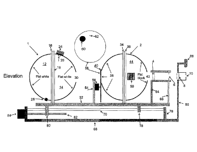
With reference to Figures 1 and 2, a flicker photometer in accordance with the
invention
comprises a first integrating sphere 1 and a second integrating sphere 2 which
respectively
provide diffuse light for a flickering blue and green stimulus and constant
green background to
be observed by the eye of the subject (not shown) through a telescope
comprising an objective
condensing lens 4, an aperture 6, an eyepiece lens 8 and an eyepiece viewing
aperture 10.
11
CA 02864154 2014-08-08
WO 2013/120085 PCT/1JS2013/025600
The integrating sphere 1 is constructed from a rear hemispherical portion 12
and front
hemispherical portion 14, both of which have radial flanges 16 and 18 at which
the two portions
are joined together by suitable means (for example by means of a suitable
adhesive or some other
type of fastener, for example bolts and nuts, acting between the flanges 16
and 18).
The interior surfaces of the two hemispheres 12 and 14 are coated in flat
white matt paint
which acts as a diffuser for uniformly diffusing light emitted by a one watt
green LED 20 and a 3
watt blue LED 22 which are mounted on the top of the front hemisphere 14 and
which each
extend through a respective hole in the hemisphere 14. Each of the LEDs 20 and
22 is also
thermally coupled to a respective heat sink 24 and 26 for dissipating heat
generated by the LEDs.
The intensity of light emitted by each LED is measured using a photodiode
detector 28 mounted
in an aperture towards the bottom of the rear hemisphere 12. In use, the flat
white inner surface
of the integrating sphere 1 acts as a highly efficient scatterer of the light
emitted by the sources
20 and 22 so that each source produces a uniform diffuse illumination of the
surface, including
an area which is diametrically opposed to a circular light outlet opening 30
which is of 32mm
diameter, and is coaxial with the horizontal axis of symmetry of the sphere 1
and with the optical
axis of the lenses 4 and 8.
The integrating sphere 1 can be formed from any suitable material, for example
a metal
or plastics material, and is mounted on a translation slide carriage 32 via a
mounting bracket (not
shown) which is attached to the carriage 32 and which clamps the two flanges
16 and 18
together.
The integrating sphere 2 is mounted on a carriage 32 in a similar fashion to
the
integrating sphere 1 and is of a similar construction to the latter, being
formed by two
hemispheres, each of which has a respective diametric, radial flange 34 and 36
which is attached
to flange of the other hemisphere to hold the two parts of the sphere 2
together.
Both spheres have a diameter of approximately 10cm.
12
CA 02864154 2014-08-08
WO 2013/120085 PCT/1JS2013/025600
The interior of the rear hemisphere 38 of the sphere 2 is coated with a flat
white paint and
includes a circular aperture 40 of 28mm diameter which is coaxial with the
aperture 30 in the
front hemisphere 14 of the sphere 1. The aperture 40 is diametrically opposed
to a 26mm
diameter circular aperture 42 in the front hemisphere 44 of the sphere 2. The
front hemisphere 44
also includes two opposed side openings, each of which accommodates a
respective one of a pair
of one watt green LEDs 46 and 48, each of which is thermally coupled to a
(through the
respective aperture in the hemisphere 44) respective heat sink 50 and 52
outside the sphere 2.
Mounted on a portion of the carriage 32 between the two spheres is a carrier
post 54 the
upper end of which supports a rotary solenoid 56 that is connected to a
circular aperture holder
58. The holder holds a large aperture 60 and a small aperture 62 which
respectively correspond
to the large and small stimuli to be provided by the photometer. The apertures
60 and 62 are
circular and their centers lie at the same radial position on the circular
holder 58. The solenoid 56
is, in use, operable to rotate the holder 58 to bring either of the apertures
60 and 62 into register
with the apertures 30 and 40. When either of the apertures 60 and 62 is in
such a position, it is
coaxial with the apertures 30, 40 and 42, and hence the optical axis of the
viewing telescope.
Each of the apertures 60 and 62 is concentric with a shallow, circular, 21 mm
diameter
recess, which is of larger diameter than either aperture, in the face of the
aperture holder 58
adjacent to sphere 1. A 21 mm diameter glass disk is inserted into each recess
and is etched with
cross-hair markings for the user to view through the viewing telescope. As can
be seen from
Figure 3, the aperture 62, when in register with the aperture 40, will subtend
an angle of 1.5 at
an eye viewing the aperture through the viewing telescope, whilst the aperture
60 will subtend an
angle of 14 at said eye when the aperture is in register with the aperture
40. In use, the light
from the sphere 1 when viewed through the apertures 60 or 62 will produce
stimuli that subtend
corresponding angles.
The front face of the circular holder 58, i.e. the face adjacent the sphere 2
is also provided
with a flat white coating so that the area of illumination provided by the
LEDs 50 and 52 extends
continuously up to the periphery of each aperture in the holder 58 when that
aperture is in
register with the aperture 40.
13
CA 02864154 2014-08-08
WO 2013/120085 PCMJS2013/025600
A flat black coating (for example from paint) provides a matt black surface on
the interior
of the hemisphere 44. This surface prevents light that has travelled from the
sphere 1 into the
sphere 2 from being reflected back onto the flat white surface on the interior
of the hemisphere
38, and the front of the aperture holder 58 and the disks therein.
Each of the objective lens 4 and aperture 6 is mounted on a respective carrier
64 and 66
which is in turn mounted on the carriage 32.
The carriage 32 is slideably mounted on a base 68 via a pair of parallel
cylindrical rails
70 and 72 attached to the plate 68 through two rectangular vertical end plates
74 and 76.
Projecting from the underside of the carriage 32 is a forward rail follower
plate 78 which has a
pair of spaced circular apertures (not shown) through each of which a
respective one of the rails
70 and 72 passes. A similarly apertured rear rail follower plate 80 also
extends from the
underside of the carriage 32 towards the rear of the latter. The plate 80 also
includes a lead screw
nut which engages a screw shaft 82 that can be rotated in a controlled manner
by a lead screw
stepper motor 84. It will be appreciated that the motor 84, shaft 82 and screw
nut provide a worm
drive by virtue of which operation of the motor 84 will cause the carriage,
and the components
that it carries, to slide back and forth along the plate 68 in the direction
of the optical axis of the
telescope.
As can be seen from Figure 1, the viewing aperture 10 and eyepiece lens 8 are
provided
on a carrier plate 86 which is itself directly mounted on the plate 68.
Consequently, the aperture
and eyepiece lens 8 do not move with the carriage. The same carrier also
supports a forehead
rest 88. The carriage 32 enables the user to adjust the focus of the
telescope, constituted by the
elements 4, 6 and 8, without having to reposition his or her head, which can
remain pressed
against the forehead rest 88.
The stepper motor 84, all the LEDs (20, 22, 46, 48), the rotary solenoid 56
and the
photodiode 28 are all connected to the circuitry shown in Figure 4. For the
sake of clarity, the
circuitry of Figure 4 and its connections to those components has not been
included in Figures 1
14
CA 02864154 2014-08-08
WO 2013/120085 PCT/1JS2013/025600
and 2. The circuitry of Figure 4 is also connected to various user operable
controls (not shown)
through which the photometer can be set up and operated.
With reference to Figure 4, the blue LED 22 of the integrating sphere 14 is
supplied with
the necessary electrical power to operate it by a driver 90 which is
controlled by logic circuit 92
to cause the LED to flicker at a frequency of 10Hz - 40Hz and at an intensity
governed by a
frequency modulator 94 which provides one of the input signals for the logic
circuit 92. The
operation of the frequency modulator is illustrated in Figures 5 and 6. The
modulator has an
input 96 via which the user or clinician can manually set a pulse frequency
corresponding to a
desired intensity of illumination by the blue LED. A frequency generator
generates a square
wave signal 98 at the chosen frequency, which is in the range of 1 kHz - 30
kHz. This signal is
supplied to wave shaping circuitry 100 in the modulator 94. This circuitry
converts the square
wave signal 98 into a train of pulses, such as the pulse 102, each of which is
of a duration of 10
micro seconds. The interval between successive pulses corresponds to the
period of the square
wave signal 98. Consequently, the frequency of the signal 98 will correspond
to the frequency of
occurrence of pulses such as the pulse 102 (i.e. the number of such pulses
that occur per unit
time). These pulses are, in turn, fed to the driver 90 that provides an output
in the form of a
driving signal for the LED constituted by said pulses. The intensity of light
emitted by the LED
will be roughly proportional to the frequency of the occurrence of the pulses.
Figure 5 shows the circuitry 100 connected directly to the buffer 90. In fact,
the
connection between the circuitry 100 and driver 90 is made via the logic
setter 92. That circuit
has been omitted from Figure 5 for the sake of clarity.
The logic circuit 92 has another input that is connected to flicker control
circuitry 160.
This will feed a square wave signal on the frequency of 10 Hz-4011z,
determined by a flicker
frequency set input 104 to the control circuitry 160. The logic circuit 92
acts as an AND gate
which is opened each time that the square wave signals from the circuitry 160
is at a maximum
value to allow the train of pulses generated by circuitry 100 to drive the LED
22.
CA 02864154 2014-08-08
WO 2013/120085 PCT/1JS2013/025600
The output of the driver 90 is shown at 103 in Figure 6, and corresponds to
the train of
pulses output by the circuitry 100.
In Figure 7, the square wave 106 represents the signal that is fed by the
circuitry 160 to
the circuitry 92, whilst the graph 108 represents the signal that is
subsequently fed from the logic
circuit 92 to the driver 90 (which outputs are corresponding driving
voltage/current to the LED
22). As can be seen, during each maximum of the wave form 106, the LED 22 is
supplied with a
burst of equal amplitude, equal duration pulses such as the pulse 102. Those
pulses are not
shown to scale in Figure 108 since in reality there will be, for example,
between 50-1000
individual pulses for each "on" part of the square wave cycle 106, for example
the portion 110,
depending upon the selected intensity, for a flicker of 10Hz. The frequency of
occurrence of the
individual pulses is so high that those pulses cannot be individually
perceived by the eye and
their frequency of occurrence can therefore be controlled to control the
perceived intensity of
light emitted by the LED 22. However, the signal 106, which corresponds to the
frequency of
flicker of the LED 22 can of course potentially be perceived by the eye under
examination.
Referring back to Figure 5, the flicker control circuitry 160 supplies a
further square
wave signal, in antiphase to the signal 106, to a logic circuit 114 which is
connected between a
frequency modulator 116 and a driver 118 for driving the green LED 20.
The components 116, 114 and 118 respectively correspond to the frequency
modulator
94, the logic circuit 92 and driver 90 and thus control the on-off operation
and intensity of the
LED 20 in a similar fashion to the way in which the LED 22 is controlled.
However, since the
square wave signals supplied by the circuitry 160 to the logic circuits 92 and
114 are in antiphase
the green LED will be switched on when the blue LED is off and vice-versa.
The flicker control circuitry 160 is also connected to a flicker/hold select
switch which in
one position switches off the flicker of the LEDs 20 and 22 so that both LEDs
remain on
continuously. When the circuitry 160 is operating in this mode, a constant
"on" signal is supplied
to both of the logic circuits 92 and 114.
16
CA 02864154 2014-08-08
WO 2013/120085 PCT/1JS2013/025600
The green LEDs 46 and 48 in the integrating sphere 2 are driven by a common
driver 120
which is functionally similar to the drivers 90 and 118, and which is
controlled by a frequency
modulator 122 which is functionally similar to the frequency modulators 94 and
116, and thus
enables the intensity of light emitted by the LEDs 46 and 48 to be controlled,
in response to
inputs from a control switch or knob 124, by a method of pulse frequency
modulation.
In Figure 7, the graph 109 represents the driving signal sent to the green LED
20.
The controls for the flicker photometer also include a focusing control 126
that controls
the operation of the stepper motor 84 (and hence the position of the carriage
32) through control
circuitry 128 that includes a motored driver circuit, limit switches for
switching off the motor
when the carriage is near the end of its permitted range of movement and a
warning buzzer
which is activated by the closing of either of the limit switches, to provide
an audio warning that
the carriage 32 has reached either end of its allowable range of movement.
The photodetector 28 is connected at its output to an amplifier 130 which is
in turn
connected to sample and hold circuitry 132 that includes an analogue to
digital converter
digitizing the signal detected by the photodetector. The sample and hold
circuitry is controlled by
a data processor which takes the form of a microcontroller 134. The
microcontroller 134 receives
a square wave signal from the flicker control circuitry 160 which corresponds
to the square wave
106, and uses this to trigger the sample and hold circuitry 132 to sample the
output of the
photodetector in synchronism with the operation of the LEDs 20 and 22. More
specifically, an
example of the way in which the sample and hold circuitry 132 is controlled is
shown at the
bottom of Figure 7. As can be seen, successive analogue to digital conversion
and sample and
hold operations are conducted when the blue LED is active, whereas the
intensity of the green
LED is measured only once (on start-up of the photometer or each time the
intensity set input of
the frequency modulator 116 is adjusted). The microcontroller 134 has further
inputs which are
connected to control buttons Fl, F2, F3 and reset, represented by block 136,
the functions of
which are discussed below.
17
CA 02864154 2014-08-08
WO 2013/120085 PCT/1JS2013/025600
Microcontroller 134 also has an output for controlling the rotary solenoid 56
for
supplying data to a printer 138 and a signal to an LCD display 140.
The microcontroller 134 may also be programmed to activate a buzzer 142 if
certain
predetermined conditions arise. The path operating the various components of
the circuitry
shown in Figure 4 is provided by a power supply 144 which, for the sake of
clarity, is shown
with its connections to the various other components omitted.
The microcontroller 134 is programmed to calculate lens optical density
equivalent age of
lens and macular pigment density of an eye under examination in the way
discussed below, by
means of intensity measurements (via the photodetector 28) made during the
course of the
observation of the flickering stimuli shown in Figures 8 and 9, as seen
through a telescope.
Figure 8 shows the small stimulus which is, in effect, the alternating blue
and green light from
the integrating sphere 1 which is viewed through the aperture 62. The sphere 2
and the front face
of the holder 58 provide a continuous green background light which is in the
shape of a ring 146
concentric with the aperture 62. The cross hairs etched onto the glass
substrate of the aperture 62
are shown at 148. Figure 9 shows the situation after the solenoid 56 has moved
the larger
aperture 60 into register with the aperture 40 to provide a larger circular
stimulus. The cross hairs
for this stimulus are shown at 150 and again, the flickering stimulus is
visible against a
continuous green annular background, here referenced 152, provided via the
interior of the
hemisphere 38 of the sphere 2.
After the photometer has been initially set up by selecting the intensity of
the continuous
green background and the light emitted by the LED, as well as the flicker
frequency for the
LEDs 20 and 22, as discussed below, the subject will attempt to adjust the
intensity of light
emitted by the blue LED 20 until, in the case of the image presented in Figure
8, the central
stimulus seen through the aperture 62 no longer appears to flicker. The
subject or operator then
presses a button, F2, which is an input to the microcontroller 134 to trigger
the recordation of the
intensity of blue light at which this occurs. Additionally, the action of
pressing button F2 causes
the blue light intensity setting to be given a small disturbance via the
microcontroller 134 prior to
the subject attempting a subsequent setting. When the subject is looking at
the larger flickering
18
CA 02864154 2014-08-08
WO 2013/120085 PCT/1JS2013/025600
stimulus, as shown in Figure 9, the object is to keep looking at the center of
the cross hairs 150
and adjust the intensity of light emitted by the blue LED until the entire
stimulus seen through
the aperture 60 (apart from that in the very center) has stopped flickering.
The subject or operator
then presses the button F2 to record the intensity of light emitted by the LED
20 once this occurs,
and this data can be used either in the calculation of the lens optical
density or equivalent age of
lens or to take the lens optical density into account in the calculation of
macular pigment density.
Figure 10 shows, within the dotted line 154, the central region in which
flicker will be retained.
On the system block diagram of Figure 4, "flicker frequency set" which refers
to the
frequency of the flicker signal, needs to be adjustable by the operator. For
example, the
frequency can initially be set to default values of 25 and 32Hz for the 1.5
and 14 stimuli,
respectively. Sometimes, there may be slight frequency adjustments to suit the
individual
subject. For example with the 1.50 stimulus, if the frequency is too high for
the subject, flicker
will be eliminated over a wide range of blue LED intensity settings, and if
the frequency is too
low, flicker can never be eliminated.
The flicker/hold select switch 112 is a 2-position switch. In one position
(hold), the
flicker is switched off and both the blue and green LEDs in the left-hand
sphere that form the
stimulus are turned on, rather than alternating. In the other position
(flicker), the LEDs alternate.
The switch is placed in the hold position during initial setup of the device
(see below), and also
while the subject is adjusting the focus. It is a little easier to do this
with a steady stimulus rather
than one that is flickering. Switching off the flicker is also an automatic
function performed by
the microcontroller at the start of a measurement session, since it is at this
time that the subject
needs to adjust the focus. The control buttons Fl, F2 and F3 are explained
below.
Flicker frequency is not measured by the photodiode but rather it is inferred
from the
frequency fed to the blue and green LEDs. Specifically, the block 160 is a
frequency generator
whose frequency is adjustable by the operator. Note that the outputs (square
wave signals)
control the logic circuits for the blue and green LEDs that form the stimulus.
The logic circuits
permit the high frequency pulses generated by the two frequency modulators to
be fed alternately
to the respective LED drivers, thus achieving flicker. The frequency generator
output is also fed
19
CA 02864154 2014-08-08
WO 2013/120085 PCT/1JS2013/025600
to the microcontroller 134 which in turn measures the frequency. The
photodetector 28 is
continuously measuring the light intensity inside the sphere 1. However, the
microcontroller 134
can now indicate to the sample and hold block when to sample the amplified
photodetector
signal and send it to the microcontroller 134. During a test, when the subject
is adjusting the blue
LED 22 intensity, sampling is performed during the phases of the cycle when
the blue LED is
on. At the end of the test, a single sample is obtained during the phase when
the green LED is on.
This latter measurement is needed for the lens optical density calculation.
Operation of photometer
In the initial, one-time set-up, the green surround luminance (sphere 2) is
adjusted to a
value of ¨ 20 candelas/m2, as measured with a Minolta Spotmeter. With the
flicker/hold switch
in the hold position, and with the blue LED in the sphere 1 turned off, the
green LED in that
sphere is adjusted in intensity until the stimulus luminance matches that of
the surround. Since
the green color appearance of stimulus and surround are identical, this
luminance matching can
be easily accomplished by eye, in fact more accurately than by using the
Spotmeter. The
luminance value of 20 candelas/m2 is chosen with two criteria in mind.
Firstly, the luminance
level should be comfortable with no high intensity glare problems. Secondly,
during the test,
subjects with macular pigment optical densities (MPOD) ranging from zero to
over 1.0 have to
be accommodated, i.e. able to match the luminances of the blue and green
components of the
stimulus (as judged by absence of flicker). If the green LED intensity is set
too high, then
subjects with high MPOD will not be able to increase the blue LED intensity
sufficiently to
achieve a match. Likewise, if it is set too low, then subjects with very low
MPOD will not be
able to decrease the blue LED intensity sufficiently to achieve a match. (Note
that the lowest
blue LED intensity setting is not zero.)
The procedure for testing a subject is as follows: When the instrument is
switched on, the
1.50 stimulus aperture is automatically positioned in the field of view. The
microcontroller
automatically switches off the flicker and the subject is asked to press
either of two focusing
buttons, which cause the translation carriage to move backwards or forwards,
until the stimulus
is seen in sharp focus. A screen displays the frequency so that the operator
can set the flicker
frequency at 25Hz and then press the Fl control button. The screen now
displays the blue LED
CA 02864154 2014-08-08
WO 2013/120085 PCT/1JS2013/025600
intensity and the stimulus automatically begins to flicker. The subject
adjusts this intensity via a
control knob to minimize flicker. The operator presses the F2 control button
to record the setting
and then to provide an automatic offset to the setting. This is repeated as
many times as required
(usually 5 times). If, during the test, the subject requires an adjustment to
the frequency, the
operator presses the F3 control button and the frequency is displayed on the
screen. After
adjusting the frequency, the operator presses F3 again and the test is
resumed. When enough
settings have been recorded, the operator presses the Fl button. This causes
the rotary solenoid to
be energized and the 14 stimulus appears in the field of view. The screen
displays the
frequency which is set by the operator at 32Hz after which Fl is pressed and
the screen again
displays the blue LED intensity. The test proceeds as with the 1.5 stimulus
except that the
subject adjusts the blue LED intensity to achieve the situation where flicker
is only perceived at
the center of the stimulus. When sufficient settings have been recorded (again
typically 5), the
operator presses Fl, and the screen displays the MPOD and the standard error
in MPOD, together
with the lens optical density and associated standard error and the equivalent
age of the lens.
This information is transmitted to a printer. Upon pressing the reset button,
the instrument is
initialized for the next subject or for a repeat test with the same subject.
The lens optical density measurement requires only the measurement with the
large
stimulus where the setting made by the subject (steady appearance in the
peripheral part of the
stimulus) is independent of macular pigment.
A control knob, which provides the 'intensity set' input from the modulator,
allows the
subject to alter the intensity of the blue component of the stimulus. For the
1.5 stimulus, this
intensity adjustment is made to minimize, or eliminate, the flicker seen in
the stimulus, and this
occurs when the blue and green components are equalized in terms of luminance.
The
wavelengths of the two colors, blue and green, are chosen to correspond to
maximum absorption
(blue) and zero, or close to zero, absorption (green) by the macular pigment.
Subjects having a
high macular pigment density will need to increase the blue intensity to
compensate for
attenuation by the macular pigment (which lies in front of the retinal
photoreceptors). Likewise
subjects with a low density of macular pigment will need to lower the
intensity. However, other
factors, particularly yellowing of the lens which increases with age, will
affect the subject's
21
CA 02864154 2014-08-08
WO 2013/120085 PCT/1JS2013/025600
intensity setting. To remove such effects, the subject repeats the test using
the 140 stimulus, also
viewed centrally. The subject adjusts the intensity of the blue component to
the point where
flicker is eliminated over most of the stimulus with the exception of a small,
residual flicker at
the center. (Increasing or decreasing the blue intensity from this setting
causes the entire stimulus
to flicker.) The steady appearance in the peripheral region of the stimulus,
where macular
pigment has negligible influence, means that the blue and green luminances
have been matched
in the peripheral retina. Subjects with a lot of lens yellowing will require a
higher blue intensity
than those with less lens yellowing.
The luminance of the green component of the flickering stimulus is pre-set to
be equal to
that of the green surround. Thus at the flicker null point, the luminance of
the stimulus matches
that of the surround. This provides the subject with an additional clue in
searching for the flicker
null point. If the stimulus is flickering but appears brighter than the
surround, the subject must
reduce the intensity of the blue component of the stimulus. If the stimulus is
flickering but
appears darker than the surround, the subject must increase the intensity of
the blue component
of the stimulus.
From the ratio of blue intensity settings obtained with the two stimuli, the
effect of lens
yellowing is eliminated, and the macular pigment optical density, D, can be
calculated:
ISmall stimulus
D = log10 ___________________________________ = (I)
IL arg esturnilus
The ratio of intensities of the blue and green components obtained with the 14
stimulus
provides an index, L Y, of the degree of lens yellowing:
LY= (2)
'Green
The blue LED has a peak wavelength of 455nm, close to that of the macular
pigment's
peak absorption wavelength. The green LED has a peak wavelength of ¨520nm
where macular
22
CA 02864154 2014-08-08
WO 2013/120085 PCT/1JS2013/025600
pigment absorbance is very small. However, the LEDs have relatively wide
bandwidths, and a
correction has to be made to the macular pigment optical density calculated
from equation (1) in
order to be able to report the peak value at 460nm, D460. This quantity is
found by solving
numerically the equation:
D = l { jEG (A)10-D4" e(2) V (A)dA 1 EB (a)V (11)d A
og,,, , ________________
j E.8 (2)10" D" =e(A) V (A)d A j" EG (.1)V (A)c 1 A
where EG(k) and 400 are the energy spectra of the green and blue LEDS, V(A) is
the 10'
photopic luminosity function, and c(X) is the normalized extinction spectrum
of macular
pigment. The microcontroller is pre-programmed to perform the calculations and
the results are
displayed on a screen.
The LEDs are fed with high frequency pulses of fixed voltage whose frequency
is
adjustable. The effect is a change in the perceived brightness. (The frequency
is much greater
than the flicker fusion frequency of the human eye.) Alternative methods, such
as simply altering
the LED voltage or using pulse-width modulation, were found to produce small
wavelength
shifts in the peak LED wavelengths. The LEDs are mounted on heat sinks without
which the
light intensity was found to drift.
Square wave alternation between the blue and green LEDs to produce flicker is
achieved
electronically. The LED intensity is measured via a photodiode detector
mounted inside the
sphere 1. Electronic gating is used to ensure that the photodiode samples the
light inside the
sphere only when the blue LED is on, or only when the green LED is on.
The instrument is provided with a low power telescope. All of the components
except the
eye-piece lens are mounted on a translation slide so that they can be moved
relative to the
eyepiece lens in order to be able to accommodate both myopic and hyperopic
subjects. The
objective lens produces a real image of the 1.5 or 14 apertures, which are
provided with cross-
hairs to facilitate central fixation, in the plane of the field stop 6. The
subject adjusts the position
of the translation slide until the entire field of view is sharply focused.
23
CA 02864154 2014-08-08
WO 2013/120085 PCT/1JS2013/025600
The spacing between the integrating spheres 1 and 2 and the sizes of the
openings are
optimized to reduce cross-over of light from one sphere to the other. To
further reduce this
problem, the front half (closer to the telescope) of the right-hand sphere 2
is coated on the
interior surface with a very low reflectance, flat black paint. The other
interior surfaces arc
painted with a flat white paint.
Numerical Solution of Equation
D = log E (2)102) V (A) d2 Efi(A)V (2)c I A
lo (1)
.E, (2)10-1)462 '(A) V (A) d A SEG (A)V (2) d A
where E0(X) and EB(k) are the energy spectra of the green and blue LEDS, V(X)
is the 100
photopic luminosity function, and e(X) is the extinction spectrum of macular
pigment,
normalized to unity at its peak value which occurs at a wavelength of 460 nm.
D is obtained from the blue LED intensity measurements:
/Sr/id/stimulus
D = logio
I L arg estomulres
The required quantity is the peak macular pigment optical density, D460,
To solve equation (1) numerically, we first approximate the integrals by
finite sums:
D ¨log10 1EG (2)10-D4'('')17(A)AA EEB(A)v(A)AR.
(2)
E, (2)10-D4 ) V (2) EG (2)11. (2)AA ....
where the interval, AA, was chosen to be 5 mu. The sums are carried out over
the wavelength
range 400 to 600 nm since EGQ,) and EB(X) are zero outside that range. The
following algorithm
is then implemented:
24
CA 02864154 2014-08-08
WO 2013/120085 PCT/US2013/025600
1, Set Ditoo to zero
2. Using known values (at 5 nm intervals) of the wavelength-dependent
quantities in
equation (2), calculate D
3. Record D and /3460
4. Increase D460 by 0.01
5. If D460 is greater than 1.5, stop, otherwise ¨
6. Go to statement 2
From the recorded values of D and D460, a graph of D460 as a function of D is
generated
covering the range 0 5. D460 1.5. (Subjects with values of 13460 outside this
range have never
been encountered.), and this is shown in Figure 11.
To facilitate the use of this graph, a polynomial is fitted to the data. (A
third order cubic
was found to be adequate). Using energy spectra obtained from typical blue and
green LEDs, the
following polynomial was generated:
1)460 = 0.006857 + 1.6021) - 0.4726132 0.9905D3
The instrument microprocessor is programmed with this equation so that a value
of D460
may be generated automatically from the subject's blue LED intensity settings.
Additionally, the
microprocessor is programmed to allow the subject to make a number (typically
5 to 10) of
settings with both the 1.5 and 14 stimuli, and to calculate the mean value
of D460 and the
associated standard error
Calculation of Lens Optical Density at 425 urn
The lens optical density at a wavelength of 425 tun, L425, is determined from
the subject's
instrument settings made with the 14 stimulus. When the subject has
determined the null point
(residual flicker in the middle of the visual field only), let the photodiode
detector readings for
the blue and green components of the stimulus be 1313 and PG respectively.
CA 02864154 2014-08-08
WO 2013/120085 PCT/US2013/025600
We first calculate the 100 photopic luminosity function, V(k), as a function
of age using a
published algorithm (Sagawa, K and Takahashi, Y (2001) Spectral luminous
efficiency as a
function of age. J. Opt. Soc. Am. 18, 2659-2667):
logioV(?) = logioV64.900 + (a ¨ 64.9)logAV(k)
where a is age in years and V64.9(k) is the 100 photopie luminosity function
for a person aged
64.9 years.
We then calculate the theoretical ratio, PB/PG as a function of age by
numerically solving
the equation:
SEG (2) V (2)d/1 E n (A)PD(2)(12
P I P =
B G C
j E,(A)V (A)c 1 )L j EG (A) PD(A) C 1
where PD(k) is the spectral sensitivity of the photodiode detector. To do
this, the integrals are
replaced by sums (over the wavelength range 400 to 600 nrn) and 0, is replaced
by A. with a
value of 5 nm. Thus:
E G (A) V (2) Aat E (2) PD (A) AA
PõIPG= ____________________________
EE,(2)V (A) AAjlEG (2) PD (A) AA
The lens density at 425 nm, L425 as a function of age is obtained from:
L425 = 1.0062 + (a ¨ 64.9)0.0143
where the figure 1.0062 is the value of L425 for a 64.9 year old and the
figure 0.0143 is the
decrease per year in logioV at 425 nm (assumed to be due to increased lens
optical density).
26
CA 02864154 2014-08-08
WO 2013/120085 PCT/US2013/025600
Since we now have both L425 and PB/PG as fimetions of age, we can prepare a
graph of
L425 as a function of PB/PG, as shown in Figure 12.
To facilitate the use of this graph, a polynomial is fitted to the data. (A
third order cubic
was found to be adequate). Using energy spectra obtained from typical blue and
green LEDs, and
the spectral sensitivity of the photodiode detector, the following polynomial
was generated:
)2
L425 = ¨1.6414+1.2585 0.2009 __ +0.0193 __
G G G
The instrument microprocessor is programmed with this equation so that a value
of L425
may be generated automatically from the subject's value of PB/PG.
Additionally, the
microprocessor is programmed to allow the subject to make a number (typically
5 to 10) of
settings with the 14 stimuli, and to calculate the mean value of L425 and the
associated standard
error Substituting the value of L425 obtained for a subject into equation 3,
we can calculate the
equivalent age, a, of the subject's lens. This step is also programmed into
the microcontroller.
Correcting the Macular Pigment Optical Density Measurement for Lens Density
Effects
In order to obtain a more accurate value of the peak macular pigment optical
density,
D460, the 10 photopic luminosity function, VQ), in equations (1) and (2) must
be adjusted based
upon the equivalent age of the subject's lens. As shown above, V(X) at any age
can be calculated
using the published algorithm of Sagawa and Takahashi (J. Opt. Soc. Am. 18,
2659-2667).
Accordingly, D450 would be calculated from equation (2) once the lens
equivalent age has been
calculated. The instrument microprocessor is programmed so that a corrected
value of D460 may
be automatically generated.
27