Note: Descriptions are shown in the official language in which they were submitted.
TRANSCUTANEOUS SPINAL CORD STIMULATION: NONINVASIVE
TOOL FOR ACTIVATION OF LOCOMOTOR CIRCUITRY
by
V. REGGIE EDGERTON
YURI GERASIMENKO
ROLAND ROY
DANIEL C. LU, M.D.
BACKGROUND
Field of the Disclosure
[0003] The present
disclosure relates to the field of neurological treatment and
rehabilitation for injury and disease including traumatic spinal cord injury,
non-traumatic
spinal cord injury, stroke, movement disorders, brain injury, ALS,
Neurodegenerative
Disorder, Dementia, Parkinson's disease, and other diseases or injuries that
result in paralysis
and/or nervous system disorder. Devices, pharmacological agents, and methods
are provided
to facilitate recovery of posture, locomotion, and voluntary movements of the
arms, trunk, and
legs, and recovery of autonomic, sexual, vasomotor, speech, swallowing, and
respiration, in a
human subject having spinal cord injury, brain injury, or any other
neurological disorder.
CA 2864473 2018-05-17
CA 02864473 2014-08-13
WO 2013/071309 PCT/US2012/064878
Description of the Related Art
[00041 Serious spinal cord injuries (SCI) affect approximately 1.3 million
people in the
United States, and roughly 12-15,000 new injuries occur each year. Of these
injuries,
approximately 50% are complete spinal cord injuries in which there is
essentially total loss of
sensory motor function below the level of the spinal lesion.
[00051 Neuronal networks formed by the interneurons of the spinal cord that
are located
in the cervical and lumbar enlargements, such as the spinal networks (SNs),
play an important
role in the control of posture, locomotion and movements of the upper limbs,
breathing and
speech. Most researchers believe that all mammals, including humans, have SNs
in the
lumbosacral cord. See Dimitrijevic, M.R, Gerasimenko, Yu., and Pinter, M.M.,
Evidence for
a Spinal Central Pattern Generator in Humans, Ann. N. Y. Acad. Sci., 1998,
vol. 860, p. 360;
Gurfinkel', V.S., Levik, Yu.S., Kazennikov, 0.V., and Selionov, V.A., Does the
Prime
Mover of Stepping Movements Exist in Humans?, Human Physiology, 1998, vol. 24,
no. 3, p.
42; Gerasimenko, Yu.P., Roy, R.R., and Edgerton, VR., Epidural Stimulation:
Comparison of
the Spinal Circuits That Generate and Control Locomotion in Rats, Cats and
Humans, Exp.
Neurol., 2008, vol. 209, p. 417. Normally, the activity of SNs is regulated
supraspinally and
by peripheral sensory input. In the case of disorders of the connections
between the brain and
spinal cord, e.g., as a result of traumatic spinal cord lesions, motor tasks
can be enabled by
epidural electrical stimulation of the lumbosacral and cervical segments as
well as the
brainstem. It has been shown that epidural electrical spinal cord stimulation
(eESCS) with
sufficient intensity can induce electromyographic (EMG) patterns in the leg
muscles of
patients with clinically complete spinal cord injury. See Dimitrijevic,
Gerasimenko, Yu., and
Pinter, supra; Minassian, K., Persy, I., Rattay, F, Pinter, M.M., Kern, H.,
and Dimitrijevic,
M.R., Human Lumbar Cord Circuitries Can Be Activated by Extrinsic Tonic Input
to
Generate Locomotor-Like Activity, Human IHovetnent Sci., 2007, vol. 26, p.
275; Harkema,
S., Gerasimenko, Y, Hodes, J., Burdick, J., Angeli, e., Chen, Y, Ferreira, e.,
Willhite, A.,
Rejc, E., Grossman, R.G., and Edgerton, VR., Epidural Stimulation of the
Lumbosacral
Spinal Cord Enables Voluntary Movement, Standing, and Assisted Stepping in a
Paraplegic
Human, Lancet, 2011, vol. 377, p. 1938. eESCS is an invasive method and
requires surgical
implantation of electrodes on the dorsal surface of the spinal cord, which
limits this method
of activating SNs to clinics.
2
CA 02864473 2014-08-13
WO 2013/071309 PCT/US2012/064878
[00061 Recently, noninvasive methods for activating the SNs by means of leg
muscle
vibration and spinal cord electromagnetic stimulation was suggested. It was
found that the
vibration of the tendons of the hip muscles initiates involuntary walking
movements in
subjects lying on their side with an external support for the legs. See
Gurfinker , VS., Levik,
Yu. S., Kazennikov, 0.V, and Selionov, VA., Locomotor-Like Movements Evoked by
Leg
Muscle Vibration in Humans, Eur. J 1Veurosci. , 1998, vol. 10, p. 1608;
Selionov, VA.,
Ivanenko, Yu.P., Solopova, 1A., and Gurfinkel', VS., Tonic Central and Sensory
Stimuli
Facilitate Involuntary Air-Stepping in Humans, J Neurophysiol., 2009, vol.
101, p. 2847. In
addition, electromagnetic stimulation of the rostral segments of the lumbar
spinal cord caused
involuntary walking movements in healthy subjects in a similar position with a
support for
the legs. See Gerasimenko, Yu., Gorodnichev, R., Machueva, E., Pivovarova, E.,
Semenov,
D., Savochin, A., Roy, R.R., and Edgerton, VR., Novel and Direct Access to the
Human
Locomotor Spinal Circuitry, J Newrosci., 2010, vol. 30, p. 3700; Gorodnichev,
R.M.,
Machueva, E.M., Pivovarova, E.A., Semenov, D.V, Ivanov, S.M., Savokhin, A.A.,
Edgerton,
VR., and Gerasimenko, Yu.P., A New Method for the Activation of the Locomotor
Circuitry
in Humans, Hum. Physiol., 2010, vol. 36, no. 6, p. 700. Step-like movements
elicited by
vibration and electromagnetic stimulation, have apparently a different origin.
In the former
case, the SN is activated by afferent input mainly due to the activation of
muscle receptors,
whereas in the latter case, the neuronal locomotor network is affected
directly. Each of these
methods has its specificity. For example, the vibratory muscle stimulation
elicits involuntary
locomotor movements only in the hip and knee joints, without the involvement
of the ankle.
In addition, these characteristic movements could be evoked only in 50% of the
subjects. See
Selionov, Ivanenko, Solopova, and Gurfinkel', supra. The percentage of
subjects in whom
the spinal cord electromagnetic stimulation evoked involuntary step like
movements was
even smaller (10%), although in this case, the kinematic structure of the
resultant movements
was consistent with the natural random step-like movements to a greater extent
than in the
case of vibration. See Gerasimenko, Gorodnichev, Machueva, Pivovarova,
Semenov,
Savochin, Roy, and Edgerton, supra; Gorodnichev, Machueva, Pivovarova,
Semenov,
Ivanov, Savokhin, Edgerton, and Gerasimenko, supra. In addition, spinal cord
electromagnetic stimulation is limited by the technical capabilities of the
stimulator. The
modem magnetic stimulator used in clinics (e.g., Magstim Rapid) can provide
only short-
exposure stimulating effects. The electromagnetic stimulator, with the
parameters required to
elicit step-like movements (5 Hz and 1.5 T), could be sustained for only 15s.
3
CA 02864473 2014-08-13
WO 2013/071309 PCT/US2012/064878
SUMMARY
[0007] Embodiments of the disclosure are for use with a mammal including a
human who
has a spinal cord with at least one selected dysfunctional spinal circuit or
other neurologically-
derived source of control of movement in a portion of the subject's body. It
has been shown
that transcutaneous electrical spinal cord stimulation (tESCS) applied in the
region of the
T11-T12 vertebrae with a frequency of 5-40 Hz elicited involuntary step-like
movements in
healthy subjects with their legs suspended in a gravity-neutral position. The
amplitude of
evoked step-like movements increased with increasing tESCS frequency. The
frequency of
evoked step-like movements did not depend on the frequency of tESCS. It was
shown that the
hip, knee, and ankle joints were involved in the evoked movements. In
conclusion,
transcutaneous electrical spinal cord stimulation (tESCS) can be used as a
noninvasive
method in rehabilitation of spinal pathology. By way of non-limiting examples,
application of
transcutaneous electrical spinal cord stimulation (tESCS) activates spinal
locomotor networks
(SNs), in part via the dorsal roots and the gray matter of the spinal cord.
When activated, the
SNs may (a) enable voluntary movement of muscles involved in at least one of
standing,
stepping, reaching, grasping, voluntarily changing positions of one or both
legs, breathing,
speech control, voiding the patient's bladder, voiding the patient's bowel,
postural activity,
and locomotor activity; (b) enable or improve autonomic control of at least
one of
cardiovascular function, body temperature, and metabolic processes; and/or (c)
help facilitate
recovery of at least one of an autonomic function, sexual function, or
vasomotor function.
According to some embodiments, the present disclosure provides that the spinal
circuitry is
neuromodulated to a physiological state that facilitates or enables the
recovery or improved
control of movement following some neuromotor dysfunction.
[0008] The paralysis may be a motor complete paralysis or a motor
incomplete paralysis.
The paralysis may have been caused by a spinal cord injury classified as motor
complete or
motor incomplete. The paralysis may have been caused by an ischemic or
traumatic brain
injury. The paralysis may have been caused by an ischemic brain injury that
resulted from a
stroke or acute trauma. By way of another example, the paralysis may have been
caused by a
neurodegenerative condition affecting the brain and/or spinal cord. The
neurodegenerative
brain injury may be associated with at least one of Parkinson's disease,
Huntington's disease,
Alzheimer's, Frontotemporal Dementia, dy-stonia, ischemic, stroke, amyotrophic
lateral
4
CA 02864473 2014-08-13
WO 2013/071309 PCT/US2012/064878
sclerosis (ALS), primary lateral sclerosis (PLS), and other conditions such as
cerebral palsy
and Multiple Sclerosis.
100091 By way of non-limiting example, a method includes applying
electrical
stimulation to a portion of a spinal cord or brainstem of the subject. The
electrical stimulation
may be applied by a surface electrode(s) that is applied to the skin surface
of the subject.
Such an electrode may be positioned at, at least one of a thoracic region, a
cervical region, a
lumbosacral region of the spinal cord and/or the brainstem. The electrical
stimulation is
delivered at 5-40Hz at 20-100 mA. While not a requirement, the electrical
stimulation may
not directly activate muscle cells in the portion of the patient's body having
the paralysis. The
electrical stimulation may include at least one of tonic stimulation and
intermittent
stimulation. The electrical stimulation may include simultaneous or sequential
stimulation of
different regions of the spinal cord.
[0010] If the paralysis was caused by a spinal cord injury at a first
location along the
spinal cord, the electrical stimulation may be applied by an electrode that is
on the spinal cord
of the patient at a second location below the first location along the spinal
cord relative to the
patient's brain.
[0011] Optionally, the method may include administering one or more
neuropharmaceutical agents to the patient. The neuropharmaceutical agents may
include at
least one of a serotonergic drug, a dopaminergic drug, a noradrenergic drug, a
GABAergic
drug, and glycinergic drugs. By way of non-limiting examples, the
neuropharmaceutical
agents may include at least one of 8-0HDPAT, Way 100.635, Quipazine,
Ketanserin, SR
57227A, Ondanesetron, SB 269970, Buspirone, Methoxamine, Prazosin, Clonidine,
Yohimbine, SKF-81297, SCH-23390, Quinpirole, and Eticlopride.
[0012] The electrical stimulation is defined by a set of parameter values,
and activation of
the selected spinal circuit may generate a quantifiable result. Optionally,
the method may be
repeated using electrical stimulation having different sets of parameter
values to obtain
quantifiable results generated by each repetition of the method. Then, a
machine learning
method may be executed by at least one computing device. The machine learning
method
builds a model of a relationship between the electrical stimulation applied to
the spinal cord
and the quantifiable results generated by activation of the at least one
spinal circuit. A new
CA 02864473 2014-08-13
WO 2013/071309 PCT/US2012/064878
set of parameters may be selected based on the model. By way of a non-limiting
example, the
machine learning method may implement a Gaussian Process Optimization.
[00131 Another exemplary embodiment is a method of enabling one or more
functions
selected from a group consisting of postural and/or locomotor activity,
voluntary movement
of leg position when not bearing weight, improved breathing and ventilation,
speech control,
swallowing, voluntary voiding of the bladder and/or bowel, return of sexual
function,
autonomic control of cardiovascular function, body temperature control, and
normalized
metabolic processes, in a human subject having a neurologically derived
paralysis. The
method includes stimulating the spinal cord of the subject using a surface
electrode while
subjecting the subject to physical training that exposes the subject to
relevant postural
proprioceptive signals, locomotor proprioceptive signals, and supraspinal
signals. At least
one of the stimulation and physical training modulates in real time provoke or
incite the
electrophysiological properties of spinal circuits in the subject so the
spinal circuits are
activated by at least one of supraspinal information and proprioceptive
information derived
from the region of the subject where the selected one or more functions are
facilitated.
[00141 The region where the selected one or more functions are facilitated
may include
one or more regions of the spinal cord that control (a) lower limbs; (b) upper
limbs and
brainstem for controlling speech; (c) the subject's bladder; (d) the subject's
bowel and/or
other end organ. The physical training may include standing, stepping, sitting
down, laying
down, reaching, grasping, stabilizing sitting posture, and/or stabilizing
standing posture.
[00151 The surface electrode may include an array of one or more electrodes
stimulated
in a monopolar biphasic configuration. Such a surface electrode may be placed
over at least
one of a lumbosacral portion of the spinal cord, a thoracic portion of the
spinal cord, a
cervical portion of the spinal cord and/or the brainstem.
[00161 The stimulation may include tonic stimulation and/or intermittent
stimulation. The
stimulation may include simultaneous or sequential stimulation, or
combinations thereof, of
different spinal cord regions. Optionally, the stimulation pattern may be
under control of the
subject.
[00171 The physical training may include inducing a load bearing positional
change in the
region of the subject where locomotor activity is to be facilitated. The load
bearing positional
6
CA 02864473 2014-08-13
WO 2013/071309 PCT/US2012/064878
change in the subject may include standing, stepping, reaching, and/or
grasping. The physical
training may include robotically guided training.
[0018] The method may also include administering one or more
neuropharmaceuticals.
The neuropharmaceuticals may include at least one of a serotonergic drug, a
dopaminergic
drug, a noradrenergic drug, a GABAergic drug, and a glycinergic drug.
[0019] Another exemplary embodiment is a method that includes placing an
electrode on
the patient's spinal cord, positioning the patient in a training device
configured to assist with
physical training that is configured to induce neurological signals in the
portion of the
patient's body having the paralysis, and applying electrical stimulation to a
portion of a spinal
cord of the patient, such as a biphasic signal of 3040Hz at 85-100 mA.
[0020] Another exemplary embodiment is a system that includes a training
device
configured to assist with physically training of the patient, a surface
electrode array
configured to be applied on the patient's spinal cord, and a stimulation
generator connected to
the electrode. When undertaken, the physical training induces neurological
signals in the
portion of the patient's body having the paralysis. The stimulation generator
is configured to
apply electrical stimulation to the electrode. Electrophysiological properties
of at least one
spinal circuit in the patient's spinal cord is modulated by the electrical
stimulation and at least
one of (1) a first portion of the induced neurological signals and (2)
supraspinal signals such
that the at least one spinal circuit is at least partially activatable by at
least one of (a) the
supraspinal signals and (b) a second portion of the induced neurological
signals.
7
CA 02864473 2014-08-13
WO 2013/071309 PCT/US2012/064878
BRIEF DESCRIPTION OF THE DRAWINGS
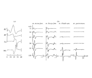
[0021] Figure 1, panels a and b, show motor responses in the muscles of the
right leg to
the tESCS with a frequency of 1 Hz and an amplitude of 75-100 nriA (showed at
the left of
the recordings). The responses in the m. rectus femoris and m. biceps femoris
(RF and BF,
respectively), as well as in the m. tibialis anterior and m. gastrocnemius (TA
and MG,
respectively) are shown. At the right bottom of the lower recording, there are
marks of time
in ms, the same for all the muscles, and marks of the amplitude in mV.
100221 Figures 2A and 2B show electrical activity of the leg muscles and
movements in
the leg joints evoked by tESCS with frequencies of 5 and 30 Hz. Figure 2A:
Subject R: the
cinematogramms of the joint movements of the right leg and the EMGs of the hip
muscles of
the right and left legs are shown. Under the EMG, there is a mark of the
stimulus. At the right
of the cincmatogram and EMGs, there are vertical marks of the amplitude in
angle degrees
and mV, respectively. The duration of records is 40 s. Figure 2B: Subject S:
the EMGs of the
hip and ankle muscles of the right leg and the goniograms of the knee joints
of the right and
left legs; the arrows at the top show the beginning and end of stimulation;
the horizontal and
vertical labels next to EMG, 10 s and 0.5 mV, respectively; the vertical mark
to the right of
the goniograms, 200 m V. H, hip; Kn, knee; Ank, ankle; RF, m. rectus femoris;
BF, m. biceps
femoris; T A, m. tibialis anterior; M G, m. gastrocnemius; (r), on the right;
(1), on the left.
100231 Figure 3 EMGs (left) and trajectories of reflective markers attached
to the right
leg; kinematograms (right) recorded during voluntary stepping movements (vol)
and
movements caused by tESCS with frequencies of 5 and 30 Hz. The duration of
records is 10
s. Black and gray lines show movements in the hip and knee joints,
respectively. The
remaining designations are the same as in FIG. 2A/2B.
100241 Figure 4, panels A-E, show interarticular coordination during
voluntary stepping
movements (vol) and movements caused by tESCS with frequencies of 5 and 30 Hz.
Reconstruction of the movements of the right leg during one stepping cycle
obtained by
processing the cincmatograms of the (Panel A) forward and (Panel B) backward
movements
of legs, respectively; the coordination of movements in the (Panel C) hip and
knee joints,
(Panel D) knee and ankle joints; and (Panel E) the trajectory of a big toe.
100251 Figure 5, panels A-F, show the average amplitude of movements in the
hip (H),
knee (Kn), and ankle (Ank) joints caused by tESCS with a frequency of 5-40 Hz
recorded
8
CA 02864473 2014-08-13
WO 2013/071309 PCT/US2012/064878
during the first 15 s after the start of stimulation. The ordinate shows
angular degrees. (Panels
A, B) Subject S, different strategies (Panels A and B); subject R (Panel C);
subject K (Panel
D); subject B (Panel E); subject G (Panel F). Error bars, standard deviation.
Asterisks,
significant differences in amplitude recorded during tESCS with a frequency of
5 Hz, p <
0.05.
DETAILED DESCRIPTION
100261 Unless defined otherwise, technical and scientific terms used herein
have the same
meaning as commonly understood by one of ordinary skill in the art to which
this disclosure
belongs.
[00271 The term "motor complete" when used with respect to a spinal cord
injury
indicates that there is no motor function below the lesion, (e.g., no movement
can be
voluntarily induced in muscles innervated by spinal segments below the spinal
lesion.
[00281 The term "monopolar stimulation" refers to stimulation between a
local electrode
and a common distant return electrode.
100291 The term "autonomic function" refers to functions controlled by the
peripheral
nervous system that are controlled largely below the level of consciousness,
and typically
involve visceral functions. Illustrative autonomic functions include, but are
not limited to
control of bowel, bladder, and body temperature.
[00301 The term "sexual function" refers to the ability to sustain a penile
erection, have
an orgasm (male or female), generate viable sperm, and/or undergo an
observable
physiological change associated with sexual arousal,
100311 It was discovered that transcutaneous electrical stimulation (TCS)
of the spinal
cord can induce activation locomotor circuitry in a mammal (e.g., in a human
or a non-human
mammal). It was demonstrated, for example, that continuous tSCS at 5-40 Hz
applied
paraspinally over T11-T12 vertebrae at 40-70 mA induced involuntary locomotor
like
stepping movements in subjects with their legs in a gravity-independent
position. The
increase of frequency of tSCS from 5 to 30 Hz resulted in augmentation of the
amplitude of
evoked stepping movements. In chronic spinal cats (3 weeks after spinal cord
transection at
9
CA 02864473 2014-08-13
WO 2013/071309 PCT/US2012/064878
Th8) tSCS at L5 (a frequency of 5 Hz and intensity ranged from 3 to 10 mA)
evoked EMG
stepping pattern in hindlimb muscles in all (N=4) of tested animals, while
locomotor-like
movements produced by tSCS were not weight-bearing.
[00321 By non-limiting example, transcutaneous electrical stimulation can
be applied to
facilitate restoration of locomotion and other neurologic function in subjects
suffering with
spinal cord injury, as well as other neurological injury and illness.
Successful application can
provide a device for widespread use in rehabilitation of neurologic injury and
disease.
[00331 In various embodiments, methods, devices, and optional
pharmacological agents
are provided to facilitate movement in a mammalian subject (e.g., a human)
having a spinal
cord injury, brain injury, or other neurological disease or injury. In certain
embodiments, the
methods involve stimulating the spinal cord of the subject using a surface
electrode where the
stimulation modulates the electrophysiological properties of selected spinal
circuits in the
subject so they can be activated by proprioceptive derived information andlor
input from
supraspinal. In various embodiments, the stimulation is typically accompanied
by physical
training (e.g., movement) of the region where the sensory-motor circuits of
the spinal cord
are located.
l00341 In particular illustrative embodiments, the devices, optional
pharmacological
agents, and methods described herein stimulate the spinal cord with, e.g.,
electrodes that
modulate the proprioceptive and supraspinal information which controls the
lower limbs
during standing and/or stepping and/or the upper limbs during reaching and/or
grasping
conditions. It is the proprioceptive and cutaneous sensory information that
guides the
activation of the muscles in a coordinated manner and in a manner that
accommodates the
external conditions, e.g., the amount of loading, speed, and direction of
stepping or whether
the load is equally dispersed on the two lower limbs, indicating a standing
event, alternating
loading indicating stepping, or sensing postural adjustments signifying the
intent to reach and
grasp.
[0035] Unlike approaches that involve specific stimulation of motor neurons
to directly
induce a movement, the methods described herein enable the spinal circuitry to
control the
movements. More specifically, the devices, optional pharmacological agents,
and methods
described herein exploit the spinal circuitry and its ability to interpret
proprioceptive
information and to respond to that proprioceptive information in a functional
way. In various
CA 02864473 2014-08-13
WO 2013/071309 PCT/US2012/064878
embodiments, this is in contrast to other approaches where the actual movement
is
induced/controlled by direct stimulation (e.g., of particular motor neurons).
[0036] In one illustrative embodiment, the subject is fitted with one or
more surface
electrodes that afford selective stimulation and control capability to select
sites, mode(s), and
intensity of stimulation via electrodes placed superficially over, for
example, the lumbosacral
spinal cord and/or cervical spinal cord to facilitate movement of the arms
and/or legs of
individuals with a severely debilitating neuromotor disorder.
[0037] The subject is provided the generator control unit and is fitted
with an electrode(s)
and then tested to identify the most effective subject specific stimulation
paradigms for
facilitation of movement (e.g., stepping and standing and/or arm and/or hand
movement).
Using these stimulation paradigms, the subject practices standing, stepping,
reaching,
grabbing, breathing, and/or speech therapy in an interactive rehabilitation
program while
being subject to spinal stimulation.
[0038] Depending on the site/type of injury and the locomotor activity it
is desired to
facilitate, particular spinal stimulation protocols include, but are not
limited to, specific
stimulation sites along the lumbosacral, thoracic, and/or cervical spinal
cord; specific
combinations of stimulation sites along the lumbosacral, thoracic, and/or
cervical spinal
cord; specific stimulation amplitudes; specific stimulation polarities (e.g.,
monopolar and
bipolar stimulation modalities); specific stimulation frequencies; and/or
specific stimulation
pulse widths.
[0039] In various embodiments, the system is designed so that the patient
can use and
control it in the home environment.
[0040] In various embodiments, the approach is not to electrically induce a
walking
pattern or standing pattern of activation, but to enable/facilitate it so that
when the subject
manipulates their body position, the spinal cord can receive proprioceptive
information from
the legs (or arms) that can be readily recognized by the spinal circuitry.
Then, the spinal cord
knows whether to step or to stand or to do nothing. In other words, this
enables the subject to
begin stepping or to stand or to reach and grasp when they choose after the
stimulation
pattern has been initiated.
11
CA 02864473 2014-08-13
WO 2013/071309 PCT/US2012/064878
[0041] Moreover, the methods and devices described herein are effective in
a spinal cord
injured subject that is clinically classified as motor complete; that is,
there is no motor
function below the lesion. In various embodiments, the specific combination of
electrode(s)
activated/stimulated and/or the desired stimulation of any one or more
electrodes and/or the
stimulation amplitude (strength) can be varied in real time, e.g., by the
subject. Closed loop
control can be embedded in the process by engaging the spinal circuitry as a
source of
feedback and feedforward processing of proprioceptive input and by voluntarily
imposing
fine tuning modulation in stimulation parameters based on visual, and/or
kinetic, and/or
kinematic input from selected body segments.
[00421 In various embodiments, the devices, optional pharmacological
agents, and
methods arc designed so that a subject with no voluntary movement capacity can
execute
effective standing and/or stepping and/or reaching and/or grasping. In
addition, the approach
described herein can play an important role in facilitating recovery of
individuals with severe
although not complete injuries.
[0043] The approach described herein can provide some basic postural,
locomotor and
reaching and grasping patterns on their own. However, they are also likely to
be a building
block for future recovery strategies. Based on certain successes in animals
and some
preliminary human studies (see below), it appears that a strategy combining
effective
transcutaneous stimulation of the appropriate spinal circuits with physical
rehabilitation and
pharmacological intervention can provide practical therapies for complete SCI
human
patients. There is sufficient evidence from our work that such an approach
should be enough
to enable weight bearing standing, stepping and/or reaching or grasping. Such
capability can
give SCI patients with complete paralysis or other neuromotor dysfunctions the
ability to
participate in exercise, which is known to be highly beneficial for their
physical and mental
health. We also expect our method should enable movement with the aid of
assistive walkers.
While far from complete recovery of all movements, even simple standing and
short duration
walking would increase these patients' autonomy and quality of life. The
stimulating array
technology described herein (e.g., transcutaneous electrical stimulation)
paves the way for a
direct brain-to-spinal cord interface that could enable more lengthy and finer
control of
movements.
[0044] While the methods and devices described herein are discussed with
reference to
complete spinal injury, it will be recognized that they can apply to subjects
with partial spinal
12
CA 02864473 2014-08-13
WO 2013/071309 PCT/US2012/064878
injury, subjects with brain injuries (e.g., ischemia, traumatic brain injury,
stroke, and the
like), and/or subjects with neurodegenerative diseases (e.g., Parkinson's
disease, Alzheimer's
disease, Huntington's disease, amyotrophic lateral sclerosis (ALS), primary
lateral sclerosis
(PLS), cerebral palsy, and the like).
[0045] In various embodiments, the methods combine the use of
transcutaneous
stimulating electrode(s) with physical training (e.g., rigorously monitored
(robotic) physical
training), optionally in combination with pharmacological techniques. The
methods enable
the spinal cord circuitry to utilize sensory input as well as newly
established functional
connections from the brain to circuits below the spinal lesion as a source of
control signals.
The approach is thus designed to enable and facilitate the natural sensory
input as well as
supraspinal connections to the spinal cord in order to control movements,
rather than induce
the spinal cord to directly induce the movement. That is, we facilitate and
enhance the
intrinsic neural control mechanisms of the spinal cord that exist post-SCI,
rather than replace
or ignore them.
Processing of Sensory Input by the Lmnbosacral Spinal Cord:
Using Afferents as a Source of Control
[00461 In various embodiments the methods and devices described herein
exploit spinal
control of locomotor activity. For example, the human spinal cord can receive
sensory input
associated with a movement such as stepping, and this sensory information can
be used to
modulate the motor output to accommodate the appropriate speed of stepping and
level of
load that is imposed on lower limbs. Moreover, we have demonstrated that the
human
lumbosacral spinal cord has central-pattern-generation-like properties. Thus,
oscillations of
the lower limbs can be induced simply by vibrating the vastus lateralis muscle
of the lower
limb, by transcutaneous stimulation, and by stretching the hip. The methods
described herein
exploit the fact that the human spinal cord, in complete or incomplete SCI
subjects, can
receive and interpret proprioceptive and somatosensory information that can be
used to
control the patterns of neuromuscular activity among the motor pools necessary
to generate
particular movements, e.g., standing, stepping, reaching, grasping, and the
like. The methods
described herein facilitate and adapt the operation of the existing spinal
circuitry that
generates, for example, cyclic step-like movements via a combined approach of
transcutaneous stimulation, physical training, and, optionally, pharmacology.
13
CA 02864473 2014-08-13
WO 2013/071309 PCT/US2012/064878
Facilitating Stepping and Standing in Humans Following a Clinically Complete
Lesion
[0047] Locomotion in mammals is attributed to intrinsic oscillating spinal
neural
networks capable of central pattern generation interacting with sensory
information (Edgerton
et al., J. American Paraplegia Soc, 14(4) (1991), 150-157; Forssberg, J.
Neurophysiol, 42(4):
936-953 (1979); Grillner and Wallen, Annu. Rev. Neurosci., 8: 233-261 (1985);
Grillner and
Zangger, Exp Brain Res, 34(2): 241-261 (1979)). These networks play critical
roles in
generating the timing of the complex postural and rhythmic motor patterns
executed by motor
neurons.
[0048] As indicated above, the methods described herein can involve
stimulation of one
or more regions of the spinal cord in combination with locomotory activities.
It was our
discovery that spinal stimulation in combination with locomotor activity
results in the
modulation of the electrophysiological properties of spinal circuits in the
subject so they are
activated by Proprioceptive information derived from the region of the subject
where
locomotor activity is to be facilitated. Further, we also determined that
spinal stimulation in
combination with pharmacological agents and locomotor activity results in the
modulation of
the electrophysiological properties of spinal circuits in the subject so they
are activated by
proprioceptive information derived from the region of the subject where
locomotor activity is
to be facilitated.
[0049] Locomotor activity of the region of interest can be accomplished by
any of a
number of methods known, for example, to physical therapists. By way of
illustration,
individuals after severe SCI can generate standing and stepping patterns when
provided with
body weight support on a treadmill and manual assistance. During both stand
and step
training of human subjects with SCI, the subjects can be placed on a treadmill
in an upright
position and suspended in a harness at the maximum load at which knee buckling
and trunk
collapse can be avoided. Trainers positioned, for example, behind the subject
and at each leg
assist as needed in maintaining proper limb kinematics and kinetics
appropriate for each
specific task. During bilateral standing, both legs can be loaded
simultaneously and extension
can be the predominant muscular activation pattern, although co-activation of
flexors can also
occur. Additionally, or alternatively, during stepping the legs are loaded in
an alternating
pattern and extensor and flexor activation patterns within each limb also
alternated as the legs
moved from stance through swing. Afferent input related to loading and
stepping rate can
14
CA 02864473 2014-08-13
WO 2013/071309 PCT/US2012/064878
influence these patterns, and training has been shown to improve these
patterns and function
in clinically complete SCI subjects.
Transcutaneous Stimulation of the Lumbosacral Spinal Cord
[00501 As indicated above, without being bound by a particular theory, it
is believed that
transcutaneous stimulation, e.g., over the throacic spinal cord in combination
with physical
training can facilitate recovery of stepping and standing in human subjects
following a
complete SCI.
100511 Spinal cord electrical stimulation has been successfully used in
humans for
suppression of pain and spasticity (see, e.g., Johnson and Burchiel,
Neurosurgery, 55(1): 135-
141 (2004); discussion 141-142; Shealy et al., AnesthAnalg, 46(4): 489-491
(1967); Campos
et al., Appl. Neurophysiol. 50(1-6): 453-454 (1987); Dimitrijevic and
Sherwood, Neurology,
30 (7 Pt 2): 19-27 (1980); Barolat Arch. Med. Res., 31(3): 258-262 (2000);
Barolat, J. Am.
Paraplegia Soc., 11(1): 9-13 (1988); Richardson et al., Neurosurgery, 5(3):
344-348). Recent
efforts to optimize stimulation parameters have led to a number of research
studies focusing
on the benefits of transcutaneous spinal cord stimulation. We have
demonstrated that the
location of the electrode and its stimulation parameters are important in
defining the motor
response. Use of surface electrode(s), as described herein, facilitates
selection or alteration of
particular stimulation sites as well as the application of a wide variety of
stimulation
parameters.
[00521 The following non-limiting examples are offered for illustrative
purposes.
Example 1: Transcutaneous Electrical Stimulation of the Spinal Cord: A
Noninvasive Tool
for the Activation of Stepping Pattern Generators in Humans
100531 A noninvasive method for activating the SN by means of
transcutaneous electrical
spinal cord stimulation (tESCS) is demonstrated in this Example. The method is
based on our
research that showed that a single dermal electric stimulus applied in the
region of the T 11-T
12 vertebrae caused monosynaptic reflexes in the proximal and distal leg
muscles in healthy
subjects (see Courtine, G., Harkema S.J, Dy, C.J., Gerasimenko, Yu.P., and
Dyhre-Poulsen,
P., Modulation of Multisegmental Monosynaptic Responses in a Variety of Leg
Muscles
during Walking and Running in Humans, J Physiology, 2007, vol. 585, p. 1125)
and in
patients with clinically complete (ASIA A) spinal cord injury. See Dy, C.J.,
Gerasimenko,
CA 02864473 2014-08-13
WO 2013/071309 PCT/US2012/064878
YP., Edgerton, VR., DyhrePoulsen P., Courtine G., Harkema S., Phase-Dependent
Modulation of Percutaneously Elicited Multisegmental Muscle Responses after
Spinal Cord
Injury, J Neurophysia, 2010, vol. 103, p. 2808. Taking into consideration that
eESCS affects
the SN through mono and polysynaptic reflexes (see Minassian, Persy, Rattay,
Pinter, Kern,
and Dimitrijevic, supra), we suggest that noninvasive tESCS can be an
effective way to
neuromodulate the SN.
Experiment
[00541 We examined six adult male subjects (students and staff of the
Velikie Luki State
Academy of Physical Education and Sports). They had given their informed
written consent
to participate in the experiment. The experiment was approved by the Ethics
Committee of
the academy and met the requirements of the Helsinki Declaration.
[00551 The subjects lay on a couch on their left side, with their feet
placed on separate
boards that were attached to a hook in the ceiling of the experimental room
with ropes, like
swings. The right (upper) leg was supported directly in the region of the
shank. The left
(lower) leg was placed in a rotating frame attached to a horizontal board.
Under these
conditions, the subjects could move their legs through maximum amplitude:
According to the
instructions, the subjects lay quietly and neither counteracted nor
facilitated the movements
caused by electrical stimulation of the spinal cord.
[00561 The tESCS was performed using a KULON stimulator (St. Petersburg
State
University of Aerospace Instrumentation, St. Petersburg, Russia). The
stimulation was
administered using a 2.5 cm round electrode (Lead-Lok, Sandpoint, United
States) placed
midline on the skin between the spinous processes of T11 and T12 as a cathode
and two 5.0 x
10.2 cm rectangular plates made of conductive plastic (Ambu, Ballerup,
Germany) placed
symmetrically on the skin over the iliac crests as anodes. The step-like
movements were
evoked by a bipolar rectangular stimulus with a duration of 0.5 ms, filled
with a carrier
frequency of 10 kHz; the intensity of stimulation ranged from 30 to 100 mA.
The stimulation
frequencies were 1, 5, 10, 20, 30, and 40 Hz; the duration of exposure ranged
from 10 to 30 s.
During the high-frequency stimulation within each stimulus, tESCS did not
cause pain even
when the amplitude was increased to 100 mA or more; allowing us to study in
detail the
dependence of the elicited movements on the amplitude and frequency of the
stimulus.
16
CA 02864473 2014-08-13
WO 2013/071309 PCT/US2012/064878
[0057] The EMGs of the muscles of both legs (m. rectus femoris, m. biceps
femoris, m.
tibialis anterior, and m. gastrocnemius) were recorded by means of bipolar
surface electrodes.
EMG signals were recorded using an ME 6000 16-channel telemetric
electroneuromyograph
(Mega Win, Finland). Flexion-extension movements in the knee joints were
recorded using a
goniometer.
[0058] The Qualisy video system (Sweden) was used to record the kinematic
parameters
of leg movements. Light-reflecting markers were attached to the pivot points
of the body,
which coincided with the rotational axis in the shoulder, hip, knee, and ankle
joints. The
angular movements in the hip joint were calculated from the location of
markers on the
lateral epicondyle of the humerus, trochanter, and lateral epicondyle of the
femur. The
markers that were attached to the trochanter, lateral epicondyle of the femur,
and lateral ankle
were used to describe the movements in the knee joint. The movements in the
ankle joint
were estimated by means of the markers located on the lateral epicondyle of
the femur, lateral
ankle, and the big toe. The reconstruction of movements in one whole step
cycle was
performed by means of special software. In order to record the movements of
the foot tip, the
marker was fixed on the big toe of the right foot.
[00591 The recording of EMG was synchronized with the recording of stepping
kinematical parameters. The average cycle duration and the amplitudes of
angular
movements were calculated from 10-12 cycles. The duration of a step cycle was
calculated
on the basis of the interval between two maximum values of angular movements
in the hip,
knee, and ankle joints. The phase shift between the hip and knee joints was
calculated from
the interval between the maximum values of angular movements in these joints.
[00601 The statistical treatment of the data was performed using a standard
software
package.
Results
[0061] Transcutaneous electrical spinal cord stimulation with a frequency
of 5-40 Hz
elicited involuntary leg movements in five out of six subjects. The threshold
intensity of the
stimulus that induced involuntary movements was 50-60 mA and was dependent on
the
frequency of stimulation. The tESCS at a frequency of 1 Hz caused reflex
responses in the
leg muscles with a threshold of 70-80 mA (FIG. 1(a)).
17
CA 02864473 2014-08-13
WO 2013/071309 PCT/US2012/064878
l00621 Original records of EMG responses in the muscles of the right leg to
the tESCS at
a frequency of 1 Hz and intensity of 75-100 InA are shown in FIG. 1.
Increasing stimulus
intensity resulted in an increase in the amplitude of responses. First, the
hip muscles (m.
rectus femoris and m. biceps femoris) were involved in the motor response;
then, the shank
muscles (m. tibialis anterior and m. gastrocnemius) were involved (FIG. 1(b)).
The response
to each stimulus is composed of the early monosynaptic responses (the same is
shown in
Courtine, Harkema, Dy, Gerasimenko, and Dyhre-Poulsen, supra) with a latency
period of
about 12-15 ms. Increasing stimulus intensity evoked responses in the biceps
femoris muscle
(flexor) with a latent period of a few tens of milliseconds, which were,
apparently,
polysynaptic. Thus, tESCS with a low frequency (1 Hz) elicited reflex
responses in the leg
muscles that contained mono and polysynaptic components.
l00631 Transcutaneous electrical spinal cord stimulation at frequencies in
the entire range
from 5 to 40 Hz caused step-like movements in five subjects (FIG. 5). There
was some
variability in the ability of tESCS to evoke step-like movements at different
frequencies of
stimulation. In two subjects (R. and S.), step-like movements were evoked by
tESCS at all
the test frequencies in the range 5-40 Hz; in subjects K and G., they were
recorded at
frequencies of 5, 10, 20, and 30 Hz; and in subject B, they were recorded at
frequencies of 5
and 30 Hz. The latent period of the starting of movements did not depend on
the frequency of
stimulation and was in the range of 0.2-2.5 s. The amplitude of movements in
subjects S, G,
and R at the beginning of stimulation gradually increased to the maximum, and
after its
termination it gradually decreased. In subjects K and S, the movements
terminated against the
background of ongoing tESCS, the duration of the stepping pattern was
approximately 10-
20s. In subjects R and S, the movements continued during the whole period of
stimulation
and ended 2-4s after its termination.
l00641 Pair wise comparison of the mean amplitudes of the movements of the
hip, knee,
and ankle joints calculated during the first and the last 15 s of stimulation
at each of the
frequencies used allowed us to determine the probability of the differences in
the amplitudes
of the induced movements at the beginning and at the end of the stimulation
(see Table 1,
below). Two rows of probabilities for subject C, calculated on the bases of
two experiments
show the different direction of the changes in the amplitudes at the beginning
and end of
stimulation. In the table, the cases when the amplitude of movements at the
end of the
stimulation was significantly greater than in the beginning are boldfaced; the
cases when the
18
CA 02864473 2014-08-13
WO 2013/071309 PCT/US2012/064878
amplitude of movements at the end of the stimulation was significantly lower
than in the
beginning are italicized. According to the data, the subjects were divided
into two groups. In
the first group (subjects R and S), step-like movements were evoked by the
stimulation at the
entire range of the frequencies studied (5-40 Hz), and the amplitude of
movements, while
growing at the beginning of stimulation, decayed after its termination. In the
second group
(subjects K and S), the movements were evoked with difficulty and with a
limited set of
frequencies. These differences could be related both to the individual
characteristics of the
electrical conductivity of the skin and to characteristics of the spinal
connections.
[00651 The involuntary movements of the legs caused by tESCS fully complied
with the
characteristics of stepping movements (FIG. 3). Like voluntary stepping
movements, the
involuntary movements caused by tESCS surely contain the alternating
contractions of the
similar muscles of the left and right legs and the alternation of antagonist
muscle activity in
the hip and shin (rectus femoris and biceps femoris, gastrocnemius and tibial
muscle of the
shin). As clearly seen in the curves reflecting the motion of the hip and knee
joints, the
movements in these joints, both voluntary and evoked by tESCS, occurred with a
phase shift
(the motion in the knee ahead of the motion in the hip).
[00661 The table below shows the probability of similarity of the mean
amplitudes of
movements, measured in the first and the last 15 s during tESCS. For subject
S., two different cases of
stimulation are shown.
Table 1: The Frequency of Stimulation
Subject Joint 5 Hz 10 Hz 20 Hz 30 Hz 40 Hz
S. (1) H 0.08 0.16 0.20 0.005 0.1
Kn 0.003 0.26 0.41 0.03 0.0003
Ank 0.08 0.07 0.18 0.20 0.07
S. (2) H 0.01 0.0001 0.004 0.82 0.92
Kn 0.04 0.0001 0.002 0.0004 0.12
Ank 0.002 0.0006 0.002 0.001 0.08
R. H 0.07 0.05 0.14 0.27 0.007
Kn 0.0001 0.001 0.03 0.01 0.15
Ank 0.02 0.008 0.003 0.47 0.68
K. H 0.99 0.002
Kn 0.03 0.008
Ank 0.21 0.001
B. H 0.03 0.16 0.27 0.68
Kn 0.12 0.06 0.04 0.02
19
CA 02864473 2014-08-13
WO 2013/071309 PCT/US2012/064878
An k 0.05 0.99 0.15 0.001
G. H 0.004 0.16 0.21 0.16
K n 0.05 0.08 0.24 0.26
An k 0.005 0.05 0.29 0.009
Notes: H, hip joint; Kn, knee joint; Ank, ankle joint. The cases where p <
0.05 are boldfaced and
italicized. Other explanations are in the text.
[0067] Stepping cycles in three joints of the right leg during voluntary
stepping
movements (FIG. 4) and movements elicited by tESCS reconstructed on the basis
of the
kinematic analyses. The swing (A) and stance (B) phase and the hip-knee (C)
and knee-ankle
(D) angles and the X,Y trajectory of the toe (E) during a step are shown for
voluntary
movement and during tESCS at 5 and 30 Hz. In step-like movements elicited by
tESCS, as in
voluntary stepping movements, the phase of carrying the leg forward and the
phase of
support during the backward leg movements were distinct (FIG. 4). During
voluntary
movements, the patterns of the knee and ankle joints are more complex than
during the
elicited movements. The coordination between the joints during the evoked
movements is
very different from that observed during voluntary movements (FIG. 4). The
same is true for
the movements of the distal region of the leg, resulting from the interaction
of movements in
all three joints, and recorded using a marker attached to the big toe. The
trajectory of the
terminal point in voluntary movements looked like an ellipse. The trajectory
of the terminal
point in the movements elicited by tESCS may be considered a confluent
ellipse, with the leg
moving forward and backward without significant vertical movements.
[0068] The frequency of step-like movements did not depend on the frequency
of
stimulation. The average periods of step-like movements in subjects R, S, K,
B, and G were
2.72 0.14, 2.39 0.55, 2.42 0.15, 3.22 0.85, and 1.9 0.09 s,
respectively.
[0069] As mentioned above, the pair wise comparison of the mean amplitudes
of the
movements in the hip, knee, and ankle joints calculated in the first and the
last 15 s of
stimulation in different subjects, showed that, regardless of the stimulation
frequency, the
amplitude of movements may either increase or decrease significantly. At the
beginning of
stimulation, there was a tendency for the amplitude of movements to increase
with increasing
frequency of stimulation in all subjects for all joints (FIG. 5). However, at
the end of
stimulation, the amplitude of movements was independent of the stimulation
frequency. In all
joints, minimum movements were observed at a stimulation frequency of 5 Hz
(FIG. 5 (b)
CA 02864473 2014-08-13
WO 2013/071309 PCT/US2012/064878
and (d)). As an exception, only in one case, when subject S. was stimulated,
the amplitude of
movements in the hip joint increased with increasing stimulation frequency and
the amplitude
of movements in the knee and ankle joints decreased with increasing frequency
[FIG. 5; table
1, subject S. OA. The trajectory of movement of the big toe of this subject,
reflecting the
amplitude of the whole leg's movement, is shown in FIG. 5(a). In this case,
the amplitude of
movement of the tip of the foot at stimulation frequencies of 10, 20, 30, and
40 Hz was,
respectively, 15.0, 19.9, 15.3, and 16.4 times greater than at 5 Hz. In the
case shown in FIG.
5(b), it was, respectively, 3.5, 9.4, 11.3, and 80.7 times greater than at 5
Hz. Thus, in this
subject, with increasing frequency of stimulation, the amplitude of leg
movements did not
decrease in any of the cases; it was minimal at a frequency of 5 Hz.
[00701 Note
that, in the cases shown in FIG. 5 (b) and (d), an increase in frequency
resulted in a significant increase in the amplitude of movements in the ankle
joint. The
possibility to control the movements in the ankle joint via the frequency of
stimulation was
an advantage of tECS, unlike the ankle joint which was not modulated in
vibration-induced
step-like movements. See
Gorodnichev, Machueva, Pivovarova, Semenov, Ivanov,
Savokhin, Edgerton, and Gerasimenko, supra.
Discussion
[0071]
Recently, it was shown that transcutaneous electrical stimulation of the
lumbar
enlargement may facilitate passive walking movements on a moving treadmill and
strengthen
the patterns of EMG activity in leg muscles in patients with complete or
partial spinal cord
lesions. See Minassian, Persy, Rattay, Pinter, Kern, and Dimitrijevic, supra.
However,
involuntary step-like movements were never successfully evoked by means of
transcutaneous
stimulation in this category of patients before. The transcutaneous electrical
stimulation
applied to the rostral segments of the lumbar enlargement (in the region of
the T11-T12
vertebrae) elicited involuntary step-like movements in healthy subjects with
their legs
suspended in a gravity-neutral position. This phenomenon was observed in five
out of the six
subjects studied. tESCS did not cause discomfort and was easily tolerated by
subjects when
biphasic stimuli filled with a carrier frequency of 10kHz which suppressed the
sensitivity of
pain receptors were used.
21
CA 02864473 2014-08-13
WO 2013/071309 PCT/US2012/064878
The proof of the reflex nature of the responses evoked by tESCS
[0072] It was found that a single transcutaneous electrical stimulation in
the region of the
T11-T12 vertebrae causes responses in leg muscles with a latency period
corresponding to
monosynaptic reflexes. See Courtine, Harkema, Dy, Gerasimenko, and Dyhre-
Poulsen,
supra. It is assumed that these responses are due to the activation of large-
diameter dorsal
root afferents. See Minassian, Persy, Rattay, Pinter, Kern, and Dimitrijevic,
supra; Dy, C.J.,
Gerasimenko, YP., Edgerton, VR., DyhrePoulsen P., Courtine G., Harkema S.,
Phase-
Dependent Modulation of Percutaneously Elicited Multisegmental Muscle
Responses after
Spinal Cord Injury, J Neurophysiol., 2010, vol. 103, p. 2808; de Noordhout,
A., Rothwell,
J.e., Thompson, P.D., Day, B.L., and Marsden, c.D., Percutancous Electrical
Stimulation of
Lumbosacral Roots in Man, J Neurol. Neurosurg. Psychiatry, 1988, vol. 51, p.
174; Troni,
W., Bianco, e., Moja, M.C., and Dotta, M., Improved Methodology for
Lumbosacral Nerve
Root Stimulation, Afuscle Nerve, 1996, vol. 19, no.Iss. 5, p. 595; Dyhre-
Poulsen, P., Dy, e.1.,
Courtine, G., Harkema, S., and Gerasimenko, YU.P., Modulation of Multi
segmental
Monosynaptic Reflexes Recorded from Leg Muscles During Walking and Running in
Human
Subjects, Gait Posture, 2005, vol. 21, p.66. The monosynaptic nature of these
responses is
confirmed by the fact that vibration of muscle tendons or paired stimulation
suppresses the
responses. We have previously shown that the responses to the second stimulus
were
suppressed in rats during epidural stimulation (see Gerasimenko, Lavrov,
Courtine, Ronaldo,
Ichiyama, Dy, Zhong, Roy, and Edgerton, supra) and in healthy humans (see
Courtine,
Harkema, Dy, Gerasimenko, and Dyhre-Poulsen, supra; Dy, Gerasimenko, Edgerton,
Dyhre-
Poulsen, Courtine, Harkema, supra) during paired tESCS with a delay between
the stimuli of
50 ms. This refractory period excludes the possibility of direct activation of
the motor
neurons in the ventral horn or ventral root activation. See Struijk, 1.1.,
Holsheimer, 1., and
Boom, H.B.K., Excitation of Dorsal Root Fibers in Spinal Cord Stimulation: A
Theoretical
Study, IEEE Trans. Biorned. Eng.,1993, vol. 40, no. 7, p. 632. The
monosynaptic nature of
the responses was also shown during vibration tests. It is well known that
vibration
suppresses monosynaptic reflex pathways in homologous muscles. See Mao, e.e.,
Ashby, P.,
Wang, M., and McCrea, D., Synaptic Connections from Large Muscle Afferents to
the
Motoneurons of Various Leg Muscles in Man, Exp. Brain Res., 1984, vol. 56, p.
341. The
suppression of responses caused by tESCS in shin muscles during the vibration
of the
Achilles tendon directly shows the monosynaptic nature of these responses. The
similarity of
modulations of the classical monosynaptic H-reflex and reflex responses caused
by tESCS
22
CA 02864473 2014-08-13
WO 2013/071309 PCT/US2012/064878
during walking in healthy subjects (see Courtine, Harkema, Dy, Gerasimenko,
and Dyhre-
Poulsen, supra) and in patients with spinal cord injuries (see Dy,
Gerasimenko, Edgerton,
Dyhre-Poulsen, Courtine, Harkema, supra) also supports the monosynaptic nature
of the
responses to transcutaneous stimulation. In both cases, the amplitude of
modulation of the
reflexes was proportional and phase-dependent on the activation level of each
muscle. All of
the above data indicate the identity of the H-reflex and reflex responses
induced by tESCS.
100731 In the flexor muscles affected by tESCS, polysynaptic reflexes were
sometimes
recorded in addition to the monosynaptic component (FIG. 1). Earlier, we
recorded
polysynaptic reflexes in the flexor the intact and spinal animals during the
single epidural
stimulation. See Gerasimenko, Lavrov, Courtine, Ronald , Ichiyama, Dy, Zhong,
Roy, and
Edgerton, supra; Lavrov, 1., Gerasimenko, YU.P., Ichiyama, R., Courtine G.,
Zhong H., Roy
R., and Edgerton R.V, Plasticity of Spinal Cord Reflexes after a Complete
Transection in
Adult Rats: Relationship to Stepping Ability, J Areurophysiol., 2006, vol. 96,
no. 4, p. 1699.
All the above data suggest that tESCS can activate mono and polysynaptic
neuronal
networks.
The characteristics of transcutaneous stimulation eliciting step-like
movements
100741 The previous experiments showed that the rostral segments of the
lumbar spinal
cord may play the role of triggers in initiating locomotor movements. See
Deliagina, T.G.,
Orlovsky, G.N., and Pavlova, G.A., The Capacity for Generation of Rhythmic
Oscillations Is
Distributed in the Lumbosacral Spinal Cord of the Cat, Exp. Brain Res., 1983,
vol. 53, p. 81.
In spinal patients (see Dimitrijevic, M.R, Gerasimenko, Yu., and Pinter, MM.,
Evidence for
a Spinal Central Pattern Generator in Humans, Ann. N. Y. Acad. Sci., 1998,
vol. 860, p. 360)
and in spinal rats (Ichiyama, R.M., Gerasimenko, YU.P., Zhong, H., Roy, R.R.,
and Edgerton
VR., Hindlimb Stepping Movements in Complete Spinal Rats Induced by Epidural
Spinal
Cord Stimulation, New=osci. Lett., 2005, vol. 383, p. 339), step-like patterns
of EMG activity
were evoked by epidural stimulation of the L2 segment. In our experiments, we
used
transcutaneous electrical stimulation in the region of Ti 1-T12 vertebrae,
which corresponds
to the cutaneous projection of the L2-L3 segments of the spinal cord. It was
previously shown
that the electromagnetic stimulation of this region in healthy subjects with
their legs
supported externally can initiate walking movements. See Gerasimenko,
Gorodnichev,
Machueva, Pivovarova, Semenov, Savochin, Roy, and Edgerton, supra;
Gorodnichev,
Machueva, Pivovarova, Semenov, Ivanov, Savokhin, Edgerton, and Gerasimenko,
supra.
23
CA 02864473 2014-08-13
WO 2013/071309 PCT/US2012/064878
These data are consistent with the current concept on the structural and
functional
organization of the SN with distributed pacemaking and pattern-generating
systems (see
McCrea, D.A. and Rybak, LA., Organization of Mammalian Locomotor Rhythm and
Pattern
Generation, Brain Res. Rev., 2008, vol. 57, no. 1, P. 134), in which the
rostral lumbar
segments of the spinal cord play the role of a trigger of the locomotor
function.
100751 The frequency of stimulation is an important characteristic of the
motor output. It
was shown that step-like movements are evoked by stimulation frequencies in
the range of 5-
40 Hz. The amplitude of step-like movements induced by high-frequency
stimulation (30-40
Hz) was usually higher than that of the movements induced by low frequency
stimulation (5
Hz), although the duration of the stepping cycle varied slightly. The fact
that a wide range of
frequencies can effectively induce step-like movements is probably due to the
functional state
of the intact spinal cord and its pathways. For example, in spinal patients,
the effective
frequency range for the initiation of step-like movements using epidural
stimulation was 30-
40 Hz (according to Dimitrijevic, Gerasimenko, and Pinter, supra); in
decerebrated cats, the
frequency of 5 Hz was the most effective to elicit locomotion (according to
our data) (see
Gerasimenko, Roy, and Edgerton, supra).
100761 The intensity of transcutaneous electrical stimulation (50-80 mA)
that causes step-
like movements is approximately 10 times higher than the intensity of the
epidural
stimulation initiating walking movements in spinal patients. See Dimitrijevic,
Gerasimenko,
and Pinter, supra. If we assume that the dorsal roots are the main target for
both types of
stimulation, we should agree that the current should be strong to activate
them by
transcutaneous electrical stimulation. Thus, we conclude that the location,
frequency, and
intensity of stimulation are the factors that determine the activation of the
SN by tESCS.
The origin of the stepping rhythm evoked by tESCS
100771 In most subjects, the involuntary step-like movements in the hip and
knee joints
were initiated by tESCS with a delay of 2-3 s after the start of stimulation.
Typically, the
amplitude of movements in the hip and knee joints increased smoothly and
gradually with the
subsequent involvement of the ankle joint (FIG. 2B). A similar character of
the initiation of
involuntary step-like movements with gradual involvement of different motor
pools of the leg
muscles was also observed during the vibration of muscles (see Gurfinkel',
Levik,
Kazennikov, and Selionov, supra; Selionov, Ivanenko, Solopova, and Gurfinkel',
supra;
24
CA 02864473 2014-08-13
WO 2013/071309 PCT/US2012/064878
Gorodnichev, Machueva, Pivovarova, Semenov, Ivanov, Savokhin, Edgerton, and
Gerasimenko, supra) and the epidural spinal cord stimulation. See
Dimitrijevic,
Gerasimenko, and Pinter, supra; Minassian, Persy, Rattay, Pinter, Kern, and
Dimitrijevic,
supra. This suggests that transcutaneous electrical stimulation, as well as
the epidural
stimulation, affects the SN through the activation of the dorsal root
afferents entering the
spinal cord. In addition to the dorsal roots and dorsal columns, the direct
stimulation of the
spinal cord may also activate the pyramidal and reticulospinal tracts, ventral
roots, motor
neurons, dorsal horn, and sympathetic tracts. See Barolat, G., Current Status
of Epidural
Spinal Cord Stimulation, Neurosurg. Quart., 1995, vol. 5, no. 2, p. 98;
Barolat, G., Epidural
Spinal Cord Stimulation: Anatomical and Electrical Properties of the
Intraspinal Structures
Relevant To Spinal Cord Stimulation and Clinical Correlations, Neuromodul.
Techn. Neur.
Intelf-, 1998, vol. 1, no. 2, p. 63. During the tESCS, the electric current
spreads perpendicular
to the spinal column with a high density under the paravertebral electrode.
See Troni, Bianco,
Moja, and Dotta, supra. This stimulation apparently activates the dorsal roots
immersed in
the cerebrospinal fluid, but not the spinal cord neurons, which have a much
lower
conductivity. See Holsheimer, J., Computer Modeling of Spinal Cord Stimulation
and Its
Contribution to Therapeutic Efficacy, Spinal Cord, 1998, vol. 36, no. 8, p.
531. We assume
that tESCS consequently involves in activity the afferents of groups Ia and lb
with the largest
diameter and, thus, the lowest threshold, then the afferents of the group II,
and the spinal
interneurons mediating polysynaptic reflexes. The presence of polysynaptic
components in
the evoked potentials in the flexor muscles (FIG. 1) confirms that they
participate in the SPG.
Thus, we can say that tESCS activates different spinal neuronal systems;
however, the dorsal
roots with their mono and polysynaptic projections to the motor nuclei are the
main ones
among them. The contribution of mono and polysynaptic components in the
formation of the
stepping rhythm caused by tESCS is not known.
100781 In our
studies, single pulse stimulation resulted in monosynaptic reflexes in the
majority of the leg muscles investigated. However, the electromyographic
trains evoked by
continuous tESCS that induced involuntary step-like movements were not formed
by the
amplitude modulation of monosynaptic reflexes, as it was in spinal rats and
during the spinal
epidural stimulation of patients. See Gerasimenko, Roy, and Edgerton, supra.
Our data
showed that the activity within electromyographic trains was not stimulus-
dependent; i.e.,
EMG trains did not consist of separate reflex responses. Similar stimulus-
independent EMG
trains were observed during involuntary movements caused by spinal cord
electromagnetic
CA 02864473 2014-08-13
WO 2013/071309 PCT/US2012/064878
stimulation. See Gerasimenko, Gorodnichev, Machueva, Pivovarova, Semenov,
Savochin,
Roy, and Edgerton, supra; Gorodnichev, Machueva, Pivovarova, Semenov, Ivanov,
Savokhin, Edgerton, and Gerasimenko, supra. In contrast, the step-like
movements evoked
by the epidural spinal stimulation in rats and spinal patients were stimulus-
dependent. See
Gerasimenko, Roy, and Edgerton, supra. In the extensor muscles, the EMG trains
consisted
mainly of monosynaptic reflexes; in the flexor muscles, polysynaptic reflexes
dominated in
the EMG trains. See Gerasimenko, Y.P.,Ichiyama, R.M., Lavrov, LA., Courtine,
G. Cai, L.,
Zhong, H., Roy, R.R., and Edgerton, V. R., Epidural Spinal Cord Stimulation
Plus Quipazine
Administration Enable Stepping in Complete Spinal Adult Rats, J Neurophysiol.,
2007, vol.
98, p.2525; Minassian, K., Jilge, B., Rattay, F., Pinter, M.M., Binder, H.,
Gerstenbrand, F.,
and Dimitrijevic, M.R., Stepping-Like Movements in Humans with Complete Spinal
Cord
Injury Induced by Epidural Stimulation of the Lumbar Cord: Electromyographic
Study of
Compound Muscle Action Potentials, Spinal Cord 2004, vol.42, p. 401. It is not
clear why
single cutaneous and, respectively, single epidural spinal cord stimulation
causes the same
monosynaptic reflexes in healthy subjects and spinal patients; however,
continuous
stimulation elicits their step-like movements through different mechanisms. We
assume that,
in healthy subjects, tESCS increases the excitability of the neuronal
locomotor network,
being a trigger for its activation, in the same way as in the case of
vibration-induced step-like
movements. See Selionov, Ivanenko, Solopova, and Gurfinkel', supra. However,
we need
additional studies to understand in detail how the tESCS elicits involuntary
step-like
movements.
Conclusions
[00791 In this study, a new noninvasive access to locomotor spinal neural
networks in
humans by means of tESCS has been described. A special design of the
stimulator, which
generated bipolar pulses filled with high-frequency carrier, allowed us to
stimulate the spinal
cord relatively painlessly and elicit involuntary step-like movements. The
fundamental
importance of our study consists in the new data in favor of the existence of
SPGs in humans
and the evidence of the possibility to control the SPGs using noninvasive
effects on the
structures of the spinal cord. This opens up good prospects for widespread use
of
transcutaneous techniques in electrical spinal cord stimulation to study the
mechanisms
underlying the regulation of the locomotor behavior in healthy subjects and
for the
rehabilitation and motor recovery of patients after spinal cord injuries.
26
[0080] It is
understood that the examples and embodiments described herein are for
illustrative purposes only and that various modifications or changes in light
thereof will be
suggested to persons skilled in the art and are to be included within the
spirit and purview of
this application and scope of the appended claims.
27
CA 2864473 2018-05-17