Note: Descriptions are shown in the official language in which they were submitted.
Nanopore Sensor for Enzyme-Mediated Protein Translocation
Inventors: Jeffrey Nivala, Douglas Marks, Mark Akeson
10 STATEMENT OF GOVERNMENTAL SUPPORT
This invention was made with Government support under contract R0IHG006321
awarded by the National Institutes of Health and contract 24033-444071 awarded
by the
National Human Genome Research institute. The Government has certain rights in
the
invention.
REFERENCE TO SEQUENCE LISTING, COMPUTER PROGRAM, OR COMPACT
DISK
In accordance with -Legal Framework for EFS-Web," (06 April 11) Applicants
submit herewith a sequence listing as an ASCII text file. The text file will
serve as both the
paper copy required by 37 CFR 1.821(c) and the computer readable form (CRF)
required by
37 CFR 1.821(e). The date of creation of the file was 2/13/2013, and the size
of the ASCII
text file in bytes is 24,576.
BACKGROUND OF THE INVENTION
FIELD OF THE INVENTION
The present invention relates to the field of single molecule protein analysis
and also
to the field of nanopore analysis.
1
CA 2864824 2019-02-19
CA 02864824 2014-08-15
WO 2013/123379
PCT/US2013/026414
RELATED ART
Presented below is background information on certain aspects of the present
invention
as they may relate to technical features referred to in the detailed
description, but not
necessarily described in detail. That is, individual compositions or methods
used in the
present invention may be described in greater detail in the publications and
patents discussed
below, which may provide further guidance to those skilled in the art for
making or using
certain aspects of the present invention as claimed. The discussion below
should not be
construed as an admission as to the relevance or the prior art effect of the
patents or
publications described.
Nanopores have been used for various biosensing applications, the most popular
of
which has been DNA analysis (e.g. sequencing). Similarly, nanopore sequencing
of proteins
has also been envisioned. However, unlike nucleic acids, proteins are
generally not
uniformly charged (making it difficult to drive translocation via an applied
voltage) and they
also fold into complex, large, and stable structures that cannot transverse a
nanopore's
aperture. More specifically, the reasons that protein sequencing is
technically more
challenging than DNA sequencing include: i) twenty different natural amino
acids are found
in proteins compared to four nucleotides for DNA sequencing (not including
post-
translational and epigenetic modifications); ii) both tertiary and secondary
structures must be
unfolded to allow the denatured protein to thread through the nanopore sensor
in single file
order; and iii) processive unidirectional translocation of the denatured
polypeptide through
the nanopore electric field must be achieved despite non-uniform charge along
the
polypeptide backbone.
The use of nanopores to sequence biopolymers was proposed more than a decade
ago
(Pennisi, E. Search for pore-fection. Science 336, 534-537 (2012), Church,
G.M., Deamer,
D.W., Branton, D., Baldarelli, R. & Kasianowicz, J. Characterization of
individual polymer
molecules based on monomer-interface interaction.)
Recent advances in enzyme-based control of DNA translocation (Cherf, G.M.,
Lieberman, K.R., Rashid, Hytham, R., Lam, C.E., Karplus, K. & Akeson, M.
Automated
"Forward and reverse ratcheting of DNA in a nanopore at 5-A precision," Nat.
Biotechnol.
30, 344-348 (2012)), and in DNA nucleotide resolution using modified
biological pores,
(Manrao, et al. "DNA at single-nucleotide resolution with a mutant MspA
nanopore and
2
CA 02864824 2014-08-15
WO 2013/123379
PCT/US2013/026414
phi29 DNA polymerase," Nat. Biotechnol. 30, 349-353 (2012)), have set the
stage for a
nanopore DNA sequencing instrument anticipated for commercial release in late
2012
(Hallam, K. "Oxford nanopore to sell tiny DNA sequencer," Bloomberg, published
online 17
February 2012, Hayden, E. Nanopore genome sequencer makes its debut. Nature,
published
online 17 February 2012).
Although protein movement through nanopores has been established (Mohammadet
al..," Controlling a single protein in a nanopore through electrostatic
traps," J. Am. Chem.
Soc. 130, 4081-4088 (2008), Talaga, D.S. & Li, J. -Single-molecule protein
unfolding in
solid state nanopores," J. Am. Chem. Soc. 131, 9287-9297 (2009), Merstorf, et
al. Wild
.. type, mutant protein unfolding and phase transition detected by single-
nanopore recording,"
ACS Chem. Biol. 7, 652-658 (2012)), a technique to unfold proteins for
controlled, sequential
translocation has until now not been demonstrated.
BRIEF SUMMARY OF THE INVENTION
The following brief summary is not intended to include all features and
aspects of the
present invention, nor does it imply that the invention must include all
features and aspects
discussed in this summary.
The present invention provides a device for translocating a protein through a
nanopore, comprising: a membrane having nanopore therein, said membrane
separating a
chamber into a cis side and a trans side, wherein the protein is to be added
to the cis side and
translocated through the nanopore to the trans side; and a protein translocase
enzyme, on one
side of said chamber, which binds to and translocates the protein through the
nanopore.
Translocation will occur in a sequential order, that is, in a defined sequence
of amino acid
residues passing into the nanopore, which will generally follow the primary
amino acid
sequence of the protein. Multiple proteins can be translocated one at a time.
The present invention also provides a device for translocating a protein
through a
nanopore, comprising: a nanopore in a membrane, said membrane separating a
fluidic
chamber into a cis side and a trans side, wherein a protein to be translocated
is added to the
cis side and is translocated through the nanopore to the trans side; a circuit
for providing a
voltage gradient between the cis side and the trans side and for measuring
ionic current
flowing through the nanopore; and a specific enzyme, such as a protein
translocase and/or an
3
CA 02864824 2014-08-15
WO 2013/123379
PCT/US2013/026414
NTP driven unfoldase added to the fluid chamber, e.g. by being allowed to
become attached
to said nanopore on the cis side, or by addition in solution to the trans
side.
In one embodiment of the present invention, the nanopore is defined by a pore
protein. In one preferred embodiment, the pore protein is a-hemolysin.
In another embodiment of the present invention, the protein translocase is an
NTP
driven unfoldase which operates on the protein molecule to be translocated. In
one preferred
embodiment, the NTP driven unfoldase is an AAA+ enzyme. In another preferred
embodiment, the AAA+ enzyme is a combination of subunits of E. coli ClpX.
In another embodiment of the present invention, the circuit for detection of
protein
translocation comprises a patch clamp amplifier applying a positive voltage to
the trans side.
The patch clamp amplifier maintains a constant voltage and measures changes in
current. In a
preferred embodiment, the device comprises a computer, attached to the patch
clamp
amplifier, for rapidly recording changes in ionic current through the
nanopore. As the protein
passes through the nanopore an ionic current signature is obtained which can
detect on the
order of 1 to 100,000 fluctuations per second, providing information about
individual amino
acids translocating through the pore. For example, recording at 100kHz can be
used to
produce one data point every 10 uS. The data can be correlated to structural
features of the
protein being translocated.
In another embodiment of the present invention, a system for translocating a
protein
through a nanopore is provided, comprising a nanopore in a membrane separating
a fluidic
chamber into a cis side and a trans side, wherein a protein to be translocated
is added to the
cis side and is translocated through the nanopore to the trans side; said
fluidic chamber
comprising an ionic buffer containing an enzyme cofactor such as NTP
(nucleoside 5'-
triphosphate, e.g. ATP and/or GTP) and a non-denatured protein to be
translocated on the cis
side; a circuit for providing a voltage between the cis side and the trans
side and measuring
ionic current flowing through the nanopore; and a protein translocase such as
an NTP driven
unfoldase in solution in the chamber on the cis side.
In an alternative embodiment of the present invention, the nanopore is defined
by a
pore protein such as a multimeric pore protein and the protein translocase
such as an NTP
driven unfoldase is attached to the multimeiic pore protein. The protein
translocase may be
4
CA 02864824 2014-08-15
WO 2013/123379
PCT/US2013/026414
covalently or non-covalently attached to the pore protein, and may be on the
cis side, the
trans side or both sides of the membrane and pore protein.
In another embodiment of the present invention, the protein to be translocated
is a
non-denatured protein (i.e. in its native state) and, further, comprises an
exogenous sequence
.. comprising a targeting domain for the protein to be targeted to pass
through the nanopore and
contact the NTP driven unfoldase. In a preferred embodiment, the NTP driven
unfoldase is
ClpX and the nanopore protein is a- hemolysin. The targeting domain in the
exogeneous
sequence serves to guide the protein to the nanopore. The targeting domain may
be
configured to be affected by the voltage across the nanopore. In one preferred
embodiment,
the targeting domain comprises at about 5 negatively charged amino acids or at
least about 5-
30 negatively charged amino acids and is drawn to the positive side of the
chamber by a
voltage gradient applied between the cis side and the trans side.
The present invention also provides a method for translocating a non-denatured
protein through a nanopore, comprising the steps of: providing a device for
translocating a
protein through a nanopore, said device comprising a nanopore in a membrane
separating a
fluidic chamber into a cis side and a trans side, wherein a protein to be
translocated is added
to the cis side and is translocated through the nanopore to the trans side; a
circuit for
providing a voltage between the cis side and the trans side and measuring
ionic current
flowing through the nanopore; and a protein translocase in solution on the
trans side; adding
to said fluidic chamber a buffer containing NTPs (where the translocase is NTP-
driven);
optionally adding a non-denatured protein to the cis side: allowing the non-
denatured protein
to be captured or threaded through the nanopore (e.g. by charge) so that it
can contact the
protein translocase; and measuring ionic current changes caused by
translocation of the non-
denatured protein through the nanopore.
In one embodiment of the present invention, the step of measuring current
changes
comprises measuring current changes for states of (i) open channel in the
nanopore. (ii),
capture of the nondenatured protein by the nanopore, and (iii) passage of a
protein from (ii)
through the nanopore. In a preferred embodiment, the measuring comprises
detecting
differences between states (i), (ii) and (iii). In another preferred
embodiment, the measuring
comprises measuring differences during state (iii) caused by amino acid
structure of the
protein passing through the nanopore. The method may further comprise the step
of
measuring a state of binding of the NTP driven unfoldase to the nondenatured
protein and
5
CA 02864824 2014-08-15
WO 2013/123379 PCT/US2013/026414
translocation of the unfoldase toward the nanopore, which occurs as a state
between states (ii)
and (iii). This would result in measuring four states. As described and
illustrated, e.g. in
Figure 2, there may be a final state (v) measured when the translocation is
complete and the
nanopore returns to initial state.
In another preferred embodiment, the nanopore is defined by a pore protein. In
another preferred embodiment, the pore protein is a- hemolysin. In another
preferred
embodiment, the NTP driven unfoldase is attached to the pore protein. ln
another preferred
embodiment, the NTP driven unfoldase is an AAA+ enzyme. In another preferred
embodiment, the AAA+ enzyme is ClpX. In another embodiment, the circuit
comprises a
patch clamp amplifier applying a constant voltage between the cis chamber and
the trans
chamber.
In certain aspects of the present invention, the nanopore is defined by a-
hemolysin or
another multimeric pore protein. The pore protein does not need to be
functionalized; that is it
may be used as the protein exits in its native environment; it does not need
to have any
molecular structures added to it to attach or bind the protein being
translocated. That is, it
may be generic for translocation of any protein sequence, and does not
specifically bind to or
recognize the protein to be translocated. The protein to be translocated is
also preferably in a
native form. It may, in certain embodiments of the present invention, have
attached to it a
molecular structure for improving "threading" of the protein through the
nanopore. In certain
aspects of the present invention, the protein to be translocated comprises an
exogeneous
sequence comprising a targeting domain for the protein translocase. The
targeting domain
may comprise at least about 5-30 amino acids, or 10-30 amino acids. The amino
acids may
be negatively charged amino acids, e.g. 30-100 glutamate or aspartate
residues, or other
negatively charged synthetic monomers, e.g. dextran sulfate, located at an
amino or carboxy
terminus of the protein to be translocated. The amino acids may also be
positively charged,
e.g. arginine or lysine, if the voltage polarity was reversed from that
exemplified below.
In certain aspects of the present invention, the protein to be translocated is
translocated in its native state, that is, without being denatured or
otherwise unfolded; the
translocase serves to unfold the protein as it traverses the voltage gradient
across the
nanopore.
6
CA 02864824 2014-08-15
WO 2013/123379
PCT/US2013/026414
BRIEF DESCRIPTION OF THE DRAWINGS
Figure 1A is a diagrammatic drawing (cartoon) of a nanopore sensor with a
single
AHL (a-hemolysin) pore embedded in a lipid bilayer.
Figure 1B is a diagrammatic drawing that shows a protein captured in the
nanopore.
Figure 1C is a diagrammatic drawing that shows the engineered proteins used in
the
present examples for translocation.
Figure 2A, 2B is a diagrammatic representation and plot that shows the ionic
current
traces during ClpX-mediated protein translocation.
Figure 2C is a trace that shows the ionic current traces during protein S2-35
translocation.
Figure 2D is a trace that shows the ionic current traces during protein S2-148
translocation.
Figure 3A, 3B and 3C is a series of bar graphs that shows the comparison of
ionic
current state dwell times for the three model proteins.
Figure 4A-D is a diagrammatic representation and current trace that shows the
ionic
current traces during voltage-mediated protein translocation without the
presence of ClpX in
the trans solution. After a highly variable capture duration (<5 sec ¨ > 2
min), all substrates
tested will eventually unfold and translocate due to the applied voltage. No
ramping states
are observed, detailed signal features are lost from S2-35 and S2-148 linker
states, and all
states have more widely distributed durations as compared to ClpX-mediated
events. Figure
4A shows the ionic current traces during voltage-mediated Si translocation.
Compared to
Fig. 2A, state iii is absent and state iv has a longer and more variable
duration on average.
Figure 4B illustrates the model of voltage-mediated protein translocation.
Figure 4B shows
four cartoon structures. i. through iv. Cartoons i-iv correspond to ionic
current states i-iv in
Fig. 4A. Figure 4C shows the ionic current traces during voltage-mediated S2-
35
translocation. Ramping of states iii and vi are absent and resolution of state
v (Fig. 2C) is
diminished. Figure 4D shows the ionic current traces during voltage-mediated
S2-148
exhibits similar behavior to S2-35 with the corresponding states omitted (Fig.
2D).
7
CA 02864824 2014-08-15
WO 2013/123379 PCT/US2013/026414
Figure 5 is a frequency bar graph that demonstrates the comparison of ionic
current
state dwell times of ClpX-dependent (with ClpX present in the trans side)
ramping state iii
for the three model proteins. Si n=45, S2-35 n=62, S2-148 n=66.
Figure 6 is a frequency bar graph that shows the comparison of ionic current
state
dwell times of the putative second 5mt3 domain translocation state vii of the
S2-35 (n=42)
and S2-148 (n=41) proteins in events that included ClpX-dependent ramping
states iii and vi.
Figure 7 is a frequency bar graph that shows the comparison of ionic current
dwell
times of ClpX-dependent ramping state vi of proteins S2-35 (n=44) and S2-148
(n=44).
Figure 8 is a frequency bar graph that shows the comparison of ionic current
dwell
times of the putative 5mt3 domain translocation states iv and vii for the
three model proteins.
The black bars represent dwell times for events that included ramping state
iii (ClpX-driven).
The gray bars represent events that did not include the ramping state (not
ClpX-driven). With
ramping n=254, without ramping n=183.
Figure 9 is a frequency bar graph that shows state v dwell times for S2-35
translocation events. The black bars represent dwell times for events that
included ramping
state iii (ClpX-driven). The gray bars represent events that did not include
the ramping state
(not ClpX-driven). With ramping n=50, without ramping n=45.
Figure 10 is a diagrammatic representation that illustrates a fusion protein
of ClpP/a-
HL embedded within a lipid membrane.
DETAILED DESCRIPTION OF THE PREFERRED EMBODIMENT
OVERVIEW
Described herein is a system for translocating an individual protein through a
nanopore so as to enable information about the protein, e.g. amino acid
content of the protein,
to be obtained through electronic signals reflecting passage of the protein
through the
nanopore. By providing a nanopore in a membrane, a voltage between the cis
side and trans
side of the membrane, and a protein translocase, the present device achieves
the enzyme-
controlled unfolding and translocation of native proteins through a nanopore
sensor using the
protein translocase in such a way that circuitry between the cis side and the
trans side can
monitor and record signals indicative of the amino acid content of the
protein, e.g. amino acid
8
CA 02864824 2014-08-15
WO 2013/123379 PCT/US2013/026414
sequence. For practical purposes, an array of nanopores and circuits can be
provided. These
can be in a single chamber or in multiple chambers.
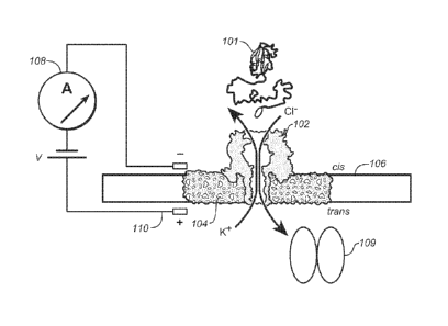
Referring now to Figure 1A, the present device operates to translocate a
substrate
protein 101 and comprises a pore protein 102 (a-hemolysin, or -AHL") embedded
in a lipid
bilayer 104 that is comprised in an - 25 pm aperture in a membrane 106
separating a fluid
compartment into a cis side, containing the protein 101, and a trans side, to
which the
protein 101 is going to be translocated through the pore 102. The device
includes a
controllable amplifier 108 for applying a constant voltage between a positive
electrode 110
on the trans side and a negative electrode on the cis side. A protein
translocase 109,
exemplified below as ClpX, is present on the trans side of the chamber.
Amplifier 108 also
provides a circuitry for detecting and, preferably, recording changes in ionic
current (i.e.
flow of ions such as the depicted Cl- and K+) that take place very rapidly as
the protein 101
translocates. In the examples, data were collected at 100 kHz, but high speed
data sampling
devices are known and may be used (e.g. 200MHz Model 7150 from Pentek, Inc).
Figure
1B shows a detailed view of the AHL pore protein 102 in the lipid bilayer 104
and also
shows the protein translocase 109 which is on the trans side and which is
acting on protein
101 which in the cartoon is threaded through and is on both sides of the pore
102. As shown
in Figure 1C, a model substrate protein bearing a Smt3 domain at its amino-
terminus is
coupled by a charged flexible linker to an ssrA tag at its carboxy-terminus.
The charged,
flexible tag is threaded through the nanopore into the trans-side solution,
while the folded
Smt3 domain at this point prevents complete translocation of the captured
protein. ClpX
present in the trans solution binds the C-terminal ssrA sequence of the
substrate protein.
Fueled by ATP hydrolysis, ClpX translocates along the protein tail toward the
channel, and
subsequently catalyzes unfolding and translocation of the Smt3 domain(s)
through the pore.
The Smt3 domains are folded, while the linker(s) are not. Smt3 is further
described in US
2009/0280535. "SUMO Fusion Protein Expression System for Producing Native
Proteins ."Demonstrated in the examples below is enzymatic control of protein
unfolding and
translocation through the sa-hemolysin nanopore. Segments of each substrate
protein were
discerned based on amino acid composition as they passed through the circa 50-
Angstrom-
long trans-membrane pore lumen (nanopore). The translocase enzyme used is
selected and
controlled to enable the device to provide protein sequence information. The
enzyme is
selected from a class of enzymes termed generally herein "protein
translocases," referring to
9
CA 02864824 2014-08-15
WO 2013/123379
PCT/US2013/026414
the ability of such enzymes to cause physical movement relative to a
substrate. Included
within the term as used herein is a class of enzymes often referred to as
"unfoldases," in that
they catalyze the unfolding of a native protein without affecting the primary
structure, i.e.
the primary sequence of the protein.
In certain embodiments, the substrate protein is tagged for recognition by the
translocase. One way to do this is the use of an ssrA tag. Various ssrA tags
are known, as
this is the mechanism used in several bacterial species for marking proteins
to be degraded
by a CIpX protease system. In the examples, the ssrA tag is a C-terminal 11
residue AA
sequence (shown at the end of SEQ ID NO: 5) of which subsets of this sequence
are
.. recognized by ClpA or ClpX uniquely. As noted, other sequences may be used.
IN certain
embodiments a protein nanopore protein or a chimeric nanopore as shown in
Figure 10 may
be embedded in a thin insulting membrane (for example, a lipid bilayer or a
graphene sheet)
separating two conductive aqueous solutions of differential voltage. Sensing
would be
imparted by the flow of ionic current through the nanopore: as the protein
translocated or
otherwise interacted with the pore, blockades of ion flow would occur,
providing an
electronic signal for subsequent analysis. This protein nanopore or chimeric
nanopore could
also be utilized in arrays or lab-on-chip devices for paralleled separation
and/or purification
of target proteins in mass.
The invention may be carried out in various apparatus for nanopore analysis,
such as
.. an array or a chip. The apparatus may be any of those described in
International Application
No. PCT/GB08/004127 (published as WO 2009/077734, entitled "Formation of
layers of
amphiphilic molecules"), PCT/GB10/000789 (published as WO 2010/122293 entitled
"Lipid
bilayer sensor array"), International Application No. PCT/GB10/002206
(published as WO
00/28132 entitled "Biochemical analysis instrument") or International
Application No.
PCT/US99/25679 (published as WO 2000/28312 entitled "Coupling method"). As
will
become apparent from the description below, the protein is translocated as a
single
polypeptide sequence, wherein individual amino acids pass sequentially through
the pore. A
number of proteins may be translocated serially.
10
CA 02864824 2014-08-15
WO 2013/123379 PCT/US2013/026414
DEFINITIONS
Unless defined otherwise, all technical and scientific terms used herein have
the same
meaning as commonly understood by those of ordinary skill in the art to which
this invention
belongs. Although any methods and materials similar or equivalent to those
described herein
can be used in the practice or testing of the present invention, the preferred
methods and
materials are described. Generally, nomenclatures utilized in connection with,
and techniques
of, cell and molecular biology and chemistry are those well-known and commonly
used in the
art. Certain experimental techniques, not specifically defined, are generally
performed
according to conventional methods well known in the art and as described in
various general
and more specific references that are cited and discussed throughout the
present specification.
For purposes of clarity, the following terms are defined below.
Ranges: For conciseness, any range set forth is intended to include any sub-
range
within the stated range, unless otherwise stated. As a non-limiting example, a
range of 120
to 250 is intended to include a range of 120-121, 120-130, 200-225, 121-250
etc. The term
"about" has its ordinary meaning of approximately and may be determined in
context by
experimental variability. In case of doubt, "about" means plus or minus 5% of
a stated
numerical value.
The term "nanopore" is used herein in its conventional sense to refer to any
small hole
or channel of the order of 0.5 to 10 nanometers in internal diameter. The term
"nanopore"
includes both biological (e.g. a-hemolysin) or artificial nanopores. The
present nanopores
can vary in dimensions, for example it can have a diameter of between about
0.5 nm and 10
nm in size. For example, the diameter can be about 0.5 nm, 1 nm. 1.25 nm. 1.5
nm, 1.75 nm,
2 nm, 2.25 nm, 2.5 nm, 2.75 nm, 3 nm. 3.5 nm, 4 nm, 4.5 nm, 5 nm, 6 nm, 7 nm,
8 nm, 9nm,
10 nm, or any dimension there between Biological nanopores can be created by
pore
proteins. Artificial nanopores can be made by micromolding or drilling. They
also can be
made by etching a somewhat larger hole (several tens of nanometers) in a piece
of silicon,
and then gradually filling it in using ion-beam sculpting methods which
results in a much
smaller diameter hole.
The term "pore protein" is used herein in its conventional sense to refer to
pore-
forming proteins (PFPs) which assemble into ring-like structures in the
vicinity of the target
membrane to expose sufficient hydrophobicity to drive spontaneous bilayer
insertion. Pore
11
CA 02864824 2014-08-15
WO 2013/123379 PCT/US2013/026414
proteins are typically (but not exclusively) produced by bacteria, such C.
septicum and S.
aureus. PFPs can be alpha-pore-forming toxins, such as Cytolysin A of E. coli;
or beta-pore-
forming toxins, such as a-hemolysin and Panton-Valentine leukocidin (PVL); or
binary
toxins, such as Anthrax toxin; or cholesterol-dependent cytolysins (CDCs),
such as
Pneumolysin; or Small pore-forming toxins, such as Gramicidin A. A preferred
pore protein
is a-hemolysin (AHL).
The term "a-hemolysin" is used herein in its conventional sense to refer to a
pore-
forming toxin from the bacterium, Staphylococcus aureus. a-hemolysin consists
mostly of
beta-sheets (68%) with only about 10% alpha-helices. The hla gene on the S.
aureus
chromosome encodes the 293 residue protein monomer, which forms heptameric
units on the
cellular membrane to form a complete beta-barrel pore. This structure allows
the toxin to
perform its major function, development of pores in the cellular membrane.
The term "membrane" is used herein in its conventional sense to refer to a
thin, film-
like structure. The membrane separating the cis and trans chambers comprises
at least one
pore or channel. Membranes can be generally classified into synthetic
membranes and
biological membranes. Any membrane may be used in accordance with the
invention.
Suitable membranes are well-known in the art. The membrane is preferably an
amphiphilic
layer. An amphiphilic layer is a layer formed from amphiphilic molecules, such
as
phospholipids, which have both at least one hydrophilic portion and at least
one lipophilic or
.. hydrophobic portion. The amphiphilic layer may be a monolayer or a bilayer.
The
amphiphilic molecules may be synthetic or naturally occurring. Non-naturally
occurring
amphiphiles which form a monolayer are known in the art and include, for
example, block
copolymers (Gonzalez-Perez et al., Langmuir, 2009, 25, 10447-10450).
The term "lipid bilayer" is used herein in its conventional sense to refer to
a thin polar
membrane made of two layers of lipid molecules, arranged so that the
hydrophilic phosphate
heads point "out" to the water on either side of the bilayer and the
hydrophobic tails point
"in" the core of the bilayer. The lipid bilayers are usually a few nanometers
in width, and
they are impermeable to most charged water-soluble molecules. Lipid bilayers
are large
enough structures to have some of the mechanical properties of liquids or
solids. The area
compression modulus Ka, bending modulus Kb, and edge energy, can be used to
describe
them. Solid lipid bilayers also have a shear modulus, but like any liquid, the
shear modulus is
12
CA 02864824 2014-08-15
WO 2013/123379
PCT/US2013/026414
zero for fluid bilayers. Lipid bilayers can also be supported by solid
substrates having
apertures, such as heat shrink tubing, fused silica, borosilicate glass, mica,
and oxidized
silicon. Lipids may be applied, e.g., through Langmuir-Blodgett technique,
vesicle fusion
processes or the combination of the two.
The term "NTP" is used herein in its conventional sense to refer to nucleoside
triphosphate, a molecule containing a nucleoside bound to three phosphates,
making it a
nucleotide. NTP can be adenosine triphosphate (ATP), guanosine triphosphate
(GTP),
cytidine triphosphate (CTP), 5-methyluridine triphosphate (m5UTP ), uridine
triphosphate
(UTP), deoxyadenosine triphosphate (dATP), deoxyguanosine triphosphate (dGTP),
deoxycytidine triphosphate (dCTP), deoxythymidine triphosphate (dTTP) or
deoxyuridine
triphosphate (dUTP). "NTP" also refers to other less abundant NTPs, such as
intermediates
of nucleotide metabolism, including less common natural
The term "NTP driven unfoldase" is used herein in its conventional sense to
refer to
an NTP-dependent enzyme that catalyzes protein unfolding. The very common NTP
driven
.. unfoldases are ATP-dependent proteases, such as proteasomal ATPases, AAA
proteases, or
AAA+ enzymes (defined below); membrane fusion proteins, such as NSF (N-
Ethylmaleimide-sensitive fusion protein)/Sacl8p (N-Ethylmaleimide-sensitive
fusion protein
homologue in yeast) or p97 / VCP / Cdc48p (97-kDa valosin-containing protein);
Pexlp and
Pex6p (peroxisomal ATPase); Katanin and SKD1 (Vps4p homolog in mouse)/Vps4p
(Vacuolar protein sorting 4 homolog in yeast); Dynein (motor protein); DNA
replication
proteins, such as ORC (origin recognition complex), Cdc6 (cell division
control protein 6),
MCM (minichromosome maintenance protein), DnaA, or RFC (replication factor
C)/clamp-
loader; RuvB (holliday junction ATP-dependent DNA helicase RuvB, EC=3.6.4.12);
TIP49a/TIP49 and TIP49b/TIP48 (eukaryotic RuvB-like protein).
The term "AAA+ enzyme" is used herein in its conventional sense to refer to
the
AAA+ superfamily of enzymes. AAA+ is an abbreviation for ATPases Associated
with
diverse cellular Activities. They share a common conserved module of
approximately 230
amino acid residues. This is a large, functionally diverse protein family
belonging to the
AAA+ superfamily of ring-shaped P-loop NTPases, which exert their activity
through the
energy-dependent remodeling or translocation of macromolecules. Examples
include ClpAP,
ClpXP, C1pCP, Hs1VU and Lon in bacteria and their homologues in mitochondria
and
chloroplasts. With the exception of Lon, AAA+ enzymes (sometimes referred to
as
13
CA 02864824 2014-08-15
WO 2013/123379
PCT/US2013/026414
unfoldases or proteases) consist of regulatory (ATPase) and proteolytic
subunits, while Lon is
a single polypeptide containing both regulatory and proteolytic domains. ClpX
and ClpA
dock with ClpP to form ClpXP and ClpAP proteases, whereas Hs1U docks with Hs1V
to form
another protease, Hs1VU. ClpA and ClpX form hexamers, in contrast to ClpP
which forms
heptamers. Hs1U and Hs1V each form hexamers, although Hs1U heptamers have also
been
reported. The regulatory subunits ClpA, ClpX and Hs1U function as chaperones.
Further
details on ClpX may be found in Maillard et al., "ClpX(P) generates mechanical
force to
unfold and translocate its protein substrates," Cell 145:459-4669 (April 29,
2011). As
reported there, the ClpX motor shares is basic design with other AAA+ enzymes,
including
prokaryotic C1pA, ClpB, HsIu, FtsH or Lon. The AAA+ enzyme is also referred to
as an
-AAA+ molecular motor". Further description of the AAA+ superfamily is found
in Ogura et
al. "AAA+ superfamily ATPases" common structure-diverse function," Genes to
Cells,
6:575-597 (2001). AS described there, the AAA+ family members associated with
mitochondria are Bcslp, Lon/Pimlp, ClpX and Hsp78.
The term "HsIU" is used herein in its conventional sense to refer to ATP-
dependent
protease ATPase subunit HsIU, also called unfoldase HsIU. HsIU is a member of
the
Hsp100 and Clp family of ATPase. It can also form complex with HsIV to act as
an
unfoldase (See, Bochtler et al., "The structures of HsIU and the ATP-dependent
protease
HsIU-HsIV," Nature 403(6771):800-805 (2000).
The term -Lon protease" is used herein in its conventional sense to refer to a
family
of proteases found in archaea, bacteria and eukaryotes. Lon proteases are ATP-
dependent
serine peptidases belonging to the MEROPS peptidase family S16 (ion protease
family, clan
SF). In the eukaryotes the majority of the Lon proteases are located in the
mitochondria'
matrix. In yeast, the Lon protease PIM1 is located in the mitochondria'
matrix. It is required
for mitochondria' function, it is constitutively expressed but is increased
after thermal stress,
suggesting that PIM1 may play a role in the heat shock response.
The term "protein translocase" is used herein in its conventional sense to
mean a
protein-binding polypeptide, such as a polypeptide which is able to control
movement of a
protein substrate, for example an enzyme, enzyme complex, or a part of an
enzyme complex
that operates on a protein substrate and moves it relative to the enzyme in a
processive
manner, i.e. as a function of enzymatic activity. The term "processive" is
understood in the
art to refer to a stepwise activity in which the enzyme "processes" the
substrate in a number
14
CA 02864824 2014-08-15
WO 2013/123379
PCT/US2013/026414
of steps. In the present case, the protein translocase generally processes the
protein to be
translocated in a sequential manner, that is, moving along the primary amino
acid sequence.
For convenience, a number of enzymes also commonly called "unfoldases" are
included in
this definition, in particular NTP driven unfoldases. Also specifically
included in this
definition is the AAA+ enzyme superfamily and the ClpX member of this
superfamily.
Also included as examples of the general term "protein translocase" are
proteases
such as Lon protease and HsIU, which enzymes are either modified to eliminate
the
enzymatic cleavage activity of the enzyme or arranged so that cleavage occurs
after the
sequence is translocated through the nanopore.
Other exemplary protein translocases are related to ClpX, (which is also an
unfoldase), e.g. ClpA, mitochondrial protein translocases TOM (translocase of
the outer
membrane) or other TOM and TIM proteins. The chosen protein translocase can
also be any
part of the mitochondrial protein translocase complex, such as the chaperones,
TOM import
receptor, TOM channel complex, and "motor" proteins.
The term "ClpX enzyme" of "ClpX" is used herein in its conventional sense to
refer
to a member of the HSP (heat-shock protein) 100 family having the Uniprot
designation
clpX and having the 424 amino acid sequence given there, processed into mature
form, as a
subunit. ClpX subunits associate to form a six-membered (homohexameric) ring
that is
stabilized by binding of ATP or nonhydrolysable analogs of ATP. The N-terminal
domain of
ClpX is a C4-type zinc binding domain (ZBD) involved in substrate recognition.
ZBD forms
a very stable dimer that is essential for promoting the degradation of some
typical ClpXP
substrates such as 10 and MuA. It is described further in Wawrzynow et al,
"The ClpX heat-
shock protein of Escherichia coli, the ATP-dependent substrate specificity
component of the
ClpP-ClpX protease, is a novel molecular chaperone," EMBO J. 1995 May 1;
14(9): 1867-
1877, An amino acid sequence is also given at eclowiki.net under "clpX: gene
products".
Similarly, ClpA refers to the UniProt/Swiss-Prot entry clpA, which has a 758
amino
acid sequence given there for the ClpA subunit. It forms a complex of six ClpA
subunits
assembled into a hexameric ring in the presence of ATP. It is a component of
the C1pAP
complex composed of six C1pA subunits assembled into a hexameric ring in the
presence of
.. ATP, and fourteen ClpP subunits arranged in two heptameric rings. Binds to
ClpS.
CA 02864824 2014-08-15
WO 2013/123379
PCT/US2013/026414
The term "non-denatured protein" is used herein in its conventional sense,
i.e. a
protein that is at least partially folded into a native secondary and tertiary
structure, with any
native cysteine bonds, hydrogen bonding and multimeric form essentially
intact. This is
contrasted with a denatured protein, which usually is insoluble and
aggregated.
The term "negatively charged amino acids" is used herein in its conventional
sense,
i.e. meaning proteins that have surfaces rich with negatively charged amino
acids like
glutamate and aspartate.
GENERAL METHOD AND APPARATUS
Translocation of proteins through a nanopore sensor device offers a number of
possible applications, including sequencing, structure/fold analysis,
purification/separation,
intracellular protein delivery, and insight into the mechanics of enzymes
driving the
translocating polypeptide. Unlike nucleic acids, proteins are generally not
uniformly charged
(making it difficult to drive translocation via an applied voltage) and fold
into complex, large,
and stable structures that cannot transverse a nanopore's aperture. To address
these issues,
unfolding and translocation of natively folded proteins through a protein
nanopore may be
accomplished via a variety of enzymes, exemplified by the E. coli ClpX (or
other types of
protein translocases/unfoldases).
The present methods and devices may be used to measure one or more
characteristics
of the protein being translocated.
A variety of different types of measurements may be made. This includes
without
limitation: electrical measurements and optical measurements. Possible
electrical
measurements include: current measurements, impedance measurements, tunnelling
measurements (Ivanov AP et al., Nano Lett. 2011 Jan 12;11(1):279-85), and FET
measurements (International Application WO 2005/124888). Optical measurements
may be
combined with electrical measurements (Soni GV et al., Rev Sci Instrum. 2010
Jan;81(1):014301). The measurement may be a transmembrane current measurement
such as
measurement of ionic current flowing through the pore.
Electrical measurements may be made using standard single channel recording
equipment as described in Stoddart D et al., Proc Natl Acad Sci,
12;106(19):7702-7,
Lieberman KR et al, J Am Chem Soc. 2010;132(50):17961-72, and International
Application
16
CA 02864824 2014-08-15
WO 2013/123379 PCT/US2013/026414
WO-2000/28312. Alternatively, electrical measurements may be made using a
multi-channel
system, for example as described in International Application WO-2009/077734
and
International Application WO-2011/067559.
The signal measurement is typically indicative of the identity of the protein
or the
amino acids in the protein. The signal can therefore be used to characterize,
such as
sequence, the protein as discussed above.
1. Enzymes used for translocation
E. coil ClpX was used in the working example of the present device; it was
selected
for initial work because it generates sufficient mechanical force (>20 pN) to
denature stable
protein folds, and because it translocates along proteins at a suitable rate
for primary
sequence analysis by nanopore sensors (up to 80 amino acids per second). ClpX
is part of the
ClpXP proteasome-like complex. ClpP is composed of a diheptameric cylinder-
like protease
that binds at one or both ends a regulatory hexameric ATP-dependent
unfoldase/translocase
complex (e.g. ClpX). ClpX acts as a gate that allows for tagged proteins to
enter into the
inner lumen of the ClpP protease complex for subsequent degradation. The ATP-
dependent
unfoldase/translocase activity of the hexameric protein complex, ClpX, is
employed to unfold
and thread proteins through a nanopore.
ClpX may be prepared (and was prepared here) as described in Martin et al. -
Rebuilt
AAA+ Motors reveal operating principles for ATP-fuelled machines," Nature
437:1115-1120
(2005). A variety of alternative enzymes may serve the function of the protein
translocase in
the present method and device. As described there, combinations of 2, 3 or 6
ClpX-deltaN
subunits, lacking N terminal amino acids 1-60, linked with a 20 amino acid
long linker were
prepared as a single polypeptide chain.
The repertoire of cellular functions involving AAA+ ATPases is diverse. A
subset of
AAA+ proteins is not active as ATPases and some do not even bind ATP. It seems
however,
that these proteins form complexes with other family members which do serve as
ATPases.
However, the ATPase subunits or domains of all known ATP-dependent proteases
belong to
the AAA+ family.
One example of a suitable AAA+ enzyme is Clp/Hsp100 ATPases. Clp/Hsp100
ATPases are responsible for selecting protein targets. For example, the two
different bacterial
17
CA 02864824 2014-08-15
WO 2013/123379 PCT/US2013/026414
ATPases ClpX and ClpA impart distinct substrate preferences to the ClpP
peptidase. The
ssrA degradation sequence, an 11-residue peptide that is appended to
polypeptides stalled on
the ribosome, is recognized by both ClpX and ClpA. Mutational analysis of the
ssrA
sequence revealed that this same tag is recognized by the two unfolding
enzymes via different
residues, further confirming the distinct binding preferences of each ATPase.
Using the energy from ATP-hydrolysis, Clp/Hsp100 enzymes actively direct
structural changes in their substrates. These ATP-driven structural changes
result in two
distinct biological outcomes for the protein substrates: degradation or
remodeling. ClpA,
based on its ability to degrade casein, was the first prokaryotic Clp/ Hsp100
protein
.. functionally identified. Accordingly, the degradation pathway for the
Clp/Hsp100 proteins is
the better characterized of the two processes. During Clp/Hsp100-facilitated
protein
degradation, first, the Clp/Hsp100 component recognizes and selects a target
protein. The
enzyme binds to a short peptide sequence (e.g., the ssrA degradation tag)
usually located near
either the C or N terminus of the substrate. Then, in a reaction that requires
multiple cycles
of ATP-hydrolysis, the enzyme unfolds and directionally translocates the
target substrate to
the peptidase chamber where it is degraded.
Mitochondrial protein translocases may also be used. These may be translocases
TOM
or TIM from human or eukaryotic cells, such as TOMM20 (translocase of outer
mitochondria' membrane 20 homolog), TOMM22 (mitochondria' import receptor
subunit 22
.. homolog), TOMM40 (translocase of outer mitochondrial membrane 40 homolog),
TOM7
(translocase of mitochondrial outer membrane 7), TOMM7 (translocase of outer
mitochondrial membrane 7 homolog), TIMM8A (translocase of inner mitochondrial
membrane 8 homolog A), TIMM50 (translocase of inner mitochondria] membrane 50
homolog). For example, TOMM40 is embedded into outer membranes of mitochondria
and is
required for the movement of proteins into mitochondria. More, precisely,
TOMM40 is the
channel-forming subunit of a translocase of the mitochondrial outer membrane
(TOM) that is
essential for protein transport into mitochondria
Another alternative protein translocase may be prepared from the Sec family of
translocases. These include SecB (chaperone protein), SecA (ATPase), SecY
(internal
membrane complex in prokaryotes), SecE (interal membrane complex in
prokaryotes), SecG
(internal membrane complex in prokaryotes) or Sec61 (internal membrane complex
in
eukaryotes), SecD (membrane protein), and SecF (membrane protein).
18
CA 02864824 2014-08-15
WO 2013/123379 PCT/US2013/026414
Another alternative protein translocase is Type III Secretion System (TTS)
Translocase, such as HrcN and any of the 20 subunits of the TTS translocases,
or Sec-
independent periplasmic protein translocase TatC.
Other alternative protein translocases are chaperones. These are proteins that
assist the
non-covalent folding or unfolding and the assembly or disassembly of other
macromolecular
structures, but do not occur in these structures when the structures are
performing their
normal biological functions having completed the processes of folding and/or
assembly.
Many chaperones are heat shock proteins, that is, proteins expressed in
response to elevated
temperatures or other cellular stresses. Hsp 70, as is known, refers to 70-kDa
heat shock
proteins (Hsp70s), such as DnaK, HscA (Hsc66), and HscC (Hsc62) in
prokaryotes, and
Hsc70, Hsp70, BiP or Grp78 (binding irnmunoglobulin protein), mtHsp70 or Grp75
in
eukaryotic organisms, and human Hsp70 proteins, such as Hsp70, Hsp70-2, Hsp70-
4, Hsp70-
4L, Hsp70-5, Hsp70-6, Hsp70-7, Hsp70-8, Hsp70-9, Hsp70-12a, Hsp70-14. Hsp70
proteins
are central components of the cellular network of molecular chaperones and
folding catalysts.
Hsp70s assist a wide range of folding processes, including the folding and
assembly of newly
synthesized proteins, refolding of misfolded and aggregated proteins, membrane
translocation
of organellar and secretory proteins, and control of the activity of
regulatory protein. ATP
binding and hydrolysis are essential in vitro and in vivo for the chaperone
activity of Hsp70
proteins.
Hsp70 chaperone families are recognized as most common remodeling enzyme
together with Hsp60 chaperone families. Hsp70s and Hsp6Os prevent off-pathway
interactions during protein folding by providing an isolated environment for
the folding
protein. In contrast, the Clp/Hsp100 unfolding enzymes actively direct the
structural
changes in their substrates. Clp/Hsp100s act on folded and assembled
complexes, as well as
improperly folded and aggregated proteins.
HSP90 aids the delivery of the mitochondrial preprotein to the TOM complex in
an
ATP-dependent process.
Hsp100 (Clp family in E. coli) proteins have been studied in vivo and in vitro
for their
ability to target and unfold tagged and misfolded proteins. Proteins in the
Hsp100/Clp family
form large hexameric structures with unfoldase activity in the presence of
ATP. These
proteins are thought to function as chaperones by processively threading
client proteins
19
CA 02864824 2014-08-15
WO 2013/123379 PCT/US2013/026414
through a small 20 A (2 nm) pore, thereby giving each client protein a second
chance to fold.
Some of these Hsp100 chaperones, like ClpA and ClpX, associate with the double-
ringed
tetradecameric serine protease Clp13: instead of catalyzing the refolding of
client proteins,
these complexes are responsible for the targeted destruction of tagged and
misfolded proteins.
Hsp104, the Hsp100 of Saccharomyces cerevisiae, is essential for the
propagation of
many yeast prions. Deletion of the HSP104 gene results in cells that are
unable to propagate
certain prions.
The enzyme used in the working examples has the sequence:
MGSSHHHHHHSSHMSALPTPHEIRNHLDDYVIGQEQAKKVLAVAVYNHYKRLRNG
DTSNGVELGKS NILLIGPTGS GKTLLAETLARLLDVPFTMADATTLTEAGYVGEDVE
NIIQKLLQKCDYDVQKAQRGIVYIDEIDKISRKSDNPSITRDVSGEGVQQALLKLIEGT
VAAVPPQGGRKHPQQEFLQVDTSKILFICGGAFAGLDKVISHRVETGSGIGFGATVKA
KSDKASEGELLAQVEPEDLIKFGLIPEFIGRLPVVATLNELSEEALIQILKEPKNALTKQ
YQALFNLEGVDLEFRDEALDAIAKKAMARKTGARGLRSWEAALLDTMYDLPSMED
VEKVVIDESVIDGQSKPLLIYGKPEAQQASGEASGAGGSEGGGSEGGTSGATMSALP
TPHEIRNHLDDYVIGQEQAKKVLAVAVYNHYKRLRNGDTSNGVELGKSNILLIGPTG
SGKTLLAETLARLLDVPFTMADATTLTEAGYVGEDVENIIQKLLQKCDYDVQKAQR
GIVYIDEIDKISRKSDNPSITRDVSGEGVQQALLKLIEGTVAAVPPQGGRKHPQQEFLQ
VDTSKILFICGGAFAGLDKVISHRVETGSGIGFGATVKAKSDKASEGELLAQVEPEDLI
.. KFGLIPEFIGRLP V VATLN ELS EEAL1QILKEPKN ALTKQ Y QALFN LEGV D LEFRDEAL
DAIAKKAMARKTGARGLRS1VEAALLDTMYDLPSMEDVEKVVIDESVIDGQSKPLLI
YGKPEA QQA SGEASG AGGSEGGGSEGGS SG ATMS A LPTPHEIRNHLDDYVIG QEQ A
KKVLAVAVYNHYKRLRNGDTSNGVELGKSNILLIGPTGSGKTLLA ETLARLLDVPFT
MADATTLTEAGYVGEDVENIIQKLLQKCDYDVQKAQRGIVYIDEIDKISRKSDNPSIT
RDVSGEGVQQALLKLIEGTVAAVPPQGGRKHPQQEFLQVDTSKILFICGGAFAGLDK
VISHRVETGSGIGFGATVKAKSDKASEGELLAQVEPEDLIKFGLIPEFIGRLPVVATLN
ELSEEALIQILKEPKNALTKQYQALFNLEGVDLEFRDEALDAIAKKAMARKTGARGL
RSWEAALLDTMYDLPSMEDVEKVVIDESVIDGQSKPLLIYGKPEAQQASGE. (SEQ
ID NO: 8)
It is a synthetically designed trimer of ClpX subunits that is expressed as a
single
chain. As noted above, a variety of translocase constructs may be used in the
present system
CA 02864824 2014-08-15
WO 2013/123379 PCT/US2013/026414
2. Enzymes may be coupled to the nanopore, free in solution on one side,
and/or present
on both sides of the nanopore
Translocase on the cis side (same side as the substrate protein)
In certain embodiments, for example as shown in Figure 10, ClpA or Clp X may
be
coupled to the nanopore. An engineered alpha- hemolysin/C1pP fusion protein
pore may be
assembled to form an active heptameric protein nanopore covalently fused at
its N-terminal
cap domain to the ClpX-binding domain of the ClpP heptamer complex. Fusion of
the ClpX-
binding domain of ClpP to the top of the nanopore will enable ClpX to assemble
in solution,
attach to the ClpP domain, and function on the top of nanopore
Figure 10 illustrates a fusion protein comprising the a-hemolysin pore protein
subunits fused to subunits of a ClpP protein. The ClpX protein translocase can
then non-
covalently "dock" onto the ClpP subunits. In this embodiment the protein
translocase is on
the cis side of, and fused to, the nanopore.
In the embodiment of Figure 10, the axial pores of the translocase and the
nanopore
must be aligned in the correct orientation; that is, ClpX must be bound to the
nanopore in
such a way that as the protein substrate is captured from solution and driven
through the
ClpX central cavity, it then directly enters into the AHL (alpha hemolysin)
upper lumen and
is eventually forced through the entire nanopore (See Figure 10). To achieve
this goal, a
ClpX-binding domain may be fused to the head of AHL. In nature, the ClpX
hexamer
naturally binds to the head domains at each opening of the tetradecameric
(double heptameric
rings) protease ClpP and acts as a gate that only allows tagged proteins to
enter the
proteolytic chamber. By fusing the ClpX-binding domain of ClpP onto the head
of AHL, it
will enable ClpX to directly bind with high affinity atop the nanopore via a
natural protein-
protein interaction. Fortunately, both AHL and ClpP assemble into
homoheptameric rings
with strikingly similar diameters; thus, fusion of the ClpX binding domain of
the ClpP
monomer to the head of each AHL monomer will create a heptameric nanopore
complex
composed of these ClpP/AHL fusion monomers. A previous study has investigated
the fusion
of AHL monomers, showing that AHL is indeed tolerant of fusions at both the N
and C
termini. In addition, a study investigated deletions at the C-terminus of
ClpP, showing that it
is not a region critical for heptamer formation or ClpX binding. This data
suggests that ClpP
would be tolerant of an AHL fusion to its C-terminus, while fusion to the ClpP
N-terminus
21
CA 02864824 2014-08-15
WO 2013/123379
PCT/US2013/026414
would almost certainly inhibit binding of ClpX as the N-terminal loops have
been shown to
be critical for such activity. Based on these previous studies. the AHL/ClpP
fusion monomer
is designed with a single AHL monomer fused to the C-terminal of a truncated
ClpP
monomer (separated by a flexible 5-15 Gly-Ser linker to allow each protein
monomer to fold
properly). These ClpP¨ peptide linker¨AHL fusion protein DNA sequences may
constructed via PCR assembly, His-tagged, and inserted inside pT7-SC1
expression vectors.
Expression of the fusion protein may be done through coupled in-vitro
transcription and
translation using the T7-S30 expression system, used previously to express AHL
fusions, and
purified with Ni-NTA affinity chromatography.
Another embodiment where the translocase is on the cis side of the device
involves
the use of accessory proteins. In this case, proteins that bind to the
substrate protein are used
to facilitate control of movement of the substrate protein to or though the
nanopore. For
example, the protein present in the trans side (as in Figure 1) need not be an
active
translocase/unfoldase but rather another protein (for example trigger factor)
that non-
specifically binds to unfolded portions of polypeptides. Trigger factor is a
ribosome-
associated molecular chaperone and is the first chaperone to interact with a
nascent
polypeptide. It acts as a chaperone by maintaining the newly synthesized
protein in an open
conformation. Other chaperonins or heat shock proteins could be used.
This trans side protein (e.g. trigger factor) would thereby sequentially
capture the
unfolded substrate protein as it is translocated into the trans solution by
the cis side
translocase/unfoldase, preventing the substrate protein from moving back into
the cis side.
In another embodiment, the substrate protein is provided with a factor that
blocks its
unfolding by the unfoldase until a predetermined state is reached. In this
embodiment, both
substrate protein and unfoldase are added to the cis side. Substrate proteins
are tagged at one
terminus with an unfoldase-binding motif (for example the ssrA tag for ClpX),
a pore-
targeting domain (for example a charged poly-peptide tail that will be pulled
into the pore
under an applied voltage), and an unfoldase-resistant protein (for example
dihydrofolate
reductase or barnase in presence of stabilizing ligands are resistant to
unfolding by ClpX; see,
Hoskins, JR et al. ClpAP and ClpXP degrade proteins with tags located in the
interior of the
primary sequence. PNAS August 20, 2002 vol. 99 no. 17 11037-11042). The
substrate
protein therefore is protected from the unfoldase with this "blocking domain"
between the its
folded domain and the pore targeting and unfoldase-binding motif domains.
22
CA 02864824 2014-08-15
WO 2013/123379 PCT/US2013/026414
The blocking domain protein (fused with the pore-targeting domain and
unfoldase-
targeting motif domain) may be chemically or enzymatically attached to the
substrate protein
post-translationally.
In the bulk cis solution, the unfoldase binds to the unfoldase-binding motif
and
translocates along the pore-targeting domain. Translocation of the unfoldase
along the tagged
substrate stops once the enzyme approaches the unfoldase-resistant protein
(the "blocking
domain"). When this tagged-substrate-protein/unfoldase complex is captured by
the
nanopore, unfolding and translocation of the blocking domain is initiated. It
is catalyzed by
the extra destabilizing forces imparted by the voltage at the pore and/or
other protein
translocases/unfoldases present in the trans solution that interact with the
tagged substrate
protein tail after capture and threading of the substrate tail through the
pore. Unfolding and
translocation of the entire substrate protein by the cis-bound unfoldase
through the nanopore
is then possible after the pore-activated unfolding of the blocking domain is
catalyzed.
An example of a blocking domain sequence strategy example sequence is as
follows:
N-terminus¨substrate protein¨blocking domain¨charged tail¨unfoldase-binding
motif¨
C-terminus.
Translocase on the trans side
ClpX complexes are placed in solution on the trans side of the nanopore, and
the
substrate protein dissolved on cis side of the nanopore will be forced to
thread through the
nanopore beginning at the N or C terminus (for example, voltage-driven by
engineering a few
charged amino acids into the protein terminus, such as 5-10 Asp, Lys or Arg
residues) where
the ClpX complex will capture this tagged polypeptide tail, and begin
mechanically pulling/
translocating the substrate down through the nanopore. Native ClpX binding,
unfolding, and
translocation activity of tagged proteins is used to control the movement of
proteins through a
nanopore sensor for subsequent analysis. A wild type protein nanopore or other
solid-state
nanopore may be utilized if ClpX or another translocase such as ClpA is placed
on the trans
side of the device, in solution, where it is allowed to capture tagged protein
tails that were
threaded from the cis side through the nanopore to the trans side solution
(that is, "fishing"
for the ClpX). Upon capture of this tagged-tail, the ClpX complex would be
able to begin
mechanically pulling on the protein from across the pore, until it is
eventually able to unfold
and thread the entire polypeptide through the pore and into the trans side
solution. The initial
23
CA 02864824 2014-08-15
WO 2013/123379 PCT/US2013/026414
threading of the tagged-tail could be accomplished via the addition of several
charged
residues proximal to the tag sequence on the N or C terminus of the target
protein, wherein a
voltage differential would drive the charged tail through the nanopore, making
it available to
"fish" for ClpX.
As shown, e.g. in Example 1 and Example 2 below, the protein translocase is
added in
solution to the trans side of the chamber; it serves to unfold the protein to
be translocated by
pulling it through the nanopore, which is too narrow to permit passage of the
folded protein.
The same or a different protein translocase may be located on the cis side of
the nanopore for
unfolding the protein.
__ 3. Nanonore sensing
The method of sensing in the present method and device may involve measuring
one,
two, three, four or five or more characteristics of the protein. The one or
more characteristics
are preferably selected from (i) the length of the protein, (ii) the identity
of the protein, (iii)
the sequence of the protein, (iv) the secondary or tertiary structures of the
protein and (v)
whether or not the protein is modified. Any combination of (i) to (v) may be
measured in
accordance with the invention.
For characteristic (i), the length of the protein may be measured using the
number of
interactions between the protein and the pore, and/or the dwell time of the
protein as it
translocates through the pore.
For characteristic (ii), the identity of the protein may be measured in a
number of
ways. The identity of the protein may be measured in conjunction with
measurement of the
sequence of the protein or without measurement of the sequence of the protein.
The former is
straightforward; the protein is sequenced and thereby identified. The latter
may be done in
several ways. For instance, the presence of a particular motif in the protein
may be measured
(without measuring the remaining sequence of the protein). Alternatively, the
measurement
of a particular electrical and/or optical signal in the method may identify
the protein as
coming from a particular source.
For characteristic (iii), the sequence of the protein can be determined as
described
herein. The sequence may be determined on an individual amino acid residue-by-
residue
basis, or may be read in blocks of amino acids, which may be mapped to known
protein
24
CA 02864824 2014-08-15
WO 2013/123379 PCT/US2013/026414
sequences, in a manner analogous to re-sequencing of DNA. Thus the method need
not
resolve each individual amino acid, but rather one could just resolve "words"
or
blocks/chunks of amino acids (e.g. 2 to 10 aa) that would still enable
identification of the
protein/polypeptide sequence.
For characteristic (iv), the secondary and tertiary structures may be measured
in a
variety of ways. For instance, if the method involves an electrical
measurement, the
secondary structure (e.g. detection of an alpha helix region versus a loop
region or a beta
sheet region) may be measured using a change in dwell time or a change in
current flowing
through the pore.
For characteristic (v), the presence or absence of any modification may be
measured.
The method preferably comprises determining whether or not the protein is
modified by
methylation phosphorylation, oxidation, by damage (e.g. misfolding or covalent
modification
of an amino acid), by glycosylation or with one or more labels, tags or
spacers. Specific
modifications will result in specific interactions with the nanopore which can
be measured
using the methods described below.
As discussed below, the present methods and device can extract protein
sequence
information from the protein being translocated by analysis of the ionic
current measured
through the nanopore. This is partly dependent on the use of sensitive
electronics such as a
patch clamp amplifier, a finite state machine, and signal processing such as
weighted
averaging, all described below. In addition, the present methods involve
analysis of various
cunent states that have now been found to be associated with the transit of
the protein
through the nanopore and the concomitant current blockage, unblockage or
modulation. As
described below, the signal detected by enzyme-mediated protein traversal will
enter a so-
called "ramping state" which is characteristic of binding of the enzyme to the
substrate
protein, then a series of separate amplitude transitions as the protein
translocates through the
nanopore, followed by an open state equivalent to current through the pore
prior to
translocation. In summary, using the exemplified experimental setup, one can
observe distinct
states (see Fig. 2) of
(i) open channel current, prior to translocation ¨ about 30-35 pA, or 32-36
pA;
(ii) decrease to about 11-15 (e.g. about 13-15) pA upon capture of the protein
to be
translocated (i.e. the protein blocks the nanopore opening;
CA 02864824 2014-08-15
WO 2013/123379 PCT/US2013/026414
(iii) decrease to or below 10 pA and various amplitude changes (including a
"ramping" effect discussed below), as the enzyme binds the protein and
translocates along the
protein tail toward the nanopore (i.e. the enzyme blocks more of the nanopore
opening);
(iv) after unfolding of the protein, the entire protein is translocated
through the
nanopore, generating a unique current pattern;
(v) return to open channel state.
Importantly, state (iv) (Fig. 2A and 2B) presents a current pattern that can
be
correlated to protein structure. As described below, the artificial linkers
used in the examples
showed different current amplitudes and duration. When analyzing a protein to
determine an
unknown feature such as sequence, one may correlate observed changes in
amplitude and
RMS noise to the amino acid-dependent features of the translocated protein.
This includes but
is not limited to tertiary and secondary structures, amino acid sequence and
post-translational
modifications
The nanopore biosensing technology is based on the blockage of ionic current
that
occurs when a molecule translocates through and/or interacts with a pore under
an applied
voltage. The current blockade is thus dependent upon the applied voltage and
the properties
of the interacting molecule (for example, charge, size).
Time-dependent transport properties of the nanopore aperture may be measured
by
any suitable technique. The transport properties may be a function of the
medium used to
transport the polypeptide, solutes (for example, ions) in the liquid, the
polypeptide (for
example, chemical structure of the monomers), or labels on the polypeptide.
Exemplary
transport properties include current, conductance, resistance, capacitance,
charge,
concentration, optical properties (for example, fluorescence and Raman
scattering), and
chemical structure. Desirably, the transport property is current.
Exemplary means for detecting the current between the cis and the trans
chambers
have been described in WO 00/79257, U.S. Patent Nos. 6,46,594, 6,673
6,673,615,
6,627,067, 6,464,842, 6,362,002, 6,267,872, 6,015,714, 6,428,959, 6,617,113
and 5,795,782
and U.S. Publication Nos. 2004/0121525, 2003/0104428, and 2003/0104428, and
can
include, but are not limited to, electrodes directly associated with the
channel or pore at or
near the pore aperture, electrodes placed within the cis and the trans
chambers, ad insulated
26
CA 02864824 2014-08-15
WO 2013/123379
PCT/US2013/026414
glass micro-electrodes. The electrodes may be capable of, but not limited to,
detecting ionic
current differences across the two chambers or electron tunneling currents
across the pore
aperture or channel aperture. In another embodiment, the transport property is
electron flow
across the diameter of the aperture, which may be monitored by electrodes
disposed adjacent
to or abutting on the nanopore circumference. Such electrodes can be attached
to an
Axopatch 200B amplifier for amplifying a signal.
In one embodiment, the medium is electrically conductive. In another preferred
embodiment, the medium is an aqueous solution. In another preferred
embodiment, the
method further comprises the steps of measuring the electrical current between
the two pools;
comparing the electrical current value (Ii) obtained at the first time the
first polarity was
induced with the electrical current value (12) obtained at the time the second
time the first
polarity was induced; and determining the difference between Ii and 12 thereby
obtaining a
difference value I. In another preferred embodiment the method further
comprises the steps
of measuring the electrical current between the two pools; comparing the
electrical current
value (I]) obtained at the first time the first polarity was induced with the
electrical current
value (I2) obtained at a later time and determining the difference between II
and 12 thereby
obtaining a difference value SI.
In an alternative embodiment, the method further comprises the steps of
providing
reagents that initiate enzyme activity; introducing the reagents to the pool
comprising the
polypeptide complex; and incubating the pool at a suitable temperature. In
another preferred
embodiment, the reagents are selected from the group consisting of an
activator and a
cofactor
4. Manufacture of Nanopore Thin Film Devices
Single-channel (nanopore) thin film devices and methods for using the same are
provided. The subject devices typically comprise a mixed-signal semiconductor
wafer, at
least one electrochemical layer, the electrochemical layer comprising a
semiconductor
material, such as silicon dioxide or the like, wherein the semiconductor
material further
comprises a surface modifier, such as a hydrocarbon, wherein the
electrochemical layer
defines a plurality of orifices, the orifices comprising a chamber and a neck
and wherein the
chamber of the orifices co-localize with a first metal composition of the
mixed-signal
27
CA 02864824 2014-08-15
WO 2013/123379
PCT/US2013/026414
semiconductor wafer, wherein a portion of the orifice is plugged with a second
metal, for
example, silver, wherein the second metal is in electronic communication with
the first metal,
and wherein the orifice further comprises a thin film, such as a phospholipid
bilayer, the thin
film forming a solvent-impermeable seal at the neck of the orifice, the thin
film further
comprising a pore, and wherein the orifice encloses an aqueous phase and a gas
phase.
In another preferred embodiment, the semiconductor material is selected from
the
group consisting of silicon dioxide (SiO2), silicon oxy nitride (SiON),
silicon nitride (SiN),
metal oxide, and metal silicate. In another preferred embodiment, the
semiconductor material
is silicon dioxide. In another preferred embodiment, the surface modifier is a
hydrocarbon. In
another preferred embodiment, the metallization composition is selected from
the group
consisting of nickel, gold, copper, and aluminum. In a most preferred
embodiment, the metal
is silver. In a preferred embodiment, the thin film is a molecular bilayer. In
another preferred
embodiment, the thin film is a phospholipid bilayer. In one alternative
embodiment, the
orifice is between 0.5 and 3iu m in size. In a preferred embodiment, the
orifice is between 1
and 2 in in size. In a most preferred embodiment, the orifice is between 1.25
and 1.5 [in) in
size. In another preferred embodiment, the pore is a biological molecule. In
another preferred
embodiment, the biological molecule is selected from the group consisting of
an ion channel,
a nucleoside channel, a peptide channel, a sugar transporter, a synaptic
channel, a
transmembrane receptor, and a nuclear pore. In a most preferred embodiment,
the biological
molecule is alpha-hemolysin. In a preferred embodiment, the pore aperture is
between about
1 and 10 nm in size. In another preferred embodiment. the pore aperture is
between about 1
and 4 nm in size. In a most preferred embodiment, the pore aperture is between
about 1 and 2
nm in size. In an alternative most preferred embodiment the pore aperture is
between about 2
and 4 nm in size.
Biological nanopores have utility in detection of polypeptides but, due to the
low
current used (approximately in the tens of picoamps). Detection using high-
through put of a
single nanop ore sequencing device may be limited to approximately 1000 amino
acid
residues per second. Manufacturing arrays of biological nanopores that can
operate
independently of each other, such as used in the manufacture of very large
arrays of
integrated circuits, allow a very large scale array of nanopores to perform
millions of
biochemical reactions and analyses in a single second.
A variety of nanopores may be used in the present system. In one embodiment,
the
28
CA 02864824 2014-08-15
WO 2013/123379
PCT/US2013/026414
pore or channel is shaped and sized having dimensions suitable for passaging a
polymer. In
another embodiment, the pore or channel accommodates a substantial portion of
the polymer.
In a preferred embodiment, the polymer is a polypeptide. The pore or channel
may also be a
pore molecule or a channel molecule and comprise a biological molecule, or a
synthetic
modified molecule, or altered biological molecule, or a combination thereof.
Such biological
molecules are, for example, but not limited to, an ion channel, such as a-
hemolysin ,MspA, a
nucleoside channel, a peptide channel, a sugar transporter, a synaptic
channel, a
transmembrane receptor, such as GPCRs and the like, a receptor tyrosine kinase
and the like,
a T-cell receptor, a MHC receptor, a nuclear receptor, such as a steroid
hormone receptor, a
nuclear pore, synthetic variants, chimeric variants, or the like.
In one preferred embodiment the biological molecule is a-hemolysin. In another
preferred embodiment the biological molecule is MspA (Mycobacteria smematis
porin A).
In yet another preferred embodiment the pore is a solid-state pore.
In an alternative, the pore or channel comprises non-enzyme biological
activity. The
pore or channel having non-enzyme biological activity can be, for example, but
not limited
to, proteins, peptides, antibodies, antigens, nucleic acids, peptide nucleic
acids (PNAs),
locked nucleic acids (LNAs), morpholinos, sugars, lipids, glycosyl
phosphatidyl inositols,
glycophosphoinositols, lipopolysaccharides or the like. The compound can have
antigenic
activity. The compound can have selective binding properties whereby the
polymer binds to
the compound under a particular controlled environmental condition, but not
when the
environmental conditions are changed. Such conditions can be, for example, but
not limited
to, change in [H], change in environmental temperature, change in stringency,
change in
hydrophobicity, change in hydrophilicity, or the like.
In another embodiment, the pore or channel further comprises a linker
molecule, the
linker molecule selected from the group consisting of a thiol group, a sulfide
group, a
phosphate group, a sulfate group, a cyano group, a piperidine group, an Fmoc
group, and a
Boc group. In another embodiment the compound is selected from the group
consisting of a
bifunctional alkyl sulfide and gold.
In one embodiment, the pore is sized and shaped to allow passage of an
activator,
wherein the activator is selected from the group consisting of ATP, NAD+,
NADP+,
diacylglycerol, phosphatidylserine, eicosinoids, retinoic acid, calciferol,
ascorbic acid,
29
CA 02864824 2014-08-15
WO 2013/123379 PCT/US2013/026414
neuropeptides, enkephalins, endorphins, 4-aminobutyrate (GABA). 5-
hydroxytryptamine (5-
HT), catecholamines, acetyl CoA, S-adenosylmethionine, and any other
biological activator.
In another embodiment the pore is sized and shaped to allow passage of a
cofactor, wherein
the cofactor is selected from the group consisting of Mg2+, Mn2+, Ca2+. ATP,
NAD+, NADP+,
and any other biological cofactor.
The array elements may be manufactured in a step-wise parallel manner, similar
to the
manufacture of transistors on integrated circuits. All, or most, of the
similar layers of each
array element are created in a sequence of single process steps that
simultaneously take place
on all, or most, of the array elements.
There appears to be no simple way to synchronize the activities of separate
molecules
of biological reagents, so each element in the array should be able to act
independently of the
other elements. This may be accomplished by including a digital logic circuit
with each
single biological nanopore that implements a finite state machine that
controls and senses the
biochemical state of the complex off single (or multiple) molecules associated
with the
.. biological nanopore. The finite state machine allows low latency control of
the complex of
molecules associated with the biological nanopore and at the same time can
store information
gathered for retrieval at another time.
In order that each of the biological nanopore elements in an array may be in
communication with one another using a minimum number of wired connections, a
serial
interface and addressable logic can be used to multiplex the large amount of
data entering and
exiting the array.
Not all of the array elements may have a thin film or bilayer across their
respective
orifice. The capacitance of the membrane present in the nanopore as measured
by the finite
state machine can be used to detect the presence of non-functional array
elements. If it is
subsequently determined that a proportion of array elements lack a thin film
or bilayer is
greater when compared with a proportion that is preferred, then the step of
overlaying the
membrane such as TEFLON film and lipid coat can be repeated.
An electrode, for example a grounded macroscopic AgC1 electrode, may be placed
in
contact with second solution. When membranes such as bilayers are positioned
in place
across all the functionable orifices, no ion current will flow from second
solution to first
solution. A predetermined amount of pore molecule or channel molecule, such as
for
CA 02864824 2014-08-15
WO 2013/123379 PCT/US2013/026414
example, alpha-hemolysin toxin or MspA, is added to second solution. The
concentration of
pore molecule or channel molecule is sufficient to form a single channel in
any of the thin
films or bilayers in approximately, for example, fifteen minutes. The time to
form such
channels can be for example, between one-half minute and one hour, for
example, about one-
.. half minute, one minute, two minutes, three minutes, four minutes, five
minutes, seven
minutes, ten minutes, fifteen minutes, twenty minutes, twenty five minutes,
thirty minutes,
thirty five minutes, forty minutes, forty five minutes, fifty minutes, fifty
five minutes, sixty
minutes, or any time therebetween. The time for formation can be altered by an
operator by
several factors or parameters, for example, increasing or decreasing the
ambient or incubation
temperature, increasing or decreasing the concentration of salt in second
solution or first
solution, placing a potential difference between the first solution and the
second solution, or
other methods know to those of skill in the art. The finite state machine can
detect and/or
sense formation of a single channel in its corresponding bilayer by reacting
to the flow of
current (ions) through the circuit, the circuit comprising the macroscopic
electrode, the
second solution, the single nanopore or channel molecule, first solution, and
the metal
electrode for any given array element.
Formation of biological channels is a stochastic process. Once a single
channel has
formed in a given array element bilayer, it is preferred that the chance that
a second channel
so forming therein is reduced or preferably, eliminated. The probability of
second channel
.. insertion can be modulated with applied potential, that is, potential
difference, across the
bilayer. Upon sensing a single channel, a finite state machine may adjust the
potential on the
metal electrode to decrease the possibility of second channel insertion into
the same bilayer.
Despite the precautions taken in the previous step(s) a second channel may
form in a
given bilayer. The finite state machine can detect the formation of the second
channel. A
pulse of precisely controlled low pressure can force one out of two channels
allowing a single
channel to remain embedded in the bilayer.
In the course of using the biological nanopore for biochemical actuation and
detection, the pore may become permanently obstructed. A finite state machine
can detect
and sense this obstructed state and can remove the blocked channel from the
bilayer by
inactivating the heating element thereby applying suction (reduced pressure)
upon the bilayer.
31
CA 02864824 2014-08-15
WO 2013/123379
PCT/US2013/026414
In an alternative embodiment, each array element may comprise a gold electrode
surrounding the orifice. This gold electrode may serve to activate chemical
reagents using
reduction or oxidation reactions and that can act specifically at the location
of a specific
orifice.
The finite state machine can be created for example using state-of-the-art
commercially available 65nm process technology, for example from Taiwan
Semiconductor
Manufacturing Company, Taiwan). A 600 x 600 array of nanopores can perform
360,000
biochemical reaction and detection/sensing steps at a rate of 1000 Hz. This
may enable
sequencing of polynucleotides, for example, to proceed at a rate of 360
million baser per
second per 1 cm x 1 cm die cut from the semiconductor wafer.
Exemplary means for applying an electric field between the cis- and trans-
chambers
are, for example, electrodes comprising an immersed anode and an immersed
cathode, that
are connected to a voltage source. Such electrodes can be made from, for
example silver
chloride, or any other compound having similar physical and/or chemical
properties.
5. Equipment
In the working examples, a patch-clamp amplifier, Molecular Devices AxoPatch
200B, regulates the applied voltage and measures the ionic current through the
channel. The
data are recorded using the Molecular Devices Digidata 1440A digitizer,
sampled at 50 kHz
and low-pass filtered at 5 kHz with a four-pole Bessel filter. One of the
station uses a
different patch clamp. the A-M systems Model 2400.
Other equipment may be used, as follows:
Control Logic: Hardware and Software
The voltage control logic is programmed using a finite state machine (FSM)
within
the Lab VIEW 8 software. The FSM logic is implemented on a field-programmable
gate
array (FPGA) hardware system, National Instruments PCI-7831R. An FPGA is a
reconfigurable hardware platform that permits fast measurement and voltage
reaction times
(1 mec output sample time). An FSM is a logic construct in which program
execution is
broken up into a series of individual states. Each state has a command
associated with it, and
transitions between states are a function of system measurements. Measurements
of the pore
32
CA 02864824 2014-08-15
WO 2013/123379
PCT/US2013/026414
current are processed and passed to the FSM as inputs. Changes in the FSM
control logic are
made as necessary, without the need to re-compile and re-route the design to
run on the
FPGA. This achieves a balance between speed and flexibility, by enabling the
system to react
to events on the order of a microsecond, while also allowing for the control
logic to be
reconfigured as necessary between experiments.
The finite state machine can be used to detect and control binding of a
molecule to a
polymer. The molecule is a protein, preferably the protein is an enzyme. The
finite state
machine can also detect a polymer compound having a structural element that
inhibits
transposition of the polymer compound through a nanopore. In one embodiment,
the
polymer compound comprises a peptide nucleic acid.
The finite state machine can control binding of a molecule to a polymer at a
rate of
between about 5 Hz and 2000 Hz. The finite state machine can control binding
of a molecule
to a polymer at, for example, about 5 Hz, at about 10 Hz. at about 15 Hz, at
about 20 Hz, at
about 25 Hz, at about 30 Hz, at about 35 Hz, at about 40 Hz, at about 45 Hz,
at about 50 Hz,
at about 55 Hz, at about 60 Hz, at about 65 Hz, at about 70 Hz, at about 75
Hz, at about 80
Hz, at about 85 Hz, at about 90 Hz, at about 95 Hz, at about 100 Hz, at about
110 Hz, at
about 120 Hz, at about 125 Hz, at about 130 Hz, at about 140 Hz, at about 150
Hz, at about
160 Hz, at about 170 Hz, at about 175 Hz, at about 180 Hz, at about 190 Hz, at
about 200 Hz,
at about 250 Hz, at about 300 Hz, at about 350 Hz, at about 400 Hz, at about
450 Hz, at about
500 Hz, at about 550 Hz, at about 600 Hz, at about 700 Hz, at about 750 Hz, at
about 800 Hz,
at about 850 Hz, at about 900 Hz, at about 950 Hz, at about 1000 Hz, at about
1125 Hz, at
about 1150 Hz, at about 1175 Hz, at about 1200 Hz, at about 1250 Hz, at about
1300 Hz, at
about 1350 Hz, at about 1400 Hz, at about 1450 Hz, at about 1500 Hz, at about
1550 Hz, at
about 1600 Hz, at about 1700 Hz, at about 1750 Hz, at about 1800 Hz, at about
1850 Hz, at
about 1900 Hz, at about 950 Hz, and at about 2000 Hz. In a preferred
embodiment, the finite
state machine can control binding of a molecule to a polymer at a rate of
between about 25
Hz and about 250 Hz. In amore preferred embodiment the finite state machine
can control
binding of a molecule to a polymer at a rate of between about 45 Hz and about
120 Hz. In a
most preferred embodiment the finite state machine can control binding of a
molecule to a
polymer at a rate of about 50 Hz.
33
CA 02864824 2014-08-15
WO 2013/123379 PCT/US2013/026414
Moving Average Filter
Every 5.3 pec, the I-PGA samples the ionic current and computes a windowed
mean
amplitude, using a window size of 0.75 msec. If the mean enters a chosen
threshold range, the
FPGA detects entry and continues to monitor the mean, re-checking the
threshold every 0.2
msec. If the mean remains within the threshold range for four consecutive
checks. the FSM
logic diagnoses the blockade as an event type known to be consistent with the
chosen
threshold.
In the absence of a change in voltage, the expected time delay between the
start of an
event and diagnosis of an event is 1.35 msec; 0.75 msec for the windowed mean
to first enter
the threshold, and 0.6 msec for three more confirmed tests. In practice, the
diagnosis time
ranges from 1.1 to 2.5 msec. The mean filter was implemented in our
invention's initial
demonstration
Exponentially-Weighted Moving Average Filter
To improve the FSM's robustness to false detections of terminal steps, an
exponentially-weighted moving average (EWMA) filter may be used to replace the
mean
filter. The EWMA filter represents a digital implementation of an analog RC
filter commonly
used for signal smoothing in electrical engineering applications. The filter
calculates a
moving average that places exponentially less significance on past samples and
allows the
filtered signal to better track the real signal. EWMA filtering also performs
signal smoothing
more efficiently than a simple moving average due to its recursive
implementation:
ihar(t) = (1¨a)ibar(t) +ai(t ¨1), (1)
where i and ibar are unfiltered and filtered current signals, respectively,
and t is the
sample number. Filtering the data from the terminal step detection experiments
offline, with
a= 0.9, showed a substantial improvement in robustness to false positives over
the mean
filter. As with the mean filter, four consecutive threshold tests will be used
for event
diagnosis. waiting 0.2 msec between threshold tests.
In the absence of a change in voltage, the expected time delay between the
start of an
event and diagnosis of an event is 0.7 msec; 0.1 msec for the EWMA to first
enter the
34
CA 02864824 2014-08-15
WO 2013/123379
PCT/US2013/026414
threshold, and 0.6 msec for three more confirmed tests. More rigorous
evaluation of EWMA
detection times will be part of our ongoing work.
Voltage Control Using FSM/FPGA
The nanopore system can be setup in a 0.3 mM KC1 solution. A patch-clamp
amplifier, Molecular Devices AxoPatch 200B, regulates the applied voltage and
measures the
ionic current through the channel. The data are recorded using the Molecular
Devices
Digidata 1440A digitizer, sampled at 50 kHz and low-pass filtered at 5 kHz
with a four-pole
Bessel filter.
The voltage control logic is programmed using a FSM within the Lab VIEW 8
software. The FSM logic is implemented on a field-programmable gate array
(FPGA)
hardware system, National Instruments PCI-7831R. An FPGA is a reconfigurable
hardware
platform that permits fast measurement and voltage reaction times (1 sec
output sample
time). An FSM is a logic construct where program execution is broken up into a
series of
individual states. Each state has a command associated with it, and
transitions between states
are a function of system measurements. Measurements of the pore current are
processed and
passed to the FSM as inputs. Changes in the FSM control logic are made as
necessary,
without the need to re-compile and reroute the design to run on the FPGA. This
achieves a
balance between speed and flexibility, by enabling the system to react to
events on the order
of a microsecond, while also allowing for the control logic to be reconfigured
as necessary
between experiments.
6. Exemplary applications
Applications and/or uses of the invention disclosed herein may include, but
not be
limited to the following: 1). Assay of protein-nucleic acid complexes in mRNA,
rRNA, and
DNA. 2). Assay of the presence of microbe or viral content in food and
environmental
samples via peptide analysis. 3). Identification of microbe or viral content
in food and
environmental samples via peptide analysis. 4). Identification of pathologies
via peptide
analysis in plants, human, microbes, and animals. 5). Assay of peptides in
medical diagnosis.
6). Forensic assays.
CA 02864824 2014-08-15
WO 2013/123379 PCT/US2013/026414
The present nanopore device can be used to monitor the turnover of enzymes
such as
proteases, kinases, and phosphatases, which have important applications in
cell proliferation.
The present nanopore device can function as a biosensor to monitor the
interaction
between soluble substances such as enzyme substrates or signaling molecules.
Examples
include blood components such as glucose, uric acid and urea, hormones such as
steroids and
cytokines, and pharmaceutical agents that exert their function by binding to
receptor
molecules.
The present nanopore device can monitor in real time the function of important
biological structures such as ribosomes, and perform this operation with a
single functional
unit.
The present methods and devices may also be used to detect and quantify
altered
protein expression, absence/presence versus excess, expression of proteins or
to monitor
protein levels during therapeutic intervention. The amount of protein in a
given sample may
be estimated using an array of nanopore devices according to the present
invention.
Polypeptides or proteins to be translocated can also be utilized as markers of
treatment
efficacy against the diseases noted above and other brain disorders,
conditions, and diseases
over a period ranging from several days to months. Qualitative or quantitative
methods for
this comparison are well known in the art.
Diagnostics
The polypeptides, fragments, oligopeptides, and PNAs that may be translocated
by the
present system may be used to detect and quantify altered protein expression,
absence/presence versus excess, expression of proteins or to monitor protein
levels during
therapeutic intervention. Conditions, diseases or disorders associated with
altered expression
include idiopathic pulmonary arterial hypertension, secondary pulmonary
hypertension, a cell
proliferative disorder, particularly anaplastic oligodendroglioma,
astrocytoma,
oligoastrocytoma, glioblastoma, meningioma, ganglioneuroma, neuronal neoplasm.
multiple
sclerosis, Huntington's disease, breast adenocarcinoma, prostate
adenocarcinoma, stomach
adenocarcinoma, metastasizing neuroendocrine carcinoma, nonproliferative
fibrocystic and
proliferative fibrocystic breast disease, gallbladder cholecystitis and
cholelithiasis,
osteoarthritis, and rheumatoid arthritis; acquired immunodeficiency syndrome
(AIDS),
Addison's disease, adult respiratory distress syndrome, allergies, ankylosing
spondylitis,
36
CA 02864824 2014-08-15
WO 2013/123379 PCT/US2013/026414
amyloidosis, anemia, asthma, atherosclerosis, autoimmune hemolytic anemia,
autoimmune
thyroiditis, benign prostatic hyperplasia, bronchitis, Chediak-Higashi
syndrome, cholecystitis,
Crohn's disease, atopic dermatitis, dermatomyositis, diabetes mellitus,
emphysema,
erythroblastosis fetalis, erythema nodosum, atrophic gastritis,
glomerulonephritis,
Goodpasture's syndrome, gout, chronic granulomatous diseases, Graves' disease,
Hashimoto's
thyroiditis, hypereosinophilia, irritable bowel syndrome, multiple sclerosis,
myasthenia
gravis, myocardial or pericardial inflammation, osteoarthritis, osteoporosis,
pancreatitis,
polycystic ovary syndrome, polymyositis, psoriasis, Reiter's syndrome,
rheumatoid arthritis,
scleroderma, severe combined immunodeficiency disease (SCID), Sjogren's
syndrome.
systemic anaphylaxis, systemic lupus erythematosus, systemic sclerosis,
thrombocytopenic
purpura, ulcerative colitis, uveitis, Werner syndrome, hemodialysis,
extracorporeal
circulation, viral, bacterial, fungal, parasitic, protozoal, and helminthic
infection; a disorder
of prolactin production, infertility, including tubal disease, ovulatory
defects, and
endometriosis, a disruption of the estrous cycle, a disruption of the
menstrual cycle,
polycystic ovary syndrome, ovarian hyperstimulation syndrome, an endometrial
or ovarian
tumor, a uterine fibroid, autoimmune disorders, an ectopic pregnancy, and
teratogenesis;
cancer of the breast, fibrocystic breast disease, and galactorrhea; a
disruption of
spermatogenesis, abnormal sperm physiology, benign prostatic hyperplasia,
prostatitis,
Peyronie's disease, impotence, gynecomastia; actinic keratosis,
arteriosclerosis,
bursitis,cirrhosis, hepatitis, mixed connective tissue disease (MCTD),
myelofibrosis,
paroxysmal, nocturnal hemoglobinuria, polycythemia vera, primary
thrombocythemia,
complications of cancer, cancers including adenocarcinoma, leukemia, lymphoma,
melanoma, myeloma, sarcoma, teratocarcinoma, and, in particular, cancers of
the adrenal
gland, bladder, bone, bone marrow, brain, breast, cervix, gall bladder,
ganglia,
gastrointestinal tract, heart, kidney, liver, lung, muscle, ovary, pancreas,
parathyroid, penis,
prostate, salivary glands, skin, spleen, testis, thymus, thyroid, and uterus.
In another aspect,
the polynucleotide of the invention.
The polypeptides, fragments, oligopeptides, and PNAs, or fragments thereof,
may be
used to detect and quantify altered protein expression, absence/presence
versus excess,
expression of proteins or to monitor protein levels during therapeutic
intervention. Disorders
associated with altered expression include akathesia, Alzheimer's disease,
amnesia,
amyotrophic lateral sclerosis, ataxias, bipolar disorder, catatonia, cerebral
palsy,
37
CA 02864824 2014-08-15
WO 2013/123379 PCT/US2013/026414
cerebrovascular disease Creutzfeldt-Jakob disease, dementia, depression,
Down's syndrome,
tardive dyskinesia, dystonias, epilepsy, Huntington's disease, multiple
sclerosis, muscular
dystrophy, neuralgias, neurofibromatosis, neuropathies, Parkinson's disease,
Pick's disease,
retinitis pigmentosa, schizophrenia, seasonal affective disorder, senile
dementia, stroke,
Tourette's syndrome and cancers including adenocarcinomas, melanomas, and
teratocarcinomas, particularly of the brain. These polypeptides or proteins
can also be utilized
as markers of treatment efficacy against the diseases noted above and other
brain disorders.
conditions, and diseases over a period ranging from several days to months.
Qualitative or
quantitative methods for this comparison are well known in the art.
For example, the polypeptide or peptide may be labeled by standard methods and
added to a biological sample from a patient under conditions for the formation
of
hybridization complexes. After an incubation period, the sample is washed and
the amount of
label (or signal) is quantified and compared with a standard value. If the
amount of label in
the patient sample is significantly altered in comparison to the standard
value, then the
presence of the associated condition, disease or disorder is indicated.
In order to provide a basis for the diagnosis of a condition, disease or
disorder
associated with protein expression, a normal or standard expression profile is
established.
This may be accomplished by combining a biological sample taken from normal
subjects,
either animal or human, with a peptide tag under conditions for hybridization
or
amplification. Standard hybridization may be quantified by comparing the
values obtained
using normal subjects with values from an experiment in which a known amount
of a
substantially purified target sequence is used. Standard values obtained in
this manner may be
compared with values obtained from samples from patients who are symptomatic
for a
particular condition, disease, or disorder. Deviation from standard values
toward those
associated with a particular condition is used to diagnose that condition.
Such assays may also be used to evaluate the efficacy of a particular
therapeutic
treatment regimen in animal studies and in clinical trial or to monitor the
treatment of an
individual patient. Once the presence of a condition is established and a
treatment protocol is
initiated, diagnostic assays may be repeated on a regular basis to determine
if the level of
expression in the patient begins to approximate the level that is observed in
a normal subject.
The results obtained from successive assays may be used to show the efficacy
of treatment
over a period ranging from several days to months.
38
CA 02864824 2014-08-15
WO 2013/123379
PCT/US2013/026414
Example 1: Construction of a nanopore device for monitoring protein
translocation
The nanopore device of this example is diagrammed in Figure 1A, which
represents
the set up used. A nanopore sensor was prepared with a single AHL pore
embedded in a lipid
bilayer separating two Teflon PT14E polymer wells each containing 100 IA of
0.2 M KC1
solution (30 C). Voltage is applied between the wells (trans side +180 mV),
causing ionic
current flow through the channel. Current diminishes in the presence of a
captured protein
molecule.
Briefly, for each experiment a single AHL nanopore was inserted into a 30 pm
diameter lipid bilayer that separates two wells (termed cis and trans) that
each contained
1001.11 of PD buffer (pH7.6). A covalently-linked trimer of an N-terminal
truncated ClpX
variant (ClpX-AN3) was used for all ClpX nanopore experiments. The ClpX-AN3
BLR
expression strain was obtained from Andreas Martin (UC Berkeley). ClpX protein
expression was induced at an OD 600 of ¨1 by addition of 0.5 mM IPTG, and
incubated at
23 C with shaking for 3-4 hours. Cultures were pelleted, resuspended in lysis
buffer (50 mM
NaH2PO4 pH 8, 300 mM NaCl. 100 mM KC1, 20 mM imidazole, 10% glycerol, 10 mM
BME) and lysed via vortexing with glass beads. After centrifugation and
filtration of the
lysate, the protein was purified on a Ni2+ -NTA affinity column (Thermo) and
an Uno-Q
anion exchange column (Bio-Rad).
Both cis compartment and trans compartment are filled with 100 pl of a 200 mM
KC1
buffer optimized for ClpX function. This buffer was supplemented with 5 mM ATP
as
indicated. A patch-clamp amplifier (Axopatch 200B. Molecular Devices) applied
a constant
180 mV potential between two Ag/AgC1 electrodes (trans side +) and recorded
ionic current
through the nanopore as a function of time. Substrate proteins were added to
the cis solution
at ¨1pM final concentration, while ¨100 nM ClpX was present in the trans
solution.
A constant 180 mV potential was applied across the bilayer and ionic current
was
measured through the nanopore between Ag/AgC1 electrodes in series with an
integrating
patch clamp amplifier (Axopatch 200B, Molecular Devices) in voltage clamp
mode. Data
were recorded using an analog-to-digital converter (Digidata 1440A, Molecular
Devices) at
100 kHz bandwidth in whole-cell configuration (13=1) then filtered at 5 kHz
using an analog
39
CA 02864824 2014-08-15
WO 2013/123379
PCT/US2013/026414
low-pass Bessel filter. Experimental conditions were prepared by the daily
preparation of
PD/ATP 5 mM and PD/ATP 4 mM. ClpX was diluted 1:10 in PD/ATP 5 mM for a final
concentration of 30-100 nM ClpX in 4.5 mM ATP final. ClpX solution was used to
fill the
entire system before isolation of a single AHL nanopore. Upon insertion, the
cis well was
perfused with ¨6 mL PD/ATP 4 mM. Experiments were conducted at 30 C with 1-
21IM
substrate added to the cis well. Protein substrate capture events were ejected
with reserve
polarity due to pore clogs or after a predetermined duration. Voltage-induced
translocations
were frequently ejected to prevent clogging and to increase the efficiency of
data collection.
A single nanopore experiment is defined as the time during which ionic current
data were
acquired from one AHL nanopore in an intact bilayer before termination by
bilayer rupture,
loss of channel conductance or completion of a preset number of translocation
events.
Example 2: Engineering protein Si, S2-35 and S2-148 for translocation
The substrate proteins used for translocation are schematically illustrated in
Figure
1C, which shows (i) Si, a protein bearing a single N-terminal 5mt3-domain
coupled to a 65-
amino-acid-long charged flexible segment capped at its carboxy-terminus with
the 11 amino
acid ClpX-targeting domain (ssrA tag); (ii) S2-35, similar to Si but appended
at its N-
terminus by a 35 amino acid linker and a second Smt3 domain; (iii) S2-148,
identical to S2-
35 except for an extended 148 amino acid linker between the Smt3 domains.
For our initial experiments, we used a modified version of the ubiquitin-like
protein
Smt3. Smt3 is comprised of ¨100 amino acids arranged into four B-strands and a
single a-
helix. We further engineer Smt3 into Si. To construct substrate protein Si,
DNA encoding
the 76 amino acid tail
(GGSSGGSGGSGSSGDGGSSGGSGGSGSSGDGGSSGGSGGDGSSGDGGSDGDSDGSD
GDGDSDGDDAANDENYALAA) (SEQ ID NO: 1) was constructed by polymerase chain
reaction (PCR) and cloned into pET-SUMO vector (Invitrogen) at the T/A-cloning
site,
fusing the tail sequence onto the 5mt3 sequence 3' end.
The 76 amino acid tail contained about 10 negatively charged residues and the
11
amino acid Clpx binding domain ssrA.
To facilitate nanopore analysis, the engineered Smt3 protein, Sl, was modified
in two
ways, 1) It was appended with a 65-amino-acid-long glycine/serine tail
including 13
CA 02864824 2014-08-15
WO 2013/123379 PCT/US2013/026414
interspersed negatively charged aspartate residues (SEQ ID NO: 6). This
unstructured
polyanion was designed to promote capture and retention of Si in the electric
field across the
nanopore. Based on its crystal structure13, the Smt3 folded domain is
predicted to sit on top of
the AHL vestibule (Fig. 1B). 2) The appended polyanion was capped at its C-
terminus with
the ssrA tag, an 11 amino acid ClpX-targeting motif14. This ssrA peptide tag
allowed ClpX to
specifically bind to the C-terminus of the protein when it threaded through
the pore into the
trans compartment.
S2-35 (SEQ ID NO:5) was constructed by PCR-based addition of DNA encoding the
35 amino acid linker (GGSGSGGSGSGGSGSQNEYRSGGSGSGGSGSGGSG) (SEQ ID
NO: 2) to the 5' end of the S1 Smt-3 sequence. This linker-modified S1 gene
was then cloned
into pE-SUMO vector (LifeSensors) at the BsaI site, fusing the added linker
and Si sequence
to the 3' end of the pE-SUMO Smt3 sequence.
DNA for the 52-148 (SEQ ID NO: 6) linker addition
(GGSGSAGSGASGSSGSEGSGASGSAGSGSAGSRGSGASGSAGSGSAGSGGAEAAKE
AAKEAAKEAAKEAAKAGGSGSAGSAGSASSGSDGSGASGSAGSGSAGSKGSGASGS
AGSGSSGS) (SEQ ID NO:3) was constructed by PCR, and cloned into the S2-35
vector
within the 35 amino acid linker region by the Gibson assembly method. These
engineered
proteins were expressed in E. coli strain BL2I (DE3)*. Expression was induced
at ¨0.6 OD
600 by addition of 0.5 mM IPTG, and incubated at 37 C with shaking for 4-6
hours. Cultures
were pelleted, resuspended in lysis buffer and lysed via vortexing with glass
beads. After
centrifugation and filtration of the lysate, the protein was purified on a
Ni2+ -NTA affinity
column (Thermo).
Example 3: Detection of translocation of protein Si
A representative ionic current trace for capture and translocation of protein
51 in the
presence of ClpX and ATP is shown in Figure 2A. Fig. 2A shows the ionic
current traces
during Si translocation. (i) Open channel current through the AHL nanopore
under standard
conditions (-34 :1: 2 pA, RMS noise 1.2 0.1 pA). (ii) Capture of the 51
substrate. Upon
protein capture, the ionic current drops to ¨14 pA (-0.7 pA RMS noise). (iii)
ClpX-mediated
ramping state. The ionic current decreases to below 10 pA and is characterized
by one or
more gradual amplitude transitions. This pattern is only observed in the
presence of ClpX
41
CA 02864824 2014-08-15
WO 2013/123379 PCT/US2013/026414
and ATP (trans compartment). (iv) Smt3 domain unfolding and translocation
through the
nanopore (-3.8 pA, 1.7 pA RMS noise). (v) Return to open channel current upon
completion
of substrate translocation to the trans compartment, From the open channel
current of -34
2 pA (Fig. 2A, i), Si capture resulted in a current drop to -14 pA (Fig. 2A,
ii). This stable
current lasted for tens of seconds and was observed in the presence or absence
of ClpX and
ATP added to the trans compartment (Fig. 4). This is consistent with the Smt3
structure held
stationary atop the pore vestibule by electrical force acting on the charged
polypeptide tail in
the pore electric field. In the presence of ClpX and ATP, this initial current
state was often
followed by a progressive downward current ramp reaching an average of -10 pA
with a
.. median duration of 4.3 seconds (Fig 2A, iii and Fig. 5). This current ramp
was observed with
protein Si a total of 45 times over -5.5 hours of experimentation when ClpX
and ATP were
present; in contrast, the ramp was never observed following state ii when ClpX
and ATP
were absent from the trans solution over -2.3 hours of experimentation. In a
majority of
events, the ClpX-dependent ramping state terminated with an abrupt ionic
current decrease to
about 3pA (Fig. 2A. iv). The median duration for state iv was -700 ms (Fig.
3A) before it
ended in a rapid increase to open channel current (Fig. 2A, v).
Based on these data we hypothesized that ClpX served as a molecular machine
that
used chemical energy derived from ATP hydrolysis to pull the Si protein
through the
nanopore. This process was intermittently assisted by electrical force as
charged amino acids
entered the pore electric field.
Figure 2B illustrates the working model of ClpX-mediated translocation of Si.
Cartoons i-v correspond to ionic current states i-v in panel a. Proposed steps
in this process
are diagrammed in Figure 2B: i) open channel; ii) Protein Si capture by the
pore with the
Stm3 segment perched above the vestibule with the slender, charged polypeptide
tail segment
.. extended into the pore lumen, and the ssrA tag in the trans compartment. In
this ionic current
state ClpX is not bound to Si or, alternatively, has bound but is still
distant from the pore; iii)
ClpX advances along the Si strand toward the trans-side orifice of the AHL
pore until it
makes contact. Ionic current decreases due to proximity of ClpX to the pore;
iv) under
combined force exerted by ClpX and the pore electric field, the Stm3 structure
atop the pore
is sequentially denatured thus allowing the polypeptide to advance relative to
the nanopore.
In this state, the ionic current has decreased because larger amino acids (or
Smt3 secondary
structures) have entered the pore lumen. This ionic current state persists
until the S -1 protein is
42
CA 02864824 2014-08-15
WO 2013/123379 PCT/US2013/026414
completely pulled into the trans compartment resulting in a return to the open
channel current
V.
Example 4: Detection of translocation of proteins S2-35 and S2-148
This model makes a testable prediction. If the observed current states are due
to
processive movement of polypeptide segments into the pore lumen driven in part
by ClpX,
then changing the protein primary structure should result in sequential
changes in the ionic
current pattern that are ClpX/ATP-dependent. In particular, addition of a
second Smt3
domain should result in a second ramping state (Fig. 2A, iii) followed by a
second state
centered at 3pA (Fig. 2A, iv). As a test, we fused a flexible glycine/serine-
rich 35 amino acid
linker to the N terminus of the Si protein and capped this with a second Smt3
domain
(protein S2-35. Fig 1C, ii and Sequence List. S2-35). Thus, the single folded-
component
sequence of S1 (C-terminus>charged flexible tail>Smt3>N-terminus) is repeated
twice in S2-
35 (C-terminus>charged flexible tail>Smt3>flexible linker>Smt3>N-terminus).
When protein S2-35 was captured in the nanopore with ClpX/ATP present in the
trans-compartment, an ionic current pattern with eight reproducible states was
observed (Fig.
2C).
Figure 2C shows the ionic current traces during protein S2-35 translocation.
Open
channel current (state i) is not shown. States ii¨iv are identical to states
ii-iv in panel a. (v)
Gradual increase in ionic current to about 10 pA. In our working model this
corresponds to a
transition from Smt3 domain translocation to linker region translocation. vi)
A second
putative ramping state that closes resembles ramping state iii. vii) A second
putative Smt3
translocation state with ionic current properties that closely resemble state
iv. viii) Return to
open channel current. The first four states (Fig. 2C, i-iv) were identical to
states i-iv caused
by Si translocation (compare Fig. 2A and 2C). This similarity included ramping
state iii that
is diagnostic for ClpX engagement, and the Smt3-dependent state iv. However,
beginning at
state v, the S2-35 pattern diverged from the Si pattern (compare Fig. 2A and
2C). That is,
following Smt3 state iv, a typical S2-35 ionic current trace did not proceed
to the open
channel current, but instead transitioned to a ¨6 pA state with a median
duration of 1.5
seconds (Fig. 2C. v and Fig. 3B). This was followed by a ¨8.5 pA state (Fig.
2C, vi) that
closely resembled ramping state iii, and a subsequent ionic current state that
closely
43
CA 02864824 2014-08-15
WO 2013/123379
PCT/US2013/026414
resembled the putative Smt3 translocation state iv (Fig. 2C, vii). In other
words, consistent
with our model, the putative ClpX-bound and Smt3-dependent states that were
observed once
during Si events (Fig. 2A) were observed twice during S2-35 events (Fig. 2C).
These
analogous states for the two constructs shared nearly identical amplitudes,
RMS noise values,
and durations (Fig. 3A, Fig. 5, and Fig. 6).
This dependence of ionic current on protein structure is consistent with ClpX-
driven
protein translocation through the nanopore. As an additional test, we re-
examined ionic
current state v observed during S2-35 translocation. This state is consistent
with movement
of the 35 amino acid linker through the nanopore based on two observations: 1)
its average
ionic current is measurably higher than surrounding states (Fig. 2C) as
expected for an amino
acid sequence with few bulky side chains; and 2) in the time domain, ionic
current state v
occurs between Smt3-dependent ionic current states iv and vi as expected given
its position
along the S2-35 primary sequence (Fig 1C, ii and related sequences).
If state v corresponds to translocation of the polypeptide linker under ClpX
control,
then changes in the length and composition of this linker should result in
duration and current
amplitude changes. For this test, we designed a third protein in which the S2-
35 linker region
was appended with an additional 113 amino acids, yielding a final construct
consisting of two
Smt3 domains separated by an extended 148 amino acid flexible linker (protein
S2-148, Fig
IC, iii, and sequences S2-148).
Figure 2D shows the ionic current traces during protein S2-148 translocation.
Ionic
current states i-v and vi-viii were nearly identical to those states for S2-35
translocation
(panel c). (v) In our working model, this ionic current state corresponds to
translocation of
the 148 amino acid linker. Its amplitude is ¨3 pA higher than the S2-35 linker
amplitude (-9
pA), and it has a median duration ¨2.5 fold longer than the comparable S2-35
state v.
Translocation events that included ramping state iii were observed 62 times
for protein S2-35
(7.3 hours of experimentation), and 66 times for protein S2-148 (4.3 hours of
experimentation), when ClpX and ATP were present. In the absence of ClpX/ATP,
these
ramping states were never observed for S2-35 (1.7 hours of experimentation),
nor for S2-148
(1.2 hours of experimentation).
Figure 3A shows state iv (putative Smt3 translocation state). These events
included
only those that manifest the ClpX-dependent ramping state iii. S1 n=45, S2-35
n=60, S2-148
44
CA 02864824 2014-08-15
WO 2013/123379
PCT/US2013/026414
n=65. Figure 3B shows Comparison of linker region state v dwell times for the
S2-35 and
S2-148 proteins. Events included in these histograms manifest ramping state
iii. S2-35 n=50,
S2-148 n=50. Figure 3C shows state v translocation dwell times for S2-148
translocation
events. The black bars represent dwell times for events that included ramping
state iii (ClpX-
driven). The gray bars represent events that did not include the ramping state
(not ClpX-
driven). With ramping n=50, without ramping n=20.
As predicted, when this protein was captured in the nanopore under standard
conditions in the presence of ClpX/ATP, eight reproducible states similar to
S2-35 events
were observed (Fig. 2d, Fig. 3A, Fig. 5, 6, and 7).
Importantly, however, the S2-35 and S2-148 events differed significantly at
state v
(compare Fig. 2c and 2d). That is, the S2-148 state v had a higher mean
residual current than
did S2-35 (-9 vs ¨6 pA, respectively), and a median duration ¨2.5 fold longer
than that of
S2-35 state v (Fig. 3B). The increased duration for S2-148 state v relative to
S2-35 is
expected as it should take ClpX longer to process the additional amino acids,
while the
increased current level is likely due to differences in linker amino acid
composition between
the two proteins (S2-35 linker: 51% Gly, 34% Ser, 15% other; S2-148 linker:
34% Gly, 32%
Ser, 19% Ala, 15% other). However, we cannot exclude relative proximity of the
proteins'
Smt3 domains to the nanopore orifice that necessarily must differ due to the
linker lengths.
We note that voltage-driven translocation of all three model proteins was
observed
absent ClpX/ATP (Fig. 4). However, these ClpX-minus translocation events
lacked the
diagnostic ramping states shown in Figure 2, and they were significantly
longer and more
variable in duration than were ClpX-mediated translocation events (Fig. 3C,
Fig. 8, and Fig.
9). This is consistent with an unregulated translocation process dependent
upon random
structural fluctuations of the captured protein molecule and intermittent
electrical force acting
on a polymer with variable charge density. Thiscontrasts with the relatively
constant ATP
hydrolysis rate and mechanical force imparted by the ClpX motor.
SEQUENCE LIST:
SEQ ID NO 1:
GGSSGGSGGSGSSGDGGSSGGSGGSGSSGDGGSSGGSGGDGSSGDGGSDGDSDGSD
GDGDSDGDDAANDENYALAA
CA 02864824 2014-08-15
WO 2013/123379 PCT/US2013/026414
SEQ ID NO 2:
GGSGSGGSGSGGSGSQNEYRSGGSGSGGSGSGGSG
SEQ ID NO 3:
GGSGSAGSGASGSSGSEGSGASGSAGSGSAGSRGS
GASGSAGSGSAGSGGAEAAKEAAKEAAKEAAKEAAK AGG' SGSAGSAGSASSGSDG
SGASGSAGSGSAGSKGSGASGSAGSGSSGS
SEQ ID NO 4 (51): MGSSHHHHHHGSG affinity purification tag region
LVPRGSASMSDSEVNQEAKPEVKPEVKPETHINLKVSDGSSEIFFKIKKTTPLRRLME
AFAKRQGKEMDSLRFLYDGIRIQADQTPEDLDMEDNDITEAHREQIGG Smt3
domain
GGSSGGSGGSGSSGDGGSSGGSGGSGSSGDGGSSGGSGGDGSSGDGGSDGDSDGSD
GDGDSDGDD charged tail
AANDENYALAA ssrA tag
SEQ ID NO 5 (S2-35):
MGHHHHHHGS affinity purification tag region
LQDSEVNQEAKPEVKPEVKPETHINLKVSDGSSEIFFKIKKTTPLRRLMEAFAKRQGK
EMDSLRFLYDGIRIQADQAPEDLDMEDNDIIEAHREQIGGGGSGSGGSGSGGSGSQNE
YRSGGSGSGGSGSGGSG Smt3 domain
MGSSHHHHHHGSG affinity purification tag region
LVPRGSASMSDSEVNQEAKPEVKPEVKPETHINLKVSDGSSEIFFKIKKTTPLRRLME
AFAKRQGKEMDSLRFLYDGIRIQADQTPEDLDMEDNDIIEAHREQIGG Smt3
domain
GGSSGGSGGSGSSGDGGSSGGSGGSGSSGDGGSSGGSGGDGSSGDGGSDGDSDGSD
GDGDSDGDD charged tail
AANDENYALAA ssrA tag
SEQ ID NO 6 (S2-148):
MGHHHHHHGS affinity purification tag region
LQDSEVNQEAKPEVKPEVKPETHINLKVSDGSSEIFFKIKKTTPLRRLMEAFAKRQGK
EMDSLRFLYDGIRIQADQAPEDLDMEDNDIIEAHREQIGG Smt3 domain
46
GGSGSGGSGSGGSGSQNEYRSGGGGSGSAGSGASGSSGSEGSGASGSAGSGSAGSRG
SGASGSAGSGSAGSGGAEAAKEAAKEAAKEAAKEAAKAGGSGSAGSAGSASSGSD
GSGASGSAGSGSAGSKGSGASGSAGSGSSGSSGGSG F linker region
MGSSHHHHHHGSG F affinity purification tag region
LVPRGSASMSDSEVNQEAKPEVKPEVKPETHINLKVSDGSSEIFFKIKKTTPLRRLME
AFAKRQGKEMDSLRFLYDGIRIQADQTPEDLDMEDNDHEAHREQIGG Smt3
domain
GGSSGGSGGSGSSGDGCISSGC3SGGSGSSGDGGSSGGSGGDGSSGDGGSDGDSDGSD
GDGDSDGDD F charged tail
AANDENYALAA ssrA tag
SEQ ID NO: 7 (S1-RQA):
MGSSHHHHHHGSG E- affinity purification tag region
LVPRGSASMSDSEVNQEAKPEVKPEVKPETHINLKVSDGSSEIFFKIKKTTPLRRLME
AFAKRQGKEMDSLRFLYDGIRIQADQTPEDLDMEDNDIIEAHREQIGG Smt3
domain
GGSSGGSGGSGSSGDGGSSGGSGGSGSSGDGGSSGGSGGDGSSGDGGSDGDSDGSD
CiDGDSDGDD F charged tail
AANDENYALAA 4- ssrA tag
RQA F additional residues added to obscure the ssrA tag
CONCLUSION
The above specific description is meant to exemplify and illustrate the
invention and
should not be seen as limiting the scope of the invention, which is defined by
the literal and
equivalent scope of the appended claims. Any patents or publications mentioned
in this
specification are intended to convey details of methods and materials useful
in carrying out
certain aspects of the invention which may not be explicitly set out but which
would be
understood by workers in the field.
47
CA 2864824 2019-02-19