Note: Descriptions are shown in the official language in which they were submitted.
. CA 02865541 2014-08-26
1
DESCRIPTION
NUCLEIC ACID DETECTION METHOD
TECHNICAL FIELD
[0001]
The present invention relates to a method for detecting nucleic acid.
BACKGROUND ART
[0002]
Analysis of genetic information of various organisms has begun, and
information on a number of genes including human genes and their base
sequences,
proteins encoded by the gene sequences, and sugar chains secondarily produced
from
these proteins, is being rapidly clarified. Functions of macromolecules such
as
genes, proteins and sugar chains whose sequences have been clarified can be
investigated by various methods. Examples of methods for investigating nucleic
acid mainly include Northern blotting and Southern blotting, by which
relationships
between various genes and expression of their biological functions can be
investigated using complementarity between various nucleic acids. Examples of
methods for investigating proteins include Western blotting, by which
functions and
expression of proteins can be investigated using reactions between proteins.
[0003]
As a method for detecting nucleic acid, sandwich hybridization is known.
Sandwich hybridization is carried out using a capture probe immobilized on a
filter.
The capture probe is complementary to a first portion of the target nucleic
acid. In
a stage of this method, the capture probe immobilized on a filter is exposed
to the
sample to be investigated for the target nucleic acid sequence, and further
exposed to
a labeled detection probe complementary to a second portion of the target
nucleic
2cid. The second portion described above is different from (that is, does not
overlap
CA 02865541 2014-08-26
2
with) the portion of the target complementary to the first probe (Patent
Documents 1
and 2, Non-patent Document 1). By this method, the labor required for
immobilizing the sample on the filter can be eliminated, and a first probe
suitable for
the support can be selected.
PRIOR ART DOCUMENTS
[Patent Documents]
[0004]
[Patent Document I] US 4,486,539 B
[Patent Document 2] JP 7-75600 A
[Patent Document 3] Japanese Translated PCT Patent Application Laid-open
No. 9-507121
[Patent Document 4] WO 99/47705
[Non-patent Document]
[0005]
[Non-patent Document I] Sinikka Parkkinen et al., Journal of Medical
Virology 20:279-288 (1986)
SUMMARY OF THE INVENTION
PROBLEMS TO BE SOLVED BY THE INVENTION
[0006]
In sandwich hybridization, detection is generally carried out after
preliminarily amplifying the sample by a nucleic acid amplification technique
such
as PCR. Amplification of the target nucleic acid increases the detection
sensitivity,
but, since contaminated DNA is also amplified, there is a concern of false
positivity.
Further, in cases where a plurality of genes are to be detected
simultaneously, the
number of primer sets increases as the number of genes increases, and
therefore the
quality control of the primer sets requires much labor.
[0007]
CA 02865541 2014-08-26
3
On the other hand, a number methods for detecting a target nucleic acid
without PCR amplification have been studied. Sensitization techniques, such as
hybridization of a target nucleic acid with a capture probe followed by
incorporation
of a number of luminous bodies or fluorescent bodies using labeling substances
or
tag sequences as a base, have been devised, but these techniques require a
special
enzyme or complex reaction, or special support (e.g., optical planar
waveguide) or
special luminous body (a label that provides a signal detectable by
evanescently
excited luminescence) (Patent Document 4).
[0008]
An object of the present invention is to provide a method for detecting a
nucleic acid, in which the target nucleic acid can be detected with high
sensitivity
even in cases where the target nucleic acid is detected by sandwich
hybridization
using neither nucleic acid amplification nor a sensitization technique.
MEANS FOR SOLVING THE PROBLEMS
[0009]
As a result of intensive study, the present inventors discovered that, in
sandwich hybridization, a target nucleic acid can be detected with high
sensitivity by
simultaneously hybridizing a plurality of detection probes that hybridize with
different regions in the target nucleic acid, even in cases where neither
nucleic acid
amplification nor a sensitization technique is used, thereby completing the
present
invention.
[0010]
That is, the present invention provides the following.
(1) A method for detecting a target nucleic acid, the method comprising
the
steps of:
sequentially or simultaneously bringing a target nucleic acid or
fragmentation product thereof, a plurality of detection probes, and a capture
probe
CA 02865541 2014-08-26
4
immobilized on a support, into contact with each other to hybridize the
capture probe
with the target nucleic acid or fragmentation product thereof and to hybridize
the
target nucleic acid or fragmentation product thereof with the plurality of
detection
probes, thereby binding the plurality of detection probes to the support
through the
capture probe and the target nucleic acid or fragmentation product thereof;
and
detecting the plurality of detection probes bound to the support.
(2) The method according to (1), wherein the step of sequentially or
simultaneously bringing a target nucleic acid or fragmentation product
thereof, a
plurality of detection probes, and a capture probe immobilized on a support,
into
contact with each other is sequentially carried out by hybridizing the target
nucleic
acid or fragmentation product thereof with the plurality of detection probes
and then
hybridizing the target nucleic acid or fragmentation product thereof
hybridized with
the plurality of detection probes with the capture probe.
(3) The method according to (1) or (2), wherein the mode of the nucleic
acid
length of the target nucleic acid or fragmentation product thereof to be
hybridized
with the capture probe is within the range of 100 bases to 1500 bases.
(4) The method according to any one of (1) to (3), wherein, in the target
nucleic
acid or fragmentation product thereof to be hybridized with the capture probe,
a
plurality of detection probes are hybridized at distances of not more than
1500 bases
from the binding position of the capture probe.
(5) The method according to any one of (1) to (3), wherein, in the target
nucleic
acid or fragmentation product thereof to be hybridized with the capture probe,
a
plurality of detection probes are hybridized at distances of not more than the
mode of
the nucleic acid length of the target nucleic acid or fragmentation product
thereof
from the binding position of the capture probe.
(6) The method according to any one of (1) to (5), wherein a fragmentation
product of the target nucleic acid prepared by carrying out a fragmentation
treatment
81781934
of the target nucleic acid such that the mode of the nucleic acid length is
within the range of
100 bases to 1500 bases is hybridized with the capture probe.
(7) The method according to any one of (1) to (6), wherein the target
nucleic acid or
fragmentation product thereof to be hybridized with the capture probe has not
undergone
5 amplification by a nucleic acid amplification method.
(8) The method according to any one of (1) to (7), wherein a human-derived
sample
containing the target nucleic acid is subjected to the detection method the
detection method
further comprising a step of detecting at least one type of repetitive
sequences present in the
human genome as an internal standard, the repetitive sequences being contained
in fragments
of the human genome.
(9) The method according to (8), further comprising a step of fragmenting
the human
genome, wherein the repetitive sequences are contained in the fragmented human
genome.
(10) The method according to (8) or (9), wherein the repetitive sequences
are short
interspersed nuclear elements.
(11) The method according to (10), wherein the short interspersed nuclear
elements are
Alu sequences.
[0010a]
The present invention thus includes a method for detecting a target nucleic
acid,
said method comprising the steps of: sequentially or simultaneously bringing a
fragmentation product of a target nucleic acid, a plurality of detection
probes, and a capture
probe immobilized on a support, into contact with each other to hybridize said
capture probe
with said fragmentation product of said target nucleic acid and to hybridize
said
fragmentation product of said target nucleic acid with said plurality of
detection probes,
thereby binding said plurality of detection probes to said support through
said capture probe
and said fragmentation product of said target nucleic acid; and detecting said
plurality of
CA 2865541 2019-02-27
81781934
5a
detection probes bound to said support; wherein in said fragmentation product
to be
hybridized with said capture probe, the plurality of detection probes are
hybridized at
distances of not more than the mode of the nucleic acid length of said
fragmentation product
from the binding position of said capture probe; wherein the mode of the
nucleic acid length
.. of said fragmentation product to be hybridized with said capture probe is
within the range of
100 bases to 1500 bases; and wherein the mode of the nucleic acid length is
the x-axis value
of the highest position in the waveform (peak top) of an electropherogram
obtained using an
electrophoretic method.
EFFECT OF THE INVENTION
[0011]
By the present invention, a nucleic acid can be detected with high sensitivity
even
without using nucleic acid amplification such as PCR or a sensitization
technique.
BRIEF DESCRIPTION OF THE DRAWINGS
[0012]
Fig. 1 is a schematic diagram for explaining the principle of the method of
the
present invention.
Fig. 2 is a diagram illustrating an example of nucleic acid length analysis.
CA 2865541 2019-02-27
CA 02865541 2014-08-26
6
Fig. 3 is a diagram schematically illustrating the positions of the capture
probe and the detection probes in Example I.
Fig. 4 is a diagram schematically illustrating the positions of the capture
probe and the detection probes in Example 2.
Fig. 5 is a schematic diagram showing sandwich hybridization in mutation
analysis.
Fig. 6 is a diagram schematically illustrating the positions of the k-ras
mutation capture probe and the detection probes in Examples 3 and 4.
Fig. 7 is a diagram schematically illustrating the positions of the EGFR
mutation capture probe and the detection probes in Examples 3 and 4.
Fig. 8 is a diagram schematically illustrating an embodiment in which the
method of the present invention is carried out using Alu sequences as an
internal
standard in the human genome contained in a human-derived sample.
BEST MODE FOR CARRYING OUT THE INVENTION
[0013]
Examples of the target nucleic acid subjected to the detection method of the
present invention include, but are not limited to, genes in pathogenic
bacteria and
viruses; causative genes for genetic diseases; and portions of such genes.
Examples
of specimens containing such a target nucleic acid include, but are not
limited to,
body fluids such as blood, serum, blood plasma, urine, stool, spinal fluid,
saliva,
swabs and tissue fluids; tissues; paraffin-embedded samples (FFPE) and
sections
thereof; and foods and beverages as well as dilutions thereof. The target
nucleic
acid as a test substance may be a sample nucleic acid extracted from blood or
cells
by a normal method, and DNA, RNA or the like extracted from the sample may be
used. Examples of the DNA include, but are not limited to, chromosomal DNAs;
viral DNAs; DNAs from bacteria, molds and the like; cDNAs obtained by reverse
transcription of RNAs; and fragments as a part of these DNAs. Examples of the
= CA 02865541 2014-08-26
7
RNA include, but are not limited to, messenger RNAs, ribosomal RNAs, small
RNAs, and fragments as a part of these RNAs. A chemically synthesized DNA or
RNA may also be used as the target nucleic acid.
[0014]
The sample nucleic acid may contain a nucleic acid component other than
the target nucleic acid to be measured (non-target nucleic acid). Such a non-
target
nucleic acid may be removed in consideration of a difference in a property
from the
target nucleic acid, or may be used as a test substance without removal.
[0015]
The target nucleic acid may be amplified by a nucleic acid amplification
method such as PCR using the target nucleic acid as a template, and this can
largely
increase the measurement sensitivity. In cases where a nucleic acid
amplification
product is used as the target nucleic acid, the amplified nucleic acid can be
labeled by
performing the amplification in the presence of a nucleoside triphosphate
labeled
with a fluorescent dye or the like. However, by the method of the present
invention,
the target nucleic acid can be detected with sufficient sensitivity even
without use of
a nucleic acid amplification method. Further, since use of a nucleic
amplification
method causes problems such as false positivity and laborious operations, the
present
invention is especially useful in cases where it is applied to a target
nucleic acid that
has not undergone amplification by a nucleic acid amplification method, or a
fragmentation product thereof.
[0016]
The method of the present invention can be used for distinctive detection of
the presence or absence of a target nucleic acid, genotype of a virus, species
or strain
of a bacterium, or species or strain of a mold; detection of an SNP (single
nucleotide
polymorphism); detection of a messenger RNA; detection of an miRNA; CGH; or
detection of copy number variation, deletion/duplication/fusion of a genomic
DNA
CA 02865541 2014-08-26
8
sequence or deletion/duplication/fusion of a transcription product. Further,
the
present invention can also be applied to quantification of a target nucleic
acid by
measuring the intensity of a signal from a detection probe. Since
quantification of a
target nucleic acid is inevitably accompanied by detection of the target
nucleic acid,
the "detection method" of the present invention also includes cases
accompanied by
quantification.
[0017]
Either the target nucleic acid per se or a fragmentation product of the target
nucleic acid can be applied to the method of the present invention. In cases
where
the target nucleic acid is long (with a length of not less than 1500 bases,
especially
not less than 4000 bases), a fragmentation product having an appropriate
length
prepared by a fragmentation treatment is preferably applied as described
later. The
fragmentation product may be applied as it is to the method of the present
invention
without selection of a specific nucleic acid fragment from the nucleic acid
fragments
produced, and, by this, the detection sensitivity can be increased.
[0018]
Examples of the method for cleaving the target nucleic acid for
fragmentation include cleavage by ultrasonic irradiation, cleavage by an
enzyme,
cleavage by a restriction enzyme, use of a nebulizer, and cleavage by an acid
or alkali.
In cases of cleavage by ultrasonic irradiation, cleavage to a desired length
is possible
by controlling the output intensity and irradiation time for ultrasonic
irradiation of
the target nucleic acid.
[0019]
The degree of fragmentation of the treated target nucleic acid can be
analyzed by analysis methods such as electrophoresis described below. In cases
where the ultrasonic treatment was found to be insufficient as a result of
analysis,
further ultrasonic treatment may be performed until a target nucleic acid
having a
= CA 02865541 2014-08-26
9
desired property can be obtained. Examples of the ultrasonic processor include
Acoustic Solubilizer (Covaris Inc.), Bioruptor (Tosho Denki) and Ultrasonic
Homogenizer (Taitec Corporation, VP-050). In Acoustic Solubilizer S220,
manufactured by Covaris Inc., cleavage to a desired length can be achieved by
setting 4 parameters¨Duty Factor, Peak incident power, Cycles per burst, and
time.
In cases where a nucleic acid having a mode of the nucleic acid length of 400
bases
is desired, Duty Factor may be set to 10%; Peak incident power may be set to
140;
Cycles per burst may be set to 200; and time may be set to 55. In cases where
cleavage to a different length is desired, the cleavage may be carried out
according to
settings recommended by Covaris Inc.
[0020]
In enzymatic cleavage, dSDNA shearase (Zymo Research), a restriction
enzyme or the like may be used to obtain a nucleic acid fragment having a
desired
length by increasing/decreasing the incubation time. For example, in cases
where
dSDNA shearase is used, the cleavage may be carried out with the incubation
time
recommended by the manufacturer. For example, in eases where a nucleic acid
having a mode of the nucleic acid length of 300 bases is desired, the
manufacturer
recommends incubation at 37 C for 40 minutes. Also in other DNA cleavage
methods, the length of the cleavage fragment can be controlled by controlling
treatment conditions.
[0021]
The fragmented target nucleic acid can be evaluated using as an index the
mode of the nucleic acid length. The mode of the nucleic acid length means the
peak top value obtained using an electrophoretic method such as agarose gel
electrophoresis or Bioanalyzer (Agilent; DNA 7500 kit, RNA 6000 nano kit). The
result of electrophoresis is shown as an electropherogram, and the highest
position in
the waveform is defined as the peak top. The value of the point where the
CA 02865541 2014-08-26
perpendicular drawn from the peak top crosses the x-axis is defined as the
mode of
the nucleic acid length. One may refer to JP 4619202 B and the like for
methods
for analyzing the nucleic acid length. In cases where the analysis is carried
out by
agarose gel electrophoresis, a DNA ladder (e.g., product number 3415A
5 manufactured by Takara Bio Inc.) may be subjected to the electrophoresis
at the same
time, and the mobilities of the ladder markers may be used as an index for
measuring
the peak top of the cleavage fragment. Fig. 2A is an agarose gel
electropherogram
of a ladder marker and a cleaved nucleic acid. This image is displayed as a
waveform based on the brightness using image processing software such as NIH
10 Image (NIH). Then, the distance of each ladder marker from the origin of
electrophoresis is determined. In the case of the cleaved nucleic acid, the
distance
to the peak top, that is, the portion with the highest brightness, is
determined.
Based on the distances obtained for the ladder markers, a calibration curve
and a
regression equation as shown in Fig. 2C are obtained. By substituting the
distance
Y into the regression equation, the mode of the cleaved target nucleic acid
can be
determined. Based on the analysis as described above, the mode of the nucleic
acid
length of the cleaved target nucleic acid used in Fig. 2 is 158 bases.
[0022]
Although the shape of the waveform is not limited, a sharp waveform
produces a more preferred result than a broad waveform.
[0023]
By the various methods described above, cleavage fragments having a
desired mode of the nucleic acid length can be obtained. The mode of the
nucleic
acid length is preferably within the range of 100 bases to 1500 bases, more
preferably within the range of 250 bases to 500 bases.
[0024]
Also in cases where a test substance contaminated with non-target nucleic
= CA 02865541 2014-08-26
11
acid is used, the fragmentation treatment may be carried out, and the mode of
the
nucleic acid length can be evaluated in the same manner as described above.
[0025]
The support may be a slide glass, membrane, beads or the like. Examples
of the material of the support include, but are not limited to, inorganic
materials such
as glass, ceramic and silicon; and polymers such as polyethylene
terephthalate,
cellulose acetate, polycarbonate, polystyrene, polymethyl methacrylate and
silicone
rubber. Thus, in the present invention, supports that have been conventionally
used
in the art can be used, and a special support such as an optical planar
waveguide does
not need to be used. From the viewpoint of avoiding costliness and
laboriousness,
the support is preferably not an optical planar waveguide.
[0026]
The capture probe means a substance that can directly and selectively bind
to the target nucleic acid contained in the test sample. More specifically, in
the
method for detecting a target nucleic acid of the present invention, DNA, RNA,
PNA
or a nucleic acid derivative such as LNA (Locked Nucleic Acid) may be used. In
cases of nucleic acid, the derivative herein means a chemically modified
derivative,
and examples of the chemically modified derivative include derivatives labeled
with
a fluorophore or the like; and derivatives containing a modified nucleotide
(e.g.,
nucleotide containing a halogen or a group such as alkyl including methyl,
alkoxy
including methoxy, thio, or carboxymethyl; or nucleotide that has undergone,
for
example, reconstruction of a base, saturation of a double bond, deamination,
or
substitution of an oxygen molecule by a sulfur molecule).
A single-stranded nucleic acid having a specific base sequence serves as the
capture probe of the present invention since it selectively hybridizes with a
single-stranded nucleic acid having a base sequence complementary to the
specific
base sequence or to a part of the specific base sequence. The capture probe
used in
CA 02865541 2014-08-26
12
the present invention may be one commercially available or may be obtained
from
living cells or the like. An especially preferred capture probe is a nucleic
acid.
Among nucleic acids, nucleic acids called oligonucleic acids, which have
lengths of
not more than 200 bases, can be easily artificially synthesized by a
synthesizer.
[0027]
As the capture probe, those containing a sequence complementary to the
target nucleic acid sequence can be employed, and any region may be selected.
Its
sequence preferably does not overlap with the sequence of the detection probe
described below. Further, a plurality of types of capture probes that
hybridize with
.. different regions in the target nucleic acid may be used. However, since,
in the
present invention, a satisfactory sensitivity can be obtained even with a
single type of
capture probe, a single type of capture probe is preferably used for each
target
nucleic acid in view of simplicity.
[0028]
In cases where the target nucleic acid is a double-stranded DNA, a sequence
complementary to the Watson strand (sense strand) or the Crick strand
(antisense
strand) may be selected as the capture probe. As the sequences of the
detection
pi obe and the capture probe described below, sequences on the same strand are
preferably selected.
.. [0029]
In cases where a target nucleic acid contained in a sample nucleic acid is to
be distinctively detected among different target nucleic acids, for example,
in cases
where the type of a virus with which a patient is infected is to be
distinctively
detected, it is preferred to select a highly specific sequence region among
the nucleic
acid sequences that may be contained in the sample nucleic acid. This means
that,
among all sequences contained in the sample nucleic acid, there is no sequence
that
is highly homologous to the sequence selected as the capture probe except for
the
81781934
13
above region.
[0030]
A capture probe that may be used for the detection of a single nucleotide
polymorphism may be designed using a method proposed in Patent Document 3.
= 5 More specifically, a base suspected of mutation is placed at
the center of the capture
probe, and 10 bases are added to each of the 5'-side and 3'-side of the base,
to
prepare a capture probe having a total length of 21 bases. A capture probe in
which
A is placed at the base position suspected of mutation, a capture probe in
which T is
placed at the base position suspected of mutation, a capture probe in which G
is
placed at the base position suspected of mutation, and a capture probe in
which C is
placed at the base position suspected of mutation, may be used as a capture
probe set
(Fig. 5). Further, in cases where a plurality of SNPs arc to be detected, the
sequences of the capture probe sets are more preferably selected such that
they have
similar Tm values. For example, the lengths of capture probe sequences may be
controlled, or an artificial nucleic acid such as LNA may be used.
[0031]
The homology (%) can be determined using a homology search program
(e.g., BLAST or FASTA) conventionally used in the art, with default settings.
In
another aspect, the homology (%) can be determined by an arbitrary algorithm
known in the art, such as the algorithm by Needleman et al. (1970) (J. Mol.
Biol.
48:444-453), or Myers and Miller (CABIOS, 1988, 4:11-17). The algorithm by
Needleman et al. is incorporated in the GAP program in the GCG software
package
and the homology (%) can he determined by using, for example, the BLOSUM 62
matrix
or PAM 250 matrix; gap weight of 16, 14, 12, 10, 8, 6 or 4; and length weight
of 1, 2, 3,4, 5 or 6.
The algorithm by Myers and Miller is incorporated in the ALIGN program, which
is a part of the
GCG sequence alignment software package.
CA 2865541 2019-02-27
CA 02865541 2014-08-26
14
[0032]
The detection probe means a substance that can directly bind to the target
nucleic acid contained in the test sample. More specifically, in the method
for
detecting a target nucleic acid of the present invention, a nucleic acid
derivative of
DNA, RNA, PNA or LNA (Locked Nucleic Acid) may be used. In cases of nucleic
acid, the derivative herein means a chemically modified derivative, and
examples of
the chemically modified derivative include derivatives labeled with a
fluorophore or
the like; and derivatives containing a modified nucleotide (e.g., nucleotide
containing
a halogen or a group such as alkyl including methyl, alkoxy including methoxy,
thio,
or carboxymethyl; or nucleotide that has undergone, for example,
reconstruction of a
base, saturation of a double bond, deamination, or substitution of an oxygen
molecule by a sulfur molecule).
[0033]
A single-stranded nucleic acid having a specific base sequence is included in
the detection probe of the present invention since it selectively hybridizes
with a
single-stranded nucleic acid having a base sequence complementary to the
specific
base sequence or to a part of the specific base sequence. The detection probe
used
in the present invention may be one commercially available or may be obtained
from
living cells or the like. An especially preferred detection probe is a nucleic
acid.
Among nucleic acids, nucleic acids called oligonucleic acids, which have
lengths of
not more than 200 bases, can be easily artificially synthesized by a
synthesizer.
[0034]
In cases where the target nucleic acid is a double-stranded DNA, a sequence
complementary to either the Watson strand or the Crick strand may be selected
as the
detection probe. As the sequence of the capture probe and the detection probe
described above, sequences on the same strand arc preferably selected.
[0035]
= CA 02865541 2014-08-26
The detection probe may contain a sequence complementary to the target
nucleic acid sequence, and any region may be selected. Its sequence preferably
does not overlap with the sequence of the capture probe described above. A
sequence at a distance of not more than 1500 bases from the position of the
capture
5 probe is preferably selected.
[0036]
The sequence of the detection probe preferably has a low homology to the
sequence of the capture probe, and the homology is preferably not more than
80%.
The sequence may be determined in consideration of the stringency during the
10 hybridization.
[00371
The stringency during the hybridization is known to be a function of the
temperature, salt concentration, chain length of the probe, GC content of the
nucleotide sequence of the probe, and the concentration of the chaotropic
agent in the
15 hybridization buffer. Examples of stringent conditions that may be used
include the
conditions described in Sambrook, J. ct al. (1998) Molecular Cloning: A
Laboratory
Manual (2nd ed.), Cold Spring Harbor Laboratory Press, New York. A stringent
temperature condition is not less than about 30 C. Examples of
other conditions
include the hybridization time, concentration of the washing agent (e.g., SDS)
and
presence or absence of a carrier DNA. Various stringencies can be set by
combining these conditions. Those skilled in the art can appropriately
determine
conditions for obtaining functions of the capture probe and detection probe
provided
for detection of a desired target nucleic acid.
[0038]
In cases where a plurality of types of target nucleic acids contained in a
sample nucleic acid are to be detected, a sequence having a homology of 100%
among the target nucleic acids to be detected may be used as a common
detection
81781934
16
probe. The common detection probe may have a degenerate sequence in cases
where the homology among the target nucleic acids to be detected is not 100%.
For
the degenerate sequence, one may refer to "Biological Experiment Illustrated -
Truly
Productive PCR", (1999), Shujunsha Co., Ltd., pp. 121 to 125.
[0039]
A labeling substance may be bound to the detection probe. In cases where
the detection probe is a nucleic acid, a labeling substance(s) may be bound to
one or
both of the 5'-end and the 3'-end. Further, a labeling substance may be
introduced
into the detection probe. The labeling substance may be bound by chemical
reaction, enzymatic reaction or the like. The reaction is preferably carried
out using
chemical reaction. More preferably, a labeling substance is bound to the
end(s)
during chemical synthesis of the detection probe. The labeling substance may
also
be bound to the inside of the detection probe. For the binding of a labeling
substance, chemical reaction may be used, and a biotin label may be inserted
by a
synthesizer. Biotin-TEO, Biotin-ON, Biotin-dT or the like may be inserted.
[00401
Examples of labeling substances that may be used in the present invention
include known substances used for labeling, such as protein-binding
substances,
fluorescent dyes, phosphorescent dyes and radioisotopes. The labeling
substance is
preferably a protein-binding substance. Examples of the protein-binding
substance
Include biotin. Biotin can bind to avidin or streptavidin. Avidin or
streptavidin to
which a fluorescent dye is bound, or avidin or streptavidin to which an enzyme
such
as alkaline phosphatase or horse radish peroxidase is bound may be used. In
cases
where alkaline phosphatase or horse radish peroxidase is used, its substrate
is added,
and reaction of the substrate with the enzyme results in occurrence of
luminous
reaction. The luminous reaction is detected using a plate reader, CCD camera
or the
CA 2865541 2019-02-27
CA 02865541 2014-08-26
17
like.
As described above, in the present invention, labels that have been
conventionally used in the art may be employed, and the invention does not
require
use of a special label such as a label that provides a signal detectable by
evanescently
excited luminescence. From the viewpoint of avoiding costliness and
laboriousness,
the label is preferably not a label that provides a signal detectable by
evanescently
excited luminescence.
[0041]
As the labeling substance, a fluorescent dye that can be simply measured
and whose signal can be easily detected may be used. Specific examples of the
fluorescent dye include known fluorescent dyes such as cyanine (Cyanine 2),
aminomethylcoumarin, fluorescein, indocarbocyanine (Cyanine 3), Cyanine 3.5,
tetramethylrhodamine, rhodamine red, Texas red, indocarbocyanine (Cyanine 5),
Cyanine 5.5, Cyanine 7, Oyster, BODIPY dyes, and phycoerythrin.
[0042]
Further, as the labeling substance, a luminescent semiconductor particle may
be used. Examples of such a semiconductor particle include cadmium selenide
(CdSe), cadmium telluride (CdTe), indium gallium phosphide (InGaP),
chalcopyrite
particles, and silicon (Si). The fluorescent dye can be detected with a
fluorescence
microscope, fluorescence scanner or the like.
[0043]
The detected signal is compared with the noise in its vicinity. More
specifically, the signal value obtained for the position where the capture
probe is
immobilized is compared with the signal value obtained for another position.
The
target nucleic acid is regarded as being detected in cases where the former
value is
higher than the latter value.
[0044]
CA 02865541 2014-08-26
18
Examples of known methods for immobilizing a capture probe on a support
include methods in which an oligo DNA is synthesized on the upper surface of
the
support, and methods in which an oligo DNA preliminarily synthesized is
dropped
onto the upper surface of the support and then immobilized. Examples of the
former methods include a method by Ronald et al. (US 5705610 B), method by
Michel et al. (US 6142266 B), and method by Francesco et al. (US 7037659 B).
Since these methods use an organic solvent for the DNA synthesis reaction, the
material of the carrier is preferably resistant to organic solvents. For
example, a
glass carrier having an irregular structure prepared using a method described
in
Japanese Translated PCT Patent Application Laid-open No. 10-503841 may be
used.
In particular, since, in the method by Francesco et al., the carrier is
irradiated with
light from the back side of the carrier to control DNA synthesis, the material
of the
carrier is preferably a translucent material. Examples of the latter methods
include
a method by Hirota et al. (JP 3922454 B) and use of a glass capillary.
Examples of
the glass capillary include, but are not limited to, self-made glass
capillaries and
commercially available products such as micropipettes (manufactured by
Microsupport Co., Ltd., MP-005).
[0045]
The preparation of DNA or RNA from living cells may be carried out by a
known method. For example, the DNA can be extracted by a method by Blin et al.
(Blin et al., Nucleic Acids Res. 3: 2303 (1976)) or the like, and the RNA can
be
extracted by a method by Favaloro et al. (Favaloro et.al., Methods Enzymol.
65: 718
(1980)) or the like. Further, as the nucleic acid to be immobilized, a linear
or
circular plasmid DNA, chromosomal DNA, DNA fragments prepared by cleavage of
such a DNA with a restriction enzyme or by chemical cleavage, DNA synthesized
in
vitro by an enzyme or the like, or a chemically synthesized oligonucleotide
may also
be used.
CA 02865541 2014-08-26
19
[0046]
Since the method of the present invention is a sandwich hybridization, the
basic operation itself of the method is the same as that of the known sandwich
hybridization. That is, a target nucleic acid or fragmentation product
thereof, a
plurality of detection probes, and a capture probe immobilized on a support,
are
sequentially or simultaneously brought into contact with each other to
hybridize the
capture probe with the target nucleic acid or fragmentation product thereof
and to
hybridize the target nucleic acid or fragmentation product thereof with the
plurality
of detection probes, thereby binding the plurality of detection probes to the
support
.. through the capture probe and the target nucleic acid or fragmentation
product
thereof; and the plurality of detection probes bound to thc support are then
detected.
The step of sequentially or simultaneously bringing a target nucleic acid or
fragmentation product thereof, a plurality of detection probes, and a capture
probe
immobilized on a support, into contact with each other may be carried out by:
(1)
first bringing the target nucleic acid or fragmentation product thereof into
contact
with the plurality of detection probes to allow hybridization, and then
bringing the
target nucleic acid or fragmentation product thereof hybridized with the
plurality of
detection probes into contact with the capture probe immobilized on the
support to
allow hybridization; (2) conversely, first bringing the target nucleic acid or
fragmentation product thereof into contact with the capture probe immobilized
on the
support to allow hybridization, and then bringing the target nucleic acid or
fragmentation product thereof hybridized with the capture probe into contact
with the
plurality of detection probes to allow hybridization; or (3) simultaneously
bringing
the target nucleic acid or fragmentation product thereof, the plurality of
detection
probes, and the capture probe immobilized on a support, into contact with each
other
to hybridize the capture probe with the target nucleic acid or fragmentation
product
thereof and to hybridize the target nucleic acid or fragmentation product
thereof with
CA 02865541 2014-08-26
the plurality of detection probes. Among these, the method (1) described
above, in
which the contacting is sequentially carried out, is preferred since it often
results in a
higher detection sensitivity.
[0047]
5 Each hybridization step may be carried out in exactly the same manner
as in
conventional methods. The reaction temperature and time may be appropriately
selected depending on the chain length of the nucleic acid to be hybridized.
In
cases of nucleic acid hybridization, the reaction is usually carried out at
about 30 C
to 70 C for 1 minute to 10 and several hours, and, in cases of immune
reaction, the
10 reaction is carried out at room temperature to about 40 C for about 1
minute to
several hours.
[0048]
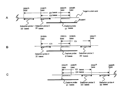
The concept of the present invention is described using Fig. 1. In this
example, a target nucleic acid having a total length of 3000 bases is
detected.
15 [0049]
The capture probe may be designed for any part of the target nucleic acid.
In this example, the region from the 22001h base to the 2230th base as counted
from
the beginning of the target nucleic acid was used as the sequence to which the
capture probe is bound. Four detection probes were designed within the region
in
20 which the distance from the binding region of the capture probe is not
more than
1500 bases. The detection probes and their sequence regions are as follows.
Detection Probe 1, from the 2000th base to the 2019th base; Detection Probe 2,
from
the 2100th base to the 2119th base; Detection Probe 3, from the 2319th base to
the
2330th base; and Detection Probe 4, from the 2419th base to the 2430th base.
[0050]
The distance from the capture probe is determined by placing the binding
position of the capture probe and the binding position of the detection probe
on the
CA 02865541 2014-08-26
21
target nucleic acid sequence, and counting the number of bases therebetween
such
that the most distal bases are counted as the end portions. An explanation is
given
using the example shown in Fig. 1B. The distance between Detection Probe 2,
from
the 2100th base to the 2119th base, and the capture probe, from the 2200th
base to
the 2230th base, is counted by regarding the positions of the most distal
bases as the
end portions. Therefore, the distance is 131 bases. The distance between
Detection Probe 3, from the 2319th base to the 2330th base, and the capture
probe,
from the 2200th base to the 2230th base, is counted by regarding the positions
of the
most distal bases as the end portions. Therefore, the distance is 131 bases.
According to such calculation, the distance of each of Detection Probes 1 and
4 from
the capture probe is similarly 231 bases.
[0051]
Subsequently, the target nucleic acid is cleaved such that the mode of the
nucleic acid is 250 bases. Fig. 1A, Fig. 1B, and Fig. 1C are schematic
diagrams
illustrating hybridization of the target nucleic acid, Detection Probes 1, 2,
3 and/or 4,
and the capture probe.
[0052]
Since the target nucleic acid is cleaved at arbitrary positions, binding
occurs
in various modes as shown in Fig. I.
Fig. IA shows a mode of hybridization of a fragment produced by cleavage
of the target nucleic acid at the 2231st or a later base. Detection Probe 1
and
Detection Probe 2 can bind to the target nucleic acid.
[0053]
Fig. 1B shows a mode of hybridization of a fragment produced by cleavage
of the target nucleic acid at a position before the 2100th base and a position
after the
2330th base. Detection Probe 2 and Detection Probe 3 can bind to the target
nucleic
acid.
CA 02865541 2014-08-26
22
[0054]
Fig. 1C shows a mode of hybridization of a fragment produced by cleavage
of the target nucleic acid at a position before the 2200th base and a position
after the
2430th base. Detection Probe 3 and Detection Probe 4 can bind to the target
nucleic
acid.
[0055]
By providing a plurality of types of detection probes and cleaving the target
nucleic acid such that the mode of the nucleic acid length is 250 bases, the
detection
can be carried out by one or more of the modes shown in Fig. 1.
[0056]
On the other hand, in cases where only a single type of detection probe is
provided, detection is impossible in one or more of the cases. For example, in
cases
where only Detection Probe 1 is used, detection is impossible in the cases of
Fig. 1B
and Fig. IC. Therefore, the detection sensitivity is low.
[0057]
In cases where the mode of the nucleic acid length is 150 bases, the
detection probes that can bind to the target nucleic acid bound to the capture
probe
are Detection Probe 2 and Detection Probe 3. Therefore, the detection
sensitivity is
low.
.. [0058]
Although Fig. 1 shows an example in which detection probes are designed
in both sides of the capture probe, the detection probes may also be
positioned only
in the 3'-end side or only in the 5'-end side.
[0059]
In general, in a method for detecting a nucleic acid such as the method of
the present invention, an internal standard is detected at the same time for
confirming
whether or not the detection method itself is properly carried out. That is,
when a
3225-40
CA 02865541 2014-08-26
A
23
target nucleic acid is not detected by the detection method, it is impossible
to judge
whether the target nucleic acid is absent in the test sample or the detection
method
was not properly carried out. Therefore, nucleic acid regions ubiquitously
present
in the test sample are used as an internal standard. In cases where this
internal
standard is detected, the detection method is judged to have been properly
carried out,
while in cases where the internal standard is not detected, the detection
method is
judged to have been improperly carried out. In cases where the test sample is
derived from human, actin or globin is generally used as an internal standard.
Also
in the method of the present invention, the actin gene or globin gene may be
used as
an internal standard similarly to conventional methods, and, in particular, in
cases
where the target nucleic acid or its fragmentation product is amplified by PCR
or the
like, the same internal standards as in conventional methods may be used
without
any problem.
[0060]
IS However, as described above, the detection method of the present
invention
is a method that exerts an excellent effect that enables detection of a target
nucleic
acid with sufficient sensitivity even without performing nucleic acid
amplification.
In cases where nucleic acid amplification is not carried out, the internal
standard may
not be detected even when the detection method is properly carried out since
the
actin gene and the globin gene are present in the genome as single-copy genes
and
hence the sensitivity may be insufficient.
[0061]
In order to overcome this problem, in a preferred embodiment of the present
invention, a sample derived from an animal such as human containing the target
nucleic acid is subjected to the detection method described above, and at
least one
type of repetitive sequences present in the animal genome is detected. as an
internal
standard. The repetitive sequences are contained in animal genome fragments.
. CA 02865541 2014-08-26
24
The animal genome fragments may be produced by the fragmentation treatment of
the animal genome, or may be naturally-occurring fragments. The preferred
length
of the genome fragment is the same as the preferred length of the target
nucleic acid
or fragmentation product thereof described above. Preferred examples of the
animal include mammals such as human; pets including dog and cat; domestic
animals including pig, cow, horse, sheep and goat; and laboratory animals
including
monkey, mouse and rat. Human is especially preferred, but other animals having
such repetitive sequences may be used. Preferred examples of the repetitive
sequences include retrotransposons, especially, short interspersed nuclear
sequences
(SINEs). In particular, Alu sequences, which are present in the human genome
in
1,000,000 copies, are preferred. The Alu sequences per se are well-known
sequences, and their base sequences are also well known (SEQ ID NO:47).
[0062]
Fig. 8 is a schematic diagram for explanation of the principle of the method
of the present invention in which a viral DNA is used as the target nucleic
acid, and a
human-derived sample containing the human genome is subjected to the detection
method. In the method shown in Fig. 8, human Alu sequences are used as an
internal standard. A human-Alu-sequence capture probe that captures human Alu
sequences is immobilized on a support, and a plurality of types of
human-Alu-sequence detection probes for detecting captured human DNA are
hybridized with captured human Alu sequences, to detect the captured human Alu
sequences. Here, the detection of animal DNA such as human Alu sequences can
be carried out in the same manner as the in the method of detection of a
target
nucleic acid or fragmentation product thereof described above, and preferred
conditions are also the same as described above.
[0063]
EXAMPLE 1
= = CA 02865541 2014-08-26
The present invention is described in more detail as an example in which the
type of the virus with which a patient is infected is distinctively detected,
by way of
an Example for detection of human papillomavirus. However, the present
invention
is not limited by the Example below.
5 [0064]
Human papillomavirus is known as a causative virus for cervical cancer.
There are not less than 100 types of human papillomaviruses, and 13 types
among
these are highly malignant and may cause cervical cancer. In preventive
medicine
for cervical cancer, it is important to know the type of the virus with which
the
10 subject is infected, in order to determine the course of treatment.
Identification of
the type of human papillomavirus is carried out using a swab obtained from the
cervix. Examples of known methods for the identification include the hybrid
capture method and PCR method. By application of the present invention to
detection of human papillomavirus, the type of the virus with which the
patient is
15 infected can be highly sensitively identified.
[0065]
(Design of Capture Probes and Detection Probes)
Studies on identification of the type of human papillomavirus have been
carried out for a long time, and results of such studies can be utilized for
selection of
20 the capture probe. In the present example, capture probe sequences
reported by a
literature (J. Clin. Microbiol., 1995. pp. 901-905) were used (Table 1). In
this
literature, capture probes are designed such that the type can be identified
using the
sequence of the L 1 gene region of human papillomavirus. The subject sequence
is
amplified using PCR primers MY11 and MY09 that are common among the types,
25 and the amplified product is hybridized with capture probes immobilized
on a filter,
which capture probes specifically bind to the respective types, thereby
achieving the
detection. Therefore, the capture probes are designed to be positioned in
almost the
CA 02865541 2014-08-26
26
same regions in the Ll gene in all types. The present invention paid attention
to the
positions of the capture probes and the sequences of the common primers, and,
by
using the common primers as common detection probes, the distance from the
capture probe was set almost the same among the types (Fig. 3, Table 2). As
the
capture probes having the sequences described above, synthetic DNAs modified
with
an amino group at the 5'-end were synthesized by Operon Biotechnologies, Inc.
As
the detection probes, those modified with biotin at the 3'-end and the 5'-end
were
synthesized by Operon Biotechnologies, Inc.
= = = CA 02865541 2014-08-26
27
[0066]
[Table 1]
name SEQ ID NO Probe sequence 5'¨>3'
Probe 6 SEQ ID NO:1 ATCCGTAACTACATCTTCCACATACACCAA
Probe 11 SEQ ID NO:2 ATCTGTGTCTAAATCTGCTACATACACTAA
Probe 16 SEQ ID NO:3 GTCATTATGTCiCTGCCATATCTACTTCAGA
Probe 18 SEQ ID NO:4 TGCTTCTACACAGTCTCCTGTACCTGGGCA
Probe 31 SEQ ID NO:5 TGTTTGTGCTGCAATTGCAAACAGTGATAC
Probe 33 SEQ ID NO:6 TTTATGCACACAAGTAACTAGTGACAGTAC
Probe 34 SEQ ID NO:7 TACACAATCCACAAGTACAAATGCACCATA
Probe 35 SEQ ID NO:8 GTCTGTGTGTTCTGCTGTGTCTTCTAGTGA
Probe 39 SEQ ID NO:9 TCTACCTCTATAGAGTCTTCCATACCTTCT
Probe 40 SEQ ID NO:10 GCTGCCACACAGTCCCCCACACCAACCCCA
Probe 42 SEQ ID NO:11 CTGCAACATCTGGTGATACATATACAGCTG
Probe 43 SEQ ID NO:12 TCTACTGACCCTACTGTGCCCAGTACATAT
Probe 44 SEQ ID NO:13 GCCACTACACAGTCCCCTCCGTCTACATAT
Probe 45 SEQ ID NO:14 ACACAAAATCCTGTGCCAAGTACATATGAC
Probe 51 SEQ ID NO:15 AGCACTGCCACTGCTGCGGTTTCCCCAACA
Probe 52 SEQ ID NO:16 TGCTGAGGTTAAAAAGGAAAGCACATATAA
Probe 54 SEQ ID NO:17 TACAGCATCCACGCAGGATAGCTTTAATAA
Probe 56 SEQ ID NO:18 GTACTGCTACAGAACAGTTAAGTAAATATG
Probe 58 SEQ ID NO:19 ATTATGCACTGAAGTAACTAAGGAAGGTAC
[0067]
[Table 2]
Detection probe SEQ ID NO Sequence 5'--->3'
Distance from the capture
name probe
MY11 SEQ ID NO:20 GCMCAGGGWCATAAYAATGG 50
GP5 SEQ ID NO:21 GAAAAATAAACTGTAAATCATATTC 10
GP6 SEQ ID NO:22 TTTGTTACTGTGGTAGATACTAC 60
MY09 SEQ ID NO:23 GATCAGTWTCCYYTDGGACG 340
00
I.
NO
81781934
29
[0068]
(Preparation of DNA Chip)
On a substrate of a DNA chip "3D-Gene" (registered trademark), all capture
probes in
Table 1 were immobilized to prepare a DNA chip. The details are as follows.
[0069]
(Preparation of DNA-immobilized Carrier)
Using a known method, the LIGA (Lithographie Galvanoformung
Abformung) process, a mold for injection molding was prepared, and injection
molding was carried out to obtain a PMMA carrier having the shape described
below.
Carbon black (Mitsubishi Chemical Corporation, #3050B) was contained in the
PMMA at a ratio of 1 wt%, and the carrier had a black color. In terms of the
shape
of the carrier, the size was 76 mm in length, 26 mm in width, and 1 nun in
thickness,
and the surface of the carrier was flat except for the central portion. At the
center of
the carrier, a recess having a diameter of 10 mm and a depth of 0.2 mrn was
provided,
and 64 (8x8) protruded portions each having a diameter of 0.2 mm and a height
of
0.2 mm were provided in the recess. The pitch between the protruded portions
(distance from the center of a protruded portion to the center of an adjacent
protruded
portion) in the irregular area was 0.6 mm.
[0070]
(Immobilization of Probe DNAs)
The capture probe DNAs in Table 1 were dissolved in pure water at a
concentration of 0.3 nmol/p.L to provide stock solutions. For spotting on the
carrier,
the final concentration of each probe DNA was adjusted to 0.03 nmol/uL with
PBS
(prepared by dissolving 8 g of NaCI, 2.9 g of Na21-1PO4-12H20, 0.2 g of KC1,
and 0.2
g of KH2PO4 in pure water to provide 1 L of a solution, and then adding
hydrochloric
acid to the solution for pII adjustment; pH 5.5), and
CA 2865541 2019-02-27
= = CA 02865541 2014-08-26
1-ethyl-3-(3-dimethylaminopropyl)carbodiimide (EDC) was added thereto at a
final
concentration of 50 mg/mL for condensation of carboxylic acid on the carrier
surface
with the amino group at the end of the probe DNA. Thereafter, the mixed
solution
was spotted on the upper surface of a protruded portion with a glass
capillary.
5 Subsequently, the carrier was placed in a tightly sealed plastic
container, and
incubated at 37 C at a humidity of 100% for about 20 hours, followed by
washing
with pure waster. After immobilization of the capture probe DNAs, a cover was
attached to the central portion of the DNA chip, and zirconia beads were
sealed
between the DNA chip and the cover for stirring the solution during
hybridization
10 reaction.
[0071]
(Preparation of Sample DNA)
For the sample DNA, a recombinant plasmid pHPV16, which contains a
cloned genomic DNA of human papillomavirus, was purchased from Health Science
15 Research Resources Bank and used. The total length of pHPV16 was 16,600
base
pairs. If the molecular weight of one base pair is regarded as 680, 1 ug
corresponds
to 89 nmol.
[0 The method for preparing the sample DNA was as follows. By an
20 ultrasonic
nie treatment (Covaris Inc., s220), li_tg of pHPV16 was fragmented. The
fragmentation treatment was carried out under conditions where fragments
having a
length of 100 bases, 150 bases, 250 bases, 400 bases, 1500 bases or 4000 bases
can
be obtained according to the method recommended by the manufacturer. The
lengths of fragments were evaluated using a Bioanalyzer (Agilent). As a
result, the
25 peak top values of the fragmented nucleic acids obtained under those
treatment
conditions were 100 bases, 150 bases, 250 bases, 400 bases, 1500 bases and
4000
bases, respectively. The concentration of the cleaved sample DNA was measured
81781934
31
using NanoDrop (Thermo Fisher Scientific K.K., ND-1000) to determine the
nucleic
acid concentration. In consideration of the nucleic acid concentration, the
cleaved
DNA contained in each solution after cleavage was diluted to I amol/nI, using
1 xhybridizaion solution to provide a sample DNA.
[0073]
(Preparation of Detection Probe Solutions)
Each detection probe was diluted with sterile water to a concentration of 100
fmollut. Also in cases where a mixture of a plurality of detection probes is
used,
the dilution was carried out with sterile water such that the concentration of
each
detection probe was 100 frnol/n1...
[0074]
(Hybridization)
To 5 I, of the sample DNA, 1 u.1., of the detection probe dilution was added,
and the resulting mixture was heated using a thermal cycler at 95 C for 5
minutes.
Thereafter, the solution was left to stand on the bench for 2 minutes to allow
the
solution to cool to room temperature. To this solution, 35 ttl, of 1
xhybridization
solution (1 wt% BSA (bovine serum albumin), 5xSSC, 1 wt% SDS (sodium dodecyl
sulfate), 50 rig/ml salmon sperm DNA solution, 5 wt% dcxtran sulfate sodium,
30%
formamidc) was added to provide a hybridization solution. The whole solution
was
injected into the DNA chip, and the DNA chip was placed in an incubator heated
at
32 C. Hybridization was carried out according to the standard protocol for
"3D-Gene" with rotary shaking at 250 rpm at 32 C for 2 hours. Thereafter, the
DNA chip was washed for 5 minutes with a washing liquid (0.5xSSC, 0,1 wt% SDS
(sodium dodecyl sulfate)) heated at 30 C, and dried using a spin dryer
(Wakenyaku
Co., Ltd.). A solution prepared by adding streptavidin-phycoerythrin (ProZyme,
inc.) to a staining solution (50 ng/u1streptavidin-phycoerythrin, 100 mM MES,
1 M
NaCI, 0.05 wt% Tweeri20, 2 mg,/m1 BSA (bovine scrum albumin)) was provided,
* Trademark
CA 2865541 2019-02-27
81781934
32
and the solution was dropped on the DNA chip. The DNA chip was then incubated
at 35 C for 5 minutes. After washing the DNA chip for 5 minutes with a washing
liquid (6xSSPE, 0.01 wt% Twee:20) heated at 30 C, the DNA chip was dried using
a spin dryer (Wakenyaku Co., Ltd.). The DNA chip after staining was subjected
to
detection of fluorescence signals using a DNA chip scanner (Toray Industries,
Inc.).
In terms of settings of the scanner, the laser output was set to 100%, and the
photomultiplier voltage was set to 70%.
[0075]
The detection results are shown in Table 3. From the results of detection
with the mixture of 4 types of detection probes, it can be seen that, compared
to the
uncleaved sample and the sample cleaved into 4000-base fragments, the samples
cleaved into fragments having lengths of not more than 1500 bases showed
predominantly stronger detection signals. In particular, it can be seen that
the
sensitivity was highest in the 250-base sample and the 500-base sample, and
that the
sensitivity began to decrease by further cleavage.
[0076]
* Trademark
CA 2865541 2019-02-27
L.,
r..)
tN_.)
(_,-,
I,¨
[Table 3]
Signal value
Detection probe used Distance between
Mode of the DNA length (bases)
the capture probe 100 150 250 500 1500
4000 8000
and the detection ,
probe (bases)
R
i) 4 types 2015 6246 14044 13005
7388 516 132 . 2
GP6+GP5+MY11+MY09
Et
ii) GP6 50 753 2337 5273 4900
3565 205 96
.."
iii) GP5 10 433 1201 3092 2800
1839 179 80
0
iv) MY11 60 98 205 403 350
160 108 96
v) MY09 340 0 0 204 800
1090 197 85
vii) Sum of the signal 1284 3743 8972 8850
6654 689 357
values detected using the
individual detection probes
(ii+iii+iv+v) .
CA 02865541 2014-08-26
34
[0077]
In terms of the results of detection obtained by using the detection probes
individually, GP5 and GP6 showed tendencies similar to the mixture of 4 types
described above. Although MY11 also showed a similar tendency, its signal
intensity was lower than those of GP5 and GP6, possibly due to a low degree of
binding of the detection probe. MY09 is a detection probe designed for a
position
that is 340 bases distant from the capture probe, and, in cases where the DNA
sample
is cleaved into fragments with DNA lengths of not more than 250 bases, there
is no
region where the probe can bind to the target nucleic acid. Therefore, MY09
was
expected to be incapable of functioning as a detection probe. Actually, in the
Example, signals could be obtained for the 500-base sample and the 1500-base
sample, but no signal was obtained for the 250-base and shorter samples.
[0078]
It can be easily assumed that the sum of the signal intensities obtained by
the individual detection probes is equivalent to the signal intensity obtained
using the
mixture of 4 types of detection probes. The sum of the signal intensities
obtained
by the individual detection probes is shown in the last line in Table 3.
Compared to
this, the signal intensity obtained using the mixture of 4 types showed much
higher
values in the cases where the target nucleic acid was cleaved into fragments
of not
more than 1500 bases. This was assumed to be due to enhancement of
hybridization between the capture probe and the target nucleic acid by binding
of the
plurality of detection probes.
[0079]
EXAMPLE 2
The present invention is described in more detail by way of another
embodiment of detection of human papillomavirus. However, the present
invention
is not limited by the Example below.
- = CA 02865541 2014-08-26
[0080]
(Design of Capture Probes and Detection Probes)
Similarly to Example 1, sequences reported in the literature (J. Clin.
Microbiol., 1995. pp. 901-905, Table 1) were employed for capture probes. In
5 general, the binding strength in nucleic acid hybridization increases
as the length of
the nucleic acid increases. In view of this, as detection probes, sequences
each
having a length of 80 to 86 bases were provided for 5 regions located upstream
and
downstream of the capture probe as shown in Fig. 4 and Table 4.
[0081]
[Table 4]
Detection probe name SEQ ID NO Sequence 51--)3 Distance
from the capture probe
Fragl SEQ ID NO:24 gcacagggc[Bio-ON]acaataatggcatttgttg[Bio-
110
ON]ggtaaccaactatttgttac[Bio-
ON]gttgttgatactacacgca[Bio-ON]tacaaatat
Frag2 SEQ ID NO:25 aactacata[Bio-ON]aaanatactaactttaagg[Bio-
116
ON]gtacctacgacatggggaggaatatg[Bio-
ON]tttacagtttatttttcaa[Bio-ON]tgtgcaaaa
Frag3 SEQ ID NO:26 taaccttaa[Bio-ON]tgcagacgttatgacatac[Bio-
202
ON]tacattctatgaattccac[Bio-
ON]attttggaggactggaatt[Bio-ON]tggtctacaacctc
w
cc
Frag4 SEQ ID NO:27 ccaggaggc[Bio-ON]cactagaagatacttatag[Bio-
288
ON]trtgtaacatcccaggcaattgcttg[Bio-
ON]caaaaacatacacctccag[Bio-ON]acctaaaga
Frag5 SEQ ID NO:28 agatccect[Bio-ON]aaaaaatacactttttggg[Bio-
372
ON]agtaaatttaaaggaaaagttttc[Bio-
ON]gcagacctagatcagtttc[Bio-ON]tttaggacg
Bio-on: biotin-on label
=, CA 02865541 2014-08-26
37
[0082]
As the capture probes having the sequences described above, synthetic
DNAs modified with an amino group at the 5'-end were synthesized by Operon
Biotechnologies, Inc. As the detection probes, those internally labeled with
biotin
were synthesized by Operon Biotechnologies, Inc. (Table 4).
[0083]
(Preparation of DNA Chip)
A DNA chip on which all capture probes in Table 1 are immobilized was
prepared. The method of preparing the DNA chip was the same as in Example 1.
[0084]
(Preparation of Sample DNA)
For the sample DNA, a recombinant plasmid pHPV16, which contains a
cloned genomic DNA of human pap illomavirus, was purchased from Health Science
Research Resources Bank and used. The total length of pHPV16 was 16,600 base
pairs. If the molecular weight of one base pair is regarded as 680, 1 ug
corresponds
to 89 nrnol.
[0085]
The method for preparing the sample DNA was as follows. By an
ultrasonic treatment (Covaris Inc., s220), 5 jig of pHPV16 was fragmented. The
fragmentation treatment was carried out under conditions where fragments
having a
length of 150 bases or 250 bases can be obtained according to the method
recommended by the manufacturer. The lengths of fragments were evaluated using
a Bioanalyzer (Agilent). As a result, the peak top values of the fragmented
nucleic
acids obtained under those treatment conditions were 150 bases and 250 bases,
respectively. The concentration of the cleaved sample DNA was measured using
NanoDrop (Thermo Fisher Scientific K.K., ND-1000) to determine the nucleic
acid
concentration. In consideration of the nucleic acid concentration, the cleaved
DNA
=
CA 02865541 2014-08-26
38
contained in each solution after cleavage was diluted to 1 amol/uL using
lxhybridization solution to provide a sample DNA.
[0086]
(Preparation of Detection Probe Solutions)
Each detection probe was diluted with sterile water to a concentration of 100
fmol/uL. Also in cases where a mixture of a plurality of detection probes is
used,
the dilution was carried out with sterile water such that the concentration of
each
detection probe was 100 fmol/pl.
[0087]
(Hybridization)
To 5 AL of the sample DNA, 1 uL of the detection probe dilution was added,
and the resulting mixture was heated using a thermal cycler at 95 C for 5
minutes.
Thereafter, the solution was left to stand on the bench for 2 minutes to allow
the
solution to cool to room temperature. To this solution, 35 11,1_, of a
hybridization
solution (1 wt% BSA (bovine serum albumin), 5x SSC, 1 wt% SDS (sodium dodecyl
sulfate), 50 ng/ml salmon sperm DNA solution, 5 wt% dextran sulfate sodium,
30%
formamide) was added to provide a hybridization solution. The whole solution
was
injected into the DNA chip, and the DNA chip was placed in an incubator heated
at
32 C. Hybridization was carried out according to the standard protocol for
"3D-Gene" with rotary shaking at 250 rpm at 32 C for 2 hours. Thereafter, the
DNA chip was washed for 5 minutes with a washing liquid (0.5 xSSC, 0.1 wt% SDS
(sodium dodecyl sulfate)) heated at 30 C, and dried using a spin dryer
(Wakenyaku
Co., Ltd.). A solution prepared by adding streptavidin-phycoerytluin (ProZyme,
Inc.) to a staining solution (50 ng/p.1 streptavidin-phycoerythrin, 100 mM
MES, 1 M
NaCl, 0.05 wt% Tween 20, 2 mg/ml BSA (bovine serum albumin)) was provided,
and the solution was dropped on the DNA chip. The DNA chip was then incubated
at 35 C for 5 minutes. After washing the DNA chip for 5 minutes with a washing
== . CA 02865541 2014-08-26
39
liquid (6x SSPE, 0.01 wt% Tween 20) heated at 30 C, the DNA chip was dried
using
a spin dryer (Wakenyaku Co., Ltd.). The DNA chip after staining was subjected
to
detection of fluorescence signals using a DNA chip scanner (Toray Industries,
Inc.).
In terms of settings of the scanner, the laser output was set to 100%, and the
photomultiplier voltage was set to 70%.
[0088]
The detection results are shown in Table 5. As a result of detection with 5
types of detection probes that were mixed together (mixture of 5 types), the
signal
intensity for the 150-base sample was stronger than the signal intensity for
the
250-base sample. While 2 detection probes are effective in the case where the
sample is cleaved into 150-base fragments, 3 detection probes are effective in
the
case where the sample is cleaved into 250-base fragments. It can be said that
the
difference in the signal intensity was due to the difference in the number of
detection
probes that could be bound. When the result obtained for the target nucleic
acid
cleaved into 250-base fragments is compared between the mixture of 4 types and
the
mixture of 5 types, the mixture of 5 types showed a stronger signal intensity.
While
2 detection probes are effective in the case where the mixture of 4 types is
used, 3
detection probes are effective in the case where the mixture of 5 types is
used.
Therefore, it can be said that the difference in the signal intensity was due
to the
difference in the number of detection probes that could be bound.
[0089]
= . CA 02865541 2014-08-26
[Table 5]
Nucleic acid Detection probes added Effective detection Detected
signal
length probes
150 bases Mixture of 5 types (Detection Detection
Probes 17,500
Probes 1, 2, 3, 4, 5) 1,2
250 bases Mixture of 4 types (Detection Detection
Probes 15,000
Probes 2, 3, 4, 5) 2,3
250 bases Mixture of 5 types (Detection Detection
Probes 30,000
Probes 1, 2, 3,4, 5) 1, 2, 3
[0090]
Thus, by providing a plurality of detection probes and performing cleavage
of the target nucleic acid in consideration of the binding regions of the
detection
5 probes, an especially sufficient signal strength can be obtained. In
cases where a
mixture of 5 types of detection probes is used, the target nucleic acid is
preferably
cleaved into fragments of about 400 bases ( about 50 bases), more preferably
cleaved into fragments of 400 bases.
[0091]
10 EXAMPLE 3
The present invention is described in more detail by way of an Example for
detection of SNPs (single nucleotide polymorphisms). However, the present
invention is not limited by the Example below. Since the response rate of
cancer
patients treated with an anticancer agent is known to be influenced by the
15 presence/absence of mutations in the EGFR gene and the k-ras gene, the
presence/absence of such mutations is used as a basis for judging whether the
anticancer agent should be administered. In existing techniques, the mutations
are
detected by various methods after performing PCR amplification.
[0092]
. CA 02865541 2014-08-26
41
(Design of Capture Probes and Detection Probes)
In the capture probe for detecting an SNP, the SNP site is placed at the
center of the probe. More specifically, the 10 bases adjacent to the SNP site
in each
of the 5'-side and 3'-side are included in the capture probe sequence. In
cases of
detection of an SNP, a combination of capture probes corresponding to all
bases at
the SNP site are provided, and used as a capture probe set. More specifically,
in
cases of an SNP site where the wild type is G and the mutant type is A,
capture
probes each having a length of 21 bases containing the 10 bases adjacent to
the G or
Amn each of the 5'-side and 3'-side are used. Further, for comparison, capture
probes each having a length of 21 bases in which the central portion is T or C
and the
10 bases adjacent to this portion in each of the 5'-side and 3'-side are
contained are
provided. These 4 sequences are used as the capture probe set. In this manner,
a
capture probe set for k-ras mutations Gly12Ser, Gly12Arg, and Gly12Cys; and a
capture probe set for an EGFR mutation T790M; were provided (Table 6).
= CA 02865541 2014-08-26
42
[0093]
[Table 6]
Probe Capture probe SEQ ID NO Mutation Seqence (5'- 3')
set name
K-ras K-ras capture SEQ ID NO:29 Wild type
Agttggagctggtggcgtagg
capture probe, wild type
probe K-ras capture SEQ ID NO:30 Gly12Ser
Agttggagctagtggcgtagg
set probe, G1y12Ser
K-ras capture SEQ ID NO:31 Gly12Arg agttggagctcgtggcgtagg
probe, G1y12Arg
K-ras capture SEQ ID NO:32 Gly12Cys agttggagottgtggcgtagg
probe, G1y12Cys
EGFR EGFR capture SEQ ID NO:33 Wild type caactcatcacgcagctcatg
capture probe, wild type
probe EGFR capture SEQ ID NO:34 T790M
caactcatcatgcagctcatg
set probe, 1790M
EGFRCapture SEQ ID NO:35 Control (a)
caactcatcaagcagctcatg
probecontrol (a)
EGFRCapture SEQ ID NO:36 Control (g)
caactcatcaggcagctcatg
probecontrol (g)
[0094]
Detection probes were designed at the positions shown in Fig. 6 and Fig. 7
in consideration of their distances from the capture probes designed. The
sequences
of the detection probes and the distances of the detection probes from the
capture
probes arc shown in Table 7 and Table 8.
[0095]
[Table 7]
Sequences of K-RAS detection probes and their distances from the capture probe
(bases)
Detection probe name SEQ ID NO
Sequence 5t--->3 Distance from
the capture
probe (bases)
K-ras detection probe 1 SEQ ID NO:37 GATCATATTCGTCCACAAAATGATTCTGAATTAGCTGTAT
75
K-ras detection probe 2 SEQ ID NO:38 CCAAGAGACAGGTTTCTCCATCAATTACTACTTGCTTCCT
135
K-ras detection probe 3 SEQ ID NO:39 TCCTCATGTACTGGTCCCTCATTGCACTGTACTCCTCTTG
191
,71
c;\
[Table 8]
Sequences of EGFR detection probes and their distances from the capture probe
(bases)
SEQ ID NO Sequence 5T--->3'
Distance from
the capture
probe (bases)
EGFR detection probe 1 SEQ ID NO:40 AATTTTAACTTTCTCACCTTCTGGGATCCAGAGTCCCTTA
198
EGFR detection probe 2 SEQ ID NO:41 CTGCACACACCAGTTGAGCAGGTACTGGGAGCCAATATTG
101
EGFR detection probe 3 SEQ ID NO:42 CCACTTGATAGGCACTTTGCCTCCTTCTGCATGGTATTCT
281
CA 02865541 2014-08-26
[0097]
As the capture probes having the sequences described above, synthetic
DNAs modified with an amino group at the 5'-end were synthesized by Operon
Biotechnologies, Inc. As the detection probes, those labeled with biotin at
the
5 3'-end and the 5'-end were synthesized by Operon Biotechnologies, Inc.
[0098]
(Preparation of DNA Chip)
A DNA chip on which all capture probes in Table 6 are immobilized was
prepared. The method of preparing the DNA chip was the same as in Example 1.
10 [0099]
(Preparation of Sample DNA)
For the sample DNA, DNA extracted from A549 cells was used. A549
cells are cultured cells derived from a lung cancer tissue, and known to have
the
G1y12Ser mutation. By an ultrasonic treatment (Covaris Inc., s220), 5 lig of
DNA
15 extracted from A549 cells was fragmented. The fragmentation treatment
was
carried out under the conditions for the method recommended by the
manufacturer.
The lengths of fragments were evaluated using a Bioanalyzer (Agilent). As a
result,
the mode of the nucleic acid length was 750 bases. The whole cleaved DNA was
used as the sample DNA.
20 [0100]
(Preparation of Detection Probe Solution)
The detection probes were mixed together, and diluted with sterile water
such that the concentration of each detection probe was 100 fmol/uL.
[0101]
25 (Hybridization)
To 5 p.1, of the sample DNA, 1 1iL of the detection probe dilution was added,
and the resulting mixture was heated using a thermal cycler at 95 C for 5
minutes.
. CA 02865541 2014-08-26
46
Thereafter, the solution was left to stand on the bench for 2 minutes to allow
the
solution to cool to room temperature. To this solution, 35 tL of a
hybridization
solution (1 wt% BSA (bovine serum albumin), 5x SSC, 0.1 wt% SDS (sodium
dodecyl sulfate), 0.01 wt% salmon sperm DNA solution) was added to provide a
hybridization solution. The whole solution was injected into the DNA chip, and
the
DNA chip was placed in an incubator heated at 32 C. Hybridization was carried
out according to the standard protocol for "3D-Gene" with rotary shaking at
250 rpm
at 32 C for 2 hours. Thereafter, the DNA chip was washed for 5 minutes with a
washing liquid (0.5x SSC, 0.1 wt% SDS (sodium dodecyl sulfate)) heated at 30
C,
and dried using a spin dryer (Wakenyaku Co., Ltd.). A solution prepared by
adding
streptavidin-phycoerythrin (ProZyme, Inc.) to a staining solution (50 ng/u1
streptavidin-phycoerythrin, 100 mM MES, 1 M NaCl, 0.05 wt% Tween 20, 2 mg/ml
BSA (bovine serum albumin)) was provided, and the solution was dropped on the
DNA chip. The DNA chip was then incubated at 35 C for 5 minutes. After
washing the DNA chip for 5 minutes with a washing liquid (6xSSPE, 0.01 wt%
Tween 20) heated at 30 C, the DNA chip was dried using a spin dryer (Wakenyaku
Co., Ltd.). The DNA chip after staining was subjected to detection of
fluorescence
signals using a DNA chip scanner (Toray Industries, Inc.). In terms of
settings of
the scanner, the laser output was set to 100%, and the photomultiplier voltage
was set
to 70%.
, = = CA 02865541 2014-08-26
47
[0102]
[Table 9]
Mutation Signal Probe with the strongest
signal
intensity in the capture probe set
EGFR capture Control (g) 935
probe set Control (a) 1086
T790M 986
Wild type 1382 0
=
K-ras capture Gly12Arg 1193
probe set G1y12Cys 1887
G1y12Ser 3925 0
Wild type 1261
[0103]
The detection results are shown in Table 9. In the cases where the EGFR
capture probe set was used, the wild-type probe showed the strongest signal
intensity.
Therefore, it is thought that A549 cells do not have the T790M mutation. In
the
cases where the K-RAS capture probes were used, the Gly12Ser probe showed the
strongest signal intensity. A549 cells are known to have the G1y12Ser
mutation,
and the present detection results were consistent with this.
[0104]
Thus, by using the sandwich hybridization, the SNPs could be properly
detected without performing PCR amplification.
[0105]
EXAMPLE 4
Although the K-ras gene mutations and EGFR gene mutation shown in
Example 3 are important indices for administration of anticancer agents, it is
also
important to confirm the presence/absence of expression of these genes by
another
. . CA 02865541 2014-08-26
48
method. The present invention can directly detect RNAs, and can also detect
mutations in the RNAs. Conventional RNA techniques require preparation of
cDNA using reverse transcriptase before amplification by PCR. In general,
since
the reverse transcriptase used for reverse transcription easily causes
mutations, it
may cause false detection. The details are described below, but the present
invention is not limited by the Example below.
[0106]
(Design of Capture Probes and Detection Probes)
The same capture probe sets and detection probes as in Example 3 were
used.
[0107]
(Preparation of DNA Chip)
A DNA chip on which all capture probes in Table 6 are immobilized was
prepared. The method of preparing the DNA chip was the same as in Example 1.
[0108]
(Preparation of Sample DNA)
For the sample DNA, total RNA extracted from A549 cells was used.
A549 cells are cultured cells derived from a lung cancer tissue, and known to
have
the Gly12Ser mutation. By an ultrasonic treatment (Covaris Inc., s220), 5 [tg
of
total RNA extracted from A549 cells was fragmented. The fragmentation
treatment
was carried out under the conditions for the method recommended by the
manufacturer. The lengths of fragments were evaluated using a Bioanalyzer
(Agilent). As a result, the mode of the nucleic acid length was 250 bases. The
whole cleaved RNA was used as the sample RNA.
[0109]
(Preparation of Detection Probe Solution)
The detection probes were mixed together, and diluted with sterile water
= = ,
CA 02865541 2014-08-26
49
such that the concentration of each detection probe was 100 fmol/uL.
[0110]
(Hybridization)
To 5 uL of the sample RNA, 1 uL of the detection probe dilution was added,
and the resulting mixture was heated using a thermal cycler at 95 C for 5
minutes.
Thereafter, the solution was left to stand on the bench for 2 minutes to allow
the
solution to cool to room temperature. To this solution, 35 uL of a
hybridization
solution (1 wt% BSA (bovine serum albumin), 5 xSSC, 0.1 wt% SDS (sodium
dodecyl sulfate), 0.01 wt% salmon sperm DNA solution) was added to provide a
hybridization solution. The whole solution was injected into the DNA chip, and
the
DNA chip was placed in an incubator heated at 32 C. Hybridization was carried
out according to the standard protocol for "3D-Gene" with rotary shaking at
250 rpm
at 32 C for 2 hours. Thereafter, the DNA chip was washed for 5 minutes with a
washing liquid (0.5 xSSC, 0.1 wt% SDS (sodium dodecyl sulfate)) heated at 30
C,
and dried using a spin dryer (Wakenyaku Co., Ltd.). A solution prepared by
adding
streptavidin-phycoerythrin (ProZyme, Inc.) to a staining solution (50 ng/u1
streptavidin-phycoerythrin, 100 mM MES, 1 M NaC1, 0.05 wt% Tween 20, 2 mg/ml
BSA (bovine serum albumin)) was provided, and the solution was dropped on the
DNA chip. The DNA chip was then incubated at 35 C for 5 minutes. After
washing the DNA chip for 5 minutes with a washing liquid (6 xSSPE, 0.01 wt%
Tween 20) heated at 30 C, the DNA chip was dried using a spin dryer (Wakenyaku
Co., Ltd.). The DNA chip after staining was subjected to detection of
fluorescence
signals using a DNA chip scanner (Toray Industries, Inc.). In terms of
settings of
the scanner, the laser output was set to 100%, and the photomultiplier voltage
was set
to 70%.
[0111]
The detection results are shown in Table 10. In the cases where the EGFR
= ' CA 02865541 2014-08-26
capture probe set was used, the wild-type probe showed the strongest signal
intensity.
Therefore, it is thought that A549 cells do not have the T790M mutation. In
the
cases where the K-RAS capture probes were used, the Gly12Ser probe showed the
strongest signal intensity. A549 cells are known to have the Gly12Ser
mutation,
5 and the present detection results were consistent with this.
[0112]
[Table 10]
Mutation Signal Probe with the strongest signal
intensity in the capture probe set
EGFR capture Control (g) 857
probe set Control (a) 793
T790M 756
Wild type 1193 0
K-ras capture Gly12Arg 1115
probe set Gly12Cys 1591
G1y12Ser 3060 0
Wild type 1267
[0113]
Thus, by using the sandwich hybridization, the RNA could be directly
10 detected and the SNPs could be properly detected without performing
reverse
transcription of the RNA.
[0114]
EXAMPLE 5
The present invention is described in more detail by way of another
15 embodiment of detection of human papillomavirus. In the Example 1 and
Example
2 described above, human papillomavirus DNA was detected. When nucleic acid is
extracted from a human specimen and analyzed, the absence of a detection
signal
= = CA 02865541 2014-08-26
51
means either the absence of human papillomavirus in the human specimen, or
failure
of the operation of extraction or detection of the nucleic acid. It is
difficult to
distinguish between these. Such a problem can be solved by use of an internal
standard.
[0115]
It is known that, in cases where human papillomavirus DNA is extracted
from a human specimen, the obtained sample solution contains a small amount of
human DNA. From a human specimen uninfected with human papillomavirus,
human papillomavirus DNA cannot be obtained, but human DNA can be obtained.
By taking advantage of this fact, human DNA contained in the sample solution
obtained by extraction from a human specimen can be used as an internal
standard
for detection of human papillomavirus DNA. In the present Example, a plasmid
DNA containing a base sequence of papillomavirus incorporated therein, and
human
genomic DNA are mixed together to provide a sample for simulation. However,
the
present invention is not limited by the Example below.
[0116]
(Design of Capture Probes and Detection Probes)
Similarly to Example 1, sequences reported in the literature (J. Clin.
Microbiol., 1995. pp. 901-905, Table 1) were employed for capture probes. In
general, the binding strength in nucleic acid hybridization increases as the
length of
the nucleic acid increases. In view of this, as detection probes, sequences
each
having a length of 80 to 86 bases were provided for 5 regions located upstream
and
downstream of the capture probe as shown in Fig. 4 and Table 4.
[0117]
As the capture probes having the sequences described above, synthetic
DNAs modified with an amino group at the 5'-end were synthesized by Operon
Biotechnologies, Inc. As the detection probes, those internally labeled with
biotin
= = CA 02865541 2014-08-26
52
were synthesized by Operon Biotechnologies, Inc. (Table 4).
[0118]
Table 11 shows a capture probe and detection probes for Alu sequences,
which are repetitively present in the human genome. As the capture probe
having
the sequence described above, a synthetic DNA modified with an amino group at
the
5'-end was synthesized by Operon Biotechnologies, Inc. As the detection
probes,
those labeled with biotin at the 5'-end and 3'-end were synthesized by Operon
Biotechnologics, Inc.
[0119]
(Preparation of DNA Chip)
A DNA chip on which all capture probes in Table 1 and the capture probe
having the sequence of SEQ ID NO:46 shown in Table 11 are immobilized was
prepared. The method of preparing the DNA chip was the same as in Example I.
[0120]
[Table 11]
Probe name SEQ ID NO Sequence 5-.4.31
Detection probe Alu detection probe I SEQ ID NO:43
GCTCACGCCTGTAATCCCAGCACTTTGGGA
Alu detection probe 2 SEQ ID NO:44 CGGTGAAACCCCGTCTCTACTAAAAATACA
Alu detection probe 3 SEQ ID NO:45 ACTCGGGAGGCTGAGGCAGGAGAATGGCG
Capture probe Probe Alu SEQ ID NO:46
CGGATCACGAGGTCAGGAGATCGAGACCAFCCTGGCTAAC
L.J
.0
CA 02865541 2014-08-26
54
[0121]
(Preparation of Sample DNA)
For the sample DNA, a recombinant plasmid pHPV16, which contains a
cloned genomic DNA of human papillomavirus, was purchased from Health Science
Research Resources Bank (Japan Health Sciences Foundation) and used. The total
length of pHPV16 was 16,600 base pairs. If the molecular weight of one base
pair
is regarded as 680, 1 ttg corresponds to 89 nmol. Human genomic DNA was
purchased from Clontech.
[0122]
The method for preparing the sample DNA was as follows. By an
ultrasonic treatment (Covaris Inc., s220), 5 jig of pHPV16 and the human
genomic
DNA were fragmented. The fragmentation treatment was carried out under
conditions where fragments having a length of 250 bases can be obtained
according
to the method recommended by the manufacturer. The lengths of fragments were
evaluated using a Bioanalyzer (Agilent). As a result, the peak top value of
the
fragmented nucleic acid obtained under the treatment conditions was 250 bases.
The concentration of the cleaved sample DNA was measured using NanoDrop
(Thermo Fisher Scientific K.K., ND-1000) to determine the nucleic acid
concentration. In consideration of the nucleic acid concentration, the cleaved
DNA
contained in each solution after cleavage was diluted such that pHPV16 was
contained at 1 amo1/4 and human genomic DNA was contained at 5 ng/ut using
lxhybridization solution.
[0123]
(Preparation of Detection Probe Solution)
The detection probes shown in Table 4 and Table 11 were dissolved in sterile
water, and mixed together such that the concentration of each probe was 100
fmol/A,
to provide a detection probe liquid.
=
CA 02865541 2014-08-26
[0124]
(Hybridization)
To 5 [tL of the sample DNA prepared by mixing the cleaved pHPV16 and
human genomic DNA according to the composition shown in Table 12, 1 .1., of
the
5 diluted detection probe liquid was added, and the resulting mixture was
heated using
a thermal cycler at 95 C for 5 minutes. Thereafter, the solution was left to
stand on
the bench for 2 minutes to allow the solution to cool to room temperature. To
this
solution, 35 1.tL of lxhybridization solution (1 wt% BSA (bovine serum
albumin),
5x SSC, 1 wt% SDS (sodium dodecyl sulfate), 50 ng/ml salmon sperm DNA
solution,
10 5 wt% dextran sulfate sodium, 30% formamide) was added to provide a
hybridization solution. The whole solution was injected into the DNA chip, and
the
DNA chip was placed in an incubator heated at 32 C. Hybridization was carried
out according to the standard protocol for "3D-Gene" with rotary shaking at
250 rpm
at 32 C for 2 hours. Thereafter, the DNA chip was washed for 5 minutes with a
15 washing liquid (0.5x SSC, 0.1 wt% SDS (sodium dodecyl sulfate)) heated
at 30 C,
and dried using a spin dryer (Wakenyaku Co., Ltd.). A solution prepared by
adding
streptavidin-phycoerythrin (ProZyme, Inc.) to a staining solution (50 ng4t1
streptavidin-phycoerythrin, 100 mM MES, 1 M NaCl, 0.05 wt% Tween 20, 2 mg/ml
BSA (bovine serum albumin)) was provided, and the solution was dropped on the
20 DNA chip. The DNA chip was then incubated at 35 C for 5 minutes. After
washing the DNA chip for 5 minutes with a washing liquid (6x SSPE, 0.01 wt%
Tween 20) heated at 30 C, the DNA chip was dried using a spin dryer (Wakenyaku
Co., Ltd.). The DNA chip after staining was subjected to detection of
fluorescence
signals using a DNA chip scanner (Toray Industries, Inc.). In terms of
settings of
25 the scanner, the laser output was set to 100%, and the photomultiplier
voltage was set
to 70%.
[0125]
CA 02865541 2014-08-26
56
The detection results are shown in Table 12.
[0126]
[Table 12]
Values of signals detected from Samples 1, 2 and 3
Sample 1 (5 ng of Sample 2 (5 ng of Sample 3 (1 amol of
human genomic human genomic DNA pHPV16)
DNA) + 1 amol of pHPV16)
Capture probe 0 15,000 15,000
Probe 16
Capture probe 5,000 5,000 0
Probe Alu
In Sample 1 (human genomic DNA, 5 ng), a signal was detected only with
the capture probe Probe Alu. Since this sample did not contain pHPV16 DNA, no
signal was obtained with the capture probe Probe 16.
[0127]
In Sample 3 (pHPV16, 1 amol), a signal was detected only with the capture
probe Probe 16. Since this sample did not contain human genomic DNA, no signal
was obtained with the capture probe Probe Alu. From these results, it can be
seen
that the capture probe and detection probe for pHPV16, and the detection probe
and
capture probe for Alu sequences do not cause erroneous hybridization with each
other.
[0128]
In Sample 2 (pHPV 16, 1 amol; human genomic DNA, 5 ng), signals were
detected with the capture probes Probe 16 and Probe Alu. Thus, it was found
that
pHPV16 and human genomic DNA can be detected simultaneously.
[0129]
Thus, irrespective of whether human papillomavirus DNA is present or
CA 02865541 2014-08-26
=
57
absent, Alu sequences in human DNA contained in the sample solution could be
detected, and it was therefore found that human DNA can be used as an internal
standard. It is thought that the present technique is effective also in cases
where
nucleic acid is extracted from a human specimen and analyzed.
SEQUENCE LISTING IN ELECTRONIC FORM
In accordance with Section 111(1) of the Patent Rules, this
description contains a sequence listing in electronic form in ASCII
text format (file: 55225-40 Seq 17-08-14 vl.txt).
A copy of the sequence listing in electronic form is available from
the Canadian Intellectual Property Office.
The sequences in the sequence listing in electronic form are
reproduced in the following table.
SEQUENCE TABLE
<110> TORAY INDUSTRIES, INC.
<120> Method of detecting nucleic acids
<130> PF477-PCT
<140> PCT/JP2013/055085
<141> 2013-02-27
<150> JP2012-040051
<151> 2012-02-27
<160> 47
<170> PatentIn version 3.5
<210> 1
<211> 30
<212> DNA
<213> Human papillomavirus type 6
<400> 1
atccgtaact acatcttcca catacaccaa 30
<210> 2
<211> 30
CA 02865541 2014-08-26
=
58
<212> DNA
<213> Human papillomavirus type 11
<400> 2
atctgtgtct aaatctgcta catacactaa 30
<210> 3
<211> 30
<212> DNA
<213> Human papillomavirus type 16
<400> 3
gtcattatgt gctgccatat ctacttcaga 30
<210> 4
<211> 30
<212> DNA
<213> Human papillomavirus type 18
<400> 4
tgcttctaca cagtctcctg tacctgggca 30
<210> 5
<211> 30
<212> DNA
<213> Human papillomavirus type 31
<400> 5 =
tgtttgtgct gcaattgcaa acagtgatac 30
<210> 6
<211> 30
<212> DNA
<213> Human papillomavirus type 33
<400> 6
tttatgcaca caagtaacta gtgacagtac 30
<210> 7
<211> 30
<212> DNA
<213> Human papillomavirus type 34
<400> 7
tacacaatcc acaagtacaa atgcaccata 30
<210> 8
<211> 30
<212> DNA
<213> Human papillomavirus type 35
CA028655412014-08-26
59
<400> 8
gtctgtgtgt tctgctgtgt cttctagtga 30
<210> 9
<211> 30
<212> DNA
<213> Human papillomavirus type 39
<400> 9
tctacctcta tagagtcttc cataccttct 30
<210> 10
<211> 30
<212> DNA
<213> Human papillomavirus type 40
<400> 10
gctgccacac agtcccccac accaacccca 30
<210> 11
<211> 30
<212> DNA
<213> Human papillomavirus type 42
<400> 11
ctgcaacatc tggtgataca tatacagctg 30
<210> 12
<211> 30
<212> DNA
<213> Human papillomavirus type 43
<400> 12
tctactgacc ctactgtgcc cagtacatat 30
<210> 13
<211> 30
<212> DNA
<213> Human papillomavirus type 44
<400> 13
gccactacac agtoccotcc qtctacatat 30
<210> 14
<211> 30
<212> DNA
<213> Human papillomavirus type 45
<400> 14
acacaaaatc ctqtgccaag tacatatgac 30
CA 02865541 2014-08-26
<210> 15
<211> 30
<212> DNA
<213> Human papillomavirus type 51
<400> 15
agcactgcca ctgctgcggt ttccccaaca 30
<210> 16
<211> 30
<212> DNA
<213> Human papillomavirus type 52
<400> 16
tgctgaggtt aaaaaggaaa gcacatataa 30
<210> 17
<211> 30
<212> DNA
<213> Human papillomavirus type 54
<400> 17
tacagcatcc acgcaggata gctttaataa 30
<210> 10
<211> 30
<212> DNA
<213> Human papillomavirus type 56
<400> 18
gtactgctac agaacagtta agtaaatatg 30
<210> 19
<211> 30
<212> DNA
<213> Human papillomavirus type 58
<400> 19
attatgcact gaagtaacta aggaaggtac 30
<210> 20
<211> 20
<212> DNA
<213> human papillomavirus
<220>
<221> misc_feature
<222> (1)..(1)
<223> biotinylated base
CA 02865541 2014-08-26
=
61
<220>
<221> misc_feature
<222> (20)..(20)
<223> biotinylated base
<400> 20
gcmcagggwc ataayaatgg 20
<210> 21
<211> 25
<212> DNA
<213> human papillomavirus
<220>
<221> misc_feature
<222> (1)..(1)
<223> biotinylated base
<220>
<221> misc_feature
<222> (25)..(25)
<223> biotinylated base
<400> 21
gaaaaataaa ctgtaaatca tattc 25
<210> 22
<211> 23
<212> DNA
<213> human papillomayirus
<220>
<221> misc_feature
<222> (1)..(1)
<223> biotiny1ated base
<220>
<221> misc....feature
<222> (23)..(23)
<223> biotinylated base
<400> 22
tttgttactg tggtagatac tac 23
<210> 23
<211> 20
<212> DNA
<213> human papillomavirus
<220>
<221> misc_feature
<222> (1)..(1)
<223> biotinylated base
CA 02865541 2014-08-26
62
<220>
<221> misc feature
<222> (20)..(20)
<223> biotinylated base
<400> 23
gatcagtwtc cyytdggacg 20
=
<210> 24
<211> 80
<212> DNA
<213> human papillomavirus
<220>
<221> misc feature
<222> (10)..(10)
<223> n is biotinylated base
<220>
<221> misc feature
<222> (30)..(30)
<223> n is biotinylated base
<220>
<221> misc_feature
<222> (51)..(51)
<223> n is biotinylated base
<220>
<221> misc feature
<222> (71)..(71)
<223> n is biotinylated base
<400> 24
gcacagggcn acaataatgg catttgttgn ggtaaccaac tatttgttac ngttgttgat 60
actacacgca ntacaaatat 80
<210> 25
<211> 86
<212> DNA
<213> human papillomavirus
<220>
<221> misc feature
<222> (10)..(10)
<223> n is biotinylated base
<220>
<221> misc feature
<222> (30)..(30)
<223> n is biotinylated base
<220>
<221> misc feature
CA 02865541 2014-08-26
=
63
<222> (57)..(57)
<223> n is biotinylated base
<220>
<221> misc_feature
= <222> (77)..(77)
<223> n is biotinylated base
<400> 25
aactacatan aaaaatacta actttaaggn gtacctacga catggggagg aatatgnttt 60
acagtttatt tttcaantgt gcaaaa 86
<210> 26
<211> 86
<212> DNA
= <213> human papillomavirus
<220>
<221> misc_feature
<222> (10)..(10)
<223> n is biotinylated base
<22C>
<221> misc_feature
<222> (30)..(30)
<223> n is biotinylated base
<220>
<221> misc_feature
<222> (50)..(50)
<223> n is biotinylated base
<220>
<221> misc_feature
<222> (70)..(70)
<223> n is biotinylated base
<400> 26 =
taaccttaan tgcagacgtt atgacatacn tacattctat gaattccacn attttggagg 60
actggaattn tggtctacaa cctccc 66
<210> 27
<211> 86
<212> DNA
<213> human papillomavirus
<220>
<221> misc_feature
<222> (10)..(10)
<223> n is biotinylated base
<220>
<221> misc_feature
CA 02865541 2014-08-26
=
64
<222> (30)..(30)
<223> n is biotinylated base
<220>
<221> misc_feature
<222> (57)..(57)
<223> n is biotinylated base
<220>
<221> misc_feature
<222> (77)..(77)
<223> n is biotinylated base
<400> 27
ccaggaggcn cactagaaga tacttatagn tttgtaacat cccaggcaat tgcttgncaa 60
aaacatacac ctccagnacc taaaga 86
<210> 28
<211> 84
<212> DNA
<213> human papillomavirus
<220>
<221> misc_feature
<222> (10)..(10)
<223> n is biotinylated base
<220>
<221> misc_feature
<222> (30)..(30)
<223> n is biotinylated base
<220>
<221> misc_feature
<222> (55)..(55)
<223> n is biotinylated base
<220>
<221> misc_feature
<222> (75)..(75)
<223> n is biotinylated base
<400> 28
agatcccctn aaaaaataca ctttttgggn agtaaattta aaggaaaagt tttcngcaga 60
cctagatcag tttcntttag gacg 84
<210> 29
<211> 21
<212> DNA
<213> Homo sapiens
<400> 29
agttggagct ggtggcgtag g 21
CA 02865541 2014-08-26
,
<210> 30
<211> 21
<212> DNA
<213> Homo sapiens
<400> 30
agttggagct agtggcgtag g 21
<210> 31
<211> 21
<212> DNA
<213> Homo sapiens
<400> 31
agttggagct cgtggcgtag g 21
<210> 32
<211> 21
<212> DNA
<213> Homo sapiens
<400> 32
agttggagct tgtggcgtag g 21
<210> 33
<211> 21
<212> DNA
<213> Homo sapiens
<400> 33
caactcatca cgcagctcat g 21
<210> 34
<211> 21
<212> DNA
<213> Homo sapiens
<400> 34
caactcatca tgcagctcat g 21
<210> 35
<211> 21
<212> DNA
<213> Homo sapiens
<400> 35
caactcatca agcagctcaL g 21
<210> 36
<211> 21
CA 02865541 2014-08-26
3 ,
66
<212> DNA
<213> Homo sapiens
<400> 36
caactcatca ggcagctcat g 21
<210> 37
<211> 40
<212> DNA
<213> Homo sapiens
<220>
<221> misc_feature
<222> (1)..(1)
<223> biotinylated base
<220>
<221> misc_feature
<222> (40)..(40)
<223> biotinylated base
<400> 37
gatcatattc gtccacaaaa tgattctgaa ttagctgtat 40
<210> 38
<211> 40
=
<212> DNA
<213> Homo sapiens
<220>
<221> misc_feature
<222> (1)..(1)
<223> biotinylated base
<220>
<221> misc_feature
<222> (40)..(40)
<223> biotinylated base
<400> 38
ccaagagaca ggtttctcca tcaattacta cttgcttcct 40
<210> 39
<211> 40
<212> DNA
<213> Homo sapiens
<220>
<221> misc_feature
<222> (1)..(1)
<223> biotinylated base
CA 02865541 2014-08-26
67
<220>
<221> misc_feature
<222> (40)..(40)
<223> biotinylated base
<400> 39
tcctcatgta ctggtocctc attgcactgt actcctcttg 40
<210> 40
<211> 40
<212> DNA
<213> Homo sapiens
<220>
<221> misc_feature
<222> (1)..(1)
<223> biotinylated base
<220>
<221> misc_feature
<222> (40)..(40)
<223> biotinylated base
<400 40
aattttaact ttctcacctt ctgggatcca gagtccctta 40
<210> 41
<211> 40
<212> DNA
<213> Homo sapiens
<220>
<221> misc_feature
<222> (1)..(1)
= <223> biotinylated base
<220>
<221> misc_feature
<222> (40)..(40)
<223> biotiny1ated base
<400> 41
ctgcacacac cagttgagca ggtactggga gccaatattg 40
<210> 42
<211> 40
<212> DNA
<213> Homo sapiens
<220>
<221> misc_feature
<222> (1)..(1)
<223> biotinylated base
CA 02865541 2014-08-26
68
<220>
<221> misc_feature
<222> (40)..(40)
<223> biotinylated base
<400> 42
ccacttgata ggcactttgc ctccttctgc atggtattct 40
<210> 43
<211> 30
<212> DNA
<213> Homo sapiens
<220>
<221> misc_feature
<222> (1)..(1)
<223> biotinylated base
<220>
<221> misc_feature
<222> (30)..(30)
<223> biotinylated base
<400> 43
gctcacgcct gtaatcccag cactttggga 30
<210> 44
<211> 30
<212> DNA
<213> Homo sapiens
<220>
<221> misc_feature
<222> (1)..(1)
<223> GCTCACGCCTGTAATCCCAGCACTTTGGGA
<220>
<221> misc feature
<222> (30)..(30)
<223> bioLinylated base
<400> 44
cggtgaaacc ccgtctctac taaaaataca 30
<210> 45
<211> 29
<212> DNA
<213> Homo sapiens
<220>
<221> misc_feature
<222> (1)..(1)
<223> GCTCACGCCTGTAATCCCAGCACTTTGGGA
' CA 02865541 2014-08-26
69
<220>
<221> misc_feature
<222> (30)..(30)
<223> biotinylated base
<400> 45
actcgggagg ctgaggcagg agaatggcg 29
<210> 46
<211> 40
<212> DNA
<213> Homo sapiens
<400> 46
cggatcacga ggtcaggaga tcgagaccat cctggctaac 40
<210> 47
<211> 287
<212> DNA
<213> Homo sapiens
<400> 47
ggccgggcgc ggtggctcac gcctgtaatc ccagcacttt gggaggccga ggcgggcgga 60
tcacgaggto eggagaLcga gaccatcctg gctaacacgg tgaaaccccg tctctactaa 120
aaatacaaaa aattagccgg gcgtggtggc gggcgcctgt agtcccagct actogggagg 180
ctgaggcagg agaatggcgt gaacccggga ggcggagctt gcagtgagcc gagatcgcgc 240
cactgcactc cagcctgggc gacagagcga gactccgtct caaaaaa 287