Note: Descriptions are shown in the official language in which they were submitted.
81781841
ADENOVIRAL TUMOR DIAGNOSTICS
CROSS-REFERENCE TO RELATED APPLICATIONS
[0001] This application claims priority to U.S. Provisional Application No.
61/610,970
filed Mar 14, 2012.
STATEMENT AS TO RIGHTS TO INVENTIONS MADE UNDER
FEDERALLY SPONSORED RESEARCH AND DEVELOPMENT
[0002] This invention was made with government support under grants
R0IHG004876,
R2 IRR024453, and R43RR031424 awarded by the National Institutes of Health.
The
Government has certain rights in the invention.
BACKGROUND OF THE INVENTION
[00031 The spread of cells from a solid tumor to remote sites in the body, a
process known as
metastasis, is responsible for over 90% of all cancer-related deaths. Cells
originating from the
primary tumor can enter the circulatory system and extravagate to invade,
colonize, and
proliferate in organs and tissues far from the primary neoplasm. Thus, the
detection of these
circulating tumor cells (CTCs) provides an invaluable opportunity for both the
early
identification and therapeutic targeting of metastatic cancer cells
(Cristofanilli M et al.,
Circulating tumor cells, disease progression, and survival in metastatic
breast cancer,
N Engl J Med. 2004 Aug 19;351(8):781-91; de Bono et al., Circulating tumor
cells predict survival benefit from treatment in metastatic castration-
resistant prostate cancer,
Clin Cancer Res. 2008 Oct 1;14(19):6302-9).
Current techniques for detection of CTCs include reverse transcriptase-
polymerase chain
reaction (RT-PCR), flow eytometry, fluorescence in situ hybridization, and,
more recently,
microfluidics. Unfortunately, RT-PCR does not distinguish between viable
metastatic CTC
versus nucleic acids or cellular fragments originating from the primary tumor.
10004] Antibody-based techniques cannot be used for detection of all cancers,
but only those
cancers that express the most common and well-characterized markers. CTC
enumeration of
current systems only provides one layer of information regarding cancer
diagnosis. One device,
CellSearchaD (Veridex, Raridan NJ), the has demonstrated commercial success
for CTC analysis
and is FDA approved for breast, prostate, and colon, while ovarian, rectum,
and lung await
approval. Limitations of the CellSearch@ system include: (a) dependence on the
level of
EpCAM expression (Punnoose EA, et al., PLoS ONE. 2010;5(9):e12517), (b) no use
of
mesenchymal markers (Punnoose EA, et al., PLa ONE. 2010;5(9):e12517), (c)
reliance on
antibody affinity for capture (Nagrath S, et al., Nature, 2007;450(7173):1235-
9.18097410), and
most importantly (d) the absence of CTC phenotypic characterization.
1
CA 2865642 2019-07-22
CA 02865642 2014-08-26
WO 2013/138650 PCT/US2013/031646
[0005] There is no antibody that is 100% tumor or tissue specific and
antibodies bind to viable
as well as dead CTCs. Thus there is a need for a more sensitive, specific, and
widely applicable
technology for detection of rare CTC in blood. Further, there is a desperate
need to develop new
diagnostic agents and tools that not only detect and capture CTCs but also
quantify their
malignant potential and identify `up-front' the therapies that are most
effective in ablating an
individual patient's tumor.
[0006] Despite thc complexity and variability of cancers at a genome scale, a
unifying theme is
their growth deregulation phenotypes, the so-called hallmarks of cancer, which
are conferred by
mutations in a relatively small number of key pathways. Rather than focus on
detecting
individual genetic lesions that are numerous and highly variable between
tumors, Applicants
created diagnostic viruses that incorporate multiple transcriptional and
molecular modules in
their genomes to infect and detect a patient's tumor, report its molecular
'hallmarks' and its
response to different therapies 'up-front'. Using these agents, the molecular
lesions and
malignant characteristics of any given tumor can be rapidly discerned (within
24 hours) and
scored via a standardized automated¨platform. Furthermore, these agents could
also be used as
reporters to determine rapidly and directly if a patient's tumor is likely to
respond to a particular
therapy.
BRIEF SUMMARY OF THE INVENTION
[0007] In one aspect, a method of detecting a cancer in a subject is provided.
The method
includes administering a recombinant reporter adenovirus to a subject. The
recombinant reporter
adenovirus is allowed to infect a cancer cell within the subject thereby
forming a reporter
infected cancel cell. A sample including the reporter infected cancel cell is
obtained from the
subject and the reporter infected cancer cell is detected thereby detecting a
cancer in the subject.
[0008] In another aspect, a method of detecting a cancer in a subject is
provided. The method
includes obtaining from a subject a sample including a cancer cell. A
recombinant reporter
adenovirus is contacted with the cancer cell. The recombinant reporter
adenovirus is allowed to
infect the cancer cell thereby forming a reporter infected cancer cell and the
reporter infected
cancer cell is detected thereby detecting a cancer in said subject.
[0009] In another aspect, a method of determining whether a test compound
inhibits growth of
a cancer cell from a cancer patient is provided. The method includes obtaining
from a subject a
sample including a cancer cell. A recombinant reporter adenovirus is contacted
with the cancer
cell. The recombinant reporter adenovirus is allowed to infect the cancer cell
thereby forming a
2
81781841
reporter infected cancer cell. The reporter infected cancer cell is allowed
sufficient time to grow. A level of
growth of the reporter infected cancer cell is determined and the level is
compared to a control level,
wherein a low level compared to the control level indicates the test compound
inhibits growth of the cancer
cell from the patient.
[0010] In another aspect, a method of isolating a reporter infected cancer
cell within a sample from a
subject is provided. The method includes separating the reporter infected
cancer cell from a non-infected
cell, wherein the separating is at least partially based on an expressed
reporter gene phenotype of the
reporter infected cancer cell.
[0011] In another aspect, a recombinant reporter adenovirus including a cancer
cell reporter module and
a cancer cell binding module is provided.
[0011a] In another aspect, a recombinant reporter adenovirus, comprising a
first cancer cell reporter
module, a second cancer cell reporter module and a cancer cell binding module,
wherein said first cancer
cell reporter module comprises a constitutive promoter active in tumor cells
and non-tumor cells operably
linked to a first reporter gene that expresses a first reporter gene phenotype
in tumor cells and non-tumor
cells, and said second cancer cell reporter module comprises a cancer
responsive promoter operably linked
to a second reporter gene that expresses a second reporter gene phenotype in
tumor cells, and wherein said
first reporter gene phenotype and said second reporter gene phenotype are
detectably different.
[0012] In another aspect, a method of detecting a cancer in a subject is
provided. The method includes
administering a recombinant reporter adenovirus provided herein including
embodiments thereof to a subject.
The recombinant reporter adenovirus is allowed to infect a cancer cell within
the subject thereby forming a
reporter infected cancer cell. A sample is obtained from the subject including
the reporter infected cancer cell
and the reporter infected cancer cell is detected thereby detecting a cancer
in the subject.
[0012a] In another aspect, a method of detecting a cancer in a subject, the
method comprising: (i)
administering the recombinant reporter adenovirus as described herein to a
subject; (ii) allowing the
recombinant reporter adenovirus to infect a cancer cell within the subject,
thereby forming a reporter infected
cancer cell; and (iii) detecting the reporter infected cancer cell in a sample
obtained from the subject, thereby
detecting the cancer in the subject.
[0013] In another aspect, a method of detecting a cancer in a subject is
provided. The method includes
obtaining from a subject a sample including a cancer cell. A recombinant
reporter adenovirus provided
herein including embodiments thereof is contacted with the cancer cell. The
recombinant reporter
adenovirus is allowed to infect the cancer cell thereby forming a reporter
infected cancer cell and the
reporter infected cancer cell is detected thereby detecting a cancer in the
subject.
3
Date Recue/Date Received 2020-08-10
81781841
[0013a] In another aspect, a method of detecting a cancer in a subject,
comprising: (i) contacting the
recombinant reporter adenovirus as described herein with a cancer cell in a
sample obtained from the
subject; (ii) allowing the recombinant reporter adenovirus to infect the
cancer cell, thereby forming a
reporter infected cancer cell; and (iii) detecting the reporter infected
cancer cell, thereby detecting the
cancer in the subject.
[0014] In another aspect, a method of determining whether a test compound
inhibits growth of a cancer
cell from a cancer patient, the method comprising: (i) obtaining from the
patient a sample comprising a
cancer cell; (ii) contacting the recombinant reporter adenovirus as described
herein with the cancer cell; (iii)
allowing the recombinant reporter adenovirus to infect the cancer cell,
thereby forming a reporter infected
cancer cell; (iv) contacting the reporter infected cancer cell with the test
compound and allowing the
reporter infected cancer cell sufficient time to grow; (v) determining a level
of growth of the reporter
infected cancer cell; and (vi) comparing the level to a control level, wherein
a low level compared to the
control level indicates the test compound inhibits growth of the cancer cell
from the patient.
[0014a] In another aspect, a method of determining whether a test compound
inhibits growth of a cancer
cell from a cancer patient, the method comprising: (i) contacting the
recombinant reporter adenovirus as
described herein with a cancer cell in a sample obtained from the patient;
(ii) allowing the recombinant
reporter adenovirus to infect the cancer cell, thereby forming a reporter
infected cancer cell; (iii) contacting
the reporter infected cancer cell with the test compound and allowing the
reporter infected cancer cell
sufficient time to grow; (iv) determining a level of growth of the reporter
infected cancer cell; and (v)
comparing the level to a control level, wherein a low level compared to the
control level indicates the test
compound inhibits growth of the cancer cell from the patient.
[0015] In another aspect, a kit for detecting cancer, comprising a reagent for
separating cells and a
recombinant reporter adenovirus as described herein.
[0016] In another aspect, a kit for screening a cancer drug, comprising a
cancer inhibiting compound and
a recombinant reporter adenovirus as described herein.
[0017] In another aspect, a kit for isolating a cancer cell, comprising a
recombinant reporter adenovirus
as described herein, and a device for detecting the reporter gene phenotype
expressed by the recombinant
reporter adenovirus.
BRIEF DESCRIPTION OF THE DRAWINGS
[0018] Figure 1. Hallmarks of cancer.
[0019] Figure 2. Adsembly assembles Ad genomes from modular parts in rapid, in
vitro reactions.
Figure 2 upper panel: Genome divided into transcriptional and functional
modules and cloned into
4
Date Recue/Date Received 2020-08-10
81781841
plasmids. Figure 2 middle panel: The El, E3, and E4 modules are modified with
tumor specific promoters
driving fluorescent proteins in order to highlight CTCs. Figure 2 lower panel:
Systematic multi-site specific
in vitro re-assembly and reconstitution of virus.
[0020] Figure 3. E2F-responsive promoters are active when p 16 is silenced.
[0021] Figure 4. Spatial filters (masks) are placed at the magnified image of
the device feature. The input
fluorescence pulse signal from stained cells is modulated by different spatial
filters before being registered
by the PMT, yielding different waveforms of photocurrents in time domain,
corresponding to different
locations of the cells as they travel through the micro fluidics channel, such
as (111), (1101) or (1011).
This space-time coding technology reduces the size and the cost of the system
by using only one PMT to
differentiate 3 signals or even more.
[0022] Figure 5. Figure 5(a) Device structure. The 250 i.iti wide main fluidic
channel is split into three
sub-channels. The center channel is for collecting waste, while the left and
the right channels are for
collecting samples. The illumination light (488 nm laser) is delivered to the
device by the optical fiber and
guided by the Teflon AF coated optofluidic waveguide. The PZT actuator is
integrated on the device. In the
square is the sorting junction of the device made of PDMS. Figure 5(b) As the
PZT actuator bends down,
the cell of interest is pushed to the right sorting channel, while the non-
targeted cell travels directly to the
center waste channel without triggering the PZT. Figure 5(c) Flow pattern
observation. Left: Trace of a
fluorescent bead sorted to the right channel by superimposing photos taken
every 0.3 ms using a high-speed
4a
Date Recue/Date Received 2020-08-10
81781841
CMOS camera. Right: The bead trajectory plot for the bead under different
voltage magnitudes
to the PZT actuator. This helps set the threshold voltage for sufficient
deflection.
[0023] Figure 6. Demonstration of sorting fluorescein stained erythroleukemic
(K562) cells
from unstained cells using the NanoSort-UCSD uFACS system. An enrichment
factor of 230-
fold was achieved.
[0024] Figure 7. Work flow and expected fluorescent readouts from transduced
CTCs.
[0025] Figure 8. CTC Phenotyping by Viral Vectors.
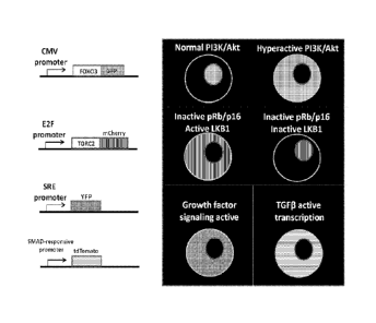
[0026] Figure 9. Fluorescent readouts for selected tumor diagnostic pathways.
This figure
lists an initial panel of four diagnostic expression cassettes (left side) and
their expected
phenotype in cells (right side). The CMV-[Foxo3-GFP] cassette is
constitutively active, and thus
GFP is expressed in all cell types where the CMV promoter is active. In cells
where PI3K/Alct
activity is low, such as non-tumor tissue, the Foxo3-GFP fusion localizes to
the nucleus.
However, in cells where PI3K/Akt activity is high, such as in tumor cells, the
Foxo3-GFP fusion
localizes to the cytoplasm. The E2F-[mCherry-CRTC2] cassette is only active in
cells that have
inactive pRB, such as in almost all tumors. In these cells, the mCherry-CRTC2
fusion is
cytoplasmic if the tumor suppressor Lkbl is intact. However, in tumor cells
that have lost Lkbl
function, the mCherry-CRTC2 fusion is located in the nucleus. The serum
response element
(SRE) promoter expresses YFP only in cells that have activated growth factor
signaling or
mitogen stimulation, indicative of rapidly dividing cells such as tumors.
Lastly, the SMAD-
responsive promoter cassette drives expression of tdTomato in cells where
TGF[3 signaling is
active, which has been linked to a metastatic phenotype in certain cancers
When combined,
these four expression cassettes provide information on five different cancer-
relevant pathways.
[0027] Figure 10. Manipulation of Adenovirus Adsembly modules to create tumor
diagnostic
viruses. Viruses were created using the Adsembly genome assembly method. This
figure
diagrams in which Adsembly modules each of the initial four cancer diagnostic
expression
cassettes was placed. Two cassettes were cloned into the El module, as it has
been shown to
tolerate dual-expression cassettes in previous experiments. The E3A/E3B
portion of the E3
module was deleted and replaced with a single cassette. Not shown is the
manipulation of the
fiber as listed in Table 1, which also occurs within the E3 module. Lastly,
the E4 region was
deleted and replaced with a single module. More specific information on the
deletions and
insertions can be found in the materials and methods. After altering these
Adsembly vectors,
5
Date Recue/Date Received 2020-08-10
CA 02865642 2014-08-26
WO 2013/138650 PCT/US2013/031646
they were used in standard Adsembly reactions to create viruses that contain
one or more of the
tumor diagnostic expression cassettes.
DETAILED DESCRIPTION OF THE INVENTION
I. Definitions
[0028] "Nucleic acid" refers to deoxyribonucleotides or ribonucleotides and
polymers thereof
in either single- or double-stranded form, and complements thereof. The term
encompasses
nucleic acids containing known nucleotide analogs or modified backbone
residues or linkages,
which are synthetic, naturally occurring, and non-naturally occurring, which
have similar binding
properties as the reference nucleic acid, and which are metabolized in a
manner similar to the
reference nucleotides. Examples of such analogs include, without limitation,
phosphorothioates,
phosphoramidates, methyl phosphonates, chiral-methyl phosphonates, 2-0-methyl
ribonucleotides, peptide-nucleic acids (PNAs).
[0029] Unless otherwise indicated, a particular nucleic acid sequence also
implicitly
encompasses conservatively modified variants thereof (e.g., degenerate codon
substitutions) and
complementary sequences, as well as the sequence explicitly indicated.
Specifically, degenerate
codon substitutions may be achieved by generating sequences in which the third
position of one
or more selected (or all) codons is substituted with mixed-base and/or
deoxyinosine residues
(Batzer et al., Nucleic Acid Res. 19:5081 (1991); Ohtsuka et al., J Biol.
Chem. 260:2605-2608
(1985); Rossolini et al., MoL Cell. Probes 8:91-98 (1994)). The term nucleic
acid is used
interchangeably with gene, cDNA, mRNA, oligonucleotide, and polynucleotide.
[0030] A particular nucleic acid sequence also implicitly encompasses "splice
variants."
Similarly, a particular protein encoded by a nucleic acid implicitly
encompasses any protein
encoded by a splice variant of that nucleic acid. "Splice variants," as the
name suggests, are
products of alternative splicing of a gene. After transcription, an initial
nucleic acid transcript
may be spliced such that different (alternate) nucleic acid splice products
encode different
polypeptides. Mechanisms for the production of splice variants vary, but
include alternate
splicing of exons. Alternate polypeptides derived from the same nucleic acid
by read-through
transcription are also encompassed by this definition. Any products of a
splicing reaction,
including recombinant forms of the splice products, are included in this
definition. An example
of potassium channel splice variants is discussed in Leicher, et al., J. Biol.
Chem.
273(52):35095-35101 (1998).
6
CA 02865642 2014-08-26
WO 2013/138650 PCT/US2013/031646
[0031] Construction of suitable vectors containing the desired therapeutic
gene coding and
control sequences may employ standard ligation and restriction techniques,
which are well
understood in the art (see Maniatis et al., in Molecular Cloning: A Laboratory
Manual, Cold
Spring Harbor Laboratory, New York (1982)). Isolated plasmids, DNA sequences,
or
synthesized oligonucleotides may be cleaved, tailored, and re-ligated in the
form desired.
[0032] Nucleic acid is "operably linked" when it is placed into a functional
relationship with
another nucleic acid sequence. For example, DNA for a presequence or secretory
leader is
operably linked to DNA for a polypeptide if it is expressed as a preprotein
that participates in the
secretion of the polypeptide; a promoter or enhancer is operably linked to a
coding sequence if it
affects the transcription of the sequence; or a ribosome binding site is
operably linked to a coding
sequence if it is positioned so as to facilitate translation. Generally,
"operably linked" means that
the DNA sequences being linked are near each other, and, in the case of a
secretory leader,
contiguous and in reading phase. However, enhancers do not have to be
contiguous. Linking is
accomplished by ligation at convenient restriction sites. If such sites do not
exist, the synthetic
oligonucleotide adaptors or linkers are used in accordance with conventional
practice.
[0033] The terms "identical" or percent "identity," in the context of two or
more nucleic acids
or polypeptide sequences, refer to two or more sequences or subsequences that
are the same or
have a specified percentage of amino acid residues or nucleotides that are the
same (i.e., about
60% identity, preferably 65%, 70%, 75%, 80%, 85%, 90%, 91%, 92%, 93%, 94%,
95%, 96%,
97%, 98%, 99%, or higher identity over a specified region, when compared and
aligned for
maximum correspondence over a comparison window or designated region) as
measured using a
BLAST or BLAST 2.0 sequence comparison algorithms with default parameters
described
below, or by manual alignment and visual inspection (see, e.g., NCBI web site
or the like). Such
sequences are then said to be "substantially identical." This definition also
refers to, or may be
applied to, the compliment of a test sequence. The definition also includes
sequences that have
deletions and/or additions, as well as those that have substitutions. As
described below, the
preferred algorithms can account for gaps and the like. Preferably, identity
exists over a region
that is at least about 25 amino acids or nucleotides in length, or more
preferably over a region
that is 50-100 amino acids or nucleotides in length.
[0034] For sequence comparison, typically one sequence acts as a reference
sequence, to
which test sequences are compared. When using a sequence comparison algorithm,
test and
reference sequences are entered into a computer, subsequence coordinates are
designated, if
7
CA 02865642 2014-08-26
WO 2013/138650 PCT/US2013/031646
necessary, and sequence algorithm program parameters are designated.
Preferably, default
program parameters can be used, or alternative parameters can be designated.
The sequence
comparison algorithm then calculates the percent sequence identities for the
test sequences
relative to the reference sequence, based on the program parameters.
[0035] A "comparison window", as used herein, includes reference to a segment
of any one of
the number of contiguous positions selected from the group consisting of from
20 to 600, usually
about 50 to about 200, more usually about 100 to about 150 in which a sequence
may be
compared to a reference sequence of the same number of contiguous positions
after the two
sequences are optimally aligned. Methods of alignment of sequences for
comparison are well-
known in the art. Optimal alignment of sequences for comparison can be
conducted, e.g., by the
local homology algorithm of Smith & Waterman, Adv. AppL Math. 2:482 (1981), by
the
homology alignment algorithm of Needleman & Wunsch, J. 11/16l. Biol. 48:443
(1970), by the
search for similarity method of Pearson & Lipman, Proc. Nat'l. Acad. Sci. USA
85:2444 (1988),
by computerized implementations of these algorithms (GAP, BESTFIT, FASTA, and
TFASTA
in the Wisconsin Genetics Software Package, Genetics Computer Group, 575
Science Dr.,
Madison, W1), or by manual alignment and visual inspection (see, e.g., Current
Protocols in
Molecular Biology (Ausubel et al., eds. 1995 supplement)).
[0036] A preferred example of algorithm that is suitable for determining
percent sequence
identity and sequence similarity are the BLAST and BLAST 2.0 algorithms, which
are described
in Altschul et al., Nuc. Acids Res. 25:3389-3402 (1977) and Altschul et al.,
J. Mol. Biol.
215:403-410 (1990), respectively. BLAST and BLAST 2.0 are used, with the
parameters
described herein, to determine percent sequence identity for the nucleic acids
and proteins of the
invention. Software for performing BLAST analyses is publicly available
through the National
Center for Biotechnology Information, as known in the art. This algorithm
involves first
identifying high scoring sequence pairs (HSPs) by identifying short words of
length W in the
query sequence, which either match or satisfy some positive-valued threshold
score T when
aligned with a word of the same length in a database sequence. T is referred
to as the
neighborhood word score threshold (Altschul et al., supra). These initial
neighborhood word
hits act as seeds for initiating searches to find longer HSPs containing them.
The word hits are
extended in both directions along each sequence for as far as the cumulative
alignment score can
be increased. Cumulative scores are calculated using, for nucleotide
sequences, the parameters
M (reward score for a pair of matching residues; always > 0) and N (penalty
score for
mismatching residues; always <0). For amino acid sequences, a scoring matrix
is used to
8
CA 02865642 2014-08-26
WO 2013/138650 PCT/US2013/031646
calculate the cumulative score. Extension of the word hits in each direction
are halted when: the
cumulative alignment score falls off by the quantity X from its maximum
achieved value; the
cumulative score goes to zero or below, due to the accumulation of one or more
negative-scoring
residue alignments; or the end of either sequence is reached. The BLAST
algorithm parameters
W, T, and X determine the sensitivity and speed of the alignment. The BLASTN
program (for
nucleotide sequences) uses as defaults a wordlength (W) of 11, an expectation
(E) of 10, M=5,
N=-4 and a comparison of both strands. For amino acid sequences, the BLASTP
program uses
as defaults a wordlength of 3, and expectation (E) of 10, and the BLOSUM62
scoring matrix (see
Henikoff & Henikoff, Proc. Nall. Acad. Sci. USA 89:10915 (1989)) alignments
(B) of 50,
expectation (E) of 10, M=5, N=-4, and a comparison of both strands.
[0037] The terms "polypeptide," "peptide" and "protein" are used
interchangeably herein to
refer to a polymer of amino acid residues. The terms apply to amino acid
polymers in which one
or more amino acid residue is an artificial chemical mimetic of a
corresponding naturally
occurring amino acid, as well as to naturally occurring amino acid polymers
and non-naturally
occurring amino acid polymer.
[0038] The term "amino acid" refers to naturally occurring and synthetic amino
acids, as well
as amino acid analogs and amino acid mimetics that function in a manner
similar to the naturally
occurring amino acids. Naturally occurring amino acids are those encoded by
the genetic code,
as well as those amino acids that are later modified, e.g., hydroxyprolinc, y-
carboxyglutamate,
and 0-phosphoserine. Amino acid analogs refers to compounds that have the same
basic
chemical structure as a naturally occurring amino acid, i.e., an a carbon that
is bound to a
hydrogen, a carboxyl group, an amino group, and an R group, e.g., homoserine,
norleucine,
methionine sulfoxide, methionine methyl sulfonium. Such analogs have modified
R groups
(e.g., norleucine) or modified peptide backbones, but retain the same basic
chemical structure as
a naturally occurring amino acid. Amino acid mimetics refers to chemical
compounds that have
a structure that is different from the general chemical structure of an amino
acid, but that
functions in a manner similar to a naturally occurring amino acid.
[0039] Amino acids may be referred to herein by either their commonly known
three letter
symbols or by the one-letter symbols recommended by the IUPAC-II5B Biochemical
Nomenclature Commission. Nucleotides, likewise, may be referred to by their
commonly
accepted single-letter codes.
9
CA 02865642 2014-08-26
WO 2013/138650 PCT/US2013/031646
[0040] "Conservatively modified variants" applies to both amino acid and
nucleic acid
sequences. With respect to particular nucleic acid sequences, conservatively
modified variants
refers to those nucleic acids which encode identical or essentially identical
amino acid
sequences, or where the nucleic acid does not encode an amino acid sequence,
to essentially
identical sequences. Because of the degeneracy of the genetic code, a large
number of
functionally identical nucleic acids encode any given protein. For instance,
the codons GCA,
GCC, GCG and GCU all encode the amino acid alanine. Thus, at every position
where an
alanine is specified by a codon, the codon can be altered to any of the
corresponding codons
described without altering the encoded polypeptide. Such nucleic acid
variations are "silent
variations," which are one species of conservatively modified variations.
Every nucleic acid
sequence herein which encodes a polypeptide also describes every possible
silent variation of the
nucleic acid. One of skill will recognize that each codon in a nucleic acid
(except AUG, which is
ordinarily the only codon for methionine, and TGG, which is ordinarily the
only codon for
tryptophan) can be modified to yield a functionally identical molecule.
Accordingly, each silent
variation of a nucleic acid which encodes a polypeptide is implicit in each
described sequence
with respect to the expression product, but not with respect to actual probe
sequences.
[0041] As to amino acid sequences, one of skill will recognize that individual
substitutions,
deletions or additions to a nucleic acid, peptide, polypeptide, or protein
sequence which alters,
adds or deletes a single amino acid or a small percentage of amino acids in
the encoded sequence
is a "conservatively modified variant" where the alteration results in the
substitution of an amino
acid with a chemically similar amino acid. Conservative substitution tables
providing
functionally similar amino acids are well known in the art. Such
conservatively modified
variants are in addition to and do not exclude polymorphic variants,
interspecies homologs, and
alleles of the invention.
[0042] The following eight groups each contain amino acids that are
conservative substitutions
for one another: 1) Alanine (A), Glycine (G); 2) Aspartic acid (D), Glutamic
acid (E); 3)
Asparagine (N), Glutamine (Q); 4) Arginine (R), Lysine (K); 5) Isoleucine (1),
Leucine (L),
Methioninc (M), Valine (V); 6) Phenylalanine (F), Tyrosine (Y), Tryptophan
(W); 7) Serino (S),
Threonine (T); and 8) Cysteine (C), Methionine (M) (see, e.g., Creighton,
Proteins (1984)).
[0043] The term "recombinant" when used with reference, e.g., to a cell,
virus, nucleic acid,
protein, or vector, indicates that the cell, virus, nucleic acid, protein or
vector, has been modified
by the introduction of a heterologous nucleic acid or protein or the
alteration of a native nucleic
CA 02865642 2014-08-26
WO 2013/138650 PCT/US2013/031646
acid or protein, or that the cell is derived from a cell so modified. Thus,
for example,
recombinant cells express genes that are not found within the native (non-
recombinant) form of
the cell or express native genes that are otherwise abnormally expressed,
under expressed or not
expressed at all.
[0044] The phrase "stringent hybridization conditions" refers to conditions
under which a
probe will hybridize to its target subsequence, typically in a complex mixture
of nucleic acids,
but to no other sequences. Stringent conditions are sequence-dependent and
will be different in
different circumstances. Longer sequences hybridize specifically at higher
temperatures. An
extensive guide to the hybridization of nucleic acids is found in Tijssen,
Techniques in
Biochemistry and Molecular Biology--Hybridization with Nucleic Probes,
"Overview of
principles of hybridization and the strategy of nucleic acid assays" (1993).
Generally, stringent
conditions are selected to be about 5-10 C lower than the thermal melting
point (T.) for the
specific sequence at a defined ionic strength pH. The T. is the temperature
(under defined ionic
strength, pH, and nucleic concentration) at which 50% of the probes
complementary to the target
hybridize to the target sequence at equilibrium (as the target sequences are
present in excess, at
T., 50% of the probes arc occupied at equilibrium). Stringent conditions may
also be achieved
with the addition of destabilizing agents such as formamide. For selective or
specific
hybridization, a positive signal is at least two times background, preferably
10 times background
hybridization. Exemplary stringent hybridization conditions can be as
following: 50%
formamide, 5x SSC, and 1% SDS, incubating at 42 C, or, 5x SSC, 1% SDS,
incubating at 65 C,
with wash in 0.2x SSC, and 0.1% SDS at 65 C.
[0045] Nucleic acids that do not hybridize to each other uncle' stringent
conditions are still
substantially identical if the polypeptides which they encode are
substantially identical. This
occurs, for example, when a copy of a nucleic acid is created using the
maximum codon
.. degeneracy permitted by the genetic code. In such cases, the nucleic acids
typically hybridize
under moderately stringent hybridization conditions. Exemplary "moderately
stringent
hybridization conditions" include a hybridization in a buffer of 40%
formamide, 1 M NaC1, 1%
SDS at 37 C, and a wash in IX SSC at 45 C. A positive hybridization is at
least twice
background. Those of ordinary skill will readily recognize that alternative
hybridization and
.. wash conditions can be utilized to provide conditions of similar
stringency. Additional
guidelines for determining hybridization parameters are provided in numerous
reference, e.g.,
and Current Protocols in Molecular Biology, ed. Ausubel, et al., John Wiley &
Sons.
11
CA 02865642 2014-08-26
WO 2013/138650 PCT/US2013/031646
[0046] For PCR, a temperature of about 36 C is typical for low stringency
amplification,
although annealing temperatures may vary between about 32 C and 48 C depending
on primer
length. For high stringency PCR amplification, a temperature of about 62 C is
typical, although
high stringency annealing temperatures can range from about 50 C to about 65
C, depending on
the primer length and specificity. Typical cycle conditions for both high and
low stringency
amplifications include a denaturation phase of 90 C - 95 C for 30 sec - 2
min., an annealing
phase lasting 30 sec. - 2 min., and an extension phase of about 72 C for 1 - 2
mm. Protocols and
guidelines for low and high stringency amplification reactions are provided,
e.g., in Innis et al.
(1990) PCR Protocols, A Guide to Methods and Applications, Academic Press,
Inc. N.Y.).
.. [0047] The terms "transfection", "transduction", "transfecting" or
"transducing" can be used
interchangeably and are defined as a process of introducing a nucleic acid
molecule or a protein
to a cell. Nucleic acids are introduced to a cell using non-viral or viral-
based methods. The
nucleic acid molecule can be a sequence encoding complete proteins or
functional portions
thereof. Typically, a nucleic acid vector, including the elements necessary
for protein expression
(e.g., a promoter, transcription start site, etc.). Non-viral methods of
transfection include any
appropriate method that does not use viral DNA or viral particles as a
delivery system to
introduce the nucleic acid molecule into the cell. Exemplary non-viral
transfection methods
include calcium phosphate transfection, liposomal transfection, nucleofection,
sonoporation,
transfection through heat shock, magnetifection and electroporation. For viral-
based methods,
any useful viral vector can be used in the methods described herein. Examples
of viral vectors
include, but are not limited to retroviral, adenoviral, lentiviral and adeno-
associated viral vectors.
In some aspects, the nucleic acid molecules are introduced into a cell using a
adenoviial vector
following standard procedures well known in the art. The terms "transfection"
or "transduction"
also refer to introducing proteins into a cell from the external environment.
Typically,
transduction or transfection of a protein relies on attachment of a peptide or
protein capable of
crossing the cell membrane to the protein of interest. See, e.g., Ford et al.
(2001) Gene Therapy
8:1-4 and Prochiantz (2007) Nat. Methods 4:119-20.
[0048] Expression of a transfected gene can occur transiently or stably in a
host cell. During
"transient expression" the transfected nucleic acid is not integrated into the
host cell genome, and
is not transferred to the daughter cell during cell division. Since its
expression is restricted to the
transfected cell, expression of the gene is lost over time. In contrast,
stable expression of a
transfected gene can occur when the gene is co-transfected with another gene
that confers a
12
CA 02865642 2014-08-26
WO 2013/138650 PCT/US2013/031646
selection advantage to the transfected cell. Such a selection advantage may be
a resistance
towards a certain toxin that is presented to the cell. Expression of a
tiansfected gene can further
be accomplished by transposon-mediated insertion into to the host genome.
During transposon-
mediated insertion, the gene is positioned in a predictable manner between two
transposon linker
sequences that allow insertion into the host genome as well as subsequent
excision.
[0049] The terms "culture," "culturing," "grow," "growing," "maintain,"
"maintaining,"
"expand," "expanding," etc., when referring to cell culture itself or the
process of culturing, can
be used interchangeably to mean that a cell is maintained outside the body
(e.g., ex vivo) under
conditions suitable for survival. Cultured cells are allowed to survive, and
culturing can result in
cell growth, differentiation, or division. The term does not imply that all
cells in the culture
survive or grow or divide, as some may naturally senesce, etc. Cells are
typically cultured in
media, which can be changed during the course of the culture.
[0050] The terms "media" and "culture solution" refer to the cell culture
milieu. Media is
typically an isotonic solution, and can be liquid, gelatinous, or semi-solid,
e.g., to provide a
.. matrix for cell adhesion or support. Media, as used herein, can include the
components for
nutritional, chemical, and structural support necessary for culturing a cell.
[0051] A "control" sample or value refers to a sample that serves as a
reference, usually a
known reference, for comparison to a test sample. For example, a test sample
can be taken from
a test condition, e.g., in the presence of a test compound, and compared to
samples from known
conditions, e.g., in the absence of the test compound (negative control), or
in the presence of a
known compound (positive control). A control can also represent an average
value gathered
from a number of tests or results. One of skill in the art will recognize that
controls can be
designed for assessment of any number of parameters. For example, a control
can be devised to
compare therapeutic benefit based on pharmacological data (e.g., half-life) or
therapeutic
measures (e.g., comparison of side effects). One of skill in the art will
understand which controls
are valuable in a given situation and be able to analyze data based on
comparisons to control
values. Controls are also valuable for determining the significance of data.
For example, if
values for a given parameter are widely variant in controls, variation in test
samples will not be
considered as significant.
[0052] In compositions including an "additional," "further," or "second"
component (e.g.
cancer cell reporter module, reporter gene phenotype), the second component as
used herein is
different from the other components or first component. A "third" component is
different from
13
CA 02865642 2014-08-26
WO 2013/138650 PCT/US2013/031646
the other, first, and second components, and further enumerated or
"additional" components are
similarly different.
[0053] As used herein, the term "cancer" refers to all types of cancer,
neoplasm, or malignant
tumors found in mammals, including leukemia, carcinomas and sarcomas.
Exemplary cancers
include cancer of the brain, breast, cervix, colon, head & neck, liver,
kidney, lung, non-small cell
lung, melanoma, mesothelioma, ovary, sarcoma, stomach, uterus and
Medulloblastoma.
Additional examples include, Hodgkin's Disease, Non-Hodgkin's Lymphoma,
multiple myeloma,
neuroblastoma, ovarian cancer, rhabdomyosarcoma, primary thrombocytosis,
primary
macroglobulinemia, primary brain tumors, cancer, malignant pancreatic
insulanoma, malignant
carcinoid, urinary bladder cancer, premalignant skin lesions, testicular
cancer, lymphomas,
thyroid cancer, neuroblastoma, esophageal cancer, genitourinary tract cancer,
malignant
hypercalcemia, endometrial cancer, adrenal cortical cancer, neoplasms of the
endocrine and
exocrine pancreas, and prostate cancer.
[0054] The term "leukemia" refers broadly to progressive, malignant diseases
of the blood-
forming organs and is generally characterized by a distorted proliferation and
development of
leukocytes and their precursors in the blood and bone marrow. Leukemia is
generally clinically
classified on the basis of (1) the duration and character of the disease-acute
or chronic; (2) the
type of cell involved; myeloid (myelogenous), lymphoid (lymphogenous), or
monocytic; and (3)
the increase or non-increase in the number abnormal cells in the blood-
leukemic or aleukemic
(subleukemic). The P388 leukemia model is widely accepted as being predictive
of in vivo anti-
leukemic activity. It is believed that a compound that tests positive in the
P388 assay will
generally exhibit some level of anti-leukemic activity in vivo regardless of
the type of leukemia
being treated. Accordingly, the present invention includes a method of
treating leukemia, and,
preferably, a method of treating acute nonlymphocytic leukemia, chronic
lymphocytic leukemia,
acute granulocytic leukemia, chronic granulocytic leukemia, acute
promyelocytic leukemia, adult
T-cell leukemia, aleukemic leukemia, a leukocythemic leukemia, basophylic
leukemia, blast cell
leukemia, bovine leukemia, chronic myelocytic leukemia, leukemia cutis,
embryonal leukemia,
eosinophilic leukemia, Gross' leukemia, hairy-cell leukemia, hemoblastic
leukemia,
hemocytoblastic leukemia, histiocytic leukemia, stem cell leukemia, acute
monocytic leukemia,
leukopenic leukemia, lymphatic leukemia, lymphoblastic leukemia, lymphocytic
leukemia,
lymphogenous leukemia, lymphoid leukemia, lymphosarcoma cell leukemia, mast
cell leukemia,
megakaryocytic leukemia, micromycloblastic leukemia, monocytic leukemia,
myeloblastic
leukemia, myelocytic leukemia, myeloid granulocytic leukemia, myelomonocytic
leukemia,
14
CA 02865642 2014-08-26
WO 2013/138650 PCT/US2013/031646
Naegeli leukemia, plasma cell leukemia, multiple myeloma, plasmacytic
leukemia,
promyelocytic leukemia, Rieder cell leukemia, Schilling's leukemia, stem cell
leukemia,
subleukemic leukemia, and undifferentiated cell leukemia.
[0055] The term "sarcoma" generally refers to a tumor which is made up of a
substance like
the embryonic connective tissue and is generally composed of closely packed
cells embedded in
a fibrillar or homogeneous substance. Sarcomas which can be treated with a
combination of
antincoplastic thiol-binding mitochondrial oxidant and an anticancer agent
include a
chondrosarcoma, fibrosarcoma, lymphosarcoma, melanosarcoma, myxosarcoma,
osteosarcoma,
Abernethy's sarcoma, adipose sarcoma, liposarcoma, alveolar soft part sarcoma,
ameloblastic
sarcoma, botryoid sarcoma, chloroma sarcoma, chorio carcinoma, embryonal
sarcoma, Wilms'
tumor sarcoma, endometrial sarcoma, stromal sarcoma, Ewing's sarcoma, fascial
sarcoma,
fibroblastic sarcoma, giant cell sarcoma, granulocytic sarcoma, Hodgkin's
sarcoma, idiopathic
multiple pigmented hemorrhagic sarcoma, immunoblastic sarcoma of B cells,
lymphoma,
immunoblastic sarcoma of T-cells, Jensen's sarcoma, Kaposi's sarcoma, Kupffer
cell sarcoma,
angiosarcoma, leukosarcoma, malignant mesenchymoma sarcoma, parosteal sarcoma,
reticulocytic sarcoma, Rous sarcoma, serocystic sarcoma, synovial sarcoma, and
telangiectaltic
sarcoma.
[0056] The term "melanoma" is taken to mean a tumor arising from the
melanocytic system of
the skin and other organs. Melanomas which can be treated with a combination
of antineoplastic
thiol-binding mitochondrial oxidant and an anticancer agent include, for
example, acral-
lentiginous melanoma, amelanotic melanoma, benign juvenile melanoma,
Cloudman's
melanoma, S91 melanoma, Hai ding-Pa ssey melanoma, juvenile melanoma, lentigo
maligna
melanoma, malignant melanoma, nodular melanoma, subungal melanoma, and
superficial
spreading melanoma.
[0057] The term "carcinoma" refers to a malignant new growth made up of
epithelial cells
tending to infiltrate the surrounding tissues and give rise to metastases.
Exemplary carcinomas
which can be treated with a combination of antineoplastic thiol-binding
mitochondrial oxidant
and an anticancer agent include, for example, acinar carcinoma, acinous
carcinoma, adenocystic
carcinoma, adenoid cystic carcinoma, carcinoma adenomatosum, carcinoma of
adrenal cortex,
alveolar carcinoma, alveolar cell carcinoma, basal cell carcinoma, carcinoma
basocellulare,
basaloid carcinoma, basosquamous cell carcinoma, bronchioalveolar carcinoma,
bronchiolar
carcinoma, bronchogenic carcinoma, cerebriform carcinoma, cholangiocellular
carcinoma,
CA 02865642 2014-08-26
WO 2013/138650 PCT/US2013/031646
chorionic carcinoma, colloid carcinoma, comedo carcinoma, corpus carcinoma,
cribriform
carcinoma, carcinoma en cuirasse, carcinoma cutaneum, cylindrical carcinoma,
cylindrical cell
carcinoma, duct carcinoma, carcinoma durum, embryonal carcinoma, encephaloid
carcinoma,
epiermoid carcinoma, carcinoma epitheliale adenoides, exophytic carcinoma,
carcinoma ex
ulcere, carcinoma fibrosum, gelatiniforni carcinoma, gelatinous carcinoma,
giant cell carcinoma,
carcinoma gigantocellulare, glandular carcinoma, granttlosa cell carcinoma,
hair-matrix
carcinoma, hematoid carcinoma, bepatocellular carcinoma, Hurthle cell
carcinoma, hyaline
carcinoma, hypemephroid carcinoma, infantile embryonal carcinoma, carcinoma in
situ,
intraepidermal carcinoma, intraepithelial carcinoma, Krompecher's carcinoma,
Kulchitzky-cell
carcinoma, large-cell carcinoma, lenticular carcinoma, carcinoma lenticulare,
lipomatous
carcinoma, lymphoepithelial carcinoma, carcinoma medullare, medullary
carcinoma, melanotic
carcinoma, carcinoma molle, mucinous carcinoma, carcinoma muciparum, carcinoma
mucocellulare, mucoepidermoid carcinoma, carcinoma mucosum, mucous carcinoma,
carcinoma
myxomatodes, nasopharyngeal carcinoma, oat cell carcinoma, carcinoma
ossificans, osteoid
carcinoma, papillary carcinoma, periportal carcinoma, preinvasive carcinoma,
prickle cell
carcinoma, pultaceous carcinoma, renal cell carcinoma of kidney, reserve cell
carcinoma,
carcinoma sarcomatodes, schneiderian carcinoma, scinhous carcinoma, carcinoma
scroti, signet-
ring cell carcinoma, carcinoma simplex, small-cell carcinoma, solanoid
carcinoma, spheroidal
cell carcinoma, spindle cell carcinoma, carcinoma spongiosum, squamous
carcinoma, squamous
cell carcinoma, string carcinoma, carcinoma telangiectaticum, carcinoma
telangiectodes,
transitional cell carcinoma, carcinoma tuberosum, tuberous carcinoma,
verrucous carcinoma, and
carcinoma villosum.
[0058] By "therapeutically effective dose or amount" herein is meant a dose
that produces
effects for which it is administered. The exact dose and formulation will
depend on the purpose
of the treatment, and will be ascertainable by one skilled in the art using
known techniques (see,
e.g., Lieberman, Pharmaceutical Dosage Forms (vols. 1-3, 1992); Lloyd, The
Art, Science and
Technology of Pharmaceutical Compounding (1999); Remington: The Science and
Practice of
Pharmacy, 20th Edition, Gennaro, Editor (2003), and Pickar, Dosage
Calculations (1999)).
[0059] The term "pharmaceutically acceptable salts" or "pharmaceutically
acceptable carrier"
is meant to include salts of the active compounds which are prepared with
relatively nontoxic
acids or bases, depending on the particular substituents found on the
compounds described
herein. When compounds of the present invention contain relatively acidic
functionalities, base
addition salts can be obtained by contacting the neutral form of such
compounds with a sufficient
16
81781841
amount of the desired base, either neat or in a suitable inert solvent
Examples of
pharmaceutically acceptable base addition salts include sodium, potassium,
calcium, ammonium,
organic amino, or magnesium salt, or a similar salt. When compounds of the
present invention
contain relatively basic fimetionalities, acid addition salts can be obtained
by contacting the
neutral form of such compounds with a sufficient amount of the desired acid,
either neat or in a
suitable inert solvent. Examples of pharmaceutically acceptable acid addition
salts include those
derived from inorganic acids like hydrochloric, hydrobromic, nitric, carbonic,
monohydrogencarbonic, phosphoric, monohydrogenphosphoric,
dihydrogenphosphoric, sulfuric,
monohydrogensulfuric, hydriodic, or phosphorous acids and the like, as well as
the salts derived
from relatively nontoxic organic acids like acetic, propionic, isobutyric,
maleic, malonic,
benzoic, succinic, suberic, fumaric, lactic, mandelic, phthalic,
benzenesulfonic, p-tolylsulfonic,
citric, tartaric, methanesulfonic, and the like. Also included are salts of
amino acids such as
arginate and the like, and salts of organic acids like glucuronic or
galactunoric acids and the like
(see, e.g., Berge et al, Journal of Pharmaceutical Science 66:1-19 (1977)).
Certain specific
compounds of the present invention contain both basic and acidic
functionalities that allow the
compounds to be converted into either base or acid addition salts. Other
pharmaceutically
acceptable carriers known to those of skill in the art arc suitable for the
present invention.
IL Methods
100601 In one aspect, a method of detecting a cancer in a subject is provided.
The method
includes administering a recombinant reporter adenovirus to a subject. The
recombinant reporter
adenovirus is allowed to infect a cancer cell within the subject thereby
forming a reporter
infected cancer cell. A sample including the reporter infected cancer cell is
obtained from the
subject and the reporter infected cancer cell is detected thereby detecting a
cancer in the subject.
A recombinant reporter adenovirus as provided herein is a recombinant
adenovirus including at
least one (e.g. one) sequence that encodes for a reporter protein. Non
limiting examples of
recombinant reporter adenoviruses are shown in Table 2 and Figure 9. The
recombinant reporter
adenoviruses provided herein including embodiments are formed according to the
methods as
described in published application PCT/US2011/048006.
The reporter protein may be a fluorescent protein (e.g. green
fluorescent protein, red fluorescent protein) or it may be a protein that can
be fluorescently
labeled thereby becoming readily detectable. Fluorescent labeling can be
achieved by binding a
fluorescently labeled antibody to the reporter protein. In some embodiments,
the recombinant
17
CA 2865642 2018-04-05
CA 02865642 2014-08-26
WO 2013/138650 PCT/US2013/031646
reporter adenovirus includes a Cytomegalovirus promoter operable linked to a
nucleic acid
encoding for a fluorescent protein. In some further embodiments, the
fluorescent protein is a
green fluorescent protein. In some embodiments, the recombinant reporter
adenovirus includes a
E2F promoter operable linked to a nucleic acid encoding for a fluorescent
protein. In some
further embodiments, the fluorescent protein is a red fluorescent protein. In
some embodiments,
the recombinant reporter adenovirus includes a SRE promoter operable linked to
a nucleic acid
encoding for a fluorescent protein. In some further embodiments, the
fluorescent protein is a
yellow fluorescent protein. In some embodiments, the recombinant reporter
adenovirus includes
a SMAD-responsive promoter operable linked to a nucleic acid encoding for a
fluorescent
protein. In some further embodiments, the fluorescent protein is a red
fluorescent protein.
[0061] In another aspect, a method of detecting a cancer in a subject is
provided. The method
includes obtaining from a subject a sample including a cancer cell. A
recombinant reporter
adenovirus is contacted with the cancer cell. The recombinant reporter
adenovirus is allowed to
infect the cancer cell thereby forming a reporter infected cancer cell and the
reporter infected
cancer cell is detected thereby detecting a cancer in said subject. In some
embodiments, the
detecting according to the methods provided herein includes detecting a
reporter gene phenotype.
In some further embodiments, the reporter gene phenotype is a fluorescent
reporter gene
phenotype. Where a cell (e.g. cancer cell) is infected with a recombinant
reporter adenovirus as
provided herein, the cell is infected with an amount of recombinant reporter
adenovirus sufficient
to express a reporter phenotype.
[0062] In another aspect, a method of determining whether a test compound
inhibits growth of
a cancel cell from a cancer patient is provided. The method includes obtaining
from a subject a
sample including a cancer cell. A recombinant reporter adenovirus is contacted
with the cancer
cell. The recombinant reporter adenovirus is allowed to infect the cancer cell
thereby forming a
reporter infected cancer cell. The reporter infected cancer cell is allowed
sufficient time to grow.
A level of growth of the reporter infected cancer cell is determined and the
level is compared to a
control level, wherein a low level compared to the control level indicates the
test compound
inhibits growth of the cancer cell from the patient. A control level as
provided herein is the level
of growth of a cancer cell in the absence of the test compound.
[0063] In some embodiments, the cancer according to the methods provided
herein is lung
cancer, skin cancer or breast cancer. In other embodiments, the cancer cell
according to the
methods provided herein is a circulating cancer cell. In some embodiments, the
cancer cell is a
18
CA 02865642 2014-08-26
WO 2013/138650 PCT/US2013/031646
premalignant cell. In some embodiments, the sample according to the methods
provided herein
is a bodily fluid. In some further embodiments, the bodily fluid is blood. In
other embodiments,
the bodily fluid is serum or plasma. In other embodiments, the bodily fluid is
urine, saliva, a
pulmonary tissue, bronchoalveolar lavage sample, or exhaled breath condensate.
[0064] In another aspect, a method of isolating a reporter infected cancer
cell within a sample
from a subject is provided. The method includes separating the reporter
infected cancer cell
from a non-infected cell, wherein the separating is at least partially based
on an expressed
reporter gene phenotype of the reporter infected cancer cell. In some
embodiments, the reporter
gene phenotype is a level of fluorescent activity. In other embodiments, the
reporter gene
phenotype is a level of cell growth. In other embodiments, the reporter gene
phenotype is a level
of aberrant cell morphology. In some embodiments, the method further includes
allowing the
reporter infected cancer cell sufficient time to grow, thereby expressing the
expressed reporter
gene phenotype. In some embodiments, the non-infected cell is a non-cancer
cell. In other
embodiments, the sample is a blood sample.
[0065] In another aspect, a method of detecting a cancer in a subject is
provided. The method
includes administering a recombinant reporter adenovirus provided herein
including
embodiments thereof to a subject. The recombinant reporter adenovirus is
allowed to infect a
cancer cell within the subject thereby forming a reporter infected cancer
cell. A sample is
obtained from the subject including the reporter infected cancer cell and the
reporter infected
cancer cell is detected thereby detecting a cancer in the subject.
[0066] In another aspect, a method of detecting a cancer in a subject is
provided. The method
includes obtaining from a subject a sample including a cancer cell. A
recombinant reporter
adenovirus provided herein including embodiments thereof is contacted with the
cancer cell.
The recombinant reporter adenovirus is allowed to infect the cancer cell
thereby forming a
reporter infected cancer cell and the reporter infected cancer cell is
detected thereby detecting a
cancer in the subject. In some embodiments, the method further includes
administering a cancer
treatment to the subject.
[0067] A method of determining whether a test compound inhibits growth of a
cancer cell
from a cancer patient is provided. The method includes obtaining from a
subject a sample
including a cancer cell and contacting a recombinant reporter adenovirus
provided herein
including embodiments thereof with the cancer cell. The recombinant reporter
adenovirus is
allowed to infect the cancer cell thereby forming a reporter infected cancer
cell. The reporter
19
CA 02865642 2014-08-26
WO 2013/138650 PCT/US2013/031646
infected cancer cell is allowed sufficient time to grow and a level of growth
of the reporter
infected cancer cell is determined. The level is compared to a control level,
wherein a low level
compared to the control level indicates the test compound inhibits growth of
the cancer cell from
the patient.
III. Compositions
[0068] Provided herein, inter alia, is a recombinant reporter adenovirus
useful for diagnosis
and detection of cancer cells. In one aspect, a recombinant reporter
adenovirus including a
cancer cell reporter module and a cancer cell binding module is provided. A
cancer cell reporter
module as provided herein includes a reporter gene encoding a reporter
protein. A reporter
protein may be a fluorescent protein (e.g. green fluorescent protein, red
fluorescent protein) or it
may be a protein that can be fluorescently labeled thereby becoming readily
detectable.
Fluorescent labeling can be achieved by binding a fluorescently labeled
antibody to the reporter
protein. A cancer cell binding molecule as provided herein is a molecule
capable of binding a
molecule expressed by a cancer cell (e.g. cellular receptor). The cell binding
molecule may be a
small molecule or a protein. In some embodiments, the cancer cell reporter
module includes a
cancer responsive promoter operably linked to a reporter gene. A cancer
responsive promoter as
provided herein is a promoter having an activity in a cancer cell, wherein the
activity is
detectably different from the activity of the promoter in a non-cancer cell.
In some
embodiments, the activity is decreased as compared to the activity of the
promoter in a non-
cancer cell. In other embodiments, the activity is increased as compared to
the activity of the
promoter in a non-cancer cell. In some further embodiments, the reporter gene
is a fluorescent
reporter gene.
[0069] In some embodiments, the recombinant adenovirus further comprises an
immune
evasion module. An immune evasion module as provided herein is a protein or
polypeptide,
which if expressed by the recombinant reporter adenovirus prevents the
recombinant reporter
adenovirus from being detected by the immune system of the cancer patient.
[0070] In some embodiments, the cancer cell reporter module is a first cancer
cell reporter
module and the recombinant reporter adenovirus further includes a second
cancer cell reporter
module and a third cancer cell reporter module. In some embodiments, the first
cancer cell
reporter module is capable of expressing a first reporter gene phenotype, the
second cancer cell
reporter module is capable of expressing a second reporter gene phenotype, and
the third cancer
cell reporter module is capable of expressing a third reporter gene phenotype.
In some further
CA 02865642 2014-08-26
WO 2013/138650 PCT/US2013/031646
embodiments, the first reporter gene phenotype, the second reporter gene
phenotype, and the
third reporter gene phenotype are each detectably different. In some
embodiments, the first
reporter gene phenotype is indicative of a first cancer, the second reporter
gene phenotype is
indicative of a second cancer, and the third reporter gene phenotype is
indicative of a third
cancer. In some further embodiments, the first cancer, the second cancer and
the third cancer are
independently different. In some embodiments, the first reporter gene
phenotype, the second
reporter gene phenotype and the third reporter gene phenotype are indicative
of a single cancer.
IV. Kits
[0071] In another aspect, a kit for detecting cancer is provided. The kit
includes a recombinant
reporter adenovirus provided herein including embodiments thereof In some
embodiments, the
kit includes reagents for separating cells (e.g. potential cancer cells) from
a tissue or cell sample
from a subject, such as those described herein (e.g. magnetic beads or other
affinity based
separation materials, stock buffers etc.). Thus, the kit can include
antibodies or other reagents
capable of specifically binding to at least one cell-specific marker. The kit
can also include tubes
.. or other containers for holding the sample during the processing and
detection. The kit further
includes instructions to administer the recombinant reporter adenovirus to the
patient under
conditions suitable for infecting a cell and detecting a cell.
[0072] In another aspect, a kit for screening a cancer drug is provided. The
kit includes a
cancer inhibiting compound and a recombinant reporter adenovirus provided
herein including
embodiments thereof. In some embodiments, the kit includes reagents for
administering the
cancer inhibiting compound (e.g. stock buffers) and table-top detection
devices for detecting the
reporter gene phenotype.
[0073] In another aspect, a kit for isolating a cancer cell is provided. The
kit includes a device
for detecting an expressed reporter gene phenotype and a recombinant reporter
adenovirus
.. provided herein including embodiments thereof In some embodiments, the kit
includes reagents
for separating (isolating) cancer cells from a tissue or cell sample from a
subject, such as those
described herein (e.g. magnetic beads or other affinity based separation
materials, stock buffers
etc.). Thus, the kit can include antibodies or other reagents capable of
specifically binding to at
least one cancer cell-specific marker. The kit can also include tubes or other
containers for
holding the sample during the processing and detection. The kit further
includes instructions to
administer the recombinant reporter adenovirus to the patient under conditions
suitable for
infecting a cancer cell and detecting a cancer cell.
21
CA 02865642 2014-08-26
WO 2013/138650 PCT/US2013/031646
V. Specific Embodiments
[0074] Applicants intend is to develop a standardized automated platform that
provides point-
of-care diagnostics to inform clinical decisions at a level of molecular
sophistication and
prognostic power that is not possible with any other detection system,
biomarkers or correlative
gene expression signatures. A non-invasive test, which detects, enumerates,
characterizes and
isolates viable CTCs from the blood have been developed. This alerts the
clinician to either the
presence or progression of cancer from a primary lesion and informs the
clinical decision as to
how aggressively a patient should be treated depending on the number nature of
circulating
tumor cells and the tumor pathways which are deregulated by genetic
aberrations. Further it can
be determined which of the key cancer pathways are deregulated based on robust
transcriptional
reporters and molecular hallmarks using a rapid economic automated platform
operated by a
technician. The CTCs can be isolated and captured and directly tested for
their ability to respond
to different potential treatment regimens and inform the clinician's decision
as to which
treatment option is most likely to achieve maximal efficacy.
[0075] Virus vectors that provide quantitative and qualitative data regarding
tumor pathways
through fluorescent protein readouts
[0076] Over 100,000 mutations have now been identified in tumor genomes
(Stratton MR,
Campbell PJ, Futreal PA, Nature. 2009:458(7239):719-24.19360079) of which
there are at least
350 genes that exhibit recurrent mutations (Futreal PA et al., Nat Rev
Cancer,. 20044(3):177-
83.14993899). Despite this new genetic knowledge, the diagnosis, prognosis and
treatment of
cancer patients still largely relies on subjective histopathology, surrogate
biomarkers of
transformation, variable surface markers or correlative gene expression
signatures. With
advances in DNA sequencing, it may soon be possible to sequence the genome of
every cancer
patient's tumor. However, even if this is possible, these data will not reveal
epigenetic
modifications and key interactions within the tumor microenvironment that
determine a tumor's
phenotype, or allow one to predict a priori how these factors interact to
determine a patient's
clinical outcome or the response of their tumors to different therapies.
[0077] Despite the complexity and genetic variability of cancers, all tumors
share phenotypes
that determine their malignant potential, the so-called 'hallmarks of cancer',
which are the result
of mutations in a relatively small number of key pathways (Figure 1, (Hanahan
D, Weinberg
RA, Cell, 2000;100(1):57-70.10647931)). In individual tumors the mutations
that deregulate
these pathways vary but converge downstream on key transcriptional elements
and effectors.
22
CA 02865642 2014-08-26
WO 2013/138650 PCT/US2013/031646
For example, tumor self-sufficiency for growth factor signaling can result
from mutations in
RTKs, RAS, PTEN, PI-3K, or RAF, while the RB tumor suppressor pathway can be
inactivated
by mutations in RB itself, loss of p16 (point mutations and epigenetic
silencing) or
overexpression of Cyclins (Du W, Searle JS, Curr Drug Targets, 2009;10(7):581-
9.19601762;
Sherr CJ, Cel, 2004;116(2):235-46.14744434; Rossi DJ, Weissman IL, Cell,
2006;125(2):229-
31.16630811; Gazdar AF. Oncogene, 2009;28 Suppl 1.S24-31.19680293; Yuan TL,
Cantley LC,
Oncogene,. 2008;27(4 l ):5497-510.18794884). The acquisition of these
mutations and their
resultant phenotypic traits is not simultaneous but often occurs over a long
period of time and
through progressive stages. The deregulation of these key molecular activities
can be
functionally determined using diagnostic transcriptional reporter and cell-
based assays. For
example, mutations in Rb or p16 result in activation of E2F driven promoters
(Du W, Searle JS,
Curr Drug Targets, 2009;10(7):581-9.19601762; Shen CJ, Cel, 2004;116(2):235-
46.14744434).
Similarly, the nuclear versus cytoplasmic localization of SMAD is an indicator
of TGFI3 pathway
signaling and metastasis (Shi Y, Massague J., Cell, 2003;113(6):685-
700.12809600). These
.. transcriptional and cell-based fluorescent localization read-outs are being
used individually as
reporters of tumor pathway activities in basic research and drug screening
applications.
[00781 Rather than focusing on detecting individual genetic lesions that are
numerous and
highly variable between tumors, Applicants created viral diagnostic drones
that incorporate
multiple transcriptional and molecular modules in their genomes to detect a
patient's tumor,
report its molecular 'hallmarks' and 'up-front' response to different
therapies. To achieve this
Applicants exploit a transformative new technological platform that Applicants
have recently
developed to create next generation tumor selective replicating adenoviruses
(O'Shea CC,
Oncogene, 2005;24(52):7636-9.16299525; O'Shea CC., Oncogene, 2005;24(52):7640-
55.16299526). Adenovirus is a natural multi-gene expression vehicle that
reaches the nucleus
within an hour of infection (O'Shea CC, Oncogene, 2005;24(52):7636-9.16299525;
O'Shea CC.,
Oncogene, 2005;24(52):7640-55.16299526; Leopold PL, Crystal RG, Adv Drug Deily
Rev.,
2007;59(8):810-21.17707546). Applicants' Adsembly' enables the rapid, de novo
assembly of
custom adenoviral genomes in vitro from a library of genomic building parts
(created from over
60 human and animal adenoviruses which have different tropisms and properties
to Ad2/5 or
which have been genetically modified to confer altered functionality) and
heterologous elements
(Figure 2) (O'Shea CC, Oncogene, 2005; 24(52):7636-9.16299525). Applicants
have already
used this technology to create over 60 new viruses with various mutations and
transgene
23
CA 02865642 2014-08-26
WO 2013/138650 PCT/US2013/031646
expression cassettes, and have shown viruses created using this method are
capable of high titer
growth.
[0079] The El, E3, and E4 regions arc either not necessary for replication in
culture or can be
complemented with available cell lines (Goncalves MA, de Vries AA, Rev Med
Virol.,
2006;16(3):167-86.16710837). Each of these regions has independent promoter
elements that
drive the expression of multiple gene products (16 genes) using alternative
splicing. Applicants
exploit this as a system to engineer single powerful diagnostic agents that
incorporate multiplex
and quantitative measurements of the pathway activities deregulated in
different tumor samples
(Table 1). The natural viral promoters are replaced with promoters that are
activated in tumors
with key mutations/phenotypes. These promoters drive the expression of four
different
fluorescent reporter gene-fusions which provide additional information on the
key pathways
deregulated in a patient's tumor, such as nuclear versus cytoplasmic NF-KB
(inflammation),
TORC2 (LKB1 mutations and metabolism), FOX (P13-K/AKT mutations) or SMAD 4
(TGFP
pathway mutations) (Shi Y, Massague J., Cell, 2003;113(6):685-700.12809600;
Oeckinghaus A,
Ghosh S., Cold Spring Harb Perspect Biol., 2009;1(4):a000034.20066092;
Wullschleger S,
Loewith R, Hall MN, Cell, 2006;124(3):471-84.16469695; Weidinger C et al.,
Endocr Re/at
Cancer, 2008;15(4):917-29.18775975). Using these agents, the molecular lesions
and malignant
characteristics of any given tumor can be rapidly discerned (within 24 hours)
and scored via the
NanoSort lab-on-a-chip ttFACS. Furthermore, these agents could also be used as
reporters to
determine rapidly and directly if a patient's tumor is likely to respond to a
particular therapy.
Applicants' technology improves on previously described virus-based methods of
CTC detection
in several important ways (Fong SM et al., Surgery, 2009;146(3):498-
505.19715807; Kojima T
et al., J Clin Invest., 2009,119(10):3172-81.19729837). Through the use of
Adsembly, libraries
of tumor responsive fluorescent elements can be created. This allows for rapid
creation of
multiple adenoviruses tailored towards the detection of particular types of
tumors and pathway
mutations. Adsembly also allows for ease of retargeting adenovirus, thus
maximizing chances of
CTC transduction. Lastly, it allows for ease of multigene expression from the
different genomic
modules, which increases the amount of information that can be gained during
CTC detection.
[0080] Lab-On-A-Chip Technology
[0081] Several methods have been proposed for improved CTC enumeration and
capture and
flow cytometry has already proven success (Allan AL, Keeney M., J
2010;2010:426218.20049168). Flow cytometry allows for rapid, yet highly
specific, quantitative
24
CA 02865642 2014-08-26
WO 2013/138650 PCT/US2013/031646
cell-by-cell analysis under multiple parameters, as well as the ability to
sort CTCs for further
molecular characterization. Additionally, flow cytometry is a mature, well-
recognized, and
commercially viable technology. However, multiple obstacles make current flow
cytometers
impractical for the point-of-care analysis of CTCs. First, cells must be
labeled by manually
pipetting individual antibodies into the cell samples. This procedure may
result in large
variations in data due to differences in antibody handling, pipetting
inaccuracies, storage
inconsistencies, and variability in antibody lots. Secondly, current flow
cytometers are very
expensive and have a large footprint (i.e. not mobile). Finally, current flow
cytometers are
technically and analytically challenging to operate.
[0082] To address these technical issues, Applicants have developed lab-on-a-
chip technology
that combines microfluidics, photonics, and microacoustics with groundbreaking
analytical
techniques. These patented technologies, exclusively licensed by NanoSort,
Inc, enable point-of-
care access to flow cytometry via a robust, portable, inexpensive device that
meets or exceeds
performance of current industry leaders at a fraction of the cost and space
(Cho SH, Chen CH,
Tsai FS, Godin JM, Lo YH, Lab Chip., 2010;10(12):1567-73.20379604; Cho SH et
al., Conf
Proc IEEE Eng Med Biol Soc., 2009;2009:1075-8.19965141; Chen CH et al., Biomed
Microdevices, 2009;11(6):1223-31.19649710; Chen CH, et al., In: Hawkins AR,
editor.
Handbook of Optofluidics: CDC Press; 2010. p. 664; Godin J, Lo YH, Biomed Opt
Express,
2010;1(5):1472-9.21258563).
[0083] Virus based detectors and diagnostics mediated fluorescent highlighting
of CTCs
[0084] Applicants created a series of tumor pathway activity modules that
replace the viral El,
E3 and E4 transcriptional units, which have been re-assembled with additional
modifications in
the viral backbone to confer novel tissue tropisms and other activities. These
viral diagnostic
agents are validated in human tumor cell-lines (which have known
phenotypes/mutations) as
well as primary cells, and done so both in culture and in the context of human
blood samples.
[0085] The first set of diagnostic adenoviruses has been engineered to express
four different
fluorescent biomarkers that are diagnostic of tumor cells with mutations in
the RB/p16, TGFI3,
Growth factor/PI-3K/Ras/MAPK, LKB1/AMPK pathways, not only labeling them for
detection
and collection, but defining their malignant potential and response to
therapy. The latest data
from the Sanger Center and Cosmic database shows that EGFR is
amplified/mutated in 28% of
tumors, RB (12%) ,p16 (13%), Ras (17%), LKB1 (9%), SMAD4 (2%). The feasibility
of such
an approach has already been demonstrated with the use of such tumor specific
promoters for the
CA 02865642 2014-08-26
WO 2013/138650 PCT/US2013/031646
development of oncolytic viruses to selectively induce the expression of viral
genes in cancer,
thereby ensuring tumor selective viral replication (O'Shea CC, Oncogene,
2005;24(52):7636-
9.16299525; O'Shea CC., Oncogene, 2005;24(52):7640-55.16299526; Huang TG et
al., Gene
Ther., 2003;10(15):1241-7.12858189; McCormick F., Cancer Biol Ther., 2003;2(4
Suppl
.. 1):S157-60.14508094; Ries SJ, Brandts CH, Drug Discov Today, 2004;9(17):759-
68.15450242;
Chiocca EA, Nat Rev Cancer, 2002;2(12):938-50.12459732).
[0086] For example, breast tissues from healthy women contain a subpopulation
of variant
human mammary epithelial cells (vHMEC) in which p1e/K4a is epigenetically
silenced (Hoist
CR, Cancer Res., 2003;63(7):1596-601.12670910) and which are thought to
represent
premalignant precursors for breast cancer (Tlsty TD, J Mammary Gland Biol
Neoplasia,
2004;9(3):263-74.15557799; Crawford YG et al., Cancer Cell, 2004;5(3):263-
73.15050918;
Romanov SR et al., Nature, 2001;409(6820):633-7.11214324). In Fig 3,
Applicants show that a
virus in which the natural El promoter is replaced with the cellular E2F
promoter (Johnson L et
al., Cancer Cell, 2002;1(4):325-37.12086848) specifically drives the
expression of downstream
viral proteins in vHMECs versus HMECs. Applicants use a similar strategy to
detect and isolate
CTCs but using replication incompetent viruses in which the viral genes are
now replaced with
fluorescent markers that enable their detection, quantification and sorting
using an integrated lab-
on-a-chip FACS.
[0087] To achieve this, the viral "El" module promoter and orfs are be
replaced with an E2F
promoter driving a TORC2-eYFP fusion. This identifies cells with mutations in
the pRb/p16
pathway and LKB1.The viral "E3" module promoter and orfs are replaced with two
cassettes. A
CMV promoter regulated by a serum response element (SRE) chives mCheity,
identifying
hyperactivation of the EGFR/RAS/RAFIMAPK pathway and a TGFp regulated promoter
drives
mOrange, identifying cells with metastatic potential. The viral "E4" module
promoter and orfs
are replaced with the CMV major IE promoter driving eGFP-FOXO, which are
expressed in
nearly all cells and serve both as a way to normalize fluorescence and
identify cells with
mutations in the PTEN/PI-3K/AKT pathways.
[0088] To ensure CTC infection, Applicants also incorporate a novel
innovation. Ad5, which
was first to be discovered, is the most predominant adenovirus used in basic
and clinical
research. The cellular receptor for Ad5 is CAR, which together with E-cadherin
marks epithelial
cells. However, CAR expression is often downregulated in cells undergoing an
epithelial to
mesenchymal transition (EMT), such as may occur in metastases. To infect and
detect these
26
CA 02865642 2014-08-26
WO 2013/138650 PCT/US2013/031646
cells Applicants have created and validated several fiber pseudotyped viruses
that bind to
different cellular receptors, such as CD46, thereby maximizing the chances of
CTC transduction
(Table 2).
[0089] This initial series of five viruses, each targeting an alternate
receptor but all containing
the same expression cassettes, are validated in primary lung and breast
epithelial cells versus a
panel of lung and breast cancer cell-lines with known molecular lesions (Neve
RM et al., Cancer
Cell, 2006;10(6):515-27.17157791). Cells are transduced at an M01=30 with each
of the five
viruses for 24 hours followed by fluorescent detection using FACS and
microscopy. Upon
confirmation of tumor selective gene expression in culture, Applicants
optimize viral
transduction in the context of human blood samples. It has previously been
demonstrated that
both replication competent (Kojima T et al., J Clin Invest., 2009;119(10):3172-
81.19729837) and
replication defective adenoviral vectors (Lyons Met al., Mol Ther.,
2006;14(1):118-
28.16580883) can transduce cells in whole blood samples, including samples
spiked with tumor
cells. 7.5 niL of expired whole blood obtained from the blood bank are treated
with an
erythrocyte lysis buffer containing ammonium chloride. The samples are then be
spiked with
lung or breast cancer cells at 1, 10, 100, or 1000 cells per mL of blood
(Punnoosc EA, et al.,
PLoS ONE. 2010;5(9):e12517). Primary lung or breast epithelial cells are
spiked as a negative
control. Two transduction scenarios are examined. In one, cells are pelleted
and the mix of five
viruses are added to the samples at concentrations of 104, 105, and 106 PFU of
each virus. In the
second, virus is added to whole blood without pelIcting the cells. After
addition of virus, the
cells are incubated at 37 C rocking for 16 or 24h, collected by
centrifugation, washed 2x with
PBS, and sorted via dFACS.
[0090] NanoSort-UCSD bench-top ,uFACS for CTC isolation
[0091] NanoSort is developing the only fully functional lab-on-a-chip micro-
fluorescence-
activated-cell-sorter (dFACS) prototype using technology from Yuhvva Lo's UCSD
laboratory
that was partially supported by several NIH NCRR grants (Cho SH, Chen CH, Tsai
FS, Godin
JM, Lo YH, Lab Chip., 2010;10(12):1567-73.20379604; Cho SH et al., Conf. Proc
IEEE Eng
Med Biol Soc., 2009;2009:1075-8.19965141;Chen CH et al., Biotned Microdevices,
2009;11(6):1223-31.19649710; Chen CH, et al., In: Hawkins AR, editor. Handbook
of
Optofluidics: CDC Press; 2010. p. 664). The lab-on-a-chip ittFACS uses on-chip
optical
waveguides and a unique design of space-time coding architecture for
fluorescence and
scattering detection. Following the optical interrogation, the device uses an
integrated
27
81781841
piezoelectric disk actuator to sort single cells by displacing a finite volume
(100pL to lnL) of
fluid. Figure 4 shows a typical space-time coded fluorescent signal (111) from
a
photomultiplier tube (PMT) detector at the detection spot, followed a short
time later by another
space-time coded signal (1011) to verify the successful sorting (Cho SH et
al., Conf Proc IEEE
Eng Med Biol Soc., 2009;2009:1075-8.19965141; Godin J, J Biophotonics.,
2008;1(5):355-
76.19343660). This offers a unique feature to verify the success of individual
sorting events and
assure retention of every CTC. Should the detection signal (encoded as 111)
register but the
subsequent (1011) signal not register, the system immediately detects that a
CTC has escaped the
sorter. In this event, the user may elect to process the sample a second time
to capture the CTC.
[0092] Figure 5 shows schematically the principle of the piezoelectric sorter
and how
effectively the on-chip sorter can switch the flow. The flow switching speed
shown here is
limited by the speed of Applicants' CCD camera, and the actual flow switching
speed, thus the
sorting speed, is several times faster in practice. Figure 6 shows the result
of cell sorting and
Table 1 summarizes the comparisons between NanoSort-UCSD's inFACS and BD's Mo-
Flow
system. Using the FAGS system, Applicants complete both the enumeration and
isolation the
CTCs.
[0093] FACS (fluorescence activated cell sorting) is used as the basic model
with modification
and optimization of the lab-on-a-chip design for CTC enumeration and
isolation. These
modifications include the design of the piezoelectric sorter and the
microfluidic flow
confinement. Optimized sorting uses a design that maximizes the collection
efficiency to assure
each individual CTC is sorted, in contrast with the current design that
optimizes speed over
specificity. Regarding flow confinement, the current design uses sheath flow
to produce lateral
flow confinement and uses "chevron patterns" invented by Naval Research
Laboratory to
achieve flow confinement in the transverse direction (Howell PB, Jr., Lab
chip, 2008;8(7):1097-
103.18584084). However, the "chevron" design is less effective for large
suspended cells (e.g.
CTCs) because of the strong lift force (Godin J. Optical Systems for
Integration with
Microfluidic,s. La Jolla: University of California, San Diego; 2010).
Applicants will investigate
and optimize alternative flow confinement designs (e.g. utilizing the inertial
forces and eccentric
force in curved channels (Bhagat A et al., Microfluidics and Nanofluidics,
2009;7(2):217-26;
Bhagat AAS et al., Physics of Fluids, 2008;20(10):101702-4; Di Carlo D et al.,
Anal Chem,
2008;80(6).2204-11.18275222; Di Carlo D et al., Proc Natl Acad Sci USA,
2007;104(48):18892-7.18025477) to improve CTC confinement and focusing in the
flow stream.
28
Date Recue/Date Received 2020-08-10
CA 02865642 2014-08-26
WO 2013/138650 PCT/US2013/031646
Improved CTC confinement in the flow stream reduces the coefficient of
variation (CV) of the
fluorescent and scattering signals, which can reduce the enumeration errors.
[0094] Samples arc tested using the NanoSort device. Applicants then use
commercial flow
cytometers (e.g. FACSAria, BD) to measure the cell concentration in the
"collected sample" and
the "waste". The cell ratio between the collected sample and the waste
produces the
enumeration accuracy and sorting efficiency
[0095] Combining use of tumor selective fluorescent viral vectors and pFACS to
detect and
isolate CTCs from clinical blood samples
[0096] In order to validate the use of both the tumor specific fluorescent
viral vectors and the
j.tFACS technologies in clinical samples, Applicants will obtain peripheral
blood samples from
Stage IV non-small cell lung cancer patients from UCSD Moore's Cancer Center.
This
particular tumor is appropriate for Applicants' viruses as it is of epithelial
origin (CD45-,
EpCAM+ and cytokeratin 8 and 18+, and/or cytokeratin 19+) and can be validated
using the
CellSearch CTC platform (Veridex). Additionally, this tumor is particularly
suitable for
Applicants' viruses as it is a natural and primary target of several different
human adenoviruses.
7.5mL of whole blood will be collected in heparinized tubes and treated with
erythrocyte lysis
buffer containing ammonium chloride. The pool of five viruses will be then
added to the sample
and incubated at 37 C with rocking for 24 hours. After transduction, cells
will be pelleted at
1000xg, washed 2x with PBS, and sorted via iffACS. Cells that fluoresce over
background will
be collected for further processing by microscopy. Applicants will determine
cytoplasmic or
nuclear staining that will be diagnostic of hallmarks of cancer (Figures 7 and
8).
[0097] The NanoSort experimental protocol will be compared to the best
commercially
available system, CellSearch (Veridex) for validation. Both methods will
analyze 7.5mLs of
blood. CellSearch will be carried out by a clinical laboratory (ApoCell,
Houston, TX). Results
will be analyzed and discussed by the team and prepared for publication in a
peer-reviewed
publication.
VI. Experimental Procedures
[0098] All vectors were manipulated from Ad5 Adsembly vectors.
[0099] Creation of the AEI 1SREp-YFP}- (CMVp-[Foxo3-GFPll plasmid
29
CA 02865642 2014-08-26
WO 2013/138650 PCT/US2013/031646
[0100] The backbone of the plasmid pENTR Ad5 AE1 CMV-GFP was obtained by PCR.
This
fragment contains a deletion of positions 376-3514 of the Ad5 genome (GenBank
Accession
AC_000008/GI:56160529F) with an insertion of the CMV promoter driving eGFP.
The SRE
promoter was obtained by PCR from plasmid pSRE-luc, and the BGH
polyadenylation signal
was obtained by PCR from the plasmid pCDNA3. The SRE promoter and BGH polyA
were
combined into the pENTR Ad5 AE1 CMV-GFP backbone using sequence and ligation
independent cloning (SLIC) to create plasmid pENTR Ad5 AEI SREp-CMV-GFP. This
also
generated a PacI restriction enzyme site between the SRE promoter and the BGH
polyA signal.
The Foxo3 cDNA was obtained by PCR from the plasmid containing its cDNA in the
Ultimate
ORF Collection (Invitrogen). It was fused by SLIC directly to the N-terminus
of GFP in the
backbone of plasmid pENTR Ad5 AEI SREp-CMV-GFP, which was obtained by PCR.
This
generated thc plasmid pENTR Ad5 AE1 SREp-CMV-[Foxo3-GFP]. Lastly, the cDNA for
lan-
YFP was obtained by PCR from the plasmid pLanYFP-NT and cloned by SLIC into
PacI-cut
plasmid pENTR Ad5 AE1 SREp-CMV-[Foxo3-GFP]. This generated plasmid pENTR AE1
{SREp-YFP}-{CMVp-[Foxo3-GFP]}.
[0101] Creation of the series of AE3 ISMADip-tdromato} plasmids
[0102] The following series of changes were made to each of these five
plasmids: pENTR Ad5
E3, pENTR Ad5 E3 Ad5/3 fiber, pENTR Ad5 E3 Ad5/11 fiber, pENTR Ad5 E3 Ad5/34
fiber,
and pENTR Ad5 E3 Ad5-RGD fiber. First, the TATA box sequence in the E3
promoter at
position 27539-27542 (GenBank Accession AC_000008) was mutated to CATC by site
directed
mutagenesis. Also, the ATF binding site at position 27509-27514 was mutated
from TCGTCA
to TAGGCA. These two changes 'educe basal activity of the E3 promoter in order
to 'educe
false positive readouts. The backbones of these vectors were then obtained by
PCR to delete the
E3A and E3B region (positions 28130-30807), and a SMAD-responsive promoter
(SMADrp)
followed by a Pad I restriction site was inserted into this backbone using
SLIC. Lastly, tdTomato
was obtained by PCR and inserted into the PacI-digested vectors using SLIC.
[0103] Creation of the AE4 {E2Flp-fmCherty-CRTC2P plasmid
[0104] The plasmid backbone of pENTR Ad5 E4 was obtained by PCR to delete
positions
32927-35815of the Ad5 genome and combined with the E2F1 promoter followed by a
PacI
restriction site using SLIC. The E2F1 promoter was obtained by PCR from the
DNA of the virus
ONYX-411. This generated plasmid pENTR Ad5 AE4 E2F 1p. The CRTC2 cDNA was
obtained by PCR from the plasmid containing its cDNA in the Ultimate ORF
Collection, and the
CA 02865642 2014-08-26
WO 2013/138650 PCT/US2013/031646
mCherry cDNA was obtained from plasmid pmcherry-Cl. CRTC2 was fused to the C-
terminus
of mCherry with an amino acid linker of SGLRS and cloned into the PacI-
digested vector
pENTR Ad5 AE4 E2F 1p using SLIC. This created plasmid pENTR Ad5 AE4 {E2F1p-
[mCherry-CRTC2]}.
[0105] Regarding PCRs, all PCRs were performed using the Phusion enzyme (NEB).
All
PCRs were performed with lx HF buffer, 200 M each dNTP, 0.5pM each primer, and
lOng of
template. PCR conditions were as follows: 98 C 30sec - 10 cycles of 98 C
lOsec, 65 C
30sec(decrease temp 1 C every 2 cycles), 72 C for 30sec for every lkb of PCR
product length,
72 C for 5min, 4 C hold.
[0106] Regarding SLIC, linear fragments are exonuclease treated for 12min at
room temp in
the following 200 reaction: 50mM Tris pH8, 10mM MgCl2, 5011g/mL BSA, 200mM
Urea,5mM
DTT, and 0.50 T4 DNA polymerase. The reaction is stopped by addition of 1111
0.5M EDTA,
followed by incubation at 75 C for 20min. An equal amount of T4-treated DNAs
are then mixed
to around 201 in volume in a new tube. For SLIC combining 2 fragments, 10 1of
each reaction
is used. For SLIC combining 3 fragments, 7 1 of each reaction is
uscd.Fragments are annealed
by heating to 65 C for 10min, followed by a slow cool down decreasing the
temperature 0.5 C
every 5 seconds down to 25 C. After annealing, 51.11 of the reaction is
transformed and clones
are screened.
[0107] Regarding the creation of viruses from the altered entry vector
plasmids, they were
created using the Adsembly genome assembly method. 20 fmol of a dual DEST
vector is
combined with 50fmo1 of the Ad5 El entry vector and 10 fmol each of the Ad5 E3
and E4 entry
vectors. These vectors are combined with 20 of LR Clonase TI (Invitrogen) in a
final volume of
10 1. The reaction is incubated at 25 C overnight (12-16 hours). The reaction
is stopped by the
addition of ljil of proteinase K (Invitrogen) and incubation at 37 C for 10
minutes. Five td of
the reaction is then transformed into high competency bacteria (>1e9 cfuii.tg)
that are sensitive to
the ccdB gene product. Colonies are subsequently isolated and screened for
complete genomes.
A positive clone was transfected into 293-E4 cells using FuGENE6 (Roche)
according to the
manufacturer's instructions, and virus recovered after five days.
[0108] Transduction of primary and tumor cells to examine fluorescent readouts
from viruses.
Normal, non-tumor cells and various tumor cells are plated onto microscope
chamber slides.
The next day, the media is removed and virus inoculum added. Virus inoculum is
at a total
multiplicity of infection equal to 30. After 2 hours, the inoculum is removed
and fresh medium
31
CA 02865642 2014-08-26
WO 2013/138650 PCT/US2013/031646
added. After 24 hours, cells are washed lx in PBS and fixed in 4%
paraformaldehyde for 30
minutes. Following fixation, the cells are washed lx in PBS and fluorescent
imaging performed.
32
CA 02865642 2014-08-26
WO 2013/138650 PCT/US2013/031646
VII. Tables
101091 Table 1. Quantitative and qualitative measurements of CTC.
riirn.or P.4tiv;
.... L1-01.40,
Grosislt-trictor RTK& TK, FAS. RAF, Pi = ,4=:. PTEN. FOX()
indeportdon4:4 Tscitzt Kel 311TOP dor.i.iondent UTRs
1-lormno tAR, ETe. r-?x.4.2) l'oPC:2nuc.ioar
tnalisloo.abon
Nuc5oarri12fp$41 MARK
ER/AR pmmoWra. (eg
5:RE promotor
irts.emitykyto Rt>,016,1'43-F0,./ChriDSCOX,14,1).0 E2F
prz.q?Itter
A:It-grew" NtiOitseriPiMptIO
4 RIK:is:moot P$3.ARF,FASITNFR, p'.33
activistqOPr=motiN'
apvtosis
=.. ¨
Su:14.0%i VF.:GF. FOF. VEG,F: rAre-M)tr
angi*iZeriesis iltF plum?, *ir
avriXiatort
E-004herin NocitrariPhotiOlo ;44,,s4,)
Wi:/f.kvitorin mut 4.4tions viol :Tooke,
I Cf'll..EF pirornotor
Te=Fil MIC:398r 0^r.tatenn
tnitisor.miltion NF-Ke Nuoloart4F-KEf..furt
yro( REpromaorii
33
CA 02865642 2014-08-26
WO 2013/138650
PCT/US2013/031646
[0110] Table 2. Viruses created for cancer diagnostics.
[0111] An initial panel of adenoviruses created for cancer diagnostics. An
abbreviated virus
name is listed in column 1. The following four columns list each part of the
cancer-diagnostic
expression cassettes encoded by the viruses. This includes the promoter used,
the fluorescent
protein readout for that promoter, the protein fused to the fluorescent
protein if applicable (Fluor
fusion), and the polyadenylation signal used (polyA signal). The sixth column
indicates the
serotype from which the fiber knob protein was obtained for that virus. Ad-RGD
refers to an
Ad5 fiber that contains an RGD peptide motif inserted into the HI-loop of the
fiber. The final
column describes what phenotype will activate the promoter and where the fluor
fusion will be
localized in the cell, if applicable. While this list only contains viruses
with one or two
expression cassettes, the cassettes from any given virus could be combined
with other cassettes,
allowing for four or more expression cassette from a single virus.
Expression cassette(s) .7:=======" =-= ===
== ======-='!:4
Fluorescent polyA
Promoter activity and =.:1
Viras. .. .. . Promoter , protein Fluor fusion
signal Fiberknob readout
1 On in response to growth
Serum response
lan-YFP none BGH 4d5 factor
signaling and
element
mitogens
AE1 ISREp-YFPI-ICMVp-
Constitutive. GFP is nuclear
[Foxo3-GFP]I
hCMV immediate eGFP FOX03 SV40 Ad5 when Akt
is inactive,
early
cytoplasmic when Akt is
active.
On in response to TGF-P
AE3 fSMADp-tdTomato} SMAD-responsive tdTomato none Ad5 E3 Ad5
signaling.
AE3 fSMADp- On in
response to TGF-I3
tc1Tomatol; Ad513 fiber SMAD-responsive tdTomato none Ad5 E3 Ad3
signaling.
AE3 {SMADp- On in
response to TGE-I3
tdTomatoI; Ad5/11 fiber SMAD-responsive tdTomato none Ad5 E3 Ad11
signaling.
4E3 {SMADp- On in
response to TGF-I3
tdTomatol; Ad5/34 fiber SMAD-responsive tdTomato none Ad5 E3 Ad34
signaling.
AE3 fSMADO-
tdTomatol; Ad5-RGD On in
response to TGF-I3
fiber SMAD-responsive tdTomato none Ad5 E3 Ad5-
RGD signaling.
On when pRB is inactive.
AE4 fE2F1p-ImCherry-
human E2F1 mCherry is nuclear when
mCherry CRTC2 Ad5 E4 Ad5
CRTC2D LKB1
is inactive, cytoplasmic
when LKB1 is active.
34
CA 02865642 2014-08-26
WO 2013/138650 PCT/1JS2013/031646
VIII. Embodiments
[0112] Embodiment 1. A method of detecting a cancer in a subject, said method
comprising:
(i) administering a recombinant reporter adenovirus to a subject; (ii)
allowing said recombinant
reporter adenovirus to infect a cancer cell within said subject thereby
forming a reporter infected
.. cancer cell; (iii) obtaining from said subject a sample comprising said
reporter infected cancer
cell; and (iv) detecting said reporter infected cancer cell thereby detecting
a cancer in said
subject.
[0113] Embodiment 2. A method of detecting a cancer in a subject, the method
comprising: (i)
obtaining from a subject a sample comprising a cancer cell; (ii) contacting a
recombinant
reporter adenovirus with said cancer cell; (iii) allowing said recombinant
reporter adenovirus to
infect said cancer cell thereby forming a reporter infected cancer cell; and
(iv) detecting said
reporter infected cancer cell thereby detecting a cancer in said subject.
[0114] Embodiment 3. A method of determining whether a test compound inhibits
growth of a
cancer cell from a cancer patient, said method comprising: (i) obtaining from
a subject a sample
comprising a cancer cell; (ii) contacting a recombinant reporter adenovirus
with said cancer cell;
(iii) allowing said recombinant reporter adenovirus to infect said cancer cell
thereby forming a
reporter infected cancer cell; (iv) allowing said reporter infected cancer
cell sufficient time to
grow; (v) determining a level of growth of said reporter infected cancer cell;
and (vi) comparing
said level to a control level, wherein a low level compared to said control
level indicates said test
compound inhibits growth of said cancer cell from said patient.
[0115] Embodiment 4. A method of one of embodiments 1, 2 or 3, wherein said
cancer is lung
cancer, skin cancer or breast cancer.
[0116] Embodiment 5. A method of one of embodiments 1, 2 or 3, wherein said
cancer cell is
a circulating cancer cell.
[0117] Embodiment 6. A method of one of embodiments 1, 2 or 3, wherein said
cancer cell is
a premalignant cell.
[0118] Embodiment 7. A method of one of embodiments 1, 2 or 3, wherein said
sample is a
bodily fluid or a tissue sample.
[0119] Embodiment 8. The method of embodiment 7, wherein said bodily fluid is
blood.
CA 02865642 2014-08-26
WO 2013/138650 PCT/US2013/031646
[0120] Embodiment 9. The method of one of embodiments 1 or 2, wherein said
detecting
comprises detecting a reporter gene phenotype.
[0121] Embodiment 10. The method of embodiment 9, wherein said reporter gene
phenotype
is a fluorescent reporter gene phenotype.
[0122] Embodiment 11. A method of isolating a reporter infected cancer cell
within a sample
from a subject, said method comprising separating said reporter infected
cancer cell from a non-
infected cell, wherein said separating is at least partially based on an
expressed reporter gene
phenotype of said reporter infected cancer cell.
[0123] Embodiment 12. The method of embodiment 11, further comprising allowing
said
reporter infected cancer cell sufficient time to grow, thereby expressing said
expressed reporter
gene phenotype.
[0124] Embodiment 13. The method of embodiment 11, wherein said non-infected
cell is a
non-cancer cell.
[0125] Embodiment 14. The method of embodiment 11, wherein said sample is a
blood
sample.
[0126] Embodiment 15. A recombinant reporter adenovirus comprising, a cancer
cell reporter
module and a cancer cell binding module.
[0127] Embodiment 16. The recombinant reporter adenovirus of embodiment 15,
further
comprising an immune evasion module.
[0128] Embodiment 17. The recombinant reporter adenovirus of embodiment 15,
wherein said
cancer cell reporter module comprises a cancer responsive promoter operably
linked to a reporter
gene.
[0129] Embodiment 18. The recombinant reporter adenovirus of embodiment 17,
wherein said
reporter gene is a fluorescent reporter gene.
[0130] Embodiment 19. The recombinant reporter adenovirus of embodiment 15,
wherein said
cancer cell reporter module is a first cancer cell reporter module and said
recombinant reporter
adenovirus further comprises a second cancer cell reporter module and a third
cancer cell
reporter module.
36
CA 02865642 2014-08-26
WO 2013/138650 PCT/US2013/031646
[0131] Embodiment 20. The recombinant reporter adenovirus of embodiment 19,
wherein said
first cancer cell reporter module is capable of expressing a first reporter
gene phenotype, said
second cancer cell reporter module is capable of expressing a second reporter
gene phenotype,
and said third cancer cell reporter module is capable of expressing a third
reporter gene
phenotype.
[0132] Embodiment 21. The recombinant reporter adenovirus of embodiment 20,
wherein said
first reporter gene phenotype, said second reporter gene phenotype, and said
third reporter gene
phenotype are each detectably different.
[0133] Embodiment 22. The recombinant reporter adenovirus of embodiment 20,
wherein said
first reporter gene phenotype is indicative of a first cancer, said second
reporter gene phenotype
is indicative of a second cancer, and said third reporter gene phenotype is
indicative of a third
cancer.
[0134] Embodiment 23. The recombinant adenovirus of embodiment 22, wherein
said first
cancer, said second cancer and said third cancer are independently different.
[0135] Embodiment 24. The recombinant reporter adenovirus of embodiment 20,
wherein said
first reporter gene phenotype, said second reporter gene phenotype and said
third reporter gene
phenotype are indicative of a single cancer.
[0136] Embodiment 25. A method of detecting a cancer in a subject, said method
comprising:
(i) administering a recombinant reporter adenovirus of one of embodiments 15-
24 to a subject;
(ii) allowing said recombinant reporter adenovirus to infect a cancer cell
within said subject
thereby forming a reporter infected cancer cell; (iii) obtaining from said
subject a sample
comprising said reporter infected cancer cell; and (iv) detecting said
reporter infected cancer cell
thereby detecting a cancer in said subject.
[0137] Embodiment 26. A method of detecting a cancer in a subject, the method
comprising:
(i) obtaining from a subject a sample comprising a cancer cell; (ii)
contacting a recombinant
reporter adenovirus of one of embodiments 15-24 with said cancer cell; (iii)
allowing said
recombinant reporter adenovirus to infect said cancer cell thereby forming a
reporter infected
cancer cell; and (iv) detecting said reporter infected cancer cell thereby
detecting a cancer in said
subject.
[0138] Embodiment 27. The method of one of embodiments 25 or 26, further
comprising
administering a cancer treatment to said subject.
37
CA 02865642 2014-08-26
WO 2013/138650 PCT/US2013/031646
[0139] Embodiment 28. A method of determining whether a test compound inhibits
growth of
a cancer cell from a cancer patient, said method comprising: (i) obtaining
from a subject a
sample comprising a cancer cell; (ii) contacting a recombinant reporter
adenovirus of one of
embodiments 15-24 with said cancer cell; (iii) allowing said recombinant
reporter adenovirus to
infect said cancer cell thereby forming a reporter infected cancer cell; (iv)
allowing said reporter
infected cancer cell sufficient time to grow; (v) determining a level of
growth of said reporter
infected cancer cell; and (vi) comparing said level to a control level,
wherein a low level
compared to said control level indicates said test compound inhibits growth of
said cancer cell
from said patient.
[0140] Embodiment 29. A kit for detecting cancer, said kit comprising a
recombinant reporter
adenovirus of one of embodiments 15-24.
[0141] Embodiment 30. A kit for screening a cancer drug, said kit comprising a
cancer
inhibiting compound and a recombinant reporter adenovirus of one of
embodiments 15-24.
[0142] Embodiment 31. A kit for isolating a cancer cell, said kit comprising a
device for
detecting an expressed reporter gene phenotype and a recombinant reporter
adenovirus of one of
embodiments 15-24.
38