Note: Descriptions are shown in the official language in which they were submitted.
CA 02865735 2014-08-27
WO 2013/130196
PCT/US2013/023094
1
DESCRIPTION
NEUROGENESIS SCREENING METHOD AND SYSTEM USING ADIPOSE TISSUE
DERIVED STEM CELLS
TECHNICAL FIELD
[0001] The present disclosure relates to methods for identifying
neurogenesis-
modulating compounds, e.g., compounds that either promote or inhibit
neurogenesis. More
specifically, the disclosure relates to methods for identifying neurogenesis-
modulating
compounds using adipose-derived stem cells (ADSCs), and more particularly,
human adipose-
derived stem cells (hADSCs).
BACKGROUND ART
[0002] Brain nutrients have become increasingly important additives in the
diets of
infants, children and pregnant and lactating women because of their ability to
promote early
brain development. Additionally, compounds useful for treating
neurodegenerative disease or
brain injury are continuously being sought. Neuro-toxic compounds, such as
environmental,
industrial or dietary toxins, need to be identified in order to remove or
reduce exposure to such
compounds. Methods for discovering such nutrients and toxins are often
extremely time
consuming and inefficient. Accordingly, there is a need to provide a reliable,
consistent, and
fast method for identifying compounds having neurological development
benefits. Additionally,
there is need to identify compounds that are neurologically harmful.
[0003] It has been demonstrated that stem cells, such as adipose-derived
stem cells
(ADSCs), can be differentiated into multiple mature cell phenotypes, including
neuronal cells, in
a reproducible manner. In particular, this has been demonstrated in human
adipose-derived stem
cells (hADSCs). hADSCs are a particularly useful research tool because they
are readily
available from commercial resources or liposuction procedures, and they do not
involve the
same potential controversies that arise from the use of embryonic stem cells.
Furthermore,
hADSCs are easily obtained from an individual patient, thus providing an
opportunity for
personalized medicine.
DISCLOSURE OF THE INVENTION
[0004] One aspect of the present disclosure provides methods for
identifying a
neurogenesis-modulating compound using ADSCs. The methods are useful for
identifying
potential brain nutrients that may be used to supplement the diets of infants,
children, and
pregnant and lactating women. The present methods also are useful for
identifying potential
drug candidates for the treatment of neurological diseases and neurological
injuries. Finally, the
CA 02865735 2014-08-27
WO 2013/130196
PCT/US2013/023094
2
present methods are useful for identifying compounds that may be neurotoxic,
for example
compounds that are harmful to neurological development in fetuses, infants and
children.
Neurotoxic compounds also may contribute to neurologic diseases or may
interfere with the
treatment and healing of neurological diseases and injury.
[0005] Thus, in certain embodiments, the present disclosure provides a
method for
identifying a neurogenesis-modulating compound, comprising: culturing adipose-
derived stem
cells (ADSCs) in the presence of a candidate compound; and determining the
extent of
neurogenesis in the ADSCs. The aforementioned method may further comprise
culturing
ADSCs in the absence of the candidate compound, determining the extent of
neurogenesis in the
ADSCs cultured in the absence of the candidate compound, and comparing the
extent of
neurogenesis in the ADSCs cultured in the presence of the candidate compound
to the extent of
neurogenesis of the ADSCs cultured in the absence of the candidate compound.
In some
embodiments, the adipose-derived stem cells are human adipose-derived stem
cells (hADSCs).
[0006] Without being bound by any particular theory, it is believed that an
increase in
the extent of neurogenesis in the ADSCs cultured in the presence of the
candidate compound
compared to the extent of neurogenesis in the ADSCs cultured in the absence of
the candidate
compound indicates that the candidate compound is a neurogenesis-promoting
compound. On
the other hand, a decrease in the extent of neurogenesis in the ADSCs cultured
in the presence of
the candidate compound compared to the extent of neurogenesis in the ADSCs
cultured in the
absence of the candidate compound indicates that the candidate compound is a
neurogenesis-
inhibiting compound.
[0007] In certain embodiments, the method further comprises culturing ADSCs
in the
presence of a known neurogenesis-promoting compound, such as docosahexaenoic
acid (DHA),
determining the extent of neurogenesis of the ADSCs cultured in the presence
of DHA, and
comparing the extent of neurogenesis in the ADSCs cultured in the presence of
the candidate
compound to the extent of neurogenesis in the ADSCs cultured in the presence
of DHA, wherein
an increase in the extent of neurogenesis in the ADSCs cultured in the
presence of the candidate
compound compared to the extent of neurogenesis in the ADSCs cultured in the
presence of
DHA indicates that the candidate compound is a neurogenesis-promoting
compound.
[0008] In certain embodiments, the extent of neurogenesis is determined by
observing a
change in cell morphology of the ADSCs. The change in= cell morphology
includes, without
limitation, shrinkage of cell cytoplasm, formation of a neurite, formation a
dendrite-like
projection, formation of an axon, or any combination thereof. Changes in cell
morphology can
be determined by any method for cellular analysis or visualization. For
example, the change in
CA 02865735 2014-08-27
WO 2013/130196
PCT/US2013/023094
3
cell morphology can be observed by microscopy, such as phase contrast
microscopy. In other
embodiments, the extent of neurogenesis is determined by observing cellular
biomarkers
indicative of neurogenesis.
[0009] In any of the aforementioned methods, the ADSCs are cultured in the
presence of
the candidate compound for a period of time sufficient for neurogenesis to
occur, for example
about 1 to about 5 days. Furthermore, the ADSCs may be cultured at an elevated
temperature,
such as from about 25 to about 45 C.
[0010] The cultureware used for culturing the ADSCs may comprises a coating
that
promotes or supports neurogenesis, such as a coating that mimics the
environment of the central
nervous. For example, the cultureware may comprise a coating comprising poly-L-
ornithine and
bovine fibronectin.
[0011] The medium used to culture the ADSCs, in some embodiments, promotes
or
supports neurogenesis. For example, the medium may comprise a neural basal
medium,
epidermal growth factor (EGF), basic fibroblast growth factor (b-FGF), N2
supplement, and L-
glutam ine.
[0012] In certain embodiments, the method comprises priming the ADSCs for
about 1 to
about 5 days in a priming medium prior to culturing the cells in the presence
of the candidate
compound. The priming medium may comprise a neural basal medium, EGF, b-FGF,
and N2
supplement. After priming, the ADSCs may be cultured in a medium comprising
MesenPRO
complete and the candidate compound for about 1 to about 5 days.
[0013] Another aspect of the present disclosure provides a method of
promoting
neurogenesis in ADSCs, comprising: culturing the ADSCs in the presence of a
neurogenesis
promoting compound. In certain embodiments, the method further comprises
determining the
extent of neurogenesis in the ADSCs.
[0014] Still another aspect of the disclosure relates to a system for
identifying a
neurogenesis-modulating compound, comprising: ADSCs; cultureware comprising a
coating
that mimics the central nervous system; and a culture medium for promoting
neurogenesis. The
coating for the cultureware may comprise, for example, bovine fibronectin and
poly-L-ornithine.
The culture medium may comprise a neural basal medium, EGF, b-FGF, N2
supplement, and L-
glutamine. In other embodiments, the system comprises: ADSCs, cultureware
comprising a
coating that mimics the central nervous system, a priming medium, and a
culture medium. The
priming medium may comprise a neural basal medium, EGF, b-FGF, and
N2supplement, while
the culture medium may comprise MesenPRO Complete.
BRIEF DESCRIPTION OF THE DRAWINGS
CA 02865735 2014-08-27
WO 2013/130196
PCT/US2013/023094
4
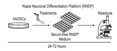
[0015] Fig. 1 is a diagram depicting a rapid neuronal differentiation
platform (RNDP)
according to an embodiment of the present disclosure. hADSCs are cultured in a
suitable
culture medium and an appropriate amount a candidate compound (treatment) for
24-72 hours.
The hADSCs are then evaluated to determining the extent of neurogenesis.
[0016] Fig. 2 is a diagram depicting an extended neuronal differentiation
platform
(ENDP) according to an embodiment of the present disclosure. hADSCs are
cultured in a
suitable priming medium for up to three days. The priming medium is then
replaced with a
suitable culture medium (differentiation medium) and an appropriate amount of
a candidate
compound and cultured for 1 to 5 days. The hADSCs are then evaluated to
determining the
extent of neurogenesis.
[0017] FIG. 3A depicts a phase contrast image of a ADSC's in a control
experiment.
FIG. 3B depicts a phase contrast image of ADSC's post brain nutrient
treatment.
[0018] FIG. 4A is a control image from a cellular expression study in which
cells are
stained with an antibody against microtubule-associated protein 2 (MAP2), a
neuronal marker.
FIG. 4B is a post brain nutrient treatment image in the MAP2 expression study.
Red
fluorescence (indicated by the white streaks) demonstrates the expression of
MAP2.
[0019] FIG. 5A is a control image from a cellular expression study in which
cells are
stained with an antibody against nestin, a neuronal marker. FIG. 5B is a post
brain nutrient
treatment image in the nestin expression study. Red fluorescence (indicated by
the white
streaks) demonstrates the expression of nestin.
[0020] FIG. 6A is a control image from a cellular expression study in which
cells are
stained with an antibody against glial fibrillary acidic protein (GFAP), a
neuronal marker. FIG.
6B is a post brain nutrient treatment image in the GFAP expression study. Red
fluorescence
(indicated by the white streaks) demonstrates the expression of GFAP.
[0021] FIG. 7A is a control image from a cellular expression study in which
cells are
stained with an antibody against beta III tubulin, a neuronal marker. FIG. 7B
is a post brain
nutrient treatment image in the beta III tubulin expression study. Red
fluorescence (indicated
by the white streaks) demonstrates the expression of beta III tubulin.
BEST MODE FOR CARRYING OUT THE INVENTION
[0022] The present disclosure provides methods for identifying a
neurogenesis-
modulating compound comprising: culturing adipose-derived stem cells (ADSCs)
in the
presence of a candidate compound, and determining the extent of neurogenesis
in the ADSCs.
[0023] "Neurogenesis" refers to the differentiation, generation or
proliferation of neural
cells from stem or progenitor cells in vitro or in vivo. The extent of
neurogenesis can be
CA 02865735 2014-08-27
WO 2013/130196
PCT/US2013/023094
determined by a variety of techniques known in the art, such as by observing
morphological
changes in the cells. Any method for cellular analysis or visualization is
suitable for use in the
present methods. For example, morphological changes in the ADSCs may be
observed using a
microscopic technique, such as phase contrast microscopy. Morphological
changes that indicate
neurogenesis include, but are not limited to, shrinkage of cytoplasm and the
presence of neurites,
axons and dendrites. In other embodiments, the extent of neurogenesis is
determined by
observing cellular biomarkers indicative of neurogenesis, such as by using
biomarker expression
experiments. Examples of such biomarkers include, but are not limited to,
proteins such as
neurofilaments, myelin basic protein, microtubule associated protein 2 (MAP2),
nestin, -III
tubulin, glial fibrillar acidic protein (GFAP), S100 (a calcium binding
protein), CNPase and
GABA receptor.
[0024] A "neurogenesis-modulating compound" refers to a compound that
affects
neurogenesis, either by promoting or inhibiting neurogenesis. Thus, in some
embodiments,
neurogenesis-modulating compounds promote neurogenesis ("neurogenesis-
promoting
compounds"), while in other embodiments, the neurogenesis-modulating compounds
inhibit or
reduce neurogenesis ("neurogenesis-inhibiting compounds").
Compounds identified as
promoting neurogenesis may advantageously be used as supplements in the diets
of infants,
children, and pregnant and lactating mothers in order to promote and support
early brain
development. These compounds also may be useful in treating neurodegenerative
diseases or
neurological injuries. Compounds identified as inhibiting neurogenesis may be
potential toxins
to be avoided or removed from the diets and environments of infants, children,
and pregnant and
lactating women. These compounds also may interfere with the treatment or
healing of
neurological diseases or injuries. Thus, neurogenesis-inhibiting compounds may
also be
avoided in the diets and environments of individuals suffering from
neurological disease or
injury.
[0025] A "candidate compound" refers to any compound to be tested for
neurogenesis-
modulating properties using the methods described herein. The candidate
compounds include,
without limitation, naturally occurring substances, synthetic compounds, or
extracts, such as
extracts of plant or animal tissues, fungi or bacteria. The candidate compound
may be tested
singly or it may be tested in combination with other candidate compounds or
known
neurogenesis-modulating compound in order to observe synergistic effects or to
achieve higher
throughput screening of compounds.
[0026] In certain embodiments, the method further comprises providing a
negative
control culture of ADSCs for comparison to the candidate compound.
Accordingly, the method
CA 02865735 2014-08-27
WO 2013/130196
PCT/US2013/023094
6
further comprises culturing ADSCs in the absence of the candidate compound,
determining the
extent of neurogenesis in the ADSCs cultured in the absence of the candidate
compound, and
comparing the extent of neurogenesis in the ADSCs cultured in the presence of
the candidate
compound to the extent of neurogenesis of the ADSCs cultured in the absence of
the candidate
compound. An increase in the extent of neurogenesis in the ADSCs cultured in
the presence of
the candidate compound compared to the extent of neurogenesis in the ADSCs
cultured in the
absence of the candidate compound indicates that the candidate compound is a
neurogenesis-
promoting compound. On the other hand, a decrease in the extent of
neurogenesis in the ADSCs
cultured in the presence of the candidate compound compared to the extent of
neurogenesis in
the ADSCs cultured in the absence of the candidate compound indicates that the
candidate
compound is a neurogenesis-inhibiting compound. The, the negative control
culture provides
additional information regarding the neurogenesis modulating properties of the
candidate
compounds.
[0027] In other embodiments, the method further comprises providing a
positive control
culture. Thus, the method further comprises culturing ADSCs in the presence a
known
neurogenesis-promoting compound, and determining the extent of neurogenesis in
the ADSCs
cultured in the presence of the neurogenesis-promoting compound. For example,
DHA is
known to promote early brain development and may be used as a positive
control. Accordingly,
the method may further comprise culturing ADSCs in the presence of DHA. An
increase in the
extent of neurogenesis in the ADSCs cultured in the presence of the candidate
compound
compared to the extent of neurogenesis in the ADSCs cultured in the presence
of DHA indicates
that the candidate compound is a superior neurogenesis-promoting compound than
DHA.
[0028] During neurogenesis, the ADSCs may differentiate into neuronal
cells, precursors
to neuronal cells, and cells having neuronal properties. Accordingly, the
extent of neurogenesis
can be determined by observing morphological changes in the cells. Changes in
cell
morphology that are indicative of neurogenesis include, but are not limited
to, shrinkage of cell
cytoplasm, formation of a neurite, formation of a dendrite-like projection,
formation of an axon,
or a combination thereof. Other changes in cell morphology indicative of
neurogenesis include
development of a morphology that resembles bi-polar, tri-polar and multi-polar
neuronal cells.
[0029] The aforementioned changes in cell morphology can be observed by a
microscopic technique, such as by phase contrast microscopy. Phase contrast
microscopy
images of the ADSCs may be multiple times during the culturing of the ADSCs.
For example,
images may be taken prior to culturing with the candidate compound, and one or
more times
CA 02865735 2014-08-27
WO 2013/130196
PCT/US2013/023094
7
after addition of the candidate compound, such as three hours after, and then
once daily
thereafter.
[0030] The extent of neurogenesis can further be determined by measuring
the
percentage of ADSCs exhibiting neuronal differentiation and the length of
cytoplasmic
projections in the cells, such as neurites, axons and dendrites. The
percentage of ADSCs
exhibiting neuronal differentiation and length of cytoplasmic projections can
be measured using
Image J open software with an appropriate plug-in.
[0031] Changes in cellular biomarkers occur during neurogenesis. Thus, in
some
embodiments, a cellular expression study for neuronal markers is used to
determine the extent of
neurogenesis. Examples of such biomarkers include, but are not limited to,
proteins such as
neurofilaments, myelin basic protein, nestin,
tubulin, glial fibrillar acidic protein (GFAP),
S100 (a calcium binding protein), microtubule associated protein 2 (MAP2),
CNPase and
GABA receptor. Additional techniques for determining neuronal differentiation
include
immunohisotlogical staining for neuronal markers, neuronal excitability
measurements and
western blotting for the expression of neural proteins.
[0032] In some embodiments, the ADSCs are human adipose-derived stem cells
(hADSCs). hADSCs can advantageously be maintained in culture and readily
passaged to
provide multiple sub-cultures. Furthermore, hADSCs are readily available
because they can be
isolated from human adipose tissue collected during routine liposuction
procedures and
cryopreserved. hADSCs have the additional advantage of being readily obtained
from an
individual patient. The hADSCs thus obtained can be used in the methods
described herein to
screen a candidate compound for individualized use. Accordingly, personalized
and optimized
nutrition, drug treatment, or determination of sensitivity to neurotoxins can
be achieved using
the methods of the present disclosure.
[0033] The ADSCs may be cultured for a sufficient amount of time for
neurogenesis to
occur. Neurogenesis may be observed at varying times, depending on the brain
nutrient tested.
Thus, in some embodiments, neurogenesis may be observed after a few hours of
culturing while
in other embodiments, neurogenesis may be observed after several days of
culturing. For
example, the ADSCs may be cultured for about 1 hour to about 5 days, about 1
hour to about 3
days, about 3 hours to about 36 hours, about 12 hours to about 24 hours, or
about 24 to about 36
hours. Furthermore, culturing of ADSC's may be continued for one, two, three
or four weeks in
order to achieve a more complete neuronal differentiation. The culturing of
the ADSCs may
further be performed at an elevated temperature, such as a temperature above
room temperature.
Such temperatures include about 25 to about 45 C, about 30 to about 40 C, or
about 37 C.
CA 02865735 2014-08-27
WO 2013/130196 PCT/US2013/023094
8
[0034] In the aforementioned methods, the ADSCs may advantageously be
cultured in a
medium that supports or promotes neurogenesis, for example by guiding the
ADSCs to
differentiate into neuronal cells. In some embodiments, the medium comprises a
neural basal
medium, epidermal growth factor (EGF), basic ilifibroblast growth factor b-
FGF, N2 supplement
and L-glutamine. The ingredients for the culture medium are available from
commercial
sources. For example, the neural basal medium can be NeurobasalTM Medium,
which is
available from Invitrogen. Neural Basal Medium TM may include the ingredients
listed in Table
1:
Table 1: NeurobasalTM Medium
Components Molecular Concentration mM
Weight (mg/L)
Amino Acids
Glycine 75 30 0.4
L-Alanine 89 2 0.0225
L-Arginine hydrochloride 211 84 0.398
L-Asparagine-H20 150 0.83 0.00553
L-Cysteine 121 31.5 0,26
L-Histidine hydrochloride-H20 210 42 0.2
L-Isoleucine 131 105 0.802
L-Leucine 131 105 0.802
L-Lysine hydrochloride 183 146 0.798
L-Methionine 149 30 0.201
L-Phenylalanine 165 66 0.4
L-Proline 115 7.76 0.0675
L-Serine 105 42 0.4
L-Threonine 119 95 0.798
L-Tryptophan 204 16 0.0784
L-Tyrosine 181 72 0.398
L-Valine 117 94 0.803
Vitamins
Choline chloride 140 4 0.0286
D-Calcium pantothenate 477 4 0,00839
Folic Acid 441 4 0.00907
Niacinamide 122 4 0.0328
Pyridoxine hydrochloride 204 4 0.0196
Riboflavin 376 0.4 0.00106
1
CA 02865735 2014-08-27
WO 2013/130196
PCT/US2013/023094
9
Thiamine hydrochloride 337 4 0.0119
Vitamin B12 1355 0.0068 0.000005
i-Inositol 180 7.2 0.04
Inorganic Salts
Calcium Chloride (CaC12) (anhyd.) 111 200 1.8
Ferric Nitrate (Fe(NO3)3"9H20) 404 0.1 0.000248
Magnesium Chloride (anhydrous) 95 77.3 0.814
Potassium Chloride (KC1) 75 400 5.33
Sodium Bicarbonate (NaHCO3) 84 2200 26.19
Sodium Chloride (NaCI) 58 3000 51.72
Sodium Phosphate monobasic (NaH2PO4- 138 125 0.906
H20)
Zinc sulfate (ZnSO4-7H20) 288 0.194 0.000674
Other Components
D-Glucose (Dextrose) 180 4500 25
HEPES 238 2600 10.92
Sodium Pyruvate 110 25 0.227
N2 supplement may be purchased from Invitrogen. The Invitrogen N2 supplement
may
comprise the following ingredients:
Table 2: N2 Supplement
Components Molecular Weight Concentration (mg/L) mM
Proteins
Human transferrin (Holo) 10000 10000 1
Insulin recombinant full chain 5807.7 500 0.0861
Other components
Progesterone 314.47 0.63 0.002
Putrescine 161 1611 10.01
selenite 173 0.52 0.00301
[0035] For
example, the medium may comprise about 1 to about 100, about 5 to about
50, about 10 to about 25 or about 20 ng/mL of EGF. The medium further
comprises about Ito
about 100 ng/mL, about 5 to about 50, about 10 to about 25, or about 20 ng/mL
of b-FGF. The
N2 supplement may be present in the medium at a concentration of about lx, and
L-glutamine
may be present in an amount of about 0.1 to about 10 mM, about 1 to about 5
mM, or about 1.3
CA 02865735 2014-08-27
WO 2013/130196
PCT/US2013/023094
to about 3 mM. The medium may further comprise a suitable amount of the
candidate
compound, for example from about 0.1 nM to about 10 mM, or 1 nM to about 1 mM.
[0036] In
certain embodiments, the culturing medium is substantially free of serum or,
preferably, completely free of serum. A culture medium substantially free of
serum refers to
medium having less than about 10% serum, more particularly less than about 2%
or 0.1% serum;
in certain embodiments, substantially free of serum refers to less than about
0.5% serum. A
culture medium completely free of serum has 0% serum. While not being bound by
any
particular theory, it is believed serum may contain inconsistent and
undetermined amounts of
growth factors, which has the potential to impact the extent of neurogenesis.
Accordingly,
serum-free media eliminate the effects of serum on the extent of neurogenesis.
Neurogenesis
observed in ADSCs cultured in serum-free media can thus be attributed to the
candidate
compound rather than the presence of serum.
[0037] The
aforementioned methods are useful in a rapid neuronal differentiation
platform ("RNDP"). The RNDP may advantageously be used to quickly screen large
numbers
of potential neurogenesis modulating compounds. Compounds can be rapidly
screened using
multi-well plates and/or by testing several compounds at once or libraries of
compounds for high
through-put results. Compounds identified in the RNDP are further investigated
using an
extended platform, if desired.
[0038] An
extended neuronal differentiation protocol ("ENDP") further comprises a
priming step. The ENDP is useful to further investigate and confirm the
results of an RNDP.
While not being bound by any particular theory, it is believed that priming
the ADSCs allows
for improved neuronal morphology, thereby providing additional insight in the
neurogenesis
modulating potential of a given compound. Accordingly, in some embodiments,
the ADSCs are
primed prior to culturing in the presence of a candidate compound. For
example, the ADSCs
can be primed for about 1 to about 5 days in a suitable priming medium prior
to culturing with
the candidate compound. In other embodiments, the ADSCs are primed for about 1
to about 3
days, or for about 3 days.
[0039] In some
embodiments, the priming medium comprises a neural basal medium
(such as Neurobasal MediumTM from Invitrogen), with suitable concentrations of
EGF, b-FGF,
and N2 supplement. Suitable concentrations of EGF include about 1 to about 100
ng/mL, about
5 to about 50, about 10 to about 25 or about 20 ng/mL. Suitable concentrations
of b-FGF
include about 1 to about 100, about 5 to about 50, about 10 to about 25, or
about 20 ng/mL of b-
FGF. The N2 supplement may be present in the medium at a concentration of
about lx. The
priming medium may be substantially free of serum or, more preferably,
completely free of
CA 02865735 2014-08-27
WO 2013/130196
PCT/US2013/023094
11
serum. A priming medium substantially free of serum refers to medium having
less than about
10% serum, for example less than about 2% or 0.1% serum, while a culture
medium completely
free of serum has 0% serum. Furthermore, the priming medium may be free of or
substantially
free of the candidate compound.
[0040] In embodiments wherein the ADSCs are primed prior to being cultured
in the
presence of a candidate compound, the ADSCs are subsequently cultured in a
suitable culture
medium for about 1 to about 5 days. In other embodiments, the ADSCs are
cultured for about 1
to about 3 days, or for about 3 days. After priming, the priming medium is
removed and a
culturing medium is added to the ADSCs. The culture medium comprises, for
example,
MesenPRO complete, available from Invitrogen. The culture medium may further
comprise a
suitable amount of the candidate compound, for example about 0.1nM to about 10
mM, or 1 nM
to about 1 mM of the candidate compound. In a negative control experiment, the
culture
medium is free of or substantially free of the candidate compound. In a
positive control
experiment, the culture medium comprises a known neurogenesis promoting
compound, such as
DHA.
[0041] In some embodiments, the cultureware used to culture the ADSCs is
coated with
a unique combination of matrix proteins designed to mimic the in vivo
environment of the
central nervous system, maximize cellular neuronal differentiation activity,
and enhance cellular
attachment. In one embodiment, the coating comprises poly-L-ornithine and
bovine plasma
fibronectin. The coated cultureware can be prepared by contacting the
cultureware with a
solution of poly-L-omithine and a solution of bovine fibronectin. The
contacting steps may be
performed in any order, simultaneously, or substantially simultaneously. For
example, the
cultureware can be contacted with the poly-L-ornithine prior to the bovine
fibronectin or after
the fibronectin. Alternatively, the poly-L-ornithine and bovine fibronectin
are contacted with
the cultureware simultaneously or substantially simultaneously.
[0042] Another aspect of the disclosure relates to an in vitro method of
promoting
neurogenesis in ADSCs comprising: culturing the ADSCs in the presence of a
neurogenesis-
promoting compound. Neuronal cells and neuron-like cells generated by the
aforementioned
methods may be maintained in culture, passaged, or cryopreserved. The method
thus can
provide human neuronal cells and neuron-like cells for use in the laboratory,
such as for drug
screening. In some embodiments, the method further comprises determining the
extent of
neurogenesis in the ADSCs, as described in the aforementioned screening
methods.
[0043] Another aspect of the disclosure relates to a system for identifying
a
neurogenesis-modulating compound, comprising: ADSCs; cultureware comprising
coating that
CA 02865735 2014-08-27
WO 2013/130196
PCT/US2013/023094
12
mimics the central nervous system; and a culture medium. In some embodiments,
the coating
comprises bovine fibronectin and poly-L-ornithine. In systems useful in the
RNDP, the culture
medium the culture medium comprises a neural basal medium, EGF, b-FGF, N2
supplement,
and L-glutamine. Systems useful in the ENDP, further comprise a priming
medium, such as a
medium comprising a neural basal medium, EGF, b-FGF, N2 supplement, and
culture medium
comprising MesenPRO Complete.
EXAMPLES
hADSCs
[0044] The hADSCs used in the following procedures are purchased from
commercial
resources and grown in the maintenance media consisting of Complete MesenPRO
RS medium
with supplement and L-glutamine. The subculture of hADSCs is performed when
cell culture
reaches confluence. To passage hADSCs, the following procedure is used: i)
aspirate the
Complete MesenPRO RS medium from the cells; ii) rinse the surface area of the
cell layer with
Dulbecco's phosphate buffered saline (DBPS) buffer by adding the DPBS to the
side of the
vessel opposite the attached cell layer and rocking the vessel back and forth
several times; iii)
remove the DPBS by aspiration and discard; iv) detach the cells by adding a
sufficient volume
of pre-warmed trypsin-EDTA solution without phenol red to cover the cell
layer; v) incubate at
37 C, for approximately 7 minutes; vi) observe the cells under a microscope to
determine if
additional incubation is needed; vii) add 3mL of the maintenance media to the
plate, mix the cell
suspension, add the suspension to a 15mL centrifuge tube and centrifuge at
210g for 5 minutes;
viii) determine the total number of cells and percent viability using a
hemacytometer; ix) add
Complete MesenPRO RS medium to each vessel so that the final culture volume is
0.2mL ¨
0.5mL per cm2; x) seed the cells by adding the appropriate volume of cells to
each vessel and
incubate at 37 C., 5% CO2 and 90% humidity; and xi) three or four days after
seeding,
completely remove the medium and replace with an equal volume of Complete
MesenPRO RS
medium.
Coating
[0045] Before seeding the passaged hADSCs on fresh culture plates, the
surfaces of the
cultureware are washed with sterile DPBS solution three times, followed by
multiple rinses with
sterile water. The first layer of coating is poly-L-ornithine. The coating is
prepared by adding
0.1 mg/mL of poly-L-ornithine and incubating at 37 C. for one hour. The plate
is washed three
times with DPBS, 15 minutes per wash. The second layer of coating is bovine
plasma
fibronectin. The fibronectin is diluted in DPBS from stock to 1:1000 and 500
CIL is added to
CA 02865735 2014-08-27
WO 2013/130196
PCT/US2013/023094
13
each well. The plate is left at room temperature for one hour. One final wash
with 500 [IL per
well of DPBS is performed and the plate is used immediately.
Medium
[0046] hADSCs
can be maintained in an undifferentiated state or guided to differentiate
using different culture media. Certain culture media are capable of guiding
ADSCs to
differentiate into neuronal cells. Exemplary media are set forth in Tables 3,
4 and 5.
Table 3.
Serum-free RNDP medium
component Final concentration
neural basal medium 500 mL
EGF 20 ng/mL
b-FGF 20 ng /mL
N2 supplement 1x
L-glutamine 2 mM
Table 4.
Serum-free ENDP priming medium
component Final concentration
neural basal medium 500 mL
EGF 20 ng/mL
bFGF 20 ng/mL
N2 supplement 1 x
Table 5.
ENDP differentiation medium
component Final concentration
MesenPRO complete 500 mL
RNDP Protocol
[0047] Two
independent screening protocols are described, designated as rapid neuronal
differentiation platform (RNDP) and extended neuronal differentiation platform
(ENDP). The
RNDP protocol provides rapid screening of large numbers of candidate compounds
in a
relatively short period of time. RNDP allows the rapid identification of
compounds that either
CA 02865735 2014-08-27
WO 2013/130196
PCT/US2013/023094
14
promote or inhibit neurogenesis, or that have no effect on neurogenesis. The
RNDP may be
followed by an ENDP in order to further investigate and confirm the results.
[0048] The subculture media of the hADSCs described above is removed from
the
culture dish, and the dish is then gently washed with 5-10 mL of sterile DPBS.
The DPBS is
removed and 1.5 mL of trypsin-EDTA is added to completely cover the cell
layer. The dish is
placed back in the incubator for seven minutes. The plate is then gently
tapped to detach cells
completely, 3 mL of the maintenance media is added to the plate, and the cell
suspension is
mixed and added to 15 mL centrifuge tube. The desired cell density (1x10⁴
cells/well) is taken
to another 15 mL tube and placed to centrifuge at 210g for 5 minutes. The cell
pellet is
resuspended in an appropriate volume of pre-warmed serum-free rapid neuronal
differentiation
medium as set forth in Table 1 and seeded onto each well of tissue culture
plate. The candidate
compounds for each well are added sequentially. The plate is put back into the
incubator. The
effects of the candidate compounds are quickly and easily observed using phase
contrast
microscopy images, which are usually taken once immediately before treatment,
three hours
post treatment and each day thereafter for three days. With a fast turnover
time, the best results
typically occur within 36 hours. After images are collected, data analysis and
comparison is
made to determine the effectiveness of each compound or mixture of compounds
in modulating
neurogenesis. Neuronal differentiation is determined by observing neuronal
morphology. Some
changes in the cells include shrinking of the cytoplasm, formation of axons
and dendrite-like
cytoplasmic projections. These changes begin with the cytoplasm of hADSCs
retracting toward
the nucleus to form contracted cell bodies with cytoplasmic extensions. Cells
eventually
develop a morphology that resembles bi-polar, tri-polar, and multi-polar
neuronal cells.
ENDP Protocol
[0049] The ENDP protocol provides a method for further investigation of the
results of
the RNDP and also allows additional time for priming the hADSCs for further
differentiation
into various neuronal cell lineages. While not being bound by any particular
theory, the priming
drives transdifferentiation of the hADSCs from mesoderm lineages to neural
ectoderm.
[0050] The hADSCs are seeded on culture plates with coated surfaces and
grown in the
serum-free ENDP priming medium (see table 2) for at least 72 hours. The
priming medium is
removed and neuronal differentiation medium added (see Table 3) in the
presence or absence of
at least one candidate compound. The cultures are then incubated for an
extended period of time
for further neuronal development. After three days of incubation, the cells
are examined under
microscope for morphological changes. The percentage and length of neurites
can be measured
CA 02865735 2014-08-27
WO 2013/130196
PCT/US2013/023094
by using open software of Image J with an appropriate plug-in. The cells can
further be studied
for various neuronal markers to further confirm neuronal differentiation.
Discovery of brain nutrients
[0051] The
purpose of this investigation is to determine the neurogenesis effect of
various nutrients (candidate compounds) using both RNDP and ENDP platforms.
The candidate
compounds are tested individually and compared to the positive control,
docosahexaenoic acid
(DHA), and the negative control. Pre-warmed serum-free medium contains Neural
Basal
medium with L-glutamine, 2Ong/mL of b-FGF, 2Ong/mL of EGF and N2 supplement.
The
candidate compound is added to individual wells at various concentrations in
the serum-free
medium. The candidate compounds are selected from the group consisting of ARA,
EPA (cis-
5,8,11,14,17-eicosapentaenoic acid) and resveratrol.
Compounds are tested in varying
concentrations, ranging in the nanomolar to micromolar range. The compounds
are tested
individually and compared to the positive control, docosahexaenoic acid (DHA),
and the
negative control. The experiments are repeated in triplicate. The nutrients
found to promote
neurogenesis or demonstrate use as a medicament are further screened in
various combinations.
These experiments are also repeated in triplicate.
[0052] The
effects of the candidate compounds are easily and quickly observed under
phase contrast microscopy for up to one week with images usually taken once
immediately
before treatment with the candidate compound, three hours post treatment, and
each day
thereafter for three days. With a fast turnover time, the best results
typically occur within 36
hours. After images are collected, data analysis and comparison is made to
determine the
effectiveness of each compound or combination of compounds in promoting
neurogenesis.
Neuronal differentiation is determined by neuronal morphology. Some of these
changes include
shrinkage of the cytoplasm, and formation of axons and dendrite-like
cytoplasmic projections
(neurites). These changes begin with the cytoplasm of hADSCs retracting
towards the nucleus
to form contracted cell bodies with cytoplasmic extensions. Cells eventually
develop a
morphology that resembles bi-polar, tri-polar and multi-polar neuronal cells.
[0053] Of the
above candidate compounds, resveratrol, ARA, EPA, cholesterol and
DHA are examples of compounds that effectively promote neurogenesis. The
test
concentrations and effective concentrations are depicted in table 6:
Table 6: Examples of compounds identified as effectively promoting
neurogenesis
Compound Testing range Effective range
DHA 1nM -1mM 5-20 LIM
ARA 1nM -1mM 2-10 DM
CA 02865735 2014-08-27
WO 2013/130196
PCT/US2013/023094
16
EPA (Cis-5,8,11,14,17-
Eicosapentaenoic acid) 1nM -1mM 10-40 DM
Cholesterol 1DM - 10DM 50-200 DM
Resveratrol 100nM -50mM 20 DM -20 mM
[0054] All references to singular characteristics or limitations of the
present disclosure
shall include the corresponding plural characteristic or limitation, and vice
versa, unless
otherwise specified or clearly implied to the contrary by the context in which
the reference is
made.
[0055] All combinations of method or process steps as used herein can be
performed in
any order, unless otherwise specified or clearly implied to the contrary by
the context in which
the referenced combination is made.
[0056] The methods and compositions of the present disclosure, including
components
thereof, can comprise, consist of, or consist essentially of the essential
elements and limitations
of the embodiments described herein, as well as any additional or optional
ingredients,
components or limitations described herein.
[0057] As used herein, the term "about" should be construed to refer to
both of the
numbers specified in any range. Any reference to a range should be considered
as providing
support for any subset within that range.