Note: Descriptions are shown in the official language in which they were submitted.
CA 02865744 2014-08-27
WO 2013/130875 PCT/US2013/028409
RAPID ANTIBIOTIC SUSCEPTIBILITY TESTING
CROSS REFERENCE TO RELATED APPLICATIONS
[0001] This Application claims the benefit under 35 U.S.C. 119(e) of U.S.
Provisional
Application Nos. 61/604,878 filed February 29, 2012; and 61/647,860 filed May
16, 2012,
the contents of which are herein incorporated by reference in their entirety.
GOVERNMENT SUPPORT
[0002] This invention was made with government support under grant no.
N66001-11-1-
4180 awarded by DARPA. The government has certain rights in the invention.
TECHNICAL FIELD
[0003] The present disclosure relates to methods, compositions and kits for
rapid
determination of antibiotic susceptibility of a microbe within hours after
sample collection.
BACKGROUND OF THE DISCLOSURE
[0004] Every year more than 18 million patients experience sepsis caused by
systemic
blood-borne infection, and more than 6 million of these people die. Mortality
rates from
sepsis in intensive-care units range from 20 to 60% worldwide, and one
retrospective study
revealed that 20% of patients with septic shock were initially treated with
inappropriate
antibiotic therapy (Kumar et al., Chest 2009; 25: 733). This is largely
because it takes days to
obtain a rigorous diagnosis of pathogen type, even in state-of-the art
clinical microbiology
laboratories. Moreover, patients who initially receive incorrect therapies
exhibit a 5-fold
lower survival rate than those who are treated with optimal therapy from early
in the course
of the disease. In fact, in one study it was shown that the risk for in-
hospital mortality
increased by 9% for every hour of delay before the correct antibiotic regimen
was
administered (Garnacho-Montero et al., Critical Care 2006; 10:R111). Thus, the
speed of
pathogen diagnosis in a patient with a blood-borne microbial infection can
mean the
difference between life and death. The current state-of-the-art for detection
of a microbial
infection in blood, which has essentially remained unchanged for the past
thirty years, is to
culture the blood in a hospital or commercial clinical microbiology
laboratory. Liquid
cultures can permit detection of the existence of some type of growing
organism in the fluid
within 16 to 30 hours (based on the analysis of more than 50,000 blood
cultures carried out in
one year at the Brigham and Women's Hospital clinical microbiological
laboratory). This
1
CA 02865744 2014-08-27
WO 2013/130875 PCT/US2013/028409
assay is not quantitative and without knowledge of the type of pathogen and
their specific
antibiotic sensitivities, only wide-spectrum antibiotics can be administered
at this time, which
are suboptimal at best. To identify the specific type of pathogen, and to
carry out sensitivity
testing to determine their responses to various potential antibiotic
therapies, the pathogens
growing in liquid medium must then be transferred to other growth media (e.g.,
agar plates).
The total time for full diagnosis and sensitivity testing is commonly 3-7 days
and empiric
antibiotic treatment based on clinical symptoms is started well before the
results of the
antibiotic sensitivity are obtained, often within 1-3 hours after blood
cultures are first drawn
from the patient.
[0005] Many patients with septicemia or suspected septicemia exhibit a
rapid decline
over a 24-48 hour period. Thus, rapid and reliable diagnostic and treatment
methods are
essential for effective patient care. Unfortunately, current antimicrobial
susceptibility testing
techniques generally require a prior isolation of the microorganism by culture
(e.g., about 12
to about 48 hours), followed by a process that requires another about 6 to
about 24 hours. For
example, a confirmed diagnosis as to the type of infection, traditionally
requires
microbiological analysis involving inoculation of blood cultures, incubation
for 16-24 hours,
plating the causative microorganism on solid media, another incubation period,
and final
identification 1-2 days later. Even with immediate and aggressive treatment,
many patients
develop multiple organ dysfunction syndrome and eventually death.
[0006] Every hour lost before a correct treatment is administered can make
a crucial
difference in patient outcome. Consequently, it is important for physicians to
determine
rapidly if the patient indeed has sepsis, and if so, what antibiotics would be
effective for the
treatment. For example, an appropriate antimicrobial therapy that can be
instated within 6
hours of the onset of sepsis can positively impact patient outcome. However,
the current
practice, i.e., blood culture, takes two days or more to yield an answer,
which quite often
proves too long. Accordingly, there is a strong need for a more rapid
antibiotic sensitivity
testing, preferably one that can identify specific antibiotic susceptibilities
within only a few
hours after blood samples are first drawn. A rapid test of this type would
therefore permit
physicians to initiate the optimal drug therapy from the start, rather than
starting with a sub-
optimal or completely ineffective antibiotic, hence greatly increasing
clinical responsiveness.
SUMMARY
[0007] Existing antimicrobial susceptibility testing (AST) techniques
generally require
the prior isolation of the microorganism by culture (-12 to ¨48 hours)
followed by a process
2
CA 02865744 2014-08-27
WO 2013/130875 PCT/US2013/028409
that requires another 6 to 24 hours. However, adapted antimicrobial therapy in
the first few
hours of septic shock is a key predictor for survival. Thus, there is a strong
need for rapid
antimicrobial susceptibility testing. Inventors have discovered inter alia
that antibiotic
susceptibility of a microbe can be determined within hours after a sample is
collected. In one
embodiment, the antibiotic susceptibility test or assay can be completed in
less than 6 hours.
The method can be used with or without first identifying the microbe.
Additionally, while
existing AST technology generally relies on an analysis of large bacterial
populations (e.g.,
¨105 to ¨107 CFU) obtained from a culture, the inventors have discovered inter
alia that
antibiotic susceptibility of a microbe can be determined by concentrating the
microbe from a
biological sample prior to culture and performing single cell analysis of
susceptibility to one
or more antibiotic agents. Accordingly, disclosed herein is an assay for
rapidly determining
antibiotic susceptibility of a microbe. Aspects of the assay are based on the
rapid separation
and concentration of microbes from a biological sample, e.g., biological
fluid. Generally the
assay comprises obtaining a microbe extracted from a biological source and
determining the
growth or a functional response of the microbe in presence of an antibiotic
agent. Reduced
growth or functional response in the presence of the antibiotic agent relative
to a control
indicates that the microbe is susceptible to the antibiotic agent tested. In
addition, the assay
can also carry antibiotic susceptibility testing in a small number of microbes
(as few as 300 in
a sample). In some embodiments, the assay can allow determination of
susceptibility of one
or more microbes or microorganisms to at least one antibiotic agent (including
antimicrobial
agent) from single microbes or microorganisms (e.g., as few as about 5-10 live
microbes). In
some embodiments, the assay described herein can be performed in as a little
as three hours
of specimen reception. The capability of determining susceptibility of a
microbe or
microorganism to an antibiotic agent using a small number of the microbe
(e.g., as few as
about 5-10 live microbes or lower) can be desirable because clinical samples
of biological
fluids (e.g., blood, cerebral spinal fluid, etc.) can have only rare microbes
(<100/mL) and
they are often limited in volume to only a few milliliters or less.
[0008] In addition, embodiments of the method utilizing Mannose Binding
Lectin (MBL)
and genetically engineered version of MBL (FcMBL and Akt-FcMBL) as broad-
spectrum
microbe binding molecules to capture and grow the microbes, can be carried out
without
identifying the microbe, either for extraction or for antibiotic sensitivity
testing.
[0009] In some embodiments, the biological fluid is collected or derived
from a subject
who is suspected of having a microbial infection. Further, once an antibiotic
agent resistance
is established, a practitioner can select a treatment regimen for the subject
afflicted with the
3
CA 02865744 2014-08-27
WO 2013/130875 PCT/US2013/028409
microbes. For example, the treatment regime can comprise administering to the
subject one
or more antibiotic agents to which the microbe showed susceptibility.
BRIEF DESCRIPTION OF THE DRAWINGS
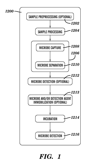
[0010] Fig. 1 is a schematic of the process described herein.
[0011] Fig. 2 is a line graph showing sensitivity (limit of detection =
LOD): Serial
dilutions of E. coli were used to determine the LOD of the MBL linked with HRP
(FcMBL-
HRP) ELISA.
[0012] Figs. 3A and 3B are bar graphs showing antibiotic sensitivity
(bactericidal
antibiotic Carbenicillin (100 [tg/m1) or the bacteriostatic antibiotic
Spectinomycin). Bacteria
were captured by FcMBL and cultured for 2 hours at 37 C. Due to low number of
bacteria
detected in the Carbenicillin samples, data is also presented in a separate
chart (Figure 3B).
[0013] Fig. 4 is a line graph showing antibiotic susceptibility of wild-
type E. coli: Serial
dilution of E. coli were captured by FcMBL beads for 10 min, transferred to
lml F media
(2% glycerol, 0.99mM KaHPO4, Supplement EZ, ACGU, MOPS, 5mM Ca ++ , 0.1%
Tween)
with or without carbenicillin (10Oug/m1), cultured for 4 hours at 37 C shaking
at 950 rpms,
and growth difference was determined by FcMBL ELISA.
[0014] Fig. 5 is a line graph showing antibiotic susceptibility of Kan' E.
coli: Serial
dilution of the two E. coli were captured by FcMBL beads for 10min,
transferred to lml F
media with or without antibiotic - carbenicillin (10Oug/m1) or kanamycin
(5Oug/m1), cultured
for 4 hours at 37 C 950 rpms, and growth difference determined by FcMBL ELISA.
[0015] Fig. 6 is a line graph showing determination of antibiotic
susceptibility using the
luciferase assay. Serial dilutions of E. coli and S. aureus were captured by
FcMBL beads
(10mins), transferred to lml Mueller Hinton broth and growth determined by
BACTITERGLO luciferase assay (100u1 of culture). The LOD was determined to be
40
cfu/ml after 3 hours of growth.
[0016] Fig. 7 shows antibiotic susceptibility of S. aureus captured from
blood. Human
blood with 1:1 TBST Ca 5mM with or without 1%Triton was spiked with lOul of
diluted S.
aureus and the bacteria captured with FcMBL beads. The captured bacteria
following 3
washes (lx capture buffer and 2x TBST Ca) were transferred to lml Mueller
Hinton broth
with or without 10Oug/m1 Carbenicillin, incubated at 37 C 950rpms for 4 hours,
and growth
determined by BACTITERGLO luciferase assay (100u1 of culture).
[0017] Fig. 8 is a bar graph showing plating captured/outgrown bacteria
generates
artificially low counts.
4
CA 02865744 2014-08-27
WO 2013/130875 PCT/US2013/028409
[0018] Figs. 9A and 9B are line graphs showing both reactive oxygen species
(ROS)
assay (Figure 9A) and ATP Luminescence (Figure 9B) assays can be used. ROS LOD
= -3
(-20,000 S. aureus added), ATP Luminescence LOD = -5 (310 S. aureus added).
[0019] Figs. 10A and 10B are bar graphs showing both ATP (Figure 10A) and
ROS
(Figure 10B) measurements can be used for determining viability of bacterial
isolated from
blood. However, ATP luciferase is more sensitive. (-4 titer = 5000 S. aureus
added).
[0020] Figs. 11A-11D are bar graphs showing ATP viability measurement of
bacteria
isolated from blood. Viability or antibiotic susceptibility can be determined
in as little as 2
hours.
[0021] Figs. 12A and 12B are bar graphs showing 50/50 Akt-FcMBL /Heparin
(The
beads were coupled to both Akt-FcMBL and heparin using an equimolar mix of
biotinylated
Akt-FcMBL and biotinylated heparin to generate an estimated 50/50 mix of Akt-
FcMBL/heparin on MyONE streptavidin beads [Invitrogen]) beads capture bacteria
with the
same efficiency as Akt-FcMBL beads. Akt-FcMBL and Akt-FcMBL are described in
PCT
application no. PCT/US2011, content of which is incorporated herein by
reference.
Figure 12A, Bead 1: Akt-FcMBL; Bead 2: 1:1 ratio - Akt-FcMBL: Biotin heparin;
Bead 3:
Heparin cross-linked to Akt-FcMBL directly; and Bead 4: Heparin only. Figure
12B, Bead
1: AOB Akt-FcMBLcoupled beads (AOB = aminooxybiotin); Bead 2: 1:1 ratio ¨ AOB
Akt-
FcMBL: Biotin heparin coupled beads; Bead 3: Heparin Beads + AKT-FcMBL- lum
beads
conjugated with heparin, then coupled with AOB AKT-FcMBL; Bead 4: AKTFcMBL
Beads-
heparin ¨ Previously coupled AOB AKT-FcMBL beads are conjugated with heparin;
Bead 5:
Heparin conjugated to Akt-FcMBL directly and coupled to luM beads; and Bead 6:
Heparin
conjugated beads.
[0022] Fig. 13 is a schematic representation of an embodiment of the
antibiotic
susceptibility assay showing how the assay can be carried out using currently
available
equipment.
[0023] Figs. 14A -14D are bar graphs showing ATP viability measurement of
bacteria
isolated from human blood. Figures 14C and 14D are corrected for blood/bead
background.
[0024] Figs. 15A -15B are line graphs showing antibiotic susceptibility of
E. coli
(Figure 15A), and S. aureus (Figure 15B) determined using an embodiment of the
assay
described herein.
[0025] Fig. 16 is a set of images showing results of an individual cell
antibiotic
susceptibility testing (microcolony-based antibiotic susceptibility testing)
according to one
embodiment described herein. E. coli were captured on 1 pm FcMBL-coated
magnetic beads
CA 02865744 2014-08-27
WO 2013/130875 PCT/US2013/028409
and overlaid with alginate gel, stains for labeling live (e.g., resazurin
stain) or dead (e.g.,
Sytox green stain) cells (live/dead stains), and growth media, e.g., RPMI
1640. Viability was
assessed at indicated time points after addition of about 128 mg/L
carbenicillin. The
microcolony-based AST can antibiotic susceptibility data within minutes,
enabling selecting
bateriocidal compounds within the clinically relevant time frame.
DETAILED DESCRIPTION OF THE INVENTION
[0026] Inventors have discovered inter alia that antibiotic susceptibility
of a microbe
(e.g., a pathogen) can be determined within hours after a sample is collected.
The method
can be used with or without first identifying the microbe. Accordingly,
disclosed herein is an
assay for determining antibiotic susceptibility of a microbe. The assay
comprises providing a
sample suspected of comprising a microbe extracted from a test sample, such as
a biological
source (e.g. a biological fluid), and determining the growth or a functional
response of the
microbe in the presence of an antibiotic agent. Reduced growth or functional
response in the
presence of the antibiotic agent relative to a control indicates that the
microbe is susceptible
to the antibiotic agent tested.
[0027] Generally, the method comprises: (i) extraction and concentration of
microbes
from a biological fluid, e.g., blood; (ii) splitting into subsamples and
incubation with
antibiotic-supplemented media (Yeast extract free media for ELISA or other for
Luciferase
based assay); and (iii) detection of microbe growth or a functional response.
The microbe
can be extracted from the test sample using a microbe-targeting substrate,
i.e., a solid
substrate coated with microbe-binding molecules. Microbe-targeting substrates
and microbe-
binding molecules are described in more detail below. Additionally, while the
method is
described in relation to biological fluids, the method can be practiced with
any test sample,
e.g., biological fluids; fluids from a culture, e.g., blood culture; a sample
taken from a colony;
and/or a sample taken from any environmental source, e.g., but not limited to,
food products,
water, ponds, food processing plants.
[0028] Without wishing to be bound by a theory, the antibiotic
susceptibility testing
method described herein can detect microbial infection of blood or septicemia
caused by
different pathogens (e.g., bacteremia, fungemia, virema) and provide the
antibiotic sensitivity
and resistance profile of the causative agent (e.g., microbial pathogen) in
less than 48 hours,
less than 24 hours, less than 12 hours, less than 10 hours, less than 8 hours,
less than 6 hours,
less than 4 hours, less than 3 hours, less than 2 hours, less than 1 hour,
less than 30 mins, less
than 15 mins, or lower. In some embodiments, the antibiotic susceptibility
testing described
6
CA 02865744 2014-08-27
WO 2013/130875 PCT/US2013/028409
herein can detect septicemia and provide the antibiotic sensitivity and
resistance profiles of
the causative agent (e.g., microbe or pathogen) in less than 6 hours, less
than 4 hours, less
than 3 hours, less than 2 hours, less than 1 hour, less than 30 mins, less
than 15 mins, or
lower.
[0029] In addition, as described below, the method can be carried out
without having to
identify the microbe (e.g., pathogen) either for extraction or for incubation
with an antibiotic
agent. For example, the microbe can be extracted from the test sample using a
substrate
coated with a broad-spectrum microbe-binding molecule. Various microbe-binding
molecules are described in more detail below.
[0030] In some embodiments, the assay described herein is based on the
direct
measurement of bacteria's ability to grow in the presence of the tested
antibiotic agents. This
direct measurement can provide the clinically relevant result that a physician
seeks and is
thus superior to methods that test for indirect properties, e.g., presence of
antibiotic-resistance
genes or enzymes. In contrast to blood culture, the method described herein is
able to detect
microbes and their antibiotic sensitivity using short growth times and
requires only one short
culture step.
[0031] In some embodiments, more than one types of microbial detection
(e.g., bacterial
and fungal detection) can be combined into the same antibiotic susceptibility
testing method
(e.g. bacterial detection, fungal detection, and antibiotic sensitivity) as
described herein.
Antibiotic susceptibility assay
[0032] An exemplary process for determining the antibiotic susceptibility
of a microbe
(e.g., a pathogen) from a test sample is shown in Figure 1. As shown in Figure
1, the
process 1200 comprises the optional step 1202 (preprocessing of the sample),
step 1204
(processing of the sample), step 1206 comprising 1208 (microbe capture, e.g.,
pathogen
capture) and 1210 (microbe separation, e.g., pathogen separation), the
optional step 1212
(detection of microbe or pathogen identity and number), the optional step 1213
(microbe
and/or detection agent immobilization), step 1214 (incubation with the
antibiotic agent), and
step 1216 (detection of microbe number and/or viability). While these are
discussed as
discrete processes, one or more of the preprocessing, processing, capture,
microbe separation,
detection, and antibiotic sensitivity can be performed in a system. In one
embodiment, one or
more of the preprocessing, processing, capture, microbe separation, detection,
and antibiotic
sensitivity can be performed in a microfluidic device. In some embodiments,
one or more of
the microbe capture or separation, microbe incubation, and microbe detection
can be included
7
CA 02865744 2014-08-27
WO 2013/130875 PCT/US2013/028409
in a microfluidic device. In some embodiments, one or more of the modules or
systems
performing microbe capture or separation, microbe incubation, and microbe
detection can
comprise a microfluidic channel. Use of a microfluidic device can automate the
process
and/or allow processing of multiple samples at the same time. One of skill in
the art is well
aware of methods in the art for collecting, handling and processing biological
fluids which
can be used in the practice of the present disclosure. Additionally, the
microfluidic devices
for the various steps can be combined into one system for carrying out the
method described
herein. For example, such a system can comprise two or more of the following:
(i) a capture
or separation system for capturing a microbe from a biological fluid; (ii) an
incubation system
for incubating the microbe with or without an antibiotic agent; and (iii) a
detection system for
detecting the microbe after incubation. Alternatively, the various steps can
also be carried
out using separate systems or devices. An example of this is illustrated in
Figure 13.
[0033] 1202 (Sample preprocessing): It can be necessary or desired that a
test sample,
such be preprocessed prior to microbe detection as described herein, e.g.,
with a
preprocessing reagent. Even in cases where pretreatment is not necessary,
preprocess
optionally can be done for mere convenience (e.g., as part of a regimen on a
commercial
platform). A preprocessing reagent can be any reagent appropriate for use with
the methods
described herein.
[0034] In some embodiments, the test sample can be a biological fluid,
e.g., blood,
plasma, serum, lactation products, amniotic fluids, sputum, saliva, urine,
semen,
cerebrospinal fluid, bronchial aspirate, perspiration, mucus, liquefied stool
sample, synovial
fluid, lymphatic fluid, tears, tracheal aspirate, and any mixtures thereof.
For example, the
sample can be a whole blood sample obtained from a subject suspected of having
a microbe
infection (e.g., a pathogen infection).
[0035] In some embodiments, the test sample can be a fluid or specimen
obtained from
an environmental source. For example, the fluid or specimen obtained from the
environmental source can be obtained or derived from food products, food
produce, poultry,
meat, fish, beverages, dairy product, water (including wastewater), ponds,
rivers, reservoirs,
swimming pools, soils, food processing and/or packaging plants, agricultural
places,
hydrocultures (including hydroponic food farms), pharmaceutical manufacturing
plants,
animal colony facilities, or any combinations thereof.
[0036] In some embodiments, the test sample can be a fluid or specimen
collected or
derived from a cell culture.
8
CA 02865744 2014-08-27
WO 2013/130875 PCT/US2013/028409
[0037] In some embodiments, the test sample can be a fluid or specimen
collected or
derived from a microbe colony.
[0038] The sample preprocessing step generally comprises adding one or more
reagents
to the sample. This preprocessing can serve a number of different purposes,
including, but
not limited to, hemolyzing cells such as blood cells, dilution of sample, etc.
The
preprocessing reagents can be present in the sample container before sample is
added to the
sample container or the preprocessing reagents can be added to a sample
already present in
the sample container. When the sample is a biological fluid, the sample
container can be a
VACUTAINER , e.g., a heparinized VACUTAINER .
[0039] The preprocessing reagents include, but are not limited to,
surfactants and
detergents, salts, cell lysing reagents, anticoagulants, degradative enzymes
(e.g., proteases,
lipases, nucleases, lipase, collagenase, cellulases, amylases and the like),
and solvents, such
as buffer solutions.
[0040] In some embodiments, a preprocessing reagent is a surfactant or a
detergent. In
one embodiment, the preprocessing reagent is Triton X100.
[0041] Amount of preprocessing reagent to be added can depend on a number
of factors.
Generally, the preprocessing reagent is added to a final concentration of
about 0.1mM to
about 10mM. If a liquid, the preprocessing reagent can be added so as to
dilute the sample at
least 10%, at least 20%, at least 30%, at least 40%, at least 50%, at least
60%, at least 60%, at
least 80%, at least 90%, at least 1-fold, at least 2-fold, at least 3-fold, or
at least 5-fold.
[0042] After addition of the preprocessing reagent, the reagent can be
mixed into the
sample. This can be simply accomplished by agitating the sample, e.g., shaking
the sample
and/or moving the sample around in a microfluidic device.
[0043] After addition of the preprocessing reagent, the sample mixture can
be incubated
for a period of time, e.g., for at least one minute, at least two minutes, at
least three minutes,
at least four minutes, at least five minutes, at least ten minutes, at least
fifteen minutes, at
least thirty minutes, at least forty-five minutes, or at least one hour. Such
incubation can be
at any appropriate temperature, e.g., room-temperature (e.g., about 16 C to
about 30 C), a
cold temperature (e.g. about 0 C to about 16 C), or an elevated temperature
(e.g., about 30 C
to about 95 C). In some embodiments, the sample is incubated for about fifteen
minutes at
room temperature. In some embodiments, incubation is for about 5 seconds to
about 60
seconds. In some embodiments, there is no incubation and the sample mixture is
used
directly in the sample processing step.
9
CA 02865744 2014-08-27
WO 2013/130875 PCT/US2013/028409
[0044] 1204 (Sample processing): After the optional preprocessing step, the
sample can
be optionally further processed by adding one or more processing reagents to
the sample.
These processing reagents can degrade unwanted molecules present in the sample
and/or
dilute the sample for further processing. These processing reagents include,
but are not
limited to, surfactants and detergents, salts, cell lysing reagents,
anticoagulants, degradative
enzymes (e.g., proteases, lipases, nucleases, lipase, collagenase, cellulases,
amylases,
heparanases, and the like), and solvents, such as buffer solutions. Amount of
the processing
reagent to be added can depend on the particular sample to be analyzed, the
time required for
the sample analysis, identity of the microbe to be detected or the amount of
microbe present
in the sample to be analyzed.
[0045] It is not necessary, but if one or more reagents are to be added
they can present in
a mixture (e.g., in a solution, "processing buffer") in the appropriate
concentrations. Amount
of the various components of the processing buffer can vary depending upon the
sample,
microbe to be detected, concentration of the microbe in the sample, or time
limitation for
analysis.
[0046] Generally, addition of the processing buffer can increase the volume
of the sample
by 5%, 10%, 15%, 20% or more. In some embodiments, about 500 to about 5000 of
the
processing buffer are added for each ml of the sample. In some embodiments,
about 100 1 to
about 250 1 of the processing buffer are added for each ml of the sample. In
one
embodiment, about 1250 of the processing buffer are added for each ml of the
sample.
[0047] In some embodiments, a detergent or surfactant comprises about 5% to
about 20%
of the processing buffer volume. In some embodiment, a detergent or surfactant
comprises
about 5% to about 15% of the processing buffer volume. In one embodiment, a
detergent or
surfactant comprises about 10% of the processing buffer volume.
[0048] Exemplary surfactants and detergents include, but are not limited
to, sulfates, such
as, ammonium lauryl sulfate, sodium dodecyl sulfate (SDS), and sodium lauryl
ether sulfate
(SLES) sodium myreth sulfate; sulfonates, such as, dioctyl sodium
sulfosuccinate
(Docusates), perfluorooctanesulfonate (PFOS), perfluorobutanesulfonate, alkyl
benzene
sulfonates, and 3-[(3-Cholamidopropyl)dimethylammonio]-1-propanesulfonate
(CHAPS); 3-
[(3-cholamidopropyl)dimethylammonio]-2-hydroxy-1-propanesulfonate (CHAPS0);
phosphates, such as alkyl aryl ether phosphate and alkyl ether phosphate;
carboxylates, such
as fatty acid salts, sodium stearate, sodium lauroyl sarcosinate,
perfluorononanoate, and
perfluorooctanoate (PFOA or PF0); octenidine dihydrochloride;
alkyltrimethylammonium
salts, such as cetyl trimethylammonium bromide (CTAB) and cetyl
trimethylammonium
CA 02865744 2014-08-27
WO 2013/130875 PCT/US2013/028409
chloride (CTAC); cetylpyridinium chloride (CPC); polyethoxylated tallow amine
(POEA);
benzalkonium chloride (BAC); benzethonium chloride (BZT); 5-Bromo-5-nitro-1,3-
dioxane;
dimethyldioctadecylammonium chloride; dioctadecyldimethylammonium bromide
(DODAB); sultaines, such as cocamidopropyl hydroxysultaine; cetyl alcohol;
stearyl alcohol;
cetostearyl alcohol (consisting predominantly of cetyl and stearyl alcohols);
oleyl alcohol;
polyoxyethylene glycol alkyl ethers (Brij) such as, octaethylene glycol
monododecyl ether
and pentaethylene glycol monododecyl ether; polyoxypropylene glycol alkyl
ethers;
glucoside alkyl ethers, such as decyl glucoside, lauryl glucoside and octyl
glucoside;
polyoxyethylene glycol octylphenol ethers, such as Triton X-100;
polyoxyethylene glycol
alkylphenol ethers, such as Nonoxyno1-9; glycerol alkyl esters, such as
glyceryl laurate;
polyoxyethylene glycol sorbitan alkyl esters, such as Polysorbate 20
(Polyoxyethylene (20)
sorbitan monolaurate), Polysorbate 40 (Polyoxyethylene (20) sorbitan
monopalmitate),
Polysorbate 60 (Polyoxyethylene (20) sorbitan monostearate), and Polysorbate
80
(Polyoxyethylene (20) sorbitan monooleate); cocamide ME; cocamide DEA;
dodecyldimethylamine oxide; poloxamers; DOC; nonyl phenoxypolyethoxylethanol
NP-40
(Tergitol-type NP-40); octyl phenoxypolyethoxylethanol (Noidet P-40);
cetyltrimethylammonium bromide; and any mixtures thereof.
[0049] In some embodiments, one ml of the processing buffer comprises about
1U to
about 100U of a degradative enzyme. In some embodiments, one ml of the
processing buffer
comprises about 5U to about 50U of a degradative enzyme. In one embodiment,
one ml of
the processing buffer comprises about 10U of a degradative enzyme. Enzyme unit
(U) is an
art known term for the amount of a particular enzyme that catalyzes the
conversion of liumol
of substrate per minute.
[0050] In some embodiments, one ml of the processing buffer comprises about
liug to
about 10[tg of an anti-coagulant. In some embodiment, one ml of the processing
buffer
comprises about liug to about Slug of an anti-coagulant. In one embodiment,
one ml of the
processing buffer comprises about 4.6[tg of an anti-coagulant.
[0051] In some embodiments, one ml of the processing buffer comprises about
lmg to
about 10mg of anti-coagulant. In some embodiment, one ml of the processing
buffer
comprises about lmg to about 5mg of anti-coagulant. In one embodiment, one ml
of the
processing buffer comprises about 4.6mg of anti-coagulant.
[0052] Exemplary anti-coagulants include, but are not limited to, heparin,
heparin
substitutes, salicylic acid, D-phenylalanyl-L-prolyl-L-arginine chloromethyl
ketone
(PPACK), Hirudin, ANCROD (snake venom, VIPRONAX ), tissue plasminogen
activator
11
CA 02865744 2014-08-27
WO 2013/130875 PCT/US2013/028409
(tPA), urokinase, streptokinase, plasmin, prothrombopenic anticoagulants,
platelet
phosphodiesterase inhibitors, dextrans, thrombin antagonists/inhibitors,
ethylene diamine
tetraacetic acid (EDTA), acid citrate dextrose (ACD), sodium citrate, citrate
phosphate
dextrose (CPD), sodium fluoride, sodium oxalate, sodium polyanethol sulfonate
(SPS),
potassium oxalate, lithium oxalate, sodium iodoacetate, lithium iodoacetate
and mixtures
thereof.
[0053] Suitable heparinic anticoagulants include heparins or active
fragments and
fractions thereof from natural, synthetic, or biosynthetic sources. Examples
of heparin and
heparin substitutes include, but are not limited to, heparin calcium, such as
calciparin; heparin
low-molecular weight, such as enoxaparin (LOVENOVD), Bemiparin, Certoparin,
Dalteparin, Nadroparin, Parnaparin, Reviparin or Tinzaparin; heparin sodium,
such as
heparin, lipo-hepin, liquaemin sodium, and panheprin; heparin sodium
dihydroergotamine
mesylate; lithium heparin; and ammonium heparin.
[0054] Suitable prothrombopenic anticoagulants include, but are not limited
to,
anisindione, dicumarol, warfarin sodium, and the like.
[0055] Examples of phosphodiesterase inhibitors suitable for use in the
methods
described herein include, but are not limited to, anagrelide, dipyridamole,
pentoxifyllin, and
theophylline.
[0056] Suitable dextrans include, but are not limited to, dextran70, such
as HYSKONTM
(CooperSurgical, Inc., Shelton, Conn, U.S.A.) and MACRODEXTM (Pharmalink,
Inc.,
Upplands Vasby, Sweden), and dextran 75, such as GENTRANTM 75 (Baxter
Healthcare
Corporation).
[0057] Suitable thrombin antagonists include, but are not limited to,
hirudin, bivalirudin,
lepirudin, desirudin, argatroban, melagatran, ximelagatran and dabigatran.
[0058] As used herein, anticoagulants can also include factor Xa
inhibitors, factor Ha
inhibitors, and mixtures thereof. Various direct factor Xa inhibitors are
known in the art
including, those described in Hirsh and Weitz, Lancet, 93:203-241, (1999);
Nagahara et al.
Drugs of the Future, 20: 564-566, (1995); Pinto et al, 44: 566-578, (2001);
Pruitt et al, Biorg.
Med. Chem. Lett., 10: 685-689, (2000); Quan et al, J. Med. Chem. 42: 2752-
2759, (1999);
Sato et al, Eur. J. Pharmacol, 347: 231 -236, (1998); Wong et al, J.
Pharmacol. Exp. Therapy,
292:351-357, (2000). Exemplary factor Xa inhibitors include, but are not
limited to, DX-
9065a, RPR-120844, BX-807834 and SEL series Xa inhibitors. DX-9065a is a
synthetic,
non-peptide, propanoic acid derivative, 571 D selective factor Xa inhibitor.
It directly
inhibits factor Xa in a competitive manner with an inhibition constant in the
nanomolar
12
CA 02865744 2014-08-27
WO 2013/130875 PCT/US2013/028409
range. See for example, Herbert et al, J. Pharmacol. Exp. Ther. 276:1030-1038
(1996) and
Nagahara et al, Eur. J. Med. Chem. 30(suppl):140s-143s (1995). As a non-
peptide, synthetic
factor Xa inhibitor, RPR-120844 (Rhone-Poulenc Rorer), is one of a series of
novel inhibitors
which incorporate 3-(S)-amino-2-pyrrolidinone as a central template. The SEL
series of
novel factor Xa inhibitors (5EL1915, SEL-2219, SEL-2489, SEL-2711: Selectide)
are
pentapeptides based on L-amino acids produced by combinatorial chemistry. They
are highly
selective for factor Xa and potency in the pM range.
[0059] Factor Ha inhibitors include DUP714, hirulog, hirudin, melgatran and
combinations thereof. Melagatran, the active form of pro-drug ximelagatran as
described in
Hirsh and Weitz, Lancet, 93:203-241, (1999) and Fareed et al. Current Opinion
in
Cardiovascular, pulmonary and renal investigational drugs, 1:40-55, (1999).
[0060] Generally, salt concentration of the processing buffer can range
from about 10mM
to about 100mM. In some embodiments, the processing buffer comprises a salt at
a
concentration of about 25mM to about 75mM. In some embodiment, the processing
buffer
comprises a salt at a concentration of about 45mM to about 55mM. In one
embodiment, the
processing buffer comprises a salt at a concentration of about 43mM to about
45mM.
[0061] The processing buffer can be made in any suitable buffer solution
known the
skilled artisan. Such buffer solutions include, but are not limited to, TBS,
PBS, BIS-TRIS,
BIS-TRIS Propane, HEPES, HEPES Sodium Salt, MES, MES Sodium Salt, MOPS, MOPS
Sodium Salt, Sodium Chloride, Ammonium acetate solution, Ammonium formate
solution,
Ammonium phosphate monobasic solution, Ammonium tartrate dibasic solution,
BICINE
buffer Solution, Bicarbonate buffer solution, Citrate Concentrated Solution,
Formic acid
solution, Imidazole buffer Solution, MES solution, Magnesium acetate solution,
Magnesium
formate solution, Potassium acetate solution, Potassium acetate solution,
Potassium acetate
solution, Potassium citrate tribasic solution, Potassium formate solution,
Potassium phosphate
dibasic solution, Potassium phosphate dibasic solution, Potassium sodium
tartrate solution,
Propionic acid solution, STE buffer solution, STET buffer solution, Sodium
acetate solution,
Sodium formate solution, Sodium phosphate dibasic solution, Sodium phosphate
monobasic
solution, Sodium tartrate dibasic solution, TNT buffer solution, TRIS Glycine
buffer solution,
TRIS acetate-EDTA buffer solution, Triethylammonium phosphate solution,
Trimethylammonium acetate solution, Trimethylammonium phosphate solution, Tris-
EDTA
buffer solution, TRIZMA Base, and TRIZMA HCL. Alternatively, the processing
buffer
can be made in water.
13
CA 02865744 2014-08-27
WO 2013/130875 PCT/US2013/028409
[0062] In some embodiments, the processing buffer comprises a mixture of
Triton-X,
DNAse I, human plasmin, CaC12 and Tween-20. In one embodiment, the processing
buffer
consists of a mixture of Triton-X, DNAse I, human plasmin, CaC12 and Tween-20
in a TBS
buffer.
[0063] In one embodiment, one ml of the processing buffer comprises 100 1
of Triton-
X100, 10 1 of DNAse (1U/10), 10 1 of human plasmin @ 4.6mg/m1 and 870 1 of a
mixture
of TBS, 0.1% Tween-20 and 50mM CaC12.
[0064] Reagents and treatments for processing blood before assaying are
also well known
in the art, e.g., as used for assays on Abbott TDx, AxSYM , and ARCHITECT
analyzers
(Abbott Laboratories), as described in the literature (see, e.g., Yatscoff et
al., Abbott TDx
Monoclonal Antibody Assay Evaluated for Measuring Cyclosporine in Whole Blood,
Clin.
Chem. 36: 1969-1973 (1990), and Wallemacq et al., Evaluation of the New AxSYM
Cyclosporine Assay: Comparison with TDx Monoclonal Whole Blood and EMIT
Cyclosporine Assays, Clin. Chem. 45: 432-435 (1999)), and/or as commercially
available.
Additionally, pretreatment can be done as described in U.S. Pat. No.
5,135,875, European
Pat. Pub. No. 0 471 293, U.S. Provisional Pat. App. 60/878,017, filed Dec. 29,
2006, and U.S.
Pat. App. Pub. No. 2008/0020401, content of all of which is incorporated
herein by reference.
It is to be understood that one or more of these known reagents and/or
treatments can be used
in addition to or alternatively to the sample treatment described herein.
[0065] In some embodiments, after addition of the processing buffer, the
sample
comprises 1% Triton-X, 10U of DNase, 4.6mg/m1 of plasmin, 5mM Calcium, 0.01%
of
Tween 20, 2.5mM of Tris, 150mM of NaC1 and 0.2mM of KC1 in addition to the
components already present in the sample.
[0066] After addition of the processing buffer, the sample can undergo
mixing. This can
be simply accomplished by agitating the sample, e.g., shaking the sample or
moving the
sample around in a microfluidic device.
[0067] After addition of the processing reagents, the sample can be
incubated for a period
of time, e.g., for at least one minute, at least two minutes, at least three
minutes, at least four
minutes, at least five minutes, at least ten minutes, at least fifteen
minutes, at least thirty
minutes, at least forty-five minutes, or at least one hour. Such incubation
can be at any
appropriate temperature, e.g., room-temperature (e.g., about 16 C to about 30
C), a cold
temperature (e.g. about 0 C to about 16 C), or an elevated temperature (e.g.,
about 30 C to
about 95 C). In some embodiments, the sample is incubated for about fifteen
minutes at
room temperature.
14
CA 02865744 2014-08-27
WO 2013/130875 PCT/US2013/028409
[0068] 1206 (1208 (microbe capture) and 1210 (microbe separation)): After
processing
of the sample, the sample can be subjected to a microbe capture process. The
microbe
capture process can allow for concentrating and/or cleaning up the sample
before proceeding
with incubation with an antibiotic agent. Without limitations, any method
known in the art
for capturing or extracting or concentrating microbes from a biological sample
(e.g., a
biological fluid) can be used. A sample comprising the extracted microbes from
the
biological fluid is also referred to as a microbe sample herein.
[0069] The extraction and concentration process can be completed in less
than 6 hours,
less than 5 hours, less than 4 hours, less than 3 hours, less than 2 hours,
less than 1 hour, less
than 30 minutes, less than 15 minutes, less than 10 minutes, or shorter. In
some
embodiments, extraction and concentration of a microbe in the sample can be
done within 10
minutes to 60 minutes of starting the process. In some embodiments, extraction
and
concentration of a microbe in the sample can be done in about 10 minutes,
e.g., mixing a
sample comprising a microbe to be extracted with at least one microbe-
targeting substrate
(e.g., a plurality of microbe-targeting magnetic particles described herein)
followed by
separation of the microbe-bound microbe-targeting substrate from the rest of
the sample.
[0070] Additionally, the extraction and concentration process described
herein can be
utilized to extract a microbe in a sample of any given volume. In some
embodiments, sample
volume is about 0.25 ml to about 50 ml, about 0.5 ml to about 25 ml, about 1
ml to about
15 ml, about 2 ml to about 10 ml. In some embodiments, sample volume is about
5 ml. In
one embodiment, sample volume is 8m1.
[0071] Generally, microbe capturing and isolating or separating microbes
from the test
sample comprises contacting the test sample (e.g., the biological fluid) with
a microbe-
targeting molecule linked to a solid substrate or scaffold (e.g., beads,
fibers, filters, beads,
filters, fibers, screens, mesh, tubes, hollow fibers, fluidic channels,
microfluidic channels, and
the like) for capturing and isolating or separating microbes from the
biological fluid.
[0072] The microbe capture process comprises mixing a solid substrate,
surface of which
is coated with microbe-binding molecules which can bind to a microbe in the
sample. By
"coated" is meant that a layer of microbe-binding molecules is present on a
surface of the
solid substrate and available for binding with a microbe. A solid substrate
coated with
microbe-binding molecules is also referred to as a "coated-substrate" or a
"microbe-targeting
substrate." The amount of the microbe-targeting molecules used to coat a
substrate surface
can vary with a number of factors such as a substrate surface area, coating
density, types of
microbe-targeting molecules, and binding performance. A skilled artisan can
determine the
CA 02865744 2014-08-27
WO 2013/130875 PCT/US2013/028409
optimum density of microbe-targeting molecules on a substrate surface using
any methods
known in the art. By way of example only, the amount of the microbe-targeting
molecules
used to coat a substrate can vary from about 1 wt% to about 30 wt% or from
about 5 wt% to
about 20 wt%. In some embodiments, the amount of the microbe-targeting
molecules used to
coat the solid substrate can be higher or lower, depending on a specific need.
However, it
should be noted that if the amount of the microbe-targeting molecules used to
coat the
substrate is too low, the microbe-targeting substrate can show a lower binding
performance
with a microbe. On the contrary, if the amount of the microbe-targeting
molecules used to
coat the substrate is too high, the dense layer of the microbe-targeting
molecules can exert an
adverse influence on the binding properties.
[0073] In some embodiments, the coated-substrate is a particle, e.g., a
nano- or micro-
particle. In some embodiments, the microbe-binding molecule coated substrate
is a MBL, a
recombinant MBL, FcMBL or AKT-FcMBL coated bead, microbead or magnetic
microbead
as described in the International Application Publication Nos. WO/2011/090954
and
WO/2013/012924, contents of both of which are incorporated herein by
reference. In some
embodiments, the microbe-targeting substrate can be coated with antibodies,
aptamers, or
nucleic acids against specific microbes, lectin (e.g., but not limited to
MBL), or any
combinations thereof.
[0074] The amount of coated-substrate added to the sample can be dependent
on a
number of different factors, such as, number of affinity molecules on the
substrate, size of the
substrate, binding affinity of the affinity molecule to the microbe, and
concentration of the
microbe in the sample. Additionally, the amount of coated-substrate added to
the sample can
be adjusted to optimize the capture of microbes. In some embodiments, the
amount of
coated-substrate added to the sample is such that a substrate binds with one
microbe.
However, each microbe can be bound to more than one coated-substrate. This can
induce
cross-linking of multiple microbes together which can lead to coagulation or
precipitation of
such cross-linked microbes from the sample. When the coated-substrate is a
bead, about 100
to about 109 beads can be added to each ml of the sample. In some embodiments,
about 104
to about 5x10⁶ beads can be added for each ml of sample.
[0075] In some embodiments, the coated-substrate can be present in the
processing
buffer. For example, one ml of the processing buffer can comprise about 100 1
of Triton-
X100, 10 1 of a solution comprising about 25million affinity molecule coated-
beads (e.g.,
AKT-FcMBL on lium MyOne Cl streptavidin beads), 10 1 of DNase (1U/10), 10 1 of
16
CA 02865744 2014-08-27
WO 2013/130875 PCT/US2013/028409
human plasmin at 4.6mg/m1 and 870 1 of a mixture of TBS, 0.1% Tween-20, and
about
50 mM CaC12.
[0076] In some embodiments, the coated-substrate and microbe to be
extracted can be
present in a processing buffering. For example, 10 !IL of a sample (e.g.,
diluted sample)
comprising a microbe can be added to about 1 mL of processing buffer
comprising a mixture
of TBS, 0.1% Tween-20, 5mM Ca2+ and 10 !IL of microbe-targeting substrates
(e.g., Akt-
FcMBL magnetic particles) at a concentration of about 1 mg/mL to about 2
mg/mL.
[0077] After addition of the coated-substrate, the coated-substrate can be
mixed in the
sample to allow microbes to bind with the affinity molecule. This can be
simply
accomplished by agitating the sample, e.g., shaking the sample and/or moving
the sample
around in a microfluidic device.
[0078] After addition of the coated-substrate, the sample mixture can be
incubated for a
period of time to allow the coated-substrate to bind with the microbes, e.g.,
for at least one
minute, at least two minutes, at least three minutes, at least four minutes,
at least five
minutes, at least ten minutes, at least fifteen minutes, at least thirty
minutes, at least forty-five
minutes, or at least one hour. Such incubation can be at any appropriate
temperature, e.g.,
room-temperature (e.g., about 16 C to about 30 C), a cold temperature (e.g.
about 0 C to
about 16 C), or an elevated temperature (e.g., about 30 C to about 95 C). In
some
embodiments, the sample can be incubated for about fifteen minutes at room
temperature. In
some embodiments, the sample can be incubated with agitation (e.g., mechanical
mixing) for
about 10-15 minutes at room temperature.
[0079] To prevent or reduce agglutination during separation of the microbes
from the
sample, additional reagents can be added to the sample mixture. Such reagents
are also
referred to as blocking reagents herein. For example, these blocking reagents
can comprise a
ligand of the target-binding molecules on the coated-substrate. Addition of
such blocking
reagents can reduce agglutination by binding with any empty ligand binding
sites on the
target-binding molecules. Accordingly, when a MBL, FcMBL, or Akt-FcMBL coated-
substrate is used for capturing the microbes the blocking reagent can be a
carbohydrate, such
as mannose. The amount of blocking reagent can depend on the amount of coated
substrate
added to the sample. Generally, the blocking reagent can be added to a final
concentration of
about 0.1mM to about 10mM. In some embodiments, the blocking reagent is added
at a final
concentration of about 10mM.
17
CA 02865744 2014-08-27
WO 2013/130875 PCT/US2013/028409
[0080] After addition of the blocking reagent, the sample mixture can be
incubated for a
period of time to allow the blocking reagent to bind with the target-binding
molecules, e.g.,
for at least one minute, at least two minutes, at least three minutes, at
least four minutes, at
least five minutes, at least ten minutes, at least fifteen minutes, at least
thirty minutes, at least
forty-five minutes, or at least one hour. Such incubation can be at any
appropriate
temperature, e.g., room-temperature (e.g., about 16 C to about 30 C), a cold
temperature (e.g.
about 0 C to about 16 C), or an elevated temperature (e.g., about 30 C to
about 95 C). In
some embodiments, the sample is incubated for about fifteen minutes at room
temperature.
In some embodiments, incubation is for about 5 seconds to about 60 seconds.
[0081] 1210 (Microbe separation from sample): The sample mixture is then
subjected to
a microbe separation process. Without wishing to be bound by a theory, capture
and
separation of the bound microbes from the sample can concentrate the microbes
and also
remove components which can interfere with the assay from the sample. Any
method known
in the art for separating the coated-substrate from the sample can be
employed.
[0082] For example, when the coated-substrate is magnetic, e.g., a magnetic
bead, a
magnet can be employed to separate the substrate bound microbes from the
sample fluid.
Without limitations, microbe capture also can be carried out by non-magnetic
means, for
example, by coating microbe-binding molecules on non-magnetic solid substrates
or
scaffolds (e.g., beads, posts, fibers, filters, capillary tubes, etc.) and
flow sample by these
affinity substrates.
[0083] The skilled artisan is well aware of methods for carrying out
magnetic separations.
Generally, a magnetic field or magnetic field gradient can be applied to
direct the magnetic
beads. Optionally, the bound microbe can be washed with a buffer to remove any
leftover
sample and unbound components. The number of wash steps can range from 1 to
many, e.g.,
1, 2, 3, 4, 5, 6, 7, 8, 9, 10 or more wash steps. Without wishing to be bound
by a theory,
capture and separation of the bound microbes from the sample can concentrate
the microbes
and also remove components, which can interfere with the assay or process,
from the test
sample.
[0084] The magnetic field source can be any magnet device positioned to
generate the
magnetic field gradient that is used to pull the captured microbe out from the
sample. An
electromagnetic controller can be used to control and adjust the magnetic
field and gradients
thereof, and to control the migration, separation and orientation of the
magnetically bound
microbes. The magnetic field gradient can be generated by a permanent magnet
or by an
electromagnetic signal generator. The electromagnetic signal generator can
include an
18
CA 02865744 2014-08-27
WO 2013/130875 PCT/US2013/028409
electromagnet or electrically-polarizable element, or at least one permanent
magnet. The
magnetic field gradient can be produced at least in part according to a pre-
programmed
pattern. The magnetic field gradient can have a defined magnetic field
strength and/or spatial
orientation. In some embodiments, the magnetic field gradient has a defined
magnetic field
strength. The term "magnetic field gradient" as used herein refers to a
variation in the
magnetic field with respect to position. By way of example only, a one-
dimensional magnetic
field gradient is a variation in the magnetic field with respect to one
direction, while a two-
dimensional magnetic field gradient is a variation in the magnetic field with
respect to two
directions.
[0085] As used herein, the term "magnetic field" refers to magnetic
influences which
create a local magnetic flux that flows through a composition and can refer to
field amplitude,
squared-amplitude, or time-averaged squared-amplitude. It is to be understood
that magnetic
field can be a direct-current (DC) magnetic field or alternating-current (AC)
magnetic field.
The magnetic field strength can range from about 0.00001 Tesla per meter (T/m)
to about
105 T/m. In some embodiments, the magnetic field strength can range from about
0.0001 T/m
to about 104 T/m. In some other embodiments, the magnetic field strength can
range from
about 0.001 T/m to about 103 T/m.
[0086] In some embodiments, microbe capture and/or microbe-targeting
substrate
separation can be performed by a rapid microbe diagnostic device as described
in Int. Pat.
App. No. WO 2011/091037, filed January 19, 2011, and/or WO 2012/135834 filed
April 02,
2012, the contents of which are incorporated herein by reference. A rapid
microbe diagnostic
device as described in Int. Pat. App. No. WO 2011/091037, filed January 19,
2011, can be
modified to replace the capture chamber or capture and visualization chamber
with an s-
shaped flow path. A magnet can then be used to capture bound microbe against
the flow path
wall; separating the bound microbe from rest of the sample.
[0087] In some embodiments, microbe capture and/or separation (e.g.,
pathogen capture
and/or separation) is by a device or method as described in U.S. Pat. App.
Pub. No.
2009/0220932, No. 2009/007861, No. 2010/0044232, No. 2007/0184463, No.
2004/0018611,
No. 2008/0056949, No. 2008/0014576, No. 2007/0031819, No. 2008/0108120, and
No.
2010/0323342, the contents of which are all incorporated herein by reference.
[0088] Methods of separating or concentrating a microbe (e.g., a pathogen)
from a
biological sample are also described in the International Application
Publication No.
WO/2013/012924, contents of which are incorporated herein by reference.
19
CA 02865744 2014-08-27
WO 2013/130875 PCT/US2013/028409
[0089] Without limitations, if a microbe-targeting substrate does not
possess a magnetic
property, isolation of a microbe-targeting substrate (e.g., particles, posts,
fibers, dipsticks,
membrane, filters, capillary tubes, etc.) from the test sample can be carried
out by non-
magnetic means, e.g., centrifugation, and filtration. In some embodiments
where the microbe-
targeting substrate is in a form a dipstick or membrane, the microbe-targeting
dipstick or
membrane can be simply removed from the test sample, where microbes, if any,
in the test
sample, remained bound to the engineered microbe-binding molecules conjugated
to the
dipstick or membrane substrate.
[0090] The extracted sample can optionally be washed any number (e.g., 1,
2, 3, 4, 5 or
more) of times before incubation with an antibiotic agent. Without wishing to
be bound by a
theory, such washing can reduce and or eliminate any contaminants from the
biological fluid
that can be problematic during incubation or detection. In one embodiment, the
microbe-
targeting substrate after isolated from the solution and/or the test sample
can be washed with
a buffer (e.g., but not limited to, TBST) for at least about 1-3 times.
[0091] Any art-recognized wash buffer that does not affect
function/viability of the
microbe bound on the microbe-targeting substrate and does not interfere with
binding of the
microbe with the microbe-targeting substrate can be used to wash the extracted
or isolated
microbe-bound microbe-targeting substrates (e.g., but not limited to microbe-
bound microbe-
targeting magnetic particles). Examples of a wash buffer can include, but are
not limited to,
phosphate-buffered saline, Tris-buffered saline (TBS), and a combination
thereof. In some
embodiments, the same processing buffer described herein without microbe-
targeting
substrates (e.g., microbe-targeting magnetic particles) and microbes can be
used as the wash
buffer. For example, in some embodiments, a wash buffer can include a mixture
of TBS,
0.1% Tween and 5 mM Ca2 .
[0092] The amount of calcium ions (Ca2 ) present in the processing buffer
and/or wash
buffer can vary from about 1 mM to about 100 mM, from about 3 mM to about 50
mM, or
from about 5 mM to about 25 mM. Calcium ions can be obtained from any calcium
salts, e.g.,
but not limited to, CaC12, CaBr2, CaI2, and Ca(NO3)2, and any other art-
recognized calcium
salts. Without wishing to be bound by theory, the presence of calcium ions in
the processing
buffer and/or wash buffer can facilitate and/or maintain calcium-dependent
binding (e.g.,
lectin-mediated binding such as MBL-mediated binding) of the microbe to a
microbe-
targeting substrate.
[0093] In some embodiments, the processing buffer and/or wash buffer can
exclude
calcium ions and/or include a chelator, e.g., but not limited to, EDTA. In
such embodiments,
CA 02865744 2014-08-27
WO 2013/130875 PCT/US2013/028409
microbes that solely depend on calcium-dependent binding (e.g., lectin-
mediated binding
such as MBL-mediated binding) to the microbe-targeting substrate will less
likely bind to the
microbe-targeting substrate in the absence of calcium ions. However, microbes
(e.g.,
pathogens such as S. aureus) that at least partly depend on non-calcium-
dependent interaction
(e.g., but not limited to, protein A/Fc-mediated binding) with the microbe-
targeting substrate
(e.g., FcMBL-coated magnetic particles) can bind to the microbe-targeting
substrate in the
absence of calcium ions, and additional information can be found, e.g., in the
International
Application Publication No. WO/2013/012924, or in the U.S. Provisional App.
No.
61/605,052 filed February 29, 2012, the content of which is incorporated
herein by reference.
[0094] In some embodiments, the capture or extraction from the biological
fluid or other
test samples can be accomplished by a method that does not require the
identity of the
microbe to be known for capture or extraction. This can be accomplished using
a substrate
coated with a broad-spectrum microbe-binding molecule for microbe extraction
from the test
sample. For example, in their previous work, the inventors described a method
for the
extraction and concentration of microbes (e.g., pathogens) from blood that
does not require
prior identification of pathogen. See PCT Application No. PCT/U52011/021603,
filed
January 19, 2011, content of which is incorporated herein by reference. The
method is based
on beads that are coated with mannose binding lectin (MBL) or a genetically
engineered
version of MBL (FcMBL or Akt-FcMBL). MBL is a key component of the innate
immune
system, which binds to carbohydrate structures containing mannose, N-acetyl
glucosamine
and fucose on the surface of microbes or pathogens and that are not found on
mammalian
cells. MBL binds to at least 36 species of bacteria (e.g. Gram positive:
Staphylococci,
MRSA, VRSA, Streptococci, Clostridium; Gram negative: Pseudomonas, E. coli,
Klebsiella,), 17 viruses (e.g. CMV, HIV, Ebola, HSV, HepB), 20 fungi (e.g.,
Candida,
Aspergillus, Cryptococcus), and 9 parasites (e.g. Malaria, Schistosoma), in
addition to at least
one molecular toxin (e.g., LPS endotoxin). Consequently, MBL can serve as a
broad-
spectrum capture reagent, allowing a wide range of microbes (e.g., pathogens)
to be extracted
and concentrated from blood samples or other biological fluids.
[0095] Accordingly, in some embodiments of the aspects described herein,
microbe
capture or extraction from a biological sample or other test sample is by
substrate coated with
a broad-spectrum microbe-targeting molecule. For example, microbe capture or
extraction
from a biological sample is by magnetic micro- or nano-beads as described in
the
International Application Publication Nos. WO/2011/090954 and WO/2013/012924,
contents
of both of which are incorporated herein by reference.
21
CA 02865744 2014-08-27
WO 2013/130875 PCT/US2013/028409
[0096] The inventors have discovered inter alia that adding a solid
substrate coated with
an anticoagulant to the extracted microbe sample can allow for better sample
division,
analysis or reproducibility. Without wishing to be bound by theory, addition
of additional
anticoagulant can reduce clumping of microbe-targeting substrates.
Accordingly, in some
embodiments, anticoagulant coated substrate can be added to the test sample
before or during
or after the capture step. Without limitations, anticoagulant can be coated on
a microbe-
targeting substrate (i.e. a substrate coated with a microbe-targeting
molecule). Generally,
coating the substrate with an anticoagulant before coating with microbe-
targeting molecule
provides substantially same efficiency as for a microbe-targeting substrate
that has not been
coated with an anticoagulant. Alternatively, or in addition, a substrate
coated only with
anticoagulant can be added.
[0097] Any amount of anticoagulant coated substrate can be added to the
test sample.
For example, amount of anticoagulant coated substrate can be from about 5 wt%
to about 500
wt% of the microbe-binding molecule coated substrate to be used for microbe
extraction.
[0098] In some embodiments, about equal amounts of anticoagulant coated and
microbe-
binding molecule coated substrate can be added to the test sample.
[0099] 1212 (Optional microbe detection): Before incubation with an
antibiotic agent,
one can optionally analyze, detect, determine identity, or confirm the
presence of a microbe
in the sample. In accordance with various embodiments described herein, the
identity of a
microbe is not required before incubation with an antibiotic agent. However,
in some
embodiments, it can be desirable to detect or determine the presence and/or
initial number of
microbes bound on a microbe-targeting substrate, prior to incubation with an
antibiotic agent,
e.g., for evaluation of efficacy of an antibiotic agent to treat the microbe.
[00100] In some embodiments, the microbes captured can be cultured to
ascertain the
vitality of the microbe prior to determination of antibiotic susceptibility.
The cultivation step
can also be used to increase the number of microbes available for antibiotic
susceptibility
testing and subsequent determination of the bacterial or microbial identity.
The microbes
captured can be cultured for any period of time. In some embodiments, the
microbes captured
can be cultured for at least about 30 seconds, at least about 1 minute, at
least about 5 minutes,
at least about 10 minutes, at least about 15 minutes, at least about 30
minutes, at least about 1
hour, at least about 2 hours, or longer. In some embodiments, the microbes
captured can be
cultured for at least about 1 hour, at least about 2 hours, at least about 3
hours, at least about 4
hours, at least about 5 hours, at least about 6 hours, at least about 12 hours
or longer. In some
embodiments, the microbes captured can be cultured for at least about 12
hours, at least about
22
CA 02865744 2014-08-27
WO 2013/130875 PCT/US2013/028409
24 hours, at least about 36 hours, at least about 48 hours, at least about 72
hours or longer.
Generally, the longer the microbes are cultured, the larger population of the
microbes
captured can become. In other embodiments, a small number of microbes can be
sufficient
for an antibiotic susceptibility assay described herein, and thus no culture
for cell expansion
is required.
[00101] Accordingly, in some embodiments, the extracted or concentrated
microbes (e.g.,
microbes bound on a microbe-targeting substrate) can be labeled with a
labeling molecule (as
described in detail hereafter) that allows detection of microbe presence, but
does not
compromise microbe viability or function (e.g., bacterial metabolism). An
exemplary labeling
molecule can be fluorescently labeled, luminescently labeled or isotopically
labeled. The
labeling molecule can be specific or non-specific to types or species of
microbes. In some
embodiments, fluorescent nano- or micro-particles coated with microbe-
targeting molecules
described herein (e.g., FcMBL) can be used to label extracted or concentrated
microbes.
After labeling, the labeled microbes can be washed (e.g., once, twice, three
times, four time,
five times or more) with a wash buffer described earlier to remove any unbound
detection
labels. In one embodiment, Akt-FcMBL coated FluoSpheres, (Life Technologies,
Carlsbad,
CA), e.g., having a size of about 40 nm can be used to label extracted or
concentrated
microbes.
[00102] Depending on types of microbe labeling methods, for example, a
detection
component, device or system can be used to optionally detect the presence of
the separated
microbe by spectroscopy, electrochemical detection, polynucleotide detection,
fluorescence
anisotropy, fluorescence resonance energy transfer, electron transfer, enzyme
assay,
magnetism, electrical conductivity, isoelectric focusing, chromatography,
immunoprecipitation, immunoseparation, aptamer binding, filtration,
electrophoresis, use of a
CCD camera, immunoassay, polymerase chain reaction (PCR), mass spectroscopy,
microcalorimetry, mass spectrometry, or substantially any combination thereof.
Without
limitations, microbe analysis or detection can be carried out using any
methods known in the
art for determining cell viability, growth or functional response including
those described
herein. Without wishing to be bound by a theory, identifying the microbe
before incubating
with antibiotic agents can reduce the number of antibiotic agents that need to
be tested. For
example, susceptibility can be tested against only those antibiotic agents
that are known to be
effective against the specific class of microbe, type of microbe or specific
microbe identified.
[00103] In some embodiments, the captured microbe (e.g., pathogen) can be
analyzed or
detected in the capture chamber or capture and visualization chamber of a
rapid pathogen
23
CA 02865744 2014-08-27
WO 2013/130875 PCT/US2013/028409
diagnostic device described in the Int. Pat. App. No. WO/2011/091037, filed
January 19,
2011. Alternatively, the captured microbe can be recovered (i.e., removed) and
analyzed
and/or detected.
[00104] In some embodiments, the captured microbe (e.g., pathogen) is
recovered and
analyzed or detected using a particle on membrane assay as described in U.S.
Patent No.
7,781,226, content of which is incorporated herein by reference. A particle on
membrane
assay as described in U.S. Patent No. 7,781,226 can be operably linked with a
rapid pathogen
diagnostic device of the Int. Pat. App. No. WO/2011/091037 to reduce the
number of sample
handling steps, automate the process and/or integrate the capture, separation
and
analysis/detection steps into a microfluidic device.
[00105] In some embodiments, microbe capture, separation and analysis can be
done using
a hybrid microfluidic SPR and molecular imagining device as described in U.S.
Pat. App.
Pub. No. US 2011/0039280.
[00106] In some embodiments, while not necessary, microbe detection 1212 can
include
determination of an identity of a microbe captured and isolated from step
1206. Methods to
identify a microbe are known in the art. For example, a portion of the
isolated microbe
(without treatment with any antibiotic agent) can be subjected to mass
spectroscopy (e.g.,
matrix-assisted laser desorption/ionization (MALDI)-time of flight (TOF) mass
spectroscopy). Alternatively or additionally, a portion of the isolated
microbe can be
subjected to a molecular assay to determine/detect specific identification
markers (e.g., but
not limited to, PCR, including in situ PCR, immunoassay, and/or
immunostaining).
[00107] In accordance with various aspects described herein, microbes in a
sample needs
not be identified prior to incubation with one or more antibiotic agents.
While Figure 1
illustrates that the optional step 1212 (microbe detection) can occur between
step 1206 and
optional step 1213, it should be readily appreciated that the optional step
1212 can be carried
out at any time after step 1206 (microbe capture and separation). In some
embodiments, step
1212 can be performed independently in parallel with any of the steps after
step 1206. In
some embodiments, step 1212 can be performed after step 1216 where antibiotic
activity is
detected in microbial cultures. For example, the captured microbes grown in
the control
antimicrobial/antibiotic-free matrix can be subjected to microbe
identification (e.g., MALDI-
TOF mass spectroscopy). In some embodiments, step 1212 can be performed once
or more
than once throughout the process 1200.
[00108] 1213 (Optional microbe and/or detection agent immobilization): Once
the
microbes or pathogens bound to the microbe-targeting substrate are isolated or
extracted from
24
CA 02865744 2014-08-27
WO 2013/130875 PCT/US2013/028409
a biological fluid, the isolated microbes from the biological fluid can be
separated into a
plurality of subsamples (e.g., 1, 2, 3, 4, 5, 6, 7, 8, 9, 10, 15, 20, 30, 40,
50, 100, 150, or more)
before incubation with different concentrations of antibiotic agents to be
tested. The number
of subsamples depends, among other factors, on the number of antibiotic agents
and control
combinations to be tested or the amount of microbes isolated. Generally, each
subsample
can comprise a substantially equal number of microbes. Determination of equal
number of
microbes in each subsample can be determined indirectly by dividing the
subsamples so as to
generate duplicates for each treatment (including control) and confirming the
same readouts
for the duplicates at assay termination.
[00109] In some embodiments, antibiotic susceptibility can be determined based
on a
collective response from a population of captured or isolated microbes, e.g.,
microbes bound
on one or more microbe-targeting substrates, e.g., microbe-targeting magnetic
particles. In
some embodiments, antibiotic susceptibility can be determined based on a
collective response
from more than 1, more than 10, more than 25, more than 50, more than 100,
more than 1000,
more than 105 captured or isolated microbes, e.g., microbes bound on one or
more microbe-
targeting substrates, e.g., microbe-targeting magnetic particles. In these
embodiments, a
population of microbes in a subsample can be subjected to incubation with at
least one
antibiotic agent (step 1214) without microbe immobilization (step 1213) as
described herein.
[00110] As used herein, the term "collective response" refers to the average
response of a
population of captured or isolated microbes (e.g., microbes bound on one or
more microbe-
targeting substrates, e.g., microbe-targeting magnetic particles). In a
population some
captured microbes can respond differently to an antibiotic agent from some
other captured
microbes in the same population. For example, while a small number of captured
or isolated
microbes may not be responsive or may be less responsive to an antibiotic
agent, the
collective response based on the total population could still indicate a
positive therapeutic
effect of an antibiotic agent on treatment of the microbes if the majority of
the captured or
isolated microbes are adversely affected by the antibiotic agent.
[00111] In some embodiments, antibiotic susceptibility can be determined based
on
individual microbes. In such embodiments, responses of individual microbes to
an antibiotic
can be independently monitored. In these embodiments, at least a portion of
the captured
microbes (e.g., microbes bound on one or more microbe-targeting substrates,
e.g., one or
more microbe-targeting magnetic particles) can be subjected to microbe and/or
detection
agent immobilization (step 1213) as described herein. For example, in one
embodiment, at
least a portion of the captured microbes (e.g., microbes bound on one or more
microbe-
CA 02865744 2014-08-27
WO 2013/130875 PCT/US2013/028409
targeting substrates, e.g., microbe-targeting magnetic particles) in each
subsample can be
immobilized in a matrix and/or on a solid support. By the term "immobilized"
is generally
meant an object or an entity (e.g., a microbe, or a detection agent) being
substantially fixed in
place or being encapsulated in a confined space for at least a period of time.
In reference to a
microbe, the term "immobilized" refers to a microbe being substantially fixed
in place, e.g.,
in a matrix and/or on a solid support, for at least a period of time so that
the same microbe
can be tracked for its response and behavior during that period of time, e.g.,
after the microbe
is treated with at least one antibiotic agent. The term "substantially fixed
in place" refers to
movement of a microbe from its initial position in a matrix and/or on a solid
support (e.g.,
before incubation with an antibiotic agent) being less than 100 jIm, less than
50 jIm, less than
25 jIm, less than 10 jIm, less than 5 jim, less than 1 jIm, less than 0.5 jIm,
less than 0.1
less than 0.01 pm or lower. In reference to a detection agent, the term
"immobilized" refers to
a detection agent being encapsulated in a confined space, e.g., in a matrix,
or being localized
at a specific location, e.g., on a solid support, for at least a period of
time.
[00112] In some embodiments, at least a portion of the captured microbes in
each
subsample can be placed on a solid support and overlaid with a matrix, e.g., a
gel matrix.
[00113] In some embodiments where the microbes are captured on microbe-
targeting
magnetic particles, the captured microbes can be immobilized on a solid
support with an
underlying magnet. However, in some embodiments where continuous exposure of
the
captured microbes to a magnetic field, e.g., during treatment with an
antibiotic agent, is not
desirable, the captured microbes can be first immobilized on a solid support
with an
underlying magnet, followed by an overlay with a matrix, e.g., a gel matrix,
such that the
captured microbes can be immobilized on a solid support by the matrix. In such
embodiments, the magnet underlying the solid support to immobilize the
captured microbes
can be removed after the matrix, e.g., gel matrix, has gelled.
[00114] In some embodiments, rather than overlaying a captured microbe placed
on a solid
support with a matrix, e.g., a gel matrix, the captured microbe can be
suspended and mixed in
a matrix, e.g., a gel matrix, when the matrix is still a liquid or a viscous
liquid. In such
embodiments, a captured microbe dispersed in a liquid matrix or a viscous
liquid matrix can
be spotted on a solid support where the liquid matrix or viscous liquid matrix
comprising the
captured microbe is allowed to gel or set.
[00115] As used herein, the term "solid support" generally refers to any
structural surface
on which an entity can deposit (or attach to). Such structural surface can
take the form of,
26
CA 02865744 2014-08-27
WO 2013/130875 PCT/US2013/028409
e.g., but not limited to, plates (e.g., multi-well plates) or slides (e.g.,
microscopic slides),
papers or strips, disks, dipsticks, tubes, discs, capillaries, cover slips,
films, containers,
microchannels, and beads. The structural surface can be substantially flat or
curved. The solid
support can be biological, nonbiological, organic, inorganic, or a combination
of any of these.
Exemplary solid support material can include, but are not limited to, glass,
PDMS, silicone
rubber, quartz, latex, polyurethane, silicon and modified silicon, Ge, gallium
arsenide, GaP,
silicon dioxide, silicon nitride, metals (such as gold, and other
derivatizable transition metals,
a variety of gels and polymers such as (poly)tetrafluoroethylene,
(poly)vinylidendifluoride,
polystyrene, polystyrene-divinylbenzene copolymer, polycarbonate,
polypropylene, and any
combinations thereof. Other suitable solid support materials will be readily
apparent to those
of skill in the art. Solid-support base materials are generally compatible
and/or inert to a
reaction condition to which they are subjected in the assays described herein.
[00116] In some embodiments, the solid support can be coated with a detection
substrate
prior to applying a captured microbe onto a solid support. As used herein, the
term "a
detection substrate" refers to a substrate immobilized with at least one
detection agent for
determining at least a function and/or response of a microbe. For example,
depending on
types of the detection agent, the detection substrate can be reactive to
microbe metabolism
(e.g., bacterial metabolism or bacterial activity), cell viability (e.g., live
vs. dead),
intracellular pH, and/or cell growth that can be analyzed by microscopy, e.g.,
a fluorescent
microscope. In some embodiments, at least one detection agent can be
encapsulated or
immobilized in a matrix or a gel matrix. A gel matrix coating comprising at
least one
detection agent can have any thickness and/or any concentrations of matrix
material. In some
embodiments, the gel matrix coating comprising at least one detection agent
can have a
thickness of about 0.001 mm to about 5 mm, about 0.01 mm to about 2.5 mm, or
about
0.1mm to about 1 mm. In some embodiments, the gel matrix can have a
concentration of the
matrix ranging from about 0.5 wt% to about 10 wt%, or about 1 wt% to about 5
wt%. A
skilled in the art can optimize the concentration of a gel matrix, depending
on desired
porosity and/or pore sizes of the resultant gel matrix (which can affect
diffusion properties of
a detection agent encapsulated therein), and/or mechanical property of the
resultant gel
matrix. In one embodiment, a gel matrix coating can comprise about 3 % agarose
gel with a
thickness of about 0.5 mm.
[00117] In other embodiments, at least one detection agent can be immobilized
on a solid
support by covalently bound to a surface of the solid support on which the
captured microbe
is subsequently applied. In such embodiments, the solid support can be
functionalized by any
27
CA 02865744 2014-08-27
WO 2013/130875 PCT/US2013/028409
chemical coupling or conjugation methods known in the art such that a
detection agent can be
directly or indirectly (e.g., via a linker) conjugated to a surface of the
solid support.
[00118] In some embodiments, the matrix or gel matrix overlaying a captured
microbe can
comprise at least one detection agent that can be used to determine at least a
function and/or
response of a captured microbe in the matrix. For example, in some
embodiments, the matrix
or gel matrix can comprise at least one detection agent that can be used to
determine
metabolism of a captured microbe in the matrix. For example, resazurin,
carboxyfluorescein
succidimyl ester, tetrazolium compounds (e.g., but not limited to, MTT, MTS,
XTT, WST-1),
or any other metabolic indicators (e.g., protease markers, or ATP detection)
that can be
converted or reduced to a different chemical by a microbe to induce a color
change of the
detection agent or a detectable signal can be included in the matrix or gel
matrix. For
example, in some embodiments, the reduction of a non-fluorescent detection
agent (e.g., but
not limited to, resazurin) to a fluorescent detection agent (e.g., resorufin)
by metabolically
active microbes can allow identification of growing microbes. In some
embodiments, a
peptide substrate (e.g., glycyl-phenylalanyl-amino fluorocoumarin; GF-AFC) can
be included
in the matrix or gel matrix and the peptide substrate can converted to a
fluorescent compound
in the presence of aminopeptidase activity present in viable cells. In other
embodiments, ATP
present in a viable microbe can be measured using a luciferase reaction to
generate light. In
such embodiments, luciferin and luciferase can be included in the matrix or
gel matrix for
ATP detection.
[00119] In some embodiments, the matrix or gel matrix overlaying a captured
microbe can
comprise at least one detection agent that can be used to determine the
presence of viable
cells in the matrix. Examples of such detection agents can include, but are
not limited to,
calcein AM, resazurin, tetrazolium compounds (e.g., but not limited to, MTT,
MTS, XTT,
WST-1), protease markers (e.g., GF-AFC), and ATP detection substrates (e.g.,
luciferin and
luciferase), and any combinations thereof.
[00120] In some embodiments, the matrix or gel matrix overlaying a captured
microbe can
comprise at least one detection agent that can be used to determine the
presence of dead cells
in the matrix. For example, propidium iodine (or Trypan blue or another
equivalent dye such
as Sytox Green 11 and other nucleic acid stain such as Syto 9) can be included
in the matrix
or gel matrix and it can penetrate the membrane of a dead microbe due to its
loss of the
plasma membrane integrity and binds to nucleic acids of the dead microbe.
[00121] Other examples of a detection agent that can be included in the matrix
(or gel
matrix) or the detection substrate described herein can include, without
limitations, any art-
28
CA 02865744 2014-08-27
WO 2013/130875 PCT/US2013/028409
recognized or commercially-available cell viability indicators (e.g., but not
limited to the
ones from Invitrogen/Life Technologies), any art-recognized or commercially-
available
protease marker (e.g., but not limited to, GF-AFC), any art-recognized or
commercially
available pH indicator (e.g., but not limited to, SNARF indicators, HPTS
(pyranine), BCECF,
fluoresceins and carboxyfluoresceins, LysoSensorm4 Green DND-189, Oregon Green
dyes,
LysoSensor Yellow/Bue DND-160 and pHrodo TM dye from Invitrogen/Life
Technologies),
any art-recognized or commercially available ATP indicator (e.g., but not
limited to, luciferin
and luciferase), any art-recognized or commercially available ROS indicator
(e.g., but not
limited to, chemically reduced and acetylated forms of 2',7'-
dichlorofluorescein (DCF) and
calcein or any commercially available ROS detection agents, e.g., from
Invitrogen/Life
Technologies), and any combinations thereof.
[00122] The matrix or gel matrix to immobilize a microbe and/or a detection
agent and/or
to form a detection substrate can be formed from any art-recognized material
that does not
interfere with biological response and/or function (e.g., metabolic activity
and/or viability) of
a microbe under a normal or untreated condition. In some embodiments, the
matrix or gel
matrix can include, without limitations, an agarose gel, a collagen gel, a
matrigel, an alginate
gel, a biocompatible polymer gel (e.g., but not limited to, PEG, PLGA and
thermally-
reversible polymer gel such as N-isopropylacrylamide), a hydrogel, gelatin, a
fibrin gel, a
hydragel, and any combinations thereof. In some embodiments, the matrix or gel
matrix used
in the assays described herein can include agarose gel (e.g., low melting
point agarose gel
such as NUSIEVE agarose gel). A skilled artisan can readily select an
appropriate matrix
for optimal growth and/or detection method (e.g., imaging) of captured
microbes.
[00123] The matrix or gel matrix for immobilization a microbe and/or a
detection agent
can have a concentration of the matrix ranging from about 0.1% to about 10%,
from about
0.2% to about 5%, or from about 0.5% to about 3%. In one embodiment, the
captured
microbe on a solid support can be overlaid with a ¨0.5% gel matrix (e.g.,
agarose gel). In
another embodiment, the captured microbe on a solid support can be overlaid
with a ¨3% gel
matrix (e.g., agarose gel).
[00124] In some embodiments, the coordinates of individual microbes on a solid
support
(e.g., but not limited to, a cover slip, a slide, a multi-well plate) can be
determined, e.g., by
microscopy or imaging, before incubation with an antibiotic agent. In such
embodiments,
antibiotic activity in each individual microbe can be determined and/or
monitored. For
example, the reduction of resazurin to resorufin (560nm excitation/590nm
emission) by
bacteria can be detected before addition of an antibiotic agent or an
antimicrobial agent. The
29
CA 02865744 2014-08-27
WO 2013/130875 PCT/US2013/028409
coordinates of individual bacteria can also be recorded before an addition of
growth media
including an antibiotic agent or an antimicrobial agent. Responses of single
bacterium can be
detected for switch between resorufin detection (red) to detection of Sytox
Green 11 uptake
(green). A red to green shift indicates antibiotic activity.
[00125] 1214 (Incubation with antibiotic agent): At least a portion of the
captured or
isolated microbes (e.g., microbes bound on one or more microbe-targeting
substrates from
step 1206 or from step 1213 (e.g., after immobilization of at least a portion
of the captured
microbes in a matrix or gel matrix) can be exposed to at least one antibiotic
agent. The
antibiotic agent in each subsample can be the same or different. Without
limitations, the
microbes in one or more subsamples can be incubated with at least one
antibiotic agent,
including at least two, at least three, at least four or more antibiotic
agent, e.g., to determine
the efficacy of a combination therapy. At least one (e.g., one, two, three,
four, five, six,
seven, eight, nine, ten, or more) of the subsamples can be incubated without
addition of any
antibiotic agents for serving as a control. Alternatively, or in addition, at
least one (e.g., one,
two, three, four, five, six, seven, eight, nine, ten, or more) of the
subsamples can be incubated
with a broad-spectrum antibiotic agent for serving as a positive control.
[00126] As used herein, the term "antibiotic agent" refers to naturally
occurring,
semisynthetic, or fully synthetic agents which inhibit the growth of microbes
(i.e., bacteria,
fungi, viruses, parasites and microbial spores) thereby preventing their
development and
microbial or pathogenic action. An antibiotic agent can be selected from the
group consisting
of small organic or inorganic molecules; saccharines; oligosaccharides;
polysaccharides;
biological macromolecules, e.g., peptides, proteins, and peptide analogs and
derivatives;
peptidomimetics; antibodies and antigen binding fragments thereof; nucleic
acids; nucleic
acid analogs and derivatives; glycogens or other sugars; immunogens; antigens;
an extract
made from biological materials such as bacteria, plants, fungi, or animal
cells; animal tissues;
naturally occurring or synthetic compositions; and any combinations thereof.
As used herein,
the term "antibiotic agent" is intended to embrace antibacterial agents or
antimicrobial agents,
antifungal agents, antiprotozoal agents, antiviral agents and mixtures
thereof.
[00127] Exemplary antibacterial agents or antimicrobial agents include, but
are not
limited to, Acrosoxacin, Amifioxacin, Amikacin, Amoxycillin, Ampicillin,
Aspoxicillin,
Azidocillin, Azithromycin, Aztreonam, Balofloxacin, lc Benzylpenicillin,
Biapenem,
Brodimoprim, Cefaclor, Cefadroxil, Cefatrizine, Cefcapene, Cefdinir,
Cefetamet,
Cefmetazole, Cefoxitin, Cefprozil, Cefroxadine, Ceftarolin, Ceftazidime,
Ceftibuten,
Ceftobiprole, Cefuroxime, Cephalexin, Cephalonium, Cephaloridine,
Cephamandole,
CA 02865744 2014-08-27
WO 2013/130875 PCT/US2013/028409
Cephazolin, Cephradine, Chlorquinaldol, Chlortetracycline, Ciclacillin,
Cinoxacin,
Ciprofloxacin, Clarithromycin, Clavulanic Acid, Clindamycin, Clofazimine,
Cloxacillin,
Colistin, Danofloxacin, Dapsone, Daptomycin, Demeclocycline, Dicloxacillin,
Difloxacin,
Doripenem, Doxycycline, Enoxacin, Enrofloxacin, Erythromycin, Fleroxacin,
Flomoxef,
Flucloxacillin, Flumequine, Fosfomycin, Gentamycin, Isoniazid, Imipenem,
Kanamycin,
Levofloxacin, Linezolid, Mandelic Acid, Mecillinam, Meropenem, Metronidazole,
Minocycline, Moxalactam, Mupirocin, Nadifloxacin, Nalidixic Acid, Netilmycin,
Netromycin, Nifuirtoinol, Nitrofurantoin, Nitroxoline, Norfloxacin, Ofloxacin,
Oxytetracycline, Panipenem, Pefloxacin, Phenoxymethylpenicillin, Pipemidic
Acid,
Piromidic Acid, Pivampicillin, Pivmecillinam, Prulifloxacin, Rufloxacin,
Sparfloxacin,
Sulbactam, Sulfabenzamide, Sulfacytine, Sulfametopyrazine, Sulphacetamide,
Sulphadiazine,
Sulphadimidine, Sulphamethizole, Sulphamethoxazole, Sulphanilamide,
Sulphasomidine,
Sulphathiazole, Teicoplanin, Temafioxacin, Tetracycline, Tetroxoprim,
Tigecyclin,
Tinidazole, Tobramycin, Tosufloxacin, Trimethoprim, Vancomycin, and
pharmaceutically
acceptable salts or esters thereof.
[00128] Exemplary antifungal agents include, but are not limited to, 5-
Flucytosin,
Aminocandin, Amphotericin B, Anidulafungin, Bifonazole, Butoconazole,
Caspofungin,
Chlordantoin, Chlorphenesin, Ciclopirox Olamine, Clotrimazole, Eberconazole,
Econazole,
Fluconazole, Flutrimazole, Isavuconazole, Isoconazole, Itraconazole,
Ketoconazole,
Micafungin, Miconazole, Nifuroxime, Posaconazole, Ravuconazole, Tioconazole,
Terconazole, Undecenoic Acid, and phramceutically acceptable salts or esters
thereof.
[00129] Exemplary antiprotozoal agents include, but are not limited to,
Acetarsol,
Azanidazole, Chloroquine, Metronidazole, Nifuratel, Nimorazole, Omidazole,
Propenidazole,
Secnidazole, Sineflngin, Tenonitrozole, Temidazole, Tinidazole, and
phramceutically
acceptable salts or esters thereof.
[00130] Exemplary antiviral agents include, but are not limited to, Acyclovir,
Brivudine,
Cidofovir, Curcumin, Desciclovir, 1-Docosanol, Edoxudine, gQ Fameyclovir,
Fiacitabine,
Ibacitabine, Imiquimod, Lamivudine, Penciclovir, Valacyclovir, Valganciclovir,
and
phramceutically acceptable salts or esters thereof.
[00131] In some embodiments, the antibiotic agent can be selected from the
group
consisting of amoxicillin/clavulanate, amikacin, ampicillin, aztreonam,
ceftrazidime,
cephalothin, chloramphenicol, ciprofloxacin, clindamycin, ceftriaxone,
cefotaxime,
cefuroxime, erythromycin, cefepime, gentamicin, imipenem, levofloxacin,
linezolid,
meropenem, minocycline, nitrofurantoin, oxacillin, penicillin, piperacillin,
31
CA 02865744 2014-08-27
WO 2013/130875 PCT/US2013/028409
ampicillin/sulbactam, trimethoprim/sulfamethoxazole or co-trimoxazole,
tetracycline,
tobramycin, vancomycin, or any combinations thereof.
[00132] Rather than individually testing every available antibiotic, prior
antibiotic
sensitivity tests have established a practice of testing only representatives
of the different
antibiotic classes. The assay disclosed herein can be used to test these class
representatives.
However, the assay disclosed herein is flexible and can easily be augmented to
include any
number of additional drugs or combinations.
[00133] In some embodiments, incubation of microbes with at least one
antibiotic agent
can be done without detaching the microbes (e.g., pathogens) from the captured
microbe-
binding molecules.
[00134] In some other embodiments, the microbes or pathogens bound to the
capture
molecules can be detached therefrom prior to incubation with or without an
antibiotic. By
way of example only, without wishing to be bound by theory, as calcium ions
are involved in
binding interaction of MBL with microbe surface, calcium ions can be depleted
from the
culture medium containing the microbes or pathogens bound to the engineered
microbe-
targeting molecules such that the microbes become free from the engineered
microbe-
targeting molecules again. For detaching the bound microbes, EDTA or other
chelating agent
can be added to the sample. Alternatively or in addition, divalent cations
other than Ca2+ can
be added to the sample. The free microbes can then be incubated in culture
medium with or
without an antibiotic. Detachment of microbes from the capture molecules can
be done
before or after splitting the microbe sample into subsamples. Exemplary
chelators include,
but are not limited to, 1,2-bis(2-Aminophenoxy)ethane-N,N,N',N'-tetraacetic
acid;
ethylenediaminetetraacetic acid (EDTA); ethylene glycol-bis(2-aminoethylether)-
N,N,N',N'-
tetraacetic acid; ethylene glycol-bis(13-aminoethyl ether)-N,N,N',N'-
tetraacetic acid; 1,2-
bi s(o-anainophenoxy)ethane-N,N,N",NLtetraacetic acid (BAPTA); and the like.
[00135] The set of subsamples comprising microbes with or without an
antibiotic agent
can be incubated under any conditions suitable for microbial growth. One of
skill in the art
can readily determine optimum culture conditions for microbial growth (e.g.,
bacterial
growth), e.g., incubating them at a suitable temperature and atmosphere such
as at from about
20 C to about 45 C in the presence or absence of adequate levels of oxygen
and/or carbon
dioxide. In some embodiments, incubation is at from about 25 C to about 40 C,
or from
about 30 C to about 42 C, or from about 35 C to about 40 C. In one embodiment,
incubation is at about 37 C. Furthermore, incubation can be with or without
agitation.
32
CA 02865744 2014-08-27
WO 2013/130875 PCT/US2013/028409
Moreover, the skilled artisan appreciates that the control and the test
subsamples should be
incubated under substantially identical conditions. A subsample incubated
without an
antibiotic agent under substantially identical conditions can be used as a
control or reference
sample.
[00136] A sufficient amount of growth medium (e.g. LB broth or agar plate) can
be added
to a subsample for incubation. In some embodiments, the growth medium can
further include
at least one detection agent that determines at least a biological function
and/or response of a
captured microbe described herein (e.g., a metabolic indicator). The growth
medium can be
supplemented with one or more antibiotic agents to which the microbe's
sensitivity is to be
determined. For a control, the growth media needs not be supplemented with an
antibiotic
agent or the growth media can be supplemented with a broad spectrum antibiotic
agent. One
of skill in the art understands that amount of growth media to be added
depends on the
amount/volume of the subsample, the antibiotic concentration to be tested,
size of the
incubation vessel, time for incubation, method of detection, or other
conditions to be used for
incubation.
[00137] In some embodiments, the growth medium is free of yeast extract.
Without
wishing to be bound by a theory, yeast extract free media allows better
detection or
sensitivity when using further MBL-based assays.
[00138] In some embodiments where the captured microbes are to be immobilized
in a
matrix or a gel matrix, e.g., as in step 1213 described herein, the growth
medium (optionally
including a detection agent with or without an antibiotic to be evaluated) can
be incorporated
or mixed into the matrix or gel matrix before depositing the matrix or gel
matrix over or
encapsulating the captured microbes in each subsample. In some embodiments, at
least one
antibiotic agent can be directly incorporated or mixed into the matrix or gel
matrix before
depositing the matrix or gel matrix over or encapsulating the captured
microbes in each
subsample.
[00139] In some embodiments, the growth medium can be added to the microbial
culture
or added to a matrix or gel matrix for diffusion of the nutrients (optionally
including a
detection agent with or without an antibiotic to be evaluated) into the matrix
or gel matrix.
[00140] In some embodiments, an antibiotic concentration gradient can be
formed in the
matrix or gel matrix for at least a period of time, for example, by diffusion
or by conjugation
of the antibiotic to the matrix or gel matrix.
[00141] The microbial culture can be incubated in the presence or absence of
an antibiotic
for any period of time. The microbial growth can be monitored during the
incubation period
33
CA 02865744 2014-08-27
WO 2013/130875 PCT/US2013/028409
and the incubation period can continue until there is a sufficient difference
in detection
signal, e.g., microbial counts, between subsamples that are antibiotic-
inhibited, and those that
are not. For example, in some embodiments, incubation can be for about 15
seconds, 30
seconds, 45 seconds, 1 minute, 3 minutes, 5 minutes, 10 minutes, 15 minutes,
about 30
mintues or longer. In some embodiments, incubation can be for about 10, 15,
20, 25, 30, 35,
40, 45, 50, 55, 60, 90, 120, 150, 180, 210, 240, 300, 360 minutes or more. In
some
embodiments, incubation time can range from a lower limit of about 10, 15, 20,
25, 30, 35,
40, 45, 50, 55, or 60 minutes to an upper limit of about 30, 35, 40, 45, 50,
55, 60, 65, 70, 75,
80, 85, 90, 95, 100, 105, 110, 115, 120, 125, 130, 135, 140, 145, 150, 155,
160, 165, 170,
175, 180 minutes, or more. In one embodiment, the incubation can be from about
fifteen minutes to about three hours, from about thirty minutes to five hours,
or from about
thirty minutes to about two hours. In one embodiment, the incubation can be
more than 2
hours, e.g., at least about 2 hours, at least about 6 hours, at least about 12
hours, at least about
24 hours, at least about 2 days, at least about 3 days. Depending on the
proliferation and/or
growth rate of captured microbes, one of skill in the art can determine
optimum incubation
duration for subsequent analysis, e.g., cell viability analysis.
[00142] Conditions for incubation (including, e.g., choice of growth media)
can be
optimized using methods well known in the art to yield optimal bacterial
growth. For
example, incubation can be done in an airlift formation setup to obtain
optimal bacterial
growth conditions. Additionally, the inventors have discovered that for the 96
well cultures,
900 RPM significantly improves growth of bacteria.
[00143] In some embodiments, incubation is carried out in incubator shaker
(3mm shake
radius) in, e.g., 96-well plates or 384-well plates, (to provide enough test
sample for multiple
assays) at 37 C and 950 rpm using, but not limited to, Mueller Hinton Broth.
The incubation
can be performed aerobically or anaerobically.
[00144] In some embodiments, the assay described herein can further include
determination of minimum inhibitory concentration (MIC) and/or minimum
bactericidal
concentration (MBC) of one or more antibiotic agents. For example, various
concentrations
of an antibiotic agent can be evaluated in a plurality of subsamples of the
captured microbes.
In some embodiments, a plurality of subsamples can be cultured in growth media
containing
different concentrations of an antibiotic agent. In some embodiments, an
antibiotic agent can
be incorporated or mixed into a matrix or gel matrix in different
concentrations before
depositing the matrix or gel matrix over or encapsulating the captured
microbes in
corresponding subsamples. In some embodiments, an antibiotic concentration
gradient can be
34
CA 02865744 2014-08-27
WO 2013/130875 PCT/US2013/028409
formed in a matrix or gel matrix or a detection substrate described herein.
For example, in
one embodiment, an antibiotic agent can be conjugated to, or cross-linked
with, a matrix or
gel matrix or a detection substrate to form an antibiotic concentration
gradient. In one
embodiment, an antibiotic concentration gradient can be formed in a matrix or
gel or gel
matrix or a detection substrate by incubation in the presence of an antibiotic
agent for a
period of time to allow diffusion of the antibiotic agent into the matrix or
gel matrix. In these
embodiments where an antibiotic gradient is formed in a matrix or gel matrix
or detection
substrate, microbial responses to an antibiotic agent at various
concentrations can be
monitored at corresponding locations in a single matrix or gel matrix or a
detection substrate.
[00145] In some embodiments, micro- or nano-compartments (e.g., an array of
micro-
wells or nano-wells) in a microfluidic device can be coated with at least one
antibiotic agent
such that captured microbes can be cultured therein. In these embodiments, the
micro- or
nano-compartments (e.g., an array of micro-wells or nano-wells) in a
microfluidic device can
also be coated with microbe-targeting molecules described herein such that
when a fluid
sample flows through such microfluidic device (an array of nanowells
potentially coated with
different antibiotics), one or more microbes can be captured by the microbe-
targeting
molecules and cultured in a single well with the antibiotic agent for
antibiotic susceptibility
test.
[00146] In some embodiments, at least one antibiotic agent can be conjugated
to a
microbe-targeting molecule described herein. Examples of microbe-targeting
molecules
comprising an antibiotic agent described in the Provisional Application No.
61/691,983 filed
August 22, 2012 entitled "Pathogen binding methods and compositions" can be
used in some
embodiments of the methods described herein. In these embodiments, microbes
present in a
test sample can be exposed to the antibiotic agent concurrently with their
capture by the
conjugated microbe-targeting molecules.
[00147] 1216 (Microbe growth or functional response detection): After
incubation,
microbe growth or a functional response of microbes can be determined using
any methods
known in the art for determining cell viability, growth or functional
response. Bacteria can be
observed for, for example, growth in the presence of the antibiotics (to
determine the
resistance of the bacteria to the particular antibiotics), cell death (to
determine bactericidal
activity), and/or inhibition of growth (to determine bacteriostatic activity).
For example,
microbe growth and/or cell death can be assessed by: (i) counting the number
of microbes in
the subsample, as compared to a control or reference; (ii) total amount of
microbes in the
subsample, as compared to a control or reference; (iii) ratio of cells
expressing at least one
CA 02865744 2014-08-27
WO 2013/130875 PCT/US2013/028409
microbe marker in the subsample, as compared to a control or reference; (iv)
relative
metabolite levels in the subsample, as compared to a control or reference; or
(v) any
combinations thereof. In some embodiments, the microbe growth or a functional
response of
microbes can be determined or monitored in real-time, e.g., by microscopy or
flow
cytometry.
[00148] For use as a control or reference, a subsample can be incubated
without any
antibiotic agents. Alternatively, or in addition, number of microbes or
functional response
level in the subsample can be determined before incubation with an antibiotic
agent.
[00149] In some embodiments, the mechanisms of resistance of a microbe (e.g.,
a
pathogen) to antibiotics can be extrapolated by comparison of the antibiotic
susceptibility
pattern or profile of the microbe (e.g., pathogen) tested to databases of
antibiotic
susceptibility patterns or profiles gathered from microbes (e.g., pathogens)
with known
resistance mechanisms.
[00150] In some embodiments, relative microbe counts in the subsample, as
compared to a
control, are used in determining the number of microbes in the subsample. Any
method
known in the art for determining the viability of cells in a sample can be
used for determining
the relative number of cells in a subsample as compared to a control or
reference. Generally,
cell viability can be assayed using cytolysis or membrane leakage assays (such
as lactate
dehydrogenase assays), mitochondrial activity or caspase assays (such as
Resazurin and
Formazan (MTT/XTT) assays), production of reactive oxygen species (ROS)
assays,
functional assays, or genomic and proteomic assays. Exemplary methods include,
but are not
limited to, ATP test, ROS test, Calcein AM, pH sensitive dyes, Clonogenic
assay, Ethidium
homodimer assay, Evans blue, Fluorescein diacetate hydrolysis/Propidium iodide
staining
(FDA/PI staining), Flow cytometry, Formazan-based assays (MTT/XTT), Green
fluorescent
protein, Lactate dehydrogenase (LDH), Methyl violet, Propidium iodide, DNA
stain that can
differentiate necrotic, apoptotic and normal cells, Resazurin, Trypan Blue (a
living-cell
exclusion dye (dye only crosses cell membranes of dead cells)), TUNEL assay,
cell labeling
or staining (e.g., a cell-permeable dye (e.g., Carboxylic Acid Diacetate,
Succinimidyl Ester
(Carboxy-DFFDA, SE)), a cell-impermeable dye, cyanine, phenantridines,
acridines, indoles,
imidazoles, a nucleic acid stain, a cell permeant reactive tracer (e.g.,
intracellularly-activated
fluorescent dyes CMRA, CMF2HC (4-Chloromethy1-6,8-Difluoro-7-Hydroxycoumarin),
CMFDA (5-Chloromethylfluorescein Diacetate), CMTMR (5-(and-6)-(((4-
Chloromethyl)Benzoyl)Amino)Tetramethylrhodamine), CMAC (7-Amino-4-
Chloromethylcoumarin), CMHC (4-Chloromethy1-7-Hydroxycoumarin)) or any
36
CA 02865744 2014-08-27
WO 2013/130875 PCT/US2013/028409
combinations thereof), fluorescent DNA dyes (e.g., DAPI, Heochst family, SYBR
family,
SYTO family (e.g., SYTO 9), SYTOX family (e.g., SYTOX green), ethidium
bromide,
propidium iodide, acridines, or any combinations thereof); chromogenic dyes
(e.g., eosin,
hematoxilin, methylene blue, azure, or any combinations thereof); cytoplasma
stain (e.g.,
calcofluor white, periodic acid-schiff stain, or any combinations thereof);
metabolic stains
(e.g., any metabolic stains described herein, any diacetate dye (including,
rhodamine based-
dye, fluorescin, or any combinations thereof), resazurin/resorufin (alamar
blue); ROS stains
(e.g., any ROS stains described herein, DCFDA and related family, calcein-
acetoxymethyl
and related family); membrane stains (e.g., bodipy, FM 1-43, FM 4-64, and
functionally
equivalent thereof, CellMaskm4 stains, Dil, DiO, DiA); biologic stains (e.g.,
labeled
antibodies, labeled chitin-binding protein), optical imaging, microscopic
imaging after
staining, ELISA, mass spectrometric analysis (e.g., of peptides, proteins,
glycopeptides,
lipopeptides, carbohydrates, and/or metabolites), modification of metabolomic
fingerprint,
degradation of RNA or of protein content and the like. In some embodiments,
the detection of
the growth or functional response of the microbe to the antibiotic agent can
be done using
solid phase, microfluidics or droplet based assays. In one embodiment, the
detection of the
growth or functional response of the microbe to the antibiotic agent can
comprise use of a
mass spectrometer. In one embodiment, the detection of the growth or
functional response of
the microbe to the antibiotic agent can comprise detection of at least one
metabolite, or a
metabolic profile. In one embodiment, the detection of the growth or
functional response of
the microbe to the antibiotic agent can comprise detection of transcriptional
changes.
[00151] As used herein, the term "cell permeant reactive tracer" refers to a
molecule that
can diffuse freely through membranes of live cells and yield a fluorescent
product that can
generally be retained in the live cells through several generations. In some
embodiments, the
fluorescent product retained in live cells cannot be transferred to adjacent
cells in a
population. In some embodiments, the fluoresecent product retained in live
cells can be
transferred to adjacent cells, e.g., by transport through gap junctions. In
some embodiments,
cell permeant reactive tracer prior to diffusing through the membranes of live
cells can
remain non-fluorescent until the molecule diffuses into the cells and
processed inside the
cells, e.g., by an enzymatic reaction inside the cells. In some embodiments,
the cell permeant
reactive tracer prior to diffusing through the membranes of live cells can be
fluorescent.
Examples of cell permeant reactive tracers can include, but are not limited
to, thiol-reactive
tracers (e.g., but are not limited to, fluorescent 7-aminocoumarin (e,g,m
CMAC), fluorescent
7-hydroxycoumarin (CMHC), fluorescent 6,8-difluoro-7-hydroxycoumarin (CMF2HC),
37
CA 02865744 2014-08-27
WO 2013/130875 PCT/US2013/028409
fluorescent 2,3,6,7-tetrahydro-9- bromomethy1-1H,5H-quinolizino(9,1-
gh)coumarin
(BMQC), fluorescent fluorescein diacetate (CMFDA), fluorescent BODIPY
derivative
(BODIPY), fluorescent tetramethylrhodamine (CMTMR), fluorescent CMRA,
fluorescent
CMTPX, chloromethyl derivatives of SNARF-1 and H2DCFDA, bimanes, or any
combinations thereof), amine-reactive tracers (e.g., but are not limited to,
CFSE and its
derivatives, carboxylic acid diacetate succinimidyl ester, DDAO-SE, SNARF-1
carboxylic
acid acetate, or any combinations thereof), or any combinations thereof. In
some
embodiments, any commercially-available cell tracers, probes and/or dyes
(e.g., but not
limited to, from INVITROGEN) can be used to label the cell and/or determine
cell viability.
[00152] Labeling molecules which bind with the microbe can be also be used to
label the
microbes for detection. As used herein, a "labeling molecule" refers to a
molecule that
comprises a detectable label and can bind with a target microbe. Labeling
molecules can
include, but are not limited to, MBL, FcMBL, AKT-FcMBL, wheat germ agglutinin,
lectins,
antibodies (e.g., gram-negative antibodies or gram-positive antibodies,
antibiotics to specific
microbial strains or species), antigen binding fragments of antibodies,
aptamers, ligands
(agonists or antagonists) of cell-surface receptors and the like. The labeling
molecule can be
a non-specific labeling molecule that non-specifically stains all viable cells
in a sample.
[00153] As used herein, the term "detectable label" refers to a composition
capable of
producing a detectable signal indicative of the presence of a target.
Detectable labels include
any composition detectable by spectroscopic, photochemical, biochemical,
immunochemical,
electrical, optical or chemical means. Suitable labels include fluorescent
molecules,
radioisotopes, nucleotide chromophores, enzymes, substrates, chemiluminescent
moieties,
bioluminescent moieties, and the like. As such, a label is any composition
detectable by
spectroscopic, photochemical, biochemical, immunochemical, electrical, optical
or chemical
means needed for the methods and devices described herein.
[00154] A wide variety of fluorescent reporter dyes are known in the art.
Typically, the
fluorophore is an aromatic or heteroaromatic compound and can be a pyrene,
anthracene,
naphthalene, acridine, stilbene, indole, benzindole, oxazole, thiazole,
benzothiazole, cyanine,
carbocyanine, salicylate, anthranilate, coumarin, fluorescein, rhodamine or
other like
compound. Exemplary fluorophores include, but are not limited to, 1,5 IAEDANS;
1,8-ANS
; 4-Methylumbelliferone; 5-carboxy-2,7-dichlorofluorescein; 5-
Carboxyfluorescein (5-FAM);
5-Carboxynapthofluorescein (pH 10); 5-Carboxytetramethylrhodamine (5-TAMRA); 5-
FAM
(5-Carboxyfluorescein); 5-Hydroxy Tryptamine (HAT); 5-ROX (carboxy-X-
rhodamine); 5-
TAMRA (5-Carboxytetramethylrhodamine); 6-Carboxyrhodamine 6G; 6-CR 6G; 6-JOE;
7-
38
CA 02865744 2014-08-27
WO 2013/130875 PCT/US2013/028409
Amino-4-methylcoumarin; 7-Aminoactinomycin D (7-AAD); 7-Hydroxy-4-
methylcoumarin;
9-Amino-6-chloro-2-methoxyacridine; ABQ; Acid Fuchsin; ACMA (9-Amino-6-chloro-
2-
methoxyacridine); Acridine Orange; Acridine Red; Acridine Yellow; Acriflavin;
Acriflavin
Feulgen SITSA; Aequorin (Photoprotein); Alexa Fluor 350TM; Alexa Fluor 430TM;
Alexa
Fluor 488TM; Alexa Fluor 532TM; Alexa Fluor 546TM; Alexa Fluor 568TM; Alexa
Fluor 594TM;
Alexa Fluor 633TM; Alexa Fluor 647TM; Alexa Fluor 660TM; Alexa Fluor 680TM;
Alizarin
Complexon; Alizarin Red; Allophycocyanin (APC); AMC, AMCA-S; AMCA
(Aminomethylcoumarin); AMCA-X; Aminoactinomycin D; Aminocoumarin; Anilin Blue;
Anthrocyl stearate; APC-Cy7; APTS; Astrazon Brilliant Red 4G; Astrazon Orange
R;
Astrazon Red 6B; Astrazon Yellow 7 GLL; Atabrine; ATTO-TAGTm CBQCA; ATTO-
TAGTm FQ; Auramine; Aurophosphine G; Aurophosphine; BAO 9
(Bisaminophenyloxadiazole); BCECF (high pH); BCECF (low pH); Berberine
Sulphate; Beta
Lactamase; BFP blue shifted GFP (Y66H); BG-647; Bimane; Bisbenzamide;
Blancophor
FFG; Blancophor SV; BOBOTM -1; BOBOTM -3; Bodipy 492/515; Bodipy 493/503;
Bodipy
500/510; Bodipy 505/515; Bodipy 530/550; Bodipy 542/563; Bodipy 558/568;
Bodipy
564/570; Bodipy 576/589; Bodipy 581/591; Bodipy 630/650-X; Bodipy 650/665-X;
Bodipy
665/676; Bodipy Fl; Bodipy FL ATP; Bodipy Fl-Ceramide; Bodipy R6G SE; Bodipy
TMR;
Bodipy TMR-X conjugate; Bodipy TMR-X, SE; Bodipy TR; Bodipy TR ATP; Bodipy TR-
X
SE; BO-PROTM -1; BO-PROTM -3; Brilliant Sulphoflavin FF; Calcein; Calcein
Blue; Calcium
CrimsonTM; Calcium Green; Calcium Green-1 Ca2+ Dye; Calcium Green-2 Ca2+;
Calcium
Green-5N Ca2+; Calcium Green-C18 Ca2+; Calcium Orange; Calcofluor White;
Carboxy-X-
rhodamine (5-ROX); Cascade BlueTM; Cascade Yellow; Catecholamine; CFDA; CFP -
Cyan
Fluorescent Protein; Chlorophyll; Chromomycin A; Chromomycin A; CMFDA;
Coelenterazine ; Coelenterazine cp; Coelenterazine f; Coelenterazine fcp;
Coelenterazine h;
Coelenterazine hcp; Coelenterazine ip; Coelenterazine 0; Coumarin Phalloidin;
CPM
Methylcoumarin; CTC; Cy2TM; Cy3.1 8; Cy3.STM; Cy3TM; Cy5.1 8; CyS.STM; CySTM;
Cy7TM;
Cyan GFP; cyclic AMP Fluorosensor (FiCRhR); d2; Dabcyl; Dansyl; Dansyl Amine;
Dansyl
Cadaverine; Dansyl Chloride; Dansyl DHPE; Dansyl fluoride; DAPI; Dapoxyl;
Dapoxyl 2;
Dapoxyl 3; DCFDA; DCFH (Dichlorodihydrofluorescein Diacetate); DDAO; DHR
(Dihydorhodamine 123); Di-4-ANEPPS; Di-8-ANEPPS (non-ratio); DiA (4-Di-16-
ASP);
DIDS; Dihydorhodamine 123 (DHR); Di0 (Di0C18(3)); DiR; DiR (DiIC18(7));
Dopamine;
DsRed; DTAF; DY-630-NHS; DY-635-NHS; EBFP; ECFP; EGFP; ELF 97; Eosin;
Erythrosin; Erythrosin ITC; Ethidium homodimer-1 (EthD-1); Euchrysin; Europium
(III)
chloride; Europium; EYFP; Fast Blue; FDA; Feulgen (Pararosaniline); FITC; FL-
645; Flazo
39
CA 02865744 2014-08-27
WO 2013/130875 PCT/US2013/028409
Orange; Fluo-3; Fluo-4; Fluorescein Diacetate; Fluoro-Emerald; Fluoro-Gold
(Hydroxystilbamidine); Fluor-Ruby; FluorX; FM i43TM; FM 4-46; Fura RedTM (high
pH);
Fura-2, high calcium; Fura-2, low calcium; Genacryl Brilliant Red B; Genacryl
Brilliant
Yellow 10GF; Genacryl Pink 3G; Genacryl Yellow 5GF; GFP (S65T); GFP red
shifted
(rsGFP); GFP wild type, non-UV excitation (wtGFP); GFP wild type, UV
excitation
(wtGFP); GFPuv; Gloxalic Acid; Granular Blue; Haematoporphyrin; Hoechst 33258;
Hoechst 33342; Hoechst 34580; HPTS; Hydroxycoumarin; Hydroxystilbamidine
(FluoroGold); Hydroxytryptamine; Indodicarbocyanine (DiD); Indotricarbocyanine
(DiR);
Intrawhite Cf; JC-1; JO-JO-1; JO-PRO-1; LaserPro; Laurodan; LDS 751; Leucophor
PAF;
Leucophor SF; Leucophor WS; Lis samine Rhodamine; Lis samine Rhodamine B; LOLO-
1;
LO-PRO-1; Lucifer Yellow; Mag Green; Magdala Red (Phloxin B); Magnesium Green;
Magnesium Orange; Malachite Green; Marina Blue; Maxilon Brilliant Flavin 10
GFF;
Maxilon Brilliant Flavin 8 GFF; Merocyanin; Methoxycoumarin; Mitotracker Green
FM;
Mitotracker Orange; Mitotracker Red; Mitramycin; Monobromobimane;
Monobromobimane
(mBBr-GSH); Monochlorobimane; MPS (Methyl Green Pyronine Stilbene); NBD; NBD
Amine; Nile Red; Nitrobenzoxadidole; Noradrenaline; Nuclear Fast Red; Nuclear
Yellow;
Nylosan Brilliant Iavin E8G; Oregon GreenTM; Oregon Green 488-X; Oregon
GreenTM 488;
Oregon GreenTM 500; Oregon GreenTM 514; Pacific Blue; Pararosaniline
(Feulgen); PE-Cy5;
PE-Cy7; PerCP; PerCP-Cy5.5; PE-TexasRed (Red 613); Phloxin B (Magdala Red);
Phorwite
AR; Phorwite BKL; Phorwite Rev; Phorwite RPA; Phosphine 3R; PhotoResist;
Phycoerythrin B [PE]; Phycoerythrin R [PE]; PKH26 ; PKH67; PMIA; Pontochrome
Blue
Black; POPO-1; POPO-3; P0-PRO-1; PO-PRO-3; Primuline; Procion Yellow;
Propidium
Iodid (PI); PyMPO; Pyrene; Pyronine; Pyronine B; Pyrozal Brilliant Flavin 7GF;
QSY 7;
Quinacrine Mustard; Resorufin; C12 resorufin derivative; RH 414; Rhod-2;
Rhodamine;
Rhodamine 110; Rhodamine 123; Rhodamine 5 GLD; Rhodamine 6G; Rhodamine B 540;
Rhodamine B 200 ; Rhodamine B extra; Rhodamine BB; Rhodamine BG; Rhodamine
Green;
Rhodamine Phallicidine; Rhodamine Phalloidine; Rhodamine Red; Rhodamine WT;
Rose
Bengal; R-phycoerythrin (PE); red shifted GFP (rsGFP, 565T); 565A; 565C; 565L;
565T;
Sapphire GFP; Serotonin; Sevron Brilliant Red 2B; Sevron Brilliant Red 4G;
Sevron Brilliant
Red B; Sevron Orange; Sevron Yellow L; 5gBFPTM; 5gBFPTM (super glow BFP);
sgGFPTM;
sgGFPTM (super glow GFP); SITS; SITS (Primuline); SITS (Stilbene
Isothiosulphonic Acid);
SPQ (6-methoxy-N-(3-sulfopropy1)-quinolinium); Stilbene; Sulphorhodamine B can
C;
Sulphorhodamine G Extra; Tetracycline; Tetramethylrhodamine ; Texas RedTM;
Texas Red-
XTM conjugate; Thiadicarbocyanine (DiSC3); Thiazine Red R; Thiazole Orange;
Thioflavin
CA 02865744 2014-08-27
WO 2013/130875
PCT/US2013/028409
5; Thioflavin S; Thioflavin TCN; Thiolyte; Thiozole Orange; Tinopol CBS
(Calcofluor
White); TMR; TO-PRO-1; TO-PRO-3; TO-PRO-5; TOTO-1; TOTO-3; TriColor (PE-Cy5);
TRITC (TetramethylRodamineIsoThioCyanate); True Blue; TruRed; Ultralite;
Uranine B;
Uvitex SFC; wt GFP; WW 781; XL665; X-Rhodamine; XRITC; Xylene Orange; Y66F;
Y66H; Y66W; Yellow GFP; YFP; YO-PRO-1; YO-PRO-3; YOYO-1; and YOYO-3. Many
suitable forms of these fluorescent compounds are available and can be used.
[00155] Other
exemplary detectable labels include radiolabels (e.g., 3H, 1251, 35s, 14,,k_.,
or
32P), enzymes (e.g., galactosidases, glucorinidases, phosphatases (e.g.,
alkaline phosphatase),
peroxidases (e.g., horseradish peroxidase), and cholinesterases), and
calorimetric labels such
as colloidal gold or colored glass or plastic (e.g., polystyrene,
polypropylene, and latex)
beads. Patents teaching the use of such labels include U.S. Pat. Nos.
3,817,837, 3,850,752,
3,939,350, 3,996,345, 4,277,437, 4,275,149, and 4,366,241, each of which is
incorporated
herein by reference.
[00156] Means of detecting such labels are well known to those of skill in the
art. Thus,
for example, radiolabels can be detected using photographic film or
scintillation counters,
fluorescent markers can be detected using a photo-detector to detect emitted
light. Enzymatic
labels are typically detected by providing the enzyme with an enzyme substrate
and detecting
the reaction product produced by the action of the enzyme on the enzyme
substrate, and
calorimetric labels can be detected by visualizing the colored label.
[00157] In some embodiments, the detectable label is a fluorophore or a
quantum dot.
Without wishing to be bound by a theory, using a fluorescent reagent can
reduce signal-to-
noise in the imaging/readout, thus maintaining sensitivity. Accordingly, in
some
embodiments, prior to detection, the microbial subsamples/cultures, e.g.,
after incubation, can
be stained with at least one stain, e.g., at least one fluorescent staining
reagent comprising a
microbe-targeting molecule, wherein the microbe-targeting molecule comprises a
fluorophore
or a quantum dot. Examples of fluorescent stains include, but are not limited
to, any
microbe-targeting element (e.g., microbe-specific antibodies or any microbe-
detecting
proteins or peptides or oligonucleotides) typically conjugated with a
fluorophore or quantum
dot, and any fluorescent stains used for detection as described herein.
[00158] In some embodiments, a labeling molecule can be configured to include
a "smart
label", which is undetectable when conjugated to the microbe-targeting
molecules, but
produces a color change when released from the engineered molecules in the
presence of a
microbe enzyme. Thus, when a microbe binds to the engineered microbe-targeting
molecules,
41
CA 02865744 2014-08-27
WO 2013/130875 PCT/US2013/028409
the microbe releases enzymes that release the detectable label from the
engineered molecules.
An observation of a color change indicates presence of the microbe in the
sample.
[00159] In some embodiments, the microbe-targeting solid substrate can be
conjugated
with a label, such as a detectable label or a biotin label.
[00160] In some embodiments, the labeling molecule comprises MBL or a microbe-
targeting molecule described herein. In one embodiment, the labeling molecule
comprises
FcMBL. Without wishing to be bound by a theory, labeling molecules based on
MBL, and
FcMBL in particular, attach selectively to a broad range of microbes or
pathogens, and so
they enable the method described herein to detect the majority of blood-borne
microbes (e.g.,
pathogens) with high sensitivity and specificity.
[00161] Any method known in the art for detecting the particular label can be
used for
detection. Exemplary methods include, but are not limited to, spectrometry,
fluorometry,
microscopy imaging, immunoassay, and the like. While the capture extraction
step can
specifically extract microbes, it can be beneficial to use a labeling molecule
that can enhance
this specificity. If imaging, e.g., microscopic imaging, is to be used for
detecting the label,
the staining can be done either prior to or after the microbes have been laid
out for
microscopic imaging. Additionally, imaging analysis can be performed via
automated image
acquisition and analysis.
[00162] For optical detection, including fluorescent detection, more than one
stain or dye
can be used to enhance the detection or identification of the microbe. For
example, a first
dye or stain can be used that can bind with a genus of microbes, and a second
dye or strain
can be used that can bind with a specific microbe. Co-localization of the two
dyes then
provides enhanced detection or identification of the microbe by reducing false
positive
detection of microbes.
[00163] In some embodiments, microscopic imaging can be used to detect signals
from
label on the labeling agent. Generally, the microbes in the subsample are
stained with a
staining reagent and one or more images taken from which an artisan can easily
count the
number of cells present in a field of view. By way of example only, microscopy
of
fluorescently stained bacteria can yield images from which one skilled in the
art can easily
count the number of bacterial cells present in a field of view. In some
embodiments, the use
of high content screening imagers is particularly well-adapted to the time-
lapse observation
of microbe behavior after exposure to antibiotic agents. For example, the
Hermes WiScan
automated imaging/ high content screening system can allow the automated
observation of
glass-bottom microplates.
42
CA 02865744 2014-08-27
WO 2013/130875 PCT/US2013/028409
[00164] Without wishing to be bound by theory, since microscopy generally
works best
when the sample is presented as a flat layer (so that the entire image is
within the
microscope's depth of field), care should be taken to present the sample
suitably. For
example, the sample (containing microbes) can be presented on a surface of a
separation
device that is used to isolate the bound microbes (e.g., microbes bound to a
microbe-targeting
substrate) from a biological fluid (e.g., on the surface of a magnetic element
used for
magnetic separation), in a microfluidic channel or slide-coverslip sandwich
with a small
channel-height/gap, or after filtration through a membrane-type filter. One
advantage of
using membrane filters is that they can also be used to remove any of the
microbe-targeting
substrates that are not bound to a microbe or pathogen, hence removing
potential
obstructions. Filtration cells that allow in situ imaging of the captured
microbes can also be
used.
[00165] In some embodiments, microbe can be detected through use of one or
more
enzyme assays, e.g., enzyme-linked assay (ELISA). Numerous enzyme assays can
be used to
provide for detection. Examples of such enzyme assays include, but are not
limited to, beta-
galactosidase assays, peroxidase assays, catalase assays, alkaline phosphatase
assays, and the
like. In some embodiments, enzyme assays can be configured such that an enzyme
will
catalyze a reaction involving an enzyme substrate that produces a fluorescent
product.
Enzymes and fluorescent enzyme substrates are known and are commercially
available (e.g.,
Sigma-Aldrich, St. Louis, Mo.). In some embodiments, enzyme assays can be
configured as
binding assays that provide for detection of microbe. For example, in some
embodiments, a
labeling molecule can be conjugated with an enzyme for use in the enzyme
assay. An
enzyme substrate can then be introduced to the one or more immobilized enzymes
such that
the enzymes are able to catalyze a reaction involving the enzyme substrate to
produce a
detectable signal.
[00166] In some embodiments, an enzyme-linked assay (ELISA) can be used to
detect
signals from label on the labeling molecule. In ELISA, the labeling molecule
comprises an
enzyme as the detectable label. Each labeling molecule can comprise one or
more (e.g., 1, 2,
3, 4, 5, 6, 7, 8, 9, 10 or more) enzymes. Additionally, each labeling molecule
can comprise
one or more (e.g., 1, 2, 3, 4, 5, 6, 7, 8, 9, 10 or more) sites for binding
with a microbe.
Without wishing to be bound by a theory, presence of multimeric probe
molecules can
enhance ELISA signal.
[00167] For ELISA, any labeling molecule conjugated to an enzyme can be used.
Exemplary labeling molecule include those comprising MBL, FcMBL, Akt-FcMBL,
wheat
43
CA 02865744 2014-08-27
WO 2013/130875 PCT/US2013/028409
germ agglutinin, lectins, antibodies (e.g., gram-negative antibodies or gram-
positive
antibodies), antigen binding fragments of antibodies, aptamers, ligands
(agonists or
antagonists) of cell-surface receptors and the like.
[00168] In some embodiments, the labeling molecule comprises MBL or FcMBL
labeled
with a detectable label.
[00169] Similarly, a variety of enzymes can be used, with either colorimetric
or
fluorogenic substrates. In some embodiments, the reporter-enzyme produces a
calorimetric
change which can be measured as light absorption at a particular wavelength.
Exemplary
enzymes include, but are not limited to, beta-galactosidases, peroxidases,
catalases, alkaline
phosphatases, and the like.
[00170] In some embodiments, the enzyme is a horseradish peroxidase (HRP).
[00171] A microbe-targeting molecule and the enzyme can be linked to each
other by a
linker. In some embodiments, the linker between the microbe-targeting molecule
and the
enzyme is an amide bond. In some embodiments, the linker between the microbe-
targeting
molecule and the enzyme is a disulfide (S-S) bond.
[00172] When the microbe-targeting molecule is a peptide, polypeptide or a
protein, the
enzyme can be linked at the N-terminus, the C-terminus, or at an internal
position of the
microbe-targeting molecule. Similarly, the enzyme can be linked by its N-
terminus, C-
terminus, or an internal position.
[00173] In one embodiment, the ELISA probe molecule comprises a MBL or FcMBL
molecule linked to a HRP. The FcMBL-HRP construct can be generated using
Lightning-
Link HRP Conjugation Kit (Innova Biosciences) a proprietary lyophilized HRP
mixture for
directional coupling to antibodies and other proteins. It is to be understood
that creation of
FcMBL-HRP is not limited to this kit as any labeling procedure for antibodies
well known in
the known art can be used.
[00174] Following incubation with the probe-molecule, the subsample can be
washed one
or more (e.g., 1, 2, 3, 4, 5 or more) times to remove any unbound probes. The
enzyme's
substrate can be added and the assay developed.
[00175] One advantage of the ELISA-based approach is that the solid substrate
does not
need to be dispersed or dissociated from the microbe before binding the
secondary reagents.
This is in contrast to microscopic techniques, in which excess residual solid
substrate may
obscure the microbe during imaging. Furthermore, the optical readout
components for
ELISA are likely cheaper than in the microscopy case, and there is no need for
focusing or
for demanding that the sample be on the same focal plane. A further advantage
of the ELISA-
44
CA 02865744 2014-08-27
WO 2013/130875 PCT/US2013/028409
based approach is that it can take advantage of commercially available
laboratory equipment.
In particular, when the solid substrate is magnetic, magnetic separation can
be automated
using the KINGFISHER system, the brief culture can be performed using an
airlift
fermenter, and the colorimetric/fluorescent readout can be attained using a
standard plate
reader.
[00176] Further amplification of the ELISA signal can be obtained by
multimerizing the
recognition molecule (i.e., the microbe-targeting molecule) or by
multimerizing the detection
enzyme (HRP, etc.). For instance, phage expression can be used to yield
multimerized MBL
and provide a scaffold to increase the concentration of HRP (either through
direct coupling of
HRP to the phage particles or using an HRP-antiM13 conjugated antibody).
[00177] In some embodiments, microbe can be detected through use of
immunoassay.
Numerous types of detection methods may be used in combination with
immunoassay based
methods.
[00178] Detection of microbes in a subsample can also be carried out using
light
microscopy with phase contrast imaging based on the characteristic size (5 um
diameter),
shape (spherical to elliptical) and refractile characteristics of target
components such as
microbes, for example, in the case of fungi that are distinct from all normal
blood cells.
Greater specificity can be obtained using optical imaging with fluorescent or
cytochemical
stains that are specific for all microbes or specific subclasses (e.g.
calcofluor (1 [t.M to 100
[t.M) for chitin in fungi, fluorescent antibodies directed against fungal
surface molecules,
gram stains, acid-fast stains, fluorescent MBL, fluorescent Fc-MBL, etc.).
[00179] Microbe detection can also be carried out using an epifluorescent
microscope to
identify the characteristic size (5 um diameter), shape (spherical to
elliptical) and staining
characteristics of microbes. For example, fungi stain differently from all
normal blood cells,
strongly binding calcofluor (1 [t.M to 100 [t.M) and having a rigid ellipsoid
shape not found in
any other normal blood cells.
[00180] In some embodiments, microbe can be detected through use of
spectroscopy.
Numerous types of spectroscopic methods can be used. Examples of such methods
include,
but are not limited to, ultraviolet spectroscopy, visible light spectroscopy,
infrared
spectroscopy, x-ray spectroscopy, fluorescence spectroscopy, mass
spectroscopy, plasmon
resonance (e.g., Cherif et al., Clinical Chemistry, 52:255-262 (2006) and U.S.
Pat. No.
7,030,989; herein incorporated by reference), nuclear magnetic resonance
spectroscopy,
Raman spectroscopy, fluorescence quenching, fluorescence resonance energy
transfer,
intrinsic fluorescence, ligand fluorescence, and the like.
CA 02865744 2014-08-27
WO 2013/130875 PCT/US2013/028409
[00181] In some embodiments, microbe can be detected through use of
fluorescence
anisotropy. Fluorescence anisotropy is based on measuring the steady state
polarization of
sample fluorescence imaged in a confocal arrangement. A linearly polarized
laser excitation
source preferentially excites fluorescent target molecules with transition
moments aligned
parallel to the incident polarization vector. The resultant fluorescence is
collected and
directed into two channels that measure the intensity of the fluorescence
polarized both
parallel and perpendicular to that of the excitation beam. With these two
measurements, the
fluorescence anisotropy, r, can be determined from the equation: r =
(Intensity parallel-
Intensity perpendicular)/ (Intensity paralle1+2(Intensity perpendicular))
where the I terms
indicate intensity measurements parallel and perpendicular to the incident
polarization.
Fluorescence anisotropy detection of fluorescent molecules has been described.
Accordingly,
fluorescence anisotropy can be coupled to numerous fluorescent labels as have
been
described herein and as have been described in the art.
[00182] In some embodiments, microbe can be detected through use of
fluorescence
resonance energy transfer (FRET). Fluorescence resonance energy transfer
refers to an
energy transfer mechanism between two fluorescent molecules. A fluorescent
donor is
excited at its fluorescence excitation wavelength. This excited state is then
nonradiatively
transferred to a second molecule, the fluorescent acceptor. Fluorescence
resonance energy
transfer may be used within numerous configurations to detect captured
microbe. For
example, in some embodiments, a first labeling molecule can be labeled with a
fluorescent
donor and second labeling molecule can be labeled with a fluorescent acceptor.
Accordingly,
such labeled first and second labeling molecules can be used within
competition assays to
detect the presence and/or concentration of microbe in a sample. Numerous
combinations of
fluorescent donors and fluorescent acceptors can be used for detection.
[00183] In some embodiments, microbe can be detected through use of
polynucleotide
analysis. Examples of such methods include, but are not limited to, those
based on
polynucleotide hybridization, polynucleotide ligation, polynucleotide
amplification,
polynucleotide degradation, and the like. Methods that utilize intercalation
dyes, fluorescence
resonance energy transfer, capacitive deoxyribonucleic acid detection, and
nucleic acid
amplification have been described, for example, in U.S. Pat. No. 7,118, 910
and No.
6,960,437; herein incorporated by reference). Such methods can be adapted to
provide for
detection of one or more microbe nucleic acids. In some embodiments,
fluorescence
quenching, molecular beacons, electron transfer, electrical conductivity, and
the like can be
used to analyze polynucleotide interaction. Such methods are known and have
been
46
CA 02865744 2014-08-27
WO 2013/130875 PCT/US2013/028409
described, for example, in Jarvius, DNA Tools and Microfluidic Systems for
Molecular
Analysis, Digital Comprehensive Summaries of Uppsala Dissertations from the
Faculty of
Medicine 161, ACTA UNIVERSITATIS UPSALIENSIS UPPSALA 2006, ISBN: 91-554-
6616-8; Singh-Zocchi et al, Proc. Natl. Acad. Sci, 100:7605-7610 (2003); Wang
et al. Anal.
Chem, 75:3941-3945 (2003); and Fan et al, Proc. Natl. Acad. Sci, 100:9134-9137
(2003) and
in U.S. Pat. No. 6,958,216; No. 5,093,268; and 6,090,545, the content of all
of which is
incorporated herein by reference. In some embodiments, the polynucleotide
analysis is by
polymerase chain reaction (PCR). The fundamentals of PCR are well-known to the
skilled
artisan, see, e.g. McPherson, et al., PCR, A Practical Approach, IRL Press,
Oxford, Eng.
(1991), hereby incorporated by reference.
[00184] In some embodiments, a metabolic assay is used to determine the
relative number
of microbes in a subsample compared to a control. As will be apparent to one
of ordinary
skill in the art any metabolic indicator that can be associated with cells can
be used, such as
but not limited to, turbidity, fluorescent dyes, and redox indicators such as,
but not limited to,
AlamarBlue, MTT, XTT, MTS, and WST. Metabolic indicators can be components
inherent
to the cells or components added to the environment of the cells. In some
embodiments,
changes in or the state of the metabolic indicator can result in alteration of
ability of the
media containing the sample to absorb or reflect particular wavelengths of
radiation.
[00185] Exemplary metabolic assays include, but are not limited to, ATP
Luminescence,
reactive oxygen species (ROS) assays, Resazurin assays, Luminol, MTT-metabolic
assays,
and the like. Further, as one of skill in the art is well aware, kits and
methods for carrying out
metabolic assays are commercially available. For example, 2-(N-(7-Nitrobenz-2-
oxa-1,3-
diazol-4-y1)Amino)-2-Deoxyglucose (2-NBDG), ATP Determination Kit, AMPLEX@ Red
Galactose/Galactose Oxidase Assay Kit, AMPLEXO Red Glucose/Glucose Oxidase
Assay
Kit, AMPLEX@ Red Glutamic Acid/Glutamate Oxidase Assay Kit, AMPLEXO Red
Hydrogen Peroxide/F'eroxidase Assay Kit, AMPLEXO Red Monoamine Oxidase Assay
Kit,
AMPLEX@ Red Neuraminidase (Sialidase) Assay Kit, AMPLEX@ Red
Phosphatidylcholine-Specific Phospholipase C Assay Kit, AMPLEX@ Red
Sphingomyelinase Assay kit, AMPLEX@ Red Uric Acid/Unease Assay Kit, AMPLEXO
Red
Xanthine/Xanthine Oxidase Assay Kit, THIOLTRACKERTm Violet (Glutathione
Detection
Reagent), THIOLTRACKERTm Violet (Glutathione Detection Reagent), and VYBRANT@
Cell Metabolic Assay Kit from Invitrogen; Adenosine 5'-triphospahte (ATP)
Luminescence
Assay Kit (ENLITEN from Promega; ATPLITETm from PerkinElmer Life Sciences;
ATP
Bioluminescence Assay kit HS II from Boehringer Mannheim, Germany; Adenosine
5'-
47
CA 02865744 2014-08-27
WO 2013/130875 PCT/US2013/028409
triphosphate (ATP) Luminescence Assay Kit from EMD Millipore; Reactive Oxygen
Species
(ROS) Assays from Cell BioLabs, Inc.; Cellular Reactive Oxygen Species
Detection Assay
Kit from ABCAM ; hROS Detection Kit from Cell Technology, Inc.; and ABTS
Antioxidant
Assay Kit, ORAC Antioxidant Assay Kit, OxiSelect HORAC Activity Assay Kit,
OxiSelect
In Vitro ROS/RNS Assay Kit (Green Fluorescence), OxiSelect Intracellular ROS
Assay Kit
(Green Fluorescence), OxiSelect ORAC Activity Assay Kit, OxiSelect Total
Antioxidant
Capacity (TAC) Assay Kit, and Total Antioxidant Capacity Assay Kit from
BioCat.
[00186] In some embodiments, microbe detection and/or identification can use
one or
more embodiments of the compositions and/or methods described in the
International
Application No. PCT/U512/71398 filed December 21, 2012, content of which is
incorporated
herein by reference.
[00187] In some embodiments, the assay or process 1200 described herein can be
adapted
for use in a high-throughput platform, e.g., an automated system or platform.
For example, in
some embodiments, one or more multi-well plates (e.g., 96 and/or 384 wells),
each well of
which comprises a gel matrix and/or detection substrate with different
antibiotic profiles, can
be set up on an imaging platform. In other embodiments, multiple capillary gel
matrices (e.g.,
in a microfluidic device) with antibiotic profiles can be set up on an imaging
platform.
Coordinates of each individual microbe can be determined, e.g., by imaging,
prior to addition
of at least one antibiotic agent. During and/or after incubation in the
antibiotic agent for a
period of time, responses of the individual microbes to the antibiotic agent
can be monitored
in real-time, e.g., by imaging a color change/shift of a metabolic indicator
or dye. In addition,
based on the previous recorded coordinates, the same microbes can be tracked
during and/or
after antibiotic incubation to determine their individual response.
Comparison with reference or control
[00188] In some embodiments, the antibiotic sensitivity testing methods
described herein
are based on the direct measurement of microbe's ability to grow in the
presence of the tested
antibiotics. This direct measurement can provide the clinically relevant
result that the
physicians require for selecting a treatment regimen, even without identifying
the causative
microbes or pathogens, and is thus generally superior to methods that test for
indirect
properties (e.g. presence of antibiotic-resistance genes or enzymes).
[00189] While similar to blood culture methods currently employed in the art,
the
antibiotic sensitivity testing method described herein is able to detect
microbes and their
antibiotic agent sensitivity using shorter growth times. Without wishing to be
bound by a
48
CA 02865744 2014-08-27
WO 2013/130875 PCT/US2013/028409
theory, this is, in part, enabled by the ability of microbe-targeting
molecules (e.g., MBL,
FcMBL, or Akt-FcMBL) to capture the microbes and thus to concentrate them from
a
biological fluid. Further, the ability of the microscopic approach to detect
and quantify small
numbers of microbial cells (e.g., bacterial cells), and in turn, small
difference in microbial
counts (e.g., bacterial counts) can increase the sensitivity of the detection.
Consequently,
whereas blood-culture based antibiotic resistance testing methods typically
requires three
lengthy incubation steps, some embodiments of the antibiotic sensitivity
testing method
described herein can require only one short incubation step.
[00190] In some embodiments, the antibiotic sensitivity testing methods
described herein
are based on the direct measurement of microbe's ability to undergo a
metabolic change (e.g.
production of ROS, change in ATP or pH levels) in response to an antibiotic
that is directly
related to its loss of viability or induction of death, without producing a
change in cell growth
(i.e., change in cell number).
[00191] Once cell counts (e.g., microbe or pathogen counts) or functional
response level
for the reference or control (i.e., the microbes or pathogens cultured in the
absence of any
antibiotic agent) and antibiotic agent-treated subsamples have been
determined, degree of
antibiotic resistance can be determined by comparing these numbers. For
example,
subsamples that display cell counts or functional response level similar
(e.g., within 0.5%,
1%, 1.5%, 2%, 2.5%. 3%, 3.5%, 4%, 4.5%, 5%, 5.5%, 6%, 6.5%, 7%, 7.5%, 8%,
8.5%, 9%,
9.5%, 10%, 15%, 20%, or any statistically significant determination) to the
reference count
can indicate that they are resistant to the antibiotic agent with which they
were treated, and
thus their growth is unencumbered.
[00192] Subsamples in which growth can be antibiotic agent inhibited, in
contrast, do not
benefit from the incubation time, and therefore, their cell counts or
functional response levels
are generally lower, for example, by at least about 5%, at least about 10%, at
least about 15%,
at least about 20%, at least about 25%, at least about 30%, at least about
35%, at least about
40%, at least about 45%, at least about 50%, at least about 55%, at least
about 60%, at least
about 70%, at least about 80%, at least about 90%, at least about 95% or
higher, as compared
to the reference or control.
[00193] In some embodiments, if one antibiotic agent treated subsample has a
higher
microbial or bacterial count than another antibiotic agent treated subsample,
e.g., by at least
about 5%, at least about 10%, at least about 20%, at least about 30% or
higher, it can indicate
that the microbes are more resistant to the former than the latter.
49
CA 02865744 2014-08-27
WO 2013/130875 PCT/US2013/028409
[00194] Based on the microbial count numbers or functional response among the
subsamples of microbes isolated from a biological fluid of a subject, in some
embodiments, a
skilled practitioner, e.g., a physician, can prescribe or administer to the
subject in need
thereof an antibiotic agent or a combination of antibiotic agent to which
reduced the cell
number or functional response level by at least about 5%, at least about 10%,
at least about
15%, at least about 20%, at least about 25%, at least about 30%, at least
about 35%, at least
about 40%, at least about 45%, at least about 50%, at least about 55%, at
least about 60%, at
least about 70%, at least about 80%, at least about 90%, at least about 95% or
higher, as
compared to a reference. In some embodiments, a skilled practitioner (e.g., a
physician) can
prescribe or administer to the subject in need thereof an antibiotic agent or
a combination of
antibiotic agents that has yielded the highest reduction in cell count or
functional response
level, as compared to a reference.
[00195] In some embodiments, there can be substantially no change in the cell
count or
functional response level in the reference subsample (i.e., microbes cultured
in the absence of
any antibiotics), which can indicate that the subject does not have any
microbial infection and
thus no treatment is required. In some embodiments, there can be substantially
no change in
the cell count or functional response level in the reference subsample (i.e.,
microbes cultured
in the absence of any antibiotics), which does not exclude the presence of
microbes whose
growth is not supported by the test media.
[00196] In some embodiments, more than one types of microbial detection
(e.g., bacterial
and fungal detection) can be combined into the same antibiotic sensitivity
testing method
(e.g. bacterial detection, fungal detection, and antibiotic sensitivity) as
described herein.
[00197] In some embodiments, a reference or control used in the assay
described herein
can refer to a sample comprising a portion of the microbe (e.g., pathogen)
captured and
isolated from a biological sample and subjected to substantially the same
microbial culture
condition in the absence of an antibiotic.
[00198] In some embodiments, a reference or control used in the assay
described herein
can refer to the number of viable microbes (e.g., viable pathogens) initially
present in a
subsample (e.g., viable cell count) before incubation with an antibiotic.
An embodiment of the assay
[00199] An exemplary protocol for determining the antibiotic susceptibility of
a microbe
(e.g., a pathogen) directly from a test sample is as follows:
(i) Collect test sample (e.g., blood in a heparin tube);
CA 02865744 2014-08-27
WO 2013/130875 PCT/US2013/028409
(ii) Add an excess of heparin (e.g., about 1 mg of additional heparin for each
ml
of test sample;
(iii) Dilute test sample about 1:1 with 1xTBST Ca2+;
(iv) Add about 5-7 pi (2 mg/ml) of Akt-FcMBL beads for each ml of test
sample;
(v) Incubate for about 20 minutes at about room temperature with mixing;
(vi) Separate the beads with attached bacteria from test sample;
(vii) Washing the beads (about 3 times) in 1xTBST Ca2+; at this step outgrowth
or culture/cell expansion can be performed if necessary to reach sufficient
numbers for sample division;
(viii) Divide the bead sample substantially equally into growth media with or
without antibiotic (possible because of additional heparin);
(ix) Grow for 1 to 8 hours (dependent on species or titer);
(x) Measure viability (e.g., ATP generation) at each hour until antibiotic
susceptibility determined; and
(xi) Optionally perform additional testing (residual growth available for
additional tests).
Alternative embodiment of the assay
[00200] An exemplary protocol for determining the antibiotic susceptibility
from
individual microbes is as follows:
(1) Capture of microorganisms or microbes (e.g., bacteria or pathogens):
Microorganisms are extracted and purified from a clinical sample using capture
beads coated
with Akt-FcMBL (e.g., 1 i.tM MyONE Ti streptavidin beads coated with
biotinylated Akt-
FcMBL). Alternatively, capture beads can also be coated with a specific
(antibody) or other
nonspecific lectin or other agent by physical or chemical means relevant to
the type of sample
processed.
- An exemplary sample capture protocol:
- 10 p1 of a bacterial dilution are added to 1xTBST Ca2+ 5mM with 10 p1 of
Akt-FcMBL magnetic beads (2mg/m1).
- Bacteria are captured by mixing, e.g., ¨10 minutes on Hula mixer
- Bead/bacteria are washed at least 3 times in TBST 5mM Ca2+
51
CA 02865744 2014-08-27
WO 2013/130875 PCT/US2013/028409
- Optional outgrowth or culture/cell expansion of bacteria from microbe-
poor
or microbe-rare samples
- Bead/bacteria are resuspended in 100 p1 TBST 5mM Ca2+
- 2 p1 of bead/bacteria are used for each subsample
(2) Labeling of the microorganisms (optional): Specific or non-specific tags
(e.g.,
fluorescent beads or any compound not compromising bacterial metabolism) can
be used to
label bacteria.
- For example, 2 p1 of biotin Akt-FcMBL coated 40nm FluoSpheres (505/515 -
Life
Technologies) can be used to label bacteria. The labeled bead/bacteria are
washed at least 2
times to remove unbound FluoSpheres and resuspended at desired concentrated
suspension
for use as described in the step above. In some embodiments, the tagged
bacteria from the
specimen can be concentrated in a suspension.
(3) Metabolic detection substrate or dyes: A metabolic detection substrate is
made by
immobilizing one or many indicators of bacterial metabolism. In such
embodiments, gels can
be reactive to bacterial metabolism and/or death and/or growth that can be
analyzed using a
microscope (e.g., a fluorescent microscope).
- For example: Inclusion of Resazurin and Sytox Green 11 in a ¨0.5 mm thick
3.0%
NUSIEVE agarose gel in TBS-T/Ca2+ 5mM over a microscope coverslip can be used
for
this embodiment of the assay described herein. Use of alternate dyes or
indicators such as
Syto9 or Propidium Iodine in a more controlled hydragel can also be used.
Additionally or
alternatively, covalent binding of such indicators of bacterial metabolism to
a surface can also
be used.
- Addition of nutrients to the detection substrate gel can be done at this
stage to
incorporate the nutrients in the matrix or they can be added later to the gel
for diffusion of
nutrients.
- An identical test gel containing the aforementioned compounds plus a
given
concentration of one or more tested antimicrobial agents is provided.
Combinations of
antimicrobial agents can also be evaluated.
(4) Addition and incubation of captured bacteria to the test gel samples: At
this stage,
captured bacteria (e.g., captured fluorescent bacteria, if labeled) are
cultured on a surface
reactive to bacterial metabolism in the presence or absence of the tested
antimicrobial agent
or by addition of antimicrobial agent following detection of growth.
52
CA 02865744 2014-08-27
WO 2013/130875 PCT/US2013/028409
- For example, the cell suspension from stage (2) can be applied to
detection substrate
gel from stage (3) and overlaid with a nutrient low melt gel not interfering
with metabolic
detection (TBS-T/Ca++ 5mM, 0.5% agarose gel, Brain/Heart Infusion (BHI)
broth).
Alternatively, the bead/bacteria suspension from stage (2) can be added to an
imaging
compatible surface (for example, a glass coverslip or a multi-well plate,
e.g., 96 well dish)
immobilized by a magnet and overlaid with a low melt agarose or other matrix
containing
appropriate reagents and growth media (example: TBS-T/Ca++ 5mM 3.0% NUSIEVE
agarose gel, Brain Heart Infusion (BHI) broth, Resazurin, and Sytox Green 11).
The gel once
set is incubated at 37 C and imaged in real time.
(5) Fluorescent microscope read of the susceptibility to the antimicrobial
agent: After
incubation for a pre-determined period of time, e.g., for about an hour or
less, a microbe's
biological response to the antimicrobial agent (e.g., metabolic activity,
growth and/or cell
viability) can be detected.
- For example, the microscope can be focused on the tested bacteria to
detect
fluorescence from the tag of bacteria, if it was labeled in stage (2), (or
luminescence or
radiological decay or magnetic spin or other, depending on the labeling
molecule or tag
applied in stage (2)). In some embodiments, the bacteria can be labeled with
fluorescent
nanospheres coated with Fc-MBL (FluoSpheres, excitation 515nm/emission 535nm,
Life
Technologies).
- Additionally or alternatively, the fluorescence (or luminescence or
radiological
decay or magnetic spin or other) of the metabolic detection substrates or dyes
can be
measured.
- For example: The reduction of resazurin to resorufin (560nm
excitation/590nm emission) by bacteria can be detected on the control gel not
containing antibiotics, whereas the abolition of the background noise is
detected on
the antibiotic containing gel.
- Alternative example: The reduction of resazurin to resorufin (560nm
excitation/590nm emission) by bacteria can be detected before addition of
antimicrobial agent. The coordinates of the bacteria on an imaging surface are
recorded and media including antimicrobial is then added. Bacteria can be
detected
for switch between resorufin detection (red) to detection of Sytox Green 11
uptake
(green). A red to green shift indicating antibiotic activity (e.g., as shown
in Figure 16)
(6) Identification of the captured microbe (optional): Addition of specific
fluorescent
identification markers or transfer to MALDI-TOF MS identification of the
tagged
53
CA 02865744 2014-08-27
WO 2013/130875 PCT/US2013/028409
microorganism or in situ PCR can provide complete microbiological
documentation of an
infection in 3 hours or less instead of the current 48 hours that are the
current standard of
care. Decreasing such a turn-around time can improve patient outcome and
increase survival
of the patient in intensive care infectious diseases and septic shock.
Microbe-targeting substrate
[00201] Suitable microbe-targeting substrate can be fabricated from a wide
variety of
materials and in a variety of formats. For example, solid substrates can be
utilized in the form
of beads (including polymer beads, magnetic beads or particles, and the like),
nanoparticles,
microparticles, filters, fibers, screens, mesh, tubes, hollow fibers,
microfluidic channels,
membranes, nucleic acid scaffolds, protein scaffolds, lipid scaffolds,
dendrimers,
nanoparticles, microtiter plates, nanotubes, magnetic particles, microchips,
filtration devices,
diagnostic strips, dipsticks, extracorporeal devices, spiral mixers, and other
like substrates
commonly utilized in assay formats, and any combination thereof. The
particular format of
the solid substrate is not critical to the invention. The solid substrate is
preferably chosen to
maximize signal-to-noise ratios, primarily to minimize background binding, as
well as for
ease of separation of reagents and cost. Without wishing to be bound by a
theory, substrates
having defined surface chemistry can be used to minimize chemical
agglutination and non-
specific binding.
[00202] In some embodiments, the microbe-targeting substrate is a particle,
e.g., a nano-
or microparticle. The particle can be of any shape, including but not limited
to spherical, rod,
elliptical, cylindrical, disc, shell, and prism and these can be part of a
network. The term
"particle" as used herein refers to a particle having a size of about 1 nm to
about 1 mm in
size. For example, a particle can be from about 0.005 pm to about 500 jIm,
about 0.01 pm
to about 250 jIm, about 0.05 pm to about 100 jIm, about 50 nm to about 250 pm,
about 50
nm to about 50 pm, about 50 nm to about 1 jim, about 80 nm to about 750 pm. In
one
embodiment, the particle is about 25 nm to about 250 nm, or about 90 nm to
about 200 nm in
size. In one embodiment, the particle can be about 0.1 jim to about 10 jim or
about 0.5 jim to
about 5 pm.
[00203] In some embodiments, the particle can be a sphere. As used herein, the
term
"sphere" refers to a particle having a substantially spherical form. A
substantially spherical
particle is a particle with a difference between the smallest radii and the
largest radii
54
CA 02865744 2014-08-27
WO 2013/130875 PCT/US2013/028409
generally not greater than about 40% of the smaller radii, and more typically
less than about
30%, less than 20%, less than 15%, less than 10%, or less than 5%.
[00204] In some embodiments, the microbe-targeting substrate is a
nanoparticle. As used
herein, the term "nanoparticle" refers to particles that are on the order of
10-9 or one billionth
of a meter and below. Generally, nanoparticles have a diameter in the range
from about 1 nm
to about 1000 nm. The term "nanoparticle" includes nanospheres; nanorods;
nanoshells; and
nanoprisms; and these nanoparticles can be part of a nanonetwork.
[00205] It will be understood by one of ordinary skill in the art that
particles usually
exhibit a distribution of particle sizes around the indicated "size." Unless
otherwise stated,
the term "size" as used herein refers to the mode of a size distribution of
microparticles, i.e.,
the value that occurs most frequently in the size distribution. Accordingly,
the particles can
be, e.g., monodisperse or polydisperse and the variation in size of the
particles of a given
dispersion can very.
[00206] Methods for measuring the microparticle size are known to a skilled
artisan, e.g.,
by dynamic light scattering (such as photocorrelation spectroscopy, laser
diffraction, low-
angle laser light scattering (LALLS), and medium-angle laser light scattering
(MALLS)),
light obscuration methods (such as Coulter analysis method), or other
techniques (such as
rheology, and light or electron microscopy).
[00207] In some embodiments, the microbe-targeting substrate is a magnetic
substrate. As
used herein, the term "magnetic substrate" can refer to a solid substrate that
is attracted or
repelled by a magnetic field gradient or has a non-zero magnetic
susceptibility. The magnetic
substrate can be ferromagnetic, paramagnetic or super-paramagnetic. In some
embodiments,
magnetic substrate can be super-paramagnetic. In some embodiments, magnetic
substrate
can have a polymer shell for protecting the microbe-targeting molecule from
exposure to iron
provided that the polymer shell has no adverse effect on the magnetic
property.
[00208] Using a magnetic substrate can be advantageous because the microbe-
bound
magnetic substrate can be easily separated from a sample fluid using a
magnetic field, be
examined for the presence of the microbe, and/or be used to transfer the
collected microbes to
conventional microbe culture (e.g., pathogen culture), analysis,
identification or sensitivity
testing assays.
[00209] In some embodiments, the magnetic substrate is a magnetic bead.
Without
limitations, the magnetic beads can range in size from 1 nm to 1 mm, i.e., a
magnetic bead
can be or nanometer or micrometer scale. For example, magnetic beads can be
about 1 nm to
about 500 lam, about 10 nm to about 250 lam, about 20 nm to about 100 lam,
about 50 nm to
CA 02865744 2014-08-27
WO 2013/130875 PCT/US2013/028409
about 250 pm in size. In some embodiments, magnetic beads can be about 0.05 pm
to about
100 pm in size. In some embodiments, magnetic beads can be about 0.05 pm to
about 10 pm
in size. In some embodiments, magnetic beads can be about 0.05 pm to about 5
pm in size. In
some embodiments, magnetic beads can be about 0.08 pm to about 1 pm in size.
In one
embodiment, the magnetic beads can be about 25 nm to about 250 nm, or about 90
nm to
about 200 nm in size. In one embodiment, the magnetic beads can be about 0.1
pm to about
pm or about 0.5 pm to about 5 pm.
[00210] In some embodiments, the magnetic bead is a magnetic nanoparticle or
magnetic
microparticle. Magnetic nanoparticles and microparticles are a class of
particle which can be
manipulated using magnetic field or magnetic field gradient. Such particles
commonly
consist of magnetic elements such as iron, nickel and cobalt and their oxide
compounds.
Magnetic nanoparticles and microparticles are well-known and methods for their
preparation
have been described in the art. See, e.g., U.S. Patents No. 6,878,445; No.
5,543,158; No.
5,578,325; No. 6,676,729; No. 6,045,925; and No. 7,462,446; and U.S. Patent
Publications
No. 2005/0025971; No. 2005/0200438; No. 2005/0201941; No. 2005/0271745; No.
2006/0228551; No. 2006/0233712; No. 2007/01666232; and No. 2007/0264199.
[00211] Magnetic beads are also widely and commercially available, with or
without
functional groups capable of binding to coupling molecules. Magnetic beads
functionalized
with various functional groups, e.g., amino groups, carboxylic acid groups,
epoxy groups,
tosyl groups, or silica-like groups, are also widely and commercially
available. Suitable
magnetic beads are commercially available such as from PerSeptive Diagnostics,
Inc.
(Cambridge, MA); Invitrogen Corp. (Carlsbad, CA); Cortex Biochem Inc. (San
Leandro,
CA); and Bangs Laboratories (Fishers, IN). In particular embodiments, magnetic
particles
that can be used herein can be any DYNABEADS magnetic microbeads (Invitrogen
Inc.),
depending on the substrate surface chemistry.
[00212] In some embodiments, the microbe-targeting substrate is a microbe-
targeting
magnetic particle or bead coated with at least one microbe-targeting molecule.
Microbe-
targeting particles and microbe-targeting magnetic beads are also described in
the
International Application Publication Nos. WO/2011/090954 and WO/2013/012924,
contents
of both of which are incorporated herein by reference.
Microbe-targeting molecules or microbe-binding molecules
56
CA 02865744 2014-08-27
WO 2013/130875 PCT/US2013/028409
[00213] The terms "microbe-targeting molecules" and "microbe-binding
molecules" are
used interchangeably herein. Any molecule or material that can bind to a
microbe can be
employed as the microbe-binding molecule (or microbe-targeting molecules).
Exemplary
microbe-binding molecules (or microbe-targeting molecules) include, but are
not limited to,
opsonins, lectins, antibodies and antigen binding fragments thereof, proteins,
peptides,
nucleic acids, carbohydrates, lipids, and any combinations thereof. The
microbe-targeting
molecule can comprise at least one (e.g., one, two, three, four, five, six,
seven, eight, nine,
ten, eleven, twelve, thirteen, fourteen, fifteen, sixteen, seventeen,
eighteen, nineteen, twenty
or more) microbe surface-binding domain ("microbe binding domain"). The term
"microbe
surface-binding domain" as used herein refers to any molecules or a fragment
thereof that can
specifically bind to the surface of a microbe, e.g., any component present on
a surface of a
microbe.
[00214] Materials or substances which can serve as microbe-binding molecules
include,
for example, peptides, polypeptides, proteins, peptidomimetics, antibodies,
antibody
fragments (e.g., antigen binding fragments of antibodies), carbohydrate-
binding protein, e.g.,
a lectin, glycoproteins, glycoprotein-binding molecules, amino acids,
carbohydrates
(including mono-, di-, tri- and poly-saccharides), lipids, steroids, hormones,
lipid-binding
molecules, cofactors, nucleosides, nucleotides, nucleic acids (e.g., DNA or
RNA, analogues
and derivatives of nucleic acids, or aptamers), peptidoglycan,
lipopolysaccharide, small
molecules, and any combinations thereof. The microbe-binding molecule can be
covalently
(e.g., cross-linked) or non-covalently linked to the substrate surface.
[00215] In some embodiments, the microbe surface-binding domain can comprise
an
opsonin or a fragment thereof. The term "opsonin" as used herein refers to
naturally-
occurring and synthetic molecules which are capable of binding to or attaching
to the surface
of a microbe or a pathogen, of acting as binding enhancers for a process of
phagocytosis.
Examples of opsonins which can be used in the engineered molecules described
herein
include, but are not limited to, vitronectin, fibronectin, complement
components such as Clq
(including any of its component polypeptide chains A, B and C), complement
fragments such
as C3d, C3b and C4b, mannose-binding protein, conglutinin, surfactant proteins
A and D, C-
reactive protein (CRP), alpha2-macroglobulin, and immunoglobulins, for
example, the Fc
portion of an immunoglobulin.
[00216] In some embodiments, the microbe surface-binding domain comprises a
carbohydrate recognition domain or a carbohydrate recognition portion thereof.
As used
57
CA 02865744 2014-08-27
WO 2013/130875 PCT/US2013/028409
herein, the term "carbohydrate recognition domain" refers to a region, at
least a portion of
which, can bind to carbohydrates on a surface of a microbe (e.g., a pathogen).
[00217] In some embodiments, the microbe surface-binding domain comprises a
lectin or a
carbohydrate recognition or binding fragment or portion thereof. The term
"lectin" as used
herein refers to any molecules including proteins, natural or genetically
modified, that
interact specifically with saccharides (i.e., carbohydrates). The term
"lectin" as used herein
can also refer to lectins derived from any species, including, but not limited
to, plants,
animals, insects and microorganisms, having a desired carbohydrate binding
specificity.
Examples of plant lectins include, but are not limited to, the Leguminosae
lectin family, such
as ConA, soybean agglutinin, peanut lectin, lentil lectin, and Galanthus
nivalis agglutinin
(GNA) from the Galanthus (snowdrop) plant. Other examples of plant lectins are
the
Gramineae and Solanaceae families of lectins. Examples of animal lectins
include, but are not
limited to, any known lectin of the major groups S-type lectins, C-type
lectins, P-type lectins,
and I-type lectins, and galectins. In some embodiments, the carbohydrate
recognition domain
can be derived from a C-type lectin, or a fragment thereof. C-type lectin can
include any
carbohydrate-binding protein that requires calcium for binding. In some
embodiments, the C-
type lectin can include, but are not limited to, collectin, DC-SIGN, and
fragments thereof.
Without wishing to be bound by theory, DC-SIGN can generally bind various
microbes by
recognizing high-mannose-containing glycoproteins on their envelopes and/or
function as a
receptor for several viruses such as HIV and Hepatitis C.
[00218] In some embodiments, the microbe-targeting molecules or microbe-
binding
molecules can comprise a microbe-binding portion of the C-type lectins,
including, e.g., but
not limited to, soluble factors such as Collectins (e.g., MBL, surfactant
protein A, surfactant
protein D and Collectin 11), ficolins (e.g. L-Ficolin, Ficolin A), receptor
based lectins (e.g
DC-SIGN, DC-SIGNR, SIGNR1, Macrophage Mannose Receptor 1, Dectin-1 and Dectin-
2),
lectins from the shrimp Marsupenaeus japonicus (e.g. Lectin A, Lectin B and
Lectin C), or
any comginations thereof.
[00219] In some embodiments, the microbe-targeting molecules or microbe-
binding
moelcules can comprise at least a portion of non-C-type lectins (e.g., but not
limited to,
Wheat Germ Agglutinin).
[00220] In some embodiments, the microbe-targeting molecules or microbe-
binding
moelcules can comprise at least a portion of lipopolysaccharide (LPS)-binding
proteins
and/or endotoxin binding proteins (e.g., but not limited to, CD14, MD2,
lipopolysaccharide
binding proteins (LBP), limulus anti-LPS factor (LAL-F), or any combinations
thereof).
58
CA 02865744 2014-08-27
WO 2013/130875 PCT/US2013/028409
[00221] In some embodiments, the microbe-targeting molecules or microbe-
binding
moelcules can comprise at least a portion of peptidoglycan binding proteins
(e.g., but not
limited to, mammalian peptidoglycan recognition protein-1 (PGRP-1), PGRP-2,
PGRP-3,
PGRP-4, or any combinations thereof.
[00222] Collectins are soluble pattern recognition receptors (PRRs) belonging
to the
superfamily of collagen containing C-type lectins. Exemplary collectins
include, without
limitations, mannan-binding lectin (MBL) or mannose-binding protein,
surfactant protein A
(SP-A), surfactant protein D (SP-D), collectin liver 1 (CL-L1), collectin
placenta 1 (CL-P1),
conglutinin, collectin of 43 kDa (CL-43), collectin of 46 kDa (CL-46), and a
fragment
thereof.
[00223] In some embodiments, the microbe-surface binding domain comprises the
full
amino acid sequence of a carbohydrate-binding protein.
[00224] In some embodiments, the microbe surface-binding molecule comprises a
mannose-binding lectin (MBL) or a carbohydrate binding fragment or portion
thereof.
Mannose-binding lectin, also called mannose binding protein (MBP), is a
calcium-dependent
serum protein that can play a role in the innate immune response by binding to
carbohydrates
on the surface of a wide range of microbes or pathogens (viruses, bacteria,
fungi, protozoa)
where it can activate the complement system. MBL can also serve as a direct
opsonin and
mediate binding and uptake of microbes or pathogens by tagging the surface of
a microbe or
pathogen to facilitate recognition and ingestion by phagocytes. MBL and an
engineered
form of MBL (FcMBL and Akt-FcMBL) are described in the International
Application
Publication Nos. WO/2011/090954 and WO/2013/012924, contents of both of which
are
incorporated herein by reference.
[00225] Without wishing to be bound by a theory, microbe binding molecules
comprising
lectins or modified versions thereof can act as broad-spectrum microbe binding
molecules
(e.g., pathogen binding molecules). Accordingly, antibiotic susceptibility
method utilizing
lectins (e.g., MBL and genetically engineered version of MBL (FcMBL and Akt-
FcMBL)) as
broad-spectrum microbe binding molecules (e.g., pathogen binding molecules) to
capture and
grow the microbes, can be carried out without identifying the microbe (e.g.,
pathogen), either
for extraction or for antibiotic sensitivity testing.
[00226] In some embodiments, at least two microbe surface-binding domains
(e.g. two,
three, four, five, six, seven or more) microbe surface-binding domains, can be
linked together
to form a multimeric microbe surface-binding domain. In such embodiments, the
distances
59
CA 02865744 2014-08-27
WO 2013/130875
PCT/US2013/028409
between microbe surface-binding domains can be engineered to match with the
distance
between the binding sites on the target microbe surface.
[00227] A multimeric microbe surface-binding domain can have each of the
individual
microbe surface-binding domains be identical. Alternatively, a multimeric
microbe surface-
binding domain can have at least one, at least two, or at least three microbe
surface-binding
domains different from the rest. In such embodiments, microbe surface-binding
domains that
share a common binding specificity for molecule on a microbe surface can be
used. By way
of example only, the fibrinogen-like domain of several lectins has a similar
function to the
CRD of C-type lectins including MBL, and function as pattern-recognition
receptors to
discriminate microbes or pathogens from self. One of such lectins comprising
the fibrinogen-
like domain is serum ficolins.
[00228] Serum ficolins have a common binding specificity for GlcNAc (N-acetyl-
glucosamine), elastin or GalNAc (N-acetyl-galactosamine). The fibrinogen-like
domain is
responsible for the carbohydrate binding. In human serum, two types of
ficolin, known as L-
ficolin (also called P35, ficolin L, ficolin 2 or hucolin) and H-ficolin (also
called Hakata
antigen, ficolin 3 or thermolabile b2-macroglycoprotein), have been
identified, and both of
them have lectin activity. L-ficolin recognizes GlcNAc and H-ficolin
recognizes GalNAc.
Another ficolin known as M-ficolin (also called P3 5-related protein, ficolin
1 or ficolin A) is
not considered to be a serum protein and is found in leucocytes and in the
lungs. L-ficolin and
H-ficolin activate the lectin-complement pathway in association with MASPs. M-
Ficolin, L-
ficolin and H-ficolin have calcium-independent lectin activity. Accordingly,
in some
embodiments, a microbe-targeting molecule can comprise MBL and L-ficolin
carbohydrate
recognition domains, MBL and H-ficolin carbohydrate recognition domains, or a
combination thereof.
[00229] Any art-recognized recombinant carbohydrate-binding proteins or
carbohydrate
recognition domains can also be used in the microbe-targeting molecules. For
example,
recombinant mannose-binding lectins, e.g., but not limited to, the ones
disclosed in the U.S.
Patent Nos. 5,270,199; 6,846,649; and U.S. Patent Application No. US
2004/0,229,212,
content of both of which is incorporated herein by reference, can be used in
constructing a
microbe-targeting molecule.
[00230] The
microbe binding molecule can further comprise at least one (e.g., one, two,
three, four, five, six, seven, eight, nine, ten, eleven, twelve, thirteen,
fourteen, fifteen, sixteen,
seventeen, eighteen, nineteen, twenty or more) substrate surface binding
domain ("substrate
binding domain") adapted for orienting the microbe binding domain away from
the substrate
CA 02865744 2014-08-27
WO 2013/130875 PCT/US2013/028409
surface. As used herein, the term "substrate-binding domain" refers to any
molecule that
facilitates the conjugation of the engineered molecules described herein to a
substrate or a
functionalized substrate. The microbe binding domain and the substrate binding
domains can
be linked by a linker. Similarly, the substrate binding domain and the
substrate surface can
be linked by a linker.
[00231] The substrate-binding domain can comprise at least one amino group
that can non-
covalently or covalently couple with functional groups on the surface of the
substrate. For
example, the primary amines of the amino acid residues (e.g., lysine or
cysteine residues) at
the N-terminus or in close proximity to the N-terminus of the microbe surface-
binding
domains can be used to couple with functional groups on the substrate surface.
[00232] In some embodiments, the substrate-binding domain can comprise at
least one, at
least two, at least three or more oligopeptides. The length of the
oligonucleotide can vary
from about 2 amino acid residues to about 10 amino acid residues, or about 2
amino acid
residues to about 5 amino acid residues. Determination of an appropriate amino
acid
sequence of the oligonucleotide for binding with different substrates is well
within one of
skill in the art. For example, an oligopeptide comprising an amino acid
sequence of AKT,
which provides a single biotinylation site for subsequent binding to
streptavidin-coated
substrate. Such single biotinylation site can also enable the microbe surface
binding domain
of a microbe binding molecule to orient away from the substrate, and thus
become more
accessible to microbes or pathogens. See, for example, Witus et al. (2010)
JACS 132: 16812.
[00233] In some embodiments, the substrate-binding domain can comprise at
least one
oligonucleotide. The sequence and length of the oligonucleotides can be
configured
according to the types of the substrate, binding density, and/or desired
binding strength. For
example, if the substrate is a nucleic acid scaffold, e.g., a DNA scaffold,
the oligonucleotide
sequence of the substrate-binding domain can be designed such that it is
complementary to a
sub-sequence of the nucleic acid scaffold to where the substrate-binding
domain can
hybridize.
[00234] In some embodiments, the oligonucleotides can include aptamers. As
used herein,
the term "aptamer" means a single-stranded, partially single-stranded,
partially double-
stranded or double-stranded nucleotide sequence capable of specifically
recognizing a
selected non-oligonucleotide molecule or group of molecules by a mechanism
other than
Watson-Crick base pairing or triplex formation. Aptamers can include, without
limitation,
defined sequence segments and sequences comprising nucleotides,
ribonucleotides,
deoxyribonucleotides, nucleotide analogs, modified nucleotides and nucleotides
comprising
61
CA 02865744 2014-08-27
WO 2013/130875 PCT/US2013/028409
backbone modifications, branchpoints and nonnucleotide residues, groups or
bridges.
Methods for selecting aptamers for binding to a molecule are widely known in
the art and
easily accessible to one of ordinary skill in the art. The oligonucleotides
including aptamers
can be of any length, e.g., from about 1 nucleotide to about 100 nucleotides,
from about 5
nucleotides to about 50 nucleotides, or from about 10 nucleotides to about 25
nucleotides.
Generally, a longer oligonucleotide for hybridization to a nucleic acid
scaffold can generate a
stronger binding strength between the engineered microbe surface-binding
domain and
substrate.
[00235] The microbe-targeting molecules can contain sequences from the same
species or
from different species. For example, an interspecies hybrid microbe-targeting
molecule can
contain a linker, e.g., a peptide linker, from a murine species, and a human
sequence from a
carbohydrate recognition domain protein, provided that they do not provide
unacceptable
levels of deleterious effects. The engineered microbe-targeting molecules
described herein
can also include those that are made entirely from murine-derived sequences or
fully human.
[00236] General methods of preparing such microbe-targeting molecules are well
known
in the art (Ashkenazi, A. and S. M. Chamow (1997), "Immunoadhesins as research
tools and
therapeutic agents," Curr. Opin. Immunol. 9(2): 195-200, Chamow, S. M. and A.
Ashkenazi
(1996). "Immunoadhesins: principles and applications," Trends Biotechnol.
14(2):52-60). In
one example, an engineered microbe-targeting molecule can be made by cloning
into an
expression vector such as Fc-X vector as discussed in Lo et al. (1998) 11:495
and PCT
application no. PCT/U52011/021603, filed January 19, 2011, content of both of
which is
incorporated herein by reference.
[00237] In one embodiment, the microbe-targeting molecule comprises an MBL, a
carbohydrate recognition domain of an MBL, or a genetically engineered version
of MBL
(FcMBL) as described in the International Application Publication Nos.
WO/2011/090954
and WO/2013/012924, contents of both of which are incorporated herein by
reference.
Amino acid sequences for MBL and engineered MBL are:
(i) MBL full length (SEQ ID NO. /): MSLFPSLPLL LLSMVAASYS
ETVTCEDAQK TCPAVIACSS PGINGFPGKD GRDGTKGEKG
EPGQGLRGLQ GPPGKLGPPG NPGPSGSPGP KGQKGDPGKS
PDGDSSLAAS ERKALQTEMA RIKKWLTFSL GKQVGNKFFL
TNGEIMTFEK VKALCVKFQA SVATPRNAAE NGAIQNLIKE
EAFLGITDEK TEGQFVDLTG NRLTYTNWNE GEPNNAGSDE
DCVLLLKNGQ WNDVPCSTSH LAVCEFPI
62
CA 02865744 2014-08-27
WO 2013/130875
PCT/US2013/028409
(ii) MBL without the signal sequence (SEQ ID NO. 2): ETVTCEDAQK
TCPAVIACSS PGINGFPGKD GRDGTKGEKG EPGQGLRGLQ
GPPGKLGPPG NPGPSGSPGP KGQKGDPGKS PDGDSSLAAS
ERKALQTEMA RIKKWLTFSL GKQVGNKFFL TNGEIMTFEK
VKALCVKFQA SVATPRNAAE NGAIQNLIKE EAFLGITDEK
TEGQFVDLTG NRLTYTNWNE GEPNNAGSDE DCVLLLKNGQ
WNDVPCSTSH LAVCEFPI
(iii) Truncated MBL (SEQ ID NO. 3): AASERKALQT EMARIKKWLT
FSLGKQVGNK FFLTNGEIMT FEKVKALCVK FQASVATPRN
AAENGAIQNL IKEEAFLGIT DEKTEGQFVD LTGNRLTYTN
WNEGEPNNAG SDEDCVLLLK NGQWNDVPCS TSHLAVCEFP I
(iv) Carbohydrate recognition domain (CRD) of MBL (SEQ ID NO. 4):
VGNKFFLTNG EIMTFEKVKA LCVKFQASVA TPRNAAENGA
IQNLIKEEAF LGITDEKTEG QFVDLTGNRL TYTNWNEGEP
NNAGSDEDCV LLLKNGQWND VPCSTSHLAV CEFPI
(v) Neck + Carbohydrate recognition domain of MBL (SEQ ID NO. 45):
PDGDSSLAAS ERKALQTEMA RIKKWLTFSL GKQVGNKFFL
TNGEIMTFEK VKALCVKFQA SVATPRNAAE NGAIQNLIKE
EAFLGITDEK TEGQFVDLTG NRLTYTNWNE GEPNNAGSDE
DCVLLLKNGQ WNDVPCSTSH LAVCEFPI
(vi) FcMBL.81 (SEQ ID NO. 6): EPKSSDKTHT CPPCPAPELL GGPSVFLFPP
KPKDTLMISR TPEVTCVVVD VSHEDPEVKFNWYVDGVEVH
NAKTKPREEQ YNSTYRVVSV LTVLHQDWLN GKEYKCKVSN
KALPAPIEKT ISKAKGQPRE PQVYTLPPSR DELTKNQVSL TCLVKGFYPS
DIAVEWESNG QPENNYKTTPPVLDSDGSFF LYSKLTVDKS
RWQQGNVFSC SVMHEALHNH YTQKSLSLSP
GAPDGDSSLAASERKALQTE MARIKKWLTF SLGKQVGNKF
FLTNGEIMTF EKVKALCVKF QASVATPRNA AENGAIQNLI
KEEAFLGITD EKTEGQFVDL TGNRLTYTNW NEGEPNNAGS
DEDCVLLLKN GQWNDVPCST SHLAVCEFPI
(vii) Akt-FcMBL (SEQ ID NO. 7): AKTEPKSSDKTHT CPPCPAPELL
GGPSVFLFPP KPKDTLMISR TPEVTCVVVD VSHEDPEVKF
NWYVDGVEVH NAKTKPREEQ YNSTYRVVSV LTVLHQDWLN
GKEYKCKVSN KALPAPIEKT ISKAKGQPRE PQVYTLPPSR
63
CA 02865744 2014-08-27
WO 2013/130875 PCT/US2013/028409
DELTKNQVSL TCLVKGFYPS DIAVEWESNG QPENNYKTTP
PVLDSDGSFF LYSKLTVDKS RWQQGNVFSC SVMHEALHNH
YTQKSLSLSP GAPDGDSSLA ASERKALQTE MARIKKWLTF
SLGKQVGNKF FLTNGEIMTF EKVKALCVKF QASVATPRNA
AENGAIQNLI KEEAFLGITD EKTEGQFVDL TGNRLTYTNW
NEGEPNNAGS DEDCVLLLKN GQWNDVPCST SHLAVCEFPI
(viii) FcMBL.111 (SEQ ID NO. 8): EPKSSDKTHT CPPCPAPELL GGPSVFLFPP
KPKDTLMISR TPEVTCVVVD VSHEDPEVKF NWYVDGVEVH
NAKTKPREEQ YNSTYRVVSV LTVLHQDWLN GKEYKCKVSN
KALPAPIEKT ISKAKGQPRE PQVYTLPPSR DELTKNQVSL TCLVKGFYPS
DIAVEWESNG QPENNYKTTP PVLDSDGSFF LYSKLTVDKS
RWQQGNVFSC SVMHEALHNH YTQKSLSLSP GATSKQVGNKF
FLTNGEIMTF EKVKALCVKF QASVATPRNA AENGAIQNLI
KEEAFLGITD EKTEGQFVDL TGNRLTYTNW NEGEPNNAGS
DEDCVLLLKN GQWNDVPCST SHLAVCEFPI
[00238] In some embodiments, microbe-targeting molecule comprises an amino
acid
sequence selected from SEQ ID NO. 1 ¨ SEQ ID NO. 8
Linkers
[00239] As used herein, the term "linker" generally refers to a molecular
entity that can
directly or indirectly connect two parts of a composition, e.g., at least one
microbe-binding
molecule and at least one substrate-binding domain or at least one enzyme and
at least one
microbe-binding molecule. In some embodiments, the linker can directly or
indirectly
connect to one or more microbe-binding molecule or microbe-binding domain.
[00240] Linkers can be configures according to a specific need, e.g., based on
at least one
of the following characteristics. By way of example only, in some embodiments,
linkers can
be configured to have a sufficient length and flexibility such that it can
allow for a microbe
surface-binding domain to orient accordingly with respect to at least one
carbohydrate on a
microbe surface. In some embodiments, linkers can be configured to allow
multimerization of
at least two engineered microbe-targeting molecules (e.g., to from a di-, tri-
, tetra-, penta-, or
higher multimeric complex) while retaining biological activity (e.g., microbe-
binding
activity). In some embodiments, linkers can be configured to facilitate
expression and
purification of the engineered microbe-targeting molecule described herein. In
some
embodiments, linkers can be configured to provide at least one recognition-
site for proteases
64
CA 02865744 2014-08-27
WO 2013/130875 PCT/US2013/028409
or nucleases. In addition, linkers should be non-reactive with the functional
components of
the engineered molecule described herein (e.g., minimal hydrophobic or charged
character to
react with the functional protein domains such as a microbe surface-binding
domain or a
substrate-binding domain).
[00241] In some embodiments, a linker can be configured to have any length in
a form of a
peptide, a protein, a nucleic acid (e.g., DNA or RNA), or any combinations
thereof. In some
embodiments, the peptide or nucleic acid linker can vary from about 1 to about
1000 amino
acids long, from about 10 to about 500 amino acids long, from about 30 to
about 300 amino
acids long, or from about 50 to about 150 amino acids long. Longer or shorter
linker
sequences can be also used for the engineered microbe-targeting molecules
described herein.
In one embodiment, the peptide linker has an amino acid sequence of about 200
to 300 amino
acids in length.
[00242] In some embodiments, a peptide or nucleic acid linker can be
configured to have a
sequence comprising at least one of the amino acids selected from the group
consisting of
glycine (Gly), serine (Ser), asparagine (Asn), threonine (Thr), methionine
(Met) or alanine
(Ala), or at least one of codon sequences encoding the aforementioned amino
acids (i.e., Gly,
Ser, Asn, Thr, Met or Ala). Such amino acids and corresponding nucleic acid
sequences are
generally used to provide flexibility of a linker. However, in some
embodiments, other
uncharged polar amino acids (e.g., Gln, Cys or Tyr), nonpolar amino acids
(e.g., Val, Leu,
Ile, Pro, Phe, and Trp), or nucleic acid sequences encoding the amino acids
thereof can also
be included in a linker sequence. In alternative embodiments, polar amino
acids or nucleic
acid sequence thereof can be added to modulate the flexibility of a linker.
One of skill in the
art can control flexibility of a linker by varying the types and numbers of
residues in the
linker. See, e.g., Perham, 30 Biochem. 8501 (1991); Wriggers et al., 80
Biopolymers 736
(2005).
[00243] In alternative embodiments, a linker can be a chemical linker of any
length. In
some embodiments, chemical linkers can comprise a direct bond or an atom such
as oxygen
or sulfur, a unit such as NH, C(0), C(0)NH, SO, SO2, SO2NH, or a chain of
atoms, such as
substituted or unsubstituted Cl-C6 alkyl, substituted or unsubstituted C2-C6
alkenyl,
substituted or unsubstituted C2-C6 alkynyl, substituted or unsubstituted C6-
C12 aryl,
substituted or unsubstituted C5-C12 heteroaryl, substituted or unsubstituted
C5-C12
heterocyclyl, substituted or unsubstituted C3-C12 cycloalkyl, where one or
more methylenes
can be interrupted or terminated by 0, S, 5(0), SO2, NH, or C(0). In some
embodiments, the
chemical linker can be a polymer chain (branched or linear).
CA 02865744 2014-08-27
WO 2013/130875 PCT/US2013/028409
[00244] In some embodiments where the linker is a peptide, such peptide linker
can
comprise at least a portion of an immunoglobulin, e.g., IgA, IgD, IgE, IgG and
IgM including
their subclasses (e.g., IgG1), or a modified thereof. In some embodiments, the
peptide linker
can comprise a portion of fragment crystallization (Fc) region of an
immunoglobulin or a
modified thereof. In such embodiments, the portion of the Fc region that can
be used as a
linker can comprise at least one region selected from the group consisting of
a hinge region, a
CH2 region, a CH3 region, and any combinations thereof. By way of example, in
some
embodiments, a CH2 region can be excluded from the portion of the Fc region as
a linker. In
one embodiment, Fc linker comprises a hinge region, a CH2 domain and a CH3
domain.
Such Fc linker can be used to facilitate expression and purification of the
engineered
microbe-targeting molecules described herein. The N terminal Fc has been shown
to improve
expression levels, protein folding and secretion of the fusion partner. In
addition, the Fc has a
staphylococcal protein A binding site, which can be used for one-step
purification protein A
affinity chromatography. See Lo KM et al. (1998) Protein Eng. 11: 495-500.
Further, such
Fc linker have a molecule weight above a renal threshold of about 45kDa, thus
reducing the
possibility of engineered microbe-targeting molecules being removed by
glomerular
filtration. Additionally, the Fc linker can allow dimerization of two
engineered microbe-
targeting molecules to form a dimer, e.g., a dimeric MBL molecule.
[00245] In various embodiments, the N-terminus or the C-terminus of the
linker, e.g., the
portion of the Fc region, can be modified. By way of example only, the N-
terminus or the C-
terminus of the linker can be extended by at least one additional linker
described herein, e.g.,
to provide further flexibility, or to attach additional molecules. In some
embodiments, the N-
terminus of the linker can be linked directly or indirectly (via an additional
linker) with a
substrate-binding domain adapted for orienting the carbohydrate recognition
domain away
from the substrate. Exemplary Fc linked MBL (FcMBL and Akt-FcMBL) are
described in
PCT application no. PCT/US2011/021603, filed January 19, 2011, content of
which is
incorporated herein by reference.
[00246] In some embodiments, the linker can be embodied as part of the microbe
surface-
binding domain, or part of the microbe surface-binding domain.
[00247] In some embodiments, the distance between the microbe surface-binding
domain
and the substrate surface can range from about 50 angstroms to about 5000
angstroms, from
about 100 angstroms to about 2500 angstroms, or from about 200 angstroms to
about 1000
angstroms.
66
CA 02865744 2014-08-27
WO 2013/130875 PCT/US2013/028409
[00248] In some embodiments, the linkers can be branched. For branched
linkers, the
linker can linked together at least one (e.g., one, two, three, four, five,
six, seven, eight, nine,
ten or more) surface binding domain and at least one (e.g., one, two, three,
four, five, six,
seven, eight, nine, ten or more) microbe surface-binding domain.
[00249] In some embodiments provided herein, the linker can further comprise a
detectable label. In some embodiments, the detectable label can be a
chromogenic or
fluorogenic microbe enzyme substrate so that when a microbe binds to the
engineered
microbe-targeting molecule, the enzyme that the microbe releases can interact
with the
detectable label to induce a color change. Examples of such microbe enzyme
substrate can
include, but are not limited to, indoxyl butyrate, indoxyl glucoside, esculin,
magneta
glucoside, red-13-glucuronide, 2-methoxy-4-(2-nitrovinyl) phenyl 13-D-glu-
copyranoside, 2-
methoxy-4-(2-nitrovinyl) phenyl p-D-cetamindo-2-deoxyglucopyranoside, and any
other art-
recognized microbe enzyme substrates. Such embodiments can act as an indicator
for the
presence of a microbe or pathogen.
Conjugation of Microbe-Binding Molecules (or Microbe-Targeting Molecules) to a
Substrate
[00250] The microbe-targeting molecules can be immobilized on any substrate
for use in
the method described herein.
[00251] For microbe-targeting molecules immobilized on a solid substrate, the
microbe-
binding molecule can further comprise a substrate-binding domain, e.g.,
adapted for orienting
the microbe surface-binding domain away from the substrate. Without
limitations, exemplary
types of substrates can be a nucleic acid scaffold, a biological molecule
(e.g., a living cell), or
a solid surface. In some embodiments, the solid surface can be functionalized
with a coupling
molecule, e.g., an amino group, to facilitate the conjugation of engineered
microbe surface-
binding domains to the solid surface.
[00252] The surface of a substrate can be functionalized to include a coupling
molecule.
As used herein, the term "coupling molecule" refers to any molecule or any
functional group
that is capable of selectively binding with a microbe surface-binding domain.
Representative
examples of coupling molecules include, but are not limited to, antibodies,
antigens, lectins,
proteins, peptides, nucleic acids (DNA, RNA, PNA and nucleic acids that are
mixtures
thereof or that include nucleotide derivatives or analogs); receptor
molecules, such as the
insulin receptor; ligands for receptors (e.g., insulin for the insulin
receptor); and biological,
chemical or other molecules that have affinity for another molecule, such as
biotin and
67
CA 02865744 2014-08-27
WO 2013/130875 PCT/US2013/028409
avidin. The coupling molecules need not comprise an entire naturally occurring
molecule but
may consist of only a portion, fragment or subunit of a naturally or non-
naturally occurring
molecule, as for example the Fab fragment of an antibody. The coupling
molecule can further
comprise a detectable label. The coupling molecule can also encompass various
functional
groups that can couple the substrate to the engineered microbe surface-binding
domains.
Examples of such functional groups include, but are not limited to, an amino
group, a
carboxylic acid group, an epoxy group, and a tosyl group.
[00253] The coupling molecule can be conjugated to the surface of a solid
substrate
covalently or non-covalently using any of the methods known to those of skill
in the art. For
example, covalent immobilization can be accomplished through, for example,
silane
coupling. See, e.g., Weetall, 15 Adv. Mol. Cell Bio. 161 (2008); Weetall, 44
Meths.
Enzymol. 134 (1976). The covalent interaction between the coupling molecule
and the
surface can also be mediated by other art-recognized chemical reactions, such
as NHS
reaction. The non-covalent interaction between the coupling molecule and the
surface can be
formed based on ionic interactions, van der Waals interactions, dipole-dipole
interactions,
hydrogen bonds, electrostatic interactions, and/or shape recognition
interactions.
[00254] In alternative embodiments, the engineered microbe surface-binding
domains can
be conjugated with the surface of the solid substrate by a coupling molecule
pair. The term
"coupling molecule pair" as used herein refers to the first and second
molecules that
specifically bind to each other. One member of the binding pair is conjugated
with the solid
substrate while the second member is conjugated with the substrate-binding
domain of an
engineered microbe surface-binding domain. As used herein, the phrase "first
and second
molecules that specifically bind to each other" refers to binding of the first
member of the
coupling pair to the second member of the coupling pair with greater affinity
and specificity
than to other molecules. Exemplary coupling molecule pairs include, without
limitations, any
haptenic or antigenic compound in combination with a corresponding antibody or
binding
portion or fragment thereof (e.g., digoxigenin and anti-digoxigenin; mouse
immunoglobulin
and goat antimouse immunoglobulin) and nonimmunological binding pairs (e.g.,
biotin-
avidin, biotin-streptavidin), hormone (e.g., thyroxine and cortisol-hormone
binding protein),
receptor-receptor agonist, receptor-receptor antagonist (e.g., acetylcholine
receptor-
acetylcholine or an analog thereof), IgG-protein A, lectin-carbohydrate,
enzyme-enzyme
cofactor, enzyme-enzyme inhibitor, and complementary oligonucleotide pairs
capable of
forming nucleic acid duplexes). The coupling molecule pair can also include a
first molecule
that is negatively charged and a second molecule that is positively charged.
68
CA 02865744 2014-08-27
WO 2013/130875 PCT/US2013/028409
[00255] One non-limiting example of using conjugation with a coupling molecule
pair is
the biotin-sandwich method. See, e.g., Davis et al., 103 PNAS 8155 (2006). The
two
molecules to be conjugated together are biotinylated and then conjugated
together using
tetravalent streptavidin. In addition, a peptide can be coupled to the 15-
amino acid sequence
of an acceptor peptide for biotinylation (referred to as AP; Chen et al., 2
Nat. Methods 99
(2005)). The acceptor peptide sequence allows site-specific biotinylation by
the E. Coli
enzyme biotin ligase (BirA; Id.). An engineered microbe surface-binding domain
can be
similarly biotinylated for conjugation with a solid substrate. Many commercial
kits are also
available for biotinylating proteins. Another example for conjugation to a
solid surface would
be to use PLP ¨mediated bioconjugation. See, e.g., Witus et al., 132 JACS
16812 (2010). As
described earlier, an AKT sequence on the N terminal of the engineered microbe-
targeting
molecule (e.g., N terminal of the linker between the substrate binding domain
and the
carbohydrate-binding molecule such as Fc region as described earlier) can
allow the substrate
binding domain to be biotinylated at a single site and further conjugated to
the streptavidin-
coated solid surface.
[00256] When the affinity of a single microbe-binding domain for a target
molecule is
relatively low, and such binding is generally driven by avidity and
multivalency,
multivalency of such engineered microbe-targeting molecules can be effectively
increased by
attachment of a plurality of microbe-targeting molecules to the solid
substrate at a high
density, which can be varied to provide optimal functionality. Alternatively,
the microbe-
targeting molecules can be immobilized on a solid substrate for easy handling
during usage,
e.g., for isolation, observation or microscopic imaging.
Kit
[00257] A kit for determining antibiotic susceptibility of a microbe (e.g., a
pathogen) is
also provided herein. In some embodiments, the kit comprises: (a) one or more
containers
containing a solid substrate coated with a plurality of microbe-targeting
molecules; (b) an
antibiotic agent; and (c) optionally a reagent.
[00258] In some embodiments, the kit further comprises at least one reagent
for assaying
microbial cell viability.
[00259] In some embodiments, the kit comprises one or more second containers
each
containing at least one antibiotic agent. Each such container can contain an
antibiotic agent
that is different from the others in the kit.
69
CA 02865744 2014-08-27
WO 2013/130875 PCT/US2013/028409
[00260] In some embodiments, the kit comprises at least microtiter plate
comprising at
least one antibiotic agent in at least one well. Without limitations, some
wells of the
microtiter plate can comprise a distinct antibiotic agents and some wells do
not comprise any
antibiotic agents, i.e., for use as a control or reference.
[00261] In some embodiments, the kit comprises at least one wafer containing
one or more
antibiotic agents. For example, the wafer containing at least one antibiotic
can be placed onto
a culture plate (e.g., an agar plate) upon which microbes, e.g., microbes
isolated from a
biological fluid using the engineered microbe-targeting molecules as described
herein, are
able to grow. If the microbes are sensitive to certain antibiotics, a clear
ring, or zone of
inhibition, will be seen around the wafer indicating poor microbial growth.
Thus, some
embodiments of the kit provided herein can be used for various antibiotic
sensitivity testing ¨
an assay to determine the susceptibility of microbes to one or more
antibiotics.
[00262] In some embodiments, the kit comprises: (a) one or more first
containers each
containing a population of magnetic microbeads coated with a plurality of
engineered
microbe-targeting molecules; (b) one or more second containers each containing
an antibiotic
agent; and (c) at least one polypeptide conjugated with a detectable label.
[00263] The polypeptide conjugated to a detectable is configured for binding
to the
microbes or pathogens of interest. For example, in some embodiments, the
polypeptide
conjugated to a detectable label can comprise the same carbohydrate
recognition domains as
used in the microbe-targeting magnetic microbeads. In such embodiments, at
least one
population of the polypeptide-detectable label conjugate can comprise a
carbohydrate
recognition domain derived from mannose-binding lectin. Such population of the
polypeptide-detectable label conjugate can further comprise a Fc region of an
immunoglobulin. In alternative embodiments, the polypeptide conjugated to a
detectable
label can comprise an antibody that binds to microbes or pathogens. The
antibody can be
specific to each type of the microbes or pathogens recognized by the microbe-
targeting
magnetic microbes, or the antibody can be specific to each types of
carbohydrate recognition
domains employed in the microbe-targeting magnetic microbes. However, the
antibody can
also be a common antibody that binds to all the microbes or pathogens
recognized by the
microbe-targeting magnetic microbes.
[00264] In some embodiments, the kit comprises: (a) one or more first
containers each
containing a population of magnetic microbeads coated with a plurality of
engineered
microbe-targeting molecules; (b) one or more second containers each containing
an antibiotic
agent; and (c) at least one reagent for determining microbial cell viability
by ELISA.
CA 02865744 2014-08-27
WO 2013/130875 PCT/US2013/028409
[00265] In some embodiment, the at least one reagent for determining microbial
cell
viability by ELISA is a polypeptide conjugated to a detectable label.
[00266] In some embodiments, the polypeptide conjugated to a detectable label
is MBL or
FcMBL conjugated with HRP, i.e. MBL-HRP or FcMBL-HRP.
[00267] In some embodiments, the kit comprises: (a) one or more first
containers each
containing a population of magnetic microbeads coated with a plurality of
engineered
microbe-targeting molecules; (b) at least one antibiotic agent; and (c) at
least one reagent for
determining microbial cell viability by measuring ATP levels or ROS levels.
[00268] In some embodiments, the at least one reagent for measuring ATP levels
is a
BACTITER-GL017\4.
[00269] In some embodiments, the at least one reagent for measuring ROS levels
is
Luminol reviative L-012.
[00270] In some embodiments, at least one of the first containers contains a
population of
microbe-targeting magnetic microbeads distinct from other populations in the
first containers,
e.g., the distinct population of engineered microbe-targeting magnetic
microbes can comprise
a distinct carbohydrate recognition domain. Depending on the configuration of
the protein-
detectable label conjugates provided in the kit, different populations of the
microbe-targeting
magnetic microbeads can be mixed together to form a single mixture for use in
a single
reaction with a sample, or each different populations can be used separately
in a different
aliquot of the same sample. After contacting the sample with the microbe-
targeting magnetic
microbeads, any microbes or pathogens recognized by the microbe-targeting
molecules will
be bound to the magnetic microbeads.
[00271] In some embodiments, at least one of the second containers can contain
a distinct
population of the protein-detectable label conjugate. The distinct population
of the protein-
detectable label conjugate can contain a unique protein with the detectable
label same as
others, or a conjugate comprising a distinct detectable label (e.g., a unique
fluorescent
molecule) and a distinct protein. As each distinct detectable label can
identify the associated
protein, conjugates comprising a distinct detectable label associated with a
distinct protein
can allow detecting in a single sample at least two or more distinct
populations of the
engineered microbe-targeting magnetic microbeads, e.g., each distinct
population comprising
a unique carbohydrate recognition domain. In alternative embodiments, the
protein-detectable
label conjugates in the second containers can comprise the same detectable
label. For
example, the detectable label can comprise an enzyme (e.g., horseradish
peroxidase) that
produces a color change in the presence of an enzyme substrate.
71
CA 02865744 2014-08-27
WO 2013/130875 PCT/US2013/028409
[00272] In some embodiments, the kit can further comprise a wash buffer, a
dilution
buffer, a stop buffer (e.g., to stop the color development), a growth media, a
substrate for an
enzyme for ELISA, heparin, or any combinations thereof.
[00273] In some embodiments, the kit can further comprise at least one
microtiter plate,
e.g., for performing the reaction and the detection.
[00274] In some embodiments, the kit can further comprise one or more
containers
containing a matrix or gel matrix (in liquid or viscous liquid format or in
powder format,
including lyophilized powder), e.g., for immobilizing a microbe and/or for
forming a
detection substrate as described herein. Examples of matrix or gel matrix in
liquid or powder
format can include, but are not limited to, agarose, collagen, matrigel,
alginate, biocompatible
polymer (e.g., but not limited to, PLGA, PEG, and/or thermally-responsive
polymer),
hydrogel, gelatin, fibrin, and any combinations thereof.
[00275] In some embodiments, the kit can further comprise a detection agent
for
determining at least metabolism (or metabolic activity) or viability of a
microbe as described
herein. In some embodiments, the detection agent can be pre-mixed with the
matrix or gel
matrix included the kit. In other embodiments, the detection agent can be
loaded in one or
more containers.
[00276] In some embodiments, the kit can further comprise at least one solid
support
described herein for immobilizing a microbe, e.g., but not limited to, multi-
well plates, slides
(e.g., microscopic slides), cover slips, paper/strips, dipsticks, tubes,
capillaries, microfluidic
devices and any combinations thereof.
Exemplary Microbes or Pathogens
[00277] As used interchangeably herein, the terms "microbes" and "pathogens"
generally
refer to microorganisms, including bacteria, fungi, protozoan, archaea,
protists, e.g., algae,
and a combination thereof. The term "microbes" also includes pathogenic
microbes, e.g.,
bacteria causing diseases such as plague, tuberculosis and anthrax; protozoa
causing diseases
such as malaria, sleeping sickness and toxoplasmosis; fungi causing diseases
such as
ringworm, candidiasis or histoplasmosis; and bacteria causing diseases such as
sepsis. The
term "microbe" or "microbes" can also encompass non-pathogenic microbes, e.g.,
some
microbes used in industrial applications.
[00278] One skilled in the art can understand that the method described herein
can be used
to determine the antibiotic susceptibility of any microorganism.
72
CA 02865744 2014-08-27
WO 2013/130875 PCT/US2013/028409
[00279] In some other embodiments, the method described herein can be used to
determine the antibiotic susceptibility of at least one of the following
pathogens that causes
diseases: Bartonella henselae, Borrelia burgdorferi, Campylobacter jejuni,
Camp ylobacterfetus, Chlamydia trachomatis, Chlamydia pneumoniae, Chylamydia
psittaci,
Simkania negevensis, Escherichia coli (e.g., 0157:H7 and K88), Ehrlichia
chafeensis,
Clostridium botulinum, Clostridium perfringens, Clostridium tetani,
Enterococcus faecalis,
Haemophilius influenzae, Haemophilius ducreyi, Coccidioides immitis,
Bordetella pertussis,
Coxiella burnetii, Ureaplasma urealyticum, Mycoplasma genitalium, Trichomatis
vaginalis,
Helicobacter pylon, Helicobacter hepaticus, Legionella pneumophila,
Mycobacterium
tuberculosis, Mycobacterium bovis, Mycobacterium africanum, Mycobacterium
leprae,
Mycobacterium asiaticum, Mycobacterium avium, Mycobacterium celatum,
Mycobacterium
celonae, Mycobacterium fortuitum, Mycobacterium genavense, Mycobacterium
haemophilum, Mycobacterium intracellulare, Mycobacterium kansasii,
Mycobacterium
malmoense, Mycobacterium marinum, Mycobacterium scrofulaceum, Mycobacterium
simiae,
Mycobacterium szulgai, Mycobacterium ulcerans, Mycobacterium xenopi,
Corynebacterium
diptheriae, Rhodococcus equi, Rickettsia aeschlimannii, Rickettsia africae,
Rickettsia conorii,
Arcanobacterium haemolyticum, Bacillus anthracis, Bacillus cereus, Lysteria
monocyto genes, Yersinia pestis, Yersinia enterocolitica, Shigella
dysenteriae, Neisseria
meningitides, Neisseria gonorrhoeae, Streptococcus bovis, Streptococcus
hemolyticus,
Streptococcus mutans, Streptococcus pyo genes, Streptococcus pneumoniae,
Staphylococcus
aureus, Staphylococcus epidennidis, Staphylococcus pneumoniae, Staphylococcus
saprophyticus, Vibrio cholerae, Vibrio parahaemolyticus, Salmonella typhi,
Salmonella
paratyphi, Salmonella enteritidis, Treponema pallidum, Human rhinovirus, Human
coronavirus, Dengue virus, Filoviruses (e.g., Marburg and Ebola viruses),
Hantavirus, Rift
Valley virus, Hepatitis B, C, and E, Human Immunodeficiency Virus (e.g., HIV-
1, HIV-2),
HHV-8, Human papillomavirus, Herpes virus (e.g., HV-I and HV-II), Human T-cell
lymphotrophic viruses (e.g., HTLV-I and HTLV-II), Bovine leukemia virus,
Influenza virus,
Guanarito virus, Lassa virus, Measles virus, Rubella virus, Mumps virus,
Chickenpox
(Varicella virus), Monkey pox, Epstein Bahr virus, Norwalk (and Norwalk-like)
viruses,
Rotavirus, Parvovirus B19, Hantaan virus, Sin Nombre virus, Venezuelan equine
encephalitis, Sabia virus, West Nile virus, Yellow Fever virus, causative
agents of
transmissible spongifonn encephalopathies, Creutzfeldt-Jakob disease agent,
variant
Creutzfeldt-Jakob disease agent, Candida, Cryptcooccus, Cryptosporidium,
Giardia lamblia,
Microsporidia, Plasmodium vivax, Pneumocystis carinii, Toxoplasma gondii,
Trichophyton
73
CA 02865744 2014-08-27
WO 2013/130875 PCT/US2013/028409
mentagrophytes, Enterocytozoon bieneusi, Cyclospora cayetanensis,
Encephalitozoon hellem,
Encephalitozoon cuniculi, among other viruses, bacteria, archaea, protozoa,
and fungi).
[00280] In some embodiments, the method described herein can be used to
determine the
antibiotic susceptibility of a bacteria present in a biofilm. For example,
Listeria
monocyto genes can form biofilms on a variety of materials used in food
processing
equipment and other food and non-food contact surfaces (Blackman, J Food Prot
1996;
59:827-31; Frank, J Food Prot 1990; 53:550-4; Krysinski, J Food Prot 1992;
55:246-51;
Ronner, J Food Prot 1993; 56:750-8). Biofilms can be broadly defined as
microbial cells
attached to a surface, and which are embedded in a matrix of extracellular
polymeric
substances produced by the microorganisms. Biofilms are known to occur in many
environments and frequently lead to a wide diversity of undesirable effects.
For example,
biofilms cause fouling of industrial equipment such as heat exchangers,
pipelines, and ship
hulls, resulting in reduced heat transfer, energy loss, increased fluid
frictional resistance, and
accelerated corrosion. Biofilm accumulation on teeth and gums, urinary and
intestinal tracts,
and implanted medical devices such as catheters and prostheses frequently lead
to infections
(Characklis W G. Biofilm processes. In: Characklis W G and Marshall K C eds.
New York:
John Wiley & Sons, 1990:195-231; Costerton et al., Annu Rev Microbiol 1995;
49:711-45).
[00281] In some embodiments, the method described herein can be used to
determine the
antibiotic susceptibility of a plant pathogen. Plant fungi have caused major
epidemics with
huge societal impacts. Examples of plant fungi include, but are not limited
to, Phytophthora
infestans, Crinipellis perniciosa, frosty pod (Moniliophthora roreri),
oomycete Phytophthora
capsici, Mycosphaerella fijiensis, Fusarium Ganodenna spp fungi and
Phytophthora. An
exemplary plant bacterium includes Burkholderia cepacia. Exemplary plant
viruses include,
but are not limited to, soybean mosaic virus, bean pod mottle virus, tobacco
ring spot virus,
barley yellow dwarf virus, wheat spindle streak virus, soil born mosaic virus,
wheat streak
virus in maize, maize dwarf mosaic virus, maize chlorotic dwarf virus,
cucumber mosaic
virus, tobacco mosaic virus, alfalfa mosaic virus, potato virus X, potato
virus Y, potato leaf
roll virus and tomato golden mosaic virus.
[00282] In yet other embodiments, the method described herein can be used to
determine
the antibiotic susceptibility of bioterror agents (e.g., B. Anthracis, and
smallpox).
Test sample
[00283] In accordance with various embodiments described herein, a test
sample,
including any fluid or specimen (processed or unprocessed), that is suspected
of comprising a
74
CA 02865744 2014-08-27
WO 2013/130875 PCT/US2013/028409
pathogen can be subjected to an assay or method, kit and system described
herein. The test
sample or fluid can be liquid, supercritical fluid, solutions, suspensions,
gases, gels, slurries,
and combinations thereof. The test sample or fluid can be aqueous or non-
aqueous.
[00284] In some embodiments, the test sample can be an aqueous fluid. As used
herein,
the term "aqueous fluid" refers to any flowable water-containing material that
is suspected of
comprising a pathogen.
[00285] In some embodiments, the test sample can include a biological fluid
obtained from
a subject. Exemplary biological fluids obtained from a subject can include,
but are not limited
to, blood (including whole blood, plasma, cord blood and serum), lactation
products (e.g.,
milk), amniotic fluids, sputum, saliva, urine, semen, cerebrospinal fluid,
bronchial aspirate,
perspiration, mucus, liquefied feces, synovial fluid, lymphatic fluid, tears,
tracheal aspirate,
and fractions thereof. In some embodiments, a biological fluid can include a
homogenate of a
tissue specimen (e.g., biopsy) from a subject. In one embodiment, a test
sample can
comprises a suspension obtained from homogenization of a solid sample obtained
from a
solid organ or a fragment thereof.
[00286] In some embodiments, the test sample can include a fluid or specimen
obtained
from an environmental source, e.g., but not limited to, food products or
industrial food
products, food produce, poultry, meat, fish, beverages, dairy products, water
supplies
(including wastewater), surfaces, ponds, rivers, reservoirs, swimming pools,
soils, food
processing and/or packaging plants, agricultural places, hydrocultures
(including hydroponic
food farms), pharmaceutical manufacturing plants, animal colony facilities,
and any
combinations thereof.
[00287] In some embodiments, the test sample can include a fluid (e.g.,
culture medium)
from a biological culture. Examples of a fluid (e.g., culture medium) obtained
from a
biological culture includes the one obtained from culturing or fermentation,
for example, of
single- or multi-cell organisms, including prokaryotes (e.g., bacteria) and
eukaryotes (e.g.,
animal cells, plant cells, yeasts, fungi), and including fractions thereof. In
some
embodiments, the test sample can include a fluid from a blood culture. In some
embodiments,
the culture medium can be obtained from any source, e.g., without limitations,
research
laboratories, pharmaceutical manufacturing plants, hydrocultures (e.g.,
hydroponic food
farms), diagnostic testing facilities, clinical settings, and any combinations
thereof.
[00288] In some embodiments, the test sample can include a media or reagent
solution
used in a laboratory or clinical setting, such as for biomedical and molecular
biology
applications. As used herein, the term "media" refers to a medium for
maintaining a tissue,
CA 02865744 2014-08-27
WO 2013/130875 PCT/US2013/028409
an organism, or a cell population, or refers to a medium for culturing a
tissue, an organism, or
a cell population, which contains nutrients that maintain viability of the
tissue, organism, or
cell population, and support proliferation and growth.
[00289] As used herein, the term "reagent" refers to any solution used in a
laboratory or
clinical setting for biomedical and molecular biology applications. Reagents
include, but are
not limited to, saline solutions, PBS solutions, buffered solutions, such as
phosphate buffers,
EDTA, Tris solutions, and any combinations thereof. Reagent solutions can be
used to create
other reagent solutions. For example, Tris solutions and EDTA solutions are
combined in
specific ratios to create "TE" reagents for use in molecular biology
applications.
[00290] In some embodiments, the test sample can be a non-biological fluid. As
used
herein, the term "non-biological fluid" refers to any fluid that is not a
biological fluid as the
term is defined herein. Exemplary non-biological fluids include, but are not
limited to, water,
salt water, brine, buffered solutions, saline solutions, sugar solutions,
carbohydrate solutions,
lipid solutions, nucleic acid solutions, hydrocarbons (e.g. liquid
hydrocarbons), acids,
gasolines, petroleum, liquefied samples (e.g., liquefied samples), and
mixtures thereof.
[00291] Embodiments of the various aspects described herein can be illustrated
by the
following numbered paragraphs.
1. A method for determining antibiotic susceptibility of a microbe, the
method
comprising:
(i) obtaining a sample suspected of comprising a microbe, wherein the
microbe
has been extracted or concentrated from a test sample using a microbe-
targeting substrate, wherein the microbe-targeting substrate comprises on its
surface a microbe-binding molecule;
(ii) incubating the substrate-bound microbe in the presence of at least one
antibiotic agent for a pre-determined period of time; and
(iii) detecting the growth or functional response of the microbe to the
antibiotic
agent,
wherein reduced growth or function in the presence of the antibiotic agent
relative to a
reference or control sample indicates that the microbe is susceptible to the
antibiotic
agent.
2. The method of paragraph 1, further comprising incubating the microbe-
targeting
substrate in a growth medium to achieve sufficient microbial numbers for use.
3. The method of paragraph 1 or 2, wherein the microbe-targeting substrate
is selected
from the group consisting of nucleic acid scaffolds, protein scaffolds, lipid
scaffolds,
76
CA 02865744 2014-08-27
WO 2013/130875 PCT/US2013/028409
dendrimers, nanoparticles, microparticles, microtiter plates, filters, fibers,
screens,
tubes, nanotubes, magnetic particles, microfluidic channels, membranes,
microchips,
filtration devices, diagnostic strips, dipsticks, extracorporeal devices,
spiral mixers,
hollow-fiber reactors, and any combination thereof.
4. The method of any of paragraphs 1-3, wherein the microbe-binding
molecule is
selected from the group consisting of opsonins, lectins, antibodies and
antigen binding
fragments thereof, proteins, peptides, peptidomimetics, carbohydrate-binding
proteins, nucleic acids, carbohydrates, lipids, steroids, hormones, lipid-
binding
molecules, cofactors, nucleosides, nucleotides, nucleic acids, peptodogylcan,
lipopolysaccharide-binding proteins, small molecules, and any combination
thereof.
5. The method of any of paragraphs 1-4, wherein the microbe-binding
molecule
comprises at least a microbial-binding portion of C-type lectins, collectins,
ficolins,
receptor-based lectins, lectins from the shrimp Marsupenaeus japonicas, non-C-
type
lectins, lipopolysaccharide (LPS)-binding proteins, endotoxin-binding
proteins,
peptidoglycan-binding proteins, or any combinations thereof.
6. The method of paragraph 5, wherein the microbe-binding molecule is
selected from
the group consisting of mannan-binding lectin (MBL), surfactant protein A,
surfactant
protein D, collectin 11, L-ficolin, ficolin A, DC-SIGN, DC-SIGNR, SIGNR1,
macrophage mannose receptor 1, dectin-1, dectin-2, lectin A, lectin B, lectin
C, wheat
germ agglutinin, CD14, MD2, lipopolysaccharide-binding protein (LBP), limulus
anti-LPS factor (LAL-F), mammalian peptidoglycan recognition protein-1 (PGRP-
1),
PGRP-2, PGRP-3, PGRP-4, or any combinations thereof.
7. The method of any of paragraphs 1-6, wherein the microbe-binding
molecule is
further conjugated to a linker.
8. The method of any of paragraphs 1-7, wherein the microbe-binding
molecule further
comprises a substrate-binding domain.
9. The method of paragraph 7 or 8, wherein the substrate binding domain is
conjugated
to a portion of the linker.
10. The method of any of paragraphs 7-9, wherein the substrate binding
domain
comprises an amino acid sequence of AKT (alanine, lysine, threonine).
11. The method of any of paragraphs 7-10, wherein the linker comprises a Fc
portion of
an immunoglobulin.
12. The method of any of paragraphs 1-11, wherein the microbe-binding
molecule is
selected from the group consisting of MBL (mannose binding lectin), FcMBL (IgG
Fc
77
CA 02865744 2014-08-27
WO 2013/130875 PCT/US2013/028409
fused to mannose binding lectin), AKT-FcMBL (IgG Fc-fused to mannose binding
lectin with the N-terminal amino acid tripeptide of sequence AKT (alanine,
lysine,
threonine)), and any combination thereof.
13. The method of any of paragraphs 1-12, wherein the microbe-binding
molecule
comprises an amino acid sequence selected from SEQ ID NO. 1, SEQ ID NO. 2, SEQ
ID NO. 3, SEQ ID NO. 4, SEQ ID NO. 5, SEQ ID NO. 6, SEQ ID NO. 6, SEQ ID
NO. 7, SEQ ID NO. 8, and any combination thereof.
14. The method of any of paragraphs 1-13, wherein the test sample is a
biological fluid
obtained or derived from a subject, a fluid or specimen obtained from an
environmental source, a fluid from a cell culture, a microbe colony, or any
combinations thereof.
15. The method of any of paragraphs 1-14, wherein the test sample is a
biological fluid
selected from blood, plasma, serum, lactation products, amniotic fluids,
sputum,
saliva, urine, semen, cerebrospinal fluid, bronchial aspirate, bronchial
lavage aspirate
fluid, perspiration, mucus, liquefied stool sample, synovial fluid, peritoneal
fluid,
pleural fluid, pericardial fluid, lymphatic fluid, tears, tracheal aspirate, a
homogenate
of a tissue specimen, or any mixtures thereof.
16. The method of any of paragraphs 1-15, wherein the test sample is a
fluid or specimen
obtained from an environmental source selected from a fluid or specimen
obtained or
derived from food products, food produce, poultry, meat, fish, beverages,
dairy
product, water (including wastewater), ponds, rivers, reservoirs, swimming
pools,
soils, food processing and/or packaging plants, agricultural places,
hydrocultures
(including hydroponic food farms), pharmaceutical manufacturing plants, animal
colony facilities, or any combinations thereof.
17. The method of any of paragraphs 1-16, further comprising adding an
anticoagulant,
such as heparin, to the biological sample before extracting the microbe from
the
biological sample.
18. The method of any of paragraphs 1-17, further comprising diluting the
biological
sample before extracting the microbe.
19. The method of any of paragraphs 1-18, further comprising incubating the
biological
sample with the microbe-targeting substrate for about 1 minute to about 60
minutes
for extracting the microbe from the biological sample.
20. The method of any of paragraphs 1-19, further comprising washing the
microbe-
targeting substrate after extracting the microbe from the biological sample.
78
CA 02865744 2014-08-27
WO 2013/130875 PCT/US2013/028409
21. The method of any of paragraphs 1-20, further comprising dividing the
sample from
step (i) into a plurality of subsamples before proceeding to step (ii).
22. The method of any of paragraphs 1-21, further comprising detaching the
microbe
from the microbe-binding substrate before proceeding to step (ii).
23. The method of any of paragraphs 1-22, wherein said incubating of step
(ii) is
performed for about 30 seconds to about 3 days as determined by the rate of
microbial
division.
24. The method of any of paragraphs 1-23, wherein said incubating of step
(ii) is
performed for about 30 seconds to about 300 minutes.
25. The method of any of paragraphs 1-24, wherein said incubating of step
(ii) is
performed for about 300 minutes to about 24 hours.
26. The method of any of paragraphs 1-25, wherein said incubating of step
(ii) is
performed for about 1 day to about 3 days
27. The method of any of paragraphs 1-26, wherein said incubating of step
(ii) is
performed at about 15 C to about 45 C.
28. The method of any of paragraphs 1-27 wherein said incubating of step
(ii) is
performed in optimized growth media.
29. The method of any of paragraphs 1-28, wherein said detecting of step
(iii) comprises
determining number, growth, proliferation, function and/or viability of
microbes in
the sample relative to the reference or the control.
30. The method of any of paragraphs 1-29, wherein said detecting of step
(iii) comprises
determining microbial cell viability.
31. The method of paragraph 30, wherein said determining microbial cell
viability is by
an assay selected from the group consisting of cytolysis or membrane leakage,
mitochondrial activity or caspase assays, Reactive Oxygen Species (ROS)
production,
ATP production, pH, functional assays, or genomic, metabolomic,
transcriptomic,
proteomic assays, and any combinations thereof.
32. The method of paragraph 30 or 31, wherein said determining microbial
cell viability
is performed by an assay comprising ATP test, Calcein AM, Clonogenic assay,
Ethidium homodimer assay, Evans blue, Fluorescein diacetate
hydrolysis/Propidium
iodide staining (FDA/PI staining), Flow cytometry, Formazan-based assays
(MTT/XTT), Green fluorescent protein, Lactate dehydrogenase (LDH), Methyl
violet,
Propidium iodide, DNA stain, Trypan Blue (a living-cell exclusion dye), TUNEL
assay, ROS test, cell labeling or staining (e.g., a cell-permeable dye (e.g.,
Carboxylic
79
CA 02865744 2014-08-27
WO 2013/130875 PCT/US2013/028409
Acid Diacetate, Succinimidyl Ester (Carboxy-DFFDA, SE)), a cell-impermeable
dye,
cyanine, phenantridines, acridines, indoles, imidazoles, a nucleic acid stain,
a cell
permeant reactive tracer (e.g., intracellularly-activated fluorescent dyes
CMRA,
CMF2HC (4-Chloromethy1-6,8-Difluoro-7-Hydroxycoumarin), CMFDA (5-
Chloromethylfluorescein Diacetate), CMTMR (5-(and-6)-(((4-
Chloromethyl)Benzoyl)Amino)Tetramethylrhodamine), CMAC (7-Amino-4-
Chloromethylcoumarin), CMHC (4-Chloromethy1-7-Hydroxycoumarin)), fluorescent
DNA dyes (e.g., DAPI, Heochst family, SYBR family, SYTO family, SYTOX
family, ethidium bromide, propidium iodide, acridines, or any combinations
thereof);
chromogenic dyes (e.g., eosin, hematoxilin, methylene blue, azure, or any
combinations thereof); cytoplasma stain (e.g., calcofluor white, periodic acid-
schiff
stain, or any combinations thereof); metabolic stains (e.g., any diacetate dye
(including, rhodamine based-dye, fluorescin, or any combinations thereof),
resazurin/resorufin (alamar blue); ROS stains (e.g., DCFDA and related family,
calcein-acetoxymethyl and related family); membrane stains (e.g., bodipy, FM 1-
43,
FM 4-64, and functionally equivalent thereof, CellMaskrTh4 stains, Dil, DiO,
DiA);
biologic stains (e.g., labeled antibodies, labeled chitin-binding protein),
optical or
microscopic imaging, ELISA, mass spectrometric analysis, or any combinations
thereof.
33. The method of paragraph 32, wherein the mass spectrometric analysis is
performed on
intracellular or extracellular peptides, proteins, glycopeptides,
lipopeptides,
carbohydrates, metabolites, or any combination thereof.
34. The method of any of paragraphs 1-33, wherein said detecting of step
(iii) comprises
ELISA.
35. The method of any of paragraphs 1-34, wherein said detecting of step
(iii) comprises
measuring ATP or ROS levels.
36. The method of any of paragraphs 1-35, wherein said detecting of step
(iii) comprises
labeling the microbe with a labeling molecule.
37. The method of any of paragraphs 1-36, wherein said detecting of step
(iii) comprises
optical or microscopic imaging.
38. The method of any of paragraphs 1-37, wherein said detecting of step
(iii) comprises a
flow cytometric assay.
39. The method of any of paragraphs 1-38, wherein said detecting of step
(iii) comprises a
colorimetric assay.
CA 02865744 2014-08-27
WO 2013/130875 PCT/US2013/028409
40. The method of any of paragraphs 1-39, wherein said detecting of step
(iii) comprises
mass spectrometry.
41. The method of any of paragraphs 1-40, wherein said detecting of step
(iii) comprises
detecting at least one metabolite.
42. The method of any of paragraphs 1-41, wherein said detecting of step
(iii) comprises
determining a metabolic profile.
43. The method of any of paragraphs 1-42, wherein said detecting of step
(iii) comprises
determining at least one transcriptional change or a transcriptional profile.
44. The method of any of paragraphs 1-43, wherein said incubating of step
(ii) further
comprises immobilizing at least a portion of the substrate-bound microbe from
step (i)
in a matrix.
45. The method of paragraph 44, wherein the matrix comprises said at least
one antibiotic
agent.
46. The method of paragraph 44 or 45, wherein the matrix is reactive to the
growth or
functional response of the microbe in the matrix.
47. The method of any of paragraphs 44-46, wherein the matrix comprises at
least one
detection agent to determine at least metabolism or viability of the microbe
in the
matrix.
48. The method of any of paragraphs 44-47, wherein the matrix is overlaid
with a
medium containing at least one antibiotic agent or detection agent or any
combination
thereof that can diffuse into the matrix to reach the microbe.
49. The method of paragraph 48, wherein said at least one detection agent
is selected
from the group consisting of resazurin or molecules derived from a nucleic
acid
binding agent, calcein AM, a tetrazolium salt, a protease marker, a pH
indicator, an
ATP indicator, a redox indicator, an esterase indicator, an ROS indicator, a
cell-
permeable dye (e.g., Carboxylic Acid Diacetate, Succinimidyl Ester (Carboxy-
DFFDA, SE)), a cell-impermeable dye, cyanine, phenantridines, acridines,
indoles,
imidazoles, a nucleic acid stain, a cell permeant reactive tracer (e.g.,
intracellularly-
activated fluorescent dyes CMRA, CMF2HC (4-Chloromethy1-6,8-Difluoro-7-
Hydroxycoumarin), CMFDA (5-Chloromethylfluorescein Diacetate), CMTMR (5-
(and-6)-(((4-Chloromethyl)Benzoyl)Amino)Tetramethylrhodamine), CMAC (7-
Amino-4-Chloromethylcoumarin), CMHC (4-Chloromethy1-7-Hydroxycoumarin)),
fluorescent DNA dyes (e.g., DAPI, Heochst family, SYBR family, SYTO family
(e.g., SYTO 9), SYTOX family (e.g., SYTOX green), ethidium bromide, propidium
81
CA 02865744 2014-08-27
WO 2013/130875 PCT/US2013/028409
iodide, acridines, or any combinations thereof); chromogenic dyes (e.g.,
eosin,
hematoxilin, methylene blue, azure, or any combinations thereof); cytoplasma
stain
(e.g., calcofluor white, periodic acid-schiff stain, or any combinations
thereof);
metabolic stains (e.g., any diacetate dye (including, rhodamine based-dye,
fluorescin,
or any combinations thereof), resazurin/resorufin (alamar blue); ROS stains
(e.g.,
DCFDA and related family, calcein-acetoxymethyl and related family); membrane
stains (e.g., bodipy, FM 1-43, FM 4-64, and functionally equivalent thereof,
CellMask17\4 stains, Dil, DiO, DiA); biologic stains (e.g., labeled
antibodies, labeled
chitin-binding protein), or any combinations thereof.
50. The method of any of paragraphs 44-49, wherein the matrix is selected
from a group
consisting of an agarose gel, a collagen gel, a matrigel, an alginate gel, a
biocompatible polymer gel, a hydrogel, gelatin, a fibrin gel, and any
combinations
thereof.
51. The method of any of paragraphs 1-50, further comprising determining an
identity of
the microbe.
52. The method of paragraph 51, wherein the identity of the microbe is
determined by
subjecting the microbe that is untreated with said at least one antibiotic
agent to mass
spectrometry or surface enhanced Raman spectroscopy or nucleic acid
amplification
or hybridization or any physical or chemical methods known to identify
microbes or a
plurality of distinct identification markers for specific microbes or any
combinations
thereof.
53. A kit for determining antibiotic susceptibility of a microbe in a
sample, the kit
comprising: (a) one or more containers containing a microbe-targeting
substrate
coated with a plurality of microbe-binding molecules; (b) an antibiotic agent;
and (c)
optionally a reagent.
54. The kit of paragraph 53, further comprising one or more containers
containing a
matrix for immobilizing a microbe.
55. The kit of paragraph 53 or 54, wherein the matrix further comprises a
detection agent
for determining at least metabolism or viability of a microbe.
56. The kit of any of paragraphs 53-55, further comprising at least one
solid support for
immobilizing the microbe thereon.
57. The kit of any of paragraphs 53-56, further comprising one or more
containers
containing the detection agent for determining at least metabolism or
viability of a
microbe.
82
CA 02865744 2014-08-27
WO 2013/130875 PCT/US2013/028409
58. A system for determining antibiotic susceptibility of a microbe
comprising:
(i) a capture or separation system for capturing a microbe from a
biological fluid,
wherein, the capture or separation system comprises a microbe-targeting
substrate, wherein the microbe-targeting substrate comprises on its surface a
microbe-binding molecule;
(ii) an incubation system for incubating the microbe with or without an
antibiotic
agent; and
(iii) a detection system for detecting microbe growth or a functional
response after
incubation.
59. The system of paragraph 58, wherein the incubation system comprises at
least one
multi-well plate holder.
60. The system of any of paragraphs 58 or 59, wherein at least one of the
capture or
separation system, the incubation system, and the detection system is adapted
to be a
module of a microfluidic device.
61. The system of any of paragraphs 58-60, wherein at least one of the
capture or
separation system, the incubation system, and the detection system comprises a
microfluidic channel.
62. The system of any of paragraphs 58-61, further comprising an
identification system
for determining an identity of the microbe.
Some Selected Definitions
[00292] Unless stated otherwise, or implicit from context, the following terms
and phrases
include the meanings provided below. Unless explicitly stated otherwise, or
apparent from
context, the terms and phrases below do not exclude the meaning that the term
or phrase has
acquired in the art to which it pertains. The definitions are provided to aid
in describing
particular embodiments of the aspects described herein, and are not intended
to limit the
claimed invention, because the scope of the invention is limited only by the
claims. Further,
unless otherwise required by context, singular terms shall include pluralities
and plural terms
shall include the singular.
83
CA 02865744 2014-08-27
WO 2013/130875 PCT/US2013/028409
[00293] As used herein the term "comprising" or "comprises" is used in
reference to
compositions, methods, and respective component(s) thereof, that are essential
to the
invention, yet open to the inclusion of unspecified elements, whether
essential or not.
[00294] As used herein the term "consisting essentially of' refers to those
elements
required for a given embodiment. The term permits the presence of additional
elements that
do not materially affect the basic and novel or functional characteristic(s)
of that embodiment
of the invention.
[00295] The term "consisting of' refers to compositions, methods, and
respective
components thereof as described herein, which are exclusive of any element not
recited in
that description of the embodiment.
[00296] Other than in the operating examples, or where otherwise indicated,
all numbers
expressing quantities of ingredients or reaction conditions used herein should
be understood
as modified in all instances by the term "about." The term "about" when used
in connection
with percentages may mean 5% of the value being referred to. For example,
about 100
means from 95 to 105.
[00297] The singular terms "a," "an," and "the" include plural referents
unless context
clearly indicates otherwise. Similarly, the word "or" is intended to include
"and" unless the
context clearly indicates otherwise. Thus for example, references to "the
method" includes
one or more methods, and/or steps of the type described herein and/or which
will become
apparent to those persons skilled in the art upon reading this disclosure and
so forth.
[00298] Although methods and materials similar or equivalent to those
described herein
can be used in the practice or testing of this disclosure, suitable methods
and materials are
described below. The term "comprises" means "includes." The abbreviation,
"e.g." is
derived from the Latin exempli gratia, and is used herein to indicate a non-
limiting example.
Thus, the abbreviation "e.g." is synonymous with the term "for example."
[00299] The term "antibody" as used herein refers to immunoglobulin molecules
and
immunologically active portions of immunoglobulin molecules (molecules that
contain an
antigen binding site which specifically binds an antigen), including
monoclonal antibodies
(including full length monoclonal antibodies), polyclonal antibodies,
multispecific antibodies
(for example, bispecific antibodies), chimeric antibodies, humanized
antibodies, human
antibodies, and single chain antibodies (scFvs).
[00300] The term "peptide" refers to a polymer of amino acids, or amino acid
analogs,
regardless of its size or function. In some embodiments, the term "peptide"
refers to small
polypeptides, e.g., a polymer of about 15-25 amino acids.
84
CA 02865744 2014-08-27
WO 2013/130875 PCT/US2013/028409
[00301] The term "oligonucleotide" as used herein refers to a short nucleic
acid polymer,
typically with twenty or fewer bases.
[00302] As used herein, a "subject" means a human or animal. Usually the
animal is a
vertebrate such as a primate, rodent, domestic animal or game animal. Primates
include
chimpanzees, cynomologous monkeys, spider monkeys, and macaques, e.g., Rhesus.
Rodents include mice, rats, woodchucks, ferrets, rabbits and hamsters.
Domestic and game
animals include cows, horses, pigs, deer, bison, buffalo, feline species,
e.g., domestic cat,
canine species, e.g., dog, fox, wolf, avian species, e.g., chicken, emu,
ostrich, and fish, e.g.,
trout, catfish and salmon. Patient or subject includes any subset of the
foregoing, e.g., all of
the above, but excluding one or more groups or species such as humans,
primates or rodents.
In certain embodiments of the aspects described herein, the subject is a
mammal, e.g., a
primate, e.g., a human. The terms, "patient" and "subject" are used
interchangeably herein.
[00303] In some embodiments, the subject is a mammal. The mammal can be a
human,
non-human primate, mouse, rat, dog, cat, horse, or cow, but are not limited to
these examples.
Mammals other than humans can be advantageously used as subjects that
represent animal
models of diseases or disorders.
[00304] A subject can be one who has been previously diagnosed with or
identified as
suffering from or having a disease or disorder caused by any microbes or
pathogens described
herein. In some embodiments, a subject can be one who is suspected of or at
risk of having a
disease or disorder caused by any microbes or pathogens described herein. By
way of
example only, a subject can be diagnosed with or suspected of having sepsis,
inflammatory
diseases, or infections.
[00305] In some embodiments, a subject can include domestic pets (e.g., but
not limited to,
dogs and cats). Accordingly, some embodiments of the assays, methods, kits
and/or systems
described herein can be used for veterinary applications.
[00306] As used herein, the term "peptidomimetic" means a peptide-like
molecule that has
the activity of the peptide on which it is structurally based. Such
peptidomimetics include
chemically modified peptides, peptide-like molecules containing non-naturally
occurring
amino acids, and peptoids, and have an activity such as the cardiac
specificity of the peptide
upon which the peptidomimetic is derived (see, for example, Goodman and Ro,
Peptidomimetics for Drug Design, in "Burger's Medicinal Chemistry and Drug
Discovery",
Vol. 1 (ed. M.E. Wolff; John Wiley & Sons 1995), pages 803-861).
[00307] A variety of peptidomimetics are known in the art and can be used with
a method
described herein, for example, peptide-like molecules which contain a
constrained amino
CA 02865744 2014-08-27
WO 2013/130875 PCT/US2013/028409
acid, a non-peptide component that mimics peptide secondary structure, or an
amide bond
isostere. A peptidomimetic that contains a constrained, non-naturally
occurring amino acid
can include, for example, an a-methylated amino acid; a,a -dialkylglycine or a-
aminocycloalkane carboxylic acid; an Na-Cacyclized amino acid; an Na -
methylated amino
acid; aI3- or y-amino cycloalkane carboxylic acid; an a,I3 -unsaturated amino
acid; a 1343 -
dimethyl or 13-methyl amino acid; aI3-substituted-2,3- methano amino acid; an
N-C6or Ca-
C6cyclized amino acid; a substituted proline or another amino acid mimetic. A
peptidomimetic which mimics peptide secondary structure can contain, for
example, a
nonpeptidic I3-turn mimic; y-turn mimic; mimic of I3-sheet structure; or mimic
of helical
structure, each of which is well known in the art. A peptidomimetic also can
be a peptide-like
molecule which contains, for example, an amide bond isostere such as a retro-
inverso
modification; reduced amide bond; methylenethioether or methylene-sulfoxide
bond;
methylene ether bond; ethylene bond; thioamide bond; transolefin or
fluoroolefin bond; 1,5-
disubstituted tetrazole ring; ketomethylene or fluoroketomethylene bond or
another amide
isostere. One skilled in the art understands that these and other
peptidomimetics are
encompassed within the meaning of the term "peptidomimetic" as used herein.
[00308] Methods for identifying a peptidomimetic are well known in the art and
include,
for example, the screening of databases that contain libraries of potential
peptidomimetics.
For example, the Cambridge Structural Database contains a collection of
greater than 300,000
compounds that have known crystal structures (Allen et al., Acta Crystallogr.
Section B,
35:2331 (1979)). This structural depository is continually updated as new
crystal structures
are determined and can be screened for compounds having suitable shapes, for
example, the
same shape as a peptide of described herein, as well as potential geometrical
and chemical
complementarity to a cognate receptor. Where no crystal structure of a peptide
described
herein is available, a structure can be generated using, for example, the
program CONCORD
(Rusinko et al., J. Chem. Inf. Comput. Sci. 29:251 (1989)). Another database,
the Available
Chemicals Directory (Molecular Design Limited, Informations Systems; San
Leandro Calif.),
contains about 100,000 compounds that are commercially available and also can
be searched
to identify potential peptidomimetics of a peptide described herein, for
example, having
specificity for the microbes.
[00309] The terms "homology" as used herein refers to sequence similarity
between two
peptides or between two nucleic acid molecules. Homology can be determined by
comparing
a position in each sequence which may be aligned for purposes of comparison.
When an
equivalent position in the compared sequences is occupied by the same base or
amino acid,
86
CA 02865744 2014-08-27
WO 2013/130875 PCT/US2013/028409
then the molecules are identical at that position; when the equivalent site
occupied by the
same or a similar amino acid residue (e.g. , similar in steric and/or
electronic nature), then the
molecules can be referred to as homologous (similar) at that position.
Expression as a
percentage of homology refers to a function of the number of identical or
similar amino acids
at positions shared by the compared sequences. A sequence which is "unrelated"
or "non-
homologous" shares less than 40% identity. Determination of homologs of the
genes or
peptides described herein can be easily ascertained by the skilled artisan.
[00310] The term "conservative substitution," when describing a polypeptide,
refers to a
change in the amino acid composition of the polypeptide that does not
substantially alter the
polypeptide's activity, fore examples, a conservative substitution refers to
substituting an
amino acid residue for a different amino acid residue that has similar
chemical properties.
Conservative amino acid substitutions include replacement of a leucine with an
isoleucine or
valine, an aspartate with a glutamate, or a threonine with a serine.
"Conservative amino acid
substitutions" result from replacing one amino acid with another having
similar structural
and/or chemical properties, such as the replacement of a leucine with an
isoleucine or valine,
an aspartate with a glutamate, or a threonine with a serine. Thus, a
"conservative substitution"
of a particular amino acid sequence refers to substitution of those amino
acids that are not
critical for polypeptide activity or substitution of amino acids with other
amino acids having
similar properties (e.g., acidic, basic, positively or negatively charged,
polar or non-polar,
etc.) such that the substitution of even critical amino acids does not
substantially alter
activity. Conservative substitution tables providing functionally similar
amino acids are well
known in the art. For example, the following six groups each contain amino
acids that are
conservative substitutions for one another: 1) Alanine (A), Serine (S),
Threonine (T); 2)
Aspartic acid (D), Glutamic acid (E); 3) Asparagine (N), Glutamine (Q); 4)
Arginine (R),
Lysine (K); 5) Isoleucine (I), Leucine (L), Methionine (M), Valine (V); and 6)
Phenylalanine
(F), Tyrosine (Y), Tryptophan (W). (See also Creighton, Proteins, W. H.
Freeman and
Company (1984).) In addition, individual substitutions, deletions or additions
that alter, add
or delete a single amino acid or a small percentage of amino acids in an
encoded sequence are
also "conservative substitutions." Insertions or deletions are typically in
the range of about 1
to 5 amino acids.
[00311] Although preferred embodiments have been depicted and described in
detail
herein, it will be apparent to those skilled in the relevant art that various
modifications,
additions, substitutions, and the like can be made without departing from the
spirit of the
invention and these are therefore considered to be within the scope of the
invention as
87
CA 02865744 2014-08-27
WO 2013/130875 PCT/US2013/028409
defined in the claims which follow. Further, to the extent not already
indicated, it will be
understood by those of ordinary skill in the art that any one of the various
embodiments
herein described and illustrated may be further modified to incorporate
features shown in any
of the other embodiments disclosed herein.
[00312] The following examples illustrate some embodiments and aspects of the
invention. It will be apparent to those skilled in the relevant art that
various modifications,
additions, substitutions, and the like can be performed without altering the
spirit or scope of
the invention, and such modifications and variations are encompassed within
the scope of the
invention as defined in the claims which follow. The following examples do not
in any way
limit the invention.
EXAMPLES
Example 1 ¨ Rapid antibiotic sensitivity testing based on magnetic separation
and
microscopy
[00313] Bacteremia, a condition that is also known as bacterial sepsis or
blood poisoning,
describes a bacterial infestation of a patient's blood and is a major killer
in the US and
worldwide. Worldwide, there are 18 million cases of sepsis per year, which
result in over 6
million deaths, and in the US alone there are 750,000 cases a year, which
result in over
200,000 deaths. When physicians suspect that a patient is suffering from
bacteremia they
must act quickly: since bacteria can divide very rapidly, every hour lost
before the correct
treatment is administered can make a crucial difference in patient outcome.
Consequently,
physicians must rush to answer two questions: does the patient indeed have
bacteremia, and if
so, what antibiotics to prescribe. Unfortunately, the present approach to
answering these
questions ¨ blood culture ¨ takes two days or more to yield an answer, which
quite often
proves too long.
[00314] The inventors have developed a technique for detecting bacteremia and
providing
the causative agent's antibiotic resistance profile that can yield its answers
in a few hours or
less. The technique consists of the following steps: (i) extraction and
concentration of
pathogens from blood using functionalized magnetic beads; (ii) splitting into
subsamples and
incubation with antibiotic-supplemented growth media; (iii) fluorescent
labeling of
pathogens; (iv) microscopic imaging and counting of the extracted, labeled
pathogens.
[00315] Pathogen extraction and concentration: Previous work by the inventors
had
demonstrated a method for the extraction and concentration of pathogens from
blood that is
based on magnetic beads that are coated with mannose binding lectin (MBL). MBL
is an
88
CA 02865744 2014-08-27
WO 2013/130875 PCT/US2013/028409
innate-immune-system protein that adheres to most blood-borne pathogens, and
so it makes
the magnetic beads suitably selective. Using this method, the inventors can
capture individual
pathogen cells from large sample volumes. The inventors have also used
alternatives to
MBL, including antibodies, other lectins and vancomycin.
[00316] Brief culture in the presence of antibiotics: Once the pathogen has
been extracted
from blood, sample is split into several subsamples. In turn, a sufficient
amount of bacterial
growth medium (e.g. LB broth) that is supplemented with one or more of the
antibiotics to
which the bacteria's sensitivity is in question is added to each subsample. At
least additional
subsample is supplemented with growth medium that is not supplemented with
antibiotics,
for use as a reference.
[00317] The subsamples are incubated under substantially identical conditions
(e.g., heated
to a suitable temperature) and enabling the bacteria to multiply. This
incubation is continued
for only as long as is necessary to create a sufficient and robust difference
in bacterial counts
between samples that are antibiotic inhibited to those that are not as
measured by the
subsequent readout steps. This incubation time is likely between half an hour
and two hours.
[00318] Fluorescent staining: Although the magnetic capture step should
specifically
extract pathogens, it is can be beneficial to use a subsequent staining with a
reagent that
enhances this specificity. Furthermore, using a fluorescent reagent can
provide excellent
signal-to-noise in the imaging/readout, thus maintaining sensitivity.
Inventors have used a
variety of fluorescent stains, which are typically conjugates of a fluorophore
or quantum dot
with a probe element. There are many suitable stains, including ones based on
MBL, gram-
specific antibodies and wheat germ agglutinin. The staining can be done either
prior to or
after the pathogens have been laid out for microscopic imaging.
[00319] Microscopic imaging: Microscopy of fluorescently stained bacteria can
yield
images from which one can easily count small numbers of bacterial cells. Since
microscopy
works best when the sample is presented as a flat layer (so that the entire
image is within the
microscope's depth of field), care must be taken to present the sample
suitably. Inventors
have identified three different ways in which one can present the sample in
this fashion: on
the surface of the magnetic element used for magnetic separation, in a
microfluidic channel
or slide-coverslip sandwich with a small channel-height/gap, and after
filtration through a
membrane-type filter. The advantage of using membrane filters is that they can
also be used
to remove many of the magnetic beads that are not bound to a pathogen, hence
removing
potential obstructions. The inventors and others have also developed
filtration cells that
allow in situ imaging of the captured portion.
89
CA 02865744 2014-08-27
WO 2013/130875 PCT/US2013/028409
[00320] Analysis: Once bacterial counts for the reference and antibiotic-
treated
subsamples have been obtained, antibiotic resistance can be determined by
comparing these
numbers. In particular, subsamples that are resistant to the antibiotics with
which they were
treated display counts that are similar to the reference, indicating that
their growth was
unencumbered. Subsamples in which growth was antibiotic-inhibited, in
contrast, do not
benefit from the incubation time, and so their counts are lower.
[00321] As with blood culture, the method disclosed herein is based on the
direct
measurement of bacteria's ability to grow in the presence of the tested
antibiotics. This direct
measurement provides the clinically relevant result that the physicians are
seeking and is thus
superior to methods that test for indirect properties, e.g., presence of
antibiotic-resistance
genes or enzymes.
[00322] In contrast to blood culture, the method disclosed herein is able to
detect bacteria
and their antibiotic sensitivity using short growth times. This is enabled by
the inventors'
microscopic approach's ability to detect and quantify small numbers of
bacterial cells, and in
turn, small difference in bacterial counts. Consequently, whereas blood-
culture based
antibiotic resistance typically requires three lengthy incubation steps, the
method disclosed
herein requires only one short step.
[00323] Further, the method disclosed herein can be applied with little
modification to the
analogous case of fungemia ¨ a fungal infestation of blood. In this case, the
antibiotic matrix
can be replaced with a suitable set of antifungal drugs, and the capture
reagent chosen or
supplemented to ensure fungal capture. Moreover, bacterial and fungal
detection can easily
be combined into the same test as is needed, e.g., bacterial detection, fungal
detection, and
antibiotic sensitivity.
Example 2¨ Rapid antibiotic sensitivity testing based on magnetic separation
and
ELISA
[00324] The inventors have developed a method for rapid isolation, detection,
and
antibiotic susceptibility determination of bacteremia or other microbial
infections using
magnetic separation and ELISA/metabolic readout. This provides a technique for
detecting
bacteremia and determining the antibiotic resistance profile of the causative
agent within a
few hours or less.
[00325] Generally, the method comprises: (i) extraction and concentration of
pathogens
from blood using functionalized magnetic beads; (ii) splitting into subsamples
and incubation
with antibiotic-supplemented media (Yeast extract free media for ELISA or
other for
CA 02865744 2014-08-27
WO 2013/130875 PCT/US2013/028409
Luciferase based assay); and (iii) detection of pathogen growth using an ELISA
assay
(Enzyme Linked Immunosorbent Assay) or Luciferase detection of ATP production
assay,
for example BACTITER-GLOTh4 Microbial Cell Viability Assay from Promega (Cat
No.
G8230).
[00326] Pathogen extraction and concentration: The inventors have previously
demonstrated a method for the extraction and concentration of pathogens from
blood that is
based on magnetic beads that are coated with an engineered mannose binding
lectin (MBL).
MBL is a key component of the innate immune system, which binds to
carbohydrate
structures containing mannose, N-acetyl glucosamine and fucose on the surface
of pathogens
and that are not found on mammalian cells. MBL binds to at least 36 species of
bacteria (e.g.
Gram positive: Staphylococci, MRSA, VRSA, Streptococci, Clostridium; Gram
negative:
Pseudomonas, E. coli, Klebsiella), 17 viruses (e.g. CMV, HIV, Ebola, HSV,
HepB), 20 fungi
(e.g., Candida, Aspergillus, Cryptococcus), and 9 parasites (e.g. Malaria,
Schistosoma), in
addition to at least one molecular toxin (e.g., LPS endotoxin). Consequently,
MBL serves as
a broad-spectrum capture reagent, allowing a wide range of pathogens to be
extracted and
concentrated from blood samples. The inventors had demonstrated magnetic
captured using
FcMBL, a form that were engineered for better properties (e.g., reduced
complement
activation) and improved recombinant expression. Nevertheless, the inventor
and others have
also used alternatives to MBL, including antibodies, other lectins and
vancomycin.
[00327] Brief culture in the presence of antibiotics: Once the pathogens are
captured from
blood, the method comprises splitting the bead/pathogens into several
subsamples. Each
subsample is added to a sufficient amount of bacterial growth medium (e.g.,
custom yeast
extract free media "F media" [2% glycerol, 0.99mM KaHPO4, Supplement EZ, ACGU,
MOPS, 5mM Ca2+, 0.1% Tween, 2.5u1 Beads] that is supplemented with one or more
of the
antibiotics to which the bacteria's sensitivity is in question, including at
least one reference
subsample of growth medium that is not supplemented with antibiotics. The set
of
subsamples samples are incubated in a culture plate (96 deep well culture
plate) under
substantially identical conditions to enable the bacteria to multiply. This
can be done, for
example, at 37 C and in high-speed shaker/incubator to yield optimal bacterial
growth
conditions. This incubation continues as only as long as is necessary to
create a sufficient
and robust difference in bacterial counts between samples that are antibiotic
inhibited and
those that are not as measured by our subsequent readout steps. This
incubation time is
typically between two hours and four hours.
91
CA 02865744 2014-08-27
WO 2013/130875 PCT/US2013/028409
[00328] Enzyme-linked assay: The antibiotic sensitivity is determined by
comparing the
growth of bacteria in the presence of antibiotics versus reference samples:
after the brief
culture period, subsamples with resistant bacteria contain approximately the
same bacterial
counts as the reference, whereas susceptible ones contain smaller numbers.
Consequently,
relative counts in the various subsamples are evaluated. To this end, the
inventors have
developed an enzyme-linked assay (ELISA) that uses an FcMBL-HRP conjugated
general
pathogen probe avoiding the need of identifying the resistant pathogen. The
inventors add
the FcMBL probe reagent that has been conjugated with a suitable enzyme
(typically
horseradish peroxidase, HRP). Following incubation the subsamples are washed
to remove
unbound probe. Each subsample now contains specifically bound probe (with it
reporter
enzyme) that is indicative of the bacterial count. The reporter-enzyme's
substrate (typically
TMB in the case of HRP) is added in order to "develop" the assay and produce a
readable
output (a colorimetric change, which is measurable as light absorption at
particular
wavelengths).
[00329] In one embodiment, the inventors have used FcMBL-HRP as the enzyme-
linked
reporter. Reagents based on MBL, and FcMBL in particular, attach selectively
to a broad
range of pathogens, and so they enable the method described herein to detect
the majority of
blood-borne pathogens with high sensitivity and specificity. Further
amplification of this
signal could be obtained by multimerizing the recognition molecule (FcMBL,
etc.) and/or the
multimerizing the detection enzyme (HRP, etc.). For instance, phage expression
can be used
to yield multimerized MBL and provide a scaffold to increase the concentration
of HRP
(either through direct coupling of HRP to the phage particles or using an HRP-
antiM13
conjugated antibody).
[00330] It is to be understood that one can also use probe reagents that
provide a different
selectivity than that used for magnetic capture (e.g., MBL). For example, one
can use wheat
germ agglutinin, which binds gram positive and gram negative bacteria but does
not bind to
fungi, or antibodies, which are specific to gram positive or gram negative
bacteria.
[00331] Preliminary results: To determine antibiotic susceptibility the
inventors used both
colony counts ("longterm" testing) and the FcMBL ELISA. As shown in Figure 2,
the
ELISA assay has a limit of detection of below 160 bacteria.
[00332] In preliminary studies, the inventors tested the discrimination of the
antibiotic
susceptibility assay described herein with a range of titers of DH5alpha E.
coli
(corresponding to blood titers from a range of sepsis patients from septic
shock to SIRS)
FcMBL bead captured bacteria were cultured for 2 hours at 37 C with vigorous
92
CA 02865744 2014-08-27
WO 2013/130875 PCT/US2013/028409
agitation/aeration in Luria Broth (LB) or in LB supplemented with the
bacteriocidal antibiotic
Carbenicillin or the bacteriostatic antibiotic Spectomycin. The bacteria were
quantified on LB
Agar plates. In all cases, the inventors were able to discriminate between the
growth in LB
compared with growth in LB with bacteriocidal or bacteriostatic antibiotic
(see Figure 3).
The counts with the bacteriocidal antibiotic were the lowest of the three
conditions tested. As
shown in Figure 3, the assay was able to discriminate between these three
conditions even at
the lowest titer of E coli ¨ 300 colonies.
[00333] Rapid detection of antibiotic susceptibility: For the rapid
determination of
antibiotic susceptibility the inventors used the FcMBL bead capture and growth
with the
FcMBL-HRP ELISA. The growth was carried out for 4 hours (grown in F media),
beads
collected, and bacteria growth measured by Fc-MBL ELISA. The experiment was
performed
with both wt E. coli (Figure 4) and a Kanamycin resistant E. coli (Figure 5).
As seen from
Figures 5 and 6, the assay was able to rapidly determine (3-5 hours) the
antibiotic
susceptibility of captured bacteria without the need to identify the bacteria.
[00334] Rapid detection of antibiotic susceptibility ¨ 2: The use of the ELISA
to determine
bacterial growth requires the use of a yeast extract free media. For pathogens
that require
yeast extract in the media the inventors developed the assay with a secondary
readout based
on the ATP levels (determines bacterial viability and growth) using a
luciferase reporter.
One exemplary assay for ATP levels is from Promega Corp. which uses a
proprietary
luciferase reporter for ATP levels. The inventors tested the secondary assay
measuring
growth of bacteria in combination with the FcMBL bead capture and growth.
Conditions of
capture and growth were as described above. The luciferase assay was performed
as outlined
by the manufacturer (only 100u1 of the culture is measured for activity -
bacterial growth).
Preliminary results are outlined in Figure 6. As seen, combination of these
procedures
allowed the determination of antibiotic susceptibility in 3-5 hours with a
detection limit of
40cfu/m1 for the E. coli.
[00335] Summary and remarks: In this example, the inventors used two different
methods
for the detection of antibiotic susceptibility of captured bacterial samples
that do not require
the previous identification of the pathogen. The methods are based on the
direct measurement
of bacteria's ability to grow in the presence of the tested antibiotic agents.
This direct
measurement provides the clinically relevant result that a physician seeks and
is thus superior
to methods that test for indirect properties, e.g., presence of antibiotic-
resistance genes or
enzymes. In contrast to blood culture, the method described herein is able to
detect
pathogens and their antibiotic sensitivity using short growth times and
requires only one short
93
CA 02865744 2014-08-27
WO 2013/130875 PCT/US2013/028409
culture step. Furthermore, since MBL and FcMBL serve as broad-spectrum
pathogen
binding molecules, there is no need to specifically identify the pathogen,
either for extraction
or for antibiotic sensitivity testing.
[00336] Further, the methods can be applied with little modification to the
analogues case
of fungemia ¨ a fungal infestation of blood. In this case, the antibiotic
matrix can be replaced
with a suitable set of antifungal drugs, and the capture reagent (e.g. MBL)
chosen or
supplemented to ensure fungal capture. Moreover, bacterial and fungal
detection can easily
be combined into the same test as is needed, e.g., bacterial detection, fungal
detection, and
antibiotic sensitivity.
Example 3¨ Plating captured/outgrown bacteria can generate artificially low
counts
[00337] Capture efficiency of Akt-FcMBL beads was determined by plate counts
- 5u1 Akt-FcMBL beads+10u1 of either S. aureus or E. coli in lmL TBST-Ca+
- Hula 10 min
- Wash 1X in TBST-Ca+ while on magnet
[00338] Counts:
- Number of bacteria added (input), supernatant from beads (uncaptured),
and
bead fraction post wash (captured)
[00339] As seen in Figure 8, plating of bead fractions can generate
artificially low
readouts of bacteria number. Thus, readouts other than plating (such as Fc-MBL
ELISA,
metabolic assays ¨ ATP luminescence) can be more useful for measuring growth
of bacteria
isolated with the Akt-FcMBL beads.
[00340] Without wishing to be bound by a theory, the FcMBL beads are
binding all the
bacteria. However, these clump together and so the standard technology of
plating out and
counting colonies may not be viable to quantify the number of pathogens
captured the results
are artificially low). Therefore, one may have to use other assays to quantify
the captured
pathogens.
Example 4¨ ATP Luminescence is a more sensitive readout of viability than ROS
[00341] Serial dilutions of S. aureus were grown in Mueller Hinton broth with
or without
carbenicillin (10Oug/ml, 500u1 media, 37 C, at 950rpm). ROS and ATP levels
were
determined at 4 hours in duplicate cultures. Figure 9A, ROS ¨ Luminol
derivative L-012
(2.5u1 of 20mM + 100u1 culture, 1 min, and luminescence determined). Figure
9B, ATP ¨
BACTITERGLO luciferase (100u1 + 100u1 culture, 5min, and luminescence
determined).
94
CA 02865744 2014-08-27
WO 2013/130875 PCT/US2013/028409
Example 5¨ Both ATP and ROS measurements can be used for determining viability
of
bacteria isolated from blood.
[00342] Dilutions of S.aureus (10-2, 10-4 fold dilutions) were added to lml
blood, captured
by Akt-FcMBL beads, washed and grown in Mueller Hinton broth with or without
carbenicillin (10Oug/m1). ROS and ATP levels were determined at 5 hour. Figure
10A, ATP
¨ BACTITERGLO luciferase (100u1 + 100u1 culture, 5min, and luminescence
determined).
Figure 10B, ROS ¨ Luminol derivative L-012 (2.5u1 of 20mM + 100u1 culture, 1
min, and
luminescence determined).
= lml of blood spiked with S. aureus, diluted 1:1 with 1xTBST Ca 1%triton
= 7u1 of Akt-FcMBL beads (luM), added and bacteria captured for 20 mins 4 C
= Samples washed lx in TBST Ca 1%triton, 2x in TBST Ca
= Resuspended in 500u1 media, 37 C, at 950rpm
[00343] As seen from Figures 10A and 10B, both read outs can be used to
determine
antibiotic susceptibility of bacteria isolated from blood.
Example 6 ¨ Antibiotic susceptibility can be determined in as little as 2
hours.
[00344] Dilutions of S. aureus or E. coli (10-2 fold dilution) were added to
lml of
heparinized sheep blood, captured by Akt-FcMBL beads, washed and grown in
Mueller
Hinton broth with or without carbenicillin (10Oug/m1) or Kanamycin (5Oug/m1).
= lml of heparinized sheep's blood spiked with S. aureus, and diluted 1:1
with
1xTBST Ca 1%triton
= 7u1 of Akt-FcMBL beads (luM), added and bacteria captured for 20 minutes
= Samples washed lx in TBST Ca 1%triton, 2x in TBST Ca
= Resuspended in 500u1 media, 37 C, at 950rpm
= ATP levels were determined at 2 and 4 hours
= ATP ¨ BACTITERGLO luciferase (100u1 + 100u1 culture, 5min, and
luminescence determined)
[00345] As seen from Figures 11A-11D, antibiotic susceptibility can be
determined in as
little as 2 hours. 300 cfu were added and the captured fraction was split into
3 sub fractions.
High heparin content allowed splitting of the sample comprising the bead
and/or bead-bound
pathogen. Without wishing to be bound by a theory, extra heparin can prevent
clotting on
beads. In addition, addition of extra heparin allows dividing the beads/bugs
into multiple
CA 02865744 2014-08-27
WO 2013/130875 PCT/US2013/028409
samples for testing different antibiotics. Moreover, reproducibility of the
assay was
improved significantly as compared to Figures 11A and 11B
Example 7¨ Culture of bacteria in the presence of FcMBL beads
[00346] Bacteria (E. coli and S. aureus) growth was compared in the presence
or absence
of FcMBL bead.
- lOul of S. aureus (110000 cfu/ml) or E. coli (90,000 cfu/ml) were added
to
1xTBST 5mM Ca2+
- One set of bacteria was captured by 5u1 of FcMBL beads (luM), washed
once,
and resuspended in Mueller Hinton broth
- The second set of bacteria were centrifuged and resuspended in Mueller
Hinton broth
- An aliquot of each was plated to determine bacterial growth during
capture
and wash steps (30 minutes total)
- Bacteria were incubate 4 hours, 37 C, at 950rpms
- Growth was determined by luminescence read of ATP generation
(BACTITERGLO )
[00347] In some embodiments, captured microbes can be maintained on the
microbe-
targeting substrate during culture. In some embodiments, culturing bacteria in
the presence of
beads can concentrate bacteria together which may localize growth factors.
Alternatively, or
in addition, the MBL on the beads could be binding yeast from the media and
giving the
bacteria higher local concentration of nutrients.
Example 8 ¨ 50/50 Akt-FcMBL/heparin beads capture bacteria with the same
efficiency
as Akt-FcMBL beads
[00348] Capture efficiency of heparinized beads were determined using rapid
ATP
quantification assay (BACTITERGL0 ).
[00349] Figure 12A, Bead 1: Akt-FcMBL; Bead 2: 1:1 ratio - Akt-FcMBL: Biotin
heparin; Bead 3: Heparin cross-linked to Akt-FcMBL directly; and Bead 4:
Heparin only.
Capture method:
- lOul bacteria spiked into lmL TBST comprising Ca2+ (TBST-Ca), with 2u1
beads.
- 10min shake, 900rpm
96
CA 02865744 2014-08-27
WO 2013/130875 PCT/US2013/028409
- Collect beads on KINGFISHER, wash once in TBST-Ca, resuspend in 100u1
TBST-Ca (working volume for BACTITERGLO ).
- Add 100u1 BACTITERGLO reagent.
- Read luminescence
[00350] Figure 12B, Bead 1: AOB Akt-FcMBLcoupled beads (AOB = aminooxybiotin);
Bead 2: 1:1 ratio ¨ AOB Akt-FcMBL: Biotin heparin coupled beads; Bead 3:
Heparin Beads
+ AKTFcMBL- lum beads conjugated with heparin, then coupled with AOB AKT-
FcMBL;
Bead 4: AKTFcMBL Beads- heparin ¨ Previously coupled AOB AKT-FcMBL beads are
conjugated with heparin; Bead 5: Heparin conjugated to Akt-FcMBL directly and
coupled to
luM beads; and Bead 6: Heparin conjugated beads. Capture method:
- lOul bacteria spiked into lmL TBST-Ca, with 2u1 beads.
- 10min shake, 900rpm
- Collect beads on KINGFISHER, wash once in TBST-Ca, resuspend in 100u1
TBST-Ca (working volume for BACTITERGLO.
- Add 100u1 BACTITERGLO reagent.
- Read luminescence
Example 9¨ Antibiotic susceptibility of pathogens from human blood can be
determined in as little as 2 hours
[00351] Viability of bacteria isolated from human blood was determined using
the ATP
assay. Dilutions of S.aureus or E.coli (10-3, 10-4, 10-5) were added to lml of
heparinized
blood (1mg/m1 added to blood collected in heparin tube) diluted 1:1 with
1xTBST Ca2+,
captured by Akt-FcMBL beads, washed, divided into 4 fractions and grown in
2501,t1 Mueller
Hinton broth with or without Kanamycin (5Oug/m1) 37 C, at 950rpm. ATP levels
were
determined at 2 hours using BACTITER-GLOTh4 luciferase (100u1 + 100u1 culture,
5min),
and luminescence determined. Data was plotted as percent of untreated culture
ATP levels.
As seen from Figures 14A-14D, antibiotic susceptibility can be determined in
as little as 2
hours. Further, addition of additional heparin allowed splitting of beads
following capture in
blood.
Example 10¨ Antibiotic susceptibility of MRSA, E. coli and S. aureus
[00352] Application of the assay to other bacteria was also determined.
Dilutions of
bacteria were added to TBST Ca 5mM. Capture was with 101Ag of AKT-FcMBL li.tM
beads
for 10 minutes. Beads were washed 3x in TBST Ca 5mM and divided. Divided beads
were
97
CA 02865744 2014-08-27
WO 2013/130875 PCT/US2013/028409
added to Mueller Hinton broth with or without antibiotic (Carb 100pg/ml, Kan
50pg/m1).
Bacteria were and grown in 96 deep well plate for 2 or 4 hours and bacterial
viability
determined by luciferase measurement of ATP. As seen in Figures 15A-15B,
antibiotic
susceptibility of E. coli and S. aureus can be determined using the assay.
Example 11 ¨ Rapid detection of antibiotic susceptibility from single
bacterium
[00353] Bacteria in a biological sample were captured by FcMBL coated magnetic
beads.
The bead/bacteria were washed and added to a microscope compatible surface
(for example,
plasma treated coverslip or glass bottom microplates) and immobilized by an
underlying
magnet. A low melt agarose gel 37 C containing growth media (BHI), resazurin
(a stain for
labeling live cells), and Sytox Green 11 (a stain for labeling dead cells) was
overlaid on the
bacteria-bound FcMBL magnetic beads to immobilize and/or secure the bacteria
in place. In
some embodiments where the bead/bacteria were spotted on glass bottom
microplates, the
wells were filled with growth medium containing fluorescent stains for live
and/or dead cells.
The captured bacteria were incubated on the slide or in the wells for about 1-
2 hours at about
36-37 C and imaged by microscopy, e.g., fluorescent microscopy. Live cells
could be
identified by resorufin (metabolized resazurin) stain and coordinates of the
live cells could be
determined and stored for iterative imaging. An antibiotic agent (e.g.,
kanamycin at 50
lig/m1) in growth media (e.g., BHI) containing live-dead stain was added.
Bacteria can be
observed for, for example, growth in the presence of the antibiotics (to
determine the
resistance of the bacteria to the particular antibiotics), cell death
determined by fluorescent
stain for dead cells (to determine bactericidal activity), and/or inhibition
of growth (to
determine bacteriostatic activity). For example, bacteria were examined for
Sytox green
staining (switching from red- resorufin to green - the Sytox green 11 entry
into the cell as a
measure of cell death). The captured E.coli grown in the gel matrix
demonstrated a reaction
to the Kanamycin addition. For example, a color change from red-resorufin to
green ¨ the
Sytox green 11 was observed for some of the bacteria within 5 minutes and a
complete effect
within 30-40 minutes for all cells in the tracked fields of vision (Figure
16).
[00354] The process described herein allows the detection of resistance to
kanamycin
without previous culture in a time frame of ¨120-150 minutes. Different
embodiments of
the assay or process described herein can be used with other classes of
antimicrobial agents,
and thus provide major clinical significance and revolutionize the field of
clinical
microbiology and infectious diseases management.
98
CA 02865744 2014-08-27
WO 2013/130875 PCT/US2013/028409
[00355] An exemplary method for detection of antibiotic susceptibility from
individual
bacteria (Figure 16) is shown below:
= Bacteria (e.g., E. coli) in a biological sample (or about 10 !IL of TBST
containing 5
mM Ca2+ washed E. coli) can be captured by about 11.1M FcMBL Beads (10 p1 of
about 1 mg/ml FcMBL beads) added in 1 about 1 mL TBST containing 5 mM Ca2+
(capture time: ¨10 min).
= The captured bacteria is washed at least 2 times with TBST containing 5
mM Ca2+
= The washed bacteria is resuspended in about 100 p1 TBST containing 5 mM
Ca2+ (2
can be used per coverslip spot)
= One or more coverslips are set up
= Coverslips are plasma treated for 60 sec to increase gel adhesion.
= ¨2 p1 of bead-captured E.coli are spotted on the coverslip and held in
place by an
underlying magnet
= 3% NUSIEVE gel in 50% TBST-Ca2 /50% growth medium (e.g., BHI broth) with
20 [t.M resazurin and 5 [t.M Sytox Green 11 is overlaid on the bead-captured
E.coli
and allowed to set.
= Captured bacteria are incubated at 37 C for about 1 hour and imaged on
SP5 confocal
microscope 40x at 37 C.
= Live bacteria are located by Alamar BLUE [reduction of resazurin to
resorufin
(560nm excitation/590nm emission)]
= An antibiotic agent (e.g., Kanamycin) is added at a concentration in the
medium (BHI
containing resazurin-Sytox green) of about 50 mg/L.
= Bacteria is imaged for acquisition of Sytox green
[00356] The method can be applied to one or multiple pathogens and one or
multiple
antimicrobial agents. Bacterial growth can be observed in real time and the
identity of the
pathogen is not required. The pathogen grown in an antimicrobial-free media
can be isolated
from the gel and used for identification (for example, but not limited to,
MALDI-TOF mass
spectroscopy), leading to rapid identification as well as antibiotic
susceptibility.
[00357] All patents and other publications identified in the specification and
examples are
expressly incorporated herein by reference for all purposes. These
publications are provided
solely for their disclosure prior to the filing date of the present
application. Nothing in this
regard should be construed as an admission that the inventors are not entitled
to antedate such
disclosure by virtue of prior invention or for any other reason. All
statements as to the date
99
CA 02865744 2014-08-27
WO 2013/130875
PCT/US2013/028409
or representation as to the contents of these documents is based on the
information available
to the applicants and does not constitute any admission as to the correctness
of the dates or
contents of these documents.
100