Note: Descriptions are shown in the official language in which they were submitted.
INTRAOCULAR LENS DELIVERY SYSTEMS AND METHODS OF USE
[0001]
[0002]
[0003]
BACKGROUND
[0004] Intraocular lenses are positioned within a patient's eye, such as
in the anterior chamber or
posterior chamber. After making a small incision in the eye, a physician
typically positions a distal
opening of a delivery device within or adjacent to the opening. The physician
then delivers the
intraocular lens out of the delivery device, through the opening, and into the
target location within
the eye. In some procedures, but not all, an intraocular lens is delivered
into a native capsule after the
native lens has been removed.
100051 Some intraocular lenses, because of their size and/or their
configuration, and possibly the
desired incision size, need to be reconfigured and/or have at least a first
portion reoriented with
respect to a second portion to be delivered into an eye. When some intraocular
lenses are advanced
through a delivery device and/or delivered out of the delivery device, forces
on the intraocular lens
can damage the intraocular lens.
[0006] What are needed are delivery systems and methods of use that can
deliver an intraocular
lens without damaging the intraocular lens.
SUMMARY OF THE DISCLOSURE
[0007] One aspect of the disclosure is a method of deploying an
intraocular lens into an eye,
comprising providing an intraocular lens within a delivery device; at least
partially plugging a gap
between the intraocular lens and an inner surface of the delivery device; and
delivering a fluid into
the delivery device to deploy the intraocular lens from the delivery device
and into an eye. In some
embodiments at least partially plugging a gap reduces the amount of fluid that
- 1 -
Date Recue/Date Received 2020-06-01
CA 02865954 2014-08-28
WO 2013/142323 PCT/US2013/032054
flows passed the intraocular lens in the delivery device. In some embodiments
at least partially
plugging a gap allow for an increase in fluid pressure in the delivery device
proximal to an optic
portion of the intraocular lens. In some embodiments at least partially
plugging a gap increase a
pressure differential in the delivery device between a location proximal to an
optic portion of the
intraocular lens and a location distal to the intraocular lens. In some
embodiments at least
partially plugging a gap comprises at least partially plugging a gap that is
disposed radially
between the intraocular lens and an inner surface of the delivery device. In
some embodiments
at least partially plugging a gap between the intraocular lens and an inner
surface of the delivery
device comprises at least plugging a gap that exists between a trailing haptic
and an inner surface
of the delivery device. In some embodiments the method further comprises
reconfiguring a
plugging element while delivering the fluid into the delivery device.
Reconfiguring the plugging
element can act to form a seal between the plugging element and an inner
surface of the delivery
device. Reconfiguring the plugging element can include unrolling the plugging
element.
[0008] In some embodiments delivering a fluid into the delivery device to
deploy the
intraocular lens from the delivery device comprises delivering a fluid through
a porous material.
[0009] One aspect of the disclosure is a method of deploying an
intraocular lens into an eye,
comprising providing an intraocular lens within a delivery device; at least
partially plugging a
gap disposed radially between the intraocular lens and an inner surface of the
delivery device;
and delivering a fluid into the delivery device to deploy the intraocular lens
from the delivery
device. In some embodiments at least partially plugging a gap disposed
radially between the
intraocular lens and an inner surface of the delivery device comprises at
least partially plugging a
gap disposed radially between a haptic extending generally longitudinally
through the delivery
device and in inner surface of the delivery device.
[00010] One aspect of the disclosure is a method of deploying an intraocular
lens into an eye,
comprising providing an intraocular lens within a delivery device; delivering
a fluid into the
delivery device to deploy the intraocular lens from the delivery device; and
increasing fluid
pressure proximal to at least an optic portion of the IOL, wherein increasing
the fluid pressure is
a step different than delivering the fluid into the delivery device. In some
embodiments
increasing fluid pressure proximal to at least an optic portion of the IOL
comprises plugging a
gap between the IOL and an inner surface of the delivery device.
[00011] One aspect of the disclosure is an apparatus for deploying an
intraocular lens into an
eye, comprising: an intraocular lens delivery device with an intraocular lens
disposed therein; a
support device adapted to be disposed within the delivery device, the support
device having a
lumen therein adapted to allow to fluid to flow therethrough; and a plug
element disposed
- 2 -
relative to the support device such that it is adapted to at least partially
plug a gap between an
intraocular lens positioned in the delivery device and an inner surface of the
delivery device.
1000121 In some embodiments the support device is secured to the plug
element. The plug
element can have a proximal portion secured to the support device. In some
embodiments the plug
element is a tubular element. In some embodiments the plug element is open at
a distal end after
deployment. In some embodiments the plug element has a fluid flow restriction
proximal to a distal
end of the plug element. In some embodiments the plug element is everted at a
distal end. In some
embodiments the plug element is flexible. In some embodiments the plug element
is porous. In some
embodiments the plug element is adapted to be reconfigured in response to
fluid flow through the
lumen. Only a distal portion of the plug element can be adapted to be
reconfigured. In some
embodiments the plug element has a flow restriction proximal to a distal end
of the plug element.
The plug element can have a portion distal to the flow restriction that is
adapted to be reconfigured.
1000131 In some embodiments the plug element is open at a distal end. In
some embodiments the
plug element is an ePTFE tube. In some embodiments a distal portion of the
support element is
oriented towards an inner wall of the delivery device. In some embodiments a
distal portion of the
support element is oriented away from a longitudinal axis of a proximal
portion of the support
element. In some embodiments a trailing haptic extends proximal relative to an
optic portion of the
intraocular lens. The plug element can be disposed radially between the
intraocular lens and an inner
surface of the delivery device. In some embodiments the apparatus further
comprises a vent adapted
.. to vent air from inside the apparatus.
1000141 One aspect of the disclosure is an apparatus for deploying an
intraocular lens into an eye,
comprising: an intraocular lens delivery device and an intraocular lens
disposed therein; an support
device adapted to be disposed within the delivery device, the support device
having a lumen therein
adapted to allow to fluid to flow therethrough; and a flexible plug element
secured to the support
device, the plug element adapted to at least partially plug a gap between an
intraocular lens
positioned in the delivery device and an inner surface of the delivery device
when fluid is flowed
into the lumen.
1000151 One aspect of the disclosure is an apparatus for deploying an
intraocular lens into an eye,
comprising: an intraocular lens delivery device with an intraocular lens
disposed therein; a support
device adapted to be disposed within the delivery device, the support device
having a lumen therein
adapted to allow to fluid to flow therethrough; and a vent that is not an
intraocular lens delivery port,
the vent adapted to vent air from inside the delivery device when fluid flows
therethrough.
- 3 -
Date Recue/Date Received 2020-06-01
CA 02865954 2014-08-28
WO 2013/142323
PCT/US2013/032054
[00016] One aspect of the disclosure is a method of deploying an intraocular
lens into an eye,
comprising providing an intraocular lens within a delivery device; delivering
a fluid into the
delivery device to deploy the intraocular lens from the delivery device; and
venting air from
within the delivery device through a vent, wherein the vent is not an
intraocular lens delivery
port.
BRIEF DESCRIPTION OF THE DRAWINGS
[00017] Figure 1 illustrates an exemplary accommodating intraocular lens that
can be
delivered into an eye using any of the delivery devices herein.
[00018] Figure 2 illustrates an exemplary intraocular lens delivery device.
[00019] Figures 3A and 3B illustrate a portion of an exemplary intraocular
lens delivery
device.
[00020] Figure 4 illustrates an exemplary intraocular lens delivery device
with an intraocular
lens therein.
[00021] Figure 5 illustrates an exemplary intraocular lens delivery device
with a sealing
element.
[00022] Figures 6A and 6B illustrate an exemplary sealing element.
[00023] Figures 7A and 7B illustrate an exemplary intraocular lens delivery
device with a
sealing element deployed and loaded.
[00024] Figures 8A-8C illustrate an exemplary intraocular lens delivery device
with a sealing,
or plugging, element delivering an intraocular lens from a distal delivery
port.
[00025] Figures 9A-9C illustrate an exemplary intraocular lens delivery device
with a sealing,
or plugging, element delivering an intraocular lens from a distal delivery
port.
[00026] Figures 10A-10C illustrate an exemplary intraocular lens delivery
device with a
sealing, or plugging, element delivering an intraocular lens from a distal
delivery port.
[00027] Figures 11A-11C illustrate an exemplary intraocular lens delivery
device with a
sealing, or plugging, element.
[00028] Figures 12A and 12B illustrate an exemplary intraocular lens delivery
device with a
sealing, or plugging, element.
[00029] Figures 13A and 13B illustrate an exemplary intraocular lens delivery
device with a
sealing, or plugging, element.
[00030] Figures 14A-14B illustrate an exemplary intraocular lens delivery
device with a
sealing, or plugging, element in a loaded configuration.
[00031] Figure 15 illustrates an exemplary plugging element in a fully
deployed
configuration.
- 4 -
CA 02865954 2014-08-28
WO 2013/142323 PCT/US2013/032054
[00032] Figures 16A-168 illustrate an exemplary intraocular lens delivery
device with a
sealing, or plugging, element in a deployed configuration.
[00033] Figures 17A-E illustrate an exemplary delivery process of an
intraocular lens with a
delivery device with a plugging element.
.. [00034] Figure 18 illustrates venting of air out of a vent that is not a
delivery port in an
intraocular lens delivery device.
[00035] Figures 19A-19C illustrate an exemplary screw drive for advancing
fluid through a an
intraocular lens delivery device.
DETAILED DESCRIPTION
[00036] The disclosure is related to methods and devices for delivering an
intraocular lens
("IOL") into an eye. The systems and methods herein can, however, be used to
advance any type
of IOL within a delivery or loading device. The intraocular lens can be
accommodating or non-
accommodating. The methods and devices herein may be beneficial when the IOL
body does
not occupy the entire volume of a section of the delivery device in which the
IOL is positioned.
[00037] An IOL is typically implanted within a patient's eye to replace or
supplement the
function of an eye's native lens. The native lens can become diseased (e.g.,
cataract), or the lens
can lose the ability to accommodate over time (i.e., presbyopia). In either
case, the native lens
can be removed and replaced with an IOL. To deliver the IOL through as small
an incision as
reasonable (e.g., about 2.8 mm to about 4.5 mm), the IOL typically undergoes
some type of
deformation or reconfiguration during the loading and/or delivery process to
reduce the profile of
the IOL. Additionally, some IOLs include components that can be reoriented
and/or
reconfigured relative to another component, such as a peripheral portion
relative to an optic
portion, and the controlled positioning or deformation of these components
during the loading
and/or delivery steps can enhance the loading and/or delivery and reduce the
likelihood of
damage to the IOL.
[00038] In some embodiments the delivery systems can be used to deliver an IOL
that have
one or more flowable media therein. For example, the delivery systems can be
used to deliver
fluid-filled accommodating IOLs, while in some embodiments the IOL may
comprise a low
viscosity polymeric material. The disclosure is not limited by the exemplary
IOLs provided
herein. Any suitable IOL that can benefit from the use of the systems and
methods of herein can
be delivered as described herein.
[00039] Figure 1 illustrates an exemplary intraocular lens that is delivered
in any of the
method herein. Accommodating intraocular lens 100 includes optic body120 with
a fluid
chamber therein that is in fluid communication with fluid chambers in haptic
130 and haptic 140.
- 5 -
Haptics 130 and 140, part of the peripheral portion of the IOL, are responsive
to capsular reshaping,
and the IOL is adapted such that flowable media, such as a fluid, is moved
between the haptics and
optic in response to capsular reshaping, Additional exemplary details of
accommodating IOL 100
can be found in U.S. Patent 10,299,913.
1000401 One first aspect of the disclosure is a delivery device adapted to
deliver an
accommodating intraocular lens, such as the intraocular lens shown in Figure
1, into the eye. While
the delivery device is described as delivering the lens shown in Figure 1, it
is understood that any
other suitable lens can be delivered using the devices, systems, and methods
described herein.
Figures 2A and 2B illustrate an exploded view and assembly view, respectively,
of an exemplary
delivery device ("device" may be used interchangeably with "system" herein
unless there is a
specific indication to the contrary). Figures 3A and 3B illustrate top
sectional views of the assembly,
with Figure 3B showing a partial sectional view of a distal portion of the
assembly (only a portion of
tray 14 and plunger 12 are shown). Delivery device 10 includes plunger 12,
tray 14, and cartridge 16.
Tray 14 is adapted to interact with plunger 12 and cartridge 16, allowing an
intraocular lens to be
delivered from within cartridge 16 into an eye. Tray 14 includes plunger guide
22 extending through
a proximal portion that is adapted to receive plunger 12 therein (see Figure
3A). Plunger 12 includes
stop 20 that is configured to engage a complimentary stop feature on the tray
to prevent further distal
movement of plunger 12 within tray 14. Plunger 12 includes lumen 32 extending
from its proximal
end to its distal end, allowing material, such as viscoelastic fluid, to be
advanced from the proximal
end of plunger into the cartridge. Plunger 12 also includes sealing members 18
in the form of 0-rings
that engage with an inner surface of cartridge 16 and provide a fluid seal
between the distal end of
plunger 12, cartridge 16, and tray 14. Cartridge 16 includes stabilizing
elements 23 that are adapted
to secure the cartridge with respect to tray 14 by engaging corresponding
stabilizing elements 24 on
tray 14. As seen in Figures 3A and 3B, when plunger 12 is fully advanced
within tray 14, and when
cartridge 16 is interfacing tray 14, the distal end of plunger 12 is within
the proximal end of channel
extending through cartridge 16. This allows a material such as a viscoelastic
fluid, or other material,
to be delivered from the proximal end of the plunger into the cartridge,
pushing the loaded
intraocular lens (not shown) from within the cartridge out the distal tip 28
(shown with a bevel) and
into the patient's eye. The tapering cartridge will previously have been
placed through an incision in
the eye allowing the lens to be delivered into the eye.
[00041] Figures 4A and 4B illustrate the exemplary intraocular lens from
Figure I already loaded
into the cartridge lumen 30. The intraocular lens can be advanced from a
staging area in the tray
(referred to generally as 34 in Figure 2A) into the cartridge by any suitable
loading technique. In one
- 6 -
Date Recue/Date Received 2020-06-01
approach, the intraocular lens is positioned within staging area 34 in a
configuration such that
leading haptic 36 is positioned generally distal to optic 38, while trailing
haptic 40 is positioned
generally proximal to optic 38. From this staged configuration, the lens can
be advanced into
cartridge. For example, the lens can be loaded into the cartridge with any
suitable plunger by being
pushed into the cartridge. The lens could also be hydraulically loaded into
the cartridge using a fluid.
The loading device and approach can vary and is not limited herein. In some
embodiments the
system includes a plunger that is both adapted to load the lens from the tray
into the cartridge, and is
also adapted to deliver the lens from the cartridge into the eye as described
below.
1000421 After the lens is loaded into the cartridge as shown in Figure
4A, a viscoelastic fluid, or
.. other type of fluid, is delivered from a syringe and into lumen 32 of
plunger 12 (see Figure 4B). The
viscoelastic fluid is delivered from the distal port of plunger 12 and into
contact with the intraocular
lens, forcing the lens distally within cartridge and out the distal end 28 of
the cartridge. In general,
the delivery of the intraocular lens from the cartridge relies on development
of pressure differential
in the viscoelastic over the lens to move it down the reducing section of the
cartridge (shown as
surface 42 in Figure 4B) and into the eye. The configuration of the lens in
general and/or the
configuration that the intraocular lens assumes when loaded into the proximal
region of the cartridge,
however, creates some gaps, which provide a path for some of the viscoelastic
to leak past the optic
portion, as shown by the flow arrows in Figure 4B. Ideally, none (or
substantially none) of the
viscoelastic fluid flows past the optic body portion. Ideally, all or
substantially all of the viscoelastic
remains proximal to at least the optic body portion, building up pressure and
forcing the lens to be
deployed from the distal end of the cartridge. When the viscoelastic does flow
past the lens body it
can create drag on the leading haptic that is efficiently filling the tip of
the cartridge (Figure 4B). The
advancing leading haptic can create a high strain at the connection between
the haptic and the optic
body, possibly causing damage at the connection point. Any intraocular lens
that may be susceptible
to damage while being delivered may benefit from the systems and methods
described herein.
[00043] One approach to preventing the fluid from flowing past the optic
body and reducing the
risk of damage to the lens is to create an efficient seal behind the lens body
to reduce the flow of
viscoelastic around the lens body. In one specific embodiment the device
includes a plunger that
includes at least one component that creates a seal behind the lens body. The
component preferably
does not restrict the deployment of the trailing haptic from the cartridge
during the last portion of the
delivery process. Additionally, the component(s) preferably do not exit the
- 7 -
Date Recue/Date Received 2020-06-01
CA 02865954 2014-08-28
WO 2013/142323
PCT/US2013/032054
distal end of the cartridge into the eye at the end of the delivery process.
While the sealing
component is described as part of the plunger herein, it is understood that
the sealing component
could be a part of the tray, the cartridge, or other part of the delivery
device.
[00044] Figures 5A-5C (side views of the device shown in Figures 2A-4B) show a
portion of
a delivery sequence of the lens in which the delivery device includes a
sealing component, or
plug, in the form of a compliant filament of material that is introduced into
the viscoelastic
stream. The filament is adapted to flow toward and into the location at which
the viscoelastic
fluid is flowing past the lens body. Once impeded by a restriction, the
filament collapses and
further obstructs the fluid flow path. This develops a soft sealing mechanism
behind the lens
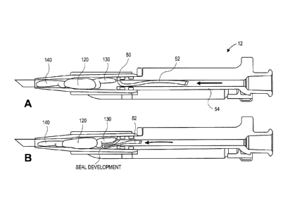
body that also follows the lens while moving for delivery. Figure 5A
illustrates the device
including sealing component 52 in the form of a filament attached to the
distal end of plunger 12
at attachment point 50. As shown in Figure 5A, filament 52 is initially
disposed within lumen 32
of plunger 12, with the filament distal end extending in the proximal
direction within the lumen.
As the viscoelastic fluid is advanced through lumen 32 from the proximal end
of the plunger, as
shown by the arrow in Figures 5A-C, filament 52 is carried by the fluid in the
distal direction, as
shown in Figure 5B. The filament flows toward the location at which the
viscoelastic fluid is
flowing past the lens body and creates a seal, as shown in Figure 5B. In
Figure 5B the filament
is shown plugging up a space adjacent the trailing haptic 130, creating at
least a substantial seal.
Once the seal is formed, the force of the viscoelastic fluid on the lens body
will properly cause
both the leading haptic and the optic portion to be advanced distally within
the cartridge. Figure
5C shows the leading haptic partially deployed from the cartridge while the
optic body has been
properly advanced through the cartridge. As the fluid continues to be advanced
through the
plunger and into the cartridge, the lens is advanced further through the
cartridge until the leading
haptic 140, optic body 120, and trailing haptic 140 are delivered, in that
order, from the cartridge
and into the eye. Once delivered the lens will inherently revert toward its
original configuration,
which is generally shown in the sectional view of Figure 1. The deployment
occurs without
damage to the lens, particularly at the attachment point between the leading
haptic and the optic
body.
[00045] Figures 6A and 6B illustrate an exemplary embodiment of filament 56
with slit 58
formed therein extending along substantially the entire length of filament 56.
The slit forms two
filament segments 60 extending along the length of filament 56. The length of
the segments 60
is substantially greater than the width of the segments. Filament 56 from
Figure 6A is then
folded to bring two ends 62 together, as shown in Figure 6B. Figures 7A and 7B
illustrate the
folded filament attached to plunger 12. The filament is secured to the distal
region of the
plunger by compressing the two filament end regions under the two o-rings 18
that are a seal to
- 8 -
CA 02865954 2014-08-28
WO 2013/142323 PCT/US2013/032054
the cartridge, as discussed above. To prepare the device for use, the segments
60, in the form of
loops, are tucked within the plunger lumen and ready for deployment during the
delivery, as
shown in Figure 7B. In use, the filament loops extend and roll out of the
lumen in the
viscoelastic stream and flow into an open volume behind the lens buttress and
adjacent the
trailing haptic as discussed above in the embodiment in Figures 5A-5C. The
filament bunches
into that area until sufficiently plugging the flow at which time the lens
body moves forward.
When the lens moves sufficiently forward to plug the tip completely and there
is substantially no
viscoelastic flow by the lens body, the filament will be left behind.
[00046] If, during the delivery, the optic body stops moving in the cartridge
and the
viscoelastic is leaking past the optic body again, the filaments are adapted
to again move to the
area of leakage to act as a plug to the viscoelastic and move the lens out of
the cartridge. The
filaments as described herein as therefore adapted to repeatedly, as may be
necessary, find or
seek out the region where fluid is flowing past the optic body, move to that
location, and plug the
leak.
[00047] The embodiment in Figures 5A-7B illustrate embodiments in which the
proximal end
of the filament is attached to the distal region and the exterior of the
plunger, and the filament is
considered to roll out of the plunger during delivery of the lens. This allows
the filament to slide
out of the plunger past the attachment location and the first contact with the
lens will be with a
region of the filament that is near the proximal end of the filament. The
filament repositions into
suitable plugging orientation. There are, however, a number of variations in
attachment points of
the filament to the plunger (or other portions of the delivery device)
yielding variations in
deployment of the filament.
[00048] Figures 8A-8C illustrate an alternative embodiment in which the
proximal end of the
filament is attached to the distal region of the plunger, but is attached at a
location on the interior
of the plunger (i.e., within the plunger lumen). As indicated in Figure 8A,
the plunger has a
proximal lumen section with a larger diameter bore than a distal tip section.
Filament 70 is
attached to stopper 72 at its proximal end. Filament 70 extends from stopper
70 towards the
distal end of the plunger. The reduced diameter distal section acts as a lumen
restriction and
prevents stopper 72 from advancing further distally, as shown in Figure 8C.
This prevents the
filament from flowing out of the distal tip of the cartridge. As the
viscoelastic is delivered
through the lumen, stopper 72 is advanced distally within the lumen, and
filament 70 flows
straight out of the lumen and does not double back (or fold) back on itself as
in the previously
described embodiments above. The filament is adapted to flow towards the flow
leakage as
described elsewhere herein.
- 9 -
CA 02865954 2014-08-28
WO 2013/142323 PCT/US2013/032054
[00049] Figures 9A-9C illustrate sectional views of an alternative embodiment
in which the
proximal end of the filament 80 is secured to the proximal region of the
plunger interior using
press-ring filament capture 78, which is shown in greater detail in Figure 15.
Filament 74
includes a coiled or stretchable section 76 that is adapted to uncoil or
stretch as the viscoelastic is
delivered. The uncoiling or stretching of the filament effectively lengthens
the filament, =
allowing the filament to be advanced into the cartridge to plug up any leaks,
but is prevented
from flowing out of the distal end of the cartridge. Figure 9B shows the
filament finding the
leaking area and plugging it.
[00050] The embodiment shown in Figures 9A-9C includes a plunger with a distal
tip region
that has a reduced diameter relative to the proximal region. This feature can
be incorporated into
any the embodiments herein. The reduced diameter can create a relatively
higher flow rate of
fluid from the plunger distal tip, which helps pull the filament out of the
distal end of the
plunger. The increased flow rate minimizes tangles and compaction of the
filament in the
proximal end of the plunger as the viscoelastic is delivered.
[00051] Figures 10A-10C illustrate an alternative embodiment similar to that
in Figures 9A-
9C in that the proximal end of the filament 82 is attached to an internal
proximal region of the
plunger 84. In this embodiment the filament 82 is a material that has a
stretching property to it,
such as a material that is perforated in such a way that it compresses
efficiently in the plunger
lumen (see Figure 10A) and yet stretches to a static length when deployed (see
Figure 10C). In
this embodiment the filament is an axially compressed perforated tubing. The
filament is
secured at its proximal end 845, and the plunger has a fluid channel radially
outward from the
attachment point. This is shown in Figure 10B, in which the fluid flow is
indicated the arrows.
The fluid causes the perforated tubing to stretch as shown in Figure 10B. This
plunger design,
with the distal reduced diameter, provides for a low velocity flow region and
a high velocity flow
region in the distal region. Figure 10B illustrates the plunger sealing the
leak, and Figure 10C
illustrates the leading haptic being deployed from the cartridge.
[00052] The filament material and design should be selected to enable the
filament to seal the
fluid flow as described above. An optimization of material structure and
properties will
generally provide a filament that is best suited to seal the fluid flow and
allow for the intraocular
lens to be delivered undamaged. It is envisioned, however, that in some
instances it may be
desirable to have some amount of fluid that does pass the optic body, after
which the sealing
should occur. The filament material can theoretically be selected that will
provide that
functionality to the system.
[00053] The properties of the filament will influence how it responds during
the delivery
process. Properties that can be modified to accomplish the specific goal
include without
- 10-
CA 02865954 2014-08-28
WO 2013/142323
PCT/US2013/032054
limitation, compliance, coefficient of friction, and elasticity. In
embodiments described above,
properties that have been shown to influence performance include compliance, a
low coefficient
of friction, and in some cases elasticity. It is understood that not all of
these need to be
optimized, and there may be other properties that can be controlled to achieve
a desired result.
[00054] In some embodiments the filament has a degree of deformability and
elasticity that
allows it to be pulled from the plunger and seal the leak. In some particular
embodiments
expanded PTFE, an expanded Teflon material, is used. In some embodiments the
filament
comprises an open cell foam. For example, a low durometer open cell silicone
foam with a
single strand form can be used in both the straight out method (e.g., Figures
8A-8C) and roll out
methods (e.g., Figures 5A-5C). In some embodiments PVA bio-absorbable type
foams that
provide good open cell performance can also be used. In some embodiments light
wall (e.g.,
0.004in) low durometer (e.g., 20-35shoreA) silicone tubing can be used,
particularly with the
roll-out method. In some embodiment electro-spun or non-woven materials are
used, and
materials that allow for compaction of the mat under low pressures can be used
in a straight out
method with a relatively large cross section. It is understood that other
suitable materials can be
used to accomplish the intended goal.
[00055] The filaments can be further manipulated to control the performance
characteristics.
For example, one or more slits formed in the filament can provide desired
functionality. Radial
and axial slits have been shown to increase compliance and bending of the
filament to optimize
sealing performance to the lens. One or more slits can be formed in the
filaments. The one or
more slits can take on any configuration within the material.
[00056] In some embodiments the filament is a monofilament ePTFE material. The
material
can be formed with one or more loops (see Figures 6A-7B), and in some
embodiments between
one and three loops to optimize cross section in the plunger lumen relative to
the tip plunger
.. lumen size.
[00057] While specific embodiments have been described herein that focus on
the use of a
filament, other material can be incorporated into the delivery device to
accomplish the goal. For
example, any suitable material that can be used to seal off the gaps can be
used. Other
deformable or flexible materials, for example, that are not described herein
could theoretically be
suitable or adapted to function as a sealing element as described herein.
[00058] In alternative embodiments, the sealing element is a sealed porous
tube of PTFE that
is filled with viscoelastic or other fluid. The porous tube is adapted to
allow viscoelastic to pass
through the tube, or "weep" through the pores. In this alternative all of the
viscoelastic fluid
delivered into the system is pushed through the porous tube. The tube is
adapted to seal off the
fluid leaks as described above. The pore size can be varied to control the
flow rate.
- 11 -
CA 02865954 2014-08-28
WO 2013/142323 PCT/US2013/032054
Additionally, different viscoelastic fluids have different viscosities and
flow properties, and thus
the fluid can be varied to modify the flow rate as well.
[00059] Figures 11A-11C and 12A and 12B illustrate an embodiment that includes
a plug
component that is a flexible porous tube 200 sealed on its distal end. The
tube is sheathed over a
hypo-tube support 202 that is sealed to the interior lumen of the proximal end
of the plunger 204.
The porous tube 200 is long relative to the length of the support tube 202 to
be able to extend to =
=
the tip of the cartridge when deployed. In a packaged state, the porous tube
200 is packed onto
the support 202 to decrease the length. The support tube 202 communicates with
the proximal
end 206 of the plunger to allow for the passage of viscoelastic (not shown) to
be delivered
directly to the tip 208 of the sealed porous tube 200. With flow of
viscoelastic through support
tube 202 and into the porous tube 200, the porous tube 200 will pressurize
slightly and extend off
of the support tubing 202 to move into a region behind the lens that both
seals, as described
above, and is able to transmit axial force mechanically to the lens. When the
lens moves forward
in the tapering cartridge and creates an efficient seal, the sealing and
mechanical action of the
porous tube will lend less influence to its functionality and it will
transition in performance to
simply pass viscoelastic therethrough (via the pores), which will move the
lens forward with
pressure differential. The functionality of the porous tubing can be modified
as needed by
modifying the properties of the tubing. For example, with a low porosity
(i.e., small pore size)
material, the tube will generally develop a higher internal pressure while the
viscoelastic tubing
is flowing, which will allow it to function more as a mechanical hydraulic
piston applying force
to the lens when contacted. With a relatively higher porosity construction
(i.e., larger pore size),
the tube will behave more similarly to the filament structures described
above, acting more
prominently as a sealing element to seal off any leaking fluid. The porosity
(or other property of
the tube) can therefore be modified as needed to achieve the desired
functionality of the porous
tubing.
[00060] Figures 13A and 13B illustrate an alternative exemplary embodiment of
an IOL
delivery system adapted to deliver an IOL into an eye of a patient. The system
includes cartridge
301, tray 302, and plunger 303. Figure 13B shows the assembled system, which
figure 13A
shows the disassembled system components. In other embodiments one or more of
the three
components may be integrally formed rather than separate parts.
[00061] In the assembly of Figure 13B, cartridge 301 is positioned with
respect to tray 302
such that cartridge 301 and tray 302 are in secured engagement. In some
embodiments cartridge
301 and tray 302 are integrally formed such that cartridge 301 is not adapted
to be disassociated
from tray 302. Tray 302 is adapted to receive a distal portion of plunger 303
therein. The distal
end 306 of plunger 303 is sized and configured to be disposed within proximal
opening 305 in
- 12 -
cartridge 301 when assembled. Plunger 303 includes seals 307 in the form of 0-
rings. Seals 307 are
adapted to create a seal between an inner surface of cartridge 301 when distal
portion 306 of plunger
is advanced into opening 305 of cartridge 301. Tray 302 facilitates the
interaction between the
cartridge and the plunger.
1000621 Plunger 303 has a proximal portion that is adapted to interact with
a fluid delivery
device, such as a syringe, so that fluid can be advanced from the fluid
delivery device and into an
inner lumen within plunger 303. Distal end 306 of plunger 303 is disposed
within the cartridge, and
thus the fluid is delivered to a location that is radially and axially within
the lumen, even if it does
not exit the plunger.
1000631 Cartridge 301 and tray 302 are in secure engagement as described in
U.S. Patent No.
9,610,155. Tray 302 includes two clips 361 with locking elements 365, wherein
the clip are adapted
to interface with camming surfaces 363 on plunger. Clips will splay outward as
plunger 303 is
advanced in tray 302, and locks 367 on plunger will lock with locks 365 on
tray 302.
1000641 Figures 14A and 14B illustrate top section views of the assembled
system from Figure
13B (IOL not shown for clarity). As can be seen, the distal portion 306 of
plunger 303 is disposed
within a proximal portion of lumen 310 of cartridge. While not shown, an IOL
will also be disposed
within lumen 310 and positioned to be deployed out of the distal end 311 of
cartridge 301.
1000651 Plunger 303 includes outer shell 313, on which seals 307 are
disposed. As can be seen,
seals 307 create a seal between outer shell 313 and an inner surface of lumen
310. Plunger 303 also
includes plug subassembly 321 within a lumen of plunger 303. The plug
subassembly is also shown
in greater detail in Figure 15. Plug subassembly 321 includes support tube
base 316, in which
support tube 314 is disposed and secured thereto, and plug element 317. Plug
element 317 is
sheathed over and secured to the outer surface of support tube 314 at location
308 (see Figure 15). In
one embodiment a heat shrunk collar secures plug element 317 to support tube
314 at location 308.
The distal end of support tube 314 extends from the distal end of base 316,
and is configured with an
orientation to one side. That is, the distal portion of tube 314 does not
extend along the longitudinal
axis of plunger 303. This helps direct support tube 314 and plug 317 away from
the trailing haptic.
Plug element 317 is long relative to the length of the support tube 314 such
that the distal end of plug
element 317 is disposed at the tip of the cartridge when the plug is fully
deployed. In a packaged, or
loaded, state (see Figures 14A and 14B), plug element 317 is packed onto
support tube 314 to
decrease its relative length. Support tube 314 communicates with the proximal
end of plunger 303 to
allow for the passage of a fluid such as a viscoelastic (not shown) into plug
element 317. Plug
subassembly 321 also include seal
- 13 -
Date Recue/Date Received 2020-06-01
CA 02865954 2014-08-28
WO 2013/142323
PCT/US2013/032054
315 adapted to create a seal between plug subassembly 321 and an inner surface
of outer shell
313 of plunger 313.
[00066] In this embodiment, plug element 317 is a tubular structure secured to
the distal end
of support tube 314 as shown in Figure 15. In this embodiment plug element is
a flexible and
porous material but need not necessary be porous. In one exemplary embodiment
plug element
is tubular ePTFE. In this embodiment the tubing is open-ended at both ends and
is tied in a knot
327 along its length, with distal section 309 of plug 317 extending distally
from knot 327. Knot
327 acts as a flow restrictor, and also helps stabilize the plug on the
support tube. In this
embodiment plug element 317 includes one or more optional perforations 325
just proximal to
knot 327. The flow restrictor can be, for example, tied, glued, crimped, or
swaged.
[00067] To load the plug subassembly into outer shell 313, distal section 39
of plug element
317 is rolled back, or folded back, towards the proximal end of the
subassembly, in the direction
of arrows shown in Figure 15. It is everted until flow restrictor 307 is
substantially at the distal
end of the plug element 317. The distal portion of plug element 317, in a
loaded configuration,
thus has an everted section of material at its distal end. Plug assembly 321
is then advanced
distally through open end 312 of outer shell 313 of plunger 303 until it is in
the loaded position
shown in Figures 14A and 14B. The open distal end of plug element 317, in its
everted
configuration, is retained within the lumen of outer shell 313, maintaining
the eversion. Figure
14A illustrates the biased configuration of the distal end of support tube
314. Figure 14B is a
side section view of the plug subassembly in a loaded configuration and
position within outer
shell 313 of plunger 303.
[00068] Figures 16A and 16B illustrate a fully deployed configuration of plug
element 317
within cartridge 301, as is also shown in Figure 15 outside of a cartridge.
For clarity, this is
illustrated without showing the IOL. A method of use with an IOL is shown
below. As
described in more detail below, after the plug is loaded (as shown in Figures
14A and 14B), a
fluid is delivered through support tube 314 to initiate the deployment of plug
element 317. As
plug element 317 continues to be deployed, the everted section 309 remains
everted until the full
extension of the proximal portion of the plug element 317, at which time
everted section 309
begins to unroll, and ultimately plug element 317 assumes the general elongate
configuration
shown in Figures 16A and 16B. The distal end of plug element 317 is
substantially at the tip of
cartridge 301 when fully deployed.
[00069] Figure 17A illustrates an IOL comprising optic 120 and haptics 130 and
140
positioned (e.g., such as the IOL shown in Figure 1) within cartridge 301. The
IOL has been
loaded into cartridge 301, and exemplary methods of loading the IOL into
cartridge are described
below. The disclosure herein is not intended to be limited to the manner in
which IOL becomes
- 14 -
CA 02865954 2014-08-28
WO 2013/142323 PCT/US2013/032054
positioned into cartridge 301. In the IOL's loaded configuration shown in
Figure 17A, leading
haptic 130 has been reoriented from an at-rest orientation (see Figure 1) and
extends distally
from optic 120. Trailing haptic 140 has also been reoriented from an at-rest
orientation (see
Figure 1) and extends relatively proximally from optic 120 within cartridge.
[00070] In general, the delivery of the IOL out of the cartridge relies on
development of a
pressure differential in the cartridge to move the IOL distally through the
cartridge and into the
eye. The configuration of the IOL in general and/or the configuration that the
IOL assumes
when loaded into the cartridge, however, creates some gaps between the IOL and
the inner
surface(s) of the cartridge. That is, the IOL does not occupy the entire
volume defined by the
inner surfaces of the cartridge. The gaps, or voids, provide a path for some
of the fluid to leak
past the optic portion as fluid is advanced during the delivery. Ideally, none
(or substantially
none) of the fluid flows past the optic body portion. Ideally, all, or
substantially all, of the fluid
remains proximal to at least the optic body portion, building up pressure and
forcing the IOL to
be deployed out of the distal end of the cartridge. When fluid does flow past
the lens body it can
create drag on leading haptic 130 that is efficiently filling the tip of the
cartridge. The advancing
leading haptic can create a high strain at the connection between the leading
haptic and the optic
body, possibly causing damage at the connection point. Any IOL that may be
susceptible to
damage while being delivered may benefit from the systems and methods
described herein.
[00071] An exemplary method of assembling the system includes placing
cartridge 301 in tray
302, loading the IOL into cartridge 301, and then positioning plunger 303
relative to tray 302
such that it extends into cartridge 301, as shown in Figure 17A. In Figure
17A, plug
subassembly 321 is in the same loaded position and configuration within
cartridge 301 as shown
in Figures 14A and 14B. In this configuration the plug element 317, and
specifically everted
portion 309, is positioned adjacent trailing haptic 140. Plug element 317 is
disposed in a gap
that exists between trailing haptic 140 and the inner surface of cartridge
301. As described
above, this distal end of support tube 314 is oriented away from trailing
haptic 140, which
disposes the plug element 317 in the position shown in Figure 17A, which is
radially adjacent to
trailing haptic 140. The support tube distal end is therefore adapted to avoid
damaging the IOL
when positioned in the cartridge. In this configuration plug element 317 acts
like a plug to fill in
the gap, or a substantial portion of the gap, to obstruct the flow of fluid,
thereby minimize the
amount of fluid that flows passed the trailing haptic 140 during the delivery.
Plug element 317
may or may not be in contact with the IOL at this time. As described below,
plug element 317
reduces the volume of fluid that flows past the optic during delivery,
increasing the pressure
differential, and thus reducing the risk of damage to the lens. Plug element
317 can also be
thought of as creating a seal, or a substantial seal, behind the IOL body to
reduce the flow of
- 15 -
CA 02865954 2014-08-28
WO 2013/142323
PCT/US2013/032054
viscoelastic around the 10L. "Plug" or "seal" are not limited to mean a
completely fluid tight
seal is created. These terms are used herein to mean that fluid flow around
the IOL is reduced
from what it would be without the plug or seal element. The plug element can
also be any of the
components described above as creating a seal behind the optic.
[00072] After the plug subassembly is positioned as shown in Figure 17A, a
fluid, such as a
viscoelastic, is advanced through support tube 314 using a fluid delivery
device such as a syringe
(not shown). With the flow of viscoelastic through support tube 314 and into
plug element 317,
plug element 317 will pressurize slightly and reconfigure off of the support
tube 314 to move
more fully into a region behind the lens that plugs the gap and is able to
transmit force
mechanically to the lens. As the IOL moves forward in the tapering cartridge
inner lumen and
creates an efficient, or substantial, seal, the sealing and mechanical action
of the porous tube will
lend less influence to its functionality and it will transition in performance
to pass viscoelastic
therethrough (via the pores, or other perforation constructs), which will move
the lens forward
with pressure differential. The functionality of the porous tubing can be
modified as needed by
modifying the properties of the tubing. For example, with a low porosity
(i.e., small pore size)
material, the tube will generally develop a higher internal pressure while the
viscoelastic is
flowing, which will allow it to function more as a mechanical hydraulic piston
applying force to
the lens when contacted. With a relatively higher porosity construction (i.e.,
larger pore size),
the tube will behave more similarly to the filament structures above, acting
more prominently as
a plug element to seal off leaking fluid. The porosity (or other property of
the tube) can
therefore be modified as needed to achieve the desired functionality of the
plug element.
[00073] As the fluid exists the distal end of support tube 314, the fluid
pressure within the
everted portion 309 of plug element 317 causes the distal end of plug element
317 to be released
from the distal end of outer shell 313 of plunger 303. As the free distal end
of the plug is
released from the inner lumen of the plunger, it begins to at least partially
seal against the inner
walls of the cartridge, further reducing the volume of fluid that flows past
the IOL. The plug
element also at least partially plugs the gap that exists radially between
adjacent trailing haptic
140 and the inner wall of the cartridge. This plugging action minimizes the
volume of fluid that
can flow passed trailing haptic and therefore passed optic portion, increasing
the pressure
differential in the cartridge.
[00074] As fluid continues to be advanced through support element 314, as
shown in Figure
17C, the everted plug element continues to follow the IOL, still plugging the
gap between
trailing haptic 140 and the cartridge. As the optic is advanced closer to the
distal port, as shown
in Figure 17D, the size of the port and the volume that the optic occupies
cause the optic to begin
to self-seal, or substantially create a seal in the distal port. In Figure 17D
the IOL begins to
- 16 -
CA 02865954 2014-08-28
WO 2013/142323
PCT/US2013/032054
move distally relative to plug element 317, or outrun the plug element 317. In
Figure 17E the
optic has been delivered out of the cartridge and trailing haptic 140 is
rolling off of everted
portion 309. This causes the everted portion of the plug element to unroll, as
shown in Figure
17E. The everted portion 309 of the plug element reduces drag on trailing
haptic 140 between at
least Figures 17D and 17E, when it is unfurling, or unrolling. A static plug
element, unlike
everted portion 309, can cause the trailing haptic to get stuck against the
wall of the cartridge due
to the radial expansion of the plug and static friction between the plug and
the haptic. When the
plug element includes a feature that can contact and unfurl with the trailing
haptic, drag on the
trailing haptic is reduced, preventing it from sticking against the cartridge
wall and not deploying
properly. This also reduces the likelihood of damage at the junction between
the optic and the
trailing haptic.
[00075] In the embodiment in Figures 17A-17E, plug 317 is a porous ePTFE
material. The
porous material is adapted to allow viscoelastic to pass through the tube, or
"weep" through the
pores. In embodiments herein plug 317 also includes optional perforations 325
(two shown in
the embodiment in Figures 17A-17E) in the plug material just proximal to the
knot location 327.
In one particular embodiment the perforations are created with a 32G surgical
needle about lmm
proximal to the knot. The perforations act as an over-pressure relief for the
viscoelastic material
(or other fluid). The porosity of the ePTFE (or other porous material) can be
variable, and in
some cases, which may depend on the viscoelastic material used, the material
may fully contain
the viscoelastic without allowing for effective weeping. If this occurs the
plug element may
disengage from the support tube 314 due to pressure at the end of extension.
The perforations
can thus serve as an over-pressure relief to prevent this possibility. In a
secondary roll, the
perforation can also direct fluid into the everted section of the plug to
facilitate its release from
the plunger and thus sealing against the inner surface of the cartridge.
[00076] The porosity of the plug allows viscoelastic to lubricate the
interfaces between the
moving plug and other system components. The porosity also allows the
continued flow of the
fluid when the plug is at full deployment and the IOL is moving due to a
hydraulic seal at the tip.
[00077] The pore size can be varied to control the flow rate. Additionally,
different
viscoelastic fluids have different viscosities and flow properties, and thus
the fluid can be varied
to modify the flow rate as well. In an exemplary embodiment the plug element
is ePTFE and the
intermodal distance (i.e., the distance between the nodes), which determines
the porosity, is 100
pm. ePTFE with other intemodal distances can also be used.
[00078] The embodiment shown in Figures 13A, 13B, 14A, 14B, 16A, 16B, and 17A-
17E are
also adapted to purge trapped air in the system that, if not purged, can
interfere with the delivery
process. Figure 18 illustrates a side section view of the assembled device
shown in Figure 14B
- 17 -
CA 02865954 2014-08-28
WO 2013/142323
PCT/US2013/032054
(IOL not shown for clarity), illustrating the purging of air from the plunger.
As described herein,
fluid travels from a syringe (not shown) through support tube 314 and exits in
proximity of the
trailing haptic of the IOL within the plug element (everted section 309
labeled). A fluid front
travels both distally in the "D" direction shown, filling the plug element,
and rearward in the "P"
direction, which evacuates dead volume air through vent 330 in the direction
of arrow "A." The
vent will not pass viscoelastic so is able to maintain pressure when fully
evacuated. This effect
purges the air from the back of the system to reduce spring effects of trapped
air during the
release of the IOL during delivery. In some instances if the air is not purged
the air can
forcefully push the IOL forward during delivery, without operator
action/input, possibly
.. damaging the IOL or the capsule in the eye, and can even cause the IOL to
be delivered outside
of the capsule. The purging of air is important for a smooth, controlled
delivery of the IOL.
Some IOLs may not require as much control in the delivery, and thus venting of
air may not be
required.
[00079] In some embodiments the delivery system includes a vent and does not
include a
plug, or sealing element. In these embodiments fluid such as viscoelastic is
delivered towards
the lens as part of the delivery process. Air venting to increase control
during delivery while
decreasing the volume of air bubbles that are moved forward through the tip
into the eye
provides a significant advantage even in the absence of a plug element. In an
alternative
embodiment, the device is similar to the delivery device in Figures 14A and
14B but does not
.. include a plug element 317.
[00080] Figures 19A-19C illustrate an exemplary way of driving fluid such as
viscoelastic
from within a delivery device into the support tube 314. In this embodiment
the delivery
assembly, including cartridge 301, tray 302, and plunger 303, with syringe 360
secured thereto,
are mounted into screwdrive assembly 370. Screwdrive assembly 370 includes
base 390 with
end posts 372 over which slots in tray 302 are aligned. The delivery assembly
self-aligns with
screw 380. Screw 380 is advanced until it touches the plunger of the syringe,
as shown in Figure
19C. Screw 380 is then turned to cause the syringe plunger to be advanced,
which drives the
fluid from the syringe and into support tube 314. The screwdrive assembly can
be modified to
more finely control the force applied to the syringe, and can include a
pressure gauge.
[00081] As set forth herein, an IOL can be positioned, or loaded, into the
cartridge using any
suitable technique. For the specific IOL described herein, the loading process
includes changing
the orientation of the haptics with respect to the optic, such that the
haptics generally extend
away from the optic. In general this process of reorienting the haptics is
referred to herein as
splaying the haptics. The loading process, for the IOL herein, also includes
reconfiguring at
least one portion of the IOL, such as the optic. Exemplary loading techniques
include without
- 18-
limitation, hydraulically loading the IOL, as is set forth in U.S. Patent No.
8,956,408. Alternatively,
the IOL can be mechanically loaded, such as is described in U.S. Patent No.
9,610,155. Another
example of mechanical loading includes using forceps to pick up the IOL,
reorient one or more
haptics, and advance the IOL into the cartridge.
1000821 The IOL can be loaded into the cartridge and stored, such as for
packaging, or loading
can occur just prior to implantation.
1000831 The devices and methods herein are able to deliver an IOL through
an incision that is
between about 2.8 mm to about 4.5 mm. In some embodiments the incision is
about 4 mm. The
devices and methods can be modified if needed to deliver an IOL through a
bigger or smaller
incision.
1000841 While the disclosure focused on a tubular member for the plug,
other sealing
mechanisms can also be inserted into the cartridge to help create at least a
partial seal between the
IOL and cartridge to aid in the delivery of the IOL.
1000851 The IOL to be delivered need not have one or more dedicated
"haptics" as described
.. herein. The IOL can more generally include a peripheral portion.
- 19 -
Date Recue/Date Received 2020-06-01