Note: Descriptions are shown in the official language in which they were submitted.
CA 02866079 2014-08-29
WO 2013/130354
PCT/US2013/027373
TARGETED TREATMENT OF ANEROBIC CANCER
FIELD OF THE INVENTION
The present invention relates to a pharmaceutical cocktail and methods of
treatment
involving said cocktail, in particular, a combination of effective amounts of
a carbonic anhydrase
inhibitor, in combination with effective amounts of an angiogenesis inhibitor,
including a vascular
endothelial growth factor (VEGF) inhibitor such as bevacizumab for the
treatment of cancer. The
merits of this invention are based on the fact that cancer in its untreated
state uses both aerobic and
anaerobic/glycolytic pathways and both must be treated if the best results are
to be achieved.
Treatment of both metabolic pathways more completely deprives cancer of ATP
energy
production, thereby producing greater damage or killing of cancerous cells.
Treatment of the
aerobic pathway alone temporarily controls cancer but it induces mutation to a
glycolytic form,
which does not respond to anti-VEGF or other anti-vascular growth factor
agents.
In other embodiments, it relates to compositions and methods of treating
cancer involving
effective amounts of a carbonic anhydrase inhibitor. Pharmaceutical
compositions and methods
of treating cancer (eliminating the tumor, shrinking the tumor, prolonging the
life of the patient,
increasing quality of life by decreasing the grade of adverse events seen with
other cancer
treatments, and/or preventing/reducing the likelihood of the tumor's
metastases) are additional
aspects of the present invention. In addition, the present invention may be
used to favorably
affect the therapeutic result of patients who have not responded to
alternative, traditional
anti-cancer therapy.
BACKGROUND OF THE INVENTION
1
CA 02866079 2014-08-29
WO 2013/130354
PCT/US2013/027373
While a number of anti-angiogenesis agents have been reported, including
bevacizumab,
it is not clear whether they possess the appropriate pharmacological
effectiveness required to be
therapeutically useful in the treatment of cancer in many situations.
Therefore, there is a
continued need for additional therapeutics to target such cancer and augment
or revive the
effectiveness of anti-angiogenesis agents to provide effective treatment of
cancer.
SUMMARY OF THE INVENTION
The present invention relates to a pharmaceutical cocktail and methods of
treatment
involving said cocktail, in particular, a combination of effective amounts of
a carbonic anhydrase
inhibitor, in combination with effective amounts of an angiogenesis inhibitor,
including a vascular
endothelial growth factor (VEGF) inhibitor such as bevacizumab for the
treatment of cancer. In
other embodiments, it relates to compositions and methods of treating cancer
involving effective
amounts of a carbonic anhydrase inhibitor. Pharmaceutical compositions and
methods of treating
cancer (eliminating the tumor, shrinking the tumor, prolonging the life of the
patient, increasing
quality of life by decreasing the grade of adverse events seen with other
cancer treatments, and/or
preventing/reducing the likelihood of the tumor's metastases) are additional
aspects of the present
invention. In addition, the present invention may be used to favorably affect
the therapeutic result
of patients who have not responded to alternative, traditional anti-cancer
therapy.
In one embodiment, the invention contemplates a method of treating cancer
comprising
administering to a patient an effective amount of a loop diuretic and an
angiogenesis inhibitor. In
one embodiment, said angiogenesis inhibitor is a humanized monoclonal
antibody. In one
embodiment, said antibody is bevacizumab. In one embodiment, said treating
comprises repeated
administration of at least one of the loop diuretic and angiogenesis
inhibitor. In one embodiment,
said loop diuretic is bumetanide. In one embodiment, said cancer is hypoxic
cancer. In one
2
CA 02866079 2014-08-29
WO 2013/130354
PCT/US2013/027373
embodiment, said administering results in the shrinkage of said cancer. In one
embodiment,
said patient has metastases and said administration reduces metastases of said
cancer.
In one embodiment, the invention contemplates a method of treating cancer
comprising administering to a patient an effective amount of a carbonic
anhydrase inhibitor
and an angiogenesis inhibitor. In one embodiment, said angiogenesis inhibitor
is a humanized
monoclonal antibody. In one embodiment, said treating comprises repeated
administration of
at least one of the carbonic anhydrase inhibitor and angiogenesis inhibitor.
In one
- embodiment, said antibody is bevacizumab. In one embodiment, said
carbonic anhydrase
inhibitor and an angiogenesis inhibitor are administered to said patient at
the same time. In
one embodiment, said cancer is hypoxic cancer. In one embodiment, said
carbonic anhydrase
inhibitor is a carbonic anhydrase 9 and carbonic anhydrase 12 inhibitor. In
one embodiment,
said administering results in the shrinkage of said cancer. In one embodiment,
said patient
has metastases and said administration reduces metastases of said cancer.
In one embodiment, the invention contemplates a pharmaceutical composition
comprising an effective amount of a loop diuretic and an angiogenesis
inhibitor. In one
embodiment, said angiogenesis inhibitor is bevacizumab. In one embodiment,
said loop
diuretic is bumetanide. In one embodiment, the invention contemplates said
pharmaceutical
composition formulated for oral administration. In one embodiment, the
invention
contemplates said pharmaceutical composition formulated for parenteral
administration. In
one embodiment, the invention contemplates said pharmaceutical composition
formulated for
intravenous administration.
In one embodiment, the invention contemplates a pharmaceutical composition
comprising an effective amount of a carbonic anhydrase inhibitor and an
angiogenesis
inhibitor. In one embodiment, said angiogenesis inhibitor is bevacizumab. In
one
3
CA 02866079 2014-08-29
WO 2013/130354
PCT/US2013/027373
embodiment, said carbonic anhydrase inhibitor and said angiogenesis inhibitor
are in a
mixture. In one embodiment, the invention contemplates said formulated for
oral
administration. In one embodiment, the invention contemplates said formulated
for parenteral
administration. In one embodiment, the invention contemplates said formulated
for
intravenous administration.
In one embodiment, the invention contemplates a method for treating a patient
with
cancer, said method comprising: a) administering to said patient a carbonic
anhydrase
inhibitor, and b) occluding =the blood vessels providing blood to said cancer.
In one
embodiment, said cancer is hypoxic cancer. In one embodiment, said treating
results in the
shrinkage of said cancer. In one embodiment, said occluding of blood vessels
providing
blood to said cancer comprises embolization. In one embodiment, said
embolization
comprises embolization with polymers embedded with carbonic anhydrase
inhibitors. In one
embodiment, said occluding of blood vessels providing blood to said cancer
comprises
thermal ablation. In one embodiment, said treating of said cancer with thermal
ablation is
preceded with bumetanide treatment. In one embodiment, said anhydrase
inhibitor is
bumetanide.
In one embodiment, the invention relates to a method of treating cancer
comprising
administering to a patient in need of therapy an effective amount of low dose,
frequently
administered combination of a carbonic anhydrase inhibitor and an angiogenesis
inhibitor. In one
embodiment, said angiogenesis inhibitor is selected from the group consisting
of ZD6474, ZD
6126, AZD2171, SU6668 and SU5416, bevacizumab, mv833, anti-FLT-1 ribozyme,
SU5416,
PTK 787, ZD4190, ZD6474, CEP-7055, SU11248, and mixtures thereof. In one
embodiment,
said angiogenesis inhibitor is bevacizumab. In one embodiment, said carbonic
anhydrase
inhibitor is bumetanide. In one embodiment, said carbonic anhydrase inhibitor
is a carbonic
4
CA 02866079 2014-08-29
WO 2013/130354
PCT/US2013/027373
anhydrase 9 and carbonic anhydrase 12 inhibitor. In one embodiment, the
treatment results in one
or more of clinical benefit remission, an increased quality of life or
prolongation of survival of the
patient. In one embodiment, said treatment results in the shrinkage of a tumor
or prolonged
stability of the cancer. In one embodiment, said treatment reduces metastases
of said cancer.
In one embodiment, the invention relates to a pharmaceutical composition
comprising an
effective amount of a combination of a carbonic anhydrase inhibitor and an
angiogenesis inhibitor.
In one embodiment, said angiogenesis inhibitor is selected from the group
consisting of ZD6474,
ZD 6126, AZD2171, SU6668 and SU5416, bevacizumab, mv833, anti-FLT- 1 ribozyme,
SU5416,
PTK 787, ZD4190, ZD6474, CEP-7055, SU11248, and mixtures thereof. In one
embodiment,
said angiogenesis inhibitor is bevacizumab. In one embodiment, said carbonic
anhydrase
inhibitor is bumetanide. In one embodiment the invention relates to the
composition described
above adapted for oral administration. In one embodiment the invention relates
to the
composition described above adapted for parenteral administration. In one
embodiment the
invention relates to the composition described above adapted for intravenous
administration.
In one embodiment, the invention relates to a method for treating a patient
with cancer,
wherein said cancer is unresponsive to traditional therapy, said method
comprising administering
to said patient a combination of a carbonic anhydrase inhibitor and an
angiogenesis inhibitor in
amounts effective to provide a clinical benefit remission, an increased
quality of life or
prolongation of survival of the patient. In one embodiment, said treatment
results in the
shrinkage of a tumor or prolonged stability of the cancer. In one embodiment,
said method results
in a complete remission of said cancer. In one embodiment, said angiogenesis
inhibitor is
bevacizumab. In one embodiment, said carbonic anhydrase inhibitor is
bumetanide.
In one embodiment, the invention relates to the treatment of hypoxic cancer.
In one
embodiment, treatment of hypoxic cancer includes targeted bloodstream
injection of a carbonic
5
CA 02866079 2014-08-29
WO 2013/130354
PCT/US2013/027373
anhydrase inhibitor, such as bumetanide.
In one embodiment, treatment comprises
catheterization of the hepatic artery. In one embodiment, treatment comprises
occluding arteries
with the treatment of bumetanide. In one embodiment, treatment comprises
embolization. In
one embodiment, treatment comprises embolization with polymers embedded with
carbonic
anhydrase inhibitors. In one embodiment, said carbonic anhydrase inhibitors
include a carbonic
anhydrase 9 or 12 inhibitor, such as bumetanide. In one embodiment, said
polymers embedded
with carbonic anhydrase inhibitors slowly release bumetanide. In one
embodiment, said
treatment= bumetanide is given intravenously in combination with artery
embolization with
polymers embedded with carbonic anhydrase inhibitors.
In one embodiment, the invention contemplates the treatment of cancer. In one
embodiment, said cancer comprises well-defined tumors. In one embodiment, said
treatment
involves thermal ablation of arteries supplying blood to well defined tumors
in combination with
treatment with bumetanide. In one embodiment, treatment comprises additional
treatment with
an angiogenesis inhibitor. In one embodiment, said angiogenesis inhibitor is
selected from the
group consisting of ZD6474, ZD 6126, AZD2171, SU6668 and SU5416, bevacizumab,
mv833,
anti-FLT-1 ribozyme, SU5416, PTK 787, ZD4190, ZD6474, CEP-7055, SU11248, and
mixtures
thereof.
In one embodiment, the invention contemplates a method for treating a patient
with cancer,
said method comprising administering to said patient a carbonic anhydrase
inhibitor and occlusion
of blood vessels providing blood to said cancer effective to provide a
clinical benefit remission, an
increased quality of life or prolongation of survival of the patient. In one
embodiment, said
cancer is hypoxic cancer. In one embodiment, said treatment results in the
shrinkage of a tumor
or prolonged stability of the cancer. In one embodiment, said method results
in a complete
remission of said cancer. In one embodiment, said occlusion of blood vessels
providing blood to
6
CA 02866079 2014-08-29
WO 2013/130354
PCT/US2013/027373
said cancer comprises embolization. In one embodiment, said embolization
comprises
embolization with polymers embedded with carbonic anhydrase inhibitors. This
embodiment
provides treatment of aerobic cancer cells by occlusion of the arteries and
treatment of the
glycolytic cancer cells by direct action of the carbonic anhydrase inhibitor
and indirectly by
inhibition of glycolysis by the induced low pH. In one embodiment, said
carbonic anhydrase
inhibitor is bumetanide. In one embodiment, said occlusion of blood vessels
providing blood to
said cancer comprises thermal ablation. In one embodiment, said treatment of
said cancer with
thermal ablation is preceded with bumetanide treatment.
The described features, structures, or characteristics of the invention may be
combined in
any suitable manner in one or more embodiments. In the following description,
numerous
specific details are recited to provide a thorough understanding of
embodiments of the invention.
One skilled in the relevant art will recognize, however, that the invention
may be practiced
without one or more of the specific details, or with other methods,
components, materials, and so
forth. In other instances, well-known structures, materials, or operations are
not shown or
described in detail to avoid obscuring aspects of the invention.
DEFINITIONS
To facilitate the understanding of this invention, a number of terms are
defined below.
Terms defined herein have meanings as commonly understood by a person of
ordinary skill in
the areas relevant to the present invention. Terms such as "a", "an" and "the"
are not intended
to refer to only a singular entity, but include the general class of which a
specific example may
be used for illustration. The terminology herein is used to describe specific
embodiments of the
invention, but their usage does not delimit the invention, except as outlined
in the claims.
The term "patient" or "subject" is used throughout the specification to
describe an animal,
7
CA 02866079 2014-08-29
WO 2013/130354
PCT/US2013/027373
generally a mammal and preferably a human, to whom treatment, including
prophylactic
treatment, with the compositions according to the present invention is
provided. For treatment of
those infections, conditions or disease states, which are specific for a
specific animal such as a
human patient, the term patient refers to that specific animal.
The term "neoplasia" or "cancer" is used throughout the specification to refer
to the
pathological process that results in the formation and growth of a cancerous
or malignant
neoplasm, i.e., abnormal tissue that grows by cellular proliferation, often
more rapidly than
normal and continues to grow after the stimuli that initiated the new growth
cease. Malignant
neoplasms show partial or complete lack of structural organization and
functional coordination
with the normal tissue and most invade surrounding tissues, metastasize to
several sites, and are
likely to recur after attempted removal and to cause the death of the patient
unless adequately
treated. As used herein, the term neoplasia is used to describe all cancerous
disease states and
embraces or encompasses the pathological process associated with malignant
hematogenous,
ascitic and solid tumors. Representative cancers include, for example,
stomach, colon, rectal,
liver, pancreatic, lung, breast, cervix uteri, corpus uteri, ovary, prostate,
testis, bladder, renal,
brain/CNS, head and neck, throat, Hodgkin's disease, non-Hodgkin's lymphoma,
multiple
myeloma, leukemia, melanoma, acute lymphocytic leukemia, acute myelogenous
leukemia,
Ewing's sarcoma, small cell lung cancer, choriocarcinoma, rhabdomyosarcoma,
Wilms' tumor,
neuroblastoma, hairy cell leukemia, mouth/pharynx, oesophagus, larynx, kidney
cancer and
lymphoma, among others, including soft tissue sarcomas, which may be treated
by the
combination of compounds according to the present invention.
The terin "remission" or "clinical benefit remission" is used to describe a
remission in a
patient's cancer, which may be a complete remission, a partial remission or
evidence of stability
of the disease.
8
CA 02866079 2014-08-29
WO 2013/130354
PCT/US2013/027373
The term "coadministration" or "combination therapy" is used to describe a
therapy in
which at least two active compounds or compositions in effective amounts (in
the present
application, at least bumetanide is coadministered with the angiogenesis
inhibitor, preferably
bevacizumab also being coadministered or being administered before or after
the administration
of bumetanide) to treat cancer, and preferably both compounds are used to
treat a disease state or
condition as otherwise described herein at the same time. In some embodiments,
the invention
involves administration of an additional chemotherapy compound(s) or
composition(s).
Although the term coadministration =preferably includes the administration of
at least two
active compounds to the patient at the same time, it is not necessary that the
compounds be
administered to the patient at the same time, although effective amounts of
the individual
compounds will be present in the patient at the same time.
The term "traditional cancer therapy" as used herein includes, but is not
limited to
radiation, surgical removal of cancerous tissue, and treatment with
chemotherapeutic drugs,
which generally have significant toxicity and undesirable side effects.
The term "carbonic anhydrase(s)" (CAs) as used herein refer to a large family
of zinc
metalloenzymes that catalyze the reversible hydration of carbon dioxide. They
participate in a
variety of biological processes, including, but not limited to, respiration,
calcification, acid-base
balance, bone resorption, and the formation of aqueous humor, cerebrospinal
fluid, saliva, and
gastric acid. Carbonic anhydrase 9 (CA9) is an enzyme that in humans is
encoded by the CA9
gene and carbonic anhydrase 12 (CA12) is an enzyme that in humans is encoded
by the CA12
gene. CA9 and CA12 are most commonly present in many cancer types, i.e. colon,
breast,
brain, kidney, lung etc. but uncommonly present in normal tissues, making them
suitable for
therapeutic targeting.
9
CA 02866079 2014-08-29
WO 2013/130354
PCT/US2013/027373
The term "angiogenesis inhibitor", "vascular endothelial growth factor
inhibitor" "VEGF
inhibitor" or "anti-VEGF therapy" all used within context, refers to a
compound, composition or
therapy which inhibits or otherwise prevents the angiogenesis effects of
vascular endothelial
growth factor (VEGF, a factor which is involved in the angiogenesis of tissue,
including growth in
and vascularization of tumors), regardless of mechanism.
As used herein, bumetanide (also known under trade names Bumex or Burinex) is
a loop
diuretic, a carbonic anhydrase inhibitor, and an aquaporin inhibitor.
Bumetanide is a thiazide
diuretic.
The IUPAC name is 3 -butylamino-4-phenoxy-5-sulfamoyl-benzoic acid.
HO 0 NH2
S
0
Bumetanide has the chemical structure: 0 0
As used herein, thiazides are a class of drug that promotes water loss from
the body
((diuretics)). They inhibit Na+/C1- reabsorption from the distal convoluted
tubules in the kidneys.
Thiazides also cause loss of potassium and an increase in serum uric acid. The
chemical structure
of the original thiazide diuretics contained a thiazide ring system; the term
is also used for drugs
with a similar action that are not chemically thiazides, such as
chorthalidone.
1 5
As used herein, aquaporins refer to proteins embedded in the cell membrane
that regulate
the flow of water. Aquaporins selectively conduct water molecules in and out
of the cell, while
preventing the passage of ions and other solutes. Also known as water
channels, aquaporins are
integral membrane pore proteins. Some of them, known as aquaglyceroporins,
transport also
other small uncharged solutes, such as glycerol, carbon dioxide, ammonia and
urea across the
membrane, depending on the size of the pore.
As used herein, embolization is a non-surgical, minimally invasive procedure
performed
CA 02866079 2014-08-29
WO 2013/130354
PCT/US2013/027373
by an interventional radiologist and interventional neuroradiologists. It
involves the selective
occlusion of blood vessels by purposely introducing emboli. The purpose of
embolization is to
prevent blood flow to an area of the body, which effectively can shrink a
tumor or block an
aneurysm and/or deliver therapeutic drugs or/and agents. The procedure is
carried out as an
endovascular procedure by a consultant radiologist in an interventional suite.
It is common for
most patients to have the treatment carried out with little or no sedation,
although this depends
largely on the organ to be embolized. Patients who undergo cerebral
embolization or portal vein
embolization are usually given a general anesthetic. Access to the organ in
question is acquired
by means of a guidewire and catheter(s). Depending on the organ, this can be
very difficult and
time consuming. The position of the correct artery or vein supplying the
pathology in question
is located by digital subtraction angiography (DSA). These images are then
used as a map for the
radiologist to gain access to the correct vessel by selecting an appropriate
catheter and or wire,
depending on the 'shape' of the surrounding anatomy. Once in place, the
treatment can begin.
The artificial embolus used is usually, but not limited to, one of the
following: Guglielmi
detachable coil or hydrocoil, particles, foam, and plug.
As used herein, theimal ablation is a method of removing aberrant tissue from
within the
body preferably via minimally invasive procedures. There are several types of
thermal ablation
used to destroy targeted tissue: cryoablation uses extremely cold temperatures
to freeze diseased
tissue, radiofrequency ablation uses heat generated by radiofrequency energy,
microwave
ablation uses heat generated by microwave energy, Laser ablation uses heat
from a laser beam,
and ultrasound ablation uses heat from focused ultrasound energy.
As used herein, the "nano knife system" is a minimally invasive cancer
treatment that
uses irreversible electroportation technology to precisely target and kill
hard-to-reach tumors at
the cellular level. It employs irreversible electroporation that uses a series
of microsecond
11
CA 02866079 2014-08-29
WO 2013/130354
PCT/US2013/027373
electrical pulses.
The term "occluding" as used herein refers to cause to become closed, such as
blood
vessels; to obstruct or occlude an artery. Embolization is one method of
occluding blood
vessels or lymphatic vessels.
The teim "salts", as used herein, refers to any salt that complexes with
identified
compounds contained herein while retaining a desired function, e.g.,
biological activity.
Examples of such salts include, but are not limited to, acid addition salts
formed with inorganic
acids (e.g. hydrochloric acid, hydrobromic acid, sulfuric acid, phosphoric
acid, nitric acid, and the
like), and salts formed with organic acids such as, but not limited to, acetic
acid, oxalic acid,
tartaric acid, succinic acid, malic acid, fumaric acid, maleic acid, ascorbic
acid, benzoic acid,
tannic acid, pamoic acid, alginic acid, polyglutamic, acid, naphthalene
sulfonic acid, naphthalene
disulfonic acid, and polygalacturonic acid. Pharmaceutically acceptable salts
also include base
addition salts, which may be formed when acidic protons present are capable of
reacting with
inorganic or organic bases. Suitable pharmaceutically-acceptable base addition
salts include
metallic salts, such as salts made from aluminum, calcium, lithium, magnesium,
potassium,
sodium and zinc, or salts made from organic bases including primary, secondary
and tertiary
amines, substituted amines including cyclic amines, such as caffeine,
arginine, diethylamine,
N-ethyl piperidine, histidine, glucamine, isopropylamine, lysine, morpholine,
N-ethyl morpholine,
piperazine, piperidine, triethylamine, and trimethylamine. All of these salts
may be prepared by
conventional means from the corresponding compound of the invention by
reacting, for example,
the appropriate acid or base with the compound of the invention. Unless
otherwise specifically
stated, the present invention contemplates pharmaceutically acceptable salts
of the considered
pro-drugs.
12
CA 02866079 2014-08-29
WO 2013/130354
PCT/US2013/027373
In addition, atoms making up the compounds of the present invention are
intended to
include all isotopic finals of such atoms. Isotopes, as used herein, include
those atoms having the
same atomic number but different mass numbers. By way of general example and
without
limitation, isotopes of hydrogen include tritium and deuterium, and isotopes
of carbon include 13C
and 14C. Similarly, it is contemplated that one or more carbon atom(s) of a
compound of the
present invention may be replaced by a silicon atom(s). Furthermore, it is
contemplated that one
or more oxygen atom(s) of a compound of the present invention may be replaced
by a sulfur or
selenium atom(s).
In structures wherein stereochemistry is not explicitly indicated, it is
assumed that all
stereochemistry is considered and all isomers claimed.
Any undefined valency on an atom of a structure shown in this application
implicitly
represents a hydrogen atom bonded to the atom. Bonds to copper (Cu) metal may
be coordinate
bonds and are not necessarily considered covalent.
The term "effective," as that term is used in the specification and/or claims,
means
adequate to accomplish a desired, or hoped for result.
The term "hydrate" when used as a modifier to a compound means that the
compound has
less than one (e.g., hemihydrate), one (e.g., monohydrate), or more than one
(e.g., dihydrate) water
molecules associated with each compound molecule, such as in solid forms of
the compound.
An "isomer" of a first compound is a separate compound in which each molecule
contains
the same constituent atoms as the first compound, but where the configuration
of those atoms in
three dimensions differs.
As used herein, the term "patient" or "subject" refers to a living mammalian
organism,
such as a human, monkey, cow, sheep, goat, dog, cat, mouse, rat, guinea pig,
or transgenic species
13
CA 02866079 2014-08-29
WO 2013/130354
PCT/US2013/027373
thereof. In certain embodiments, the patient or subject is a primate. Non-
limiting examples of
human subjects are adults, juveniles, infants and fetuses.
The teim "Pharmaceutically acceptable" means that which is useful in preparing
a
pharmaceutical composition that is generally safe, non-toxic and neither
biologically nor
otherwise undesirable and includes that which is acceptable for veterinary use
as well as human
pharmaceutical use.
"Pharmaceutically acceptable salts" means salts of compounds of the present
invention
which are pharmaceutically acceptable, as defined above, and which possess the
desired
pharmacological activity. Such salts include acid addition salts formed with
inorganic acids such
as hydrochloric acid, hydrobromic acid, sulfuric acid, nitric acid, phosphoric
acid, and the like; or
with organic acids such as 1,2-ethanedisulfonic acid, 2-hydroxyethanesulfonic
acid,
2-naphthalenesulfonic acid, 3-phenylpropionic acid, 4,4'-methylenebis(3-
hydroxy-2-ene-1
-carboxylic acid), 4-methylbicyclo[2.2.2]oct-2-ene-1-carboxylic acid, acetic
acid, aliphatic mono-
and dicarboxylicacids, aliphatic sulfuric acids, aromatic sulfuric acids,
benzenesulfonic acid,
benzoic acid, camphorsulfonic acid, carbonic acid, cinnamic acid, citric acid,
cyclopentanepropionic acid, ethanesulfonic acid, fumaric acid, glucoheptonic
acid, gluconic acid,
glutamic acid, glycolic acid, heptanoic acid, hexanoic acid, hydroxynaphthoic
acid, lactic acid,
laurylsulfuric acid, maleic acid, malic acid, malonic acid, mandelic acid,
methanesulfonic acid,
muconic acid, o-(4-hydroxybenzoyl)benzoic acid, oxalic acid, p-
chlorobenzenesulfonic acid,
phenyl-substituted alkanoic acids, propionic acid, p-toluenesulfonic acid,
pyruvic acid, salicylic
acid, stearic acid, succinic acid, tartaric acid, tertiarybutylacetic acid,
trimethylacetic acid, and the
like. Pharmaceutically acceptable salts also include base addition salts,
which may be formed
when acidic protons present are capable of reacting with inorganic or organic
bases. Acceptable
inorganic bases include sodium hydroxide, sodium carbonate, potassium
hydroxide, aluminum
14
CA 02866079 2014-08-29
WO 2013/130354
PCT/US2013/027373
hydroxide and calcium hydroxide.
Acceptable organic bases include ethanolamine,
diethanolamine, triethanolamine, tromethamine, N-methylglucamine and the like.
It should be
recognized that the particular anion or cation forming a part of any salt of
this invention is not
critical, so long as the salt, as a whole, is pharmacologically acceptable.
Additional examples of
pharmaceutically acceptable salts and their methods of preparation and use are
presented in
Handbook of Pharmaceutical Salts: Properties, and Use (P. H. Stahl & C. G.
Wermuth eds.,
Verlag Helvetica Chimica Acta, 2002) [1] herein incorporated by reference.
Unless otherwise
specifically stated, the present invention contemplates phainiaceutically
acceptable salts of the
considered pro-drugs.
As used herein, "predominantly one enantiomer" means that a compound contains
at least
about 85% of one enantiomer, or more preferably at least about 90% of one
enantiomer, or even
more preferably at least about 95% of one enantiomer, or most preferably at
least about 99% of one
enantiomer. Similarly, the phrase "substantially free from other optical
isomers" means that the
composition contains at most about 15% of another enantiomer or diastereomer,
more preferably
at most about 10% of another enantiomer or diastereomer, even more preferably
at most about 5%
of another enantiomer or diastereomer, and most preferably at most about 1% of
another
enantiomer or diastereomer.
The term"Prevention" or "preventing" as used herein includes: (1) inhibiting
the onset of a
disease in a subject or patient which may be at risk and/or predisposed to the
disease but does not
yet experience or display any or all of the pathology or symptomatology of the
disease, and/or (2)
slowing the onset of the pathology or symptomatology of a disease in a subject
or patient which
may be at risk and/or predisposed to the disease but does not yet experience
or display any or all of
the pathology or symptomatology of the disease.
CA 02866079 2014-08-29
WO 2013/130354
PCT/US2013/027373
The terms "reduce," "inhibit," "diminish," "suppress," "decrease," "prevent"
and
grammatical equivalents (including "lower," "smaller," etc.) when in reference
to the expression
of any symptom in an untreated subject relative to a treated subject, mean
that the quantity and/or
magnitude of the symptoms in the treated subject is lower than in the
untreated subject by any
amount that is recognized as clinically relevant by any medically trained
personnel. In one
embodiment, the quantity and/or magnitude of the symptoms in the treated
subject is at least 10%
lower than, at least 25% lower than, at least 50% lower than, at least 75%
lower than, and/or at
least 90% lower than the quantity and/or magnitude of the symptoms in the
untreated subject.
The term "saturated" when referring to an atom means that the atom is
connected to other
atoms only by means of single bonds.
A "stereoisomer" or "optical isomer" is an isomer of a given compound in which
the same
atoms are bonded to the same other atoms, but where the configuration of those
atoms in three
dimensions differs. "Enantiomers" are stereoisomers of a given compound that
are mirror images
of each other, like left and right hands. "Diastereomers" are stereoisomers of
a given compound
that are not enantiomers.
Enantiomers are compounds that individually have properties said to have
"optical
activity" and consist of molecules with at least one chiral center, almost
always a carbon atom. If
a particular compound is dextrorotary, its enantiomer will be levorotary, and
vice-versa. In fact,
the enantiomers will rotate polarized light the same number of degrees, but in
opposite directions.
"Dextrorotation" and "levorotation" (also spelled laevorotation) refer,
respectively, to the
properties of rotating plane polarized light clockwise (for dextrorotation) or
counterclockwise (for
levorotation). A compound with dextrorotation is called "dextrorotary," while
a compound with
levorotation is called "levorotary."
A standard measure of the degree to which a compound is dextrorotary or
levorotary is
16
CA 02866079 2014-08-29
WO 2013/130354
PCT/US2013/027373
the quantity called the "specific rotation" "[a]". Dextrorotary compounds have
a positive
specific rotation, while levorotary compounds have negative. Two enantiomers
have equal and
opposite specific rotations. A dextrorotary compound is prefixed "(+)-" or "d-
". Likewise, a
levorotary compound is often prefixed "(+" or "1-". These "d-" and "1-"
prefixes should not be
confused with the "D-" and "L-" prefixes based on the actual configuration of
each enantiomer,
with the version synthesized from naturally occurring (+)-compound being
considered the D-
form. A mixture of enantiomers of the compounds is prefixed "( )-". An equal
mixture of
enantiomers of the compounds is considered "optically inactive."
The invention contemplates that for any stereocenter or axis of chirality for
which
stereochemistry has not been defined, that stereocenter or axis of chirality
can be present in its R
form, S form, or as a mixture of the R and S forms, including racemic and non-
racemic mixtures.
The present invention contemplates the above-described compositions in
"therapeutically
effective amounts" or "pharmaceutically effective amounts", which means that
amount which,
when administered to a subject or patient for treating a disease, is
sufficient to effect such
treatment for the disease or to ameliorate one or more symptoms of a disease
or condition (e.g.
ameliorate pain).
As used herein, the tellas "treat" and "treating" are not limited to the case
where the subject
(e.g. patient) is cured and the disease is eradicated. Rather, the present
invention also
contemplates treatment that merely reduces symptoms, improves (to some degree)
and/or delays
disease progression. It is not intended that the present invention be limited
to instances wherein a
disease or affliction is cured. It is sufficient that symptoms are reduced.
"Subject" refers to any mammal, preferably a human patient, livestock, or
domestic pet.
17
CA 02866079 2014-08-29
WO 2013/130354
PCT/US2013/027373
In a specific embodiment, the term "pharmaceutically acceptable" means
approved by a
regulatory agency of the federal or a state government or listed in the U.S.
Pharmacopeia or other
generally recognized pharmacopeia for use in animals, and more particularly in
humans. The
temi "carrier" refers to a diluent, adjuvant, excipient or vehicle with which
the active compound is
administered. Such pharmaceutical vehicles can be liquids, such as water and
oils, including
those of petroleum, animal, vegetable or synthetic origin, such as peanut oil,
soybean oil, mineral
oil, sesame oil and the like. The phainiaceutical vehicles can be saline, gum
acacia, gelatin, starch
paste, talc, keratin, colloidal silica, urea, and the like. In addition,
auxiliary, stabilizing,
thickening, lubricating and coloring agents can be used. When administered to
a subject, the
pharmaceutically acceptable vehicles are preferably sterile. Water can be the
vehicle when the
active compound is administered intravenously. Saline solutions and aqueous
dextrose and
glycerol solutions can also be employed as liquid vehicles, particularly for
injectable solutions.
Suitable pharmaceutical vehicles also include excipients such as starch,
glucose, lactose, sucrose,
gelatin, malt, rice, flour, chalk, silica gel, sodium stearate, glycerol
monostearate, talc, sodium
chloride, dried skim milk, glycerol, propylene glycol, water, ethanol and the
like. The present
compositions, if desired, can also contain minor amounts of wetting or
emulsifying agents, or pH
buffering agents.
Pharmaceutically acceptable sugars include but are not limited to sucrose,
dextrose,
maltose, galactose, rhamnose, and lactose. Pharmaceutically acceptable sugar
alcohols include
but are not limited to mannitol, xylitol, and sorbitol.
As used herein, "extended release" refers to providing continuous therapeutic
level of an
active agent (e.g., neuregulin) over a period of time. The extended release
includes, without
limitation various forms of release, such as continuous release, controlled
release, delayed release,
depot, gradual release, long-term release, programmed release, prolonged
release, proportionate
18
CA 02866079 2014-08-29
WO 2013/130354
PCT/US2013/027373
release, protracted release, repository, retard, slow release, spaced release,
sustained release, time
coat, timed release, delayed action, extended action, layered-time action,
long acting, prolonged
action, repeated action, slow acting, sustained action, sustained-action
medications, and controlled
release. The ability to obtain extended release, controlled release, timed
release, sustained
release, delayed release, long acting, pulsatile delivery or immediate release
is performed using
well-known procedures and techniques available to the ordinarily skilled
artisan.
The amount of time over which the active agent continues to be released
depends on the
characteristics of the active agent and the extended release technology or
technologies used, but in
all cases is longer than that of administration of the active agent without
the extended release
technology or technologies. Other forms of slow release compositions are
described in the
following: U.S. Patent No. 4,828,836 [2], 6,190,591 [3].
DESCRIPTION OF THE FIGURES
The accompanying figures, which are incorporated into and form a part of the
specification, illustrate several embodiments of the present invention and,
together with the
description, serve to explain the principles of the invention. The figures are
only for the purpose
of illustrating a preferred embodiment of the invention and are not to be
construed as limiting the
invention.
Figure 1 shows a multidetector computed tomography (MDCT) of the abdomen
performed
on a patient with severe abdominal pain.
Figure 2 shows blood volume calculated using the area under the contrast curve
over time
(AUC).
19
CA 02866079 2014-08-29
WO 2013/130354
PCT/US2013/027373
Figure 3 shows a scatter plot of relative cerebral blood volume (rCBV) ratios
for each
tumor shows significant difference between the low-grade and high-grade
oligodendroglial tumors
(p < 0.05)
Figure 4 shows scans of a 44-year-old man with low-grade oligoastrocytoma.
Figure 4A
shows a T2-weighted image. Figure 4B shows a relative cerebral blood volume
map shows low
tumoral vascularity.
Figure 5 shows scans of a 64-year-old man with anaplastic oligodendroglioma.
FLAIR
image corresponding to Figure 5A shows a right frontal cortex-based mass
(arrow). Figure 5B
shows a relative cerebral blood volume map shows elevated tumor
vascularization of tumor.
Figure 6 shows that benign lesions typically have a kinetic curve which shows
an increase
or plateau flow, Ia and lb. Cancer shows a decreasing "washout " type II and
type III.
Figure 7A and Figure 7B show a region of interest (black oval on the left
image) and
corresponding time signal curve of an enhancing mass in the right breast, with
an irregular shape,
speculated borders, herterogeneous internal enhancement, and first initial
enhancement followed
by early washout.
Figure 8 shows a Myxoid fibroadenoma. Figure 8A shows the region of interest
(black
oval on the left image) and Figure 8B shows the corresponding time-signal
intensity curve.
Figure 9 shows a graph with three curves measured at different sites in the
same breast
cancer.
Figure 10 shows changes in kinetic curves are also useful for assessing
treatment response,
as they show the early changes in the washout curve.
Figure 11 shows MR imaging of breats and show the graphs illustrating the
absolute
decreases in Kt from the baseline to cycles 1 and 4.
CA 02866079 2014-08-29
WO 2013/130354
PCT/US2013/027373
Figure 12 shows a gadolinium-enhanced MRI of liver metastases showing washout
characteristic of malignancy.
Figure 13A shows characteristic growth curve of an iris implant (BP No.29R)
plotted on a
semi-logarithmic scale. Figure 13B. Diagram shows an overlay over the original
Gimbrone
Figure 14 shows FDG PET scan of metastatic colon cancer in the liver.
Figure 15 shows substrate and metabolic profiles found in premalignant
intraductal tumor
using reaction-diffusion modeling.
Figure 16 shows a map of peritumoral H+ flow using vectors generated from the
pH,
Figure 17 shows hyaluronan attaches to the cell membrane receptor, RHAMM, thus
permitting transcription of motogenic genes.
Figure 18 show a CT scan showing a mass in the medial side of the breast,
horizontal arrow
as well as early metastases to small axillary node, vertical arrow.
15 Figure 19 shows arterial and venous EC have molecularly defined
identities that are
evident before circulatory flow or even tubulogenesis.
Figure 20 shows ear lymphatics after intravital infusion of colloidal carbon
in a control
mouse and in mice injected at the indicated intervals with Ad-P1GF or Ad-VEGF-
A164.
Figure 21 shows angiogenic response to Ad-VEGF-A164 in the ears of nude mice
at the
Figure 22 show vessels in ear skin at 18 hours after local injection of adeno-
vpf/vegf.
Figure 23 shows a schematic diagram of mother vessel formation and evolution
into
daughter capillaries, vascular malformations and glomeruloid bodies.
Figure 24 shows FGF-2 stimulates corneal lymphangiogenesis.
21
CA 02866079 2014-08-29
WO 2013/130354
PCT/US2013/027373
Figure 25 shows an overview of the dilated main ovarian vein located close to
the tumor
margin (at the right) and near the ovarian artery (at the left).
Figure 26 show the rate of lymphatic endothelial cell proliferation is greater
than that of
vascular endothelial cells during the transition into the malignant form (SCC-
I-P, SCC-I-C,
SCC-II-P).
Figure 27 shows the growth of a tumor from single 4T1 cells in a BALB/c mouse
window
chamber.
Figure 28 shows a summary of the microenvironment.
Figure 29 shows graphs demonstrating the effects of bFGF and VEGF on MOLT-3
tumor
growth.
Figure 30 shows suppression of hypoxic response by selectively killing hypoxic
cells does
not delay incipient tumor angiogenesis.
DETAILED DESCRIPTON OF THE INVENTION
I. INTRODUCTION
The currently accepted oxygen based arteriogenesis concept evolved from an
experiment
by Gimbrone [4] and Folkman [5] (both herein incorporated by reference) which
reported the
interruption of tumor dormancy by vasculogenesis. Although no oxygen
measurements were
made, it has since been inferred that hypoxia induces the VEGF (vascular
endothelial growth
factor) which initiates arterial growth.
Of the voluminous amounts of research data on angiogenesis, numerous data has
been
contradictory and inconsistent with the current hypoxia/arterial based theory,
Sheikh, A. Y. et al.
(2000) [6] herein incorporated by reference. Hypoxia is not necessary for
angiogenesis because it
22
CA 02866079 2014-08-29
WO 2013/130354
PCT/US2013/027373
occurs in normoxic wounds. Relative to treatment, it had been believed that
anti-VEGF drugs
would destroy arteries and cancer but recently the FDA withdrew its approval
of the use of Avastin
as a primary treatment for breast cancer (Stein 2011) [7], incorporated herein
by reference. This
negative action was based on lack of effectiveness and increased incidence of
complications with
Avastin, most notably venous thrombophlebitis [8-11], incorporated herein by
reference.
Another contradictory observation regarding anti-VEGF drugs are that they
transiently increase
arterial flow (or normalize) rather than decreasing it [12-15], incorporated
herein by reference.
Vascular physiology dictates that arterial flow cannot occur without pre-
existing venous outflow,
(Figure 1); ingrowth of arteries without veins cannot occur. Perfusion studies
using MRI
(magnetic resonance imaging), MDCT (multidetector computed tomography), and
ultrasound
show that the most reliable vascular parameters are venous not arterial.
In an attempt to resolve these inconsistencies, data was studied from diverse
fields (i.e.
bioenergetics, biomechanics, genetics, biomarkers, cytoarchitecture,
proteonics, and signaling
pathways) related to angiogenesis and found that much of the reported data can
be interpreted to
suggest an alternate angiogenesis theory. By collating these data, the
following concept was
formulated: Cancers prefer glycolytic metabolism, requiring only glucose and
not oxygen, which
makes ample ATP energy but also creates large amounts of lactate and low pH.
Depending upon
the concentration levels these waste products may provide specific benefits to
cancer, cause tumor
dottnancy, and transform the microenvironment. Angiogenesis follows
transformation and
interrupts tumor dormancy, thus promoting cancer growth. The vascular changes
occur
sequentially in the lymphatics, veins, and lastly, the arteries (not first, as
previously believed).
23
CA 02866079 2014-08-29
WO 2013/130354
PCT/US2013/027373
We propose the newly formulated concept, designated by the acronym A3L2PHA
(Aerobic Anaerobic Acid and Lactate sequentially induced Lymphatics,
PHlebosiveins
Arteries) for consideration by the scientific community.
II. CONTRADICTIONS AND INCONSISTENCIES OF THE CURRENT THEORY
The impetus for this new angiogenesis concept has been the revelation of
numerous
inconsistencies and paradoxes. Some will only be mentioned and others
discussed more fully to
emphasize the need for a new paradigm. From the basic science arena, it has
been noted that
anti-VEGF drugs do not decrease central arterial blood flow but actually
increases it, in a process
called "normalization" [12-15]. Interruption of the arterial supply to a tumor
by surgical ligature
or angiographic bland (no chemical agents) embolization has little long-term
effect on tumor
viability. Although cancer becomes hypovascular as they enlarge, their
aggressive nature
increases when hypoxia is present.
There are two inconsistencies that will be more fully discussed: 1) the lack
of effectiveness
of anti-VEGF drugs for the primary treatment of tumors; and 2) the
inconsistencies noted in
perfusion imaging of cancer in clinical patients.
FDA withdew its Approval of an Anti-VEGF Drug
In July 2010, the Oncologic Drug Advisory Committee withdrew its approval of
Avastin
for the treatment of breast cancer. This action was taken because of its lack
of effectiveness and its
association with higher complications Nalluri, S. R. et al. (2008) [11],
incorporated herein by
reference.
24
CA 02866079 2014-08-29
WO 2013/130354
PCT/US2013/027373
Angiographic principles and modern perfusion studies demonstrate the
importance of the
venous system for cancer
Considering the numerous and varying reports, greater significance must be
given to
patient studies reflecting clinical reality. In the clinical imaging realm,
experience based on
angiography and the vascular perfusion of tumors, the importance of the venous
system is quite
evident.
The concept that arteries form first is contrary to basic vascular physiology
because
without venous outflow arterial inflow cannot occur or be sustained. This is
unequivocally well
known to angiographers and surgeons as surgical repair of an occluded vascular
stenosis cannot
succeed unless there is adequate downstream flow. Most intestinal infarctions
treated by
abdominal surgeons are caused by venous occlusion which impair arterial flow
and causes
infarction.
Using modern multiphasic contrast enhanced CTA (computed tomographic
arteriography)
and CTV (computed tomographic venography), with reconstructions, such venous
infarctions can
now be imaged, (Figure 1).
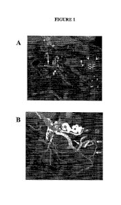
Figure 1 shows a multidetector computed tomography (MDCT) of the abdomen
performed
on a patient with severe abdominal pain, mulitplanar reconstructions were
obtained. Arteries and
veins are displayed. Figure 1A: The coronal plane shows the inferior
mesenteric vein with
contrast flow noted in the lateral branch, indicated by the vertical arrow,
but with no flow in the
main vein, as indicated by the horizontal veins. Note the edema and stranding
for the splenic
flexture, which is infracted because there is no arterial flow. Extensive
edema of the splenic
flexure region is also noted. Figure 1B: Combined Arterial and venous
enhancement shows
collateral veins draining the descending colon, but not the splenic flexure
(SF). Arterial flow is
maintained to the descending colon but there is no arterial supply to splenic
flexure.
CA 02866079 2014-08-29
WO 2013/130354
PCT/US2013/027373
Additional data on cancer perfusion obtained from MRI, MDCT and ultrasound
reveal that
the most consistently useful assessments of tumor vascular perfusion are
derived from the venous
and not the arterial system. The specific assessment techniques will only be
discussed only in
general terms, although all of the modern methods, such as DCE MRI, MDCT, and
ultrasound use
similar techniques. With each modality, baseline unenhanced images are
obtained and subsequent
repetitive images are obtained at varying time intervals during an intravenous
bolus injection of
appropriate contrast material (i.e. gadolinium, iodinated, or microbubbles)
The contrast-enhanced images can be analyzed visually or more vigorously by
graphing or
analyzed mathematically. Such contrast time curves are an essential component
of MR' vascular
imaging. The typical graph shows the density or intensity curve over the time
intervals, (Figure
2a). Depending upon the character of the arteries, veins, and arteriovenous
shunts the shape of
the curve varies (Figure 2b). With MRI, semiquantitative measurements are made
because
absolute values of intensity, density, or flow measurements are quite variable
due to the technical,
paramagnetic, physiologic and equipment factors. Mathematical calculation of
the permeability
expressed as Ktrans or Kep can be calculated Workman, P. et al. (2006) [16]
and Miller, J. et al.
(2005) [17], incorporated herein by reference.
Figure 2a. shows data obtained from a contrast enhanced study graph is used to
construct
an intensity/time or density/time curve. The diagram compares the contrast
time curve for the aorta
(A) and a typical density time curve over a mass. The AUC (area under the
curve) represents the
opacified blood as seen during the arterial and venous outflow phase. The
shape of the curve can
be visually analyzed as a "kinetic curve", as is commonly done with gadolinium
enhanced DCE
MRI mammography. Although inflow and outflow are related, the ouflow curve
mostly depends
upon the venous characteristics. The second image in Figure 2b shows, a
"spike" which requires
26
CA 02866079 2014-08-29
WO 2013/130354
PCT/US2013/027373
rapid inflow and rapid outflow. In either curve, the outflow down slope
depends on the venous
system.
Figure 2b shows a demonstration of the CBV calculation method by which
integrates from
the start to the end of the R2* (t) curve first-pass bolus, using the baseline
subtraction method from
T2/T2* - weighted leakage correction [18], Hu, L. S. et al. (2009)
incorporated herein by
reference.
Blood Volume
Blood volume is calculated using the area under the contrast curve over time
(AUC),
(Figure 2). This area represents the total blood volume including the arteries
and the veins,
although as can be seen, the greatest contributor to the total volume is the
venous volume. Duong
et al. [19], incorporated herein by reference, calculated that in the noimal
blood volume, the
venous space represents 70%, with the arteries contributing the rest.
Permeability values
Permeability represents the exchange of fluid or small particles in the
intravascular and
extravascular spaces. This exchange depends somewhat on the arterial inflow
and the venous
outflow characteristics but also on the nature of the exchange sites at the
capillary level. Dvorak
[20, 21], Nagy [22, 23], and Kohn [24] ,all incorporated herein by reference,
have shown that
permeability occurs in venules through fenestra as well as in the vesiculo-
vacuolar transport
organelles which traverse the venous wall. Dvorak [20, 21] and Kohn [24]
studied tracer
macromolecular transport across vessels. Nagy et al. [22, 23] studied vascular
permeability in an
27
CA 02866079 2014-08-29
WO 2013/130354
PCT/US2013/027373
adenovirus transfected VEGF model and determined permeability occurred in
veins not arteries
using electron microscopy, Evan's blue dye, and albumin dual radiotracers.
Permeability values can be calculated from both CT and MRI, but they are most
commonly
used in conjunction with gadolinium-enhanced DCE MRI (dynamic contrast-
enhanced magnetic
resonance imaging). According to Workman [16], these are "Kfrans (min-1), the
rate of flux of
contrast agent into the extracellular extravascular space within a given
volume, or volume transfer
constant); p.õ the volume of the extracellular extravascular space; and kep
(min-1), the rate constant
for the back flux from the extracellular extravascular space to the
vasculature. These parameters
are related to each other by the equation, /cep= Ktrans/ve." The mathematical
derivations of these
values are beyond the scope of this commentary, and the reader is referred to
several excellent
reports [16, 17] .
Kinetic curves
A subjective evaluation of the shape of the inflow and outflow portions of the
time contrast
curves has been found to be a useful interpretive tool for DCE MRI of the
breast. Many sources
especially Kuhl [25-27], all incorporated herein by reference, have used
analysis of these "kinetic"
curves for the diagnosis of breast cancer. However, attempts to apply these
curves to other organ
systems have been less successful.
Looking at the curve, Figure 2a and Figure 2b, it is apparent that the inflow
slope
represents the arterial inflow rate. The peak correlates with the maximal
enhancement and the
outflow portion reflects venous properties. In general, the inflow slope has
been considered to be
less useful in the analysis because it is too dependent upon technical factors
related to contrast
injection, e.g. rate, volume, etc. A very high peak is considered a spike if
it is 60% above the
28
CA 02866079 2014-08-29
WO 2013/130354
PCT/US2013/027373
baseline; although a spike is typically thought of as being characteristic of
arteries, it is apparent
that if there is not rapid outflow representing veins, it could not be a
spike.
Washout
"Wash out" of contrast material is a simple interpretive sign based on the
observation that
an enhancing focal mass quickly shows decreased enhancement and compared to
nolinal tissue
enhancement, it "washes out" earlier. This has been most commonly used with
hepatic masses,
during a bolus of contrast material on DCE MRI, MDCT or ultrasound imaging.
Perfusion parameters of different organ systems
The literature shows that while the above mentioned parameters depend upon the
venous
properties, their usefulness in the different organs varies greatly. For
example, permeability or
kinetic curve analysis are worthwhile in some organs but not others. The most
plausible
explanation is of course that the receptors, physiology, chemistry of the
organs differ greatly so the
individual characteristics dictate the vascular properties.
Brain
Using DCE MRI and bold imaging, numerous sources have reported that blood
volume
measurements can be used without factors to predict the degree of a parotid
[28] malignant brain
tumor differentiation [14, 15, 29-32]. Spampinato et al. [32], incorporated
herein by reference,
concluded that, "Relative cerebral blood volume measurement and MRS (MRI
spectroscopy) are
helpful in differentiating low-grade from anaplastic oligodendroglial tumors",
(Figure 3, Figure 4,
and Figure 5). Jain et al. [31], incorporated herein by reference, noted that
differentiating high
29
CA 02866079 2014-08-29
WO 2013/130354
PCT/US2013/027373
and low grade astroglial tumors was possible using the PS (peuneability
surface area) and CBV
(cerebral blood volume). Hu et al. [33], incorporated herein by reference,
reported that cerebral
blood volume measurements could differentiate high-grade glioma recurrence
from post-radiation
therapy changes.
Figure 3 shows a scatter plot of relative cerebral blood volume (rCBV) ratios
for each
tumor shows significant difference between the low-grade and high-grade
oligodendroglial tumors
(p < 0.05) [32].
Figure 4 shows a 44-year-old man with low-grade oligoastrocytoma. T2-weighted
image.
Figure 4b Relative cerebral blood volume map shows low tumoral vascularity
[32].
Figure 5a. shows a 64-year-old man with anaplastic oligodendroglioma. FLAIR
image
corresponding to A shows a right frontal cortex-based mass (arrow). Figure 5b.
Relative cerebral
blood volume map shows elevated tumor vascularization of tumor. [32].
Breast
To diagnose breast cancer using MRI, kinetic curves and permeability
measurements have
become widely accepted as useful diagnostic tools for both diagnosing and
characterizing breast
cancer. When the morphologic MRI appearance is not diagnostic, kinetic flow
curves from
gadolinium-enhanced dynamic contrast MRI have been proven quite useful for
differentiating
cancer from a benign lesion [25-27, 34-37], incorporated herein by reference.
Kinetc curves can
be interpreted by visual analysis; however, computer software programs
facilitate their use.
The appearance of the contrast time-flow curves has been well described by
Kuhl [25-27]
and others (Figure 6, Figure 7, Figure 8 and Figure 9) for benign and
malignant lesions.
According to Kuhl, cancer has two characteristic appearances, i.e. the rapid
contrast spike and the
appearance of the outflow curve.
CA 02866079 2014-08-29
WO 2013/130354
PCT/US2013/027373
The rapid enhancement spike is considered cancerous if the rapid early peak is
60% above
the baseline (Figure 2). Although there are only a few comments regarding the
outflow curve of a
spike, it is quite evident that the spike appearance depends upon rapid
outflow (due to veins) as
well as on rapid inflow.
When there is not a spike, correct diagnosis depends upon the shape of the
outflow curve,
which reflects venous drainage, (Figure 6, Figure 7, and Figure 8). Benign
lesions typically have
a kinetic curve which shows an increase or plateau flow, Ia and lb. Cancer
shows a decreasing
"washout "type II and type III, (Figure 6). The outflow characteristics are
determined by venous
flow, permeability, and arteriovenous shunting [25-27, 38]. The steeper the
outflow slope the more
likely it is that there is cancer. For the best results, careful attention
must be given to detail and the
appearance of the kinetic curve; Comprehensive discussion of the technique
should be reviewed in
the article by Kuhl et al. [25-27], (Figure 9).
Figure 6 shows a schematic drawing of the time-signal intensity curve types.
Type I
corresponds to a straight (Ia) or curved (Ib) line; enhancement continues over
the entire dynamic
study. Type II is a plateau curve with a sharp bend after the initial
upstroke. Type III is a washout
time course Kuhl et al., [25].
Figure 7a and Figure 7b show a region of interest (black oval on the left
image) and
corresponding time signal curve of an enhancing mass in the right breast, with
an irregular shape,
speculated borders, herterogeneous internal enhancement, and first initial
enhancement followed
by early washout. The mass was determined to be Bi-Rads category 5, as the
morphologic and
kinetic criteria were both highly suggestive of malignancy Kuhl et al., [26].
Figure 8 shows a Myxoid fibroadenoma. (Figure 8a) Region of interest (black
oval on the
left image) and (Figure 8b) the corresponding time-signal intensity curve. The
mass has a
lobulated shape, smooth borders, heterogeneous internal enhancement with dark
internal
31
CA 02866079 2014-08-29
WO 2013/130354
PCT/US2013/027373
septations, and fast initial enhancement followed by persistent enhancement.
The mass was
deteiiiiined to be BI-RADS category 2, as the morphologic and kinetic criteria
were concordantly
benign, Kuhl et al., [26].
Figure 9 shows a computer evaluation of kinetic curves is more consistent and
convenient.
This graph shows three curves measured at different sites in the same breast
cancer, and displaying
some variability but still showing the characteristic cancer signature of
rapid washout. Note that
the inflow curve is quite steep, and it is because of the shape of the outflow
that this is not a
"spike." Spike enhancement also depends upon the venous outflow.
Changes in kinetic curves are also useful for assessing treatment response, as
they show the
early changes in the washout curve (Figure 10). Kuhl et al. [25-27] stated:
"As the earliest sign of
response, a change of enhancement kinetics was observed (slower wash-in rate,
absence of a
washout pattern¨ie, flattening of the enhancement curve), which preceded a
change in tumor
morphology by several weeks."
Penneability measurements have proven quite useful for the diagnosis and
therapeutic
follow-up of breast cancer. Radjenovic et al. [39], incorporated herein by
reference, found, that
"Parameters kep and Kt' were significantly higher in Grade 3 tumours than in
low-grade
tumours."
When an untreated tumor shows increased permeability, anti-VEGF drugs change
the
permeability and kinetic curve [40-42], all incorporated herein by reference.
Raatschen et al. [40]
concluded that, "The MR imaging¨assayed acute change in vascular leakiness
after a single dose
of bevacizumab was an early, measurable predictive biomarker of tumor
angiogenesis treatment
response", (Figure 11). Thukral et al. [42] reported that with effective
treatment with
bevacizumab, the permeability Ktrans and blood volume changes were
statistically significant,
(Figure 11). Basic science reports by Jain [14, 15] and Boucher [43, 44] ,
incorporated herein by
32
CA 02866079 2014-08-29
WO 2013/130354
PCT/US2013/027373
reference, have confirmed that the increased permeability and interstitial
edema are reduced by the
effects of anti-VEGF.
Important to the ALPHA thesis is that the VEGFR receptor sites are producing
peimeability on the peripheral veins at the margin of tumors [20-25]. The
location of the action
sites of VEGF and anti-VEGF drugs on veins explains the increased incidence of
the venous
thromboembolism reported by Nalluri [11] and using anti-VEGF drugs.
Figure 10 show a change in serial transverse GKM Kfrans parametric maps
(calculated from
the transverse T1 -weighted spoiled gradient-echo sequence {8/4.2, 25 flip
angle, 4-5-mm section
thickness}) (images at the top) and in the gadolinium (Gd) concentration-time
curves (graphs at
the bottom) for one patient from baseline to cycle 7 (C7). Tumor enhancement
in the involved
breast can be seen in the following colors: Red and green indicate high
enhancement, and blue
indicates low enhancement. Gadolinium concentration-time curves show the
rate of
gadolinium-based contrast material perfusion throughout the tumor. The blue
line represents
arterial input function (Alfn). ROI data, CI= cycle 1, C4 = cycle 4, LMB =
left mouse button , RMB
= right mouse button Thurkal et al. [42].
Figure 11 show the graphs illustrating the absolute decreases in Kfralls from
the baseline to
cycles 1 and 4. Two-sided P values were calculated with the Wilcoxon signed
rank test (P=.003
for the difference in Kfrans between cycle 1 and the baseline, P<.001 for
difference between cycle 4
and baseline). The horizontal line inside each box represents the median
quartile, the horizontal
line below the box is the lower quartile, and the line above the box is the
upper quartile. The
vertical lines connect the quartiles, Thurkal et al., [42].
Prostate
33
CA 02866079 2014-08-29
WO 2013/130354
PCT/US2013/027373
Early reports on the usefulness of MRI of the prostate, were less than
enthusiastic [45, 46],
incorporated herein by reference, although there have been subsequent reports
of considerable
success in both the localization and differentiation of normal from cancerous
tissues [47-51],
incorporated herein by reference. Blood volume and kinetic curves [52],
incorporated herein by
reference, have not been consistently helpful, although permeability
characteristics are quite
useful. Jackson et al. [47] indicated that "quantitative parameter maps showed
a significant
difference between the benign peripheral zone and tumour for the parameters
Kt, ve and kep."
Liver
Washout or rapid clearance of intravenous contrast material after the peak
enhancement
has proven to be a reliable indicator of malignancy. This interpretative sign
has been used with
ultrasound, CT, and MRI and depends upon the rapid clearance of contrast
material through
tumors as compared to through normal liver.
With microbubble-enhanced ultrasound, sources [53-57], incorporated herein by
reference,
reported that HCC could be characterized by delayed washout after early
enhancement. Jang et al.
[53, 54] used ultrasound with microbubble-contrast material to study 97
hepatocellular cancers.
Jang et al. [53, 54] reported that 43% showed washout by 90 seconds, 26%
washed out at between
91-180 seconds, and 22% washed out in 181-300 second period. Only 8% of
cancers showed no
washout and they were well differentiated HCC's.
Sources [58-60], incorporated herein by reference, reporting on gadolinium-
enhanced MRI
indicated that washout could distinguish benign and malignant lesions (Figure
12). After studying
70 nodules, Ito, K. et al. (2004) [61], incorporated herein by reference,
stated, "Rapid central
washout after the early enhancement of the lesion and coronal enhancement
surrounding the lesion
are highly specific and diagnostic findings of small hypervascular
hepatocellular carcinomas."
34
CA 02866079 2014-08-29
WO 2013/130354
PCT/US2013/027373
Figure 12 shows a gadolinium-enhanced MRI of liver metastases showing washout
characteristic of malignancy. Figure 12A. shows multiple subtle small masses
(arrow) before
enhancement. Figure 12B. During gadolinium administration, these lesions
showed increased
enhancement. Figure 12C. The lesions showed contrast washout at 70 seconds
after contrast
injection.
Multiple sources [62, 63] using MDCT reported the value of the washout sign.
Lee et al.
[63] reported, "Both subjective and objective washout correlated with an
elevated
alpha-fetoprotein level (p = 0.01).
Re-examination of seminal Gimbrone/Folkman Vasculogenesis Report
Finally, retrospective review and reinterpretation of the original
vasculogenesis report by
Gimbrone and Folkman [4] reveals inconsistencies (Figure 4). Case Western
Reserve
Engineering school scientists, Dean and Professor Norman Tien and Professor
Vera Chankong,
re-analyzed all of Gimbrone's published 10 experiment data set, relative to
the single
"representative" graph from one animal. Tien and Chankong concluded with 95%
certainty that
the initial rapid tumor growth preceded arterial flow by at least one day.
Figure 13A shows "The characteristic growth curve of an iris implant (BP
No.29R) plotted
on a semi-logarithmic scale. Positive fluorescein test on day 6 represents
earliest evidence of
perfusion of the tumor and coincides with the beginning of exponential volume
increase. Slopes
"a," "b,", and "c," corresponding to prevascular, vascular, and late phases of
growth, are
indicated." Note the arrow indicating the arterial flow occurs after the rapid
growth is initiated.,
Journal of Experimental Medicine, 1972; 136, p.261-76 [4]. As discussed,
statistical analysis of
ten data sets, published but not used in this single graph reveals initiation
of rapid tumor growth
CA 02866079 2014-08-29
WO 2013/130354
PCT/US2013/027373
preceded arterial flow by at least one day. Therefore, the cause of the
interruption of dormancy and
growth cannot simply be elimination of hypoxia by arterialization.
Figure 13B. Diagram shows an overlay over the original Gimbrone diagram
illustrating the
ALPHA concept. The contention being described herein is that the dormancy can
only be
explained by high lactate levels which may exist with or independent of
hypoxia via aerobic
glycolysis (glycolysis occurs in inflammatory, immune, or cancer cells even in
normoxia). When
high lactate levels produce dormancy reduction to moderate levels by
lymphatics and veins
interrupt tumor dormancy. As will be noted later, lymphatics and veins develop
before arteries
[20].
III. CANCER METABOLISM: ENERGY PRODUCTION, WASTE MANAGEMENT,
GLUCOSE AND OXYGEN AVAILABILITY, DISADVANTAGES AND ADVANTAGES
OF AEROBIC AND GLYCOLYTIC METABOLISM
Cancer consumes glucose by aerobic and/or glycolysis (anaerobic) processes [64-
67],
incorporated herein by reference. Aerobic metabolism using glucose and oxygen
occurs in
mitochondria while glycolysis using only glucose without oxygen occurs in the
cytoplasm.
Warburg [66], Pederson [67] and others have reported that glycolysis is the
preferred
metabolic path for cancer. Pederson [67] noted that even in normoxia 50% of
cancer metabolism
can be from glycolysis and is even greater in hypoxia.
Benefits of glycolysis and lactate are numerous. Glyeolysis stimulates the
production of
critical substrates for cell proliferation (such as pentose from PPP pathway,
acetyl-CoA, NADH).
Acidic lactate 1) creates a selective adaptive environment which kills normal
cells and selects
cancer clones with specialized waste enzymes (carbonic anhydrase IX, CAIX),
monocarboxylic
36
CA 02866079 2014-08-29
WO 2013/130354
PCT/US2013/027373
transporter 4, MCT4), 2) induces hyaluronan which a) activates motogenic genes
by cell
membrane attachment and TGFb, b) hyaluronan stabilizes the mitotic spindle so
aberrant clones
can replicate, c) hyaluronan gradient aligns lymphatic endothelial cells via
LYVE-1, a hyaluronan
receptor, 3) induces TGFb which activates metalloproteases (Bauman 2009)[68],
4)Combination
of MCT4, hyaluronan dependent molecule CD147 (Tang 2004 [69], Le Floch 2011
[70]) activates
anti-apoptotic pathways including NFkB (Brown 2008 [71]), 5) acidic lactate
releases VEGF and
FGF from heparin sulfate and induces NFkB vasculogenic cytokines. (Brown 2008
[71]) 6) lactate
induced specific cytokines 17/23 induce VEGF (Shime 2008 [72]) 7)induce COX2
stimulating
VEGFC/D and cancer supporting prostaglandin E2.
Energy and Waste Production
Metabolism of glucose in normal and cancerous tissues occurs by two pathways,
glycolysis
and aerobic metabolism. Glycolysis creates ATP energy and pyruvate from
glucose without
oxygen. In the normal state, pyruvate moves into mitochondria to be processed
with oxygen
through the Krebs cycle. In the cancerous state, most pyruvate does not enter
the Krebs cycle but
is predominantly changed into lactate. In the cancerous state, the lactate
feed back controls are
altered and all pyruvate is completely converted into lactate by the cancer
enzyme lactate
dehydrogenases A [63]. The excessive lactate is detrimental to normal cells
but cancer cells are
unaffected because of specialized enzymes which protect the chemical balance
of cancer cells.
Advocates of the current angiogenesis theory infer that the aerobic pathway
should be
preferred because of the efficient use of glucose, but Warburg [66] and others
have confirmed that
glycolysis is the preferred metabolic pathway for cancer. While glycolysis is
chemically
inefficient, it suits cancer well because its reaction speed is 100 times
faster than aerobic processes
37
CA 02866079 2014-08-29
WO 2013/130354
PCT/US2013/027373
and can generate ample energy. Very high lactate levels may cause cancer cell
dormancy, but
moderately high levels provide many benefits as noted below.
Taking a different perspective in examining the energy and waste production by
the two
metabolic pathways, a different teleologic rationale for angiogenesis related
to glycolysis can be
proposed.
With aerobic metabolism, one molecule of glucose and one molecule of oxygen
produce
38 ATP's and CO2 molecules. Glycolysis uses one molecule of glucose and no
oxygen to make 2
ATP's and 2 lactates. Assuming a cell needs 38 ATP molecules to sustain life,
a cancer cell
would require 19 molecules of glucose and no oxygen to produce 38 ATP 's which
would also
create 38 lactate molecules.
From this perspective and data, it is difficult to accept the current
teleologic rationale that
cancer angiogenesis is intended to grow arteries for improved oxygenation,.
The more logical
teleologic rationale would be that if cancer has adequate glucose supply for
glycolysis, its earliest
immediate vascular need is to grow drainage vessels to remove the waste
products rather than
arteries to improve oxygen delivery.
Cancer's increased requirement for glucose is clinically confirmed by the
characteristic
images produced by FDG (fluorodeoxyglucose) PET (positron emission tomography
imaging.
The rapid turnover of glucose appears as an increased signal indicating
hypermetabolism, (Figure
14). Figure 14 shows a FDG PET scan of metastatic colon cancer in the liver.
The lesions (arrow)
show increased signal because more glucose must be processed for the same
amount of energy,
(see text).
Experimental and clinical reports confirm when both aerobic and aerobic
metabolism are
used by cancer, the overall metabolism is more dependent upon glucose than
oxygen supply.
Eskey et al. [73], incorporated herein by reference, elegantly confirmed the
importance of glucose
38
CA 02866079 2014-08-29
WO 2013/130354
PCT/US2013/027373
over that of oxygen for cancer metabolism. They used an animal model with an
exteriorized
tumor and separately varied the inflow of oxygen and glucose while they
measure the effects on
energy production. Energy production was directly related to glucose supply
but not oxygen
supply. Lui and Matsui [74], incorporated herein by reference, reported an
interesting model
which can be used to speculate further about glucose supply over arterial
oxygen supply. In a
mouse model, tumor cells were injected into an exteriorized live and observed
with
videomicroscopy. The first vessels to develop in the tumors was the portal
vein, i.e. high glucose,
followed later by the arterial system [74].
Clinical reports [75-78], incorporated herein by reference, confirmed the lack
of
correlation between oxygenated blood flow and energy in numerous PET studies.
Vaupel et al.
[79], incorporated herein by reference, reported lactate production is
directly correlated with
glucose uptake, as 40-85% may be released as lactate.
Adequate glucose supply is provided without normal arterialization by
effective diffusion
and active glucose transporters (GLUTS)
Certainly at the organ level, arteries are necessary to supply the inflow of
glucose, but the
movement of glucose across tissues and cells is quite efficient, without
flotilla' arterial supply. The
effect of the increased distance between tumor and arteries on the supply of
glucose and oxygen
has been discussed by Gilles [80] and Gatenby [81], incorporated herein by
reference. Gilles et al.
[80] reported (Figure 14), that when the distance from arteries to tumors is
more than 100-150
microns, the oxygen supply is restricted because of its poor diffusion. This
hypoxia prevents
aerobic metabolism.
Conversely, under the same circumstances the glucose supply is unaffected
because of its
favorable diffusion properties and active transport by glucose transporters
(GLUTS). These
39
CA 02866079 2014-08-29
WO 2013/130354
PCT/US2013/027373
up-regulated transporters are part of the metabolic adaptation (as well as
waste enzymes)
supported by HIFla to initiate and support metabolic adaptation to glycolysis.
Figure 15 shows substrate and metabolic profiles found in premalignant
intraductal tumor
using reaction-diffusion modeling. Oxygen concentrations (solid line), glucose
concentrations
(dashed line), and H+ concentrations (dotted line) are shown, from Gillies and
Gatenby, J
Bioenerg Biomembr (2007) 39:251-257 [80],
Depending upon concentration: moderate lactate elevations enhance metastatic
properties
and high levels induce dormancy
Although not generally recognized, molecular or drug effects can vary
according to the
concentration. An excellent clinical example is Dopamine, which affects blood
pressure
differently with different concentrations. Likewise, moderate or high
elevations of lactate affect
cancer cells differently. At moderately high levels, the neoplastic properties
are enhanced and at
very high levels, dormancy is induced. Whether dormancy is a positive or
negative process
depends upon the circumstances for the cancer cells.
Moderate lactate elevation/low pH provides advantages for cancer
If lactate is maintained at moderately elevated levels, there are advantages
for cancer
survival, proliferation and metastases. Moderate lactate levels create a
locally hostile
environment with low pH, toxic to normal cells to which. cancer cells can
adapt by genetic
mutation and survive [82], incorporated herein by reference. To ensure such
adaptation, HIF has
been recognized as critical for several dozen target genes that are
transactivated by HIF-1 have
been identified, including those encoding erythropoietin, glucose
transporters, glycolytic
enzymes, and vascular endothelial growth factor. The products of these genes
either increase 02
CA 02866079 2014-08-29
WO 2013/130354
PCT/US2013/027373
delivery or allow metabolic adaptation to reduced 02 availability [83],
incorporated herein by
reference. While hypoxia is the best recognized inducing agent for HIF, other
factors such as
lactate [84, 85], incorporated herein by reference, and metabolic
intermediates could increase HIF
levels (see angiogenic mediators in Figure 16).
Figure 16 shows a map of peritumoral H+ flow using vectors generated from the
pH,
distribution around PC3N/Efgp. The tumor is the darker region (left) and the
tumor-host interface
is drawn based on the GFP image. Arrows indicate the direction of II+ (the
steeper the gradient,
the longer the arrow). Note the general flow of fl+ from the tumor core to its
periphery, and from
there, into the nonnal tissue, although there is significant heterogeneity,
Gatenby, Cancer
Research, 2006, vol. 66, May 15, 2006 [81].
Such clones possess specialized waste enzymes, such as carbonic anhydrase IX
and lactate
transporters increase the acidic environment, activating enzymes which enhance
local invasion,
Figure 16. Unusual clones which might not be capable of normal mitotic
division can successfully
reproduce because the mitotic spindle is stabilized by the molecule hyaluronan
which is induced
by lactate [86], incorporated herein by reference, (Figure 16).
Metastatic potential is enhanced by lactate because of numerous effects.
Cancer cells and
endothelial cells become capable of "locomotion" when the hyaluronan molecule
(induced by
lactate) attaches to the RHAMM receptor of the cell wall. This action signals
the cytoskeleton to
transfolin and produce the contractile protein actin [86] (Figure 16).
Lymphatic channels form
from lymphatic endothelial cells with the unique biomarker hyaluronan receptor
1 (LYVE-1)
Lymph cells under the influence of VEGF3, align according to the hyaluronan
gradient (see later
transformation section). Hamilton et al. [87], incorporated herein by
reference, reported that
hyaluronan sustained high basal motility in breast cancer. The large amounts
of lactate produced
41
CA 02866079 2014-08-29
WO 2013/130354
PCT/US2013/027373
increased interstitial fluid pressure which creates convective current toward
lymphatic channels
[88], incorporated herein by reference.
Figure 17 shows hyaluronan attaches to the cell membrane receptor, RHAMM, thus
permitting transcription of motogenic genes. Hyaluronan stabilizes the mitotic
spindle, thus
permitting more effective cell division and mitosis. Extracellular and
intracellular functions of
RHAMM are: (Figure 17A) Cell-surface RHAMM promotes the activation of
signaling cascades.
Shown is one molecular mechanism for this. Cell-surface RHAMM, which is not an
integral
membrane protein, partners with CD44 and, in the presence of hyaluronan,
activates ERK1/2
(indicated as phosphorylated (PO4) ERK1,2), which results in the expression of
genes that are
required for motility and invasion. (Figure 17B) In X, laevis egg extracts, a
RAN-GTP gradient,
which is established by chromosome-bound guanine nucleotide-exchange factor
RCC1 activity, is
required for anastral mitotic-spindle assembly. RAN-GTP activity regulates the
function of a
number of mitotic-spindle proteins, including importins that then form
inhibitory complexes with
both spindle-assembly factors and TPX2. For example, by binding importins
(indicated as step 1),
RAN-GTP releases TPX2 (step 2), which is a major activator of Aurora kinase A
(AURKA).
TPX2 directly activates AURKA by protecting an autophosphorylated residue
(step 3). AURKA,
in turn, can phosphorylate (PO4) BRCA1 to facilitate G2-M transition (step 4).
Via an interaction
with the dynein complex, RHAMM localizes to the spindle pole, at which it
interacts with 7-tubulin
(step 5). RHAMM also interacts with TPX2 and dynein, thereby having the
potential to localize
TPX2 to spindle poles (step 5). The BRCA1 -BARD1 complex modifies TPX2
localization and
spindle assembly by attenuating RHAMM function through ubiquitylation (Ub)
(step 6).
Ubiquitylation of RHAMM, and subsequently its degradation, probably releases
TPX2 from the
spindle pole (step 7), thus affecting AURKA activation and G2-M progression.
42
CA 02866079 2014-08-29
WO 2013/130354
PCT/US2013/027373
Tumor Dormancy
Dormancy of cancer cells is an inactive state from which tumor cells must
emerge to grow,
proliferate, and metastasize. Because interruption of dormancy has been the
benchmark to judge
effective vasculogenesis, a better understanding of its causative mechanisms
is important. Because
the traditional angiogenesis theory is derived from Gimbrone et al. inferring
hypoxia causes
doiniancy and restoration of oxygen interrupts dormancy, it is interesting to
note their direct
statement [4]. Gimbrone et al. stated," "The mechanism of this population
dormancy is not
elucidated by these experiments." [4].
Surprisingly, direct data about hypoxia adversely reducing cancer cell
activity leading to
dormancy is not only lacking but there is abundant contrary information.
Voluminous data
indicates that hypoxia enhances the invasive metastatic process which is the
essence of
malignancy it does not retard such processes. As has been discussed, lactate
that is produced by
hypoxia produces lactate that enhances motility, mitosis, and local invasion
as noted.
However, increased lactate levels, which is produced by cancer in either
aerobic or hypoxic
environment, has been reported to produce effects that would support dormancy,
i.e. slowed
metabolism, decreased proliferation, and anti-apoptotic effects. These data
come from basis
chemistry, simple cell culture experiments, clinical studies, and some
specific signaling pathways.
If one considers the basic mass action dynamics of chemistry, the excess
accumulation of
an end product will decrease the forward reaction by mass action effect in
reverse. Hence because
cancer uses glycolysis, accumulation of lactate would decrease metabolic rate
[89-93],
incorporated herein by reference. Excess lactate impairs protein synthesis,
growth and antibody
production [88]. It also reduces cancer cell proliferation [90, 93].
Until this time, lactate was not evaluated as a possible cause of donnancy.
Basic research
has been down relative to the signaling pathways but there are a number which
support this
43
CA 02866079 2014-08-29
WO 2013/130354
PCT/US2013/027373
premise because lactate and glycolysis has been shown to prevent cell death by
anti-apoptotic
pathways. One of the major death pathways, FAS was reported by Erkilla [94],
incorporated
herein by reference, to be suppressed in germ cells by lactate. Erkilla [94]
stated, "The final site
of the death suppressing action of lactate appeared to take place in germ
cells downstream of the
FAS receptor activation." Thangarju et al. [95], incorporated herein by
reference, studied the
effect of lactate on the SLC5A8 trigger pathway for tumor cell apoptosis.
Because this pathway
depends upon pyruvate, they stated, "Tumor cells silence SLC5A8 and convert
pyruvate into
lactate as complementary mechanisms to avoid pyruvate induced cell death. The
important
inflammatory pathway NFkB is known to induce anti-apoptotic genes Bc1-3, IAP-
1, and IAP-2
[71], incorporated herein by reference. Samuvel [96], incorporated herein by
reference, reported
that lactate boosts TLR4 signaling and NFkB pathway mediated gene
transcription in
macrophages. Also COX2 is up-regulated by NFkB and COX2 inhibits DNA damage
induced
apoptosis by p53 [97], incorporated herein by reference.
To understand causes of dormancy, changes in cell cycle control would need to
be
elucidated according to Blackstone [98], incorporated herein by reference.
Rutz [99],
incorporated herein by reference, noted "lactate interferes with mechanisms of
cell-cycle control at
two different points in the cell-cycle, depending on cell density and the
resulting absence or
presence of inhibition of cell proliferation. Interference with cell-cycle
control may underlie the
modification by exogenous lactate of radiosensitivity and postirradiation
repair capacity in
mammalian cells." Also several other papers suggest there might be a
relationship between
lactate and the chief regulator of cell cycle, pRb (protein retinoblastoma).
Lactate induces and
modulates both TNF (and TGFb, which have their own interaction) [100-102],
incorporated herein
by reference, which TGFb interacts with pRb [97]. If ALPHA is more completely
investigated,
44
CA 02866079 2014-08-29
WO 2013/130354
PCT/US2013/027373
perhaps other pathways related to cell cycle arrest will be elucidated.
Glycolytic enzymes related
to lactate and the Akt pathway is also known to inhibit apoptosis [98, 103].
There has been extensive study of tumor recurrence and its dependency on
dormancy [71]
but in clinically oriented reports there is an obvious lack of discussion
about hypoxia. Blackstone
et al. [98] extensively discussed tumor dormancy/recurrence and emphasized the
importance of
cell cycle pathways (no mention of hypoxia even after 40 years of study).
Other clinical reports are more consistent with lactate induced dormancy as it
relates to
cancer recurrence and treatments. Recognizing that dormant cells do not
respond well to
treatment, several sources have noted that treatment resistance may be
associated with elevated
lactate and that restoration of normoxia does not increase recurrence. Quennet
et al. [104] and
Sattler [105], both incorporated herein by reference, noted correlation
between radioresistance and
glycolysis and acidic lactate concentration. Feldmeyer [106] and Schonmeier
[107], both
incorporated herein by reference,dispelled the concern that tumor cells would
be activated by
restoration of nomioxia by hyperbaric oxygen. Both sources found no increased
local tumor
recurrence as would be expected from the currently accepted concept that
hypoxia causes tumor
dormancy.
Lactate level of interstitial fluid modulated predominantly by lymphovenous
drainage and
partially by aerobic metabolism of stromal or cancer cells
After recognizing the cancerous processes controlled by elevated glycolytic
lactate, in
hypoxia (anaerobic) or normoxia (aerobic), the importance of maintaining
appropriate levels can
be appreciated. Increased glycolytic activity produces excessive lactate in
the extracellular space
producing increased interstitial pressure [108], incorporated herein by
reference. This occurs
because the 6 carbon glucose being split into two carbon lactate doubles the
oncotic pressure. With
CA 02866079 2014-08-29
WO 2013/130354
PCT/US2013/027373
convectional movement of free water [79] into the site hydrostatic pressure is
produced
stimulating flow into the lymphatics. Lactate levels depend predominantly upon
the removal of
lactate by the lymphatic and venous system.
Earlier studies by Gullino [88] on lactate reported that tumor interstitial
fluid always had
higher lactate than that of the inflow with the concentration being 25-100%
higher. From baseline
levels, the tumor levels increased until about 10 days when the levels were
stabilized and
maintained. Lymphatic drainage consistently measured 2-4 times that of tumor
interstitial fluid.
Studying a de novo squamous cell cancer model in mice, Eitchen et al. [109]
verified that
interstitial fluid was maintained by lymphatic flow. In the transition from
normal to premalignant
state, the host lymphatics dilated and increased in size due to the effects of
VEGF-C. With the
development of squamous cancer, the fluid increased and neolymphangiogenesis
occurred, (see
later Lymphangiogenesis section). Such lymphatics induced by VEGF-C are known
to cause early
local metastases [110, 111], both incorporated herein by reference.
The only incidental advantage of aerobic metabolism for cancer cells is
indirectly related to
lactate reduction by aerobically competent cells. When cancer cells retain
mitochondria and
oxygen is available, lactate is consumed, reducing local levels [112],
incorporated herein by
reference. Similarly, Kourakis [113], incorporated herein by reference,
emphasized that adjacent
stromal cells could reduce local lactate and pH levels, by metabolizing
lactate.
Figure 18 show a CT scan showing a mass in the medial side of the breast,
horizontal arrow
as well as early metastases to small axillary node, vertical arrow.
Hyaluronan, which is produced
by breast cancer, and fibroblasts stimulate cancer cell migration, and enhance
mitotic activity and
lymphatic development. Hamilton et al. [87] reported hyaluronan maintained
breast cancer cell
motility.
46
CA 02866079 2014-08-29
WO 2013/130354
PCT/US2013/027373
Lactate and low pH induce vascular growth factors which induce
lymphangiogenesis,
venogenesis, and arteriogenesis
As will be discussed in subsequent sections, virtually all of the angiogenic
growth factors
are released and/or produced by the effects of lactate and low pH. The
processes include both the
release of dormant growth factor in the microenvironment and production of new
factors made by
numerous cells. The stimulus for the production is the transformation of the
microenvironment
through numerous pathways most importantly through the TGF (transforming
growth factor) by
means of NFId3 (nuclear factor kappa beta) pathway. The transformation
includes endothelial
cells, cancer cells, stromal fibroblasts and many immune cells.
There are no real advantages of aerobic metabolism for cancer
The only incidental advantage of aerobic metabolism for cancer cells is that
local lactate
levels are reduced when adjacent aerobic cancer or stromal cells metabolize
lactate [112-114] ,
incorporated herein by reference. As discussed earlier, some sources refer to
the chemical
efficacy of aerobic metabolism but the fast reaction speed of glycolysis more
than compensates to
produce ample energy.
IV. SIGNALING PATHWAYS, VASCULAR GROWTH MEDIATORS, ACTION SITE OF
MEDIATORS FOR LYMPHATICS, VEINS, AND ARTERIES
Signaling pathways for angiogenic growth mediators
One of the most prevalent misconceptions supporting the current angiogenesis
concept is
that hypoxia is the sole mediator for VEGF and other vascular mediators. As
will be discussed
47
CA 02866079 2014-08-29
WO 2013/130354
PCT/US2013/027373
later, angiogenic growth mediators are induced by hypoxia but also many other
pathways
independent of the oxygenation level, i.e. normoxia, and hyperbaric oxygen.
Among these other
pathways lactate and low pH have been extensively discuss in the literature,
especially in the early
stages of angiogenesis and in wound healing.
Angiogenesis growth mediators are induced in hypoxia, normoxia, and hyperbaric
oxygen
A most intriguing study by Heinzman et al. [115], incorporated herein by
reference,
demonstrated that the production of many angiogenic growth factors is
essentially equivalent in
both hypoxia and normoxia. Cancer cells in hypoxia or normoxia produced almost
equivalent
amounts of angiogenic growth factors. Heinzman et al. [115] quantitated 11
angiogenic growth
factors (VEGF,PDGF-AA, PDGF-AA/BB, IL-8, bFGF, EGF, IP-10, Flt-3 ligand, TGF-b
1 ,
TGF-b2, and TGF-B3) produced by different cancer cell lines in hypoxia and/or
normoxia.
Comparing the angiogenic products, they showed no or only a moderate increase
of VEGF and no
significant increase in bFGF in hypoxia. Of the other products, only IL-8 was
generally higher and
the levels in 8 of 11 mediators were closely correlated.
Hypoxic expression levels were generally higher than normoxic for IL-8 (r2>
and VEGF
(r2> 0.60), although only modestly. Heinzman [115] noted, "The degree of
difference was
surprising, as both IL-8 and VEGF have been reported to be up-regulated in
response to hypoxic
conditions." "It is remarkable to note that hypoxia did not increase bFGF
compared to normoxia.
Another in vitro study showed that bFGF was unaffected by hypoxia in cell
lines" [116],
incorporated herein by reference.
Reports regarding angiogenesis in hyperbaric oxygenation provide unique
evidence
indicating that factors other than oxygen levels are responsible for
angiogenesis. In his study of
squamous cell cancer, Schonmeyr et al. [107] observed that hyperbaric oxygen
eliminated hypoxia
48
CA 02866079 2014-08-29
WO 2013/130354
PCT/US2013/027373
and restored nounoxia in squamous cell tumors. They unexpectedly found that
with the restoration
of tumor hypoxia to normoxia the amount of VEGF and vessel growth did not
change compared to
the preceding hypoxic state.
Signaling pathways for angiogenesis include HIF (hypoxia induction factor),
Acidic Lactate
and other pathways
HIF-la (Hypoxia Induction Factor) is the most important regulator for VEGF and
FGF and
well as most of the other enzymes which support glycolysis and the neoplastic
processes.
Although infrequently discussed, other processes affect HIF concentration. Of
special note is that
the increases of HIF can also be caused by non hypoxic inteimediary metabolic
imbalances [37,
72, 117-119], incorporated herein by reference. In addition to up-regulating
angiogenesis, HIF is
absolutely critical for adapting cellular metabolic processes to glycolysis
associated with hypoxia
or aerobic glycolysis [120]. In addition to up-regulating angiogenesis, HIF is
absolutely critical
for adapting cellular metabolic processes to glycolysis which is essential in
hypoxia and also
occurs with cancer it normoxia [114]. Interacting with cMyc, these include
processes for
substrate transport, expedited PPP pathways, rapid conversion of pyruvate to
lactate and waste
product management to maintain suitable cellular pH. These include enzymes
involved with PPP
cycle such as ketolases, glycolysis such LDHA, glucose transporters (GLUT I,
II, IV), and waste
enzymes carbonic anhydrases IX and XII, and lactate transporters MCT1,4 [83,
84].
The best described and recognized mechanism for controlling HIF concentrations
is
regulated by the degradation enzyme PhD (prolyl dehydrogenase). The enzyme
increases in
noinial oxygen reducing levels and decreases in hypoxia to increase the HIF
levels [83].
Less well known to most investigators is that HIF can be increased by non
hypoxic
intermediary metabolic imbalances [85, 121-123], incorporated herein by
reference, and even low
49
CA 02866079 2014-08-29
WO 2013/130354
PCT/US2013/027373
pH [124]. Lu et al. [122] stated, "with aerobic glycolysis (not aerobic
metabolism but glycolysis
in presence of oxygen), glucose metabolites can up-regulate HIF levels by
preventing its
degradation." McFate et al. [123] noted, "these data suggest that the buildup
of glycolytic
metabolites, resulting from high PDK-1 expression, may in turn promote HIF-1
activation, thus
sustaining a feed-forward loop for malignant progression. 'Furthermore,
Mekhail et al. [125],
incorporated herein by reference, reported that as a result of the low pH
induced predominantly by
lactate, that "a decrease in environmental pH triggers the relocation of VHL
(also degrades HIF),
neutralizing its ability to degrade nuclear HIF even in the presence of
oxygen."
Walenta [84] stated, "Demonstrating various biologic activities of lactate
that can enhance
the malignant behavior of cancer cells. These mechanisms include the
activation of hyaluronan
synthesis by tumor associated fibroblasts, up-regulation of VEGF and of HIF-
alpha, and direct
enhancement of cellular motility which generates favorable conditions for
metastases."
Signaling angiogenesis pathways other than hypoxia and waste products
Recognizing the essential role of vasculogenesis for the success of tumors, it
is no surprise
there are many redundant vasculogenic pathways. Many diverse induction factors
include
hypoglycemia [45, 126], genetic anomalies i.e. VHL, PTEN, p53, RAS and
oncogene [127-132],
incorporated herein by reference. A complete discussion of these many factors
is not possible nor
is it relevant to the purpose of proposing an alternate angiogenesis concept
to interrupt tumor
dormancy.
Lactate and low pH increase vascular growth mediators Independent of
oxygenation level
Although it is has not become widely known, there are many reports confirming
that both
lactate and low pH induce angiogenic growth factors. The origins of the acidic
lactate are both
CA 02866079 2014-08-29
WO 2013/130354
PCT/US2013/027373
macrophages and cancer cells which use glycolysis even when oxygen is present
(aerobic
glycolysis simply means oxygen is present with glycolysis). As noted above
elevated metabolites
especially lactate increases HIF as discussed above even in normoxia. When
hypoxia occurs, even
more acidic lactate is produced which likely acts in synergy with the HIF
degradation enzyme PhD
to elevated HIF further.
There are two mechanisms which increase local VEGF levels, the release of
dormantly
store VEGF and FGF [133-135], incorporated herein by reference, and the active
production. First
in early angiogenesis before hypoxia (see angiogenesis below) there is the
release of dormant FGF
and VEGF stored in the heparan sulfate matrix. In the later hypoxic phase the
up-regulation of
many other pathways occur from the effects of acidic lactate which is
amplified and synergized by
hypoxia.
It is odd that although many sources over many years have reported that low pH
and lactate
can increase vascular mediators, it has not become generally recognized [85,
100, 133-144],
incorporated herein by reference. To amplify these facts and to forestall any
concerns about
paraphrasing errors, direct "quotes" are provided. If this exercise is
tedious, the reader is invited
to move to the next section.
D'Arcangelo et al. [133] reported "Acidosis Inhibits Endothelial Cell
Apoptosis and
Function and Induces Basic Fibroblast Growth Factor and Vascular Endothelial
Growth Factor
Expression.
Hunt stated [85],"Lactate, on the other hand is also a known instigator of
cytokines and
growth factors such as VEGF, TGF-13, and IL-1. Lactate stabilizes HIF-la even
in the presence of
oxygen because lactate and pyruvate bind to and inhibit the HIF prolyl
hydroxylases that would
otherwise hydroxylate HIF-la and mark it for rapid degradation."
51
CA 02866079 2014-08-29
WO 2013/130354
PCT/US2013/027373
Fukumora et al. stated [134], "VEGF-promoter activity increased, with a
decrease in pH
and independent of p02." "VEGF transcription in brain tumors is regulated by
both tissue p02
and pH via distinct pathways."
Xu et al. noted (140) that "acidic extracellular pH induces VEGF ...via ERK1/2
MAPK
signaling pathway." Kato et al. [137] stated, "Acidic pHe has also been shown
to increase the
expression of platelet-derived endothelial cell growth factor/thymidine
phosphorylase, IL-8, and
VEGF in varies types of cells."
Beckert [140] noted that "Lactate induces VEGF synthesis in endothelial cells
and that this
results in enhanced endothelial cell migration even in the absence of
hypoxia." "Endothelial cells
showed increased migration only when lactate was added in combination with
endothelial cells" (it
is now known lactate activates motogenic genes [86].
The emerging important role of cancer related inflammation and NFkB is
enormous, which
prompted Colotta et al. to call it the "seventh halhnark of cancer." As will
be described below,
these processes are part of a multi step process, which includes preangiogenic
transformation of
the microenviromnent, early/incipient as well as delayed/maintenance
angiogenesis.
Samuvel et al. observed, "Lactate boosts TLR4 activation and NF- B-dependent
inflammatory gene expression via monocarboxylate transporters and MD-2 up-
regulation." NFkB
is the key orchestrator of innate immunity/inflammation and aberrant NFkB
regulation has been
observed in many cancers." Cytokines such as IL-1, IL-6, IL-8, and IL-23 are
pro angiogenesis.
IL-1 induces FGF2 [63] and VEGF. Mizukami et al. [117], stated "NFkB is
induced by hypoxia
specifically through accumulation of hydrogen peroxide when HIF-1 is blocked,
and this
compensatory pathway plays an important role to maintain angiogenesis in the
absence of HIF-1
52
CA 02866079 2014-08-29
WO 2013/130354
PCT/US2013/027373
by up-regulating IL-8. Shime et al. [143] reported that lactate through NFIdi
induced IL-17, IL-23
which are proangiogenic inflammatory cytokines.
V. MODERN IMMUNOHISTOCHEMICAL BIOMARKERS INDICATE THAT TARGET
RECEPTOR SITES OF VASCULAR GROWTH MEDIATORS
Prior to 2000, it was believed that the lymphatic, venous or arterial
character of vessels
depended upon the nature, pressure and direction of fluid/blood flow. The
development of specific
immunohistochemical biomarkers combined with embryologic studies permits
definition of the
vascular mediator target receptor sites. Furthermore, retrospective review of
earlier angiogenesis
reports indicates early reports mistakenly labeled some venous structures as
arterial. An excellent
review of vascular specification was reported by Swift [119] in the journal
Circulation Research
provides valuable insights for reinterpretation of other reports.
Embryologic origins of vessels
The origins of the vascular and lymphatic vessels have been phylogenetically
determined
from tissue dissections and the immunohistologic biomarkers. Both the
lymphatics and venous
system evolve from the cardinal veins [120, 145], incorporated herein by
reference. The arteries
evolve from the dorsal aortas [119].
Figure 19 shows arterial and venous EC have molecularly defined identities
that are
evident before circulatory flow or even tubulogenesis. Expression of artery
markers such as
ephrinB2a (Figure 19C) and vein markers such as flt4 (Figure 19D) is evident
by in situ
hybridization of 25 somite stage zebrafish embryos, several hours before
circulation begins in the
trunk. Expression of EphrinB2a within the dorsal aorta begins just as the
migratory EC's arrive at
the trunk midline from the lateral mesoderm and begin to aggregate into a cord
of cells. Figure 19B
53
CA 02866079 2014-08-29
WO 2013/130354
PCT/US2013/027373
shows Expression of the pan-endothelial marker flt 1 * is shown for the
comparison. Box in upper
diagram (Figure 19A) shows approximate location of in situ images, for
reference. Light arrows
indicate dorsal aorta; dark arrow, posterior cardinal vein. * VEGFR-1 is
receptor for VEGF-A,.
VEGFR-1 is significantly more enriched in veins during early embryologic
vessel formation, but
later is present in both arteries and veins (personal communication with Brant
Weinstein, Director
Molecular Genetics Laboratory. NIH). This is consistent with observations by
Dvorak et aL who
describe the first changes of transfected VEGF-A gene on blood vessels is on
the venules.
Specification of vascular identity by biomarkers
As a matter of record, specific markers for lymphatics, veins, and arteries
have been
defined and used in most of the studies referenced. The lymphatic marker is
LYVE-1 (lymphatic
endothelial cell hyaluronan receptor site-1). The venous markers are VEGFR1*
(flt1), TIE-2,
Ephrin 4, and COUP TF11. Both VEGFR1 and TIE-2 are very interesting in that
these have been
used by many sources and noted as being arterial markers [12, 146] when in
fact they are
associated with veins [147]. Moyon [147], incorporated herein by reference,
showed that after
seven days in the embryo, TIE-2 specifies veins. Earlier sources, such as
Holash [146] assumed
that TIE-2 receptor was the target site for angiopoietin was an arterial
marker but this is not
correct.
Also of note , according to Swift et al., the activation of PI3K/Akt pathway
as commonly
occurs in cancer, induces venous cell fate. PI3K promotes venous fate by
suppressing NP1 and
Notch gene activation [119, 148].
Using these biomarkers, numerous scientists have clarified the mechanistic
action of the
various growth mediators. Furthermore, because of the specificity of these
markers, the sequence
of vessel development can be accurately ascertained.
54
CA 02866079 2014-08-29
WO 2013/130354
PCT/US2013/027373
IV. Animal Models studying growth mediators VEGF, FGF, Ephrin, and
others support ALPHA sequence of vessel development
The vascular mediator receptor action sites and the observed sequential
developmental
changes in the vessels form an essential basis for ALPHA. As will be noted,
the sequence of
vascular changes occurs on the lymphatics, veins, and arteries.
VEGF A,-C,-D/ VPF
The VEGF (vascular endothelial growth factor) family is the most important
group of
mediators for vasculogenesis, and consists of VEGF-A, VEGF-C, and VEGF-D.
VEGF affecting angiogenesis originates from two processes. Firstly, release of
VEGF
from a dounant form in the heparan matrix occurs during the early/initial
phase of angiogenesis,
see transformation section below. The initial elevation is from the release
from the matrix by the
effects of lactate, low pH, and induced inflammation [135, 136, 149, 150],
incorporated herein by
reference. As will be noted later, this may occur in hypoxia or normoxia [144,
151]. Secondly,
VEGF is produced by tumor-associated cells occurs in hypoxia during the
delayed/maintenance
phase with other vascular growth mediators [71, 96, 144, 151].
In 1991, using immunohistochemical stains, Dvorak [20, 21] determined that the
morphogeneic changes caused by VEGF-A is on the host veins adjacent to the
tumor site. Dvorak
[20, 21] stated, "Immunoreactive vessels (to VPF/VEGF antibodies) were venules
and small
veins." Kohn et al. [24] reported that the permeability of vessels occurred in
the veins, and stated,
"All tracers leaked primarily from venules and small veins at the tumor-host
interface."
CA 02866079 2014-08-29
WO 2013/130354
PCT/US2013/027373
More recent sophisticated models using a transfected VEGF-A164 gene in a mouse
model
by Dvorak [20, 21] and Nagy [22, 23] studied the development of surrogate
tumor vessels over a
128-day period. They reported that vessel morphogeneis occurred sequentially
on lymphatics,
veins, and arteries (Figure 20, Figure 21, Figure 22, and Figure 23).
Regarding lymphatic changes, these sources observed that dilatation of host
lymphatics
occurs 1-3 days before blood vessel changes occur [22, 23, 109] (Figure 21).
Nagy [22, 23] stated
"Lymphatics at 3daysafter Ad-VEGF-A are distended from dermal edema but have
already
enlarged further as the result of endothelial cell division and are
transitioning into giant
lymphatics." Eitchen [109] using a de novo skin squamous cell carcinoma showed
lymphatics
proliferated and dilated before blood vessels. Hong et al. [152], incorporated
herein by reference,
reported that the action of VEGF-A promoted wound-associated lymphangiogenesis
by means of
VEGFR-2 and integrins.
Figure 20 shows ear lymphatics after intravital infusion of colloidal carbon
in a control
mouse and in mice injected at the indicated intervals with Ad-P1GF or Ad-VEGF-
A164. (Figure
20a) Control ear. Multiple injection sites (black blotches at top) were
required to fill the lymphatic
network. Pattern of lymphatic filling in the ears of mice previously injected,
as indicated, with
Ad-VEGF-Al 64. Giant lymphatics are apparent as early as 3 days following
injection (Figure 20e)
and persist through day 270. Kinetics of lymphatic filling in the ear of a
mouse 84 days following
injection with Ad-VEGF-A164. Nagy JA,Vasile E, Feng D, J Exp Med, vol. 196, No
11,
1497-1506, 2002 [23].
For lymphangiogenesis, VEGF ¨C and ¨D are specific and more effective than
VEGF-A
as they induce sprouting and lymphatic proliferation. Sato et al. [153],
incorporated herein by
reference, VEGF A attracted macrophages which in turn expressed VEGF-C and
VEGF-D which
induced new lymphatic formation. COX2 [154, 155], incorporated herein by
reference, is
56
CA 02866079 2014-08-29
WO 2013/130354
PCT/US2013/027373
up-regulated through the NFkB pathway induces VEGF-C production (Lactate
initiates the
induction of the NFkB inflammatory pathway [71, 100, 144, 151, 154, 155].)
Enholm et al. [156]
transfected the VEGF¨C gene into a model showing its specificity for
lymphangiogenesis.
Relative to the surrogate tumor blood vessels produced by VEGFA, Nagy [22, 23]
stated
"They arise from preexisting normal venules and are large, thin-walled,
serpentine, pericyte-poor
sinusoids that over express both of the VEGF-A receptor tyrosine kinases
(VEGFR-1, VEGFR-2).
MV (mother veins) then evolved into GMP and vascular malformations and also
into structurally
normal capillaries by a process of transcapillary bridging." Further Nagy
said, "MV formed
initially (1-5 days) and, from about 7 days, evolved into GMP (Glomeruloid
bodies), vascular
malformations, and capillaries." (Figure 21, Figure 22, and Figure 23). It is
interesting to note a
similar time frame of the vessel formation as the 6.5 days in the Gimbrone
experiment.
Figure 21 shows angiogenic response to Ad-VEGF-A164 in the ears of nude mice
at the
indicated times and magnifications, from Nagy, J. A. et al. (2002) Cold Spring
Harbor Symp
Quant Biol 2002, 67:227-237 [157]
Figure 22 show vessels in ear skin at 18 hours after local injection of adeno-
vpf/vegf.
Figure 22a. Normal sized venule with slightly detached pericyte (arrow). Note
the extensive
edema separating adjacent muscle fibers. Figure 22b. Evolving mother vessels
illustrating striking
vessel enlargement. EC (endothelial cell) activation (enlargement, prominent
nucleoli) and
pericytes (arrows) in various stages of detachment from vessels. Figure 22c.
Higher power
magnification captures three sections through mother vessels, thus
illustrating highly irregular
luminal surfaces and EC (endothelial cell) bridging to form additional lumens
(arrows) from
Dvorak et al., Laboratory Investigation) .
57
CA 02866079 2014-08-29
WO 2013/130354
PCT/US2013/027373
Figure 23 shows a schematic diagram of mother vessel formation and evolution
into
daughter capillaries, vascular malformations and glomeruloid bodies, modified
from Pettersson,
A. et al. (2000) Lab Invest, 80: 99-115 [158], incorporated herein by
reference.
Fibroblast Growth Factor (bFGF or FGF2)
The FGF (fibroblast growth factor) family has two molecules, FGF2 (FGFb) and
FGF1,
although FGF2 is the more important and is typically referred to as FGF. FGF
is produced by
endothelial, cancer, stromal and inflammatory cells. As with VEGF, the local
levels of FGF
increase by the same two mechanisms, i.e. release from the heparan sulfate
matrix due to the
effects of waste products and the secondary production in the
delayed/maintenance stage by a
variety of cells.
The primary action of FGF2 in the lowest concentration is the stimulation of
lymphangiogenesis, while the secondary action is induction of VEGF A, C, and D
via the promoter
element AP1. Chang [126] said, "Low-Dose FGF-2 Selectively Stimulates
Lymphangiogenesis", (Figure 24). "Although the effects of 12.5 ng FGF-2 pellet
are mediated
through different cytokines, i.e. VEGF-A, -C, and -D, the predominant result
is
lymphangiogenesis."
Figure 24 shows FGF-2 stimulates corneal lymphangiogenesis. Figure 24A. In the
traditional corneal assay, 80 ng of FGF2 (P) stimulates blood vessel growth
from the peripheral
limbal vaseulature (arrow). Figure 24B The traditional assay is viewed under
fluorescent
microscopy after labeling blood vessels yellow-green and lymphatic vessels
red, (arrow).
Sueralfate in the FGF2 pellet autofluoresces green. Figure 24C. At the
opposite end of the cornea
only lymphatic vessels sprout. Limbal vessels in the control corneas. Figure
24D. Lowering the
dose of the FGF2 pellet to 12.5 ng (P) and moving it farther from the limbus
results in less
58
CA 02866079 2014-08-29
WO 2013/130354
PCT/US2013/027373
angiogenesis, although lymphatic vessels still reach the pellet. Figure 24E.
Corneal lymphatic
vessels were morphologically different from blood vessels. In additional
corneal lymphatic vessels
did not express CD34 (Figure 24F) or Tie 2(Figure 24G, Figure 24H) (arrow
heads), but did
express VEGFR3, (I). from, Chang et al., Proceedings of the National Academy
of Science, 2004
[126].
Ephrin -2B, 4B
The ephrin family induced by hypoxia and HIF [159] affects the
neovascularization
processes [160, 161] after the early effects of VEGF. They are bidirectional
markers.
Ephrin-2B, an arterial marker, is the ligand for the downstream Ephrin-4B
(venous receptor). The
Ephrin-4b is enriched in veins as the principle functional partner for ephrin-
2B [152]. When
Ephrin-4B levels are high they reverse signal the Ephrin-2B to decrease
arterial induction [161].
Hayashi [161], incorporated herein by reference, reported that VEGF had a
stimulatory effect on
ephrinB2 expression. Although this complicated feed forward/feedback process
employs both
Ephrin-2B and arterial marker and Ephrin-4b a venous marker, functionally the
vascular
development is based on the venous system. Hayashi et al. [161] stated, "An
Ephrin-2B-rich
environment was shown to induce neovascularization mainly through venous
angiogenesis."
Angiopoietin
Angiopoietin 1 induces maturation of the newly formed vessels by increasing
pericyte
coverage and restoration of the basement membrane to its normal structure.
Angiopoietin 2 blocks
the angiogenic functions of Angiopoietin 1, Yancopoulos, G. D. et al. (2000)
[162] , incorporated
herein by reference. Their receptor site, TIE-2 observed in early animal
models was believed to
be an arterial marker [146] but modern specification data confirms it is
venous [119, 147, 162].
59
CA 02866079 2014-08-29
WO 2013/130354
PCT/US2013/027373
The most recent and definitive report was by Swift in Circulation Research
(2009) [119] and
emanating from the NIH Laboratory for Molecular Genetics.
Other growth factors related to VEGF and FGF
There are many other angiogenic factors, which will only be discussed briefly
for the sake
of brevity. Most of these act or are formed by the actions of VEGF and FGF.
These include
platelet derived growth factor (PDGF), epidermal growth factor (EGF), IL-1 and
others.
Prostaglandins presensitize vessels to the effects of VEGF.
VII. HISTOPATHOLOGY OF IMPLANTED TUMOR SHOWS THE FIRST VESSEL
INGROWTH TO BE VENOUS LOOPS FROM EXISTING VEINS
Patan et al. [163], incorporated herein by reference, implanted human colon
carcinoma into
the ovarian pedicle of nude mice and permitted growth for 21 days. At 3,7,14,
and 21 days, the
tumors were harvested, and microscopic serial sections made, i.e. as many as
3500 serial histologic
sections. At approximately seven days, they noted that vessel morphogenesis
occurred in the small
veins surrounded by tumor aggregates. They also noted venous loop formation,
which began from
larger veins in different tissue sites. The vessels were divided by
intussceptive microvascular
growth in the lumens as well as by segmentation.
Patan [163] stated, "Reconstruction of 3500 histological serial sections
demonstrated that a
new vascular network composed of venous-venous loops of varying sizes grows
inside the tumor
from the wall of the adjacent main vein." It should be noted that the source
did not note any
arterial changes in these dissections (Figure 25). Dr. Patan verified that she
saw no arterial
morphogenesis in the study sections.
CA 02866079 2014-08-29
WO 2013/130354
PCT/US2013/027373
Figure 25 shows an overview of the dilated main ovarian vein located close to
the tumor
margin (at the right) and near the ovarian artery (at the left). The venous
lumen is divided by folds
and ITSs, intussuceptive changes, arrows. A-L, from Patan et al., Circ. Res
2001, 89;732-739
[163].
Vasculogenesis Models document sequential development of Lymphangiogenesis,
Phlebogenesis and Arteriogenesis Consistent with ALPHA
The metabolic and sipaling data discussed relative to ALPHA correlates well
with
observations from published animal models [109, 149, 150, 152, 164],
incorporated herein by
reference. In the medical literature, reports on blood vessel vasculogenesis
were studied earlier
than lymphangiogenesis because visualization of lymphatics in models was
difficult because of
their transparency. With the development of specific biomarkers, a more
comprehensive
appreciation of vasculogenesis is possible by collating the recent reports on
lymphangiogenesis
with earlier reports on blood vessel angiogenesis.
Lymphangiogenesis
The mechanisms and processes of lymphangiogenesis were recently reported by
Eitchen et
al. [109] using a de nova squamous cell cancer murine model. With these mice,
skin lesions evolve
over six months from dysplastic sites into squamous cell in situ and
subsequently locally invasive
squamous cell tumors. Eitchen et al. [109] quantitated the proliferation rate
of lymphatic
endothelial cells and blood vessels endothelial cells (it is not explained why
stains were not use
stains to differentiate veins and arteries).
Figure 26 show the rate of lymphatic endothelial cell proliferation is greater
than that of
vascular endothelial cells during the transition into the malignant form (SCC-
I-P, SCC-I-C,
61
CA 02866079 2014-08-29
WO 2013/130354
PCT/US2013/027373
SCC-II-P). VEC and LEC proliferation in premalignant and carcinoma tissue.
Quantitative
analysis of proliferating VECs and LECs in ¨LM, premalignant and carcinoma
tissue.
Proliferating LECs were identified in the periphery and center of well-
differentiated grade 1 SCCs
(SCC-/) but limited to periphery of less-differentiated grade 2 SCCs. Absence
of open lumen
lymphatic vessels SCC-II centers precluded analysis of LECs in that locale. *,
P < 0.05, two-tailed
unpaired nonparametric Mann-Whitney U test. Dashed line, basement membrane.
Blue staining,
SYT062-nuclear counterstain, Cancer Res, 2007; 67(11): 5211-20 [109].
The sequential vasculogenic changes observed Eitchen et al. confirmed that
morphologic
changes of lymphatics occur before blood vessel changes, Figure 26. During the
premalignant
state, the proliferation rate of the lymphatic endothelial cells was less than
the blood vessel rate,
but increased dramatically when squamous cell carcinoma in situ developed.
Lymphatics were
abundant centrally and peripherally. As the squamous cell became less
differentiated the center
portion was devoid of open lumen lymphatics, but the peripheral areas had
increased lymphatics.
With blood vessel endothelial cells, they were present in both the center and
the periphery in the
less differentiated tumors. The phenotypic changes differed between blood
vessels and
lymphatics. The lymphatic vessels increased only in size and not density,
while the morphogenic
changes caused by increased diameter and number/density.
The same processes described by Eichten et al. [109] were manifested in the
transfected
VEGF/VPF DNA model described by Nagy and Dvorak [20-23] and shown in Figure
18. They
observed the earliest lymphatic changes induced by VEGF/VPF were dilatation of
the host
lymphatics. These changes occurred at 1-3 days, before there were subsequent
changes in the veins
and then arteries.
Eichten et al. [109] elegantly expressed the dynamic relationship between
blood vessels
and lymphatics. They noted that as the blood vessels become more permeable and
leak into
62
CA 02866079 2014-08-29
WO 2013/130354
PCT/US2013/027373
extracellular space during transition between the premalignant and malignant
phases the excess
tissue fluids are efficiently drained by the enlarged lympatics balancing the
fluid dynamics. As
discussed earlier, cancer produces excess lactate in the extracellular space
which stimulates
hyaluronan foiming lymphatics, which modulate amount of lactate laden extra
cellular fluid [88,
108, 165, 166], incorporated herein by reference.
Blood vessel Angiogenesis: veins and arteries
While there have been many vasculogenesis reports, most were published before
the
modern data was available. Rather than attempting to summarize them all and
collate them with
ALPHA, the discussion is framed around several reports by recognized
investigators from only a
few of the high quality journals, i.e. Journal of the National Cancer
Institute [164], Proceedings of
the National Academy [72], Cancer Research [150].
The primary report serving as the central discussion focus is by Li et al.
[164] in the Journal
of the National Cancer Institute, titled, "Initial Stages of Tumor Cell-
Induced Angiogenesis:
Evaluation Via Skin Window Chambers in Rodent Models." The uniqueness of this
report is that
they made numerous observations which were not understood at the time but in
the light of modern
data are completely consistent with the ALPHA concept.
The specifics of their experiment were as follows. A transparent window model
in mice
created and 20-50 cells were injected into the subcutaneous space. The cells
were transfected with
a green fluorescent protein so they were clearly visible during the
morphologic angiogenic
changes in the tissues. The tumor was observed for up to 4 weeks.
Figure 27 shows the growth of a tumor from single 4T1 cells in a BALB/c mouse
window
chamber. Approximately 20 cells were injected in a BALB/c mouse window chamber
and their
growth followed serially after the initial implantation (white arrows on Day 1
indicate reference
63
CA 02866079 2014-08-29
WO 2013/130354
PCT/US2013/027373
vessels seen on all images) which show references visible Red arrow in the day-
2 panel indicates
an elongated cell. Red arrows in the day-6 panel indicate dilated host vessels
compared with the
day-4 panel. Arrows in the day-8 panel indicate new microvessels. Pink arrows
point to tumor
(localized in the marked circle)-associated microvessels, and red arrows
beneath the circled area
point to dilated and/or tumor-induced vasculature outside the tumor. . Size
bars in the day-1 to
day-8 panels represent 200 gm; size bar in the day-20 panel represents 500 gm,
from J. Nat.
Cancer Institute, 2000; 92(2):143-7 [164].
Li et al. [164] stated three phases for angiogenesis, Figure 27: "1) the
initial orchestration
of tumor angiogenesis involved migration of tumor cells toward existing
vasculature before
neovascularization, Day 1-4. 2) Changes in surrounding microvessel structure,
such as
vasodilation and increased tortuosity, were seen at the approximately 60- to
80-cell stage of tumor
growth, day 6-8. 3) Clear demonstration of new vessel formation was seen at
the approximately
100- to 300-cell stage of tumor growth."
Their observations that angiogenic changes occurred when the tumor cell masses
were so
small inferred that the tumor was not hypoxic because the cell number did not
exceed 105 cells or
the overall size of 1-2 mm . They stated "Angiogenesis induced by tumor cells
after implantation
in the host begins at a very early stage, i.e., when the tumor mass contains
roughly 100-300 cells."
The variance with other sources discussing hypoxia and tumor size was noted
but no cogent
explanation was offered. As will be discussed later other sources [150, 167]
have specifically
stated this.
Their observation about, "Identification of chemotactic signals that initiate
tumor cell
migration toward the existing vasculature" indicates the activity of acidic
lactate during
transformation in the microenvironment. Although the causation of cell
mobility and spindle
configuration was not known at that time, it has since been proven that these
changes can only
64
CA 02866079 2014-08-29
WO 2013/130354
PCT/US2013/027373
occur by the lactate induction of hyaluronan in the tissues [86, 165, 166,
168, 169], incorporated
herein by reference. Lactate is the only molecule which up-regulates the
production of
hyaluronan from fibroblasts, endothelial cells, and cancer cells [86, 165, 168-
170] which is
essential for motion and spindle shape. As mentioned earlier, hyaluronan
produces these changes
by attaching to specific hyaluronan receptor RHAMM on the cells membranes [86,
170],
incorporated herein by reference. This attachment induces changes in the
cytoskeleton, motogenic
genes and mitotic spindle.
With transfotination of the microenvironment, two processes which increase
vascular
growth mediators occur. VEGF and FGF have been found to be the essential
growth mediators
for the initial incipient angiogenic burst [149]. The low pH and lactate
initiate early release of FGF
and VEGF, which is stored dormantly [121, 122]. As previously discussed,
dilatation of host
vessels observed by Li et al. at Day 4-6, Figure 27, is the first blood vessel
change in blood vessels
(after lymphatics) caused by VEGF [22, 23]. The second phase,
delayed/maintenance
neoangiogenesis observed at Day 8, is due to increased production of diverse
vascular growth
mediators from multiple pathways including hypoxia induced HIF, TGFb, NFkB and
other
signaling pathways [71, 85, 96, 100, 171-173], incorporated herein by
reference, see Indracolla in
Figure 28.
Figure 28 shows a summary of the microenvironment. The tumor mass environment
is
composed of heterogeneous mixture of stromal cells (such as fibroblasts,
endothelial cells, and
immune cells such as macrophages and ECM (extracellular matrix) components).
Transformation of the microenvironment occurs with the activation of the
metalloproteases which
occurs as result of lactate effect on TGF b [68] and on macrophages with the
release of IL-23/17
[72], and FGF2. Lactate induced MCT lactate transporters also activate
metalloproteses in
conjunction with lactate induced hyaluronan/CD147, and caveolin-1 [69] and
FGF. IL-17
CA 02866079 2014-08-29
WO 2013/130354
PCT/US2013/027373
enhances the vascular growth effects of FGF2 and VEGF. Lactate induces the
NFkB inflammatory
pathway which increases various cytokines and COX 1 and COX-2 and anti
apoptotic pathways
[71]. The resulting prostaglandins enhance vasculogenesis. The tumor mass uses
these various
cell types to secrete cytokines, growth factors such as VEGF and TGF-b
(transforming growth
factor b), and matrix degrading proteins (MMP's) to create a prornetastatic
niche that supports the
tumor during invasion, angiogenesis, and extravasatin. In addition, integrins
and their receptors
mediate cellular attachment and communication, from Alphonso and Alahari,
Neoplasia,
p1264-71, December 2009 [174], incorporated herein by reference.
With recognition of the importance of transformation of the microenvironment
it is
__________________________________________________ relevant to note the role
of acidic lactate in inducing the transfot 'nation and the effects on the
metalloproteases, certain signaling pathways, and different cellular elements.
Angiogenesis produced by acidic lactate effects on microenvironment
The merits of the ALPHA concept for angiogenesis are definitely supported by
recent data
reported by Indracollo et al. in the Proceedings of the National Academy
[149]. In their report
they noted that there were two distinct phases of angiogenesis, an early and a
delayed/maintenance
phase. The first phase was supported solely by FGF and VEGF while the later
phase was supported
by numerous other vascular growth factors, Figure 29.
The other diverse vascular growth factors for the delayed/maintenance
angiogenesis
included COX2, Angiopoietin 1, IL-6, IL-8, IL-15 and others. The total
dependence of the first
stage on FGF and VEGF and the interaction and synergy of the many factors for
the later phase has
been emphasized by numerous sources [175-178], all incorporated herein by
reference.
Figure 29 shows graphs demonstrating the effects of bFGF and VEGF on MOLT-3
tumor
growth. Figure 29A shows MOLT cells with matrigel (MG) and different vascular
mediators.
66
CA 02866079 2014-08-29
WO 2013/130354
PCT/US2013/027373
MG-MOLT3 +bFGF pellets showed early/incipient and delayed/maintenance
angiogenesis and
growth. MG-MOLT3 +KS (Kaposi Sarcoma)- IMMirr shows similar early and delayed
growth.
Indraccolo et al. state, "The phenomenon depends mainly on the influence of KS
cells on the host
microenvironment" [149]. Delayed growth of MG-MOLT3 +VEGF produced delayed
growth and
angiogenesis. Without Kaposi cell sarcoma co injection the two phases
early/incipient and
late/delayed did not occur.
Figure 29B shows the growth curves of MOLT3 cells which were transfected with
retroviral vectors for bFGF and VEGF. MOLT3 cells that were transfected
whether irradiated or=
not were capable of sustaining progressive growth. The delay in onset of
growth was due to delay
in mediator production. The cells not irradiated (MOLT3-VEGF and MOLT3-bFGF)
grew earlier
than the irradiated ones (MOLT3-MOLT-3VEGFirr, MOLT3-MOLT-3bFGF irr). The
MOLT3
GFP labeled with fluorescence did not grow because no growth factors were
provided. As an
aside it is interesting to note the animals were not submitted to hypoxia so
it is likely that the
tissues were at least initially normoxic.
Finally the most important conclusion by Indracolla et al. [149] is that the
initial
angiogenic burst which interrupted dormancy was not due to the tumor cells
themselves but their
effects on the microenvironment. This was definitively emphasized by direct
statements by the
sources. Indracolla titled the report, "Interruption of tumor dormancy by a
transient angiogenic
burst within the tumor microenvironment." Indracolla et al. further stated
that the angiogenic
"phenomenon observed depends mainly on the effect of the KS (Kaposi Sarcoma)
cells on the host
microenvironment." They also stated "A transient change in the
microenvironment, such as that
provided by local inflammation, would suffice for tumor cells with even low
angiogenic potential
to escape from dormancy and give rise to progressively growing lesions." It is
a reasonable
conclusion that acidic lactate may be the prime cause of such changes.
67
CA 02866079 2014-08-29
WO 2013/130354
PCT/US2013/027373
Oxygenation Levels: Normoxic state of incipient angiogenesis and Hypoxic state
of Delayed
Angiogenesis
In addition to the inferences by Li et al. and others a very sophisticated
study by Cao et al.
[150] confirmed the different oxygenation levels of the two stages of
angiogenesis. Their findings
were clearly indicated by the title, "Observation of Incipient Tumor
Angiogenesis That Is
Independent of Hypoxia and Hypoxia Inducible Factor-1 Activation."
Their experimental design was simple, elegant, and sophisticated using a
murine
transparent widow model, as follows. They used "genetically engineered HCT116
human colon
carcinoma cells and 4T1 mouse mammary carcinoma cells with constitutively
expressed red
fluorescence protein as a tumor marker and green fluorescence protein (GFP) as
a reporter for
hypoxia and HIF-1 activation.", see Figure 30.
Figure 30 shows suppression of hypoxic response by selectively killing hypoxic
cells does
not delay incipient tumor angiogenesis. Figure 30A shows a representative
window chamber
images of a saline-treated HCT116 tumor revealing the incipient angiogenesis
(Day 2; black
arrows) before the hypoxic response (Day 3; green arrow). Endothelial cords
and sprouts
surrounding the hypoxic region (Day 3; black arrows) developed into a vascular
plexus (Day 4;
white dashed circle). Bar, 0.3 mm. Figure 30B shows a representative window
chamber images
of a tirapazamine-treated HCT116 tumor revealing incipient angiogenesis (Day
2; black arrows)
and its development into a vascular plexus (Day 10; white field) in the
absence of hypoxic
response (no GFP fluorescence). Bar, 0.3 mm. Figure 30C shows probabilities of
time required
for the initial hypoxic response in tirapazamine versus saline-treated HCT116
window chamber
tumors. Tirapazamine treatment significantly delayed the initial hypoxic
response when compared
with saline treatment (median time: 9.5 days in the tirapazamine-treated group
versus 3.5 days in
68
CA 02866079 2014-08-29
WO 2013/130354
PCT/US2013/027373
the saline-treated group; Kaplan-Meier analysis, n = 8, log-rank test, P <
0.001). Figure 30D
shows probabilities of times required for onset of incipient angiogenesis in
tirapazamine versus
saline-treated HCT116 window chamber tumors. No significant difference was
found between
tirapazamine treatment and saline treatment (Kaplan-Meier analysis, n = 8, log-
rank test, P = 0.33).
Figure 30E, representative window chamber images of a saline-treated 4T1 tumor
revealing
incipient angiogenesis (Day 2; black arrows) before the hypoxic response (Day
4; given arrow).
Bar, 0.3 mm. Figure 30F, representative window chamber images of a
tirapazamine-treated 4T1
tumor revealing incipient angiogenesis (Day 3; black arrows) before the
hypoxic response (Day 7;
green arrow). Bar, 0.3 mm. Figure 30G, probabilities of the time required for
the initial hypoxic
response in tirapazamine versus saline-treated 4T1 window chamber tumors.
Tirapazamine
treatment significantly delayed the initial hypoxic response when compared
with saline treatment
(median time: 5.5 days in the tirapazamine-treated group versus 4 days in the
saline-treated group;
Kaplan-Meier analysis, n = 8,log-rank P < 0.05). Figure 30H, probabilities of
time required for
incipient angiogenesis in tirapazamine versus saline-treated 4T1 window
chamber tumors. No
significant difference was found between tirapazamine and saline treatment
(Kaplan-Meier
analysis, n = 8, log-rank P = 0.66). Figure 301, VEGF levels in the culture
media of HCT116 and
4T1 cells treated with hypoxia versus normoxia. Hypoxia significantly
stimulates VEGF secretion
(n = 6, t test, *P < 0.001). Columns, mean; bars, SE. Notably, both cell lines
secrete low levels of
VEGF under normoxic conditions, from Cao et al., Cancer Research, 2004 [150].
The reference stated, "Mouse dorsal skin-fold window chambers showed that
incipient
angiogenesis preceded a detectable level of hypoxia. The detectable levels of
hypoxia were
spatially and temporally related with more intensive secondary angiogenesis
following the initial
onset of new vessel formation. Selective killing of hypoxic cells by
tirapazamine efficiently
eliminated or delayed the detection of hypoxic cells, but it did not
significantly delay the onset of
69
CA 02866079 2014-08-29
WO 2013/130354
PCT/US2013/027373
incipient angiogenesis", Figure 30. Other sources [117, 167] have also
confirmed angiogenesis
may be initiated without hypoxia or HIF. Hendriksen [167] studied glioblastoma
implanted tumors
in a murine model and noted no hypoxia or HIF1a in small tumors. He stated,
"in the tumors of one
to four millimeters little or no hypoxia was detectable together with an
increasing vascular
development."
Summary Observations supporting ALPHA
A brief, simple summary of their observations and relevance to ALPLHA is as
follows.
Cell movement and spindle shape of the tumor cells occurred at Day 1-4, which
indicated the
presence of low pH and lactate in the microenvironment. The presence of acidic
lactate indicated
transformation of the microenvironment which increases vascular mediators by
two distinct
pathways, one is release of dormantly stored angiogenic growth factors FGF and
VEGF and the
second is production of additional growth factors by other pathways (NFkB).
Two distinct stages
of angiogenesis were observed by Li et al. [164], early incipient at Day 4-6
and a delayed
neoangiogenesis at Day 8. The early phase consisted of dilatation and
enlargement of co-opted
host vessels, Day 6, is known to be caused by the action of VEGF. This early
phase was inferred
by Li et al. [164] to be nounoxic by virtue of the small tumor size and proven
to be normoxic by
others. In normoxia, release of VEGF from heparan matrix is dependent only
upon acidic lactate
not the oxygenation level. The delayed neoangiogenesis, which occurred at Day
8, is due to the
effects of numerous vascular growth factors, likely by hypoxia and acidic
lactate (see below
oxygenation section). As noted above reports by Cao and Indracolla support the
two critical
premises of ALPHA that the early incipient angiogenesis is normoxic and that
the vasculogenesis
process occurs because of the effects of tumor on the microenvironment.
CA 02866079 2014-08-29
WO 2013/130354
PCT/US2013/027373
ANALYSIS CONCLUSIONS
In a broad sense, the traditional angiogenesis theory has produced remarkable
benefits to
healthcare in that it has stimulated extensive research into molecular
signaling pathways.
However, it has not specifically fulfilled its promise to revolutionize the
diagnosis and treatment of
cancer, indicating that a new paradigm is needed as are cancer treatments that
conform to this new
paradigm Major deficiencies and numerous inconsistencies in the application of
the traditional
theory have become apparent with the premise that hypoxia drives angiogenesis
and that arterial
growth and oxygenation drives tumor growth. The traditional theory cannot
explain why
anti-VEGF drugs do not work as a single agent. Current theory does not explain
how
angiogenesis can occur in normoxic or hyperbaric situations. Normalization of
oxygenation in
cancer patients does not increase cancer recurrence. Furthermore, successful
modern imaging
perfusion methods depend upon venous, not arterial, attributes. Review and
reinterpretation of
the original Gimbrone and Folkman study [4] supports the ALPHA concept. If one
considers the
abundant data in many fields it can be used to formulate the proposed A3L2PHA
concept which
provides an alternate perspective on vasculogenesis.
Glycolysis is preferred by cancer because of numerous reasons. First it
produces
abundant energy but with large amounts of lactate. It is not intended that
embodiments of the
invention be limited to any particular mechanism; however, it is believed that
moderate acidic
lactate levels enhance cancerous process but excessive levels causes changes
supporting
dormancy, i.e. lower metabolism, reduced protein synthesis, reduced mitosis
and proliferation, and
lack of apoptosis). It is not intended that embodiments of the invention be
limited to any
particular mechanism; however, it is believed that removal or reduction of
lactate (by change of
culture medium or increased transport by lymphovenous drainage) restores tumor
growth
teleologically,
71
CA 02866079 2014-08-29
WO 2013/130354
PCT/US2013/027373
The needs of cancer and normal cells are completely different, one depends
upon glucose
while the other depends upon oxygen and glucose. Teleologically, why would an
organism
preferring glycolysis not requiring oxygen want to grow arteries? It is more
logical that it would
require lymphovenous drainage to modulate and optimize the appropriate level
of lactate.
It is not intended that embodiments of the invention be limited to any
particular
mechanism; however, it is believed that low pH and elevated lactate have well
defined signaling
pathways which induce most of the vascular mediators (FGF, VEGF, ephrin, PDGF,
etc).
Embryologic models, animal dissection studies and immunopathologic for
vascular specification
have shown that the mechanistic site of the vascular growth mediators
sequentially occur on the
lymphatics, veins, and finally arteries.
Modern angiogenesis models correlate well with the extensive basic science
data. T It is
not intended that embodiments of the invention be limited to any particular
mechanism; however,
it is believed that the recognized stages of angiogenesis (transformation of
the microenvironment,
early incipient angiogenesis, delayed maintenance angiogenesis can all be
explained by the effects
of low pH and elevated lactate. Modern models show that angiogenesis is not a
single trigger
step, but occurs in two stages or phases, with the first likely being
normoxic. It is not intended
that embodiments of the invention be limited to any particular mechanism;
however, it is believed
that the killing of hypoxic cells does not prevent incipient early
angiogenesis. These models
confirm the initial phase is normoxic and the angiogenic burst, which
interrupts tumor dormancy
results from tumor effects on the microenvironment. The most recent models
show that the initial
release of FGF and VEGF required for incipient angiogenesis occurs from
transformation of the
microenvironment induced by acidic lactate. It is not intended that
embodiments of the invention
be limited to any particular mechanism; however, it is believed that the later
neoangiogenic phase
occurs because of hypoxia but also likely from the diverse effects of acidic
lactate both locally and
72
CA 02866079 2014-08-29
WO 2013/130354
PCT/US2013/027373
as an induction agent for inflammatory pathways and diverse angiogenic growth
factors.
Correlation of the many models shows that vasculogenesis sequentially develops
lymphangiogenesis, phlebogenesis and finally arteriogenesis.
The role of the ALPHA paradigm has yet to be determined but it seems
complementary,
synergistic and perhaps dominant to the traditional hypoxic vasculogenesis
concept. Cancer uses
both the aerobic and the glycolytic pathways, which have different teleologic
needs (aerobic
requires oxygen and glycolysis requires efficient waste drainage). Depending
upon the oxygen
state, normoxia or hypoxia, either the traditional or the ALPHA vasculogenesis
paradigm is more
important than the traditional paradigm. ALPHA is well founded in the
literature a vigorous
vetting in the scientific community is warranted. It is not intended that
embodiments of the
invention be limited to any particular mechanism; however, it is believed that
ALPHA paradigm's
role is likely complementary to the hypoxic process but perhaps dominant
because it operates at all
oxygenation levels. It is not intended that embodiments of the invention be
limited to any
particular mechanism; however, it is believed that ALPHA can initiate and
support angiogenesis in
normoxia and supplement hypoxic angiogenic effects with increased production
of acidic lactate.
Effective anti angiogenic treatment will require treatment of both major
vasculogenesis pathways.
ALPHA emphasizes the importance of low pH and elevated lactate for the
induction of
vasculogenesis (lymphatics, veins, and arteries) and the growth and malignant
spread of cancer.
Interruption or diminution of the acidic lactate by treatment will reduce or
eliminate
vasculogenesis caused by this waste product and also reduce or eliminate the
many advantages of
elevated lactate on the cancerous processes discussed herein, i.e. adaptive
selective environment,
facilitation of cancer cell mutation and proliferation, induction of
hyaluronan which enhances cell
migration and metastases, induction of NFkB pathways known to upregulate anti-
apoptotic
73
CA 02866079 2014-08-29
WO 2013/130354
PCT/US2013/027373
pathways, transform macrophages and fibroblasts to tumor foims, impairment of
the local immune
system, induction of cytokines and COX2 known to induce and support cancer,
and others.
The proposed treatment with CAIX,CAXII, and aquaporin blockage will: 1)reduce
and
block the ALPHA vasculogenesis mechanism 2) reduce intracellular pH in cancer
cells causing
damage or death 3) the induced lower intracellular pH will block glycolysis
(there by reduce
extracellular and intracellular lactate) due to end product inhibition as well
as inhibit
phosphofrutose kinase by the decreased pH which is the dominant regulatory
step of glycolysis
4)impair the metabolon of CAIX and MCT1/MCT4 which are codependent and
spatially
correlated ( pearson correlation of Grillon, E. et al. (2011)[179])
NEW METHODS OF TREATING HYPDXIC CANCER
Selvakumaran, M. et al. (2008) [180], incorporated herein by reference,
discloses that the
addition of the anti-vascular endothelial growth factor (anti-VEGF) monoclonal
antibody
bevacizumab to a chemotherapy regimen resulted in improved response rates and
survival in
patients with advanced disease. Selvakumaran, M. et al. determined that
bevacizumab treatment
is an effective inducer of a hypoxic environment, but the resulting cell death
and tumor shrinkage
is determined by the susceptibility of the tumor to apoptosis. The induction
of apoptosis by
hypoxia may contribute to the benefits of such treatment in the clinical
setting. In many cases,
hypoxia induction does not induce apoptosis, such cases present a significant
challenge in the
treatment of cancer.
It is not intended that embodiments of the invention be limited to any
particular
mechanism; however, it is believed that cancers prefer glycolytie metabolism,
requiring only
glucose and not oxygen, which makes ample ATP energy but also creates large
amounts of lactate
and low pH. Although it is not necessary to understand the mechanism of an
invention, it is
believed that depending upon the concentration levels these waste products may
provide specific
74
CA 02866079 2014-08-29
WO 2013/130354
PCT/US2013/027373
benefits to cancer, cause tumor dormancy, and transform the microenvironment.
It is not
intended that embodiments of the invention be limited to any particular
mechanism; however, it is
believed that stabilizing the macroenvironment of hypoxic cancer tissues can
significantly
contribute to the treatment of said cancer. It is not intended that
embodiments of the invention be
limited to any particular mechanism; however, it is believed that in as much
angiogenesis follows
transformation and interrupts tumor dormancy, thus promoting cancer growth,
complementation
of a treatment to stabilize the microenvironment of cancer with an
angiogenesis inhibitors could be
an effective treatment for various cancers. It is not intended that
embodiments of the invention be
limited to any particular mechanism; however, it is believed that new evidence
suggests that
vascular changes occur sequentially in the lymphatics, veins, and lastly, the
arteries (not first, as
previously believed).
It is not intended that embodiments of the invention be limited to any
particular
mechanism; however, it is believed that the use of carbonic anhydrase 9 or
carbonic anhydrase 12
inhibitors, such as bumetanide, could be used to destabilize the pH
homeostasis of the cancer
tissues inducing severe or lethal damage selectively to cancer cells which are
rich in CAIX and
XII, as compared to normal tissue. Further, with inhibition of CAIX and CAXII,
the cancer cell
internal pH will decrease increasing acidity. Basic biochemistry of glycolysis
indicates the main
regulatory enzyme/molecule phosphofructokinses is inhibited by low pH reducing
or stopping
g,lycolysis. With reduced or cessation of glycolysis, reduce lactate levels
will deprive the cancer of
the modulated benefits including anti-apoptosis, selective adaptive
environment, "stemcell
"properties permitting mutation. Butamide blocking of aquaporin will prevent
oncotic
equilibration and thereby induce additional hyperosmotic damage. With
restoration of the normal
cellular microenvironment will enable effective treatment of cancer with other
chemotherapeutic
agents, including, but not limited to angiogenesis inhibitors. In one
embodiment, the invention
CA 02866079 2014-08-29
WO 2013/130354
PCT/US2013/027373
relates to a method of treating cancer comprising targeted delivery of
carbonic anhydrase inhibitor,
such as bumetanide, to cancerous tissues, lesions, or tumors. In one
embodiment, the invention
relates to the delivery of a carbonic anhydrase inhibitor, such as bumetanide,
to cancerous tissues
in an effective amount necessary to prevent hypoxic conditions or reverse
hypoxic conditions.
In one embodiment, prevention of hypoxic conditions will effectively treat
said cancer. In some
embodiments, various thiazide diuretics, such as bumetanide, can be considered
carbonic
anhydrase inhibitor.
In one embodiment, the invention relates to a method of treating cancer
comprising
administering to a patient in need of therapy an effective amount of low dose,
frequently
administered combination of a carbonic anhydrase inhibitor and an angiogenesis
inhibitor. In one
embodiment an angiogenesis inhibitor includes tumor-vascular disrupting agents
described by
Siemann (2011) [181], incorporated herein by reference. In one embodiment,
said angiogenesis
inhibitor is selected from the group consisting of ZD6474, ZD 6126, AZD2171,
SU6668 and
SU5416, bevacizumab, mv833, anti-FLT-1 ribozyme, SU5416, PTK 787, ZD4190,
ZD6474,
CEP-7055, SU11248, and mixtures thereof. In one embodiment, said angiogenesis
inhibitor is
bevacizumab. In one embodiment, said carbonic anhydrase inhibitor is
bumetanide. In one
embodiment, said carbonic anhydrase inhibitor is a carbonic anhydrase 9 and
carbonic anhydrase
12 inhibitor. In one embodiment, the treatment results in one or more of
clinical benefit
remission, an increased quality of life or prolongation of survival of the
patient. In one
embodiment, said treatment results in the shrinkage of a tumor or prolonged
stability of the cancer.
In one embodiment, said treatment reduces metastases of said cancer.
In one embodiment, the invention relates to a pharmaceutical composition
comprising an
effective amount of a combination of a carbonic anhydrase inhibitor and an
angiogenesis inhibitor.
In one embodiment, said angiogenesis inhibitor is selected from the group
consisting of ZD6474,
76
CA 02866079 2014-08-29
WO 2013/130354
PCT/US2013/027373
ZD 6126, AZD2171, SU6668 and SU5416, bevacizumab, mv833, anti-FLT-1 ribozyme,
SU5416,
PTK 787, ZD4190, ZD6474, CEP-7055, SU11248, and mixtures thereof. In one
embodiment,
said angiogenesis inhibitor is bevacizumab. In one embodiment, said carbonic
anhydrase
inhibitor is bumetanide. In one embodiment the invention relates to the
composition described
above adapted for parenteral administration. In one embodiment the invention
relates to the
composition described above adapted for intravenous administration.
In one embodiment the invention relates to a method for treating a patient
with cancer,
wherein said cancer is unresponsive to traditional therapy, said method
comprising administering
to said patient a combination of a carbonic anhydrase inhibitor and an
angiogenesis inhibitor in
amounts effective to provide a clinical benefit remission, an increased
quality of life or
prolongation of survival of the patient. In one embodiment, said cancer is
hypoxic cancer. In
one embodiment, said treatment results in the shrinkage of a tumor or
prolonged stability of the
cancer. In one embodiment, said method results in a complete remission of said
cancer. In one
embodiment an angiogenesis inhibitor includes tumor-vascular disrupting agents
described by
Siemann (2011) [181], incorporated herein by reference. In one embodiment,
said angiogenesis
inhibitor is bevacizumab. In one embodiment, said carbonic anhydrase inhibitor
is bumetanide.
In one embodiment, the invention relates to the treatment of hypoxic cancer.
In one
embodiment, treatment of hypoxic cancer includes targeted bloodstream
injection of a carbonic
anhydrase inhibitor, such as bumetanide. In one embodiment, treatment
comprises
catheterization of the hepatic artery. In one embodiment, treatment comprises
occluding arteries
with the treatment of bumetanide. In one embodiment, treatment comprises
embilization. In
one embodiment, treatment comprises embilization with polymers embedded with
carbonic
anhydrase inhibitors. In one embodiment, said carbonic anhydrase inhibitors
includes a carbonic
anhydrase 9 or 12 inhibitor, such as bumetanide. In one embodiment, said
polymers embedded
77
CA 02866079 2014-08-29
WO 2013/130354
PCT/US2013/027373
with carbonic anhydrase inhibitors slowly release bumetanide. Some non-
limiting example of
such polymers includes: United States Patents 5,384,333 [182], 5,302,397
[183], and 5,626,877
[184] (all herein incorporated by reference). In one embodiment, said polymers
embedded with
carbonic anhydrase inhibitors includes a bumetanide-loaded polymeric implant
for the treatment
of solid tumors, for example using a system described by Wadee et al. (2011)
[185]. In one
embodiment, said polymers embedded with carbonic anhydrase inhibitors, such as
bumetanide,
release bumetanide over a long period of time. In one embodiment, slow
delivery of bumetanide
is from the extended release formulation. In one embodiment, said polymers
embedded with
carbonic anhydrase inhibitors are introduced in a single step. In one
embodiment, said polymers
embedded with carbonic anhydrase inhibitors are introduced several times over
the course of
treatment. In one embodiment, said treatment bumetanide is given intravenously
in combination
with artery embilization with polymers embedded with carbonic anhydrase
inhibitors.
An important permutation will be a slow release form of bumetanide, in one
embodiment,
over 8-12 hours. This will be important for continuing oral therapy because IV
treatment may be
necessary One reference that describes various slow release forms of
bumetanide is Hamed, E. A.
M. (2002) Application and Evaluation of Extended Release Technology to Loop,
Diuretics
Doctoral Thesis [186], herein incorporated by reference. Other types of
carbonic anhydrase
inhibitors are know to be used in slow release fowl., such as those described
in U.S. Patent No.
5,095,026 [187], herein incorporated by reference. A slow release pill form of
bumetanide and as
well any other CAIX, CAXII inhibitor on the market are considered forms of
carbonic anhydrase
therapy. The action of such drugs is quite rapid and therefore one embodiment
involves slow
release formulas of such carbonic anhydrase inhibitors, such as bumetanide. In
one embodiment
the invention relates to both acute and long term treatmen with a slow release
carbonic anhydrase
inhibitor to chronically suppress CAIX and CAXII. .
78
CA 02866079 2014-08-29
WO 2013/130354
PCT/US2013/027373
In one embodiment, the invention contemplates methods and compositions for the
treatment of cancer. In one embodiment, the invention relates to the treatment
of hypoxic cancer.
In one embodiment, said cancer comprises well defined tumors. In one
embodiment, said
treatment involves thermal ablation of arteries or other blood vessels
suppling blood to well
defined tumors in combination with treatment with bumetanide. In one
embodiment, said
treatment of said cancer with thermal ablation is preceeded with bumetanide
treatment. In one
embodiment, a catheter is introduced to the hepatic artery for the thermal
ablation and delivery of a
carbonic anhydrease inhibitor, such as bumetanide, for treatment to occlude
arteries of interest
supplying blood to said cancer. In one embodiment, thermal ablation includes,
but is not limited
to radiofrequency thermal ablation (RFA), cryoablation, microwave ablation,
laser ablation, and
ultrasound ablation. In one embodiment, treatment comprises additional
treatment with an
angiogenesis inhibitor. In one embodiment an angiogenesis inhibitor includes
tumor-vascular
disrupting agents described by Siemann (2011) [181], incorporated herein by
reference. In one
embodiment, said angiogenesis inhibitor is selected from the group consisting
of ZD6474, ZD
6126, AZD2171, SU6668 and SU5416, bevacizumab, mv833, anti-FLT-1 ribozyme,
SU5416,
PTK 787, ZD4190, ZD6474, CEP-7055, SU11248, and mixtures thereof.
In one embodiment, said treatment involves electroporation with a nano knife
system of
arteries or other blood vessels suppling blood to tumors or the cancer cells
themselves in
combination with treatment with bumetanide. In one embodiment, said treatment
of said cancer
with electroporation is preceeded with bumetanide treatment.
In one embodiment, the invention relates to a composition for the treatment of
cancer in a
subject. The composition comprises an angiogensis inhibitor or
pharmaceutically acceptable salt
or prodrug thereof and a carbonic anhydrase inhibitor or pharmaceutically
acceptable salt or
prodrug thereof In one embodiment, said antiogenesis inhibitor is bevacizumab.
In one
79
CA 02866079 2014-08-29
WO 2013/130354
PCT/US2013/027373
embodiment, said carbonic anhydrase inhibitor is a carbonic anhydrase 9 or
carbonic anhydrase 12
inhibitor. In one embodiment, said carbonic anhydrase inhibitor is bumetanide.
In another
aspect, the method comprises administering to the subject a angiogensis
inhibitor or
pharmaceutically acceptable salt or prodrug thereof and a carbonic anhydrase
inhibitor or
pharmaceutically acceptable salt or prodrug thereof
Although the invention has been described with reference to these preferred
embodiments,
other embodiments can achieve the same results. Variations and modifications
of the present
invention will be obvious to those skilled in the art and it is intended to
cover in the appended
claims all such modifications and equivalents. The entire disclosures of all
applications, patents,
and publications cited above, and of the corresponding application are hereby
incorporated by
reference.
Thus, specific compositions and methods of targeted treatment of anerobic
cancer have
been disclosed. It should be apparent, however, to those skilled in the art
that many more
modifications besides those already described are possible without departing
from the inventive
concepts herein. The inventive subject matter, therefore, is not to be
restricted except in the spirit
of the disclosure. Moreover, in interpreting the disclosure, all terms should
be interpreted in the
broadest possible manner consistent with the context. In particular, the terms
"comprises" and
"comprising" should be interpreted as referring to elements, components, or
steps in a
non-exclusive manner, indicating that the referenced elements, components, or
steps may be
present, or utilized, or combined with other elements, components, or steps
that are not expressly
referenced.
All publications mentioned herein are incorporated herein by reference to
disclose and
describe the methods and/or materials in connection with which the
publications are cited. The
publications discussed herein are provided solely for their disclosure prior
to the filing date of the
CA 02866079 2014-08-29
WO 2013/130354
PCT/US2013/027373
present application. Nothing herein is to be construed as an admission that
the present invention is
not entitled to antedate such publication by virtue of prior invention.
Further, the dates of
publication provided may be different from the actual publication dates, which
may need to be
independently confirmed.
DETAILED DESCRIPTION OF DRUGS
VEGF inhibitors or anti-VEGF therapy may involve binding of an agent to VEGF
to
prevent its modulation of a receptor such as VEGFR-1 (flt-1), VEGFR-2 (flk-1
or KDR), or
through inhibition of tyrosine kinase in promoting angiogenesis or it may
inhibit the binding of
VEGF to one or more of its receptors by any one or more mechanisms. Regardless
of the
mechanism of action, anti-VEGF activity associated with the use of an
effective amount of a
VEGF inhibitor in the present invention results in a reduction in VEGF
activity
(angiogenesis/vascularization) in the tumor, and a response which is
inhibitory to cancer growth,
elaboration and metastases and which helps to promote cancer remission in
combination with the
other agents. Bevacizumab is a preferred VEGF inhibitor for use in the present
invention.
Compounds/compositions according to the present invention which represent anti-
VEGF therapy
(angiogenesis inhibitors) include for example, ZD6474, ZD 6126, AZD2171 (Astra
Zeneca),
SU6668 and SU5416 (Sugen), bevacizumab (Avastatin), mv833, anti-FLT-1 ribozyme
(Angiozyme), and the tyrosine kinase inhibitors SU5416 (Semaxanib), PTK 787
(ZK 222584),
ZD4190, ZD6474, CEP-7055, SU11248 and mixtures thereof.
In one embodiment
anti-angiogenic agents include tumor-vascular disrupting agents described by
Siemann (2011)
[181], incorporated herein by reference.
81
CA 02866079 2014-08-29
WO 2013/130354
PCT/US2013/027373
Vandetanib (rINN, trade name Caprelsa), also known as ZD6474, is an antagonist
of the
vascular endothelial growth factor receptor (VEGFR) and the epidermal growth
factor receptor
(EGFR). It is a tyrosine kinase inhibitor, being developed by AstraZeneca.
ZD6126 is a vascular-targeting agent and a prodrug of N-acetylcolchinol,
related to
colchicine.
Cediranib (tentative trade name Recentin), also known as AZD2171, is a potent
inhibitor of
vascular endothelial growth factor (VEGF) receptor tyrosine kinases developed
by Astra Zeneca.
SU6668, a multitargeted angiogenesis inhibitor described in Klenke, F. et al.
(2007) [188],
incorporated herein by reference.
Semaxanib (SU5416) is a tyrosine-kinase inhibitor drug designed by SUGEN as a
cancer
therapeutic. It is an experimental stage drug, not licensed for use on human
patients outside of
clinical trials. Semaxanib is a potent and selective synthetic inhibitor of
the Flk-1/KDR vascular
endothelial growth factor (VEGF) receptor tyrosine kinase. It targets the VEGF
pathway, and
both in vivo and in vitro studies have demonstrated antiangiogenic potential.
Mv833 is anti-human VEGF monoclonal antibody.
Anti-FLT-1 ribozyme or Angiozyme is a substance that is being studied in the
treatment of
kidney cancer. It may prevent the growth of blood vessels from surrounding
tissue to the tumor. It
belongs to the families of drugs called VEGF receptor and angiogenesis
inhibitors. Angiozyme is
also called RPI.4610.
The tyrosine kinase inhibitors include, but are not limited to: SU5416
(Semaxanib), PTK
787 (Vatalanib), ZD4190, ZD6474 (Vandetanib), CEP-7055, and SU11248
(Sunitinib).
Semaxanib (SU5416) is a tyrosine-kinase inhibitor drug designed by SUGEN as a
cancer
therapeutic. Semaxanib is a potent and selective synthetic inhibitor of the
Flk-1/KDR vascular
82
CA 02866079 2014-08-29
WO 2013/130354
PCT/US2013/027373
endothelial growth factor (VEGF) receptor tyrosine kinase. It targets the VEGF
pathway, and both
in vivo and in vitro studies have demonstrated antiangiogenic potential.
Vatalanib (also known as PTK787 or PTKJZK) is a small molecule protein kinase
inhibitor
that inhibits angiogenesis. Vatalanib is being developed by Bayer Schering and
Novartis. It
inhibits all known VEGF receptors, as well as platelet-derived growth factor
receptor-beta and
c-kit, but is most selective for VEGFR-2.
Vandetanib (trade name Caprelsa), also known as ZD6474, is an antagonist of
the vascular
endothelial growth factor receptor (VEGFR) and the epidermal growth factor
receptor (EGFR).
It is a tyrosine kinase inhibitor, being developed by Astra7eneca.
Sunitinib (marketed as Sutent by Pfizer, and previously known as SU11248) is
an oral,
small-molecule, multi-targeted receptor tyrosine kinase (RTK) inhibitor.
Bevacizumab (Avasting) (rhuMAb-VEGF)( Anti-VEGF monoclonal antibody) is a
recombinant human/murine chimeric monoclonal antibody directed against
vascular endothelial
growth factor (VEGF).). It is prepared by engineering VEGF-binding residues of
a murine
anti-VEGF monoclonal antibody into framework regions of human immunoglobulin-1
(IgG1)
(Prod Info Avastin, 2004). Only 7% of the amino acid sequence is derived from
the murine
antibody, with 93% from IgG1 , Figg, W. D. et al. (2002) [189] incorporated
herein by reference.
Human VEGF mediates neoangiogenesis in normal and malignant vasculature; it is
overexpressed in most malignancies and high levels have correlated with a
greater risk of
metastases and poor prognosis in many. When VEGF interacts with its receptor
in in vitro models
of angiogenesis, endothelial cell proliferation and new blood vessel formation
occur. In animal
models, VEGF has been demonstrated to induce vascular endothelial-cell
proliferation/migration,
sustain survival of newly-formed blood vessels, and enhance vascular
permeability. Bevacizumab
binds and neutralizes all human VEGF forms via recognition of binding sites
for the two human
83
CA 02866079 2014-08-29
WO 2013/130354
PCT/US2013/027373
VEGF receptor types (flt-1 and flk-1). In animal models, the antibody has been
shown to stabilize
established tumors or suppress tumor growth by inhibiting angiogenesis induced
by VEGF,
Gordon, M. S. et al. (2001) [190] incorporated herein by reference.
Toxicology of Bevaeizumab: Minor bleeding or hemorrhage (eg, epistaxis, and
hemoptysis), and thromboembolic events (eg, deep vein thrombosis) have
accompanied
administration of bevacizumab in some cancer patients. Other serious but
uncommon events
included; gastrointestinal hemorrhage, subarachnoid hemorrhage, fatal
pulmonary hemorrhage,
and hemorrhagic stroke (Prod Info Avastin(Tm), 2004). Grade 'A hypertension
(12%), deep venous
thrombosis (9%), and intra-abdominal thrombosis (3%) occurred in patients
receiving bolus
irinotecan/5-fluorouracil/leucovorin plus bevacizumab in a trial of patients
with untreated
metastatic colorectal cancer. Myocardial infarction and hypotension have also
been reported.
Modest increases in diastolic and systolic blood pressures and clinical
hypertension have been
reported frequently during bevacizumab therapy (23% to 34% of patients) and
may need to be
controlled with antihypertensive medications. Mild asthenia and headache have
been common
during therapy (up to 70% and 50% of patients, respectively), but may be dose-
dependent.
Dizziness (22%), hypokalemia (14%) and bilirubinemia (4%) vomiting (50%),
anorexia (40%),
constipation (30%), stomatitis (30%), dyspepsia (20%), weight loss (15%),
taste disorder (16%)
and flatulence (16%), myalgia (10%), skin ulcer (6%) and confusion (3%) may
occur. Grade 1/4
diarrhea (30%) and abdominal pain (6%) were also reported. Nausea and vomiting
may be more
severe with higher doses. Gastrointestinal perforation occurred in 2% of
patients receiving bolus
irinotecan/5-fluorouracil/leucovorin plus bevacizumab versus 4% of patients
receiving
5-fluorouracil/leucovorin plus bevacizumab in a trial of patients with
untreated metastatic
colorectal cancer; a typical presentation included abdominal pain,
constipation, and vomiting,
Hurwitz, H. (2003)[191], incorporated herein by reference.
84
CA 02866079 2014-08-29
WO 2013/130354
PCT/US2013/027373
Proteinuria of varying severity or nephrotic syndrome has been described
during therapy
with bevacizumab, Cobleigh, M. A. et al. (2003) [192] incorporated herein by
reference. Life
threatening or fatal pulmonary hemorrhage occurred in 3 to 1% of patients with
squamous cell
non-small cell lung cancer (4% nonsquamous cell histology) receiving
bevacizumab in
combination with chemotherapy compared to 0% in the chemotherapy alone group;
these events
presented suddenly as major hemoptysis and occurred in patients with
cavitation and/or necrosis of
the tumor, either preexisting or developing during therapy, Chen, et al.
(2001) [193] incorporated
herein by reference. Skin rash (type unspecified) has been described in some
patients following
infusion. Low-grade fever and infection have occurred with variable frequency
during therapy.
The incidence of immunogenicity with bevacizumab exists, but has not been
determined (prod
info avastin(Tm), 2004). No antibodies to bevacizumab were reported in a phase
I study (n=25)
where patients received four doses of 0.1 to 10 mg/kg over 42 days, and assays
were performed for
up to 70 days, Gordon, M. S. et al. (2001) [190] incorporated herein by
reference. There is
insufficient clinical experience with bevacizumab to confirm its safety in
pregnancy.
Black Box Warnings for Bevacizumab: Gastrointestinal Perforations/Wound
Healing
Complications: Avastin administration can result in the development of
gastrointestinal
perforation and wound dehiscence, in some instances resulting in fatality.
Gastrointestinal
perforation, sometimes associated with intra-abdominal abscess, occurred
throughout treatment
with Avastin (ie, was not correlated to duration of exposure). The incidence
of gastrointestinal
perforation in patients receiving bolus-IFL with Avastin was 2%. The typical
presentation was
reported as abdominal pain associated with symptoms such as constipation and
vomiting.
Gastrointestinal perforation should be included in the differential diagnosis
of patients presenting
with abdominal pain on Avastin. Avastintherapy should be permanently
discontinued in patients
with gastrointestinal perforation or wound dehiscence requiring medical
intervention. The
CA 02866079 2014-08-29
WO 2013/130354
PCT/US2013/027373
appropriate interval between termination of Avastin and subsequent elective
surgery required to
avoid the risks of impaired wound healing/wound dehiscence has not been
determined.
Hemorrhage: Serious, and in some cases fatal, hemoptysis has occurred in
patients with
non-small cell lung cancer treated with chemotherapy and Avastin. In a small
study, the incidence
of serious or fatal hemoptysis was 31% in patients with squamous histology and
4% in patients
with adenocarcinoma receiving Avastin as compared to no cases in patients
treated with
chemotherapy alone. Patients with recent hemoptysis should not receive
Avastin.
Pharmacology of Bevacizumab:The pharmacokinetics of bevacizumab are linear
after
doses of 0.3 mg/kg or greater. Following 90-minute intravenous infusions of
0.3, 1, 3, and 10
mg/kg in advanced cancer patients (n=25), peak serum concentrations of
bevacizumab ranged
from 5 to 9 mcg/mL, 21 to 39 mcg/mL, 52 to 92 mcg/mL, and 186 to 294 mcg/mL,
respectively;
slight accumulation was observed with repeat doses (weekly), but this was not
significant and
pharmacokinetics remained linear. Steady-state levels of bevacizumab were
obtained in 100 days
in 491 patients who received 1 to 20 mg/kg weekly, every 2 weeks, or every 3
week Following
90-minute intravenous infusions of 0.3, 1, 3, and 10 mg/kg in advanced cancer
patients (n=25),
AUC infvalues ranged from 31 to 87, 240 to 382, 550 to 1720, and 2480 to 6010
mcg/mLxday,
respectively, Gordon, M. S. et al. (2001) [190] incorporated herein by
reference. Central volume
of distribution of bevacizumab was greater in males than in females (3.25 L
vs. 2.66 L) in 491
patients who received 1 to 20 mg,/kg weekly, every 2 weeks, or every 3 week.
The clearance of
bevacizumab was higher (0.262 L/day vs. 0.207 L/day) in males than females;
patients with a
higher tumor burden (at or above median value of tumor surface area) also had
a higher clearance
(0.249 L/day vs. 0.199 L/day). The estimated elimination half-life of
bevacizumab was 20 days
(range 11 to 50 days) in a phannacokinetic population analysis of 491 patients
receiving 1 to 20
mg/kg weekly, every 2 weeks, or every 3 weeks.
86
CA 02866079 2014-08-29
WO 2013/130354
PCT/US2013/027373
VEGF Serum Level Changes: In advanced cancer patients, free VEGF serum levels
were
reduced significantly following the first dose of bevacizumab 1 to 10 mg/kg,
and remained below
the limit of detection for the duration of the study (repeat doses at 28, 35,
and 42 days). Levels of
total VEGF increased with all doses (0.1 to 10 mg/kg), presumably as a result
of increased VEGF
synthesis/distribution or reduced VEGF clearance secondary to complex
formation (between
VEGF and bevacizumab), Gordon, M. S. et al. (2001) [190] herein incorporated
by reference.
Storage And Stability: Store bevacizumab vials protected from light, under
refrigeration at
2 to 8 degrees Celsius/36 to 46 degrees Fahrenheit. Do not freeze or shake.
This product contains
no preservative (Prod Info Avastin(Tm), 2004).
Diluted solutions of bevacizumab in 100 mL 0.9% Sodium chloride Injection may
be
stored for up to 8 hours under refrigeration (2 to 8 degrees Celcius/36 to 46
degrees Fahrenheit)
(Prod Info Avastin(Tm), 2004). Early phase I trials were conducted with
bevacizumab diluted in
5% Dextrose for Injection. However, results indicate that dextrose inactivates
bevacizumab.
Dosage and Administration: The recommended dose of bevacizumab is 5
milligrams/kilogram infused intravenously over 30 minutes every 2 weeks until
disease
progression diminishes. Bevacizumab should follow chemotherapy. Efficacy of
single-agent
bevacizumab has not been established. The calculated dose of bevacizumab in
100 milliliters of
0.9% Sodium Chloride Injection should initially be infused over 90 minutes;
subsequent doses can
be administered in shorter periods of time (60 minutes for the second infusion
and 30 minutes for
the third infusion, if well-tolerated). Do not administer as an intravenous
bolus or push (Prod Info
Avastin(Tm), 2004).
The term "effective" or "effective amount" means an amount of a compound which
is used
to effect an intended result. In the present application, the favorable
treatment of cancer is the
intended effect, manifest in a remission or shrinkage of the cancer/tumor
and/or the prevention or a
87
CA 02866079 2014-08-29
WO 2013/130354
PCT/US2013/027373
reduction in or the likelihood of the spread (metastases) of the cancer and a
substantial increase in
the time of survival. The present method will result in an increase in
survival of a patient
diagnosed with cancer to at least about 1.5 times, at least about 2 times, at
least about 2.5 times, at
least about 3 times, at least about 3.5 times, at least about 4 times, at
least about 5 times, at least
about 6 times, at least about 7 times, at least about 8 times, at least about
9 times and at least about
times or more the length of time of survival of the untreated patient
determined from the time
the cancer is diagnosed in the patient. Optimally, the present invention will
result in the
improvement of the well being of the patient, a shrinkage of the tumor, a
prolongation of survival,
the remission of cancer and the prevention (as a manifestation of a reduced
likelihood or
10 prevention) of metastases of the cancer to other areas of the patient's
body. In general, effective
amounts of each of the compounds used in the combined therapy according to the
present
invention include:
Bumetanide ¨between about 100 mg and 2.5 grams, preferably about 500 mg to
about
2000 mg, preferably about 800 mg, about 1000 mg or about 1500 mg/mm 2. A slow
release form
of bumetanide is preferably used such that release of the drug would be evenly
released over 8 to
12 hours. In another embodiment, the bumetanide is ineoprated into polymers
for much longer
term release.
Bevacizumab (which may be coadministered with bumetanide, or within a week
before or
after chemotherapy), is administered intravenously, at about 1 mg/kg to about
15 mg/kg,
preferably about 5 mg/kg.
The above combination is preferably administered once about every one-two
weeks
(preferably about every two weeks twice with each course¨one course equals 2
dosages¨(preferably a total of 6 courses) preferably being administered over a
4-8 week period
(preferably over 4 weeks), although the regimen may be administered more
frequently depending
88
CA 02866079 2014-08-29
WO 2013/130354
PCT/US2013/027373
upon the disease state. Of course, further courses of the combination therapy
may be given, as the
disease state merits. The dosage of each of the components may be modified to
reflect the size and
weight of the patient, as well as the severity of the disease state to be
treated.
In some aspects of the present invention, the combined therapy described above
is
administered once every two weeks for a total of 12 dosages. The components
are preferably
co-administered, although it is sometimes desirable to administer the
bevacizumab (anti-VEGF
therapy) within one week of the chermotheraputic compounds or compositions
and/or a carbonic
anhydrase inhibitor, such as bumetanide.
In additional aspects of the present invention, the premedications
dexamethasone, at about
5-10 (preferably 8 mg) mg every 12 hours for six doses (three days) and/or
zofran (5-10 mg,
preferably 8 mg IV) are administered in effective amounts prior to
chemotherapy and then
intermittently during further therapy pursuant to physician discretion. The
dosage schedules
according the present invention are referred to herein as low dose, frequent
administration.
FORMULATIONS
A "pharmaceutically acceptable monosaccharide" is a pharmaceutically
acceptable aldose
sugar, a pharmaceutically acceptable ketose sugar, or other specified sugar.
Among the
pharmaceutically acceptable aldose sugars within the contemplation of the
present invention are
erythrose, threose, ribose, arabinose, xylose, lyxose, allose, altrose,
glucose, mannose, gulose,
idose, galactose and talose. Among the pharmaceutically acceptable ketose
sugars preferred for
use in the composition of the present invention are erythrulose, ribulose,
xylulose, psicose,
fructose, sorbose, tagatose, and sedoheptulose. Among the other specified
sugars preferred for
use in the composition of the present invention are fucose, fuculose,
rhamnose, or any other
89
CA 02866079 2014-08-29
WO 2013/130354
PCT/US2013/027373
deoxy sugar. Although either (D) or (L) isomers may be employed, the (D) form
is generally
preferable.
The pharmaceutical compositions of the present invention may be prepared by
formulating them in dosage forms which are suitable for peroral, rectal or
nonparenteral
administration, the last-mentioned including intravenous injection and
administration into the
cerebrospinal fluid. For this purpose, common carriers and routine formulation
techniques may
be employed.
"API" or "active pharmaceutical ingredient" means the substance in a
pharmaceutical
drug that is biologically active.
"Common carriers" means those which are employed in standard pharmaceutical
preparations and includes excipients, binders and disintegrators the choice of
which depends on
the specific dosage form used. Typical examples of the excipient are starch,
lactose, sucrose,
glucose, mannitol and cellulose; illustrative binders are
polyvinylpyrrolidone, starch, sucrose,
hydroxypropyl cellulose and gum arabic; illustrative disintegrators include
starch, agar, gelatin
powder, cellulose, and CMC. Any other common excipients, binders and
disintegrators may
also be employed.
In addition, of the carriers described above, the pharmaceutical composition
of the
present invention preferably contains antioxidants for the purpose of
stabilizing the effective
ingredient. Appropriate antioxidants may be selected from among those which
are commonly
incorporated in pharmaceuticals and include ascorbic acid, N-acetylcysteine,
acetylcysteine,
L-cystein, D, L-a-tocopherol, and natural tocopherol.
Formulations of the pharmaceutical composition of the present invention which
are
suitable for peroral administration may be provided in the form of tablets,
capsules, powders,
granules, or suspensions in non-aqueous solutions such as syrups, emulsions or
drafts, each
CA 02866079 2014-08-29
WO 2013/130354
PCT/US2013/027373
containing one or more of the active compounds in predetermined amounts.
The granule may be provided by first preparing an intimate mixture of one or
more of the
active ingredients with one or more of the auxiliary components shown above,
then granulating
the mixture, and classifying the granules by screening through a sieve.
The tablet may be prepared by compressing or otherwise forming one or more of
the
active ingredients, optionally with one or more auxiliary components.
The capsule may be prepared by first making a powder or granules as an
intimate mixture
of one or more of the active ingredients with one or more auxiliary
components, then charging
the mixture into an appropriate capsule on a packing machine, etc.
The pharmaceutical composition of the present invention may be formulated as a
suppository (for rectal administration) with the aid of a common carrier such
a cocoa butter.
The pharmaceutical composition of the present invention may also be formulated
in a dosage
form suitable for non-parenteral administration by packaging one or more
active ingredients as
dry solids in a sterile nitrogen-purged container. The resulting dry
formulation may be
administered to patients non-parenterally after being dispersed or dissolved
in a given amount of
aseptic water.
The dosage forms are preferably prepared from a mixture of the active
ingredients,
routine auxiliary components and one or more of the antioxidants listed above.
If desired, the
formulations may further contain one or more auxiliary components selected
from among
excipients, buffers, flavoring agents, binders, surfactants, thickening
agents, and lubricants.
The dose of the various pro-drugs will of course vary with the route of
administration, the
severity of the disease to be treated, and the patient to be treated, but the
exact dose ultimately
chosen should be left to the good discretion of the doctor responsible for the
treatment. If a
desired dose is determined, the active ingredient may be administered once a
day or, alternatively,
91
CA 02866079 2014-08-29
WO 2013/130354
PCT/US2013/027373
it may be administered in up to as many portions as deemed appropriate at
suitable intervals.
The active ingredient may be straightforwardly administered without being
mixed with any other
components. However, for several reasons, typically for the purpose of
providing ease in
controlling the dose level, the active compound is preferably administered in
a pharmaceutical
dosage form.
EXPERIMENTAL
Initial laboratory studies will be performed to determine if inhibiting or
impairing the
cancerous waste enzymes (CAIX,CAXII) can improve two types of treatment for
cancer, arterial
closure by embolization and Nanoknife (cancer electroporation).
1. Glioblastoma rat model consisting of tumor implants in rat brain.
Protocol will likely
include four groups: 1. control, 2. GBM Rx'd with anti-VEGF, 3. GBM Rx'd with
anti-VEGF and Bumex , 4. GBM Rx'd with anti-VEGF, Bumex and monocarboxylic
transport inhibitor, such as pleuronic polymer 85 (other MCT)
2. Hepatoma rat model: implanted rat tumor in liver will be Rx'd with 4
groups: 1. control,
2. occlusion of arterial flow, 3. occlusion of arterial flow and Bumex, 4.
anti-VEGF and
Bumex (anti-VEGF to include Avastin and other agents with multiple targets,
i.e.
Sorafenib)
92
CA 02866079 2014-08-29
WO 2013/130354
PCT/US2013/027373
REFERENCES:
1. Stahl, P. H. and Wermuth, C. G, (Eds.) (2002) Handbook of Pharmaceutical
Salts:
Properties Selection and Use, Verlag Helvetica Chimica Acta/Wiley-VCH, Zurich.
2. Eiger, G A. et al. "Controlled release pharmaceutical composition,"
United States Patent
4,828,836 Application 07/052580, filed May 19, 1987. (Published May 9, 1989).
3. Van Lengerich, B. H. "Embedding and encapsulation of controlled release
particles,"
United States Patent 6,190,591 Application 09/269763, filed May 17, 1999.
(Published
February 20, 2001).
4. Gimbrone, M. A. et al. (1972) Tumor Dormancy in Vivo by Prevention of
Neovascularization, The Journal of Experimental Medicine 136(2), 261-276.
5. Folkman, J. (1990) What Is the Evidence That Tumors Are Angiogenesis
Dependent?, J.
Natl. Cancer Inst. 82(1), 4-7.
6. Sheikh, A. Y et al. (2000) Effect of hyperoxia on vascular endothelial
growth factor
levels in a wound model, Arch. Surg. 135(11), 1293-1297.
7. Stein, R. (2011) FDA revokes Avastin's approval for breast cancer
treatment, Washington
Post, Washington, D.C. (November 18, 2011).
8. Jain, R. K. et al. (2006) Lessons from phase III clinical trials on anti-
VEGF therapy for
cancer, Nature Clinical Practice Oncology 3(1), 24-40.
9. Vaupel, P. (2004) The Role of Hypoxia-Induced Factors in Tumor
Progression, The
Oncologist 9(suppl 5), 10-17.
10. Keunen, O. et al. (2011) Anti-VEGF treatment reduces blood supply and
increases tumor
cell invasion in glioblastoma, Proc. Natl. Acad. Sci. U. S. A. 108(9), 3749-
3754.
11. Nalluri, S. R. et al. (2008) Risk of Venous Thromboembolism With the
Angiogenesis
93
CA 02866079 2014-08-29
WO 2013/130354
PCT/US2013/027373
Inhibitor Bevacizumab in Cancer Patients, JAMA: The Journal of the American
Medical
Association 300(19), 2277-2285.
12. Winkler, F. et al. (2004) Kinetics of vascular normalization by VEGFR2
blockade
governs brain tumor response to radiation: Role of oxygenation, angiopoietin-
1, and
matrix metalloproteinases, Cancer Cell 6(6), 553-563.
13. Tong, R. T. et al. (2004) Vascular Normalization by Vascular
Endothelial Growth Factor
Receptor 2 Blockade Induces a Pressure Gradient Across the Vasculature and
Improves
Drug Penetration in Tumors, Cancer Res. 64(11), 3731-3736.
14. Jain, R. K., Tong, R. T., and Munn, L. L. (2007) Effect of Vascular
Normalization by
Antiangiogenic Therapy on Interstitial Hypertension, Peritumor Edema, and
Lymphatic
Metastasis: Insights from a Mathematical Model, Cancer Res. 67(6), 2729-2735.
15. Jain, R. K. (2001) Normalizing tumor vasculature with anti-angiogenic
therapy: A new
paradigm for combination therapy, Nat. Med. 7(9), 987-989.
16. Workman, P. et al. (2006) Minimally invasive pharmacokinetic and
pharmacodynamic
technologies in hypothesis-testing clinical trials of innovative therapies, J.
Natl. Cancer
Inst. 98(9), 580-598.
17. Miller, J. et al. (2005) Imaging angiogenesis: applications and
potential for drug
development, J. Natl. Cancer Inst. 97(3), 172-187.
18. Hu, L. S. et al. (2009) Relative Cerebral Blood Volume Values to
Differentiate
High-Grade Glioma Recurrence from Posttreatment Radiation Effect: Direct
Correlation
between Image-Guided Tissue Histopathology and Localized Dynamic
Susceptibility-Weighted Contrast-Enhanced Perfusion MR Imaging Measurements,
American Journal of Neuroradiology 30(3), 552-558.
19. Duong, T. Q. and Kim, S. G (2000) In vivo MR measurements of regional
arterial and
94
CA 02866079 2014-08-29
WO 2013/130354
PCT/US2013/027373
venous blood volume fractions in intact rat brain, Magn. Reson. Med. 43(3),
393-402.
20. Dvorak, H. et al. (2009) Distribution of Vascular Permeability Factor
(Vascular
Endothelial Growth Factor) in Tumors: Concentration in Tumor Blood Vessels,
The
Journal of Experimental Medicine 174, 1275-1278.
21. Dvorak, H. et al. (1988) Identification and characterization of the
blood vessels of solid
tumors that are leaky to circulating macromolecules, Am. J. PathoL /33, 95-
109.
22. Nagy, J. A. et al. (2006) Permeability properties of tumor surrogate
blood vessels induced
by VEGF-A, Lab. Invest. 86(8), 767-780.
23. Nagy, J. A. et al. (2002) Vascular Permeability Factor/Vascular
Endothelial Growth
Factor Induces Lymphangiogenesis as well as Angiogenesis, The Journal of
Experimental
Medicine 196(11), 1497-1506.
24. Kohn, S. et al. (1992) Pathways of macromaolecular tracer transport
across venules and
small veins. Structural basis for the hyperpeimeability of tumor blood
vessels, Lab. Invest.
67(5), 596-607.
25. Kuhl, C. K. et al. (1999) Dynamic Breast MR Imaging: Are Signal
Intensity Time Course
Data Useful for Differential Diagnosis of Enhancing Lesions?, Radiology
211(1),
101-110.
26. Kuhl, C. (2007) The Current Status of Breast MR Imaging Part I. Choice
of Technique,
Image Interpretation, Diagnostic Accuracy, and Transfer to Clinical Practicel,
Radiology
244(2), 356-378.
27. Kuhl, C. K. (2007) Current Status of Breast MR Imaging Part 2. Clinical
Applicationsl,
Radiology 244(3), 672-691.
28. Bisdas, S. et al. (2007) Differentiation of benign and malignant
parotid tumors using
deconvolution-based perfusion CT imaging: Feasibility of the method and
initial results,
CA 02866079 2014-08-29
WO 2013/130354
PCT/US2013/027373
Eur. J. Radiol. 64(2), 258-265.
29. Caseiras, G B. et al. (2008) Inclusion or Exclusion of Intratumoral
Vessels in Relative
Cerebral Blood Volume Characterization in Low-Grade Gliomas: Does It Make a
Difference?, American Journal of Neuroradiology 29(6), 1140-1141.
30. Jain, R. et al. (2008) Quantitative estimation of peimeability surface-
area product in
astroglial brain tumors using perfusion CT and correlation with
histopathologic grade,
AJNR, American journal of neuroradiology 29(4), 694-700.
31. Provenzale, J. M. et al. (2006) Correlation of Relative Permeability
and Relative Cerebral
Blood Volume in High-Grade Cerebral Neoplasms, Am. J. Roentgenol. 187(4),
1036-1042.
32. Spampinato, M. V. et al. (2007) Cerebral blood volume measurements and
proton MR
spectroscopy in grading of oligodendroglial tumors, AJR, American journal of
roentgenology 188(1), 204-212.
33. Hu, L. S. et al. (2009) Relative cerebral blood volume values to
differentiate high-grade
glioma recurrence from posttreatment radiation effect: direct correlation
between
image-guided tissue histopathology and localized dynamic susceptibility-
weighted
contrast-enhanced perfusion MR imaging measurements, AJNR, American journal of
neuroradiology 30(3), 552-558.
34. Weidner, N. et al. (1991) Tumor Angiogenesis and Metastasis ¨
Correlation in Invasive
Breast Carcinoma, N. Engl. J. Med. 324(1), 1-8.
35. Buckley, D. et al. (2005) Microvessel density in invasive breast cancer
assessed by
dynamic gd-dtpa enhanced MRI, Magn. Reson. Imaging 7(3), 461-464.
36. Buadu, L. D. et al. (1996) Breast lesions: correlation of contrast
medium enhancement
patterns on MR images with histopathologic findings and tumor angiogenesis,
Radiology
96
CA 02866079 2014-08-29
WO 2013/130354
PCT/US2013/027373
200(3), 639-649.
37. Kriege, M. et al. (2004) Efficacy of MRI and Mammography for Breast-
Cancer
Screening in Women with a Familial or Genetic Predisposition, N. Engl. J. Med.
351(5),
427-437.
38. El Khoury, C. et al. (2005) MR Quantification of the Washout Changes in
Breast Tumors
Under Preoperative Chemotherapy: Feasibility and Preliminary Results, Am. J.
RoentgenoL 184(5), 1499-1504.
39. Radjenovic, A. et al. (2008) Measurement of pharmacokinetic parameters
in
histologically graded invasive breast tumours using dynamic contrast-enhanced
MRI, Br.
J. RadioL 8/(962), 120-128.
40. Raatschen, H.-J. et al. (2008) Vascular Permeability during
Antiangiogenesis Treatment:
MR Imaging Assay Results as Biomarker for Subsequent Tumor Growth in Ratsl,
Radiology 247(2), 391-399.
41. Pham, C. et al. (1998) Magnetic resonance imaging detects suppression
of tumor vascular
permeability after administration of antibody to vascular endothelial growth
factor,
Cancer Investig. 16(4), 225-230.
42. Thukral, A. et al. (2007) Inflammatory Breast Cancer: Dynamic Contrast-
enhanced MR
in Patients Receiving Bevacizumab¨Initial Experiencel, Radiology 244(3), 727-
735.
43. Boucher, Y., Lee, I., and Jain, R. K. (1995) Lack of General
Correlation between
Interstitial Fluid Pressure and Oxygen Partial Pressure in Solid Tumors,
Microvasc. Res.
50(2), 175-182.
44. Boucher, Y and Jain, R. K. (1992) Microvascular Pressure Is the
Principal Driving Force
for Interstitial Hypertension in Solid Tumors: Implications for Vascular
Collapse, Cancer
Res. 52(18), 5110-5114.
97
CA 02866079 2014-08-29
WO 2013/130354
PCT/US2013/027373
45. Engelbrecht, M. R. et al. (2003) Discrimination of Prostate Cancer from
Normal
Peripheral Zone and Central Gland Tissue by Using Dynamic Contrast-enhanced MR
Imaging, Radiology 229(1), 248-254.
46. Kim, J. K. et al. (2005) Wash-in rate on the basis of dynamic contrast-
enhanced MRI:
Usefulness for prostate cancer detection and localization, J. Magn. Reson.
Imaging 22(5),
639-646.
47. Jackson, a. S. N. et al. (2009) Dynamic contrast-enhanced MRI for
prostate cancer
localization, Br. J. Radiol. 82(974), 148-156.
48. Franiel, T. et al. (2010) Differentiation of Prostate Cancer From Non-
nal Prostate Tissue:
Role of Hotspots in Pharmacokinetic MRI and Histologic Evaluation, Am. J.
Roentgenol.
194(3), 675-681.
49. Ito, H. et al. (2003) Visualization of prostate cancer using dynamic
contrast-enhanced
MRI: comparison with transrectal power Doppler ultrasound, Br J. Radiol.
76(909),
617-624.
50. Ocak, I. et al. (2007) Dynamic Contrast-Enhanced MRI of Prostate Cancer
at 3 T: A
Study of Pharmacokinetic Parameters, Am. J. Roentgenol. 189, W192-W201.
51. Schlemmer, H.-P. et al. (2004) Can pre-operative contrast-enhanced
dynamic MR
imaging for prostate cancer predict microvessel density in prostatectomy
specimens?, Eur.
Radiol. 14(2), 309-317.
52. Padhani, A. R. et al. (2000) Dynamic Contrast Enhanced MRI of Prostate
Cancer:
Correlation with Morphology and Tumour Stage, Histological Grade and PSA,
Clin.
Radiol. 55(2), 99-109.
53. Jang, H.-J. et al. (2007) Enhancement Patterns of Hepatocellular
Carcinoma at
Contrast-enhanced US: Comparison with Histologic Differentiation, Radiology
244(3),
98
CA 02866079 2014-08-29
WO 2013/130354
PCT/US2013/027373
898-906.
54. Jang, H.-J., Kim, T. K., and Wilson, S. R. (2006) Imaging of Malignant
Liver Masses:
Characterization and Detection, Ultrasound Quarterly 22(1), 19-29.
55. Fan, Z.-H. et al. (2006) Evaluation of Primary Malignancies of the
Liver Using
Contrast-Enhanced Sonogaphy: Correlation With Pathology, Am. J. RoentgenoL
186(6),
1512-1519.
56. Liu, L. P. et al. (2009) Focal Hypoechoic Tumors of Fatty Liver.
Characterization of
Conventional and Contrast-Enhanced Ultrasonography, J. Ultrasound Med. 28,
1133-1142.
57. Quaia, E. et al. (2007) Diagnostic Value of Hepatocellular Nodule
Vascularity After
Microbubble Injection for Characterizing Malignancy in Patients with
Cirrhosis, Am. J.
Roentgenol. 189(6), 1474-1483.
58. Frericks, B. et al. (2009) Qualitative and quantitative evaluation of
hepatocellular
carcinoma and cirrhotic liver enhancement using Gd-EOB-DTPA, AJR, American
journal
of roentgenology 193(4), 1053-1060.
59. Cheung, O. and Sanyal, A. J. (2010) Recent advances in nonalcoholic
fatty liver disease,
Current Opinion in Gastroenterology 26(3), 202-208.
60. Goshima, S. et al. (2009) Optimal Acquisition Delay for Dynamic
Contrast-Enhanced
MRI of Hypervascular Hepatocellular Carcinoma, Am. J. RoentgenoL 192(3), 686-
692.
61. Ito, K. et al. (2004) Multiarterial Phase Dynamic MRI of Small Early
Enhancing Hepatic
Lesions in Cirrhosis or Chronic Hepatitis: Differentiating Between
Hypervascular
Hepatocellular Carcinomas and Pseudolesions, Am. J. RoentgenoL 183(3), 699-
705.
62. Yoon, S. et al. (2009) Multiphasic MDCT enhancement pattern of
hepatocellular
carcinoma smaller than 3 cm in diameter: tumor size and cellular
differentiation, AJR,
99
CA 02866079 2014-08-29
WO 2013/130354
PCT/US2013/027373
American journal of roentgenology 193(6), W482-489.
63. Lee, K. H. Y. et al. (2004) Triple-Phase MDCT of Hepatocellular
Carcinoma, Am. J.
Roentgenol. 182(3), 643-649.
64. Fantin, V. R., St-Pierre, J., and Leder, P. (2006) Attenuation of LDH-A
expression
uncovers a link between g,lycolysis, mitochondrial physiology, and tumor
maintenance,
Cancer Cell 9(6), 425-434.
65. Li, Y M. et al. (2005) A Hypoxia-Independent Hypoxia-Inducible Factor-1
Activation
Pathway Induced by Phosphatidylinosito1-3 Kinase/Akt in HER2 Overexpressing
Cells,
Cancer Res. 65(8), 3257-3263.
66. Kim, J.-w. and Dang, C. V. (2006) Cancer's Molecular Sweet Tooth and
the Warburg
Effect, Cancer Res. 66(18), 8927-8930.
67. Pedersen, P. (2007) Warburg, me and Hexokinase 2: Multiple discoveries
of key
molecular events underlying one of cancers' most common phenotypes, the
"Warburg
Effect", i.e., elevated glycolysis in the presence of oxygen, J. Bioenerg.
Biomembr. 39(3),
211-222.
68. Baumann, F. et al. (2009) Lactate promotes glioma migration by TGF-
beta2-dependent
regulation of matrix metalloproteinase-2, Neuro-oncol. 11(4), 368-380.
69. Tang, W. and Hemler, M. E. (2004) Caveolin-1 Regulates Matrix
Metalloproteinases-1
Induction and CD147/EMMPRIN Cell Surface Clustering,' Biol. Chem. 279(12),
11112-11118.
70. Le Floch, R. et al. (2011) CD147 subunit of lactate/H+ symporters MCT1
and
hypoxia-inducible MCT4 is critical for energetics and growth of glycolytic
tumors, Proc.
Natl. Acad. Sci. U. S. A. 108(40), 16663-16668.
71. Brown, M. et al. (2008) NF-KB in carcinoma therapy and prevention,
Expert Opin. Ther
100
CA 02866079 2014-08-29
WO 2013/130354
PCT/US2013/027373
Targets 12(9), 1109-1122.
72. Shime, H. et al. (2008) Tumor-Secreted Lactic Acid Promotes IL-23/IL-17
Proinflammatory Pathway, The Journal of Immunology 180(11), 7175-7183.
73. Eskey, C. J. et al. (1993) Role of oxygen vs. glucose in energy
metabolism in a mammary
carcinoma perfused ex vivo: direct measurement by 31P NMR, Proc. Natl. Acad.
Sci.
S. A. 90(7), 2646-2650.
74. Liu, Y. and Matsui, O. (2007) Changes of Intratumoral Microvessels and
Blood Perfusion
during Establishment of Hepatic Metastases in Micel, Radiology 243(2), 386-
395.
75. Mankoff, D. A. et al. (2002) Blood Flow and Metabolism in Locally
Advanced Breast
Cancer: Relationship to Response to Therapy, J. NucL Med. 43(4), 500-509.
76. Picchio, M. et al. (2008) Intratumoral Spatial Distribution of Hypoxia
and Angiogenesis
Assessed by 18F-FAZA and 125I-Gluco-RGD Autoradiography, J. NucL Med. 49(4),
597-605.
77. Eby, P. R. et al. (2008) Metabolic and Vascular Features of Dynamic
Contrast-enhanced
Breast Magnetic Resonance Imaging and 150-Water Positron Emission Tomography
Blood Flow in Breast Cancer, Acad. RadioL 15(10), 1246-1254.
78. Mayer, A. et al. (2005) Microregional Expression of Glucose Transporter-
1 and
Oxygenation Status: Lack of Correlation in Locally Advanced Cervical Cancers,
Clinical
Cancer Research 11(7), 2768-2773.
79. Vaupel, P. and Thews, G (1976) Pathophysiological aspects of glucose
uptake by the
tumor tissue under various conditions of oxygen and glucose supply, Adv. Exp.
Med. Biol.
75, 547-553.
80. Gillies, R. and Gatenby, R. (2007) Adaptive landscapes and emergent
phenotypes: why
do cancers have high glycolysis?, J. Bioenerg. Biomembr. 39(3), 251-257.
101
CA 02866079 2014-08-29
WO 2013/130354
PCT/US2013/027373
81. Gatenby, R. et al. (2006) Acid-mediated tumor invasion: a
multidisciplinary study,
Cancer Res. 66(10), 5216-5223.
82. Graeber, T. G et al. (1996) Hypoxia-mediated selection of cells with
diminished
apoptotic potential in solid tumours, Nature 379(6560), 88-91.
83. Semenza, G L. et al. (1996) Hypoxia Response Elements in the Aldolase
A, Enolase 1,
and Lactate Dehydrogenase A Gene Promoters Contain Essential Binding Sites for
Hypoxia-inducible Factor 1,1 Biol. Chem. 271(51), 32529-32537.
84. Walenta, S., Schroeder, T., and Mueller-Kliesser, W. (2004) Lactate in
solid malignant
tumors: Potential basis of a metabolic classification in clinical oncology,
Curr. Med.
Chem. 11(16),2195-2204.
85. Hunt, T. et al. (2007) Aerobically derived lactate stimulates
revascularization and tissue
repair via redox mechanisms, Antioxid. Redox. Signal. 9(8), 1115-1124.
86. Assmann, V. et al. (1999) The intracellular hyaluronan receptor
RHAMM/IHABP
interacts with microtubules and actin filaments, J. Cell Sci. 112(22), 3943-
3954.
87. Hamilton, S. R. et al. (2007) The Hyaluronan Receptors CD44 and Rhamm
(CD168)
Form Complexes with ERK1,2 That Sustain High Basal Motility in Breast Cancer
Cells,
J. Biol. Chem. 282(22), 16667-16680.
88. Gullino, P. M., Clark, S. H., and Grantham, F. H. (1964) The
Interstitial Fluid of Solid
Tumors, Cancer Res. 24(5), 780-797.
89. Lao, M.-S. and Toth, D. (1997) Effects of Ammonium and Lactate on
Growth and
Metabolism of a Recombinant Chinese Hamster Ovary Cell Culture, Biotechnol.
Prog.
13(5), 688-691.
90. Patel, S. D. et al. (2000) The Lactate Issue Revisited: Novel Feeding
Protocols To
Examine Inhibition of Cell Proliferation and Glucose Metabolism in
Hematopoietic Cell
102
CA 02866079 2014-08-29
WO 2013/130354
PCT/US2013/027373
Cultures, BiotechnoL Prog. 16(5), 885-892.
91. Cruz, H. et al. (2000) Effects of ammonia and lactate on growth,
metabolism, and
productivity of BHK cells, Enzyme Microb. Technol. 27(1-2), 43-52.
92. Ozturk, S. S., Riley, M. R., and Palsson, B. O. (1992) Effects of
ammonia and lactate on
hybridoma growth, metabolism, and antibody production, BiotechnoL Bioeng.
39(4),
418-431.
93. Marx, E., Mueller-Klieser, W., and Vaupel, P. (1988) Lactate-induced
inhibition of tumor
cell proliferation, Int. J. Radiat. OncoL Biol. Phys. /4(5), 947-955.
94. Erkkild, K. et al. (2002) Lactate inhibits germ cell apoptosis in the
human testis, MoL
Hum. Reprod. 8(2), 109-117.
95. Thangaraju, M. et al. (2006) SLC5A8 Triggers Tumor Cell Apoptosis
through
Pyruvate-Dependent Inhibition of Histone Deacetylases, Cancer Res. 66(24),
11560-11564.
96. Samuvel, D. J. et al. (2009) Lactate Boosts TLR4 Signaling and NF-KB
Pathway-Mediated Gene Transcription in Macrophages via Monocarboxylate
Transporters and MD-2 Up-Regulation, The Journal of Immunology 182(4), 2476-
2484.
97. Choi, E.-M. et al. (2005) COX-2 regulates p53 activity and inhibits DNA
damage-induced apoptosis, Biochem. Biophys. Res. Commun. 328(4), 1107-1112.
98. Brackstone, M., Townson, J. L., and Chambers, A. (2007) Tumour dormancy
in breast
cancer: an update, Breast Cancer Res. 9(3), 208.
99. Rutz, H. P. and Little, J. B. (1995) Exogenous lactate interferes with
cell-cycle control in
Balb 3T3 mouse fibroblasts, Int. J. Radiat. Oncol. Biol. Phys. 3/(3), 525-528.
100. Hunt, T. K. et al. (2008) Lactate, with Oxygen, Incites Angiogenesis, in
Oxygen
Transport to Tissue XXIX (Kang, K. A., Harrison, D. K., and Bruley, D. F.,
Eds.), pp
103
CA 02866079 2014-08-29
WO 2013/130354
PCT/US2013/027373
73-80, Springer US.
101. Dietl, K. et al. (2010) Lactic Acid and Acidification Inhibit TNF
Secretion and Glycolysis
of Human Monocytes, The Journal of Irnmunology 184(3), 1200-1209.
102. Majewski, N. et al. (2004) Akt Inhibits Apoptosis Downstream of BID
Cleavage via a
Glucose-Dependent Mechanism Involving Mitochondrial Hexokinases, Mol. Cell.
Biol.
24(2), 730-740.
103. Kondoh, H. et al. (2005) Glycolytic Enzymes Can Modulate Cellular Life
Span, Cancer
Res. 65(1), 177-185.
104. Quennet, V. et al. (2006) Tumor lactate content predicts for response to
fractionated
irradiation of human squamous cell carcinomas in nude mice, Radiotherapy and
oncology: Journal of the European Society for Therapeutic Radiology and
Oncology
81(2), 130-135.
105. Sattler, U. G A. et al. (2010) Glycolytic metabolism and tumour response
to fractionated
irradiation, Radiotherapy and oncology: Journal of the European Society for
Therapeutic
Radiology and Oncology 94(1), 102-109.
106. Feldmeier, J. et al. (2003) Hyperbaric oxygen: does it promote growth or
recurrence of
malignancy?, Undersea Hyper. Med. 30(1), 1-18.
107. Schonmeyr, B. et al. (2008) The effect of hyperbaric oxygen treatment on
squamous cell
cancer growth and tumor hypoxia, Ann. Plast. Surg. 60(1), 81-88.
108. Rutz, H. P. (1999) A biophysical basis of enhanced interstitial fluid
pressure in tumors,
Med. Hypotheses 53(6), 526-529.
109. Eichten, A., Hyun, W. C., and Coussens, L. M. (2007) Distinctive Features
of
Angiogenesis and Lymphangiogenesis Determine Their Functionality during De
novo
Tumor Development, Cancer Res. 67(11), 5211-5220.
104
CA 02866079 2014-08-29
WO 2013/130354
PCT/US2013/027373
110. Hoshida, T. et al. (2006) Imaging Steps of Lymphatic Metastasis Reveals
That Vascular
Endothelial Growth Factor-C Increases Metastasis by Increasing Delivery of
Cancer
Cells to Lymph Nodes: Therapeutic Implications, Cancer Res. 66(16), 8065-8075.
111. Karpanen, T. et al. (2001) Vascular Endothelial Growth Factor C Promotes
Tumor
Lymphangiogenesis and Infralymphatic Tumor Growth, Cancer Res. 61(5), 1786-
1790.
112. Sonveaux, P. et al. (2008) Targeting lactate-fueled respiration
selectively kills hypoxic
tumor cells in mice, J. Clin. Invest. 118(12), 3930-3942.
113. Koukourakis, M. I. et aL (2006) Comparison of Metabolic Pathways between
Cancer
Cells and Stromal Cells in Colorectal Carcinomas: a Metabolic Survival Role
for
Tumor-Associated Stroma, Cancer Res. 66(2), 632-637.
114. Semenza, G L. (2008) Tumor metabolism: cancer cells give and take
lactate, J. Clin.
Invest. 118(12), 3835-3837.
115. Heinzman, J. M., Brower, S. L., and Bush, J. E. (2008) Comparison of
angiogenesis-related factor expression in primary tumor cultures under normal
and
hypoxic growth conditions, Cancer Cell International 8, 11.
116. Mukherjee, A. et al. (2005) Cytotoxic and antiangiogenic activity of
AW464 (NSC
706704), a novel thioredoxin inhibitor: an in vitro study, Br. J. Cancer
92(2), 350-358.
117. Mizukami, Y. et al. (2004) Hypoxia-Inducible Factor-1-Independent
Regulation of
Vascular Endothelial Growth Factor by Hypoxia in Colon Cancer, Cancer Res.
64(5),
1765-1772.
118. Xiong, B. et al. (2002) TGF betal expression and angiogenesis in
colorectal cancer tissue,
World 1 Gastroenterol. 8(3), 496-498.
119. Swift, M. R. and Weinstein, B. M. (2009) Arterial¨Venous Specification
During
Development, Circ. Res. 104(5), 576-588.
105
CA 02866079 2014-08-29
WO 2013/130354
PCT/US2013/027373
120. Srinivasan, R. S. et al. (2007) Lineage tracing demonstrates the venous
origin of the
mammalian lymphatic vasculature, Genes Dev. 21, 2422-2432.
121. Hopfl, G, Ogunshola, O., and Gassmann, M. (2004) HIFs and tumors - causes
and
consequences, American Journal of Physiology - Regulatory, Integrative and
Comparative Physiology 286(4), R608-R623.
122. Lu, H. et al. (2005) Reversible Inactivation of HIF-1 Prolyl Hydroxylases
Allows Cell
Metabolism to Control Basal HIF-1, J. Biol. Chem. 280(51), 41928-41939.
123. McFate, T. et al. (2008) Pyruvate Dehydrogenase Complex Activity Controls
Metabolic
and Malignant Phenotype in Cancer Cells, J. Biol. Chem. 283(33), 22700-22708.
124. Hagg, M. and Wennstrom, S. (2005) Activation of hypoxia-induced
transcription in
normoxia, Exp. Cell Res. 306(1),180-191.
125. Mekhail, K. et al. (2004) Oxygen Sensing by H+: Implications for HIF and
Hypoxic Cell
Memory, Cell Cycle 3(8), 1025-1027.
126. Chang, L. K. et al. (2004) Dose-dependent response of FGF-2 for
lymphangiogenesis,
Proc. Natl. Acad. Sci. U. S. A. 101(32), 11658-11663.
127. Pore, N. et al. (2004) Spl Is Involved in Akt-mediated Induction of VEGF
Expression
through an HIF-1¨independent Mechanism, MoL Biol. Cell 15(11), 4841-4853.
128. Pore, N. et al. (2006) Aktl Activation Can Augment Hypoxia-Inducible
Factor-1a
Expression by Increasing Protein Translation through a Mammalian Target of
Rapamyein¨Independent Pathway, MoL Cancer Res. 4(7), 471-479.
129. Mizukami, Y. et al. (2006) Hypoxic Regulation of Vascular Endothelial
Growth Factor
through the Induction of Phosphatidylinositol 3-Kinase/Rho/ROCK and c-Myc, J.
Biol.
Chem. 281(20), 13957-13963.
130. Stein, I. et al. (1995) Stabilization of vascular endothelial growth
factor mRNA by
106
CA 02866079 2014-08-29
WO 2013/130354
PCT/US2013/027373
hypoxia and hypoglycemia and coregulation with other ischemia-induced genes,
MoL
Cell. Biol. /5(10), 5363-5368.
131. Zhong, H. et al. (2000) Modulation of Hypoxia-inducible Factor la
Expression by the
Epiden-nal Growth Factor/Phosphatidylinositol 3-Kinase/PTEN/AKT/FRAP Pathway
in
Human Prostate Cancer Cells: Implications for Tumor Angiogenesis and
Therapeutics,
Cancer Res. 60(6), 1541-1545.
132. Song, Y. et al. (2009) Sp-1 and c-Myc Mediate Lysophosphatidic
Acid¨Induced
Expression of Vascular Endothelial Growth Factor in Ovarian Cancer Cells via a
Hypoxia-Inducible Factor- 1¨Independent Mechanism, Clinical Cancer Research
15(2),
492-501.
133. D'Arcangelo, D. et al. (2000) Acidosis Inhibits Endothelial Cell
Apoptosis and Function
and Induces Basic Fibroblast Growth Factor and Vascular Endothelial Growth
Factor
Expression, Circ. Res. 86(3), 312-318.
134. Fukumura, D. et al. (2001) Hypoxia and Acidosis Independently Up-Regulate
Vascular
Endothelial Growth Factor Transcription in Brain Tumors in Vivo, Cancer Res.
61(16),
6020-6024.
135. Goerges, A. L. and Nugent, M. A. (2004) pH Regulates Vascular Endothelial
Growth
Factor Binding to Fibronectin, J. Biol. Chem. 279(3), 2307-2315.
136. Saksela, O. and Rifkin, D. B. (1990) Release of basic fibroblast growth
factor-heparan
sulfate complexes from endothelial cells by plasminogen activator-mediated
proteolytic
activity, The Journal of Cell Biology 110(3), 767-775.
137. Kato, Y. et al. (2005) Acidic Extracellular pH Induces Matrix
Metalloproteinase-9
Expression in Mouse Metastatic Melanoma Cells through the Phospholipase
D-Mitogen-activated Protein Kinase Signaling,1 Biol. Chem. 280(12), 10938-
10944.
107
CA 02866079 2014-08-29
WO 2013/130354
PCT/US2013/027373
138. Shi, Q. et al. (2001) Regulation of vascular endothelial growth factor
expression by
acidosis in human cancer cells, Oncogene 20(28), 3751-3756.
139. Constant, J. S. et al. (2000) Lactate elicits vascular endothelial growth
factor from
macrophages: a possible alternative to hypoxia, Wound Repair. Regen. 8(5), 353-
360.
140. Beckert, S. et al. (2006) Lactate stimulates endothelial cell migration,
Wound Repair.
Regen. /4(3), 321-324.
141. Jensen, J. A. et al. (1986) Effect of lactate, pyruvate, and pH on
secretion of angiogenesis
and mitogenesis factors by macrophages, Lab. Invest. 54(5), 574-578.
142. Kumar, V. B. S. et al. (2007) Endothelial cell response to lactate:
Implication of PAR
modification of VEGF, J. Cell. PhysioL 211(2), 477-485.
143. Xu, L., Fukumura, D., and Jain, R. K. (2002) Acidic Extracellular pH
Induces Vascular
Endothelial Growth Factor (VEGF) in Human Glioblastoma Cells via ERK1/2 MAPK
Signaling Pathway, J. Biol. Chem. 277(13), 11368-11374.
144. Colotta, F. et al. (2009) Cancer-related inflammation, the seventh
hallmark of cancer:
links to genetic instability, Carcinogenesis 30(7), 1073-1081.
145. Hong, Y-K., Shin, J. W., and Detmar, M. (2004) Development of the
lymphatic vascular
system: A mystery unravels, Dev. Dyn. 231(3), 462-473.
146. Holash, J., Wiegand, S. J., and Yancopoulos, G. D. (1999) New model of
tumor
angiogenesis: dynamic balance between vessel regression and growth mediated by
angiopoietins and VEGF, Oncogene /8(38), 5356-5362.
147. Moyon, D. et al. (2001) Plasticity of endothelial cells during arterial-
venous
differentiation in the avian embryo, Development 128(1 7), 3359-3370.
148. Hong, C. C. et al. (2006) Artery/Vein Specification Is Governed by
Opposing
Phosphatidylinosito1-3 Kinase and MAP Kinase/ERK Signaling, Current biology.
CB
108
CA 02866079 2014-08-29
WO 2013/130354
PCT/US2013/027373
/6(13), 1366-1372.
149. lndraccolo, S. et al. (2006) Interruption of tumor dormancy by a
transient angiogenic
burst within the tumor microenvironment, Proc. Natl. Acad. Sci. U. S. A.
103(11),
4216-4221.
150. Cao, Y. et al. (2005) Observation of Incipient Tumor Angiogenesis That Is
Independent of
Hypoxia and Hypoxia Inducible Factor-1 Activation, Cancer Res. 65(13), 5498-
5505.
151. Schmidt, D. et al. (2007) Critical role for NF-KB-induced JunB in VEGF
regulation and
tumor angiogenesis, EMBO J. 26(3), 710-719.
152. Hong, Y-K. et al. (2004) VEGF-A promotes tissue repair-associated
lymphatic vessel
foimation via VEGFR-2 and the al131 and a2131 integrins, The FASEB Journal.
153. Sato, Y. (2008) VEGFR1 for Lymphangiogenesis. An Alternative Signaling
Pathway?,
Arterioscler. Thromb. Vasc. Biol. 28, 604.
154. Kyzas, P. A., Stefanou, D., and Agnantis, N. J. (2004) COX-2 expression
correlates with
VEGF-C and lymph node metastases in patients with head and neck squamous cell
carcinoma, Mod. PathoL 18(1), 153-160.
155. Timoshenko, A. V. et al. (2006) COX-2-mediated stimulation of the
lymphangiogenic
factor VEGF-C in human breast cancer, Br. J. Cancer 94(8), 1154-1163.
156. Enholm, B. et al. (2001) Adenoviral Expression of Vascular Endothelial
Growth Factor-C
Induces Lymphangiogenesis in the Skin, Circ. Res. 88(6), 623-629.
157. Nagy, J. A. et al. (2002) VEGF-A induces angiogenesis, arteriogenesis,
lymphangiogenesis, and vascular malformations, Cold Spring Harbor Symp. Quant.
Biol.
67, 227-237.
158. Pettersson, A. et al. (2000) Heterogeneity of the Angiogenic Response
Induced in
Different Normal Adult Tissues by Vascular Permeability Factor/Vascular
Endothelial
109
CA 02866079 2014-08-29
WO 2013/130354
PCT/US2013/027373
Growth Factor, Lab. Invest. 80(1), 99-115.
159. Vihanto, M. M. et al. (2005) Hypoxia up-regulates expression of Eph
receptors and
ephrins in mouse skin, The FASEB Journal.
160. Huang, X. et al. (2007) EphB4 Overexpression in B16 Melanoma Cells
Affects
Arterial-Venous Patterning in Tumor Angiogenesis, Cancer Res. 67(20), 9800-
9808.
161. Hayashi, S.-i. et al. (2005) Functional Ephrin-B2 Expression for
Promotive Interaction
Between Arterial and Venous Vessels in Postnatal Neovascularization,
Circulation
111(17), 2210-2218.
162. Yancopoulos, G D. et al. (2000) Vascular-specific growth factors and
blood vessel
formation, Nature 407(6801), 242-248.
163. Patan, S. et al. (2001) Vascular Morphogenesis and Remodeling in a Human
Tumor
Xenograft, Circ. Res. 89(8), 732-739.
164. Li, C.-Y et al. (2000) Initial Stages of Tumor Cell-Induced Angiogenesis:
Evaluation Via
Skin Window Chambers in Rodent Models, J. Natl. Cancer Inst. 92(2), 143-147.
165. Koyama, H. et al. (2008) Significance of Tumor-Associated Stoma in
Promotion of
Intratumoral Lymphangiogenesis: Pivotal Role of a Hyaluronan-Rich Tumor
Microenvironment, The American journal of pathology 172(1), 179-193.
166. Pasqui, D. et al. (2005) Hyaluronan and sulphated hyaluronan
micropatterns: effect of
chemical topographic cues on lymphatic endothelial cell alighment and
proliferation,
Lymphology 38(2), 50-65.
167. Hendriksen, E. M. et al. (2009) Angiogenesis, hypoxia and VEGF expression
during
tumour growth in a human xenograft tumour model, Microvasc. Res. 77(2), 96-
103.
168. Itano, N. et al. (2002) Abnormal accumulation of hyaluronan matrix
diminishes contact
inhibition of cell growth and promotes cell migration, Proc. Natl. Acad. Sci.
U. S. A.
110
CA 02866079 2014-08-29
WO 2013/130354
PCT/US2013/027373
99(6), 3609-3614.
169. Koyama, H. et al. (2007) Hyperproduction of Hyaluronan in Neu-Induced
Mammary
Tumor Accelerates Angiogenesis through Stromal Cell Recruitment: Possible
Involvement of Versican/PG-M, The American journal of pathology 170(3), 1086-
1099.
170. Hall, C. L. and Turley, E. A. (1995) Hyaluronan: RHAMM mediated cell
locomotion and
signaling in tumorigenesis, J. Neurooncol. 26(3), 221-229.
171. Fischer, K. et al. (2007) Inhibitory effect of tumor cell¨derived lactic
acid on human T
cells, Blood 109(9), 3812-3819.
172. Kuang, D.-M. et al. (2007) Tumor-derived hyaluronan induces formation of
immunosuppressive macrophages through transient early activation of monocytes,
Blood
110(2), 587-595.
173. Lin, E. Y. et al. (2006) Macrophages Regulate the Angiogenic Switch in a
Mouse Model
of Breast Cancer, Cancer Res. 66(23), 11238-11246.
174. Alphonso, A. and Alahari, S. K. (2009) Stromal cells and integrins:
Conforming to the
needs of the tumor microenvironment, Neoplasia 11(12),1264-1271.
175. Nissen, N. N. et al. (1999) Heparin and heparan sulphate protect basic
fibroblast growth
factor from non-enzymic glycosylation, Biochem. J. 338(3), 637-642.
176. Arbiser, J. L. (2007) Why targeted therapy hasn't worked in advanced
cancer,1 Clin.
Invest. 117(10), 2762-2765.
177. Yoshiji, H., Harris, S. R., and Thorgeirsson, U. P. (1997) Vascular
Endothelial Growth
Factor Is Essential for Initial but not Continued in Vivo Growth of Human
Breast
Carcinoma Cells, Cancer Res. 57(18), 3924-3928.
178. Giavazzi, R. et al. (2001) Modulation of Tumor Angiogenesis by
Conditional Expression
of Fibroblast Growth Factor-2 Affects Early but not Established Tumors, Cancer
Res.
111
CA 02866079 2014-08-29
WO 2013/130354
PCT/US2013/027373
61(1), 309-317.
179. Grillon, E. et al. (2011) The spatial organization of proton and lactate
transport in a rat
brain tumor, PLoS One 6(2), e17416.
180. Selvakumaran, M. et al. (2008) Antitumor effect of the angiogenesis
inhibitor
bevacizumab is dependent on susceptibility of tumors to hypoxia-induced
apoptosis,
Biochem. Pharmacol. 75(3), 627-638.
181. Siemann, D. W. (2011) The unique characteristics of tumor vasculature and
preclinical
evidence for its selective disruption by Tumor-Vascular Disrupting Agents,
Cancer Treat.
Rev. 37(1), 63-74.
182. Davis, P. A. and Cousins, S. "Biodegradable injectable drug delivery
polymer," United
States Patent 5,384,333, filed 03/17/1992. (Published January 24, 1995).
183. Amsden, B. G and Cheng, Y-1. "Polymer-based drug delivery system," United
States
Patent 5,302,397, filed 11/19/1991. (Published April 12, 1994).
184. Amsden, B. G and Cheng, Y-1. "Polymer-based drug delivery system," United
States
Patent 5,626,877, filed 02/09/1994. (Published May 6, 1997).
185. Wadee, A. et al. (2011) Recent advances in the design of drug-loaded
polymeric implants
for the treatment of solid tumors, Expert Opin. Drug Deliv. 8(1 0), 1323-1340.
186. Hamed, E. A. M. (2002) Application and Evaluation of Extended Release
Technology to
Loop Diuretics, in Department of Pharmaceutical Sciences of the College of
Pharmacy, p
208, University of Cincinnati.
187. Schoenwald, R. D. and Barilnecht, C. F. "Prodrugs of carbonic anhydrase
inhibitors,"
United States Patent 5,095,026 Application 07/410982, filed September 22,
1989.
(Published March 10, 1992).
188. Klenke, F. et al. (2007) Tyrosine kinase inhibitor SU6668 represses
chondrosarcoma
112
CA 02866079 2014-08-29
WO 2013/130354
PCT/US2013/027373
growth via antiangiogenesis in vivo, BMC Cancer 7(1), 49.
189. Figg, W. D. et al. (2002) Inhibition of Angiogenesis: Treatment Options
for Patients with
Metastatic Prostate Cancer, Invest. New Drugs 20(2), 183-194.
190. Gordon, M. S. et al. (2001) Phase I Safety and Pharmacokinetic Study of
Recombinant
Human Anti-Vascular Endothelial Growth Factor in Patients With Advanced
Cancer,
Journal of Clinical Oncology 19(3), 843-850.
191. Hurwitz, H. (2003) Bevacizumab (Avastin, a monoclonal antibody to
vascular endothelial
growth factor) prolongs survival in first-line colorectal cancer (CRC):
results of a phase
III trial of bevacizumab in combination with bolus IFL (irinotecan, 5-
fluorouracil,
leucovorin), in Presented at the 39th Annual American Society of Clinical
Oncology
Meeting, Chicago, IL.
192. Cobleigh, M. A. et al. (2003) A phase I/II dose-escalation trial of
bevacizumab in
previously treated metastatic breast cancer, Semin. Oncol. 30, 117-124.
193. Chen, H. X., Gore-Langton, R. E., and Cheson, B. D. (2001) Clinical
trials referral
resource: Current clinical trials of the anti-VEGF monoclonal antibody
bevacizumab,
Oncology (Williston Park) 15(8), 1017, 1020, 1023-1016.
113