Note: Descriptions are shown in the official language in which they were submitted.
CA 02866126 2014-08-29
WO 2013/134138 PCT/US2013/028899
ENGINEERED ANTIBODY-INTERFERON MUTANT FUSION MOLECULES
CROSS-REFERENCE TO RELATED APPLICATIONS
The application claims priority to and benefit of U.S. Provisional Application
No.
61/634,565, filed March 3, 2012, which is incorporated herein by reference in
its entirety for all
purposes.
STATEMENT OF GOVERNMENT SUPPORT
This invention was made with United States government support pursuant to
Grant No.
1R43CA162762-01A1 awarded by the National Institutes of Health. The United
States
government has certain rights in this invention.
TECHNICAL FIELD
The field of the present invention relates to genetically engineered fusion
molecules,
methods of making said fusion molecules, and uses thereof in anti-tumor
immunotherapies.
BACKGROUND ART
Interferon is an important cytokine which has multiple effects on the immune
response
(Theofilopoulos et al., Annu. Rev. Immunol., 23:307-336, 2005). Interferons
include type 1
interferons (e.g., interferon-alpha (IFN-cc) and interferon-beta (IFN-13)) and
type 2 interferons
(e.g., interferon-gamma (IFN-y)). All type 1 IFNs are recognized by a shared
receptor (IFN-ccR)
composed of two transmembrane proteins, IFN-cal and IFN-ccR2. IFN-cc' s are
known to
inhibit angiogenesis (Sidky YA and EC Borden, Cancer Res., 47:5155, 1987),
mediate
stimulation and differentiation of dendritic cells (Santini et al., J Exp Med,
191:1777, 2000), and
are important in in vivo proliferation, expansion and long-term survival of
antigen specific CD8+
T cells (Tough DF et al., Science, 272:1947, 1996). Although first described
for their ability to
inhibit viral replication, IFN-cc' s have multiple properties exhibiting anti-
proliferative effects,
induction of apoptosis (Rodriguez-Villanueva J and TJ McDonnell, Int J Cancer,
61:110, 1995)
and induction of the tumor suppressor gene, P53, in tumor cells (Takaoka A et
al., Nature,
1
CA 02866126 2014-08-29
WO 2013/134138 PCT/US2013/028899
424:516, 2003). Thus, IFN-cc's were the first recombinant proteins used for
the treatment of
various cancers.
Unfortunately, the use of IFN-cc to treat cancer has been limited by its short
half-life and
associated systemic toxicities (Weiss K, Semin Oncol, 25:9, 1998; Jones GJ and
Itri LM, Cancer,
57:1709, 2006). Because of the short in vivo half-life of IFN-cc, frequent
administration is
required. Pharmacokinetic (PK) studies have indicated that only 0.01% of
subcutaneously
injected IFN-a reaches the target tumor site (Suzuki K et al., Gene Ther.,
10(9):765-773, 2003).
The most common adverse events associated with IFN-cc therapy are flu-like
symptoms, fatigue,
anorexia, and central nervous system and psychiatric reactions, and some of
these side-effects
may become dose-limiting (Jones GJ and Itri LM, Cancer, 57:1709, 2006). Given
these
limitations, it is difficult to achieve effective IFN-a concentrations at
sites of malignant disease
without causing systemic toxicity. The limitations of systemic IFN-a therapy
have led to the
exploration of alternative strategies to deliver IFN-a safely and effectively
into the tumor
vicinity.
DISCLOSURE OF THE INVENTION
In one aspect, the present invention provides novel genetically engineered
tumor
associated antigen (TAA) antibody (Ab)-interferon alpha (IFN-a) mutant
molecules.
In one aspect, the present invention provides genetically engineered fusion
molecules
comprising a TAA Ab attached to a IFN-a mutant molecule, wherein said antibody
is attached
directly to said IFN-a mutant molecule, wherein said fusion molecule
demonstrate improved PK
properties as compared to a TAA Ab-wildtype (wt) IFN-cc fusion molecule.
In one aspect, the present invention provides genetically engineered fusion
molecules
comprising a TAA Ab attached to a IFN-a mutant molecule, wherein said antibody
is attached
directly to said IFN-a mutant molecule, wherein said fusion molecule when
contacted to a tumor
cell results in the killing or inhibition of growth or proliferation of said
tumor cell.
In various embodiments of the present invention, the interferon alpha mutant
molecule of
the genetically engineered fusion molecules comprises a mutated human IFN-a2
molecule
comprising at least one mutation in SEQ ID NO: 13, wherein said mutation is
selected from the
group consisting of H57Y, E58N, Q615, H575, E585, H57A, E58A, Q61A, R149A,
R162A,
L30A, D35E, E165D, L26A, F27A, L135A, A145V; and combinations thereof.
2
CA 02866126 2014-08-29
WO 2013/134138 PCT/US2013/028899
In various embodiments, the fusion molecule comprises a TAA antibody selected
from
the group consisting of anti-HER2/neu, anti-HER3, anti-HER4, anti-CD4, anti-
CD19, anti-
CD20, anti-CD22, anti-CD25, anti-CD33, anti-CD138, anti-CD200, anti-CD276,
anti-CXCR3,
anti-CXCR5, anti-CCR3, anti-CCR4, anti-CCR9, anti-CRTH2, anti-PMCH, and anti-
endoplasmin antibody.
In one embodiment, the fusion molecule comprises an anti-HER2/neu antibody and
a
mutated human IFN-a2 molecule comprising the mutation F27A in SEQ ID NO: 13.
In one embodiment, the fusion molecule comprises an anti-CD20 antibody and a
mutated
human IFN-a2 molecule comprising the mutation F27A in SEQ ID NO: 13.
In one embodiment, the fusion molecule comprises an anti-CD138 antibody and a
mutated human IFN-a2 molecule comprising the mutation F27A in SEQ ID NO: 13.
In one embodiment, the fusion molecule comprises an anti-endoplasmin antibody
and a
mutated human IFN-a2 molecule comprising the mutation F27A in SEQ ID NO: 13.
In one embodiment, the fusion molecule comprises an anti-CD33 antibody and a
mutated
human IFN-a2 molecule comprising the mutation F27A in SEQ ID NO: 13.
In one embodiment, the fusion molecule comprises an anti-CD276 antibody and a
mutated human IFN-a2 molecule comprising the mutation F27A in SEQ ID NO: 13.
In one embodiment, the fusion molecule comprises an anti-HER2/neu antibody and
a
mutated human IFN-a2 molecule comprising the two mutations R149A and R162A in
SEQ ID
NO: 13.
In one embodiment, the fusion molecule comprises an anti-CD20 antibody and a
mutated
human IFN-a2 molecule comprising the two mutations R149A and R162A in SEQ ID
NO: 13.
In one embodiment, the fusion molecule comprises an anti-CD138 antibody and a
mutated human IFN-a2 molecule comprising the two mutations R149A and R162A in
SEQ ID
NO: 13.
In one embodiment, the fusion molecule comprises an anti-endoplasmin antibody
and a
mutated human IFN-a2 molecule comprising the two mutations R149A and R162A in
SEQ ID
NO: 13.
In one embodiment, the fusion molecule comprises an anti-CD33 antibody and a
mutated
human IFN-a2 molecule comprising the two mutations R149A and R162A in SEQ ID
NO: 13.
3
CA 02866126 2014-08-29
WO 2013/134138 PCT/US2013/028899
In one embodiment, the fusion molecule comprises an anti-CD276 antibody and a
mutated human IFN-a2 molecule comprising the two mutations R149A and R162A in
SEQ ID
NO: 13.
In another embodiment, the fusion molecule comprises an antibody selected from
the
group consisting of a fully human antibody, a humanized antibody, a chimeric
antibody, a
monoclonal antibody, a polyclonal antibody, a recombinant antibody, an antigen-
binding
antibody fragment, Fab, Fab', Fab2, Fab'2, IgG, IgM, IgA, IgE, scFv, dsFv,
dAb, nanobodies,
unibodies, and diabodies.
Another aspect of the present invention relates to a pharmaceutical
composition, and
method of preparing said pharmaceutical composition, wherein said composition
comprises the
genetically engineered fusion molecule of the present invention as an active
ingredient, in a
pharmaceutically acceptable carrier.
Another aspect of the present invention relates to a method of treating tumors
or tumor
metastases in a patient, comprising administering to said patient a
therapeutically effective
amount (either as monotherapy or as part of a combination therapy regimen) of
a genetically
engineered fusion molecule of the present invention in pharmaceutically
acceptable carrier,
wherein such administration promotes tumor regression and/or tumor death.
Another aspect of the present invention relates to the use of a genetically
engineered
fusion of the present invention for the preparation of a medicament for
treating tumors or tumor
metastases in a patient in need thereof.
Other aspects of the present invention relate to nucleic acids that encode the
genetically
engineered fusion molecules of the present invention; vectors comprising
nucleic acid molecules
encoding fusion molecules of the invention, optionally, operably-linked to
control sequences
recognized by a host cell transformed with the vector; host cells comprising
vectors comprising
nucleic acid molecules encoding fusion molecules of the invention; a process
for producing a
fusion molecule of the invention comprising culturing host cells comprising
vectors comprising
nucleic acid molecules encoding fusion molecules of the invention so that the
nucleic acid is
expressed and, optionally, recovering the fusion molecule from the host cell
culture medium. In
various embodiments the nucleic acid encodes a fusion molecule comprising a
tumor associated
antigen antibody attached to an interferon mutant molecule. In various
embodiments the nucleic
4
CA 02866126 2014-08-29
WO 2013/134138 PCT/US2013/028899
acid encodes a peptide linker (e.g., as described herein) attaching the
antibody to the interferon
mutant molecule.
BRIEF DESCRIPTION OF THE DRAWINGS
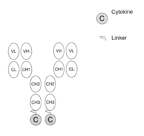
Figure 1 depicts one proposed design for a genetically engineered fusion
molecule of the
present invention. In Figure 1, the ovals labeled as VL, VH, CL, CH1, CH2 and
CH3 represent a full
length antibody (Ab) as defined herein. The oval labeled C represents a
cytokine, e.g., an IFN-cc
mutant. A linker is represented by the squiggled line. As depicted in Figure
1, C is attached to
the Ab via a linker at the two CH3 sites. In one alternative embodiment, C is
attached to the Ab
via a linker at the two VL sites. In yet another alternative embodiment, C
will be attached to the
Ab via a linker at the two VH sites. In yet another alternative, C will be
attached to the Ab via a
linker at an internal site rather than at the CH3, VL, or VH sites.
Figure 2 depicts another proposed design for a genetically engineered fusion
molecule of
the present invention. In Figure 3, the ovals labeled as VL, VH, CL, CH, CH1,
and CH2 represent a
Fab2 as defined herein. The oval label C represents a cytokine. A linker is
represented by the
squiggled line. As depicted in Figure 3, C is attached to the Fab2 via a
linker at the two CH2 sites.
In one alternative embodiment, C will be attached to the Fab2 via a linker at
the two VL sites
rather than the CH2 sites. In yet another alternative, C will be attached to
the Fab2 via a linker at
the two VH sites rather than two VL or two CH2 sites. In yet another
alternative, C will be
attached to the Fab2 via a linker at an internal site rather than at the CH2,
VL, or VH sites.
Figure 3 depicts another proposed design for a genetically engineered fusion
molecule of
the present invention. In Figure 4, the ovals labeled as VL, VH, CL, and CH1
represent a Fab as
defined herein. The oval label C represents a cytokine. A linker is
represented by the squiggled
line. As depicted in Figure 3, C is attached to the Fab via a linker at the
CH1 site. In one
alternative embodiment, C will be attached to the Fab via a linker at the VL
site rather than the
CH1. In yet another alternative, C will be attached to the Fab via a linker at
the VH site rather
than the VL or CH1 sites. In yet another alternative, C will be attached to
the Fab via a linker at
an internal site rather than at the CH1, VL, or VH sites.
CA 02866126 2014-08-29
WO 2013/134138 PCT/US2013/028899
Sequence Listings
The amino acid sequences listed in the accompanying sequence listing are shown
using
standard three letter code for amino acids, as defined in 37 C.F.R. 1.822.
SEQ ID NO: 1 is the amino acid sequence of the heavy chain of an anti-Her2/neu
antibody wherein amino acid residues 1-19 represent a signal peptide. SEQ ID
NO: 2 is the
amino acid sequence encoding the light chain of an anti-Her2/neu antibody
wherein amino acid
residues 1-19 represent a signal peptide.
SEQ ID NO: 3 is the amino acid sequence of the heavy chain of an anti-CD20
antibody
wherein amino acid residues 1-19 represent a signal peptide. SEQ ID NO: 3 is
the amino acid
sequence encoding the light chain of an anti-CD20 antibody wherein amino acid
residues 1-19
represent a signal peptide.
SEQ ID NO: 5 is the amino acid sequence of the heavy chain of an ant-CD138
antibody
wherein amino acid residues 1-19 represent a signal peptide. SEQ ID NO: 6 is
the amino acid
sequence encoding the light chain of an anti-CD138 antibody wherein amino acid
residues 1-22
represent a signal peptide.
SEQ ID NO: 7 is the amino acid sequence of the heavy chain of an anti-
endoplasmin
antibody wherein amino acid residues 1-19 represent a signal peptide. SEQ ID
NO: 8 is the
amino acid sequence encoding the light chain of an anti-endoplasmin antibody
wherein amino
acid residues 1-20 represent a signal peptide.
SEQ ID NO: 9 is the amino acid sequence of the heavy chain of an anti-CD33
antibody
wherein amino acid residues 1-19 represent a signal peptide. SEQ ID NO: 10 is
the amino acid
sequence encoding the light chain of an anti-CD33 antibody wherein amino acid
residues 1-20
represent a signal peptide.
SEQ ID NO: 11 is the amino acid sequence of the heavy chain variable region of
an anti-
CD276 antibody wherein amino acid residues 1-19 represent a signal peptide.
SEQ ID NO: 12 is
the amino acid sequence encoding the light chain variable region of an anti-
CD276 antibody
wherein amino acid residues 1-20 represent a signal peptide.
SEQ ID NO: 13 is the amino acid sequence of a human wildtype IFN-a2 molecule.
SEQ ID NO: 14 is the amino acid sequence of a peptide linker.
SEQ ID NO: 15 is the amino acid sequence of a peptide linker.
6
CA 02866126 2014-08-29
WO 2013/134138 PCT/US2013/028899
MODE(S) FOR CARRYING OUT THE INVENTION
U.S. Patent No. 8,258,263 (Morrison et al.) demonstrates that targeted
wildtype (wt)IFN-
cc may have a considerably greater therapeutic index than non-targeted wtIFN-
cc, making it
possible to administer it at effective doses. Specifically, Morrison et al.
demonstrated that
various tumor associated antigen Ab-wtIFN-cc chimeric constructs demonstrated
substantially
improved therapeutic efficacy (-100-fold more potent), with an apparent
reduction of systemic
toxicity, as compared to non-fused wtIFN-cc.
In various embodiments of the present invention, genetically engineered fusion
molecules
comprising a tumor associated antigen antibody attached via a linker to an IFN-
cc mutant
molecule are prepared for purposes of utilizing the specificity of the
antibody to target the IFN-cc
mutant molecule to the tumor cells.
The IFN-cc mutant molecules used in the preparation of the fusion molecules of
the
present invention have varying affinity for IFNaR complex. The present
inventors evaluate the
relationship between IFNaR affinity and in vitro therapeutic index, and the
anti-tumor efficacy
of the fusion protein in vivo, and identify Ab-IFN-cc mutant fusion molecules
which demonstrate
improved therapeutic index, and preserved or improved in vivo efficacy, as
compared to Ab-
wtIFN-cc fusion molecules. The present inventors also identify Ab-IFN-cc
mutant fusion
molecules which demonstrate improved PK properties as compared to an Ab-wtIFN-
cc fusion
molecule. The therapeutic index is defined as: the EC50 of the Ab-IFNa mutant
fusion molecule
for cells which express the antigen recognized by the Ab and which express
IFNaR ("targeted")
divided by the EC50 of the Ab-IFNa mutant fusion molecule for cells which
express only IFNaR
("non-targeted"). Efficacy is defined as potency of the fusion molecule at
killing cancer cells
which express the antigen to which the Ab portion of the fusion molecule
binds.
The approach used to identify such Ab-IFN-cc mutant fusion molecules is as
follows: 1)
an IFN-cc mutant was prepared; 2) an antibody which binds to a tumor
associated antigen was
prepared; 3) several Ab-IFN-cc mutant fusion molecules comprising the IFN-cc
mutants and
antibodies from steps 1) and 2) were constructed via chemical conjugation or
direct attachment
via a linker; 4) the resulting chemical conjugates or fusion molecules were
systematically tested,
at varying doses, in several in vitro functional assays to identify those
having improved
7
CA 02866126 2014-08-29
WO 2013/134138 PCT/US2013/028899
therapeutic index; and 5) in vivo studies using the chemical conjugates or
fusion molecules
demonstrating the best therapeutic index were performed to determine efficacy
in treating in vivo
tumors. As relates specifically to step 4), in vitro functional assays were
used to determine: a)
the ability of the fusion molecules to bind the IFN-ccR complex on non-
targeted cells; b) the
ability of the fusion molecule to bind cells expressing the IFN-ccR complex
and the antigen
targeted by the Ab; c) the ability of the fusion molecule to bind FcRn
receptor; d) the IFN-cc
bioactivity of the fusion molecules on non-targeted cells; e) the
antiproliferative activity of the
fusion molecules on targeted cells; and f) the ability of the fusion molecule
to induce apoptosis.
As relates specifically to step 5), in vivo assays were used to: a) confirm
efficacy of a given
fusion for treating tumors; and b) confirm improved PK properties for a given
fusion molecule.
Definitions
Unless otherwise defined herein, scientific and technical terms used in
connection with
the present invention shall have the meanings that are commonly understood by
those of ordinary
skill in the art. Further, unless otherwise required by context, singular
terms shall include
pluralities and plural terms shall include the singular. Generally,
nomenclatures used in
connection with, and techniques of, cell and tissue culture, molecular
biology, immunology,
microbiology, genetics and protein and nucleic acid chemistry and
hybridization described herein
are well known and commonly used in the art. The methods and techniques of the
present
invention are generally performed according to conventional methods well known
in the art and
as described in various general and more specific references that are cited
and discussed
throughout the present specification unless otherwise indicated. See, e.g.,
Sambrook et al.
Molecular Cloning: A Laboratory Manual, 2d ed., Cold Spring Harbor Laboratory
Press, Cold
Spring Harbor, N.Y. (1989) and Ausubel et al., Current Protocols in Molecular
Biology, Greene
Publishing Associates (1992), and Harlow and Lane Antibodies: A Laboratory
Manual Cold
Spring Harbor Laboratory Press, Cold Spring Harbor, N.Y. (1990), which are
incorporated
herein by reference. Enzymatic reactions and purification techniques are
performed according to
manufacturer's specifications, as commonly accomplished in the art or as
described herein. The
terminology used in connection with, and the laboratory procedures and
techniques of, analytical
chemistry, synthetic organic chemistry, and medicinal and pharmaceutical
chemistry described
herein are well known and commonly used in the art. Standard techniques can be
used for
8
CA 02866126 2014-08-29
WO 2013/134138 PCT/US2013/028899
chemical syntheses, chemical analyses, pharmaceutical preparation,
composition, and delivery,
and treatment of patients.
The terms "polypeptide", "peptide" and "protein" are used interchangeably
herein to refer
to a polymer of amino acid residues. Preferred "peptides", "polypeptides", and
"proteins" are
chains of amino acids whose alpha carbons are linked through peptide bonds.
The terminal
amino acid at one end of the chain (amino terminal) therefore has a free amino
group, while the
terminal amino acid at the other end of the chain (carboxy terminal) has a
free carboxyl group.
As used herein, the term "amino terminus" (abbreviated N-terminus) refers to
the free cc-amino
group on an amino acid at the amino terminal of a peptide or to the cc-amino
group (imino group
when participating in a peptide bond) of an amino acid at any other location
within the peptide.
Similarly, the term "carboxy terminus" refers to the free carboxyl group on
the carboxy terminus
of a peptide or the carboxyl group of an amino acid at any other location
within the peptide.
Peptides also include essentially any polyamino acid including, but not
limited to, peptide
mimetics such as amino acids joined by an ether as opposed to an amide bond.
The term "polypeptide fragment" as used herein refers to a polypeptide that
has an
amino-terminal and/or carboxy-terminal deletion as compared to a corresponding
full-length
protein. Fragments can be, for example, at least 5, 6, 7, 8, 9, 10, 11, 12,
13, 14, 15, 20, 50, 70,
80, 90, 100, 150 or 200 amino acids in length. Fragments can also be, for
example, at most
1000, 750, 500, 250, 200, 175, 150, 125, 100, 90, 80, 70, 60, 50, 40, 30, 20,
15, 14, 13, 12, 11, or
amino acids in length. A fragment can further comprise, at either or both of
its ends, one or
more additional amino acids, for example, a sequence of amino acids from a
different naturally-
occurring protein (e.g., an Fc or leucine zipper domain) or an artificial
amino acid sequence (e.g.,
an artificial linker sequence).
Polypeptides of the invention include polypeptides that have been modified in
any way
and for any reason, for example, to: (1) reduce susceptibility to proteolysis,
(2) reduce
susceptibility to oxidation, (3) alter binding affinity for forming protein
complexes, (4) alter
binding affinities, and (5) confer or modify other physicochemical or
functional properties. For
example, single or multiple amino acid substitutions (e.g., conservative amino
acid substitutions)
may be made in the naturally occurring sequence (e.g., in the portion of the
polypeptide outside
the domain(s) forming intermolecular contacts). A "conservative amino acid
substitution" is one
that does not substantially change the structural characteristics of the
parent sequence (e.g., a
9
CA 02866126 2014-08-29
WO 2013/134138 PCT/US2013/028899
replacement amino acid should not tend to break a helix that occurs in the
parent sequence, or
disrupt other types of secondary structure that characterize the parent
sequence or are necessary
for its functionality). Examples of art-recognized polypeptide secondary and
tertiary structures
are described in Proteins, Structures and Molecular Principles (Creighton,
Ed., W. H. Freeman
and Company, New York (1984)); Introduction to Protein Structure (C. Branden
and J. Tooze,
eds., Garland Publishing, New York, N.Y. (1991)); and Thornton et al. (1991)
Nature 354:105).
A "variant" of a polypeptide comprises an amino acid sequence wherein one or
more
amino acid residues are inserted into, deleted from and/or substituted into
the amino acid
sequence relative to another polypeptide sequence. Variants of the invention
include fusion
proteins.
A "derivative" of a polypeptide is a polypeptide that has been chemically
modified, e.g.,
conjugation to another chemical moiety such as, for example, polyethylene
glycol, albumin (e.g.,
human serum albumin), phosphorylation, and glycosylation.
The term "isolated molecule" (where the molecule is, for example, a
polypeptide, a
polynucleotide, or an antibody) is a molecule that by virtue of its origin or
source of derivation
(1) is not associated with naturally associated components that accompany it
in its native state,
(2) is substantially free of other molecules from the same species (3) is
expressed by a cell from
a different species, or (4) does not occur in nature. Thus, a molecule that is
chemically
synthesized, or expressed in a cellular system different from the cell from
which it naturally
originates, will be "isolated" from its naturally associated components. A
molecule also may be
rendered substantially free of naturally associated components by isolation,
using purification
techniques well known in the art. Molecule purity or homogeneity may be
assayed by a number
of means well known in the art. For example, the purity of a polypeptide
sample may be assayed
using polyacrylamide gel electrophoresis and staining of the gel to visualize
the polypeptide
using techniques well known in the art. For certain purposes, higher
resolution may be provided
by using HPLC or other means well known in the art for purification.
As used herein, an "antibody" refers to a protein comprising one or more
polypeptides
substantially or partially encoded by immunoglobulin genes or fragments of
immunoglobulin
genes and having specificity to a tumor antigen or specificity to a molecule
overexpressed in a
pathological state. The recognized immunoglobulin genes include the kappa,
lambda, alpha,
gamma, delta, epsilon and mu constant region genes, as well as subtypes of
these genes and
CA 02866126 2014-08-29
WO 2013/134138 PCT/US2013/028899
myriad of immunoglobulin variable region genes. Light chains are classified as
either kappa or
lambda. Heavy chains are classified as gamma, mu, alpha, delta, or epsilon,
which in turn define
the immunoglobulin classes, IgG, IgM, IgA, IgD and IgE, respectively. A
typical
immunoglobulin (e.g., antibody) structural unit comprises a tetramer. Each
tetramer is composed
of two identical pairs of polypeptide chains, each pair having one "light"
(about 25 kD) and one
"heavy" chain (about 50-70 kD). The N-terminus of each chain defines a
variable region of
about 100 to 110 or more amino acids primarily responsible for antigen
recognition. The terms
variable light chain (VL) and variable heavy chain (VH) refer to these light
and heavy chains,
respectively.
In a full-length antibody, each heavy chain is comprised of a heavy chain
variable region
(abbreviated herein as HCVR or VH) and a heavy chain constant region. The
heavy chain
constant region is comprised of three domains, CHi, CH2 and CH3 (and in some
instances, CH4).
Each light chain is comprised of a light chain variable region (abbreviated
herein as VL) and a
light chain constant region. The light chain constant region is comprised of
one domain, CL.
The VH and VL regions can be further subdivided into regions of
hypervariability, termed
complementarity determining regions (CDR), interspersed with regions that are
more conserved,
termed framework regions (FR). Each VH and VL is composed of three CDRs and
four FRs,
arranged from amino-terminus to carboxy-terminus in the following order: FRi,
CDRi, FR2,
CDR2, FR3, CDR3, FR4. The extent of the framework region and CDRs has been
defined (see,
Kabat et al., Sequences of Proteins of Immunological Interest, U.S. Department
of Health and
Human Services, 1991, which is hereby incorporated by reference). The Kabat
database is now
maintained online and CDR sequences can be determined, for example, see IMGT/V-
QUEST
programme version: 3.2.18 ., March 29, 2011, available on the intern& and
Brochet, X. et al.,
Nucl. Acids Res., 36:503-508, 2008). The sequences of the framework regions of
different light
or heavy chains are relatively conserved within a species, such as humans. The
framework
region of an antibody, that is the combined framework regions of the
constituent light and heavy
chains, serves to position and align the CDRs in three-dimensional space.
Immunoglobulin
molecules can be of any type (e.g., IgG, IgE, IgM, IgD, IgA and IgY), class
(e.g., IgGl, IgG2,
IgG 3, IgG4, IgA 1 and IgA2) or subclass.
The CDRs are primarily responsible for binding to an epitope of an antigen.
The CDRs
of each chain are typically referred to as CDRi, CDR2, CDR3, numbered
sequentially starting
11
CA 02866126 2014-08-29
WO 2013/134138 PCT/US2013/028899
from the N-terminus, and are also typically identified by the chain in which
the particular CDR is
located. Thus, a VH CDR3 is located in the variable domain of the heavy chain
of the antibody in
which it is found, whereas a VL CDRi is the CDRi from the variable domain of
the light chain of
the antibody in which it is found. Antibodies with different specificities
(i.e. different combining
sites for different antigens) have different CDRs. Although it is the CDRs
that vary from
antibody to antibody, only a limited number of amino acid positions within the
CDRs are
directly involved in antigen binding. These positions within the CDRs are
called specificity
determining residues (SDRs).
The term "Fc region" is used to define the C-terminal region of an
immunoglobulin
heavy chain, which may be generated by papain digestion of an intact antibody.
The Fc region
may be a native sequence Fc region or a variant Fc region. The Fc region of an
immunoglobulin
generally comprises two constant domains, a CH2 domain and a CH3 domain, and
optionally
comprises a CH4 domain. The Fc portion of an antibody mediates several
important effector
functions e.g. cytokine induction, ADCC, phagocytosis, complement dependent
cytotoxicity
(CDC) and half-life/clearance rate of antibody and antigen-antibody complexes
(e.g., the
neonatal FcR (FcRn) binds to the Fc region of IgG at acidic pH in the endosome
and protects
IgG from degradation, thereby contributing to the long serum half-life of
IgG). Replacements of
amino acid residues in the Fc portion to alter antibody effector function are
known in the art (see,
e.g., Winter et al., U.S. Patent No. 5,648,260 and 5,624,821).
Antibodies exist as intact immunoglobulins or as a number of well
characterized
fragments. Such fragments include Fab fragments, Fab' fragments, Fab2,
F(ab)'2fragments,
single chain Fv proteins ("scFv") and disulfide stabilized Fv proteins
("dsFv"), that bind to the
target antigen. A scFv protein is a fusion protein in which a light chain
variable region of an
immunoglobulin and a heavy chain variable region of an immunoglobulin are
bound by a linker,
while in dsFvs, the chains have been mutated to introduce a disulfide bond to
stabilize the
association of the chains. While various antibody fragments are defined in
terms of the digestion
of an intact antibody, one of skill will appreciate that such fragments may be
synthesized de
novo either chemically or by utilizing recombinant DNA methodology. Thus, the
term antibody,
as used herein also includes antibody fragments either produced by the
modification of whole
antibodies or synthesized de novo using recombinant DNA methodologies,
including, but are not
limited to, Fab, Fab', Fab2, Fab'2, IgG, IgM, IgA, IgE, scFv, dsFv, dAb,
nanobodies, unibodies,
12
CA 02866126 2014-08-29
WO 2013/134138 PCT/US2013/028899
and diabodies. In various embodiments antibodies include, but are not limited
to Fab, Fab2, IgG,
IgM, IgA, IgE, and single chain Fv (scFv) antibodies in which a variable heavy
and a variable
light chain are joined together (directly or through a peptide linker) to form
a continuous
polypeptide.
Diabodies are bivalent antibodies comprising two polypeptide chains, wherein
each
polypeptide chain comprises VH and VL regions joined by a linker that is too
short to allow for
pairing between two regions on the same chain, thus allowing each region to
pair with a
complementary region on another polypeptide chain (see, e.g., Holliger et al.,
1993, Proc. Natl.
Acad. Sci. USA 90:6444-48 (1993), and Poljak et al., Structure 2:1121-23
(1994)). If the two
polypeptide chains of a diabody are identical, then a diabody resulting from
their pairing will
have two identical antigen binding sites. Polypeptide chains having different
sequences can be
used to make a diabody with two different antigen binding sites. Similarly,
tribodies and
tetrabodies are antibodies comprising three and four polypeptide chains,
respectively, and
forming three and four antigen binding sites, respectively, which can be the
same or different.
In certain embodiments, antibodies and antibody fragments used in the
constructs of the
present invention can be bispecific. Bispecific antibodies or fragments can be
of several
configurations. For example, bispecific antibodies may resemble single
antibodies (or antibody
fragments) but have two different antigen binding sites (variable regions). In
various
embodiments bispecific antibodies can be produced by chemical techniques
(Kranz et al., Proc.
Natl. Acad. Sci., USA, 78:5807, 1981), by "polydoma" techniques (see, e.g.,
U.S. Patent No.
4,474,893), or by recombinant DNA techniques. In certain embodiments
bispecific antibodies of
the present invention can have binding specificities for at least two
different epitopes at least one
of which is a tumor associate antigen. In various embodiments the antibodies
and fragments can
also be heteroantibodies. Heteroantibodies are two or more antibodies, or
antibody binding
fragments (e.g., Fab) linked together, each antibody or fragment having a
different specificity.
The term "monoclonal antibody" as used herein refers to an antibody obtained
from a
population of substantially homogeneous antibodies, i.e., the individual
antibodies comprising
the population are identical except for possible naturally occurring mutations
that may be present
in minor amounts. Monoclonal antibodies are highly specific, being directed
against a single
antigen. Furthermore, in contrast to polyclonal antibody preparations that
typically include
different antibodies directed against different determinants (epitopes), each
monoclonal antibody
13
CA 02866126 2014-08-29
WO 2013/134138 PCT/US2013/028899
is directed against a single determinant on the antigen. The modifier
"monoclonal" is not to be
construed as requiring production of the antibody by any particular method.
The term "chimeric antibody" as used herein refers to an antibody which has
framework
residues from one species, such as human, and CDRs (which generally confer
antigen binding)
from another species, such as a murine antibody that specifically binds
targeted antigen.
The term "human antibody", as used herein, is intended to include antibodies
having
variable and constant regions derived from human germline immunoglobulin
sequences. The
human antibodies of the invention may include amino acid residues not encoded
by human
germline immunoglobulin sequences (e.g., mutations introduced by random or
site-specific
mutagenesis in vitro or by somatic mutation in vivo), for example in the CDRs
and in particular
CDR3. However, the term "human antibody", as used herein, is not intended to
include
antibodies in which CDR sequences derived from the germline of another
mammalian species,
such as a mouse, have been grafted onto human framework sequences.
The term "humanized antibody" as used herein refers to an antibody comprising
a
humanized light chain and a humanized heavy chain immunoglobulin. A humanized
antibody
binds to the same antigen as the donor antibody that provides the CDRs. The
acceptor
framework of a humanized immunoglobulin or antibody may have a limited number
of
substitutions by amino acids taken from the donor framework. Humanized or
other monoclonal
antibodies can have additional conservative amino acid substitutions which
have substantially
no effect on antigen binding or other immunoglobulin functions. Humanized
immunoglobulins
can be constructed by means of genetic engineering (see for example, U.S.
Patent No.
5,585,089).
The term "recombinant human antibody", as used herein, is intended to include
all human
antibodies that are prepared, expressed, created or isolated by recombinant
means, such as
antibodies expressed using a recombinant expression vector transfected into a
host cell;
antibodies isolated from a recombinant, combinatorial human antibody library;
antibodies
isolated from an animal (e.g., a mouse) that is transgenic for human
immunoglobulin genes; or
antibodies prepared, expressed, created or isolated by any other means that
involves splicing of
human immunoglobulin gene sequences to other DNA sequences. Such recombinant
human
antibodies have variable and constant regions derived from human germline
immunoglobulin
sequences. In certain embodiments, however, such recombinant human antibodies
are subjected
14
CA 02866126 2014-08-29
WO 2013/134138 PCT/US2013/028899
to in vitro mutagenesis (or, when an animal transgenic for human Ig sequences
is used, in vivo
somatic mutagenesis) and thus the amino acid sequences of the VH and VL
regions of the
recombinant antibodies are sequences that, while derived from and related to
human germline VH
and VL sequences, may not naturally exist within the human antibody germline
repertoire in vivo.
All such recombinant means are well known to those of ordinary skill in the
art.
An antigen binding protein including an antibody "specifically binds" to an
antigen if it
binds to the antigen with a high binding affinity as determined by a
dissociation constant (Kd, or
corresponding Kb, as defined below) value of at least 1 x 10⁻⁶ M, or at least
1 x 10⁻⁷ M, or at
least 1 x 10⁻⁸ M, or at least 1 x 10⁻⁹ M, or at least 1 x 10⁻¹⁰ M, or at least
1 x 10-11 M. An antigen
binding protein that specifically binds to the human antigen of interest may
be able to bind to the
same antigen of interest from other species as well, with the same or
different affinities.
An "epitope" is the portion of a molecule that is bound by an antigen binding
protein
(e.g., by an antibody). An epitope can comprise non-contiguous portions of the
molecule (e.g., in
a polypeptide, amino acid residues that are not contiguous in the
polypeptide's primary sequence
but that, in the context of the polypeptide's tertiary and quaternary
structure, are near enough to
each other to be bound by an antigen binding protein).
The terms "polynucleotide," "oligonucleotide" and "nucleic acid" are used
interchangeably throughout and include DNA molecules (e.g., cDNA or genomic
DNA), RNA
molecules (e.g., mRNA), analogs of the DNA or RNA generated using nucleotide
analogs (e.g.,
peptide nucleic acids and non-naturally occurring nucleotide analogs), and
hybrids thereof. The
nucleic acid molecule can be single-stranded or double-stranded. In one
embodiment, the
nucleic acid molecules of the invention comprise a contiguous open reading
frame encoding an
antibody, or a fragment, derivative, mutein, or variant thereof, of the
invention.
Two single-stranded polynucleotides are "the complement" of each other if
their
sequences can be aligned in an anti-parallel orientation such that every
nucleotide in one
polynucleotide is opposite its complementary nucleotide in the other
polynucleotide, without the
introduction of gaps, and without unpaired nucleotides at the 5' or the 3' end
of either sequence.
A polynucleotide is "complementary" to another polynucleotide if the two
polynucleotides can
hybridize to one another under moderately stringent conditions. Thus, a
polynucleotide can be
complementary to another polynucleotide without being its complement.
A "vector" is a nucleic acid that can be used to introduce another nucleic
acid linked to it
CA 02866126 2014-08-29
WO 2013/134138 PCT/US2013/028899
into a cell. One type of vector is a "plasmid," which refers to a linear or
circular double stranded
DNA molecule into which additional nucleic acid segments can be ligated.
Another type of
vector is a viral vector (e.g., replication defective retroviruses,
adenoviruses and adeno-
associated viruses), wherein additional DNA segments can be introduced into
the viral genome.
Certain vectors are capable of autonomous replication in a host cell into
which they are
introduced (e.g., bacterial vectors comprising a bacterial origin of
replication and episomal
mammalian vectors). Other vectors (e.g., non-episomal mammalian vectors) are
integrated into
the genome of a host cell upon introduction into the host cell, and thereby
are replicated along
with the host genome. An "expression vector" is a type of vector that can
direct the expression
of a chosen polynucleotide.
A nucleotide sequence is "operably linked" to a regulatory sequence if the
regulatory
sequence affects the expression (e.g., the level, timing, or location of
expression) of the
nucleotide sequence. A "regulatory sequence" is a nucleic acid that affects
the expression (e.g.,
the level, timing, or location of expression) of a nucleic acid to which it is
operably linked. The
regulatory sequence can, for example, exert its effects directly on the
regulated nucleic acid, or
through the action of one or more other molecules (e.g., polypeptides that
bind to the regulatory
sequence and/or the nucleic acid). Examples of regulatory sequences include
promoters,
enhancers and other expression control elements (e.g., polyadenylation
signals). Further
examples of regulatory sequences are described in, for example, Goeddel, 1990,
Gene
Expression Technology: Methods in Enzymology 185, Academic Press, San Diego,
Calif. and
Baron et al., 1995, Nucleic Acids Res. 23:3605-06.
A "host cell" is a cell that can be used to express a nucleic acid, e.g., a
nucleic acid of the
invention. A host cell can be a prokaryote, for example, E. coli, or it can be
a eukaryote, for
example, a single-celled eukaryote (e.g., a yeast or other fungus), a plant
cell (e.g., a tobacco or
tomato plant cell), an animal cell (e.g., a human cell, a monkey cell, a
hamster cell, a rat cell, a
mouse cell, or an insect cell) or a hybridoma. Typically, a host cell is a
cultured cell that can be
transformed or transfected with a polypeptide-encoding nucleic acid, which can
then be
expressed in the host cell. The phrase "recombinant host cell" can be used to
denote a host cell
that has been transformed or transfected with a nucleic acid to be expressed.
A host cell also can
be a cell that comprises the nucleic acid but does not express it at a desired
level unless a
regulatory sequence is introduced into the host cell such that it becomes
operably linked with the
16
CA 02866126 2014-08-29
WO 2013/134138 PCT/US2013/028899
nucleic acid. It is understood that the term host cell refers not only to the
particular subject cell
but to the progeny or potential progeny of such a cell. Because certain
modifications may occur
in succeeding generations due to, e.g., mutation or environmental influence,
such progeny may
not, in fact, be identical to the parent cell, but are still included within
the scope of the term as
used herein.
Tumor Associated Antigens and Antibodies
The term "antigen" as used herein refers to a compound, composition, or
substance that
can stimulate the production of antibodies or a T cell response in an animal,
including
compositions that are injected or absorbed into an animal. An antigen reacts
with the products of
specific humoral or cellular immunity, including those induced by heterologous
immunogens.
The term "antigen" includes all related antigenic epitopes. Epitopes can be
formed both from
contiguous amino acids or noncontiguous amino acids juxtaposed by tertiary
folding of a protein.
Epitopes formed from contiguous amino acids are typically retained on exposure
to denaturing
solvents whereas epitopes formed by tertiary folding are typically lost on
treatment with
denaturing solvents. An epitope typically includes at least three, at least
five, or at least eight to
ten amino acids in a unique spatial conformation. Methods of determining
spatial conformation
of epitopes include, for example, x-ray crystallography and 2-dimensional
nuclear magnetic
resonance.
As relates to "targeted antigens", virtually any antigen may be targeted by
the molecules
of the present invention. Certain targeted antigens include those associated
with a pathology
characterized by hyperproliferation of a cell (i.e., a hyperproliferative
disorder). Illustrative
hyperproliferative disorders include, but are not limited to psoriasis,
neutrophilia, polycythemia,
thrombocytosis, and cancer. Hyperproliferative disorders characterized as
cancer include but are
not limited to solid tumors, cancers of the breast, respiratory tract, brain,
reproductive organs,
digestive tract, urinary tract, eye, liver, skin, head and neck, thyroid,
parathyroid and their distant
metastases. These disorders also include lymphomas, sarcomas, multiple
myelomas and
leukemias. Examples of breast cancer include, but are not limited to invasive
ductal carcinoma,
invasive lobular carcinoma, ductal carcinoma in situ, and lobular carcinoma in
situ. Examples of
cancers of the respiratory tract include, but are not limited to small-cell
and non-small-cell lung
carcinoma, as well as bronchial adenoma and pleuropulmonary blastoma. Examples
of brain
17
CA 02866126 2014-08-29
WO 2013/134138 PCT/US2013/028899
cancers include, but are not limited to brain stem and hypophtalmic glioma,
cerebellar and
cerebral astrocytoma, medulloblastoma, ependymoma, as well as neuroectodermal
and pineal
tumor. Tumors of the male reproductive organs include, but are not limited to
prostate and
testicular cancer. Tumors of the female reproductive organs include, but are
not limited to
endometrial, cervical, ovarian, vaginal, and vulvar cancer, as well as sarcoma
of the uterus.
Tumors of the digestive tract include, but are not limited to anal, colon,
colorectal, esophageal,
gallbladder, gastric, pancreatic, rectal, small-intestine, and salivary gland
cancers. Tumors of the
urinary tract include, but are not limited to bladder, penile, kidney, renal
pelvis, ureter, and
urethral cancers. Eye cancers include, but are not limited to intraocular
melanoma and
retinoblastoma. Examples of liver cancers include, but are not limited to
hepatocellular
carcinoma (liver cell carcinomas with or without fibrolamellar variant),
cholangiocarcinoma
(intrahepatic bile duct carcinoma), and mixed hepatocellular
cholangiocarcinoma. Skin cancers
include, but are not limited to squamous cell carcinoma, Kaposi's sarcoma,
malignant melanoma,
Merkel cell skin cancer, and non-melanoma skin cancer. Lymphomas include, but
are not
limited to AIDS-related lymphoma, non-Hodgkin's lymphoma, cutaneous T-cell
lymphoma,
Hodgkin's disease, and lymphoma of the central nervous system. Sarcomas
include, but are not
limited to sarcoma of the soft tissue, osteosarcoma, malignant fibrous
histiocytoma,
lymphosarcoma, and rhabdomyosarcoma. Leukemias include, but are not limited to
acute
myeloid leukemia, acute lymphoblastic leukemia, chronic lymphocytic leukemia,
chronic
myelogenous leukemia, and hairy cell leukemia.
In various embodiments of the present invention, the targeting moiety is a
moiety that
binds a cancer marker, e.g., a tumor associated antigen (TAA). A wide variety
of cancer markers
are known to those of skill in the art. The cancer markers need not be unique
to cancer cells, but
can also be effective where the expression of the cancer marker is elevated in
a cancer cell (as
compared to normal healthy cells) or where the cancer marker is not present at
comparable levels
in surrounding tissues (especially where the fusion molecule is delivered
locally). In various
embodiments the cancer marker includes: Her2/neu (Lewis et al, Semin. Cancer
Biol., 6(6): 321-
327, 1995), Her3, EGF, Her4, B7 family members (Collins et al., Genome Biol.,
6:223.1-223.7,
2005), the TNF superfamily members (see, e.g., "Therapeutic Targets of the TNF
Superfamily",
edited by Iqbal S. Grewal, Landes Bioscience/Springer Science+Business Media,
LLC dual
imprint / Springer series: Advances in Experimental Medicine and Biology,
2009), CD1, CD2,
18
CA 02866126 2014-08-29
WO 2013/134138 PCT/US2013/028899
CD3, CD5, CD7, CD13, CD14, CD15, CD19, CD20 (Cragg et al., Curr. Dir.
Autoimmun., 8:
140-174, 2005), CD21, CD23, CD25, CD33 (Nakase et al., Am J Clin Pathol.,
105(6): 761-768,
1996), CD34, CD38, CD46, CD55, CD59, CD123, CD138 (O'Connell, et al., Am. J.
Clin.
Pathol., 121(2):254-263, 2004), CD200, CD276 (Hofmeyer et al., Proc. Natl.
Acad. Sci. (U.S.A.)
105(30):10277-10278, 2008), 5E10, CEA, endoplasmin (U.S. Patent Application
No.
U520120009194 (Ferrone et al)), HLA-DR, HM 1.24, HMB 45, Ia, Leu-M1, MUC1,
PMSA,
EGFR, glycosphingolipid GD2, SLAM family members, gp100, tyrosinase, MAGE, TAG-
72,
SE10, phosphatidyl serine antigen, and the like. The genetically engineered
fusion molecules of
the present invention may bind one antigen or multiple cancer markers.
Antibodies to these and other cancer markers are known to those of skill in
the art and
can be obtained commercially or readily produced. For example, antibodies can
be produced by
immunizing an animal with a target antigen or an immunogenic fragment thereof
and raising the
antibodies in that animal, and single chain antibodies can be produced using
phage-display
technology according to methods well known to those of skill in the art.
Antibodies
contemplated for use as targeting moieties in the fusion molecules of the
present invention
include depleting antibodies to specific tumor associated antigens, including,
but not limited to,
anti-HER2/neu, anti-HER3, anti-HER4, anti-CD20, anti-CD19, anti-CD22, anti-
CD33, anti-
CXCR3, anti-CXCR5, anti-CCR3, anti-CCR4, anti-CCR9, anti-CRTH2, anti-PMCH,
anti-CD4,
anti-CD25, anti-CD200, anti-CD138, anti-CD276 and anti-endoplasmin antibodies.
All such
tumor and inflammatory cell-specific, depleting antibodies have been well
described in the
literature.
In various embodiments the antibody is an anti-Her2/neu antibody which
comprises the
heavy chain having an amino acid sequence as set forth in SEQ ID NO: 1:
MECSWVMLFLLS VTAGVHSEVQLVESGGGLVQPGGSLRLSCAASGFNIKD
TYIHWVRQAPGKGLEWVARTYPTNGYTRYADSVKGRFTISADTSKNTAYL
QMNSLRAEDTAVYYCSRWGGDGFYAMDYWGQGTLVTVSSASTKGPSVFPL
APSSKSTSGGTAALGCLVKDYFPEPVTVSWNSGALTSGVHTFPAVLQSSG
LYSLSSVVTVPSSSLGTQTYICNVNHKPSNTKVDKKVEPKSCDKTHTCPP
CPAPELLGGPSVFLFPPKPKDTLMISRTPEVTCVVVDVSHEDPEVKFNWY
VDGVEVHNAKTKPREEQYNSTYRVVSVLTVLHQDWLNGKEYKCKVSNKAL
PAPIEKTISKAKGQPREPQVYTLPPSRDELTKNQVSLTCLVKGFYPSDIA
VEWESNGQPENNYKTTPPVLDSDGSFFLYSKLTVDKSRWQQGNVFSCSVM
HEALHNHYTQKSLSLSPGK (SEQ ID NO: 1)
19
CA 02866126 2014-08-29
WO 2013/134138 PCT/US2013/028899
wherein amino acid residues 1-19 represent a signal peptide; and the light
chain having an amino
acid sequence as set forth in SEQ ID NO: 2:
MEWSCVMLFLLS VTAGVHSDIQMTQSPS SLS AS VGDRVTITCRAS QDVNT
AVAWYQQKPGKAPKLLIYS ASFLYSGVPSRFSGSRSGTDFTLTIS SLQPE
DFATYYCQQHYTTPPTFGQGTKVEIKRTVAAPS VFIFEPSDEQLKS GTAS
VVCLLNNFYPREAKVQWKVDNALQSGNS QES VTEQDS KDS TYS LS STLTL
SKADYEKHKVYACEVTHQGLSSPVTKSFNRGEC (SEQ ID NO: 2)
wherein amino acid residues 1-19 represent a signal peptide.
In various embodiments the antibody is an anti-CD20 antibody which comprises
the
heavy chain having an amino acid sequence as set forth in SEQ ID NO: 3:
MYLGLNCVIIVFLLKGVQS QVQLQQPGAELVKPGAS VKMSCKASGYTFTS
YNMHWVKQTPGRGLEWIGAIYPGNGDTS YNQKFKGKATLTADKS S S TAYM
QLSSLTSEDSAVYYCARSTYYGGDWYFNVWGAGTTVTVSAASTKGPSVFP
LAPS S KS TSGGTAALGCLVKDYFPEPVTVSWNSGALTSGVHTFPAVLQS S
GLYS LS S VVTVPS S SLGTQTYICNVNHKPSNTKVDKKVEPKSCDKTHTCP
PCPAPELLGGPS VFLFPPKPKDTLMISRTPEVTCVVVDVSHEDPEVKFNW
YVDGVEVHNAKTKPREEQYNSTYRVVSVLTVLHQDWLNGKEYKCKVSNKA
LPAPIEKTIS KAKGQPREPQVYTLPPSRDELTKNQVS LTCLVKGFYPSDI
AVEWESNGQPENNYKTTPPVLDSDGSFFLYS KLTVDKSRWQQGNVFSCS V
MHEALHNHYTQKS LS LSPGK (SEQ ID NO: 3)
wherein amino acid residues 1-19 represent a signal peptide; and the light
chain having an amino
acid sequence as set forth in SEQ ID NO: 4:
MKLPVRLLVLMFWIPAS S S QIVLS QSPAILS ASPGEKVTMTCRAS S S VS Y
IHWFQQKPGSSPKPWIYATSNLASGVPVRFSGSGSGTSYSLTISRVEAED
AATYYCQQWTSNPPTFGGGTKLEIKRTVAAPS VFIFPPSDEQLKSGTAS V
VCLLNNFYPREAKVQWKVDNALQSGNS QES VTEQDS KDS TYS LS S TLTLS
KADYEKHKVYACEVTHQGLSSPVTKSFNRGEC (SEQ ID NO: 4)
wherein amino acid residues 1-19 represent a signal peptide.
In various embodiments the antibody is an anti-CD138 antibody which comprises
the
heavy chain having an amino acid sequence as set forth in SEQ ID NO: 5:
CA 02866126 2014-08-29
WO 2013/134138 PCT/US2013/028899
MGWSYIILFLVATATDVHS QVQLQQSGSELMMPGASVKISCKATGYTFSN
YWIEWVKQRPGHGLEWIGEILPGTGRTIYNEKFKGKATFTADIS SNTVQM
QLS SLTSEDS AVYYCARRDYYGNFYYAMDYWGQGTS VTVS S AS TKGPS VF
PLAPS S KS TSGGTAALGCLVKDYFPEPVTVSWNSGALTSGVHTFPAVLQS
SGLYSLS S VVTVPS S SLGTQTYICNVNHKPSNTKVDKKVEPKSCDKTHTC
PPCPAPELLGGPS VFLFPPKPKDTLMISRTPEVTCVVVDVSHEDPEVKFN
WYVDGVEVHNAKTKPREEQYNSTYRVVSVLTVLHQDWLNGKEYKCKVSNK
ALPAPIEKTIS KAKGQPREPQVYTLPPSRDELTKNQVSLTCLVKGFYPSD
IAVEWESNGQPENNYKTTPPVLDSDGSFFLYS KLTVDKSRWQQGNVFSCS
VMHEALHNHYTQKSLSLSPGK (SEQ ID NO: 5)
wherein amino acid residues 1-19 represent a signal peptide; and the light
chain having an amino
acid sequence as set forth in SEQ ID NO: 6:
MDMRVPAQLLGLLLLWLRGARCDIQMTQS TS SLS ASLGDRVTISCS AS QG
INNYLNWYQQKPDGTVELLIYYTS TLQSGVPSRFSGSGSGTDYSLTISNL
EPEDIGTYYCQQYSKLPRTFGGGTKLEIKRTVAAPSVFIFPPSDEQLKSG
TASVVCLLNNFYPREAKVQWKVDNALQSGNSQESVTEQDSKDSTYSLSST
LTLSKADYEKHKVYACEVTHQGLSSPVTKSFNRGEC (SEQ ID NO: 6)
wherein amino acid residues 1-22 represent a signal peptide.
In various embodiments the antibody is an anti-endoplasmin antibody which
comprises
the heavy chain having an amino acid sequence as set forth in SEQ ID NO: 7:
MYLGLNCVIIVFLLKGVQS QVQLVQSGAEVKKPGASVKVSCKASGYTFTS
YAMHWVRQAPGQRLEWMGWINAGNGNTKYS QKFQGRVTITRDTS AS TAYM
ELS S LRSEDTAVYYCARAHFDYWGQGTLVTVS AAS TKGPS VFPLAPS S KS
TSGGTAALGCLVKDYFPEPVTVSWNSGALTSGVHTFPAVLQSSGLYSLSS
VVTVPS S SLGTQTYICNVNHKPSNTKVDKKVEPKSCDKTHTCPPCPAPEL
LGGPS VFLFPPKPKDTLMISRTPEVTCVVVDVSHEDPEVKFNWYVDGVEV
HNAKTKPREEQYNS TYRVVS VLTVLHQDWLNGKEYKCKVSNKALPAPIEK
TIS KAKGQPREPQVYTLPPSRDELTKNQVSLTCLVKGFYPSDIAVEWESN
GQPENNYKTTPPVLDSDGSFFLYSKLTVDKSRWQQGNVFSCSVMHEALHN
HYTQKSLSLSPGK (SEQ ID NO: 7)
wherein amino acid residues 1-19 represent a signal peptide; and the light
chain having an amino
acid sequence as set forth in SEQ ID NO: 8:
MEAPAQLLFLLLLWLPDTTGEIELTQSPS S LS AS VGDRVTITCRAS QS IS
SYLNWYQQKPGKAPKLLIYAASSLQSGVPSRFSGSGSGTDFTLTISSLQP
21
CA 02866126 2014-08-29
WO 2013/134138 PCT/US2013/028899
EDFATYYCQQS YS TPPTFGQGTKVEIKRTVAAPS VFIFPPSDEQLKSGTA
SVVCLLNNFYPREAKVQWKVDNALQSGNSQESVTEQDSKDSTYSLSSTLT
LS KADYEKHKVYACEVTHQGLS SPVTKSFNRGEC (SEQ ID NO: 8)
wherein amino acid residues 1-20 represent a signal peptide.
In various embodiments the antibody is an anti-CD33 antibody which comprises
the
heavy chain having an amino acid sequence as set forth in SEQ ID NO: 9:
MEWSWVFLFFLS VTTGVHS QVQLVQSGAEVKKPGS S VKVSCKASGYTITD
SNIHWVRQAPGQSLEWIGYIYPYNGGTDYNQKFKNRATLTVDNPTNTAYM
ELS S LRSEDTAFYYCVNGNPWLAYWGQGTLVTVS S AS TKGPS VFPLAPS S
KS TSGGTAALGCLVKDYFPEPVTVSWNSGALTSGVHTFPAVLQS SGLYS L
S S VVTVPS S SLGTQTYICNVNHKPSNTKVDKKVEPKSCDKTHTCPPCPAP
ELLGGPS VFLFPPKPKDTLMISRTPEVTCVVVDVSHEDPEVKFNWYVDGV
EVHNAKTKPREEQYNS TYRVVS VLTVLHQDWLNGKEYKCKVSNKALPAPI
EKTIS KAKGQPREPQVYTLPPSRDELTKNQVSLTCLVKGFYPSDIAVEWE
SNGQPENNYKTTPPVLDSDGSFFLYSKLTVDKSRWQQGNVFSCSVMHEAL
HNHYTQKSLSLSPGK (SEQ ID NO: 9)
wherein amino acid residues 1-19 represent a signal peptide; and the light
chain having an amino
acid sequence as set forth in SEQ ID NO: 10:
MS VPTQVLGLLLLWLTDARCDIQLTQSPS TLS AS VGDRVTITCRASESLD
NYGIRFLTWFQQKPGKAPKLLMYAASNQGSGVPSRFSGSGSGTEFTLTIS
SLQPDDFATYYCQQTKEVPWSFGQGTKVEVKRTVAAPS VFIFPPSDEQLK
SGTASVVCLLNNFYPREAKVQWKVDNALQSGNSQESVTEQDSKDSTYSLS
STLTLSKADYEKHKVYACEVTHQGLSSPVTKSFNRGEC (SEQ ID NO: 10)
wherein amino acid residues 1-20 represent a signal peptide.
In various embodiments the antibody is an anti-CD276 antibody which comprises
the
heavy chain variable region having an amino acid sequence as set forth in SEQ
ID NO: 11:
MNFGFRLIFLALILKGVQCEVQLVESGGGLVKPGGSLKLSCEASRFTFS S
YAMSWVRQTPEKRLEWVAAISGGGRYTYYPDSMKGRFTISRDNAKNFLYL
QMSSLRSEDTAMYYCARHYDGYLDYWGQGTTLTVSSAKTTAPSVYPLAPG
SL (SEQ ID NO: 11)
wherein amino acid residues 1-19 represent a signal peptide; and the light
chain having an amino
22
CA 02866126 2014-08-29
WO 2013/134138 PCT/US2013/028899
acid sequence as set forth in SEQ ID NO: 12:
MKS QS QVFVFVFLWLSGVDGDIVMTQFAGVDGDIVMTQSHKFMSTSVGDR
VSITCKAS QDVSTTVAWYQQKPGQSPKLLIYSASYRYTGVPDRFTGSGSG
TDFTFTISSVQAEDLAVYYCQQHYSTPPTFGGGTKLEIKRADAAPTVSIF
PPSSKLG (SEQ ID NO: 12)
wherein amino acid residues 1-20 represent a signal peptide.
Interferon and interferon mutants
The term "interferon" refers to a full-length interferon or to an interferon
fragment
(truncated interferon) or an interferon mutant (truncated interferon and
interferon mutant
collectively referred to herein as 'modified interferon'), that substantially
retains the biological
activity of the full length wild-type interferon (e.g., retains at least 50%).
Interferons include
type I interferons (e.g., interferon-alpha and interferon-beta) as well as
type II interferons (e.g.,
interferon-gamma). The interferon can be from essentially any mammalian
species. U.S. Patent
No. 6,610,830 (Goeddel et al) describes various mature human leukocyte
interferons, e.g.,
interferon-alpha, useful in the treatment or viral and neoplastic diseases.
In various embodiments of the present invention, the interferon mutant
comprises one or
more amino acid substitutions, insertions, and/or deletions. Means of
identifying such modified
interferon molecules are routine to those of skill in the art. In one
illustrative approach, a library
of truncated and/or mutated IFN-cc is produced and screened for IFN-cc
activity. Methods of
producing libraries of polypeptide variants are well known to those of skill
in the art. Thus, for
example error-prone PCR can be used to create a library of mutant and/or
truncated IFN-cc (see,
e.g., U.S. Patent No. 6,365,408). The resultant library members can then be
screened according
to standard methods know to those of skill in the art. Thus, for example, IFN-
cc activity can be
assayed by measuring antiviral activity against a particular test virus. Kits
for assaying for IFN-
cc activity are commercially available (see, e.g., ILITETm alphabeta kit by
Neutekbio, Ireland).
The use of chemically modified interferons is also contemplated. For example,
in certain
embodiments, the interferon is chemically modified to increase serum half-
life. Thus, for
example, (2-sulfo-9-fluorenylmethoxycarbony1)7-interferon-cc2 undergoes time-
dependent
spontaneous hydrolysis, generating active interferon (Shechter et al., Proc.
Natl. Acad. Sci.,
23
CA 02866126 2014-08-29
WO 2013/134138 PCT/US2013/028899
USA, 98(3): 1212-1217, 2001). Other modifications, include for example, N-
terminal
modifications in including, but not limited to the addition of PEG, protecting
groups, and the like
(see, e.g., U.S. Patent No. 5,824,784)
In various embodiments use of truncated interferons is also contemplated.
Human INFcc,
for example, with deletions of the first 15 amino-terminal amino acid residues
and/or the last 10-
13 carboxyl-terminal amino acid residues, have been shown to exhibit virtually
the same activity
as the parent molecules (see, e.g., Ackerman (1984) Proc. Natl. Acad. Sci.,
USA, 81: 1045-1047).
Accordingly the use of IFN-ccs having 1, 2, 3, up to 13 carboxyl terminal
amino acid residues
deleted and/or 1, 2, 3, up to 15 amino terminal amino acid residues deleted
are contemplated. It
has also been demonstrated that activity resides in huIFN-cc fragment HuIFN-cc
(1-110) (Id.).
Accordingly carboxyl truncated IFNs with truncations after residue 110 and/or
with 1, 2, 3, up to
15 amino terminal amino acid residues deleted are contemplated.
Single point mutations contemplated for use herein include, but are not
limited to, a series
of mostly single point mutants (see Table 1 below) designed specifically to
increase the affinity
between IFN-cc and IFN-ccR and others expected to decrease the affinity
between IFN-cc and
IFN-aR by specifically modeling the changes based on published phage display
studies and the
NMR structure (Kalie E et al., J. Biol. Chem., 282:11602, 2007; Gomez D and
Reich NC, J.
Immunol., 170:5373, 2003; Quadt-Akabayov SR et al., Protein Science, 15:2656,
2006;
Akabayov SR et al., Biochemistry, 49:687, 2010). The strategy was based on the
belief that a
single point mutation may change the binding affinity but will not completely
knock off the
activity of IFN-a, therefore still retaining the antiproliferative properties
albeit at much higher
concentrations, i.e., the goal is to improve the therapeutic index of fusion
molecules comprising
the interferon-alpha mutants as compared to fusion molecules comprising
wildtype interferon-
alpha. As described herein and as depicted in Table 1, a single mutation will
be identified by the
particular amino acid substitution at a specific amino acid position within
the full length wild
type interferon sequence. For example, a mutation comprising a tyrosine
substituted for the full
length wild type histidine at amino acid 57 is identified as H57Y. The wild
type IFN-a2 amino
acid sequence from which the mutants described in Table 1 are derived is
provided below as
SEQ ID NO: 13:
CDLPQTHSLGSRRTLMLLAQMRRISLFSCLKDRHDFGFPQEEFGNQFQKA
24
CA 02866126 2014-08-29
WO 2013/134138 PCT/US2013/028899
ETIPVLHEMIQQIFNLFSTKDS SAAWDETLLDKFYTELYQQLNDLEACVI
QGVGVTETPLMKEDSILAVRKYFQRITLYLKEKKYSPCAWEVVRADIVIRS
FSLSTNLQESLRSKE (SEQ ID NO: 13)
Table 1
List of certain proposed IFN-a Mutant Molecules.
IFN-a sequence Selection Criteria
mutations
M1 H57Y, E58N, Phage display optimization of selected IFN-a residues to
increase IFN-a-IFN-aR1
Q61S binding affinity of Site 1
M2 H57S, E585, Decrease the IFN-a-IFN-aR1 binding affinity at Site 1
based on triple mutations
Q61S predicted to result in a loss of binding contacts between
IFNa and IFN-aR1
M3 H57A Decrease the IFN-a-IFN-aR1 binding affinity at Site 1
similar to M2 but only single point
M4 E58A Decrease the IFN-a-IFN-aR1 binding affinity at Site 1
similar to M2 but only single point
M5 Q61A Decrease the IFN-a-IFN-aR1 binding affinity at Site 1
similar to M2 but only single point
M6 R149A Decrease the IFN-a-IFN-aR1 binding affinity at Site 2
based on loss of binding contacts
M7 R162A Decrease the IFN-a-IFN-aR1 binding affinity at Site 2
based on loss of binding contacts
M8 R149A, R162A Decrease the IFN-a-IFN-aR1 binding affinity at Site 2
based on loss of binding contacts
M9 L30A Decrease the IFN-a-IFN-aR1 binding affinity at Site 2
based on loss of binding contacts
M10 D35E Alter the IFN-a-IFN-aR1 binding at Site 2 based on minimal
change in structure
M11 E165D Alter the IFN-a-IFN-aR1 binding at Site 2 based on minimal
change in structure
M12 L26A Alter the IFN-a-IFN-aR1 binding at Site 2 based on minimal
change in structure
M13 F27A Alter the IFN-a-IFN-aR1 binding at Site 2 based on minimal
change in structure
M14 L153A Alter the IFN-a-IFN-aR1 binding at Site 2 based on minimal
change in structure
M15 A145V Alter the IFN-a-IFN-aR1 binding at Site 2 based on minimal
change in structure
In various embodiments of the present invention, either the N- or C- terminus
of an
antibody heavy or light chain will be genetically constructed with one of
several contemplated
interferon alpha mutants. The fusion molecule may have any of the general
constructs as
depicted in, e.g., Figures 1-3.
Generally speaking, the antibody and interferon mutant molecule of the
genetically
engineered fusion molecules of the present invention can be joined together in
any order. Thus,
for example, the interferon mutant molecule can be joined to either the amino
or carboxy
terminal of the antibody; or conversely, the interferon mutant molecule can be
joined to an
internal location of the antibody, so long as the attachment does not
interfere with binding of the
antibody to the target antigen. Alternatively, the antibody can be joined to
either the amino or
carboxy terminal of the interferon mutant molecule; or joined to an internal
region of the
interferon mutant molecule.
CA 02866126 2014-08-29
WO 2013/134138 PCT/US2013/028899
The present invention relates to genetically engineered fusion molecules
comprising at
least one antibody linked to at least one interferon mutant formed through
genetic fusion or
chemical coupling. By "linked" we mean that the first and second sequences are
associated such
that the second sequence is able to be transported by the first sequence to a
target cell, i.e., fusion
molecules in which the antibody is linked to a interferon mutant via their
polypeptide backbones
through genetic expression of a DNA molecule encoding these proteins, directly
synthesized
proteins, and coupled proteins in which pre-formed sequences are associated by
a cross-linking
agent. Many procedures and linker molecules for attachment of various
compounds including
radionuclide metal chelates, toxins and drugs to proteins such as antibodies
are known. See, for
example, U.S. Patent Nos. 4,671,958, 4,659,839, 4,414,148, 4,699,784;
4,680,338; 4,569,789;
and 4,589,071.
In certain embodiments, the antibody and interferon mutant are linked directly
to each
other and synthesized using recombinant DNA methodology, e.g., creating a DNA
sequence that
encodes the antibody- interferon mutant fusion protein, placing the DNA in an
expression
cassette under the control of a particular promoter, expressing the fusion
protein in a host, and
isolating the expressed fusion protein. In one embodiment of the present
invention, nucleic acid
sequences encoding the appropriate antibody framework are optionally cloned
and ligated into
appropriate vectors (e.g., expression vectors for, e.g., prokaryotic or
eukaryotic organisms).
Additionally, nucleic acid sequences encoding the appropriate interferon
mutant are optionally
cloned into the same vector in the appropriate orientation and location so
that expression from
the vector produces an antibody- interferon mutant fusion molecule. Some
optional
embodiments also require post-expression modification, e.g., assembly of
antibody subunits, etc.
The techniques and art for the above (and similar) manipulations are well
known to those skilled
in the art. Pertinent instructions are found in, e.g., Sambrook et al.,
Molecular Cloning--A
Laboratory Manual (2nd Ed.), Vols. 1-3, Cold Spring Harbor Laboratory, Cold
Spring Harbor,
N.Y., 1989 and Current Protocols in Molecular Biology, F. M. Ausubel et al.,
eds., Current
Protocols, a joint venture between Greene Publishing Associates, Inc. and John
Wiley & Sons,
Inc. (supplemented through 1999).
In certain embodiments, the two molecules can be separated by a peptide spacer
("linker") consisting of one or more amino acids. Generally, the peptide
linker will have no
specific biological activity other than to join the proteins or to preserve
some minimum distance
26
CA 02866126 2014-08-29
WO 2013/134138 PCT/US2013/028899
or other spatial relationship between them. In certain embodiments, however,
the constituent
amino acids of the spacer can be selected to influence some property of the
molecule such as the
folding, net charge, or hydrophobicity. Suitable linkers are well known to
those of skill in the art
and include, but are not limited to, straight or branched-chain carbon
linkers, heterocyclic carbon
linkers, or peptide linkers. In certain embodiments, the linker(s) can be
joined to the constituent
amino acids of the antibody and/or the interferon through their side groups
(e.g., through a
disulfide linkage to cysteine). In certain embodiments, the linkers are joined
to the alpha carbon
amino and/or carboxyl groups of the terminal amino acids of the antibody
and/or the interferon.
In certain embodiments, the linker is a proteolysis-resistant linker such as
those described in U.S.
Patent Application Publication No. 20100172868 (Morrison et al.). In certain
embodiments, the
proteolysis-resistant linker is SGGGGS (SEQ ID NO: 14) or AEAAAKEAAAKAGS (SEQ
ID
NO: 15).
In certain alternative embodiments, the antibody is chemically conjugated to
the
interferon mutant molecule. Means of chemically conjugating molecules are well
known to
those of skill. The procedure for conjugating two molecules varies according
to the chemical
structure of the agent. Polypeptides typically contain variety of functional
groups; e.g.,
carboxylic acid (COOH) or free amine (--NH2) groups, that are available for
reaction with a
suitable functional group on the other peptide, or on a linker to join the
molecules thereto.
Alternatively, the antibody and/or the interferon mutant can be derivatized to
expose or attach
additional reactive functional groups. The derivatization can involve
attachment of any of a
number of linker molecules such as those available from Pierce Chemical
Company, Rockford
Ill.
Cells suitable for replicating and for supporting recombinant expression of
fusion protein
are well known in the art. Such cells may be transfected or transduced as
appropriate with the
particular expression vector and large quantities of vector containing cells
can be grown for
seeding large scale fermenters to obtain sufficient quantities of the protein
for clinical
applications. Such cells may include prokaryotic microorganisms, such as E.
coli; various
eukaryotic cells, such as Chinese hamster ovary cells (CHO), NSO, 293; HEK
Yeast; insect
cells; hybridomas; human cell lines; and transgenic animals and transgenic
plants, and the like.
Standard technologies are known in the art to express foreign genes in these
systems. The
recombinant protein gene is typically operably linked to appropriate
expression control
27
CA 02866126 2014-08-29
WO 2013/134138 PCT/US2013/028899
sequences for each host. For E. coli this includes a promoter such as the T7,
trp, or lambda
promoters, a ribosome binding site and preferably a transcription termination
signal. For
eukaryotic cells, the control sequences will include a promoter and preferably
an enhancer
derived from immunoglobulin genes, SV40, cytomegalovirus, etc., and a
polyadenylation
sequence, and may include splice donor and acceptor sequences.
Once expressed, the recombinant fusion proteins can be purified according to
standard
procedures of the art, including ammonium sulfate precipitation, affinity
columns, column
chromatography, gel electrophoresis and the like. Substantially pure
compositions of at least
about 90 to 95% homogeneity are preferred, and 98 to 99% or more homogeneity
most preferred
for pharmaceutical uses. Once purified, partially or to homogeneity as
desired, the polypeptides
may then be used therapeutically.
In certain embodiments, the expressed fusion protein may possess a
conformation
substantially different than the native conformations of the constituent
polypeptides and it may
thus be necessary to denature and reduce the polypeptide and then cause the
polypeptide to re-
fold into the conformation. Methods of reducing and denaturing proteins and
inducing re-folding
are well known to those of skill in the art (see, e.g., Debinski et al., J.
Biol. Chem., 268:14065-
14070, 1993.
The pharmaceutical compositions of the present invention comprise a
genetically
engineered fusion molecule of the invention and a pharmaceutically acceptable
carrier. As used
herein, "pharmaceutically acceptable carrier" means any and all solvents,
dispersion media,
coatings, antibacterial and antifungal agents, isotonic and absorption
delaying agents, and the
like that are physiologically compatible. Some examples of pharmaceutically
acceptable carriers
are water, saline, phosphate buffered saline, dextrose, glycerol, ethanol and
the like, as well as
combinations thereof. In many cases, it will be preferable to include isotonic
agents, for
example, sugars, polyalcohols such as mannitol, sorbitol, or sodium chloride
in the composition.
Additional examples of pharmaceutically acceptable substances are wetting
agents or minor
amounts of auxiliary substances such as wetting or emulsifying agents,
preservatives or buffers,
which enhance the shelf life or effectiveness of the antibody. Except insofar
as any conventional
excipient, carrier or vehicle is incompatible with the genetically engineered
fusion molecules of
the present invention, its' use thereof in the pharmaceutical preparations of
the invention is
contemplated.
28
CA 02866126 2014-08-29
WO 2013/134138 PCT/US2013/028899
As used herein, the term "administration" refers to the act of giving a drug,
prodrug, or
other agent, or therapeutic treatment (e.g., radiation therapy) to a
physiological system (e.g., a
subject or in vivo, in vitro, or ex vivo cells, tissues, and organs). The
compositions of this
invention may be in a variety of forms, for example, liquid, semi-solid and
solid dosage forms,
such as liquid solutions (e.g., injectable and infusible solutions),
dispersions or suspensions,
tablets, pills, powders, liposomes and suppositories. A pharmaceutical
composition of the
invention is formulated to be compatible with its intended route of
administration and therapeutic
application. Methods of administering the pharmaceutical compositions of the
present invention
are via any route capable of delivering the composition to a tumor cell and
include, but are not
limited to, oral, intradermal, intramuscular, intraperitoneal, intravenous,
intratumor, inhalation,
subcutaneous, and the like. As will be appreciated by the skilled artisan, the
route and/or mode
of administration will vary depending upon the desired results. Typical
pharmaceutical
compositions are in the form of injectable or infusible solutions, such as
compositions similar to
those used for passive immunization of humans. In one embodiment, the
composition is
administered by intravenous infusion or injection. In another embodiment, the
composition is
administered by intramuscular or subcutaneous injection.
The fusion molecules of the present invention and pharmaceutical compositions
comprising them can be administered in combination with one or more other
therapeutic,
diagnostic or prophylactic agents. Additional therapeutic agents include
alkylating agents,
antimetabolites, immunomodulators, and other anti-neoplastic, anti-tumor, anti-
angiogenic or
chemotherapeutic agents. Such additional agents may be included in the same
composition or
administered separately.
Therapeutic pharmaceutical compositions typically must be sterile and stable
under the
conditions of manufacture and storage. Sterile injectable solutions can be
prepared by
incorporating the fusion molecule in the required amount in an appropriate
solvent with one or a
combination of ingredients enumerated above, as required, followed by filtered
sterilization.
Generally, dispersions are prepared by incorporating the active compound into
a sterile vehicle
that contains a basic dispersion medium and the required other ingredients
from those
enumerated above. In the case of sterile powders for the preparation of
sterile injectable
solutions, the methods of preparation are vacuum drying and freeze-drying that
yields a powder
of the active ingredient plus any additional desired ingredient from a
previously sterile-filtered
29
CA 02866126 2014-08-29
WO 2013/134138 PCT/US2013/028899
solution thereof. The proper fluidity of a solution can be maintained, for
example, by the use of
a coating such as lecithin, by the maintenance of the required particle size
in the case of
dispersion and by the use of surfactants. Prolonged absorption of injectable
compositions can be
brought about by including in the composition an agent that delays absorption,
for example,
monostearate salts and gelatin.
In certain embodiments, the pharmaceutical compositions active compounds may
be
prepared with a carrier that will protect the composition against rapid
release, such as a
controlled release composition, including implants, transdermal patches, and
microencapsulated
delivery systems. Biodegradable, biocompatible polymers can be used, such as
ethylene vinyl
acetate, polyanhydrides, polyglycolic acid, collagen, polyorthoesters, and
polylactic acid. Many
methods for the preparation of such compositions are patented or generally
known to those
skilled in the art. See, e.g., Sustained and Controlled Release Drug Delivery
Systems, J. R.
Robinson, ed., Marcel Dekker, Inc., New York, 1978.
In certain embodiments, the fusion molecules of the present invention can be
orally
administered, for example, with an inert diluent or an assimilable edible
carrier. The compound
(and other ingredients, if desired) can also be enclosed in a hard or soft
shell gelatin capsule,
compressed into tablets, or incorporated directly into the subject's diet. For
oral therapeutic
administration, the fusion molecules can be incorporated with excipients and
used in the form of
ingestible tablets, buccal tablets, troches, capsules, elixirs, suspensions,
syrups, wafers, and the
like. To administer a compound of the invention by other than parenteral
administration, it may
be necessary to coat the compound with, or co-administer the compound with, a
material to
prevent its inactivation.
Additional active compounds also can be incorporated into the pharmaceutical
compositions of the present invention. In certain embodiments, the fusion
molecule of the
invention is co-formulated with and/or co-administered with one or more
additional therapeutic
agents. These agents include, without limitation, antibodies that bind other
targets,
antineoplastic agents, antitumor agents, chemotherapeutic agents, and/or other
agents known in
the art that can enhance an immune response against tumor cells, e.g., IFN-
131, IL-2, IL-8, IL-12,
IL-15, IL-18, IL-23, IFN-y, and GM-CSF. Such combination therapies may require
lower
dosages of the fusion molecule as well as the co-administered agents, thus
avoiding possible
toxicities or complications associated with the various monotherapies.
CA 02866126 2014-08-29
WO 2013/134138 PCT/US2013/028899
The pharmaceutical compositions of the invention may include a
"therapeutically
effective amount" or a "prophylactically effective amount" of the fusion
molecule of the
invention. As employed herein, the phrase "an effective amount," refers to a
dose sufficient to
provide concentrations high enough to impart a beneficial effect on the
recipient thereof. The
specific therapeutically effective dose level for any particular subject will
depend upon a variety
of factors including the disorder being treated, the severity of the disorder,
the activity of the
specific compound, the route of administration, the rate of clearance of the
compound, the
duration of treatment, the drugs used in combination or coincident with the
compound, the age,
body weight, sex, diet, and general health of the subject, and like factors
well known in the
medical arts and sciences. Various general considerations taken into account
in determining the
"therapeutically effective amount" are known to those of skill in the art and
are described, e.g., in
Gilman et al., eds., Goodman And Gilman's: The Pharmacological Bases of
Therapeutics, 8th
ed., Pergamon Press, 1990; and Remington's Pharmaceutical Sciences, 17th ed.,
Mack
Publishing Co., Easton, Pa., 1990. A "prophylactically effective amount"
refers to an amount
effective, at dosages and for periods of time necessary, to achieve the
desired prophylactic result.
Typically, since a prophylactic dose is used in subjects prior to or at an
earlier stage of disease,
the prophylactically effective amount will be less than the therapeutically
effective amount.
A therapeutically effective dose can be estimated initially from cell culture
assays by
determining an IC50. A dose can then be formulated in animal models to achieve
a circulating
plasma concentration range that includes the IC50 as determined in cell
culture. Such
information can be used to more accurately determine useful doses in humans.
Levels in plasma
may be measured, for example, by HPLC. The exact composition, route of
administration and
dosage can be chosen by the individual physician in view of the patient's
condition.
Dosage regimens can be adjusted to provide the optimum desired response (e.g.,
a
therapeutic or prophylactic response). For example, a single bolus can be
administered, several
divided doses (multiple or repeat or maintenance) can be administered over
time and the dose
can be proportionally reduced or increased as indicated by the exigencies of
the therapeutic
situation. It is especially advantageous to formulate parenteral compositions
in dosage unit form
for ease of administration and uniformity of dosage. Dosage unit form as used
herein refers to
physically discrete units suited as unitary dosages for the mammalian subjects
to be treated; each
unit containing a predetermined quantity of active compound calculated to
produce the desired
31
CA 02866126 2014-08-29
WO 2013/134138 PCT/US2013/028899
therapeutic effect in association with the required pharmaceutical carrier.
The specification for
the dosage unit forms of the present invention will be dictated primarily by
the unique
characteristics of the antibody and the particular therapeutic or prophylactic
effect to be
achieved.
An exemplary, non-limiting range for a therapeutically or prophylactically
effective
amount of an antibody or antibody portion of the invention is 0.025 to 50
mg/kg, more preferably
0.1 to 50 mg/kg, more preferably 0.1-25, 0.1 to 10 or 0.1 to 3 mg/kg. It is to
be noted that
dosage values may vary with the type and severity of the condition to be
alleviated. It is to be
further understood that for any particular subject, specific dosage regimens
should be adjusted
over time according to the individual need and the professional judgment of
the person
administering or supervising the administration of the compositions, and that
dosage ranges set
forth herein are exemplary only and are not intended to limit the scope or
practice of the claimed
composition.
Another aspect of the present invention relates to a method of treating cancer
cells in a
patient, comprising administering to said patient a therapeutically effective
amount (either as
monotherapy or in a combination therapy regimen) of a genetically engineered
fusion molecule
of the present invention in pharmaceutically acceptable carrier, wherein such
administration
promotes growth inhibition and/or proliferation of a cancer cell.
Specifically, the genetically
engineered fusion molecules of the present invention are useful in treating
disorders
characterized as cancer. Such disorders include, but are not limited to solid
tumors, such as
cancers of the breast, respiratory tract, brain, reproductive organs,
digestive tract, urinary tract,
eye, liver, skin, head and neck, thyroid, parathyroid and their distant
metastases, lymphomas,
sarcomas, multiple myeloma and leukemias. Examples of breast cancer include,
but are not
limited to invasive ductal carcinoma, invasive lobular carcinoma, ductal
carcinoma in situ, and
lobular carcinoma in situ. Examples of cancers of the respiratory tract
include, but are not
limited to small-cell and non-small-cell lung carcinoma, as well as bronchial
adenoma and
pleuropulmonary blastoma. Examples of brain cancers include, but are not
limited to brain stem
and hypophtalmic glioma, cerebellar and cerebral astrocytoma, medulloblastoma,
ependymoma,
as well as neuroectodermal and pineal tumor. Tumors of the male reproductive
organs include,
but are not limited to prostate and testicular cancer. Tumors of the female
reproductive organs
include, but are not limited to endometrial, cervical, ovarian, vaginal, and
vulvar cancer, as well
32
CA 02866126 2014-08-29
WO 2013/134138 PCT/US2013/028899
as sarcoma of the uterus. Tumors of the digestive tract include, but are not
limited to anal, colon,
colorectal, esophageal, gallbladder, gastric, pancreatic, rectal, small-
intestine, and salivary gland
cancers. Tumors of the urinary tract include, but are not limited to bladder,
penile, kidney, renal
pelvis, ureter, and urethral cancers. Eye cancers include, but are not limited
to intraocular
melanoma and retinoblastoma. Examples of liver cancers include, but are not
limited to
hepatocellular carcinoma (liver cell carcinomas with or without fibrolamellar
variant),
cholangiocarcinoma (intrahepatic bile duct carcinoma), and mixed
hepatocellular
cholangiocarcinoma. Skin cancers include, but are not limited to squamous cell
carcinoma,
Kaposi's sarcoma, malignant melanoma, Merkel cell skin cancer, and non-
melanoma skin cancer.
Head-and-neck cancers include, but are not limited to nasopharyngeal cancer,
and lip and oral
cavity cancer. Lymphomas include, but are not limited to AIDS-related
lymphoma, non-
Hodgkin's lymphoma, cutaneous T-cell lymphoma, Hodgkin's disease, and lymphoma
of the
central nervous system. Sarcomas include, but are not limited to sarcoma of
the soft tissue,
osteosarcoma, malignant fibrous histiocytoma, lymphosarcoma, and
rhabdomyosarcoma.
Leukemias include, but are not limited to acute myeloid leukemia, acute
lymphoblastic leukemia,
chronic lymphocytic leukemia, chronic myelogenous leukemia, and hairy cell
leukemia.
This invention also relates to pharmaceutical compositions for inhibiting
abnormal cell
growth in a mammal comprising an amount of a fusion molecule of the invention
in combination
with an amount of a chemotherapeutic, wherein the amounts of the fusion
molecule and of the
chemotherapeutic are together effective in inhibiting abnormal cell growth.
Many
chemotherapeutics are presently known in the art. Chemotherapeutic agents can
be protein or
non-protein agents, such as small molecule drugs, antibodies, peptides,
proteins, and
immunomodulators. In some embodiments, the chemotherapeutic is selected from
the group
consisting of mitotic inhibitors, alkylating agents, anti-metabolites,
intercalating antibiotics,
growth factor inhibitors, cell cycle inhibitors, enzymes, topoisomerase
inhibitors, biological
response modifiers, anti-hormones, e.g. anti-androgens, and anti-angiogenesis
agents. One of
skill in the art can readily identify a chemotherapeutic agent (for instance,
see Slapak and Kufe,
Principles of Cancer Therapy, Chapter 86 in Harrison's Principles of Internal
Medicine, 14th
edition; Perry et al., Chemotherapy, Ch. 17 in Abeloff, Clinical Oncology
2nd ed.,
© 2000 Churchill Livingstone, Inc; Baltzer L, Berkery R (eds):
Oncology Pocket
Guide to Chemotherapy, 2nd ed. St. Louis, Mosby-Year Book, 1995; Fischer D S,
Knobf M F,
33
CA 02866126 2014-08-29
WO 2013/134138 PCT/US2013/028899
Durivage H J (eds): The Cancer Chemotherapy Handbook, 4th ed. St. Louis, Mosby-
Year Book,
1993).
The following examples are provided to describe the invention in further
detail.
Example 1
This example describes the preparation of genetically engineered fusion
molecules
comprising a tumor associated antigen antibody and an IFN-cc mutant molecule
(or wildtype
IFN-cc molecule).
The fusion molecules of the present invention were prepared using methods and
techniques well known and understood by one of ordinary skill in the art and
can be generally
described as follows: the heavy chain of the antibody was recombinantly
engineered with an
IFN-cc molecule at the carboxy-terminus using a peptide linker, e.g., SGGGGS
(SEQ ID NO: 14)
or AEAAAKEAAAKAGS (SEQ ID NO: 15). After verifying that the fusion protein
vector has
the correct nucleotide sequence, it was transfected, along with the antibody
light chain vector
into CHO cells. Transfectants were screened by ELISA for the production of the
complete
fusion molecule. The clone giving the highest signal was expanded and
following sub-cloning
was grown in roller bottles. Conditioned medium was collected, concentrated,
and the protein of
interest purified using a single Protein A affinity chromatography step or
appropriate alternative
chromatography methods. The final product was formulated in a desired buffer
and at a desired
concentration (the protein concentration is confirmed by UV absorption). The
purity of the final
product was determined by SDS-PAGE both under reducing and non-reducing
conditions.
Western blot analysis was used to confirm the expected size of the molecule.
In this example, an anti-CD20 antibody comprising the heavy chain having an
amino acid
sequence as set forth in SEQ ID NO: 3 and the light chain having an amino acid
sequence as set
forth in SEQ ID NO: 4 was used to prepare Ab-IFN-cc fusion molecules
comprising wildtype
IFN-cc (SEQ ID NO: 13) or one of the fifteen IFN-cc mutant molecules
identified as M1-M15 in
Table 1. The molecules were initially constructed as depicted in Figure 1,
with the IFN-cc
mutant molecules (or wildtype IFN-cc) attached via the peptide linker SGGGGS
(SEQ ID NO:
14) to the heavy chain of the antibody.
Example 2
34
CA 02866126 2014-08-29
WO 2013/134138 PCT/US2013/028899
This example describes the testing of the anti-CD20Ab-IFN-cc mutant fusion
molecules
of Example 1 at varying doses in various in vitro functional assays to
identify anti-CD20Ab-IFN-
a mutant fusion molecules with the best therapeutic index. The assays
described below are used
in the analysis of the anti-CD20Ab-IFN-a mutant fusion molecules.
A. Evaluation of the ability of the fusion molecules to bind the IFN-ca
complex
Various cell lines and methods previously described in the art will be used to
determine
the best methodology for assessing the binding of the fusion molecules to the
IFN-ca complex.
Such methods may include Alexa fluor 555 (Invitrogen), Qdot 565 (Invitrogen),
or
primary/secondary Ab FACS methodology (Invitrogen). Cells lines to be used may
include
Daudi cells, U266 cells, and Hel 92.1.7 cells.
Daudi cells are B lymphoblast cells and are very sensitive to the inhibitory
effect of IFN-
a on cell proliferation. CD20 expression in this cell line is very high, which
makes this cell line
well suited to track CD20 positive B cell lymphomas. A base medium for Daudi
is RPMI with
2mM L-glutamine, 50mM b-mercaptoethanol and 10% fetal bovine serum, maintained
in an
incubator at 37 degrees and 5% carbon dioxide (CO2). Medium has to be replaced
every 2-3
days.
U266 is a human myeloma cell line (B lymphocyte cell type) and is very
sensitive to the
inhibitory effect of IFN-cc on cell proliferation. There is no CD20 expression
in this cell line,
which makes this cell line well suited to be a negative control. A base medium
for U266 is
RPMI with 2mM L-glutamine, 50mM b-mercaptoethanol and 10% fetal bovine serum,
maintained in an incubator at 37 degrees and 5% carbon dioxide (CO2). Medium
has to be
replaced every 2-3 days.
Hel 92.1.7 is a human erythroleukemia cell line, which also has a negative
CD20
expression. A base medium for Hel 92.1.7 is RPMI with 2mM L-glutamine, 50mM
b-mercaptoethanol and 10% fetal bovine serum, maintained in an incubator at 37
degrees and 5%
carbon dioxide (CO2). Medium has to be replaced every 2-3 days.
B. Evaluation of the ability of the fusion molecules to bind cells
expressing the CD20
CA 02866126 2014-08-29
WO 2013/134138 PCT/US2013/028899
Flow Cytometry Analysis
To determine the fusion protein binding to the CD20, 38C13-huCD20 cells (1 x
106)
were incubated with the corresponding anti-CD20Ab-IFN-cc mutant fusion
proteins or the
control reagents. The binding of the fusion proteins was confirmed and
compared with the non-
fused anti-CD20Ab (RITUXAN (Genentech)). Cells were reacted with biotinylated
rat anti-
human IgG (BD Biosciences), followed by PE-labeled streptavidin (BD
Biosciences) and then
analyzed by flow cytometry using a FACScan (Becton Dickinson) and analyzed
using FlowJo
software (TreeStar Inc).
C. Evaluation of the ability of the fusion molecules to bind FCRN receptor
HuFCRN/B2MG Biotinylation
Purified recombinant HuFCRN/B2MG was solubilized in PBS at a final
concentration of
lmg/ml. NHS-LC-Biotin was prepared in DMSO at a final concentration of 4mg/ml.
For a?
20-fold molar excess of biotin for a lmg/mL protein solution, 4 1 of
biotin/DMSO was added to
the lmg/m1 HuFCRN/B2MG protein solution (see
http://piercenet.com/instructions/2160237.pdf
for complete instructions). Biotinylation was carried out at room temperature
for 2 hours and
then dialyzed overnight in PBS. The biotinylated reagent was stored at 4 C at
a concentration of
0.1mg/ml.
HuFCRN/B2MG ¨ Ig:IFN Kinetic Analysis
SA Biosensors were prehydrated in 200 1 of 1X kinetic buffer for 10 minutes in
a black
96 well plate. One milliliter of biotinylated HuFCRN/B2MG was prepared at a
final
concentration of 5p.g/m1 in 1X kinetic buffer. Ig:IFN was titrated in 2-fold
steps starting at
100 g/m1 for three additional dilutions with final volumes of 200 1 each. The
sample plate was
set up as follows: during the kinetic determination protocol, the ForteBio
Octet instrument
measures binding in each well taking readings every 1.6 seconds for 2 minutes.
One entire
column of four wells (rows A-D) is read in parallel, in real time, before
moving to the next
column.
Data Processing
36
CA 02866126 2014-08-29
WO 2013/134138 PCT/US2013/028899
The raw data acquired for the interaction between the FCRN/B2TvIG and the
Ig:IFN
fusion molecules was processed and fit to a curve in order to extract values
of Icon, kdiss and K.
Processing begins with reference correction to compensate for signal drift of
the immobilized
biosensor with the assay buffer. Y-axis alignment, inter-step correction and
savitzky-Golay
filtering were applied. Note that various data fitting models may be used
depending on the
characteristics of the data set. Specific processing settings can vary due to
both the experimental
setup and analyte-ligand system under investigation. The results of the
FCRN/B2MCi binding
data are as follows:
Table 2
Kon Koff kD nM
Rituxan 7.15E+05 4.98E-02 6.97E-08 69.70
wtIFN 3.55E+05 2.01E-02 5.67E-08 56.70
MI 1.38E+05 3.11E-03 3.96E-08 39.60
M2 4.10E+04 1.64E-03 3.99E-08 39.90
M3 5.23E+04 9.63E-04 1.84E-08 18.40
M4 7.10E+04 7.90E-04 1.11E-08 11.10
M5 3.21E+05 1.51E-02 4.71E-08 47.10
M6 2.46E+04 4.92E-04 2.00E-08 20.00
M7 8.72E+03 3.24E-04 3.72E-08 37.20
M8 1.19E+04 1.69E-04 1.42E-08 14.20
M9 2.59E+04 5.92E-04 2.29E-08 22.90
M10 3.89E+05 2.08E-02 5.35E-08 53.50
M11 4.05E+05 1.93E-02 4.76E-08 47.60
M12 3.04E+05 2.78E-02 9.14E-08 91.40
M13 8.09E+03 2.20E-04 2.72E-08 27.20
M14 3.78E+05 2.08E-02 5.51E-08 55.10
M15 3.73E+05 1.74E-02 4.66E-08 46.60
37
CA 02866126 2014-08-29
WO 2013/134138 PCT/US2013/028899
The neonatal FcR (FcRn) binds to the Fc domain of IgG at acidic pH in the
endosome and
protects IgG from degradation, thereby contributing to the long serum half-
life of IgG. To
understand the pharmacokinetics behavior of mutant fusion proteins, we
determined the binding
of to all fifteen mutant fusion proteins to recombinant FcRn in vitro. Binding
affinities in terms
of Kon, Koff and KD values for each mutant fusion protein to FcRn/B2MG were
calculated and
presented in the above table. A profound effect of IFN-cc mutations on the
affinities of these
mutant fusion proteins to FcRn was clearly evident, which manifested as either
improvement or
decrease in the binding affinity as compared to the wild-type IFN-cc fusion
protein, suggesting
the important role played by various mutations to affect the ability of the
binding of the fusion
proteins to FcRn. This profound effect on FcRn binding by simple mutations in
the IFN-cc
payload attached at the C-terminal of the heavy chains of the antibodies, at a
site distal from the
FcRn binding to the Fc domain, is completely unexpected and a surprising
finding. As binding
affinity of FcRn to the Fc portion of the antibodies has been shown to
correlate with the serum
half-lives (Yeung et al., J. Immunol., 182:7663-7671, 2009), this effect of
mutations in the IFN-
cc payload may regulate serum half-lives of fusion protein. Thus, this novel
finding of strong
influence of IFN-cc mutations in altering the FcRn binding affinity teaches us
a completely new
way to optimize the PK properties of our fusion proteins, and molecules with
improved PK
properties are provided herein.
D. Evaluation of the IFN bioactivity of the fusion molecules
To assess the anti-viral activity of the anti-CD20Ab-IFN-cc mutant fusion
proteins, WISH
cells (transformed human cell line of epitheloid morphology) were seeded at 2
X 105 cells/ml
and treated with two-fold serial dilutions of the fusion proteins or Roferon
(recombinant
human interferon alpha-2a) for 24 hrs. Cells were then infected with VSV
(vesicular stomatitis
virus) at a concentration of 4000 pfu/100 i.fl. After 72 hrs, cells were
stained with 0.1% crystal
violet. Protection against viral infection was determined either by
quantitating the cells
surviving the infection by staining with 0.1% crystal violet and determining
the amount of dye in
each well using a spot densitometer of by counting the number of plaques.
E. Evaluation of the antiproliferative activity of the fusion molecules
38
CA 02866126 2014-08-29
WO 2013/134138 PCT/US2013/028899
Assays such as those described below are to be used in the antiproliferative
activity
analysis of the anti-CD20Ab-IFN-a mutant fusion molecules.
MTS Assay for the Antiproliferative Activity of Fusion Proteins
Tumor cells were plated in a 96-well tissue culture plate at a density of 1.25
x 104
cells/well and serial dilutions of different fusion proteins added. After 48
hrs at 37 C in a 5%
CO2 atmosphere, plates were developed by addition of 20 i.il of MTS solution
(Promega,
Madison, WI) and measured on an ELISA reader at 490 nm. Percent inhibition of
proliferation
was calculated.
3H-Thymidine Incorporation to measure Antiproliferative Effects
Tumor cells were plated in a 96-well tissue culture plate at a density of 1.25
x 104
cells/well and serial dilutions of different fusion proteins added. After 24
hr, [methy1-3H]-
thymidine (ICN Biomedicals, Inc., Irvine, CA) was added to a final
concentration of 4 tCi/ml.
Cells were cultured for an additional 24 hr and then harvested onto glass
fiber filters using a
11050 Micro Cell Harvester, (Skatron, Norway) and counted in a 1205 Betaplate
Liquid
Scintillation Counter (WALLAC Inc., Gaithersburg, MD) and the percent
inhibition of
proliferation calculated.
Inhibition of Proliferation of CFSE Labeled Tumor Cells
Tumor cells (1 x 10⁶) were incubated with 2.5 1AM CFSE (Molecular Probes) for
10 min
at 37 C. Cells were then treated with 1 nM of different fusion proteins for 48
hours, and
analyzed by flow cytometry following procedures suggested by the manufacturer,
using the
CellTraceTm CFSE Cell Proliferation Kit (Molecular Probes).
F. Evaluation of the ability of the fusion molecules to induce apoptosis
Assays such as the assays described below is to be used in the analysis of the
anti-
CD20Ab-IFN-a mutant fusion molecule's ability to induce apoptosis.
39
CA 02866126 2014-08-29
WO 2013/134138 PCT/US2013/028899
Determination of Apoptosis
Tumor cells (1 x 10⁶) were treated with different fusion proteins for 72
hours. The cells
were then washed with ice-cold PBS. The Annexin V/propidium iodide (PI) assay
was
conducted using the Vybrant Apoptosis Assay Kit #2 following procedures
suggested by the
manufacturer (Molecular Probe). The percentage of apoptotic cells was
calculated as the sum of
the percentages of early apoptotic cells and late apoptotic cells.
Example 3
This example describes in vivo studies using Ab-IFN-cc mutant fusion molecules
which
demonstrated increased binding affinity for the FcRn receptor in the in vitro
assays to determine
whether such Ab-IFN-cc mutant fusion molecules demonstrate improved PK
properties. The
assay described below is used in the analysis of the Ab-IFN-a mutant fusion
molecules.
Murine rIFN-cc (PBL Biomedical Laboratories), IgG3-IFN-cc, and anti-CD20Ab-IFN-
cc
mutant fusion proteins were iodinated to 10 liCi/i.tg with 1251 using Iodo-
Beads (Pierce)
according to the manufacturer's protocol. Mice were injected i.p. with 66 liCi
of 125I-labeled
proteins. At various intervals after injection of 125I-labeled rIFN-cc, IgG3-
IFN-cc, or anti-
CD20Ab-IFN-cc mutant fusion protein residual radioactivity was measured using
a mouse whole
body counter (Wm. B. Johnson and Associates).
Example 4
This example describes in vivo studies using Ab-IFN-cc mutant fusion molecules
which
demonstrated improved therapeutic index in the in vitro assays (Example 2) to
determine
efficacy in treating in vivo tumors. Assays such as those described below are
to be used in the
analysis of the anti-CD20Ab-IFN-a mutant fusion molecules.
Mice (groups of 4) were injected subcutaneously with 5000 38C13-CD20 cells on
day
zero. On days 1, 2 and 3 they were treated intravenously with hepes buffered
saline solution
(HBSS) or 0.4 1..tg, 2 1..tg, or 10 1..tg of anti-CD20Ab-IFN-a mutant fusion
molecules and tumor
growth monitored.
CA 02866126 2014-08-29
WO 2013/134138 PCT/US2013/028899
C3H/HeJ mice were inoculated with 5000 38C13-CD20 cells on day O. On days 5, 6
and
7 they were treated with HBSS or 10 1..tg of anti-CD20Ab-IFN-a mutant fusion
molecules. They
were monitored for tumor growth and survival.
C3H/H3J mice were inoculated with 5000 38C13-CD20 cells on day 0 and treated
on
days 5, 6 and 7 with 10 lig of anti-CD20-IgG3, or 10 1..tg of anti-CD20Ab-IFN-
a mutant fusion
molecules and followed for tumor growth and survival.
Example 5
In this example, a fusion molecule comprising: 1) an anti-CD33 antibody and
wildtype
IFN-cc molecule; 2) an anti-CD33 antibody and the IFN-cc mutant molecule M1 of
Table 1; 3) an
anti-CD33 antibody and the IFN-cc mutant molecule M8 of Table 1; and 4) an
anti-CD33
antibody and the IFN-cc mutant molecule M13 of Table 1 were constructed as
depicted in Figure
1 and as described in Example 1. In the fusion molecule constructs of this
embodiment, the
wildtype IFN-cc molecule comprised the amino acid sequence depicted in SEQ ID
NO: 13.
Constructs were made using the peptide linker SGGGGS (SEQ ID NO: 14), and
constructs were
made using the peptide linker AEAAAKEAAAKAGS (SEQ ID NO: 15). The anti-CD33
antibody comprised the heavy chain amino acid sequence and light chain amino
acid sequence
depicted in SEQ ID NO: 9 and SEQ ID NO: 10, respectfully.
The various fusion molecules were tested in various in vitro functional assays
to identify
anti-CD33Ab-IFN-cc mutant fusion molecules with the best therapeutic index and
preserved or
improved efficacy in vivo as compared to that of an anti-CD33Ab-wtIFN-a fusion
molecule, and
the fusion molecule which demonstrated the best PK properties.
Example 6
In this example, a fusion molecule comprising: 1) an anti-CD138 antibody and
wildtype
IFN-cc molecule; 2) an anti-CD138 antibody and the IFN-cc mutant molecule M1
of Table 1; 3)
an anti-CD138 antibody and the IFN-cc mutant molecule M8 of Table 1; and 4) an
anti-CD138
antibody and the IFN-cc mutant molecule M13 of Table 1 were constructed as
depicted in Figure
1 and as described in Example 1. In the fusion molecule constructs of this
embodiment, the
41
CA 02866126 2014-08-29
WO 2013/134138 PCT/US2013/028899
wildtype IFN-cc molecule comprised the amino acid sequence depicted in SEQ ID
NO: 13.
Constructs were made using the peptide linker SGGGGS (SEQ ID NO: 14), and
constructs were
made using the peptide linker AEAAAKEAAAKAGS (SEQ ID NO: 15). The anti-CD138
antibody comprised the heavy chain amino acid sequence and light chain amino
acid sequence
depicted in SEQ ID NO: 5 and SEQ ID NO: 6, respectfully.
The various fusion molecules were tested in various in vitro functional assays
to identify
anti-CD138 Ab-IFN-cc mutant fusion molecules with the best therapeutic index
and preserved or
improved efficacy in vivo as compared to that of an anti-CD138Ab-wtIFN-a
fusion molecule,
and the fusion molecule which demonstrated the best PK properties.
Example 7
In this example, a fusion molecule comprising: 1) an anti-HER2/neu antibody
and
wildtype IFN-cc molecule; 2) an anti-HER2/neu antibody and the IFN-cc mutant
molecule M1 of
Table 1; 3) an anti-HER2/neu antibody and the IFN-cc mutant molecule M8 of
Table 1; and 4) an
anti-HER2/neu antibody and the IFN-cc mutant molecule M13 of Table 1 were
constructed as
depicted in Figure 1 and as described in Example 1. In the fusion molecule
constructs of this
embodiment, the wildtype IFN-cc molecule comprised the amino acid sequence
depicted in SEQ
ID NO: 13. Constructs were made using the peptide linker SGGGGS (SEQ ID NO:
14), and
constructs were made using the peptide linker AEAAAKEAAAKAGS (SEQ ID NO: 15).
The
anti-HER2/neu antibody comprised the heavy chain amino acid sequence and light
chain amino
acid sequence depicted in SEQ ID NO: 1 and SEQ ID NO: 2, respectfully.
The various fusion molecules were tested in various in vitro functional assays
to identify
anti-HER2/neu Ab-IFN-cc mutant fusion molecules with the best therapeutic
index and preserved
or improved efficacy in vivo as compared to that of an anti-HER2/neu Ab-wtIFN-
a fusion
molecule, and the fusion molecule which demonstrated the best PK properties.
Example 8
In this example, a fusion molecule comprising: 1) an anti-endoplasmin antibody
and
wildtype IFN-cc molecule; 2) an anti-endoplasmin antibody and the IFN-cc
mutant molecule M1
of Table 1; 3) an anti-endoplasmin antibody and the IFN-cc mutant molecule M8
of Table 1; and
4) an anti-endoplasmin antibody and the IFN-cc mutant molecule M13 of Table 1
were
42
CA 02866126 2014-08-29
WO 2013/134138 PCT/US2013/028899
constructed as depicted in Figure 1 and as described in Example 1. In the
fusion molecule
constructs of this embodiment, the wildtype IFN-cc molecule comprised the
amino acid sequence
depicted in SEQ ID NO: 13. Constructs were made using the peptide linker
SGGGGS (SEQ ID
NO: 14), and constructs were made using the peptide linker AEAAAKEAAAKAGS (SEQ
ID
NO: 15). The anti-endoplasmin antibody comprised the heavy chain amino acid
sequence and
light chain amino acid sequence depicted in SEQ ID NO: 7 and SEQ ID NO: 8,
respectfully.
The various fusion molecules were tested in various in vitro functional assays
to identify
anti-endoplasmin Ab-IFN-cc mutant fusion molecules with the best therapeutic
index and
preserved or improved efficacy in vivo as compared to that of an anti-
endoplasmin Ab-wtIFN-a
fusion molecule, and the fusion molecule which demonstrated the best PK
properties.
Example 9
In this example, a fusion molecule comprising: 1) an anti-CD276 antibody and
wildtype
IFN-cc molecule; 2) an anti-CD276 antibody and the IFN-cc mutant molecule M1
of Table 1; 3)
an anti-CD276 antibody and the IFN-cc mutant molecule M8 of Table 1; and 4) an
anti-CD276
antibody and the IFN-cc mutant molecule M13 of Table 1 were constructed as
depicted in Figure
1 and as described in Example 1. In the fusion molecule constructs of this
embodiment, the
wildtype IFN-cc molecule comprised the amino acid sequence depicted in SEQ ID
NO: 13.
Constructs were made using the peptide linker SGGGGS (SEQ ID NO: 14), and
constructs were
made using the peptide linker AEAAAKEAAAKAGS (SEQ ID NO: 15). The anti-CD276
antibody comprised the heavy chain variable region amino acid sequence and
light chain variable
region amino acid sequence depicted in SEQ ID NO: 11 and SEQ ID NO: 12,
respectfully.
The various fusion molecules were tested in various in vitro functional assays
to identify
anti-CD276 Ab-IFN-cc mutant fusion molecules with the best therapeutic index
and preserved or
improved efficacy in vivo as compared to that of an anti-CD276 Ab-wtIFN-a
fusion molecule,
and the fusion molecule which demonstrated the best PK properties.
All of the articles and methods disclosed and claimed herein can be made and
executed
without undue experimentation in light of the present disclosure. While the
articles and methods
of this invention have been described in terms of various embodiments, it will
be apparent to
those of skill in the art that variations may be applied to the articles and
methods without
43
CA 02866126 2014-08-29
WO 2013/134138 PCT/US2013/028899
departing from the spirit and scope of the invention. All such variations and
equivalents
apparent to those skilled in the art, whether now existing or later developed,
are deemed to be
within the spirit and scope of the invention as defined by the appended
claims. All patents,
patent applications, and publications mentioned in the specification are
indicative of the levels of
those of ordinary skill in the art to which the invention pertains. All
patents, patent applications,
and publications are herein incorporated by reference in their entirety for
all purposes and to the
same extent as if each individual publication was specifically and
individually indicated to be
incorporated by reference in its entirety for any and all purposes. The
invention illustratively
described herein suitably may be practiced in the absence of any element(s)
not specifically
disclosed herein. Thus, for example, in each instance herein any of the terms
"comprising",
"consisting essentially of', and "consisting of' may be replaced with either
of the other two
terms. The terms and expressions which have been employed are used as terms of
description
and not of limitation, and there is no intention that in the use of such terms
and expressions of
excluding any equivalents of the features shown and described or portions
thereof, but it is
recognized that various modifications are possible within the scope of the
invention claimed.
Thus, it should be understood that although the present invention has been
specifically disclosed
by certain embodiments and optional features, modification and variation of
the concepts herein
disclosed may be resorted to by those skilled in the art, and that such
modifications and
variations are considered to be within the scope of this invention as defined
by the appended
claims.
44