Note: Descriptions are shown in the official language in which they were submitted.
CA 2866175 2019-08-28
CELL BASED QUALITY CONTROL BIOASSAYS FOR NUTRICEUTICAL
AND MEDICINAL PRODUCTS
STATEMENT OF GOVERNMENT INTERESTS
The invention was made with Government support under R01CA078411 awarded by
the
National Institutes of Health. The Government has certain rights in the
invention.
CROSS-REFERENCE TO RELATED APPLICATION
This application claims priority from U.S. provisional patent application
number
61/674,180 filed July 20, 2012.
FIELD OF THE INVENTION
Embodiments of the present invention relate in general to the assay of food,
nutriceutical and
medicinal products for properties beneficial to human or animal health.
Embodiments of the
present invention further include improved methods, employing cell based
assays using novel
cell lines for the assay of such products for activity as inhibitors of
translation initiation.
BACKGROUND OF THE INVENTION
Messenger RNA (mRNA) translation initiation plays a critical role in the
regulation of cell
growth and malignant transformation because expression of most oncogenic and
cell growth
regulatory proteins is translationally regulated (Flynn et al., 1996, Cancer
Surv. 27:293;
Sonenberg et al., 1998, Curr. Opin. Cell Biol. 10:268). For this reason,
translation initiation is
a tightly regulated cellular process. Failure in negative regulation of
translation initiation may
lead to the induction, onset and progression of cancer (Donze et al., 1995,
Embo J. 14: 3828;
Rosenwald, 1996, Bioessays 18: 243-50; De Benedetti etal., 2004, Oncogene 23:
3189-99; and
Rosenwald, 2004, Oncogene 23:3230). Inhibition of poorly-regulated translation
initiation
also can cause reversion of transformed phenotypes (Jiang et al., 2003, Cancer
Cell Int. 3:2;
Graff et al., 1995, Int. J. Cancer 60:255). The eIF2 GTP Met-tRNA, complex
(also known as
the ternary complex) is a key positive regulator of translation initiation.
Limiting its
availability curtails initiation of new rounds of protein translation. While
translation of many
oncogenic proteins and other cell growth factors relies heavily on the ternary
complex, the
1
CA 02866175 2014-09-02
WO 2014/015328
PCT/US2013/051433
same is not true of housekeeping genes, for which reason food, nutriceutical
and medicinal
products that help to limit the amount, availability or activity of the
ternary complex
potentially offer a safe means of preventing and treating disease. In
addition, expression of
certain tumor suppressors and pro-apoptotic genes and/or proteins actually
increases in the
presence of inhibitors of the ternary complex or, more generally, of
translation initiation.
Reduced translation of oncogenic proteins, especially combined with up-
regulation of tumor
suppressors and pro-apoptotic genes, tends overall to prevent and/or repress
the malignant
phenotype.
Eicosapentaenoic acid (EPA), an n-3 polyunsaturated fatty acid (n-3 PUFA), is
found in large
quantities in oil derived from fish, particularly those of wild populations
native to cold oceanic
waters. Farmed fish typically contain far lower levels of n-3 PUFAs than do
wild fish. It has
been observed that when marine fish oil is administered to human prostate
cancer patients,
eIF2u is phosphorylated, suggesting that the availability of functional eIF2
to the ternary
complex has been reduced, in accordance with the findings using EPA and
synthetic inhibitors
of the ternary complex in animal models or cell-based experimental systems.
Accordingly,
dietary supplements that contain translation initiation inhibitors represent
attractive
commercial products for treatment and/or prevention of cancer and/or
proliferative diseases in
which abnormal cell proliferation is a characteristic pathological
abnormality. Such dietary
supplements can also act as translation initiation regulators, and represent
attractive
commercial products for treatment and/or prevention of metabolic diseases such
as obesity and
diabetes.
Fish oil from a variety of sources is widely available to consumers as a food
product or
nutritional supplement. The oil, or oil-derived fractions or components,
contained in different
production lots, batches, samples or doses of a product may vary in quality or
potency,
depending on their sources (e.g., climates, fish species or growth conditions,
suppliers) or
processing conditions. The same even may be true of the contents of a single
lot, batch,
sample or dose of product. Other food, nutriceutical or medicinal products
that contain natural
or synthetic inhibitors of translation initiation can vary in quality or
potency for similar
reasons.
There is a need for quality control and/or assurance with respect to a
product's physiological or
medicinal effects on potential consumers.
2
CA 02866175 2014-09-02
WO 2014/015328
PCT/US2013/051433
SUMMARY OF THE INVENTION
Dietary supplements with therapeutic/preventive effects for human diseases
represent a fast
growing, multibillion dollar industry worldwide. However, a major unresolved
problem in this
industry is the lack of quality control of products that are extracted from
natural sources to
assure a specific biological activity and potency, a homogeneity in biological
activity among
different preparations extracted/produced from the same plant/animal source,
and a comparable
potency among the preparations extracted from the same plant or animal species
but
originating from different geographical regions and/or industry sources.
Inhibitors, upregulators or other modulators of translation initiation have
broad-spectrum anti-
cancer, anti-cell proliferation effects as well as broad-spectrum effects on
energy balance.
Nutriceuticals containing inhibitors, upregulators or other modulators of
translation initiation,
including but not limited to fish oil preparations, could be used for
prevention of human
diseases characterized by abnormal cell proliferation, including cancer.
However, the current
absence of bioassays to determine the biological activity of nutriceuticals of
this kind makes it
impossible to control their quality, potency and/or homogeneity among
different brands or
sources or among different batches or products from a single brand or source.
Accordingly, in certain exemplary embodiments, methods for the quality control
and/or
assurance of food, nutriceutical and medicinal products with respect to such
products' ability to
modulate mRNA translation initiation, thereby addressing the need to supply
accurate
information to consumers regarding the potential health benefits of such
products are provided.
Translation initiation-specific bioassays that can be used to quantitatively
assess the biological
activity of compounds, e.g., nutriceuticals that contain inhibitors,
upregulators or modulators
of translation initiation are provided. The translation initiation-specific
assays provided herein
assess the quality (e.g., biological activity), potency and batch homogeneity
of nutriceuticals
that contain products, e.g., endogenous products or additives that act as
inhibitors, upregulators
or other modulators of translation initiation.
These assay methods offer accurate and rapid means of determining the extent
to which a
given sample of a food, nutriceutical or medicinal product can modulate
translation initiation,
3
CA 02866175 2014-09-02
WO 2014/015328
PCT/US2013/051433
and thereby benefit a human or animal that consumes such product or to whom
such product is
administered. These assay methods generally permit a sample of such a product
to be tested
for its ability to inhibit mRNA translation initiation. Exemplary assays
described herein enable
detection of a sample's ability to inhibit formation, availability or activity
of the ternary
complex, whether through phosphorylation of eIF2a or otherwise.
In certain exemplary embodiments, a sample of a product may be tested for its
ability to
upregulate translation of certain mRNA transcripts. Upregulation of
translation of such
transcripts may indicate the presence, level, availability and/or activity of
EPA or other 3-n
PUFAs contained in such sample. In certain embodiments, a sample's ability to
increase
.. translation of certain mRNA transcripts whose 5' untranslated regions (5'
UTRs) contain two or
more open reading frames (ORFs) may be detected. In certain embodiments, a
sample's ability
to increase translation of one or more of ATF-4, BRCA1 mRNAb. CD59, TCTP and
GCN4
may be tested as a measure of such sample's potency and/or ability to confer
health benefits on
a human or animal that consumes the corresponding product or to whom the
corresponding
product is administered. Such assays may detect increased amounts,
availability or activities
of proteins made as a result of upregulated translation of these mRNAs. The
extent to which
translation of marker proteins is increased, upregulated or otherwise
modulated may be
determined by comparison of test results with controls. Without wishing to be
bound by any
particular theory, such increased translation may be facilitated by
phosphorylation of eIF2a
and/or inhibition of the ternary complex.
The invention enables a sample of a food, nutriceutical or medicinal product
to be assayed for
beneficial activities by detecting nucleic acid products of genes whose
transcription is
increased, upregulated or otherwise modulated in the presence of EPA or other
3-n PUFAs
contained in such sample.
.. In certain embodiments, the invention provides for the detection of gene
transcripts that are
increased, upregulatcd or otherwise modulated in the presence of EPA, other 3-
n PUFAs or
other beneficial agents. Such transcripts may include, in non-limiting
fashion, those that
encode ATF-4, BiP, CHOP, Xpb-1 and amino acid synthetases. Certain embodiments
of the
invention provide for detection of mRNA transcripts that encode such proteins,
such as
through reverse transcription, nucleic acid amplification (e.g., PCR or
isothermal amplification
methods known in the art) or nucleic acid hybridization methods. Detection of
increased,
4
CA 02866175 2014-09-02
WO 2014/015328
PCT/US2013/051433
upregulated or otherwise modulated gene transcription also may be performed
using reporter
gene assays, e.g., such that the promoter of the gene of interest is operably
linked to a reporter
gene prior to contact with the test or control sample in a system that permits
DNA transcription
to occur. The extent to which transcription is increased, upregulated or
otherwise modulated is
determined through comparison of transcription levels or the reporter gene
activity observed in
the test sample with those observed for an external or internal (e.g., dual-
reporter) standard or
control.
Certain exemplary embodiments provide for the assay of food, nutriceutical and
medicinal
products by detecting proteins encoded by gene transcripts that are increased,
upregulated or
otherwise modulated in the presence of EPA, other 3-n PUFAs or other
beneficial agents.
Such proteins may include, in non-limiting fashion, ATF-4, BiP, CHOP, Xbp-1
and amino acid
synthetases. Levels of such proteins observed in the presence of a test sample
may be
compared to those observed in the presence of a standard or other control
sample to determine
the potency of the test sample.
In another embodiment, a method of determining batch homogeneity of a
plurality of
individual compositions comprising the steps of detecting translation
initiation inhibition,
upregulation or other modulation activity of at least one of the individual
compositions, and
comparing the translation initiation inhibition, upregulation or other
modulation activity of the
at least one of the individual compositions to a standard to determine batch
homogeneity is
provided.
Accordingly, in certain exemplary embodiments, a method for determining
whether a
substance (e.g., a substance derived from fish oil and/or a substance
containing EPA) has one
or more beneficial biological, nutriceutical or medicinal properties is
provided. The method
includes the steps of providing a second sample including a second mRNA
sequence having at
least two open reading frames at its 5' UTR, wherein the second mRNA sequence
encodes a
second biomarker protein, contacting the second sample with the substance, and
detecting
translation levels of the first and second biomarker proteins, wherein the
translation level of the
second biomarker protein is greater than the translation level of the first
biomarker if the
substance has one or more beneficial biological, nutriceutical or medicinal
properties. In
.. certain aspects, the first sample is contacted with a standard substance or
a control substance.
In other aspects, the first mRNA and the second mRNA have the same sequence.
In other
5
CA 02866175 2014-09-02
WO 2014/015328
PCT/US2013/051433
aspects, the first biomarker protein and the second biomarker protein are the
same protein. In
certain aspects, the first and second biomarker proteins are selected from the
group consisting
of breast cancer susceptibility gene 1 (BRCA1) transcript b product,
activating transcription
factor 4 (ATF-4), translationally controlled tumor protein (TCTP), protectin
(CD59) and
general control nonderepressible 4 (GCN4). In other aspects, the step of
detecting translation
levels is performed by one or more of Western analysis, ELISA and
immunocytochemistry. In
certain aspects, the sample is an animal, a cell or a cell free system (e.g.,
a rabbit reticulocyte
lysate system) in which DNA transcription and/or mRNA translation, as
appropriate, can
occur. Cells may be derived from humans, other mammals (including without
limitation mice
and rats), chickens or other birds or yeast. Cell Free systems include rabbit
reticulocyte,
wheat-germ, or mammalian cell cytoplasmic extracts such as HeLa S100 extracts.
In certain
aspects, the 5' UTR is naturally occurring or synthetic. In other aspects, the
5' UTR is operably
linked to a coding sequence that encodes a reporter protein. In certain
aspects, translation
levels are determined by assaying one or more activities of the reporter
protein. In other
aspects, the translation level of the second biomarker protein is at least
150% of the translation
level of the first biomarker. In certain aspects, the substance is being
assayed for an n-3
polyunsaturated fatty acid (PUFA) (e.g., an eicosapentaenoic acid (EPA))
activity. In certain
aspects, the substance is a food product sample, a nutriceutical product
sample or a
pharmaceutical product sample.
In certain exemplary embodiments, a method for determining whether a substance
(e.g., a
substance derived from fish oil and/or a substance containing EPA) has one or
more beneficial
biological, nutriceutical or medicinal properties is provided. The method
includes the steps of
providing a sample including an mRNA sequence having at least two open reading
frames at
its 5' UTR, wherein the mRNA sequence encodes a biomarker protein, contacting
the sample
with the substance, detecting a translation level of the biomarker protein,
and detecting a
translation level of an internal standard protein, wherein the translation
level of the biomarker
protein is greater than the translation level of the internal standard protein
if the substance has
one or more beneficial biological, nutriceutical or medicinal properties. In
certain aspects, the
internal standard protein is encoded by an mRNA sequence having one or no open
reading
frames at its 5' UTR. In other aspects, the biomarker protein is selected from
the group
consisting of BRCA1 transcript b product, ATF-4, TCTP, CD59 and GCN4. In other
aspects,
the step of detecting the translation level is performed by one or more of
Western analysis,
6
CA 02866175 2014-09-02
WO 2014/015328
PCT/US2013/051433
ELISA and immunocytochemistry. In yet other aspects, the 5' UTR is naturally
occurring or
synthetic. In other aspects, the 5' UTR is operably linked to a coding
sequence that encodes a
reporter protein. In other aspects, translation levels are determined by
assaying one or more
activities of the reporter protein. In other aspects, the translation level of
the biomarker protein
is at least 150% of the translation level of the internal standard. In certain
aspects, the
substance is being assayed for an n-3 PUFA (e.g., EPA) activity. In certain
aspects, the
substance is a food product sample, a nutriceutical product sample or a
pharmaceutical product
sample.
In certain exemplary embodiments, this invention provides a method for
detecting whether a
substance (e.g., a substance derived from fish oil and/or a substance
containing EPA) mediates
transcriptional upregulation of a biomarker gene. The method includes the
steps of providing a
first test system including a mRNA sequence having a coding region for a first
reporter protein
operably linked to a first biomarker promoter, providing a second test system
including a
second mRNA sequence having a coding region for a second reporter protein
operably linked
to a second biomarker promoter, contacting the second test system with the
substance,
detecting transcription levels of the first and second mRNA sequences,
comparing the
transcription level of the first and second mRNA's and determining whether the
transcription
level of the second mRNA sequence is greater than the transcription level of
the first mRNA
sequence, and identifying the substance as an upregulator of the biomarker
gene if the
transcription level of the second mRNA is greater than the transcription level
of the first
mRNA, if the substance mediates transcriptional upregulation of the biomarker
gene. In
certain aspects of the invention, the first and second test systems are an
animal assay, a cell
based assay or a cell free assay. In certain aspects, the first test system is
contacted with a
standard substance or a control substance. In other aspects the first mRNA and
the second
mRNA have the same sequence and/or the first reporter protein and the second
reporter protein
are the same protein. In certain aspects, transcription levels are determined
by real time PCR
(e.g., in vitro or in vivo (e.g., in cells)). In certain aspects,
transcriptional activity is determined
by detecting one or more reporter protein activities. In other aspects, the
biomarker gene
encodes a pro-apoptotic protein or a tumor suppressor protein (e.g., CHOP,
BiP, ATF-4, Xbp-
1, an amino acid synthetase or the like). In certain aspects, transcription of
the second mRNA
sequence is at least 150% of the transcription level of the first mRNA
sequence. In certain
aspects, the substance is being assayed for an n-3 PUFA (e.g., EPA) activity.
In certain
7
CA 02866175 2014-09-02
WO 2014/015328
PCT/US2013/051433
aspects, the substance is a food product sample, a nutriceutical product
sample or a
pharmaceutical product sample.
In certain exemplary embodiments, the invention provides a method for
manufacturing a
quality controlled fish oil product. The method includes the steps of
providing a first sample
including a first mRNA sequence having at least two open reading frames at the
5' untranslated
region of the first mRNA sequence, wherein the first mRNA sequence encodes a
first
biomarker protein, providing a second sample including a second mRNA sequence
having at
least two open reading frames at the 5' untranslated region (UTR) of the
second mRNA
sequence, wherein the second mRNA sequence encodes a second biomarker protein,
contacting the second sample, comprising the translation levels and
identifying with the fish oil
product, detecting translation levels of the first and second biomarker
proteins, wherein the
translation level of the second biomarker protein is greater than the
translation level of the first
biomarker if the fish oil product can provide one or more beneficial
biological, nutriceutical or
medicinal properties to a subject, and selecting a fish oil product that has a
greater translation
level as a quality controlled fish oil product. In certain aspects of the
present invention, the
first sample is contacted with a standard substance or a control substance. In
other aspects, the
first mRNA and the second mRNA have the same sequence. In yet other aspects,
the first
biomarker protein and the second biomarker protein are the same protein (e.g.,
BRCA1
transcript b product, ATF-4, TCTP, CD59 and GCN4).
In certain exemplary embodiments, the invention provides a method for
manufacturing a
quality controlled fish oil product. The method includes the steps of
providing a sample
including an mRNA sequence having at least two open reading frames at the 5'
untranslated
region of the mRNA sequence, wherein the mRNA sequence encodes a biomarker
protein,
contacting the sample with the fish oil product, detecting a translation level
of the biomarker
protein, detecting a translation level of an internal standard protein,
comparing and identifying
wherein the translation level of the biomarker protein is greater than the
translation level of the
internal standard protein if the fish oil product can provide one or more
beneficial biological,
nutriceutical or medicinal properties to a subject, and selecting a fish oil
product that has a
greater translation level as a quality controlled fish oil product. In certain
aspects, the
biomarker protein is selected from the group consisting of BRCA1 transcript b
product, ATF-
4, TCTP, CD59 and GCN4.
8
CA 2866175 2019-08-28
In some aspects, described herein are one or more of the following items:
1. A method for determining the translation initiation inhibitory potency
of a composition
having an unknown level of translation initiation inhibitory activity, the
method
comprising:
(a) contacting an ATCC accession No. PTA-13010 cell with said composition for
a
time and at a temperature effective to inhibit proliferation of said cell;
(b) contacting an ATCC accession No. PTA-13011 cell with said composition
for the
same time and at the same temperature as step (a);
(c) measuring the level of inhibition of proliferation of said ATCC
accession No.
PTA-13010 cell and said ATCC accession No. PTA-13011 cell induced by said
composition; and
(d) comparing the level of inhibition of proliferation induced by said
composition
with the level of inhibition of proliferation induced by a standard having a
known amount of said activity,
wherein the composition is identified as not having translation initiation
inhibitory
activity if said composition inhibits the proliferation of said ATCC accession
No.
PTA-13011 cell.
2. The method of item 1, wherein said composition is a nutriceutical.
3. The method of item 1, wherein said composition is an omega-3
concentrate.
4. The method of any one of items Ito 3, wherein said time is about 5 days.
5. The method of any one of items 1 to 4, wherein said temperature is about
37 C.
6. The method of any one of items Ito 5, wherein the ATCC accession No. PTA-
13011
cell is a negative control.
7. The method of any one of items 1 to 6, wherein said standard is a
composition
comprising a known amount of said translation initiation inhibitory activity.
8. The method of any one of items Ito 7, which comprises determining the
amount of
translation initiation inhibitory activity of said composition by comparing
the amount
of said activity in said composition with the amount of translation initiation
inhibitory
activity in said standard.
9. The method of item 8, wherein said activity in said composition is
expressed as a
percent of the activity in said standard.
10. The method of item 8 or 9, wherein said standard is a known amount of
eicosapentaenoic acid (EPA).
9a
Ii. The method of any one of items 1 to 10, wherein said composition is
hydrolyzed
before said contacting step.
12. The method of any one of items 1 to 11, which comprises measuring
said inhibition or
proliferation of said cell by a sulforhodamine B dye assay.
13. A method for determining the amount of translation initiation inhibitory
activity in a
sample, the method comprising:
(a) incubating an ATCC accession No. PTA-13010 cell with said sample
for a time
and at a temperature effective to inhibit proliferation of said ATCC accession
No. PTA-13010 cell;
(b) incubating an ATCC accession No. PTA-13011 cell with said sample for the
same time and at the same temperature as step (a); and
(c) measuring the level of inhibition of cell proliferation of said
ATCC accession
No. PTA-13010 cell induced by said sample, wherein said level of inhibition of
cell proliferation of said ATCC accession No. PTA-13010 cell induced by said
sample is proportional to the amount of said translation initiation inhibitory
activity in said sample,
wherein the sample is identified as not having translation initiation
inhibitory activity
if said sample inhibits the proliferation of said ATCC accession No. PTA-13011
cell.
14. The method of item 13, wherein the level of inhibition of cell
proliferation is expressed
as a percent of the proliferation of an untreated ATCC accession No. PTA-13010
cell.
15. The method of item 13 or 14, which comprises hydrolyzing said sample
before
determining said inhibitory activity.
16. The method of any one of items 13 to 15, wherein said time of
incubation is about 5
days.
17. The method of any one of items 13 to 16, which comprises comparing the
level of
inhibition of proliferation induced by said sample to the level of inhibition
of cell
proliferation induced by a standard having a known amount of said activity.
18. The method of item 17, wherein said standard is a previously assayed
sample
containing a known level of said activity.
19. The method of item 18, wherein said standard is a known amount of
eicosapentaenoic
acid (EPA).
20. The method of any one of items 13 to 19, wherein said ATCC accession
No. PTA-
13011 cell is a negative control.
9b
CA 2866175 2020-03-03
CA 2866175 2019-08-28
21. The method of any one of items 13 to 20, wherein said sample is an
omega-3
concentrate.
22. The method of any one of items 13 to 21, wherein said time is about 5
days.
23. The method of any one of items 13 to 22, wherein said temperature is
about 37 C.
24. The method of any one of items 13 to 23, wherein said inhibition of
cell proliferation is
determined by sulforhodamine B dye assay.
25. The method of any one of items 13 to 24, wherein said sample comprises
a fish oil
extract.
26. A human prostate cancer cell line PC-3 eIF2a-WT having ATCC accession
No. PTA-
13010.
27. A human prostate cancer cell line PC-3 eIF2a-S51A having ATCC accession
No.PTA-
13011.
9c
CA 02866175 2014-09-02
WO 2014/015328
PCT/US2013/051433
BRIEF DESCRIPTION OF THE DRAWINGS
The foregoing and other features and advantages of the present invention will
be more fully
understood from the following detailed description of illustrative embodiments
taken in
conjunction with the accompanying drawings in which:
Figure I is a Western blot using anti-total eIF2a or I3-actin antibodies Lane
1 is cells
transduced with the pLVTHM vector without shRNA, lanes 2 and 3 are cells
transduced with
the pLVTHM vector containing eIF2a-WT and eIF2a- S51A ORF and shRNA #1098
cassettes.
Figure 2 is a graph showing endogenous eIF2ct mRNA levels determined by real
time PCR in
maternal (Mat) and recombinant (Rec) eIF2a-WT (GFP) and eIF2a-S51A/RFP or
eIF2a-
WT/RFP cells. Lane 1 is cells transduced with the pLVTHM without shRNA, lanes
2 and 3 are
cells transduced with the pLVTHM vector containing eIF2a-WT and eIF2a-S51A ORF
and
shRNA #1098 cassettes.
Figure 3 is a Western blot. Cells in Figure 2 were treated with vehicle or EPA
and
lysates were probed with antibodies to pS51.-eIF2a (top) or total eIF2a
(bottom). Rec=
recombinant, End = endogenous eIF2a.
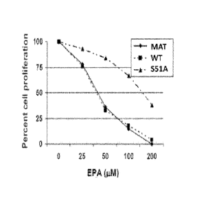
Figure 4 is a graph showing eIF2a-S51.A. expressing cells are resistant to
inhibition of cell
proliferation induced by EPA whereas proliferation of maternal PC-3 cells
(MAT) or
PC-3 cells transduced with recombinant eIF2a RFP and shRNA are sensitive to
inhibition of cell proliferation induced by EPA in a dose-dependent manner.
DETAILED DESCRIPTION OF THE INVENTION
The paradoxical observation has been made that some mRNAs are translated more
efficiently
when the ternary complex is scarce than when it is abundant (Aktas et al.,
2004, Journal of
Nutrition 134(9): 2487S-2491S; Halperin and Aktas, International Patent
Application
publication No. WO 2008/008333). These include the mRNA encoding for the
transcription
factor ATF-4, which transcriptionally up-regulates many of the ER stress
response genes such
as pro-apoptotic C/EBP-homologous protein (CHOP) or the ER chaperone binding
protein
(BiP) (Harding et al., 2000, Mal. Cell 6:1099). An isoform of the BRCA1 mRNA,
designated
mRNAb, also is more efficiently translated when the ternary complex is scarce.
It was
observed that the n-3 polyunsaturated fatty acid eicosapentaenoic acid (EPA)
up-regulated
CHOP (GenBank accession number S40706) and Glucose regulated protein 78 (BiP,
RefSeq
accession number NM 005347) in cancer cells and in tumors excised from either
animal
CA 02866175 2014-09-02
WO 2014/015328
PCT/US2013/051433
cancer models or human patients, and that it increased the translation of
BRCA1 mRNAb in
breast cancer cell lines and animal tumors.
Each of BRCA1 mRNA and the mRNA that encodes Activating Transcription Factor 4
(ATF-
4, RefSeq accession number NM_001675) contains multiple open reading frames
(ORFs) in its
5' untranslated region (5' UTR). Without intending to be bound by scientific
theory, additional
mRNAs that contain two or more ORFs in their respective 5" UTRs have now been
identified.
Such mRNAs include, without limitation, the mRNA transcripts of the genes that
encode
translationally controlled tumor protein (TCTP, RefSeq accession number NM
003295.2),
protectin (CD59) and general control nondepressible 4 (GCN4, RefSeq accession
NC_00113).
According to certain exemplary embodiments, a sample of a food, nutriceutical
or medicinal
product may be assayed for its ability to increase the presence, level or
biological activity of a
protein encoded by an mRNA transcript having multiple ORFs in its 5' UTR. In
particular,
such a sample may be assayed for its ability to mediate an increase in the
presence, level or
activity of one or more of BRCA I, ATF-4, TCTP, CD59 and GCN4.
Increased transcription of certain genes also occurs in the presence of
inhibitors of the ternary
complex. In addition to genes that encode ATF-4, BiP and CHOP, genes that
exhibit increased
transcription in the presence of inhibitors of translation initiation are
those that encode X-box
binding protein 1(Xbp-1, RefSeq accession number NM_001079539.1) and amino
acid
synthetases. Such genes provide appropriate test biomarkers for translation
initiation inhibitors
assayed according to the invention, such as those found in fish oil. These
gene transcripts can
be detected, and their levels quantitated, before and after exposure of test
animals, cells or cell-
free systems to the test food, nutriceutical or medicinal product sample by
methods known in
the art, and the levels of the transcripts compared to determine the extent to
which the test
sample facilitated transcription of the marker gene. Alternatively, the levels
of the test
biomarker transcripts can be compared to those of control transcripts (e.g.,
of housekeeping
genes) or to transcripts isolated from animals, cells or cell-free systems
exposed to standards or
controls of known biological activity. Similarly, the protein products of the
biomarker
transcripts can be detected and quantified, and their levels compared to those
of untreated
animals, cells or cell-free systems or to animals, cells or systems exposed to
a standard or
control of known biological activity.
11
CA 02866175 2014-09-02
WO 2014/015328
PCT/US2013/051433
The term "nutriceutical," as used herein, is a combination of "nutritional"
and
"pharmaceutical," and refers to an ingestible substance that has one or more
beneficial effects
on an organism such as a human. The term nutriceutical can also refer to one
or more
compounds which are present in an ingestible substance. Ingestible substances
include, but not
limited to dietary supplements, foods, beverages and the like. The terms
"nutriceutical" and
"nutritional supplement" may be used interchangeably. A substance (e.g., a
food product, a
nutriccutical product or a pharmaceutical) having beneficial biological,
nutriceutical or
medicinal properties refers to the ability of the substance to provide an
individual one or more
health benefits as described herein (e.g., in the prevention, reduction and/or
cure of one or
more diseases and/or disorders described herein).
Nutriceuticals of the present invention include oils derived from fish such as
cold water fish,
warm water fish, fresh water fish, salt water fish, brackish water fish, wild
fish, farm-raised
fish and the like, and preparations of fatty acids such as those containing
omega-3 fatty acids.
The term "omega-3 fatty acid," as used herein, refers to polyunsaturated fatty
acids such as
those found in oil from oily fish such as mackerel, salmon, sardines and the
like, or vegetable
sources such as the seeds of chia, perilla, flax, walnut, purslane,
ligonberry, scabuckthorn,
hemp, and the like, and fruits from plants such as the acai palm. Omega-3
fatty acids include,
but are not limited to, cc-linoleic acid (ALA), eicosapentaenoic acid (EPA),
docosahexaenoic
acid (DHA) and the like.
Certain aspects of the present invention are directed to methods of
determining the potency of
a composition to inhibit upregulate or modulate translation initiation or gene
transcription.
The term "potency," as used herein, is intended to include, but is not limited
to, the
effectiveness of a compound, e.g., a nutriceutical, to inhibit, upregulate or
otherwise modulate
translation initiation or gene transcription. The potency of a composition can
be defined as the
ability of the composition to inhibit upregulate or otherwise modulate
translation initiation or
gene transcription relative to a standard or control.
A standard or control of the present invention is a compound or composition
having a
translation initiation- or transcription inhibition, upregulation or
modulation activity as
determined by one or more of the bioassays described herein. Standards may be
obtained from
a variety of sources such as the sources of omega-3 fatty acids or other
agents described herein.
12
CA 02866175 2014-09-02
WO 2014/015328
PCT/US2013/051433
Standards may be synthesized in the laboratory or obtained from commercial
sources. A
standard may be diluted or concentrated to decrease or increase its
translation inhibition,
upregulation or modulation activity, respectively. Alternatively, a standard
or control may be
internal to the test system, e.g., a gene, gene promoter, mRNA transcript or
protein (e.g., a
housekeeping gene, promoter, transcript or protein) whose transcription or
translation is
substantially unaffected by the test substance, e.g., 13-actin, ubiquitin, b-
tubulin, GADPH and
the like.
In certain aspects, a standard or control is an omega-3 fatty acid, such as
eicosapentaenoic acid.
The standard or control may be derived from fish oil (e.g., marine fish oil)
or flax seed oil.
In other aspects, a standard or control is a biomarker that is substantially
insensitive to the
effects of the substance whose potency or biological activity is being
assayed. As used in this
context with respect to transcriptional regulation of a gene or gene promoter,
or translational
regulation of an mRNA transcript or protein, the term "substantially
insensitive" means either
wholly unaffected or modulated to significantly lesser extent (e.g., at least
10-fold, 100-fold,
1000-fold or greater than 1000-fold less) by the test substance than is a
biomarker for activity
of the test substance.
In certain aspects, a test sample is calibrated such that its active
components are in the linear
range and do not saturate the test system. Methods of calibrating are well
known in the art and
include simple dilutions, serial dilutions and the like.
The present invention provides assays in which the translation or
transcription inhibition,
upregulation or modulation activity of a composition is compared to a standard
using one or
more of the bioassays described herein. A composition may have an activity
level that is
0.001%, 0.01%, 0.1%, 1%, 5%, 10%, 15%, 20%, 25%,
30%, 35%, 40%, 45%, 50%, 55%,
60%, 65%, 70%, 75%, 80%, 85%, 90%, 91%, 92%, 93%, 94%, 95%, 96%, 97%, 98%,
99%,
100%, 101%, 102%, 103%, 104%, 105%, 106%, 107%, 108%, 109%, 110%, 115%, 120%,
125%, 130%, 135%, 140%, 145%, 150%, 155%, 160%, 165%, 170%, 175%, 180%, 185%,
190%, 195%, 200%, 250%, 300%, 350%, 400%, 450%, 500%, 550%, 600%, 650%, 700%,
750%, 800%, 850%, 900%, 950%, 1000%, or greater than 1000% of the activity of
the
standard or control.
13
CA 02866175 2014-09-02
WO 2014/015328
PCT/US2013/051433
In certain aspects, inhibition, upregulation or other modulation of activity
with respect to
translation initiation or gene transcription is between about 1% and 200%,
between about 5%
and 195%, between about 10% and 190%, between about 20% and 180%, between
about 30%
and 170%, between about 40% and 160%, between about 50% and 150%, between
about 60%
and 140%, between about 65% and 135%, between about 70% and 130%, between
about 75%
and 125%, between about 80% and 120%, between about 85% and 115%, between
about 90%
and 110%, between about 91% and 109%, between about 92% and 108%, between
about 93%
and 107%, between about 94% and 106%, between about 95% and 105%, between
about 96%
and 104%, between about 97% and 103%, between about 98% and 102%, or between
about
99% and 101%, of the activity of the standard or control. In other aspects,
inhibition,
upregulation or other modulation of activity with respect to translation
initiation or gene
transcription is between about 50% and about 150% of the activity of the
standard, between
about 80% and about 120% of the activity of the standard, between about 90%
and about 110%
of the activity of the standard, or between about 95% and about 105% of the
activity of the
standard or control.
The term "about" or "approximately" usually means within an acceptable error
range for the type
of value and method of measurement. For example, it can mean within 20%, more
preferably
within 10%, and most preferably still within 5% of a given value or range.
Alternatively,
especially in biological systems, the term "about" means within about a log
(i.e., an order of
magnitude) preferably within a factor or two of a given value.
In certain embodiments of the present invention, a control or standard may
have zero activity.
Thus, a binary result (i.e., positive or negative) may be obtained for a given
activity. In such a
case, if precise quantitation of activity is needed, it would be measured on
an absolute scale or
in comparison to a standard that has at least some activity of a known level.
A nutriceutical or composition including a nutriceutical of the present
invention may be diluted
or concentrated to decrease or increase its translation or transcription
inhibition, upregulation
or modulation activity relative to the control/standard, respectively.
The present invention also provides assays in which batch or lot homogeneity
of compositions
is determined by comparing the relative activity of two or more (e.g., 10,
100, 1000, 10,000
1,000,000 or more) compositions using one or more of the bioassays described
herein. As
14
CA 02866175 2014-09-02
WO 2014/015328
PCT/US2013/051433
used herein, the terms "batch homogeneity" or "lot homogeneity" are intended
to refer, but are
not limited to, the relative translation initiation inhibition upregulation or
modulation activity,
or transcriptional upregulation activity, of two or more compositions in a
batch or lot. As used
herein, the terms "batch" or "lot" refer, but are not limited to, a group of
two or more
compositions. A batch or lot includes compositions prepared together or
compositions from
two or more sources (e.g., geographical, plant, animal, commercial, and/or
synthetic sources).
As used herein, the term "batch" or "lot" also may refer to a single pool of a
composition, from
which units of products or test samples are to be drawn or produced, or which
will be
otherwise further divided or fractionated.
In at least certain examples, the nutriceuticals disclosed herein can be used
in the treatment of
disorders associated with aberrant cellular proliferation such as cellular
proliferative disorders,
(e.g., cancer). Treatment of cellular proliferative disorders is intended to
include inhibition of
proliferation including rapid proliferation. As used herein, the term
"cellular proliferative
disorder" includes disorders characterized by undesirable or inappropriate
proliferation of one
or more subset(s) of cells in a multicellular organism. The term "cancer"
refers to various
types of malignant neoplasms, most of which can invade surrounding tissues,
and may
metastasize to different sites (see, for example, PDR Medical Dictionary 1st
edition, 1995).
The terms "neoplasm" and "tumor" refer to an abnormal tissue that grows by
cellular
proliferation more rapidly than normal and continues to grow after the stimuli
that initiated
proliferation is removed (see, for example, PDR Medical Dictionary 1st
edition, 1995). Such
abnormal tissue shows partial or complete lack of structural organization and
functional
coordination with the normal tissue which may be either benign (i.e., benign
tumor) or
malignant (i.e., malignant tumor).
The language "treatment of cellular proliferative disorders" is intended to
include the
prevention of the induction, onset, establishment or growth of neoplasms in a
subject or a
reduction in the growth of pre-existing neoplasms in a subject. The language
also can describe
inhibition of the invasion of neoplastie cells into neighboring tissues or the
metastasis of a
neoplasm from one site to another. Examples of the types of neoplasms intended
to be
encompassed by the present invention include but are not limited to those
neoplasms
associated with cancers of the breast, skin, bone, prostate, ovaries, uterus,
cervix, liver, lung,
CA 02866175 2014-09-02
WO 2014/015328
PCT/US2013/051433
brain, larynx, gallbladder, pancreas, rectum, parathyroid, thyroid, adrenal
gland, immune
system, neural tissue, head and neck, colon, stomach, bronchi, and/or kidneys.
Cellular proliferative disorders can further include disorders associated with
hyperproliferation
of vascular smooth muscle cells such as proliferative cardiovascular
disorders, e.g.,
atherosclerosis and restenosis. Cellular proliferation disorders can also
include disorders such
as proliferative skin disorders, e.g., X-linked ichthyosis, psoriasis, atopic
dermatitis, allergic
contact dermatitis, epidermolytic hyperkeratosis, and seborrheic dermatitis.
Cellular
proliferative disorders can further include disorders such as autosomal
dominant polycystic
kidney disease (ADPKD), mastocystosis, and cellular proliferation disorders
caused by
infectious agents such as viruses.
In at least certain examples, the nutriceuticals assayed and/or produced
according to the
methods disclosed herein can be used in the treatment of disorders associated
with energy
balance, such as metabolic disorders including, but not limited to, diabetes,
obesity, glycogen
storage diseases, lipid storage disorders, mitochondrial diseases and the like
(see also the
Worldwide Website: emedicine.com/ped/GENETICS_AND_ METABOLIC_DISEASE.htm).
In certain aspects, the nutriceuticals assayed and/or produced according to
the methods
disclosed herein modulate weight gain by interacting with the 5' UTR of the
leptin receptor.
Detection methods described herein can be used to detect one or more DNA
sequences, RNA
sequences, proteins or polypeptides of interest in a biological sample in
vitro as well as in vivo.
For example, in vitro techniques for detection of mRNA include Northern
hybridizations and
in situ hybridizations. In vitro techniques for detection of a polypeptide
corresponding to a
marker of the invention include enzyme linked immunosorbent assays (ELISAs),
Western
blots, immunoprecipitations and immunofluorescence. In vitro techniques for
detection of
genomic DNA include Southern hybridizations. Furthermore, in vivo techniques
for detection
of a protein and/or polypeptide include introducing into a subject a labeled
antibody directed
against the protein and/or polypeptide. For example, the antibody can be
labeled with a
radioactive marker whose presence and location in a subject can be detected by
standard
imaging techniques.
A general principle of detection and/or quantification involves preparing a
sample or reaction
mixture that may contain one or more DNA sequences, RNA sequences, proteins or
16
CA 02866175 2014-09-02
WO 2014/015328
PCT/US2013/051433
polypeptides of interest and a probe under appropriate conditions and for a
time sufficient to
allow the marker and probe to interact and bind, thus forming a complex that
can be removed
and/or detected in the reaction mixture. These assays can be conducted in a
variety of ways.
For example, one method to conduct such an assay would involve anchoring the
DNA
sequence, RNA sequence, protein or polypeptide of interest or a probe onto a
solid phase
support, also referred to as a substrate, and detecting target DNA sequence,
RNA sequence,
protein or polypeptide of interest/probe complexes anchored on the solid phase
at the end of
the reaction. In one embodiment of such a method, a sample which is to be
assayed for
presence and/or concentration of marker, can be anchored onto a carrier or
solid phase support.
In another embodiment, the reverse situation is possible, in which the probe
can be anchored to
a solid phase and a sample from a subject can be allowed to react as an
unanchored component
of the assay.
There are many established methods for anchoring assay components to a solid
phase. These
include, without limitation, marker or probe molecules which are immobilized
through
conjugation of biotin and streptavidin. Such biotinylated assay components can
be prepared
from biotin-NHS(N-hydroxy-succinimidc) using techniques known in the art
(e.g.,
biotinylation kit, Pierce Chemicals, Rockford, IL), and immobilized in the
wells of
streptavidin-coated 96 well plates (Pierce Chemical). In certain embodiments,
the surfaces
with immobilized assay components can be prepared in advance and stored.
Other suitable carriers or solid phase supports for such assays include any
material capable of
binding the class of molecule to which the marker or probe belongs. Well known
supports or
carriers include, but are not limited to, glass, polystyrene, nylon,
polypropylene, nylon,
polyethylene, dextran, amylases, natural and modified celluloses,
polyacrylamides, gabbros,
and magnetite.
In order to conduct assays with the above mentioned approaches, the non-
immobilized
component is added to the solid phase upon which the second component is
anchored. After
the reaction is complete, uncomplexed components may be removed (e.g., by
washing) under
conditions such that any complexes formed will remain immobilized upon the
solid phase.
The detection of DNA sequence, RNA sequence, protein or polypeptide of
interest/probe
17
CA 02866175 2014-09-02
WO 2014/015328
PCT/US2013/051433
complexes anchored to the solid phase can be accomplished in a number of
methods outlined
herein.
In certain exemplary embodiments, the probe, when it is the unanchored assay
component, can
be labeled for the purpose of detection and readout of the assay, either
directly or indirectly,
with detectable markers which are well-known to one skilled in the art.
Examples of
detectable markers include various radioactive moieties, enzymes, prosthetic
groups,
fluorescent markers, luminescent markers, bioluminescent markers, metal
particles, protein-
protein binding pairs, protein-antibody binding pairs and the like. Examples
of fluorescent
proteins include, but are not limited to, yellow fluorescent protein (YFP),
green fluorescence
protein (GFP), cyan fluorescence protein (CFP), umbelliferone, fluorescein,
fluorescein
isothiocyanate, rhodamine, dichlorotriazinylamine fluorescein, dansyl
chloride, phycoerythrin
and the like. Examples of bioluminescent markers include, but are not limited
to, luciferase
(e.g., bacterial, firefly, click beetle and the like), luciferin, aexporin and
the like. Examples of
enzyme systems having visually detectable signals include, but are not limited
to,
galactosidases, glucorinidases, phosphatases, peroxidases, cholinesterases and
the like.
Identifiable markers also include radioactive compounds such as 125T, 35S,
'4C,
3H or 32P.
Identifiable markers are commercially available from a variety of sources.
Fluorescent labels and their attachment to nucleotides and/or oligonucleotides
are described in
many reviews, including Haugland, Handbook of Fluorescent Probes and Research
Chemicals, Ninth Edition (Molecular Probes, Inc., Eugene, 2002); Keller and
Manak, DNA
Probes, 2nd Edition (Stockton Press, New York, 1993); Eckstein, editor,
Oligonucleotides and
Analogues: A Practical Approach (IRL Press, Oxford, 1991); and Wetmur,
Critical Reviews in
Biochemistry and Molecular Biology, 26:227-259 (1991). Particular
methodologies applicable
to the invention are disclosed in the following sample of references: U.S.
Patent Nos.
4,757,141, 5,151,507 and 5,091,519. In one aspect, one or more fluorescent
dyes are used as
labels, e.g., as disclosed by U.S. Patent Nos. 5,188,934 (4,7-
dichlorofluorescein dyes);
5,366,860 (spectrally resolvable rhodamine dyes); 5,847,162 (4,7-
dichlororhodamine dyes);
4,318,846 (ether-substituted fluorescein dyes); 5,800,996 (energy transfer
dyes); Lee et al.;
5,066,580 (xanthine dyes); 5,688,648 (energy transfer dyes); and the like.
Labelling can also
be carried out with quantum dots, as disclosed in the following patents and
patent publications:
U.S. Patent Nos. 6,322,901, 6,576,291, 6,423,551, 6,251,303, 6,319,426,
6,426,513, 6,444,143,
18
CA 02866175 2014-09-02
WO 2014/015328
PCT/US2013/051433
5,990,479, 6,207,392, 2002/0045045 and 2003/0017264. As used herein, the term
"fluorescent
label" includes a signaling moiety that conveys information through the
fluorescent absorption
and/or emission properties of one or more molecules. Such fluorescent
properties include
fluorescence intensity, fluorescence lifetime, emission spectrum
characteristics, energy
transfer, and the like.
In another embodiment, determination of the ability of a probe to recognize a
marker can be
accomplished without labeling either assay component (probe or marker) by
utilizing a
technology such as real-time Biomolecular Interaction Analysis (BIA) (see,
e.g., Sjolander et
al. (1991) Anal. Chem. 63:2338 2345 and Szabo et al. (1995) Curr. Opin.
Struct. Biol. 5:699
705). As used herein, "BIA" or "surface plasmon resonance" is a technology for
studying
biospecific interactions in real time, without labeling any of the
interactants (e.g., BIAcore).
Changes in the mass at the binding surface (indicative of a binding event)
result in alterations
of the refractive index of light near the surface (the optical phenomenon of
surface plasmon
resonance (SPR)), resulting in a detectable signal which can be used as an
indication of real-
time reactions between biological molecules.
Alternatively, in another embodiment, analogous detection and/or
quantification assays can be
conducted with one or more DNA sequences, RNA sequences, proteins or
polypeptides of
interest and probe as solutes in a liquid phase. In such an assay, the
complexed DNA
sequence, RNA sequence, protein or polypeptide of interest and probe are
separated from
uncomplexed components by any of a number of standard techniques, including
but not limited
to: differential centrifugation, chromatography, electrophoresis and
immunoprecipitation. In
differential centrifugation, DNA sequence, RNA sequence, protein or
polypeptide of interest
/probe complexes may be separated from uncomplexed assay components through a
series of
centrifugal steps, due to the different sedimentation equilibria of complexes
based on their
different sizes and densities (see, for example, Rivas and Minton (1993)
Trends Bioehem Sei.
18:284). Standard chromatographic techniques may also be utilized to separate
complexed
molecules from uncomplexed ones. For example, gel filtration chromatography
separates
molecules based on size, and through the utilization of an appropriate gel
filtration resin in a
column format; for example, the relatively larger complex may be separated
from the relatively
smaller uncomplexed components. Similarly, the relatively different charge
properties of the
DNA sequence, RNA sequence, protein or polypeptide of interest /probe complex
as compared
19
CA 02866175 2014-09-02
WO 2014/015328
PCT/US2013/051433
to the uncomplexed components may be exploited to differentiate the complex
from
uncomplexed components, for example through the utilization of ion-exchange
chromatography resins. Such resins and chromatographic techniques are well
known to one
skilled in the art (see, e.g., Heegaard (1998)J. Mol. Recognit. 11:141; Hage
and Tweed (1997)
J Chromatogr. B. Biomed. Sci. Appl. 12:499). Gel electrophoresis may also be
employed to
separate complexed assay components from unbound components (see, e.g.,
Ausubel et al., ed.,
Current Protocols in Molecular Biology, John Wiley & Sons, New York, 1987
1999). In this
technique, protein or nucleic acid complexes are separated based on size or
charge, for
example. In order to maintain the binding interaction during the
electrophoretic process, non-
denaturing gel matrix materials and conditions in the absence of reducing
agent are typically
preferred. Appropriate conditions to the particular assay and components
thereof will be well
known to one skilled in the art.
In certain exemplary embodiments, the level of an mRNA sequence of interest
can be
determined either by in situ and/or by in vitro formats in a biological sample
using methods
known in the art. Many expression detection methods use isolated RNA. For in
vitro methods,
any RNA isolation technique that does not select against the isolation of mRNA
can be utilized
for the purification of RNA from blood cells (see, e.g., Ausubel et al, ed.,
Current Protocols in
Molecular Biology, John Wiley & Sons, New York 1987 1999). Additionally, large
numbers
of cells and/or samples can readily be processed using techniques well known
to those of skill
in the art, such as, for example, the single-step RNA isolation process of
Chomczynski (1989,
U.S. Patent No. 4,843,155).
Isolated mRNA can be used in hybridization or amplification assays that
include, but are not
limited to, Southern or Northern analyses, polymerase chain reaction analyses
and probe
arrays. In certain exemplary embodiments, a diagnostic method for the
detection of mRNA
levels involves contacting the isolated mRNA with a nucleic acid molecule
(probe) that can
hybridize to the mRNA encoded by the gene being detected. The nucleic acid
probe can be,
for example, a full-length cDNA, or a portion thereof, such as an
ohgonucleotide of at least 7,
15, 30, 50, 100, 250 or 500 nucleotides in length and sufficient to
specifically hybridize under
stringent conditions to an mRNA or genomic DNA encoding a marker of the
present invention.
Other suitable probes for use in the diagnostic assays of the invention are
described herein.
CA 02866175 2014-09-02
WO 2014/015328
PCT/US2013/051433
In one format, the mRNA is immobilized on a solid surface and contacted with a
probe, for
example by running the isolated mRNA on an agarose gel and transferring the
mRNA from the
gel to a membrane, such as nitrocellulose. In an alternative format, the
probe(s) are
immobilized on a solid surface and the mRNA is contacted with the probe(s),
for example, in a
gene chip array. A skilled artisan can readily adapt known mRNA detection
methods for use
in detecting the level of mRNA encoded by the markers of the present
invention.
An alternative method for determining the level of mRNA corresponding to a
marker of the
present invention in a sample involves the process of nucleic acid
amplification, e.g., by rtPCR
(the experimental embodiment set forth in U.S. Patent Nos. 4,683,195 and
4,683,202), COLD-
PCR (Li et al. (2008) Nat. Med. 14:579), ligase chain reaction (Barany, 1991,
Proc. Natl.
Acad. Set. USA, 88:189), self sustained sequence replication (Guatelli et al.,
1990, Proc. Natl.
Acad. Set. USA 87:1874), transcriptional amplification system (Kwoh et al.
(1989) Proc.. NatL
Acad. ScL USA 86:1173), Q-Beta Replicase (Lizardi et al. (1988) Bio/Technology
6:1197),
rolling circle replication (U.S. Patent No. 5,854,033) or any other nucleic
acid amplification
method, followed by the detection of the amplified molecules using techniques
well known to
those of skill in the art. These detection schemes are especially useful for
the detection of
nucleic acid molecules if such molecules are present in very low numbers. As
used herein,
amplification primers are defined as being a pair of nucleic acid molecules
that can anneal to 5'
or 3' regions of a gene (plus and minus strands, respectively, or vice-versa)
and contain a short
region in between. In general, amplification primers are from about 10 to 30
nucleotides in
length and flank a region from about 50 to 200 nucleotides in length. Under
appropriate
conditions and with appropriate reagents, such primers permit the
amplification of a nucleic
acid molecule comprising the nucleotide sequence flanked by the primers.
For in situ methods, mRNA does not need to be isolated from the sample (e.g.,
a bodily fluid
(e.g., blood cells)) prior to detection. In such methods, a cell or tissue
sample is
prepared/processed using known histological methods. The sample is then
immobilized on a
support, typically a glass slide, and then contacted with a probe that can
hybridize to mRNA
that encodes the marker.
As an alternative to making determinations based on the absolute expression
level of the DNA
sequence, RNA sequence, protein or polypeptide of interest, determinations may
be based on
the normalized expression level of the DNA sequence, RNA sequence, protein or
polypeptide
21
CA 02866175 2014-09-02
WO 2014/015328
PCT/US2013/051433
of interest. Expression levels are normalized by correcting the absolute
expression level of a
DNA sequence, RNA sequence, protein or polypeptide of interest by comparing
its expression
to the expression of a gene that is not a marker, e.g., a standard or control.
This normalization
allows the comparison of the expression level in a sample from one source to a
sample from
another source.
In another exemplary embodiment, a protein or polypeptide is detected. In
certain exemplary
embodiments, an agent for detecting a polypeptide of the invention is an
antibody capable of
binding to a polypeptide corresponding to a marker of the invention, such as
an antibody with a
detectable label. Antibodies can be polyclonal, or more preferably,
monoclonal. An intact
antibody, or a fragment thereof (e.g., Fab or F(ab')2) can be used. The term
"labeled," with
respect to the probe or antibody, is intended to encompass direct labeling of
the probe or
antibody by coupling (i.e., physically linking) a detectable substance to the
probe or antibody,
as well as indirect labeling of the probe or antibody by reactivity with
another reagent that is
directly labeled. Examples of indirect labeling include detection of a primary
antibody using a
fluorescently labeled secondary antibody and end-labeling of a DNA probe with
biotin such
that it can be detected with fluorescently labeled streptavidin.
Polyclonal antibodies can be prepared by immunizing a suitable subject with a
protein or
polypeptide of choice. The protein of choice titer in the immunized subject
can be monitored
over time by standard techniques, such as with an enzyme linked immunosorbent
assay
(ELISA) using immobilized protein. If desired, the antibody molecules directed
against the
protein of choice can be isolated from the mammal (e.g., from the blood) and
further purified
by well known techniques, such as protein A chromatography to obtain the IgG
fraction. At an
appropriate time after immunization, e.g., when the anti-protein of choice
antibody titers are
highest, antibody-producing cells can be obtained from the subject and used to
prepare
monoclonal antibodies by standard techniques, such as the hybridoma technique
originally
described by Kohler and Milstein (1975) Nature 256:495-497) (see also, Brown
et al. (1981) J.
Immunol. 127:539-46; Brown ct al. (1980) J. Biol. Chem. 255:4980-83; Yeh et
al. (1976) Proc.
Natl, Acad. Sci. USA 76:2927-31; and Yeh et al. (1982) Int. J. Cancer 29:269-
75), the human
B cell hybridoma technique (Kozbor et al. (1983) Immunol. Today 4:72), the EBV-
hybridoma
technique (Cole et al. (1985), Monoclonal Antibodies and Cancer Therapy, Alan
R. Liss, Inc.,
pp. 77-96) or trioma techniques. The technology for producing monoclonal
antibody
22
CA 02866175 2014-09-02
WO 2014/015328
PCT/US2013/051433
hybridomas is well known (see generally R. H. Kenneth, in Monoclonal
Antibodies: A New
Dimension In Biological Analyses, Plenum Publishing Corp., New York, N.Y.
(1980); E. A.
Lerner (1981) Yale J Biol. Med. 54:387-402; Gefter et al. (1977) Somatic Cell
Genet. 3:231-
36). Briefly, an immortal cell line (typically a myeloma) is fused to
lymphocytes (typically
.. splenocytes) from a mammal immunized with a protein of choice as described
above, and the
culture supernatants of the resulting hybridoma cells are screened to identify
a hybridoma
producing a monoclonal antibody that binds the protein of choice.
A variety of formats can be employed to determine whether a sample contains a
protein that
binds to a given antibody. Examples of such formats include, but are not
limited to, enzyme
immunoassay (ER), radioimmunoassay (R1A), Western blot analysis, enzyme linked
immunoabsorbant assay (ELISA) and the like. A skilled artisan can readily
adapt known
protein/antibody detection methods for use in determining whether cells (e.g.,
bodily fluid cells
such as blood cells) express a marker of the present invention.
In one format, antibodies, or antibody fragments, can be used in methods such
as Western blots
.. or immunofluorescence techniques to detect the expressed proteins. In such
uses, it is
generally preferable to immobilize either the antibody or proteins on a solid
support. Suitable
solid phase supports or carriers include any support capable of binding an
antigen or an
antibody. Well known supports or carriers include glass, polystyrene,
polypropylene,
polyethylene, dextran, nylon, amylases, natural and modified celluloses,
polyacrylamides,
gabbros, magnetite and the like.
One skilled in the art will know many other suitable carriers for binding
antibody or antigen,
and will be able to adapt such support for use with the present invention. For
example, protein
isolated from cells (e.g., bodily fluid cells such as blood cells) can be run
on a polyacrylamide
gel electrophoresis and immobilized onto a solid phase support such as
nitrocellulose. The
.. support can then be washed with suitable buffers followed by treatment with
the detectably
labeled antibody. The solid phase support can then be washed with the buffer a
second time to
remove unbound antibody. The amount of bound label on the solid support can
then be
detected by conventional means.
In certain exemplary embodiments, assays of the invention may be performed in
animal
models (including, but not limited to horses, cows, sheep, pigs, goats,
rabbits, guinea pigs, rats,
23
CA 2866175 2019-08-28
mice, gerbils, non-human primates and the like), cells (e.g., cells from
microorganisms (e.g.,
bacterial cells, viral cells, yeast cells and the like)) or cell-free systems
(e.g., in vitro
transcription assays, in vitro translation assays, cell lysate assays,
fractionated cell lysate
assays and the like).
It is to be understood that the embodiments of the present invention which
have been described
are merely illustrative of some of the applications of the principles of the
present invention.
Numerous modifications may be made by those skilled in the art based upon the
teachings
presented herein without departing from the true spirit and scope of the
invention.
The following examples are set forth as being representative of the present
invention. These
examples are not to be construed as limiting the scope of the invention as
these and other
equivalent embodiments will be apparent in view of the present disclosure,
figures, tables, and
accompanying claims.
EXAMPLE I
Preparation of Samples for Bioassay
The active ingredient of many nutriceuticals such as fish oil is released upon
digestion. It is
therefore necessary to mimic this digestion in the test tube in order to test
the in vitro activity
of fish oil (such as cell culture). There are several ways of achieving this,
one such method is
described below as a non-limiting example.
Fish Oil Hydrolysis
Fish Oil (10g. ¨12 mmol) and NaOH (2.16g. 54 mmol) were mixed in water (50
ml), absolute
ethanol (70 ml), and toluene (10 ml). The mixture was magnetically stirred and
refluxed under
N2 for 1.5h. The reaction mixture was cooled to room temperature, treated with
IN Ha (81
ml) and extracted with n-hexane (100 m1). The organic phase was washed with a
mixture of
ethanol/water (1:1, v/v) until reaching an aqueous phase of pH 5. The
separated organic phase
was dried over anhydrous Na2SO4, filtered and the solvent removed under vacuum
at room
24
CA 02866175 2014-09-02
WO 2014/015328
PCT/US2013/051433
temperature. The residue obtained is the fish oil hydrolysate that is
subjected to quantitative
composition analysis and biological activity characterization.
EXAMPLE 2
Detection of biomarker mRNA by real time PCR
Real-time PCR is a quantitative method for detecting changes in the levels of
specific RNAs;
therefore, real-time PCR for pro-apoptotic or tumor suppressor genes
transcriptionally
upregulated in the presence of inhibitors of the ternary complex provides a
rapid and accurate
quantitative assay for evaluating the availability of the ternary complex, and
is an effective
surrogate assay for detection of the phosphorylation of eIF2a induced by omega-
3 fatty acids.
It has been determined that this new assay also has shown remarkable
correlation with those
obtained through use of an existing ATF-4 cell-based assay that is highly
dependent on
availability of the ternary complex. It is, therefore, an improved method for
quality-control
and assurance of food, nutriceutical and medicinal products with respect to
the activity of
omega-3 fatty acids and other beneficial compounds that influence availability
of the ternary
complex to initiate mRNA translation.
Standard Real-Time PCR Assay
1. Plate cells of either human mouse or rat origin such as, e.g., rat
hepatocytes, mouse or
human fibroblast grown in standard culture media such as, e.g., DMEM or RPMI
1640 with 5-
10% fetal bovine or bovine calf serum (either three wells in 6-well or 100 mm
plate, or other
container) for each condition;
2. Treat with compound to be evaluated or with control/standard vehicle;
3. Harvest cells after six hours;
4. Isolate RNA;
5. Reverse transcribe RNA;
6. Amplify reverse transcripts of biomarker mRNA (e.g., that which encodes
CHOP, BiP,
ATF-4, Xbp-1 or an amino acid synthetase) and those of 18S RNA (internal
standard);
CA 02866175 2014-09-02
WO 2014/015328
PCT/US2013/051433
7. Quantify amount of biomarker reverse transcript after normalization
against 18S
reverse transcript; and
8. Compare amounts of biomarker reverse transcript across differently-
treated samples
(e.g., treated with test compounds or vehicle).
In Cell, Real Time PCR Assay
1. Plate cells (e.g., in 96-well plates or other multi-chamber format);
2. Treat with different doses of compounds or vehicle;
3. Lyse cells after 6 hours;
4. Reverse transcribe in the same wells;
5. Amplify reverse transcript of biomarker mRNA (e.g., that which encodes
CHOP, BiP,
ATF-4, Xbp-1 or an amino acid synthetase) and that of 18S RNA in the same
well;
6. Quantify amount of biomarker reverse transcript after normalization
against 18S
reverse transcript; and
7. Compare amounts of biomarker reverse transcript across differently-
treated samples
(e.g., treated with test compounds or vehicle).
Results obtained when a CHOP-encoding mRNA transcript was amplified are shown
in Figure
1, in comparison to results obtained in the existing ATF-4 assay.
EXAMPLE 3
Detection of Transcriptional Activity of Biomarker Genes via Reporter Gene
Assay
Another means by which to assay the presence and activity of omega-3 fatty
acids in food,
nutriceutical and medicinal compositions is to measure upregulation of marker
gene
transcriptional activity using reporter gene constructs. According to this
method, each such
construct contains a nucleic acid sequence that encodes a reporter protein
(e.g., luciferase,
Green Fluorescent Protein, Red, far Red, dsRed, dsRed2, orange, yellow, cyan,
beta
galactosidase, horseradish peroxidase, aquaporins, chloramphenicol acetyl
transferase, or other
26
CA 02866175 2014-09-02
WO 2014/015328
PCT/US2013/051433
protein that generates a detectable signal or has an enzymatic or other
activity that is
susceptible to detection by methods known to those of skill in the art).
Operably linked to the
reporter protein coding sequence of the construct is a naturally-occurring or
synthetic promoter
region that is transcriptionally upregulated in the presence of omega-3 fatty
acids or other
suitable inhibitors of mRNA translation initiation, e.g., ternary complex
inhibitors. Suitable
promoters include, in non-limiting fashion, those of the genes that encode
biomarkers such as
CHOP, BiP, ATF-4, Xbp-1 or amino acid synthetases. Under conditions that
permit nucleic
acid transcription and mRNA translation to occur, the system is contacted or
treated with a test
sample. Suitable negative controls include, but are not limited to, a parallel
test system not so
contacted or treated, or one that is contacted or treated with an appropriate
standard sample. In
both the test and control systems, reporter protein function is detected and
quantified by
methods well known in the art, and the levels of reporter function in the test
and standard
systems are compared to determine the potency of omega-3 fatty acids present
in the test
sample. In such an assay, the signal obtained using an ATF-4 promoter
construct or CHOP
promoter construct that also includes the native ATF-4 5.UTR is amplified to a
greater degree
than would those resulting from use of the other promoters, as ATF-4 is both
transcriptionally
and translationally upregulated in the presence of omega-3 fatty acids.
Reporter constructs
may be designed with this in mind, so as to keep signal levels in the linear
range for the level
of reporter activity that is anticipated to result from exposure to test
samples, depending on
their estimated potency prior to assay, e.g., based on results obtained with
samples or
comparable origin, processing or the like.
Certain reporter constructs of the present invention also combine high
efficiency transcription
promoters and high efficiency translation 5'UTRs, such as with the CHOP
promoter and ATF-
4 5'UTR, to provide signal amplification that is preferably geometric thereby
providing an
advantageous signal-to-noise ratio compared to reporter constructs including
only one of the
two elements. Such a reporter construct is particularly useful in assays where
high sensitivity
is desired such as when comparing dilute or weakly-positive samples commonly
encountered
during early stages of processing or when detecting a rare activity. Promoters
and 5'UTRs can
be combined into a single reporter construct using methods known to those of
skill in the art
and as described herein. Useful promoters include, in non-limiting fashion,
those of the genes
that encode biomarkers such as CHOP, BiP, ATF-4, Xbp-1 or amino acid
synthetases and the
like and others known in the art and described herein. Useful 5'UTRs include
ATF-4, BRCA1
27
CA 02866175 2014-09-02
WO 2014/015328
PCT/US2013/051433
mRNAb, CD59, TCTP and GCN4 and the like and others known in the art and
described
herein. A useful reporter construct including both elements are particularly
advantageous
when the selected elements, i.e. 5'UTR and promoter pairings, respond to the
same or similar
agent or signal.
EXAMPLE 4
Detection of Increased Translation of Biomarker Proteins
To determine the potency of omega-3 fatty acids or other beneficial agents in
food,
nutriccutical or medicinal products, mRNA transcripts in which 5'UTR sequences
containing
two or more ORFs are operably linked to sequences that encode reporter
proteins are exposed
to conditions under which protein translation is permitted to occur, e.g., an
animal, cell or cell-
free translation system, such as a rabbit reticulocyte lysate or other in
vitro system containing
the cellular components necessary to effect translation of mRNA to protein.
The transcripts
may be produced within the system (e.g., expressed in an animal, cell or other
mixture that
contains the reporter construct and an appropriate nucleic acid polymerase,
e.g., an RNA
polymerase), or may be exogenously produced and added to the system. The
system
optionally may contain an internal or other control reporter mRNA whose
translational
efficiency is not affected by the presence of omega-3 fatty acids or other
beneficial agents that
the assay is intended to detect, thereby allowing for levels of test and
control translational
activity to be normalized against the relative amounts of test and control
transcripts available to
be translated. Alternatively, reporter mRNA levels may be normalized between
various test
samples and levels of reporter function compared.
Appropriate 5'UTRs for test transcripts include, in non-limiting fashion,
those of the genes or
mRNA transcripts that encode biomarker proteins such as BRCA1, ATF-4, TCTP,
CD59 or
GCN4. Appropriate 5'UTRs for control transcripts may be drawn from genes or
mRNA
transcripts that encode housekeeping proteins and/or that have one or fewer
(i.e., zero) ORFs in
their respective 5'UTRs.
The systems described above are treated or contacted with a sample of a food,
nutriceutical or
medicinal composition, and the amounts of test and control reporter protein
function (e.g.,
within a test sample; between a test sample and an untreated sample; between a
test sample and
28
CA 02866175 2014-09-02
WO 2014/015328
PCT/US2013/051433
a sample treated with a standard for potency of an omega-3 fatty acid or other
beneficial
agent), detected and quantified by methods well known in the art, wherein
elevated levels of
reporter function correlate positively with translational activity of the test
reporter mRNAs
and, consequently, potency of an omega-3 fatty acid or other beneficial agent
contained in the
test sample.
EXAMPLE 5
Manufacture of Quality-Controlled Nutriceutical and other Products from Fish
Oil
As mentioned above, fish oil is a significant source of omega-3 fatty acids;
however, supplies
of fish oil differ greatly from one another in content and bioactivity of
these beneficial
compounds. The invention provides methods for the manufacture of fish-oil-
derived products
that possess known, uniform bioactivity of omega-3 fatty acids.
Fish are caught and, while live or fresh-killed, pressed under food-grade
manufacturing
conditions to effect extraction of oil from the flesh. Heavy metals and other
environmental
contaminants are removed by filtration, chelation and/or other methods known
to those of skill
in the relevant art. Optionally, fish oil then may be further processed, for
example, to improve
taste, aroma and/or appearance, to add other beneficial agents (including, in
non-limiting
fashion, phytosterols or other beneficial compounds) or to concentrate omega-3
fatty acids
and/or other beneficial agents. Optionally, unprocessed, partially-processed
(e.g., detoxified),
fractionated or otherwise processed fish oil may be packaged, for example, in
bottles or other
non-consumable containers, or in consumable containers, such as food- or
pharmaceutical
grade capsules, e.g., gel capsules or caplets. Optionally, omega-3 fatty acids
and/or other
beneficial agents contained in fish oil may be enriched, partially purified or
even fully purified,
i.e., isolated.
At one or more of the aforementioned stages of manufacture, the ability of the
fish oil,
intermediate product or finished product to effect increased transcription of
one or more of the
genes that encode biomarkers such as ATF-4, CHOP, BiP, Xbp-1 or amino acid
synthetases,
or, alternatively, increased translation of mRNAs containing in their 5'UTRs
two or more
ORFs, including, in non-limiting fashion, 5'UTRs from mRNAs that encode
biomarkers such
as BRCA1, ATF-4, TCTP, CD59 or GCN4, is assayed using the methods described in
the
preceding Examples herein. Assay results obtained during early stages of
manufacture may
29
CA 02866175 2014-09-02
WO 2014/015328
PCT/US2013/051433
enable adjustments to be made in concentration of omega-3 fatty acids during
further
production steps, or may otherwise guide assembly, mixture or formulation of
product
components to result in a food, nutriceutical or medicinal product of known
potency with
regard to omega-3 fatty acid bioactivity. Optionally, samples of the finished
product (e.g., an
aliquot of a liquid or powder, or a single caplet, each representative of the
batch or lot from
which is has been drawn or selected) may be assayed prior to distribution,
thereby enabling
label or other marketing claims to be made with respect to the level of a
beneficial biological,
nutriceutical or medicinal property possessed by the product, e.g., with
respect to the inhibition
of translation initiation, or therapeutic or preventive properties associated
with the inhibition,
.. upregulation or other modulation of biomarker gene transcription or mRNA
translation.
EXAMPLE 6
Development or robust, sensitive cell-based assays that allow for the
quantification of
the anti-cancer biological activity of nutriceutical-grade fish oil (NFO)
preparations/batches.
Study Design:
Generation of transgenic human prostate cancer cell lines expressing mutant
(eIF2a-
S51A EPA-resistant) or wild type (eIF2a -WT2 EPA-sensitive) eIF2a.
Objective: To determine the cause-effect relationship between phosphorylation
of eIF2a and
anti-cancer activity of n-3 PUFAs in human prostate cancer cell lines. Cells
were engineered
.. that express non-phosphorylatable mutant (eIF2a-S5 1A) or recombinant wild
type eIF2a
(e1F2a-WT) and red fluoroscent or green fluorescent proteins, respectively in
the absence of
endogenous eIF2a. To differentiate recombinant eIF2a proteins from endogenous
eIF2a
proteins, recombinant eIF2a was N-terminally tagged with a hemagglutinin (HA)
tag to
ensure co-expression of recombinant eIF2a (S5 lA mutant or WT) and the
fluorescent protein.
A recently disclosed technique was used wherein two proteins can be translated
at a 1:1 ratio.
This is accomplished by cloning their coding sequences as a single
monocistronic mRNA
provided that the amino acid sequences of these two proteins are separated by
a protease 2A
cut site. In other words, a pro-protein translated from a single Open reading
frame (ORF) is
cut by the protease 2A to generate HA-tagged eIF2a (WT or S5 IA) and RFP or
GFP proteins.
CA 02866175 2014-09-02
WO 2014/015328
PCT/US2013/051433
In the specific construct herein, cleavage by protease 2A created HA-tagged
native eIF2a and
a fusion of fluorescent protein with the protease 2A recognition sequence. In
order to silence
endogenous eIF2a with shRNA without affecting recombinant eIF2a (WT or S51A),
all 5'
and 3'UTR elements of the eIF2a gene were excised from the plasmid.
Experimental Design: The design required replacement of endogenous eIF2a with
recombinant eIF2a (WT or S51A) protein to be temporally regulated. To
accomplish this a
pLVTHM lentiviral vector was utilized. This vector contains a human elongation
factor 1
promoter-controlled cassette for expression of an ORF in mammalian cells and a
viral
LTR/SIN-controlled cassette for shRNA mediated gene silencing. eIF2a-WT in
tandem with
GFP and eIF2a-51A in tandem with RFP coding sequences were used. The protease
2A cut
site was inserted between eIF2a (WT or S51A) and fluorescent proteins. RFP and
GFP used
to tag the cells. These two reporters were selected because they can easily be
distinguished
under the microscope using appropriate filters in vitro and in vivo. For
optimal translation,
the ORFs were preceded by a perfect Kozak consensus sequence (GCCACCATGG). To
identify the best shRNA sequence that targets endogenous but not recombinant
eIF2a, several
candidate lentiviral shRNAs targeting 5' or 3'UTRs of endogenous eIF2a were
screened.using Western Blot analysis to evaluate each shRNA. Through these
studies it was
demonstrated that one of the shRNA sequences, shRNA #1098, caused near-total
abolishment
of the endogenous eIF2a expression. This shRNA was cloned into the shRNA
expression
cassette of the pLVTHM vector.
Generation of transgenic human prostate cancer cell lines that replace
expression of
endogenous eIF2a with recombinant protein ( eIF2a-S51A or eIF2ct -WT).
Human PC-3 prostate cancer cell lines were transduced with the pLVTHM vector
coding for
eIF2a-S5 1A or eIF2a-WT and RFP and cells expressing similar levels of RFP by
FACS
sorting were selected. The cells were expanded and characterized for
expression of transgenic
eIF2a (WT or S51A) relative to endogenous eIF2a using high resolution SDS-PAGE
electrophoresis and Western BFlot analysis with goat anti-eIF2a antibodies
that recognizes
both endogenous and recombinant eIF2a and Alexa-680 conjugated anti-goat
antibodies.
Prostate cancer cells transduced with the above-described lentiviral vector
express two eIF2a
isoforms, a faster migrating protein corresponding to the endogenous eIF2a and
a slower
migrating protein corresponding to the tagged recombinant eIF2a (the HA tag
adds about 1.5
31
CA 02866175 2014-09-02
WO 2014/015328
PCT/US2013/051433
kd). That this slower migrating protein was indeed transgenic eIF2a was
confirmed by blotting
the same gels with monoclonal anti-HA antibodies and Alexa-800 conjugated anti-
mouse
antibodies (not shown). Because the anti-eIF2a antibody can recognize both
endogenous and
recombinant eIF2a, presumably with the same affinity, relative expression of
endogenous and
recombinant eIF2a can be quantified by Western blot analysis with a single
anti-eIF2a
antibody.
PC-3 human prostate cancer cell lines were transduced with the pLVTHM vector
coding for
eIF2a-S51A/RFP or eIF2a-WT/RFP and shRNA#1098 and cell lysates were blotted
with anti-
total eIF2a or 3-actin antibodies. In Figure 1, Lane 1 is cells transduced
with pLVTHM vector
without shRNA, lanes 2 and 3 are cells transduced with pLVTHM vector
containing e1F2a-
WT or eIF2a- S51A ORF and shRNA #1098 cassettes. Cells transduced with the
pLVTHM
vector without the shRNA insert expressed about as much recombinant protein as
endogenous
eIF2(1.
Expression of endogenous eIF2a protein was dramatically reduced in cells
transduced with the
pLVTHM vector containing the shRNA #1098. This viral vector consistently
reduced
endogenous eIF2a mRNA expression by ¨85% (Figure 2.). These data indicate that
endogenous eIF2a was successfully replaced with recombinant eIF2a (either WT
or S5 IA
mutant) while maintaining overall eIF2a levels as close to those in parental
cells as possible.
The transgenic cell lines were characterized for their response to EPA-induced
eIF2a
phosphorylation. EPA caused phosphorylation of both the endogenous eIF2a and
recombinant
eIF2a-WT but not recombinant eIF2a-S51A (see for example Figure 3 for effects
of EPA).
Consequently, EPA caused a significant phosphorylation of eIF2a in maternal or
recombinant
eIF2a-WT expressing cells but not in recombinant eIF2a-S51A expressing cells.
Maternal PC-3 cells (MAT) or PC-3 cells transduced with recombinant eIF2a RFP
and shRNA
(#1 at position 1098 in 3'UTR of endogenous but not recombinant mRNA)
expression vector
targeting the expression of endogenous eIF2a were cultured in the presence of
increasing
concentrations of EPA. Net cell proliferation was quantified by SRB assay
after five days of
incubation and expressed as percent of control cells treated with vehicle. As
shown in Figure 4,
PC-3 cells expressing recombinant e1F2a-WT were sensitive to inhibition of
cell proliferation
32
CA 02866175 2014-09-02
WO 2014/015328
PCT/US2013/051433
by EPA in a dose dependent manner while those expressing recombinant eIF2a-
S51A were
resistant.
In conclusion a cell-based assay has been described herein that utilizes
molecularly engineered
cells to measure the translation-initiation inhibitory specific activity of
omega-3 concentrates
or other nutriceuticals that exert their biological activity by eIF2a-mediated
inhibition of
translation initiation. The assays are accurate and sensitive because they
measure activity in a
sample in the absence of endogenous eIF2a activity. .
These findings demonstrate that the transgenic human cancer cells disclosed
herein are
excellent tools for assessing the biological activity of the nutriceutical
preparations of the
present invention and omega-3 concentrates that induce phosphorylation of
eIF2a and for
quality control of such preparations.
PC-3 eIF2a-WT (strain 351) and PC-3 eIF2a-S5 IA (strain 411) cell cultures
were deposited
with the American Type Culture Collection (ATCC Manassas, VA) on June 22, 2012
and
received ATCC accession Nos. PTA-13010 and PTA-13011, respectively.
The cell- based assay of the present invention is performed as follows:
Cells: Between about 1000 and about 2000 eIF2a-S51A or eIF2a-WT cells are
cultured In
each well of a 96 well plate at a temperature of about 37 C for about one day
Media: Complete (5% fetal calf serum added) tissue culture media RPM1-1640
(lnvitrogen,
CA)
Materials:
96-well tissue culture plates
Sulforhodamine B dye (SRB, 0.57% v/w, Sigma, IL)
Tricarbocilicacetic acid (TCA, 10%, Sigma IL)
Acetic Acid glacial (1%, Sigma, IL)
10 mM Tris base (Sigma, IL)
100 mM compound stock
Prepare/plate the cells
33
CA 02866175 2014-09-02
WO 2014/015328
PCT/US2013/051433
Grow cancer cells to 80% confluency
Trypsinize per standard protocol
Neutralize trypsin, dissociate cells and count.
Plate 1000 cells in 100 ul media per each well of 96-well plate
Leave wells at the edges empty
Need 1 plate for 4 compounds
Plate cells in another plate (12 wells per cell line), label this as "day 0"
plate
Add compounds (next day)
Add 50 pl 10% TCA to day 0 plate, store at 4 C
Prepare 40, 12, 3.6, 1.62, and 0 (solvent) t.tM compound in culture medium
Maintain solvent (DMSO) concentration the same across dilutions
Add 100 p,1 of each compound dilution to three wells of each plate for a cell
line
Final compound concentrations are 20, 6, 1.8, 0.54, and 0 pM
Return cells to incubator
Five days after compound additions add 100 p110% TCA
Incubate at 4 C minimum of 1 h.
SRB staining
Follow the protocol of Vichai and Kirtikara (Nature Methods 2006, vol 1:1112-
1115)
a) Stain the cells
Remove cells from cold room
Decant the content
Wash four times with single distilled H20
Remove excess HO
Dry the plates (blow dry or air dry)
Add 100 p.1 0.057% SRB solution to each well
Incubate RI 30 min
34
CA 02866175 2014-09-02
WO 2014/015328
PCT/US2013/051433
Decant the dye
Wash with 1% acetic acid four times
Air dry plates
b) Measure the OD
Add 200 jai 10 mM TRTS-base (¨pH 10.5) to each well
Shake plate for 5-10 min
Read OD at 510 nM in microplate reader
Calculate percent cell growth inhibition as % of control cell growth =
((Mean OD sample-Mean OD day 0)/(mean OD vehicle-Mean OD day0)) X 100
% growth inhibition = 100- % of control cell growth
The assay uses a standard for comparative purposes, which is a previously
assayed
nutriceutical or a predetermined amount of EPA. e1f2a-S51A cells are prepared
and used
as a negative control for substances which inhibit cell proliferation
independently of eIF2a.
Figure 4 is an example of a standard curve. The standard curve shows that the
amount of
inhibition of proliferation is proportional to the amount of nutriceutical of
the present
invention or EPA. The standard is performed with every assay to determine the
amount of
activity in a sample because the degree of inhibition of proliferation is
proportional to the
amount of nutriceutical of the present invention.