Note: Descriptions are shown in the official language in which they were submitted.
CA 02866315 2014-09-03
GLYCOSAMINOGLYCAN AND SYNTHETIC POLYMER MATERIALS FOR BLOOD-
CONTACTING APPLICATIONS
CROSS-REFERENCE TO RELATED APPLICATIONS
[0001] This application claims priority to U.S. Ser. No. 61/609,818 filed
March 12, 2012.
FIELD OF THE INVENTION
[0002] Aspects of the present invention relate to biocompatible materials and
medical
apparatus and methods. More specifically, the present invention relates to a
biocompatible composite, such as an interpenetrating polymer network (IPN),
and
apparatuses made from those composites, such as heart valves and other devices
that
contact blood.
BACKGROUND OF THE INVENTION
[0003] Heart valve (HV) replacements of diseased cardiac valves by prostheses
are
common and often lifesaving for patients with significant valvular lesions,
stenosis, or
regurgitation. Depending on the severity of the condition, HV replacement is
an expensive
yet critical procedure used to restore proper valve function with an
increasing number of
replacements each year. For example, in 2012 over 290,000 HV procedures were
performed worldwide. That number is estimated to triple to over 850,000 by
2050. Thus,
the demand for artificial HVs is expanding at a rate of 10-12% per year. With
changing
demographics and lifestyle choices, demand for a more durable and
biocompatible
prosthesis is rising. Factors supporting the need to increase research efforts
on HV
replacements include, but are not limited to, an increasing United States
population over
the age of 65 years old, an increasing life expectancy and an increasing
occurrence of
valvular heart disease.
Mechanical heart valves, which have no biologic component, are thrombogenic,
causing
thrombus formation and thromboennboli. For this reason, anticoagulation must
1
CA 02966315 2014-09-03
WO 2013/138240 PCT/US2013/030230
be robust for mechanical HVs. Bioprosthetic heart valves, made from fixed
porcine
aortic leaflets or bovine pericardium do not have long-term thrombogenicity
problems in
patients without other risk factors, but have a shorter lifespan due to poor
fatigue
characteristics used on the natural tissues. HV replacements are frequently
revised due
to this tendency for mechanical heart valves to form thrombus and
bioprosthetic heart
valves lack of durability. The need for improved biomaterials in HV therapy
has recently
intensified with the advent of minimally invasive approaches, which presently
use
bioprosthetic HVs in a deployable stent or frame, but suffer from the same
drawbacks
that plague traditional bioprosthetic HVs. Thus, there is a need to increase
the longevity
and reduce thrombogenicity of HVs and to reduce the number of revision
surgeries
performed each year. In particular, an improved hemocompatibility of polymeric
heart
valve leaflets is needed, which is easy and inexpensive to produce and to
surgically
implement. Also there is a need for HVs engineered specifically for future
minimally
invasive HV configuration, and for small-diameter vascular grafts that do not
suffer from
poor patency due to intimal hyperplasia, and thrombus formation.
BRIEF SUMMARY OF THE INVENTION
[0005] The surface chemistry of the polymer is improved for long-term use in
vivo.
Commercial production of hyaluronan-containing materials is feasible and
affordable.
The high molecular weight enables production of a composite between hyaluronan
and
synthetic polymers, maintaining the desirable physical properties of the host
polymer,
such as its strength and durability, with the added biocompatibility and
hydrophilicity of
the hyaluronan in a form much more durable than mere surface grafting or
coating.
[0006] In some embodiments, this disclosure provides a composite, comprising:
a
polymer host selected from the group consisting of low-density polyethylene
(LDPE),
linear low-density polyethylene (LLDPE), polyethylene terephthalate (PET),
polytetrafluoroethylene (PTFE), and polypropylene (PP), polyurethane,
polycaprolactone (PCL), polydimethylsiloxane (PDMS), polymethylmethacrylate
(PMMA), and polyoxymethylene (P0M); and a guest molecule comprising a
glycosaminoglycan (GAG); wherein the guest molecule is disposed within the
polymer
2
CA 02966315 2014-09-03
WO 2013/138240 PCT/US2013/030230
host, and wherein the guest molecule is covalently bonded to at least one
other guest
molecule. In particular, the GAG is hyaluronic acid.
[0007] The PET may be a fabric. The PTFE may be expanded PTFE (ePTFE). The
polymer host may be a film with a thickness of 25 pm to 100 pm, such as 50 pm.
The
percentage of crystallinity of the composite may be 10% to 65%, such as 25% to
40%.
[0008] The percentage of cross-linked guest molecules within the composite is
0.2% to
3.5% or higher. The concentration of guest molecule in the composite may be
greater
near the surface of the polymer host than at the core of the polymer host, or
it may be
uniformly distributed throughout the polymer host The modulus of the composite
may
be 70 MPA to 100 MPA, or may be substantially similar to the modulus of the
polymer
host The elongation to failure of the composite may be 100% to 1000%, such as
450%
to 900%. The aqueous contact angle of the composite may be 10 to 90 , such as
40 to
80 . The average molecular weight of the guest molecule may be 0.75 kDa to
1,500
kDa, such as 1 kDa to 10 kDa.
[0009] In another embodiment, this disclosure provides A method for preparing
a
composite, comprising: providing a polymer host selected from the group
consisting of
low-density polyethylene (LDPE), linear low-density polyethylene (LLDPE),
polyethylene
terephthalate (PET), and polytetrafluoroethylene (PTFE); protecting a guest
molecule
comprising hyaluronic with a protecting group before the soaking step; soaking
the
polymer host in a solution of a protected guest molecule, whereby the guest
molecule is
disposed within the polymer host; exposing the soaked polymer host to a cross-
linking
agent, whereby the protected guest molecule is covalently bonded to at least
one other
protected guest molecule; and deprotecting the protected guest molecule to
remove the
protecting group. The method may further comprise removing solvent from the
soaked
polymer host. The method may also further comprise dipping the composite in a
second
solution of a guest molecule.
[0010] The protecting group may be a trialkylsilyl group, such as a
trimethylsilyl group.
The solvent may be xylenes. The soaking step may occur at a temperature of 25
C to
100 C, such as 45 C to 65 C. The soaking step may occur for 10 minutes to 90
3
CA 02866315 2014-09-03
minutes, such as for 60 minutes. The concentration of guest molecule in the
solution may
be 0.5 mg/mL to 250 mg/mL, such as 1.5 mg/mL to 150 mg/mL, 01 2.5 mg/mL to 50
mg/mL. The cross-linking agent may be a diisocyanate, such as
poly(hexamethylene
diisocyanate).The drying step may occur under vacuum.
[0011] In still other embodiments, this disclosure provides a blood-contacting
device
formed from a composite comprising: a polymer host selected from the group
consisting of
low-density polyethylene (LDPE), linear low-density polyethylene (LLDPE),
polyethylene
terephthalate (PET), and polytetrafluoroethylene (PTFE); and a guest molecule
comprising
hyaluronic acid; wherein the guest molecule is disposed within the polymer
host, and
wherein the guest molecule is covalently bonded to at least one other guest
molecule.
[0012] The device may be selected from the group consisting of heart valve,
vascular
graft, intravascular catheter, sensor, stent, annulus, insulator for
electrical leads,
extracorporeal blood-loop circuit, implantable cardiac assist device for
prolonged
circulatory support, left ventricular assist device (LVAD), polyethylene
braid, artificial cord,
tether, suture, peripherally inserted central catheter (PICC) line, fistula
plug, membrane,
blood bag; blood processing, transportation and storage equipment and
materials; Luer
connector, aneurysm patch, conduit, coil, roller pump, patent foramen ovale
(PFO),
reconstruction patch, transapical device, angioplasty tool, cannula, and
annuloplasty ring.
In a particular embodiment, the device is a heart valve.
[0013] The composite, upon contact with blood, may substantially reduce
thrombogenesis
or substantially improve endothelialization compared to the polymer host
without a guest
molecule disposed therein. The device may be a vascular graft, particularly
wherein
polymer host is expanded PTFE (ePTFE) and the vascular graft is a small-
diameter
vascular graft.
[0014] In another embodiment, this disclosure provides a heart valve,
comprising: a leaflet
formed from a first composite comprising a first polymer host selected from
the group
consisting of low-density polyethylene (LDPE), linear low-density polyethylene
(LLDPE)
film and polyethylene terephthalate (PET) fabric, and a first guest molecule
4
CA 02866315 2014-09-03
comprising hyaluronic acid; wherein the first guest molecule is disposed
within the second
polymer host, and wherein the first guest molecule is covalently bonded to at
least one
other guest molecule. The heart valve may further comprise a sewing cuff made
from a
second composite, comprising a second polymer host comprising PET fabric, and
a
second guest molecule comprising hyaluronic acid; wherein the second guest
molecule is
disposed within the second polymer host, and wherein the second guest molecule
is
covalently bonded to at least one other second guest molecule.
[0015] The first polymer host may have a thickness of 25 pm to 100 pm, such as
50 pm.
The percentage of crystallinity of the composite may be 10% to 65%, such as
25% to
40%. The percentage of cross-linked guest molecules within the first composite
may be
0.2% to 3.5%, or higher. The concentration of first guest molecule in the
first composite
may be greater at the surface of the first polymer host than at the core of
the first polymer
host. The modulus of the first composite may be 70 MPA to 100 MPA. The
elongation to
failure of the first composite may be 450% to 900%. The aqueous contact angle
of the first
composite may be 40 to 80 . The average molecular weight of the first guest
molecule
may be 1 kDa to 10 kDa.
[0016] In yet another embodiment, this disclosure provides a vascular graft
formed from a
composite comprising a polymer host comprising polytetrafluoroethylene (PTFE);
and a
guest molecule comprising hyaluronic acid; wherein the guest molecule is
disposed within
the polymer host, and wherein the guest molecule is covalently bonded to at
least one
other guest molecule. In particular, the PTFE may be expanded PTFE, and the
vascular
graft may be a small diameter vascular graft.
[0017] In some other embodiments, this disclosure provides a heart valve,
comprising: a
tilting disk formed from a first composite comprising: a first polymer host
comprising ultra-
high molecular weight polyethylene (UHMWPE), and a first guest molecule
comprising
hyaluronic acid; wherein the first guest molecule is disposed within the
second polymer
host, and wherein the first guest molecule is covalently bonded to at least
one other guest
molecule; and a suture ring made from a second composite, comprising: a second
polymer host comprising PET fabric, and a second guest
,
,
,
CA2866315
molecule comprising hyaluronic acid; wherein the second guest molecule is
disposed
within the second polymer host, and wherein the second guest molecule is
covalently
bonded to at least one other second guest molecule.
[0018] In other embodiments, this disclosure provides a heart valve,
comprising: a ball
formed from a first composite comprising: a first polymer host comprising
polyoxymethylene (POM), and a first guest molecule comprising hyaluronic acid;
wherein
the first guest molecule is disposed within the second polymer host, and
wherein the first
guest molecule is covalently bonded to at least one other guest molecule; and
a cage
made from a second composite, comprising: a second polymer host, and a second
guest
molecule comprising hyaluronic acid; wherein the second guest molecule is
disposed
within the second polymer host, and wherein the second guest molecule is
covalently
bonded to at least one other second guest molecule.
[0018A] Various embodiments of the claimed invention relate to a composite,
comprising:
a polymer host selected from the group consisting of low-density polyethylene
(LDPE),
linear low-density polyethylene (LLDPE), polyethylene terephthalate (PET),
polytetrafluoroethylene (PTFE), and polypropylene (PP), polyurethane,
polycaprolactone (PCL), polydimethylsiloxane (PDMS), polymethylmethacrylate
(PMMA), and polyoxymethylene (POM); and a guest molecule comprising hyaluronic
acid; wherein the guest molecule is disposed within the polymer host, and
wherein the
guest molecule is covalently bonded to at least one other guest molecule
forming
cross-linked guest molecules, such that the cross-linked guest molecules
interpenetrate the polymer host molecule at a nanometer scale.
[0018B] Various embodiments of the claimed invention relate to a method for
preparing
a composite, comprising: providing a polymer host selected from the group
consisting
of low-density polyethylene (LDPE), linear low-density polyethylene (LLDPE),
polyethylene terephthalate (PET), and polytetrafluoroethylene (PTFE);
protecting a
guest molecule comprising hyaluronic with a protecting group before the
soaking step;
soaking the polymer host in a solution of a protected guest molecule, whereby
the
guest molecule is disposed within the polymer host; exposing the soaked
polymer host
to a cross-linking agent, whereby the protected guest molecule is covalently
bonded to
6
CA 2866315 2019-07-22
CA2866315
at least one other protected guest molecule forming cross-linked guest
molecules,
such that the cross-linked guest molecules interpenetrate the polymer host
molecule at
a nanometer scale; and deprotecting the protected guest -molecule to remove
the
protecting group.
[0018C] Various embodiments of the claimed invention relate to a blood-
contacting
device formed from a composite comprising: a polymer host selected from the
group
consisting of low-density polyethylene (LDPE), linear low-density polyethylene
(LLDPE), polyethylene terephthalate (PET), polytetrafluoroethylene (PTFE),
polypropylene (PP), polyurethane, polycaprolactone (PCL), polydimethylsiloxane
(PDMS), polymethylmethacrylate (PMMA), and polyoxymethylene (POM); and a guest
molecule comprising hyaluronic acid; wherein the guest molecule is disposed
within
the polymer host, and wherein the guest molecule is covalently bonded to at
least one
other guest molecule forming cross-linked guest molecules, such that the cross-
linked
guest molecules interpenetrate the polymer host molecule at a nanometer scale.
[0018D] Various embodiments of the claimed invention relate to a heart valve,
comprising: a leaflet formed from a first composite comprising: a first
polymer host
selected from the group consisting of low-density polyethylene (LDPE) film,
linear low-
density polyethylene (LLDPE) film, and polyethylene terephthalate (PET)
fabric, and a
first guest molecule comprising hyaluronic acid; wherein the first guest
molecule is
disposed within the second polymer host, and wherein the first guest molecule
is
covalently bonded to at least one other guest molecule forming cross-linked
guest
molecules, such that the cross-linked guest molecules interpenetrate the
polymer host
molecule at a nanometer scale.
[0018E] Various embodiments of the claimed invention relate to a heart valve,
comprising: a leaflet formed from a first composite comprising: a first
polymer host
selected from the group consisting of linear low-density polyethylene (LLDPE)
film and
polyethylene terephthalate (PET) fabric, and a first guest molecule comprising
hyaluronic acid; wherein the first guest molecule is disposed within the
second
polymer host, and wherein the first guest molecule is covalently bonded to at
least one
6a
CA 2866315 2019-07-22
other guest molecule forming cross-linked guest molecules, such that the cross-
linked first
guest molecules interpenetrate the polymer host molecule at a nanometer scale;
and a
suture ring made from a second composite, comprising: a second polymer host
comprising PET fabric, and a second guest molecule comprising hyaluronic acid;
wherein
the second guest molecule is disposed within the second polymer host, and
wherein the
second guest molecule is covalently bonded to at least one other second guest
molecule
forming cross-linked guest molecules, such that the cross-linked second guest
molecules
interpenetrate the polymer host molecule at a nanometer scale.
[0018F] Various embodiments of the claimed invention relate to a small-
diameter
vascular graft formed from a composite comprising: a polymer host comprising
expanded polytetrafluoroethylene (ePTFE); and a guest molecule comprising
hyaluronic acid; wherein the guest molecule is disposed within the polymer
host, and
wherein the guest molecule is covalently bonded to at least one other guest
molecule,
such that the cross-linked guest molecules interpenetrate the polymer host
molecule at
a nanometer scale.
[0018G] Various embodiments of the claimed invention relate to a method for
preparing a
composite, comprising: providing a polymer host selected from the group
consisting of
linear low-density polyethylene (LLDPE), polyethylene terephthalate (PET), and
polytetrafluoroethylene (PTFE); protecting a guest molecule comprising
hyaluronic
acid with a protecting group; soaking the polymer host in a 0.5 mg/mL to 250
mg/mL
solution of the protected guest molecule in a solvent comprising xylenes,
wherein the
soaking is at a temperature of 25 C to 100 C for 10 minutes to 90 minutes,
and
whereby the protected guest molecule is disposed within the polymer host;
exposing
the soaked polymer host to a diisocyanate cross-linking agent, whereby the
protected
guest molecule is covalently bonded to at least one other protected guest
molecule
forming cross-linked guest molecules, such that the cross-linked guest
molecules
interpenetrate the polymer host molecule at a nanometer scale; deprotecting
the
protected guest molecule to remove the protecting group; and removing solvent
from
the soaked polymer host under vacuum.
6b
CA 2866315 2020-03-20
,
,
CA2866315
[0018H] Various embodiments of the claimed invention relate to a heart valve,
comprising: a ball formed from a first composite comprising: a first polymer
host
comprising polyoxymethylene (P0M), and a first guest molecule comprising
hyaluronic
acid; wherein the first guest molecule is disposed within the second polymer
host, and
wherein the first guest molecule is covalently bonded to at least one other
guest
molecule forming cross-linked guest molecules, such that the cross-linked
first guest
molecules interpenetrate the polymer host molecule at a nanometer scale; and a
cage
made from a second composite, comprising: a second polymer host, and a second
guest molecule comprising hyaluronic acid; wherein the second guest molecule
is
disposed within the second polymer host, and wherein the second guest molecule
is
covalently bonded to at least one other second guest molecule forming cross-
linked
guest molecules, such that the cross-linked second guest molecules
interpenetrate the
polymer host molecule at a nanometer scale.
[0019] Unless otherwise defined, all technical and scientific terms used
herein have the
same meaning as is commonly understood by one of skill in the art to which
this invention
belongs at the time of filing. If specifically defined, then the definition
provided herein
takes precedent over any dictionary or extrinsic definition. Further, unless
otherwise
required by context, singular terms shall include pluralities, and plural
terms shall include
the singular. Herein, the use of "or" means "and/or" unless stated otherwise.
BRIEF DESCRIPTION OF THE DRAWINGS
[0020] FIG. 1 shows three basic types of mechanical heart valves: (a) ball and
cage valve,
(b) tilting disk valve, and (c) bileaflet valve.
[0021] FIG. 2 shows three types of bioprosthetic heart valves: (a) stented
porcine valve,
(b) stented bovine pericardial valve, and (c) stentless porcine valve.
[0022] FIG. 3 represents a method used to make Biopoly .
[0023] FIG. 4 shows the percentage volume change of commercial DowlexTM 2344
LLDPE film in xylenes at various temperatures.
6c
CA 2866315 2019-07-22
CA 02966315 2014-09-03
WO 2013/138240 PCT/US2013/030230
[0024] FIG. 5 shows the percentage volume change of commercial DowlexTM 2056
LLDPE film in xylenes at various temperatures.
[0025] FIG. 6 shows the percentage volume change of commercial DowlexTM 2036G
LLDPE film in xylenes at various temperatures.
[0026] FIG. 7 shows the percentage volume change of commercial DowlexTM 2036G
LLDPE film in xylenes at various temperatures.
[0027] FIG. 8 shows the percentage volume change of commercial PET fabric in
xylenes at various temperatures.
[0028] FIG. 9 shows the percentage volume change of PET fabric in xylenes at
various
temperatures.
[0029] FIG. 10 shows the crystallinity of commercial DowlexTM 2344 LLDPE
following
swelling at different temperatures.
[0030] FIG. 11 shows the modulus of elasticity of commercial DowlexTM 2344
LLDPE
following swelling at different temperatures.
[0031] FIG. 12 shows the crystallinity of commercial DowlexTM 2056 LLDPE
following
swelling at different temperatures.
[0032] FIG. 13 shows the modulus of elasticity of commercial DowlexTM 2056
LLDPE
following swelling at different temperatures.
[0033] FIG. 14 shows the crystallinity of commercial DowlexTM 2036G LLDPE
following
swelling at different temperatures.
[0034] FIG. 15 shows the modulus of elasticity of commercial DowlexTM 2036G
LLDPE
following swelling at different temperatures.
[0035] FIG. 16 shows the crystallinity of commercial DowlexTM 2036G LLDPE
following
swelling at different temperatures.
7
CA 02966315 2014-09-03
WO 2013/138240 PCT/US2013/030230
[0036] FIG. 17 shows the HA content (by weight %) for treated LLDPE samples.
[0037] FIG. 18 shows the viscosity of HA solution.
[0038] FIG. 19 shows the HA Content (by weight %) for treated PET samples.
[0039] FIG. 20 shows the modulus of elasticity and yield strength of reference
LLDPE
film and treated LLDPE samples using treatment parameters listed in Table 1.
[0040] FIG. 21 shows the elongation to failure of reference LLDPE film and
treated
LLDPE samples using treatment parameters listed in Table 1.
[0041] FIG. 22 shows the bending stiffness values for reference tissue and all
treated
and untreated LLDPE samples using treatment parameters listed in Table 1.
[0042] FIG. 23 shows the bending stiffness values for reference tissue and all
treated
and untreated PET samples using treatment parameters listed in Table 1.
[0043] FIG. 24 shows a correlation between the HA content and the contact
angle for
the treated LLDPE samples that did not receive an additional HA dip.
[0044] FIG. 25 shows no significant correlation between the bulk HA content
and the
contact angle for the treated LLDPE samples that did receive an additional HA
dip due
to the increased HA content at the surface.
[0045] FIG. 26 shows TBO-stained PET fabric samples.
[0046] FIG. 27 shows the clotting resistance (free hemoglobin absorbance) for
non-
dipped samples for the 30-minute and 60-minute time points. The solid
horizontal line is
the mean, and the dashed lines above and below the solid horizontal line are
the - 0.
Contact angles and overlaid images are shown for 10 minutes after drop
application.
The asterisk indicates significant differences (p < 0.05) from the LLDPE-
reference.
[0047] FIG. 28 shows the clotting resistance on left axis (free hemoglobin
absorbance)
for dipped samples for the 30-minute and 60-minute time points. The solid
horizontal
line is the mean, and the dashed lines above and below the solid horizontal
line are the
8
CA 02966315 2014-09-03
WO 2013/138240 PCT/US2013/030230
a. Contact angles (right axis) and overlaid images 10 minutes after drop
application.
The asterisk indicates significant differences (p < 0.05) from the LLDPE-
reference.
[0048] FIG. 29 shows the resulting clotting resistance (in terms of hemoglobin
absorbance) versus time for the LLDPE-T-2.5-Dip.
[0049] FIG. 30 shows the resulting free hemoglobin concentrations (in terms of
absorbance) for PET samples for the 30-minute and 60-minute time points.
[0050] FIG. 31 shows the scanning electron microscopy (SEM) images of LLDPE
samples prior to blood clotting compared to the same microcomposite and
reference
samples following 30-minute whole blood clotting.
[0051] FIG. 32 shows the SEM images of LLDPE samples before blood clotting
compared to the same microcomposite and reference samples following 60-minute
whole blood clotting.
[0052] FIG. 33 shows the SEM images of PET samples prior to blood clotting
compared
to the same microcomposite and reference samples following 30-minute whole
blood
clotting.
[0053] FIG. 34 shows the SEM images of PET samples prior to blood clotting
compared
to the same microcomposite and reference samples following 60-minute whole
blood
clotting.
[0054] FIG. 35 shows platelet adhesion and activation of LLDPE-reference (A)
and
LLDPE-T-1.0 (B).
[0055] FIG. 36 shows representative platelet data on pyrolytic carbon (A),
polyethylene
(B), glutaraldehyde-fixed bovine pericardium (GFBP) (C) and, GFPB with heparin
(D).
[0056] FIG. 37 shows a single frame of high-speed (1000 fps) leaflet
kinematics study of
composite HV in the aortic position during diastole (A) and systole (B).
9
CA 02966315 2014-09-03
WO 2013/138240 PCT/US2013/030230
[0057] FIG. 38 shows measured flow rate curves for the tested composite HVs
under
mean aortic pressure of 100 mmHg and cardiac output of 5 liters/min (Left)
[0058] FIG. 39 shows a composite HV ready for in vivo implantation.
[0059] FIG. 40 shows an optically clear straight aorta model with three
sinuses.
[0060] FIG. 41 shows a schematic of the physiological left heart simulator for
in-vitro
hemodynamic testing, time-resolved particle image velocimetry, and valve
kinematics
measurements.
[0061] FIG. 42 shows an example of measured turbulent velocity field
downstream of
the composite HV using TRPIV.
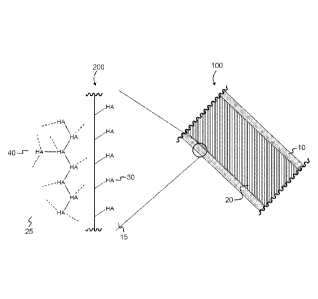
[0062] FIG. 43 depicts a cross-section of a medical device, which is coated
with a guest
molecule and contains a cross-linked guest molecule.
[0063] FIG. 44 depicts TBO staining, which indicates that ePTFE wicked up the
silyl HA-
CTA using the soaking method for 15 minutes, followed by hydrolysis.
DETAILED DESCRIPTION
[0064] Many medical devices contact blood, including heart valves, vascular
conduits,
vascular grafts, catheters, tools, and stents. It is desirable that blood-
contacting
surfaces resist blood clotting and thrombogenesis. The compositions and
methods
presented herein provide such hemocompatibility, and do so with resilience and
great
stability.
[0065] To illustrate this concept, FIG. 43 shows a cross-section 100 of a
medical device
with surface 10 and substrate 20. Surface 10 is modified with a coating of a
guest
molecule. Substrate 20 is interpenetrated with a guest molecule. Close-up 200
magnifies a part of cross-section 100. Guest molecule 30 (HA) is covalently
bonded to
surface 15, forming a coating on surface 15. Beneath the surface, guest
molecules are
covalently bonded to each other within substrate 25, forming network 40. In
this way,
the guest molecules 30, 40 are stabilized against unwanted degradation while
providing
= CA 02866315 2014-09-03
A
beneficial biological properties, such as resistance to blood clotting and
thrombogenesis,
or promoting endothelialization. All the while, substrate 20 maintains the
mechanical
properties that make it useful as a material for constructing medical devices,
such as heart
valves and vascular stents.
I. Composite
[0066] The substrate may comprise a composite. The composite may be an
interpenetrating polymer network (IPN), which is an intermingling of protected
guest
molecule and a polymer host, wherein molecules of the guest have been
crosslinked with
each other. A composite is a material made from two or more components that
are
physically blended or mixed together. The components may be covalently bonded
to each
other or to themselves. In particular, the components may both be polymers. In
general, in
an IPN, at least one component is synthesized or cross-linked in the presence
of the
other, although the two components may be bound together. Semi-IPNs fall
within the
category of IPNs and, thus, composites. The interpenetration many occur at the
nanometer scale, the micron scale, or both. "Microcomposite" refers to a
composite where
the interpenetration of the guest molecule is substantially on the micron
scale, but does
not preclude interpenetration and crosslinking on the nanometer scale. The
term
"composite" does not limit the scale on which the polymer host and the guest
molecule
interact with each other.
[0067] The mechanical and physical properties of the composite, such as its
percentage
of crystallinity, modulus, elongation to failure, and aqueous contact angle,
may be
substantially similar to the properties of the polymer host. The composite may
be
amorphous, semi-crystalline, or crystalline. The percentage of crystallinity
of the
composite may be, for example, about 0% to about 100%, about 5% to about 90%,
about
10% to about 65%, such as about 25% to about 40%, about 10% to about 15%,
about
15% to about 20%, about 20% to about 25%, about 25% to about 30%, about 30% to
about 35%, about 35% to about 40%, about 40% to about 45%, about 45% to about
50%,
about 50% to about 55%, about 55% to about 60%, or about 60% to about 65%.
11
CA 02966315 2014-09-03
WO 2013/138240 PCT/US2013/030230
[0068] The modulus of the composite may be about 0.1 MPA to about 5200 MPA,
for
example about 10 MPA to about 900 MPA, about 140 MPA to about 1550 MPA, about
180 MPA to about 500 MPA, or about 1800 MPA to about 5200 MPA. In some
embodiments, the modulus of the composite may be about 50 MPA to about 150
MPA,
for example about 70 MPA to about 100 MPA, such as about 70 MPA to about 80
MPA,
about 80 MPA to about 90 MPA, or about 90 MPA to about 100 MPA. In still other
embodiments, the modulus of the composite may be about 0.1 MPA to about 10
MPA,
for example about 0.2 MPA to about 1 MPA, such as from about 0.2 MPA to about
0.3
MPA, from about 0.3 MPA to about 0.4 MPA, from about 0.4 MPA to about 0.5 MPA,
from about 0.5 MPA to about 0.6 MPA, from about 0.6 MPA to about 0.7 MPA, from
about 0.7 MPA to about 0.8 MPA, from about 0.8 MPA to about 0.9 MPA, from
about
0.9 MPA to about 1.0 MPA. In yet other embodiments, the modulus of the
composite
may be about 1800 MPA to about 5200 MPA, such as about 1800 MPA to about 2000
MPA, about 2000 MPA to about 2500 MPA, about 2500 MPA to about 3000 MPA, about
3000 MPA to about 3500 MPA, about 3500 MPA to about 4000 MPA, about 4000 MPA
to about 4500 MPA, or about 4500 MPA to about 5000 MPA.
[0069] The elongation to failure of the composite may be about 50% to about
1500%, for
example about 100% to about 1000%, such as about 200% to about 900%, about
450%
to about 500%, about 500% to about 550%, about 550% to about 600%, about 600%
to
about 650%, about 650% to about 700%, about 700% to about 750%, about 750% to
about 800%, about 800% to about 850%, or about 850% to about 900%. In some
embodiments, the elongation to failure of the composite may be about 1000% to
about
1500%, such as about 1000% to about 1100%, about 1100% to about 1200%, about
1200% to about 1300%, about 1300% to about 1400%, or about 1400% to about
1500%.
[0070] The aqueous contact angle on the surface of the composite may about 10
to
about 90 , for example about 40 to about 80 , such as about 40 to about 459,
about
45 to about 50 , about 40 to about 45 , about 459 to about 50 , about 50 to
about 559,
about 55 to about 60 , about 60 to about 65 , about 65 to about 70 , about
70 to
about 75 , or about 759 to about 80 .
12
CA 02966315 2014-09-03
WO 2013/138240 PCT/US2013/030230
A. Polymer Host
[0071] In the composite, the polymer host may be any hydrophobic polymer with
mechanical properties suitable to the material's application. Examples of
suitable
polymer hosts include, but are not limited to, polyolefins, such as
polyethylene (PE),
ultrahigh molecular weight polyethylene (UHMWPE), low-density polyethylene
(LDPE),
linear low-density polyethylene (LLDPE); polyurethane, polycaprolactone (PCL),
polydimethylsiloxane (PDMS), polymethylmethacrylate (PMMA); polyoxymethylene
(POM), such as DelrinTM; polyesters, such as polyethylene terephthalate (PET)
or
Dacron TM ; or polytetrafluoroethylene (PTFE), such as Teflon TM. Extrusion
and sintering
processing techniques may make PTFE more porous, forming expanded PTFE
(ePTFE), which is not biodegradable.
[0072] The polymer host may be a powder, film, fabric (woven or non-woven), or
other
bulk form. The polymer host may be molded, ram-extruded, blown, a virgin
resin, or an
expanded foam. Generally, the polymer host may be porous, such as a fabric,
electrospun scaffold, or sintered construct. A polymer host may be swollen in
an organic
solvent.
[0073] In some embodiments, the host may be a non-polymeric material, for
example a
biological material, such as an allograft, xenograft, tissue, submucosa, swine
heart
value, a vessel graft, or a skin graft. The biological material may be with or
without
fixation, such as glutaraldehyde fixation. The host may also be a metal foam,
such as a
tantalum foam.
[0074] The polymer host may be amorphous, semi-crystalline, or crystalline.
The
percentage of crystallinity of the polymer host may be, for example, about 10%
to about
65%, such as about 25% to about 40%, about 10% to about 15%, about 15% to
about
20%, about 20% to about 25%, about 25% to about 30%, about 30% to about 35%,
about 35% to about 40%, about 40% to about 45%, about 45% to about 50%, about
50% to about 55%, about 55% to about 60%, or about 60% to about 65%
[0075] The polymer host may be a film with a thickness of about 25 pm to about
100
pm, for example about 25 pm to about 30 pm, about 30 pm to about 35 pm, about
35
13
CA 02966315 2014-09-03
WO 2013/138240 PCT/US2013/030230
pm to about 40 pm, about 40 pm to about 45 pm, about 45 pm to about 50 pm,
about
50 pm to about 55 pm, about 55 pm to about 60 pm, about 60 pm to about 65 pm,
about 65 pm to about 70 pm, about 70 pm to about 75 pm, about 75 pm to about
80
pm, about 80 pm to about 85 pm, about 85 pm to about 90 pm, about 90 pm to
about
95 pm, about 95 pm to about 100 pm. In a particular embodiment, the film is
about 50
pm thick.
[0076] By way of example, no clinically acceptable polymeric leaflet valves
are available
beyond those used short-term in artificial hearts. Polyurethanes have been
used in
these devices because they exhibit acceptable mechanical properties and
performance
in the short-term, however, they tend to be very vulnerable to many types of
biodegradation and have a tendency to calcify and eventually tear and fail
which has
limited their successful use. Polycarbonate urethane valves were developed to
optimize
hemodynamics with the goal to increase durability, but the material does not
prevent
calcification. A material originally developed for vascular grafts, 2%
polyhedral
oligomeric silsesquioxane-polycarbonate-urea urethane (POSS-PCD), shows good
mechanical properties due to the addition of the POSS. However, both the POD
and the
FOSS-POD are hydrophobic, with water contact angles over 100 degrees. Both
valves
exhibit calcification during in vitro performance.
[0077] ePTFE grafts are commonly used in bypass procedures of the lower limbs
where
arteries are 7-9 mm in diameter. Additionally, ePTFE grafts have been used for
hemodialysis access in patients with renal failure. ePTFE grafts do not
develop an
endothelial cell layer, potentially leading to thrombus formation. However,
the patency
of ePTFE grafts in femoropopliteal grafts was determined to be about 45%,
whereas the
patency of autologous vein grafts was about 77%. ePTFE grafts are generally
preferred
for peripheral artery bypass in the UK, but many studies have not shown a
difference in
long-term patency between ePTFE and PET grafts.
B. Guest Molecule
[0078] A main reason for long-term failure of blood-contacting devices is
thrombus
formation at an early stage followed by excessive tissue ingrowth at a later
stage. An
14
CA 02966315 2014-09-03
WO 2013/138240 PCT/US2013/030230
effective way to prevent thrombus formation and enhance vascular graft
performance is
to encourage the endothelial cells (ECs) to re-grow over the blood-contacting
device.
This process where a thin layer of tissue lining forms over the device surface
is called
endothelialization. The process of endothelialization is critical to enhance
the
biocompatibility as well as the anti-thrombogenecity of the device after
implantation.
ECs release factors that control the thrombogenesis, fibrinolysis and platelet
activation/inhibition. A key to endothelial cell functionality is their
proliferation on
vascular graft surfaces.
[0079] A guest molecule may provide these beneficial biological properties,
including
resistance to thrombogenesis and enhanced endothelialization. The guest
molecule
may comprise a compound selected from the group consisting of polyions,
polysaccharides including glycosaminoglycans (GAGs); salts of
glycosaminoglycans,
nucleic acids, polyvinylpyrrolidones, peptides, polypeptides, proteins,
lipoproteins,
polyamides, polyamines, polyhydroxy polymers, polycarboxy polymers,
phosphorylated
derivatives of carbohydrates, sulfonated derivatives of carbohydrates,
interleukin-2,
interferon, and phosphorothioate oligomers, with or without amino acids, as
well as
other hydrophilic polymers. Polyhydroxy polymers include, for example,
polyvinyl
alcohol and polyethylene glycol. Polycarboxy polymers include, for example,
carboxymethylcellulose, alginic acid, sodium alginate, and calcium alginate.
[0080] In some embodiments, the guest molecule may be any glycosaminoglycan
(GAG). GAGs include any of a group of linear polysaccharides with various
disaccharide repeating units and usually occurring in proteoglycans, including
chondroitin sulfate, dermatan sulfate, heparan sulfate, and heparin, keratan
sulfates,
and hyaluronic acid. GAGs may be high molecular weight, low molecular weight,
or
oligomeric. GAGs or mucopolysaccharides are long unbranched polysaccharides
consisting of a repeating disaccharide unit. The repeating unit consists of a
hexose (six-
carbon sugar) or a hexuronic acid, linked to a hexosamine (six-carbon sugar
containing
nitrogen). In a particular embodiment, the GAG is a chondroitin sulfate or a
hyaluronan,
such as hyaluronic acid.
CA 02966315 2014-09-03
WO 2013/138240 PCT/US2013/030230
[0081] Hyaluronan ("hyaluronic acid" or "HA") is a naturally occurring
polysaccharide
found in tissues and body fluids of vertebrates and in some bacteria. It is a
linear
polymer with high molecular weight linear polysaccharide containing
alternating N-
acetyl-D-glucosamine and D-glucuronic acid residues, with relatively high
concentrations in the vitreous humor of eye, the umbilical cord, synovial
joint fluid,
rooster combs, and in native heart valve leaflets, particularly those regions
of the valve
subject to compression. A carboxyl group (-COOH) is attached to each
disaccharide
unit of hyaluronic acid. When in solution at physiological pH, hyaluronic acid
is ionized,
resulting in negatively charged -COO. The negatively charged flexible chains
take on an
expanded conformation and entangle with each other at very low concentrations,
acting
as a stiff random coil. In solutions with higher concentration of hyaluronic
acid, stiff
random coils entangle, forming viscoelastic solutions retaining flow without
gelling.
[0082] Hyaluronan solutions are viscous at low shear rates, but elastic at
high shear
rates. Hyaluronic acid's molecular structure leads to its viscoelastic
property,
hydrophilicity, and lubricity. Use of HA in a composite is more durable than
heparin
surface treatments and coatings. HA is easily produced commercially via
fermentation
and its availability in high molecular weights results in composites with
large, relatively
mobile HA molecules at the surface which should enhance antithrombogenicity
and
permit efficient, cost-effective commercial scale-up. HA is also available in
oligomeric
forms, which permits tuning to different biological effects than the higher
molecular
weight species.
[0083] HA is known to bind to three different receptors on ECs: 0D44,
hyaluronan-
mediated motility receptor (RHAMM), and toll-like receptor 4 (TLR4). 0D44 is a
cell-
surface glycoprotein involved in cell-cell interactions, cell adhesion and
migration.
RHAMM normally is localized inside the cell and may be involved in transport
channels
or proteins, flippase activity, and exocytosis. Intracellularly, RHAMM is
associated with
microtubules and plays a role in the regulation of mitosis. Extracellularly,
RHAMM is
associated with 0044, and, upon binding to HA, activates intracellular
signaling
pathways. TLR4 plays a fundamental role in pathogen recognition and activation
of
innate immunity, recognizing pathogen-associated molecular patterns expressed
on
16
CA 02966315 2014-09-03
WO 2013/138240 PCT/US2013/030230
infectious agents, and mediating the production of cytokines necessary to
develop
effective immunity. ECs show enhanced expression of CD44 and TLR4 under
inflamed
conditions. The interaction of CD44 receptor with HA has been shown to enhance
the
production of VEGF and thus promotes cell proliferation. The chain length of
HA
molecules may significantly affect its interaction with these receptors on
ECs. Longer
chain HA molecules will most likely have ligands for these receptors which are
not as
accessible as those on shorter chain HA molecules. HA may also regulate
embryonic
development, tissue organization, wound healing and angiogenesis.
[0084] Salt complexes of hyaluronic acid may be used in forming the composite.
Examples of suitable cations include, but are not limited to,
alkyltrimethylammonium
chloride, alkylamine hydrochloride, alkylpyridinium chloride,
alkyldimethylbenzyl
ammonium chloride, alkyltrimethylammonium bromide, alkylamine hydrobromide,
alkylpyridinium bromide, and alkyldimethylbenzyl ammonium bromide. Optionally,
the
HA is temporarily protected with a protecting group.
[0085] HA may be present in the composite from about 0.001% to about 15% by
weight,
or 0.2% to about 1.5% by weight. In some embodiments, the HA concentration is
from
about 0.2% to about 10% by weight, such as about 5% to about 10% by weight,
about
0.5% to about 3.5% by weight, about 0.5% to about 1.0% by weight, about 1.0%
to
about 1.5% by weight, about 1.5% to about 2.0% by weight, about 2.0% to about
2.5%
by weight, about 2.5% to about 3.0% by weight, about 3.0% to about 3.5% by
weight,
about 3.5% to about 4.0% by weight, about 4.0% to about 4.5% by weight, about
4.5%
to about 5.0% by weight, about 5.5% to about 6.0% by weight, about 7.0% to
about
7.5% by weight, about 7.5% to about 8.0% by weight, about 8.0% to about 8.5%
by
weight, about 8.5% to about 9.0% by weight, about 9.0% to about 9.5% by
weight, or
about 9.5% to about 10.0% by weight. In other embodiments, the HA
concentration in
the composite may be about 0.2%, about 0.3%, about 0.4%, about 0.5%, about
0.6%,
about 0.7%, about 0.8%, about 0.9%, about 1.1%, about 1.2%, about 1.3%, about
1.4%, about 1.5%, about 1.6%, about 1.7%, about 1.8%, about 1.9%, about 2.0%,
about 2.1%, about 2.2%, about 2.3%, about 2.4%, about 2.5%, about 2.6%, about
17
CA 02966315 2014-09-03
WO 2013/138240 PCT/US2013/030230
2.7%, about 2.8%, about 2.9%, about 3.0%, about 3.1%, about 3.2%, about 3.3%,
about 3.4%, or about 3.5%.
C. Crosslinking agents
[0086] The guest molecules are crosslinked to each other within the polymer
host. To
achieve crosslinkage, crosslinking agents are used, such as aliphatic
polyisocyanates
include, for example, bis(4 isocyanatocyclohexyl) methane (H12MDI) such as
available
from Bayer Corp., Pittsburgh, Pa. under the trade designation DesmodurTM W;
isophorone diisocyanate (I PDI) such as commercially available from HueIs
America,
Piscataway, N.J.; hexamethylene diisocyanate (HDI) such as commercially
available
from Aldrich Chemical Co., Milwaukee, Wis.; trimethylhexamethylene
diisocyanate such
as commercially available from Degussa, Corp., Dusseldorf, Germany under the
trade
designation VestanateTM TMDI; and m-tetramethylxylene diisocyanate (TMXDI)
such as
commercially available from Aldrich Chemical Co., Milwaukee, Wis. Although
typically
less preferred, aromatic isocyanates such as diphenylmethane diisocyanate
(MDI) such
as commercially available from Bayer Corp., Pittsburgh, Pa. under the trade
designation
MondurTM M; toluene 2,4-diisocyanate (TDI) such as commercially available from
Aldrich Chemical Co., Milwaukee, Wis., and 1,4-phenylene diisocyanate are also
useful.
[0087] Polyisocyanates include derivatives of the above-listed monomeric
isocyanates.
These derivatives include, but are not limited to, polyisocyanates containing
biuret
groups, such as the biuret adduct of hexamethylene diisocyanate (HDI)
available from
Bayer Corp. under the trade designation DesmodurTM N-100, polyisocyanates
based on
HDI containing isocyanurate groups, such as that available from Bayer Corp.
under
trade designation DesmodurTM N-3300, as well as polyisocyanates containing
urethane
groups, uretdione groups, carbodiimide groups, allophonate groups, and the
like. These
derivatives are preferred as they are polymeric, exhibit very low vapor
pressures and
are substantially free of isocyanate monomer. Other polyisocyanates that may
be used
are available from Bayer Polymers LLC of Pittsburgh, Pa. under the trade
designations
DesmodurTM 1PLS2294 and DesmodurTM N 3600.
18
CA 02966315 2014-09-03
WO 2013/138240 PCT/US2013/030230
[0088] In a particular embodiment, the GAG may be crosslinked at the
carboxylic acid
groups and/or hydroxyl groups using poly(ethylene glycol) diglycidyl ether.
DesmodurTM
N-3200, a biuret isocyanate derived from hexamethylene diisocyanate,
crosslinks
hyaluronic acid at the hydroxyl groups, rather than the carboxylic acid
groups,
preserving hyaluronic acid's lubricity.
[0089] Different sized GAGs, such as cross-linked HA molecules, may induce
different
signaling mechanisms in ECs to promote their adhesion and proliferation. The
molecular weight ranges for the cross-linked guest molecules may be varied
based on
cross-linking conditions and the desired biological effect. In some
embodiments, the
guest molecule may have a large molecular weight, for example from about 10
kDa to
about 1 MDa, such as from about 10 kDa to about 50 kDa, from about 50 kDa to
about
100 kDa, from about 100 kDa to about 200 kDa, from about 100 kDa to about 200
kDa,
from about 100 kDa to about 200 kDa, from about 200 kDa to about 300 kDa, from
about 300 kDa to about 400 kDa, from about 400 kDa to about 500 kDa, from
about 600
kDa to about 700 kDa, from about 800 kDa to about 900 kDa, or from about 900
kDa to
about 1,000 kDa (1 MDa). In other embodiments, the guest molecule may have a
molecular weight from about 1 kDa to about 15 kDa, for example from about 1
kDa to
about 10 kDa, such as from about 1 kDa to about 2 kDa, from about 2 kDa to
about 3
kDa, from about 3 kDa to about 4 kDa, from about 4 kDa to about 5 kDa, from
about 5
kDa to about 6 kDa, from about 6 kDa to about 7 kDa, from about 7 kDa to about
8 kDa,
from about 8 kDa to about 9 kDa, or from about 9 kDa to about 10 kDa. In yet
other
embodiments, the guest molecule may be oligomeric, comprising from about 2 to
about
15 monomeric units of guest molecules, for example, 6 units or 12 units. In
this
embodiment, the molecular weight of the oligomeric crosslinked guest molecule
is about
0.75 kDa to about 10 kDa, such as for example about 0.75 Da to 1 kDa, from
about 1
kDa to about 2 kDa, from about 2 kDa to about 3 kDa, from about 3 kDa to about
4 kDa,
from about 4 kDa to about 5 kDa, from about 5 kDa to about 6 kDa, from about 6
kDa to
about 7 kDa, from about 7 kDa to about 8 kDa, from about 8 kDa to about 9 kDa,
or
from about 9 kDa to about 10 kDa.
19
CA 02966315 2014-09-03
WO 2013/138240 PCT/US2013/030230
D. Method of Making the Composite
[0090] The host polymer may be soaked in a solution of the protected guest
molecule.
Depending on the nature of the polymer host, the polymer host may swell as it
absorbs
the solution and the guest molecule diffuses into the host polymer. The
polymer host
may also wick the soaking solution, such that the solution fills interstitial
spaces within
the physical structure of the polymer host. The solution may be prepared from
a solvent,
such as supercritical carbon dioxide, toluene, decalin, trichlorobenzene, or
xylenes, and
combinations thereof. In a particular embodiment, the solvent is xylenes
Viscosity of the
soaking solution may be selected to control the rate of diffusion of the guest
molecule in
to the polymer host.
[0091] In a particular embodiment, sodium hyaluronic acid was complexed with
quaternary an ammonium cation, hexadecetyltrimethylammonium bromide, followed
by
silylation with hexamethyldisilazane to produce silyl HA-CTA. Silylating the
hyaluronic
acid increases the hydrophobicity of the GAG, by replacing the active
hydrogens of the
hydroxyl groups and amino groups with trimethylsilyl groups. After soaking and
crosslinking, the protecting group is removed to free the hydroxyl groups and
amino
groups of the hyaluronic acid. After deprotection, the polymerized guest
molecule is
typically hydrophilic.
[0092] The soaking step may occur at a temperature of about 25 C to about 100
C, for
example about 45 C to about 65 C, such as about 45 C to about 50 C, about
50 C
to about 55 C, about 55 C to about 60 C, or about 60 C to about 65 C.
[0093] The soaking step may occur for about 10 minutes to about 90 minutes,
such as
about 10 minutes to about 15 minutes, about 15 minutes to about 20 minutes,
about 20
minutes to about 25 minutes, about 25 minutes to about 30 minutes, about 30
minutes
to about 35 minutes, about 35 minutes to about 40 minutes, about 40 minutes to
about
45 minutes, about 45 minutes to about 50 minutes, about 50 minutes to about 55
minutes, about 55 minutes to about 60 minutes, about 60 minutes to about 65
minutes,
about 65 minutes to about 70 minutes, about 70 minutes to about 75 minutes,
about 75
minutes to about 80 minutes, about 80 minutes to about 85 minutes, or about 85
CA 02966315 2014-09-03
WO 2013/138240 PCT/US2013/030230
minutes to about 90 minutes. In a particular embodiment, the soaking step
takes about
60 minutes.
[0094] Any concentration below the guest molecule's solubility limit in the
selected
solvent may be used. In some embodiments, the concentration of guest molecule
in the
solution may be about 0.5 mg/mL to about 250 mg/mL, for example about 1.5
mg/mL to
about 150 mg/mL, or about 2.5 mg/mL to about 50 mg/mL, such as about 2.5 mg/mL
to
about 5.0 mg/mL, about 5.0 mg/mL to about 10.0 mg/mL, about 10.0 mg/mL to
about
15.0 mg/mL, about 15.0 mg/mL to about 20.0 mg/mL, about 20.0 mg/mL to about
25.0
mg/mL, about 25.0 mg/mL to about 30.0 mg/mL, about 30.0 mg/mL to about 35.0
mg/mL, about 35.0 mg/mL to about 40.0 mg/mL, about 40.0 mg/mL to about 45.0
mg/mL, or about 45.0 mg/mL to about 50.0 mg/mL.
[0095] After formation, the polymer host may be thermally molded in the
presence of the
protected guest molecule then cross-linking simultaneously. A diffusion
profile of the
composite, with its gradual concentration of guest from the outer surface a
depth, d,
provides structural integrity of the surface and its associated structure by
removing the
sharp change in modulus inherent in superficially coating or grafting a
surface according
to known techniques. Crosslinking to finally produce the composite may be done
chemically, thermally, or photochemically.
E. Surface Modification
[0096] Surfaces may be modified to improve their performance and
biocompatibility,
such as their hemocompatibility. Glycosylated surfaces may mimic the
biochemical
activity of the glycocalyx of the blood vessel lumen, which presents heparin-
like GAGs.
GAGs, particularly heparin, improve hemocompatibility of surfaces. Numerous
synthetic
plastics and metals that have been modified with heparin show improved
hemocompatibility. Hyaluronan and chondroitin sulfate are GAGs used as
coatings to
reduce platelet adhesion in small diameter vascular grafts. For example,
grafting
sulfonated polyethylene oxide to the surface of polyurethane reduces
calcification and
thromboembolism. Increasing hydrophilicity of glutaraldehyde-fixed
bioprosthetic tissue
valves may decrease calcification and thromboembolism.
21
CA 02966315 2014-09-03
WO 2013/138240 PCT/U S2013/030230
[0097] Formula (I) represents an unprotected hyaluronic acid.
.1 -o2c HoH2c \
HO
OH NH
\ I
COCHy
n (I)
Possible counterions, generically referred to as "QN+", include, but are not
limited to,
cetyltrimethylammonium bromide (Formula II) and cetylpyridinium chloride
(Formula III).
Reaction with the QN+ produces the hyaluronan salt complex HA2-QN+ (Formula
IV),
which may be protected by reaction with a trimethylsilylation agent, such as
chlorotrimethylsilane or hexamethyldisilazane, to yield a trimethylsilane-
protected (TMS-
protected) hyaluronan salt complex (Formula V). By protecting HA2-QN+
complexes,
hydrophilic groups are replaced with silylated functional groups; the
hydrogens on the
hydroxyl groups and on the amine are replaced with the TMS groups.
+,...-
N Br
I" (II)
CI-
1410
/ (III)
QN+ ...4 QN+ TMSOH2C
-02C HOH2C -02C TMSO n \
OH NH OTMS NTMS
I
COCH3
n \ I
COCH/
n
(IV) (V)
22
CA 02966315 2014-09-03
WO 2013/138240 PCT/US2013/030230
II. Devices
[0098] A composite may be used to manufacture devices used in or contacting
the body
of a mammal, for example inside a human body. In some embodiments, the
composite-
containing device contacts blood. In other embodiments, the composite may be
used to
produce heart valves. In yet other embodiments, the composite may be used to
produce
vascular grafts, such as small-diameter vascular grafts.
A. Heart Valves and Vascular Grafts
[0099] Valvular heart disease can be the result of either congenital or
developed
defects, including rheumatic fever, endocarditis, calcific degeneration, or
congenital
anomalies. The two largest problems associated with valvular disease are
regurgitation
and stenosis. In the former case, the valve does not close completely, and
some of the
pumped blood flows backwards back into the left ventricle. In the latter case,
the
opening through which blood can pass becomes narrowed due to the leaflets
either
becoming rigid or fused together. Both of these valvular diseases result in
blood
accumulation in the chamber, and the heart must work harder to supply the
body. This
increased workload leads to the thickening of the heart muscle and dilatation,
which can
result in congestive heart failure. Once the heart valve no longer maintains
its normal
functionality, drugs can be used to relieve the symptoms but not reverse and
disease.
Valve replacement surgery is recommended when damage to the valve is
considered to
be significant enough to pose a life threatening risk.
[00100] Complete replacement of damaged and diseased heart valves by
prostheses
is routine. Factors used to determine which valve is most suited to a patient
include the
patient's age, comorbidities, need for associated procedures, availability of
a given
replacement, patient agreement, and surgeon expertise. Current commercially
available
valves are divided into two primary classes, mechanical and bioprosthetic,
each with its
associated advantages and disadvantages.
23
CA 02966315 2014-09-03
WO 2013/138240 PCT/US2013/030230
(1) Mechanical Heart Valves
[00101] Due to their high durability and longevity, mechanical valves are
preferred for
individuals under the age of 65. Current designs implanted include the tilting
disc design
(FIG. 1B), the bileaflet design (FIG. 2C), and to a lesser extent, the ball
and cage design
(FIG. 1A). The low profile of the bileaflet mechanical valves allows them to
be implanted
into smaller hearts without obstruction of other structures such as the mitral
valve or
coronaries. Bileaflet valve have good hemodynamics with low transvalvular
pressure
gradient is and minimal regurgitation. They are durable, showing a low rate of
mechanical failure. The tilting disc valves are the second most commonly
implanted
mechanical valves. Like the bileaflet valves, the tilting disc valves have
shown to be
durable, but the hemodynamics of the tilting disk valves is not ideal with
lower effective
orifice areas and turbulent flow around the disk. The caged ball valve does
not have as
favorable hemodynamics as the bileaflet and tilting disc valves, but it is
still sometimes
used when surgeons require a valve that is easy to handle under difficult
surgical
circumstances. One common problem for all the mechanical valve designs is the
resulting partial obstruction of blood flow, leading to non-physiological
hemodynamic
characteristics, which contribute to thrombosis, embolism, and bleeding
complications,
often resulting to morbidity and mortality. Consequently, patients receiving
mechanical
valves are subjected to life-long anticoagulation therapy. Lifetime
anticoagulation
therapy has many problems associated with it often resulting in either under
or over
anticoagulation, and complication associated with hemorrhaging.
(2) Bioprothestic Heart Valves
[00102] The two main bioprosthetics heart valves are either homografts
(from human
cadavers) or xenografts, such as glutaraldehyde-fixed procine aortic valves
and
glutaraldehyde-fixed bovine pericardium (FIG. 2). The homografts are the least
frequently used due to a shortage in number and size and their difficulty to
insert. The
stented porcine (FIG. 2A) and bovine pericardium (FIG. 2B) valves are the most
commonly implanted. Both valves have issues with durability with an
approximate
lifespan of 1 0-1 5 years. The trileaf let design reproduces the central flow
characteristics
24
CA 02966315 2014-09-03
WO 2013/138240 PCT/US2013/030230
of the natural valve and is less thrombogenic than mechanical valves. Thus,
long-term
anticoagulation treatment is not required for most recipients. Bioprosthetic
valves have
also become a popular choice for younger patients to prevent the need for
lifetime
anticoagulation therapy at such a young age, but this often means additional
surgeries
to replace deteriorating bioprosethetic valves at a later age.
[00103] Metallic or polymer structures may be used to support the porcine
and bovine
pericardium valves. This stent allows the valve to be implanted easily,
however, this
results in a stenotic region caused by partial orifice obstruction. Stentless
porcine valves
(FIG. 20) were developed to help combat this obstruction. The stentless valves
consist
of aortic roots modified with a sewing ring, which is either implanted within
the native
root or replaces the root with an increase in effective orifice area.
Stentless valves are
significantly more complicated to implant than the stented version, and
conclusive long-
term data of durability of these valves is still unknown but assumed to be
similar to
stented bioprosthetic valves. Porcine valves are much more restrictive on
design due to
the valve anatomy. Stented pericardial valves can be fabricated in to much
more
complex designs. Pericardial valves are fabricated from glutaraldehyde-fixed
sheets of
bovine pericardium that can be oriented to mimic the natural valve in both
form and
function. The pericardial valves tend to have more desirable hemodynamics than
the
porcine valves as a result of their improved effective orifice area and
leaflet dynamics
during forward flow; however, the traditional designs have been made to
exhibit
significantly higher stresses during diastole when they are under tension.
[00104] The main problem with xenogenic prostheses is tissue failure, which
usually
is onset within 10 years of implantation. This degradation of the valve is as
a result of
mechanical damage, calcification, or a combination of both, and has been
linked to the
glutaraldehyde fixation and the stent-valve interaction. Glutaraldehyde
treatment
effectively cross-links the tissue and reduces its antigenicity while
preventing proteolytic
degradation. As a result, the tissue loses its mechanical compliance causing
an
increase in leaflet stress concentrations, accelerating fatigue of the tissue.
The
presence of calcium deposits on the leaflets can result in stenosis and
leaflet tearing.
, = CA 02866315 2014-09-03
[00105] The composite of the present disclosure may be used in any component
of a
heart valve. For example, the composite may be used in a heart valve leaflet,
a sewing
ring, sewing cuff, a tilting disc, stent, suture ring, or annulus. One of
skill in the art would
understand how to modify the design of the valve based on the nature of the
composite,
for example the shape of the leaflet, including its three-dimensional
curvature, thickness,
uniformity, stent post asymmetry, and profile height. Other design
modifications may
include the absence of sutures to install leaflets into the heart valve stent.
Stents may be
formed from the composite, and the whole HV may be molded in a single piece or
manufactured by three-dimensional printing.
[00106] In some other embodiments, this disclosure provides a heart valve
using a tilting
disc mechanism. The tilting disk may be formed from a first composite
comprising: a first
polymer host, such as ultra-high molecular weight polyethylene (UHMWPE), and a
first
guest molecule comprising hyaluronic acid; wherein the first guest molecule is
disposed
within the second polymer host, and wherein the first guest molecule is
covalently bonded
to at least one other guest molecule. The heart valve may also comprise a
suture ring
made from a second composite, comprising: a second polymer host comprising PET
fabric, and a second guest molecule comprising hyaluronic acid; wherein the
second guest
molecule is disposed within the second polymer host, and wherein the second
guest
molecule is covalently bonded to at least one other second guest molecule.
[00107] In other embodiments, this disclosure provides a heart valve using a
ball-in-cage
mechanism. The ball may be formed from a first composite comprising: a first
polymer
host, such as polyoxymethylene (POM), and a first guest molecule comprising
hyaluronic
acid; wherein the first guest molecule is disposed within the second polymer
host, and
wherein the first guest molecule is covalently bonded to at least one other
guest molecule.
The heart valve may further comprise a cage made from a second composite. The
second
polymer host may be selected as to have the desired physical or mechanical
properties.
The second guest molecule may comprise hyaluronic acid; wherein the second
guest
molecule is disposed within the second polymer host, and
26
CA 02966315 2014-09-03
WO 2013/138240 PCT/US2013/030230
wherein the second guest molecule is covalently bonded to at least one other
second
guest molecule.
(3) Vascular Grafts
[00108] The two synthetic grafts most commonly used for small diameter
bypass
procedures for vessels less than 6mm are PET and ePTFE. Polyurethane materials
may also be used in peripheral bypass procedures due to their mechanical
property
matching to natural vessels. PET and ePTFE grafts often fail due to early
thrombosis or
late intimal hyperplasia, are more stiff and have a different elastic modulus
than natural
arteries.
[00109] PET is used to treat large diameter vascular grafts but has low
patency as a
small diameter vascular graft, particularly for lower limb bypass procedures.
Untreated
PET grafts do not develop an endothelial cell layer on the lumen when
implanted,
leading to platelet adhesion, fibrin layer formation, and potentially
subsequent
thrombosis.
[00110] ePTFE grafts are commonly used in bypass procedures of the lower
limbs
where arteries are 7-9 mm in diameter. Additionally, ePTFE grafts have been
used for
hemodialysis access in patients with renal failure. ePTFE grafts do not
develop an
endothelial cell layer, either, potentially leading to thrombus formation.
Patency of
ePTFE grafts in femoropopliteal grafts was determined to be 45%, whereas the
patency
of autologous vein grafts was 77%. ePTFE grafts may be used for peripheral
artery
bypass, but most studies have not shown a difference in long-term patency
between
ePTFE and PET grafts.
[00111] Polyurethane may be used in small diameter vascular grafts because
mechanical properties can be tailored to match those of native blood vessels.
Particularly, polyurethane is more compliant than ePTFE. Polyurethane has been
used
in hemodialysis, and may be modified with NO-releasing peptides to inhibit
platelet
activation. Polyurethane materials may be susceptible to degradation in vivo
and
subsequent aneurismal degeneration.
27
CA 02966315 2014-09-03
WO 2013/138240 PCT/US2013/030230
[00112] Poor long-term performance may be low compliance and a lack of
functional endothelial cell coverage. Intimal hyperplasia is characterized by
migration of
smooth muscle cells from the media to the intima. After migration, smooth
muscle cells
synthesize matrix proteins and other extracellular material. This can cause
the blood
vessel to become stenosed. A mismatch between compliance of synthetic and
natural
vessels may contribute to intimal hyperplasia formation at the downstream
anastomosis.
Patency has been correlated to compliance. Viscoelastic properties are
important at
low flow rates, such as in the peripheral arteries below the knees. Intimal
hyperplasia
may develop when blood flow is disrupted and vessel walls are injured. A
compliance
mismatch may alter the haemodynamics at the anastomosis. Specifically, a
compliance
mismatch at the anastomosis can increase shear stress under flow conditions,
reducing
perfusion and potentially leading to rupture. Synthetic grafts may become less
compliant
upon implantation. Post-implantation stiffening should be considered when
matching
mechanical properties.
[00113] A layer of endothelial cells on the surface of the graft in contact
with blood
may reduce thrombosis and increase the patency of synthetic vascular grafts.
Surface
treatments used improve cell retention include attachment of ROD peptides,
matrix
proteins (fibronectin), growth factors (fibroblast growth factor or
endothelial cell growth
factor), or a combination of coatings. Endothelial cell coverage is important
because it
may limit inflammation. Anti-coagulant phenotype endothelial cells produce
vasoprotective factors. They also inhibit the production of factors that cause
inflammation. One such factor, inducible nitric oxide (iNOS), forms NO and
decreases
the adhesion of platelets. Another factor, tissue factor (TF), is a
procoagulant protein,
which, in combination with fV11a, activates FX and leads to the production of
thrombin.
Tissue plasminogen activator (tPA) plays a role in plasminogen activation,
fibrinolysis,
and fibrin clot degradation. Vascular cell adhesion molecule 1 (VCAM-1)
supports white
blood cell adhesion, including monocytes and lymphocytes. A lack of functional
endothelial cell coverage on the lumen surface of a graft leads to thrombosis
and
subsequent occlusion of the vessel.
28
CA 02966315 2014-09-03
WO 2013/138240 PCT/US2013/030230
[00114] Grafts that have surface thromobogenicity and limited
biocompatibility at
the graft/vessel interface lead to low patency rates. The smaller the graft
diameter, the
higher the rate of graft occlusion. Several factors may contribute to graft
thrombosis,
including graft surface properties, graft hemodynamics, blood flow, surgical
technique,
patient thrombotic profile, and the degree of neointimal formation and
endothelialization.
Thrombogenesis causes occlusion and decreases blood flow through veins and
arteries, possibly causing failure or vessel narrowing, such as stenosis and
intimal
hyperplasia.
B. Other Devices
[00115] In some embodiments, the composite may be used in vascular grafts,
including venous grafts and arterial grafts. The grafts may be formed from any
polymer
host, such as PET, PE, PP, or PTFE, especially ePTFE, or the graft made from
allograft
tissue or decelluralized xenograft tissue.
[00116] As discussed above in Section 1(A)(3), composites may be used to
form
small-diameter vascular grafts. Currently, in limited situations, autografts
or allografts
may be used, but are unsuitable in most applications. Thus, there is a long-
felt and
unmet need for the easily produced, high-performing small-diameter vascular
grafts
made from the composites provided herein, especially grafts which do not
suffer from
narrowing such as stenosis or intimal hyperplasia.
[00117] In other embodiments, the composite may be used in, for example, an
intravascular catheter, blood-contacting sensor, stent, annulus, an insulator
for electrical
leads, an extracorporeal blood-loop circuit; implantable cardiac assist
devices for
prolonged circulatory support, such a left ventricular assist device (LVAD); a
blood-
contacting cardiomyopathy treatment, such polyethylene braids, for example an
artificial
cord, tether, or suture inside a heart; peripherally inserted central catheter
(PICC) line,
fistula plug, membrane, blood bag; blood processing, transportation and
storage
equipment and materials; Luer connector, suture, aneurysm patch, conduit,
coil, roller
(peristaltic) pump, patent foramen ovale (PFO), reconstruction patch,
transapical
device, angioplasty tools, cannulae, and annuloplasty rings.
29
CA 02966315 2014-09-03
WO 2013/138240 PCT/US2013/030230
[00118] The insulator for electrical leads may be present in, for example,
a
pacemaker or defibrillator. The blood oxygenator may be part of a heart-lung
machine,
perfusion unit, or hemodialysis machine. Stents may include a coronary artery
stent or
vascular stent, as well as other angioplasty devices and tools used for stent
delivery,
such as balloons. Substrates may be for any in vitro diagnostic tool or assay,
for
example, tissue culture plate, 3D tissue cultures, microfluidics, or a lab-on
chip device.
III. Definitions
[00119] As used herein, the terms "about" and "approximately" designate
that a value
is within a statistically meaningful range. Such a range can be typically
within 20%,
more typically still within 10%, and even more typically within 5% of a given
value or
range. The allowable variation encompassed by the terms "about" and
"approximately"
depends on the particular system under study and can be readily appreciated by
one of
ordinary skill in the art.
[00120] As used herein, the term "w/w" designates the phrase "by weight"
and is used to describe the concentration of a particular substance in a
mixture or
solution.
[00121] As used herein, the term "ml/kg" designates milliliters of
composition per
kilogram of formula weight.
[00122] As used herein, the term "monomer" refers to any chemical compound
that is
capable of forming a covalent bond with itself or a chemically different
compound in a
repetitive manner. The repetitive bond formation between monomers may lead to
a
linear, branched, super-branched, or three-dimensional product. Furthermore,
monomers may themselves comprise repetitive building blocks, and when
polymerized
the polymers formed from such monomers are then termed "block polymers".
Monomers may belong to various chemical classes of molecules including
organic,
organometallic or inorganic molecules. The molecular weight of monomers may
vary
greatly between about 40 Dalton and 20000 Dalton. However, especially when
= CA 02866315 2014-09-03
monomers comprise repetitive building blocks, monomers may have even higher
molecular weights. Monomers may also include additional reactive groups.
[00123] Contemplated polymers may also comprise a wide range of functional or
structural moieties, including aromatic systems, and halogenated groups.
Furthermore,
appropriate polymers may have many configurations, including a homopolymer,
and a
heteropolymer. Moreover, alternative polymers may have various forms, such as
linear,
branched, super-branched, or three-dimensional. The molecular weight of
contemplated
polymers spans a wide range, typically between 400 Daltons and 400,000
Daltons, and
may be greater than 1,000,000 Daltons or more, in some embodiments.
[00124] "Wettability" refers to the ability of a liquid, such as water, to
spread on a solid
surface. "Hydrophilic" and "hygrophilic" refer to an intrinsic or average
chemical property of
a surface or bulk solid to allow a polar liquid, such as water, to spread on
the surface, with
typical water contact angles from about 0 to about 90 . "Hydrophobic" refers
to an
intrinsic or average chemical property of a surface or bulk solid that
prevents a polar liquid,
such as water, from spreading on the surface, with typical water contact
angles from about
90 to about 180 , such as from about 1000 to about 150 . When the surface
roughness
enhances or reduces the hydrophilic or hydrophobic properties of a surface or
bulk solid,
the effect is "parahydrophilic" or "parahydrophobic," respectively. For very
rough surfaces,
the enhancement or reduction in hydrophilic or hydrophobic properties of the
surface or
bulk solid may be very great; the effect is referred to as "superhydrophilic"
or
"superhydrophobic," respectively. Surface roughness is usually defined on the
microscopic
or molecular scales. For further definition of wettability and surface
classifications, please
refer to Marmur, "Hydro- hygro- oleo- omni-phobic? Terminology of wettability
classification," Soft Matter, 8:6867 (2012).
[00125] The compounds described herein have asymmetric centers. Compounds of
the
present disclosure containing an asymmetrically substituted atom may be
isolated in
optically active or racemic form. All chiral, diastereomeric, racemic forms
and all
31
CA 02866315 2014-09-03
WO 2013/138240 PCT/US2013/030230
geometric isomeric forms of a structure are intended, unless the specific
stereochemistry or isomeric form is specifically indicated.
[00126] The term "acyl," as used herein alone or as part of another group,
denotes
the moiety formed by removal of the hydroxy group from the group COOH of an
organic
carboxylic acid, e.g., RC(0)¨, wherein R is R1, R10_, Ri
N , or R1S-, R1 is hydrocarbyl,
heterosubstituted hydrocarbyl, or heterocyclo, and R2 is hydrogen,
hydrocarbyl, or
substituted hydrocarbyl.
[00127] The term "acyloxy," as used herein alone or as part of another
group,
denotes an acyl group as described above bonded through an oxygen linkage (0),
e.g.,
RC(0)0¨ wherein R is as defined in connection with the term "acyl."
[00128] The term "allyl," as used herein not only refers to compound
containing the
simple allyl group (CH2=CH¨CH2¨), but also to compounds that contain
substituted allyl
groups or allyl groups forming part of a ring system.
[00129] The term "alkyl" as used herein describes groups which are
preferably lower
alkyl containing from one to eight carbon atoms in the principal chain and up
to 20
carbon atoms. They may be straight or branched chain or cyclic and include
methyl,
ethyl, propyl, isopropyl, butyl, hexyl and the like.
[00130] The term "alkenyl" as used herein describes groups which are
preferably
lower alkenyl containing from two to eight carbon atoms in the principal chain
and up to
20 carbon atoms. They may be straight or branched chain or cyclic and include
ethenyl,
propenyl, isopropenyl, butenyl, isobutenyl, hexenyl, and the like.
[00131] The term "alkynyl" as used herein describes groups which are
preferably
lower alkynyl containing from two to eight carbon atoms in the principal chain
and up to
20 carbon atoms. They may be straight or branched chain and include ethynyl,
propynyl, butynyl, isobutynyl, hexynyl, and the like.
[00132] The term "aromatic" as used herein alone or as part of another
group
denotes optionally substituted homo- or heterocyclic conjugated planar ring or
ring
32
CA 02966315 2014-09-03
WO 2013/138240 PCT/US2013/030230
system comprising delocalized electrons. These aromatic groups are preferably
monocyclic (e.g., furan or benzene), bicyclic, or tricyclic groups containing
from 5 to 14
atoms in the ring portion. The term "aromatic" encompasses "aryl" groups
defined
below.
[00133] The terms "aryl" or "Ar" as used herein alone or as part of another
group
denote optionally substituted homocyclic aromatic groups, preferably
monocyclic or
bicyclic groups containing from 6 to 10 carbons in the ring portion, such as
phenyl,
biphenyl, naphthyl, substituted phenyl, substituted biphenyl, or substituted
naphthyl.
[00134] The terms "carbocyclo" or "carbocyclic" as used herein alone or as
part of
another group denote optionally substituted, aromatic or non-aromatic,
homocyclic ring
or ring system in which all of the atoms in the ring are carbon, with
preferably 5 or 6
carbon atoms in each ring. Exemplary substituents include one or more of the
following
groups: hydrocarbyl, substituted hydrocarbyl, alkyl, alkoxy, acyl, acyloxy,
alkenyl,
alkenoxy, aryl, aryloxy, amino, amido, acetal, carbamyl, carbocyclo, cyano,
ester, ether,
halogen, heterocyclo, hydroxy, keto, ketal, phospho, nitro, and thio.
[00136] The terms "halogen" or "halo" as used herein alone or as part of
another
group refer to chlorine, bromine, fluorine, and iodine.
[00136] The term "heteroatom" refers to atoms other than carbon and
hydrogen.
[00137] The term "heteroaromatic" as used herein alone or as part of
another group
denotes optionally substituted aromatic groups having at least one heteroatom
in at
least one ring, and preferably 5 or 6 atoms in each ring. The heteroaromatic
group
preferably has 1 or 2 oxygen atoms and/or 1 to 4 nitrogen atoms in the ring,
and is
bonded to the remainder of the molecule through a carbon. Exemplary groups
include
furyl, benzofuryl, oxazolyl, isoxazolyl, oxadiazolyl, benzoxazolyl,
benzoxadiazolyl,
pyrrolyl, pyrazolyl, imidazolyl, triazolyl, tetrazolyl, pyridyl, pyrimidyl,
pyrazinyl,
pyridazinyl, indolyl, isoindolyl, indolizinyl, benzimidazolyl, indazolyl,
benzotriazolyl,
tetrazolopyridazinyl, carbazolyl, purinyl, quinolinyl, isoquinolinyl,
imidazopyridyl, and the
like. Exemplary substituents include one or more of the following groups:
hydrocarbyl,
33
CA 02966315 2014-09-03
WO 2013/138240 PCT/US2013/030230
substituted hydrocarbyl, alkyl, alkoxy, acyl, acyloxy, alkenyl, alkenoxy,
aryl, aryloxy,
amino, amido, acetal, carbamyl, carbocyclo, cyano, ester, ether, halogen,
heterocyclo,
hydroxy, keto, ketal, phospho, nitro, and thio.
[00138] The terms "heterocyclo" or "heterocyclic" as used herein alone or
as part of
another group denote optionally substituted, fully saturated or unsaturated,
monocyclic
or bicyclic, aromatic or non-aromatic groups having at least one heteroatom in
at least
one ring, and preferably 5 or 6 atoms in each ring. The heterocyclo group
preferably
has 1 or 2 oxygen atoms and/or 1 to 4 nitrogen atoms in the ring, and is
bonded to the
remainder of the molecule through a carbon or heteroatom. Exemplary
heterocyclo
groups include heteroaromatics as described above. Exemplary substituents
include
one or more of the following groups: hydrocarbyl, substituted hydrocarbyl,
alkyl, alkoxy,
acyl, acyloxy, alkenyl, alkenoxy, aryl, aryloxy, amino, amido, acetal,
carbamyl,
carbocyclo, cyano, ester, ether, halogen, heterocyclo, hydroxy, keto, ketal,
phospho,
nitro, and thio.
[00139] The terms "hydrocarbon" and "hydrocarbyl" as used herein describe
organic
compounds or radicals consisting exclusively of the elements carbon and
hydrogen.
These moieties include alkyl, alkenyl, alkynyl, and aryl moieties. These
moieties also
include alkyl, alkenyl, alkynyl, and aryl moieties substituted with other
aliphatic or cyclic
hydrocarbon groups, such as alkaryl, alkenaryl and alkynaryl. Unless otherwise
indicated, these moieties preferably comprise 1 to 20 carbon atoms.
[00140] The term "protecting group" as used herein denotes a group capable
of
protecting a particular moiety, wherein the protecting group may be removed,
subsequent to the reaction for which the protection is employed, without
disturbing the
remainder of the molecule. Where the moiety is an oxygen atom (and hence,
forming a
protected hydroxy), exemplary protecting groups include ethers (e.g., allyl,
triphenylmethyl (trityl or Tr), p-methoxybenzyl (P MB), p-methoxyphenyl
(PMP)), acetals
(e.g., methoxymethyl (MOM), 6-methoxyethoxymethyl (M EM), tetrahydropyranyl
(THP),
ethoxy ethyl (EE), methylthiomethyl (MTM), 2-methoxy-2-propyl (MOP), 2-
trimethylsilylethoxymethyl (SEM)), esters (e.g., benzoate (Bz), allyl
carbonate, 2,2,2-
34
CA 02966315 2014-09-03
WO 2013/138240 PCT/US2013/030230
trichloroethyl carbonate (Troc), 2-trimethylsilylethyl carbonate), silyl
ethers (e.g.,
trimethylsilyl (TMS), triethylsilyl (TES), triisopropylsilyl (TIPS),
triphenylsilyl (TPS), t-
butyldimethylsilyl(TBDMS), t-butyldiphenylsilyl (TBDPS) and the like. When the
moiety
is an nitrogen atom (and hence, forming a protecting amine) exemplary
protecting
groups include benzyl, p-methoxyphenyl (PMP), 3,4-dimethoxybenxyl (PMB)), n-
silyl
groups, esters (e.g., benzoate (Bz), carbonyl (e.g. p-methoxybenzyl carbonyl
(Moz),
tert-butyloxycarbonyl (BOO), 9-fluorenylmethyloxycarbonyl (FMOC)), acetyl,
carbamates, n-silyl groups and the like. A variety of protecting groups and
the synthesis
thereof may be found in "Protective Groups in Organic Synthesis" by T.W.
Greene and
P.G.M. Wuts, John Wiley & Sons, 1999.
[00141] The "substituted hydrocarbyl" moieties described herein are
hydrocarbyl
moieties which are substituted with at least one atom other than carbon,
including
moieties in which a carbon chain atom is substituted with a heteroatom such as
nitrogen, oxygen, silicon, phosphorous, boron, or a halogen atom, and moieties
in which
the carbon chain comprises additional substituents. These substituents include
alkyl,
alkoxy, acyl, acyloxy, alkenyl, alkenoxy, aryl, aryloxy, amino, amido, acetal,
carbamyl,
carbocyclo, cyano, ester, ether, halogen, heterocyclo, hydroxy, keto, ketal,
phospho,
nitro and thio.
[00142] When introducing elements of the present disclosure or the
exemplary
embodiments(s) thereof, the articles "a", "an", "the" and "said" are intended
to mean that
there are one or more of the elements. The terms "comprising", "including" and
"having" are intended to be inclusive and mean that there may be additional
elements
other than the listed elements.
[00143] Having described the disclosure in detail, it will be apparent that
modifications and variations are possible without departing from the scope of
the
disclosure defined in the appended claims.
CA 02966315 2014-09-03
WO 2013/138240 PCT/US2013/030230
EXAMPLES
Example 1: Swelling of the host polymer
[00144] To form hyaluronic acid (HA) in a linear low-density polyethylene
(LLDPE)
polyethylene terephthalate (PET), or expanded polytetrafluoroethylene (ePTFE)
microcomposite, the degree of swelling and swelling kinetics in a solvent of
interest
were analyzed. A study was performed to understand the above parameters for
the
swelling of the LLDPE in a range of solvent temperatures in order to identify
the swelling
parameters to be used in the microcomposite synthesis.
[00145] LLDPE samples were blow-molded from known resins by Flex-Pack
Engineering, Inc. (Uniontown, OH) with known melt indexes, densities, and
crystallinities. Samples had a specified thickness of 0.002" (0.0508 mm)
without
additional fillers or surface treatment. The first type of LLDPE used was film
molded
from DowlexTM 2344 resin with a melt index of 0.7 g/10 min, a density of 0.933
g/cm3
and a crystallinity of 42.26 1.35%. The second type of LLDPE used study was
film
molded from DowlexTM 2056 resin with a melt index of 1.0 g/10 min, a density
of 0.920
g/cm3 and a crystallinity of 28.71 2.14%. The third type of LLDPE used was
film
molded from DowlexTM 2036G resin with a melt index of 2.5 g/10 min, a density
of 0.935
g/cm3 and a crystallinity of 45.21 1.66%. Crystallinity of the films was
calculated using
differential scanning calorimetry (DSC). The samples were cut into squares of
about 3
cm by about 3 cm.
[00146] BARD Peripheral Vascular OEM Products (Tempe, AZ) supplied the PET
samples made from Style 6010 thin polyester tubular woven (uncrimped)
specimens
with a nominal diameter of 22 mm and wall thickness of 0.010" 0.001". All
PET fabrics
were woven without additional surface treatment. The resulting PET fabric had
a density
of 1.78 g/cm3 and a crystallinity of 38.28 0.54%, as calculated by DSC.
Samples were
cut into squares of about 3 cm by about 3 cm. Xylenes showed the greatest
degree of
swelling, possibly due to the closeness of the solvent's Hildebrand solubility
parameter.
[00147] Two systems were used to test swelling. The first system was an
open-
cup, consisting of a 50-mL beaker covered with a watch glass in a controlled-
36
CA 02966315 2014-09-03
WO 2013/138240 PCT/US2013/030230
temperature oil bath. The second system was a 250-mL round-bottom flask fitted
with a
24/40 standard taper ground glass joint and a serum stopper. The ground joint
was
fitted with a 100-mm West condenser capped with a rubber septum. The sealed
round-
bottom flask was lowered into a heated oil bath with a temperature probe. No
differentiation is made between the two swelling methods. Both methods were
used in
experiments and yielded similar results.
[00148] Samples were weighed before submersion in solvent. After samples
were
allowed to swell for a desired amount of time, dried of surface solvent and
weighed.
Reported data are the average of the three samples the standard deviation.
When the
averaged masses of the samples reached equilibrium, the temperature of the
solvent
was increases and the weighing procedure repeated until equilibrium was again
reached. The temperature of the solvent was increased until the LLDPE film
degraded.
[00149] The following equation was used to calculate the percent change in
volume
of the sample (dVNo%):
Wt Wo
P.s{-$f-wmt
/ PpOiyTner
where Wf is the weight of the sample at time t, Wo is the weight of the sample
at time to,
Psolvern is the density of the solvent, and n
,polymer is the density of the polymer.
[00150] Changes in crystallinity and tensile properties guided selection of
the polymer
host and swelling parameters to achieve the selected volumetric expansion
without
compromising the material's mechanical properties. The % xc was measured by a
TA
Instruments DSC 2920 in a dry nitrogen atmosphere per ASTM D34I8-03. LLDPE
samples were heated from 24 C to 180 C at a rate of 10 C/minute and held at
equilibrium for one minute. PET samples were heated from 24 C to 275 C at a
rate of
`C/minute, and held at equilibrium for one minute. The heat of formation (Hf)
was
determined to be 288 J/g for 100% crystalline PE and 113 J/g for 100%
crystalline PET.
The % xc, of the sample was calculated by dividing the Hf of the sample by 288
J/g or
113 J/g based on base polymer and multiplying by 100. Sample control and
treatment
37
CA 02966315 2014-09-03
WO 2013/138240 PCT/US2013/030230
groups that were characterized, including LLDPE virgin film, PET virgin
fabric, LLDPE
and PET sham controls, and all LLDPE-T and PET-T samples.
[00151] Representative data of the percent volume change (dVNo /o) of the
three
commercial LLDPE films in xylenes versus time at different temperatures are
shown in
FIGS. 4, 5, and 6 for the DowlexTM 2344, 2056, and 2036G, respectively. Data
in these
figures resulted from the open-cup swelling method except for the 70 C data,
which
resulted from the round-bottom flask method.
[00152] FIG. 7 represents the percentage volume change (dVNo /0) of the
DowlexTM
LLDPE films in xylenes versus temperature. Focusing on the degree of swelling
of the
LLDPE films in xylenes vs. temperature, there appears to be a non-linearity
for
DowlexTM 2056 starting around 60 C. It is believed that the swelling to this
point has
taken place mainly in the amorphous regions of the film. Beyond this point,
the
crystalline regions prevent the film from swelling further prior to melting of
the crystalline
regions. The DowlexTM 2036G continues to be linear due to its higher melt flow
rate,
preventing a rapid expansion of the material in the solvent. If only the
amorphous
regions of the LLDPE are swelled it would be expected that the lower
crystallinity
DowlexTM 2056 material would swell to a greater extent. Representative data of
the
percent volume change (dVNo /0) of the PET fabric in xylenes versus time at
different
temperatures are shown in FIG. 8. Data in this figure result from the open-cup
swelling
method. Temperature increases were halted at 60 C due to satisfactory
swelling at
lower temperatures and no significant differences in swelling with previous
temperature
increases. Also, temperatures greater than 60 C may lead to HA degradation.
[00153] FIG. 9 represents the percentage volume change (dVNo /0) of the PET
fabric
in xylenes versus temperature. The apparent nonlinearity for the PET fabric
starting
around 60 C is statistically insignificant at different temperatures. Even
though the
solubility parameters for xylenes and PET do not predict significant swelling,
PET fabric
fibers may swell in xylenes at an increased temperature. Lower temperatures do
not
influence the amount of swelling. The weave of the fabric may wick solvent,
occupying
the voids between fibers and yarns. At elevated temperatures, in contrast, the
fibers
38
CA 02966315 2014-09-03
WO 2013/138240 PCT/US2013/030230
swell with the solvents, thus increasing the amount of swelling. At 50 C and
below
wicking into the fabric open voids is likely. At 60 C and above, wicking and
swelling
both occur, leading to an IPN at fiber level and a microcomposite at fabric
level.
[00154] DowlexTM 2344 and 2056 LLDPE samples reached 90% of the equilibrium
swelling value at 50 C in approximately 1 hour. DowlexTM 2036G reached 100% of
its
equilibrium value within 1 hour. These values indicate the end of active
solvent
transport. The PET fabric reached 100% of its equilibrium swelling value at
each
temperature within 15 minutes of placement into solvent bath. Extended
exposure to
solvents did not increase the volumetric expansion of the fabric, suggesting
that the
solvent was only penetration voids between fibers and yarns instead of
swelling the
PET fibers.
[00155] With more swelling with use of xylenes and elevated temperatures,
crystallinity of the DowlexTM 2056 was increased while the DowlexTM 2344 and
2036G
were much more thermally stable and did not increase crystallinity. This
increase in
crystallinity subsequently caused an increase in the modulus and yield
strength of the
DowlexTM 2056 as well. Crystallinity increased (FIGS. 10, 12, and 14) and the
tensile
increased (FIGS. 11, 13, and 15) for DowlexTM 2344, 2056, and 2036G,
respectively.
[00156] Since the solvents only penetrated voids within the PET fabric and
did not
swell the fibers, drying the PET fabrics removed solvents and returned the
fabric to its
original state. The crystalline structure remained unchanged during soaking
(FIG. 16).
[00157] Xylenes provided the greatest degree of swelling in the DowlexTM
2056 film.
The temperature of swelling had the largest increase in degree of swelling for
the
DowlexTM 2056 film and provided inconsistent swelling in the other films. For
this
reason, the DowlexTM 2056 film was chosen as the LLDPE polymer host in the
examples below. The crystallinity changes were largest at the higher
temperatures,
increasing tensile properties, and particularly tensile modulus. This
increases the
bending stiffness, which may not be desirable in heart valve leaflets. The
percent
volume change (dVNo%) at 50 2C was equivalent to that at 60 C at 45 and 60
minutes
without the associated increase in crystallinity and modulus of elasticity.
For this reason,
39
CA 02966315 2014-09-03
WO 2013/138240 PCT/US2013/030230
50 QC was chosen for the swelling temperature for the LLDPE-HA microcomposite
synthesis in the example below.
Example 2: Synthesis of Silyl-HA-CTA
[00158] To produce
cetyltrimethylammonium silylhyaluronate (silyl HA-CTA),
dimethyl sulfoxide (DMSO) was added to cetyltrimethylammonium hyaluronate (HA-
CTA) under dry N2 flow. The solution was stirred at 50 C until the HA-CTA was
completely dissolved. The HA-CTA and DMSO solution temperature was increased
to
75 C, and hexamethyldisilazane (HMDS) was added under dry N2 flow. The
reaction
was carried out for at least about 36 hours. Once stirring ceased, the
resultant biphasic
solution was separated. The top layer was saved and vacuum dried at 50 C until
no
change in weight was observed. The bottom layer was discarded. The dry powder,
characterized to be silyl HA-CTA, was washed five times with xylenes. The
washed silyl
HA-CTA was dried again under vacuum at 50 C vacuum until no change in weight
was
observed.
Example 3: Synthesis composites from LLDPE, PET, and ePTFE
[00159] All treated LLDPE BioPolyTM (LLDPE-T) samples were fabricated from
blown
LLDPE film. All treated PET BioPolyTM (PET-T) samples were fabricated from
stretch
knit PET, as described in Example 1. The synthesis parameters of LLDPE-T and
PET-T
samples are shown in Table 1.
Table 1: Table of synthesis paramaters
Sample Conc. of sill(' Conc. of Hydrolysis Dip with Conc. Of
HA Crosslinker HA? HA?
LLDPE-T Silyl HA-CTA DesmodurTM After No n.a
0.5-50 mg/mL 2% Treatment
1.5-150 mg/mL
2.5-250 mg/mL
LLDPE-T-D Silyl HA-CTA DesmodurTM Before HA Yes 1%
0.5-50 mg/mL 2% Dip
CA 02966315 2014-09-03
WO 2013/138240 PCT/US2013/030230
1.5-150 mg/mL
2.5-250 mg/mL
PET-T Silyl HA-CTA Desmodu rTM After No n.a
0.5-50 mg/mL 2% Treatment
1.5-150 mg/mL
2.5-250 mg/mL
PET-T-D Silyl HA-CTA Desmodu rTM Before HA Yes 1%
0.5-50 mg/mL 2% Dip
1.5-150 mg/mL
2.5-250 mg/mL
* n.a. = not applicable
[00160] The HA treatment processes for LLDPE-T and PET-T differed due to
the
swelling kinetics (FIG. 15). LLDPE film and PET fabric were soaked in xylenes
for 12
hours and vacuum dried another 12 hours. The LLDPE films were then swelled at
50 C
in a silyl-HA-CTA xylenes solution with a varying silyl-HA concentration,
ranging from
0.5 to 2.5% (w/v) (to achieve a range of XL HA final bulk weight in the film)
for 60
minutes, saturating the entire film sample. The treated LLDPE films were
vacuum dried
at 50 C for 3 hours. Following the 12-hour xylenes wash and dry cycle, the
PET
samples were then soaked in a silyl-HA-CTA xylenes solution with a varying
silyl-HA
concentration, ranging from 0.5 to 2.5% (w/v), at ambient temperature for 15
minutes,
saturating the entire fabric sample. The treated PET fabric samples were
vacuum dried
at 50 C for 3 hours. The treated LLDPE films and PET fabric were hydrolyzed in
by the
same procedure. Following hydrolysis, LLDPE and PET samples requiring a final
HA
dip were dipped in a 1% (w/v) aqueous HA solution; the samples were submerged
for
several minutes to create an HA film on the surface. The dip-coated sample was
vacuum dried at 50 C. The LLDPE and PET hydrolyzed, treated samples were
dipped
in a 2% (v/v) poly(hexamethylene diisocyanate) xylenes solution and vacuum
dried for 3
hours at 50 C, washed in acetone for 15 minutes, and vacuum dried at room
temperature.
41
CA 02866315 2014-09-03
WO 2013/138240
PCT/US2013/030230
[00161] The treated LLDPE films were then swelled at 50 C in a 2% (v/v)
poly
(hexamethylene diisocyanate) xylenes solution for 60 minutes, and the
crosslinker was
cured in a vacuum oven at 50 C for 3 hours. The treated PET fabric samples
were then
soaked in a 2% (v/v) poly (hexamethylene diisocyanate) xylenes solution for 15
minutes
at ambient temperature, and the crosslinker was cured in a vacuum oven at 50
C for 3
hours. The treated samples were then washed with acetone to remove excess
poly(hexamethylene diisocyanate) and vacuum dried at room temperature.
[00162] Hydrolysis was conducted in 45 C 0.2 M NaCI solution (1:1
H20/ethanol) in
an ultrasonic bath for 60 minutes. After one hour, the process was repeated
twice more
with fresh ethanolic sodium chloride, and once with aqueous sodium chloride.
The
treated film and fabric samples were soaked in a 3:2 H20 ethanol (v/v) for two
hours,
followed by sonication in water for 30 minutes. The hydrolyzed treated samples
were
removed from the bath, washed with water, soaked in acetone for 1 hour, dried
under
vacuum at 50 C. A summary of the hydrolysis procedure is shown in Table 2.
[00163] Table 2: Hydrolysis procedure for silyl HA-CTA
Step Time Bath Composition
Sonication
(hours) Time (hours)
1 1 0.2 M NaCI (1:1 H20/ethanol) 1
2 1 0.2 M NaCI (1:1 H20/ethanol) 1
3 1 0.2 M NaCI (1:1 H20/ethanol) 1
4 1 0.2 M NaCI aqueous 1
2 H20/ethanol (3:2) n.a.
6 0.5 Water 0.5
7 1 Acetone n.a
Total time 7.5
* n.a. = not applicable % xc
[00164] The % xc
was measured with a TA Instruments DSC 2920 under dry N2
per ASTM 03418-03. Samples were heated from 24 C to 180 C at a rate of 10
42
CA 02966315 2014-09-03
WO 2013/138240 PCT/US2013/030230
C/minute, and held at equilibrium for one minute. The H, was determined to be
288 J/g
for 100% crystalline PE, and 113 J/g for 100% crystalline PET. The % xc of the
sample
was calculated by dividing the Hf of the sample by 288 J/g or 113 J/g based on
base
polymer and multiplying by 100. The sample control and treatment groups that
were
characterized include LLDPE virgin film, PET virgin fabric, LLDPE and PET sham
controls, and all LLDPE-TIPET-T samples. All reported average values and
standard
deviation for % x, were calculated from a sample size of three per group.
[00165] The degradation temperatures (Td) and composition of the samples
were
determined using a TA Instruments thermal gravimetric analyzer (TGA) 2950 at a
heating rate of 10 C/minute in helium. Masses of individual specimens ranged
from 5-
15 mg. Sample control and treatment groups that were characterized: LLDPE
virgin film,
PET virgin fabric, LLDPE and PET sham controls, and all LLDPE-T/PET-T samples.
All
reported average values and standard deviations for compositions and Td were
calculated from a sample size of three per group.
[00166] For tensile testing, ASTM D882-10 standard tensile specimens of
film
thickness were stamped out of treated LLDPE samples. An electromechanical
Tinius
Olsen UTM axial test system (Horsham, PA) was used in conjunction with Test
Navigator software from Tinius Olsen to perform all tensile tests; a uniaxial
(tension/compression) 1000 N load cell (Model HIK-S) was used. Five tensile
bars were
stamped out of each sample. Two tensile bars were used for the modulus test
for each
treatment group, while three tensile bars were used for the measurement of
yield
strength, tensile strength and elongation to failure for each treatment group.
Samples
were pulled at a crosshead speed of 500 mm/minute. These strain rates follow
the
ASTM standard, which states that the time to failure of a polymeric sample
must fall
within a certain time limit. This can be adjusted for different materials by
changing the
strain rate. Elongation data was calculated from crosshead data. The change in
gage
length was divided by the original gage length of the sample, which is
specified in the
standard.
[00167] For bending stiffness, the ASTM D1388-08 testing standard was used
to
determine the bending modulus of the PET samples. Bending specimens of fabric
43
CA 02966315 2014-09-03
WO 2013/138240 PCT/US2013/030230
thickness were stamped out of treated PET samples and a Shirley Stiffness
Tester
(Model M003B) was used. One sample of each treatment group was used to measure
bending stiffness at both ends, on opposite faces, for a total of four
measurements per
sample group. The samples were conditioned to the standard atmosphere for at
least
24 hours, or until the mass of the specimen did not change by more than 0.25%
in 2-
hour intervals. All samples were tested (tensile, bending) in a hydrated
condition.
Tensile was also tested dry to determine any changes in tensile properties due
to
hydration which there were none. Specimens were slid at a uniform rate until
the
bending length is determined. This was used to calculate the flexural rigidity
G (mg/cm):
G = 0.10MC3
where M is the mass per unit area (g/m2), and C is the bending length (cm).
[00168] The bending modulus K (kg/cm2) is given by the following formula:
12G 10-6
K ............................... - _______
where G is the flexural rigidity (mg/cm), and t is the fabric thickness (cm).
[00169] Statistical analysis was performed using SigmaStat software (Systat
Software Inc.; Richmond, CA). A single-factor ANOVA test with a 95% confidence
interval was performed. The Holm-Sidak method was used for multiple
comparisons
when sample population standard deviations and population sample sizes were
similar.
Population means, which had unequal variances, were analyzed using non-paired
t-
tests (a = 0.5). Average values and standard deviation for all treatment group
populations were calculated. Crosslinked HA weight percentages (where
applicable) for
all LLDPE and PET composites are summarized in Tables 3 and 4.
[00170] Table 3: Crosslinked hyaluronic acid composition of treated LLDPE
samples
Bulk Weight Surface Weight
Treatment Group
% XL HA % XL HA
44
CA 02966315 2014-09-03
WO 2013/138240
PCT/US2013/030230
LLDPE-T-0.5 0.51 n.a.*
LLDPE-T-1.5 1.32 n.a
LLDPE-T-2.5 1.00 n.a
LLDPE-T-0.5-D 0.54 0.035
LLDPE-T-1.5-D 1.47 1.146
LLDPE-T-2.5-D 1.05 0.043
* not applicable
[00171] Table 4:
Crosslinked hyaluronic acid composition of treated PET samples
Bulk Weight Surface Weight
Treatment Group
% XL HA % XL HA
PET-T-0.5 0.24 n.a.*
PET -T-1.5 0.97 n.a
PET -T-2.5 1.23 n.a
PET -T-0.5-D 1.26 1.02
PET -T-1.5-D 2.00 1.02
PET -T-2.5-D 3.51 2.28
*not applicable
[00172] The reported values were determined from weight loss/gain
calculations
measured throughout the treatment processes and confirmed using TGA. PET
samples
comprised of high weight percentages of crosslinked HA exhibited an increased
bending stiffness when dry. This effect, however, was removed once the sample
was
placed in solution, allowing the HA to swell and become lubricious.
[00173] The percent crystallinity of treatment groups LLDPE-T and PET-T
versus
controls are listed in Tables 5 and 6.
[00174] Table 5: Crystallinity of LLDPE controls and HA-treated samples
(average
standard deviation)
Treatment Group % Xc
CA 02966315 2014-09-03
WO 2013/138240 PCT/US2013/030230
LLDPE-Ref 28.14 2.36
LLDPE-T-0.5 32.97 1.07
LLDPE-T-0.5-Dip 31.54 1.88
LLDPE-T-1.5 30.13 1.88
LLDPE-T-1.5-Dip 31.74 3.01
LLDPE-T-2.5 32.66 2.31
LLDPE-T-2.5-Dip 31.86 1.59
Table 6: Crystallinity of PET controls and HA-treated samples (average
standard
deviation)
Treatment Group % Xc
PET-Ref 38.28 0.54
PET-T-0.5 38.98 3.09
PET-T-0.5-Dip 36.28 0.42
PET-T-1.5 34.30 0.13
PET-T-1.5-Dip 33.51 3.91
PET-T-2.5 39.44 1.51
PET-T-2.5-Dip 39.36 3.85
[00175] The crystallinity of the LLDPE film was not significantly altered
during the
treatment compared to the reference. The thermal processing of LLDPE film was
maintained by the selected swelling parameters. The lack of swelling of the
individual
fibers of PET within the fabric reduced changes in crystallinity for the
fabric. Since the
silyl-HA-CTA solution only penetrated voids within the structure, drying the
PET
samples after swelling removed trace solvents. Thus, the crystalline structure
remained
generally unchanged during the swelling process.
[00176] Multiple microcomposites with differing quantities of HA were
successfully
synthesized with a range from 0.5 to 1.5% HA for LLDPE samples (FIG. 17) and
from
0.25 to 3.5% HA for PET (FIG. 19). An increase in HA concentration was seen
from
LLDPE-T- 0.5 to LLDPE-T-1.5, due to the increased swelling solution
concentration.
46
CA 02966315 2014-09-03
WO 2013/138240 PCT/US2013/030230
This increase was not observed when increasing from LLDPE-T-1.5 to LLDPE-T-
2.5.
HA concentration in the microcomposite decreased by -33%. With the higher
concentration of silyl-HA-CTA in xylenes during swelling, specific viscosity
increased
linearly (FIG. 18) and permitted diffusion only into the outer polymer
structure. In other
words, higher HA concentration increased solution viscosity, reducing polymer
swelling
because of limited infusion of the solution into the LLDPE.
[00177] The incorporated HA was concentrated at the surface, potentially
providing superior hemocompatibility qualities in that region. Additional post-
treatment
dip coating of HA did not significantly increase the samples' HA
concentrations.
Samples with the highest HA content based on the non-dipped samples may gain
the
highest amount of HA through a successful surface dip of HA. With a higher
bulk
concentration of HA, the additional dip would have more attached HA to link
to.
[00178] The dipped samples examined were removed from the aqueous HA
solution and hung horizontally in a vacuum oven. Droplets of the HA solution
collected
at the bottoms the film and dripped off, consequently preventing a uniform
application of
HA to the surface. Alternatively, the film samples are left in a Petri dish of
the aqueous
HA solution placed in a vacuum oven at 50 C until the water evaporates,
leaving a
uniform coating of HA. Other methods of application include spin-coating and
spray-
coating the HA solution onto the microcomposite samples.
[00179] Expected increases in HA concentration were seen with increasing
concentration of the swelling solution. The increased viscosity of the
solution, which
affected the LLDPE samples, was not seen in the PET samples due to the fact
that they
are not swelling in the solvent, but rather are wicking the solution into the
open weave of
the fabric. Unlike the LLDPE samples, post-treatment dip coating significantly
increased
the HA concentration in the PET samples. Dip coating fully penetrated the
fabric
structure and allowed easy of control uniformity. Therefore, the additional HA
applied is
not concentrated only at the surface, as in the LLDPE samples.
[00180] Samples were pulled at a strain rate of 500 mm/minute (FIGS. 21 and
22,
and Table 7). Percent elongation values were calculated from crosshead
displacement.
47
CA 02966315 2014-09-03
WO 2013/138240
PCT/US2013/030230
Small increases in yield strength were observed among all treatment groups did
not
warrant concern for cardiovascular applications. The modulus is the property
of most
concern with the LLDPE film. Only the T-1.5 treatment group had a
significantly (p
0.05) higher modulus (99.71 MPa) compared to Reference film (73.82 MPa). Other
sample groups did not vary significantly from each other. These small
increases are
associated with the small, but not significant increases in crystallinity
(Table 5).
[00181] Elongation to failure did not significantly change in the treated
LLDPE
films compared to reference films. The variation in the elongation was
increased with
the treatment process. All films still exhibited elongations to failure far
beyond that
needed for satisfactory in vivo performance, and showed no signs of
embrittlement due
to the treatment. Table 7 summarizes tensile data and % x, for LLDPE reference
film
and treated LLDPE samples.
[00182] Table 7:
Mechanical properties and % x, of control and treated LLDPE
samples (average standard deviation)
Modulus Yield (MPA) Elongation to % xc
(MPA) Failure (%)
Reference 73.82 6.83 7.29 0.29 582 23 28.14
2.36
T-0.5 76.49 1.86 8.23 0.35* 787 76 32.97
1.07
T-0.5 81.56 4.44 8.61 0.30* 757 70 31.54
1.12
T-1.5 99.71 12.62* 9.74 0.61* 476 85 30.13
1.88
T-1.5-Dip 89.92 9.64 8.70 0.08* 601 147 31.74
3.01
T-2.5 84.05 15.30 8.59 0.90* 755 75 32.66
2.31
T-2.5-Dip 85.12 11.01 9.04 0.47* 728 168 31.86
1.59
*represent a significant difference (p 0.05) compared to the reference film
[00183] Bending stiffness is the principal property of concern for heart
valve
applications. The resulting bending stiffness of LLDPE and PET treatment
groups
versus controls are shown in FIGS. 22 and 23. The comparison is also made to
stiffness
values for native valve leaflets and glutaraldehyde-fixed xenograft leaflets
to confirm
that the treated specimens were within physiological ranges. Bending stiffness
values
48
CA 02966315 2014-09-03
WO 2013/138240 PCT/US2013/030230
were calculated from the bending length and known densities. The bending
stiffnesses
of the LLDPE samples were within the physiological range of the native tissue
and
glutaraldehyde-fixed xenograft tissue with the exception of the untreated
reference film.
No significant differences were seen between the treatment groups, indicating
that HA
incorporation into the host polymer would have acceptable bending properties
with any
of the tested HA concentrations for a heart valve application. The bending
stiffness of
glutaraldehyde-treated tissue can be up to four times greater than fresh
tissue. This
increased bending stiffness of the treated tissue may ultimately lead to the
observed
leaflet tearing, calcification, and eventual failure from tissue anisotropy.
With isotropic
LLDPE, fatigue performance is satisfactory in general for this application and
exceeds
fatigue strength of polyurethane used in leaflets. Calcification of the
material could be
avoided with the HA treatment of the LLDPE. Moreover, composite materials may
be
independently tuned to both bending stiffness and fatigue properties. The
polymeric
base is not restricted to LLDPE, as used in this example.
[00184] The increased bending stiffness of the treated PET fabric
containing
1.0% HA is likely correlated to HA linking to the fibers. Expansion of HA with
exposure
to an aqueous solution helps reduce bending stiffness and alleviate fiber
fatigue and
frictional stress between fibers. Native valves must function such that the
stresses
generated within the material are low enough to prevent fatigue failure during
the
normal lifetime of a healthy valve. One of the factors reducing stresses is
its extreme
pliability. The microcomposites exhibit pliability that makes them a
preferable material
for leaflet replacements.
Example 4: Hemocompatibility of LLDPE-HA and PET-HA
[00185] When a foreign material comes into contact with blood, plasma
proteins
rapidly adsorb onto its surface, followed by platelet adhesion and activation.
Platelet
activation initiates coagulation, resulting in a clot. Generally, hydrophobic
surfaces
adsorb larger amounts of proteins than hydrophilic surfaces. Therefore,
hydrophilic
surfaces may increase hemocompatibility. Because, interactions of various
blood
components initiate at implantation, microcomposites should not cause protein
49
CA 02966315 2014-09-03
WO 2013/138240
PCT/US2013/030230
adhesion, platelet aggregation, blood coagulation, or fibrin deposition.
Hemocompatibilty may also be related to HA's bioactivity and ionic character.
[00186] Absorption and desorption of blood proteins on polymeric materials
depend on the surface characteristics, such as hydrophilicity/hydrophobicity.
Toluidine
blue 0 (TBO) dye staining and surface contact angle measurements demonstrated
presence of HA on the microcomposites. Compared to controls, contact angles of
treated LLDPE microcomposites significantly decreased, and the degree of
decrease
was directly proportional to the HA surface density. The intensity of TBO
within the PET
samples shows a sharp contrast to the control PET representative of a gradient
of HA
content.
[00187] Static water contact angles were measured for the LLDPE samples
produced in Example 3, using the sessile drop method with a Kruss DSA 10
goniometer
(Kruss GmbH, Hamburg). Samples were conditioned in deionized water (diH20) for
24
hours before testing. At room temperature, a diH20 drop with a known volume (3
pL)
was automatically dosed onto the sample. The contact angles were determined
with
circle fitting profile after the video system imaged the H20 drop. The time
duration was
about two seconds. Two different locations on each sample surface were tested
in
triplicate. The contact angle was recorded immediately after the droplet of
fluid had
been placed on the sample surface. Cast HA film, LLDPE-Ref, and all LLDPE-T
samples, with and without final HA dip with several HA concentrations, were
characterized. PET samples were not tested due to the morphology of the weave
producing unreliable results.
[00188] Toluidine
blue (TBO) was used to identify the integration of HA within the
microcomposite. A 0.1% TBO solution with 8 M urea was added dropwise to the
surface
of samples. After 10 minutes, the TBO solution was rinsed away with H20,
leaving
behind bound TBO. Three samples from each treatment group were photographed,
including PET-Ref and all PET-T samples, with and without final HA dip with
several HA
concentrations.
CA 02966315 2014-09-03
WO 2013/138240 PCT/US2013/030230
[00189] Samples may also be visualized using calcein-AM lysate stain. The
calcein-
AM lysate is reconstituted with 50 I of DMSO. Ten I calcein-AM were mixed in
5 mL
phosphate-buffered saline (PBS) to obtain a 2 M solution. The cell-rich media
were
aspirated, washed twice in PBS, and moved to well plates. Five hundred L of
stock
solution are added to each well and then incubated with the sample for 20
minutes at
room temperature. The staining solution was then aspirated from the wells,
washed
once in PCBE, and images obtained using fluorescence microscope imaging (62 HE
BP
474/28, green).
[00190] An in vitro study was conducted to establish the biocompatibility
of LLDP E-
HA and PET-HA microcomposites. Reference and treated LLDPE and PET samples
were sterilized with ethanol and ultraviolet irradiation, then placed for 24
hours in sterile
24-well plates containing sterile saline to enable sample hydration. Whole
blood was
acquired by venipuncture from healthy non-medicated adults, and collected into
6-mL
vacuum tubes coated with ethylenediaminetetraacetic acid (EDTA) as
ananticoagulant.
The first 6 mL was discarded to prevent contamination from tissue
thromboplastin
activated by the needle puncture. Vacuum tubes were centrifuged at 150 g for
15 min,
and plasma was pooled into a fresh tube. Blood was used within 2 hours of
collection.
[00191] Five L of whole blood were placed onto each sample. At identified
time
points (30 min and 60 min), samples were placed into a secondary sterile 24-
well plate
containing 500 I_ diH20. The well plates were agitated for 30 seconds and
rested for a
total of 5 minutes. Samples were removed from the water-filled well plates and
placed in
a dry, sterile well plate to be processed for scanning electron microscopy
(SEM).
[00192] Two hundred L of the water/blood mixture from each well was placed
into a
96-well plate for examination with a BMG Labtech FLOUstar Omega Plate Reader.
An
absorbance program was run using the plate reader. The red blood cells not
trapped in
a thrombus were lysed with distilled water, releasing hemoglobin into the
water. The
hemoglobin concentration in each well was measuring with the absorbance at 540
nm
with 20 flashes per well. Omega MARS Data Analysis Software determined the
free
hemoglobin based on absorbance. The size of the clot was inferred as being
inversely
proportional to the absorbance value.
51
CA 02966315 2014-09-03
WO 2013/138240 PCT/US2013/030230
[00193] Platelet and leukocyte adhesion were assessed using the calcein-AM
live
stain (Invitrogen). Following incubation, plasma was aspirated and samples
were rinsed
twice with PBS to remove non-adherent cells. Samples were transferred to a
new,
sterile well plate and incubated in darkness in 500 iL of 5- M calcein-AM
solution at
room temperature for 20min. Samples were then rinsed in PBS and imaged using a
fluorescence microscope (Zeiss) with filter set 62 HE BP 474/28 (green).
Platelet and
leukocyte adhesion were determined from resulting fluorescent images using
ImageJ
software.
[00194] Platelet and leukocyte morphology and activation were assessed
using SEM.
After sample incubation in plasma for two hr, samples were bathed in a primary
fixative
[6% gluteraldehyde (Sigma), 0.1M sodium cacodylate (Alfa Aesar), and 0.1M
sucrose
(Sigma)] for 45min, then in a buffer solution (primary fixative without
gluteraldehyde) for
2 hr, followed by consecutive 35%, 50%, 70%, and 100% ethanol baths for 1 0
mintes
each. Samples were air dried and stored in a vacuum desiccator prior to
preparation for
SEM imaging. For SEM, samples were gold-coated (10 nm). Prepared specimens
were
stored under vacuum before imaging. Images were taken using a JOEL JSM-6500F
field emission SEM (Tokyo, Japan). Images of the samples and the HA dipped
surfaces
were taken at 2000x, 5000x, and 10000x at 10.0 keV or 15.0 keV. One sample per
group was selected for SEM analysis. Platelet and leukocyte morphology have
been
assessed on LLDPE 1%HA without surface dip and a tissue culture polystyrene
(TOPS)
control.
[00195] Aqueous contact angle measurements indicated that carboxylates were
present and did affect the surface properties of the HA-treated
microcomposites (Table
8).
[00196] Table 8: Aqueous contact angle measurements of sample verse
controls
at 10 minutes
Sample Aqueous Contact Angle (2)
LLDPE-Ref 86.7 2.3
LLPE-T-0.5 62.3 2.6
52
CA 02966315 2014-09-03
WO 2013/138240
PCT/US2013/030230
LLPE-T-0.5-Dip 39.0 1.1
LLPE-T-1.5 42.5 2.7
LLPE-T-1.5-Dip 43.5 6.7
LLPE-T-2.5 54.4 1.0
LLPE-T-2.5-Dip 39.1 5.9
[00197] The aqueous contact angles of those composites, which had a final
HA
dip, were significantly different from those that did not receive the
additional dip
treatment, except the LLDPE-T-1.5 and LLDPE-T-1.5-Dip samples. All samples
were
hydrophilic. The contact angle of the LLDPE control was very high exhibiting
hydrophobic surfaces (FIG. 24). LLDPE-T sample groups exhibited significantly
lower
contact angles (p 0.001) compared to LLDPE samples. With increasing HA surface
density, contact angles decreased. Although less HA was in the LLDPE samples
treated
with the highest swelling solution concentration, those samples had the lowest
contact
angle with the additional dip treatment.
[00198] Samples that received the 1.5% w/v swelling treatment showed no
difference with the addition of a post-hydrolysis HA dip treatment. The other
two
treatments benefited from this dip. Since the T-1.5 samples had the highest
bulk HA
concentration, the amount of HA in the microcomposite may have equilibrated or
marks
variances from the dip coating application. The additional % (w/w) XL HA on
the surface
could be the main contributor to the composite's lubricious properties and
further
reduction contact angle.
[00199] SEM images of treated and control PET samples, which had been
stained
with TBO, are shown in FIG. 26. The intensity of TBO is linearly proportional
to the
amount of HA on the surface: brighter blues correspond to higher
concentrations of HA.
[00200] Property values obtained from surface analyses, such as contact
angle
measurements, significant contribute to the understanding surface morphology
and in
vivo biocompatibility. Higher absorbance values correlate with improved
thrombo-
resistance of the material (FIGS. 27-30). FIGS. 27 and 28 show the resistances
to
53
CA 02966315 2014-09-03
WO 2013/138240 PCT/US2013/030230
clotting for LLDPE, with and without additional dip coating. The reference
lines indicate
the average absorbance for whole blood with zero clotting one standard
deviation.
This line was used as a reference to gauge clotting percentages. Starred
values (*)
represent a significant difference (p 0.001) compared to the control, which is
the
LLDPE-Ref sample.
[00201] The reference control was the DowlexTM 2056 film washed in xylenes
and
dried before use. Blood incubated with untreated LLDPE completely clotted
within 60
minutes. The LLDPE-T-2.5 treatment group had significantly higher (p 0.001)
resistance to clotting compared to LLDPE-Ref at 30 minutes while the other
treatment
groups did not have significant reduction in clotting, but did trend toward
clotting
reduction. In all treatment groups, clotting reduced significantly after 60
minutes
compared to the untreated LLDPE-Ref, on which nearly all blood had clotted.
The
clotting was not significantly different between the treatment groups,
suggesting that an
equilibrium point for clotting was reached. The LLDPE-T-2.5 sample at 30
minutes was
the only sample that did not show a significant amount of clotting (p 0.001).
SEM also
provided similar results, where the degree of clotting did not vary
significantly between
the treatment groups. The overlaid plot of contact angle demonstrates a
correlation
between the reduction of contact angle and the increased clotting resistance.
At 60
minutes, contact angles correlated well to the hemocompatibility.
[00202] The LLDPE-T-2.5-Dip treatment group had significantly higher (p
0.001)
resistance to clotting compared to LLDPE-Ref at 30 minutes, while the other
treatment
groups did not have significant reduction in clotting. In all treatment
groups, clotting
reduced significantly after 60 minutes compared to the untreated LLDPE-Ref, on
which
nearly all blood had clotted. The clotting resistance was significantly
different between
the treatment groups, with significantly less clotting on the LLDPE-T-2.5-Dip
samples.
Even though these samples did not have the highest HA content in the bulk
polymer,
the viscous swelling solution may have limited diffusion into the film. The
LLDPE-T-2.5-
Dip sample was the only sample that did not show a significant amount of
clotting (p
0.001) for all time points (FIG. 29). Similar results were also observed using
SEM,
where the degree of clotting did not vary significantly between treatment
groups until 60
54
CA 02966315 2014-09-03
WO 2013/138240 PCT/US2013/030230
minutes, at which point blood incubated with the LLDPE-T-2.5-Dip had a lower
degree
of clotting than both T-1.5-Dip and T-0.5-Dip treatments (p s 0.05), and the
LLDPE-T-
1.5-Dip had a lower degree of clotting than T-0.5-Dip treatments (p s 0.05).
The overlaid
plot of contact angle correlates the reduced contact angle with the increased
clotting
resistance. While the decrease in surface angle does not necessarily correlate
directly
to the clotting kinetics, it is a good indicator over the untreated LLDPE
film. In other
words, HA incorporation affects more than just the contact angle.
[00203] For PET fabric whole blood clotting, the reference control was the
BARD
Style 6010 thin polyester tubular woven (uncrimped) fabric, washed in xylenes,
and
dried before use. The material's morphology allowed the whole blood to pass
through
the sample and remain in the first well plate. Thus, the results for whole
blood clotting
time with the PET fabric were inconclusive (FIG. 30). Qualitative analysis
using SEM,
however, showed a thromboresistance for the treated fabrics, which increased
with
increasing HA content. Unlike the LLDPE film, the higher viscosity of the T-
2.5 swelling
solution did not alter the swelling kinetics of the PET. The high porosity of
the fabric
allowed for greater penetration and absorption of the swelling solutions.
[00204] Scanning electron micrographs of the LLDPE and PET after contact
with
whole blood for 30 and 60 minutes are presented in FIGS. 31-34. Unmodified
LLDPE
and PET samples were covered with an accumulation of fibrin and thrombus,
while
treated LLDPE and PET samples showed almost no sign of cellular matter. The
inhibition may be caused by reduction in contact angle at the interface,
reducing protein
absorption and, consequently, progression of the coagulation cascade.
[00205] Fibrin develops on the untreated LLDPE samples within 30 minutes of
exposure with whole blood (FIG. 31). After 60 minutes, fibrin attachment
progressed to
form thrombus on the untreated samples. Fibrin attachment is not seen in the
treated
LLDPE sample. In the images of the treated sample before blood testing, the HA
addition is seen. The surface looks very similar after exposure to whole blood
for 30
minutes. Islands of HA are correlated to the non-uniform distribution of
surface HA. After
60 minutes, thromboresistance is still seen (FIG. 32). Some cellular
attachment is seen
in clumps of fibrin; however, these spots were very scattered.
CA 02966315 2014-09-03
WO 2013/138240 PCT/US2013/030230
[00206] PET fabric had more astounding results from the SEM imaging. The
addition of the HA is seen between the fibers (FIGS. 33 and 34). This HA is
links some
fibers together, explaining the increased bending stiffness of the higher HA
content
samples. Both the treated and untreated PET samples were permeable to blood,
allowing most cells to pass between the fibers. Nonetheless, fibrin attachment
still
occurs on untreated samples after 30 minutes of blood exposure. In some areas,
voids
between yams were almost completely occluded. Fibrin attachment was not seen
for
the HA treated samples; HA connections between fibers were still visible with
no fibrin
attachment. After 60 minutes, the untreated samples have significant clotting,
covering
many fibers and voids. Some fibrin may be seen in the treated PET sample after
60
minutes of exposure to whole blood, but it is significantly less than the
untreated
samples after only 30 minutes. This reduction in thrombus indicates a good
hemocompatibility with the addition of the HA to the structure. SEM images
demonstrate
the excellent hemocompatibility with whole blood. PET-T-2.5 samples showed the
greatest thromboresistance.
[00207] Taken together, these data suggest that under the conditions
tested,
treated PET and LLDPE are less thrombogenic than untreated reference samples.
The
reduced contact angles of LLDPE following treatment, compared to those of non-
treated
LLDPE controls, correlate to reduced thrombus formation, shown by increased
absorbance and decreased cellular attachment. Sample groups that exhibited
lower
contact angles generally displayed better in vitro hemocompatibility.
[00208] Expanded polytetrafluoroethylene (ePTFE) was also treated with
silyl HA-
CTA using the procedures discussed above. TBO staining indicated that the
ePTFE
wicked up the silyl HA-CTA using the soaking method for 15 minutes, followed
by
hydrolysis (FIG. 44).
Example 5: Human Platelet Adhesion Study
[00209] We also investigated human platelet adhesion and activation on
LLDPE
and LLDPE-T-1.0 after 2 hours of incubation. SEM images shown in FIG. 35
indicate
significantly reduced platelet adhesion on the LLDPE + 1.0% HA sample. The
platelets
56
CA 02866315 2014-09-03
WO 2013/138240 PCT/US2013/030230
have dendritic morphology on untreated LLDPE with many platelets showing
longer
dendrites than those on LLDPE+1.0%HA (FIG. 35).
[00210] Most hydrophobic synthetic polymers are not very hemocompatible.
Furthermore, although bioprosthetic HV leaflets are more hemocompatible than
mechanical valve (such as, pyrolytic carbon) leaflets, both materials result
in platelet
adhesion and activation, as shown in FIG. 36. Composite leaflets may be at
least be as
hemocompatible as fixed-tissue, bioprosthetic leaflets, if not more so.
Despite the
different magnifications in FIGS. 35 and 36, FIG. 35B shows that composite
elicits
almost no platelet adhesion, while the untreated polyethylene does. The
polyethylene
results (FIG. 36B) are very similar to our untreated polyethylene results
(FIG. 35A), and
the pyrolytic carbon (FIG. 36A) results in more platelet adhesion than the
composite
(FIG. 35B). FIG. 36C-D compares platelet adhesion on fixed pericardium and
fixed
pericardium treated with heparin. None of these materials is as resistant to
platelet
adhesion as the composite.
Example 6: Hemodynamic Testing of Heart Values Using composite Leaflets
[00211] A snap-on design was developed with CAD and three-dimensional
printing
technology (Stratysys Inc.) to rapidly assemble HVs from pre-cut leaflets. The
geometry
of the stent and the profile height were based on the Carpentier Edwards
pericardial
valve. Preliminary trileaf let HVs were made from sheets of LLDPE, LLDPE +
0.5% HA +
surface-dip, LLDPE + 1.3% HA, LLDPE + 1.5 /0HA + surface-dip, and LLDPE + 1.0%
HA. FIG. 38 shows exemplary frames/snapshots from high-speed video studies of
these
valves in the closed and open configurations under physiological loading in
the left heart
simulator. FIG. 39 shows ensemble averaged flow rate waveforms. The valve with
the
least regurgitation (LLDPE + 1.5 /01-IA + surface-dip) showed only 4.77
0.42% of the
forward flow regurgitating during diastole. The corresponding regurgitate
volume was
4.6 0.4 mL/beat, which is slightly above the range for stented bioprostheses
but well
below that of mechanical valves. For all the valves measured the effective
orifice area
(BOA) was in the range 2.34 0.52 cm2 for the same valve size. A composite
valve
prosthesis was manufactured that could be use in the animal studies. FIG. 39
shows the
valve prosthesis with sewing cuff using the Autogenics model (vandeWal H,
Bennink G,
57
=
,
CA2866315
Haanschoten MC, Meijboom EJ., "Autologous tissue cardiac valve: Implantation
in
children." Journal of Thoracic and Cardiovascular Surgery, 112:846-848 (1996).
[00212] Table 9: Comparison of composite heart valve to mechanical and
bioprosthetic
valves
composite Mechanical Bioprosthetic
Valve Valve Valve
Valve Characteristic
Natural Fluid Dynamics Yes No Yes
Durable Yes Yes No
Antithrombogenic materials Yes No Yes
No long-term calcification Yes Yes No
Clinical Program
No need for strong Yes No Yes
anticoagulation
Younger patients Yes Yes No
Transcatheter feasible Yes No Yes
Manufacturing
Composition control and Yes Yes No
uniformity
Easily shaped, low cost, Yes No No
automated
Example 7: LPN HV Assembly: Parameters, Valve Stent Profile, and Perimeter
Geometry
[00213] The overall aspect ratio of the valve prosthesis, which is defined as
the ratio of the
height of stent post to the inner diameter of the valve annulus, governs the
stent profile.
This parameter may control the amount of leaflet area available for
coaptation.
58
CA 2866315 2019-07-22
CA 02966315 2014-09-03
WO 2013/138240 PCT/US2013/030230
Too small a profile for leaflets made from flat sheets may lead to increased
regurgitation. Valves of size 25 mm with an aspect ratio 0.5, 0.65, and 0.8
are made
and tested. This parameter also helps identify optimal geometry to avoid "pin-
wheeling"
known to induce additional structural stresses within the leaflets and impact
long-term
durability. The leaflet perimeter shape is studied by comparing closing
dynamics and
regurgitation levels for flat edged leaflets to circular edged leaflets. This
guides
improvement in leaflet coaptation and reduce regurgitation. The axial length
of the
leaflet at the tip is adjusted to be higher than the length at the commissures
at the time
of cutting leaflets. There levels of differences, 0 mm, 2 mm, and 4 mm, are
studied. The
higher the difference, the more leaflet area is available for coaptation at
the center.
Example 8: LPN HV Hemodynamics, Kinematics Characterization, and Durability:
[00214] Hemodynamics and kinematics of the different configurations of
composite
HVs (defined in Example 7) are compared to that of a clinical quality 25 mm
St. Jude
Bileaflet Mechanical HV (donated by St. Jude Medical) and the Carpentier-
Edwards
Pericardial Tissue HV (obtained through the Veterinary Hospital). These
measurements
are performed using the dynamic in vitro left heart simulator system (FIG.
41). The
valves are placed in a specialized straight three-sinus aorta model for highly
controlled
comparison while permitting full optical access. The aorta model is shown in
FIG. 40
with the three sinuses designed based on the art. Viscosity and refractive-
index
matched water-glycerin-Nal Blood analog are used as the flow loop fluid for
composite
HVs and mechanical HV. Saline will be used as the working fluid for THV (to
preserve
tissue mechanical properties). The flow loop is tuned to physiological and
pathophysio-
logical conditions described in the section "matrix of experiments" below. For
each
condition, the flow field downstream is measured using TRPIV in addition to
bulk
hemodynamic performance parameters (EOA, pressure gradient, and regurgitant
fraction), high-speed videos of marked leaflets are collected.
A. Valve kinematics measurements
[00215] Valve leaflet motion is mapped in detail using high-speed video
(LaVision
Inc.). Leaflet opening and closing times are compared between composite HVs,
59
CA 02966315 2014-09-03
WO 2013/138240 PCT/US2013/030230
mechanical HV, and bioprosthetic HV, and to data of numerous other clinical
prosthetic
valves. Example frames from the high-speed video are shown at FIG. 37. Using a
marking dye (Thermoelectron Corporation, Pittsburg, PA), a regular array of
markers
are placed on the leaflet surface. These markers are tracked over the cardiac
cycle for
leaflet kinematics and stretch computations. Two views are mapped into the
single high-
speed camera using mirror arrangements to gain a stereoscopic view of each
leaflet.
This image acquisition is gated to the acquisition of hemodynamic data through
the
pulse programmer. At the end of dynamic image acquisition, without draining
fluid from
the loop, both the ventricular and aortic chambers are exposed to atmospheric
pressure
and the valve assumed its static, zero-transvalvular pressure configuration.
Images of
the valve leaflets in this state are captured, and the corresponding leaflet
geometry are
used as the zero-pressure reference configuration for stretch computation. The
arrays
of markers at the region of interest are tracked using a custom Matlab program
from 2D
images from both cameras. Direct Linear Transformation converts these 20
coordinates
of the markers to 3D coordinates through the resolution of the relative angle
between
the two views. To calibrate for the angle between the stereoscopic views, a 5-
mm metal
cube is inserted into the chamber at the location of the leaflets, and images
of the cube
are captured from both views. Coordinates of the seven visible vertices of the
cube are
used to compute view angle and position. Shell-based 2D isoparametric finite
element
shape functions are used to fit leaflet surface geometry described by the 3D
coordinates
of markers. These shape functions may be used to compute the dynamic principal
stretches. The unstretched reference state is taken as the state when the flow
loop is
stopped and pressure in both the ventricular and atrial chambers are
equilibrated.
B. Valve Hennodynamic Performance
[00216] All standard prosthetic valve hemodynamic measures, such as
effective
orifice area (EOA), regurgitant volume fractions, mean and peak pressure
gradient,
valve opening and closing times, define the bulk hemodynamic performance
endpoints
for the above conditions. These parameters are evaluated on each of the valves
tested,
for a minimum N = 50 (cycles) each
CA 02966315 2014-09-03
WO 2013/138240 PCT/US2013/030230
C. Flow field measurements
[00217] Detailed measurements of the turbulent velocity field are acquired
in the
immediate vicinity of the valves (both upstream and downstream). TRPIV methods
include the use of the PIV system (LaVision, Germany) for data acquisition and
processing. The flow loop fluid is seeded with 1-20 microns melamine resin
particles
coated with Rhodamine-B. The Neodymium-doped Yttrium Lithium Fluoride
(Nd:YLF) Single Cavity Diode Pumped Solid State High Repetition Rate Laser
(Photonics Industries, Bohemia, NY) is used with a combination of lenses to
illuminate a
0.2 mm thick measurement plane through the valve holder. A double frame
complementary metal-oxide-semiconductor (CMOS) camera (Photronix, Inc) is
positioned orthogonally to the laser sheet to gain a good field of view of the
particle-
laden flow distal to the leaflets. To correct image distortion due to camera
angle and
chamber geometry, a calibration grid is inserted into the field of view
region, and DaVis
(Lavision, Inc) image calibration algorithm is applied to images of the grid.
Measurements are acquired across a stack of PIV slices spanning the valve
model with
slice spacing of 3 mm. For each slice an ensemble of approximately phase
locked 500
measurements are captured at a given cardiac phase to enable statistical
characterization of the flow field and capture cycle-to-cycle variations in
the flow.
Simultaneous ventricular and atrial pressure measurements are made for at
least 500
phases of the cardiac cycle.
[00218] The results yield viscous and turbulent shear stress estimates in
the vicinity
of the valve. FIG. 42 shows a snapshot of the particle image velocimetry raw
image
overlaid with the computed turbulent velocity field along the center plane
during peak
forward flow through the composite HV. The PIV measurements are gated with the
pulse programmer of the flow loop, and programmed to record 500 phases over
the
cardiac cycle. Detailed characterization of the data, which includes viscous
and
turbulent stresses, are performed with protocols known in the art.
61
CA 02966315 2014-09-03
WO 2013/138240 PCT/US2013/030230
D. Fatigue measurements
[00219] Test valves are placed in a HV fatigue tester and subjected to
testing under
left heart conditions, as detailed above, at a pulse rate of 30 Hz to cycle
levels of 0, 8 x
104, 4 x 10⁵, 2 x 10⁶, 10 x 106, and 50 x 106 cycles. Two valves run at each
accelerated
testing level, resulting in six leaflets per accelerated testing level. These
levels are
chosen to plot fatigue data described below on log base 5 plots and project
damage to 1
billion cycles. After each fatigue test, bulk hemodynamic properties are
reevaluated
under conditions detailed above. Structural damage is assessed macroscopically
and
microscopically. Tensile testing quantifies the reduction in strength. The
amount of HA
remaining in the bulk of the leaflets is quantified using TGA, and the
remaining surface
density (nmol/cm2) of HA is quantified using TBO staining. Scanning electron
microscopy is used to examine the leaflets for any signs of fatigue damage.
E. Matrix of Experiments
[00220] P IV and Kinematics measurements are conducted for the following
variations:
[00221] (1) Stroke volume (50 mL, 70 mL, and 90 mL): These three stroke
volumes
determine the overall cardiac output for a given heart rate. They also
determine the flow
Reynolds number (as high as 6000) and dictate the systolic pressures.
[00222] (2) Heart rate (normal = 60 bpm and high = 120 bpm): Heart rate
governs the
Womersley number of the flow and dictates the extent to which unsteady flow
develops.
The systolic duration fraction is one-third for 60 bpm and one-half for 100
bpm. High
Womersley numbers produce high shear rates at the aorta wall and significant
phase
lag between the near leaflet flows and the flow in the core of the lumen. Two
different
heart rates are therefore be tested, corresponding to normal (60 bpm) and
tachycardia
(120 bpm) conditions.
[00223] (3) Mean Aortic Pressure (normotensive = 100 mmHg, hypertensive =
130
mmHg, severe hypertensive = 160 mmHg): Hypertension may significantly alter
leaflet
kinematics and, therefore, the leaflet strain distributions. Normotensive,
hypertensive,
62
CA 02966315 2014-09-03
WO 2013/138240 PCT/US2013/030230
and severe hypertensive conditions are achieved by adjusting the downstream
resistance and compliance of the flow loop.
[00224] 4. Statistical Analysis and experimental repetitions: For each
parameter
combination, eight repeated measurements are conducted on a total of n = 3 HVs
each.
Shear stresses, flow fields and stretch are displayed as the mean and standard
deviation of the trend over the cardiac cycle. Average stretch over diastole
and over
systole, and the stretch rates during closing and opening phases are displayed
as the
mean and standard deviation.
Example 9: Further LLDPE-HA compositions
[00225] The 1.5% and 2.5% silyIHA swelling treatment groups are repeated
with a
new treatment group of 2.0% silyIHA. Contact angles are measured on all
samples.
TGA and weight change measure the bulk HA concentration. Surface density
(nmol/cm2) of HA is quantified using TBO staining. Cross sections of the TBO
stained
samples are examined with optical microscopy to determine if the HA
concentration is
uniform throughout the cross section, elucidating whether swelling in the more
concentrated viscous HA solutions results in more HA near the surfaces even
though
the overall amount of bulk HA is less than that achieved with the lower
viscosity, lower
concentration swelling solutions. If significant differences are found between
the bulk
amount of HA (or the surface density of HA) in the three different treatment
groups, the
1.75% and 2.25% treatments will also be made. Half the samples from these bulk
treatment groups are put through the improved surface dipping protocol. The
%HA gain
are estimated by weight gain and measured with TGA, and the HA surface density
and
cross-sectional distribution quantified with TBO staining and microscopy.
Contact angle
measurements are made on all samples. All samples show contact angles well
below
60 and in some cases below 40 . All treatment conditions which result in
samples with
statistically significant different bulk HA%, surface HA% or HA surface
density and
exhibit contact angles are put through hemocompatibility testing. Correlations
and
interactions are observed between those results and the aqueous contact
angles, bulk
%HA, the surface %HA, and/or the surface density of HA for all treatment
groups.
63
CA 02966315 2014-09-03
WO 2013/138240 PCT/US2013/030230
Example 10: Ex Vivo Hemocompatibility
[00226] Which of the samples have the best in vitro hemocompatibility
before
proceeding to the in vivo animal studies is determined. The exposure of
materials to
blood introduces serious and ongoing concerns regarding poor blood-biomaterial
interactions, such as undesired protein adsorption, platelet
adhesion/activation,
leukocyte recruitment and further immune response, potentially leading to
thrombus and
clinical failure. A minimum sample size of n = 9 is used in each test
described below.
Plain LLDPE, glutaraldehyde-fixed bioprosthetic tissue like that used in
bioprosthetic
HVs, and pyrolytic carbon surfaces similar to that used in mechanical HVs, are
used as
controls. The following tests evaluate the effect of these various material
compositions
on whole blood, platelets, leukocytes, and monocytes/macrophages. The effect
is
evaluated for these various material compositions on endothelial cells (ECs)
under
static and dynamic conditions.
A. Evaluate blood serum protein adsorption on leaflet materials.
[00227] Whole human blood is centrifuged to separate plasma from the red
blood
cells. The leaflet materials are incubated with plasma for 2 hours.
Fibrinogen, albumin,
and immunoglobulin-G adsorption on leaflet materials is evaluated using an
ELISA to
understand how serum proteins interact with the surfaces.
B. Evaluate whole blood clotting kinetics on leaflet materials.
[00228] To evaluate the clotting properties of leaflet materials, their
interaction with
whole blood is investigated. Whole human blood is dropped on leaflet materials
and
allowed to clot for up to 60 min. The free hemoglobin concentration is
measured at 10-
min intervals. Leaflet materials are imaged via SEM to visualize the fibrin
clot formation.
C. Evaluate the effect of leaflet materials on platelet and leukocyte
interaction.
[00229] Whole blood plasma contains four main components: platelets,
leukocytes,
complement, and coagulation, which may play an important role in implant
failure in
64
CA 02966315 2014-09-03
WO 2013/138240 PCT/US2013/030230
viva Thus, thrombogenicity of leaflet materials after 2 hours of incubation in
whole
blood plasma are evaluated. Indirect immunofluorescence staining determines
the
cellular expression through the presence of specific marker proteins for
platelets (P-
selectin), leukocytes (CD45), monocytes/macrophages (CD14), and neutrophils
(CD16).
The platelet-leukocyte morphology is investigated using SEM imaging to
visualize the
platelet-leukocyte interaction. Complement activation is assessed using an
ELISA to
evaluate the degree of SC5b-9 complement activation. Contact activation is
assessed to
evaluate the degree of plasma kallikrein present on the substrate-exposed
plasma using
an acid stop method. PF-4 expression is assessed using ELISA to evaluate the
degree
of platelet activation.
D. Evaluate the effect of leaflet materials on monocytes and
macrophages.
[00230] Whole blood lysate also contains monocytes and is used for these
studies.
Cell viability is characterized using a 2-(4,5-dimethylthiazol-2-y1)-2,5-
diphenyltetraxolium
bromide (MTT) assay. Cell adhesion and proliferation is characterized by
staining the
cytoplasm of adhered cells with 5-chloromethylfluoresciein diacetate (CMFDA)
and the
nuclei with 4',4-diamidino-2-phenylindole dilactate (DAPI). Cell morphology is
investigated using SEM imaging. The cell-released nitric oxide (NO) is
detected using a
Griess reagent kit. Human inflammatory cytokines/chemokines (TNF, IFN-y, TGF-
131,
MIP-18., MCP-1, IL-18., IL-6, IL-8, IL-10 and IL-12p70) is detected using
cytometric bead
array human plex flex sets. All immunoassays are run together.
E. Evaluate the effect of leaflet materials on EC adhesion, proliferation
and differentiation.
[00231] Since ECs are involved in the mechanotransduction of the natural HV
leaflet,
the ability of the leaflet material to endothelialize is investigated. Primary
human
microvascular ECs isolated from neonatal dermis is used for these studies. EC
adhesion and proliferation is investigated using live/dead fluorescence
microscopy
imaging, MTT assay and SEM. Along with DAPI, the cells are immunostained for
actin
and vinculin to visualize the changes in their cytoskeleton. The oxidative
stress states
CA 02966315 2014-09-03
WO 2013/138240 PCT/US2013/030230
are investigated under dynamic flow conditions. Endothelial cell monolayers
are
exposed to static and shear conditions for 24 hours. ELISA is used to
determine the
differential expression of cytokines such as TNF-a and IL-113 as well as
expression of
leukocyte adhesion molecules such as VCAM-1, ICAM-1, and E-selectin. Because
of
the low immunogenicity of HA, EC on the leaflet materials may downregulate
stress
marker expression. The anti-thrombogenic function of ECs is evaluated by
measuring
the secretion of anticoagulation factors such as prostacyclin and heparin
sulfate using
ELISA.
Example 11: Mechanical Properties of further LLDPE-HA compositions
[00232] Tensile testing is performed, in both the machine direction and
transverse
direction of the film, on the most hemocompatible materials to confirm
anisotropy and
no significant change in mechanical properties. If any tensile properties
change
significantly, testing is repeated on HVs made with leaflets of the new
composition to
confirm function and durability.
[00233] A new composite in accordance with this disclosure may achieve
greater
than 1.4% HA in the bulk, or greater than an additional 0.05% HA on surface-
dipped
samples. With a dipping process, homogeneous HA surface densities may occur on
all
dipped groups. Excellent hemocompatibility of all surfaces with low contact
angles at
short time points may be achieved, but that those surfaces with the greatest
HA surface
density exhibit the lowest contact angle and the best hemocompatibility over
longer
times. Those samples with highest surface concentration of HA may show the
largest
decrease in fibrinogen adsorption, platelet adhesion/activation, and clotting
kinetics.
[00234] lnhomogeneous coverage may be avoided with the final HA dipping
process.
The more uniform surface is used for testing. If neither surface is uniform,
the films are
dried on a rotisserie where the film is stretched in a frame and then slowly
rotated
during drying. Inherent in all biological studies is the risk of finding no
cell response.
Endothelialization may not be achieved; however, leaflet materials should
maintain and
augment cell function. Hyaluronidase does not degrade the crosslinked, high
molecular
weight HA on the surface, likely because the crosslinking into the composite
limits its
66
CA 02866315 2014-09-03
= '=
.. = =
molecular mobility, possibly limiting its effect on endothelial cells.
Oligomeric (low mW) HA
may stimulate the proliferation of ECs in vitro. Thus, if there is little or
no difference
between our composite leaflet and control materials in EC response, the use of
oligomeric
HA prepared by hyaluronidase digestion will be explored in the final surface
dip with
varying amounts of crosslinking, including very light or no crosslinking to
see if the
oligomeric physically entangled HA coating may be more bioactive to ECs.
Example 12: In Vivo Hemocompatiblity of composite HV leaflets
[00235] Composite HVs are less thrombogenic and are more calcification
resistant than
bioprosthetic HVs. Two separate in vivo studies are conducted: (A) a swine
study to
validate low or minor thrombogenic levels of composite HVs relative to a gold
standard
bioprosthetic HV, and (B) a juvenile sheep study to validate superior
calcification
resistance of composite HVs to the gold standard bioprosthetic HV. In both
studies, the
control valve is the Carpentier Edwards Perimount valve and the test composite
HV
corresponds to the best composition combined with the best leaflet geometric
configuration.
A. Swine Study
[00236] The swine model is both anatomically and hemodynamically appropriate
for
studies of human cardiovascular devices, and the coagulation system closely
approximates that of the human neonate. The best composition composite
leaflets are
assembled into the HV using the Autogenics model. Valves are sterilized with
ethylene
oxide. This model provides an in vivo test of the composite materials to
demonstrate the
lack or need for anticoagulation. The valves are implanted in the pulmonary
position in the
pig for 8 weeks. Pulmonary position is chosen as the surgery may be performed
without
full bypass by cannulating the right atrium and pulmonary artery. The
pulmonary position
is fluid dynamically equivalent to the aortic position except for lower
pressures. These
lower pressures do not impact fluid shear and material-initiated coagulation.
Six pigs are in
each treatment group (12 total). The pigs weight about 60 kg and are implanted
with a 25-
mm valve.
67
CA 02966315 2014-09-03
WO 2013/138240 PCT/US2013/030230
[00237] The pig is fasted overnight with water ad libitum. Premedication is
performed
with Ketamin, midazolam, and morphine. Anesthesia is induced with propofol and
then
maintained at a surgical level of anesthesia after endotracheal intubation
with oxygen
and isoflurane. The pig is mechanically ventilated.
[00238] The pig is placed in the right lateral recumbency for surgery. A
peripheral
intravenous line is placed in an ear vein to administer fluid and medication.
A left
thoracotomy is performed. The pericardium is opened to expose the heart.
Heparin (300
U/kg) is administered intravenously. The main pulmonary artery is isolated and
purse
string sutures placed on the distal part of the pulmonary artery with 4-0
polypropylene,
and the right atrium with polypropylene 3-0. The pulmonary artery is
cannulated for
arterial perfusion using a 24-Fr size cannula, and the right atrium is
cannulated for
venous return using a 34-Fr two-stage atriocaval cannula. Both cannulae are
connected
to a standard cardiopulmonary bypass machine with a reservoir without an
oxygenator.
The pulmonary artery is clamped upstream of the cannula and the pulmonary
artery
opened. The native pulmonary valve is excised. The test HV (composite or
control) is
sutured into the annulus with pledgeted 3-0 TicronTm mattress sutures. A
continuous 4-0
polypropylene suture pattern closes the pulmonary artery. After de-airing, the
clamps
are released and right heart bypass is discontinued. The cannulae are removed.
Heparin is reversed with protamine. The chest is closed in layers after
inserting a drain.
This drain is removed 2 hours postoperatively in all cases.
[00239] The pig undergoes transesophageal and transthoracic
echocardiographic
evaluation after stabilization from the surgical implantation to assess
valvular and right
ventricular function. Indices of valvular performance include transvalvular
flow velocity
and pressure gradient (stenosis), color-flow and spectral Doppler analysis for
valve
regurgitation, M-mode analysis of leaflet motion, and 2-D analysis for
presence of
thrombus or pannus growth. These are standard cardiac diagnostic procedures
routinely conducted at a Veterinary Hospital.
[00240] Leaflet function and the presence of thrombus are evaluated
echocardiographically at implantation, as well as at 1 and 4 weeks, and before
sacrifice
68
CA 02966315 2014-09-03
WO 2013/138240 PCT/US2013/030230
at 8 weeks. Several plasma markers elevated upon activation of platelets and
coagulation enzymes are measured at these time points. Specifically,
consumption of
fibrinogen and its cleavage by thrombin are assessed by measurements of plasma
clottable fibrinogen and fibrinopeptide A (FPA) levels, respectively.
Activation of
platelets is judged from the change in circulating platelet count and by
plasma levels of
releasable platelet a-granule proteins, 8-thromboglobulin, and platelet factor
4. Leaflets
are photographed for measurements of thrombus free surface and the dimensions
of
the leaflet are compared with pre-implant dimensions. The %HA in the leaflets
are
measured using TBO staining and TGA, and SEM is used to examine the leaflets
for
structural damage.
B. Juvenile Sheep Study
[00241] Juvenile sheep are a standard animal model to assess calcification
in
prosthetic HVs.
[00242] The sheep is fasted overnight with water ad libitum. Premedication
is
performed with ketamine, midazolam, and morphine. Anesthesia is induced with
propofol and then maintained at a surgical level of anesthesia after
endotracheal
intubation with oxygen and isoflurane. The sheep is mechanically ventilated.
[00243] The sheep is placed in the right lateral recumbency for surgery. A
peripheral
intravenous line administers fluid and medication. A left thoracotomy is
performed at the
second intercoastal space. The pericardium is opened to expose the heart.
Heparin
(300 U/kg) is administered intravenously. The main pulmonary artery is
isolated and
purse string sutures placed on the distal part of the pulmonary artery with 4-
0
polypropylene, and the right atrium with polypropylene 3-0. The pulmonary
artery is
cannulated for arterial perfusion using a 24-Fr size cannula and the right
atrium is
cannulated for venous return using a 34-Fr two-stage atriocaval cannula. Both
cannulae
are connected to a standard cardiopulmonary bypass machine with a reservoir
without
an oxygenator. The pulmonary artery are clamped upstream of the cannula and
the
pulmonary artery opened. The native pulmonary valve will be excised. The test
HV
(composite or control) will be sutured into the annulus with pledgeted 3-0
TicronTm
69
CA 02966315 2014-09-03
WO 2013/138240
PCT/US2013/030230
mattress sutures. A continuous 4-0 polypropylene suture pattern are used to
close the
pulmonary artery. After de-airing, the clamps are released and right heart
bypass will be
discontinued. The cannulae are removed. Heparin is reversed with protamine.
The
chest is closed in layers after inserting a drain. This drain is removed 2
hours
postoperatively in all cases. The sheep is given analgesic, antibiotic, and/or
diuretic
agents as necessary. Low molecular weight heparin (enoxaparin sodium, 20 mg
twice
daily) is administered for the first 6 days.
[00244] The sheep
receives a transthoracic echocardiographic follow up every two
weeks. Three of the six implanted valves are explanted at 3 months and the
remaining
at 6 months. Explanted valves are imaged from both directions and examined
grossly
with commentary noted. Leaflets are cut out of the explanted valve for
Roentgenogram
assessment in both directions. The degree of calcification is scored into
three
categories: 0 for no calcification, 1 for slight calcification, and 2 for
severe calcification.
Histology is performed with hematoxylin and eosin, Masson's trichrome stain
for
collagen, an elastic Von Giesson stain, a phosphotungstic-acid-hematoxylin for
fibrin,
and a Von Kossa calcium staining on one of the three leaflets. Another leaflet
undergoes transmission electron microscopy (TEM) analysis. The images are
scored for
calcification. The last leaflet undergoes quantification of calcification. The
leaflet is
further cut into three parts: free edge, the commissural area, and basal part.
After
lyophilization, the sample is pulverized and desiccated, followed by dilution
in 20%
hydrochloric acid. Calcium content, expressed as microgram per milligram of
dry weight,
is evaluated using absorption spectrometry.
[00245] Valve
sizing is a potential problem for testing any prosthetic valve in a live
animal. The pig/sheep are pre-evaluated for annulus size. If thrombus is noted
at end of
week 1, the pigs are placed on a daily aspirin regime (1 mg/kg/day). If
aspirin does not
sufficiently control thrombus, anti-platelet therapy is used, failing which
low dose anti-
coagulation therapy is used. Once the appropriate anticoagulation therapy is
determined, the sheep study continues.
CA 02966315 2014-09-03
WO 2013/138240 PCT/US2013/030230
[00246] Composite HVs show no or little signs of thrombus in sheep,
demonstrating
at the minimum equivalence to bioprosthetic HVs. Very little calcification
occurs in
juvenile sheep demonstrating superiority to bioprosthetic HVs. composite HVs
also do
not mechanically degrade or have fatigue damage in the study.
[00247] Results are analyzed using SigmaStat software version 11.2.
Statistical
comparisons of parametric data are made using the Student's T test for two-
treatment
comparisons, ANOVA for multiple treatment comparisons, and Newman-Keuls post
hoc
analysis with the Holm-Sidak adjustment when sample population variances are
similar.
The Shapiro-Wilk normality test is performed on all treatments. Significance
is assessed
at p <0.05.
71