Note: Descriptions are shown in the official language in which they were submitted.
CA 02866316 2015-11-12
WO 2013/138263 PCT/US2013/030320
CERVICAL CERCLAGE ASSISTANCE DEVICE
CROSS-REFERENCE TO RELATED APPLICATIONS
[0001] This application claims priority from U.S. Provisional Application
Number 61/610,263, filed on March 13, 2012.
TECHNICAL FIELD
[0002] The cervical cerclage procedure is often indicated for expecting
female
patients that have an incompetent cervix, or a cervix that is incapable of
remaining
closed during pregnancy, prior to the onset of labor. The cordage procedure
most
often involves the physician vaginally suturing the patient's cervical tissue
closed, or
to the neighboring portions of the cervical tissue to prevent fluid or other
communication through the cervix and into or out of the uterus prior to the
onset of
labor. There are several common types of vaginal cerclage stitches, such as
the
McDonald stitch and the Shirdokar stitch. While these types of cerclage
stitches
are well known in the art, they are often difficult to perform due to the
number of
instruments needed to both prepare and compress the cervical tissue to close
the
cervix prior to the cervical stitching, as well the needles needed to perform
the
cerclage stitch itself. The patient's vagina includes a relatively small space
for all
of these tools and therefore the cerclage procedure is overly complicated and
time consuming.
BRIEF SUMMARY
[0003] A first representative embodiment of the disclosure provides a
device for
assistance with the cervical cerclage procedure. The device includes a barrier
structure configured to allow gripping and compression of a patient's cervical
tissue
when disposed proximate a patient's cervix. The device additionally includes
an
elongate catheter with a distal portion with an inflatable balloon and a
proximal
portion configured to allow selective inflation and deflation of the balloon.
The distal
portion of the catheter is selectively releasably connected to the barrier
structure.
- 1 -
CA 02866316 2014-09-03
WO 2013/138263
PCT/US2013/030320
[0004] Another representative embodiment of the disclosure provides a
device for
assistance with the cervical cerclage procedure. The device includes a barrier
structure configured to allow gripping and compression of a patient's cervical
tissue
when disposed proximate a patient's cervix. The barrier structure includes
first and
second blind holes that each extend within the barrier structure from a base
thereof.
The first and second holes intersect each other. A catheter is releasably
connected
to the barrier structure and includes a tip that extends from a distal end
thereof and
is releasably disposed within the first hole. A wire guide is threaded through
a lumen
in the catheter and the second hole in the barrier surface to interact with
the tip.
[0005] Another representative embodiment of the disclosure is provides a
medical
device configured for assistance with the cervical cerclage procedure. The
device includes a barrier structure with a finger that extends from a base of
the
barrier structure to define a first void between the finger and the base,
wherein a
first hole extends through both the finger and blindly through a portion of
the
base. The embodiment may include one or more arms that are fixed to the
barrier structure to support and align a tape or band around a patient's
cervix. A
hub is fixed to a distal portion of an elongate catheter, which comprises an
extension that extends from an end surface of the hub to define a second void
between the end surface of the hub and the extension. A lumen extends through
the catheter and further communicates through a blind second hole through the
hub and the second finger. The barrier structure and hub are configured to
mate
together such that the extension extends within the first void and the finger
extends within the second void, and such that the lumen, second blind hole,
and
the first blind hole are coaxially aligned.
[0006] Another representative embodiment of the disclosure provides a
method
of performing cervical cerclage. The method includes the steps of inserting a
catheter with an inflatable balloon and supporting a removable barrier
structure at
a distal end portion thereof to a position proximate a patient's cervix,
wherein the
barrier structure comprises a first aperture with a flexible band disposed
therethrough. Aligning the barrier structure such that the flexible band is in
registry with the patient's cervix and inflating the balloon to compress the
cervical
tissue. The method further comprises wrapping the flexible band around the
- 2 -
CA 02866316 2015-11-12
WO 2013/138263 PCT/US2013/030320
cervical tissue and tying opposite ends of the flexible band to maintain the
cervical tissue in a closed configuration and then deflating the balloon,
disconnecting the barrier structure from the distal end portion of the
catheter, and
removing the catheter from the patient. The method further comprises
performing one or more vaginal cerclage stitches upon the cervical tissue, and
cutting the flexible band and removing the flexible band and barrier structure
from
the patient.
[0007] Advantages of the present disclosure will become more apparent to
those
skilled in the art from the following description of the preferred embodiments
of the
disclosure that have been shown and described by way of illustration. As will
be
realized, the disclosed subject matter is capable of other and different
embodiments,
and its details are capable of modification in various respects. Accordingly,
the
drawings and description are to be regarded as illustrative in nature and not
as
restrictive.
BRIEF DESCRIPTION OF THE DRAWINGS
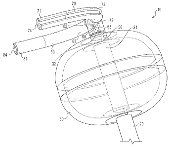
[0008] FIG. 1 is a perspective view of a cervical cerclage assistance
device,
showing a balloon disposed upon a catheter in an inflated configuration.
[0009] FIG. 2 is another perspective view of the cervical cerclage
assistance
device of FIG. 1.
[0010] FIG. 3 is a perspective view of the barrier structure and first and
second
arms of the device of FIG. 1.
[0011] FIG. 4 is a side view of the second arm and barrier structure of the
device
of FIG. 1, showing the first and second blind holes of the barrier structure.
[0012] FIG. 5 is another perspective view of the barrier structure and
first and
second arms of the device of FIG. 1, showing an elongate tape threaded through
the
first and second arms and a through hole in the barrier structure.
[0013] FIG. 6 is a cross-sectional view of the barrier structure and distal
end
portion of the catheter, with the tip and wire guides engaged within the
barrier
structure.
[0014] FIG. 7 is a perspective view of the support portion and tip of the
catheter of
the device of FIG. 1.
- 3 -
CA 02866316 2014-09-03
WO 2013/138263
PCT/US2013/030320
[0015] FIG. 8 is a side schematic view of a patient's vagina, cervix, and
uterus,
showing the device of FIG. 1 positioned therein in preparation for a cervical
cerclage
procedure.
[0016] FIG. 9 is the view of FIG. 8 with the balloon expanded and the tape
fixed
to the cervical tissue.
[0017] FIG. 10 is the view of FIG. 8 with the cervical cerclage stitching
completed
and the catheter released from the barrier structure.
[0018] FIG. 11 is a perspective view of another embodiment of a cervical
cerclage
assistance device.
[0019] FIG. 12 is a perspective view of another embodiment of a cervical
cerclage
assistance device.
[0020] FIG. 13 is a side view of the device of FIG. 12, showing the barrier
structure and hub maintained connected with a wire.
[0021] FIG. 14 is the view of FIG. 13 with the barrier structure
disconnected from
the hub with the wire removed.
[0022] FIG. 15 is a side view of a catheter supporting the device of FIG.
12, with
the barrier structure disconnected from the hub.
[0023] FIG. 16 is a perspective view of the barrier structure of the device
of FIG.
12.
[0024] FIG. 17 is a perspective view of the hub of the device of FIG. 12.
DETAILED DESCRIPTION OF THE DRAWINGS AND THE PRESENTLY
PREFERRED EMBODIMENTS
[0025] Turning now to FIGs. 1-7, a cerclage assistance device 10 is
provided.
The device 10 includes a catheter 20 that extends from a proximal end portion
22
to a distal end portion 21. An inflatable balloon 30, similar to a Foley
balloon, is
provided at the distal end portion 21. A first lumen 26 is disposed along the
length of the catheter 20 to allow for selective inflation of the balloon 30
from the
proximal end portion 22 of the catheter 22. The proximal end portion 22 of the
catheter may receive a syringe through a luer lock fitting or similar
structure (not
shown) that is disposed in fluid communication with the first lumen 26 to
allow for
selective inflation and deflation of the balloon 30. The catheter 20 may
additionally include a second lumen 24 along its length between the proximal
and
- 4 -
CA 02866316 2014-09-03
WO 2013/138263
PCT/US2013/030320
distal end portions 22, 21 to allow a guide wire 50 or similar elongate, thin,
flexible member therethrough.
[0026] The distal end portion 21 of the catheter 20 includes a support
portion 32
that may reside upon the catheter 20 distally of the balloon 30, and provide
the distal
end surface 21a (FIG. 7) of the catheter 20. The support portion 32 includes
tip 36
that extends distally from the distal end surface 21a. In some embodiments,
the tip
36 extends at an acute angle a with respect to the longitudinal axis L of the
catheter
20 at the distal end portion 21 (best shown in FIGs. 6-7), the angle a is
measured
between the longitudinal axis L and an axis 36a formed through the tip 36. In
some
embodiments the tip 36 may be substantially straight, that extends along the
line
36a, while in other embodiments, the tip 36 may be arcuate and have a changing
angle with respect to the longitudinal axis L of the catheter 20 along its
length. The
tip 36 may have a relatively rectangular cross-section along its length, which
may
have substantially the same dimensions along its length, or may have
decreasing
dimensions to reduce the size of the tip 36 as it moves toward the distal end
36c of
the tip 36. In some embodiments, the edges 36b of the tip 36 may be chamfered
or
rounded, as shown in FIG. 7.
[0027] The tip 36 may include a first aperture 37 that is disposed through
the
body of the tip 36. The first aperture 37 is configured to receive a wire
guide 50
therethrough, as discussed in greater detail below. The first aperture 37 of
the tip 36
is positioned such that the first aperture 37 is in registry with the second
hole 68 of
the barrier structure 60 when the tip 36 is disposed within the first hole 66
of the
barrier structure 60 (discussed in greater detail below) such that a guide
wire 50
extending through the second hole 68 additionally extends through the first
aperture
37 in the tip 36, as best shown in FIG. 6. The support portion 32 additionally
includes a second aperture 38 that is disposed in registry with the second
lumen 24
for the wire guide 50 to pass through. The second aperture 38 and the first
aperture
37 are each positioned with respect to each other such that a typical guide
wire 50,
such as a 0.032 inch wire guide extending through the second aperture 38 can
easily
bend to extend through the first aperture 37.
[0028] The catheter 20 is releasably engaged with a barrier structure 60 at
the
distal end portion 21 of the catheter 20. The barrier structure 60 may include
a
base 62, or bottom surface, that includes first and second holes 66, 68 that
- 5 -
CA 02866316 2014-09-03
WO 2013/138263
PCT/US2013/030320
blindly extend within the base 60. The first hole 66 is configured to receive
the tip
36 and may be formed with a size and shape just slightly larger than the size
and
shape of the tip 36, such that the tip 36 easily slides through the first hole
66
when the barrier structure 60 is engaged with the support portion 32 of the
catheter 20.
[0029] The barrier structure 60 may additionally include a second hole that
extends blindly therethrough from the base 62, and may be oriented such that
the
second hole 68 intersects the first hole 66, as best shown in FIG. 6. The
second
hole may have a circular or other profile, and have a diameter (or smallest
cross-
sectional dimension) to allow a 0.032 inch wire guide 50 (or similar diameter
wire
guide) to pass therethrough. In some embodiments, when the support portion 32
engages the barrier structure 60, and specifically, the tip 36 of the support
portion
32 extends through the first hole 66, the second hole 68 of the barrier
structure
60 aligns with the second aperture 38 of the support portion 32 and urges the
wire guide 50 extending through the second aperture 38 toward and through the
first aperture 37 of the tip 36. The engagement of the wire guide 50 into and
through the first aperture 37 in the tip 36 locks the barrier structure 60
with
respect to the catheter 20. In some embodiments, the device 10 is constructed
such that the barrier structure 60 is engaged with the catheter 20 when the
medical professional accesses the device 10 from the packaging.
[0030] As can be understood with reference to FIGs. 6 and 7, the barrier
structure
60 is releasable from the catheter 20, by urging the wire guide 50
proximately, which
causes the distal tip of the wire guide 50 to pull out of the first aperture
37 in the tip
36. When the wire guide 50 is pulled out of the first aperture 37, the
catheter 20 and
the barrier structure 60 can become decoupled. As discussed in greater detail
herein, the catheter 20 and the barrier structure 60 are normally decoupled
when the
barrier structure 60 is either directly (in embodiments with one or more arms
70, 80
extending therefrom) or indirectly (in embodiments without the one or more
arms 70,
80) fixed to the patient's cervix, the catheter 20 may be released from the
barrier
structure 60 by urging the catheter 20 proximally. Because the barrier
structure 60 is
either directly or indirectly fixed to the patient's cervical tissue, the tip
36 is pulled
- 6 -
CA 02866316 2014-09-03
WO 2013/138263
PCT/US2013/030320
from the first hole 66 in the barrier structure 60 thereby separating the two
components.
[0031] As best shown in FIGs. 1-5, the barrier structure 60 may support one
or
more arms 70, 80 that extend therefrom and are sized and oriented to surround
a
portion of a patient's cervical tissue to assist with temporarily compressing
the
cervical tissue to allow the physician to easily and conveniently perform a
cervical
cerclage procedure. The arms 70, 80 are disposed such that their respective
first
ends 72, 82 (the ends that contact or are connected to the outer surface of
the
barrier structure 60) are each aligned with a through hole 64 disposed through
the
barrier structure 60, as best shown in by the common axis D. As best shown in
FIGs. 2, 4, and 6, the through hole 64 is disposed substantially perpendicular
to the
longitudinal axis 60a of the barrier structure 60. In some embodiments, the
through
hole 64 may be generally parallel with a plane through one or both of the
first and
second holes 66, 68, while in other embodiments, best shown in FIG. 6, the
through
hole 64 is generally perpendicular to a plane through one or both of the first
and
second holes 66, 68. In still other embodiments, the through hole 64 may be
disposed at an oblique angle with respect to a plane through one or both of
the first
and second holes 66, 68.
[0032] One or both of the first and second arms 70, 80 may be constructed with
an central arcuate portion 73, 83, and may additionally be constructed with
relatively
straight portions at one or both of the first (inner) ends 72, 82 and the
second (outer)
ends 71, 81. Generally, the arms 70, 80 are constructed with a shape and size
such
that the arms are adapted to wrap around a significant portion of a female
patient's
(either human or mammal) cervical tissue when the device 10 is positioned such
that
the barrier structure 60 is disposed within or abutting, the posterior fornix
1104 of the
patient's cervix (shown in FIG. 8). In some embodiments, the first and second
arms
70, 80 are constructed to wrap around a significant portion of a patient's
cervical
tissue when the balloon 30 is inflated, which compresses the cervical tissue
together
due to the size of the inflated balloon, within the limited space available
within the
patient's vagina 1106.
[0033] Each of the first and second arms 70, 80 may have a substantially
constant cross-section along their length, with some embodiments formed with a
cross-section like a "C" along all or a portion of the length of the
respective arm. In
- 7 -
CA 02866316 2014-09-03
WO 2013/138263
PCT/US2013/030320
these embodiments, the first and second arms 70, 80 each include an inner
volume
74, 84, which is configured to receive and allow a flexible member, such as a
tape or
mersilene band 1001 (shown schematically in FIG. 5), to be threaded
therethrough.
In some embodiments (best understood with reference to FIG. 5), the first and
second arms 70, 80 are constructed such that the opening into the internal
volume
74, 84 along the length of the arm is positioned such that the opening points
generally away from the patient's cervical tissue 1102 (FIG. 8) when deployed
within
the patient. This construction has been found to be beneficial because it
allows the
tape 1001, discussed in detail below, to be partially removed from the arms
70,80
when the arms 70, 80 engage the cervical tissue, to assist with tying or
otherwise
fixing the ends 1002 of the tape 1001 together.
[0034] The inner volume 74, 84 of each arm is configured to be in registry
with
the through hole 64 of the barrier structure 60, such that the tape 1001 may
be
threaded through the through hole 64 as well as the inner volume 74, 84 of
each of
the first and second arms 70, 80, and be staged for being positioned and
temporarily
secured around the patient's cervical tissue when the barrier structure is
disposed at
the proximal fornix of the patient's cervix in preparation for the cervical
cerclage
procedure. In some embodiments, one or both of the first ends 71, 81 of the
first and
second arms 70, 80 are disposed coaxially with the through hole 64, as shown
with
axis D (FIG. 3). The tape 1001 may include a needle on one or both ends 1002
for
engaging the opposite end 1002 of the tape 1001 for quickly fixing the tape
1001
around the cervix while applying compressing to the cervical tissue. In other
embodiments, the ends of the tape 1001 may be tied together or otherwise fixed
together to maintain the cervical tissue in compression.
[0035] In some embodiments, the barrier structure 60 may be formed without
the first and second arms 70, 80, but otherwise is constructed like the
barrier
structure 60 discussed elsewhere herein. Specifically, the through hole 64 is
configured to receive the tape 1001 therethrough, which is disposed in
position to
be wrapped around a patient's cervical tissue when the barrier structure 60 is
disposed at the posterior fornix of the patient's cervix, and the proper
placement
of the cervix to receive the tape 1001 may be provided when the balloon 30 is
inflated. In embodiments without first and second arms 70, 80, the tape 1001
is
- 8 -
CA 02866316 2014-09-03
WO 2013/138263
PCT/US2013/030320
pulled around the cervical tissue using a forceps or other appropriate devices
to
temporarily close the cervix in preparation for the cervical cerclage
procedure.
[0036] Turning now to FIGs. 8-10, the device 10 is shown deployed within a
patient's vagina 1106 with the barrier structure 60 proximate or within the
posterior fornix 1104 of a patient's cervix 1102. In FIG. 8, the barrier
structure 60
is properly positioned, and the first arm 70 is around the patient's cervix
1102,
and the second arm 80 (not visible with the view of FIG. 8) is wrapped around
the
opposite side of the patient's cervix 1102. As shown in FIG. 9, the balloon 30
may be inflated which compresses the cervical tissue 1102 together to prevent
any communication from the vagina 1106 to the uterus 1100. As also shown in
FIG. 9, the ends 1002 of the tape 1001 have been fixed together (shown
schematically with a knot, but in some embodiments, a needle on one end of the
tape 1001 may interact with the opposite end of the tape 1001 to fix the tape
around the cervix 1102). The engagement of the tape 1001 (normally in tension)
maintains the uterus 1100 closed due to the compression of the cervical tissue
1102, and after the tape 1001 is fixed around the cervix 1102, the balloon 30
may
be deflated.
[0037] FIG. 10 depicts the catheter 20 disengaged from the barrier
structure
60, which is fixed in position at the posterior fornix 1104 due to the
engagement
of the tape 1001 around the cervical tissue 1102, and the threading of the
tape
1001 through the through hole 64. As discussed above, the barrier structure 60
may be disengaged from the catheter 20 by pulling with the wire guide 50
proximally, as shown schematically by arrow W, at the proximal end portion 22
of
the catheter 20. As best understood with reference to FIG. 6, as the wire
guide
50 is pulled proximally, the distal tip 51 of the wire guide 50 slides though
the
second hole 68 in the barrier structure 60 and out of the first aperture 37 of
the tip
36. After the wire guide 50 is pulled proximally, the catheter 20 is
additionally
pulled proximally (in the same direction as arrow W in FIG. 10) to withdraw
the tip
36 from the first hole 66 of the barrier structure 60, and when the tip 36 is
fully
withdrawn from the first hole 66, the catheter 20 may be removed from the
patient.
- 9 -
CA 02866316 2014-09-03
WO 2013/138263
PCT/US2013/030320
[0038] FIG. 10 further schematically depicts the cervical cerclage stitch
upon
the patient's tissue at 700. As discussed above, the cervical cerclage stitch
may
be the McDonald stitch, the Shirdokar stitch, or other types of stitch
patterns
known in the art. Upon completion of the desired (clinically) cervical
cerclage
procedure, the physician may cut or otherwise remove the tape 1001 from around
the cervix, and withdraw the barrier structure 60, tape 1001, and first and
second
arms 70, 80 from the patient using a forceps or other tool.
[0039] Turning now to FIG. 11, an alternate device 100 is provided. The
device 100 includes an elongate catheter 120 that extends between a distal end
portion 121 and a proximal end portion 122. The catheter 120 includes balloon
130, similar to balloon 30 discussed above, that may be inflated or deflated
by
injecting fluid therein through a balloon lumen 124 from a syringe or other
fluid
source through a luer lock or similar structure at the proximal end portion
122 of
the catheter 120.
[0040] The end surface of the distal end portion 121 may receive another style
barrier structure, such as an eyelet 160 or other similar structure with an
aperture
164 therein, which allows a tape 1001 to be threaded therethrough, for
engaging
a patient's cervical tissue 1101 when the eyelet 160 is disposed at the
proximal
fornix 1104 of the cervix 1102. In some embodiments, the eyelet 160 may
additionally support one or more arms that are aligned with the aperture 164
of
the eyelet 160, with the arms being constructed similarly to the two or more
arms
70, 80 for engaging the cervical tissue when the eyelet 160 is properly
positioned
and the balloon 130 is inflated, as discussed above.
[0041] The catheter 120 may further include a second lumen 126 extending
from the proximal end portion 122 of the catheter toward the distal end
portion
121 of the catheter. The catheter 120 may further include a weakened region
129 upon the distal end portion 121 and distally of the balloon 130. The
weakened region 129 may be formed from a portion of the catheter that has a
thinner wall around the circumference thereof, may include a plurality of cuts
along the circumference of the catheter 120 (either through the entire
thickness of
the material, or through only a portion of the thickness of the material), or
in other
-10-
CA 02866316 2014-09-03
WO 2013/138263
PCT/US2013/030320
known ways to form a circumference of the catheter 120 that is configured to
easily break from the remainder of the catheter 120.
[0042] The second lumen 126 may communicate through the wall of the
catheter 120 through one or more holes 128 formed either in the weakened
region 129 or just proximal of the weakened region 129. In some embodiments,
the one or more holes 128 may define at least a portion of the weakened region
129, while in other embodiments, the one or more holes 128 may be disposed
proximally of the weakened region 129. The second lumen 126 may receive a
wire guide 150 (such as a 0.032 inch wire guide) threaded therethrough, which
extends therethrough from the proximal end portion 122 of the catheter and out
the one or more holes 128. In other embodiments, the wire guide 150 may be
other types of strong but thin wires, such as a piano wire or the like to
allow the
wire guide 150 to cut through the material forming the catheter 120 at the
weakened region 129, as discussed below.
[0043] The wire guide 150 may form a loop 152 that is configured to interact
with the weakened region 129, and configured to break the weakened region 129
when the wire guide 150 is pulled proximally at the proximal end portion 122
of
the catheter 120. In some embodiments, the wire guide 150 may be woven
through the plurality of holes defining the weakened region to form the loop
152,
while in other embodiments, the loop 152 of the wire guide 150 may otherwise
interact with the weakened region 129 to cut the material forming the catheter
120 at the weakened region 129 when the wire guide 150 is pulled. As
understood with reference to the remainder of this specification, the device
100 is
initially positioned with the eyelet 160 disposed proximate to or contacting
the
posterior fornix 1104 of the patient's cervix, and the tape 1001 extending
through
the eyelet 160 (and arms, when provided) is wrapped around and tied (or
otherwise fixed) around the cervix, while the cervical tissue is urged into
positioned by the inflation of the balloon 130. Once the tape 1001 is fixed
around
the cervix, the wire guide 150 is pulled proximally, causing the wire guide
150 to
cut through the weakened region 129, thus allowing the catheter 120 to be
removed from the patient's vagina, with the tape 1001 remaining engaged with
the cervix (and the eyelet 160 and remainder of the distal end portion 121
-11 -
CA 02866316 2014-09-03
WO 2013/138263
PCT/US2013/030320
remaining within the patient. The physician next performs the cervical
cerclage
procedure and then cuts the tape 1001 and removes the tape 1001 and eyelet
160 from the patient.
[0044] Turning now to FIGs. 12-17, another device 200 configured to a
cervical cerclage procedure is provided. The device 200 includes an elongate
catheter 220 that extends between a distal end portion 221 and a proximal end
portion 222 (FIG. 15). The catheter 220 includes a balloon 230, similar to the
balloon 30 discussed above, that may be inflated or deflated by injecting
fluid
therein through a lumen (not shown) from a syringe or other fluid source, as
mated by a luer lock or similar structure disposed upon the proximal end
portion
222 of the catheter 220.
[0045] The distal end portion 221 of the catheter may receive a hub 250
mounted thereto at the distal end face of the catheter. In some embodiments,
the
hub 250 may include a projection 260 that nests within a similar hole or void
in
the catheter 220, with the two components being fixed together with adhesive,
with a press-fit or with other attachment methods. The projection 260 may be
of
a non-circular cross-section (such as with a bite or recess 260a) to provide
for a
single possible alignment between the hub 250 and the catheter 220. In some
embodiments, the hub 250 may further include a plug 261, which may cap the
balloon inflation lumen, as well as provide additional alignment structure
between
the hub 250 and the catheter 220.
[0046] The catheter 220 may include at least two lumens that extend from a
proximal end portion 222 and in parallel through the length of the catheter
220
and to the distal end portion. A first lumen is configured to provide fluid
communication between a luer lock adaptor and a balloon 230, which is disposed
upon the distal end portion 221. The balloon 230 is configured to be inflated
and
deflated based upon the injection of fluid, such as water or saline into the
balloon
through the luer lock. The balloon 230 and the first lumen is similar to the
balloon
30 and first lumen 26 discussed above. The catheter 220 may include a second
lumen along its length between the proximal end distal end portions 222, 221,
which allows a guide wire 50 or similar elongate, thin, flexible member
therethrough. The second lumen of the catheter 220 is similar to the second
- 12-
CA 02866316 2014-09-03
WO 2013/138263
PCT/US2013/030320
lumen 24 discussed above. The second lumen is disposed coaxially and in
communication with an aperture 259 through the hub 250, which is mounted to
the distal end portion 221 of the catheter 220.
[0047] The hub 250 is fixed to the end face of the distal end portion 221 of
the
catheter 220. The hub 250 includes an extension 254 that extends from a body
251 of the hub 250. In some embodiments, the extension 254 extends in a
direction away from the catheter 220, and defines a void 255 between the body
251 of the hub 250 and the inner surface of the extension 254. In some
embodiments as shown on FIG. 14, the extension 254 may extend from the body
251 of the hub 250 along an axis X that extends in an obtuse angle a to the
longitudinal axis L of the hub 250 (or the axis of the hole 259). In some
embodiments, the angle a may be between about 100 and about 135 degrees
(inclusive of all angles within this range), while in other embodiments, the
angle a
may be about 100 to about 120 degrees (inclusive of all angles within this
range).
The inner surface 254a of the extension 254 (i.e. the surface facing the body
251
of the hub 250 may be parallel to an axis through the hub 250.
[0048] In some embodiments, the upper surface of the body 251 of the hub
250 may be parallel with the extension 254, such that the void 255 (and both
the
surfaces of the body 251 and the extension 254 that define the void 255) of
the
hub 250 extends in the same angle a as the extension 254 with respect to the
longitudinal axis L of the hub 250. In some embodiments, the upper surface
254b of the extension 254 is also parallel with the extension 254 (and in some
embodiments the lower surface 254a). The extension 254 additionally includes a
hole 258 that is coaxial with the aperture 259 in the base 251 of the hub 250.
The hole 258 is configured such that a guide wire 50 that extends through the
catheter 220 and the aperture 259 of the hub 250 additionally extends through
the hole 258 in the extension 254.
[0049] The barrier structure 270 is additionally provided and is removably
attached to the hub 250. The barrier structure 270 may retain one or more arms
70,
80 thereon, which may be the same (in operation and/or structure) as the arms
70,
80 discussed above and are configured to support a tape or band 1001 for use
in
conjunction with a cervical cerclage procedure as discussed above. The barrier
-13-
CA 02866316 2014-09-03
WO 2013/138263
PCT/US2013/030320
structure 270 includes a base 271 that defines a hole 280 that is aligned with
the first
and second arms 70, 80 (or one of the first or second arms 70, 80 in
embodiments
where only one arm 70 80 is provided) such that a tape or band 1001 that is
threaded through the hole 280 additionally is threaded through the first
and/or
second arms 70, 80.
[0050] The barrier structure 270 additionally includes a finger 274 that
extends
from the base 271 of the barrier structure 270. The finger 274 may extend from
the
base 271 such that an axis Y through the finger 274 extends at the angle y to
the
longitudinal axis L through a first hole 278 through the finger 274 and a
coaxial
hole 279 that extends blindly into the base 271 of the barrier structure 270.
In
some embodiments the angle y may be the same or similar to the angle a, while
in other embodiments it may be different. In embodiments where the angles y
and a are different, the finger 274 and the extension 254 fit together
(discussed in
more detail below) with the various sizes and shapes of the finger 274 and
extension 254 (as well as the two voids 275, 255, to allow engagement between
the barrier structure 270 and the hub 250.
[0051] The finger 274 defines a second void 275 between an inner surface
274a
of the finger 274 and a bottom surface 271a of the base. The second void 275
is
sized to receive the extension 254 therein and the first void 255 is sized to
receive
the finger 274 therein, such that the barrier structure 270 and the hub 250
are
removably connected together. When the barrier structure 270 and the hub 250
are
connected together, a guide wire 50 may extend through the lumen of the
catheter
220, through the hole 259 of the hub 250, through hole 278 in the finger 274,
then
through the hole 258 in the extension and finally within the blind hole 279
within the
barrier structure 270. The extension of the guide wire 50 through the
plurality of
aligned holes when the barrier structure 270 is fixed to the hub 250 (and
therefore
the catheter 220) prevents disconnection because the guide wire 50 prevents
the
finger and extension 274, 254 from sliding out of the respective void 255,
275.
[0052] The barrier structure 270 can be decoupled or disconnected from the
hub
250 when the guide wire 50 is withdrawn from the barrier structure 270
(including the
finger 274 as well as the extension 255. The withdrawal of the guide wire 50
allows
each of the finger 274 and extension 254 to be able to be slid out of the void
(255,
- 14-
CA 02866316 2014-09-03
WO 2013/138263
PCT/US2013/030320
275) on the opposite component, which decouples the barrier structure 270 from
the
catheter 220.
[0053] As discussed elsewhere herein, the device 200 is configured to
assist with
the performance of a cervical cerclage procedure, and the device 200 is used
clinically in a similar manner to the device 10 discussed above. The steps
discussed
above and depicted in FIGs. 8-10 are equally applicable with the device 200 as
with
the device 10 (with the structural differences of the connection between the
barrier
structure 60 and the catheter 20 and the barrier structure 270 and the
catheter 220
causing a difference in response when the guide wire 50 is pulled proximally
with
both embodiments, with these difference easily appreciated with a thorough
review
of the subject specification and figures).
[0054] As will be understood with reference to FIGs. 13 and 14, when the
guide
wire 50 is withdrawn from the barrier structure 270 and the extension 254 of
the hub
250, the barrier structure 270 may be released from the hub 250 and the
catheter
220. As can be understood, the two structures are released when the catheter
220
and hub 250 are moved sideways (i.e. in the direction Z of FIG. 14), which
causes
the extension 254 to slide out of the void 275 and causes the finger 274 to
slide out
the void 255. Upon removal of the hub 250 from the barrier structure 270, the
catheter 220 is withdrawn from the patient, and the barrier structure can be
removed
from the patient after the cervical stitch(s) are completed and the tape or
band 1001
is cut.
[0055] While the preferred embodiments of the disclosure have been
described, it
should be understood that the disclosure is not so limited and modifications
may be
made without departing from the disclosure. The scope of the invention is
defined by
the appended claims, and all devices that come within the meaning of the
claims,
either literally or by equivalence, are intended to be embraced therein.
-15-