Note: Descriptions are shown in the official language in which they were submitted.
CA 02866330 2014-09-03
WO 2013/181022
PCT/US2013/042025
INJECTABLE BIODEGRADABLE PARTICLES FOR CONTROLLED
THERAPEUTIC AGENT RELEASE
STATEMENT OF RELATED APPLICATION
100011 This application claims the benefit of U.S. Serial No. 61/653,233,
filed May 30, 2012
and entitled: "INJECTABLE BIODEGRADABLE PARTICLES FOR CONTROLLED
THERAPETUIC AGENT RELEASE," which is hereby incorporated by reference in its
entirety
FIELD OF THE INVENTION
[0002] The invention relates to polymeric particles for injection which
exhibit controlled
therapeutic agent release.
BACKGROUND OF THE INVENTION
[0003] Many clinical situations benefit from regulation of the vascular,
lymphatic or duct
systems by restricting the flow of body fluid or secretions. For example, the
technique of
embolization involves the introduction of particles into the circulation to
occlude blood
vessels, for example, so as to either arrest or prevent hemorrhaging or to cut
off blood flow to
a structure or organ. Temporary occlusion of blood vessels is desirable for
managing various
diseases and conditions.
[0004] In one example of an embolization procedure, local anesthesia is first
given over a
common artery. The artery is then percutaneously punctured and a catheter is
inserted and
fluoroscopically guided into the area of interest. An angiogram is then
performed by
injecting contrast agent through the catheter. An embolic agent is then
deposited through the
catheter. The embolic agent is chosen, for example, based on the size of the
vessel to be
occluded, the desired duration of occlusion, and/or the type of disease or
condition to be
treated (e.g., hypervascular tumors, uterine fibroids, etc.), among others
factors. A follow-up
angiogram may be performed to determine the specificity and completeness of
the arterial
CA 02866330 2014-09-03
WO 2013/181022
PCT/US2013/042025
occlusion. Blocking the blood supply to the tissue is intended to result in
shrinkage and/or
death of the tissue.
[0005j Various microspheres are currently employed to embolize blood vessels.
These
microspheres are usually introduced to the location of the intended
embolization through
microcatheters. Many commercially available embolic microspheres are composed
of
polymers. Materials used commercially for this purpose include polyvinyl
alcohol (PVA),
acetalized PVA (e.g., Contour SETM embolic agent, Boston Scientific, Natick,
MA, USA) and
crosslinked acrylic hydrogels (e.g., Embospheres0, Biosphere Medical,
Rockland, MA,
USA). Similar microspheres have been used in chemoembolization to increase the
residence
time of the therapeutic after delivery. Other examples of commercially
available
microspheres include glass microspheres with entrapped radioisotopes (e.g.,
90Y), in
particular, TheraSpheresTm, MDS Nordion, Ottowa, Canada and polymer
microspheres that
are capable of chelating radioisotopes (90Y), in particular, SIR-Spheres ,
SIRTex Medical,
New South Wales, Australia. In one specific instance, a therapeutic agent
(doxorubicin) has
been directly added to polyvinyl alcohol hydrogel microspheres such that it
can be released
locally after delivery (e.g., DC BeadTM drug delivery chemoembolization
system,
Biocompatibles International plc, Farnham, Surrey, UK). There are also
particles currently
on the market (e.g. Embosphere , Merit Medical Systems, Inc., South Jordan,
Utah USA)
that allow the health care provider to load "empty" porous beads with a drug.
Loading is
typically achieved by ionic bonding of the drug to the particle, resulting in
a relatively low
drug uptake by the particles and a relatively fast release (e.g., within 48
hours).
SUMMARY OF THE INVENTION
[0006] In accordance with one aspect of the invention, embolic particles are
provided that
comprise a biodegradable polymer and a therapeutic agent, wherein the
particles are
configured such that, upon administration to a body lumen of a subject, the
therapeutic agent
is released from the time of administration up until a first point in time
that ranges anywhere
from about 1 week after administration to about 4 weeks after administration,
at which point
in time the therapeutic agent release ceases. The particles are also
configured such that
particles remain present in the body lumen from the first point in time at
which therapeutic
agent release ceases up to a second point in time that ranges anywhere from
about 2 weeks to
2
CA 02866330 2014-09-03
WO 2013/181022
PCT/US2013/042025
about 12 months after the first point in time, at which point the particles
are completely
degraded.
[0007] Other aspects of the invention pertain to methods of making such
particles.
[0008] Still other aspects of the invention pertain to injectable compositions
that comprise
such particles and to methods of treatment that employ such injectable
compositions.
[0009] These and various additional aspects, embodiments and advantages of the
present
invention will become immediately apparent to those of ordinary skill in the
art upon review
of the Detailed Description and any appended claims to follow.
BRIEF DESCRIPTION OF THE DRAWINGS
[0010] Fig. 1 is a schematic illustration of a spherical core-shell
biodegradable particle in
accordance with an embodiment of the present invention.
[0011] Fig. 2 is a schematic illustration of the particle of Fig. 1 upon
degradation of the
particle shell.
[0012] Fig. 3 is a schematic illustration of a spherical core-shell
biodegradable particle in
accordance with another embodiment of the present invention.
[0013] Fig. 4 is a schematic illustration of the particle of Fig. 3 upon
degradation of the
particle core.
[0014] Fig. 5 is a schematic illustration of a process of producing spherical
core-shell
biodegradable particles using a triple nozzle apparatus, in accordance with an
embodiment of
the present invention.
[0015] Fig. 6 is a schematic illustration of a process of producing spherical
core-shell
biodegradable particles using a microfluidic double T-channel apparatus, in
accordance with
an embodiment of the present invention.
[0016] Fig. 7 is a schematic illustration of a spherical core-shell
biodegradable particle
having multiple cores, in accordance with an embodiment of the present
invention.
3
CA 02866330 2014-09-03
WO 2013/181022
PCT/US2013/042025
[0017] Fig. 8 is a schematic illustration of the particle of Fig. 7 upon
degradation of the
particle cores.
DETAILED DESCRIPTION
[0018] In accordance with various aspects, the invention provides injectable
biodegradable
polymeric particles that contain a biodegradable polymer and a therapeutic
agent.
[0019] The particles are configured such that, upon administration to a body
lumen (e.g., a
blood vessel such as an artery, lymphatic vessel, etc.) of a subject, the
therapeutic agent is
released from the point of administration up until a first point in time that
ranges anywhere
from about 1 week after administration to about 4 weeks after administration,
at which point
in time the therapeutic agent release ceases. For example, the first point in
time where the
therapeutic agent release ceases may range anywhere from 1 week to 2 weeks to
3 weeks up
to 4 weeks after administration.
[0020] The particles are also configured such that the particles remain in the
body lumen for
a second period of time after the first period of time has expired (i.e.,
after therapeutic agent
release ceases). For example, the particles may be further configured such
that the particles
remain present in the body lumen from the first point in time (the time at
which therapeutic
agent release ceases) up to a second point in time that ranges anywhere from
about 2 weeks
to about 12 months after the first point in time, at which point the particles
become
completely degraded. For example, the second point in time may range anywhere
from 2
weeks to 3 weeks to 4 weeks to 2 months to 4 months to 6 months to 8 months to
10 months
to 12 months after the first point in time.
[0021] As defined herein, the point in time where therapeutic agent ceases is
the point where
the release rate from the particle(s) drops to below about 5 % of the maximum
rate of release.
[0022] As defined herein, the point of complete degradation the point where at
least 95 wt%
degradation of the particle(s) has occurred relative to initial weight of the
particle(s) (i.e., the
weight at the point of injection).
[0023] As used herein a "polymeric particle" is one that contains polymers,
typically, from
50 wt% to 75 wt% to 90 wt% to 95 wt% to 97.5 wt% to 99 wt% or more polymers.
4
CA 02866330 2014-09-03
WO 2013/181022
PCT/US2013/042025
[0024] As used herein a "biodegradable polymeric particle" is one that
undergoes chain
cleavage in vivo. As used herein, a polymer is "biodegradable" if it undergoes
bond cleavage
along the polymer backbone in vivo, regardless of the mechanism of bond
cleavage (e.g.,
enzymatic breakdown, hydrolysis, oxidation, etc.).
[0025] As used herein, "polymers" are molecules that contain multiple copies
of one or more
types of constitutional species, commonly referred to as monomers. The number
of
monomers within a given polymer may vary widely, ranging, for example, from 5
to 10 to 25
to 50 to 100 to 1000 to 10,000 or more constitutional units. As used herein,
the term
"monomer" may refer to the free monomers and those that are incorporated into
polymers
(also referred to herein as monomer "residues"), with the distinction being
clear from the
context in which the term is used.
[0026] As used herein, a "moiety" is a subunit of a polymer and includes
monomers and
collections of monomers. Moieties may be named herein based on species that
are used, or
could have been used, to create the moiety (e.g., via a condensation reaction,
resulting in the
generation of a water molecule, etc.). As a specific example, a "succinic acid
moiety", ¨
COCH2CH2C0¨, may be produced using succinic acid, HOOCCH2CH2COOH, via
condensation reaction, but can also be produced using another species such as
succinyl
chloride, C1OCCH2CH2C0C1.
[0027] The injectable particles of the present disclosure may be non-
crosslinked or they may
be covalently and/or non-covalently crosslinked. Thus, in some embodiments,
crosslinking
agents such as covalent crosslinking agents or ionic crosslinking agents may
be present in the
injectable particles, whereas in other embodiments crosslinking agents are
absent from the
particles. In some embodiments the particles may be crosslinked by exposure to
radiation
(e.g., gamma or e-beam radiation), which may occur in conjunction with
sterilization of
particles.
[0028] The injectable particles may be used to treat various diseases and
conditions in a
variety of subjects. Subjects include vertebrate subjects, particularly humans
and various
warm-blooded animals, including pets and livestock. As used herein,
"treatment" refers to
the prevention of a disease or condition, the reduction or elimination of
symptoms associated
with a disease or condition, or the substantial or complete elimination of a
disease or
condition. Preferred treatments are embolization treatments.
CA 02866330 2014-09-03
WO 2013/181022
PCT/US2013/042025
[0029] The injectable particles of the invention may vary in shape. In certain
embodiments,
they are substantially spherical, for example, having the form of a perfect
(to the eye) sphere
or the form of a near-perfect sphere such as a prolate spheroid (a slightly
elongated sphere) or
an oblate spheroid (a slightly flattened sphere), among other regular or
irregular near-
spherical geometries. In embodiments where the particles are substantially
spherical, at least
half of the particles (50% or more, for example, from 50% to 75% to 90% to 95%
or more of
a particle sample) may have a sphericity of 0.8 or more (e.g., from 0.80 to
0.85 to 0.9 to 0.95
to 0.97 or more). The sphericity of particles can be determined, for example,
using a
Beckman Coulter RapidVUE Image Analyzer version 2.06 (Beckman Coulter, Miami,
FL).
Briefly, the RapidVUE takes an image of continuous-tone (gray-scale) form and
converts it to
a digital form through the process of sampling and quantization. The system
software
identifies and measures the particles in an image. The sphericity of a
particle, which is
computed as Da/Dp (where Da = A4A/R); Dp = Phi; A = pixel area; P = pixel
perimeter), is a
value from zero to one, with one representing a perfect circle. A particle is
"spherical" if it
has a sphericity of 0.8 or more (e.g., from 0.80 to 0.85 to 0.9 to 0.95 to
0.97 or more).
[0030] The injectable particles of the present disclosure can vary
significantly in size, with
typical longest linear cross-sectional dimensions (e.g., the diameter of a
sphere, the length of
a rod or fiber, etc.) of the particles ranging, for example, from 40 to 5000
microns ( m) (e.g,
from 40 to 50 to 100 to 150 to 200 to 250 to 300 to 400 to 500 to 750 to 1000
to 1500 to 2000
to 2500 to 5000 microns), more preferably from 45 to 300 microns. Such
particles can be
delivered, for example, using a microcatheter (e.g., one having an inside
diameter ranging
from 530 to 690 microns, among other sizes).
[0031] For a collection of particles, the arithmetic mean maximum dimension
for the group is
preferably within the preceding ranges. The arithmetic mean maximum dimension
of a group
of particles can be determined using a Beckman Coulter RapidVUE Image Analyzer
version
2.06 (Beckman Coulter, Miami, FL), described above. The arithmetic mean
maximum
dimension of a group of particles (e.g., in a composition) can be determined
by dividing the
sum of the maximum dimensions (which, for a sphere, is the diameter) of all of
the particles
in the group by the number of particles in the group.
[0032] In certain embodiments, multimodal distributions of particles sizes may
be employed.
For example, a collection of particles may have a first group of particles
with a first
arithmetic mean maximum dimension of 40 to 50 microns and a second group of
particles
6
CA 02866330 2014-09-03
WO 2013/181022
PCT/US2013/042025
having a second arithmetic mean maximum dimension of 100 to 150 microns, among
other
possibilities.
[0033] Biodegradable polymers for use in the embolic particles of the present
disclosure
include biodegradable polyesters (e.g., polyhydroxy acids), polyorthoesters,
polyether esters,
polyamides, polyesteramides, polydepsidpetides, polyurethanes,
polysaccharides, and
polyhydroxyalkanoates, among others.
[0034] Depending on the biodegradation mechanism, a polymer can undergo
surface
degradation, bulk degradation or a combination of both. Surface versus bulk
degradation is
often dependent on whether the degradation is via a hydrolytic mechanism
(e.g., ester
hydrolysis) or via an enzymatic mechanism. In case of degradation by
hydrolysis, bulk
degradation takes place, but can be controlled by exerting control over the
rate of water
penetration and material swelling, which are governed by the hydrophilicity of
the polymer.
In the case of enzyme- or cellular-mediated biodegradation, the mechanism may
be mainly
via surface degradation. Enzymatic degradation can occur, for example, via
hydrolytic or
oxidative mechanisms. These degradation mechanisms can occur as a result of
the
inflammatory foreign body response that occurs upon implantation of the
polymeric drug
delivery system. Enzymes commonly involved in biodegradation include
esterases,
proteases, elastases, and peroxidases. See, e.g., Aylvin A. Dias and Marc
Hendriks, "Amino
Acid-Containing Degradable Polymers & Their Potential in Controlled Drug
Delivery,"
Drug Delivery Technology, May 2010, Vol. 10, No. 4, 20-25.
[0035] Biodegradable polymers useful in the present disclosure include those
in which the in
vivo degradation mechanism is dominated by surface degradation. Where a
therapeutic
agent is dispersed throughout such a polymer (i.e., where the polymeric
material acts as a
matrix that entraps the therapeutic agent), the rate of therapeutic agent
release from the
polymeric material will be controlled by surface degradation as well, allowing
drug release to
continue until the polymeric material is substantially entirely eroded.
[0036] One group of polymers that has demonstrated degradation dominated by
surface
erosion are the amino-acid-based poly(ester amides) (AA-REAs). For example. in
A.
Ghaffar et al., Biomacromolecules 2011,12, 3243-3251, a class of AA-PEAs was
subjected
to in vitro enzymatic degradation with a-chymotrypsin and proteinase K. The
polymers were
found to degrade at a steady rate using both enzymes, with a lack of
significant changes in the
7
CA 02866330 2014-09-03
WO 2013/181022
PCT/US2013/042025
average molecular weight of the remaining polymer, indicating that surface
erosion occurred
during the enzyme-mediated degradation. No accumulation of acidic byproducts
was
observed during the course of the experiment. The class of polymers also
showed a
remarkable hydrolytic stability in the absence of enzymes.
[0037] AA-PEAs appear to support a more natural wound healing process than
aliphatic
polyester-based biomaterials by promoting reendothelialization and lowering
inflammatory
response. See Kai Guo and C. C. Chu, Journal of Biomedical Materials Research
Part B:
Applied Biomaterials, Volume 89B, Issue 2, 2008, 491-500. For example, AA-PEAs
have
shown good tissue and blood compatibility in stent coating applications, with
in vivo
biocompatibility as tested in porcine coronary arteries showing that the
polymer-coated stents
had similar injury and inflammation scores to a bare metal stent. See Aylvin
A. Dias and
Marc Hendriks, Drug Delivery Technology, May 2010, Vol. 10, No. 4, 20-25.
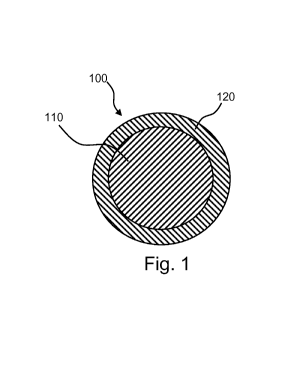
[0038] Referring now to Fig. 1, a spherical biodegradable particle 100 in
accordance with the
present disclosure is shown, which includes (a) a biodegradable core 110 that
contains one or
more biodegradable polymers and no therapeutic agent and (b) a biodegradable
shell 120 that
contains one or more biodegradable polymers and one or more therapeutic agents
dispersed
throughout.
[0039] As noted above, upon administration to a subject, biodegradable
particles in
accordance with the present disclosure display a first period (e.g., 3 weeks,
among other
values) over which release of one or more therapeutic agents accompanies
degradation and a
following second period (e.g., 4 weeks, among other values) during which time
degradation
of the particles continues, but with no accompanying release of therapeutic
agent, until
complete degradation of the particles is achieved. In a particle 100 like that
of Fig. 1, this can
be achieved by employing a shell 120 that undergoes surface degradation with
little to no
bulk degradation, with the time for complete surface degradation of the shell
120
corresponding to the first period during which drug release occurs. After the
first period (and
compete surface degradation of the shell) only the core 110 of the polymer
remains, as shown
schematically in Fig. 2. The core 110 then continues to degrade without any
accompanying
release of therapeutic agent until complete degradation of the particle is
achieved.
[0040] Other strategies may be employed to produce particles with release
characteristics in
accordance with the present disclosure. Referring now to Fig. 3, a
substantially spherical
8
CA 02866330 2014-09-03
WO 2013/181022
PCT/US2013/042025
biodegradable particle 200 in accordance with the present disclosure is shown,
which
includes (a) a biodegradable core 210 that contains one or more biodegradable
polymers and
one or more therapeutic agents dispersed throughout and (b) a biodegradable
shell 220 that
contains one or more biodegradable polymers and no therapeutic agent. As
previously noted,
upon administration to a subject, biodegradable particles in accordance with
the present
disclosure display a first period over which release of one or more
therapeutic agents
accompanies degradation and a second period during which time degradation of
the particles
continues, with no accompanying release of therapeutic agent, until complete
degradation of
the particles is achieved. In a particle 200 like that of Fig. 3, this can be
achieved by
employing a core 210 that undergoes surface degradation with little to no bulk
degradation,
with the time for complete degradation of the core 210 corresponding to the
first period
during which drug release occurs. The shell 220, on the other hand, is formed
of a material
that degrades more slowly than the core. Moreover, the shell is provided with
apertures 22a,
which allow diffusion of external species (e.g., water, enzymes, etc.) into
the core and which
also allow diffusion of internal species (e.g., therapeutic agent and polymer
breakdown
products) out of the core. In such a system, after the first period (and
compete degradation
of the core), only the shell 220 remains, as shown schematically in Fig. 4.
The shell 220 then
continues to degrade without an accompanying release of therapeutic agent
until complete
degradation of the particle is achieved at the end of the second period.
[0041] In accordance with another strategy, referring now to Fig. 7, a
substantially spherical
biodegradable particle 700 in accordance with the present disclosure is shown,
which
includes (a) multiple biodegradable cores 710 that contain one or more
biodegradable
polymers and one or more therapeutic agents dispersed throughout and (b) a
biodegradable
shell 720 that contains one or more biodegradable polymers and no therapeutic
agent. As
previously noted, upon administration to a subject, biodegradable particles in
accordance
with the present disclosure display a first period over which release of one
or more
therapeutic agents accompanies degradation and a second period during which
time
degradation of the particles continues, with no accompanying release of
therapeutic agent,
until complete degradation of the particles is achieved. In a particle 700
like that of Fig. 7,
this can be achieved by employing multiple cores 710 that are bulk erodible
or, more
preferably, that undergo surface degradation with little to no bulk
degradation, with the time
for complete degradation of the core 710 corresponding to the first period
during which drug
release occurs. The shell 720, on the other hand, is formed of a material that
degrades more
9
CA 02866330 2014-09-03
WO 2013/181022
PCT/US2013/042025
slowly than the core. By using a multiple core (e.g., 2, 3, etc.)
construction, one is able to
produce particles where only a very thin layer of the shell material (if any
at all) separates the
core from the exterior environment which quickly biodegrades, allowing
diffusion of external
species (e.g., water, enzymes, etc.) into the core and which also allow
diffusion of internal
species (e.g., therapeutic agent and polymer breakdown products) out of the
core.
Consequently, there is no need for apertures like those formed in Fig. 3. In
such a system,
after the first period (and compete degradation of the cores) only the shell
720 remains, as
shown schematically in Fig. 8. The shell 720 then continues to degrade without
an
accompanying release of therapeutic agent until complete degradation of the
particle is
achieved at the end of the second period.
[0042] In another strategy, a first particle comprising a polymer and a
therapeutic agent is
employed along with a second particle that comprises a polymer but no
therapeutic agent,
which second particle degrades more slowly than the first particle. For
example, a
therapeutic-agent-containing smaller particle (e.g., a spherical particle
having a first diameter
ranging from 40 to 60 microns) may be employed, along with a larger particle
that does not
contain a therapeutic agent (e.g., a spherical particle having a second
diameter ranging from
150 to 200 microns). The smaller and larger particles may be formed of the
same polymer
material. The smaller and larger particles may also be formed of different
polymeric
materials, so long as the smaller particle completely degrades during the
first time period and
the larger particle remain for a second period after complete degradation of
the first particle.
As another example, the first and second particles may be of substantially the
same size and
comprise different polymeric materials, such that the first particle
completely degrades during
the first time period and the second particle remains for a second period
after complete
degradation of the particle.
[0043] As noted above, polymers for forming the particles described herein
include amino-
acid-based poly(ester amides) (AA-PEAs) which comprise an amino acid moiety,
preferably
an a-amino acid moiety, and additional moieties selected, for example, from
polyol moieties
(e.g., diol, triol, etc.), polyacid moieties (e.g., diacid, triacid, etc.) and
hydroxyacid moieties,
among others.
10044] Various AA-PEAs suitable for use in the present disclosure are
described in the
polymer art and include, for example, those described in the following: Kai
Guo and C. C.
Chu, "Biodegradable and Injectable Paclitaxel-Loaded Poly(esteramide)s
Microspheres:
CA 02866330 2014-09-03
WO 2013/181022
PCT/US2013/042025
Fabrication and Characterization," journal of Biomedical Materials Research
Part B:
Applied .Biomaterials, Volume 89B, Issue 2, 2008, 491- 500; M. Vera et al.,
"Mierospheres
from new biodegradable poly(ester amide)s with different ratios of L- and D-
alanine for
controlled drug delivery," Journal of Microencapsulation, September 2006;
23(6): 686- 697;
Aylvin A. Dias and Marc Hendriks, "Amino Acid-Containing Degradable Polymers &
Their
Potential in Controlled Drug Delivery," Drug Delivery Technology, May 2010,
Vol. 10, No.
4, 20-25; A. Ghaffar et al., "Monitoring the in Vitro Enzyme-Mediated
Degradation of
Degradable Poly(ester amide) for Controlled Drug Delivery by LC-ToF-MS,"
Biomacromolecules 2011,12, 3243-3251; Alfonso Rodriguez-Galan, et al.,
"Degradable
Poly(ester amide)s for Biomedical Applications," Polymers 2011, 3, 65-99; Xuan
Pang et al.,
"Synthesis, characterization and biodegradation of functionalized amino acid-
based
poly(ester amide)s," Biomaterials 31 (2010) 3745- 3754; U.S. Patent No.
7,304,122 to Chu et
al. and U.S. Patent Pub. No. 2004/0063606 to Chu et al.
[0045] In certain embodiments, the AA-PEAs of the present disclosure comprise
one or more
a-amino acid moieties, one or more diol moieties and one or more diacid
moieties.
[0046] Examples of ci-amino acid moieties include moieties of the formula (I),
H 0
I II
¨ N¨C¨C ¨
II R3 , wherein R3 is independently hydrogen (i.e., a glycine moiety), a
hydrocarbon group such as (Ci-C6)allcyl, for example, ¨CH3 (i.e., an alanine
moiety), ¨
CH(CH3)CH3 (i.e., a valine moiety),
¨CH(CH3)CII2CII3 (i.e., an isoleucine moiety) and ¨CII2CH(CII3)CII3 (i.e., a
leucine
moiety), (C2.-C6)alkenyl, for example, ¨CH2CHCH2 (i.e., an allylglycine
moiety), (C2-
C6)alkynyl, (C6-Cio)aryl(Ci-C6)alkyl, for example, ¨CH2C6H5 (i.e., a
phenylalanine moiety),
a hydroxyl-substitued hydrocarbon group such as hy-droxy(Ci-C6)alky, for
example, ¨CHANT
(i.e., a serine moiety) and ¨CH(OH)CH3 (i.e., a threonine moiety), and
hydroxy(C6-
Cio)aryl(Ci-C6)alkyl, for example, ¨CF2C61-140H (i.e., a tyrosine moiety), a
carboxy-
substitued hydrocarbon group such as carboxy(Ci-C6)alkyl, for example,
¨CH7(X)OH (i.e.,
an asparatic acid moiety) and
¨C-F2CH2C00.H. (i.e., a glutarnie acid moiety), carboxy(Ci-C6)alkene and
carboxy(C6-
Cio)aryl(Ci-C6)alkyl, and amine-containing hydrocarbon groups such as amino(Ci-
C6)alkyl,
11
CA 02866330 2014-09-03
WO 2013/181022 PCT/US2013/042025
for example, --CH2CH2CH2CH2NH2 (i.e., a lysine moiety moiety) and -
CII2CH2NII(NITI)NH2
1
72
i
(i.e., an arginine moiety), or other amine-containing hydrocarbon groups such
as \--.---11
I
rq-12
-iµ
1 I /
(i.e., histidine moiety) and .----,--;"' 'NH (i.e., tryhptophan moiety), amide-
containing
hydrocarbon groups such as (Ci-C6)alkyl-amides, for example, ----CH2CONH2
(i.e., an
asparagine moiety) and -CH2CH2CONH2 (i.e., a glutamine moiety), thio-
containing
hydrocarbon groups such as thio(Ci-C6)alkyl, for example, -CH2SH (i.e., a
cysteine moiety)
and -CH2CH2SCH,3 (i.e., a methionine moiety).
[0047] Examples of a-amino acid moieties further include moieties of the
formula (II),
II
1
I I 1
H C¨ 0¨ R.?. H
11
0 , wherein R2 is independently hydrogen (i.e., a lysine
moiety), (C1-
C6)allcyl or (C6-Cio)aryl(Ci-C6)allcyl (e.g., a lysine methyl ester moiety, a
lysine ethyl ester
moiety, a lysine benzyl ester moiety, etc.).
0 0
II II
[0048] Examples of diacid moieties include moieties of the formula (III), -
C¨RJ¨C-,
wherein R1 is independently (Ci-C20)alkyl, for example, methyl, ethyl (i.e., a
succinic acid
moiety), n-propyl, isopropyl, n-butyl (i.e., an adipic acid moiety), iso-
butyl, n-hexyl (i.e., a
suberic acid moiety), isohexyl, n-octyl (i.e., a sebacic acid moiety), n-
decyl, n-dodecyl, n-
tetradecyl, etc., (C2-C20)alkenyl, or (Ci-C8)alkyloxy(Ci-C8)alkyl, for
example, ethoxyethyl,
ethoxy-n-butyl, n-butoxyethyl, n-butoxy-n-butyl, etc.
[0049] Examples of diacid moieties further include oxo-diacid moieties of the
formula (11V),
0 0
11 11
-C-0¨ le¨ 0¨ C ¨, wherin R6 is independently (Ci-C20)alkyl, for example,
methyl,
ethyl, n-propyl, isopropyl, n-butyl, iso-butyl, n-hexyl, isohexyl, n-octyl, n-
decyl, n-dodecyl,
n-tetradecyl, etc., (C2-C20)alkenyl, or (Ci-C8)alkyloxy(Ci-C8)alkyl, for
example, ethoxyethyl,
ethoxy-n-butyl, n-butoxyethyl, n-butoxy-n-butyl, etc.
12
CA 02866330 2014-09-03
WO 2013/181022
PCT/US2013/042025
[0050] Examples of diol moieties include moieties of the formula (V), -O¨R4¨O-
,
wherein R4 is independently (Ci-C2o)alkyl, for example methyl, ethyl (e.g., a
1,2-ethane diol
moiety), n-propyl, isopropyl, n-butyl (i.e., a 1,4-n-butane diol moiety), iso-
butyl, n-hexyl
(i.e., a 1,6-n-hexane diol moiety), isohexyl, n-octyl (i.e., a 1,8-n-octane
diol moiety), n-decyl
(i.e., a 1,10-n-decane diol moiety), n-dodecyl (i.e., a 1,12-n-dodecane diol
moiety), n-
tetradecyl, etc., or (Ci-C8)alkyloxy(Ci-C8)alkyl, for example, ethoxyethyl,
ethoxy-n-butyl, n-
butoxyethyl, n-butoxy-n-butyl, etc.
[0051] In certain embodiments, AA-PEAs for use herein comprise one or more
poly(esteramide) units of the formula (VI),
0 H o 0 fl
III II I
N-C-C-O-R4 -0-C C-N --
1 I I
IT R3 -
Fl where R1 is defined above in
conjuction with formula (III), where R3 is defined above in conjuction with
formula (I) and
where R4 is defined above in conjuction with formula (V). In certain, more
specfic
embodiments, RI- is -(CH2)n-, where n is an integer of 1 or more, for examle,
n=2, 4, 6, 8, 10
or 12, R3 is selected from isopropyl, isobutyl and benzyl, and R4 is -(CH2)n-,
where n is an
integer of one or more, for examle, n=2, 4, 6, 8, 10 or 12.
[0052] In certain embodiments, AA-PEAs for use herein comprise one or more
polyamide
it I
-C-R1-C- N-C- (CH N
I I
H H
units of the formula (VII), , where
R1 is defined above
in conjuction with formula (III) and R2 is defined above in conjuction with
formula (II). In
certain more specfic embodiments, R1 is -(CH2)n-, where n is an integer of one
or more, for
example, n=2, 4, 6, 8, 10 or 12 and R2 is selected from hydrogen, methyl,
ethyl and benzyl.
[00531 In certain embodiments, AA-PEAs for use herein comprise one or more
units of the
formula (VI) and one or more units of the formula (VW.
In certain embodiments, AA-PEAs for use herein comprise one or more units of
the
0 H 0 0 H
I II 11
I
form u la (VIII), H , where
13
CA 02866330 2014-09-03
WO 2013/181022
PCT/US2013/042025
R3 is defined above in conjuction with formula (I), where R4 is defined above
in conjuction
with formula (V), and where R6 is defined above in conjuction with formula
(1V). In certain
more specfic embodiments, R3 is selected from isopropyl, isobuty] and benzyl,
and R4 is -
(Cf12)n-, where n is an integer of one or more, for examle, n=2, 4, 6, 8, 10
or 12 and R6 is --
(CH2)õ-- or
-(CH2)õ-0-(CH2)õ-, where n is an integer of one or more, for examle, n=2, 3 4,
6 or 8.
[0054] In certain embodiments, AA-PEAs for use herein comprise one or more
units of the
0
II
I
11 C¨)R2 If
formula (IX), 0 , R2 is defined above in
conjuction with formula (II) and where R6 is defined above in conjuction with
formula (IV).
In certain more specfic embodiments, R2 is selected from hydrogen, methyl,
ethyl and benzyl
and R6 is -(CH2).- or -(CH2).-0-(CH2).-, where n is an integer of one or more,
for examle,
n=2, 3, 4, 6 or 8.
[0055] In certain embodiments, AA-PEAs for use herein comprise one or more
units of the
formula (Viii) and one or more units of the formula (IX).
[0056] "Therapeutic agents," "biologically active agents," "drugs,"
"pharmaceutically active
agents," "pharmaceutically active materials," and other related terms may be
used
interchangeably herein and include genetic therapeutic agents, non-genetic
therapeutic agents
and cells. Numerous therapeutic agents can be employed in conjunction with the
present
disclosure, including those used for the treatment of a wide variety of
diseases and conditions
(i.e., the prevention of a disease or condition, the reduction or elimination
of symptoms
associated with a disease or condition, or the substantial or complete
elimination of a disease
or condition). Numerous therapeutic agents are described here.
[0057] Examples of therapeutic agents vary widely and include antioxidants;
anti-angiogenic
agents; calcium entry blockers (e.g., verapamil, diltiazem, nifedipine);
steroidal and non-
sterioidal anti-inflammatory agents (e.g., dexamethasone, prednisolone,
corticosterone,
budesonide, estrogen, acetyl salicylic acid, sulfasalazine, mesalamine, etc.);
anesthetic agents
(e.g., lidocaine, bupivacaine and ropivacaine); protein kinase and tyrosine
kinase inhibitors;
anti-proliferative agents; cytostatic agents (i.e., agents that prevent or
delay cell division in
proliferating cells, for example, by inhibiting replication of DNA or by
inhibiting spindle
14
CA 02866330 2014-09-03
WO 2013/181022
PCT/US2013/042025
fiber formation) (e.g., toxins, methotrexate, adriamycin, radionuclides,
protein kinase
inhibitors such as staurosporin and diindoloalkaloids, etc.), agents that
inhibit intracellular
increase in cell volume (i.e., the tissue volume occupied by a cell) such as
cytoskeletal
inhibitors (e.g., colchicine, vinblastin, cytochalasins, paclitaxel, etc.) or
metabolic inhibitors
(e.g., staurosporin, Pseudomonas exotoxin, modified diphtheria and ricin
toxins, etc.);
trichothecenes (e.g., a verrucarin or roridins); agents acting as an inhibitor
that blocks cellular
protein synthesis and/or secretion or organization of extracellular matrix
(i.e., an "anti-matrix
agent" such as colchicine or tamoxifen); various pharmaceutically acceptable
salts and
derivatives of the foregoing, and combinations of the foregoing, among other
agents
[0058] Examples of therapeutic agents which may be used in the compositions of
the present
disclosure thus include toxins (e.g., ricin toxin, radioisotopes, or any other
agents able to kill
undesirable cells, such as those making up cancers and other tumors such as
uterine fibroids)
and agents that arrest growth of undesirable cells.
[0059] Specific examples of therapeutic agents include anti-tumor agents and
may be
selected from suitable members of the following: radioisotopes including 90y,
32p, 18F, 140La,
1535m, 165Dy, 166H0, 169Er, 169yb, 177Lu, 186Re, 188Re, 103pd, 198Au, 1921r,
905r, 111In or 67Ga,
antineoplastic/antiproliferative/anti-miotic agents including antimetabolites
such as folic acid
analogs/antagonists (e.g., methotrexate, etc.), purine analogs (e.g., 6-
mercaptopurine,
thioguanine, cladribine, which is a chlorinated purine nucleoside analog,
etc.) and pyrimidine
analogs (e.g., cytarabine, fluorouracil, etc.), alkaloids including taxanes
(e.g., paclitaxel,
docetaxel, etc.), alkylating agents such as alkyl sulfonates, nitrogen
mustards (e.g.,
cyclophosphamide, ifosfamide, etc.), nitrosoureas, ethylenimines and
methylmelamines,
other aklyating agents (e.g., dacarbazine, etc.), antibiotics and analogs
(e.g., daunorubicin,
doxorubicin, idarubicin, mitomycin, bleomycins, plicamycin, etc.), platinum
complexes (e.g.,
cisplatin, carboplatin, etc.), antineoplastic enzymes (e.g., asparaginase,
etc.), agents affecting
microtubule dynamics (e.g., vinblastine, vincristine, colchicine, Epo D,
epothilone), caspase
activators, proteasome inhibitors, angiogenesis inhibitors (e.g., statins such
as endostatin,
cerivastatin and angiostatin, squalamine, etc.), rapamycin (sirolimus) and its
analogs (e.g.,
everolimus, tacrolimus, zotarolimus, etc.), etoposides, and many others (e.g.,
hydroxyurea,
flavopiridol, procarbizine, mitoxantrone, campothecin, etc.), various
pharmaceutically
acceptable salts and derivatives (e.g., esters, etc.) of the foregoing, and
combinations of the
foregoing, among other agents.
CA 02866330 2014-09-03
WO 2013/181022
PCT/US2013/042025
[0060] Further therapeutic agents include thrombogenic agents such as
homocysteine.
[0061] Further therapeutic agents include chemical ablation agents (materials
whose
inclusion in the formulations of the present disclosure in effective amounts
results in necrosis
or shrinkage of nearby tissue upon injection) including osmotic-stress-
generating agents (e.g.,
salts, etc.). Specific examples of chemical ablation agents from which
suitable agents can be
selected include the following: basic agents (e.g., sodium hydroxide,
potassium hydroxide,
etc.), acidic agents (e.g., acetic acid, formic acid, etc.), enzymes (e.g.,
collagenase,
hyaluronidase, pronase, papain, etc.), free-radical generating agents (e.g.,
hydrogen peroxide,
potassium peroxide, etc.), other oxidizing agents (e.g., sodium hypochlorite,
etc.), tissue
fixing agents (e.g., formaldehyde, acetaldehyde, glutaraldehyde, etc.),
coagulants (e.g.,
gengpin, etc.), non-steroidal anti-inflammatory drugs, contraceptives (e.g.,
desogestrel,
ethinyl estradiol, ethynodiol, ethynodiol diacetate, gestodene, lynestrenol,
levonorgestrel,
mestranol, medroxyprogesterone, norethindrone, norethynodrel, norgestimate,
norgestrel,
etc.), GnRH agonists (e.g, buserelin, cetorelix, decapeptyl, deslorelin,
dioxalan derivatives,
eulexin, ganirelix, gonadorelin hydrochloride, goserelin, goserelin acetate,
histrelin, histrelin
acetate, leuprolide, leuprolide acetate, leuprorelin, lutrelin, nafarelin,
meterelin, triptorelin,
etc.), antiprogestogens (e.g., mifepristone, etc.), selective progesterone
receptor modulators
(SPRMs) (e.g., asoprisnil, etc.), various pharmaceutically acceptable salts
and derivatives of
the foregoing, and combinations of the foregoing, among other agents.
[0062] The amount of therapeutic agent within the compositions of the present
disclosure
will vary widely depending on a number of factors, including the disease or
condition being
treated, the potency of the therapeutic agent, and the volume of particulate
composition that is
ultimately injected into the subject, among other factors, with the
therapeutically effective
amount being readily determined by those of ordinary skill in the art. Typical
therapeutic
agent loadings range, for example, from 0.1 wt% or less, to 0.2 wt% to 0.5 wt%
to 1 wt% to 2
wt% to 5 wt% to 10 wt% to 20 wt% or more of the dry weight of the composition.
[0063] In certain embodiments, the particles of the present disclosure will
optionally include
imaging contrast agents in amounts useful to enhance in vivo imaging of the
particles. For
example, the imaging contrast agents may be provided in particle cores,
particle shells, or
both. Examples of imaging agents include (a) contrast agents for use in
conjunction with
magnetic resonance imaging (MRI), including contrast agents that contain
elements with
relatively large magnetic moment such as Gd(III), Dy(III), Mn(II), Fe(III) and
compounds
16
CA 02866330 2014-09-03
WO 2013/181022
PCT/US2013/042025
(including chelates) containing the same, such as gadolinium ion chelated with
diethylenetriaminepentaacetic acid, and (b) contrast agents for use in
connection with x-ray
fluoroscopy, including metals, metal salts and oxides (particularly bismuth
salts and oxides),
and iodinated compounds, among others.
[0064] In certain embodiments, the particles of the present disclosure are
rendered magnetic
(e.g., they contain magnetized materials) or are rendered susceptible to
magnetic fields (e.g.,
they contain paramagnetic or ferromagnetic materials such as iron). For
example, magnetic,
paramagnetic or ferromagnetic materials may be provided in particle cores,
particle shells, or
both. Examples of magnetic, paramagnetic or ferromagnetic materials metals,
alloys or
compounds (e.g., oxides, etc.) of certain transition, rare earth and actinide
elements,
preferably, iron or iron oxide. In some embodiments, the magnetic,
paramagnetic or
ferromagnetic materials are in the form or nanoparticles with typical longest
linear cross-
sectional dimensions (e.g., the diameter of a sphere, the length of a rod or
fiber, etc.) ranging,
for example, from 1 to 500 nm (e.g., from 1 to 2 to 5 to 10 to 25 to 50 to 100
to 250 to 500
nm).
[0065] Particles suitable for injection can be prepared using any suitable
technique.
Techniques for forming particles in accordance with the disclosure include
those wherein
particles are formed from one or more liquid phases (e.g., solutions,
suspensions, polymer
melts) that contain the polymer of interest and any further ingredients such
as solvents,
therapeutic agents, imaging contrast agents,
magnetic/paramagnetic/ferromagnetic materials,
and so forth.
[0066] As noted above, in various embodiments, particles are formed which have
a core-shell
structure.
100671 Such a structure may be achieved, for example, using one of the
technologies
explained in K.K. Kim and D.W. Pack (2006) "Microspheres for Drug Delivery,"
in
BioMEMS and
Biomedical Nanotechnology Volume 1: Biological and Biomedical Nanotechnology,
(M.
Ferrari, A.P. Lee and L.J. Lee, Eds.), pp. 19-50, Springer, New York.
[0068] In a specific embodiment, a first core solution comprising dissolved
polymer (e.g.,
AA-PEA), any optional agents (e.g., image contrast agent,
17
CA 02866330 2014-09-03
WO 2013/181022
PCT/US2013/042025
magnetic/paramagnetic/ferromagnetic material, etc.) and a suitable solvent
(e.g., chloroform,
dichloromethane, tetrahydrofuran, etc.) is formed, along with a second shell
solution
comprising dissolved polymer (e.g., AA-PEA), therapeutic agent (e.g.,
paclitaxel, etc.), any
optional agents (e.g., image contrast agent,
magnetic/paramagnetic/ferromagnetic material,
etc.) and a suitable solvent (e.g., chloroform, dichloromethane,
tetrahydrofuran, etc.). In
this embodiment, the core solution may be of a higher viscosity than the shell
solution (or
vice versa) in order to minimize diffusion of agents (e.g., therapeutic agent,
etc.) between the
solutions during particle formation. The core and shell may also be formed
using immiscible
solvents (e.g., polar vs. non-polar solvents) in some embodiments.
[00691 Referring now to Fig. 5, three solutions are simultaneously injected
through a triple
nozzle apparatus 500. The solutions include the core solution 510, an annular
shell solution
520 and an annular, non-sol-vent carrier stream 530, which allows further
control of the
droplet size. The non-solvent carrier stream 530 surrounding the coaxial jet
formed by core
solution 510 and shell solution 520 accelerates the coaxial jet and makes it
thinner, eventually
causing it to break into particles. More particularly, carrier stream 530 is
pumped at a linear
velocity greater than that of the polymer streams 510, 520. Thus, frictional
contact between
the carrier stream 530 and polymer streams 510, 520 generates an additional
downward force
that effectively pulls the polymer streams 510, 520 away from the tip of the
nozzle. The
polymer streams 510, 520 are accelerated by this force and, therefore, thinned
to a degree
depending on the difference in the linear velocities of the carrier stream 530
and polymers
streams 510, 520, The carrier stream 530 allows production of core-shell
inicrospheres 540
that are much smaller than the orifice size. The addition of the carrier
stream 530
accommodates higher-viscosity materials and reduces the risk of clogging by
allowing use of
larger nozzles. The core shell particles 540 produced are analogous to those
of Fig. 1.
[00701 Particles analogous to Fig. 3 may also be produced by employing first
core solution
comprising a first dissolved polymer (e.g., a more rapidly degradable AA-PEA),
therapeutic
agent (e.g., paclitaxel, etc.), any optional agents (e.g., image contrast
agent,
magnetic/paramagnetic/ferromagnetic material, etc.) and a suitable solvent
(e.g., chloroform,
tetrahydrofuran, etc.) and a second shell solution comprising a second
dissolved polymer
(e.g., less rapidly degradable AA-PEA), any optional agents (e.g., image
contrast agent,
magnetic/paramagnetic/ferromagnetic material, etc.) and a suitable solvent
(e.g., chloroform,
18
CA 02866330 2014-09-03
WO 2013/181022
PCT/US2013/042025
tetrahydrofuran, etc.). Apertures may be formed, for example, by laser
drilling, among
other techniques.
[0071] In another embodiment, an ultrasonic spray apparatus with a dual feed
solution feed
may be employed. Such a system is available from Sono-Tek Corporation, Milton,
NY,
USA.
[0072] In another embodiment, a core comprising polymer (e.g., AA-PEA) and any
optional
agents (e.g., image contrast agent, magnetic/paramagnetic/ferromagnetic
material, etc.) is
first formed using a suitable technique, for example, from a first solution
comprising polymer
and any optional agents using a suitable technique, for example, using an
ultrasonic spray
technique, a single nozzle droplet generation technique, or a double nozzle
technique
employing a central polymer solution stream and an annular carrier stream
analogous to that
described above or an emulsification/solvent evaporation method (see, e.g., M.
Vera et al.,
Journal of Microencapsulation, September 2006; 23(6): 686- 697 and Kai Guo and
C. C.
Chu, Journal of Biomedical Materials Research Part B: Applied Biomaterials,
Volume 89B,
Issue 2, 2008, 491-500). Rod shaped (cylindrical) cores may be created, for
example, using a
technique like that described in W. Engl, et al., "Millifluidic as a versatile
reactor to tune size
and aspect ratio of large polymerized objects," International Journal of
Multiphase Flow 33
(2007) 897-903 ), whereby such particles are produced in a microfluidic
device. In embolic
drug eluting particles, a rod (cylindrical) shape may be advantageous over
spherical shapes in
some embodiments. In this regard, the minimum dimensional cross-section
(diameter) of the
rod determines the diameter of the vessel to be blocked, whereas the amount of
drug being
stored is dependent on both the diameter and length of the rod. Consequently,
larger volume
particles that thus larger drug doses may be allowed to advance into the
smaller vessels.
Preferred aspect ratios for cylindrical/rod shaped particles (length divided
by diameter) range
from 2 to 3 to 4 to 5 to 7 to 10 or more.
[0073j Once the core is formed, a fluidized bed coating system may be used to
apply a
suitable shell on the core, for example, using a second solution comprising
dissolved
polymer, therapeutic agent, any optional agent and a suitable solvent. The
thickness of the
shell layer depends on the residence time in the fluidized bed. In certain
embodiments, two
fluid inlets can be employed, one introducing therapeutic agent in solution
and another
introducing polymer in solution, which would allow one to modify the
composition within
19
CA 02866330 2014-09-03
WO 2013/181022
PCT/US2013/042025
the shell in a radial direction, for example, an increase or decrease in
therapeutic agent
content as one proceeds radially from the core,
[0074] Particles like those of Fig. 7 may be produced using tnierolluidic
technology. such as
that described in A. R. Abate, et al., "Microfluidic techniques for
synthesizing particles,"
Book Chapter Preprint, 2011, pages 1-21 and Wynter J. Duncanson, et al.,
"Microfluidic
synthesis of advanced microparticles for encapsulation and controlled
release," Lab Chip,
2012, April 17,1)01: 10.1039/C2LC21164E.
[0075] In certain embodiments, the particles of the present disclosure are
stabilized via
covalent crosslinking, non-covalent crosslinking, or both. As a specific
example of a
covalent crosslinking technique, an ally' substituted polymer may be formed
(e.g., an AA-
PEA having an allylglyeine moiety, among many other possibilities). A suitable
crosslinking agent is then provided. For example, a molecule having multiple
unsaturated
groups, for instance, a diacrylate such as polyethylene glycol diacrylate (see
Alfonso
Rodriguez-Galan, et al., Polymers 2011, 3, 65-99) may be provided. The
crosslinking agent
may then be introduced during polymer particle formation (e.g., by including a
crosslinking
agent in a polymer solution used in one of the preceding techniques), with
crosslinking
subsequently carried out based on a suitable curing mechanism (e.g., using UV
irradiation,
among other mechanisms).
[0076] Regardless of the method of formation, once formed, the particles may
then be
washed, isolated, sized and lyophilized, as desired.
[0077] The particle compositions of the present disclosure may be stored and
transported in
wet form, for instance, as an aqueous suspension (e.g., AA-PEAs are known
which, although
enzymatically degradable, demonstrate hydrolytic stability in the absence of
enzymes). The
particle compositions of the present disclosure may be also be stored and
transported in a
sterile dry form. In addition to polymer, therapeutic agent and optional
contrast agent and
optional magnetic/paramagnetic/ferromagnetic material, described above, the
wet or dry
composition may also optionally contain additional agents, for example,
selected from one or
more of the following, among others: (a) tonicity adjusting agents such as
sugars (e.g.,
dextrose, lactose, etc.), polyhydric alcohols (e.g., glycerol, propylene
glycol, mannitol,
sorbitol, etc.) and inorganic salts (e.g., potassium chloride, sodium
chloride, etc.), among
others, (b) suspension agents including various surfactants, wetting agents,
and polymers
CA 02866330 2014-09-03
WO 2013/181022
PCT/US2013/042025
(e.g., albumen, PEO, polyvinyl alcohol, block copolymers, etc.), among others,
and (c) pH
adjusting agents including various buffer solutes.
[0078] Dry or wet compositions may be shipped, for example, in a syringe,
catheter, vial,
ampoule, or other container. Dry forms may be mixed with a suitable liquid
carrier (e.g.
sterile water for injection, physiological saline, phosphate buffer, a
solution containing an
imaging contrast agent, magnetic/paramagnetic/ferromagnetic material, etc.)
prior to
administration. In this way the concentration of the composition to be
injected may be varied
at will, depending on the specific application at hand, as desired by the
healthcare practitioner
in charge of the procedure. Wet forms (e.g., aqueous suspensions) may also be
mixed with a
suitable liquid carrier (e.g. sterile water for injection, physiological
saline, phosphate buffer, a
solution containing contrast agent, magnetic/paramagnetic/ferromagnetic
material, etc.) prior
to administration, allowing the concentration of administered particles (as
well as other
optional agents) in the suspension to be reduced prior to injection, if so
desired by the
healthcare practitioner in charge of the procedure. One or more containers of
liquid carrier
may also be supplied and shipped, along with the dry or wet particles, in the
form of a kit.
Kits may also include one or more instruments to assist in delivery such as
catheters (e.g.,
microcatheters), guidewires, endoscopes, hypodermic needles and so forth.
[0079] As indicated above, controlled, selective obliteration of the blood
supply to tumors is
used in treating solid tumors, such as renal carcinoma, bone tumor and liver
cancer, among
various others. The concept behind this treatment is that preferential blood
flow toward a
tumor will carry the embolization agent to the tumor thereby blocking the flow
of blood
which supplies nutrients to the tumor, causing it to shrink. Treatment is
enhanced in the
present disclosure by including a therapeutic agent (e.g., an anti-tumor agent
such as an
antineoplastic/antiproliferative/anti-miotic agent, toxin, ablation agent,
etc.) in the particulate
composition. Embolization may be conducted as an enhancement to chemotherapy
or
radiation therapy. In other embodiments, the particles may be used to treat
benign tumors.
For example, fibroids, also known as leiomyoma, leiomyomata or fibromyoma, are
the most
common benign tumors of the uterus.
[0080] The present disclosure also encompasses various methods of
administering the
particulate compositions of the disclosure to effect embolization. One skilled
in the art can
determine the most desirable way of administering the particles depending on
the type of
treatment and the condition of the patient, among other factors. Methods of
administration
21
CA 02866330 2014-09-03
WO 2013/181022
PCT/US2013/042025
include, for example, percutaneous techniques as well as other effective
routes of
administration. For example, the particulate compositions of the present
disclosure may be
delivered through a syringe or through a catheter, for instance, a Tracker
microcatheter
(Boston Scientific, Natick, MA, USA), which can be advanced over a guidewire,
a steerable
microcatheter, or a flow-directed microcatheter (MAGIC, Balt, Montomorency,
France).
EXAMPLES
Example 1: Preparation of core-shell particles using a microfluidic system.
[0081] A microfluidic double T-channel device is produced using
polydimethylsiloxane
(PDMS, SYLGARDO 184 SILICONE ELASTOMER KIT, Dow Corning, Midland,
Michigan, USA) by a method along the lines described by Brian N. Johnson,
"Creation and
Application of PDMS Microfluidic Devices," National Nanotechnology
Infrastructure
Network (NNIN) Lurie Nanofabrication Facility (LNF), University of Michigan,
June 11,
2009, 23 pages. The device includes a first T-junction structure with a
central inlet channel
having a diameter of 10 i.tm and first side channels having a diameter at the
T-junction of 10
rim, and an outlet channel having an outlet diameter of 40 rim, which is fed
into a secondary
T-junction structure with second side channels having a diameter at the
secondary T-junction
of 10 i.tm and an outlet diameter of 130 i.tm . The cured and removed PDMS
structure is
sealed to an acrylic plate after applying a plasma oxidation step to both
surfaces. The sealed
microfluidic device is connected by means of Teflon tubes to two dual
digitally controlled
syringe pumps (Fusion 100 Touch, Dual syringe Infusion only pump, KR
Analytical Ltd,
Sandbach, Cheshire, UK). A first solution of a water soluble poly(ester-amide)
(Hybrane
H/580 1700, Polymer Factory Sweden AB, Stockholm, Sweden) is prepared at a
concentration of 40 mg/ml. For a second solution, poly (ester-amide) (Hybrane
D 2800,
Polymer Factory Sweden AB, Stockholm, Sweden) is dissolved in dichloromethane
in a
nitrogen atmosphere for at least 4 hours to obtain an approximately 40 mg/ml
solution of
polyester amide. To this solution is added an amount of 10% w/w of paclitaxel
(versus
weight of dissolved PEA). As illustrated schematically in Fig. 6, the Hybrane
H/S80 1700
solution is injected via the central channel 610 of the primary T-junction of
the at a rate of 1
ml/hr. The second solution (Hybrane D-paclitaxel) is injected in the two side
channels 620 of
the primary T-junction at a rate of 2 ml/hr. An aqueous solution (Millipore,
deionized) of
Tris buffer (pH = 8.5, 0.2 M, Sigma-Aldrich Co., US) containing polyvinyl
alcohol (PVA;
1.0 % w/w, MW = 6000, Polysciences Europe GmbH, Germany) is introduced in the
two side
22
CA 02866330 2014-09-03
WO 2013/181022
PCT/US2013/042025
channels 625 of the secondary T-junction at a rate of 4 ml/hr. The PVA acts as
a surfactant to
avoid agglomeration of the particles. The liquid including PEA droplets is
collected in a
flask containing an initial amount of 10 ml of the same aqueous solution as is
introduced in
the side channels 625. After 30 minutes, the process is stopped and the flask
is introduced in
a rotary vacuum evaporator (Heidolph Instruments GmbH, Schwabach, Germany) to
remove
the dichloromethane solvent at reduced pressure (70 mTon-) at room temperature
for 30
minutes. After removal of the dichloromethane, solidified microparticles
(having PEA cores
and PEA/paclitaxel shells) are collected in a 50-mL conical tube (Becton,
Dickinson and
Company, Franklin Lakes, NJ, USA), centrifuged (1500 rpm, 5 min), and rinsed
with
deionized water (30 mL) three times to remove excess PVA. The particle
suspension is then
quick-frozen in liquid nitrogen and lyophilized under reduced pressure to
remove the aqueous
phase. About 80 mg of particles are obtained per hour, passing inside of the
outlet channel
with an approximate average speed of 15 cm/s, and approximate size of 43
micrometers and
containing approximately 600 beads per second.
Example 2: Preparation of core-shell particles using an ultrasonic spray
system.
[0082] A dual liquid feed spray nozzle (Sono-Tek Corporation, Milton, NY, USA)
is
mounted vertically 5 cm from the inlet inside of a circular flask (Heidolph
Instruments
GmbH, Schwabach, Germany). The flask is prefilled 20 ml of an aqueous solution
(Millipore,
deionized) of Tris buffer (pH = 8.5, 0.2 M, Sigma) containing polyvinyl
alcohol (PVA; 1.0 %
w/w, MW = 6000, Polysciences GmbH, Germany). The dual feed spray nozzle is
connected
to two syringe pumps (Sono-Tek, Model 997 syringe pumps) and the nozzle
connected to a
Precision Ultrasonic Generator (Sono-Tek) set at a frequency of 35 kHz. A
first solution
(Solution A) of poly (ester-amide) (Hybrane D 2800, Polymer Factory Sweden AB,
Stockholm, Sweden) is dissolved in dichloromethane in a nitrogen atmosphere
for at least 4
hours to obtain an approximately 10% by weight solution of polyester amide. To
the solution
is added an amount of 10% w/w of paclitaxel (versus weight of dissolved PEA).
A second
solution ( Solution B) of poly (ester-amide) (Hybrane D 1500, Polymer Factory
Sweden
AB, Stockholm, Sweden) is dissolved in dichloromethane in a nitrogen
atmosphere for at
least 4 hours to obtain an approximately 10% by weight solution. Solution A is
fed into the
outer orifice of the spray nozzle at a rate of 1 ml/min and solution B at the
inner inlet at a rate
of 0.8 ml/min. The droplet size obtained is approximately 80 i.tm. After 10
minutes, the
process is stopped and the flask is introduced to a rotary vacuum evaporator
(Heidolph
Instruments GmbH, Schwabach, Germany) to remove the dichloromethane solvent at
reduced
23
CA 02866330 2014-09-03
WO 2013/181022
PCT/US2013/042025
pressure (70 mTorr) at room temperature for 30 minutes. After removal of
dichloromethane,
solidified microparticles having a PEA core and a PEA-paclitaxel shell were
collected in a
50-mL conical tube (Becton, Dickinson and Company, USA), centrifuged (1500
rpm, 5 min),
and rinsed with deionized water (30 mL) three times to remove excess PVA. The
particle
suspension is then quick-frozen in liquid nitrogen and lyophilized under
reduced pressure to
remove the continuous aqueous phase.
Example 3: Preparation of core-shell particles with internalized super-
paramagnetic
nanoparticles.
[0083] The apparatus of Example 2 is employed, except that the syringe of
solution A (PEA
+ drug) is replaced by a SonicSyringeTM Ultrasonic Dispersion Syringe (Sono-
Tek).
Magnetic iron oxide Nanocrystals (20 nm) In Water coated with PEG (MKN-IOW-PEG-
020)
are obtained from MKnano, Mississauga, Canada. The nanoparticle solution is
dried at 60 C
for 3 days. The dried sample is sonicated for 15 minutes in a sonic bath and
then re-dispersed
in dichloromethane to a concentration of 0.1 mg/ml. This solution was added to
Solution A as
used in Example 2 in a ratio of 1:1. Process settings remain similar those of
Example 2.
[0084] Although various embodiments are specifically illustrated and described
herein, it will
be appreciated that modifications and variations of the present invention are
covered by the
above teachings and are within the purview of any appended claims without
departing from
the spirit and intended scope of the invention.
24