Note: Descriptions are shown in the official language in which they were submitted.
CA 02866514 2014-09-05
WO 2013/133886
PCT/US2012/070649
-1-
SYSTEM AND METHOD FOR TREATING BONE FRACTURES
BACKGROUND OF THE INVENTION
HELD OF THE INVENTION
[0001] This invention relates to bone fractures and, more particularly,
to a method and apparatus for treating bone fractures utilizing cooperating
implants.
BACKGROUND ART
[0002] As seen in Figs, 1 and 2, the human elbow joint at 10 is
essentially a hinge joint, formed from the articulation of the lower portion
of
the distal humerus 12 with the proximal portions of the two bones of the
forearm -- the radius 14 and ulna 16. Where these three bones come in
contact with each other, the surface of the bone is covered with articular
cartilage which provides a slippery joint lining that allows gliding and
motion
of the bone on one side of the joint against the bone on the other side of the
joint. Anatomically, the articular surface of the distal humerus 12 is formed
into two condyles that act like curved runners to allow tracking of the
articular
surface of the corresponding proximal end of each forearm bone 14, 16. The
medial condyle of the distal humerus 12 articulates with the olecranon (the
proximal articulating surface of the ulna 16) and the lateral condyle of the
CA 02866514 2014-09-05
WO 2013/133886
PCT/US2012/070649
-2-
distal humerus articulates with the radial head (the proximal articulating
surface of the radius 14). Because of this anatomical arrangement, motion
between the humerus 12 and the proximal portion of the two forearm bones
14, 16 is limited to simple flexion and extension,
[0003] The medial condyle is trochlear or hourglass in shape and is
called the trochlea. The trochlea conforms to the C-shaped structure of the
olecranon (proximal ulna) and allows simple flexion and extension. The
anterior end of the C-shaped proximal ulna at 18 is called the coronoid
process which recesses into a corresponding depression on the anterior
surface of the distal humerus at 20, called the coronoid fosse, with extremes
of elbow flexion, The posterior end of the C-shaped proximal ulna at 22 is
called the olecranon process. It recesses into a corresponding depression at
24 on the posterior surface of the distal humerus 12 called the olecranon
fosse with extension. Because the coronoid fosse 20 and the olecranon
fosse 24 are diametrically positioned on the anterior and posterior surfaces
of
the distal humerus 12 directly proximal to the articular surface, this central
triangular portion of the bone can be quite thin. Occasionally, this portion
of
the bone is actually absent.
[0004] In contrast to the medial condyle at 26, the lateral condyle at 28
is basically spherical in shape and called the capitellum. It conforms with
the
cup- shaped end of the radial head (proximal radius) 30 and allows gliding of
the radial head 30 over the capitelium 28 during simple flexion and extension
of the elbow. In addition, it also allows the radial head 30 to rotate on the
capitellurn 28 with movement of the forearm into pronation and supination.
CA 02866514 2014-09-05
WO 2013/133886
PCT/US2012/070649
-3-
[0005] Proximal to the articular condyles of the distal humerus 12, the
distal end of the humerus has bony prominences on both the medial and
lateral aspects of the bone. These prominences are called the medial
epicondyle 32 and the lateral epicondyle 34, respectively, Each of these
epicondyles functions as an anchor point for attachment of the strong
muscles of the forearm, with the strong flexor and pronator group of muscles
attached to the medial epicondyle and the strong extensor and supinator
group of muscles attached to the lateral epicondyle 34. Because of the
combination of the bony pillars that make up the medial and lateral sides of
the distal humerus 12 with the thin central area formed from the olecranon
and coronoid fosses, the distal humerus 12 structurally is essentially
triangular, with medial and lateral columns of bone that are connected
distally
with a horizontal osseous pillar made up by the combination of the capitellum
28 and troch lea 36.
[0006] Fractures of the distal humerus 12 can be simple or
complicated. Reference is made to Figs, 3-8 which show different types of
fractures, successively at Ft F2, F3, F4, F5, F6. Supracondylar fractures
describe fractures that extend across the bone with a fracture line that
typically crosses the region of the thin olecranon and coronoicl fosses.
Supracondylar fractures may be simple with a single transverse fracture line,
comminuted with intermediate segments that extend up the shaft, or involve
fragmentation of the articular surface, such as T-condylar fractures, as seen
in Figs. 7 and 8. In contrast, the term condylar fractures describe fractures
that involve only the medial or lateral condyle. Lateral condylar fractures
are
shown in Figs. 3 and 4 with medial condylar fractures shown in Figs. 5 and 6.
CA 02866514 2014-09-05
WO 2013/133886
PCT/US2012/070649
-4-
[0007] Treatment of condylar and supracondylar fractures can be
challenging. Because large bending forces are generated by the long lever
arms of the humerus and forearm, closed methods of treatment such as
simple cast immobilization often are ineffective. lnterfragmentary pins 38, as
seen in Figs. 9a-9d , have been used to supplement fixation, but this fixation
is often tenuous and poses difficulties to obtain and hold an accurate
reduction (restoration of the joint anatomy). These pins 38 are shown in
Figs. 9a-9d on bone parts produced by a supracondylar fracture, in addition,
since pin fixation lacks structural rigidity, treatment typically requires
prolonged immobilization and can often result in permanent stiffness and
dysfunction of the joint.
[0008] In an effort to overcome these problems, open reduction and
internal fixation have been used in an attempt to achieve anatomic
restoration of the joint that is rigid enough to allow early motion.
Typically,
open reduction internal fixation uses standard pins, screws and plates or
combinations of these components. In addition to the objective of restoration
of joint anatomy, open reduction internal fixation should avoid further
morbidity and complications from the internal fixation itself. Unfortunately,
existing methods of internal fixation often fall far short of achieving these
goals.
[0009] As seen in Fig. 10, use of medial plates MP, lateral plates LP,
and screws S is the most common form of internal fixation. With this method,
the fracture is reduced and temporarily held in place while plates LP, MP are
applied to the bone surface and secured with screws S. Because the plates
LP, MP are on the surface of the bone, they are subject to the large bending
CA 02866514 2014-09-05
WO 2013/133886
PCT/US2012/070649
-5-
moments alluded to previously. As a result, the plates LP, MP are usually
quite thick in order to prevent breakage. Because of this bulk, application on
a small bone is difficult and it is extremely difficult to bend the plates LP,
MP
to fit the contour and shape of a particular bone. In addition, thick plates
also
have the disadvantage of causing significant soft tissue irritation and often
require removal.
[0010] Because bending forces on these devices are high, plates
require bone screws that are large and strong enough to handle the applied
loads. However, these larger screw sizes are often too large for the
relatively
small size of the distal fragments, resulting in problems that include tenuous
or failed fixation, iatrogenic fragmentation of the bone fragment through the
relatively large hole that is needed for placement of the screw, and
irritation
of the soft tissues from bulky hardware. Furthermore, fixation with standard
plates is completely dependent on the quality of the screw thread purchase in
the bone; severe osteoporosis or highly comminuted fractures result in poor
thread purchase and significantly increase the risk of failure. Fragments are
typically small and often with a large part of the bone surface covered with
articular cartilage (plates/screws cannot be applied to the surface of the
joint)
leaving little to no room for plate application. Plates cannot interfere or
cross
in the coronoid or olecranon fosse, resulting in further reduction of the area
available for plate application,
[0011] Plates and screws are subject to large bending moments from
cantilever bending as load is applied to the bone. Plates fixed with standard
screws are completely dependent on thread purchase in the bone in order to
achieve structural rigidity. Unfortunately, often the size and quality of the
CA 02866514 2014-09-05
WO 2013/133886
PCT/US2012/070649
-6-
soft cancellous bone in the supracondylar fragments is insufficient to provide
this strength, resulting in screw cutout, failure, or loss of reduction.
[0012] Locking screws (i.e., screws that lock into the plate by
threading into the plate) tend to reduce some of the failures related to poor
thread purchase. However, since locking screws require a threaded hole in
the plate, this design increases the bulk of the plate further. In addition,
since locking screws are still subject to the same cantilever bending loads,
the use of locking screws does not eliminate the need for relatively large
screws for strength. Large screws introduce the related problems of soft
tissue irritation, bulky hardware, and iatrogenic fracture from placement of
large screw holes in small fragments.
[0013] The many variations on basic plate and screw design are a
reflection on the multiple attempts to address these issues with
supracondylar fracture fixation. Most changes simply involve varying the
location of plate application or variation of the shape of the plate to match
the
surface bone contour. All share the common problem faced by the
conflicting need to use a large enough plate to handle the load while avoiding
the problems associated with bulk and screw purchase and strength in the
distal fragments. In all of these designs, the generation of large cantilever
bending loads can create large stresses on both implants and the bone
implant interface,
[0014] For instance, one known approach is to use a 'Y shaped plate
applied to the posterior surface with arms that extend down the medial and
lateral column. This plate design is unable to address fixation of very distal
articular fragments since screw fixation of such fragments must enter from
CA 02866514 2014-09-05
WO 2013/133886
PCT/US2012/070649
-7-
the non-articular surfaces directly from the medial or lateral side and not
posteriorly. Also, these plates are at a mechanical disadvantage and subject
to very large bending moments, since the primary arc of motion in flexion and
extension occurs in a plane that is perpendicular to the plate surface. Unless
the plate is quite thick, it will bend or break.
[0015] Another approach is to apply plates on the medial column, the
lateral columns, or both, as in Fig. 10. These plates are oriented in a plane
that is parallel to the arc of motion. Since these plates are subject to large
medial/lateral bending loads, they still need to be thick enough to resist
breakage. On the other hand, since they lie directly on the medial and lateral
surfaces, they are relatively subcutaneous and prone to cause soft tissue
irritation. Another problem with medial/lateral plate application is that the
medial and lateral sides of the bone have a complex shape, making it difficult
to design and manufacture a plate that matches the complex bone
morphology.
[0016] Another problem with medial or lateral plates is that they have
to be applied over the medial or lateral epicondyle respectively.
Unfortunately, these locations are the attachment sites for the strong forearm
muscles, requiring the surgeon to detach or release the muscles from bone
in order to apply the plate; this can result in tendinitis and loss of muscle
strength. It is difficult for these detached tendon groups to heal back to the
bone since there is a bulky plate applied to the normal site of attachment.
Moreover, the extensive dissection often strips the bone fragment of its only
blood supply, resulting in delayed union, non-union, or even bone death
(osteonecrosis).
CA 02866514 2014-09-05
WO 2013/133886
PCT/US2012/070649
-8-
[0017] One approach to treating humerus shaft fractures HF is to use
an intramedullary nail 40, as shown in Figs. 11 a and 11b. Intrainedullary
nails 40 can be effective for treatment of shaft fractures of the long bones
and are placed through the central canal of the bone and can additionally be
secured on the proximal and/or distal sides with interlocking crossing screws
42, as shown in Fig. 11b. Nails have the advantage of a central position in
the canal of the bone, aligned with the neutral axis of the bone and better
positioned to resist bending loads. Since they reside inside the bone, nails
can be relatively bulky yet avoid the issue of soft tissue irritation.
Moreover,
since the nail achieves some purchase along the entire inner canal, bending
forces are distributed over a wider area of the implant, creating a stronger
construct. Nails have an additional advantage over plates since they are not
as dependent on thread purchase in bone. Unfortunately, these standard
nails are not an effective solution for supracondylar fractures of the
humerus,
since the canal does not extend into the supracondylar region and the nail
would obstruct and interfere with the coronoid/olecranon fossas. There is no
rigid nail yet designed that extends along the lateral or medial column
distally
up into the central canal in the shaft,
[0018] Another type of nail that has been used for the treatment of
supracondylar fractures of the elbow are the so-called flexible nails 44, such
as Enders' nails, as shown in Fig. 12 at the site of a humeral diaphysis
fracture HDF. These nails 44 have some degree of flexibility and are passed
up through a hole in the medial or lateral epicondyles and directed into the
canal proximally. These nails are thin enough to have some flex to them to
allow them to curve up past the junction between the central canal in the
CA 02866514 2014-09-05
WO 2013/133886
PCT/US2012/070649
-9-
micishaft of the bone and then flare into the medial or lateral epiconclyle.
However, because these nails 44 are thin enough to be flexible, they do not
provide any effective means for rigid fixation proximally, resulting in
motion.
In addition, these nails 44 do not provide a means for distal fixation of
articular fragments. Finally, because there is a limit to the amount of
flexibility in these nails, they have been limited to entry sites at the
epicondyles and do not extend fixation down to the condyles where it is often
needed. For this reason they have been ineffective for these types of
fractures.
[0019] A variation of
Enders' nails uses a clip that could be attached to
the distal end of the nail at the entry site and screwed into the adjacent
bone.
Although this clip and screw help prevent the nail from backing out and
rotating, they do not provide resistance to bending moments or fixation of
articular fragments.
[0020] Finally,
another method of treating supracondylar fractures is to
use an external fixator as seen at 46 in Fig. 13, either alone or in
combination
with other methods. External fixation may not completely or adequately
reduce the fracture, and often results in prolonged immobilization and
significant residual stiffness and dysfunction. Its use is primarily limited
to
salvage of very difficult cases. Some of the external fixation devices use
rods outside the body on either side of the arm to provide paired attachment
sites to crossing pins or wires.
(0021) Similar
problems and fixation challenges occur with periarticular
fractures of long bones at other anatomic locations. For example,
supraconclylar fractures of the femur, fractures of the proximal tibial
plateau,
CA 02866514 2014-09-05
WO 2013/133886
PCT/US2012/070649
-10--
and pilon fractures of the lower tibia are other sites subject to similar
issues
caused by large cantilever bending loads, small periarticular fragments size,
poor bone quality, and intimate proximity of adjacent vital soft tissue
structures at risk with bulky hardware. These other anatomic locations often
present nearly identical problems related to existing methods of fixation.
[0 0 22] Implants exist
that have a portion extended into an
intramedullary canal/cavity on a bone with a fracture. One exemplary
construction is shown in U.S. Patent No. 6,706,046. U.S. Patent No.
6,706,046 discloses an implant with an intramedullary portion that transitions
to an offset extension that is secured to a bone part that is produced by a
fracture. In this design, the extension is offset from the long axis of the
nail
toward the side of entry of screws that penetrate the extension, thereby
positioning the extension more superficial than the superficial surface of the
nail. This configuration allows a nail to be inserted into a tubular bone
while
facilitating apposition outside the surface of said tubular bone. As depicted
in Fig. 14 of U.S. Patent No. 6,706,046, the implant must initially be placed
at
a relatively large angle to allow introduction into the intramedullary
cavity/canal. As the implant
is advanced into the cavity/canal, it is
progressively angularly reoriented to allow the offset extension to seat at
the
unstable bone fragment for connection thereto. Based upon the depicted
geometry, the implant would have to be sufficiently flexible to allow
placement in its operative position through the above-mentioned assembly
routine. The ability to reconfigure the implant lessens its rigidity and thus
its
ability to stably maintain a relationship between stable and unstable bone
parts that are set, utilizing the implant, preparatory to the healing process.
In
CA 02866514 2014-09-05
WO 2013/133886
PCT/US2012/070649
-11-
addition, since the geometry of this design is intended to position the
extension out through the side of a tubular bone for fixation along the
surface
of the tubular bone, it cannot be used for fixation of a terminal fragment
that
extends beyond the tubular portion of the bone, whether said fragment is
either inline with or deep to the longitudinal axis of the intramedullary axis
of
the tubular bone.
[0023] Further, the configuration of the implant makes it impractical for
use at many fracture sites.
[0024] Implant designers continue to be challenged to make implants
with ever greater strength and stability within the geometrical confines of
the
human body. This is particularly a challenge with implants that reside
partially, or fully, within an intramedullary cavity/canal when operatively
positioned.
[0025] Typically, the intramedullary portion of the implant has
strategically located openings to accept fixation components/elements.
Jigs/guides are commonly utilized to produce bores in the bone to axially
coincide with implant openings that reside within the intramedullary
cavity/canal with the implant operatively positioned.
[0026] The structural integrity of implants of this type is dictated by
the
rigidity of the implant itself, the rigidity of the fixation
components/elements,
and tenacity of the engagement of the fixation components/elements with
bone. It is not possible to individually focus on any of these design criteria
in
attempting performance optimization since these criteria compete with each
other.
CA 02866514 2014-09-05
WO 2013/133886
PCT/US2012/070649
-12-
[0027] For example,
effective anchoring of the fixation
components/elements to the bone generally demands a relatively large
diameter, threaded construction to minimize the likelihood of releasing of the
fixation components/elements from the bone or bending of the fixation
components/elements. Each fixation component/element demands the same
diameter opening in the intramedullary portion of the implant. These implant
openings potentially weaken the intramedullary portion of the implant.
[0028] Designers are
thus left with the options of either contending
with a weakened implant or increasing the dimensions of the intramedullary
portion of the implant to accommodate more robust fixation
components/elements. The former
option has potentially dangerous
consequences. The latter option may produce a construction that is
impractical or difficult to use.
[0029] The medical
profession has generally contended with, and
continues to contend with, these problems since no viable solution has been
developed to date.
SUMMARY OF THE INVENTION
[0030] In one form,
the invention is directed to a system for treating a
fracture of a bone that produces first and second bone parts separated by a
fracture line. The system includes: a first implant having a first body to be
placed in an operative position against the bone at one location; a second
implant having a second body to be placed in an operative position against
the bone at another location spaced from the one location; and a first
CA 02866514 2014-09-05
WO 2013/133886
PCT/US2012/070649
13-
elongate component that can be directed through the bone to cooperate with
each of the first and second bodies so that the first and second bodies and
first component together act to maintain the first and second bone parts in a
desired set relationship. The first component has a lengthwise axis and
cooperates with the first body so that the operatively positioned first
component is stabilized by the first body in a first plane that is transverse
to
the lengthwise axis of the first component. The first component cooperates
with the second body so that the first component is stabilized by the
operatively positioned second body.
[0031] In one form, the first component cooperates with the second
body so that the first component is stabilized by the second body in a second
plane that is transverse to the lengthwise axis of the first component.
[0032] In one form, the first component has first and second ends that
engage the first and second bodies respectively at first and second locations
so that the first and second ends of the first component are substantially
fixed against movement relative to each of the first and second bodies in a
direction transverse to the lengthwise axis at the first component.
[0033] In one form, the system further includes at least a first fixation
element for fixing the first body to the bone at the first location and at
least a
second fixation element for fixing the second body to the bone at the second
location.
[0034] In one form, the first and second bodies each has a length and
first and second ends spaced in a lengthwise direction, With the first and
CA 02866514 2014-09-05
WO 2013/133886
PCT/US2012/070649
-14-
second bodies operatively positioned, the lengths of the first and second
bodies are at least nominally aligned.
[0035] In one form, one of the first and second bone parts is a stable
bone part and the other of the first and second bone parts is an unstable
bone part. The first ends of the first and second bodies are configured to be
fixed to the stable bone part each by at least one fixation element with the
first and second bodies operatively positioned. The lengthwise axis of the
first component is at an angle with respect to the lengths of each of the
operatively positioned first and second bodies so as to extend through the
unstable bone part.
[0036] In one form, the first body is configured to conform to an
exposed surface of one of a tibial bone and a humerus bone.
[0037] In one form, the second body is configured to reside within an
intramedullary cavity of one of a tibia bone and a humerus bone.
[0038] In one form, the second body has a portion that projects from
the intramedullary cavity with the second body operatively positioned.
[0039] In one form, the portion of the second body has a shape that
conforms to an exposed surface of a humerus at one of a lateral condyle
region and a medial epiconclyle region on the humerus.
[0040] In one form, there are cooperating locking components on the
first component and one of the first and second bodies that interact and lock
the first component against movement relative to the one of the first and
second bodies with the first component in its operative position.
CA 02866514 2014-09-05
WO 2013/133886
PCT/US2012/070649
-15-
[0041] In one form, the cooperating locking components are
cooperating threads.
[0042] In one form, the cooperating locking components lock the first
component relative to the one of the first and second bodies at a selected
angular orientation from within a range of angular orientations.
[0043] Cooperating locking components may interact between the first
component and each of the first and second bodies to lock the first
component relative to each of the first and second bodies at selected angular
orientations from within a range of angular orientations,
[0044] Cooperating locking components may interact between the first
component and at least one of the first and second bodies to lock the one of
the first and second bodies against axial movement relative to the first
component.
[0045] Cooperating locking components may interact between the first
component and each of the first and second bodies to lock the first and
second bodies against axial movement relative to the first component,
[0046] In one form, the first component has spaced first and second
ends and there are first cooperating locking components on the first end of
the first component and first body and second cooperating locking
components on the second end of the first component and second body.
With the first and second bodies and first component operatively positioned,
the first and second cooperating locking components interlock and lock the
first and second ends of the first component substantially against movement
relative to the first and second bodies.
CA 02866514 2014-09-05
WO 2013/133886
PCT/US2012/070649
-16-
[0047] In one form, the first component has a body with a stepped
diameter with smaller and larger diameter lengths.
[0048] In one form, the smaller and larger diameter lengths are both
threaded to define the first and second locking components on the first
component.
[0049] In one form, the portion of the second body has a paddle
shape.
[0050] In one form, the first component has an end that extends into
one of the first and second bodies without being locked to the one of the
first
and second bodies with the first and second bodies and first component
operatively positioned.
[0051] In one form, the system is provided in combination with a jig
that can cooperate with one of the first and second bodies to facilitate
formation of a bore for the first component.
[0052] In one form, the second body has at least one opening therein.
The system is provided in combination with a jig that can cooperate with the
second body to facilitate formation of a bore in bone that aligns with the
opening in the second body.
[0053] In one form, the first and second bodies and first component
are configured so that with the first and second bodies and first component
operatively positioned the first and second bone parts reside captively
between the first and second bodies.
[0054] In one form, the first body has a curved body part and a paddle
shape on the curved body part.
CA 02866514 2014-09-05
WO 2013/133886
PCT/US2012/070649
-11-
[0055] In one form, the at least first fixation element is in the form of
a
pin or a screw.
[0056] In one form, the at least second fixation element is in the form
of a pin or a screw.
[0057] In one form, each of the first and second bodies is configured to
conform over substantially its entire length to an exposed surface on a bone
with the first and second bodies operatively positioned.
[0058] In one form, the system further includes a fixation element that
extends fully through the second body.
[0059] In one form, the fixation element has an unthreaded length that
extends through the second body.
[0060] In one form, the fixation element has an entry end and a head
end. The head end is threaded and the entry end is unthreaded.
[0061] In one form, the invention is further directed to a method of
treating a fracture of a bone that produces first and second bone parts
separated by a fracture line. The method includes the steps of: providing a
system as described above; fixing the first body in its operative position
against the bone; fixing the second body in its operative position against the
bone; and placing the first component in an operative position by directing
the first component through the bone and supportingly against each of the
first and second bodies.
[0062] At least one of the first and second bodies may be fixed in its
operation position after the first component is placed in its operative
position.
CA 02866514 2014-09-05
WO 2013/133886
PCT/US2012/070649
-18-
[0063] In one form, the first and second bodies are fixed in their
operative positions before the first component is placed in its operative
position.
[0064] In one form, the step of fixing the first body involves using at
least a first fixation element to fix the first body to the bone.
[0065] At least one of the first and second bodies may be fixed in its
operative position after the first component is placed in its operative
position.
[0066] In one form, the step of fixing the second body involves using at
least a second fixation element to fix the second body to the bone.
[0067] In one form, the first component has first and second ends and
the step of providing a system involves providing a system wherein the first
and second bodies each is elongate with a length between spaced first and
second ends, The lengths of the operatively positioned first and second
bodies are nominally aligned. The first end of the first component cooperates
with the first body so that the first end of the first component and
operatively
fixed first body are substantially fixed against relative movement in a first
line
that is substantially parallel to the length of the first body.
[0068] In one form, the second end of the first component and the
second body cooperate so that the second end of the first component and
operatively fixed second body are substantially fixed against relative
movement in a second line that is substantially parallel to the length of the
second body.
[0069] In one form, the bone has a length that is at least nominally
aligned with the lengths of each of the first and second bodies.
- 19 -
[0070] In one form, one of the first and second bone parts is an unstable
bone part and the first
component extends into the unstable bone part.
[0071] In one form, the first component extends into the stable bone part.
[0072] The step of fixing the first body in the operative position may
involve fixing the first
body against an outside surface of the bone.
[0073] The step of fixing the second body may involve directing the second
body into an
intramedullary cavity on the bone.
[0074] In one form, the second body has an exposed portion that projects
from the intramedulary
cavity on the bone and the step of placing a first component in an operative
position involves extending
the first component between the first body and the exposed portion of the
second body.
[0075] The bone may be one of a tibial bone and a humerus bone.
BRIEF DESCRIPTION OF THE INVENTION
[0075A] In a broad aspect, the invention pertains to a system for treating
a fracture of a bone, the
fracture producing first and second bone parts (BP) separated by a fracture
line (FL). The system
comprises a first implant having a first body to be placed in an operative
position against the bone at one
location, a second implant having a second body to be placed in an operative
position against the bone at
another location spaced from the one location, and a first elongate component
that can be directed through
the bone, to cooperate with each of the first and second bodies so that the
first and second bodies and first
component together act to maintain the first and second bone parts (BP) in a
desired set relationship. The
first component has a lengthwise axis and cooperates with the first body so
that the operatively positioned
first component is stabilized by the first body in a first plane that is
transverse to the lengthwise axis of
the first component. The first component cooperates with the second body so
that the first component is
stabilized by the operatively positioned second body. The first component is
in the form of a crossing
screw which is locked to at least one implant, either at the head of the screw
or at the tip of the screw, on
an implant on the opposite side of the bone. The crossing screw and the at
least one implant are
configured so that the crossing screw can engage the at least one implant with
the cross screw at different
angles with respect to the at least one implant.
CA 2866514 2018-06-04
- 19a -
10075B] In a further aspect, the invention provides a system for treating a
fracture of an elongate
bone with a length, the fracture producing first and second bone parts
separated by a fracture line. The
system comprises a first implant having a first elongate body with a length to
be placed in an operative
position against the bone at one location, a second implant having a second
elongate body with a length to
be placed in an operative position against the bone at another location spaced
from the one location, and a
first elongate component that can be directed into the bone to cooperate with
each of the first and second
bodies so that the first and second bodies and first component together act to
maintain the first and second
bone parts in a desired set relationship. The first component has a lengthwise
axis and cooperates with
the first body, so that the operatively positioned first component is
stabilized by the first body in a first
plane that is transverse to the lengthwise axis of the first component at a
selected angle in relationship to
the first body, within a range of angular relationships permitted between the
first component and first
body. The first component cooperates with the second body so that the first
component is stabilized by
the operatively positioned second body. The first body is configured to
conform to an exposed surface of
one of a tibial bone and a humerus bone each with a length, with the lengths
of the first body and one of
the tibial bone and humerus bone being aligned. The second body is configured
to conform to an exposed
surface of the one of the tibial and humerus bone, with the lengths of the
second body and one of the tibial
bone and humerus bone being aligned.
[0075C] Still further, the invention provides a system for treating a
fracture of a bone, the fracture
producing first and second bone parts separated by a fracture line. The system
comprises a first implant
having a first body to be placed in an operative position against the bone at
one location, and a second
implant having a second body to be placed in an operative position against the
bone at another location
spaced from the one location. Each of the first and second bodies has at least
a portion that is configured
to overlie and conform to an exposed surface of the bone. There is a first
elongate component that can be
directed through the bone to cooperate with each of the first and second
bodies so that the first and second
bodies and first component together act to maintain the first and second bone
parts in a desired set
relationship. The first component has a lengthwise axis and cooperates with
the first body so that the
operatively positioned first component is stabilized by the first body in a
first plane that is transverse to
CA 2866514 2018-06-04
- 19b -
the lengthwise axis of the first component. The first component cooperates
with the second body so that
the first component is stabilized by the operatively positioned second body,
and a second elongate
component that can be directed through the bone, extends between the first and
second bodies, and is
connected to each of the first and second bodies to be stabilized by the first
and second bodies. The first
component and first and second bodies are configured to allow the first
component to be placed in a
selected angular relationship with each of the first and second bodies within
a range of angular
relationships permitted between the first component and the first and second
bodies.
[0075D] In a still further aspect, the invention embodies a system for
treating a fracture of a bone,
wherein the fracture produces first and second bone parts separated by a
fracture line. The system
comprises a first implant having a first body to be placed in an operative
position against the bone at one
location, a second implant having a second body to be placed in an operative
position against the bone at
another location spaced from the one location, and a first elongate component
configured to be directed
through the bone to cooperate with each of the first and second bodies, so
that the first and second bodies
and first component together act to maintain the first and second bone parts
in a desired set relationship.
The first component has a lengthwise axis and cooperates with the first body
so that the operatively
positioned first component is stabilized by the first body in a first plane
that is transverse to the
lengthwise axis of the first component. The first component cooperates with
the second body so that the
first component is stabilized by the operatively positioned second body. There
are cooperating locking
components on the first component and each of the first and second bodies that
interact and lock the first
component relative to each of the first and second bodies at selected angular
orientations within a
permitted range of angular orientations.
BRIEF DESCRIPTION OF THE DRAWINGS
[0076] Fig. I is a fragmentary, elevation view of a human elbow joint;
[0077] Fig. 2 is a view as in Fig. 1 from a different perspective;
[0078] Figs. 3-8 are fragmentary views of a human elbow joint with
different fractures;
CA 2866514 2018-06-04
CA 02866514 2014-09-05
WO 2013/133886
PCT/US2012/070649
-20-
[0079] Figs. 9a-9d are fragmentary views of a human elbow joint with
fractures treated using conventional interfragmentary pins;
[0080] Fig. 10 is a fragmentary view of a fractured humerus treated
with conventional implants/plates that are fixed with screws to separate bone
parts separated by a fracture line;
[0081] Fig. 11a is a fragmentary view of a humerus bone with a shaft
fracture;
[0082] Fig. lib is a view as in Fig. 11a with the fracture treated
utilizing a conventional intramedullary nail secured using screws;
[0083] Fig. 12 is a view as in Fig. lib wherein the fracture is treated
using conventional flexible nails;
[0084] Fig. 13 is a fragmentary view of a humerus bone with a fracture
treated using a conventional external fixator;
[0085] Figs. 14-17 are fragmentary views of a fractured humerus bone
with an implant along the lateral column thereof, according to the invention,
with each of the Figures showing the components from different
perspectives;
[0086] Figs. 18-20 are fragmentary views of a fractured humerus bone
with an implant, according to the invention, within the medullary canal of the
lower humerus and with an exposed portion thereof conforming to a medial
portion of the humerus and with the components in Figs. 18-20 seen from
different perspectives;
CA 02866514 2014-09-05
WO 2013/133886
PCT/US2012/070649
-21-
[0087] Fig. 21 is an elevation view of the components in Figs. 18-20,
with the bone shown schematically, and with a cooperating jig/guide that
facilitates direction of fixation elements into the implant and bone:
[0088] Fig. 22 is a view as in Fig. 21 with fixation elements in place;
[0089] Fig. 23 is an exploded, elevation view of a modified form of
jig/guide for the intramedullary implant in Figs. 18-23;
[0090] Fig. 24 is a fragmentary view of a fractured humerus with the
implants in Figs. 14-17 and 18-22 both utilized and operatively positioned,
according to the invention, on the humerus;
[0091] Fig. 25 is a view as in Fig, 24 wherein a guide pin is directed
through the separate implants;
[0092] Fig. 26 is a view as in Figs. 24 and 25 wherein a threaded
component is extended into an operative position between the implants
utilizing the guide pin:
[0093] Fig, 27 is a view as in Fig. 26 with the guide pin removed;
[0094] Fig. 28 is a schematic depiction of the components, generally
as in Figs. 24-27, and showing a component operatively positioned between
the implants and cooperating structure between the component and implants;
[0095] Fig. 29 is a view as in Figs. 24-27 wherein a jig/guide is
operatively positioned to facilitate introduction of a drill or guide pin as
shown
in Figs. 24 and 25;
CA 02866514 2014-09-05
WO 2013/133886
PCT/US2012/070649
-22-
[0096] Fig. 30 is a view as in Fig. 29 wherein the jig/guide is
reconfigured to engage both implants preparatory to drill or guide pin
insertion;
[0097] Fig, 31 is a view as in Fig. 30 with a guide pin partially
inserted;
[0098] Fig. 32 is a view as in Fig, 31 wherein a drill is advanced over
the inserted guide pin to form a bore through the bone between the implants;
[0099] Fig, 33 is a view as in Fig, 32 wherein the drill and guide pin are
removed and a fixation screw is advanced through one implant and partially
into the bone;
[00100] Fig. 34 is a view as in Fig. 33 with a fixation bolt fully
operatively positioned;
[00101] Fig. 35 is a fragmentary, partially schematic representation of a
connection between a screw and component that extends between implants
and allows different angular relationships to be selected between the
component and the implant;
[00102] Fig. 36 is a schematic representation of components of the type
in Fig. 34;
[00103] Fig. 37 is a schematic depiction of a drill guide for facilitating
insertion of a component as in Fig. 34;
[00104] Fig, 38 is a schematic representation of a system as shown in
Fig, 22;
[00105] Fig, 39 is a schematic representation of a modified form of
system, generally as shown in Fig. 38;
CA 02866514 2014-09-05
WO 2013/133886
PCT/US2012/070649
-23-
[00106] Figs. 40-43 are fragmentary, elevational views of a fractured
distal humerus with different combinations of implants contemplated by the
invention;
[00107] Fig, 44 is a fragmentary, elevational view of a distal tibia region
with implants according to the present invention utilized to treat a fracture
thereat;
[00108] Fig. 45 is a view as in Fig, 44, with the distal tibia turned
through 180 and with different arrangements of different implants, according
to the present invention;
[00109] Fig, 46 is a view of the components as in Fig. 45 but taken from
the medial side;
[00110] Fig. 47 is a view of the components as in Fig. 45 but taken from
the lateral side;
[00111] Fig. 48 is a fragmentary, elevation view of a distal humerus
region with a single intrarnedullary implant according to the invention at a
lateral location;
[00112] Fig. 49 is a view as in Fig. 48 wherein the intramedullary
implant is at a medial location:
[00113] Figs. 50-52 are different perspective views of another form of
intramedullary implant, according to the invention;
[00114] Fig. 53 is a flow diagram representation of a system for treating
a fracture of a bone utilizing a system as in Figs. 50-52;
CA 02866514 2014-09-05
WO 2013/133886
PCT/US2012/070649
-24-
[00115] Fig. 54 is a schematic representation of the system for treating
a fracture of a bone, as shown in Figs. 18-22:
[00116] Fig. 55 is a fragmentary, partial cross-sectional view of one
specific form of system shown in Fig. 54, and including a fixation component
secured to an implant within an intramedullary canal;
[00117] Fig. 56 is a side elevation view of a modified form of fixation
component usable in a system as in Figs. 54 and 55;
[00118] Fig. 57 is a view as in Fig. 56 of a further modified form of
fixation component: and
[001191 Fig. 58 is a flow diagram representation of a method of treating
a fracture of a bone utilizing a system as in Figs. 54 and 55.
DETAILED DESCRIPTION OF THE PREFERRED EMBODIMENT
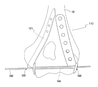
[00120] One form of implant, according to the invention and as seen at
100 in Figs. 14-17, utilizes a plate-like body 110 that, when operatively
positioned as depicted, extends along the lateral column 111 of a humerus
bone 12 and terminates in a crescent-shaped paddle 112 that fits
conformingly against the flat lateral portion of the capitellum 28. The paddle
112 has an enlarged bone contact area that stabilizes the fixation in multiple
directions. The plate 110 is fixed with fixation elements such as bone screws
114, either locking or non-locking, through openings 115 along a proximal
shaft 116 that has a curvature at least nominally matched to the bone surface
it overlies. Distally, the paddle 112 on the body 110 is secured to the
-25-
capitellum 28 with fixation elements that may be in the form of screws
(locking or non-locking) and/or pins (locking or non-locking) 118 as described
in U.S. Patent No. 5,931,839, which may be referred to for details. One
exemplary screw 118 is shown with multiple openings 119 in the paddle 112
for pins. In this way, the implant 100 allows secure fixation of fractures
that
involve the lateral column 111 with additional fixation of articular
fragmentation that involves the capitellum 28. The body 110 extends
conformingly along the posterior surface of the distal humerus 12 and then
wraps around the lateral epicondyle to terminate in the flat distal paddle 112
that at least nominally matches the shape of, and fits over, the lateral
surface
of the capitellum 28.
[00121] A second form
of implant, according to the invention, is shown
in Figs. 18-22 at 120. The second implant 120 is a medial hybrid implant
with a body 121 having both a curved intramedullary portion 122 that, when
operatively positioned, extends up through the cancellous bone of the medial
column and into the medullary canal/cavity of the lower humerus, and an
exposed distal portion/end 123 including a flat paddle 124 corresponding to
the paddle 112 on the implant 100 that is configured to conform to the
humerus 12 at the lateral epicondyle region. The distal end 123 extends out
of the lower portion of the medial epicondyle and medial column to terminate
at the paddle 124 that sits conformingly against the medial surface of the
trochlea to be fixed by fixation elements such as screws and/or pins 126. Pin
openings 127 are provided in the paddle 124 with one screw 126 shown
therethrough inserted with the assistance of a separable guide pin GP.
CA 2866514 2017-09-15
CA 02866514 2014-09-05
WO 2013/133886
PCT/US2012/070649
-26
-
Proximally, the implant 120 is secured with interlocking fixation elements in
the form of screws 128 directed through holes 129,
[00122] As with many other intramedullary devices, a jig/guide 130 is
used to drill through the holes 129 for the interlocking screws 128 and
attaches at the paddle end of the implant 120, as seen in Fig, 21 Similar
jigs/guides can be used for any implant, whether intramedullary or not. The
screws 128 may be in the form of set screws that are inserted through one
cortex and sandwich a shaft 131, corresponding to the shaft 116 on the body
110, against the opposite cortex, or interlocking screws that may be partially
or fully threaded and pass through holes/openings 129 in the proximal
intrarnedullary portion of the implant 120. The latter is preferred.
[00123] In Fig. 22, the screws 128 are shown with bodies each having a
head H, threads T at a head end, and an unthreaded length UL between the
threads at the head end and the entry end E that is opposite the head end.
The entry end E is directed through the bone and into an opening 129 so that
the threads T engage the bone and the unthreaded length UL extends into
and beyond the body 121.
[00124] As seen in Figs, 20 and 21, the guide 130 may be secured
utilizing holes formed in the paddle 124 or, more preferably, by forming a
referencing hole 132 to support and align the guide 130. The hole may be
provided elsewhere but is preferably provided at the location where the
referencing hole 132 is shown at the bottom of the rod-like shape of the body
within the cavity.
CA 02866514 2014-09-05
WO 2013/133886
PCT/US2012/070649
-27-
[00125] In Fig. 23, a modified form of jig/guide 130' is shown with a
frame F that is elongate with offset legs Li, L2, aligned generally with the
length of the curved intramedullary portion 122 of the second implant 120.
The leg L2 is aligned so that a securing component is guided through a bore
13 into the implant 120, adjacent to the paddle 124, to allow fixation of the
jig/guide 130' to the implant 120. Separate guide bores 131, 132, 133 align
with
bores B3,134, 135 successively.
[00126] For fractures that involve both condyles of the distal humerus,
the two implants 100, 120 can be used together, and form an innovative
construct, as seen in Figs. 23-26.
[00127] Generally, the invention contemplates fixing the first and
second operatively positioned bodies 110, 121 to the bone 12 at spaced
locations by performing at least the step of extending a first component into
an operative position between the bodies 110, 121, so that: a) the first
component engages each of the bodies 110, 121, so as to be stabilized by
the bodies 110, 121; and b) separate stable and unstable bone parts, as
shown in Fig. 27 and produced by a fracture line FL The exemplary
condition depicted is an unstable, segmental supraconduiar fracture wherein
there is a stable bone part BPI and separate unstable bone parts BP2, BP3.
The bone parts BPI, BP2, BP3 are maintained in a desired set relationship
with each other across the fracture line FL between the bodies 110, 121,
The bodies 110, 121 have an elongate form with lengths PL1, PL2,
respectively between ends El, E2 and E3, E4 that are at least nominally
aligned with each other and a lengthwise extent of the bone 12, as indicated
CA 02866514 2014-09-05
WO 2013/133886
PCT/US2012/070649
-28-
by the double-headed arrow PL3, with the bodies 110, 121 operatively
positioned.
[00128] The invention contemplates that there could be more than the
two depicted unstable bone parts BP2, BP3 or a single unstable bone part,
[00129] Further, the component 144 may extend through only unstable
bone parts or through both a stable bone part and one or more unstable
bone parts. The component 14 and its counterparts herein inherently
function as implant fixation components/elements for their associated
implant,
[00130] In the simplest form, as shown schematically in Fig. 28, a cross
screw/bolt 136 defines the first component and is directed through a hole in
the paddle 112, 124 on one implant 100, 120, across a bone 138, which may
be the humerus bone, tibial bone, or another bone, and out a hole in the
paddle 124, 112 on the opposite implant 120, 100, with each of the implants
100, 120 operatively positioned. It is then secured with a nut 140 to produce
a captive arrangement between the nut 140 and a head 142 on the cross
screw 136. The screw 136 could be alternatively threaded at both ends to
accept a nut at each end. This method may not create a rigid lock to the
implants 110, 120, depending upon how the screw 136 interacts with the
implant 100, 120, but may, without a locking interaction at the implants 100,
120 eliminate translational movement, angular movement and axial
movement of the crossing screw and, when used on the humerus, or other
bone, does connect the medial and lateral implants as a structural unit with
obvious biomechanical advantages.
CA 02866514 2014-09-05
WO 2013/133886
PCT/US2012/070649
-29-
[00131] Another method is to pass a first component, in the form of a
transcondylar screw 144, as shown in Figs. 25 and 26, that locks into either
one or both of the medial/lateral implants 100, 120. However, since it is
nearly impossible to exactly predict the trajectory of this transcondylar
screw
144 and the specific location of the paddles on the condyles, it is
anticipated
that if the holes in the paddles were simply threaded, it would be nearly
impossible to match the trajectory of a locking transcondylar screw with the
axis of the threads in the plate. There are a number of ways to address this
issue, however. One method is to design the implants and the transcondylar
crossing screw with different materials so that the threads on the
transcondylar crossing screw cut a matching thread in the corresponding
hole in the implant. Although this technique has been used to lock the head
of a screw within a hole in a surface plate so that it is fixed against any
movement within the hole, it has never been used to create a threaded lock
of the tip of a screw on implants applied to the opposite side, much less
threaded lock on both sides of the crossing screw, Other methods include
using an expandable bearing, using a scalloped hole, cross threading, or
using a medical grade plastic insert. Locking a crossing screw to only one
implant either at the head of the screw or at the tip on an implant on the
opposite side of the bone, or locking a crossing screw both at the tip and
under the screw head with opposing implants, are preferred, as described
below. It has the benefit of markedly reducing loads on both the crossing
screw and the implants, allowing the benefit of significantly smaller implants
with a construct that has better strength, Locking of the crossing screw at
both ends is also contemplated.
CA 02866514 2014-09-05
WO 2013/133886
PCT/US2012/070649
-30-
[00132] While the precise order and manner of assembling the system
components are not critical and limited, in one preferred form, one of the
implants, such as the implant 120, may be proximally fixed with the other
implant 110 loosely and temporarily held, as by pins and/or clamps,
preparatory to drilling for the first component/transcondylar screw 144. After
drilling and inserting the component/transcondylar screw 144, the implant
110 may be fixed permanently at the proximal location. By performing this
sequence, if there is a slight misalignment of the drill relative to the
implant
110, the deviation can be compensated for by a slight shifting of the implant
110, on the order of 1 mm, to effect alignment. This sequence can be used
for all paired implants utilized. However, as noted above, this sequence is
not required as, alternatively, the implants might both be fixed before
drilling
is carried out to accommodate the first component.
[00133] Regardless of the precise mechanism utilized, it is desirable
that the ends of the first component be stabilized by the respective body. In
one form, this stabilization occurs at one or both of the bodies 110, 121 in a
plane transverse to the lengthwise axis of the first component. This
stabilization may involve engagement/interaction that blocks relative
movement in one or all directions within these planes. In the former case,
the ends of the first component may be fixed against movement in one
direction (or opposite directions) along a line parallel to the length of the
bodies 110, 121. This will avoid unwanted shifting of an unstable bone part,
through which the first component extends, relative to a stable bone part to
which one or both of the bodies 110, 121 is fixed, Alternatively the ends of
CA 02866514 2014-09-05
WO 2013/133886
PCT/US2012/070649
-31-
the first component may be rigidly fixed to the bodies 110, 121 so that no
relative movement can occur.
[00134] One preferred method is disclosed in Figs. 28-33, and is as
used to insert the transcondylar screw 144 with the implants 100, 120 shown
in Figs. 23-26. A hole is formed in the paddle 120 with the assistance of a
jig/guide 148. A medical grade plastic insert (PEEK, poly-ether-ether-ketone,
or the like) is placed in the hole. Alternatively, injection molded or welded
PEEK can be directed into the hole. This plastic would have a small central
hole (typically about .035" - .150") that allows passage of a guide pin 150
through its center, as seen in Fig. 31 and also in Figs. 25 and 26. Once the
guide pin 150 is passed across the bone, a cannulated drill 152 is passed
over the guide pin 150, as seen in Fig. 32. This drill 152 not only drills a
track across the condyles, but also is used to drill an appropriate sized hole
through the PEEK insert along the particular trajectory needed for the locking
threads of a transconclylar screw 154 that defines the aforementioned first
component,
[00135] It is also possible to effect drilling without using the guide pin
150.
0o136] Alternatively, a locking nut 156/bolt 158 combination is
possible, as seen in Fig, 34, with the bolt 158 defining the aforementioned
first component and having a head 159. A captive arrangement results
between the nut 156 and head 159.
[00137] .. Locking screws, such as the screw 154 shown in Figs. 33 and
34, are screws that have a thread at the screw head 159 that screws into a
-32-
threaded hole on a cooperating implant. The depicted screw 154, as the
screw 144, has a non-uniform diameter with smaller and larger diameter
lengths SD, LID, respectively. The smaller diameter progressively blends into
the larger diameter. This design, which may alternatively be stepped sharply
at an intermediate location between the smaller and larger diameter lengths,
has been available in orthopedics and provides the advantage of eliminating
angular motion between the screw and cooperating plate. Because it
reduces the need for the screw to rigidly compress the bone to the
undersurface of the plate in order to get stability, it creates a stronger
construct that is more resistant to pull-out and can be used in situations in
which thread purchase is poor (such as osteoporosis). Original locking
screws were placed perpendicular to the surface of a cooperating plate and
required a drill guide that screws into the hole in order to align the
trajectory
of the drill with the threads in the plate so that the screw would not bind or
cross thread during insertion.
[00138] Alternatively, a uniform diameter screw could be used with a
taper only at one end, or both ends, thereof.
[00139] Polyaxial locking screws allow a screw to be directed in a
variety of angles and then form an angularly stable lock to the plate when the
screw is fully seated. One of the early methods of forming a polyaxial
angular locking screw is disclosed in U.S. Patent No. 7,195,633, which uses
an expandable bearing within the plate to lock as the screw is seated. The
disclosure of this patent may be reviewed for further details. Other designs
have used dissimilar metals in which the screw metal is harder than the plate
metal and cuts a screw track in the hole as it is seated. Another design uses
CA 2866514 2018-06-04
CA 02866514 2014-09-05
WO 2013/133886
PCT/US2012/070649
-33-
a triangularly-shaped, or other polygonally shaped, head that creates a lock
in a hole with a complementary triangular, or other polygonally shaped
recess, in it. Another design utilizes a threaded screw in a threaded hole
with threads only present partially around the circumference of the hole or
the circumference of the screw.
[00140] In another
form, as show in Fig. 35, a locking hole LH is shown
in a paddle P that is part of an implant. The locking hole LH has cutouts CO
that interrupt threads over approximately 50% of the circumference of the
locking hole LH. A cooperating screw 15 can then cross thread at different
angles. Partial threads could be present alternatively on the screw or both
on the screw and around the locking hole LH. Potentially, this connection will
lock the screw against both translational and angular movement within the
locking hole LH. The screw becomes effectively fixed in a specific orientation
and depth within the locking hole LH. Different angular and depth locations
can be selected.
[00141] In addition,
locking of the leading tip of the screw can be
achieved by selecting the appropriate design parameters that include hole
diameter, number of thread leads, top and bottom surface chamfer, material
properties of the implant and screw, percentage of engaged thread
circumference, and leading and trailing screw diameters.
[00142] Yet another
design uses an insert of PEEK that is either
pressed into or welded around the hole or injection molded into the hole.
Like the dissimilar metals, the threads of the screw cut a threaded track into
the PEEK to lock it in place. The screw head can be conical to expand in the
PEEK as it is screwed home.
CA 02866514 2014-09-05
WO 2013/133886
PCT/US2012/070649
-34-
[00143] To date, all locking screw designs are limited to screws that are
placed into a single plate. They all lock with threads at the screw head.
Because locking screws have this limitation, they are all loaded in cantilever
bending, with the result that the screws need to have a relatively large
diameter to handle the applied load. There are situations in which this
relatively large size is detrimental.
[00144] In one form of the invention, as shown schematically in Fig, 36,
a pair of implants 170, 172 is provided with the implants 170, 172 having
bodies partially or entirely in spaced relationship, as on opposite sides of a
bone 174 that may be any bone such as, but not limited to, a tibial bone,
humeral bone, or other bone at which there is a fracture. A component, as in
the form of a locking screw 176, is locked simultaneously through: a)
cooperating components 178, 180, respectively on the locking screw 176 and
implant 170 at one location; and b) cooperating components 182, 184,
respectively on the locking screw 176 and implant 172 at a second location
that is spaced from the first location, The components 178, 180, 182, 184
are preferably, but not necessarily, cooperating threads. The locking screw
may have a head and tip at which the threads 178, 182 are formed. The
locking screw 176 may have other configurations,
[00145] Alternatively, the locking screw might lock to either one, but not
the other, of the implants 170, 172. The locking screw might, with this
variation, be supported in non-locking relationship by the other of the
implants 170, 172.
[00146] The generic depiction of implants 170, 172 is intended to
encompass any arrangement of separate, cooperating implant components,
CA 02866514 2014-09-05
WO 2013/133886
PCT/US2012/070649
-35-
including intramedullary implants in medial/lateral columns, plates along
medial/lateral columns, etc., including different combinations thereof. For
example, both implants 170, 172 may be elongate plate-like structures
configured to conform over substantially their entire length to exposed
surfaces on a bone that reside therebetween as in the prior art system of Fig.
10.
[00147] It is believed that this concept has not been derived from
existing technology by those skilled in this field because of at least the
following. If the holes are pre-formed as with the original locking screw
technology, it would require that a hole is drilled first along a trajectory
that
allows both plates to be applied with both holes aligned exactly at the same
trajectory, in order to allow the threads of the crossing screw to engage both
sides. This is too difficult to be surgically practical.
[00148] However, by combining and modifying polyaxial locking
technology as described herein, this problem can be overcome. A jig/guide
is used, as shown schematically at 186 in Fig. 37, for a drill 188 to form the
aligned holes in the implants 170, 172, and requires that the angle of
trajectory falls within the polyaxial locking range that is possible
(currently a
max of 30-35 degrees).
[00149] With the supracondylar implant, a PEEK insert might be
utilized. Since it may not be known if the angle of insertion will fall
outside of
the 30 degree range, the appropriate drill hole can be formed through the
PEEK during a surgery using a guide pin 150 placed first as described above
relative to Fig. 32,
CA 02866514 2014-09-05
WO 2013/133886
PCT/US2012/070649
-36-
[00150] The invention can be practiced and adapted, based upon the
principles herein, to address one or more of at least the following
objectives.
[00151] 1. The design may improve the stability of fixation by
triangulating the fixation along the medial bone column, along the lateral
bone column, and coupling these two columnar fixation implants with a
horizontal fixation element that connects the two, Since the horizontal
crossing screw distally is captured on both sides by the columnar implants,
the two point fixation eliminates the cantilever bending on the crossing
screw.
In turn, this allows the crossing screw to be much smaller in diameter,
decreasing the risk of iatrogenic comminution of a distal fragment with a
large screw hole and reducing the risk of soft tissue irritation from the head
of
the screw.
[00152] Connecting the medial and lateral column implants with a
captured crossing screw also distributes the bending load over a wider
composite structure, thereby reducing the implant loads and allowing thinner
implants to be used. It is also better at maintaining length of the bone, even
in situations in which there may be segmental fracture elements between the
shaft of the bone proximally and the joint surface distally,
[00153] Ideally, this horizontal element is locked to both the medial and
lateral columnar implants, but it is still an improvement if it is locked on
only
one side, or even unlocked at both sides.
[00154] 2. The design may reduce soft tissue irritation by using an
intramedullary implant that is designed to extend from the shaft down into the
central portion of either the medial or lateral column. In one preferred form,
CA 02866514 2014-09-05
WO 2013/133886 PCT/US2012/070649
this is a hybrid intramedullary implant, being intramedullary in the shaft and
medial (or lateral) column and terminating in a superficial plate that
provides
holes for screw fixation into the peri-articular fragments distally.
[00155] 3. The design may
achieve fixation of supracondylar elbow
fractures with fixation on both columns where at least one implant is
intramedullary and designed to extend from the shaft down into the medial
(or lateral) column. Specifically, this includes: (1) fixation with a medial
intramedullary implant and a lateral plate; (2) fixation with a lateral
intramedullary implant and medial plate; or (3) fixation in which both columns
are fixed with intramedullary implants.
[00156] 4. The design may
avoid stripping of the flexor tendon
attachments on the medial epiconclyle by eliminating the need for a plate that
is applied to the surface of the medial column. If a lateral
column
intramedullary implant is used, this eliminates stripping of the extensor
tendon attachments to the lateral epicondyle.
[00157] 5. The design may
provide a solution for what intuitively
seems an impossible design issue -- using a solid intramedullary device that
also extends and captures the distal end of the humerus. The intramedullary
canal of the humerus is a long, straight open canal within the central shaft
and terminates just proximal to the coronoid/olecranon fossa. Because of
this anatomy, the possibility of using a solid nail that allows intramedullary
fixation proximally yet provides fixation of the peri-articular surface seems
intuitively impossible. The current invention solves this problem by drilling
and/or broaching an intramedullary track up through the porous metaphyseal
bone of the medial (or lateral) column and then accurately designing a fixed
CA 02866514 2014-09-05
WO 2013/133886
PCT/US2012/070649
-38-
implant with a specific diameter and curve to compensate for the curvilinear
path into the central canal of the humeral shaft. The implant design must
allow insertion of the implant through the curved track, yet be large enough
to
provide interlocking screw holes for stabilization of the implant proximally
within the shaft. Accurate placement of the starting hole and broaching of
the passage along the column is also important to allow the nail to pass up
into the bone.
(00158] 6. The design may
combine the benefits of plate and screw
fixation at the distal end of the bone with the benefits of an intramedullary
rod
fixation in the canal of the long bone (humerus). The distal peri-articular
fragments have limited area for purchase; these are best secured with a low
profile plate and screws. On the other hand, by combining this distal plate
portion or paddle with an intramedullary implant proximally, the implant can
be thicker and better suited to resist the large bending moments, In addition,
since it is contained within the bone canal, it is better suited mechanically
since distribution of bending forces occurs over a large distance within the
canal. The intramedullary position also eliminates the problem of soft tissue
irritation (since it is within the bone). The position
of the paddle
predominantly in line to the long axis of the intramedullary canal reduces
bending loads on the implant that can occur with prior designs that utilize a
significant superficial offset.
[00159] This implant
design may overcome the problem of stabilizing
small articular fragments to the stable proximal shaft which are at a
considerable distance from the articular surface. The design may overcome
the problem of maintaining position of the distal articular surface in terms
of
CA 02866514 2014-09-05
WO 2013/133886
PCT/US2012/070649
-39-
joint anatomy and maintenance of length in the context of segmental fracture
components extending up into the medial/lateral columns. The design may
overcome the difficulty of fitting a plate to the complex geometry of the
medial column of the distal humerus. The design may overcome problems of
external bulky hardware interfering with the soft tissues and avoids extensive
stripping of critical tendons and other soft tissues. When both implants are
locked to each other with a distal transcondylar cross bolt/screw, this design
creates an integrated structural unit that extends from the medial to the
lateral side, vastly improving the stability of fixation and allowing
accelerated
rehabilitation and improved recovery of motion.
[00160] Another aspect of the invention relates to the fixation of
intramedullary nails, as seen in Figs. lie and lib, and implants such as the
implant 120, as described herein.
[00161] The problem with interlocking a supracondylar nail in the
humerus or other bones with small canal diameter relates to size. Standard
interlocking screws are fully threaded (with a self-tapping tip). The hole in
the nail needs to be larger than the thread diameter in order to allow the
screw to pass. The interlocking screw obtains thread purchase on either side
of the bone.
[00162] A supracondylar nail would need to be typically only about 5mrn
6rnrn in diameter in order to allow it to pass through the medial or lateral
column. A standard screw size is a 3.2mm screw (3.2rnrn thread diameter,
2.3mm core diameter). If a 3.5mm hole is provided that is wide enough to
allow the interlocking screw to pass, there is less than 1 mm 1.3mrri of wall
thickness on either side of the nail at the region of the hole. This is likely
to
CA 02866514 2014-09-05
WO 2013/133886
PCT/US2012/070649
-40-
cause the nail to break at the screw hole. Although a 2.3mm screw would
improve the wall thickness of the nail, this smaller screw has only a 1.75mm
core diameter and is not strong enough to handle the required loads.
[00163] The current invention offers a solution to allow cross fixation of
a nail/implant, corresponding to the implant 120, of limited diameter with an
interlocking screw that maximizes strength. In its simplest form, the screw is
threaded for a length just beneath the screw head to provide thread purchase
of the bone cortex immediately under the screw head. This locks the
interlocking screw to the bone and prevents the screw from angulating or
backing out. The remainder of the screw is smooth with a diameter that is
just smaller than the hole in the nail and extends into a nearly press-fit
hole
through the opposite bone cortex. This provides further stability against
angulation of the screw, but allows a larger screw diameter through the nail
than if the screw was fully threaded.
[001641 As shown schematically in Fig. 38, a 3.2mm screw 190 can be
used with greater strength of fixation resulting. The locking screw 190 has a
locking component 192 at one location that may be in the form of threads
right below a head of the screw 190. The locking component 192 fixes, as by
being threaded, within a hole 194 formed in a bone 196. The remainder of
the length of the screw 190 extending beyond the locking component 192
would be a smooth, unthreaded cylinder which passes through a nail/implant
198 and into an aligned hole 200 in the bone 196 on the other side. In this
way, the connection affords the strength of the 2,3mm core diameter passing
through the nail/implant 198 and out of the far surface of the bone 196, but
retains a threaded purchase of the bone 196 on the near side. All that is lost
CA 02866514 2014-09-05
WO 2013/133886
PCT/US2012/070649
-41-
is the thread purchase of the bone on the far side, but since the threads
primarily only keep the screw from backing out, this should not be
significant.
[00165] Alternatively, the locking screw 190 could lock to the
nail/implant 198, as through cooperating threads, or another mechanism.
[00166] This is a solution that has particular utility for other small
implants that use small interlocking nails. The concept can be practiced at
any bone location. One or more locking screws 190 can be utilized and may
be directed into the nail/implant 198 from one direction or from opposite
directions.
[00167] The nails/implants 198 can be made with a flexible
configuration. More preferably, the nails/implants 198 have a fixed shape
that is either straight or bent. The fixation of the nails/implants 198 is
strengthened by the presence of a paddle, such as the aforementioned
paddle 124. The enlarged area of the paddle 124 affords a potentially
substantial bone contact area that makes possible multi-directional
reinforcement of the associated nail plate.
[00168] In another form, as shown in Fig. 39, an intramedullary
nail/implant 202 corresponding to the implant 120, has a plate/shaft 203 that
is contiguous with a flat paddle 204. The flat paddle 204 is contoured to fit
along a surface 206 of a bone 208, which bone surface 206 is perpendicular
to a convex articular surface (i.e., capitellum or trochlea as opposed to a
concave articular surface such as the distal radius), with the nail/implant
202
in a central bone canal 210. Fixation elements (screws, pins, etc) 212 that
are distributed through a corresponding arrangement of holes, as described
CA 02866514 2014-09-05
WO 2013/133886
PCT/US2012/070649
-42-
above, fix the plate/shaft 203 relative to the bone 208. Fixation elements
214, as described above, secure the paddle 204 to the bone surface 206.
[00169] In Figs. 40-43, different combinations of implants contemplated
by the invention are shown. The implants are not described hereinbelow in
any detail but could take the form of any of their counterparts, described
above, for external surface application and for at least partial placement
within an intramedullary canal/cavity.
[00170] In Fig. 40, implant 11 is shown as a medial plate with implant 12
as a lateral plate, with both implants on the outer surface of the distal
humerus and used to stabilize bone parts 13PA, BPB relative to a stable bone
part BPC. Both implants 11, 12 are on the external surface of the distal
humerus 12. A component C, corresponding to the first components
described hereinabove, extends between the implants 11, 12 and interacts
therewith utilizing any of the structures described above.
[00171] In Fig. 41, implants 13, 14 each resides within the intramedullary
cavity on the distal humerus 12 and has a paddle P1, P2 projecting
therefrom, respectively at medial and lateral locations. The component C
extends between the paddles P1, P2.
[00172] In Fig. 42, the medial column intramedullary implant 13 is used
in combination with the lateral plate implant 14. The component C connects
between the paddle P1 and the externally located implant 12.
[00173] In Fig. 43, the medial plate implant 11 is used in combination
with the lateral column intramedullary implant 14. The component C connects
between the externally located implant 11 and the paddle P2.
CA 02866514 2014-09-05
WO 2013/133886
PCT/US2012/070649
-43-
[00174] In Figs. 44-47, the invention is shown as used to treat a fracture
of a tibia 250 that in Fig. 45 produces a stable bone part SBP and unstable
bones parts UBP1-UBP4,
[00175] Referring initially to Fig, 44, an implant TI 1 is located on the
outside of the tibia 250, with a separate implant T12 situated in an
intramedullary cavity 252 on the tibia 250. Fixation elements/components
254 secure the implant TH.
[00176] A component C, corresponding to the aforementioned first
components, interacts between the implants TM, 112, in any one of the
various manners described above.
[00177] In this embodiment, the component C extends through the
unstable bone parts UBP3, UBP4. The implant Ill is shown at a lateral
location with a configuration conforming to the tibial surface 256,
[00178] In Figs. 45-47, implants T13, T14 and T15, T16 are paired
respectively at lateral and medial locations on the anterior and posterior
surfaces of the distal tibia 250 that is fractured so as to produce a stable
bone part SBP and unstable bone parts UBP1-UBP5. The implant pairs are
substantially the same in terms of their basic function, with exemplary
implant
pair 113, T14 described hereinbelow.
[00179] The implants 1I3, TI4 are mounted in spaced relationship to
each other and conform to a respective tibia surface portion. The implants
TI3, T14 are each designed to use either or both of two components Cl, C2,
corresponding to the component C in Fig. 44. However, the multiple
component arrangement is not required. By crossing the components Cl,
CA 02866514 2014-09-05
WO 2013/133886
PCT/US2012/070649
-44-
C2, as in the "X" shape shown, additional stability is afforded. Each of the
components C1, C2 has opposite ends, connected one each to the implants
T13, T14, using any of the structures described above.
[00180] Fixation elements 258 fix the implants T13, T14 to the stable
bone part SBP, With the tibia 250 fractured as shown, the components Cl,
C2 extend only through the unstable bone parts Ul3P1 UBP5.
[00181] In a further embodiment, as shown in Fig. 48, a single,
intramedullary implant TI7 is shown on a tibia 250 at a lateral location to
accept a threaded component C3 to stabilize unstable bone parts UBP1,
UBP2 in relation to a stable bone part SBP.
[00182] Fig. 49 shows an arrangement similar to that in Fig. 48 but with
the single, intramedullary implant T18 located at a medial location on a tibia
250 fractured to produce unstable bone parts UBP1, UBP2 separated from a
stable bone part SBP. A threaded component C4 extends into the implant
TI8 and each of the unstable bone parts UBP1, UBP2.
[00183] In Figs. 50-53, a modified and preferred form of implant,
corresponding to the implant 120, described above, is shown at 120'.
Corresponding parts are identified with the same reference numeral, with the
parts identified in Figs, 50-52 including a "'"designation.
[00184] The implant 120' has an elongate body 121' with an
intramedullary portion 122' having a length between a first end 258 and a
distal second end 123' at which a paddle 124' is located and mounted in
cantilever fashion. The implant 120' is configured for use at a medial
location at the distal humerus up into the canal and could be easily adapted
CA 02866514 2014-09-05
WO 2013/133886
PCT/US2012/070649
-45-
for directing from a lateral location on the distal humerus up into the canal.
Use on still other bones is contemplated.
[00185] The
intramedullary portion 122' has a substantially constant
dimension/diameter D, as taken in cross-section transversely to the length of
the implant 120', as indicated generally by the double-headed arrow 258.
The paddle 124', and a bone engaging surface 260 thereon, have a
dimension D1, extending transversely to the length of the implant 120', that
is
substantially greater than the dimension D. In this
embodiment, the
dimension D1 is approximately three times the dimension ft Preferably, the
dimension D1 is at least two times the dimension D so that the portion of the
paddle 124' projecting past the body 121' projects a distance D2 that is equal
to or greater than the dimension D, to provide the desired stabilization of
the
implant 120' relative to the bone that is being treated. However, at certain
sites, because of geometrical constraints, the dimension D1 may be the
same as, or smaller than, the dimension D. In this embodiment, the paddle
124' projects in only one direction away from the intramedullary portion 122'
to define an "L" shape in conjunction therewith.
[00186] In alternate
forms, not specifically shown, the paddle may
project in more than one direction away from the intramedullary portion so as
to define a "Y" shape, a "T" shape, or another shape in conjunction therewith.
[00187] The paddle
124' and bone-engaging surface 260 thereon have
a dimension D3, generally parallel to the lengthwise extent of the
intramedullary portion 122', that is substantially greater than the dimension
D; preferably on the order of at least twice that dimension, again for
purposes
of stability.
CA 02866514 2014-09-05
WO 2013/133886
PCT/US2012/070649
-46-
[00188] In this embodiment, the paddle 124' cantilevers from the
second lengthwise end 262 of the intramedullary portion 122', to project
transversely to the length of the intramedullary portion 122', and has an
overall generally 'U" shape opening towards the first end 258 of the implant
120' and intramedullary portion 122'
[00189] While the implant 120 has a somewhat different configuration
than the implant 120', the general layout and relationship of parts, in both
shape, orientation, and dimension, is generally similar with the overall
function being the same.
[00190] The intramedullary portion 122' is made up of a primary length
portion 266 and a second length portion 268. The length portions 266, 268
have lengthwise central axes 270, 272, respectively, that make an angle 0
with respect to each other. The angle G may be in the range of 5-15 . As
depicted, the angle 0 is 90.
[00191] A paddle support 274 projects from the second end 262 in a
line Ll from the second axis 272 at the second end 262 . The support 274
produces a cantilever mount for the paddle 124'.
[00192] The primary length portion 266 has a first length with the
second length portion 268 having a second length. The first length is greater
than the second length. In the depicted embodiment, the first length is less
than twice the second length.
[001931 The paddle surface 260 has an area. Preferably, at least a
portion of the surface area faces away from the axis 270. As depicted,
CA 02866514 2014-09-05
WO 2013/133886
PCT/US2012/070649
-47-
substantially the entirety of the area is spaced from and faces away from the
axis 270.
[00194] While the cantilever paddle mount is depicted to establish the
relationship between the paddle surface 260 and axis 270, this same
relationship could be produced without the cantilever design.
[00196] The surface 260 has separate, angled, first and second flat
portions 276, 278 residing substantially in first and second planes that are
at
an angle 01 that is in the range of 165-172 . However, it should be
understood that the paddle could be made without the angled surface
portions.
[00196] The implant 120' has openings 280 to accept the fixation
elements 128' that are directed in a line that is transverse to the length of
the
implant 120', potentially at different angles with respect thereto, as to
avoid
anatomical structures and/or to provide a biomechanical advantage.
[00197] The implant 120' is configured so that the implant end 264 can
be introduced to the intramedullary canal/cavity and moved in a substantially
straight line fully operatively therewith parallel to the length of the
intramedullary portion 122' without requiring any significant angular
reorientation of the implant 120'. Preferably, the operative placement of the
implant 120' does not require any bending, or other reconfiguration, of the
implant 120', and particularly the intramedullary portion 122'. The
operatively
placed implant 120' is then secured in the same general manner as
described for the implant 120. The implant may be used as the sole implant
or in conjunction with another implant, as described hereinabove.
CA 02866514 2014-09-05
WO 2013/133886
PCT/US2012/070649
-48-
[00198] The second length portion 268 has a peripheral surface on
which a discrete undercut/recess 282 is formed to provide a clearance
volume to avoid encroachment of the implant 120' upon adjacent anatomical
parts. For example, in one application, the undercut/recess 282 avoids
impingement in the olecranon fossa with the ulna as the elbow is extended.
The recess 282 has a generally obround shape bounded by spaced,
elongate edges 284, 286 that have lengths aligned with the length of the
second length portion 268. The recess 282 extends over a majority of the
lengthwise extent of the second length portion 268.
[00199] The paddle 124' is secured to the bone B using one or more
fixation elements, such as screws/pins 126', for which suitable
openings/bores are preferably pre-formed,
[00200] The implant 120' is particularly adaptable to facilitating setting
of fractures on the distal humerus or distal femur, but is not so limited and
could be used as depicted, or modified for use at other sites,
[00201] In one application, it may be desirable to have the bone
engaging surface of the paddle arranged so that at least a portion of the
bone engaging paddle surface; a) is in line with an axis that is parallel to
the
central axis of the intramedullary portion; and b) intersects a substantial
length of the intramedullary portion. That is, an extension of the diameter of
the intramedullary portion would intersect at least a portion of the bone
engaging paddle surface. The paddle in this arrangement may be mounted
other than by using a cantilevered support.
CA 02866514 2014-09-05
WO 2013/133886
PCT/US2012/070649
-49-
[00202] With the structure as described above, a method of treating
fracture of a bone with an intrarnedullary canal can be carried out as showed
in flow diagram form in Fig. 53. As shown at block 288, therein, a system is
provided, as described above. As shown at block 290, the intramedullary
portion of the first implant is directed into the intramedullary canal to
place
the first implant into an operative position. As shown at block 292, the
operatively positioned first implant is secured to the bone.
[00203] In Fig. 54, a system is schematically depicted for treating a
fracture of a bone 300 with an intramedullary canal 302. The fracture
produces first and second bone parts separated by a fracture line.
[00204] The system includes a first implant 304 with an elongate body
306 configured to be directed into the intramedullary canal 302 to place the
first implant 304 in an operative position on the bone 300. At least a first
fixation component 308 can be advanced into the bone 300 and into the
elongate body 306 with the first implant 304 in the operative position.
[00205] The system in Fig. 54 is shown schematically to encompass a
system as shown in Figs. 18-22, other embodiments described herein, and
further variations. The first implant 304 corresponds to the implant 120, with
the main difference relating to openings 310 in the body 306, corresponding
to the openings 129 in the body 121. Of course, the configuration of the first
implant 304 is not limited to the configuration shown in Figs. 18-22. The
fixation component(s) 308 encompasses the fixation components 128 shown
in Figs. 18-22, and variations thereof, such as those described herein.
CA 02866514 2014-09-05
WO 2013/133886
PCT/US2012/070649
-50-
[00206] One specific form of system, as shown generically in Fig. 54, is
shown in Fig. 55. The fixation component 308 therein has a leading end 312
and a trailing end 314. The leading end 312 has a tip 316. The trailing end
314 has a first set of threads 318 to engage the bone 300 over a first
lengthwise extent E of the first set of threads 318 with the fixation
component
308 advanced in the direction of the arrow 320 into the bone 300 and
elongate body 306 of the first implant and secured, as shown in Fig. 55. The
fixation component 308 has a first length FL that advances into the elongate
body 306, with the first implant 304 operatively positioned, as the fixation
component 308 is advanced and secured.
[00207] The first length FL is capable of passing through a bore of a
diameter that is less than a diameter of a bore required to pass through the
first extent E of the first set of threads 318 that engages the bone 300.
[00208] In this embodiment, the first length FL is shown with a
substantially uniform diameter, over substantially the entirety of its length,
that is less than the diameter of at least a portion of the fixation component
308 over the portion thereof where the first extent of threads E is formed. A
generic showing of these components is intended to encompass the specific
forms shown and any other form whereby the first length FL is capable of
passing through a bore with a diameter that is less than required to pass the
first extent of the corresponding first set of threads 318. As one possible
variation, the diameter of the first length of the fixation component 308
might
be non-uniform. Essentially, the invention is intended to encompass a
construction wherein at least some of the threads in the first set of threads
318, that engage the bone 300, have a diameter greater than that of the
CA 02866514 2014-09-05
WO 2013/133886
PCT/US2012/070649
-51-
implant opening 310. This allows the implant opening 310 to be made
relatively smaller to maintain the integrity of the implant 304.
[00209] in Fig. 55, the fixation component 308 is shown advanced fully
through the elongate body 306. However, this is not a requirement, as the tip
316 may reside within the thickness of the implant body 306 between
opposite sides 322, 324.
[00210] In this embodiment, no part of the first set of threads 318 is
shown advanced into the opening 310. It is possible with a tapered
configuration for the first set of threads 318 that a certain number of the
threads might be advanced into the opening 310 without requiring a diameter
larger than that required to closely accept an unthreaded portion 326 of the
fixation component 308 that extends between the tip 316 and a lengthwise
location at 328 where the first set of threads 318 begins. Alternatively, what
is shown as the unthreaded portion 326 could have threads with a smaller
diameter.
[00211] In Fig. 22, the corresponding fixation component 128 is shown
to be advanced through the corresponding elongate body 121 to fully span
the intrameduilary canal and engage the bone at spaced locations, The
fixation component 308 might be modified to similarly fully span the
intramedullary canal.
[00212] The fixation component 308 has a second length between the
tip 316 and the location 328 at which threads are formed. Alternatively, a
second set of threads 330 may be provided over this corresponding length as
shown on a modified form of fixation component 308' in Fig, 56, The second
CA 02866514 2014-09-05
WO 2013/133886
PCT/US2012/070649
-52-
set of threads 330 may be provided to engage the bone 300 or the implant
304.
[00213] While the fixation component 308 is shown with a substantially
uniform unthreaded diameter over a length between the tip 316 and location
328, the invention contemplates variations in diameter, such as a progressive
or localized variation.
[00214] Preferably, the opening 310 in the implant 304 that
accommodates the fixation component 308 has a diameter slightly larger
than the diameter of the first length FL of the fixation component 308. This
allows the first length FL to freely pass through the opening without
resistance that might interfere with the surgeon's ability to readily insert
the
fixation component 308 during a procedure. As with the embodiment in Figs.
18-22, it is contemplated that multiple openings 310 can be provided in the
implant 304 to accommodate a corresponding number of fixation
components 308.
[00215] It should be understood that "diameter", as used herein, is
intended to encompass cross-sectional shapes that are not circular/round, or
continuous at a peripheral surface thereof. Other shapes have an "effective
diameter". The invention relates similarly to the effective diameters of the
fixation components and openings.
[00216] The first set of threads 318 is shown with a tapering diameter.
The invention also contemplates that corresponding threads 318", as shown
on a fixation component 308" in Fig. 57, may have a uniform diameter over a
length of the fixation component 308". The diameter of the set of threads
CA 02866514 2014-09-05
WO 2013/133886
PCT/US2012/070649
-53-
318" is greater than the diameter of the unthreaded portion of the fixation
component 3080 that is advanced into/through the implant opening 310.
[00217] In the depicted embodiment, the fixation component 308 is
tapered and rounded at the tip 316 at the leading end 312 to facilitate
advancing of the leading end 312 into and through the opening 310 in the
implant 304.
[00218] With the system as described above, a method of treating a
fracture of a bone can be carried out as shown in flow diagram form in Fig.
58.
[00219] As shown at block 332, a system is provided, of a type
described above. As shown at block 334, the elongate body is directed into
the intramedullaiy canal to place the implant into its operative position. As
shown at block 336, a fixation component is advanced through the bone on
at least one of two sides of the intrarnedullary canal and into the elongate
body so that the first set of threads engages the bone at the one side.
[00220] A jig/guide can be utilized to drill aligned holes in the bone to
accept the fixation component(s).
[00221] The invention contemplates that all embodiments herein could
be adapted to treat a fracture of any bone that produces at least one
unstable bone part adjacent to a stable bone part.
[00222] As described herein, the interaction of the first components and
implants may be different in terms of how relative movement therebetween is
confined. The first component may be blocked in movement in one or more
directions/dimensions by the implant without being locked thereto, "Locking"
CA 02866514 2014-09-05
WO 2013/133886
PCT/US2012/070649
may occur when the interacting parts thereof fix the interacting parts against
relative; a) movement, in one or more different ways. For example, the parts
may be locked against relative movement parallel to the length of the first
component; b) angular movement; c) movement within a plane transverse to
the length of the first component, etc. The locking could be a fixed locking
against any relative movement.
[00223] The foregoing disclosure of specific embodiments is intended to
be illustrative of the broad concepts comprehended by the invention. While
much of the discussion has been described with fractures of the distal
humerus, this is only by example and it is obvious to one skilled in the art
that
application to other areas such as pilon fractures of the distal tibia,
plateau
fractures of the proximal tibia, supracondylar fractures of the distal femur,
or
other sites would lend themselves to the principles of this invention.