Note: Descriptions are shown in the official language in which they were submitted.
CA2866828
METHODS AND COMPOSITIONS FOR TREATING WOUNDS AND REDUCING THE RISK
OF INCISIONAL HERNIAS
SEQUENCE LISTING
This application contains a sequence listing in electronic form in ASCII text
format. A
copy of the sequence listing is available from the Canadian Intellectual
Property Office.
INTRODUCTION
Incisional hernias are a frequent complication of abdominal surgery, resulting
in
considerable patient morbidity and increased health care costs. There are 4-5
million
abdominal incisions (laparotomies) performed annually in the United States
with hernias
resulting after 11-23% of these procedures. Incisional hernias may result in
severe morbidity
beyond the cosmetic deformity of a visible bulge in the anterior abdominal
wall, including
intestinal obstruction, bowel ischemia, enterocutaneous fistula and
significant limitations on a
patient's physical activity and gainful employment. Consequently, there are
over 400,000
incisional hernia repairs performed each year making it one of the most common
procedures
performed by general surgeons. The increase in U.S. health care costs due to
incisional hernia
repair is estimated to currently exceed eight billion dollars per year, not
including the costs of
unemployment benefits for this moderately young patient population. Research
indicates that
incisional hernias result from inadequate or impaired healing of the
myofascial abdominal wall
following surgery. Accordingly, each of the recognized risk factors for hernia
formation
inhibits wound healing, including morbid obesity, diabetes, smoking, chronic
lung disease,
surgical site infection and poor surgical technique. Since the incidence of
the major risk factors
is increasing, the prevalence of incisional hernias is predicted to increase
as well.
Despite the magnitude and significance of the clinical condition, research
focused on
the prevention of incisional hernias is sparse. While current studies and
research efforts are
focused on improved repair materials and surgical techniques, the optimal
solution to the
problem of incisional hernias is prevention.
1
Date Recue/Date Received 2021-05-28
CA 02866828 2014-09-08
WO 2013/138238
PCT/US2013/030213
SUMMARY
Provided are methods and compositions for treating a wound in a subject. The
methods
include applying a pharmaceutical composition that includes a first precursor
material agent
including fibrinogen, a second precursor material agent including thrombin,
and silver particles
to an abdominal incision site in an amount effective to treat the abdominal
incision site. Also
provided are pharmaceutical compositions and devices for use in the subject
methods.
In some embodiments, a method for treating a wound in a subject is provided.
The
method includes applying a pharmaceutical composition that includes a first
precursor material
agent including fibrinogen, a second precursor material agent including
thrombin, and silver
particles to an abdominal incision site in an amount effective to treat the
abdominal incision site.
Embodiments of the method may also include that the first precursor material
agent, the
second precursor material agent and the silver particles are adapted to be
combined in situ.
Embodiments of the method may also include that the applying includes applying
the
first precursor material agent prior to applying the second precursor material
agent.
Embodiments of the method may also include that the applying includes applying
the
second precursor material agent prior to applying the first precursor material
agent.
Embodiments of the method may also include that the silver particles are
silver
microparticles.
Embodiments of the method may also include that the silver particles are
spherical.
Embodiments of the method may also include that the silver microparticles have
an
average diameter of 5 tim or more.
Embodiments of the method may also include that the silver mi croparti cl es
have an
average diameter of 200 [im or more.
Embodiments of the method may also include that the pharmaceutical composition
includes 25 mginciL silver particles.
Embodiments of the method may also include that the pharmaceutical composition
includes 250 mg/mL silver particles.
2
CA2866828
In some embodiments, a pharmaceutical composition for treating a wound in a
subject is
provided. The pharmaceutical composition includes a fibrin glue and silver
particles in an amount
effective to treat an abdominal incision site.
Embodiments of the pharmaceutical composition may also include that the silver
particles
are spherical.
Embodiments of the pharmaceutical composition may also include that the silver
particles
have an average diameter of 5 pm or more.
Embodiments of the pharmaceutical composition may also include that the silver
particles
have an average diameter of 200 pm or more.
Embodiments of the pharmaceutical composition may also include that the
pharmaceutical
composition includes 25 mg/mL silver particles.
Embodiments of the pharmaceutical composition may also include that the
pharmaceutical
composition includes 250 mg/mL silver particles.
In some embodiments, a device for applying a pharmaceutical composition for
treating a
wound in a subject is provided. The device includes a sterile container
containing a first precursor
material agent including fibrinogen, a second precursor material agent
including thrombin, and
silver particles in an amount effective to treat an abdominal incision site.
Embodiments of the device may also include that the sterile container includes
a first
chamber containing the first precursor material agent, a second chamber
containing the second
precursor material agent, and a third chamber containing the silver particles.
Embodiments of the device may also include that the sterile container includes
a syringe.
In some embodiments, a kit is provided. The kit includes a sterile container
containing a
first precursor material agent including fibrinogen, a second precursor
material agent including
thrombin, and silver particles in an amount effective to treat an abdominal
incision site. The kit
also includes a sealed package configured to maintain the sterility of the
sterile container.
Various embodiments of the claimed invention relate to use of a pharmaceutical
composition comprising a first precursor material agent comprising fibrinogen,
a second
precursor material agent comprising thrombin, and silver particles for
treating a wound in a
subject.
Various embodiments of the claimed invention also relate to use of a first
precursor
material agent comprising fibrinogen, a second precursor material agent
comprising thrombin,
3
Date Re9ue/Date Received 2020-06-17
CA2866828
and silver particles in preparation of a pharmaceutical composition for
treating a wound in a subject.
Various embodiments of the claimed invention also relate to a device for
applying a
pharmaceutical composition for treating a wound in a subject, the device
comprising a sterile container
containing a first precursor material agent comprising fibrinogen, a second
precursor material agent
comprising thrombin, and silver particles for treating the wound.
Various embodiments of the claimed invention also relate to a kit comprising:
a sterile container
containing a first precursor material agent comprising fibrinogen, a second
precursor material agent
comprising thrombin, and silver particles for treating a wound; and a sealed
package configured to
maintain the sterility of the sterile container.
Various embodiments of the claimed invention also relate to a pharmaceutical
composition for
treating a wound in a subject, the composition comprising a first precursor
material agent comprising
fibrinogen, a second precursor material agent comprising thrombin, and silver
particles for treating a
wound.
Brief Description of the Figures
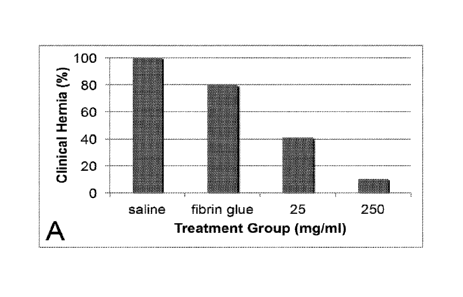
FIG. 1 shows graphs of: FIG. 1A, the percentage of clinical incisional hernias
in Sprague-
Dawley rats (male, 250-300 g) treated with varying doses of silver
microparticles versus saline and
fibrin glue alone (controls); and FIG. 1B, the anatomic hernia areas with
varying doses of
3a
Date Recue/Date Received 2021-05-28
CA 02866828 2014-09-08
WO 2013/138238
PCT/US2013/030213
silver microparticles and saline and fibrin glue controls, according to
embodiments of the present
disclosure. Results are mean and standard deviation.
FIG. 2 shows a photomicrograph of a hematoxylin-eosin stained cross-section of
a silver
-
treated healing fascial incision on postoperative day 28, which revealed
silver microparticles, a
foreign body reaction (e.g., inflammatory infiltrate consisting of giant cells
without epitheliod
histiocytes), and early wound fibrosis consisting of collagen fibers and no
true granulomas (40X
magnification), according to embodiments of the present disclosure.
FIG. 3 shows a graph of wound size (%) vs. time (weeks) for diabetic mouse
wound
healing experiments for compositions of the present disclosure as compared to
a negative
control, according to embodiments of the present disclosure.
Before the present invention is described in greater detail, it is to be
understood that this
invention is not limited to the particular embodiments described, and as such
may, of course,
vary. It is also to be understood that the terminology used herein is for the
purpose of describing
particular embodiments only, and is not intended to be limiting, since the
scope of the present
invention is embodied by the appended claims.
Where a range of values is provided, it is understood that each intervening
value, to the
tenth of the unit of the lower limit unless the context clearly dictates
otherwise, between the
upper and lower limit of that range and any other stated or intervening value
in that stated range,
is encompassed within the invention. The upper and lower limits of these
smaller ranges may
independently be included in the smaller ranges and arc also encompassed
within the invention,
subject to any specifically excluded limit in the stated range. Where the
stated range includes
one or both of the limits, ranges excluding either or both of those included
limits are also
included in the invention.
Unless defined otherwise, all technical and scientific terms used herein have
the same
meaning as commonly understood by one of ordinary skill in the art to which
this invention
belongs. Although any methods and materials similar or equivalent to those
described herein can
also be used in the practice or testing of the present invention,
representative illustrative methods
and materials are now described.
It is noted that, as used herein and in the appended claims, the singular
forms "a", "an",
and "the" include plural referents unless the context clearly dictates
otherwise. It is further noted
4
CA2866828
that the claims may be drafted to exclude any optional element. As such, this
statement is intended
to serve as antecedent basis for use of such exclusive terminology as
"solely," "only" and the like in
connection with the recitation of claim elements, or use of a "negative"
limitation.
As will be apparent to those of skill in the art upon reading this disclosure,
each of the
individual embodiments described and illustrated herein has discrete
components and features
which may be readily separated from or combined with the features of any of
the other several
embodiments without departing from the scope or spirit of the present
invention. In addition, it will
be readily apparent to one of ordinary skill in the art in light of the
teachings herein that certain
changes and modifications may be made thereto without departing from the
spirit and scope of the
appended claims. Any recited method can be carried out in the order of events
recited or in any
other order which is logically possible.
To the extent such publications may set out definitions of a term that
conflicts with the
explicit or implicit definition of the present disclosure, the definition of
the present disclosure
controls. The citation of any publication is for its disclosure prior to the
filing date and should not
be construed as an admission that the present invention is not entitled to
antedate such publication
by virtue of prior invention. Further, the dates of publication provided may
be different from the
actual publication dates which may need to be independently confirmed.
DETAILED DESCRIPTION
Provided are methods and compositions for treating a wound in a subject. The
methods
include applying a pharmaceutical composition that includes a first precursor
material agent
including fibrinogen, a second precursor material agent including thrombin,
and silver particles to
an abdominal incision site in an amount effective to treat the abdominal
incision site. Also
provided are pharmaceutical compositions and devices for use in the subject
methods.
Below, the subject methods for treating a wound in a subject are described
first in greater
detail, followed by a review of the compositions and devices that find use in
the subject methods,
CA 2866828 2019-07-29
CA 02866828 2014-09-08
WO 2013/138238 PCT/US2013/030213
as well as a discussion of various representative applications in which the
subject methods,
compositions and devices find use.
ilitETHoDs
Aspects of the present disclosure include a method for treating a wound in a
subject. The
method includes applying a pharmaceutical composition that includes a first
precursor material
agent including fibrinogen, a second precursor material agent including
thrombin, and silver
particles to an abdominal incision site in an amount effective to treat the
abdominal incision site.
As such, in some cases, treating a wound in a subject includes applying the
pharmaceutical
composition to a wound in the subject, such as an abdominal incision site in
the subject.
By "treatment" or "treating" is meant alleviating, preventing, curing,
reducing the
occurrence, etc. of a condition in a subject. In some cases, reducing the
occurrence includes
decreasing the severity and/or incidence of a condition in a subject. In some
instances, reducing
the occurrence includes reducing the risk of occurrence of a condition in a
subject or group of
subjects. For example, in a group of subjects, a 50% reduction in the risk of
occurrence of a
condition means that, on average, the condition is detectable in 50% of the
subjects in the group,
while the condition is not significantly detectable in the other 50% of the
subjects in the group.
Percentages may be used when referring to a group of subjects or to an
individual subject. In
certain instances, the condition includes a hernia, such as an incisional
hernia. For example, the
incisional hernia may be at an abdominal incision site, such as an abdominal
incision site made
during a surgical procedure.
In certain instances, treating a wound includes promoting healing of the
wound. In some
instances, promoting healing of a wound includes an increase in the efficiency
of wound healing
and/or an increase in the strength of the resulting healed wound site as
compared to a wound that
has not been treated with the methods and compositions of the present
disclosure. In some
cases, promoting healing of a wound includes reducing the occurrence of
defective wound
healing and/or reducing the severity of defective wound healing as compared to
a wound that has
not been treated with the methods and compositions of the present disclosure.
In certain
embodiments, the method for treating a wound in a subject reduces the risk of
incisional hernia
in the subject. By "incisional hernia" is meant a hernia that occurs at an
incision site and
involves defective or incomplete wound healing at the subcutaneous level
(e.g., at the level of the
6
CA 02866828 2014-09-08
WO 2013/138238 PCT/US2013/030213
muscle or fascia). In some instances, defective or incomplete wound healing
may result in an
increased susceptibility to an incisional hernia. By "reduce the risk" is
meant that the risk of the
occurrence of incisional hernia in a subject treated by the method of the
present disclosure is
lower that that in a subject that has not been treated by the method of the
present disclosure. In
some instances, the method reduces the risk of incisional hernia by 30% or
more, such as 35% or
more, including 40% or more, or 45% or more, or 50% or more, or 55% or more,
or 60% or
more, or 65% or more, or 70% or more, or 75% or more, or 80% or more, or 85%
or more, or
90% or more, or 95% or more, for example by 99% or more. In certain cases, the
method
reduces the risk of incisional hernia by 60% or more. For example, the method
may reduce the
risk of a clinical hernia in a subject. By "clinical hernia" is meant a hernia
that is observed (e.g.,
by sight, touch, sound, smell, etc.) during treatment of a patient, rather
than determined through
laboratory studies. For example, a clinical hernia may be observed as a
visible bulge in the
abdominal wall.
In certain embodiments, the method for treating a wound in a subject reduces
severity of
a hernia in a subject should a hernia occur in the subject. In some cases, a
reduction in the
severity of the hernia corresponds to a reduction in the size of the hernia in
the subject. For
example, the method may reduce the size of an anatomic hernia in a subject. By
"anatomic
hernia" is meant a hernia that is detectable by methods other than, or in
addition to, clinical
observation (e.g., by dissection of the subject, MRI, CT, ultrasound, and the
like). The size of an
anatomic hernia may be measured by determining the separation between the
abdominal muscles
(e.g., rectus muscles) at the incision site. For instance, the size of an
anatomic hernia may be
estimated by multiplying the maximal craniocaudal diameter by the average of
two transverse
diameter measurements (e.g., approximation of an ellipse). In some instances,
the method
reduces the size of incisional hernia by 15% or more, such as 20% or more,
including 25% or
more, or 30% or more, such as 35% or more, including 40% or more, or 45% or
more, or 50% or
more, or 55% or more, or 60% or more, or 65% or more, or 70% or more, or 75%
or more, or
80% or more, or 85% or more, or 90% or more, or 95% or more, for example by
99% or more.
In certain cases, the method reduces the risk of incisional hernia by 55% or
more.
In certain embodiments, the first precursor material agent, the second
precursor material
agent and the silver particles are adapted to be combined in situ. For
example, the first precursor
material agent, the second precursor material agent and the silver particles
may be adapted to be
7
CA 02866828 2014-09-08
WO 2013/138238 PCT/US2013/030213
combined at the abdominal incision site as the first precursor material agent,
the second
precursor material agent and the silver particles are applied to the abdominal
incision site.
Accordingly, in some instances, the method includes combining the first
precursor material
agent, the second precursor material agent and the silver particles in situ
(e.g., at the abdominal
incision site). For instance, applying the pharmaceutical composition may
include applying the
first precursor material agent prior to applying the second precursor material
agent. In some
cases, the first precursor material agent is applied immediately prior to
applying the second
precursor material agent. In other cases, applying the pharmaceutical
composition includes
applying the second precursor material agent prior to applying the first
precursor material agent.
For example, the second precursor material agent may be applied immediately
prior to applying
the first precursor material agent. In yet other embodiments, the first and
second precursor
material agents are applied substantially simultaneously. In still other
embodiments, the first and
second precursor material agents are combined together to form the
pharmaceutical composition
prior to applying the pharmaceutical composition to the abdominal incision
site. For example,
the first and second precursor material agents may be combined together to
form the
pharmaceutical composition immediately before applying the pharmaceutical
composition to the
abdominal incision site.
The silver particles may be combined with the first and second precursor
material agents
at any desired step of the application process. For example, the silver
particles may be combined
with the first precursor material agent prior to combining the first precursor
material agent with
the second precursor material agent as described above. In other embodiments,
the silver
particles may be combined with the second precursor material agent prior to
combining the first
precursor material agent with the second precursor material agent as described
above. In certain
other embodiments, the silver particles may be combined with the first and
second precursor
material agents after combining the first precursor material agent with the
second precursor
material agent with each other as described above. In certain other
embodiments, the first and
second precursor material agents and silver particles are combined at
substantially the same time.
For instance, the first and second precursor material agents and the silver
particles may be
combined in situ as described above. In yet other embodiments, the silver
particles are combined
with the first and second precursor material agents to form the pharmaceutical
composition prior
to applying the pharmaceutical composition to the abdominal incision site. For
example, the first
8
CA 02866828 2014-09-08
WO 2013/138238 PCT/US2013/030213
and second precursor material agents and silver particles may be combined
together to form the
pharmaceutical composition immediately before applying the pharmaceutical
composition to the
abdominal incision site. In yet other embodiments, the silver particles are
combines with both
the first precursor material agent and the second precursor material agent
prior to combining the
first and second precursor material agents together to form the pharmaceutical
composition.
In certain embodiments, the method includes administering the pharmaceutical
composition in an amount effective to treat the abdominal incision site. By
"effective amount" is
meant a dosage sufficient to cause a significantly detectable effect in the
target subject, as
desired. In some instances, an effective amount of the pharmaceutical
composition is an amount
of the pharmaceutical composition sufficient to induce a foreign body reaction
in the subject at
the site of application. In certain instances, the foreign body reaction
includes inflammatory
infiltrate consisting of giant cells without epitheliod histiocyces. In
certain cases, an effective
amount of the pharmaceutical composition includes 10 mg/mL silver particles or
more, such as
25 mg/mL silver particles or more, including 50 mg/mL silver particles or
more, or 75 mg/mL
silver particles or more, or 100 mg/mL silver particles or more, or 150 mg/mL
silver particles or
more, or 200 mg/mL silver particles or more, or 250 mg/mL silver particles or
more, or 300
mg/mL silver particles or more, or 350 mg/mL silver particles or more, or 400
mg/mL silver
particles or more, or 450 mg/mL silver particles or more, or 500 mg/mL silver
particles or more,
or 550 mg/mL silver particles or more, or 600 mg/mL silver particles or more,
or 650 mg/mL
silver particles or more, or 700 mg/mL silver particles or more, or 750 mg/mL
silver particles or
more. In certain instances, an effective amount of the pharmaceutical
composition includes 50
mg/mL silver particles. In some cases, an effective amount of the
pharmaceutical composition
includes 500 mg/mL silver particles.
In certain embodiments, an effective amount of the pharmaceutical composition
includes
a weight/weight ratio of silver particles to fibrinogen of 0.1 (wt/wt) or
more, or 0.2 (wt/wt) or
more, or 0.3 (wt/wt) or more, or 0.4 (wt/wt) or more, or 0.5 (wt/wt) or more,
or 0.6 (wt/wt) or
more, or 0.7 (wt/wt) or more, or 0.8 (wt/wt) or more, or 0.9 (wt/wt) or more,
or 1 (wt/wt) or
more, or 1.1 (wt/wt) or more, or 1.2 (wt/wt) or more, or 1.3 (wt/wt) or more,
or 1.4 (wt/wt) or
more, or 1.5 (wt/wt) or more, or 1.6 (wt/wt) or more, or 1.7 (wt/wt) or more,
or 1.8 (wt/wt) or
more, or 1.9 (wt/wt) or more, or 2 (wt/wt) or more, or 2.1 (wt/wt) or more, or
2.2 (wt/wt) or
more, or 2.3 (wt/wt) or more, or 2.4 (wt/wt) or more, or 2.5 (wt/wt) or more,
or 2.6 (wt/wt) or
9
CA 02866828 2014-09-08
WO 2013/138238 PCT/US2013/030213
more, or 2.7 (wt/wt) or more, or 2.8 (wt/wt) or more, or 2.9 (wt/wt) or more,
or 3 (wt/wt) or
more. For example, an effective amount of the pharmaceutical composition may
include a
weight/weight ratio of silver particles to fibrinogen of 2.2 (wt/vvt) or more.
In certain embodiments, an effective amount of the pharmaceutical composition
includes
an amount of silver particles, as described herein, applied to a certain wound
surface area, such
as 0.5 cm2 or more, or 1 cm2 or more, or 2 cm2 or more, or 3 cm2 or more, or 4
cm2 or more, or 5
cm2 or more, or 6 cm2 or more, or 7 cm2 or more, or 8 cm2 or more, or 9 cm2 or
more, or 10 cm2
or more, or 11 cm2 or more, or 12 cm2 or more, or 13 cm2 or more, or 14 cm2 or
more, or 15 cm2
or more, or 16 cm2 or more, or 17 cm2 or more, or 18 cm2 or more, or 19 cm2 or
more, or 20 cm2
or more, or 25 cm2 or more, or 30 cm2 or more, or 35 cm2 or more, or 40 cm2 or
more, or 45 cm2
or more, or 50 cm2 or more. For example, an effective amount of the
pharmaceutical
composition may include an amount of silver particles, such as 250 mg/mL,
applied to a wound
surface area of 10 cm2 or more. In some instances, an effective amount of the
pharmaceutical
composition may include an amount of silver particles, such as 2.2 (wt/wt),
applied to a wound
surface area of 10 cm2 or more.
PHARMACEUTICAL COMPOSITIONS
Aspects of the present disclosure include a pharmaceutical composition for
treating a
wound in a subject. In certain embodiments, the pharmaceutical composition
includes a fibrin
glue and silver particles in an amount effective to treat an abdominal
incision site. By
"pharmaceutical composition" is meant a composition that includes one or more
therapeutic
agents used in the prevention, diagnosis, alleviation, treatment, or cure of a
disease or condition
in a subject (e.g., an animal or human subject).
In certain embodiments, the fibrin glue includes at least a first precursor
material agent
.. and a second precursor material agent. The first precursor material agent
may include
fibrinogen, and the second precursor material agent may include thrombin.
Fibrin glue is a
biopolyrner formed by the addition of thrombin to fibrinogen. Thrombin is an
initiator or
catalyst that enzymatically cleaves fibrinogen which alters the charge and
conformation of the
molecule, forming a fibrin monomer. The fibrin monomers then aggregate forming
the
biopolymer fibrin. After combination of the two thrombin and fibrinogen
components, the
solution remains liquid for several seconds before polymerizing. Fibrin glue
agent, either
CA2866828
immediately following mixture of the precursor materials, or by delivering the
materials separately
to mix in situ, is thus adapted to be delivered to the wound site in the
subject via a syringe, catheter
or other injectors, thus requiring only a minimally invasive procedure. Fibrin
glue is also
biocompatible and non-toxic to the subject. Further examples of fibrin glue
that may be useful
according to various aspects of the present disclosure are described in the
following references:
Sierra, DH, "Fibrin sealant adhesive systems: a review of their chemistry,
material properties and
clinical applications." J Biomater Appl. 1993;7:309-52; and U.S. Patent No.
5,962,405. In certain
embodiments, the fibrin glue includes additional components, such as, but not
limited to, a
fibrinolysis inhibitor, albumin (e.g., human albumin), tri-sodium citrate,
histidine, niacinamide,
polysorbate 80, water (e.g., sterile water, such as water for injection),
calcium chloride, sodium
chloride, combinations thereof, and the like. For example, the first material
precursor agent may
include, in addition to fibrinogen, one or more of a fibrinolysis inhibitor,
albumin (e.g., human
albumin), tri-sodium citrate, histidine, niacinamide, polysorbate 80, water
(e.g., sterile water, such
as water for injection), and the like. In some instances, the second material
precursor agent may
include, in addition to thrombin, one or more of albumin (e.g., human
albumin), water (e.g., sterile
water, such as water for injection), calcium chloride, sodium chloride, and
the like. Additional
examples of components that may be included in the fibrin glue include, but
are not limited to,
protease inhibitors, such as aprotinin.
As described above, in certain embodiments, the pharmaceutical composition
includes
silver particles. The silver particles may be silver microparticles. In some
instances, the size of the
silver microparticles is sufficient to induce a foreign body reaction in the
subject at the site of
application. For example, microparticles have an average diameter ranging from
2 pm to 1000 pm.
In comparison, nanoparticles have an average diameter of 1 nm to 1000 nm. In
some instances, the
silver microparticles have an average diameter of 2 pm or more, such as 3 pm
or more, including 4
p.m or more, or 5 pm or more, or 7 p.m or more, or 10 pm or more, or 15 pm or
more, or 20 p.m or
more, or 25 p.m or more, or 50 pm or more, or 75 pm or more, or 100 pm or
more, or 150 pm or
more or 200 gm or more, or 250 pm or more or 500 pm or more. For example, the
silver
microparticles may have an average diameter ranging from 2 p.m to 1000 um,
such as from 2 gm to
750 pm, including from 3 p.m to 500 p.m, or from 5 pm to 250 p.m. In certain
instances, the silver
microparticles have an average diameter of 5 gm or more. In some
11
CA 2866828 2019-07-29
CA 02866828 2014-09-08
WO 2013/138238 PCT/US2013/030213
cases, the silver microparticles have an average diameter of 2001..tm or more.
In some
embodiments, the silver microparticles include a mixture of silver
microparticles having a range
of different sizes in the sizes as described above. As used herein, the term
"average" is the
arithmetic mean.
In certain instances, the silver particles have a substantially symmetrical
shape. For
example, the silver particles may have a shape that is substantially
spherical, elliptical,
cylindrical, and the like. In some embodiments, the silver particles have a
substantially spherical
shape. In other embodiments, the silver particles may have an irregular shape.
In certain cases,
the silver particles have a substantially smooth outer surface. In other
cases, the silver particles
have a textured (e.g., rough) outer surface. In some cases, the silver
particles include a mixture
of silver particles having different shapes and/or textures as described
above. In certain
instances, the silver particles have a shape and/or texture sufficient to
induce a foreign body
reaction in the subject at the site of application. For instance, the silver
particles may have a
spherical shape, a rod shape, a star shape, an irregular shape, combinations
thereof, and the like.
In certain embodiments, the pharmaceutical composition includes 10 mg/mL
silver
particles or more, such as 25 mg/mL silver particles or more, including 50
mg/mL silver particles
or more, or 75 mg/mL silver particles or more, or 100 mg/mL silver particles
or more, or 150
mg/mL silver particles or more, or 200 mg/mL silver particles or more, or 250
mg/mL silver
particles or more, or 300 mg/mL silver particles or more, or 350 mg/mL silver
particles or more,
or 400 mg/mL silver particles or more, or 450 mg/mL silver particles or more,
or 500 mg/mL
silver particles or more, or 550 mg/mL silver particles or more, or 600 mg/mL
silver particles or
more, or 650 mg/mL silver particles or more, or 700 mg/mL silver particles or
more, or 750
mg/mL silver particles or more. In certain instances, the pharmaceutical
composition includes
50 mg/mL silver particles. In some cases, the pharmaceutical composition
includes 500 mg/mL
silver particles.
In certain embodiments, the pharmaceutical composition includes a
weight/weight ratio
of silver particles to fibrinogen of 0.1 (wtiwt) or more, or 0.2 (wt/wt) or
more, or 0.3 (wt/wt) or
more, or 0.4 (wt/wt) or more, or 0.5 (wt/wt) or more, or 0.6 (wt/wt) or more,
or 0.7 (wt/wt) or
more, or 0.8 (wt/wt) or more, or 0.9 (wt/wt) or more, or 1 (wt/wt) or more, or
1.1 (wt/wt) or
more, or 1.2 (wt/wt) or more, or 1.3 (wt/wt) or more, or 1.4 (wt/wt) or more,
or 1.5 (wt/wt) or
more, or 1.6 (wt/wt) or more, or 1.7 (wt/wt) or more, or 1.8 (wt/wt) or more,
or 1.9 (wt/wt) or
12
CA 02866828 2014-09-08
WO 2013/138238 PCT/US2013/030213
more, or 2 (wt/wt) or more, or 2.1 (wt/wt) or more, or 2.2 (wt/wt) or more, or
2.3 (wt/wt) or
more, or 2.4 (wt/wt) or more, or 2.5 (wt/wt) or more, or 2.6 (wt/wt) or more,
or 2.7 (wt/wt) or
more, or 2.8 (wt/wt) or more, or 2.9 (wt/wt) or more, or 3 (wt/wt) or more.
For example, the
pharmaceutical composition may include a weight/weight ratio of silver
particles to fibrinogen of
2.2 (wt/wt) or more.
In certain embodiments, the pharmaceutical composition includes an amount of
silver
particles, as described herein, applied to a certain wound surface area, such
as 0.5 cm2 or more,
or 1 cm2 or more, or 2 cm2 or more, or 3 cm2 or more, or 4 cm2 or more, or 5
cm2 or more, or 6
cm2 or more, or 7 cm2 or more, or 8 cm2 or more, or 9 cm2 or more, or 10 cm2
or more, or 11 cm2
or more, or 12 cm2 or more, or 13 cm2 or more, or 14 cm2 or more, or 15 cm2 or
more, or 16 cm2
or more, or 17 cm2 or more, or 18 cm2 or more, or 19 cm2 or more, or 20 cm2 or
more, or 25 cm2
or more, or 30 cm2 or more, or 35 cm2 or more, or 40 cm2 or more, or 45 cm2 or
more, or 50 cm2
or more. For example, the pharmaceutical composition may include an amount of
silver
particles, such as 250 mg/mL, applied to a wound surface area of 10 cm2 or
more. Other
amounts of silver particles per wound surface area may be used, such as any of
a variety of
mg/mL of silver particles described herein applied to any of a variety of
wound surface areas as
described herein. In some instances, the pharmaceutical composition may
include an amount of
silver particles, such as 2.2 (wt/wt), applied to a wound surface area of 10
cm2 or more.
In certain embodiments, the silver particles are substantially solid.
Substantially solid
particles may, in some instances, be porous (e.g., micro-porous, nano-porous,
etc.). However,
substantially solid particles do not encompass hollow particles that have a
void space surrounded
by shell. In these embodiments, the silver particles do not include hollow
particles. In certain
instances, the silver particles do not include a polymeric material. For
example, the silver
particles may include only silver (e.g., silver, silver oxide, silver ions,
etc.).
DEVICES
Aspects of the present disclosure include a device for applying a
pharmaceutical
composition for treating a wound in a subject. The device includes a sterile
container containing
a first precursor material agent including fibrinogen, a second precursor
material agent including
thrombin, and silver particles in an amount effective to treat an abdominal
incision site. By
13
CA 02866828 2014-09-08
WO 2013/138238 PCT/US2013/030213
"sterile" is meant that there are substantially no microbes (such as fungi,
bacteria, viruses, spore
forms, etc.).
In certain embodiments, the sterile container is configured to maintain the
first and
second precursor material agents in separate chambers during storage and until
use. In some
cases, the sterile container includes two or more chambers that include the
precursor material
agents for fibrin glue. For example, the sterile container may include a first
chamber that
includes a first precursor material agent (e.g., fibrinogen) and a second
chamber that includes a
second precursor material agent, (e.g., thrombin) The first and second
chambers may be
separate chambers that do not allow the first and second precursor material
agents to contact
each other until use.
In some instances, the sterile container is configured to maintain the first
and second
precursor material agents and the silver particles in separate chambers during
storage and until
use. For example, the sterile container may include first and second chambers
as described
above, and a third chamber that contains silver particles. The silver
particles may be provided in
an appropriate solvent. For instance, the silver particles may be provided in
a solvent, such as,
but not limited to, water, a solution (e.g., calcium chloride solution), a
buffer, and the like. The
first, second and third chambers may be separate chambers that do not allow
the first and second
precursor material agents and the silver particles to contact each other until
use.
In some cases, the sterile container includes a nozzle. The nozzle may be in
fluid
communication with the two or more chambers of the sterile container. For
example, the nozzle
may be in fluid communication with the first, second and third chambers as
described above. In
some embodiments, the sterile container is configured to dispense the first
precursor material
agent, the second precursor material agent and the silver particles through a
single nozzle. In
these embodiments, the sterile container may be configured to mix the first
precursor material
agent, the second precursor material agent and the silver particles as the
first precursor material
agent, the second precursor material agent and the silver particles are
dispensed from the sterile
container through the nozzle. Embodiments of the sterile container that
include a nozzle as
described above may facilitate the in situ mixture and application of the
first precursor material
agent, the second precursor material agent and the silver particles to an
abdominal incision site.
In certain embodiments, the sterile container includes a syringe. The syringe
may include
a first chamber, a second chamber and a third chamber as described above. For
example, the
14
CA 02866828 2014-09-08
WO 2013/138238 PCT/US2013/030213
syringe may be configured as a three-barreled syringe with each separate
barrel containing one of
the first precursor material agent, the second precursor material agent or the
silver particles.
Embodiments of the device for applying a pharmaceutical composition for
treating a
wound in a subject may also include other types of devices suitably adapted
for applying the
pharmaceutical composition to the subject. For example, the device may include
a pressurized
container. The pressurized container may include one or more chambers as
described above
(e.g., a first chamber, a second chamber, a third chamber, etc.) The
pressurized container may
be configured to maintain the contents of the one or more chambers at a
pressure greater than
standard atmospheric pressure. In some instances, the pressurized container
includes a valve,
and may be configured to dispense the contents of the one or more chambers
when the valve is in
an open position thereby allowing the pressurized contents of the container to
be released from
the container. For example, the pressurized container may be configured as a
spray container
(e.g., a pump handle spray container), an aerosol container, and the like.
UTILITY
The subject methods and compositions find use in a variety of different
applications
where the treatment of a wound in a subject is desired. In some instances, the
wound may be a
chronic wound, such as an ulcer (e.g., a pressure ulcer, a diabetic foot
ulcer, and the like). In
certain cases, the wound may be a surgical wound or a trauma wound, such as,
but not limited to,
a surgical or traumatic soft tissue wound (e.g., a surgical or traumatic
muscle wound, a surgical
or traumatic fascia wound, etc.).
For example, the wound may be a wound at an incision site in a subject, such
as an
abdominal incision site. In certain embodiments, the methods and compositions
find use in the
treatment of a wound in a subject, where the treatment includes promoting
healing of the wound
in the subject. The subject methods and compositions also find use in the
prevention or
reduction in the risk of occurrence of undesired side effects associated with
a wound in a subject.
For example, the subject compositions find use as a therapeutic agent
indicated for prophylactic
use in the prevention and/or reduction in the risk of occurrence of undesired
side effects
associated with a wound, such as an abdominal incision site. Thus, the subject
methods and
compositions find use in abdominal surgery protocols to promote healing of an
abdominal
incision site in a subject.
CA 02866828 2014-09-08
WO 2013/138238 PCT/US2013/030213
KITS
Also provided are kits that find use in practicing the subject methods, as
described above.
For example, kits for practicing the subject methods may include a sterile
container containing a
first precursor material agent, a second precursor material agent, and silver
particles in an
amount effective to promote healing at an abdominal incision site. As
described above, the first
precursor material agent may include fibrinogen and the second precursor
material agent may
include thrombin. In certain embodiments, the kits include a sealed package
configured to
maintain the sterility of the sterile container. The sealed package may be
sealed such that
substantially no external contaminants, such as dirt, microbes (e.g., fungi,
bacteria, viruses, spore
forms, etc.), liquids, gases, and the like, are able to enter the sealed
package. For example, the
sealed package may be sealed such the package is water-tight and/or air-tight.
In certain embodiments, the kit may include one or more separate containers.
The one or
more separate containers may each include a different component of the
pharmaceutical
composition, such as the first precursor material agent in a first container,
a second precursor
material agent in a second container, and silver particles in a third
container. The one or more
containers may be provided as separate individual containers, or may be
connected or formed
together as a single unit. In some cases, the containers are configured to be
frozen during
storage. In certain instances, the containers are configured to contain a
lyophilized component,
such as the first precursor material agent or the second precursor material
agent. For example,
the container may be a vial, a bottle, and the like.
In addition to the above components, the subject kits may further include
instructions for
practicing the subject methods. These instructions may be present in the
subject kits in a variety
of forms, one or more of which may be present in the kit. One form in which
these instructions
may be present is as printed information on a suitable medium or substrate,
e.g., a piece or pieces
of paper on which the information is printed, in the packaging of the kit, in
a package insert, etc.
Another means would be a computer readable medium, e.g., CD, DVD, Blu-ray,
computer-
readable memory, etc., on which the information has been recorded or stored.
Yet another
means that may be present is a website address which may be used via the
Internet to access the
information at a removed site. Any convenient means may be present in the
kits.
16
CA 02866828 2014-09-08
WO 2013/138238 PCT/US2013/030213
As can be appreciated from the disclosure provided above, the present
disclosure has a
wide variety of applications. Accordingly, the following examples are offered
for illustration
purposes and are not intended to be construed as a limitation on the invention
in any way. Those
of skill in the art will readily recognize a variety of noncritical parameters
that could be changed
or modified to yield essentially similar results. Thus, the following examples
are put forth so as
to provide those of ordinary skill in the art with a complete disclosure and
description of how to
make and use the present invention, and are not intended to limit the scope of
what the inventors
regard as their invention nor are they intended to represent that the
experiments below are all or
the only experiments performed. Efforts have been made to ensure accuracy with
respect to
numbers used (e.g. amounts, temperature, etc.) but some experimental errors
and deviations
should be accounted for. Unless indicated otherwise, parts are parts by
weight, molecular weight
is weight average molecular weight, temperature is in degrees Celsius, and
pressure is at or near
atmospheric.
EXAMPLES
EXAMPLE 1
METHODS
Animal Models
All procedures were performed with the prior approval of the University of
California,
San Francisco Institutional Animal Care and Use Committee. The animals were
acclimated to
laboratory conditions for a minimum of 5 days before undergoing surgery and
provided access to
water and standard rat chow ad libitum.
Incisional Hernia Model
Sprague-Dawley rats (male, 250-300 g, Charles River, Cambridge, MA) underwent
an
established incisional hernia model procedure, where >80% of the animals
develop incisional
hernias within 28 days. The animals were placed under isoflurane anesthesia,
the ventral
abdominal wall hair shaved with electric clippers and the surgical field
prepared with 70%
alcohol. A 6-cm x 3-cm, rectangular, full-thickness skin flap based 2 cm
lateral to the ventral
midline was raised through the avascular prefascial plane, thereby separating
the skin incision
17
CA 02866828 2014-09-08
WO 2013/138238 PCT/US2013/030213
from the underlying fascial wound-healing environment. The 1:2 ratio of flap
length to width
was maintained to prevent ischemia of the skin flap. A 5-cm midline laparotomy
incision was
made, the intestines manipulated and then the myofascial incision closed with
2 interrupted 5-0
plain catgut (rapidly absorbable) sutures placed 5 mm from the cut myofascial
edges and one-
third the distance from the cranial and caudal ends of thc midline laparotomy
incision,
respectively, before the skin flap was closed with a continuous 4-0 vicryl
suture to prevent
intestinal evisceration. Immediately after the surgery, 0.4 ml of bupivacaine
0.25% was infused
subcutaneously around the abdominal incision and the rats were observed every
2 minutes until
they awoke and resumed normal activity. The rats were returned to individual
cages and
monitored twice daily. At 12 and 18 hours post-operation, 0.05 mg/kg
buprenorphine was
injected subcutaneously.
Modified Incisional Hernia Model
Sprague-Dawley rats (male, 250-300 g) underwent a modified incisional hernia
model
where the animals were placed under isoflurane anesthesia and the ventral
abdominal wall
prepared and opened as described above. In these animals, the 5-cm, full-
thickness, midline
laparotomy incision was closed with a continuous 4-0 vicryl suture placed 5 mm
from the cut
myofascial edges to promote normal wound healing. The skin closure and post-
surgical
management of the animals was identical to that previously described.
Study Designs
Prevention of Incisional Hernias
The first set of experiments was performed to determine whether a combination
of silver
metal microparticles and fibrin glue could reduce the risk of occurrence of
incisional hernias in
rats using an established experimental model. In the experiments described
herein, three dosages
of silver microparticles (0, 25, and 250 mg/ml) were administered in
combination with sterile
fibrin glue (TISSEELO, Baxter Healthcare Corp., Hayward, CA). These dosages
were
approximately 10- to 100-fold lower than the chronic oral reference dose (RID)
for orally
administered silver in humans. Silver microparticles with an average diameter
of 250 pm were
used.
18
CA 02866828 2014-09-08
WO 2013/138238 PCT/US2013/030213
Fifty-three animals were randomly assigned to each of two treatment groups and
two
control groups, saline and fibrin glue (vehicle) alone. Animals in the
treatment groups had either
25 mg/ml (low-dose) or 250 mg/ml (high-dose) silver microparticles dispersed
in fibrin glue (0.1
ml applied per cm2 of the myofascial incision) topically applied to the
sutured myofascial
incisions before skin closure. Animals in the control groups had an equal
volume of either sterile
saline (0.5 ml) or fibrin glue alone applied to their sutured myofascial
incisions before skin
closure. For the treatment groups, the fibrin glue was mixed with apyrogenic
silver
microparticles (Sigma Chemicals, St. Louis, MO) at the time of topical
administration. On day
28 all animals were euthanized by anesthetic overdose and bilateral
thoracotomy, the entire
ventral abdominal wall excised and the skin separated from the myofascial
tissue. The
abdominal wall muscle was photographed and evaluated for the presence of a
hernia defect, and
sections of tissue that included the wound-healing interface along with normal
adjacent tissue
were immediately fixed in 10% neutral-buffered formalin in preparation for
histology. Biopsies
were taken of the muscle-hernia/healing incision interface and frozen in
liquid nitrogen for
collagen mRNA analysis. Rats with a visible bulge in the abdominal wall prior
to euthanasia
were classified as having a clinical hernia. Once the abdominal wall was
excised, it was
carefully examined for visible separation between the rectus muscles. If
present, the total area of
separation was estimated by multiplying the maximal craniocaudal diameter x
the average of two
transverse diameter measurements (e.g., approximation of an ellipse) and
classified as an
anatomic hernia.
Additional experiments were conducted to determine whether the combination of
another
type of metal microparticles (gold) and fibrin glue, or silver metal
microparticles and another
natural protein matrix (MatrigelTm) had the same effect on preventing
incisional hernias in rats
using the same experimental model described above. In this set of experiments
described herein,
three dosages of gold microparticles were administered in combination with
sterile fibrin glue (0,
25, and 250 mg/ml gold microparticles dispersed in TISSEELO with 0.1 ml
applied per cm2 of
the myofascial incision). These dosages were the same as used in the
experiments involving
silver metal microparticles above. Gold microparticles with an average
diameter of 45 [tm were
used.
Thirty-two animals were randomly assigned to each of two treatment groups and
received
either 25 mg/ml (low-dose) or 250 mg/ml (high-dose) gold microparticles
dispersed in fibrin
19
CA 02866828 2014-09-08
WO 2013/138238 PCT/US2013/030213
glue (0.1 ml applied per cm2 of the myofascial incision) topically applied to
the sutured
myofascial incisions before skin closure. The fibrin glue was mixed with
apyrogenic gold
microparticles (Sigma Chemicals) at the time of topical administration. On day
28 all animals
were euthanized by anesthetic overdose and bilateral thoracotomy, the entire
ventral abdominal
wall excised and the skin separated from the myofascial tissue. The animals
were evaluated as
described above.
An additional twenty-five animals were randomly assigned to each of three
treatment
groups receiving either 0, 25 mg/ml (low-dose) or 250 mg/ml (high-dose) silver
microparticles
dispersed in MatrigelTm(BD Biosciences, San Jose, CA, 0.1 ml applied per cm2
of the myofascial
incision) topically applied to the sutured myofascial incisions before skin
closure. The
MatrigelTM was mixed with silver microparticles at the time of topical
administration. On day 28
all animals were euthanized by anesthetic overdose and bilateral thoracotomy,
the entire ventral
abdominal wall excised, skin separated from the myofascial tissue and the
animals evaluated as
previously described.
Normal Myofascial Wound Healing
A second set of experiments was performed to determine the effect of silver
microparticles on normal myofascial wound healing, as measured by the tensile
strength,
histology and collagen I gene expression of the incision, using a modified
incisional hernia
model.
Sixty animals were randomly assigned to each of two treatment groups and two
control
groups (saline or fibrin glue alone) before closing the skin flap with a
continuous 4-0 vicryl
suture. Animals in the treatment groups had either low- or high-dose silver
microparticles
combined with fibrin glue (1 m1/10 cm2 or 0.5 m1/5-cm myofascial incision)
topically applied to
their sutured myofascial incisions before skin closure, covering a total
surface area of 5 cm2.
Animals in the control groups had either an equal volume of sterile saline
(0.5 ml) or fibrin glue
applied to their sutured myofascial incisions before skin closure. On day 28,
all of the animals
were euthanized, the ventral abdominal wall excised, and the skin separated
from the myofascial
tissue. The fascial sutures were removed and two fascial strips measuring 5-cm
x 2-cm
(transverse x craniocaudal orientation) were cut from each resected abdominal
wall using a
cutting template to minimize variability between the resected specimens. The
fascial strips then
CA2866828
underwent tensiometric mechanical analysis. Additional muscle tissue that
included the muscle-healing
incision interface was taken for histology along with biopsies that were
frozen in liquid nitrogen for
collagen analysis.
Histology
Fresh biopsies of the abdominal wall fascia-fascia interface were fixed in
formalin, embedded in
paraffin, sectioned, and stained with hematoxylin and eosin or trichrome. An
independent pathologist
blinded to the different treatment groups analyzed tissue sections.
Collagen, Inflammatory Cytokine, and Growth Factor mRNA Expression
mRNA expression of collagen (types I and III), inflammatory cytokines
(interleukin(IL)-1, IL-6,
tumor necrosis factor (TNF-a), and growth factors specific for wound healing
(transforming growth
factor (TGF-13), platelet derived growth factor (PDGF), vascular endothelial
growth factor (VEGF),
fibroblast growth factor (FGF), and insulin-like growth factor (IGF)) were
analyzed using reverse
transcriptase-PCR. Total RNA was extracted from homogenized fascia' specimens
using TRIZOL
reagent. After extraction with chloroform and isopropanol precipitation, the
RNA pellet was washed
with 75% ethanol and then resuspended in 40.51AL of diethylpyrocarbonate
(DEPC)-treated water. One
microliter of the RNA solution was then taken for spectrophotometric
verification of RNA presence
using the NanoDrop 1000 (Thermo Scientific, Wilmington, DE). All reverse
transcript (RT) reactions
were performed simultaneously using a master mix to eliminate variability and
ensure fidelity of RT
efficiency. cDNA was synthesized using the GeneAmp RNA PCR Kit (Applied
Biosystems, Foster
City, CA) in 201EL volumes, which contained less than 1 jig of total RNA, 2.5
M of random hexamers,
1U/ L of RNase Inhibitor, 1mM of each dNTP, 5mM of MgCl2 solution, and 2.5U/4
of murine
leukemia virus reverse transcriptase. This reaction was incubated at 42 C for
15 minutes, 99 C for 5
minutes, and 5 C for 5 minutes. Collagen I and III, IL-1, IL-6, TNF-a, TGF-
I3, PDGF, VEGF, FGF,
IGF, and glyceraldehyde-3-phosphate dehydrogenase (GAPDH) primers were
designed using conserved
sequences from published GenBank complete and partial mRNA sequences of
various species. Primers
used were: Coll Fwd: CGGTGGTTATGACTTCAGCTTC (SEQ. ID NO: 1); Coll Rev:
TCAGGCTCTTGAGGGTAGTGTC (SEQ ID NO: 2; Col3 Fwd: GGTCCTGCAGGTAACAGTGGT
(SEQ ID NO: 3); Col3 Rev: CATCACCTTTTGGTCCAGCTAC (SEQ ID NO: 4); IL-1 Fwd:
ACAATGAGTGACACTGCCTTCC (SEQ ID NO: 5); IL-1 Rev: AGCATCCAGCTTCAAATCTCAC
(SEQ ID NO: 6); IL-6 Fwd: AAGCACAAATAGTGCCCAGTG (SEQ ID NO: 7); IL-6 Rev:
TGTACTCAGGCTCACAGAGCAG (SEQ ID NO: 8); TNF-a Fwd:
CAGCAGATGGGCTGTACCTTAT (SEQ ID NO: 9); TNF-a Rev:
21
Date Recue/Date Received 2021-05-28
CA2866828
CCTTGTCCCTTGAAGAGAACCT (SEQ ID NO: 10); TGF-I3 Fwd: GACATGAACCGACCCTTCCT
(SEQ ID NO: 11); TGF-b Rev: TAGTTGGTATCCAGGGCTCTCC (SEQ ID NO: 12); PDGF Fwd:
CAAGACCAGGACGGTCATTTAC (SEQ ID NO: 13); PDGF Rev:
GATCAAGAAGTTGGCCGATGT (SEQ ID NO: 14); VEGF Fwd: ACGTCTACCAGCGCAGCTATT
(SEQ ID NO: 15); VEGF Rev: ATCGGGGTACTCCTGGAAGAT (SEQ ID NO: 16); FGF Fwd:
TGTTGGTCACACAAGCGTAGAG (SEQ ID NO: 17); FGF Rev:
ATGATGTGCAGAGCATCAACTG (SEQ ID NO: 18); IGF Fwd: ATTGTGGATGAGTGTTGCTTCC
(SEQ ID NO: 19); IGF Rev: GTACATCTCCAGCCTCCTCAGA (SEQ ID NO: 20); GAPDH Fwd:
GTTACCAGGGCTGCCTTCTC (SEQ ID NO: 21); GAPDH Rev: GGGTTTCCCGTTGATGACC
(SEQ ID NO: 22). PCR amplification was conducted in a total of 5111 with the
following components:
2.5111 of 2X Power Sybr Green Master Mix (Applied Biosystems), 1.0 1 of 0.2[im
primer (forward +
reverse), 1.0 1 of DNA (lOng for the first test, 1:10 dilution for the second
test), and 0.5[d of dH20.
The amplification reactions were conducted in 60 sequential cycles of 95 C
for 10 minutes, 95 C for
15 seconds, and 60 C for 1 minute reactions on a 7900HT instrument (Applied
Biosystems). PCR
products were digitally analyzed in real time to determine the fluorescence
intensity by calculating the
Ct value for each curve, and quantified using the Delta-Delta Ct calculation
method.
Tensiometric analysis
Mechanical testing of the abdominal wall fascia' strips was performed within 4
h of necropsy.
Breaking strength analysis of the fascia-fascia interface was performed using
an Instron Tensiometer
(model MicroTestere; Instron Corporation, Canton, MA) set at a crosshead speed
of 10 mm/min.
Breaking strength was defined as the force in Newtons required to rupture the
healed myofascial
incision for each fascia' strip. The fascia' strips were mounted into the load
frame via pneumatic
graspers, preloaded to 0.1 Newtons with the gauge length measured between the
grips. The load frame
applied testing loads to the fascia' strips until mechanical tissue disruption
occurred. Force and tissue
deformation data was simultaneously captured via computer and data analysis
performed using
Bluehill Software (Instron Corporation).
22
Date Recue/Date Received 2021-05-28
CA 02866828 2014-09-08
WO 2013/138238 PCT/US2013/030213
Statistical analysis
Student's t test was used to determine differences in tensiometric mechanical
measurements. The Fischer exact test was used to determine differences in the
incidence of
incisional hernias. Values are reported as the mean standard deviation. P
values of < 0.05
were considered significant.
RESULTS
Prevention of Incisional Hernias
Treatment with silver microparticles significantly reduced the formation of
incisional
hernias in rats. Clinical hernias developed after 28 days in 100% of saline-
treated and 65% of
fibrin glue-treated controls compared to 41% and 11% of rats treated with low-
and high-dose
silver microparticles, respectively (p<0.05; FIG. 1A). Similarly, low- and
high-dose silver
microparticles significantly reduced the size of anatomic hernias in rats
after 28 days by 57% and
88%, respectively (p<0.05; FIG. 1B). Fibrin glue alone reduced the incidence
of clinical hernias
by 35% and anatomic hernia size by 16%. Histology of silver-treated myofascial
incisions in
cross-section reliably demonstrated three distinct, concentric zones of
tissue. In the center of the
healing incision were microparticles (indicated by the arrow) within a
nongranulomatous
inflammatory infiltrate, adjacent to a zone of newly synthesized collagen,
next to rectus
abdominis (skeletal) muscle (FIG. 2). There was no significant difference in
collagen 1 or 3,
cytokine or growth factor gene expression at the muscle-hernia/healing
incision interface
between the different experimental groups at the 28-day time point (data not
shown). Treating
myofascial incisions with silver metal microparticles after suture closure
augmented wound
healing and thus decreased the rate of incisional hernia formation.
Normal Myofascial Wound Healing
Silver microparticles had no significant effect on normal myofascial wound
healing in
rats after 28 days. Tensiometric analysis of abdominal wall fascia' strips
from all experimental
groups revealed no significant difference in relative tensile strengths
(saline: 16.4+6.8 N, fibrin
glue alone: 12.7+5.3 N, low-dose: 15.6+5.3 N, high-dose: 13.0+4.6 N; p>0.4).
Similarly, there
was no significant difference in histology or gene expression between the
experimental groups
(data not shown).
23
CA 02866828 2014-09-08
WO 2013/138238 PCT/US2013/030213
DISCUSSION
The topical application of silver microparticles in fibrin glue to midline
laparotomies
significantly reduced the development of incisional hernias in a dose-
dependent manner. Low-
dose (25 mg/ml) and high-dose (250 mg/ml) silver decreased the incidence of
hernias by
approximately 59% and 89%, respectively. Also, the two dosages of silver
microparticles
reduced the average size of the hernia defects that developed to a similar
degree. Histology of
the healing laparotomies revealed the formation of three distinct zones of
tissue concentrically
arranged around the wound. Localized in the center zone were the silver
microparticles that
appeared embedded within a non-granulomatous, foreign body reaction
(inflammatory infiltrate)
containing multinucleated giant cells. Adjacent to the center zone was an area
of early fibrosis
composed of fibroblasts and maturing collagen fibers. The third and outermost
zone consisted of
rectus abdominis muscle. The wound histology revealed that the microparticles
were
concentrated in the middle of the healing incision where they appeared to
induce a foreign body
reaction, including collagen and early fibrosis, which bridged the gap
separating the medial
borders of the rectus muscles. The silver microparticle treatment decreased
incisional hernia
formation. However, silver microparticle treatment was not associated with a
dose-dependent
increase in collagen, cytokine or growth factor gene activity at the 28-day
time point. Since
these genes are normally expected to be quiescent four weeks after wounding,
the silver
microparticle treatment did not result in a prolonged change in gene
expression. The topical
application of fibrin glue without silver microparticics to midlinc
laparotomics reduced the
development of clinical and anatomic incisional hernias by 16-35%. The
histology analysis in
this group showed that fibrin glue alone induced a foreign body reaction,
however to a lesser
degree than the foreign body reaction induced by the silver microparticles.
While less significant
than the foreign body reaction induced by the silver microparticles, these
data indicated a direct,
mechanistic correlation between the induction of a foreign body reaction and
the prevention of
incisional hernias.
Silver microparticles had no measured effect on the histology, gene expression
or tensile
strength of normally healing abdominal incisions. Generally, one would predict
that silver
microparticles alone would increase the fibrotic healing response under all
circumstances, and
that silver treatment alone would not only reduce incisional hernia formation,
but also increase
24
CA 02866828 2014-09-08
WO 2013/138238
PCT/US2013/030213
the strength of a normally healing wound. Unexpectedly, as shown in the
experiments discussed
above, the silver microparticles improved myofascial wound healing under
conditions in an
impaired wound healing model while having no significant adverse effects (such
as excessive
scarring) in a normal wound healing model. Without being limited to any
particular theory, in
certain embodiments, the mechanism by which silver improves myofascial wound
healing may
be associated with the development of a foreign body reaction to the silver
metal particles.
As shown in the experiments discussed above, treatment with silver particles
alone had
no measured effect as described above. Treatment with fibrin glue alone
reduced the incidence
of clinical hernias by 35% and anatomic hernia size by 16%. However, fibrin
glue with silver
particles reduced the incidence of clinical hernias by 59% and 89% in rats
treated with the low-
and high-dose silver microparticles, respectively, and reduced the size of
anatomic hernias by
57% and 88% in rats treated with the low- and high-dose silver microparticles,
respectively.
Thus, treatment with fibrin glue with silver microparticles showed an effect
that was not merely
additive in reducing both the incidence of clinical hernias and the size of
anatomic hernias as
compared to the administration of silver particles alone or fibrin glue alone.
The experiments discussed above that included treatment with gold particles
dispersed in
fibrin glue showed no measured effect on reducing the incidence of clinical
hernia. In addition,
the experiments discussed above that included treatment with silver particles
dispersed in a
natural protein matrix (MatrigelTm) also showed no measured effect on reducing
the incidence of
.. clinical hernia. Thus, treatment with fibrin glue with silver
microparticles reduced both the
incidence of clinical hernias and the size of anatomic hernias as compared to
the administration
of gold particles in fibrin glue or silver particles in a natural protein
matrix (Matrigelm).
EXAMPLE 2
Experiments were performed using a diabetic mouse wound healing model to test
the
efficacy of compositions of the present disclosure as compared to a negative
control.
All procedures were performed with the prior approval of the University of
California,
San Francisco Institutional Animal Care and Use Committee. Genetically
diabetic C57BL/KsJ-
db/db mice were obtained from Jackson Laboratories (Bar Harbor, ME) and were
between 8-10
weeks of age at the time of testing. The animals were acclimated to laboratory
conditions for a
CA 02866828 2014-09-08
WO 2013/138238 PCT/US2013/030213
minimum of 2 days prior to undergoing surgery and all were provided access to
water and
standard rat chow ad libitum.
Mice were placed under isoflurane anesthesia and the dorsum was shaved with
electrical
clippers. 0.1m1 of 1/30 dilution of 0.3mg/m1 buprenorphine was injected
subcutaneously, then
the dorsum was prepared with betadine antiseptic solution. A 2.0 cm diameter
circle was traced
on the prepared area. 0.5m1 of bupivacaine (0.25%) was injected around the
perimeter of the
tracing for local analgesia. The traced circular area was then excised
including the panniculus
carnosus layer. Each animal had their wound traced on individual clear plastic
sheets for weekly
tracings, and then animals were then randomly assigned into either a control
group (n=19) or an
experimental group (n=33). Approximately 0.2 to 0.3 ml of saline was applied
to the open
wounds on the control mice, and Composition A was topically applied to the
wounds on the
experimental mice. Composition A included silver microparticles dispersed in
fibrin glue
(250mm average diameter at a concentration of 250 mg/ml in fibrin glue with
0.1 ml applied per
cm2 of the open wound). The mice were then returned to individual cages and
allowed to
awaken and resume normal activity. The mice were examined at weekly intervals
and the healing
wound was traced on the clear plastic sheets each week until the eschar fell
off, indicating that
the underlying wound had healed. After the wounds had epithelialized, the mice
were
euthanized by anesthetic overdose and bilateral thoracotomy, and the healed
tissue was excised
for histochemical analysis. The areas from the wound tracing were measured
using an imaging
program, Image J.
Statistical analysis on wound size (e.g., area of the wound) was performed
comparing the
mean values from the control and experimental groups using a Student t-test
calculator
(GraphPad Software, La Jolla, CA).
The results are shown in Table 1 and FIG. 3.
Table 1
Wound Standard Standard
Wound Size, %
Time Size, % Deviation Deviation
(experimental
(weeks) (control (control (experimental
group)
group) group) group)
0 100.0 100.0
1 103.0 86.7 17.0 12.0
2 80.6 52.8 19.9 16.2
26
CA 02866828 2014-09-08
WO 2013/138238 PCT/US2013/030213
3 22.7 0.9 14.9 3.4
4 2.1 0.0 5.4
0.0 0.0
The results shown in Table 1 and FIG. 3 indicate that mice treated with
Composition A
had an average wound healing time that was about 1 week less than the control
mice.
5 The preceding merely illustrates the principles of the disclosure. All
statements herein
reciting principles, aspects, and embodiments of the disclosure as well as
specific examples
thereof, are intended to encompass both structural and functional equivalents
thereof.
Additionally, it is intended that such equivalents include both currently
known equivalents and
equivalents developed in the future, e.g., any elements developed that perform
the same function,
regardless of structure. The scope of the present disclosure, therefore, is
not intended to be
limited to the exemplary embodiments shown and described herein. Rather, the
scope and spirit
of present disclosure is embodied by the appended claims.
27