Note: Descriptions are shown in the official language in which they were submitted.
CA 02867375 2014-09-12
WO 2013/138237
PCT/US2013/030208
METHODS AND COMPOSITIONS FOR THE DIAGNOSIS,
PROGNOSIS AND TREATMENT OF ACUTE MYELOID
LEUKEMIA
[001] CROSS REFERENCE TO RELATED APPLICATION
[002] This application claims priority to U.S. provisional patent application
no.
61/609,723 filed March 12, 2012. The entire content of each prior application
is hereby
incorporated by reference.
[003] SEQUENCE LISTING
[004] The instant application contains a Sequence Listing which has been
submitted in
ASCII format via EFS-Web and is hereby incorporated by reference in its
entirety. Said
ASCII copy, created on March 7, 2013, is named 3314.002AWO_SL.txt and is
75,356
bytes in size.
[005] FEDERALLY-SPONSORED RESEARCH OR DEVELOPMENT
[006] This invention was made with Government support under contract
U54CA143798-
01 awarded by the National Cancer Institute Physical Sciences Oncology Center.
The
U.S. Government has certain rights in this invention.
[007] FIELD OF INVENTION
[008] The invention described herein relates to methods useful in the
diagnosis,
treatment and management of cancers. The field of the present invention is
molecular
biology, genetics, oncology, clinical diagnostics, bioinformatics. In
particular, the field of
the present invention relates to the diagnosis, prognosis and treatment of
blood cancer.
CA 02867375 2014-09-12
WO 2013/138237
PCT/US2013/030208
[009] BACKGROUND OF THE INVENTION
[0010] The following description of the background of the invention is
provided simply as
an aid in understanding the invention and is not admitted to describe or
constitute prior art
to the invention.
[0011] After cardiovascular disease, cancer is the leading cause of death in
the developed
world. In the United States alone, over one million people are diagnosed with
cancer each
year, and over 500,000 people die each year as a result of it. It is estimated
that 1 in 3
Americans will develop cancer during their lifetime, and one in five will die
from cancer.
Further, it is predicted that cancer may surpass cardiovascular diseases as
the number one
cause of death within 5 years. As such, considerable efforts are directed at
improving
treatment and diagnosis of this disease.
[0012] Most cancer patients are not killed by their primary tumor. They
succumb instead
to metastases: multiple widespread tumor colonies established by malignant
cells that
detach themselves from the original tumor and travel through the body, often
to distant
sites. In the case of blood cancers, there are four types depending upon the
origin of the
affected cells and the course of the disease. The latter criterion classifies
the types into
either acute or chronic. The former criterion further divides the types as
lymphoblastic or
lymphocytic leukemias and myeloid or myelogenous leukemias. These malignancies
have
varying prognoses, depending on the patient and the specifics of the
condition.
[0013] Blood primarily consists of red blood cells (RBC), white blood cells
(WBC) and
platelets. The red blood cells' function is to carry oxygen to the body, the
white blood
cells protect our body, and platelets help clot the blood after injury.
Irrespective of the
types of the disease, any abnormality in these cell types leads to blood
cancer. The main
2
CA 02867375 2014-09-12
WO 2013/138237
PCT/US2013/030208
categories of blood cancer include Acute Lymphocytic or Lymphoblastic
Leukemias
(ALL), Chronic Lymphocytic or Lymphoblastic Leukemias (CLL), Acute Myelogenous
or
Myeloid Leukemias (AML), and Chronic Myelogenous or Myeloid Leukemias (CML).
[0014] In the case of leukemia, the bone marrow and the blood itself are
attacked, such
that the cancer interferes with the body's ability to make blood. In the
patient, this most
commonly manifests itself in the form of fatigue, anemia, weakness, and bone
pain. It is
diagnosed with a blood test in which specific types of blood cells are
counted. Treatment
for leukemia usually includes chemotherapy and radiation to kill the cancer,
and measures
like stem cell transplants are sometimes required. As outlined above, there
are several
different types of leukemia, with myeloid leukemia being usually subdivided
into two
groups: Acute Myeloid Leukemia (AML) and Chronic Myeloid Leukemia (CML).
[0015] AML is characterized by an increase in the number of myeloid cells in
the
marrow and an arrest in their maturation, frequently resulting in
hematopoietic
insufficiency. In the United States, the annual incidence of AML is
approximately 2.4 per
100,000 and it increases progressively with age to a peak of 12.6 per 100,000
adults 65
years of age or older. Despite improved therapeutic approaches, prognosis of
AML is
very poor around the globe. Even in the United States, the five-year survival
rate among
patients who are less than 65 years of age is less than 40%. During
approximately the last
decade this value was 15. Similarly, the prognosis of CML is also very poor in
spite of
advancement of clinical medicine.
[0016] Acute myeloid leukemia (AML) is a heterogeneous disorder that includes
many
entities with diverse genetic abnormalities and clinical features. The
pathogenesis has
only been fully delineated for relatively few types of leukemia. Patients with
intermediate
3
CA 02867375 2014-09-12
WO 2013/138237
PCT/US2013/030208
and poor risk cytogenetics represent the majority of AML; chemotherapy based
regimens
fail to cure most of these patients, and stem cell transplantation is
frequently the treatment
choice. Since allogeneic stem cell transplantation is not an option for many
patients with
high risk leukemia, there is a need to improve our understanding of the
biology of these
leukemias and to develop improved therapies.
[0017] Since not enough is known of the etiology, cell physiology and
molecular genetics
of acute myeloid leukemia, the development of effective,new agents and novel
treatment
and/or prognostic methods against myeloid leukemia, and in particular acute
myeloid
leukemia, is a major focal point today in translational oncology research.
However, there
are inherent difficulties in the diagnosis and treatment of cancer including,
among other
things, the existence of many different subgroups of cancer and the
concomitant variation
in appropriate treatment strategies to maximize the likelihood of positive
patient outcome.
[0018] One relatively new approach is to investigate the genetic profile of
cancer, an
effort aimed at identifying perturbations in genes that lead to the malignant
phenotype.
These gene profiles, including gene expression and mutations, provide valuable
information about biological processes in normal and disease cells. However,
cancers
differ widely in their genetic "signature," leading to difficulty in diagnosis
and treatment,
as well as in the development of effective therapeutics.
[0019] Increasingly, genetic signatures are being identified and exploited as
tools for
disease detection as well as for prognosis and prospective assessment of
therapeutic
success. Genetic profiling of cancers, including leukemias, may provide a more
effective
approach to cancer management and/or treatment. In the context of the present
invention,
specific genes and gene products, and groups of genes and their gene products,
involved in
4
CA 02867375 2014-09-12
WO 2013/138237
PCT/US2013/030208
progression of meyoloblasts into a malignant phenotype is still largely
unknown. As such,
there is a great need in the art to better understand the genetic profile of
acute myeloid
leukemia, in an effort to provide improved therapeutics, and tools for the
treatment,
therapy and diagnosis of acute myeloid leukemia and other cancers of the
blood. There is
a great need for improved methods for diagnosing acute myeloid leukemia and
for
determining the prognosis of patients afflicted by this disease.
[0020] SUMMARY OF THE INVENTION
[0021] One aspect of the present disclosure is a method of predicting survival
of a
patient with acute myeloid leukemia, said method comprising: analyzing a
genetic sample
isolated from the patient for the presence of cytogenetic abnormalities and a
mutation in at
least one of FLT3, NPM1, DNMT3A, NRAS, CEBPA, TET2, W7'1, IDH1, IDH2, KIT,
RUNX1, MLL-PTD, ASXL1, PHF6, KRAS, PTE1V, P53, HRAS, and EZH2 genes; and (i)
predicting poor survival of the patient if a mutation is present in at least
one of FLT3,
MLL-PTD, ASXL1and PHF6 genes, or (ii) predicting favorable survival of the
patient if a
mutation is present in IDH2R140 and/or a mutation is present in CEBPA. In one
embodiment, the method further comprises, predicting intermediate survival of
the patient
with cytogenetically-defined intermediate risk AML if: (i) no mutation is
present in any
of FLT3-ITD, TET2, MLL-PTD, DNMT3A, ASXL1 or PHF6 genes, (ii) a mutation in
CEBPA is present in the presence of a FLT3-ITD mutation, or (iii) a mutation
is present in
FLT3-ITD but trisomy 8 is absent. In another embodiment, the method further
comprises
predicting unfavorable survival of the patient if (i) a mutation in TET2,
ASXL1, or PHF6
or an MLL-PTD is present in a patient without the FLT3-ITD mutation, or (ii)
the patient
has a FLT3-ITD mutation and a mutation in TET2, DNMT3A, MLL-PTD or trisomy 8.
CA 02867375 2014-09-12
WO 2013/138237
PCT/US2013/030208
[0022] Unless context demands otherwise, in this and any other aspect of the
invention,
the mutation may be any one of those described in the Table below entitled
"Specific
somatic mutations identified in the sequencing of 18 genes in AML patients,
and the
nature of these mutations".
[0023] In one embodiment, the sample is DNA and it is extracted from bone
marrow or
blood from the patient. The extraction may be historical, and in all
embodiments herein
the sample may be utilized in the invention as a previously provided sample
i.e. the
extraction or isolation is not part of the method per se. In a related
embodiment, the
genetic sample is DNA isolated from mononuclear cells (MNC) from the patient.
In one
embodiment, poor or unfavorable survival of the patient is survival of less
than or equal to
about 10 months. In another embodiment, intermediate survival the patient is
survival of
about 18 months to about 30 months. In another embodiment, favorable survival
of the
patient is survival of about 32 months or more.
[0024] In one aspect, the present disclosure is a method of predicting
survival of a
patient with acute myeloid leukemia, said method comprising, assaying a
genetic sample
from the patient's blood or bone marrow for the presence of a mutation in at
least one of
genes FLT3, NPMI, DNMT3A, NRAS, CEBPA, TET2, WT1, IDH1, IDH2, KIT, RUNX1,
MLL-PTD, ASXL1, PHF6, KRAS, PTEN, P53, HRAS, and EZH2 in said sample; and
predicting a poor survival of the patient if a mutation is present in at least
one of genes
FLT3-ITD, MLL-PTD, ASXLI, PHF6; or predicting a favorable survival of the
patient if a
mutation is present in CEBPA or a mutation is present in IDH2 at R140. In one
embodiment, the patient is characterized as intermediate-risk on the basis of
cytogenetic
analysis.
6
CA 02867375 2014-09-12
WO 2013/138237
PCT/US2013/030208
100251 In one embodiment, amongst patients with cytogenetically-defined
intermediate-
risk acute myeloid leukemia who have FLT3-ITD mutation, at least one of the
following:
trisomy 8 or a mutation in TET2, DNMT3A, or the MLL-PTD are associated with an
adverse outcome and poor overall survival of the patient. In another
embodiment,
amongst patients with cytogenetically-defined intermediate-risk acute myeloid
leukemia
who have a mutation in FLT3-ITD gene, a mutation in CEBPA gene is associated
with
improved outcome and overall survival of the patient. In one embodiment, in a
cytogenetically-defined intermediate risk AML patient with both IDH1/IDH2 and
NPM1
mutations, the overall survival is improved compared to NPM/-mutant patients
wild-type
for both IDHI and IDH2. In one embodiment, amongst patients acute myeloid
leukemia,
IDH2R140 mutations are associated with improved overall survival. Poor or
unfavorable
survival (adverse risk) of the patient, in one example, is survival of less
than or equal to
about 10 months. Favorable survival of the patient, in one example, is
survival of about
32 months or more.
[00261 One aspect of the present disclosure is a method of predicting survival
of a
patient with acute myeloid leukemia, said method comprising assaying a genetic
sample
from the patient's blood or bone marrow for the presence of a mutation in
genes ASXLI
and WTI; and determining the patient has or will develop primary refractory
acute
myeloid leukemia if mutated ASXL1 and WTI genes are detected.
100271 Another aspect of the present disclosure is a method of determining
responsiveness of a patient with acute myeloid leukemia to high dose therapy,
said method
comprising analyzing a genetic sample isolated from the patient for the
presence of a
mutation in genes DNM7'3A, and NPM1, and for the presence of a MLL
translocation; and
(i) identifying the patient as one who will respond to high dose therapy if a
mutation in
7
CA 02867375 2014-09-12
WO 2013/138237
PCT/US2013/030208
DNMT3A or NPM1 or an MLL translocation are present, or (ii) identifying the
patient as
one who will not respond to high dose therapy in the absence of mutations in
DNMT3A or
NPM1 or an MLL translocation.
[0028] In one embodiment, the therapy comprises the administration of
anthracycline.
In one example, the anthracycline is selected from the group consisting of
Daunorubicin,
Doxorubicin, Epirubicin, Idarubicin, Mitoxantrone, and Adriamycin. In a
particular
example, the anthracycline is Daunorubicin. In one embodiment, the high dose
administration is Daunorubicin administered at 60mg per square meter of body-
surface
area (60mg/m2), or higher, daily for three days. In a particular embodiment,
the high dose
administration is Daunorubicin administered at about 90mg per square meter of
body-
surface area (90mg/m2), daily for three days. In one embodiment, the high dose
daunorubicin is administered at about 70mg/m2 to about 140mg/m2. In a
particular
embodiment, the high dose daunorubicin is administered at about 70mg/m2 to
about
120mg/m2. In a related embodiment, this high dose administration is given each
day for
three days, that is for example a total of about 300mg/m2 over the three days
(3x100mg/m2). In another example, this high dose is administered daily for 2-6
days. In
other clinical situations, an intermediate daunorubicin dose is administered.
In one
embodiment, the intermediate dose daunorubicin is administered at about
60mg/m2. In
one embodiment, the intermediate dose daunorubicin is administered at about
30mg/m2 to
about 70mg/m2. Additionally, the related anthracycline idarubicin, in one
embodiment, is
administered at from about 4mg/m2 to about 25mg/m2. In one embodiment, the
high dose
idarubicin is administered at about 10mg/m2 to 20mg/m2. In one embodiment, the
intermediate dose idarubicin is administered at about 6mg/m2 to about 10mg/m2.
In a
8
CA 02867375 2014-09-12
WO 2013/138237
PCT/US2013/030208
particular embodiment, idarubicin is administered at a dose of about 8 mg/m2
daily for
five days. In another example, this intermediate dose is administered daily
for 2-10 days.
[0029] In one aspect, the present disclosure is a method of predicting whether
a patient
suffering from acute myeloid leukemia will respond better to high dose
chemotherapy than
to standard dose chemotherapy, the method comprising: obtaining a DNA sample
obtained
from the patient's blood or bone marrow; determining the mutational status of
genes
DNMT3A and NPM1, and the presence of a MLL translocation; and predicting that
the
subject will be more responsive to high dose chemotherapy than standard dose
chemotherapy where the sample is positive for a mutation in DNMT3A or NPMI or
an
MLL translocation, or predicting that the subject will be non-responsive to
high dose
chemotherapy compared to standard dose chemotherapy where the sample is wild
type
with no mutations in DNMT3a or NPMI genes and no translocation in MLL.
[0030] One aspect of the present disclosure is a method of screening a patient
with acute
myeloid leukemia for responsiveness to treatment with high dose of
Daunorubicin or a
pharmaceutically acceptable salt, solvate, or hydrate thereof, comprising:
obtaining a
genetic sample comprising an acute myeloid leukemic cell from said individual;
and
assaying the sample and detecting the presence of a mutation in DNMT3A or NPM1
or an
MLL translocation; and correlating a finding of a mutation in DNMT3A or NPMI
or an
MLL translocation, as compared to wild type controls where there is no
mutation, with
said acute myeloid leukemia patient being more sensitive to high dose
treatment with
Daunorubicin or a pharmaceutically acceptable salt, solvate, or hydrate
thereof. In one
embodiment, the method further comprises predicting the patient is at a lower
risk of
relapse of acute myeloid leukemia following chemotherapy if a mutation in
DNMT3A or
NPM1 or an MLL translocation is detected.
9
CA 02867375 2014-09-12
WO 2013/138237
PCT/US2013/030208
[0031] Another aspect of the present disclosure is a method of determining
whether a
human has an increased genetic risk for developing or developing a relapse of
acute
myeloid leukemia, comprising, analyzing a genetic sample isolated from the
human's
blood or bone marrow for the presence of a mutation in at least one gene from
FLT3,
NPM1, DNMT3A, NRAS, CEBPA, TET2, WTI, IDH1, IDH2, KIT, RUNX1, MLL-PTD,
ASXLI, PHF6, KRAS, PTEIV, P53, HRAS, and EZH2; and determiningthe individual
with
cytogenetically-defined intermediate risk AML has an increased genetic risk
for
developing or developing a relapse of acute myeloid leukemia, relative to a
control human
with no such gene mutations in said genes, when: (i) a mutation in at least
one of TET2,
MLL-PTD, ASXLI and PHF6 genes is detected when the patient has no FLT3-ITD
mutation, or (ii) a mutation in at least one of TET2, MLL-PTD, and DNMT3A
genes or
trisomy 8 is detected when the patient has a FLT3-ITD mutation.
[0032] In one aspect, the present disclosure is a method for preparing a
personalized
genomics profile for a patient with acute myeloid leukemia, comprising:
subjecting
mononuclear cells extracted from a bone marrow aspirate or blood sample from
the patient
to gene mutational analysis; assaying the sample and detecting the presence of
a
cytoegentic abnormality and one or more mutations in a gene selected from the
group
consisting of FLT3, NPM1, DNMT3A, NRAS, CEBPA, TET2, WTI, IDHI, IDH2, KIT,
RUNXI, MLL-PTD, ASXLI, PHF6, KRAS, PTEIV, P53, HRAS, and EZH2 in said cells;
and
generating a report of the data obtained by the gene mutation analysis,
wherein the report
comprises a prediction of the likelihood of survival of the patient or a
response to therapy.
[0033] In one aspect, the disclosure is a kit for determining treatment of a
patient with
AML, the kit comprising means for detecting a mutation in at least one gene
selected from
the group consisting of ASXL1, DNMT3A, NPM1, PHF6, W7'1, TP53, EZH2, CEBPA,
CA 02867375 2014-09-12
WO 2013/138237
PCT/US2013/030208
TET2, RUNX1, PTEN, FLT3, HRAS, KRAS, NRAS, KIT, IDHI, and IDH2; and
instructions for recommended treatment based on the presence of a mutation in
one or
more of said genes. In one example, the instructions for recommended treatment
for the
patient based on the presence of a DNMT3A or NPMI mutation or MLL
translocation
indicate high-dose daunorubicin as the recommended treatment.
100341 One aspect of the present disclosure is a method of treating,
preventing or
managing acute myeloid leukemia in a patient, comprising, analyzing a genetic
sample
isolated from the patient for the presence of a mutation in genes DNMT3A, and
NPMI, and
for the presence of a MLL translocation; identifying the patient as one who
will respond to
high dose chemotherapy better than standard dose chemotherapy if a mutation in
DNMT3A or NPMI or a MLL translocation are present; and administering high dose
therapy to the patient. The patient, in one example, is characterized as
intermediate-risk
on the basis of cytogenetic analysis. In one example, the therapy comprises
the
administration of anthracycline. In a related embodiment, administering high
dose therapy
comprises administering one or more high dose anthracycline antibiotics
selected from the
group consisting of Daunorubicin, Doxorubicin, Epirubicin, Idarubicin,
Mitoxantrone, and
Adriamycin.
100351 One aspect of the present disclosure is directed to a method of
predicting survival
of a patient with acute myeloid leukemia, comprising: (a) analyzing a sample
isolated
from the patient for the presence of (i) a mutation in at least one of FLT3,
MLL-PTD,
ASXL1, and PHF6 genes, plus optionally one or more of NPM1, DNMT3A, NRAS,
CEBPA, TET2, WT1, IDH1, IDH2, KIT, RUNXI, KRAS, PTEN, P53, HRAS, and EZH2
genes; or (ii) a mutation in IDH2 and/or CEBPA genes, plus optionally one or
more of
FLT3, MLL-PTD, ASXL1, PHF6, NPMI, DNMT3A, NRAS, TET2, WTI, IDHI, KIT,
11
CA 02867375 2014-09-12
WO 2013/138237
PCT/US2013/030208
RUNX1, KRAS, PTEN, P53, HRAS, and EZH2 genes; and (b) (i) predicting poor
survival
of the patient if a mutation is present in at least one of FLT3, MLL-PTD,ASXL1
and PHF6
genes, or (ii) predicting favorable survival of the patient if a mutation is
present in
IDH2R140 and/or a mutation is present in CEBPA. The method further comprises
analyzing the sample for the presence of cytogenetic abnormalities. The method
further
comprises predicting favorable survival of the patient if the following
mutation is present:
IDH2R140Q.
100361 Other aspects of the present disclosure include the chemotherapeutics
for use in
the methods described herein, or use of those in the preparation of a
medicament when
used in the methods described herein.
100371 BRIEF DESCRIPTION OF THE DRAWINGS
[00381 Figure 1 shows the mutational complexity of AML. Circos diagram
depicting
relative frequency and pairwise co-occurrence of mutations in de novo AML
patients
enrolled in the ECOG protocol E1900 (Panel A). The arc length corresponds to
the
frequency mutations in the first gene and the ribbon width corresponds to the
percentage
of patients that also have a mutation in the second gene. Pairwise co-
occurrence of
mutations is denoted only once, beginning with the first gene in the clockwise
direction.
Since only pairwise mutations are encoded for clarity, the arc length was
adjusted to
maintain the relative size of the arc and the correct proportion of.patients
with a single
mutant allele is represented by the empty space within each mutational subset.
Panel A
also contains the mutational frequency in the test cohort. Panels B and C show
the
mutational events in DNMT3A and FLT3 mutant patients respectively.
12
CA 02867375 2014-09-12
WO 2013/138237
PCT/US2013/030208
100391 Figure 2 shows multivariate risk classification of intermediate-risk
AML.
Kaplan-Meier estimates of overall survival (OS) are shown for the risk
stratification of
intermediate-risk AML (p-values represent a comparison of all curves). For
FLT3-ITD
negative, intermediate-risk AML (Panel A) there are three genotypes: poor
defined by
mutant TET2 or ASXL1 or PHF6 or MLL-PTD, good defined by mutant IDHI or IDH2
and mutant NPMI, and intermediate defined by all other genotypes. For FLT3-ITD
positive, intermediate-risk AML (Panel B), there is the mutant CEBPA genotype,
poor
defined by mutant TET2 or DNMT3A or MLL-PTD or trisomy 8, and all other
genotypes.
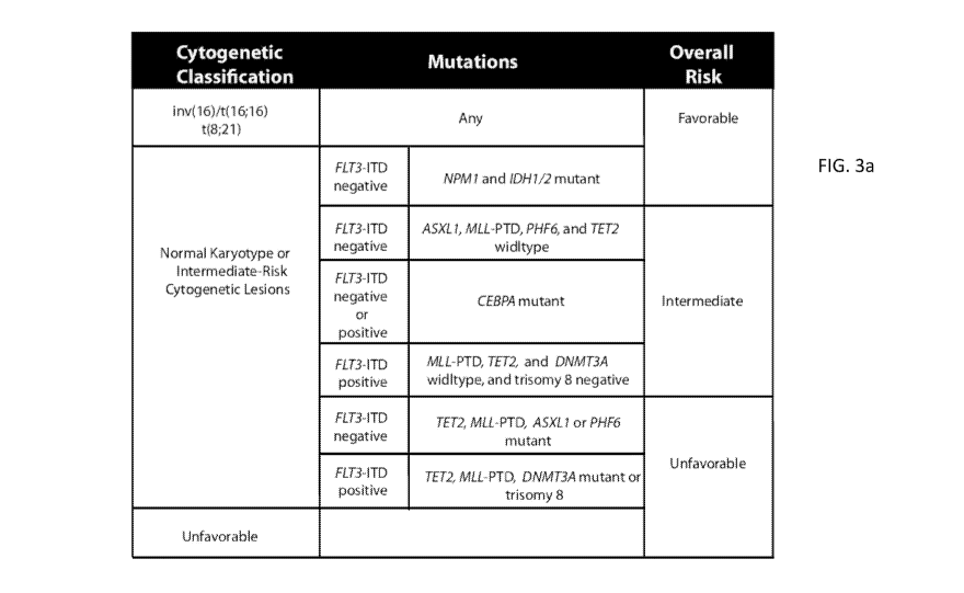
10040] Figure 3 shows revised AML risk stratification based on integrated
genetic
analysis. Figure 3A shows a revised risk stratification based on integrated
cytogenetic
and mutational analysis. Final overall risk groups are on the right. Figure 3B
shows the
impact of integrated mutational analysis on risk stratification in the test
cohort of AML
patients (p-values represent a comparison of all curves). The black curves
show the
patients in the cytogenetic risk groups that remained unchanged. The green
curve shows
patients that were reclassifed from intermediate-risk to favorable-risk. The
red curve
shows patients that were reclassified from intermediate-risk to unfavorable-
risk. Figure
3C confirms the reproducibility of the genetic prognostic schema in an
independent cohort
of 104 samples from the E1900 trial (p-values represent a comparison of all
curves).
100411 Figure 4 shows the molecular determinants of response to high-dose
Daunorubicin induction chemotherapy. Kaplan-Meier estimates of OS in the
entire cohort
according to DNMT3A mutational status (Panel A) and DNMT3A status in patients
=
receiving high-dose or standard-dose daunorubicin (Panel B). OS in patients
according to
treatment arm is shown in patients with DNMT3A or NPM1 mutations or MLL
13
CA 02867375 2014-09-12
WO 2013/138237
PCT/US2013/030208
translocations (Panel C) and patients lacking DNM7'3A or NPM1 mutations or MLL
translocations (Panel D).
[0042] Figure 5 shows comprehensive mutational profiling improves risk-
stratification
and clinical management of patients with acute myeloid leukemia (AML). Use of
mutational profiling delineates subsets of cytogenetically defined
intermediate-risk
patients with markedly different prognoses and reallocates a substantial
proportion of
patients to favorable or unfavorable-risk categories (A). In addition,
mutational profiling
identifies genetically defined subsets of AML patients with improved outcome
with high-
dose anthracycline induction chemotherapy (B).
[0043] Figure 6 shows Circos diagrams for each gene.
[0044] Figure 7 shows Circos diagrams for all genes and some relevant
cytogenetic
abnormalities in patients within cytogenetically-defined favorablerisk (Panel
A),
intermediate-risk (Panel B), and unfavorable-risk (Panel C) subgroups. The
percentage of
patients in each cytogenetic risk category with > 2 mutations is displayed in
Panel D. The
proportion of intermediate risk patients with 2 or more somatic mutations was
significantly higher than of patients in the other 2 cytogenetic subgroups
[0045] Figure 8 is a Circos diagram, showing the mutual exclusivity of IDH1,
IDH2,
TET2, and WT1 mutations.
[0046] Figure 9 shows Kaplan-Meier estimates of OS according to mutational
status:
data are shown for OS in the entire cohort according to the mutational status
of PHF6
(Panel A) and ASXL1 (Panel B).
14
CA 02867375 2014-09-12
WO 2013/138237
PCT/US2013/030208
[0047] Figure 10 shows Kaplan-Meier survival estimates shown for IDH2 (Panel
A),
IDH2 R140 (Panel B), IDH1 (Panel C) and the IDH2 R172 allele (Panel D) in the
entire
cohort. Panel E shows both IDH2 alleles while Panel F shows all three IDH
alleles (pvalue
represents comparison of all curves). These data show that the IDH2 R140
allele is the
only IDH allele to have prognostic relevance in the entire cohort.
[0048] Figure 11 shows Kaplan-Meier estimates of OS in patients from the test
cohort
with core-binding factor alterations with mutations in KIT versus those
wildtype for KIT.
KIT mutations were not associated with a difference in OS when patients with
any
corebinding factor alteration (i.e. patients with t(8;21), inv(16), or
t(16;16)) were studied
(A). In contrast, KIT mutations were associated with a significant decrease in
OS in
patients bearing t(8;21) specifically (B). KIT mutations were not associated
with adverse
OS in patients with inv(16) or t(16;16) (C).
[0049] Figure 12 shows Kaplan-Meier survival estimates for TET2 in
cytogenetically
defined intermediate-risk patients in the cohort.
[0050] Figure 13 shows Kaplan-Meier survival estimates for NPM/-mutant
patients
with cytogenetically-defined intermediate-risk in the cohort. Only those with
concomitant
IDH mutations have improved survival.
[0051] Figure 14 shows the risk classification schema for FLT3-ITD widltype
(A) and
mutant (B) intermediate-risk AML shown in Figure 3 is shown here for normal-
karyotype
patients only.
[0052] Figure 15 shows that the mutational prognostic schema predicts outcome
regardless of post-remission therapy with no transplantation (A), autologous
CA 02867375 2014-09-12
WO 2013/138237
PCT/US2013/030208
transplantation (B), and allogeneic transplantation (C) (p-value represents
comparison of
all curves). Note, curves represent overall risk categories integrating
cytogenetic and
mutational analysis (as shown in final column in Figure 3A).
[0053] Figure 16 shows Kaplan-Meier estimates of OS in the entire cohort
according to
DNMT3A mutational status (Panel A and B), MLL translocation status (Panel C
and D) or
NPM1 mutational status in patients receiving high-dose or standard-dose
daunorubicin
(Panels E and F). OS in patients according to treatment arm is shown in DNMT3A
mutant
(Panel A) and wild-type (Panel B) patients. Panel C shows OS in MLL
translocated
patients receiving high-dose or standard-dose daunorubicin while Panel D shows
OS in
non-MLL translocated patients depending on daunorubicin dose. OS in patients
according
to treatment arm is shown in NPM1 mutant (Panel E) and wild-type (Panel F)
patients as
well.
[0054] Table 1 shows baseline characteristics of the samples in the test,
validation, and
entire cohort from the ECOG E 1 900 trial.
[0055] Table 2 shows genomic DNA primer sequences utilized for comprehensive
genetic analysis. All primer sequences are displayed with M 1 3F2/M13R2 tags.
[0056] Table 3 shows P-values for the test of proportional hazards for all
mutations
identified in the test cohort.
[0057] Table 4 shows mutational frequency of genes sequenced in patients in
the overall
ECOG E1900 cohort and within each cytogenetic risk group.
16
CA 02867375 2014-09-12
WO 2013/138237
PCT/US2013/030208
[0058] Table 5 shows co-occurrences of somatic mutations and cytogenetic
abnormalities in the test cohort of 398 AML patients with de novo AML from the
ECOG
E1900 trial.
[0059] Table 6 shows pairwise correlations between all genetic abnormalities.
[0060] Table 7 shows frequently co-occurring genetic abnormalities.
[0061] Table 8 shows mutually exclusive genetic abnormalities.
[0062] Table 9 shows univariate analysis of the effects of mutations in
individual genes
on overall survival in the ECOG E1900 cohort.
[0063] Table 10 shows univariate analysis of mutations in individual genes on
intermediate-risk group in the ECOG E1900 cohort.
[0064] Table 11 shows a revised AML risk stratification based on integrated
genetic
analysis with frequency and number of patients in each genetic risk category
displayed.
[0065] Table 12 shows that genetic prognostic schema is independent of
treatment-
related mortality and chemotherapy resistance in the test cohort and the
entire cohort of
the analyzed ECOG E1900 patients.
[0066] Table 13 shows differential response to high-dose versus standard-dose
daunorubicin induction chemotherapy based on genotype of AML patients.
[0067] DETAILED DESCRIPTION OF THE INVENTION
[0068] To facilitate understanding of the invention, the following definitions
are
provided. It is to be understood that, in general, terms not otherwise defined
are to be
17
CA 02867375 2014-09-12
WO 2013/138237
PCT/US2013/030208
given their meaning or meanings as generally accepted in the art. The
terminology used
herein is for the purpose of describing particular embodiments only and is not
intended to
limit the scope of the present invention which will be limited only by the
appended claims.
[0069] In practicing the present invention, many conventional techniques in
molecular
biology are used. These techniques are described in greater detail in, for
example,
Molecular Cloning: a Laboratory Manual 3rd edition, J.F. Sambrook and D.W.
Russell, ed.
Cold Spring Harbor Laboratory Press 2001 and DNA Microarrays: A Molecular
Cloning
Manual. D. Bowtell and J. Sambrook, eds. Cold Spring Harbor Laboratory Press
2002.
Additionally, standard protocols, known to and used by those of skill in the
art in
mutational analysis of mammalian cells, including manufacturers' instruction
manuals for
preparation of samples and use of microarray platforms are hereby incorporated
by
reference.
[0070] In the description that follows, a number of terms are used
extensively. The
following definitions are provided to facilitate understanding of the
invention. Unless
otherwise specified, "a," "an," "the," and "at least one" are used
interchangeably and mean
one or more than one.
[0071] The terms "cancer", "cancerous", or "malignant" refer to or describe
the
physiological condition in mammals that is typically characterized by
unregulated growth
of tumor cells. Examples of a blood cancer include but are not limited to
acute myeloid
leukemia.
[0072] The term "diagnose" as used herein refers to the act or process of
identifying or
determining a disease or condition in a mammal or the cause of a disease or
condition by
the evaluation of the signs and symptoms of the disease or disorder. Usually,
a diagnosis
18
CA 02867375 2014-09-12
WO 2013/138237
PCT/US2013/030208
of a disease or disorder is based on the evaluation of one or more factors
and/or symptoms
that are indicative of the disease. That is, a diagnosis can be made based on
the presence,
absence or amount of a factor which is indicative of presence or absence of
the disease or
condition. Each factor or symptom that is considered to be indicative for the
diagnosis of
a particular disease does not need be exclusively related to said particular
disease; i.e.
there may be differential diagnoses that can be inferred from a diagnostic
factor or
symptom. Likewise, there may be instances where a factor or symptom that is
indicative
of a particular disease is present in an individual that does not have the
particular disease.
[0073] "Expression profile" as used herein may mean a genomic expression
profile.
Profiles may be generated by any convenient means for determining a level of a
nucleic
acid sequence e.g. quantitative hybridization of microRNA, labeled microRNA,
amplified
microRNA, cRNA, etc., quantitative PCR, ELISA for quantitation, and the like,
and allow
the analysis of differential gene expression between two samples. A subject or
patient
tumor sample, e.g., cells or collections thereof, e.g., tissues, is assayed.
Samples are
collected by any convenient method, as known in the art.
[0074] "Gene" as used herein may be a natural (e.g., genomic) gene comprising
transcriptional and/or translational regulatory sequences and/or a coding
region and/or
non- translated sequences (e.g., introns, 5'- and 3 '-untranslated sequences).
The coding
region of a gene may be a nucleotide sequence coding for an amino acid
sequence or a
functional RNA, such as tRNA, rRNA, catalytic RNA, siRNA, miRNA or antisense
RNA.
The term "gene" has its meaning as understood in the art. However, it will be
appreciated
by those of ordinary skill in the art that the term "gene" has a variety of
meanings in the
art, some of which include gene regulatory sequences (e.g., promoters,
enhancers, etc.)
and/or intron sequences, and others of which are limited to coding sequences.
It will
19
CA 02867375 2014-09-12
WO 2013/138237
PCT/US2013/030208
further be appreciated that definitions of "gene" include references to
nucleic acids that do
not encode proteins but rather encode functional RNA molecules such as tRNAs.
For the
purpose of clarity we note that, as used in the present application, the term
"gene"
generally refers to a portion of a nucleic acid that encodes a protein; the
term may
optionally encompass regulatory sequences. This definition is not intended to
exclude
application of the term "gene" to non-protein coding expression units but
rather to clarify
that, in most cases, the term as used in this document refers to a protein
coding nucleic
acid.
[0075] "Mammal" for purposes of treatment or therapy refers to any animal
classified as
a mammal, including humans, domestic and farm animals, and zoo, sports, or pet
animals,
such as dogs, horses, cats, cows, etc. Preferably, the mammal is human.
[0076] "Microarray" refers to an ordered arrangement of hybridizable array
elements,
preferably polynucleotide probes, on a substrate.
[0077] Therapeutic agents for practicing a method of the present invention
include, but
are not limited to, inhibitors of the expression or activity of genes
identified and disclosed
herein, or protein translation thereof. An "inhibitor" is any substance which
retards or
prevents a chemical or physiological reaction or response. Common inhibitors
include but
are not limited to antisense molecules, antibodies, and antagonists.
[0078] The term "poor" as used herein may be used= interchangeably with
"unfavorable."
The term "good" as used herein may be referred to as "favorable." The term
"poor
= responder" as used herein refers to an individual whose cancer grows
during or shortly
thereafter standard therapy, for example radiation-chemotherapy, or who
experiences a
clinically evident decline attributable to the cancer. The term "respond to
therapy" as used
CA 02867375 2014-09-12
WO 2013/138237
PCT/US2013/030208
herein refers to an individual whose tumor or cancer either remains stable or
becomes
smaller / reduced during or shortly thereafter standard therapy, for example
radiation-
chemotherapy.
[0079] "Probes" may be derived from naturally occurring or recombinant single-
or
double-stranded nucleic acids or may be chemically synthesized. They are
useful in
detecting the presence of identical or similar sequences. Such probes may be
labeled with
reporter molecules using nick translation, Klenow fill-in reaction, PCR or
other methods
well known in the art. Nucleic acid probes may be used in southern, northern
or in situ
hybridizations to determine whether DNA or RNA encoding a certain protein is
present in
=
a cell type, tissue, or organ.
100801 "Prognosis" as used herein refers to a forecast as to the probable
outcome of
cancer, including the prospect of recovery from the cancer. As used herein the
terms
prognostic information and predictive information are used interchangeably to
refer to any
information that may be used to foretell any aspect of the course of a disease
or condition
either in the absence or presence of treatment. Such information may include,
but is not
limited to, the average life expectancy of a patient, the likelihood that a
patient will
survive for a given amount of time (e.g., 6 months, 1 year, 5 years, etc.),
the likelihood
that a patient will be cured of a disease, the likelihood that a patient's
disease will respond
to a particular therapy (wherein response may be defined in any of a variety
of ways).
Prognostic and predictive information are included within the broad category
of diagnostic
information.
100811 The term "prognosis" as used herein refers to a prediction of the
probable course
and outcome of a clinical condition or disease. A prognosis of a patient is
usually made
21
CA 02867375 2014-09-12
WO 2013/138237
PCT/US2013/030208
by evaluating factors or symptoms of a disease that are indicative of a
favorable or
unfavorable course or outcome of the disease. The phrase "determining the
prognosis" as
used herein refers to the process by which the skilled artisan can predict the
course or
outcome of a condition in a patient. The term "prognosis" does not refer to
the ability to
predict the course or outcome of a condition with 100% accuracy. Instead, the
skilled
artisan will understand that the term "prognosis" refers to an increased
probability that a
certain course or outcome will occur; that is, that a course or outcome is
more likely to
occur in a patient exhibiting a given condition, when compared to those
individuals not
exhibiting the condition. A prognosis may be expressed as the amount of time a
patient
can be expected to survive. Alternatively, a prognosis may refer to the
likelihood that the
disease goes into remission or to the amount of time the disease can be
expected to remain
in remission. Prognosis can be expressed in various ways; for example
prognosis can be
expressed as a percent chance that a patient will survive after one year, five
years, ten
years or the like. Alternatively prognosis may be expressed as the number of
months, on
average, that a patient can expect to survive as a result of a condition or
disease. The
prognosis of a patient may be considered as an expression of relativism, with
many factors
effecting the ultimate outcome. For example, for patients with certain
conditions,
prognosis can be appropriately expressed as the likelihood that a condition
may be
treatable or curable, or the likelihood that a disease will go into remission,
whereas for
patients with more severe conditions prognosis may be more appropriately
expressed as
likelihood of survival for a specified period of time.
[0082] The terms "favorable prognosis" and "positive prognosis," or
"unfavorable
prognosis" and "negative prognosis" as used herein are relative terms for the
prediction of
the probable course and/or likely outcome of a condition or a disease. A
favorable or
22
CA 02867375 2014-09-12
WO 2013/138237
PCT/US2013/030208
positive prognosis predicts a better outcome for a condition than an
unfavorable or
negative or adverse prognosis. In a general sense a "favorable prognosis" is
an outcome
that is relatively better than many other possible prognoses that could be
associated with a
particular condition, whereas an "unfavorable prognosis" predicts an outcome
that is
relatively worse than many other possible prognoses that could be associated
with a
particular condition. Typical examples of a favorable or positive prognosis
include a
better than average cure rate, a lower propensity for metastasis, a longer
than expected life
expectancy, differentiation of a benign process from a cancerous process, and
the like. For
example, if a prognosis is that a patient has a 50% probability of being cured
of a
particular cancer after treatment, while the average patient with the same
cancer has only a
25% probability of being cured, then that patient exhibits a positive
prognosis. A positive
prognosis may be diagnosis of a benign tumor if it is distinguished over a
cancerous
tumor.
[0083] The term "relapse" or "recurrence" as used in the context of cancer in
the present
application refers to the return of signs and symptoms of cancer after a
period of remission
or improvement.
[0084] As used herein a "response" to treatment may refer to any beneficial
alteration in
a subject's condition that occurs as a result of treatment. Such alteration
may include
stabilization of the condition (e.g., prevention of deterioration that would
have taken place
in the absence of the treatment), amelioration of symptoms of the condition,
improvement
in the prospects for cure of the condition. One may refer to a subject's
response or to a
tumor's response. In general these concepts are used interchangeably herein.
23
CA 02867375 2014-09-12
WO 2013/138237
PCT/US2013/030208
[0085] "Treatment" or "therapy" refer to both therapeutic treatment and
prophylactic or
preventative measures. The term "therapeutically effective amount" refers to
an amount
of a drug effective to treat a disease or disorder in a mammal. In the case of
cancer, the
therapeutically effective amount of the drug may reduce the number of cancer
cells;
reduce the tumor size; inhibit (i.e., slow to some extent and preferably stop)
cancer cell
infiltration into peripheral organs; inhibit (i.e., slow to some extent and
preferably stop)
tumor metastasis; inhibit, to some extent, tumor growth; and/ or relieve to
some extent one
or more of the symptoms associated with the disorder.
[0086] For the recitation of numeric ranges herein, each intervening number
there
between with the same degree of precision is explicitly contemplated. For
example, for
the range of 2-5, the numbers 3 and 4 are contemplated in addition to 2 and 5,
and for the
range 2.0-3.0, the number 2.0, 2.1, 2.2, 2.3, 2.4, 2.5, 2.6, 2.7, 2.8, 2.9 and
3.0 are explicitly
contemplated. As used herein, the term "about" X or "approximately" X refers
to +/-10%
of the stated value of X.
[0087] Inherent difficulties in the diagnosis and treatment of cancer include
among other
things, the existence of many different subgroups of cancer and the
concomitant variation
in appropriate treatment strategies to maximize the likelihood of positive
patient outcome.
Current methods of cancer treatment are relatively non-selective. Typically,
surgery is
used to remove diseased tissue; radiotherapy is used to shrink solid tumors;
and
chemotherapy is used to kill rapidly dividing cells.
[0088] In the case of blood cancers, it is worthy to begin by noting that
blood primarily
consists of red blood cells (RBC), white blood cells (WBC) and platelets. Red
blood cells
carry oxygen to the body, the white blood cells police and protect the body,
and platelets
24
CA 02867375 2014-09-12
WO 2013/138237
PCT/US2013/030208
help clot the blood when there is injury. Abnormalities in these cell types
can lead to
blood cancer. The main categories of blood cancer are Acute Lymphocytic or
Lymphoblastic Leukemias (ALL), Chronic Lymphocytic or Lymphoblastic Leukemias
(CLL), Acute Myelogenous or Myeloid Leukemias (AML), and Chronic Myelogenous
or
Myeloid Leukemias (CML).
100891 Both leukemia and lymphoma are hematologic malignancies (cancers) of
the
blood and bone marrow. In the case of leukemia, the cancer is characterized by
abnormal
proliferation of leukocytes and is one of the four major types of cancer. The
cancer
interferes with the body's ability to make blood, and the cancer attacks the
bone marrow
and the blood itself, causing fatigue, anemia, weakness, and bone pain.
Leukemia is
diagnosed with a blood test in which specific types of blood cells are
counted; it accounts
for about 29,000 adults and 2,000 children diagnosed each year in the United
States.
Treatment for leukemia typically includes chemotherapy and radiation to kill
the cancer,
and may involve bone marrow transplantation in some cases.
100901 Leukemias are classified according to the type of leukocyte most
prominently
involved. Acute leukemias are predominantly undifferentiated cell populations
and
chronic leukemias have more mature cell forms. The acute leukemias are divided
into
lymphoblastic (ALL) and non-lymphoblastic (ANLL) types, with ALL being
predominantly a childhood disease while ANLL, also known as acute myeloid
leukemia
(AML), being a more common acute leukemia among adults.
[0091] AML is characterized by an increase in the number of myeloid cells in
the marrow
and an arrest in their maturation, frequently resulting in hematopoietic
insufficiency. In
the United States, the annual incidence of AML is approximately 2.4 per
100,000 and it
CA 02867375 2014-09-12
WO 2013/138237
PCT/US2013/030208
increases progressively with age to a peak of 12.6 per 100,000 adults 65 years
of age or
older. Despite improved therapeutic approaches, prognosis of AML is very poor
around
the globe. Even in the United States, five-year survival rate among patients
who are less
than 65 years of age is less than 40%.
[0092] Acute myeloid leukemia (AML) is a heterogeneous disorder that includes
many
entities with diverse genetic abnormalities and clinical features. The
pathogenesis is
known for relatively few types of leukemia. Patients with intermediate and
poor risk
cytogenetics represent the majority of AML; chemotherapy based regimens fail
to cure
most of these patients and stem cell transplantation is frequently the
treatment choice.
Since allogeneic stem cell transplantation is not an option for many patients
with high risk
leukemia, there is a need to improve our understanding of the biology of these
leukemias
and to develop improved therapies. Despite considerable advances, not enough
is known
of the etiology, cell physiology and molecular genetics of acute myeloid
leukemia. As
such, the development of effective new agents and novel treatment and/or
prognostic
methods against myeloid leukemia, and in particular acute myeloid leukemia,
remains a
focal point today in translational oncology research.
[0093] Significant progress has been made in understanding risk factors,
including
genetic factors, that may contribute to AML, but the relevance of these
factors to clinical
outcome remains unclear. In addition, the expression level and antibody
staining pattern
of several proteins have been shown to be predictive of outcome and of the
likelihood of
response to therapy. However, the clinical outcome of individual patients
remains
uncertain, and the ability to predict which patients are likely to benefit
from a particular
type of therapy (e.g., a certain drug or class of drug) remains elusive.
26
CA 02867375 2014-09-12
WO 2013/138237
PCT/US2013/030208
[0094] In the present disclosure, leukemic samples from patients with
diagnosed AML
were obtained. Bone marrow or peripheral blood samples were collected,
prepared by
Ficoll-Hypaque (Nygaard) gradient centrifugation. Cytogenetic analyses of the
samples
were performed at presentation, as previously described (Bloomfield; Leukemia
1992;
6:65-67. 21). The criteria used to describe a cytogenetic clone and karyotype
followed the
recommendations of the International System for Human Cytogenetic
Nomenclature.
DNA was extracted from diagnostic bone marrow aspirate samples or peripheral
blood
samples using method described previously (Zuo et al. Mod Pathol. 2009; 22,
1023-1031).
[0095] The present disclosure is based on mutational analysis of 18 genes in
398
patients with AML younger than 60 years of age randomized to receive induction
therapy
including high-dose or standard dose daunorubicin. Prognostic findings were
further
validated in an independent set of 104 patients.
[0096] The inventors of the instant application have identified >1 somatic
alteration in
97.3% of patients. These Applicants discovered (1) that FLT3-ITD (p=0.001),
MLL-PTD
(v0.009), ASXL1 (p=0.05), and PHF6 (p=0.006) mutations are associated with
reduced
overall survival ("OS"); and (2) that CEBPA (p=0.05) and IDH2R140Q (p=0.01)
mutations were associated with improved OS.
[0097] Accordingly, in one aspect of the present disclosure is a method of
predicting
survival of a patient with acute myeloid leukemia, said method comprising:
analyzing a
genetic sample isolated from the patient for the presence of cytogenetic
abnormalities and
a mutation in at least one of FLT3, NPM1, DNM7'3A, NRAS, CEBPA, TET2, W71,
IDH1,
IDH2, KIT, RUNX1, MLL-PTD, ASXL1, PHF6, KRAS, PTEN, P53, HRAS, and EZH2
genes; and (i) predicting poor survival of the patient if a mutation is
present in at least one
27
CA 02867375 2014-09-12
WO 2013/138237 PCT/US2013/030208
of FLT3, MLL-PTD, ASXLI and PHF6 genes, or (ii) predicting favorable survival
of the
patient if a mutation is present in IDH2R140 (e.g. IDH2R140Q) and/or a
mutation is
present in CEBPA. In one embodiment, the method further comprises, predicting
intermediate survival of the patient with cytogenetically-defined intermediate
risk AML if: =
(i) no mutation is present in any of FLT3-ITD, TET2, MLL-PTD, DNMT3A, ASXL1 or
PHF6 genes, (ii) a mutation in CEBPA is and the FLT3-ITD is present, or (iii)
a mutation
is present in FLT3-ITD but trisomy 8 is absent. In another embodiment, the
method
further comprises predicting unfavorable survival of the patient if (i) a
mutation in TET2,
ASXLI, or PHF6 or an MLL-PTD is present in a patient without the FLT3-ITD
mutation,
or (ii) the patient has a FLT3-ITD mutation and a mutation in TET2, DNMT3A,
MLL-PTD
or trisomy 8.
[0098] The genetic sample may be obtained from a bone marrow aspirate or the
patient's blood. Once the sample is obtained, in one example, the mononuclear
cells are
isolated according to known techniques including Ficoll separation and their
DNA is
extracted. In a particular embodiment, poor survival or adverse risk of the
patient is
survival of less than or equal to about 10 months. Whereas, in one embodiment,
intermediate survival the patient is survival of about 18 months to about 30
months. In
another embodiment, favorable survival of the patient is survival of about 32
months or
more.
[0099] In another aspect, the present disclosure teaches a method of
predicting survival
of a patient with acute myeloid leukemia, said method comprising, assaying a
genetic
sample from the patient's blood or bone marrow for the presence of a mutation
in at least
one of genes FLT3, NPM1, DNMT3A, NRAS, CEBPA, TET2, WTI, IDHI, 1DH2, KIT,
RUNX1, MLL-PTD, ASXL1, PHF6, KRAS, PTEN, P53, HRAS, and EZH2 in said sample;
28
CA 02867375 2014-09-12
WO 2013/138237
PCT/US2013/030208
and predicting a poor survival of the patient if a mutation is present in at
least one of genes
FLT3-ITD, MLL-PTD, ASXL1, PHF6; or predicting a favorable survival of the
patient if a
mutation is present in CEBPA or a mutation is present in IDH2 at R140. In one
embodiment, the patient is characterized as intermediate-risk on the basis of
cytogenetic
analysis.
[00100] In one embodiment, amongst patients with cytogenetically-defined
intermediate-
risk acute myeloid leukemia who have FLT3-ITD mutation, at least one of the
following:
trisomy 8 or a mutation in TET2, DNM7'3A, or the MLL-PTD are associated with
an
adverse outcome and poor overall survival of the patient. In another
embodiment,
amongst patients with cytogenetically-defined intermediate-risk acute myeloid
leukemia
who have a mutation in FLT3-ITD gene, a mutation in CEBPA gene is associated
with
improved outcome and overall survival of the patient. In one embodiment, in a
cytogenetically-defined intermediate risk AML patient with both IDH1/IDH2 and
NPMI
mutations, the overall survival is improved compared to NPM/-mutant patients
wild-type
for both IDH1 and IDH2. In one embodiment, amongst patients with acute myeloid
leukemia, IDH2R140 mutations are associated with improved overall survival.
Poor or
unfavorable survival (adverse risk) of the patient, in one example, is
survival of less than
or equal to about 10 months. Favorable survival of the patient, in one
example, is survival
of about 32 months or more.
[00101] In one embodiment, the favorable impact of NPM1 mutations was
restricted to
patients with co-occurring IDH1/IDH2 and NPM1 mutations. Further, Applicants
identified genetic -predictors of outcome that improved risk stratification in
AML
independent of age, WBC count, induction dose, and post-remission therapy and
validated
their significance in an independent cohort. Applicants discovered that high-
dose
29
CA 02867375 2014-09-12
WO 2013/138237
PCT/US2013/030208
daunorubicin improved survival in patients with DNM7'3A or NPM1 mutations or
MLL
translocations (p=0.001) relative to treatment with standard dose
daunorubicin, but not in
patients wild-type for these alterations (p=0.67).
[00102] These data provide clinical implications of genetic alterations in AML
by
delineating mutations that predict outcome in AML and improve AML risk
stratification.
Applicants have herein discovered and demonstrated the utility of mutational
profiling to
improve prognostic and therapeutic decisions in AML, and in particular, have
shown that
DNMT3A or NPMI mutations or MLL translocations predict for improved outcome
with
high-dose induction chemotherapy.
[00103] Previous studies have highlighted the clinical and biologic
heterogeneity of acute
myeloid leukemia (AML). However, a relatively small number or cytogenetic and
molecular lesions have sufficient relevance to influence clinical practice.
The prognostic
relevance of cytogenetic abnormalities has led to the widespread adoption of
risk
stratification into three cytogenetically-defined risk groups with significant
differences in
OS. Although progress has been made in defining prognostic markers for AML, a
significant proportion of patients lack a specific abnormality of prognostic
significance.
Additionally, there is significant heterogeneity in outcome for individual
patients in each
risk group.
[00104] Recent studies have identified a number of recurrent somatic mutations
in
patients with AML, however, to date, whether mutational profiling of a larger
set of genes
would improve prognostication in AML has not been investigated in a clinical
trial cohort.
Here, Applicants conceived that integrated mutational analysis of all known
molecular
alterations occurring in >5% of AML patients would allow for the
identification of novel
CA 02867375 2014-09-12
WO 2013/138237
PCT/US2013/030208
molecular markers of outcome in AML and allow for the identification of
molecularly
defined subsets of patients who benefit from dose-intensified induction
chemotherapy.
[00105] High-Throughput Mutational Profiling in AML: Comprehensive Genetic
Analysis
[00106] Clinical studies have demonstrated that acute myeloid leukemia (AML)
is
heterogeneous with respect to presentation and to clinical outcome, and
studies have
shown that cytogenetics can be used to improve prognostication and to guide
therapeutic
decisions. More recently, genetic studies have improved our understanding of
the genetic
basis of AML. Applicants recognized genetic lesions represent prognostic
markers which
can be used to risk stratify AML patients and guide therapeutic decisions.
However,
although a number of gene mutations occur at significant frequency in AML,
their
prognostic value is not known in large phase III clinical trial cohorts.
[00107] Applicants report for the first time in a uniformly treated clinical
cohort, the
mutational status of all genes known to be significantly (>5%) mutated in AML
as well as
the effect of mutations in these genes on outcome and response to therapy.
Applicants
used a high throughput re-sequencing platform to perform full length
resequencing of the
coding regions of FLT3, NPM1, DNMT3A, NRAS, CEBPA, TET2, WT1, IDH1, IDH2,
KIT, RUNX1, MLL-PTD, ASXL1, PHF6, KRAS, PTEN, P53, HRAS, and EZH2 in pre-
treatment genomic DNA from 398 patients with de novo AML enrolled in the ECOG
E1900 Study.
[00108] Including both mutations and cytogenetic abnormalities, Applicants
were able to
identify a clonal alteration in 91.2% of all patients in the E 1 900 cohort;
42% had 1 somatic
alteration, 36.4% had 2 alterations, 11.3% had 3 alterations and 1.5% had 4
alterations.
31
CA 02867375 2014-09-12
WO 2013/138237
PCT/US2013/030208
Mutational data from each patient was correlated with overall survival,
disease-free
survival, and with treatment assignment (standard dose or high dose
daunorubicin).
Applicants discovered somatic mutations in FLT3 (37% total; 30% ITD, 7% TKD),
DNMT3A (23%), NPM1 (14%), CEBPA (10%), TET2 (I0%), NRAS (10%), WT1 (1 0%),
KIT (9%), IDH2 (8%), IDHI (6%), RUNXI (6%), ASXLI (4%), PHF6 (3%), KRAS
(2.5%), TP53 (2%), PTEN (1.5%); the only genes without mutations in
Applicants' screen
were HRAS and EZH2.
1001091 Applicants next used correlation analysis to assess whether mutations
were
positively or negatively correlated (Figure 1). In addition to identified
mutational
correlations (FLT3 and NPMI, KIT and core binding factor leukemia), Applicants
found
that FLT3 and ASXL1 mutations were mutually exclusive in this large cohort (p
= 0.0008).
Further, Applicants found that IDHI/IDH2 mutations were mutually exclusive of
both
TET2 (p= 0.02), and W7'1 (p= 0.01) mutations, suggesting these mutations have
overlapping roles in AML pathogenesis.
[00110] Applicants next set out to investigate if any mutations were
associated with lack
of response to chemotherapy; notably mutations in ASXL1 (p= 0.0002) and WTI
(p=0.03)
were enriched in patients with primary refractory-AML. Integration of
mutational data
with outcome in the ECOG El 900 trial revealed significant effects that
mutations in FLT3
(p=0.0005), ASXL1 (p=0.005), and PHF6 (p=0.02) were associated with reduced
overall
survival. In addition, Applicants found that mutations in CEBPA (p= 0.04) and
in IDH2
(p= 0.003) were associated with improved overall survival; the favorable
impact of IDHI
mutations on outcome was exclusively seen in patients with IDH2R140 mutations.
32
CA 02867375 2014-09-12
WO 2013/138237
PCT/US2013/030208
[00111] This data represents a comprehensive mutational analysis of 18 genes
in a
uniformly-treated de novo AML cohort, which allowed Applicants to delineate
the
mutational frequency of this gene set in de novo AML, the pattern of
mutational
cooperativity in AML and the clinical effects of gene mutations on survival
and response
to therapy in AML. In one embodiment, Applicants identified mutations in ASXL1
and
WT1 as being significantly enriched in patients who failed to respond to
standard induction
chemotherapy in AML. This data provides important clinical implications of
genetic
alterations in AML and provides insight into the multistep pathogenesis of
adult AML. In
one embodiment, the acute myeloid leukemia is selected from newly diagnosed,
relapsed
or refractory acute myeloid leukemia.
[00112] Accordingly, one aspect of the present disclosure is a method of
predicting
survival of a patient with acute myeloid leukemia, said method comprising
assaying a
genetic sample from the patient's blood or bone marrow for the presence of a
mutation in
genes ASXL1 and WT1; and determining the patient has or will develop primary
refractory
acute myeloid leukemia if mutated ASXL1 and WT1 genes are detected. The sample
can
be a bone marrow aspirate or the patient's blood. Further, in one embodiment,
the
mononuclear cells are isolated for use in the assay.
= [00113] Applicants have developed a mutational classifier which can be
used to predict
risk of relapse in adults with acute myeloid leukemia by combining standard
analysis of
cytogenetics with full length sequencing of FLT3, NPM1, DNM7'3A, NRAS, CEBPA,
TET2, W7'1, IDH1, IDH2, KIT, RUNX1, MLL-PTD, ASXL1, PHF6, KRAS, PTEN, P53,
HRAS, and EZH2. The teachings of the instant application allow for the
development of
an integrated mutation classifier which can more accurately predict outcome
and relapse
risk than currently available techniques. In one embodiment, poor survival is
survival of
33
CA 02867375 2014-09-12
WO 2013/138237
PCT/US2013/030208
less than or equal to about ten months. In another embodiment, intermediate
survival of
the patient is survival of about 18 months to about 30 months. In a related
embodiment,
favorable survival of the patient is survival of about 32 months or more.
1001141 In one embodiment, in patients with FLT3-ITD wild-type intermediate-
risk acute
myeloid leukemia, TET2, ASXL1, PHF6, and MLL-PTD gene mutations were
independently shown to be associated with adverse outcome and poor overall
survival of
the patient. In another embodiment, in patients with FLT3-ITD mutant
intermediate-risk
acute myeloid leukemia, CEBPA gene mutations were associated with improved
outcome
and overall survival of the patient. In yet another embodiment, in
cytogenetically-defined
intermediate risk AML patients with FLT3-ITD mutant intermediate-risk acute
myeloid
leukemia, trisomy 8 and TET2, DNMT3A, and MLL-PTD mutations were associated
with
an adverse outcome and poor overall survival of the patient. In one
embodiment,
cytogenetically-defined intermediate risk AML patients with both IDH1/IDH2 and
NPM1
mutations have an improved overall survival compared to NPM/-mutant patients
wild-
type for both IDH1 and IDH2. In a related embodiment, IDH2 R140Q mutations are
associated with improved overall survival in the overall cohort of AML
patients.
1001151 One aspect of the present disclosure is directed to a method of
predicting survival
of a patient with acute myeloid leukemia, comprising: (a) analyzing a sample
isolated
from the patient for the presence of (i) a mutation in at least one of FLT3,
MLL-PTD,
ASXL1, and PHF6 genes, plus optionally one or more of NPM1, DNMT3A, NRAS,
CEBPA, TET2, WTI, IDH1, IDH2, KIT, RUNX1, KRAS, PTE1V, P53, HRAS, and EZH2
genes; or (ii) a mutation in IDH2 and/or CEBPA genes, plus optionally one or
more of
FLT3, MLL-PTD, ASXL1, PHF6, NPM1, DNMT3A, NRAS, TET2, WTI, IDH1, KIT,
RUNX1, K1?AS, PTE1V, P53, HRAS, and EZH2 genes; and (b) (i) predicting poor
survival
34
CA 02867375 2014-09-12
WO 2013/138237
PCT/US2013/030208
of the patient if a mutation is present in at least one of FLT3, MLL-PTD,
ASXL1 and PHF6
genes, or (ii) predicting favorable survival of the patient if a mutation is
present in
IDH2R140 and/or a mutation is present in CEBPA. The method may further
comprise
analyzing the sample for the presence of cytogenetic abnormalities. The method
may
further comprise predicting favorable survival of the patient if the following
mutation is
present: IDH2R140Q.
1001161 Furthermore, Applicants have discovered that DNMT3A mutations, NPM1
mutations or MLL fusions predict for improved outcome with high dose
chemotherapy,
which includes dose-intensified induction therapy. The teachings of the
instant
application provide for accurate risk stratification of AML patients and the
ability to
decide which patients need more agreessive therapy given high risk, and
identification of
low risk patients less in need of intensive post remission therapy. Moreover,
it is possible
to identify genotypically defined subsets of patients who would benefit from
induction
with dose-intensified anthracyclines, for example, daunorubicin. The present
disclosure
provides for more accurate assessment in risk classification. Presently, there
is no
effective way to determine which patients suffering from AML benefit from high
dose
daunorubicin. In one embodiment, the present disclosure provides for a novel
classifier as
well as a predictor of response.
1001171 Accordingly, one aspect of the present disclosure is a method of
determining
responsiveness of a patient with acute myeloid leukemia to high dose therapy,
said method
comprising analyzing a genetic sample isolated from the patient for the
presence of a
mutation in genes DNMT3A, and NPM1, and for the presence of a MLL
translocation; and
(i) identifying the patient as one who will respond to high dose therapy if a
mutation in
DNMT3A or NPM1 or an MLL translocation are present, or (ii) identifying the
patient as
CA 02867375 2014-09-12
WO 2013/138237
PCT/US2013/030208
one who will not respond to high dose therapy in the absence of mutations in
DNMT3A or
NPM1 or an MLL translocation. In one embodiment, the sample is DNA extracted
from
bone marrow or blood from the patient. The genetic sample may be DNA isolated
from
mononuclear cells (MNC) from blood or bone marrow of the patient. In one
embodiment,
the therapy comprises the administration of anthracycline. Examples of
anthracyclines
include Daunorubicin, Doxorubicin, Epirubicin, Idarubicin, Mitoxantrone, and
Adriamycin. In a particular example, the anthracycline is Daunorubicin.
[00118] The method may be used to predict a patient's response to therapy
before
beginning therapy, during therapy, or after therapy is completed. For example,
by
predicting a patient's response to therapy before beginning therapy, the
information may
be used in determining the best therapy option for the patient.
[00119] One embodiment of the present invention is directed to methods to
screen a
patient for the prognosis for acute myeloid leukemia. The invention may
provide
information concerning the survival rate of a patient, the predicted life span
of the patient,
and/or the *dieted likelihood of survival for the patient. In one embodiment,
poor
survival is referred generally as survival of about 10 months or less, and
good prognosis or
long-term survival is considered to be more than about 36 months or longer. In
one
embodiment, poor survival is considered as about one to 16 months, whereas
good,
favorable or long-term survival is considered to be range of about 30 to 42
months, more
than about 46 months, or more than about 60 months. In one embodiment, good
survival
is considered to be about 30 months or longer.
[00120] In any aspect of the invention, unless context demands otherwise, the
following
combinations of genes and\or cytogenetic defects may be analyzed or assayed:
FLT3 and
36
CA 02867375 2014-09-12
WO 2013/138237
PCT/US2013/030208
CEBPA; FLT3 and trisomy 8; FLT3 and TET2; FLT3 and DNMT3A; FLT3 and MLL;
FLT3, MLL, ASXL1 and PHF6, optionally with TET2 or DNMT3A; IDH2 and CEBPA;
IDH1, IDH2 and NPM I ; IDH2, ASXL1 and WTI; DNMT3A, NPM1 and MLL. Any of
these combinations may be combined with any one or more other genes shown in
the
Table entitled 'Genes analyzed for somatic mutations in genomic DNA of
patients with
AML and their clinical associations'. Optionally at least 2, 3, 4, 5, 6, 7, 8,
9, 10, 11, 12,
13, 14, 15, 16, 17, 18 or 19 genes are analyzed or assayed, which genes are
listed in said
table.
[00121] The present invention is also directed to a method for determining if
an
individual will respond to one or more therapies for acute myeloid leukemia.
The therapy
may be of any kind, but in specific embodiments it comprises chemotherapy,
such as one
or more anthracycline antibiotic agents. In one embodiment, the chemotherapy
comprises
the antimetabolite cytarabine in combination with an anthracycline.
[00122] In certain embodiments of the invention the therapy is chemotherapy,
immunotherapy, antibody-based therapy, radiation therapy, or supportive
therapy
(essentially any implemented for leukemia). In a particular embodiment, the
therapy
comprises the administration of a chemotherapeutic agent comprising
anthracycline
antibiotics. Examples of such anthracycline antibiotics include, but are not
limited to,
Daunorubicin, Doxorubicin, Epirubicin, Idarubicin, Mitoxantrone, and
Adriamycin. In
some embodiments, the chemotherapy is Gleevac or idarubicin and ara-C. In a
particular
embodiment, daunorubicin is used.
[00123] Often, diagnostic assays are directed by a medical practitioner
treating a patient,
the diagnostic assays are performed by a technician who reports the results of
the assay to
37
CA 02867375 2014-09-12
WO 2013/138237
PCT/US2013/030208
the medical practitioner, and the medical practitioner uses the values from
the assays as
criteria for diagnosing the patient. Accordingly, the component steps of the
method of the
present invention may be performed by more than one person.
1001241 Prognosis may be a prediction of the likelihood that a patient will
survive for a
particular period of time, or said prognosis is a prediction of how long a
patient may live,
or the prognosis is the likelihood that a patent will recover from a disease
or disorder.
There are many ways that prognosis can be expressed. For example prognosis can
be
expressed in terms of complete remission rates (CR), overall survival (OS)
which is the
amount of time from entry to death, disease-free survival (DFS) which is the
amount of
time from CR to relapse or death. In one embodiment, favorable likelihood of
survival, or
overall survival, of the patient includes survival of the patient for about
eighteen months or
more.
1001251 A prognosis is often determined by examining one or more prognostic
factors or
indicators. These are markers, the presence or amount of which in a patient
(or a sample
obtained from the patient) signal a probability that a given course or outcome
will occur.
The skilled artisan will understand that associating a prognostic indicator
with a
predisposition to an adverse outcome may involve statistical analysis.
Additionally, a
change in factor concentration from a baseline level may be reflective of a
patient
prognosis, and the degree of change in marker level may be related to the
severity of
adverse events. Statistical significance is often determined by comparing two
or more
populations, and determining a confidence interval and/or a p value. See,
e.g., Dowdy and
Wearden, Statistics for Research, John Wiley & Sons, New York, 1983. In one
embodiment, confidence intervals of the invention are 90%, 95%, 97.5%, 98%,
99%,
99.5%, 99.9% and 99.99%, while preferred p values are 0.1, 0.05, 0.025, 0.02,
0.01, 0.005,
38
CA 02867375 2014-09-12
WO 2013/138237
PCT/US2013/030208
0.001, and 0.0001. Exemplary statistical tests for associating a prognostic
indicator with a
predisposition to an adverse outcome are described.
[00126] One approach to the study of cancer is genetic profiling, an effort
aimed at
identifying perturbations in gene expression and/or mutation that lead to the
malignant
phenotype. These gene expression profiles and mutational status provide
valuable
information about biological processes in normal and disease cells. However,
cancers
differ widely in their genetic signature, leading to difficulty in diagnosis
and treatment, as
well as in the development of effective therapeutics. Increasingly, gene
mutations are
being identified and exploited as tools for disease detection as well as for
prognosis and
prospective assessment of therapeutic success.
[00127] The inventors of the instant application hypothesized that genetic
profiling of
acute myeloid leukemia would provide a more effective approach to cancer
management
and/or treatment. The inventors have herein identified that mutations of a
panel of genes
lead to the malignant phenotype.
[00128] The present inventors have used a molecular approach to the problem
and have
identified a set of gene mutations in acute myeloid leukemia correlates
significantly with
overall survival. Accordingly, the present invention relates to gene mutation
profiles
useful in assessing prognosis and/or predicting the recurrence of acute
myeloid leukemia.
In one aspect, the present invention relates to a set of genes, the mutation
of which in bone
marrow or blood cells, in particular mononuclear cells, of a patient
correlates with the
likelihood of poor survival. The present invention relates to the prognosis
and/or therapy
response outcome of a patient with acute myeloid leukemia. The present
invention
39
CA 02867375 2014-09-12
WO 2013/138237
PCT/US2013/030208
provides several genes, the mutation of which, alone or in combination, has
prognostic
value, specifically with respect to survival.
1001291 In one example, the disclosure is a method of determining whether a
human has
an increased genetic risk for developing or developing a relapse of acute
myeloid
leukemia, comprising, analyzing a genetic sample isolated from the human's
blood or
bone marrow for the presence of a mutation in at least one gene from FLT3,
NPMI,
DNMT3A, NRAS, CEBPA, TET2, WTI, IDHI, IDH2, KIT, RUNXI, MLL-PTD, ASXL1,
PHF6, KRAS, PTEN, P53, HRAS, and EZH2; and determining the individual with
cytogenetically-defined intermediate risk AML has an increased genetic risk
for
developing or developing a relapse of acute myeloid leukemia, relative to a
control human
with no such gene mutations in said genes, when: (i) a mutation in at least
one of TET2,
MLL-PTD, ASXL1 and PHF6 genes is detected when the patient has no FLT3-ITD
mutation, or (ii) a mutation in at least one of TET2, MLL-PTD, and D1VM7'3A
genes or
trisomy 8 is detected when the patient has a FLT3-ITD mutation.
1001301 To date, no test exists that predicts outcome in acute myeloid
leukemia, where
one can stratify AML patients into good versus poor responders, and in
particular, identify
patients who would respond better to high dose chemotherapy. As a consequence,
some
individuals may be overtreated, in that they unnecessarily receive treatment
that has
minimal effect. Alternatively, some individuals may be undertreated, in that
additional
agents added to standard therapy may improve outcome for these patients who
would be
refractory to standard treatment alone. As such, it is desirable to
prospectively distinguish
responders from non-responders to standard therapy prior to the initiation of
therapy in
order to optimize therapy for individual patients.
CA 02867375 2014-09-12
WO 2013/138237
PCT/US2013/030208
1001311 Accordingly, one aspect of the present disclosure is a method of
predicting
whether a patient suffering from acute myeloid leukemia will respond better to
high dose
chemotherapy than to standard dose chemotherapy, the method comprising,
obtaining a
DNA sample obtained from the patient's blood or bone marrow; determining the
mutational status of genes DNMT3A and NPMI, and the presence of a MLL
translocation;
and predicting that the subject will be more responsive to high dose
chemotherapy than
standard dose chemotherapy where the sample is positive for a mutation in
DNMT3A or
NPMI or an MLL translocation, or predicting that the subject will be non-
responsive to
high dose chemotherapy compared to standard dose chemotherapy where the sample
is
wild type with no mutations in DNMT3A or NPMI genes and no translocation in
MLL.
1001321 In one embodiment, the invention provides a clinical test that is
useful to predict
outcome in acute myeloid leukemia. The mutational status and/or expression of
one or
more specific genes is measured in the sample. Individuals are stratified into
those who
are likely to respond well to therapy vs. those who will not. The information
from the
results of the test is used to help determine the best therapy for the patient
in need of
therapy. Patients are stratified into those who are likely to have a poor
prognosis vs. those
who will have a good prognosis with standard therapy. A health care provider
uses the
results of the test to help determine the course of action, for example the
best therapy, for
the patient in need of therapy.
1001331 Because certain markers from a patient relate to the prognosis of a
patient in a
continuous fashion, the determination of prognosis can be performed using
statistical
analyses to relate the determined marker status to the prognosis of the
patient. A skilled
artisan is capable of designing appropriate statistical methods. For example
the methods
of the present invention may employ the chi-squared test, the Kaplan-Meier
method, the
41
CA 02867375 2014-09-12
WO 2013/138237
PCT/US2013/030208
log-rank test, multivariate logistic regression analysis, Cox's proportional-
hazard model
and the like in determining the prognosis. Computers and computer software
programs
may be used in organizing data and performing statistical analyses.
[00134] In one embodiment, a test is provided whereby a sample, for example a
bone
marrow or blood sample, is profiled for a gene set and, from the mutation
profile results,
an estimate of the likelihood of response to standard acute myeloid leukemia
therapy is
determined. In another embodiment, the invention concerns a method of
predicting the
prognosis and/or likelihood of response to standard and/or high dose
chemotherapy,
following treatment, in an individual with acute myeloid leukemia, comprising
determining the mutational status of one or more genes, in particular one to
DNMT3A or
NPM1 genes, or a MLL translocation, in a genetic sample obtained from the
patient,
normalized against a control gene or genes. A total value is computed for each
individual
from the mutational status of the individual genes in this gene set.
[00135] The present invention relates to the diagnosis, prognosis and
treatment of blood
cancer, including predicting the response to therapy and stratifying patients
for therapy.
The present disclosure teaches the mutational frequency, prognostic
significance, and
therapeutic relevance of integrated mutation profiling in 398 patients from
the ECOG
E1900 phase III clinical trial and validates these data in an independent
cohort of 104
patients from the same trial. Previous studies have suggested that mutational
analysis of
CEBPA, NPM1, and FLT3-ITD can be used to risk stratify intermediate-risk AML
patients. By performing comprehensive mutational analysis on a large cohort of
patients
treated on a single clinical trial, Applicants demonstrate that more extensive
mutational
analysis can better discriminate AML patients into relevant prognostic groups
(Figure 3).
For example, FLT3-ITD-negative NPM1IIDH mutant patients represent a favorable
risk
42
CA 02867375 2014-09-12
WO 2013/138237
PCT/US2013/030208
AML subset defined by a specific mutational genotype, whereas FLT3-ITD-
negative
NPM/-mutant patients without concurrent IDH mutations had a much less
favorable
outcome, particularly in patients with concurrent poor-risk mutations.
[00136] Furthermore, Applicants discovered that TET2, ASXL1, MLL-PTD, PHF6,
and
DNMT3A mutations can be used to define patients with adverse outcome in
cytogenetically-defined intermediate-risk AML patients without the FLT3-ITD.
Taken
together, these data demonstrate that mutational analysis of a larger set of
genetic
alterations can be used to discriminate AML patients into more precise subsets
with
favorable, intermediate, or unfavorable risk with marked differences in
overall outcome.
This approach can be used to define an additional set of patients with
mutationally defined
favorable outcome with induction and consolidation therapy alone, and a set of
patients
with mutationally defined unfavorable risk who are candidates for allogeneic
stem cell
transplantation or clinical trials given their poor outcome with standard AML
therapy
(Figure 5A).
[00137] The two recent randomized trials examining the benefits of
anthracycline dose-
intensification in AML demonstrated that more intensive induction chemotherapy
improves outcomes in AML. (Fernandez et al., N Engl J Med, 2009, 361, 1249-59;
Lowenberg et al., N Engl J Med, 2009, 361, 1235-48). Notably, re-evaluation of
the
original El 900 trial using our 502 patient cohort revealed that there was an
even
distribution of patients within each genetic risk category in both treatment
arms of the
original trial (p=0.41, Pearson's Chi-squared test). However, the initial
reports of these
studies did not identify whether dose-intensified induction therapy improved
outcomes in
different AML subgroups.
43
CA 02867375 2014-09-12
WO 2013/138237
PCT/US2013/030208
1001381 Applicants have discovered that anthracycline dose-intensification
markedly
improves outcomes in patients with mutations in DNM7'3A or NPM1 or MLL
translocations, suggesting mutational profiling can be used to determine which
patients
benefit from dose-intensive induction therapy (Figure 5B).
1001391 Applicants also discovered mutational combinations that commonly occur
in
AML patients and those that rarely, if ever, co-occur consistent with the
existence of
additional mutational complementation groups. For example, the observation
that TET2
and IDH mutations are mutually exclusive in this AML cohort led to functional
studies
linking IDH mutations and loss-of-function TET2 mutations in a shared
mechanism of
hematopoietic transformation.
100140] As is true in the case of many treatment regimens, some patients
respond to
treatment with chemotherapy, for example an anthracycline antibiotic,
daunorubicin, and
others do not. Prescribing the treatment to a patient who is unlikely to
respond to it is not
desirable. Thus, it would be useful to know how a patient could be expected to
respond to
such treatment before a drug is administered so that non-responders would not
be
unnecessarily treated and so that those with the best chance of benefiting
from the drug are
properly treated and monitored. Further, of those who respond to treatment,
there may be
varying degrees of response. Treatment with therapeutics other than
anthracycline or
treatment with therapeutics in addition to the anthracycline daunorubicin may
be
beneficial for those patients who would not respond to a particular
chemotherapy or in
whom response to the particular chemotherapy, e.g. daunorubicin, or a similar
anthracycline antibiotic, alone is less than desired.
44
CA 02867375 2014-09-12
WO 2013/138237
PCT/US2013/030208
1001411 The present disclosure demonstrates the ability of integrated
mutational profiling
of a clinical trial cohort to advance our understanding of AML biology,
improve current
prognostic models, and inform therapeutic decisions. In particular, these data
indicate that
more detailed genetic analysis can lead to improved risk stratification and
identification of
patients who benefit from more intensive induction chemotherapy.
1001421 In a specific aspect, the present disclosure is a method of screening
a patient with
acute myeloid leukemia for responsiveness to treatment with high dose of
Daunorubicin or
a pharmaceutically acceptable salt, solvate, or hydrate thereof, comprising:
obtaining a
genetic sample comprising an acute myeloid leukemic cell from said individual;
and
assaying the sample and detecting the presence of a mutation in DNMT3A or NPM1
or an
MLL translocation; and correlating a finding of a mutation in DNMT3A or NPM1
or an
MLL translocation, as compared to wild type controls where there is no
mutation, with
said acute myeloid leukemia patient being more sensitive to high dose
treatment with
Daunorubicin or a pharmaceutically acceptable salt, solvate, or hydrate
thereof. In one
embodiment, the method further comprises predicting the patient is at a lower
risk of
relapse of acute myeloid leukemia following chemotherapy if a mutation in
DNMT3A or
NPM1 or an MLL translocation is detected. In one embodiment, the method
further
comprises predicting the patient is at a lower risk of relapse of acute
myeloid leukemia
following chemotherapy if either DNMT3A or NPM1 mutations or an MLL
translocation
are detected.
1001431 Stratification of patient populations to predict therapeutic response
is becoming
increasingly valuable in the clinical management of cancer patients. For
example,
companion diagnostics are required for the stratification of patients being
treated with
targeted therapies such as trastuzumab (Herceptin, Genentech) in metastatic
breast cancer,
CA 02867375 2014-09-12
WO 2013/138237 PCT/US2013/030208
and cetuximab (Erbitux, Merck) in colorectal cancer. Predictive biomarkers are
also being
utilized for imatinib (Gleevec, Novartis) in gastrointestinal stromal tumors,
and for
gefitinib (Iressa, Astra-Zeneca) in lung cancer. Currently there is no method
available to
predict response to an anthracycline antibiotic in acute myeloid leukemia. To
identify
genes that are associated with greater sensitivity to an anthracycline
antibiotic, and in
particular to daunorubicine, Applicants assayed for the presence of mutations
in certain
genes as described above.
Genes analyzed for somatic mutations in genomic DNA of patients with AML and
their
clinical associations, as presently disclosed
GENE CLINICAL ASSOCIATION IN AML
FLT3 Internal tandem duplications or mutations in the
tyrosine kinase
domain of the receptor tyrosine kinase FLT3 are important for
predicting survival in the overall cohort of AML patients as well as
those with cytogenetically-defined intermediate-risk AML.
DNM T3 A Mutations in DNMT3A were relevant for (a) predicting
for adverse
overall survival in the presence of the FLT3-ITD in patients with
cytogenetically-defined intermediate-risk AML and (b) predicting
for responsiveness to high-dose induction chemotherapy with
daunorubicin and cytarabine.
NPM 1 Mutations in NPM1 were relevant for (a) predicting for
improved
overall survival when they co-occurred with IDH1/2 mutations in
cytogenetically-defined intermediate-risk AML and (b) predicting
for responsiveness to high-dose induction chemotherapy with
daunorubicin and cytarabine.
NRAS Activating mutations in NRAS were seen in 10% of AML
patients
studied here.
46
CA 02867375 2014-09-12
WO 2013/138237 PCT/US2013/030208
CEBPA Mutations in CEBPA were relevant for (a) predicting
for improved
overall survival in the overall cohort of AML patients regardless of
cytogenetic risk (b) predicting for intermediate overall risk in
patients with cytogenetically-defined intermediate-risk AML and
the presence of the FLT3ITD.
TET2 Mutations in TET2 were relevant for predicting for
worsened
overall risk in patients with cytogenetically-defined intermediate-
risk AML regardless of the presence of the FLT3ITD.
WTI Mutations in WTI were present in 8% of AML patients
here overall
but were enriched amongst patients who were refractory to initial
induction chemotherapy.
IDH2 Mutations in IDH2 were relevant for (a) predicting for
improved
overall survival in the overall cohort of AML patients regardless of
cytogenetic risk specifically when mutations were present at
Arginine 140; (b) predicting for favorable overall risk in patients
with cytogenetically-defined intermediate-risk AML and no
FLT3ITD when accompanied by an NPMI mutation.
IDH1 Mutations in IDH1 were relevant for predicting for
favorable
overall risk in patients with cytogenetically-defined intermediate-
risk AML and no FLT3ITD when accompanied by an NPM1
mutation.
KIT Mutations in KIT were seen in 6% of AML patients
overall but
were enriched in patients with core-binding factor translocations. In
the presence of a mutation in KIT, patients with t(8;16) had an
worsened overall survival compared to t(8;16) AML patients who
were KIT wildtype.
RUNX1 Mutations in RUNX1 were present in 5% of AML patients
here.
MLL Partial tandem duplications in MLL were relevant for
(a) predicting
for improved overall survival in patients receiving high-dose
induction chemotherapy and (b) predicting for adverse overall
survival in patients with cytogenetically-defined intermediate-risk
AML regardless of mutations in FLT3.
47
CA 02867375 2014-09-12
WO 2013/138237 PCT/US2013/030208
ASXL1 Mutations in ASXL1 were relevant for (a) predicting
for adverse
overall survival in the entire cohort of AML patients (b) predicting
for adverse overall survival in cytogenetically-defined
intermediate-risk AML patients who did not have the FLT3ITD and
(c) were enriched amongst patients who failed to respond to initial
induction chemotherapy.
PHF6 Mutations in ASXLI were relevant for (a) predicting
for adverse
overall survival in the entire cohort of AML patients and (b)
predicting for adverse overall survival in cytogenetically-defined
intermediate-risk AML patients who did not have the FLT3ITD.
KRAS Mutations in KRAS were present in 2% of AML patients
studied
here.
PTEN Mutations in PTEN were present in 2% of AML patients
studied
here.
TP53 Mutations in TP53 were present in 2% of AML patients
studied
here.
HRAS Mutations in HRAS were found in none of the AML
patients
studied here.
EZH2 Mutations in EZH2 were found in none of the AML
patients
studied here.
Specific somatic mutations identified in the sequencing of 18 genes in AML
patients, and
the nature of these mutations
GENE NATURE AND TYPE OF SOMATIC MUTATIONS
IDENTIFIED
FLT3 Numerous somatic internal tandem duplications in FLT3
were identified.
These have been shown to result in constitutive activation of FLT3
signaling and are listed below. In addition, mutations in the tyrosine
48
CA 02867375 2014-09-12
WO 2013/138237 PCT/US2013/030208
kinase domain of FLT3 were also identified and also shown to result in
hyperactive signaling of FLT3.
The specific intemal tandem duplication mutations identified were as
followed, though any in-frame insertion of nucleotides in the
juxtamembrane domain of FLT3 is scored as an internal tandem
duplication.
FLT3 p.Q580 V581 ins12; FLT3 p.D586 N587ins15;
FLT3
p.F590 Y591in14; FLT3 p.Y591
T592ins23; FLT3
p.D5931F594ins12; FLT3 p.F594 R595ins14; TLT3
p.R595 E596ins12;
FLT3 p.Y597 E598ins17; ¨FLT3 p.E598
Y599ins FLT3
p.Y599_D600insi4; FLT3 p.D600
L&Olins21; FLT3
p.K602 W603 ins14; FLT3 p.E604_F605ins15; TLT3 p.L610_E611ins11;
FLT3 p1612_G613ins30
Tyrosine kinase domain mutations identified:
FLT3 D835Y; FLT3 D835E; FLT3 D835H; FLT3 D835V
DNMT3A Mutations in DNMT3A were found as (1) out-of-frame
insertion/deletions
predicted to result in loss-of-function of the protein, (2) somatic nonsense
mutations also predicted to result in loss-of-function of the protein, and
(3) somatic missense mutations. Any out-of-frame insertion/deletion or
somatic nonsense mutation would be scored as a mutation in the
algorithm.
Insertions/Deletions:
FS at amino acid (AA) 296; FS at AA 458; FS at AA 492; FS at AA
537; FS at AA 571; FS at AA 592; FS at AA 639; FS at AA 695; FS at
AA 706; FS at AA 731; FS at AA 765; FS at AA 772; FS at AA 804; FS
at AA 902.
Nonsense mutations:
DNMT3A W58 1 C; DNMT3A W581R; DNMT3A Y660X; DNMT3A
Q696X; DNMT3A W753X; DNMT3A Q816X; DNMT3A Q886X;
DNMT3A S892X.
Missense mutations:
DNMT3A E30A; DNMT3A P76Q; DNMT3A S105N; DNMT3A L125V;
DNMT3A W297S; DNMT3A G298W; DNMT3A V328F; DNMT3A
G511E; DNMT3A C537Y; DNMT3A W581C; DNMT3A W581R;
DNMT3A R635W; DNMT3A V636L; DNMT3A S663P; DNMT3A
E664K; DNMT3A R676W; DNMT3A I681T; DNMT3A G699S;
DNMT3A S714C; DNMT3A V716I; DNMT3A T727A; DNMT3A F734L;
DNMT3A T862N; DNMT3A R882C; DNMT3A R882H; DNMT3A
R882S;
49
CA 02867375 2014-09-12
WO 2013/138237 PCT/US2013/030208
Insertion/deletion mutations in NPMI which disrupt the N-terminal
NPM1 nucleolar localization signal of nucleophosmin and
generate a nuclear
export signal in its place were identified.
NPMI p.W288fs*12; NPMI p.W288fs*16; NPMI p.W290fs*8; NPMI
p.W290fs*10; NPMI p.W290_K292>CFSK
NRAS Activating mutations in NRAS were identified.
NRas G12A; NRas G12D; NRas Gl2S, NRas G13D; NRas Gl3R; NRas
Q61R; NRas Q61 E; NRas Q61H; NRas Q61K; NRas Q1R; NRas
Q64D
CEBPA Mutations in CEBPA were identified as (1) out-of-
frame
insertions/deletions (2) nonsense mutations and (3) somatic missense
mutations. All of these mutations have been previously identified as
somatic mutations and were shown to either result in a predicted shorter
protein product with altered function or to affect dimerization of CEBPA.
Insertions/deletions:
CEBPA FS at AA 13; CEBPA FS at AA 15; CEBPA FS at AA 20;
CEBPA FS at AA 28; CEBPA FS at AA 35; CEBPA FS at AA 50;
CEBPA FS at AA 93; CEBPA FS at AA 190; CEBPA FS at AA 195;
CEBPA FS at AA 197; CEBPA FS at AA301; CEBPA FS at AA 303;
CEBPA FS at AA 305; CEBPA FS at AA 308; CEBPA FS at AA 309;
CEBPA FS at AA 311; CEBPA FS at AA 312; CEBPA FS at AA 313;
CEBPA FS at AA 315.
Nonsense mutations:
CEBPA K275X; CEBPA E329X
Somatic missense mutations:
CEBPA R29 1 C; CEBPA R300H; CEBPA L335R; CEBPA R339P.
TET2 Mutations in TET2 were found as out-of-frame
insertions/deletions
predicted to result in loss of functional protein, nonsense mutations also
predicted to result in loss of functional protein, and somatic missense
mutations. Any out-of-frame insertion/deletion or somatic nonsense
mutation would be scored as a mutation in our algorithm.
Insertions/deletions:
TET2 FS at AA 270; TET2 FS at AA 586; TET2 FS at AA 912; TET2 FS
at AA 921; TET2 FS at AA 958; TET2 FS at AA 966; TET2 FS at AA
CA 02867375 2014-09-12
WO 2013/138237 PCT/US2013/030208
1034; TET2 FS at AA 1114; TET2 FS at AA 1118; TET2 FS at AA
1299; TET2 FS at AA 1322; TET2 FS at AA 1395; TET2 FS at AA
1439; TET2 FS at AA1448; TET2 FS at AA 1893; TET2 FS at AA1960.
Nonsense mutations:
TET2 S327X; TET2 K433X; TET2 R544X; TET2 R550X; TET2 Q622X;
TET2 Q891X; TET2 Q916X; TET2 W1003X; TET2 E1405X; TET2
51486X; TET2 Q1524X; TET2 Y 1902X
Missense mutations:
TET2 P426L; TET2 E452A; TET2 F868L; TET2 Q1021R; TET2
Q1084P; TET2 E1 141K; TET2 H1219Y; TET2 N1260K; TET2 R1261C;
TET2 G1283D; TET2 W1292R; TET2 R1365H; TET2 G1369V; TET2
R1572W; TET2 H1817N; TET2 E1851K; TET2 I1873T; TET2 R1896M;
TET2 S1898F; TET2 P1962L
Mutations in WT1 were identified as out-of-frame insertion/deletions as
WTI well as somatic nonsense mutations all of which are
predicted to disrupt
function of W77. Somatic missense mutations were also identified.
Insertions/Deletions:
WTI FS at AA 95; WTI FS at AA 123; WTI FS at AA 303; WTI FS at
AA 368; WTI FS at AA 369; WTI FS at AA 370; WT1 FS at AA 371;
WT] FS at AA 377; WTI FS at AA 380; WTI FS at AA 381; WTI FS at
AA 390; WTI FS at AA 395; WTI FS at AA 409; WTI FS at AA 420;
WTI FS at AA 471.
Nonsense mutations:
WTI E302X; WTI C350X; WTI S381X; WT1 K459X
Missense mutations:
WTI G6OR; WTI M250T; WTI C350R; WTI T337R.
IDH2 Gain-of-function point mutations in IDH2 were found.
IDH2 R140Q, IDH2 RI72K
IDH1 Gain-of-function point mutations in IDHI were found.
IDHI R132C, IDHI R132G, IDHI R132H, IDHI R132S.
51
CA 02867375 2014-09-12
WO 2013/138237 PCT/US2013/030208
Somatic missense mutations in KIT which result in hyperactivation of
KIT KIT signaling were identified. These are found as
missense mutations at
amino acid 816 or in-frame deletions in exon 8.
In-frame deletions:
KIT FS at AA 418; KIT FS at AA 530.
Somatic missense mutations:
KIT D816Y; KIT D816V.
Mutations in RUNX1 were found as somatic out-of-frame
RUNXI insertion/deletion mutations and nonsense mutations
which are all
predicted to result in loss-of-function. Somatic missense mutations were
also found. Any out-of-frame insertion/deletion or somatic nonsense
mutation would be scored as a mutation in the algorithm.
Somatic insertions/deletions:
RUNXI FS at AA 135.; RUNXI FS at AA 147; RUNXI FS at AA 183;
RUNXI FS at AA 185; RUNXI FS at AA 220; RUNXI FS at AA 236;
RUNX1 FS at AA 321; RUNXI FS at AA 340; RUNX1 FS at AA 415.
Somatic nonsense mutations:
RUNXI Y140X; RUNXI R204X; RUNXI Q272X; RUNX1 E316X;
RUNXI Y414X.
Somatic missense mutations:
RUNXI E8Q; RUNXI G24A; RUNXI V31A; RUNXI L56S; RUNXI
W106C; RUNXI F158S; RUNXI D160A; RUNXI D160E; RUNXI
R166G; RUNXI S167T; RUNX1 G168E; RUNXI D198N; RUNX1
R232W.
MLL Somatic insertions which result in partial tandem
duplications in MLL
were identified.
Mutations in ASXLI were found as somatic out-of-frame
ASXL 1 insertion/deletion mutations and nonsense mutations
which are all
predicted to result in loss-of-function. Somatic missense mutations were
also found. Any out-of-frame insertion/deletion or somatic nonsense
mutation would be scored as a mutation in the algorithm.
ASXLI FS at AA 590; ASXLI FS at AA 630; ASXL1 FS at AA 633;
ASXL1 FS at AA 634; ASXLI FS at AA 640; ASXLI FS at AA 685;
ASXLI FS at AA 890.
52
CA 02867375 2014-09-12
WO 2013/138237 PCT/US2013/030208
Somatic nonsense mutations:
ASXL1 C594X; ASXL1 R693X; ASXL1 R1068X
Somatic missense mutations:
ASXL1 E348Q; ASXL I M1050V.
Somatic out-of-frame insertion/deletion mutations, missense mutations,
PHF6 and nonsense mutations were seen in PHF6, all of which
are predicted
to result in a loss-of-function. Any out-of-frame insertion/deletion or
somatic nonsense mutation would be scored as a mutation in the
algorithm.
Insertion/deletions:
PHF6 FS at AA 176.
Nonsense mutations:
PHF6 R274X; PHF6 G291X; PHF6 Y301X.
Somatic missense mutations:
PHF6 11 I 5K; PHF6 I314T; PHF6 H329L; PHF6 L362P.
KRAS Activating mutations in KRAS were seen.
KRas G12 D; KRas G 1 2S; KRas G 1 2V; KRas G I 3D; KRas I36M; KRas
Q61H.
Somatic missense mutations in PTEN were identified which result in
PTEN loss-of-function of PTEN. Any out-of-frame
insertion/deletion or
somatic nonsense mutation would be scored as a mutation in the
algorithm.
PTEN H75L; PTEN N82Y; PTEN R142W; PTEN R308H; PTEN
P339S; PTEN S380C; PTEN D386G
TP53 Mutations in TP53 were found as somatic out-of-frame
insertion/deletions, nonsense mutations, and missense mutations all of
which are predicted to result in loss of TP53 function. Any out-of-frame
insertion/deletion or somatic nonsense mutation would be scored as a
mutation in our algorithm.
Insertion/Deletions:
TP53 FS at AA 30; TP53 FS at AA 31; TP53 FS at AA 45; TP53 FS at
AA 93; TP53 FS at AA 337.
53
CA 02867375 2014-09-12
WO 2013/138237 PCT/US2013/030208
Nonsense mutations:
TP53 R213X
Misense mutations:
TP53 S2OL; TP53 F54L; TP53 H193R; TP53 R196Q; TP53 C242Y;
TP53 R267Q); TP53 R273H; TP53 T284P; TP53 G356R.
1001441 Based on the present studies, a revised risk stratification for AML
patients was
devised. First, patients with internal tandem duplications in FLT 3, partial
tandem
duplications in MLL, or mutations in ASXL1 or PHF6 are considered to have
adverse
overall survival regardless of cytogenetic characteristics. In contrast,
patients with
mutations in IDH2 at R140 or mutations in CEBPA are predicted to have
favorable overall
risk. For patients who do not have any of the above molecular alterations,
cytogenetic
status is then considered in order to determine overall risk. Cytogenetic
status is defined in
this prediction algorithm based on the study by Slovak, M et al. Blood
2000;96:4075-83.
In this cytogenetic classification, patients with cytogenetic alterations
denoted as
predicting for favorable cytogenetic risk (t(8;21), inv(16), or t(1 6; 16)) or
adverse
cytogenetic risk (del(5q)/25, 27/del(7q), abn 3q, 9q, 11q, 20q, 21q, 17p,
t(6;9), t(9;22) and
complex karyotypes (>3 unrelated abn)) are predicted to have an overall
favorable risk or
an overall adverse risk respectively. Patients which do not have any of the
aforementioned
favorable or adverse cytogenetic alterations, are then considered to have
cytogenetically
defined intermediate-risk AML. Such patients with cytogenetically defined
intermediate-
risk AML are further subdivided based on the presence or absence of the
FLT3ITD
mutation to determine overall risk. Patients with cytogenetically-defined
intermediate risk
AML and no FLT3ITD mutation are expected to have (1) a favorable overall risk
if they
54
CA 02867375 2014-09-12
WO 2013/138237
PCT/US2013/030208
have mutations in both NPMI and IDH1/2, (2) an unfavorable overall risk if
they have
mutations in any one of TET2, ASXLI, PHF6, or have the MLL-PTD mutation, (3)
an
intermediate overall risk if they have no mutations in TET2, ASXLI, PHF6, and
no MLL-
PTD mutation and no NPMI mutation in the presence of an IDHI or IDH2 mutation.
In
contrast, patients with cytogenetically-defined intermediate risk AML and the
presence of
the FLT3ITD mutation are expected to have (1) an intermediate overall risk if
they have a
CEBPA mutation as well, (2) an unfavorable overall risk if they have a
mutation in TET2
or DNMT3A, or have the MLL-PTD mutation or trisomy 8, (3) an intermediate
overall risk
if they have no mutations in TET2, DNMT3A, and no MLL-PTD mutation and no
trisomy
8.In addition to the above algorithm which serves to predict overall risk at
the time of
diagnosis of AML patients, the present study also identified molecular
predictors for
response to high-dose induction chemotherapy for AML. In this part of the
study, patients
with mutations in any one of DNMT3A or NPMI or an MLL-
translocation/rearrangement
were found to have an improved overall survival after induction chemotherapy
compared
with patients with no mutations in DNMT3A or NPM1 and no MLL-
trans locati on/rearrangement.
1001451 In one embodiment, expression of nucleic acid markers is used to
select clinical
treatment paradigms for acute myeloid leukemia. Treatment options, as
described herein,
may include but are not limited to chemotherapy, radiotherapy, adjuvant
therapy, or any
combination of the aforementioned methods. Aspects of treatment that may vary
include,
but are not limited to: dosages, timing of administration, or duration or
therapy; and may
or may not be combined with other treatments, which may also vary in dosage,
timing, or
duration.
CA 02867375 2014-09-12
WO 2013/138237
PCT/US2013/030208
[00146] One of ordinary skill in the medical arts may determine an appropriate
treatment
paradigm based on evaluation of differential mutational profile of one or more
nucleic acid
targets identified. In one embodiment, cancers that express markers that are
indicative of
acute myeloid leukemia and poor prognosis may be treated with more aggressive
therapies, as taught above. In another embodiment, where the gene mutations
that are
indicative of being a poor responder to one or more therapies may be treated
with one or
more alternative therapies.
[00147] In one embodiment, the sample is obtained from blood by phlebotomy or
by any
suitable means in the art, for example, by fine needle aspirated cells, e.g.
cells from the
bone marrow. The sample may comprise one or more mononuclear cells. A sample
size
required for analysis may range from 1, 100, 500, 1000, 5000, 10,000, to
50,000,
10,000,000 or more cells. The appropriate sample size may be determined based
on the
cellular composition and condition of the sample and the standard preparative
steps for
this determination and subsequent isolation of the nucleic acid and/or protein
for use in the
invention are well known to one of ordinary skill in the art.
[00148] Without limiting the scope of the present invention, any number of
techniques
known in the art can be employed for profiling of acute myeloid leukemia. In
one
embodiment, the determining step(s) comprises use of a detection assay
including, but not
limited to, sequencing assays, polymerase chain reaction assays, hybridization
assays,
hybridization assay employing a probe complementary to a mutation, fluorescent
in situ
hybridization (FISH), nucleic acid array assays, bead array assays, primer
extension
assays, enzyme mismatch cleavage assays, branched hybridization assays, NASBA
assays,
molecular beacon assays, cycling probe assays, ligase chain reaction assays,
invasive
cleavage structure assays, ARMS assays, and sandwich hybridization assays. In
some
56
CA 02867375 2014-09-12
WO 2013/138237
PCT/US2013/030208
embodiments, the detecting step is carried out using cell lysates. In some
embodiments,
the methods may comprise detecting a second nucleic acid target. In one
embodiment, the
second nucleic acid target is RNA. In one embodiment, the determining step
comprises
polymerase chain reaction, microarray analysis, immunoassay, or a combination
thereof.
[00149] In one embodiment of the presently claimed method, mutations in one or
more of
the FL T3-ITD, DNMT3A, NPMI, IDHI, TET2, KIT, MLL-PTD, ASXLI, WTI, PHF6,
CEBPA, IDH2 genes provides information about survival and/or response to
therapy,
wherein mutations in one or more of said genes is associated with a change in
overall
survival. One embodiment of the present invention further comprises detecting
the
mutational status of one or more genes selected from the group consisting of
TET2,
ASXL1, DNMT3A, PHF6, WTI, TP53, EZH2, RUNXI, PTE1V, FLT3, CEBPA, MLL,
HRAS, KRAS, NRAS, KIT, IDH1, and IDH2.
[00150] Identification of predictors that precisely distinguish individuals
who will and
will not experience a durable response to standard acute myeloid leukemia
therapy is
needed. The inventors of the present application identified a need for a
consensus gene
profile that is reproducibly associated with patient outcome for acute myeloid
leukemia.
In particular, the inventors of the present application have discovered
certain mutations of
genes in patients with acute myeloid leukemia correlate with poor survival and
patient
outcome. In one embodiment, the method is screening an individual for acute
myeloid
leukemia prognosis. In another embodiment, the method is screening an
individual for
response to acute myeloid leukemia therapy.
[00151] In one embodiment, the coding regions of one or more of the genes from
the
group consisting of TET2, ASXL1, DNMT3A, PHF6, WTI, TP53, EZH2, NPMI, CEBPA,
57
CA 02867375 2014-09-12
, WO 2013/138237
PCT/US2013/030208
RUNX1, and PTEN, and coding exons of one or more of the genes from the group
consisting of FLT3, HRAS, KRAS, NRAS, KIT, IDH1, and IDH2 were assayed to
detect the
presence of mutations. In a particular embodiment, the mutational status of
one or more of
the FLT3-ITD, MLL-PTD, ASXL1, PHF6, DNMT3A, IDH2, and NPMI genes provides
information about survival and/or response to therapy. The acute myeloid
leukemia can
be newly diagnosed, relapsed or refractory acute myeloid leukemia.
1001521 One embodiment of the present invention is directed to a kit for
determining
treatment of a patient with AML, the kit comprising means for detecting a
mutation in at
least one gene selected from the group consisting of ASXLI, DNMT3A, NPM 1,
PHF6,
WTI, TP53, EZH2, CEBPA, TET2, RUNX1, PTEN, FLT3, HRAS, KRAS, NRAS, KIT,
IDH1, and IDH2; and instructions for recommended treatment based on the
presence of a
mutation in one or more of said genes. In one example, the instructions for
recommended
treatment for the patient based on the presence of a DNMT3A or NPMI mutation
or MLL
translocation indicate high-dose daunorubicin as the recommended treatment.
1001531 Kits of the invention may comprise any suitable reagents to practice
at least part
of a method of the invention, and the kit and reagents are housed in one or
more suitable
containers. For example, the kit may comprise an apparatus for obtaining a
sample from an
individual, such as a needle, syringe, and/or scalpel. The kit may include
other reagents,
for example, reagents suitable for polymerase chain reaction, such as
nucleotides,
thermophilic polymerase, buffer, and/or salt. The kit may comprise a substrate
comprising
polynucleotides, such as a microarray, wherein the microarray comprises one or
more of
the genes ASXL1, DNMT3A, PHF6, NPMI, CEBPA, TET2, WTI, TP53, EZH2, RUNXI,
PTEN, FL T3, HRAS, KRAS, NRAS, KIT, IDIH, and IDH2.
58
CA 02867375 2014-09-12
WO 2013/138237
PCT/US2013/030208
[00154] In another embodiment, an array comprises polynucleotides hybridizing
to at least
2, or at least 3, or at least 5, or at least 8, or at least 11, or at least 18
of the genes: TET2,
ASXL1, DNMT3A, PHF6, WTI, TP53, EZH2, RUNXI, PTEN, FLT3, HRAS, KRAS, NRAS,
NPMI, CEPA, KIT, IDH1, and IDH2. In one embodiment, the arrays comprise
polynucleotides hybridizing to all of the listed genes.
[00155] As noted, the drugs of the instant invention can be therapeutics
directed to gene
therapy or antisense therapy. Oligonucleotides with sequences complementary to
an
mRNA sequence can be introduced into cells to block the translation of the
mRNA, thus
blocking the function of the gene encoding the mRNA. The use of
oligonucleotides to
block gene expression is described, for example, in, Strachan and Read, Human
Molecular
Genetics, 1996. These antisense molecules may be DNA, stable derivatives of
DNA such
as phosphorothioates or methylphosphonates, RNA, stable derivatives of RNA
such as 2'-
0-alky1RNA, or other antisense oligonucleotide mimetics. Antisense molecules
may be
introduced into cells by microinjection, liposome encapsulation or by
expression from
vectors harboring the antisense sequence.
[00156] One aspect of the present disclosure is a method of treating,
preventing or
managing acute myeloid leukemia in a patient, comprising, analyzing a genetic
sample
isolated from the patient for the presence of a mutation in genes DNMT3A, and
NPM1, and
for the presence of a MLL translocation; identifying the patient as one who
will respond to
high dose chemotherapy better than standard dose chemotherapy if a mutation in
DNMT3A or NPM1 or a MLL translocation are present; and administering high dose
therapy to the patient. The patient, in one example, is characterized as
intermediate-risk
on the basis of cytogenetic analysis. In one example, the therapy comprises
the
administration of anthracycline. In a related embodiment, administering high
dose therapy
59
CA 02867375 2014-09-12
WO 2013/138237
PCT/US2013/030208
comprises administering one or more high dose anthracycline antibiotics
selected from the
group consisting of Daunorubicin, Doxorubicin, Epirubicin, Idarubicin,
Mitoxantrone, and
Adriamycin. In one embodiment, Daunorubicin, Idarubicin and/or Mitoxantrone is
used.
[00157] In one embodiment, the high dose administration is Daunorubicin
administered
at 60mg per square meter of body-surface area (60mg/m2), or higher, daily for
three days.
In a particular embodiment, the high dose administration is Daunorubicin
administered at
about 90mg per square meter of body-surface area (90mg/m2), daily for three
days. In one
embodiment, the high dose daunorubicin is administered at about 70mg/m2 to
about
140mg/m2. In a particular embodiment, the high dose daunorubicin is
administered at
about 70mg/m2 to about 120mg/m2. In a related embodiment, this high dose
administration is given each day for three days, that is for example a total
of about
300mg/m2 over the three days (3x100mg/m2). In another example, this high dose
is
administered daily for 2-6 days. In other clinical situations, an intermediate
daunorubicin
dose is administered. In one embodiment, the intermediate dose daunorubicin is
administered at about 60mg/m2. In one embodiment, the intermediate dose
daunorubicin
is administered at about 30mg/m2 to about 70mg/m2. Additionally, the related
anthracycline idarubicin, in one embodiment, is administered at from about
4mg/m2 to
about 25mg/m2. In one embodiment, the high dose idarubicin is administered at
about
10mg/m2 to 20mg/m2. In one embodiment, the intermediate dose idarubicin is
administered at about 6mg/m2 to about 10mg/m2. In a particular embodiment,
idarubicin
is administered at a dose of about 8 mg/m2 daily for five days. In another
example, this
intermediate dose is administered daily for 2-10 days.
[00158] In another aspect, the present disclosure is a method for preparing a
personalized
genomics profile for a patient with acute myeloid leukemia, comprising:
subjecting
CA 02867375 2014-09-12
WO 2013/138237
PCT/US2013/030208
mononuclear cells extracted from a bone marrow aspirate or blood sample from
the patient
to gene mutational analysis; assaying the sample and detecting the presence of
trisomy 8
and one or more mutations in a gene selected from the group consisting of
FLT3ITD,
NPM1, DNMT3A, NRAS, CEBPA, TET2, WTI, IDH1, IDH2, KIT, RUNXI, MLL-PTD,
ASXL1, PHF6, KRAS, PTEN, P53, HRAS, and EZH2 in said cells; and generating a
report
of the data obtained by the gene mutation analysis, wherein the report
comprises a
prediction of the likelihood of survival of the patient or a response to
therapy.
1001591 Methods of monitoring gene expression by monitoring RNA or protein
levels are
known in the art. RNA levels can be measured by any methods known to those of
skill in
the art such as, for example, differential screening, subtractive
hybridization, differential
display, and microarrays. A variety of protocols for detecting and measuring
the
expression of proteins, using either polyclonal or monoclonal antibodies
specific for the
proteins, are known in the art. Examples include Western blotting, enzyme-
linked
immunosorbent assay (ELISA), radioimmunoassay (RIA), and fluorescence
activated cell
sorting (FACS).
[00160] EXAMPLES
[00161] The invention, having been generally described, may be more readily
understood
by reference to the following examples, which are included merely for purposes
of
illustration of certain aspects and embodiments of the present invention, and
are not
intended to limit the invention in any way.
[00162] Each of the applications and patents cited in this text, as well as
each document
or reference cited in each of the applications and patents ("application cited
documents"),
and each of the PCT and foreign applications or patents corresponding to
and/or
61
CA 02867375 2014-09-12
WO 2013/138237
PCT/US2013/030208
paragraphing priority from any of these applications and patents, and each of
the
documents cited or referenced in each of the application cited documents, are
hereby
expressly incorporated herein by reference. More generally, documents or
references are
cited in this text, either in a Reference List or in the text itself; and,
each of these
documents or references ("herein-cited references"), as well as each document
or
reference cited in each of the herein-cited references (including any
manufacturer's
specifications, instructions, etc.), is hereby expressly incorporated herein
by reference.
[00163] Patients
[00164] Mutational analysis was performed on diagnostic patient samples from
the ECOG
E1900 trial in the test (n=398) and validation (n=104) cohorts. The test
cohort comprised
of all El 900 patients for whom viably frozen cells were available for DNA
extraction and
mutational profiling. The validation cohort comprised of a second set of
patients for whom
samples were banked in Trizol, which was used to extract DNA for mutational
studies.
[00165] Clinical characteristics of the patients studied compared to the
complete E1900
trial cohort are in Table 1. The median follow-up time of patients included
for analysis
was 47.4 months from induction randomization. Cytogenetic analysis,
fluorescent in situ
hybridization, and RT-PCR for recurrent cytogenetic lesions was performed as
described
initially by Slovak et al. and utilized previously with central review by the
ECOG
Cytogenetics Committee (see ref. 16 and 17).
[00166] Mutational Analysis
[00167] Source of the DNA was bone marrow for 55.2% (277/502) and peripheral
blood
for 44.8% (225/502) of the samples. Applicants sequenced the entire coding
regions of
62
CA 02867375 2014-09-12
WO 2013/138237
PCT/US2013/030208
TET2, ASXLI, DNMT3A, CEBPA, PHF6, WT1, TP53, EZH2, RUNXI, and PTEN and the
regions of previously described mutations for FLT3, NPM1, HRAS, KRAS, NRAS,
KIT,
IDHI, and IDH2.
[00168] The genomic coordinates and sequences of all primers utilized in the
instant
disclosure are provided for in Table 2. Paired remission DNA was available
from 241 of
the 398 samples in the initially analyzed cohort and 65 of the 104 in the
validation cohort.
Variants that could not be validated as bona fide somatic mutations due to
unavailable
remission DNA and their absence from the published literature of somatic
mutations were
censored with respect to mutational status for that specific gene. Further
details of the
sequencing methodology are provided infra.
[00169] Statistical Analysis
[00170] Mutual exclusivity of pairs of mutations was evaluated by fourfold
contingency
tables and Fisher's exact test. The association between mutations and
cytogenetic risk
classification was tested using the chi-square test. Hierarchical clustering
was performed
using the Lance-Williams dissimilarity formula and complete linkage.
[00171] Survival time was measured from date of randomization to date of death
for those
who died and date of last follow-up for those who were alive at the time of
analysis.
Survival probabilities were estimated using the Kaplan-Meier method and
compared across
mutant and wild-type patients using the log-rank test. Multivariate analyses
were
conducted using the Cox model with forward selection. Proportional hazards
assumption
was checked by testing for a non-zero slope in a regression of the scaled
Schoenfeld
residuals on functions of time (Table 3).
63
CA 02867375 2014-09-12
WO 2013/138237
PCT/US2013/030208
[00172] When necessary, such as the analyses performed in various subsets,
results of the
univariate analyses were used to select the variables to be included in the
forward variable
search. Final multivariate models informed the development of novel risk
classification
rules. When indicated, p-values were adjusted to control the family wise error
rate (FWER)
using the complete null distribution approximated by resampling obtained
through PROC
MULTTEST in SAS or the multtest library in R19. These adjustements were
performed to
adjust for the probability of making one or more false discoveries given that
multiple
pairwise tests were being performed. The only exception is adjustment for
tests regarding
effect of mutations on response to induction dose where a step-down Holm
procedure was
used to correct for multiple testing. All analyses were performed using SAS
9.2
(www.sas.com) and R 2.12 (www.r-project.org).
[00173] Supplementary Methods
[00174] Diagnostic Samples from ECOG 1900 Clinical Trial: DNA was isolated
from
pretreatment bone marrow samples of 398 patients enrolled in the ECOG E1900
trial; DNA
was isolated from mononuclear cells after Ficoll purification. IRB approval
was obtained
at Weill Cornell Medical College and Memorial Sloan Kettering Cancer Center.
All
genomic DNA samples were whole genome amplified using 029 polymerase.
Remission
DNA was available from 241 patients who achieved complete remission after
induction
chemotherapy. Cytogenetic, fluorescent in situ hybridization, and RT-PCR for
recurrent
cytogenetic lesions was performed as described previously (Bullinger et al., N
Engl J Med
2004, 350, 1605-1616) with central review by the ECOG Cytogenetics Committee.
[00175] Integrated Mutational Analysis: Mutational analysis of the entire
coding regions
of TET2, ASXL1, DNMT3A, PHF6, WT1, TP53, NPM1, CEBPA, EZH2, RUNX1, and
64
CA 02867375 2014-09-12
WO 2013/138237
PCT/US2013/030208
PTEN and of coding exons of FLT3, HRAS, KRAS, NRAS, KIT, IDH1, and IDH2 with
known somatic mutations was performed using PCR amplification and
bidirectional Sanger
sequencing as previously described. 13 Primer sequences and PCR conditions are
provided
in Table 1.
[00176] Target regions in individual patient samples were PCR amplified using
standard
techniques and sequenced using conventional Sanger sequencing, yielding 93.3%
of all
trimmed reads with an average quality score of 20 or more. All traces were
reviewed
manually using Mutation Surveyor (SoftGenetics, State College, PA). All
variants were
validated by repeat PCR amplification and Sanger resequencing of unamplified
diagnostic
DNA. All mutations which were not previously reported to be either somatic or
germline
were analyzed in matched remission DNA, when available, to determine somatic
status.
All patients with variants whose somatic status could not be determined were
censored with
regard to mutational status for the specific gene.
[00177] NPM1/CEBPA Next-Generation Sequencing Analysis: A mononucleotide tract
near the canonical frameshift mutations in NPM1 and the high GC content of the
CEBPA
gene limited Applicants' ability to obtain sufficiently high quality Sanger
sequence traces
for primary mutation calling. Applicants therefore performed pooled
amplicon
resequencing of NPM1 and CEBPA using the SOLiD 4 system. We performed PCR
amplification followed by barcoding (20 pools each with 20 samples) and SOLiD
sequencing. The data was processed through the Bioscope pipeline: all variants
not present
in reference sequence were manually inspected and validated by repeat PCR
amplification
and Sanger sequencing.
CA 02867375 2014-09-12
WO 2013/138237
PCT/US2013/030208
[00178] Mutational Cooperativity Matrix: Applicants adapted the Circos
graphical
algorithm to visualize co-occuring mutations in AML patients. The arc length
corresponds
to the proportion of patient with mutations in the first gene and the ribbon
corresponds to
the percentage of patients with a coincident mutation in the second gene.
Pairwise
cooccurrence of mutations is denoted only once, beginning with the first gene
in the
clockwise direction. Since only pairwise mutations are encoded for clarity,
the arc length
was adjusted to maintain the relative size of the arc and the correct
proportion of patients
with a single mutant allele is represented by the empty space within each
mutational subset.
[00179] Statistical Analysis: Mutual exclusivitity of pairs of mutations were
evaluated by
fourfold contingency tables and Fisher's exact test. The association between
mutations and
cytogenetic risk classification was tested using the chi-square test.
Hierarchical clustering
was performed using the Lance-Williams dissimilarity formula and complete
linkage.
Survival time was measured from date of randomization to date of death for
those who died
and date of last follow-up for those who were alive at the time of analysis.
Survival
probabilities were estimated using the Kaplan-Meier method and compared across
mutant
and wildtype patients using the log-rank test. Multivariate analyses were
conducted using
the Cox model. Proportional hazards assumption was checked by testing for a
non-zero
slope in a regression of the scaled Schoenfeld residuals on functions of time.
Many of the
statistical analyses conducted in this study use Cox regression which depends
on the
assumption of proportional hazards.
[00180] Table 3 shows the results of the checks which were conducted for each
mutation
to determine whether the resultant survival curves (one curve for mutant and
one curve for
wildtype for each mutation) satisfy this assumption. A significant p-value
indicates a
departure from the proposal hazard assumption. Out of the 27 mutations
included in this
66
CA 02867375 2014-09-12
WO 2013/138237
PCT/US2013/030208
study, only a single one significantly deviated from proportional hazards (MLL-
PTD,
p=0.04). Considering the possible multiple testing problem, one would have
expected 1-2
significances in this table by chance only hence Applicants conclude that it
is acceptable to
use the Cox regression model for all mutations. Forward model selection was
employed.
When necessary, such as the analyses performed in various subsets, results of
the univariate
analyses were used to select the variables to be included in the forward
variable search.
Final multivariate models informed the development of novel risk
classification rules. All
analyses were performed using SAS 9.2 (www.sas.com) and R 2.12 (www.r-
project.org).
1001811 Frequency of genetic alterations in de novo AML. Somatic
alterations
were identified in 97.3% of patients. Figures 1A-C show the frequency of
somatic
mutations in the entire cohort and the interrelationships between the various
mutations
visually represented using a Circos plot. Data for all molecular subsets are
provided in
Figures 6 and 7 and Tables 4 and 5. In particular, mutational heterogeneity in
patients
with intermediate risk AML was higher than in patients with favorable or
unfavorable risk
AML (p=0.01; Figure 7D).
[00182] Mutational complementation groups in AML. Integrated mutational
analysis allowed Applicants to identify frequently co-occurring mutations and
mutations
that were mutually exclusive in the El 900 patient cohort (Table 6). In
addition to noting a
frequent co-occurrence between KIT mutations and core-binding factor
alterations t(8;21)
and inv(16)/t(16;16) (p<0.001), Applicants found significant co-occurrence of
IDH1 or
IDH2 mutations with NPM1 mutations (p<0.001), and DNMT3A mutations with NPM1,
FLT3, and IDH1 alleles (p(0.001 for all) (Table 7). Applicants previously
reported IDH1
and IDH2 mutations were mutually exclusive with TET2 mutations; detailed
mutational
analysis revealed that IDH1/2 mutations were also exclusive with WT1 mutations
67
CA 02867375 2014-09-12
WO 2013/138237
PCT/US2013/030208
(p<0.001; Figure 8 and Table 8). Applicants also observed that DNMT3A
mutations and
MLL-translocations were mutually exclusive (p(0.01).
1001831 Molecular determinants of overall survival in AML. Univariate
analysis
revealed that FLT3 internal tandem duplication (FLT3-ITD) (p=0.001) and MLL
partial
tandem duplication (MLL-PTD) (p=0.009) mutations were associated with adverse
OS
(Table 9), while CEBPA (p=0.05) mutations and patients with core-binding
factor
alterations t(8;21) and inv(16)/t(16;16) (p(0.001) were associated with
improved OS.2=23
In addition, PHF6 (p=0.006) and ASXL1 (p=0.05) mutations were associated with
reduced
OS (Figure 9). IDH2 mutations were associated with improved OS in the entire
cohort
(Figure 10) (p=0.01; 3 year OS=66%). The favorable impact of IDH2 mutations
was
exclusive to patients with IDH2 R140Q mutations (p=0.009; Figure 10). All
findings in
univariate analysis were also statistically significant in multivariate
analysis (adjusted
p(0.05) (taking into account age, white blood cell count, transplantation and
cytogenetics)
(Table 9) with the exception of MLL-PTD, PHF6 and ASXL1 mutations. KIT
mutations
were associated with reduced OS in t(8;21)-positive AML (p=0.006) but not in
patients
with inv(16)/t(16;16) (p=0.19) (Figure 11).
1001841 Prognostic Value of Molecular Alterations in Intermediate-risk
AML.
Amongst patients with cytogenetically-defined intermediate-risk AML (Table
10), FLT3-
ITD mutations were associated with reduced OS (p=0.008). Similar to their
effect on the
entire cohort, ASXL1 and PHF6 mutations were associated with reduced survival
and
IDH2 R140Q mutations were associated with improved survival (Table 10). In
addition,
Applicants found that TET2 mutations were associated with reduced OS in
patients with
intermediate-risk AML (p=0.007; Figure 12).
68
CA 02867375 2014-09-12
WO 2013/138237
PCT/US2013/030208
1001851 Multivariate statistical analysis revealed that FLT3-ITD mutations
represented the primary predictor of outcome in patients with intermediate-
risk AML
(adjusted p<0.001). Applicants then performed multivariate analysis to
identify mutations
that affected outcome in patients with FLT3-ITD wild-type and mutant
intermediate-risk
AML, respectively. In patients with FLT3-ITD wild-type intermediate-risk AML,
TET2,
ASXLI, PHF6, and MLL-PTD mutations were independently associated with adverse
outcome. Importantly, patients with both IDHI/IDH2 and NPMI mutations (3 year
OS=89%) but not NPM/-mutant patients wild-type for both 1DH1 and IDH2 (3 year
OS=31%), had improved OS within this subset of patients (p<0.001, Figure 13).
We then
classified patients with FLT3-ITD wild-type intermediate-risk AML into three-
categories
with marked differences in OS (adjusted p<0.001, Figure 2A): patients with
IDH1/IDH2
and NPMI mutations (3 year OS=89%), patients with either TET2, ASXLI, PHF6, or
MLL-PTD mutations (3 year OS=6.3%), and patients wild-type for TET2, ASXLI,
PHF6,
and MLL-PTD without co-occurring IDHINPMI mutations (3 year OS=46.2%). Similar
results were obtained when analysis was restricted to patients with a normal
karyotype
(Figure 14A).
1001861 In patients with FLT3-ITD mutant, intermediate-risk AML,
Applicants
found that CEBPA mutations were associated with improved outcome and that
trisomy 8
and TET2, DNM7'3A, and MLL-PTD mutations were associated with adverse outcome.
We
used these data to classify patients with FLT3-ITD mutant intermediate-risk
AML into
three categories. The first category included patients with trisomy 8 or TET2,
DNM7'3A,
or MLL-PTD mutations, which were associated with adverse outcome (3 year
OS=14.5%)
significantly worse than for patients wild-type for CEBPA, TET2, DNMT3A, and
MLL-
PTD (3 year OS=35.2%; p<0.001) or for patients with CEBPA mutations (3 year
69
CA 02867375 2014-09-12
WO 2013/138237
PCT/US2013/030208
OS=42%; p<0.001, Figure 2B). The survival of patients with FLT3-ITD mutant
intermediate-risk AML who were wild-type for CEBPA, TET2, DNMT3A, and MLL-PTD
did not differ from patients with CEBPA-mutant/FLT3-ITD mutant AML (p=0.34),
suggesting that the presence of poor risk mutations more precisely identifies
FLT3-ITD
mutant AML patients with adverse outcome than the absence of CEBPA mutations
alone.
These same three risk groups also had significant prognostic value in FLT3-ITD
mutant,
normal karyotype AML (Figure 14B).
1001871 Prognostic Schema Using Integrated Mutational and Cytogenetic
Profiling. These results allowed us to develop a prognostic schema integrating
our
findings from comprehensive mutational analysis with cytogenetic data into 3
risk groups
with favorable (median: not reached, 3-year: 64%), intermediate (25.4 months,
42%), and
adverse risk (10.1 months, 12%) (Figure 3A and 3B, Table 11). The mutational
prognostic schema predicted for outcome independent age, WBC count, induction
dose,
and transplantation status in multivariate analysis (adjusted p<0.001). Our
classification
held true regardless of post-remission therapy with autologous, allogeneic, or
consolidation chemotherapy alone (Figure 15). Given the number of variables on
our
prognostic classification, we tested the reproducibility of this predictor in
an independent
cohort of 104 patients from the ECOG E1900 trial. Importantly, mutational
analysis of the
validation cohort confirmed the reproducibility of our prognostic schema to
predict
outcome in AML (adjusted p<0.001; Figure 3C). The mutational prognostic schema
was
independent of treatment-related mortality (death within 30 days) or lack of
response to
induction chemotherapy (inability to achieve complete remission) in the test
cohort and in
the combined test/validation cohorts (Table 12).
CA 02867375 2014-09-12
WO 2013/138237
PCT/US2013/030208
[00188] Genetic predictors of response to induction chemotherapy. Recent
studies noted that DNMT3A-mutant AML is associated with adverse outcome.
However,
Applicants here found that DNMT3A mutations were not associated with adverse
outcome
in the ECOG 1900 cohort (Figure 4A; p=0.15). The ECOG 1900 trial randomized
patients to induction therapy with cytarabine plus 45 or 90 mg/m2 daunorubicin
(Fernandez et al. N Eng J Med 2009, 361: 1249-1259). Applicants therefore
conceived
that high dose daunorubicin improved outcomes in AML patients with DNMT3A
mutations. Indeed Applicants found that DNMT3A mutational status had a
significant
impact on the outcome with dose-intensive chemotherapy (Figure 4B; p=0.02).
[00189] Applicants then assessed the effects of DNMT3A mutational status
on
outcome according to treatment arm, and found that high-dose daunorubicin was
associated with improved survival in DNMT3A mutant patients (Figure 16A;
p=0.04) but
not in patients wild-type for DNMT3A (Figure 16B; p=0.15). In addition to
DNMT3A
mutations, univariate analysis revealed that dose-intensified induction
therapy improved
outcome in AML patients with MLL translocations (Figure 16C and 11D; p=0.01; p-
value adjusted for multiple-testing=0.06) and NPM1 mutations (Figure 16E and
11F;
p=0.01; p-value adjusted for multiple-testing=0.1; Table 13).
[00190] Applicants then separated the patients in our cohort into two
groups:
patients with mutations in DNMT3A or NPM1 or MLL translocations, and patients
wild-
type for these 3 genetic abnormalities. Dose-intensive induction therapy was
associated
with a marked improvement in survival in DNMT3A/NPM1IMLL translocation-
positive
patients (Figure 4C; p=0.001) but not in patients wild-type for DNMT3A, NPMI,
and
MLL translocations (Figure 4D; p=0.67). This finding was independent of the
clinical co-
variates of age, WBC count, transplantation status, treatment-related
mortality, and
71
CA 02867375 2014-09-12
WO 2013/138237
PCT/US2013/030208
chemotherapy resistance (adjusted p=0.008 and p=0.34 for mutant and wild-type
patients
respectively), suggesting that high-dose anthracycline chemotherapy offers
benefit to
genetically defined AML subgroups.
[00191] All publications, patents, and patent applications mentioned herein
are
hereby incorporated by reference in their entirety as if each individual
publication or
patent was specifically and individually indicated to be incorporated by
reference. In case
of conflict, the present application, including any definitions herein, will
control. While
several aspects of the present invention have been described and depicted
herein,
alternative aspects may be effected by those skilled in the art to accomplish
the same
objectives. Those skilled in the art will recognize, or be able to ascertain
using no more
than routine experimentation, many equivalents to the specific embodiments of
the
invention described herein. Accordingly, it is intended by the appended claims
to cover
all such alternative aspects as fall within the true spirit and scope of the
invention.
72
Variable Test cohort
Validation Entire cohort
(N = 398) cohort
(N=657) 0
(N = 104) = n.)
o
Age
1--,
c.,.)
TABLE la = _ Group -no (%)
< 50yr '22-7: (57.0) - -----4-2 (40.8) - 360 (54.8)
1--,
(4.)
oe
> __ 50 yr _ 17114_31k 61 (59.2) 297 (45.2) n.)
z:........... _
._..... _ (4.)
Median- yr 46.5 53 48.0 --
.1
Range- yr 18-60 18-60 17-60 .
Sex - no. (%)
Male 207 (52.0) 51 (49.5) 335 (51.0)
Female 191 (48.0) 52 (50.5) 322 (49.0)
Peripheral blood white-cell
count
_ Level- no. (%)_
< 10,000/mm3 123 (30.9) ---------a-(iff.-6)-- 306 (46.6)
> 10.000/mm3 275 (69.1) ' 18 (17.5) 350 (53.3)
Missing data 0 (0) 1 (1) 1 (0.2)
P
= Median- cells/rnm3x
1000 19.9 2.5 12.3 0
r.,
Range - cells/nil? x 1000 1 - 213 1-117
1 - 366 .
...]
Hemoglobin
...]
(4.) Level - no. (%)
N,
<10g/d1 276,(69.3) 77 (74.8) 464
(70.6)
,
>10_g/dI 121130.41 2524.3) 191
(29.1) ..
1
Missing data 1 (0:3) 1 (1) 2 (0.3)
,
,
Median - o/d 9.2 9.2 9.2
"
Rang: -_9/511 5 - 30 5-14 5 - 30
Peripheral-blood platelet count
<50,000/mm3 194 (48.7) 43 (41.7) 305 (46.4)
>50,000/mm3 204.(51.3> 59 (57.3) 351 (53.4)
-Missing:data 010)_._ 1(1) 1(0.2)
Median - g/c11 50.0 61 50.0
Range - Oil 1 - 650 6-995 1 - 995
Blasts
old
n
Peripheral blood' ______________________________________________ 1-3
Median % 47.5 8
.
31
=
Range % 0-98 0-99 0-99 cp
n.)
Bone Marrow o
1--,
Median % 68.5 49- 64.0
(4.)
.
C-3
Range % 3 - 100 17-100 3 - 100
(4.)
o
Leukemia Classification - no
n.)
(" )o
oe
=
Not reviewed 0(0) 0 21 (3.2)
AML Minimally Differentiated 20.(5.0) 5 (4.9) 29 (4.4)
o
TAB L E b
AML w/o Maturaticin 96 (24.1) 22 (21.4) I 155 (23.6)
AML w/ Maturation 61 (15.3) 27 (26.2) 112 (17.0)
Acute myelornopocytic 52 (13.1) 7 (6.8) 63 (9.6)
Leukemia
Acute rn,o_nboytiOrignoblpStiC 2.7 (6.8) 3 (2.9) 40 (6.1)
LeUkeniia
Acute erytbrOid'Leukemia 8 (2,0) 6 (5.8) 29 (4.4)
_________________________________________________________________ =
Acute megakaryoblastic 0 (0) 2 (1.9) 3 (0.5)
Leukemia
Cytogenetic profile no. (%)
Favorable I 67 (16.8) 10 (9.7) 89 (13.5)
Indeterminate 85 (21.4) 22 (21.4) _ 176 (26.8)
Intermediate I 180 (45.2) 42 (40.8) 267 (40.6)
Normal karygtype. I 163 (41.0) 42 (40.4) 244 (37.1)
Unfavorable 65 (16.3). 29 (28.2) 122(1,8.6)
Patients,with secondary:AML = 11i398(2;8): 43.9) -
22/657 (3.3)
Survival(days)
Median- 535.2 650:9 - 621 "
oe
=
TABLE 2. .
0
w
.Genomic DNA primer sequences utilized for comprehensivelenetic analysis.
o
,-,
All prirner sequences aee diWayed with M3.3F2/M13R2 tags.
,-,
oe
Gene Genomic Forward Primer Sequence ,
SE0 ID ' Reverse Primer Sequence : SEG ID NO: N
W
coordinates of NO:
--I
target region .
ASXL chr20:3041019 GTAAAACGACGGCCAGTGGTCCTGICTCAGTCCCTCA 1
CAGGAAACAGCTATGACCTCTTAAAGGAAGATGGCCCC 166 1
1 4-30410296
chr20:3041784 GTAAAACGACGGCCAGTCCAGCGGTACCTCATAGCAT 2
CAGGAAACAGCTATGACCGCGTTAGGCACAATAGAGGC 167
7-30417930
chr20:3042047 GTAAAACGACGGCCAGTTGGATTTCGGGTATCACATAA a
CAGGAAACAGCTATGACCtccaagaatcaCTGCACCAA 168 j
8-30420587 ..
chr20:1347959 GTAAAACGACGGCCAGTTCCCTCTTTTTCAAAAGCATACA 4 =
CAGGAAACAGCTATGACCACCCATCCATTAAAGGGTCC 169
1-30479712
chr20:3047978 GTAAAACGACGGCCAGTITGCTGTCACAGAAGGATGC 5
CAGGAAACAGCTATGACCTGTCATCATTCATCCTCCCA 170
.....:8-30479886
chr20:3048080 .GTAAAACGACGGCCAGTAATGATGCTTGGCACAGTGA ' ' 6 '
CAGGAAACAGCTAT = GACCCAGAGCCCAGCACTAGAACC 171
1-30480895
=chr20:3048136 GTAAAACGACGGCCAGTGGTTCTAGTGCTGGGCTCTG 7
CAGGAAACAGCTATGACCAAAATAGAGGGCCACCCAAG 172
. 4-30481517
P
chr20:3048278 GTAAAACGACGGCCAGTGCTTIGTGGAGCCTGITCTC 8
CAGGAAACAGCTATGACCAGAAGGATCAAGGGGGAAAA .173 c,
n,
4-30482948
_______________________________________________________________________________
_____ . 00
,
.
=
chr20:3048304 GTAAAACGACGGCCAGTGTCAAATGAAGCGCAACAGA 9
CAGGAAACAGCTATGACCGGAGACATGCAACACCACAC 1 174 ..]
LO
.6-30483143
..]
=====1 '-
UI Ul , chr20:3048434
GTAAAACGACGGCCAGTCAAGGAGTTGCTTGGICTCA 10
CAGGAAACAGCTATGACCCACGTTCTGCTGCA.ATGACT r 175 -I
3-30464449
n,
c,
:
chr20:3048474 IGTAAAACGACGGCCAGTCGACAGGAAATGGAGAAGGA í 11
CAGGAAACAGCTATGACCTTCTGATCCITGGGTTCCTG = 176 1-
.r.
, 7-30485127
I
c,
: cht20:3048512 GTAAAACGACGGCCAGTAAAAGTGGCTTGTGTGTCCC i
12 CAGGAAACAGCTATGACCGGCTGTCTCAAGCAAACCTC 177 .
=
8-30485381 1
1-
n,
chr20:3048589 GTAAAACGACGGCCAGTGAGGITTGCTTGAGACAGCC 13
CAGGAAACAGCTATGACCGAAGGCAGGTCCICTUCCT 178
5-30486275 ___________________
= ¨
.chr20:3048627 GTAAAACGACGGCCAGTGGACCCTCGCAGACATTAAA 14
CAGGAAACAGCTATGACCTGTTCTGCAGGCAATCAGTC 179
' .6-30486655
=
. ,chr20:3048665 ,GTAAAACGACGGCCAGTGCCATGTCCAGAGCTAGGAG
15 CAGG MACAGCTATGACCIGGCACAGTCCAGAGTGAAG 180
: .6-30487035 =.
chr20:3048703 GTAAAACGACGGCCAGTCTTGAAAACCAAGGCTCTCG 16
CAGGAAACAGCTATGACCCACAAGTGGGTTAGTGGCCT 181
6-30487415
' chr20:3048741 GTAAAACGACGGCCAGTCAAGGTGAATGGTGACATGC
17 CAGGAAACAGCTATGACCCTGGATGGAGGGAGTCAAAA 182
6-30487795
,
chr20:3048779 GTAAAACGACGGCCAGTCTGAGTACCAGCCAAGAGCC 18
CAGGAAACAGCTATGACCAAGTGACCCACCAGTTCCAG 183
6-30488175 .
chr20:3048817 1 GTAAAACGACGGCCAGITTTTGACTCCCTCCATCCAG 19
CAGGAAACAGCTATGACCACACTGGAGCGAGATGCTTT 184 IV
6-30488555 1
n
chr20:3648855 1 GTAAAACGACGGCCAGTCTGGAACTGGTGGGTCACTT 20 [
CAGGAAACAGCTATGACCTATACCCAGGAAACCCCTCC 185
_______________________ 6-30488935 1
_I
CP
N
0
I¨,
W
Ci5
W
0
N
0
00
o
TABLE 2
chr19:3848315 _________ GTAAAACGACGGCCAGTGCAAGTATCCGAGCAAAACC
21 CAGGAAACAGCTATGACCGAGGAGGGGAGAATTCTTGG 186
a 6.38483535 =
chr19:3848315 GTAWCGACGGCCAGTCCGACGGAGAGTCTCATITT 22I
CAGGAAACAGCTATGACCCCTGCTATAGGCTGGGCTTC 187
6-38483535
chr19:3848315 GTAAAACGACGGCCAGTGGAGAGGCGTGGAACTAGAG
23 CAGGAAACAGCTATGACCCTTGGTGCGTCTAAGATGAGG 188
6.38483535
chr191848353 GTAMACGACGGCCAGTTCATAACTCCGGTCCCTCTG
24 CAGGAAACAGCTATGACCCTGGAGCTGACCAGTGACAA 189
638483915
0
chr19:3848391 GTAWCGACGGCCAGTCATTTCCAAGGCACAAGGTT 25
CAGGAAACAGCTATGACCTGGACAAGAACAGCAACGAG 190
6-38484295
chr19:3848429 GTAAAACGACGGCCAGMGTCACTGGTCAGCTCCAG
CAGGAAACAGCTATGACCCCTTCAACGACGAGTTCCTG. 191
6-38484675
chr19:3846429 dy:AAAACGAPPGCCAGT1TPT4CTGqd,PCIdc,!kG
CAPGAAACAGCTATGACCCACCTGCAGITCCAGATCO 192
6-38484675!
0
chr19:3848429 .GTMAACGACGGCCAGTCAGGTGCATGGTGGTCTG 28
CAGGAAACAGCTATGACCATCGACATCAGCGCCTACAT 193
6-38484675
0
chrl 9:3848467 GTAAAACGACGGCCAGTCTCGTTGCTGTTCTTGTCCA
29 l CAGGAAACAGCTATGACCCGGGAGAACTCTAACTCCCC 194
6-38485055
chr19:3848467 GTAAAACGACGGCCAGTCTCGTTGCTGTTCTTGTCCA 30 j
CAGGAAACAGCTATGACCCAGGCTGGAGCCCCTGTA 195
6-38485055'
chr19:3848467 GTAAAACGACGGCCAGTGCTIGGCTICATCCTCCTC 31 I
CAGGAAACAGCTATGACCTCGGCCGACTTCTACGAG 196
6-3648506'S
chr19:3848505 GTAAAACGACGGCCAGTATGTAGGCGCTGATGTCGAT
32 CAGGAAACAGCTATGACCCGGGAGAACTCTAACTCCCC 197
oe
0
t.)
0
DNMT I chr2:25310489- GTAAAACGACGGCCAGTCCTCTCTCCCACCTTTCCTC 33
ICAGGAAACAGCTATGACCCTGAGTGCCGGGTTGTTTAT 198
W
3a i 25310793
TABLE 21.chr2:25312079-
1
GTAAAACGACGGCCAGTGGAAAACAAGTCAGGTGGGA 34 1
CAGGAAACAGCTATGACCTGGATCTAAGATTGGCCAGG
i
199
25312198
1¨,
r.,.)
C4
t.)
[ c hr2: 25313308- GTAAAACGACGGCCAGTccacactagctggagaagca 35 I
CAGGAAACAGCTATGACCggggctcttaccctgtgaac 200 W
I 25313378
.--.1
i chr2:25315502- GTAAAACGACGGCCAGTcatggcagagcagctagtca 36
CAGGAAACAGCTATGACCtgtgtggctcctgagagaga 201
I 25315588
I chr2: 25316674- GTAAAACGACGGCCAGTAATACCCAACCCCAGGAGTC 37
.CAGGAAACAGCTATGACCCTTCCTGICTGCCTCTGTCC 202
I 25316823
I chr2:25317012r GTAAAACGACGGCCAGTGAAGCCATTAGTGAGCTGGC 38
.DAGGAAACAGCTATGACCCAACTTGGICCCGTTCTTGT 203
i 25317103
1 chr2:25317934- GTAAAACGACGGCCAGTTTGCCAAAAGTATTGGGAGG 39
CAGGAAACAGCTATGACCCCAGTTGGATCCAGAAAGGA 204
. 25318080
chr2:253202704 GTAAAACGACGG.CCAGTaagottcccattgggataa 40
.CAGGAAACAGCTATGACCcagggtgtgtgggtclagga 205
25320355
,
chr2:25320527- GTAAAACGACGGCCAGTAGGGTCCTAAGCAGTGAGCA 41 i
CAGGAAACAGCTATGACCCGGICMCCATTCCAGGTA 206
253207.11
chr2: 25320912. GTAAAACGACGGCCAGTaggtgtgctacctggaatgg 42 1
CAGGAAACAGCTATGACCcagggcttaggctctgtgag 207
25321025
P
chr2:25321625- GTAAAACGACGGCCAGTATCTGGGGACTAAAATGGGG 43
CAGGAAACAGCTATGACCCCTGGACTCTITTCTGGCTG 208 c,
25321765
^,
0
chr2: 25322392- GTAAAACGACGGCCAGTAGCAAAGGTGAAAGGCTGAA 44
¨CAGGARACAGCTATGACCAGCCCAAGGTCAAGGAGATT 209 o,
....1
I --------------------- 25322437
L..
chr2: 25322532- GTAAAACGACGGCCAGTTCCCAGGCAACAAACTTACC 45
CAGGAAACAGCTATGACCGAACAAGTTGGAGACCAGGC 210 ....1
Ul
.---.1 25322682
1.,
chr225322992- GTAAAACGACPDCCAGTTCTTCTGGAGGAGGAAAGCA 48
CAGGAAACAGCTATGACCCCTGTGCCACCCTCACTACT 211
1-
25323149
A.
1
' chr2:25323423- GIAAAACGACGGCCAGTAGTAGTGAGGGIGGCACAGG q
GAGGAAACAGCTATGACCCTCCTCTTTGCATCGGGTAA 212 0
L.
1
25323531,
.chi225323963- GTAAAACGACGGCCAGTCTTACACTTGCAAGCACCCA 48
CAGGAAACAGCTATGACCGCCTCGTGACCACTGTGTAA 213 1-
1.,
25324122
chr2; 25324409- GTAAAACGACGGCCAGTCATCCACCAAGACACAATGC 49 1
CAGGAAACAGCTATGACCCIGTCACTGITCCGGGITTT 214
.25324625 ,
chr2:25326029- ,GTAAAACGACGGCCAGTTCTICTCCACAATTCCCCTG 50
CAGGAAACAGCTATGACCAGGGCCGTGTTTCCTAGATT 215
25326097
c N2:25328565- GTAAAACGACGGCCAGTCACTCTTTTCAAACCCGGAG 51
CAGGAAACAGCTATGACCgcgcTAATCTCTTCCAGAGC 216
' 25328684
,chr2:25351313- GTAAAACGACGGCCAGTactgaggcccatcacttctg 52
CAGGAAACAGCTATGACCcattgtgtagaggcgagtg 217
'25351460
chr2:25351872- GTAAAACGACGGCCAGICTTCCCACAGAGGGATGIGT 53
CAGGAAACAGCTATGACCgaaCAGCTAAACGGCCAGAG 218
25351916
chr2: 25358585- GTAAAACGACGGCCAGTTACAATCACCCAGCCCTCTC 54
CAGGAAACAGCTATGACCAGCGGICAATGATCCAAAAC 219
IV
, 25358964
n
0(2:25358955- GIAAAACGACGGCCAGTAGCCAAGTCpCTGACTCTCA 55
CAGGAAACAGCTATGACCAGCGGTCAATGATCCAAAAC 220
25359064
.
thr2:25376511, GTAAAAcGACGGCCAGTTTGAAGAATGGGGTACCTGC 56
CAGGAAACAGCTATGACCGGTGGGGGCATATTACACAG 221 ..
CP
25376616
t.)
'chr2:25390285- ,GTAAAACGACGGCCAGTtgcg tcatgcaCTCAGTAT 57
CAGGAAACAGCTATGACCGATCCTCTTCTCTCCCCCAC 222 I
I
0
25396534'_
1¨,
_
_______________________________________________________________________________
________________________________________ W
Ci5
W
0
t.)
0
QC
-
o
w
-
,.,.,
TABLE 2
-
,.,.,
w
,.,.,
-.,
rEZI12 chr7:1481.3540 I GTAAAACGACGGCCAGIcttccacatattcacaggcagt
. 58 ¨ CAGGAAACAGCTATGACCcttcagcaggctttgttgtg
223 1
35731 I ...
$ chr714813709 I
GTAAAACGACGGCCAGTGCGGCATGATATGAGAAGGT 59
=CAGGAAACAGCTATGACCCGCAAGGGTAACAAAATTCG 224
I_5-148137180 I _________________________________________________
= .chr7:14813733 I GTAAAACGACGGCCAGTIggtgtcagtgagcalgaaga .60:
CAGGAAACAGCTATGACCUttagatttlgtgOggalgc 225
4-148137415 I
. . ....=,chr714813835 I
GTAAAACGACGGCCAGTCACAAGAGGTGAGGTGAGCA 61
CAGGAAACAGCTATGACCGTGACCCTTTTTGTTGCGTT 226
7-148138439 I
=chr7:14813964 I
GTAAAACGACGGCCAGTAGCATGCAAATCCACAAACA 62
CAGGAAACAGCTATGACCGTGTGCCCAATTACTGCCTT 227
9-148139745 i
chr7:14814198 ' GTAAAACGACGGCCAGTTTTGCCCCAGCTAAATCATC 63
CAGGAAACAGCTATGACCgtacagcccttgccacgtaT 228
õ 3-148142162
chr7:14814293 GTAAAACGACGGCCAGTCCTGCCTCACACACACAGAC
64 CAGGAAACAGCTATGACCCTTGGGGGTGGGAGAGTATT 229
i 8,148143084'
P
! chr7:14814353
õGTAAAACGACGGCCAGTCGGCTACATCTCAGTCCCAT 65
.CAGGAAACAGCTATGACCATTIGTAGCTTCCCGCAGAA 230 o
0-148143571
n,
= ot,
chr7: 14814470 GTAAAACGACGGCCAGTCCAACAACAGCCCTTAGGAA
66 I CAGGAAACAGCTATGACCCCCAGCATCTAGCAGTGTCA 231
.
=
8-1481448th ....1
LO
.---1 chr7:14814524 GTAAAACGACGGCCAGTTGACACTGCTAGATGCTGGG
67
CAGGAAACAGCTATGACCGCCGATTGGATTTGAGTTGT 1 232 ....1
Ul
00 6-148145416
Iv
chr7:14814590 GTAAAACGACGGCCAGTACAACTCAAATCCAATCGGC
68 ' CAGGAAACAGCTATGACCTGCCCTGATGTTGACATTT.T 233
0
1-
1-148146142
õ
4õ
1
chr7i 14814762 GTAAAACGACGGCCAGTGAGAGGGGCTTGGGATCTAC
89
CAGGAAACAGCTATGACCTGCGCATCAGTMACTTGC 234 o
' 0-145147712
,õ-õ
1
='chr7: 14815447
GTAAAACGACGGCCAGTTCAGAGCAATCCTCAAGCAA 70
CAGGAAACAGCTATGACCTICTTGATAACACCATGCACAA 235 1-
n,
: .8-148154657
chr7: 14815518 GTAAAACGACGGCCAGTAAGTGTAGTGGCTCATCCGC 71
CAGGAAACAGCTATGACCt1c19atcccanctctT 236
8-148155291.
i .chr7: 14815676 I
GTAAAACGACGGCCAGTccaccctacctggccATAAT 72
CAGGAAACAGCTATGACCTGCTTCCTTTGCCTAACACC 237.
t .4-148156905 I
I :chr7: 14815775
GTAAAACGACGGCCAGTGAGCCCCTATATGCCACAGA = 73-
CAGGAAACAGCTATGACCTGCTTATTGGTGAGAGGGGT 238:
t
/2-148157873
,chr7:14816065 @TAAAA_ , PGAC .P,Pc
cAPTct9t0YRttc.acClit3! 74
CAGGAAACAGCT,ATGACCggctapagcttaaggttgtcct .239
/ ,8-148160775
chr7:14817449 I GTAAAACGACGGCCAGTGGTCAATGATTTCCT.CCCAA .
75 CAGGAAACAGCTATGACCATGGCAATCGTTTCCTGTTC 240
4-148174623 I
.chr7: 14817520 I CAGGAAACAGCTATGACCATGGCAATCGTTTCCTGTTC I
76 CAGGAAACAGCTATGACCgcaguicaaatgagcacct 241
IV
= 6-148175330 1
n
cp
t..,
=
,¨,
= c,.,
,T:-.5
=
t..,
=
oe
o
w
-
,.,.,
.
TAB L E 2-
,.,.,
,
.
w
,.,.,
-.,
,FLT3 .01013:2749060 ! GTAAAACGACGGCCAGICCTGAAGCTGCAGAAAAACC 1
77 CAGGAAACAGCTATGACCICCATCACCGGTACCT.CCTA 242
i347490726 1
chi-13:2749060 i :GTAAAACGACGGCCAGTGTTGACACCCCAATCCACTC 1
78 CAGGAAACAGCTATGACCGTGACCGGCTCCTCAGATAA 243
347490726 i
chr13;2750621 i ____________ GTAAAACGACGGCCAG 1 fp
CCAAAAGCACCTGATCC I 79 CAGGWCAGCTATGACCTCATTGTCGITTTAACCCTGC 244
84756635:1 i . 1 . .;.,...,.. .
iflitAS Chr,11523766-; " F----dfA"-Am,ccAd-Oc-clo-A7dfdA7dfdbTcccTGAG"AdGT-G--
T-1"F. CAGG'AAAci!cOdf.Krcict:t7A-aAG-dt-rfpGCfprdfd:A-Ac-r ----A5 --
52944, 1
chrl 1,623765, 1 GTAAAACGACGGCCAGTCTCCCTGGTACCTCTCATGC 81 -
,CAGGAAACAGCTATGACCGTGGGITIGCCCTICAGAT 246
523944 1
1
ID)-41 =cht2:20882133 i GTAAAACGACGGCCAGTTGTGTTGAGATGGACGCCTA
82: CAGGAAACAGCTATGACCGGTGTACTCAGAGCCTTCGC 247 P
7-208821629
P112 :chr156843282 ! GTAAAACGACGGCCAGTCTGCCICITTGTGGCCTAAG=
ea CAGGAAACAGCTATGACCATTCTGGTTGAAAGATGGCG 248 n,
00
2-88432983 1
o,
tIAKZ: dhr.9:5063697,- I GTAAAACGACGGCCAGTGGGTTICCICAGAACGTTGA
.84 CAGGAAACAGCTATGACCCTGACACCTAGCTGTGATCCTG 249 ....1
LO
'-.4 5063785 ==
....1
Ul
KIT Che4:55284506; I
'GTAAAACGACGGCCAGITTC1GCCCITTGAACTTGCT 85
CAGGA.NACAGCTATGACCAAAGCCACATGGCTAGAAAAA 1 250! n,
55284621 '
0
1-
chr4:55258388+ :qTAAm .c,GAcGpcp5.*:7ccoaceceTprrccrpc-7,
T. 150 CAGGAAACAGCTATGAPCTGGCAAACCTATCAAAAGGG i 251 A.
1
55288465
=0
chr4:55293992; GTAAAACGACGGCCAGTTGTGAACATCATTCAAGGCG =
fp CAGGAAACAGCTATGACCIGITCAGCATACCATGCAAA L 252 ,..
1
55294115:
1-
n,
KFtas, !Chr12:2527143 GTAAAACGACGGCCAGTTGCAIGGCATTAGCAAAGAC
88 CAGGAAACAGCTATGACCGGTGCTTAGIGGCCATTIGT 253:
4-25271613
=
"
chr122528947 GTAAAACGACGGCCAGTCCAAGGAAAGTAAAGTTCCCA
89 CAGGAAACAGCTATGACCCGTCTGCAGTCAACTGGAAT 254
445289596
NPM11 chr5:17077013 GTAAAACGACGGCCAGTCTCGGGAGATGAAGTTGGAA
r 90 CAGGAAACAGCTATGACCactccagcctaggggaAAAA: 255'
5,170770493 ... ....
p =.Nlif!' , 00:11505704 cTAAA'AccAcqGqcApTcrcarmccrpArrrcpcga -
I- -91 cApc.AAACAGCTATpAcCGGGA9AAACCAGATAGpCAp =256
3,115058122 . =
i ch r1
:11506019 ,GTAAAACGACGGCCAGTCAGGTTTTAGAAACTTCAGCAGC ! 92,
CAGGAAACAGCTATGACCATTAATCCGGTGITTTTGCG'
257
3-115060321 .1 =
IV
.
n
cp
t.,
,¨,
,T:-.5
t.,
oe
,
=
.
o
w
-
,.,.,
TABLE 2
-
,.,.,
w
,.,.,
-.,
P..HF6 chrX:13333926 1 GTAAAACGACGGCCAGTggggcttagagtggcttaath
93 CAGGAAACAGCTATGACCgtdctgttgctgccggtat 258
7-133339451 ,
chrX:13333970. I GTAAAACGACGGCCAGTTCTGAAAACCAGAAGGTGGC
94 CAGGAAACAGCTATGACCGGA i i i i GCTGGCTCAGAGA 259 =
0-133339802 I
=ChrX1 3335519 1 GTAAAACGACGGCCAGTACCAATTIGTITTCCTIGACAGA.
95 ' CAGGAAACAGCTATGACCCGAGCAGTACACTTCACCCA 260
6-133355330 I
chrX:13335560 i GTAAAACGACGGCCAGTACCACTGTGPATTGCATGAT
' 96 CAGGAAACAGCTATGACCTGAAAAGTGGCTGAAACGTG 261
.4,133355648 i
chrX:13,337518 , GTAAAACGACGGCCAGTCTGAAACATTGGGTGGCT1T
97 CAGGAAACAGCTATGACCTTGGGCTTTAGATCACAGGG 262
3-133375353 i
¨1
.thrX13337551 i GTAAAACGACGGCCAGTATGAACATGAACTGGAGCCC
98 CAGGAAACAGCTATGACCTTGGGCTTTAGATCACAGGG 263
8-133375662 I,
011:13337671 i GTMAACGACGGCCAGTTTAATCTTGGCTCCACACTGG 99
CAGGAAACAGCTATGACCGCTTGCAAATGCCTTGAAAT 264
P
1.133376997 I
chIX:13337886 = j GTAAAACGACGGCCI.k,GT!hcitgappecg gchacg a
100
CAGGAAACAGCTATGACCccggcccagtgtatgtpgh 295 o
n,
, 4-133374244 =;
a,
.chrX:13338689 i 'GTAAAACGACGGCCAGTCCCATGTTTTAAATGGG CAC
. 101
CAGGAAACAGCTATGACCATGATGCTTGAGGGGAACAC 266 ....1
LO
Oe 6-133387276
....1
Ul
,PTEN ,chr10:8961409 1 GTAAAACGACGGCCAGT atcagctaccgcca agtcc
I '102 CAGGAAACAGCTATGACCgcaacctgaccagggttaap ' 267
n,
8-89614406 I
o
chr10:8964376 i GTAAAACGACGGCCAGTCTCCAGCTATAGTGGGGAAA
103 CAGGAAACAGCTATGACCCTGTATCCCCCTGAAGTCCA 268 1-
A.
1.89643846 '
1
o
chr10:8967524 .GTAAAACGACGGCCAGTCCATAGAAGGGGTATTTGTTGG 1 104
CAGGAAACAGCTATGACCIGCCAACAATGTITTACCTCA 269 .
,
9-89675294
1-
chr10:8968078 GTAAAACGACGGCCAGTAAAGATTCAGGCAATGTTTG7T . 105
CAGGAAACAGCTATGACCTCTCACTCGATAATCTGGATGAC 270
2.89680826 I
,
chr10:8968274 . .GTAAAACGACGGCCAGTGGAATCCAGTGTTTCTTTTAAATACC 1 108
CAGGAAACAGCTATGACCGAAACCCAAAATCTGTTTICCA 271
9-89682988 =
chr10:8970185 'GTAAAACGACGGCCAGTGGCTACGACCCAGTTACCAT
107 CAGGAAACAGCTATGACCTAAAACCCATTGCTTTTGGC 272
4-89701996
chr10:8970758 GTAAAACGACGGCCAGTTGCTTGAGATCAAGATTGCAG
108 CAGGAAACAGCTATGACCGCCATAAGGCCTITTCCTIC 273
9-89707756
,chr10:8971063 GTAAAACGACGGCCAGTGCAACAGATAACTCAGATTGCC , 109
CAGGAAACAGCTATGACCTITTGACGCTGTGTACATTGG= 274 i
0-89710855
I
chr10:8971502 GTAAAACGACGGCCAGTTGTTCATCTGCAAAATGGAAT 1 110
CAGGAAACAGCTATGACCTAAAACGGGAAAGTGCCATC 275 I'
3.89715403
i
IV
n
cp
t..,
,¨,
,T:-.5
t..,
oe
-
o
.
w
=
-
,.,.,
.
TAB L E 2
w
,.,.,
-..,
0 RuNx chr21:3508614 1 4GTAAAACGACGGCCAGTCTICCIGITTGCMCCAGC l'
111 CAGGAAACAGCTATGACCCACGCGCTACCACACCTAC 276
'1 8-35086527 I
1,.--
_______________________________________________________________________________
___________________
thr21:3508652 i
'GTAAAACGACGGCCAGTACCACGTCGCTCTGGTTC 11= 2
CAGGAAACAGCTATGACCATCCTCGTCCTCTTGGGAGT 277
8-35086777 i
chr21:3509346 i GTAAAA. CGACGGCCAGTAA, GAAAATCAGTGCATGpGG
113
.CAGGAAACAGCTATGACCACCCTGGTACATAGGCCACA 278: P
7-35093829 ; =
c,
chr21:3511582 1 GTAAAACGACGGCCAGTTGTTACGACGGTTTGCAGAG
114 , C A G G A A A C A G C T A T
G A C C G G A A G G G A A G G G A A A T C T T G 279 n,
00
.4-35115663 i
o,
...1
,chr21:3512857 1 ,GTAAAACGACGGCCAGTAGTTGGICTGGGAAGGIGTG- 1115
=CAGGAAACAGCTATGACCGGAAAGACAAGAAAAGCCCC 280 L.
00 6-35128768
...1
Ul
c h r2 1:3515364 GTAAAACGACGGCCAGTGCAAC1 __ r CI iaGCITTACGG
118- ci.gAmc,5ccTATeccp.GreTT:Tpc.T.
qmppGc 28-1 n, .
0-35153745 ' . .
0
11-
t chr21:3517472
GTAAAACGACGGCCAGTCCGAGTTTCTAGGGATTCCA . 117.
CAGGAAACAGCTATGACCCATTGCTATTCCTCTGCAACC. 282 A.
1
I. .3-35174880
0
I chr21:3515100
GTAAAACGACGGCCAGTAGAAAGCTGAGACGAGTGCC ¨ 118 ¨
CAGGAAACAGCTATGACCGCAGAACCAGAACGITTTCC 283 1
=
0,35181389 1-
n,
i chr21:3518700
' 1:3516713o p-TwAccAccc,ccacjcGmrcAGcAGAAAcAccci.
1 119 CAGGAAAPAGCTATGACCAACCACGTGCATAAGGAACA 284
,
chr21:3534300. 4 1
GTAAAACGACGGCCAGTGGTGAAACAAGCTGCCATTT 1 120
CAGGAAACAGCTATGACCTTTGGGCCTCATAAACAACC 285 '13,35343388
IV
n
.
cp
w
,.,.,
-:-,.5
,.,.,
w
oe
o
w
-
,.,.,
TABLE 2
-
,.,.,
w
,.,.,
: TET2 1 c2hr14=6160367347848529 GTAAAACGACGGCCA
GICACCCTIGTTCTCCATGACC 121
CAGGAAACAGCTATGACCTGGTTGACTGCTTTCACCTG 286 ---.1
= j chr4:10637488
GTAAAACGACGGCCAGTAAATGGAGACACCAAGTGGC 122
CAGGAAACAGCTATGACCGAGGTATGCGATGGGTGAGT 287
1 34 06375262
.
'
1 1 chr4:10637526 -
PiroWcPACGGpCAGTATGAGCAGPAGGGGAMAGT 123
CAGGAAACAGCTATGACCTGGTGTG,GTAGTGGCAGAAA 258
J L 34 06375842.
1 I 1 .
chr4: 10637564 GTAAMCG,ACGGCCAGTACTCAeCCATCGcATApeTC
124 ;CAGGAAA .P:Tq
,AGCPACCA GATAGT9PTGTPTic GOGG 289
. -
L ,,106376i32
=
,
i 1 6hr/1:10637602
1 :3106376402. GTAAAACGACGGCCAGITTCCACAGGITCCICAGCTT
125 CAGGAAACAGCTATGACCGAGAAGTGCACCIGGIGTGA 290
I 61r4:10637678
3-106377162 GTAAAACGACGGCCAGTAAGGCAAGCTTACACCCAGA
126 CAGGAAACAGCTATGACCGGTTCCACCTTAATTGGCCT 291
1,chr410637716
GIAMACGACGGCCAGTAATGTCCAAATGGSACTGGA . 127
:CAGGAAACAGCTATGACCACTGGCCCTGATTTCMC 292
1 . 3t;r14?=16:37377574524
GTAAAACGACGGCCAGTCCCCAGAAGGACACTCAAAA '128
.CAGGAAACAGCTATGACCCAAATTGCTGCCAGACTCAA 293
= P
3406377922.
chr4: 10637792 GTAAAACGACGGCCAGTACTTGATAGCCACACCCCAG
I 129 CAGGAAACAGCTATGACCTTCCCCCAACTCATGAAGAC 294 c,
n,
i 13-106378302:
00
! chi410638172
G7AAAACGACGGCCAGTtg.ca0aaggtagaatOcaa 130
CAGGAAACAGCTATGACCacgtg gatticacacaac6 295 ...1
LO
Oe
3-106382102
...1
Ul
chr4:10636343 1 GTAAAACpACGGcCAGTTT7CCCATTITcACqcAW
141: PAPGNACAGPMTPACPACc9MTTCTP.9PGTGA, 29,6
5106383533 n,
0
,
1-
1 thrt 10638417
GTAAAACGACGGCCAGTAGGGTCAAAGCCCAC1 ________________________ :1 9,2
ck99AC lG CTA T 9 kc pi qA999c.AT97 G7ACAA 297
.5406384384 r .
.r.
,
,D
.ttir.4 10640022 GTAAAACGACGGCCAGTGTGTGGTTATGCCACAGCTIF
133 CAGGAAACAGCTAIGACCCCAAAGAGGAAG It I it GTTGC 298
,..
,
4-106400375
1-
1.,
1 Chr4:10640236
GTAAAACGACGGCCAGTACCATACGGCTJAATTCCCC j 134
CAGGAAACAGCTATGACCTGTTACAATTGCTGCCAATGA, 299
4406402454'
,
chi4: loci4192,1
GTAAAACGACGGCCAGTTGTCATTCCATTTIGITTCTGG 1 135
CAGGAAACAGCTATGACCCTGCTAAGCTGTCCTCAGCC 300
] :5.106410353:
chr4:10641316 GTAAAACGACGGCCAGTTCTGGATCAACIAGGCCACC
136 CAGGAAACAGCTATGACCGGGG.GCAWcCWATAAT 3.01
9-106413524
chr4: 10641565 GTAAAACGACGGCCAGTICAAGCAGAGGCATGITCAG
137 CAGGAAACAGCTATGACCTATTTCCAAACCTTGGCTGG 302 1
3406416033'
. chr4: 10641603
GTAAAACGACGGCCAGTAATCCCATGAACCCTTACCC 138 =
CAGGAAACAGCTATGACCACCAGACCTCATCGTTGTCC 303
4406416413'
chr4:10641641 GTAAAACGACGGCCAGTATCAGTGGACAACTGCTCCC
139 'CAGGAAACAGCTAT,GACCATGAAACGCAGGTAAGTGGG 304
4406416798
IV
' rchr4:10641679 GTAAAAGGACq' GCCAGTATT,Gg' CAC' TAGTCCAGGGIG 140
1 C.
G
q-P' 1M
' 4C.Gcl'ATGACcACTGTGACUTTcpC6,4,cTp 305
n
.44 06417173.
¨
CP
0
W
-1
W
0
0
00
'
0 =
n.)
o
' TP53 chr17:7505821- I GTAAAACGACGGCCAGTCG.GAACTCCTGAGCTGAAAG
' 141 CAG.QAAACAGCTATGACCGCAGGAGAGTTG,CTTGAACC 306
W
7506057 1
TABLE 2 cl1r1775101.28 1 grAisAACGAcGGCCAGTOTPcIGTGTGQTGG
pATTAC
7510287 'I
142 CAGGAAACAGCTATGACCGTGCCAGGAGCTGTTCTAGG, 307
W
00
chr17:7513585- 1 GTAAAACGACGGCCAGTCCACAACAAAACACCAGTGC
'143 CAGGAAACAGCTATGACCAAAGCATTGGTCAGGGAAAA 308 1,4
W
7513733 i
¨4
chr17:7514651- ; GTAAAACGACGGCCAGTTCAACCGGAGGAAGACTAAAAA 144
CAGGAAACAGCTATGACCATCAGCCAAGATTGCACCAT 309
7514758 i _______________________
chr17:7517249: I GTAAAACGACGGCCAGTaagaggctaggctaagctatg
145 CAGGAAACAGCTATGACCaaggaccagaccagchtca. 310
7517309 i
chr17:7517577- GTAAAACGACGGCCAGTTGTCTTTGAGGCATCACTGC
146 CAGGAAACAGCTATGACCGCGCACAGAGGAAGAGAATC 311
7517651 _________________________
chr17:7517743- i GTAAAACGACGGCCAGTGTGGITTCTTCITTGGCTGG
'147 CAGGAAACAGCTATGACCCAAGGGTGGTTGGGAGTAGA 312
7517880 i
chr17:7518223, 1 GTAAAACGACGGCCAGT1ggaagaaatcggtaagaggtg
148 CAGGAAACAGCTATGACCctgcttgccacaggtctcc 313 :
7518333 i
chr17i7518901- 1 .GTAAAACGACGGCCAGMGCACATCTCATGGGGTTA
149 CAGGAAACAGCTATGACCAGTCACAGCACATGACGGAG 314
7519014. i
chr17:7519095- .1 GTAAAACGACGGCCAGTTTACCTGCAATTGGGGCATT
150 CAGGAAACAGCTATGACCGCAGGCTAGGCTAAGCTATGATG 315
7519475
P
chr17:7520036- .GTAAAACGACGGCCAGIGCCAAAGGGTGAAGAGGAAT
151 CAGGAAACAGCTATGACCGTAAG,GACAAGGGTTGGGCT 31,6 o
7520315
1.,
...... o
chr17:7520424-. GTAAAACGACGGCCAGTTCATCTGGACCTGGGTCTTC
152 CAGGAAACAGCTATGACCCCCCTCTGAGTCAGGAAACA 317 o
..]
7520446 ==
L..
00
chr17:7520563-. GTAAAACGACGGCCAGTAGCCCAACCCTTGTCCTTAC I 153
CAGGAAACAGCTATGACCCAGCCATTCTTTTCCTGCTC 318 ..]
Ul
W
7520665'
1.,
ANT1 .chr11 :3236704 ' GTAAAACGACGGCCAGTGGGGACATGATCAGCTATGG
154 CAGGAA.ACAGCTATGACCTCCTTAAAGCCCCAAGAGGT 319 o
1-
1 -32367301 '
.r..
1
chr11.:3237009 CAGGAAACAGCTATGACCGCCACGCACTATTCCTTCTC
155 GTAAAACGACGGCCAGTGGGAAATCTAAGGGTGAGGC 320 o
3-32370166
1
,
,
chr11:3237078 CAGGAAA.
CAGCTATGACCTGIGGGGTGTTTCC1TTICT ' ' 158
GTAAAACGACGGCCAGTGTTGGGGATCATCCTACCCT ' 321
. 7-32370877. __________________
chill:3237437. , CAGGAAACAGCTATGACCTAGCAGIGTGAGAGCCIGGA
157 .GTAAAACGACGGCCAGTGGAGTGTGAATGGGAGTGGT 322
= 8:32374529 '
cht1 1:3237808 CAGGAAACAGCTATGACCTAAGGAACTAAAGGGCCGGT
158 GTAAAACGACGGCCAGTCCATCATTCCCTCCTGATTG 323 =
9-32378166
ahr11:3239461 CAGGAAACAGCTATGACCGAATAAGAAGAGGTGGGGGC
159 .GTAAAACGACGGCCAGTGGCTTTTCACTGGATTCTGG 324
1-32394662
chr113239569 CAG.GAAACAGCTATGACCACCAACTAGGGGAAGGAGGA
160 .GTAAAACGACGGCCAGTCTGTGCAGAGATCAGTGGGA '325
8-32395776
I
chr11:3240607 GTAAAACGACGGCCAGTCAGAGACCAGGGAGATCAGC 161
GTAAAACGACGGCCAGTGACTGCTAGGGGAATGCAAA 326, 1
7-32406180
I
chr11:3240661 GTAAAACGACGGCCAGTTGCCATTGGGGTAATGATTT
162 CAGGAAACAGCTATGACCCAAGGTCACATCCAGGGACT 327
8-32406741
n
¨c7ii7i 1:3Y40865 G TAAAACGACGGCCAGTAGTGAAGGCCG AATTICTGA
163 CAGGAAACAGCTATGACCTCCAAGGCCTGTACAAGGAG 328
1-32408935
_______________________________________________________________________________
________________________ I
chr11:3241282 GTAAAACGACGGCCAGTGGTAAGAGCTGCGGTCAAAA
16. 4 CAGGAAACAGCTATGACCCTACAGCAGCCAGAGCAGC 329 ,
CP
1-32413201
= 1,4
.chr11:3241320 GTAAAACGACGGCCAGTGGCTCCTGTTTGATGAAGGA
165 CAGGAAACAGCTATGACCGTAAGGAGTTCAAGGCAGCG 330 0
1 2,32413581
==W
-1
W
0
N
0
00
Gene p-value
TABLE 3 ONMT3A 0.1.7
(44
IDH1 0.24
(44
IDH2 '0.59
(44
IDH2R140Q 0.61
IDH2R172K 0.13
TET2 0.92
ASXL1 0.16
FLT3 0.6
NPM1 0.23
PHF6 =0.09
KIT _ 0 24_
CEBPA 0.23
u,
WT1 0.68
Ras = 0.45
NRas 0.49
P53 0.85 =
PTEN 0.95
RUNX1 0.09
CBF 10.67
Del(5q) 0.66
EVI 0.9
IVILL-PTD 0.04
Split MLL 0.21
(44
Monosomy 7 0.97
(44
t.(6;9) =0.36
Trisomy 8 0.89
AML1-ETO 0.08
Gene Overall Favorable
Intermediate Unfavorable o
TABLF 4 Frequency (%) Risk
Risk Risk w
=
FLT3 (ITD, ' 37 (30, 7) 8 (3, 5)
52 (42, 7)* 36 (35, 1) E
TKD)1
wce
NPM1 29 4
49* 12 ,
DNMT3A 23 4
33* 15
NRAS, 10 12
5 2
CEBPA 9 5
12 5
TET2 8 5
8 10
WTI 8 1
12* 5 P
IDH2 8 3
9 9
.03
. IDH1 7 3
9 3 d
.-
,
KIT 6 28*
1 0 .-
RUNX1 5 3
6 6 ,
.-
,
MLL-P1D2 5 0
5 8
ASXL1 3 0
4 2
PHF6 3 1
2 3
KRAS 2 7
5 3
PTEN 2 1
2 1
,-;
TP53 2 0
1 6 n
,-i
HRAS 0 0
0 0
w
EZH2 0 0
0 0 -=
1) ITD - internal tandem duplication; TKD - tyrosine kinase domain mutation.
w
2) PTD - partial tandem duplication. =
* denotes mutations which were significantly enriched in a specific
cytogenetic
risk group compared to the entire cohort (p<0.01 for all).
TABLE 5a . .
=...
, 0
N
DNMT3a IDH1 IDH2 TET2 : ASXL1 I FLT3 NPM1
CEBPA WT1 KRas NRas PHF6r : =
1
1-,
.
(....)
,
DN MT3a .3.3% 1.5% 1..5','. :)% 13.3% 14.3%
1 75% 0.75% 0.75% . 2.5% 0%
(....)
- 13/398 (61398) .(6/398)
(0/398) (53/398) . (57/398) (7/398) (3/398) (3/398)
(10/398) (0/398) oe
IDH1 3 :,114 0% = 0% 025%. 1% 1.5% '0.25% 0%
0,25% 0,75% 0.5% n.)
(....)
(13/398) (0/398) (0/398) ,
(1/398) (4/398) (6/398) (1/398) (0/398) (1/398)
(3.1398) ., (2/398) ---.1
IDH2 1.5% : '0%
. . :015: 0.5% 2% : 2% 0% 0% 0% 0,75%
0%
(6/398) (0/398) . 01398) (2/398)
(8/398) . (8/398) (01398). (0)398) (0/398) (3/398) (0r395)
TET2 1.5% : 0% 0% 0.75% 3% 1.5% 0.5% 0.5%
'0% 1% 0.25%
(6/398) (0/398) (0/398) (3/398)
(121398) ' (6/398) (2/398) (2/398) (0/398) . (4/398) (1/398)
ASXL1 0% (0/398) 0.25% 0.5% 0.75% 0% 025% 0.5%
0% 0% 0.25% 0.25%
= (1/398) (2/398)
(3/398) (0/398) (1/398) (2/398) (0/398) (0/398) (1/398)
(1/398) ,.
FLT3 4 3 . 3 % 1% 2% 3% 0% 6.8% 3.5% 5%
0.25% 0.5% 1%
r,531398) (4/398) (8)398) : (12/398)
(0/398) (27/398 . (14/398), (20/398) . (11398) (2)398) ,
(4398)
NPM1 14.3% 1,5% 2% 1.5% 025%. :6.8% '
9..5% 0.25% ' 0.5% 1.3% P%
(57/398) (6/398) (81398: (6/398) (1/398) (27/398)
12/398) (1/398) (2/398) (5/398) m(398).
CEBPA. 1,75% 0,25%.= co, 0.5% 0.5%
35% ' 0.5% , 1:3% 0%, 0.5% 0.5%
. . -
... .......................... = (7/398) (1(398) (0/398)
(2/398) (2/398) (14/398) . (21398) = 5/398 (0/398)
(2/398) (2/398) .
P
WT1 9.75% 9% 9% 0.5% .0% 5% ' 0.25% 1.3%
0% 0.75% 0% o
'(3/398) (0/398) (0/398) (2(398)
(0/398) (20/398) (1/398) (5/398) ' (0/398 (3/398)
(0/398)
03
KRas 015% 0.25% 0% 6% 0% 0,25% 0.5% 0% 0% 0% 0% o
...3
(3/398) (1/398) :, (0/398) (0/398)
(0/398) __ (1/398) (2/398) (0/398) (0/398) (0)398) 0/398)
oe
...]
cA
NRas 2.5% 0,75% 0.75% 1% 0.25% 0.5% 1.3% 0,5% 0.75% 0% 0%
u,
(10/398) (31398) (3/398) (4(398) (1/398) (2/398) (5/398) (2/398). (3t398)
(0/398) was) "
PH F6 0%(8/398) 0-5% 0% 0.25% 0.25% 1% 0% 0.5% 0%
0% 0%
___
1-
0.
o
(2/398) (0/398) -(1/398) (1/398) (4/398) (0/398) (2/398) (0/398) (0/398)
(0/398) 1 _
KIT 0'5% 0,25% 0% 0% 0% 0% 0.25% 0,5% 0%
0% 0,25% 0% I o
1
. (2/398) (1/398) (0/398) (0/398) (0/398) (0/398) j (1/398)
(2/398) (0/398) (0/398) (1/398) (01398) 1-
1.,
Tp53 0.25% 0% 0% 0:25% 0% 0.25%. 0%, JO% 0% (%
0% 0%
= ' ._'11/398) (0(398) (0/398) (1(398) (0/398) (1/398) (0/398) (0/398)-
(0ass) ,(0/398) (0/398) (0/398)
PTEN 0/5% l0.5%. ow 0% :0% .05% 0.5% 0% 0%.
0%, 6:5% 0%
' (3/398) (21398) (0/398) ,(0/398) (0/398) (2(398) ! (2/398)
-(0/398) (0/398) (0/398) (2/398) (0/398)
RUNX 1 9,35% O.25%, 0175% 0.25% .1% 1:5% 0:5% 0%.
0.75% 0.25% 0.5% 0%
13/398) (1/398) (3)398) 111898)'
(4/398) (61398) i (2/398) (0/398) (3)398 (1/398) (2/398)
(0/398)
CBF 0,25% 0.25% 0% '1.3% .1.3% 1:5% 0% 1% 1%
0.5% 3% 0.25%
j (1/398) (1/398) (0/398) (5/398) (51398) (&398) (01398) (4/398) (4/398)
(21398) (12/398) (1)398)
Del (5q' 0% (0/398) 0% 0% 0.25% 0% 0.25% 0% 0% 0%
0% 0% 0.25%
(0/398) (0/398) . (1/398)
(0/398) . (1/398) (0/398) (0/398) (0/398) (0/398) (0/398)
(1/398)
EV11 0% (0/398) . 0% 0% 0,25% 0.25% 0,25% , 0% 0%
0% 056 0.25% 0.25% IV
(0/398) (0/398) (.1/398)
(1/398) (1/398) (0/398) (0/398) (0/398) (0/398)
(1/398) (1(398) n
m LL-PTD 1% (4/398). ,0:5% 0.75% 0% 0.5% 2.5% 0% 0,5%
0.5% 0% 0% 0.25%
(2/398) (3/398) j 10(398) (21398)
(10/398) (0/398) (2/398) (2/398) (0/398) (0/398) (1/398)
CP
SP 1 it Mil 025% 0:28% 9.5%= o% 028% 0.5% 0%. 0%
0% 0.25% 0.75% 0%
' /398) (1/396) (2)398) (0/398)
(1/398) (2/398) ' (0/398) (0/398) (01398) (1/398)
(3/398) (0/398) 0
1-,
monosorny 0:2556'. 0.25% 0,25%. . 0,25% 6% 0% 1 0%v
0.25% 0% 0% ' 0% 0%
(717q)
.0 /398), (1)398) (1/398) (1/393):
(01398) papa) (0/398) (1/398) (0/398) (opsai
(cwasa) (0/398) -a-,
,,
(....)
((6:9)o
. 0% (0/398) .0% 0% 0% 0.25% 0.25% 0% 0% 0.2556,
0% 0% 0%
(0/398) (0/398) (01398)
(1/398) (1/398) ' (0/398) (0/398) (1/398) (0/398)
(0/398) (0/398) o
oe
Tri(8) 1.5% 0.5% 0% 025% 0.25% 2.26% 0.25% 0.25% 0% 0% 0% 0%
'
(6/398) (21398) (0/398) (1/398)
(1/398) (9/398) (1/398) (1/398) (0/398) (0/398) (0/398)
(0/398)
l AML1-ETO 0% (0/398) 0% 0% 0% 0% 0% 0% 0% 0% 0%
0% 0%
[
_ (0/398) (0/398) (0/398) (0/398)
(0/398) (0/39E) (0/398) (0)398) (0/398) (0/398) (0/398)
_
=
TABLE 513 1 :. K I T7-7: -, ATIP-63 PT5N 1011950 - VBEhrT; -,Del,,,. :EVI 1 - -
1 ML L- St ' p6somy 1(6'.4) ilf(8) Mu
, , :f- ='. = -," ---.7-.1 iliatv-'''. ' '-u
:
___________________________________________________________________ PT __ D
MLL , " (7/7q) 0
r..)
. .. , .. '
;= e.2,4 .46 WC o
1-
0.5% 0.25% 0.75% 0.75% 0.25% '
'. 7'1:(50Cle . '''' ()%. C' 1% 0.25% O.25%(11398) 0%
1.5% 0% Co.)
(2/398). (1/398) (3/398) (31398) (1/398) (0/398) (0/398) (4/398) (1/398)
(0/398) (6/398) (0/398)
Co.)
: 0:25% 0% 0.5% -0.25% 0.25% 0% 0% 0.5% 025%
0.25% (1/398) 0% 0.5% 0% 00
, (1/398) (0/398) (2/398) (1/398) (1/398) (0/398) (0/398)
(2/398) (1/398). (0/398) (2/398) (0/398)
Co.)
0% 0% 0% 0.75% .0% 0% 0%
Q.75% 0.5% 0.25% (1/398) 0% 0% 0% .---.1
-(0/398) (0/398) (6/398) 01398) (0/398) (0)398) (0/308) (3/398) (2/398).
(0/398) (0/398) (0/398)
Q% 0.25,% 0% - 0.28% 1.3% o.25%
0.25% 1)% o% 0.25% (1/398) 0% Q25% 0%
(6/398). (it398) (6/398) (1/398) (51398) (1/398) 01398) (0/398) (0/398),
(0/398) (1/398) (0/398)
0%0% 0% '1%. 1.3% 0% 0.25%
0.5% 025% 0% (01398) -0.25% 0.25% 0%
(0/398) (0/398) (0/398) , (4/398) (5/398)
(0/398) (11398) (2/398) (11398) (1/398) (1/398) (0/398)
= 0% 0.25% 0.5% 1.5% 1.5% 0.25%
0.25% 2.5%. 0.5% 0% (0/398) 0.25% 2.26% 0%
(0/398) (1/398) (2)398) (6/398) (6/398) (1/398) (1/398) (10698) (2/398).
(1/398) (9/398) (0/398)
- 1125% 0% 0.5% 0.5% 0% 0% 0% 0% 0% 0% (0/398)
0% 0.25% 0%
: -(1/398). (01398) (2/398) (2/398) (0/398)
(0/398) (0398) (0/398) (0)398) (0/398) (1/398) (0/398)
'0.5% 0% 0% 0% 1% 0% 0% 0.5%
0% 0.25% (1139.5) 0% 0.25% 0%
(2/398) (0(398) (0/398) (0/398) (4(398)
(0/398) (0(398) (2/398) (0/398), ,. (0/398) (1/398)
(0(398)
' 0% 0% 0% 0.75% 1% 0% D.A'
0.5% 0% 0% (0/398) Q.25% 0% 0%, P
.(0/398) (0(398) (0/398) (3/398) (4)398) . (0/398) (0(398)
(2/398) (0/398) (1/398) (0/398) ,(01398) o
- 0% 0% 0% .0;25% o.5% 0% 0% 0% 025% O%(01398) 0% 0% 0% Iv
o
(0/398)' (0/398) (0/398) (1/398) (2/398) _
(0/398) (0/398 (01398) (1/398) (0/398) (0/398) (0/398)
...3
w
: 9.25% 0% 0,5% 0.5% 3% 0% 0.25% 0% 0.75%
0% (01398) 0% 0% 0% ...3
00
u.1
.---.1
'(1/398) 0/398) (2/398) (2/398) (12(398) (0/398) (1/398) (0/398) (3/398)
(0/398) (0/398) (0/398) Iv
0%: 0% 0% 0%, 0.25% 025% 0.25%
0.25% 0% 0% (0/398) 0% 0% 0%
r
(0/398) (0/398) (0/398) (0/398) (1/398)
(1/398) (1/398) (1/398) (0/398) , (0/398)
(0/398) (0/398) o.
1
0% 0% 0% 5.3% 0% 0% 0% 0%
0% (0(398) 0% 0% 0% o
o
0/398) (0/398) (0/398) (21/398)
(0/398) (0/398) (0/398) (0/398)
(0/398) (0/398) , (0/398) 1
r
Iv
0%
0.25% -0.25% 0% O.25%. D% 0.25% 0% 0%(0f398) 0% 0% 0%
(0/398)
1/398) (1/398) (0/398) (1/398) (01398) (1/398) (0/398) (0/398) (0/398)
(0/398)
0% 0.25% 0% 0,25% 0% 0% 0% 0% 0% (0/398) 0% 0%
0%
(0/398) (1/398)
0/398) (1/398) (0/398) (01398) (0/398) (0/398) (0/398) (0/398) (0/398)
0% 0.25% 0% 0.5% 0.75% 0% 1% 0% 0.25%
(1/398) 0% 0% 0%
(0/398) (1/3913) (0/398) 2/398) (3/398) ,
(0/398) (4/398) (0/398) (0/398) (0(398) (0/398)
5.3% 0% 0.25% 0.5% 0% 0%
0%0% 0% (0/398) 0% 0% 0.25%
_ _: õ(211398) ..õ(01398)õ ...(1/398) . __ (2/398)
...... ..., 0/398 ,.õ .., (0/398), _, (0/398) . . ,(6/398) ._
(0/398) 10(398), . (1/398) .
0% 0.25% 0% Ø75% 0% 0%. ' 1% 0% 0%(O/398),
0% 0% 0%'
(0/398) (1/398) (0/398) (3/398) (0/398)
0/398 (4/398) (0/398) (0/398) (0/398) (0/398)
0% 0% 0% 0% 0% 0% 0% 0% 0%(Q/398) 0% 0%
0% IV
(0/398), (0/398) (0/398) (01398) (0/398) (0/398)
0/398) (0/398) (0/398) (0/398) (0/398) n
0% 0.25% 0% 1% 0% 1% 0% 0.5% O.25%(1/398) 0% 0.25% 0% *3
(0/398) (1/398) (0/398) (4/398) (0/398) (4(398) (0/398)
(2/398) (0/398) (1/398) (0/398)
0% 0% 0% 0%, 0% 0% 0% 0% 0% (0/398) 0% 0%
0% CP
),..)
(0/398) (0(398) (0/398) (0/39,8) (0/398) (0)398) (0/398) (0/398)
(0/398) (0/398) (0/398) 0
0% 0% 0% 0.25% 0% 0% 0% 0.25% 0% 0% 0% 0%
Co.)
(0/398) (0/398) (0/398) (1/398) (0/398) (0/398) (01398) (1/398) (0/398)
(0/398) (0/398) (0(398) -a-,
0% 0% 0% 0% 0% 0% 0% 0% 0% 0% (0/398) 0% 0%
0
),..)
(0/398) (0/398) (0/398) (0/398) (0/398) (0/398) (0/398) (0/398) (0/398)
0/398) (0/398) 0
00
0% 0% 0% 0%. 0% 0% 0% 0,25% 0% O%(0/398) 0%
0%
(0/398) (0/398) (0/398) (0/398) (0/398)
(0/398) (0/398) (1/398) (0/398) , (0/398) (0/398).
0% 0% 0% 0% 015% 0% 0% 0% 0% 0% (0/398) 0%
0%
(0/398). (0/398) (0(398) (0/398) (1/398) (0/398) (0/398) (0/398) (0/398)
(0/398) (0/398)
Abnormality #1 Abnormality n
mim2 wrim3 MIV0 WT/VVT5
0
TABLE '5.0, 1 DNMT3A 1DH 19.
32j 70 262 t=.)
o
2 DNMT3A IDH1 13 9 [ 76 286
_.. _
3 DNMT3A 1DH2 6 23 I
83 272 1¨,
tA)
4 DNMT3A 1DH2 R140Q 3.
= 18 I 86 277 oe
t=.)
5 DNMT3A IDH2 R172K 3 5
86 290 tA)
¨.1
6 DNMT3A TET2 6 26 I 83 266
7ONMT3A ASXL1 0 10 88 285
8 DNMT3A FLT3 52' 92 37 204
9 DNMT3A ____________ NPM1 57 57 32 239
10 F DNMT3A PHF6. o 9 88 284
11 DNMT3A = KIT 2,
21 87 275_
12 [ DNMT3A CEBPa 6- 26 82 267
13 I DNMT3A VVT1 =3 25 =86 264
14 DNMT3A KRAS 2. 6
P
15, DNMT3A NRAS 10 28'f
79 79 267 .
N,
16 DNMT3A, TP53 1 7 86 283 '
..,
...]
" oe 17 1 ¨DNMT3A PTEN 3 2
86 293 L.
...] u,
oe 18 DNMT3A RUNX1 , 3
16 I 85 267 N,
. 19 1 DNMT3A CBF 1 71 =88 225
= . . .
. . ,
,
20 1 DNMT3A del5q 1 5 88 291,
,
21 1 DNMT3A EVI 1pos 0 5 I
89 291 ,
N,
i
1 MLLPTD or split
22 1 DNMT3A MLLPTD 4 13 85 283
i splitMLLPTD or
23 1 DNMT3A _ split MLL 1 21 88 275 .
, MLLPTD orsplit
24 1 DNMT3A MLL 5 32 84 , 264
= 25 I DNMT3A Monosomy7
1 2 88 294
= 26 1 ONMT3A . ..t(6:9) =
=. , O. 2 89 =294
, 27 I DNMT3A trisomy 8
6 , 9 83 287 =
IV
, .28l DNMT3A AML1ETO 0 1
89 295 n
,-i
I=29 1 DNMT3A,11:2882 =IDli 13 38 ,
50 282
' 10 I DNMT3A 42882m_ IDH1 9 13
54 308 cp
t=.)
o
31 1 DNMT3A_R882 10H2 4 25 i
59 296
tA)
32 I DNMT3A_R882 IDH2 R1400 2 19
61 302 -1
33 I ONMT3A_R882IDH2_R172K 2 61
61 315 tA)
o
t=.)
34 rDNMT3A_R882 . 1DH1 ID1712 R172K 11
19 52 302 o
oe
35 1..DNMT3A2882 TET2 4 28 1
59 290
36 1 DNMT3A_R882 ASXL1 0 101
62 311
37 1 DNMT3A_R882 FLT3 41 103 I
22 219
38 I DNMT3A_R882 NPM1 I 43 I 71 1
20 I 251
,
39.1 DNMT3A_R882 PI-1F6 0 9 I 62
I 310
. .
40 i DNMT3A R882 .' KIT 2 21 I
61 301 o
TABLE 6b
w
. 41 1:0NWIT3A_R882 ' CEBPa 4
28 58 291 o
42 I ..DINIMI3A. R882. ! VVT10 29 :
, i! :
_ _ . ...
63 , 287
c.,.)
_______________________________________________________ ¨
1-,
43 I DNMT3A R882. I. KRAS _____ 2 :6 I
.61 314. (44
. , oe
44 I DNMT3A R882. ' NRAS 5, 33 :
sa 288 = t=.)
(44
46 ! DNMT3A R882 , TP53 1 7 i
so: 309
_
46 1 DNMT3A R882, '. PTEN ______ 2. 3 1
:61 318.
47 1 DNMI3A R882 Y: RUNXI 2 11 I
.61 291
411 I DNMT3A, R882' CBF 0 72 '
63 250
49, ! DNMT3A R882 del5q 1317
50 I ONMT3A R882 ' EVIl_pos ' .0' 1 1
q :317
r=-= =
I MLLPID or split =
,
5f :DNMT3A _882 = MLLPTD 3' ______ 14 II _
60. 308,
. splitMLLPT.DiOr
52 LIINMT3kR882. , splitMLL __ =.1, 22 11
:63 300
, . MLLPM or split I
P
53 I DNMT3A_R682 : MLL 3: .34 [
sa 288 .
..,
õ .
54 !,ONMT3A_R882. , Mohosomy7. 0 3 r 63
319' ...]
,..
oe 56 DNMT3A_R882 t(6;9). 0 :2 !!
63 320: ...]
56 DNMT3A_R882 trisomy 8 5 1.0 1
58 312
,
57 DNMT3A R882 ' AML1ETO I 0 ...1
i 63 321 .
,
58 DNMI3A Other= IDH= 6 _______ 45 I22
310
._ -
'
,
,
59 DNMT3A_other . IDH1 4 18 I.24
338 "
.. .
60. DNMT3A,..cittier _________________________ IbH2 2 __________ 27 1:
26, 329
61 DINIIVIT3A_other _:.: IbH2 R140Q __ 1 20 f
:27 336
62 DNMT3A other ' IDH2 .R172K 1 7 I:
27 349
_ 83 DNMT3A_ottler ID1711_ID,H22172K 5 25.
I. 21 - 331
64 DNMT3A other TET2 _______ 2 .30 !
26 323
65 DNMT3A other , ASXL1 Q 10 L 728
345
66 DNMT3A other : FLT3 12 132 I'
16. 225,
IV
67 DNMT3A other NPM1 15= 99 I
13 258 n
58 DNMT3A other ' PHF6 0 9 I
28: 344 1-3
69 DNMT3A other , KIT 0 23 I, .
28' . . 334
cp
70 EiNMT3PLother ! CEE3Pa 2. 30 I.
26 323 n.)
o
1-,
L 71! :DNMT3A other L VVT1 . 3- 26 I
25 325. (44
72 DNMT3Aother KRAS l o 8I. 28-
347 'a
(44
o
73 !DNMT3A_Other : NRAS 6' 3- I2 '=
22 324
= t..)
o
74 DNMT3A other TP53 0; 8 1 28
341 oe
76 NMI3kother PTEN _t_____ 1 4
I -
27
352 1
T
, ,76 DNMT3A_other ' RUNX1 I 2. 1.7 I.
25 327 I
. .
,
l 77- DNMT3A_Other CBF 1 1 71 1
27 286
TABL T,E 6c- 78 ,DNM 3A. other del5q!
1 =' :5 ' 27 352. 0
n.)
_
. 79 ,DNMT3A_other EV11pos. 0 .5 '
23 352
1-,
MLLPTD or split
80- DNMT3A other MLLPTD 1 j 16 27
341
cA)
. splitMLLPT,0 or
oe
r..)
81_ .DNMT3A_other _split MLL. 1 21 27
1336' cA)
-.1
MLLPTD or split
:82 ONMT3A_other, ; MLL. 2 35 I
26 322,
. 83:, õDNIMT3kother . ..1,Monosomy7 .....
...1.1 . 2 1. .27 356
'84 .bNMT3Aoiher lt(6;9) . 0 I '2 1
28 355
416 DNMI3A_Other 1triSorny.8 1 1 14 .1
27 346
66 DNMI3A Other 46:1 ET0 04_ 1 =f
28 356,
, 67 'IDH1 i TET2: _.. . . , 0 i .
33 1 56 , 301
68. Ili* AS.XL-1 . 3 i 7 1
54 , 329,
.89' IDH,_ .......... ... . __,1_FLT3.
..........._. .Iõ .... 13 .I .. . 14. 1........... 44 .......205
....I.90. IDHõ .... . . . r N PM 1 . . 31 1
.__ 87 = 26,. 251 P
-91 IDH PHF.6 21 7 I 54
__ 328. .
r.,
. .921 IDH 1KIT 1 '1. 22 I
56' 316, ..,
....]
,..
. '93., .IDH" . 10EBPa = . 1 33
56 ' 302 ....]
o
,94. :15HI _ .. I IvitnI = 0 30 '
56 303;
,
.1.71511- Tar - ¨ --- - - r 'Ri4A-6------ - - ---- - ..-i- . ' 7 56 I'
329 .
,
- -96: it* ¨ ---- - - - -1.,.Fit.4A7 " . . ' . ' - - - - . - -4- .=-= - 31, ,
61 , . - . 303 '
,
r. .
,
97 iDk. ..__ j TP56 0 8
. 57 . 323, "
aft ,IDH. 1 PI-EN 2, 4 ' 55 ,
333
99 _ID1-1 _ RUNX1 4 16 52
308
. ;
100 IDH 1 CBF . 1 71 58 '
267
. ==
. .. .
. 1.01 IDH ciel5q 0 . 6 57 I
332.
1021_1011, EVII pos. 0 ,5 ,
57 333'
MLLPTD 'or split .
. 1101 .10H MLLPTD :5 13 1
52' 325'
, splitMLLPTD or
IV
104 .IDH" split MLL: '2 19 55 ,
319 n
I MLLPTD or split 1
1-3
105 IDH' MLL 6 I 31 51 :
307
cp
106. IDH' I Monosomy7 2 2 55
336 n.)
o
107 .IDH - .1(6;9) . o. 2 '
57
cA)
108, IDH tritomy 8 2Ill - 55 I
325 -1
cA)
109 IDH AML1IETO , 0 1 57
337 o
t,..)
, 110 01;11 ' IDH2 0 33 241
338 o
oe
. ... . 24
..,.
111 ID141 ', iDH2 JR140Q. . 0 .
241 347
, 112. :83"H1 :itj1.42. 'R172K 0 1 :9.
! 24 362. =
10._ Ibi4i TET2 ' 0 1 33:
24 334 .
. .
114 10111 .ASXL1 1 : 9, .23
. 361 0
0
TA 13- L EH 6 d 1:1115 1DH1
1,116 1041 . FL.T3
1NPM1
14 ' ' 1D4: 10 .2681
tµ.)
o
I
1-,
117 10H1 PHF6, 2 : 7 21
362
1-,
1 118 10H1 l'KIT 1 ;_ 22.
23. 350. (44
oe
1 119 , 10H1 , CEBPa 1 : 33 23
336 tµ.)
I - .
(44
1 120 IDH1 1 ,vsai 0, 30, 23
337
[ 121 IDH1 i KRAS 1 7' 23.
363 =
1 122 IDH1 1 'NRAS 3 37 21
334
f 1--
123 IDH1 TP53. 0 ' 8. 24.
. 356
1
124 IDH1 ' PTEN :2 ! 4 22
367' "
,
125 IDH1 :RUNX1 111= la
=22 .339
126 10F.11 . CBF 1:1 71 23_
'30.1
, 127; 10111 del5q '01 6: 24.
'366:
i
I .128 1DH1 1 "EV.11pOs 0 . 5
24.1 .367'
I 1 PILLPTP.Of s.plit'
P
1 129 IDH1 IVILLPTD: 2 . 16,
22. 356_ .
N,
splitMLLPTD or
00
..,
= 1 .130
IDH1_s"_plit WILL 0 21 24
351 ..,
L.
I 1 MLLPTD or.split
..,
u,
1-, 1'131 . IDH1 1 MLL 2 35 22
337 N,
132 IDH1 . Monosomy7 1 .. 3 23
369 ,--;
,
1 133 10H1 t(69), .0 = 2 24
,370
,
1 134 IDH1 I iriSbrnif 8 12 13.
22, 359 ,--;
N,
' 135: IDH1 AliAL1ETC: ;0 ,11 1
24, .371
136 ! IDH2 .1 ASXL1. '2 8 31
353
_
-137 ID1-12 I FLT3 a 1.36
24.. 225
.. . .
! 138 . IDH2 1 NPM11. 17 ______ 101 16
262'
.139 10H2 I PHF6 . 0 = 9.
33 -,360
140 IDH2 KIT 0 1 3
I-- I - 2 .33
340
1 141 IDH2 I CEBPa 0 34. 33
325
1 142 IDH2 I \Am 0 30 .33
, 327 old
1 143 IDH2 _ I,ARAS .0 8 ,33
-353 n
1-i
! 144 IDH2 1 NRAS 3 3.7
30. .325
,
1 145 IDH2 1. TP53- .. .0 8
33, 348" c)
tµ.)
146 . IDH2 I PTEN, = -0 6, 33
356 =
1-,
147 IDH2 I RUNX1 .3 17 30
331 (44
_
1 148. IDH21 'CBF 0 72 33
291 (44
. ,
o
1 149 . 10H2 = i. -- -
,, deI5q 0 6 33
357
o
150 10H2 -1 EVIlOOs -0 5 33
:358 oe
I 1 MLLPTD or split
151. IDH2 I MLLPTD , 3 . 15
30 348
.152 IDH2 I SplitMLLPTD or '2 .
20. 31 . 343
,.
split MLL
. TABLE. 6 MLLPTD or split
0
e, 153 IDH2
MLL 4 34
29 329
r..)
o
154 10H2 MonosomyT 1 3
32 360
r.,.)
155, IDH2 t(6;9) 0 2
33 361
(44
156 IDH2 Trisomy 8 0 15
33 348 oe
=
. r..)
157 IDH2 ' AML1ETO 0 'I
33 ,362 (44
-...1
158 IDH2õR140Q. I0H2 R172K. 0 '9
24 363
159 IDH2_,R1400 TET2 = 0 33
23 . 335 "
160 IDH2,R1400 ASXL1 1 9
23 361
161 IDH2 R140Q FLT3 . 8.
139 16 233
162 IDH2 R1400 NPM1 _ 16
102 8 270
,
163 IDH2õR140Q PHF6 0 9
24 359
164 IDH2õR140Q KIT 0 23
24 349
165 IDH2,R140Q CEBPa 0 34
24 . .334
166 IDH2 R1400 VVT1 0 30
24 336
167 IDH2 R1400 KRAS 0 8
24 362 P
168 IDH2 R14010 NRAS . 3 37 ,
21 334 "
o,
169 I0H2 R1400 W53 0 8
24 357'
170
L.
...,
r..) 170 IDH2,R1400 PTEN 0 6
24 365
171 10H22140Q RUNX1 2 18
22 = 339
0
,
172 10H.2õR140Q 'C8F 0 72
24 300 .
,
,D
173 IDH2 R1400 del5O. 0 6
24 366 '
,
,
174 IDH2,R140Q EVIlpos 0 -5
24 367 "
MLLPTD or.Split
175 IDH2 R140Q MLLPTb 1 17
23 355
splitMLLPTD or
.
176 IDH2,R1400 split MLL 2 20
22 352
MLLPTD or split
177 _IDH2,R140Q MLL 2. 36
22, 336
178 IDH2 R-140Q Monosomg 1 3
23E 369
. r
179 IDH2õR140Q t(6;,91 0 2
24 ' 370
180 IDH22140Q trisomy 8 0 15
24 = 357 IV
n
181 101-122140Q AML1ETO 0 1
24 371 1-3
182 IDH22172K TET2 0 33
9 349
183 IDH22172K ASXL1 1 9
8 376 cp
r..)
184 IDH2 R172K FLT3 1 146
8 241 o
1¨,
185 IDH22172K. NPM1 1 117
8 : 270 (44
'a
186 IDH2õR172K PHF6. 0, 9
9 374 (44
o
'
187 IDH2 R172K KIT 0 23
9 364 r..)
o
188 IDH2 R172K CEBPa 0 34
9 349 oo
189 IDH2 R172K VVT1 0 30
9 351
190 I0H2 R172K KRAS 0 8
9 377
191 = IDH2- R112K NRAS ____ . 0 40
9 346
,
TABLE 6f 192 IDH2_R172K TP53 .
Q a 9
372 0
r=.)
193 IDH2_1R172K PTEN 0 0 I
9 380
1-,
194 IDH2_R172K RUNX1 1 19 1
8 353
. ,
195 IDH2 j2172K CBF 0 = 72 1
9 315
(44
oe
196 'I0H2_R172K del5q o 6 1
9 381 r=.)
(4.)
197 1D1-12 13172K=
EVI1 pos._ .... __. ..1 .. 0, ., _ 5 I9 382
MLLPTD or split
198 = IDH2_R172K= MLLPTD. . . . .. 2
. . 16.A 7 371
SptItMLLPID: or !
199 iDHal:k172K s=Iit MLL 0 22 I
9 365
= MLLPTD
or split I
200 IDH2 R172K MLL 1 2 3a ;
7 351
.
201 IDH2L,R172K . Monosorny7 . .. .. -
0 . .4: .. 9 383
.
202 IDH2. R172K 46;9) _______ 0 2
9 385
203 22172K Trisomy 8
0 15 9 372
204 IDH2 R172K AML1ETO 0 9
386
P
205 TET2 ASXL1 ;. 4 6 i.
29 351 .
206 TET2 FLT3 I 12 134 I
21 225 N,
00
..,
,
207 TET2 NPM1 . 10 1.06 1
23 253 ,
L.
208 TET2 _________________________________________ PHF6,
u,
(44
,_, ._
209 TET2 KIT . 1 22
32 337 N,
0
1-
210 ' TET2 CEBPa= 2' 31 i
30 325 ..
1
211 TET2 W.T1 3 27 I
30. 326 '
,
1-
212 TET2 KRAS o 8 1
33! 349 "
1.
213 , TET2 NRAS 4 34 !
29 325
214 TET2 TP53 1 . 7 1
32 344
215 TET2 ________________________________________ PTEN i 5 I
32 353
216 TET2 RUNX1 3 1,5 1
29 330
217 TET2 _________________________________________ CBF . 4 . 67 I
_29 _ 292
218 TET2 ............................
de15. . ........................._..... . 0.....
_......_......6..._ . 33' 353
219 TET2 EVI1pos 1 A
32 355
MLLPTD or split
IV
220 TET2 MLLPTD 0 18
33' 341 n
. splitMLLPTD or
1-3
221 TET2 . .... . . . split MLL
1 21 .. 32 338
MLLPTD or split
cp
n.)
222 TET2 ________________________________________ MLL 1 = 37
32. 322 o
1-,
223 TET2 Monosom. 7 . 1 2
32 357 (4.)
=7:-:--;
224 TET2 t(6:91._ .._ o 2 -
............33 357
_ _
(4.)
o
225 TET2 Trisomy 8 1 14
32 345 r=.)
o
226 TET2 AML1ETO o 1 1
33 358 oe
227 ASXL1 FLT3 0 146
10 239
I 228 ASXL1 NPM1 1 117 9
268
TAB 0 22 10
363LE 6g
, =, õ. , 1 229 ASXL1
I 230 ASXL1 PHF6
KIT' 1 8 9
373
.
0
n.)
o
1¨,
231 ASXL1 CEOPa_________ 2 32
_ 8 349 c,.)
232 ASXL1 VVT1 0 30 1-0
349
cA)
i
oe
1 233 ASXL1 KRAS 0 8 10
375 n.)
cA)
I 234 ASXL1 , NRAS 1 38 9
346
235 ASXL1 TP53 0 8 9
370
. 1 236 ASXL1 PTEN 0 6 10
378
237 ASXL1 RUNX1 5 15 4
356
' 238 ASXL1 = CBF 0 71 10
314
. 239 ASXL1 de1513_ 0 6 10
379
I 240 =ASXL1 EVI1pos 0 5 10
380
1 MLLPTD or split
1_241 ASXL1 MLLPTD = 0 17 10
368
splitMLLPTO or
1 242 ASXL1 split MLL 0 22 10
363 P
MLLPTD or split
0
N,
1 243 ASXL1 MLL 0 37 10
348 .
cn
...]
1 244 ASXL1 Monosomy7 0 4 10
381 L.
...]
u,
4=. 1 245 ASXL1 t(6;9) 0 2 10
383 N,
I 246 ASXL1 Trisorry 8 0 15 10
370 ,
,
r247 ASXL1 AML1ETO 0 1 10.
384 0
,
1=248 FLT3 NPM1 63 55:
84 195 ,
N,
=
249 =FLT3 PHF6 3 6 143
241
250 FLT3 KIT 0 23 147
227
251 FLT3 CEBPa '13 21
131 228
252 FLT3 = WT1 18 12 1-
27 = 234
253 FLT3 KRAS 1 7 146
241
254 FLT3 = NRAS 3 37 144
212
1 255 FLT3 .TP53 1 7 144
237
1 256 FLT3 PTEN 2 4 144
246 IV
257 FLT3 RUNX1 6 14 139
223 n
258 =FLT3 CBF 6 66 141
184 1-3
1:259 = FLT3 del5c1 1 5 146
245 cp
n.)
1260 FLT3 EVI1pos 1 4= 146
246 o
1¨,
i MLLPTD or split
cA)
1
i 261 FLT3 MLLPTD 10 8 137
242 C-3
.
cA)
1 splitMLLPTD or
o
1 262 FLT3 split MLL 2 20 145
230 n.)
o
MLLPTD or split
oe
1 263 FLT3 MLL 11 27
136, 223 =
264 FLT3 Monosomy7 0 4 147
246
,
265 FLT3 I t(6,9) 1 I 1
146 249
TABLE 611 267 FLT3 o 266 FLT3
I Trisomy 8
I AML1ETO 9
6 138 244
1 147 249 0
tµ.)
=
1¨,
268 NPM1 PHE6 0 9 118
266
1¨,
269 NPM1 = KIT 2 21
116 258 c,.)
oo
270 NPM1 ______________________________________ I CEBPa 3 31=_
113 _ 246 t..)
(4.)
271 NPM1 WT1 6 -2-74
111 250
272 NPM1 KRAS 3 5 115
272
273 NPM1 ________________________________________ NRAS14 26
103 253
_
274 NPM1 i TP53 1 7 115
266
275 NPM1 ______________________________________ I PTEN ____ 3 I 3
115 275
_
=
276 NPM1 = I RUNX1 . 4 16
114 = =248
277 NPM1 ______________________________________ I CBF ______ 0 72
118 207
278 NPM1 1
, del5q o 6 118
273
279 NPM1 _______________________________________ EVI1pos 0 5 118
274
14LPTD or split =
280 NPM1 = MLLPTD 0 18
118 261 P
splitMLLPTD or =
0
281 NPM1 split MLL 0 22
118 257 0
0
...]
, MLLPTD or split
L.
282 NPM1 1 MLL 0 38
118_ 241 . . ...]
u,
'A =
283 I NPM1_ _ I Monosomy7 0 4 118
275 0
0
284 NPM1 I t(6,9) 0 I 2
118 277 .
_
285 i NPM1 I Trisomy 8 2 13
116 266
=
,
286 I NPM1 I
1 AML1ETO 0 I 1
118 278
287 I PHF6 KIT 0 I 23 9
361
288 PHF6 =CEBP2 2, , 32 = 7
= 348
289 = PHF6 I wri 0 30 9
348
290 PHF6 KRASI o 8 9
374 ,
291 PHF6 NRAS 0 1 39 9
344
292 PHF6 I TP53 0.i L 9
368
293 PHF6 I PTEN 0 6 9
377
,
294 I PHF6 I RUNX1 1 I 19
8 350 old
295 I PHF6 I CBF 1 I 70 8
314 n
1-i
296 I PHF6 I del5q 1 5 8
379 I
297 I PHF6 EVI1pos 1 4 8
380 ci)
I MLLPTD or split
tµ.)
298 1 PHF6 I MUFTI) 1 17 8
367
(4.)
=
splitMLLPTO or
-I
299 _ PHF6 split MLL 0 22 9
362 (4.)
MLLPTD or split
t.)
300 PHF6 _______________________________________ MLL 1 37 8
347 oo
301 PHF6 =Monosomy7 0 4 9
380
. .
302 PHF6 ........ ,........... 1
469) ........... I... . 0 . ..._2......... ,9
õ 382 0
TABLE 61 303 PI-1F6 . 1 TrisOmy 8 : 1
13 8 371
,304 PHF6 1 AML1ETO . 0 1
9 383
r.,.)
305 KIT 1 CEBPa !j 2 32
21 338
(44
306 KIT 1 WT1 0: 30
22 339 oo
t,..)
307 KIT 1 KRAS =: o. 8
22 365 c...)
--..1
308 KIT 1 NRA6 I .21 38
.21. 335
309. KIT i TP53:: '= o .8
23 358
310 .KIT PTEN: ....... ..
0, 6 . 23 . . 367
"311 KIT' ! RUNX:i I ii 26
li . .340
312 KIT ' 1:CBF ! =21
'51 : 2 = 323
313 , KIT 1..1del5qL... .! O'
6 .23 . 368
114 KIT __________________________________________ t:EVI1pos',___ 0 t.
.23 369
MLLPTO orspl_it 7
315 KIT' I MLLPTD , a 113-
= 23 . 356
1 splitMLLPTD=Or !
316 KIT. , split MLL I a
22 23 = 352 =P
I muyTo Air sp.14 .
0
N,
"317 KIT i .WILL . : O.
'38 23 336
...]
318 KIT __________________________________________ 1.Monosomy7..... :
Ø 4 23 370 L.
,
u,
cA
i o; a .2.3 372 , 319 KIT , 46;9)
i 320 = KIT 1 Trisomy 8
! a '15
.23 359
1-
1121 KIT 1 AML1ET01 ' 0 1
23 373 ==1
i =322 =CEBPa 1 vvri , 4 26
28 329
1-
1 323 CEBPa 1 KRAS. 0 6
34 349 "
i
1.324 CEBPa 1 NRAS' =2i
.38: 32 320
i. 326 CEBPa _____________________________________ L TP53 . 0' .8:
--- 34 I, 343
I .
. 326 CEBPa 1 PTEN 0 6
34 1- 352
i
1 327 CEBPa 1 RUNX1 _______ ' . .
0 20 33 326
,----- .._.......... .... r
......,... .
L328 CEBPa ,..4.c13F. . . .
_. 4 . 68 30 291
1....329 CEBPa I. del5q 1 0; 6.
34 353
1 330 CEBPa .1 EVIlpos I 1 4
= 33 155 '
IIMLIPTD:*".001,i( . .
[331 CEBPa _______________________________________ i.1MLLPTD, 1 2=
46. 32 3473 'V
n
I1
332 CEBPa=
1-i 1 sPlitMLLPTDor ' 1
slit MLL '. 0. 21
34 338
i 1 MLLPTD:or'SOlit
: ci)
,
1 333 CEBPa 1 MLL ! 2 35.
32 324
1
1¨, 334, CEBPa I MonosornYT.
" 0 3 34 356 =(44
1.336- '10B13Pa 1 t(6!_9I : a
'34 357 -a--,
cA,
1 336 CEBPa i Trisomy. 8 1 14
33 345
1===
tµS:1
i_337, CEBPa. ._ 1 AML1ETO. .. . ... .
0 1 . 34 t_. ._358 .
oo
.1.
38 Val KRAS ' 0 e . , *) 351
=
,
339 VVT1 NRAS 3 37 27 I
323
0
TA B L E 6j 340 VVI-1 TP53 0 8 30 1
345 r..)
341 VVT1 PTEN 0 6 30
354 o
1¨,
342 VVT1 RUNX1 3 17 26330
. 1¨,
343 VVT1 CBF 1 69 29
=292 (4.)
oe
344 WT1 1 del5q , 0 6 30 I,
355 r..)
(4.)
345 WT1 I EVI1pos 0 4 30
357 -..1
MLLPTD or split
346 WT1 MLLPTD 2 16 28
345 .
splitMLLPTD or
347 .WT 1 split MLL 0 22 30
339
MLLPTD or split
348 VVT1 MLL 2 36. 28
325
349 VVT1 Monosomyr 0 4 30
357
350 VVT1 t(6;9) 1 1 29
360
351 VVT1 'Trisomy 8 1 14 29
347
362 VVT1 AM L1ETO 0 1. 30 -
360 P
353 KRAS NRAS 0 40 8
346 .
r.,
354 KRAS TP53
0 8 8
371 .
...]
,..
355 KRAS PTEN 0 6 8 I
380 ...]
_
356 KRAS RUNX1 1 19 7 1
353= "
,
357 KRAS CBF 2 68 6!
319 .
,
=
358 KRAS del5q 0 6 8 I
381 .
=
,
359 =KRAS EVI1pos= 0 5 8
=382 ,
N,
MLLPTD or split
= 360= KRAS mup-rD 0 18
8 369
splitMLLPTD or
= 361 KRAS split MLL 1
21 7 366
MLLPTD or split
362 KRAS MLL
. . 1 37 7
350
363 KRAS Monosomy7 0 4 8 I
383
364 KRAS t(6;9) 0 2 8 '
385
365 KRAS Trisomy 8 0 15 8
372 IV
n
366 KRAS AM L1ETO 0 1 8 I
386 1-3
367 _ NRAS TP53 0 8 39 I
341,
368' NRAS PTEN 2 4 38
351 cp
r..)
o
369' NRAS RUNX1 2 18= 35
=326
(4.)
370 NRAS =CBF 12 60 28 I
296 C-3
371 =NRAS del5q= 0 6 40
=350 , (4.)
o
=r..)
372 NRAS EVI loos 1= 4 39
352 o
MLLPTD or split
I oe
373 NRAS MLLPTD 0 18
40j_,__ 338
= splitMLLPTD or
374 NRAS split MLL =2 20 38 ,
336
.
.
I 1 MLLPTD or split .
=
=
. 1 .
i
1 375 I NRAS =MLL 2 36 i
38 1 320 1 o
TABLE 6k 1 376 I NRAS MOnosomy7 0 4 1
40 1 352.1 n.)
1 377 r NRAS t(6;9) 0 ' .2-1
40 1 354
c.,.)
1 378 1 NRAS = Trisomy 8 0 15 I
40 1 341 I = 1¨,
379 I ,NRAS AML1ETO 0 i I
40 1 3554 oe
n.)
. 380 r TP53 PTEN 1 .
'5 i 7 ,1 375
--..1
381, TP53 =RUNX1 1 . 19 1 7..1 348 I
382 TP53 CBF , 0 72 I 8 1 309 1
383 'TP53 del5q. 1 5 1 7 1 376.1
1 384 TP53 EVI1pos 0 5 I 8
1 376 1
MLLPTD or split I 1
1
385 TP53 MLLPTD 0 , '17 i 8.1 364 1
splitMLLPTD or
=i
:
TP53 split MLL _ 0 ' 22 ' 8 I 359
MLLPfD¨or split
. r
387 I=TP53 MLL = 0 37 1 8 1 344
' 388 TP53 Monosorny7 0 4 1
8....I. 377 I P
1 389 TP53, t(6-.9). = . 0 ,
2 ' 8 1379 1 0
, I
390 53' trisomy 8 0 . 15 I
8 1 366 1 0:,
...]
i
391 'TP53 AML1ETO 0 1 I
8 1 380 1 ,.. ...]
u,
oe 1 392_I.PTEN RUNX1 -- 0
20 6 1 355 '
1
1 3n IPTEN CBF 1 1 71I 5H
319 1 .
1-'
..
I 394 ,PTEN del5q 0 ' 13 6
I 384 1 . =
,
I 395.. .PTEN EVI1 pps 0 5 I 6
1.. 385 1 '
1-'
, 1...- MLLPTD or split
I 396 I PTEN ________________________________ MLLPTD 0 -18 I
61 372 1
i splitMLLPTD or I
_______________________________________________ Spilt MLL _. 0
.... ,221_.. Al 368 õ1
1 MLLPTD or split .
. 1 398 1PTEN. MLL 0 38 I
6 1 352 I
I 399 I PTEN =Monosomy7 = 0 A
I 6, 386 1 .
I 400 IPTEN t(6;9) 0 , 2 6
1 388 I
I
1
401 PTEN trisomy 8 0 15 I
6 1 375 I
1 402 I =PTEN AML1ETO 0 1 1 6
1 389 I 'V
1 403 l'RUNX1 CBF 2 66 I
18 1 296 1 n
I 404 I RUNX1 del5q 3 3 1
17 =1 359 I
1 405 I RUNX1 EVI lpos 0 4 I
20 1 358 ci)
1 I
0.)
MLLPTD or split 1
I 1¨,
i 406 i RUNX1 MLLPTD 3 15 17
1 347
I splitMLLPTD Or
:
cA,
I 407 RUNX1 split MLL 0 19 1
20 1 343
1 i 'MU
I 1
.MLLPTD or split
, i
408 RUNXII
3, ' 32 1
17 1 330
oe
1_4109I I AUNX1 Monoson4.7 I 1 2
.1._ 19 1 360 1
'
=
410 RUNX1 .:1,(6f9) 0 2i 20 1 360
. ____õ_____
0
TABLE-61 4ii. RuNxi trisorrik..? 0 141 20 1
348 tµ.)
412 RUNX1 AML1ETO 0 ---Tr¨ - 20 f
361. o
.413 :COF del5q o 6, 72 ! . 319
=1¨,
414 CBF 'EVI1pos o 5 1 72
320 (4.)
oo_
MLLPTD or split I
= n.)
415 CBF MLLPTD 0 18 I 72 1
307 (4.)
¨.1
splitMLLPTD or . I
416 CBF split MLL 0 22 i 72 I 303
I ,
.MLLPTD or split i
. 417 CBF MLL = 0 38= I
72 287
õ .õ . 7..
.:418 ...CBF ......õ... ,....:.
MonOsOmy7........._ ....... ,., : , ,.. _A 7 7. . __A I._ _.. 72 . _ 7.321
.419 CU __________________________ t(0',.9). b 2 72 .1
323 =
420 .CBF trisomy 8 0 f5 1 72 1 .310
421= CBF AML1ETO 1 0 1 71 1 325
422 del5q ,EVI1 pos o 5 1 6 386
=
MLLP1D or split ,
=423 del5q MLLPTD = 0 18 1 6 I 373 P
splitMLLPTD or
424 del5q split MLLi
0 22 [ 61 369
....]
,..
MLLPTD or Split , ,
....]
1 425 -del5q_ MLL 0 38 I ,6 353
I .426 , del5q Monosomy7 0 4 1 6 387
,
,
1,427 del5q 1(6;9)= o 2'1 6 =389
0
.428 .del6q,trisomy8 0 15 6 376
,
,
1:429 del5q . AML1ETO . o i 6..390
, MLLPTD or split " " ' " . "
1, 430, EVI1pos MLLPTD 0 , iil :6 1 374
1 i
= splitMLLPTD or
I 431 .EVI1pos split MLL 0 :22 I 51 370
. MLLPTD or split 1
I 432 .EVI1pos MLL 0 38 1 5 1 354
433 EVI1pos 'Monosotny7 0 4 ........_
..6. _388_
'
434 'EV,11pos t(69) =0_ 2, 6 390 .
= IV
435 EVI1pOs triSonly 8 0 15 5 377
n
436 EVI1 Os AML1ETO 0 1= 5 391
1-3
MLLPTD or split
1 437 MLLPTD Monosomy7 1r..)
o
, MLLPTD or split 3 17 376
1¨,
i 438 MLLPTD t(6;9) o 2 18 li
377= (4.)
--,d5
1 MLLPTD or split
1 '
439 µ MLLPTD Irisomy 8 0 15 1 18 ,I
364 = o
tµ.)
. MLLPTD or split, 1
o
, oo
l 440 MLLPTD AML1ETO 0 1 1 18 i 378
i splitMLL,PTD or
1 441 =split IVILL Monosomy7 0 4 , 22 371,
-
0
TAKE 6m
t-
=
splitMLLPTD or
..
,...,
442 split MLL t(6;9) 0 2
22 373 ..
,...,
splitMLLPTD or
w
,...,
443 split MLL trisorny 8 0 15
22 360 --1
splitMLLPTD or
444 split MLL AML1ETO 0 1
22 374
MLLPTD or split
445 MLL Monosomy7 1 3
37 356
MLLPTD or split
449 MLL 46;9) 0 2
38 357
MLLPTD or split
P
447 MLL = trisomy 8 0 15
38 344 0
MLLPTD or split
,
. 448 MLL AML1ETO 0 1
38 358
,
=
=
449 Monosomy7 t(6;9) 0 2
4 391
,
=
,
450 Monosomy7 trisorny 8 0 15
4 378
,
,
451 Monosomy7 AML1ETO 0 =1
4 392 "
452 t(6;9) trisomy 8 0 15
2 380
453 t(6;9) AML1ETO 0 1
2 394
454 Trisomy 8 AML1ETO 0 1
15 381
1) Single nucleotide variants which could not be verified as bona fide somatic
mutations were censored from analysis, therefore sample number does not add
.0
up to 398 in all instances.
n
,-i
2) Number of patients mutated for both gene #1 and gene #2.
cp
3) Number of patients =wildtype for gene #1 but mutant for gene #2. t,
=
4) Number of patients mutated for gene #1 and wildtype for gene #2. .
,...,
-a
5) Number of patients vvildtype for both genes. ,...,
=
w
=
oe
' Mutated Mutated M/M2 WT/M3 % M/VVT'
VVTNVTb %P- Adjusted
0
TABLE 7 , Gene #1 Gene #2 M/M4
M/VVT', valueg p-valueg n.)
ASXL1 RUNX1 5 15 25.0 4 356 1.1 <0.001
<0.001 o
1--,
DNMT3A NPM1= 57 57 50.0 32 239 11.8 <0.001
<0.001 c,.)
1--,
DNMT3A FLT3 =52 92 36.1 37 204 15.4 <0.001
<0.001 c,.)
oe
ITD
n.)
DNMT3A 101-11 13 9 59.1 76 286 21.0 <0.001
0.008 --.1
DNMT3A IDH1 or 19 32 37.3 70 262 21.1 0.02
0.91
IDH2
FLT3 ITD NPM1 63 55 53.4 84 195 30.1 <0.001
<0.001
FLT3 ITD WT1 18 12 60.0 127 234 35.2 0.01
0.94
IDH1 or NPM1 31 87 26.3 26 251 9.4 <0.001
0.002
IDH2
IDH1 NPM1 14 104 11.9 10 268 3.6 0.004 0.38
IDH1 PTEN 2 4 33.3 22 367 5.7 0.05 0.69
IDH2 NPM1 17 101 14.4 16 262 5.8 0.01 0.67
P
IDH2 NPM1 16 102 13.6 8 270 2.9 <0.001 0.01
2
0
R140Q
=
..,
1--, KIT CBF 21 51 29.2 2 323
0.6 <0.001 <0.001 ..,
o
1--,=NRAS CBF 12 60= 16.7 =28 = 296 = 8.6
= 0.05 0.1
0
RUNX1 Del 5q 3 3 50.0 17 359 4.5 0.002
1.0 ,
,
TET2 ASXL1 4 6 40.0 29 351 = 7.6 0.006
0.03 0
,
1) Single nucleotide variants which could not be verified =as bona fide
somatic ,
mutations were censored from analysis, therefore sample number does
not sum up to 398 in all instances.
2) Number of patients mutated for both gene #1 and gene #2.
3) Number of patients wildtype for gene #1 but mutant for gene #2.
4), Percentage of patients mutant for gene #1 and gene #2 over all patients
mutated for either gene.
5) Number of patients mutated for gene #1 and wildtype for gene #2.
=n
6) Number of patients wildtype for both genes.
7) Percentage of patients mutant for either gene over all patients wildtype
for
cp
t..)
either gene.
=
8) P-value by Fisher's exact test.
'a
9) P-value adjusted for multiple comparisons.
=
t..)
=
oe
Mutated Mutated A/M2 WT/M3 % WW1- WT/WT %
p-value Adjusted o
TABLE 8 Gene #1 Gene #2 M/M4
nivvv-r7 p-value9 N
0
ASXL1 FLT3 '0 146 0 10 239 4.0 0.02 0.94
CBF MLL a 38 0 72 l 287 20.1 <0.001
0.99
1-,
abnormalities
l(44
CBF Split MLL 0 22 0 72 303 = 19.2
0.02 1.0 or:
tµ.)
CBF= MLL PFD 0 18 1 0 72 307
19.0 0.05 1.0 (44
--..1
DNMT3A CBF 1 71 1 1,4 88 l 225 28.1 <0.001
0.11
ONMT3A Spit MLL 1. 21 1 4.6 88 275 24.2 0.04
0.97
DNMT3A'WTI 0 29 I 0 63 287 18.0 0.01
0.92
R882
FLT3 CBF 6 66 8.3 141 , 184 43.4
<0.001 0.02
FLT3 'NRAS ,3 37 7.5.144 212 40 5 <0.001 ,
0.008
, .
FLT3 KIT ,0 23 0 147 = 227 39.3 <0.001 0.04
FLT3 Splt=MLL 2 20 9.1 145 230 38.7 0.005 = 0.39 '
IDH1 or CBF 1 71 1.4 56 267 17.3 <0.001 0.63
IDH2
IDH1 or TET2 0 33 0 56 301 15.7 0.008 0.97
IDH2
P
_ _________________________________________________
_______________________________________________________________________________
_____ -
IDH1 or WT1 0 30 0 56 303 15.6 0.01
0.98 0
i.,
IDH2 i
0
IDH1 or FLT3 13 133 11 8.9 44 205
17.7 0.02 1.0 ...i
0
1-,
...i
o IDH2
Iu,
tµ.) 101-11 or CEBPA 1 33 2.9 56
302 15.6 0.04 0.99 "
0
.IDH2
1-
0
.
i
IDH1 FLT3 4 142 __I 2.7 .201 230 8.0
0.04 1.0 0
0
IDH2 CBF 0 72 1 '0 33 l 291 10.2 0.002 0.99
1
1-
NPM1 CBF 0 72 0 118 207 36:3 <0.001
0.001 "
NPM1 MLL 0 38 =0 118 241 32.9 <0.001 0.02
abnormalities
NPM1 Slit 'MLL 0 22 I 0 118 l 257 ' 31.5 <0.001
0.59
NPM1 MLL PTD 0 18 0 118 I 261 31.1 0.002
0.59_
NPM1 CEBPA 3 31 8.2 113 ir 246 .--1.--5-- Viit-i 0.-
34 1
NPM1 KIT 2 21 8.7 116 2___31O 0.03 0.99 I
W71 CBF 1 69 I 1.4 29 292 9.0 0.03 1.0 1
1) Single nucleotide variants which could not be verified as bona fide somatic
mutations were censored from analysis, therefore sample number does
1-d
riot sum up to 398 in all instances:
n
2) Number of patients mutated for both gene #1 and gene #2
1-3
.) tquerthet- of patients wildtype for gene #1 but mutant for gene #2
cp
4) Percentage of patients mutant far gene #1 and gene #2 over all patients
o
mutated for either gene
1-
(44
5) Number of patients mutated for gene #1 and wildtype for gene #2
-a-,
(44
6) Number of patients wildtype for both genes
=
i,..)
7) Percentage of patients mutant for either genes over all patients wildtype
=
oe
for either gene
8) P-value by Fisher's exact test.
9) P-value adjusted for multiple comparisons
=
Gene/Cytogenetic Mutational Status Number of Median UV MV
9a
TABLE
. . , .. Abnormality patients
Survival analysis analysis
(months)
p-value2 p-
value n.)
o
1--,
value3 w
`DNMT3A Mutant 88 14.1 0.19 0.29 '
c.,.)
Wildtype 296 21.3
oe
n.)
'DNMT3A R882 Mutant _ 63 14.1 0.14 0.26
--.1
WilcitYPe 321 21.3
.
A DNMT3A Non-R882 27 18.2 0.90 = 0.91
Mutant
Wildtype 357 18.0
IDH1/2 l Mutant for IDH1 56 42.4 0.009 0.001
or IDH2
_Wildtype 358 16.2
IDI-11 Mutant 23 38.7 0.42 0.59
Wildtype 372 =
17.0
IDH2 Mutant 33 49.4 0.01 0.001
P
=
Wildtype 362 16.3
"
IDH2 R140Q Mutant 240.009 0.001
..,
1-,
..,
= VVildtype 371 16.6
u,
c.,.) . - .
IDH2 R172K Mutant 9 41.3 0.58 0.46
.
,
Wildtype 386 16.9
.
,
TET2 Mutant 33 13.2 0.16 0.61
'
,
,
Wildtype = 358 18.0
"
ASXL1 Mutant 10 10.3 0.05 0.22
Wildtype 384 17/
FLT3 Mutant 148 = 13.8 0,.006 0003
= Wildtype 248
22.0'
NPM1Mutant' 118_ ....
... _22.3.. 0.07 0.005
._ .
Wildtype .7--6 -16.5
PHF6 Mutant 9 4.3 0.006 0.08
Wildtype 383
= 17.7 _ Iv
KIT Mutant 23 57.9 0.08 0.6
n
Wildtype 373 16.6
,
CEBPa Mutant 34 31.7 0.05 0.03 :
cp
n.)
Wildtype 358 16.9.
W)1
1-,
WT1 Mutant 30 12.2 l 0.23 0.19
-a-,
Wildtype 360 17.7
c.,.)
o
KRAS Mutant 8 - 1 0.17 0.19
n.)
o
Wildtype 386 16.9
l oe
NRAS Mutant 40 21.3 0.13 0.19
Wildtype 355 16.9
o
TABLE 9b
w
TP53 Mutant 8
12.4 0.14 0.83 =
(44
Wildtype 380 18.2 .
(44
PTEN Mutant 6
15.2 0.68 0.68
w
(44
Wildtype 389 17.9 -1
RUNX1 Mutant 20
16.9 0.90 0.63
Wildtype 361 16.9
CBF Resent 43
0.001 0.47
translocations Absent 353 16.2
Del 5q Present 12
7.0 0.001 0.46
Absent 384
18.0
EVI positive Present 8
2.8 <0.001 0.02 P
Absent 388
'18.0 .
MLLIPTDPresent 19
12.6 0.009 0.19 .a.
. . . .__ _ ... ..._
_ . . .. _ ,
. Absent 377
18.0
,
=
,
.6. Split MLL Present 25
11.7 0.05 0.44 "
4
Absent 371
18.2 t
Any MLL Present 39
10.9 <0.001 0.33 4
IV
abnormalities Absent 357 19.7
Monosomy 7 _____________ Present __ 9 3.5 <0.001 0.18
--
--
Absent 387
18.0
t(6;9) Present 2
15.8 0.42 0.81
Absent 394
17.5=
Trisony 8 Present 19
10.2 0.06 0.03
Absent 377
18.0 .o
n
t(8;21) Present 29
47,1 0.02 0.37
Absent 367
16.5
cp
w
1) Absence of value under column for overall survival indicates that deaths
=
(44
were not observed. 'a
2) Univariate (UV) analysis p-value (calculated by Log-rank test). (44
0
w
3) Multivariate (MV) analysis p-value taking into account WBC count, age, =
oe
transplantation, and cytogenetics.
Gene/Cytogenetic Mutational Status
Number of Median p-value2 o
TABLE 10a Abnormality patients
Survival n.)
.
o
(months)
DNMT3A Mutant 75
14.06 0.17 1 1-
VVildtype 151
22.83 oc,
tµ.)
DNMT3A F. R882 Mutant 56
14.08 0.07 c,.)
--.1
Wildtype 170
22,83
DNMT3A I Non-R882 Mutant
21 23,52 0.57
Wildtype 205
17.96
IDH1/2 Mutant for IDH1 or
46 - 0.001
IDH2
Wildtype 188
15.53
IDH1 Mutant 21
38.65 0.49
VVildtype 213
17.53
10E12 Mutant 25 -
0.001
VVildtype 209
16.15 P
IDH2 R140Q Mutant 18 -
0.001
VVildtype216
16.91
I=.
=
..,
..,
1- IDH2 R172K Mutant 7
37.96 0.44 u,
o
vi VVildtype 227
16.94 "
TET2 Mutant 17
8.82 0.008 .
,
VVildtype
..................214 19.0 _____
8 -
,
_
ASXL1 Mutant 16
24.42 0.48
r.,
________________________________________________ VVildty_pe j__.
227 17.66 .
_
FLT3 Mutant 1 120 =
13.52 0.001
VVildtype 114
34.31
NPM1 Mutant ___________________
110 = 23.52 0.04
WildVe 124
PHF 16.15
_
-6-----= Mutant 3 2.53 <0.0001
= Wildtype
229 17.96
KIT Mutant 2 -
0.98 1-d
n
Wildtype 232
'17.66 1-3
CEBPa Muthnt 26 =
31.68 0.14
c)
VVildtype ____________________________________________________ 207
16.91 tµ.)
WT1 Mutant 26
10.94 0.12 1-
Wildtype 205
18.26
KRAS Mutant 5
0.09 c,.)
o
VVIldtype 229
17.53 tµ.)
o
NRAS Mutant 1 20
0.10 oe
VVildtype I 213
16.94
TP53 Mutant L 2
0.57
VVildtype=229
17.89
0
TAKE 10
w
E4
(44
GC
N
(44
1
PTEN Mutant 4
- 0.99
Wildtype 229
17.89
RUNX1 Mutant 13
16.91 =0.54 P
VVildtype
=215 17.89
EVI positive Present 2
1.25 <0.0001
-
u,-
=
c, Absent 232
17.89
,
MLL PTD =Present 12
16.54 0.04 1
,
Absent 222
18.26
Split MLL Present 7
21.71 0.96
Absent 227
17.77
Any=MLL Present 17
16.15 0.08
abnormalitiy Absent 217
18.95
Trisomy 8 Present 19
10.16 0.04= .0
Absent = 215
18.25 n
,-i
1) Absence of value under column foroverall survival indicates that deaths
cp
w
=
were not observed.
-
(44
2) P-value calculated by Log-rank test.
(44
0
N
0
GC
0
TABLE ila
w
Cytogenetic Test
Validation Overall
(...)
Classification Mutations cohort
cohort Risk
(...)
(%(N))
(%(N)) oe
t..)
(...,
Inversion (16), Any. 1'9.7%
15.5%
t(8;21) (71)
(13) Favorable
RT3-ITD NPildi and 5.8%
7.1%
negative IDH1/2 mutant (21)
(6) .
FLT3-ITD ASA 1, MU-
negative. PTD, PHF6
p
and TET2-
wildtype .
,
.FLT3-ITD CEBPA
35.5% 27.4% ,
u,
-1
Normal negative mutant (129)
(23) Intermediate .
,
Karyotype or or_positive__ _
0'
Intermediate FLT3-ITD MLL2-15M-D,
r.,
Risk positive TET2, and
,Cytogenetic DNMT3A
Lesions wildtype, and
: trisomy 8
. nnative
_
FET. ITD TET2; MLL-
negatiVe PTO, ASA /,
oo
or PHF6 n
mutant 20.9% 21.4%
FLT3-ITD TET2, MU- (76)
(18) cp
t..)
positive PTD,
Unfavorable =
,-,
(...)
DNMT3A
(...)
mutant or
=
t..)
trisomy '8
18.2%
28.6%
,
Unfavorable Any (66)
(24)
0
TAB L.E
Test cohort (n=398)
Hazard Ratio Confidence Interval
p-value
Favorable Reference
<0.001
Intermediate 1,88 1.15 ¨ 3.05
Unfavorable 6.16 3,83 ¨ 9,88
Entire cohort (n=502)
Hazard Ratio Confidence Interval
p-value
Favorable Reference
<0.001
Intermediate 1.83 1.18 ¨ 2.85
,Unfavorable 5.76 3.76 ¨ 8.82
1Treatment-related mortality defined as death within 30 days after beginning
induction
chemotherapy.
2Chemotherapy resistance defined as failure to enter complete remission
despite not
incurring treatment-related morality, or relapse.
,
Gene/Cytogenetic Mutational Status p-valuel
Adjusted p-value2 o
TABLE 13a Abnormali
n.)
o
DNIVIT3A Mutant 0.01 0.10
1--,
Wildtype 0.14
0.28 1--,
IDH1 Mutant 0.62
- oe
=
n.)
'Wildtype 0.01
- =
--4
IDH2 Mutant 0.33
-
VVildtypa
IDH2 R140Q R140Q Mutant 0.15
1.0
Wi..._IdtYPe 0.05
0.22
IDH2 R172K R172K Mutant 0.73
-
._
_________________________________________________ Wildtype
TET2 Mutant __________________________________________________ 0.45
___________ 1.8-
Wildtype 0.006
0.04 _
ASXL1 Mutant =0.08 0.50
Wildtype 0.009
0.05 P
FLT3 Mutant 0.14
0.71 .
r.,
...
'Wildtype 0.10
0.30 .
-,
1--, NPM1 Mutant 0.01
0.11 -,
u,
o
vD VVildtype 0.20
'0.20
PHF6 Mutant 0.19
0.77 ,
..
,
Wildtype 0:005
0.04 .
,
KIT Mutant 0.12
- ,
r.,
VVildtype 0.004
-
CEBPa Mutant 0.56 0.56
Wildtype 0.003
0.03
WT1 Mutant 0.2
- .
Wildtype 0.02
-
KRAS Mutant 0.62
-
'Wildtype 0.01
-
NRAS = Mutant 0.15
- Iv
_=
n
Wildtype 0.04
- 1-3
TP53 Mutant 0.75
-
c)
VVildtyp_a 0.01
PTEN Mutant 0.78
- o
1--,
Wildtype 0.02
- c,.)
'a
RUNX1 Mutant 0.47
- c,.)
o
Wildtype 0,01
- n.)
o
EVI positive Present 0.90
- oe
Absent 0.03
-
MLL PTD Present 0.27
-
Absent 0.01
-
0
TABLE :134
Split MLL Present 0.007
0.07
Absent 0.06
0.25
1) P-value calculated by Log-rank test.
2) P-value-adjusted for multiple testing by a step-down Holm procedure (see
Supplementary Methods). '!-" indicates adjustedp-value not performed.
,õ