Note: Descriptions are shown in the official language in which they were submitted.
INTEGRATED OPTOELECTRONIC READ HEAD AND FLUIDIC
CARTRIDGE USEFUL FOR NUCLEIC ACID SEQUENCING
This application is based on, and claims the benefit of, U.S. Provisional
Application No. 61/619,784, filed April 3, 2012.
BACKGROUND
Embodiments of the present disclosure relate generally to apparatus and
methods for fluidic manipulation and optical detection of samples, for
example, in
nucleic acid sequencing procedures.
Our genome provides a blue print for predicting many of our inherent
predispositions such as our preferences, talents, susceptibility to disease
and
responsiveness to therapeutic drugs. An individual human genome contains a
sequence of over 3 billion nucleotides. Differences in just a fraction of
those
nucleotides impart many of our unique characteristics. The research community
is
making impressive strides in unraveling the features that make up the blue
print and
with that a more complete understanding of how the information in each blue
print
relates to human health. However, our understanding is far from complete and
this
is hindering movement of the information from research labs to the clinic
where the
hope is that one day each of us will have a copy of our own personal genome so
that
we can sit down with our doctor to determine appropriate choices for a healthy
lifestyle or a proper course of treatment.
The current bottleneck is a matter of throughput and scale. A fundamental
component of unraveling the blue print for any given individual is to
determine the
exact sequence of the 3 billion nucleotides in their genome. Techniques are
available to do this, but those techniques typically take many days and
thousands
upon thousands of dollars to perform. Furthermore, clinical relevance of any
individual's genomic sequence is a matter of comparing unique features of
their
genomic sequence (i.e. their genotype) to reference genomes that are
correlated with
known characteristics (i.e. phenotypes). The issue of scale and throughput
becomes
evident when one considers that the reference genomes are created based on
1
CA 2867665 2019-06-18
CA 02867665 2014-09-17
WO 2013/151622
PCMJS2013/025963
correlations of genotype to phenotype that arise from research studies that
typically
use thousands of individuals in order to be statistically valid. Thus,
billions of
nucleotides can eventually be sequenced for thousands of individuals to
identify any
clinically relevant genotype to phenotype correlation. Multiplied further by
the
number of diseases, drug responses, and other clinically relevant
characteristics, the
need for very inexpensive and rapid sequencing technologies becomes ever more
apparent.
What is needed is a reduction in the cost of sequencing that drives large
genetic correlation studies carried out by research scientists and that makes
sequencing accessible in the clinical environment for the treatment of
individual
patients making life changing decisions. Embodiments of the invention set
forth
herein satisfy this need and provide other advantages as well.
BRIEF SUMMARY
The present disclosure provides a detection apparatus that includes (a) a
carriage including a plurality of microfluorometers, wherein each of the
microfluorometers has an objective configured for wide-field image detection,
wherein the plurality of microfluorometers is positioned to simultaneously
acquire a
plurality of the wide-field images in a common plane, and wherein each of the
wide-
field images is from a different area of the common plane; (b) a translation
stage
configured to move the carriage in at least one direction parallel to the
common
plane; and (c) a sample stage configured to hold a substrate in the common
plane.
This disclosure further provides a method of imaging a substrate, including
the steps of (a) providing a substrate including fluorescent features on a
surface; (b)
acquiring a plurality of wide-field images of a first portion of the surface
using a
plurality of microfluorometers, wherein each of the microfluorometers acquires
a
wide-field image from a different location of the surface, wherein the
plurality of
microfluorometers are affixed to a carriage; and (c) translating the carriage
in a
direction parallel to the surface and repeating (b) for a second portion of
the surface.
The method can use any of the apparatus set forth elsewhere herein, but need
not be
so limited in all embodiments.
2
Also provided is a fluidics cartridge that includes (a) a flow cell having an
optically
transparent surface, an inlet and an outlet; and (b) a housing made of a
material that is
optically opaque and impermeable to aqueous liquids, wherein the housing
holds: (i) a
sample reservoir; (ii) a fluidic line between the sample reservoir and the
inlet of the flow
cell; (iii) a plurality of reagent reservoirs in fluid communication with the
flow cell via the
inlet of the flow cell, (iv) at least one valve configured to mediate fluid
communication
between the reservoirs and the inlet of the flow cell; and (v) at least one
pressure source
configured to move liquids from the sample reservoir or the reagent reservoirs
to the flow
cell via the inlet of the flow cell, wherein an optically transparent window
interrupts the
housing and an inlet port interrupts the housing, wherein the inlet port is in
fluid
communication with the sample reservoir, and wherein the optically transparent
surface is
positioned in the window.
This disclosure further provides a sequencing method that includes the steps
of (a)
providing a fluidic cartridge having (i) a flow cell having an optically
transparent surface, (ii) a
nucleic acid sample, (iii) a plurality of reagents for a sequencing reaction,
and (iv) a fluidic
system for delivering the reagents to the flow cell; (b) providing a detection
apparatus having (i) a
plurality of microfluorometers, wherein each of the microfluorometers
comprises an objective
configured for wide-field image detection in an image plane in x and y
dimensions, and (ii) a
sample stage; (c) delivering the fluidic cartridge to the sample stage,
wherein the optically
transparent surface is placed in the image plane; and (d) carrying out fluidic
operations of a
nucleic acid sequencing procedure in the fluidic cartridge and detection
operations of the nucleic
acid sequencing procedure in the detection apparatus, wherein (i) the reagents
are delivered to the
flow cell by the fluidic system, and (ii) the nucleic acid features are
detected by the plurality of
microfluorometers.
Also provided is a detection apparatus, comprising (a) a carriage comprising a
plurality of
microfluorometers, wherein the plurality of microfluorometers form a read
head, wherein the
microfluorometers are permanently fixed in the read head such that the
microfluorometers are not
independently moveable with respect to each other in a direction parallel to a
common plane,
wherein the read head comprises a co-molded assembly of the plurality of
microfluorometers,
wherein each of the microfluorometers comprises an objective having a field
diameter of at least
1 mm, wherein the plurality of microfluorometers is positioned to
simultaneously acquire a
plurality of wide-field images in the common plane, and wherein each of the
wide-field images is
from a different area of the common plane; (b) a translation stage configured
to move the carriage
in at least one direction parallel to the common plane; and (c) a sample stage
configured to hold a
substrate in the common plane.
3
Date Recue/Date Received 2021-01-21
Also provided is a method of imaging a substrate, comprising (a) providing a
substrate
comprising fluorescent features on a surface; (b) acquiring a plurality of
wide-field images of a
first portion of the surface using a co-molded assembly of a plurality of
microfluorometers,
wherein each of the microflurometers comprises an objective having a field
diameter of at least 1
mm, wherein each of the microfluorometers acquires a wide-field image from a
different location
of the surface, wherein the plurality of microfluorometers are affixed to a
carriage; and (c)
translating the carriage in a direction parallel to the surface and repeating
(b) for a second portion
of the surface.
BRIEF DESCRIPTION OF THE DRAWINGS
Figure 1 shows an optoelectronic detection device (left) and a fluidic
cartridge
(right) useful for nucleic acid sequencing.
3a
Date Recue/Date Received 2021-01-21
CA 02867665 2014-09-17
WO 2013/151622
PCMJS2013/025963
Figure 2 shows an optical layout for an individual microfluorometer having
orthogonal excitation and emission beam paths.
Figure 3 shows an optical layout for a microfluorometer.
Figure 4 shows an arrangement of four microfluorometers in relation to a
flow cell having two channels.
Figure 5 shows an autofocus apparatus that can be used in a
microfluorometer.
Figure 6 shows in Panel A: top views of an arrangement of four channels in a
flow cell (left) and a linear arrangement of objectives in a single row
(right), and in
Panel B: a flow cell having eight channels (left) and an arrangement of eight
objectives in two linear rows of four..
Figure 7 shows top views of an arrangement of six channels in a flow cell
(left) and a hexagonal packed arrangement of objectives in two rows (right).
Figure 8 shows a perspective view of an arrangement of eight
microfluorometers for a detection apparatus.
Figure 9 shows a bottom plan view of an arrangement of eight
microfluorometers for a detection apparatus.
Figure 10 shows an optical layout for an individual microfluorometer having
parallel excitation and emission beam paths.
Figure 11 shows a top perspective view of a Y-stage for a detection
apparatus.
Figure 12 shows a bottom perspective view of a Y stage for a detection
apparatus.
Figure 13 shows a top perspective view of a Y-stage holding an arrangement
of eight microfluorometers.
Figure 14 shows an electrical block diagram for a detection apparatus.
Figure 15 shows an exploded view of a fluidic cartridge with flow cell.
Figure 16 shows a fluidics map for a fluidic cartridge.
Figure 17 shows a four-sample injection rotary valve.
Figure 18 shows a fluidics map for a reagent re-use system using
unidirectional flow and two valves.
Figure 19 shows a fluidics map for a reagent re-use system using
reciprocating flow and a single valve.
4
CA 02867665 2014-09-17
WO 2013/151622
PCMJS2013/025963
DETAILED DESCRIPTION
This disclosure provides methods and apparatus for high-resolution detection
of planar areas such as those present on substrate surfaces. A particularly
useful
application is optically based imaging of a biological sample that is present
on a
surface. For example, the methods and apparatus set forth herein can be used
to
obtain images of nucleic acid features that are present in nucleic acid
arrays, such as
those used in nucleic acid sequencing applications. A variety of nucleic acid
sequencing techniques that utilize optically detectable samples and/or
reagents can
be used. These techniques are particularly well suited to the methods and
apparatus
of the present disclosure and therefore highlight various advantages for
particular
embodiments of the invention. Some of those advantages are set forth below for
purposes of illustration and, although nucleic acid sequencing applications
are
exemplified, the advantages can be extended to other applications as well.
In regard to some of the examples set forth herein, salient characteristics of
many nucleic acid sequencing techniques are (1) the use of multicolor
detection (e.g.
often four different fluorophores are used, one for each of the different
nucleotide
types A, C, G and T (or U) present in nucleic acids), (2) distribution of
large
numbers of different fragments from a nucleic acid sample (e.g. fragments from
a
genome sample, RNA sample, or derivative thereof) onto the surface of an array
and
(3) repeated cycles of fluidic processing and imaging of the arrays.
Embodiments of
the methods and apparatus disclosed herein are particularly useful for nucleic
acid
sequencing because they can provide the capability of high resolution imaging
of
array surfaces in multiple colors and in multiple repetitions. For example,
embodiments set forth herein allow an image of a surface to be obtained at a
resolution that is in the range of hundreds, tens or even single digit
microns. As
such, nucleic acid features having nearest neighbor, average center-to-center
spacing
that is lower than 100 microns, 50 microns, 10 microns, 5 micron or fewer can
be
resolved. In particular embodiments, wide-field images of surfaces can be
acquired,
including for example, images that cover an area of 1 mm2 or more of an array.
The
images can be acquired in multiple colors simultaneously or sequentially, for
example, to identify fluorescent labels uniquely associated with different
nucleotide
5
CA 02867665 2014-09-17
WO 2013/151622
PCMJS2013/025963
types. Moreover, images can be acquired sequentially for multiple cycles of a
sequencing technique. The images from a given area of the array can be
reliably
compared from each cycle to determine the sequence of color changes detected
for
each nucleic acid feature on the array. The sequence of color changes can in
turn be
used to infer the sequences of the nucleic acid fragments in each feature.
In particular embodiments, an apparatus of the present disclosure includes
one or more microfluorometers. Each of the microfluorometers can include an
excitation radiation source, a detector and an objective to form an integrated
subunit
of a read head. Other optical components can be present in each
microfluorometer.
For example a beam splitter can be present to provide for a compact
epifluorescent
detection configuration, whereby the beam splitter is positioned to direct
excitation
radiation from the excitation radiation source to the objective and to direct
emission
radiation from the objective to the detector.
An advantage of using an integrated microfluorometer design is that the
microfluorometer can be conveniently moved, for example in a scanning
operation,
to allow imaging of a substrate that is larger than the field of view of the
microfluorometer. In particular embodiments, several microfluorometers can be
combined to form a read head. Various configurations for the combination of
read
heads are set forth below and can be selected to suit a particular format for
a
substrate that is to be imaged, while maintaining relatively compact size for
the
overall read head. The relatively small size and low mass of the read head in
several
embodiments of the present disclosure results in relatively low inertia such
that the
read head comes to rest quickly after being moved, thereby favoring rapid
scanning
of a nucleic acid array or other substrate. In some cases, the
microfluorometers can
be affixed to a carriage such that they are not independently moveable in at
least
some dimensions during the course of an analytical application such as a
nucleic
acid sequencing run. For example, multiple microfluorometers can be
permanently
fixed such that they are not independently moveable with respect to each other
in x
and y dimensions (where at least one of x or y is the direction of scan). The
microfluorometers may, however, be independently actuated in the z dimension
to
provide for independent focus control. Reducing degrees of freedom between
several different microfluorometers of an apparatus of the present disclosure
6
CA 02867665 2014-09-17
WO 2013/151622
PCMJS2013/025963
provides for protection against loss of alignment during shipping, handling
and use
of the apparatus.
In some embodiments, multiple microfluorometers that are present in a read
head or carriage can each have a dedicated autofocus module. Accordingly, each
microfluorometer can be independently focused. In some embodiments, a
particular
autofocus modules in a read head, although dedicated to actuation of a
particular
microfluorometer, can nevertheless receive information from at least one other
autofocus module in the read head and the information from that particular
autofocus module and from the at least one other autofocus module can be used
to
determine an appropriate actuation to achieve desired focus for the particular
microfluorometer. In this way focus for any given microfluorometer can be
determined by consensus between two or more microfluorometers present in the
same read head or carriage.
In particular embodiments, a sample that is to be detected in a method or
apparatus set forth herein can be provided in a cartridge format. For example,
the
cartridge can include a substrate to be detected along with other fluidic
components
used to process the substrate for detection. Taking the more specific example
of a
nucleic acid sequencing application, the cartridge can include a flow cell
capable of
presenting an array of nucleic acid features to a detection device, and
optionally one
or more of reservoirs for holding sequencing reagents, reservoirs for holding
sample
preparation reagents, reservoirs for holding waste products generated during
sequencing, and/or pumps, valves and other components capable of moving fluids
through the flow cell. A fluidic cartridge as such can provide the advantages
of a
convenient and compact format for storage and processing of a sample and
reagents
for nucleic acid sequencing.
In particular embodiments a fluidic cartridge can be configured to allow re-
use of one or more reagents. For example, the fluidic cartridge can be
configured to
deliver a reagent to a flow cell, then remove the reagent from the flow cell,
and then
re-introduce the reagent to the flow cell. An advantage of re-using reagents
is to
reduce waste and reduce the cost of processes that utilize expensive reagents
and/or
reagents that are delivered at high concentrations (or in high amounts).
Fluidic cartridges of the present disclosure can provide an advantage of
modularity whereby different samples can be fluidically processed in a first
module
7
CA 02867665 2014-09-17
WO 2013/151622
PCMJS2013/025963
(i.e. the fluidic cartridge) that is in optical communication with a second
module
(e.g. a microfluorometer, read head or detection apparatus). A fluidic
cartridge can
contain sample(s), reagents and fluidic hardware sufficient for an entire
fluidic
processing procedure (e.g. a nucleic acid sequencing procedure) and the
fluidic
cartridge can be delivered to a detection apparatus. Once the fluidic and
detection
procedures are complete, the fluidic cartridge can be removed such that the
detection
apparatus is ready for another procedure. Because the fluidic module and
detection
module are separable, the present system allows multiple different samples to
be
evaluated while avoiding the risk of cross contamination between samples. This
provides advantages for embodiments where the detection components are
relatively
expensive and technically difficult to assemble, by avoiding unnecessary
maintenance, decontamination or disposal of optical components that may be
necessary when fluidic components and optical components are not modular.
Fig. 1 shows an exemplary optical scanning device 1 that exploits
advantages of integrated optoelectronics and cartridge-based fluidics that are
provided by several embodiments set forth herein. The exemplary device 1
includes
a housing 2 that contains various fixed components including, for example,
optical
components, computational components, power source, fan and the like. A screen
3
present, for example, on the front face of the housing 2 functions as a
graphical user
interface that can provide various types of information such as operational
status,
status of an analytical procedure (e.g. a sequencing run) being carried out,
status of
data transfer to or from the device 1, instructions for use, warnings or the
like. A
cartridge receptacle 4 is also present on the front face of the housing 2. As
shown,
the cartridge receptacle 4 can be configured as a slot having a protective
door 5. A
status indicator 6, in the form of an indicator light on the frame of the
cartridge
receptacle in this example, is present and can be configured to indicate the
presence
or absence of a cartridge in the device 1. For example the indicator light 6
can
change from on to off or from one color to another to indicate presence or
absence
of a cartridge. A power control button 7 is present on the front face of the
housing
2 in this example as is identifying indicia 8 such as the name of the
manufacturer or
instrument.
Also shown in Fig. 1 is an exemplary fluidic cartridge 10 that can be used to
provide a sample and reagents to the device 1. The fluidic cartridge 10
includes a
8
CA 02867665 2014-09-17
WO 2013/151622
PCMJS2013/025963
housing 11 that protects various fluidic components such as reservoirs,
fluidic
connections, pumps, valves and the like. A flow cell 12 is integrated into the
fluidic
cartridge in a position where it is in fluid communication with reagents
within the
housing. The housing 11 has an opening 13 through which a face of the flow
cell 12
is exposed such that it can interact optically with the optical scanning
device 1 when
the fluidic cartridge 10 is placed in the cartridge receptacle 4. The
cartridge
housing 11 also includes a sample port 14 for introduction of a target nucleic
acid
sample. A bar code 15 or other machine readable indicia can optionally be
present
on the cartridge housing 11, for example, to provide sample tracking and
management. Other indicia 16 can also be present on the housing for convenient
identification by a human user, for example, to identify the manufacturer,
analytical
analysis supported by the fluidic cartridge, lot number, expiration date,
safety
warnings and the like.
The apparatus shown in Fig. 1 is exemplary. Further exemplary
embodiments of the methods and apparatus of the present disclosure that can be
used alternatively or additionally to the example of Fig. 1 are set forth in
further
detail below.
Provided herein is a detection apparatus, having (a) a carriage including a
plurality of microfluorometers, wherein each of the microfluorometers includes
an
objective configured for wide-field image detection, wherein the plurality of
microfluorometers is positioned to simultaneously acquire a plurality of the
wide-
field images in a common plane, and wherein each of the wide-field images is
from
a different area of the common plane; (b) a translation stage configured to
move the
carriage in at least one direction parallel to the common plane; and (c) a
sample
stage configured to hold a substrate in the common plane.
A detection apparatus (or an individual microfluorometer) of the present
disclosure can be used to obtain one or more images at a resolution that is
sufficient
to distinguish features on a micron scale. For example, a microfluorometer
that is
used in a detection apparatus can have a resolution that is sufficient to
distinguish
features that are separated by at most 500 gm, 100 gm, 50 gm, 10 gm, 5 gm, 4
gm, 3
gm, 2 gm or 1 gm. Lower resolution is also possible, for example, a resolution
that
distinguishes features that are separated by more than 500 gm.
9
A detection apparatus (or an individual microfluorometer) of the present
disclosure is well suited for high-resolution detection of surfaces.
Accordingly,
arrays having features with average spacing in the micron range are especially
useful
substrates. In particular embodiments, a detection apparatus or
microfluorometer
can be used to obtain one or more images of an array having features with
center-to-
center spacing for nearest neighbors that is on average at or below 500 gm,
100 gm,
50 gm, 10 gm, 5 gm, 4 gm, 3 gm, 2 gm or 1 p.M. In many embodiments the
features
of an array are non-contiguous being separated, for example, by less than 100
gm,
50 gm, 10 gm, 5 gm, 1 gm, or 0.5 gm. However, the features need not be
separated.
Instead some or all of the features of an array can be contiguous with each
other.
Any of a variety of arrays (also referred to as "microarrays") known in the
art can be used. A typical array contains features, each having an individual
probe
or a population of probes. In the latter case, the population of probes at
each site is
typically homogenous having a single species of probe. For example, in the
case of
a nucleic acid array, each feature can have multiple nucleic acid species each
having
a common sequence. However, in some embodiments the populations at each
feature of an array can be heterogeneous. Similarly, protein arrays can have
features
with a single protein or a population of proteins typically, but not always,
having the
same amino acid sequence. The probes can be attached to the surface of an
array for
example, via covalent linkage of the probes to the surface or via non-covalent
interaction(s) of the probes with the surface. In some embodiments, probes,
such as
nucleic acid molecules, can be attached to a surface via a gel layer as
described, for
example, in US 2011/0059865 Al .
Exemplary arrays include, without limitation, a BeadChip Array available
from Illumina , Inc. (San Diego, CA) or others such as those where probes are
attached to beads that are present on a surface (e.g. beads in wells on a
surface) such
as those described in U.S. Patent Nos. 6,266,459; 6,355,431; 6,770,441;
6,859,570;
or 7,622,294; or PCT Publication No. WO 00/63437. Further examples of
commercially available microarrays that can be used include, for example, an
Affymetrix GeneChip microarray or other microarray synthesized in accordance
with techniques sometimes referred to as VLSIPSTM (Very Large Scale
Immobilized
Polymer Synthesis) technologies. A spotted microarray can also be used in an
apparatus or system according to some
CA 2867665 2019-06-18
embodiments of the invention. An exemplary spotted microarray is a CodeLink'm
Array available from Amersham Biosciences. Another microarray that is useful
is
one that is manufactured using inkjet printing methods such as SurePrinfrm
Technology available from Agilent Technologies.
Other useful arrays include those that are used in nucleic acid sequencing
applications. For example, arrays having amplicons of genomic fragments (often
referred to as clusters) are particularly useful such as those described in
Bentley et
al., Nature 456:53-59 (2008), WO 04/018497; US 7,057,026; WO 91/06678; WO
07/123744; US 7,329,492; US 7,211,414; US 7,315,019; US 7,405,281, or US
2008/0108082. Another type of array that is useful for nucleic acid sequencing
is an
array of particles produced from an emulsion PCR technique. Examples are
described in Dressman et al., Proc. Nall. Acad. Sci. USA 100:8817-8822 (2003),
WO 05/010145, US 2005/0130173 or US 2005/0064460. Although the above arrays
have been described in the context of sequencing applications, it will be
understood
that the arrays can be used in other embodiments including, for example, those
that
do not include a sequencing technique.
Whether configured for detection of an array or other sample, one or more
microfluorometers that are present in a detection apparatus can be configured
for
wide-field detection. The field diameter for an individual microfluorometer
can be,
for example, at least 0.5 mm, 1 mm, 2 mm, 3 mm. 4 mm, 5 mm or larger. By
choice
of appropriate optical components the field diameter can be limited to a
maximum
area as well and, as such the field diameter can be, for example, no larger
than 5
mm, 4 mm, 3 mm, 2 mm or 1 mm. Accordingly, in some embodiments an image
obtained by an individual microfluorometer can have an area that is in a range
of
0.25 mm2 to 25 mm2.
In addition to being configured for wide-field detection, a microfluorometer
can be configured to have a numerical aperture (NA) that is greater than 0.2.
For
example, the NA of an objective used in a microfluorometer of the present
disclosure can be at least 0.2, 0.3, 0.4, or 0.5. Alternatively or
additionally, it may
be desirable to restrict the NA of the objective to be no greater than 0.8,
0.7, 0.6 or
0.5. The methods and apparatus set forth herein are particularly useful when
detection occurs through an objective having a NA between 0.2 and 0.5.
11
CA 2867665 2019-06-18
CA 02867665 2014-09-17
WO 2013/151622
PCMJS2013/025963
In array detection embodiments, a detection apparatus (or individual
microfluorometer) can be configured to obtain a digital image of the array.
Typically, each pixel of the digital detection apparatus (or individual
microfluorometer) will collect signal from no more than a single feature in
any
given image acquisition. This configuration minimizes unwanted 'cross talk'
between features in the image. The number of pixels that detect signal from
each
feature can be adjusted based on the size and shape of the features imaged and
based
on the configuration of the digital detection apparatus (or individual
microfluorometer). For example, each feature can be detected in a given image
by
no more than about 16 pixels, 9 pixels, 4 pixels, or 1 pixel. In particular
embodiments, each image can utilize on average 6.5 pixels per feature, 4.7
pixels
per feature or 1 pixel per feature. The number of pixels used per feature can
be
reduced, for example, by reducing variability in the position of features in
the
pattern of the array and tightening the tolerance for alignment of the
detection
apparatus to the array. Taking as an example a digital detector that is
configured to
use fewer than 4 pixels per feature, image quality can be improved by using an
array
of ordered nucleic acid features in place of an array of randomly distributed
nucleic
acid clusters.
It will be understood that a detection apparatus having multiple
microfluorometers can detect an area of a common plane that is roughly
equivalent
to the number of microfluorometers multiplied by the wide-field area detected
by
each microfluorometer. The areas need not be contiguous. For example, 2 or
more
microfluorometers can be positioned to detect discrete regions of a common
plane
that are separated by an undetected area. However, if desired, multiple
microfluorometers can be positioned to detect areas that are contiguous, but
not
overlapping. In alternative embodiments a detection apparatus having multiple
microfluorometers can detect an area of a common plane that is substantially
less
than the number of microfluorometers multiplied by the wide-field area
detected by
each microfluorometer. This can result, for example, when multiple
microfluorometers are positioned to detect areas that have at least a partial
overlap.
As sct forth in further detail elsewhere herein, multiple images need not be
acquired
in a format that is used for or that even supports reconstruction of a
complete image
of an array or other common plane that has been detected.
12
CA 02867665 2014-09-17
WO 2013/151622
PCMJS2013/025963
An exemplary optical layout for a microfluorometer 100 is shown in Fig. 2.
The microfluorometer 100 is directed to a flow cell 170 having an upper layer
171
and a lower layer 173 that are separated by a fluid filled channel 175. In the
configuration shown, the upper layer 171 is optically transparent and the
microfluorometer 100 is focused to an area 176 on the inner surface 172 of the
upper
layer 171. In an alternative configuration the microfluorometer 100 can be
focused
on the inner surface 174 of the lower layer 173. One or both of the surfaces
can
include array features that are to be detected by the microfluorometer 100.
The microfluorometer 100 includes an objective 101 that is configured to
direct excitation radiation from a radiation source 102 to the flow cell 170
and to
direct emission from the flow cell 170 to a detector 108. In the exemplary
layout,
excitation radiation from the radiation source 102 passes through a lens 105
then
though a beam splitter 106 and then through the objective on its way to the
flow cell
170. In the embodiment shown the radiation source includes two light emitting
diodes (LEDs) 103 and 104, which produce radiation at different wavelengths
from
each other. The emission radiation from the flow cell 170 is captured by the
objective 101 and is reflected by the beam splitter through conditioning
optics 107
and to the detector 108 (e.g. a CMOS sensor). The beam splitter 106 functions
to
direct the emission radiation in a direction that is orthogonal to the path of
the
excitation radiation. The position of the objective can be moved in the z
dimension
to alter focus of the microfluorometer. The microfluorometer 100 can be moved
back and forth in the y direction to capture images of several areas of the
inner
surface 172 of the upper layer 171 of the flow cell 170.
Fig. 3 shows an exploded view of an exemplary microfluorometer for
purposes of demonstrating functional arrangement for various optical
components.
Two excitation sources are shown, including a green LED (LEDG) and a red LED
(LEDR). Excitation light from each passes through a green LED collector lens
(L6)
and red LED collector lens (L7), respectively. An LED fold mirror (M1)
reflects the
green excitation radiation to a combiner dichroic (F5) which reflects the
green
excitation radiation through an excitation filter (F2), then through a laser
diode beam
splitter (F3), then through an excitation field stop (FS), then through an
excitation
projection lens group L2 to an excitation/emission dichroic (F4) which
reflects the
green excitation radiation through a stationary objective lens group (L3) and
a
13
CA 02867665 2014-09-17
WO 2013/151622
PCMJS2013/025963
translating objective lens group (L4) to the surface of a flow cell (FC). The
red
excitation radiation passes from the red LED collector lens (L7) to the
combiner
dichroic (F5) after which the red excitation radiation follows the same path
as the
green excitation radiation to the surface of the flow cell (FC). As shown in
the
figure, focusing is actuated by moving the translating objective lens group
(L4) up
and down (i.e. along the z dimension). Emission from the flow cell (FC)
surface
passes back through the translating objective lens group (L4), and then
through the
stationary objective lens group (L3) to the excitation/emission dichroic (F4)
which
passes the emission radiation to the emission projection les group (L1)
through to
the emission filter and then to the CMOS image sensor (Si). A laser diode (LD)
is
also directed via a laser diode coupling lens group (L5) to the laser diode
beam
splitter (F3) which reflects the laser diode radiation through the excitation
field stop
(FS), the excitation projection lens group (L2), the excitation/emission
dichroic (F4),
the stationary objective lens group (L3) and the translating objective lens
group (L4)
to the flow cell (FC).
As demonstrated by the exemplary embodiments of Fig. 2 and Fig. 3, each
of the microfluorometers can include a beam splitter and a detector, wherein
the
beam splitter is positioned to direct excitation radiation from an excitation
radiation
source to the objective and to direct emission radiation from the objective to
the
detector. As shown in the figures, each microfluorometer can optionally
include an
excitation radiation source such as an LED. In this case, each
microfluorometer can
include a dedicated radiation source, such that the read head includes several
radiation sources each separated into individual microfluorometers. in some
embodiments, two or more microfluorometers can receive excitation radiation
from
a common radiation source. As such the two or more microfluorometers can share
a
radiation source. In an exemplary configuration, a single radiation source can
direct
radiation to a beam splitter that is positioned to separate the excitation
radiation into
two or more beams and directs the beams to two or more respective
microfluorometers. Additionally or alternatively, excitation radiation can be
directed
from a radiation source to one, two or more microfluorometers via one or more
optical fibers.
It will be understood that the particular components shown in the figures are
exemplary and can be replaced with components of similar function. For
example,
14
CA 02867665 2014-09-17
WO 2013/151622
PCMJS2013/025963
any of a variety of radiation sources can be used instead of an LED.
Particularly
useful radiation sources are arc lamps, lasers, semiconductor light sources
(SLSs), or
laser diodes. LEDs can be purchased, for example, from Luminus (Billerica,
Mass).
Similarly, a variety of detectors are useful including, but not limited to a
charge-
coupled device (CCD) sensor; photomultiplier tubes (PMT's); or complementary
metal¨oxide¨semiconductor (CMOS) sensor. A particularly useful detector is a 5-
megapixel CMOS sensor (MT9P031) available from Aptina Imaging (San Jose,
CA).
Fig. 2 and Fig. 3 provide exemplary embodiments of a microfluorometer that
includes two excitation sources. This configuration is useful for detecting at
least
two fluorophores that are excited at different wavelengths, respectively. If
desired, a
microfluorometer can be configured to include more than two excitation
sources.
For example, a microfluorometer can include at least 2, 3, 4 or more different
excitation sources (i.e. sources producing different wavelengths from each
other).
Alternatively or additionally, beam splitters and optical filters can be used
to expand
the range of excitation wavelengths available from an individual radiation
source.
Similar use of multiple radiation sources and/or optical filtering of split
excitation
beams can be used for embodiments where several microfluorometers share
excitation from one or more radiation sources. As set forth in further detail
elsewhere herein, the availability of multiple excitation wavelengths is
particularly
useful for sequencing applications that utilize several different fluorophore
labels.
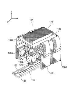
Fig. 4 shows an exemplary arrangement of four microfluorometers in a
single read head 150. The four microfluorometers are arranged in a staggered
layout
with respect to the channels 161 and 162 of a flow cell 160. In the
arrangement
shown, two of the microfluorometers (corresponding to detectors 108a and 108c)
are
configured to image separate regions of a first channel 161 and the other two
microfluorometers (corresponding to detectors 108b and 108d) are configured to
image separate regions of a second channel 162. As shown, the
microfluorometers
(corresponding to detectors 108a and 108c) are staggered with respect to the
microfluorometers (corresponding to detectors 108b and 108d) in the x
dimension
such that the two pairs of microfluorometers can detect the adjacent channels
161
and 162 respectively. The microfluorometers each have an orthogonal emission
and
excitation path (as shown in Fig. 2) with the radiation sources 102 positioned
on the
CA 02867665 2014-09-17
WO 2013/151622
PCMJS2013/025963
same side of the read head, opposite the flow cell 160. Two of the detectors
108a
and 108c are positioned on a first side of the read head and the other two
detectors
108b and 108d are positioned on the opposite side, both sides being orthogonal
to
the side where the excitation sources are positioned. In the exemplary
embodiment
shown in Fig. 4 the four radiation sources are in thermal contact with a
single large
heat sink 120. A single large heat sink provides a greater degree of heat
dissipation
than many configurations that use an individual heat sink for each radiation
source.
However, if desired individual radiation sources can be thermally coupled to
individual heat sinks (see, for example, Fig. 8 and related description
below). An
advantage of the arrangement of microfluorometers shown in Fig. 4 is the
provision
of a compact read head. Similar advantages can be derived for embodiments
where
the relative positions of the excitation source and detector in each
microfluorometer
are exchanged (see, for example, Fig. 8 and related description below).
The read head 150 shown in Fig. 4 is positioned to scan in the y dimension.
They dimension is parallel to the length of the flow cell 160 such that
movement of
the read head 150 in a scanning operation will result in imaging of areas
along the
length of the flow cell 160. The detectors 108a, 108b, 108c and 108d are
positioned
on opposite sides of the read head 150, and on opposing sides of the flow cell
160,
the sides of the flow cell running along the scan direction. The orientation
of the
scan head 150 with respect to the flow cell 160 and scan direction is
exemplary.
Other orientations are also useful including for example, the orientation
shown in
Fig. 13 wherein the detectors are positioned on opposite sides of the read
head but in
a forward and backward position relative to the scan direction.
A microfluorometer, or read head having several microfluorometers, can be
positioned above a flow cell (with respect to gravity's arrow) as exemplified
for
several embodiments set forth herein. However, it is also possible to position
a
microfluorometer, or a read head, underneath a flow cell. Accordingly a flow
cell
can be transparent on the top side, bottom side or both sides with respect to
the
wavelengths of excitation and emission radiation used. Indeed, in some
embodiments it may be desirable to position microfluorometers on both sides of
a
flow cell or to position read heads on both sides of a flow cell. Other
orientations
with respect to gravity are also possible, including for example a side to
side
orientation between a flow cell and microfluorometer (or read head).
16
A microfluorometer or read head can be configured to detect the two
opposing, inner surfaces of a flow cell from a single side of the flow cell.
For
example, the microfluorometer or read head can employ an optical compensator
that
is inserted and removed to detect alternative surfaces of the flow cell,
Exemplary
methods and apparatus for detecting opposing inner surfaces of a flow cell
such as
the use of optical compensators are described in US 8,039,817. A compensator
is
optional, for example, depending upon the NA and/or optical resolution of the
apparatus.
A microfluorometer used in an apparatus or method set forth herein can
include an autofocus module. Accordingly, multiple microfluorometers that are
present in a read head can each have a dedicated autofocus module. An
exemplary
autofocus module 1600 is shown in Fig. 5. The module includes a receptacle
1602
for an objective of a microfluorometer (for example, the translating objective
lens
shown in Fig. 3). The receptacle 1602 is affixed to a sliding support 1603
having a
lever arm 1604. The lever arm 1604 interacts functionally with a motor 1610
that is
configured to move the lever arm up and down (along the 7 direction). As such
the
motor 1610 actuates movement of the objective in the z direction to alter
focus. The
motor 1610 is a linear actuator using a lead screw. Rotation of an internal
lead screw
under rotational force of the motor causes lead nut 1613, through which the
lead
screw is threaded, to move up and down. Lead nut 1613 is positioned between
two
bearings 1611a and 1611b. Movement of the lead nut is biased against spring
1608.
The lead nut 1613 is in physical contact with the lever arm 1604 such that the
up and
down movement of the lead nut actuates the up and down movement of the sliding
support 1603 and consequently the objective. A sensor 1609 is located on the
lower
side of the autofocus module separated from the actuator by a spacer 1612.
The autofocus module 1600 shown in Fig. 5 further includes a structural
support having a side body 1607 connected to a back plane 1614 and connected
to a
top flexure 1606 and a bottom flexure 1605. Rigidity can be provided by the
box
frame structure of the side body 1607. Further rigidity is provided by two
triangle
supports 1615a and 1615b between the side body 1607 and back plane 1614. The
flexures 1606 and 1605 can be co-molded with the sliding support to provide
high
tolerance between sliding support 1603 and side body 1607.
17
CA 2867665 2019-06-18
CA 02867665 2014-09-17
WO 2013/151622
PCMJS2013/025963
As shown by the exemplary embodiment of Fig. 5, an autofocus module that
is used in a microfluorometer can include a detector and an actuator, wherein
the
actuator is configured to alter the focus of the microfluorometer with respect
to the
common plane, and wherein the detector is configured to direct movement of the
actuator. As such an autofocus module can include a dedicated detector that
directs
movement of the actuator. The dedicated detector can operate in a closed loop
with
the actuator without a need to communicate data outside of the
microfluorometer or
outside of the detection head in order to achieve automatic focusing.
Alternatively
or additionally, a detector outside of the autofocus module, such as the
imaging
detector that is used for wide-field imaging, can direct movement of the
actuator.
Thus, the same detector that is used for wide-field imaging and for outputting
image
data to a processing unit outside of the microfluorometer or read head can
also be
used to achieve automatic focusing.
In particular embodiments, autofocus modules for two or more
microfluorometers in a read head can be configured to communicate with each
other. For example, an autofocus module for a first microfluorometer of a read
head
can be configured to integrate data from an autofocus module for a second
microfluorometer of the apparatus. In this way the autofocus module for the
first
microfluorometer can alter the focus of the first microfluorometer based on
the
perceived focus position of the first microfluorometer and the perceived focus
position of the second microfluorometer. Thus, a detector for an autofocus
module
can be configured in a way that it is dedicated to focusing generally across a
read
head while not being configured for analytical image acquisition. Information
from
two different autofocus modules can be useful in determining tip-tilt of the
read
head. Undesirable tip-tilt can be corrected by compensatory actuation of one
or
more microfluorometers based on the tip-tilt information.
Although automatic focusing has been exemplified with respect to a lead
screw motor, it will be understood that autofocus modules using other
actuation
modalities can be used including for example, those that use a piezo motor or
voice
coil motor in place of the lead screw motor exemplified above.
A read head can include two or more microfluorometers, for example,
attached to a carriage. For embodiments that utilize a multichannel flow cell,
the
read head can include a number of microfluorometers that correspond to the
number
18
CA 02867665 2014-09-17
WO 2013/151622
PCMJS2013/025963
of channels in the flow cell. As demonstrated previously by the example of
Fig. 4,
more than one microfluorometer per flow cell channel can be present. In
particular
embodiments, a read head can provide a single microfluorometer per flow
channel.
In the exemplary arrangement shown in Fig. 6, the flow cell has four channels
and
the read head has four microfluorometers. The figure shows a top plan view of
the
flow cell and objectives of the microfluorometers. For ease of demonstration
components of the microfluorometers other than the objectives are not shown;
however, those components can be positioned to achieve a compact design, for
example, along the lines exemplified elsewhere herein. As shown in panel A of
Fig.
6, the four objectives can be arranged in a linear relationship such that the
objectives
are closely packed and an imaginary straight line passes through the center
point of
each objective. The imaginary line can be offset at an angle with respect to
the y
dimension, the y dimension corresponding to the longest dimension of the flow
cell
(or direction of scan). The angle can be between 00 and 900 in the x-y
quadrant and
can be selected to accommodate the spacing of the channels in the flow cell
(and the
spacing of the objectives in the read head). Fig. 6A shows a relatively low
angle of
offset for a line passing through closely packed objectives which accommodates
relatively closely packed channels. A higher angle of offset can be used to
accommodate channels that are separated by greater distances from each other
or
objectives that are less closely packed.
Panel B of Fig. 6 shows an arrangement of multiple objectives in two lines.
Here the flow cell includes eight channels and the read head has eight
microfluorometers. The overall packing of the objectives in the two lines is
approximately rectilinear. The arrangement accommodates closely packed
objectives and two sets of closely packed channels (i.e. a first set of four
closely
packed channels and a second set of four closely packed channels). In this
example,
the two sets of closely packed channels are separated by a larger spacing than
the
spacing that separates individual channels in each set of four. It will be
understood
that the overall packing of the objectives in the two lines can be offset from
rectilinear to accommodate different channel arrangements. Furthermore, as set
forth in regard to a single line of objectives, the offset angle of the
imaginary line
running through the centers of both lines of objectives can be altered and/or
the
19
CA 02867665 2014-09-17
WO 2013/151622
PCMJS2013/025963
distance between objectives can be altered to accommodate different channel
arrangements.
Fig. 7 demonstrates an arrangement of multiple objectives in which an
imaginary line running through the centers of the objectives is at a 90 angle
with
respect to the longest dimension of the flow cell (or direction of scan). The
imaginary line runs along the x axis. In this example, the objectives are in
two rows
and they are hexagonally packed. Hexagonal packing provides the advantage of
maximum compaction in the x-y plane. The read head is shown with six
objectives
and the flow cell has six channels. It will be understood that similar
arrangements
can be used for a read head having only four objectives or for read heads
having
more than six objectives (e.g. eight objectives as shown in Fig. 8, Fig. 9,
and Fig.
13). As evident by visual comparison, the flow cell channels are spaced
further
apart in the arrangement of Fig. 7 than in the arrangement of Fig. 6. However,
the
spacing of channels in both cases is within a useful and convenient range, for
example, for nucleic acid sequencing applications.
As demonstrated by the examples of Fig. 6 and Fig. 7, each objective in a
read head can be positioned to image at least a portion of an individual flow
channel.
Each objective can be positioned to image one and only one channel of a flow
cell
having several channels. An individual objective can be positioned to image a
portion of one and only one channel, for example, when located at a particular
y-
stage position. Scanning in they dimension can allow all or part of the
channel to be
imaged through the objective. In some cases, for example when the field
diameter
of the objective (or other limiting optical components of a microfluorometer)
is less
than the width of the channel, the objective can also be scanned in the x
dimension
to image all or part of the channel. Multiple objectives and their respective
microfluorometers can be positioned such that several of the objectives are
positioned to each obtain images for at least a portion of one and only one
channel.
Of course movement of a read head containing the multiple objectives and their
respective microfluorometers can occur in the y and/or x direction to image
all or
part of each respective channel. These particular configurations are useful
for
multichannel flow cells as exemplified above. However, it will be understood
that
the configurations and underlying principles set forth above can be applied to
an
appropriate arrangement of several individual flow cells, each having only a
single
CA 02867665 2014-09-17
WO 2013/151622
PCMJS2013/025963
channel. Furthermore, as is the case generally for the methods and apparatus
set
forth herein, the arrangements can be applied to substrates other than flow
cells.
A perspective view of a read head 1000 having an arrangement of eight
microfluorometers is shown in Fig. 8. Each microfluorometer has a compact
design
similar to that shown in Fig. 3. For ease of demonstration the components of
only
one of the microfluorometers are labeled in Fig. 8 and will be described here.
However, as visible in Fig. 8, each of the microfluorometers has similar
components
and configuration. Two excitation sources are present in each
microfluorometer,
including a green LED 1040 and a red LED 1030. Excitation light from the LEDs
passes through a green LED collector lens 1075 and red LED collector lens
1076,
respectively. An LED fold mirror 1074 reflects the green excitation radiation
to a
combiner dichroic 1073 which reflects the green excitation radiation through a
laser
diode beam splitter 1072, then through an excitation projection lens 1071 to
an
excitation/emission dichroic 1060 which reflects the green excitation
radiation
through an objective 1010. The red excitation radiation passes from the red
LED
collector lens 1076 to the combiner dichroic 1073 after which the red
excitation
radiation follows the same path as the green excitation radiation. The
objective
1010 is positioned to collect emission radiation and direct it through
excitation/emission dichroic 1060 which passes the emission radiation to the
CMOS
image sensor 1080. A laser diode 1301 is positioned to direct radiation via a
laser
diode coupling lens group 1401 to laser diode beam splitter 1072 which
reflects the
laser diode radiation through the excitation projection lens 1071, the
excitation/emission dichroic 1060, and the objective 1010. An autofocus module
1600 is coupled to at least part of the objective 1010 and configured to
translate the
objective 1010 up and down (i.e. along the 7 dimension). The autofocus module
can
but need not include components of the autofocus apparatus exemplified in Fig.
5. It
will be understood that additional optical components can be present in read
head
1000 including, but not limited to those exemplified for Fig B. Furthermore,
certain
optical components can be absent from read head 1000 or modified in read head
1000 to suit particular applications. Printed circuit boards 1701 and 1702 can
be
configured to communicate with the detectors, autofocus modules and/or
excitation
sources.
21
CA 02867665 2014-09-17
WO 2013/151622
PCMJS2013/025963
Fig. 9 shows a bottom plan view of the read head 1000. Again for ease of
demonstration, the components of only one of the microfluorometers are labeled
in
Fig. 9 and described herein. The red LED 1030 is shown in thermal
communication
with heat sink 1201 and in optical alignment with the red LED collector lens
1076.
The green LED is obscured by the red LED 1030 and most of the excitation path
is
obscured by the autofocus module 1600 in this view. The objective 1010 is
visible
as is a portion of the CMOS image sensor 1080; however, most of the emission
path
is obscured in this view. As is evident from the figures, the objectives are
arranged
in two rows and hexagonally packed.
The configurations described above exemplify a read head wherein each of
the microfluorometers includes at least one radiation source, a beam splitter
and a
detector, wherein the beam splitter is positioned to direct excitation
radiation from
the excitation radiation source to the objective and to direct emission
radiation from
the objective to the detector, wherein the excitation radiation and emission
radiation
are directed in mutually orthogonal directions. In the embodiments,
exemplified in
Fig. 8 and Fig. 9, the detectors for several microfluorometers are arranged on
a first
side of the read head that is opposite the common plane to which the
objectives are
focused, a subset of the radiation sources is arranged on a second side of the
read
head (the second side being orthogonal to the first side and orthogonal to the
common plane) and a second subset of the radiation sources is arranged on a
third
side of the read head (the third side being opposite the second side,
orthogonal to the
first side and orthogonal to the common plane). Alternatively, and as
exemplified in
Fig. 4., the radiation sources for several microfluorometers are arranged on a
first
side of the read head that is opposite the common plane to which the
objectives are
focused, a first subset of the detectors is arranged on a second side of the
read head
(the second side being orthogonal to the first side and orthogonal to the
common
plane) and second subset of the detectors is arranged on a third side of the
carriage
(the third side being opposite the second side, orthogonal to the first side
and
orthogonal to the common plane).
In addition to the embodiments above wherein the excitation and emission
paths are orthogonal, configurations where emission and excitation paths are
parallel
can also be useful. In this case, the excitation radiation source(s) and
detector can
be present on the same side of the read head. An exemplary layout for a
22
CA 02867665 2014-09-17
WO 2013/151622
PCT/1JS2013/025963
microfluorometer 800 is shown in Fig. 10, where excitation radiation from
excitation source 805 passes through excitation optics 806 to prism surface
807
which reflects the excitation radiation to prism surface 802 which reflects
the
excitation radiation through objective 801. Emission passes through objective
801,
then through beam splitter 802 to projection lens 803 and then to detector
804. The
emission path is parallel to much of the excitation path. The detector and
excitation
radiation source are located on the same side of the microfluorometer,
opposite and
parallel to the detection plane. A guide 810 is configured to interface with a
flow
cell or substrate to align the objective. A similar guide can be used in other
microfluorometers set forth herein. The layout for microfluorometer 800 is
exemplary for purposes of demonstrating a parallel arrangement of excitation
and
emission paths. Other components can be included such as those shown in other
figures herein including, but not limited to an autofocus module. For example,
an
excitation source 809 for an autofocus module is shown and produces excitation
that
passes through prism surface 807 and is reflected by prism surface 802 to pass
through objective 801. Several microfluorometers 800 can be arranged to have
objectives in one or more lines as exemplified in Fig. 6 and Fig. 7.
As demonstrated by the exemplary embodiments above, a read head can
include a plurality of objectives, each objective being dedicated to a single
microfluorometer. Thus, a microfluorometer of the present disclosure can
include a
variety of optical components, such as one or more detectors, excitation
radiation
sources, beam splitters lenses, mirrors, or the like, that form an optical
train that
directs excitation radiation through a single objective and/or that receives
emission
radiation through a single objective. In such embodiments, the objective can
be
configured as a macro-lens having a wide field of view. In alternative
embodiments,
a microfluorometer of the present disclosure can include a variety of optical
components that directs excitation radiation through several objectives and/or
that
receives emission radiation through several objectives. Thus, an individual
microfluorometer can include several optical trains that include several
objectives.
In embodiments that include several objectives per microfluorometer, the
objectives
can optionally be configured as an array of micro-lenses. Each objective among
several in a particular microfluorometer (e.g. each micro-lens in an array of
micro-
lenses) can optionally be configured for independent focusing, whereby each
23
CA 02867665 2014-09-17
WO 2013/151622
PCMJS2013/025963
objective can be moved in the z dimension independent of other objectives in
the
same microfluorometer. Alternatively or additionally, the several objectives
can be
configured for global focus such that they can all be moved together in the z
dimension.
It will be understood that the various components of a read head that arc set
forth herein can be mixed and matched in various ways to achieve similar
function
to those exemplified herein. For example, as set forth in the previous
paragraph, a
read head can include several objectives and each of those objectives can be
dedicated to a single microfluorometer or, alternatively, several of those
objectives
can be shared by a single microfluorometer. Similarly, and as set forth
previously
herein, each microfluorometer can include at least one dedicated excitation
source
or, alternatively, two or more microfluorometers can receive excitation
radiation
from a shared radiation source. Thus, there need not be a one to one
correspondence
between the number of microfluorometers in a particular read head and the
number
of components exemplified herein for any microfluorometer embodiment. Instead,
one or more of the components exemplified herein as being useful in a
microfluorometer can be shared by several microfluorometers in a particular
read
head.
A read head of the present disclosure is particularly useful for scanning
methods and apparatus, for example, due to its relatively compact size and low
mass
which provides low inertia. Reduced inertia allows the read head to come to
rest
more quickly following movement, thereby allowing high resolution images to be
obtained more rapidly than would be the case for a higher inertia read head
for
which residual movement of the read head would cause blurring and loss of
resolution. Configurations for achieving movement of the read head will be set
forth
in further detail below. However, first it should be noted that the advantage
of low
inertia, is not intended to be a limitation or requirement for an apparatus or
method
set forth herein. Rather, a read head of the present disclosure can be
maintained in a
static position for all or part of a detection protocol. For example, a
sequencing
method, such as those using the fluidic and imaging steps set forth herein,
can be
carried out using a read head that is static during at least one and perhaps
all of the
cycles of the sequencing method.
24
CA 02867665 2014-09-17
WO 2013/151622
PCMJS2013/025963
As a first example of a static read head embodiment, a read head can include
a sufficient number of microfluorometers to detect or image a desired portion
of a
surface or other object. Thus, the read head need not move in the x or y
dimensions.
For example, several microfluorometers can be linearly arranged to capture
image
frames along the full length (or at least along the effective target length)
of a flow
cell channel. Similarly, using an appropriate packing arrangement of several
rows of
microfluorometers, such as those set forth herein, several flow cell channels
(present
in one or more flow cell) can be imaged along their full length (or at least
along the
effective target length). As set forth previously herein, the image frames
obtained
for an individual channel can be, but need not be, contiguous.
As a second example of a static read head embodiment, a read head can
remain at a fixed position with respect to the x and y dimensions while a
substrate
that is being detected by the read head is translated in the x and or y
dimension. For
example, an apparatus can be provided having a translation stage that is
configured
to present a substrate to the read head. The translation stage can move in a
step-and-
shoot or continuous motion to allow scanning of the substrate by the static
read
head. In particular embodiments, the substrate is a flow cell that can be
affixed to
the translation stage. The flow cell can be translated as part of a fluidic
cartridge,
such as those exemplified below, or the flow cell can be translated
independently of
any fluidic cartridge. Thus, the translation stage may be configured to affix
a fluidic
cartridge to which a flow cell is attached and to move the fluidic cartridge
along
with the flow cell or the translation stage can be configured to move only the
flow
cell while the fluidic cartridge remains in a static or fixed position.
In accordance with the above examples, relative motion between a scan head
(or microfluorometer) and a substrate can be achieved by physical movement of
the
scan head (or microfluorometer), physical movement of the substrate, or
physical
movement of both. It will be understood that the static read heads referred to
in the
first and second exemplary embodiments above need not be static with respect
to
movement in the z dimension. Rather the static read heads can include one or
more
microfluorometers having autofocus modules. Alternatively or additionally, the
read heads can be moved as a whole in the z dimension, for example, to achieve
global focus at least to a rough approximation.
CA 02867665 2014-09-17
WO 2013/151622
PCMJS2013/025963
Returning now to embodiments wherein a read head is translated, Fig. 11
and Fig. 12 show top and bottom views, respectively, of an exemplary y
translation
stage 200 for a read head. In this exemplary embodiment, the y stage is
configured
for translation in the y dimension but not in the x dimension. Thus, a read
head
carried by y translation stage 200 will be capable of movement in the y
dimension
and the read head or individual microfluorometers therein may be capable of
movement in the z dimension (e.g. via autofocusing), but the read head will
not be
capable of movement in the x dimension. A read head can be affixed to carriage
201
having a base area 241 positioned to support the bottom side of the read head
and a
frame 240 configured to restrain the read head from side to side motion. The
carriage 201 further includes a flange guide 243 and a collar guide 242. An
opening
244 in base area 241 provides a window between a read head and substrate to be
detected by the read head. The aforementioned components of the carriage 201
can
form a monolithic structure.
The carriage is configured to move along a y stage frame 207 via a first shaft
203, along which the collar guide 242 runs and a second shaft 204 along which
the
flange guide 243 runs. The shafts are oriented along the y axis such that the
carriage
201 is directed to slide back and forth along the y dimension via the guides.
The
first shaft 203 is held to the y stage frame 207 by insertion into datum 215
in a first
side wall 250 and into datum 218 in a second sidewall 251. The first shaft 203
is
clamped into datum 215 by support member 252 and clamped into datum 218 by
support member 253. The second shaft 204 is held to the y stage frame 207 by
insertion into datum 214 in a first side wall 250 and into datum 217 in a
second
sidewall 251. The first shaft 204 is clamped into datum 214 by shaft clamp 206
and
clamped into datum 217 by shaft clamp 205.
Movement of carriage 201 is driven by rotation of lead screw 202 which is
threaded through a lead nut 260 and which is affixed to the y stage frame 207
by
insertion into a datum on the first side wall 250 and into a datum 219 in the
second
sidewall 251. The lead screw 202 is clamped in place by the same support
members
252 and 253 that clamp the first shaft 203. The rotation of lead screw 202 is
driven
by motor 212 which is mounted to support member 252. An encoder 208 is
configured to interact with the motor 212 via a belt 210 that interacts with
rotor 209
26
CA 02867665 2014-09-17
WO 2013/151622
PCMJS2013/025963
on the encoder and rotor 211 on the motor 212. A belt tensioner 220 interacts
with
the belt 210.
An opening 230 passes through the floor 216 of y stage frame 207. The
opening 230 is positioned to accommodate the trajectory of opening 244 in the
base
area 241 of the carriage 201 as it traverses the y stage frame. A read head is
positioned in the carriage such that the objectives are directed through
opening 244
and through opening 230 along a trajectory traversed by the carriage.
Accordingly,
the opening 230 accommodates imaging of an elongated area along the y axis via
movement of a read head affixed to the carriage.
The structural and functional relationship between y translation stage 200
and read head 1000 is shown in Fig. 13. The orientation of the objectives 1010
with respect to the scanning direction of y translation stage 200 is similar
to that
exemplified in Fig. 7 (except that read head 1000 has an additional two
objectives).
A flow cell can be oriented with respect to y translation stage 200 as shown
in Fig.
7.
As exemplified above a carriage can be configured to move a read head, for
example, in a scanning operation. Alternatively or additionally, a carriage
can be
configured to prevent relative movement between individual microfluorometers
of a
read head in the x and y dimensions. A carriage need not provide this
function, for
example if the read head includes other structure elements that prevent
relative
transverse motion between individual microfluorometers, For example, a read
head
may be formed from a co-molded assembly. The co-molded assembly can in turn be
affixed to a carriage. Nevertheless, in some embodiments, the carriage may
play at
least an auxiliary role in preventing relative transverse motion between
individual
microfluorometers of a read head. Furthermore it will be understood that a
read
head that is formed from a co-molded assembly can be used for embodiments that
do not employ a carriage.
A y stage that is used in a method or apparatus set forth herein can be
configured to scan via a discontinuous or continuous motion. Discontinuous
scanning, often referred to as step-and-shoot scanning, generally involves
incremental movement of a microfluorometer or scan head in the y (or x)
direction
and detection (e.g. image acquisition) between movements, while the
microfluorometer or scan head is in a temporarily static state. Continuous
scanning
27
on the other hand generally involves detection or image acquisition while the
microfluorometer or scan head is moving. In a particular embodiment continuous
scanning can be carried out in time delay integration (TDI) mode. Accordingly,
signal obtained by pixel elements along the scan dimension can be collected in
a
common bin and read out as a single value. TDI mode can provide advantages of
increased signal processing rate and increased accuracy. Exemplary optical
arrangements that can be included in a microfluorometer or read head to
accommodate TDI mode detection are described, for example, in US 7,329,860.
Movement of a microfluorometer or scan head in an x or y dimension, for
example to accommodate continuous or discontinuous scanning modalities, can be
controlled by an encoder or other device. In the example of y-stage 200, the
motion
can be controlled by encoder 208. As set forth previously herein, scanning
(whether
by continuous or discontinuous techniques) can result in acquisition of
contiguous or
non-contiguous frames from a substrate or other object under detection. Thus,
the
sum total of portions that are imaged by scanning can be contiguous (but not
overlapping), non contiguous, or overlapping. The system need not be
configured to
obtain images of the entire substrate or object (e.g. array surface) and need
not do so
in a way that allows a composite image to be produced.
An electrical block diagram for a detection apparatus is shown in Fig. 14. A
readout printed circuit board (PCB) is present in a read head (see, for
example, PCB
1701 and 1702 in Fig. 8) and is connected to a main PCB that is typically
contained
within the detection apparatus housing. In alternative embodiments the main
PCB
can be located exterior to the instrument. Data can be communicated between
the
readout PCB and main PCB via the LVDS line. For example, a 0.5mm-pitch, 36-
cond flat flex cable (FFC) can be used for the LVDS line. The LVDS line can be
configured to communicate image data from the readout PCB to the main PCB, and
instructions for camera control from the main PCB to the readout PCB. The two
PCBs are also connected by a power line such as a copper-backed lmm-pitch 30-
cond FFC that transmits power from a 24 volt power supply via the main PCB.
The
FFC connections are configured to have sufficient length and flexibility to
allow the
readout PCB to move with the read head while the main PCB remains stationary.
28
CA 2867665 2019-06-18
CA 02867665 2014-09-17
WO 2013/151622
PCMJS2013/025963
In the example of Fig. 14, the main PCB is also connected to an exterior
primary analysis personal computer (PC) via USB 3.0 SS I/F connectors. In some
embodiments the primary analysis computer can be located within the housing of
the
detection apparatus. However, placing the primary analysis computer off-
instrument allows for interchangeable use of a variety of computers to be used
for
different applications, convenient maintenance of the primary analysis
computer by
replacement without having to interrupt the activity of the detection
apparatus and
small footprint for the detection apparatus. Any of a variety of computers,
can be
used including, for example, a desktop computer, laptop computer, or server
containing a processor in operational communication with accessible memory and
instructions for implementation of the computer implemented methods described
herein. The main PCB is also connected to a liquid crystal display (LCD) for
communication to a human user. Other user interfaces can be used as well.
In some embodiments, a user interface may include a display (e.g. an LCD)
to display or request information from a user and a user input device (e.g. a
keyboard) to receive user inputs. In some embodiments, the display and the
user
input device are the same device. For example, the user interface may include
a
touch-sensitive display configured to detect the presence of an individual's
touch and
also identify a location of the touch on the display. However, other user
input
devices may be used, such as a mouse, touchpad, keyboard, keypad, handheld
scanner, voice-recognition system, motion-recognition system, and the like.
The readout PCB includes eight DS9OCR217 transmitters for transferring
data from individual sensors (i.e. detectors) to the LVDS line, 3.3 volt
switching
regulator, a 5 volt switching regulator, and LED buck drives for the LED
excitation
radiation sources.
The main PCB includes an FPGA + processor configured to accept image
data from the LVDS. A DDR3 DIMM frame buffer is electronically connected to
the FPGA + processor. The main PCB also includes a thermal control regulator
and
control circuitry for various drive motors such as a y-axis motor, cartridge
motor,
valve motor, and pump motor.
This disclosure further provides a method of imaging a substrate, including
the steps of (a) providing a substrate including fluorescent features on a
surface; (b)
acquiring a plurality of wide-field images of a first portion of the surface
using a
29
CA 02867665 2014-09-17
WO 2013/151622
PCMJS2013/025963
plurality of microfluorometers, wherein each of the microfluorometers acquires
a
wide-field image from a different location of the surface, wherein the
plurality of
microfluorometers are affixed to a carriage; and (c) translating the carriage
in a
direction parallel to the surface and repeating (b) for a second portion of
the surface.
The method can use any of the apparatus set forth elsewhere herein, but need
not be
so limited in all embodiments.
Embodiments of the present methods find particular use for nucleic acid
sequencing techniques. For example, sequencing-by-synthesis (SBS) protocols
are
particularly applicable. In SBS, extension of a nucleic acid primer along a
nucleic
acid template is monitored to determine the sequence of nucleotides in the
template.
The underlying chemical process can be polymerization (e.g. as catalyzed by a
polymerase enzyme) or ligation (e.g. catalyzed by a ligase enzyme). In a
particular
polymerase-based SBS embodiment, fluorescently labeled nucleotides are added
to a
primer (thereby extending the primer) in a template dependent fashion such
that
detection of the order and type of nucleotides added to the primer can be used
to
determine the sequence of the template. A plurality of different templates can
be
subjected to an SBS technique on a surface under conditions where events
occurring
for different templates can be distinguished. For example, the templates can
be
present on the surface of an array such that the different templates are
spatially
distinguishable from each other. Typically the templates occur at features
each
having multiple copies of the same template (sometimes called "clusters" or
"colonies"). However, it is also possible to perform SBS on arrays where each
feature has a single template molecule present, such that single template
molecules
are resolvable one from the other (sometimes called "single molecule arrays").
Flow cells provide a convenient substrate for housing an array of nucleic
acids. Flow cells are convenient for sequencing techniques because the
techniques
typically involve repeated delivery of reagents in cycles. For example, to
initiate a
first SBS cycle, one or more labeled nucleotides, DNA polymerase, etc., can be
flowed into/through a flow cell that houses an array of nucleic acid
templates.
Those features where primer extension causes a labeled nucleotide to be
incorporated can be detected, for example, using methods or apparatus set
forth
herein. Optionally, the nucleotides can further include a reversible
termination
property that terminates further primer extension once a nucleotide has been
added
to a primer. For example, a nucleotide analog having a reversible terminator
moiety
can be added to a primer such that subsequent extension cannot occur until a
deblocking agent is delivered to remove the moiety. Thus, for embodiments that
use
reversible termination a deblocking reagent can be delivered to the flow cell
(before
or after detection occurs). Washes can be carried out between the various
delivery
steps. The cycle can then be repeated n times to extend the primer by n
nucleotides,
thereby detecting a sequence of length n. Exemplary sequencing techniques are
described, for example, in Bentley et al., Nature 456:53-59 (2008), WO
04/018497:
US 7,057,026; WO 91/06678; WO 07/123744; US 7,329.492: US 7,211,414; US
7,315,019; US 7.405,281, and US 2008/0108082.
For the nucleotide delivery step of an SBS cycle, either a single type of
nucleotide can be delivered at a time, or multiple different nucleotide types
(e.g. A,
C, T and G together) can be delivered. For a nucleotide delivery configuration
where
only a single type of nucleotide is present at a time, the different
nucleotides need
not have distinct labels since they can be distinguished based on temporal
separation
inherent in the individualized delivery. Accordingly, a sequencing method or
apparatus can use single color detection. For example, microfluorometer or
read
head need only provide excitation at a single wavelength or in a single range
of
wavelengths. Thus, a microfluorometer or read head need only have a single
excitation source and multiband filtration of excitation need not be
necessary. For a
nucleotide delivery configuration where delivery results in multiple different
nucleotides being present in the flow cell at one time, features that
incorporate
different nucleotide types can be distinguished based on different fluorescent
labels
that are attached to respective nucleotide types in the mixture. For example,
four
different nucleotides can be used, each having one of four different
fluorophores. In
one embodiment the four different fluorophores can be distinguished using
excitation in four different regions of the spectrum. For example, a
microfluorometer or read head can include four different excitation radiation
sources. Alternatively a read head can include fewer than four different
excitation
radiation sources but can utilize optical filtration of the excitation
radiation from a
single source to produce different ranges of excitation radiation at the flow
cell.
31
CA 2867665 2019-06-18
CA 02867665 2014-09-17
WO 2013/151622
PCT/1JS2013/025963
In some embodiments, four different nucleotides can be detected in a sample
(e.g. array of nucleic acid features) using fewer than four different colors.
As a first
example, a pair of nucleotide types can be detected at the same wavelength,
but
distinguished based on a difference in intensity for one member of the pair
compared to the other, or based on a change to one member of the pair (e.g.
via
chemical modification, photochemical modification or physical modification)
that
causes apparent signal to appear or disappear compared to the signal detected
for the
other member of the pair. As a second example, three of four different
nucleotide
types can be detectable under particular conditions while a fourth nucleotides
type
lacks a label that is detectable under those conditions. In an SBS embodiment
of the
second example, incorporation of the first three nucleotide types into a
nucleic acid
can be determined based on the presence of their respective signals, and
incorporation of the fourth nucleotide type into the nucleic acid can be
determined
based on absence of any signal. As a third example, one nucleotide type can be
detected in two different images or in two different channels (e.g. a mix of
two
species having the same base but different labels can be used, or a single
species
having two labels can be used or a single species having a label that is
detected in
both channels can be used), whereas other nucleotide types are detected in no
more
than one of the images or channels. In this third example, comparison of the
two
images or two channels serves to distinguish the different nucleotide types.
The three exemplary configurations in the above paragraph are not mutually
exclusive and can be used in various combinations. An exemplary embodiment is
an
SBS method that uses reversibly blocked nucleotides (rbNTPs) having
fluorescent
labels. In this format, four different nucleotide types can be delivered to an
array of
nucleic acid features that are to be sequenced and due to the reversible
blocking
groups one and only one incorporation event will occur at each feature. The
nucleotides delivered to the array in this example can include a first
nucleotide type
that is detected in a first channel (e.g. rbATP having a label that is
detected in the
first channel when excited by a first excitation wavelength), a second
nucleotide
type that is detected in a second channel (e.g. rbCTP having a label that is
detected
in the second channel when excited by a second excitation wavelength), a third
nucleotide type that is detected in both the first and the second channel
(e.g. rbTTP
having at least one label that is detected in both channels when excited by
the first
32
and/or second excitation wavelength) and a fourth nucleotide type that lacks a
label
that is detected in either channel (e.g. rbGTP having no extrinsic label).
Once the four nucleotide types have been contacted with the array in the
above example, a detection procedure can be carried out, for example, to
capture
two images of the array. The images can be obtained in separate channels and
can
be obtained either simultaneously or sequentially. A first image obtained
using the
first excitation wavelength and emission in the first channel will show
features that
incorporated the first and/or third nucleotide type (e.g. A and/or T). A
second image
obtained using the second excitation wavelength and emission in the second
channel
will show features that incorporated the second and/or third nucleotide type
(e.g. C
and/or T). Unambiguous identification of the nucleotide type incorporated at
each
feature can be determined by comparing the two images to arrive at the
following:
features that show up only in the first channel incorporated the first
nucleotide type
(e.g. A), features that show up only in the second channel incorporated the
second
nucleotide type (e.g. C). features that show up in both channel incorporated
the third
nucleotide type (e.g. T) and features that don't show up in either channel
incorporated the fourth nucleotide type (e.g. G). Note that the location of
the
features that incorporated G in this example can be determined from other
cycles
(where at least one of the other three nucleotide types is incorporated).
Exemplary
apparatus and methods for distinguishing four different nucleotides using
detection
of fewer than four colors are described for example in US Pat. App. Ser. No.
61/538,294.
In a sequencing method, a microfluorometer can acquire at least two wide-
field images of the same area of a surface during each cycle, wherein each of
the at
least two wide-field images is acquired using different excitation or emission
wavelengths. For example, during each cycle a microfluorometer can acquire
two,
three or four wide-field images of the same area of a surface during each
cycle,
wherein each of the two wide-field images detect fluorescence at different
regions of
the spectrum. Alternatively or additionally, a microfluorometer may acquire
wide-
field images that detect fluorescence at no more than two, three or four
different
regions of the spectrum for a given area of a surface during a given
sequencing
cycle. For example, a microfluorometer may excite an area of a flow cell
surface
with radiation in no more than two, three or four different regions of the
spectrum
33
CA 2867665 2019-06-18
during a given cycle and/or a microfluorometer may acquire wide-field images
from
a given area of a surface in no more than two, three or four different regions
of the
spectrum during a given cycle. Different wide-field images can be obtained at
different times (e.g. sequentially) or in some embodiments two or more wide-
field
images can be obtained simultaneously.
In the context of the present disclosure "different wide-field images of an
area" refers to two or more wide-field images of the same area that are
acquired
under different excitation and/or emission conditions. Alternatively, two or
more
separate wide-field images of the same area can be acquired under the same or
at
least similar excitation and emission conditions. For example, multiple frames
can
be obtained for an area of a given object under a given fluorescence detection
condition and the frames can be co-added. Co-adding can provide the advantage
of
increasing signal to noise as compared to obtaining a single frame under the
same
conditions. A further example is that co-adding can be performed in
conjunction
with pulsed excitation to reduce photo-damage to the sample as compared to
continuous excitation of the sample over a prolonged period of time (that may
or
may not achieve similar signal intensity or signal to noise ratio).
In some embodiments, nucleic acids can be attached to a surface and
amplified prior to or during sequencing. For example, amplification can be
carried
out using bridge amplification to form nucleic acid clusters on a surface.
Useful
bridge amplification methods are described, for example, in US 5,641,658; US
2002/0055100; US 7,115,400; US 2004/0096853; US 2004/0002090; US
2007/0128624; or US 2008/0009420. Another useful method for amplifying nucleic
acids on a surface is rolling circle amplification (RCA), for example, as
described in
Lizardi et al., Nat. Genet. 19:225-232 (1998) and US 2007/0099208 Al. Emulsion
PCR on beads can also be used, for example as described in Dressman et al.,
Proc.
Natl. Acad. Sci. USA 100:8817-8822 (2003), WO 05/010145, US 2005/0130173 or
US 2005/0064460.
As set forth above, sequencing embodiments are an example of a repetitive
process. The methods of the present disclosure are well suited to repetitive
processes. Some embodiments are set forth below.
34
CA 2867665 2019-06-18
CA 02867665 2014-09-17
WO 2013/151622
PCMJS2013/025963
This disclosure provides a method of imaging a substrate, including the steps
of (a) providing a substrate including fluorescent features on a surface; (b)
acquiring
a plurality of wide-field images of a first portion of the surface using a
plurality of
microfluorometers, wherein each of the microfluorometers acquires a wide-field
image from a different location of the surface, wherein the plurality of
microfluorometers are affixed to a carriage; (c) translating the carriage in a
direction
parallel to the surface and repeating (b) for a second portion of the surface;
and (d)
returning the carriage to a position to acquire a second plurality of wide-
field images
of the first portion of the surface. Optionally, the method can further
include a step
of modifying the fluorescent features on the surface after (c) and before (d),
wherein
the second plurality of wide-field images are different from the first
plurality of
wide-field images.
In particular embodiments, steps (a) through (c) of the above method
correspond to the detection step(s) of a sequencing technique. In a related
embodiment, step (d) whereby the carriage is returned corresponds to a second
cycle
of a sequencing technique. In this example, the modification of the
fluorescent
features on the surface can include one or more of the biochemical steps of a
sequencing technique. Exemplary sequencing techniques that can be used in the
method are set forth above or otherwise known in the art.
Also provided is a fluidics cartridge that includes (a) a flow cell having an
optically transparent surface, an inlet and an outlet; and (b) a housing made
of a
material that is optically opaque and impermeable to aqueous liquids, wherein
the
housing holds: (i) a sample reservoir; (ii) a fluidic line between the sample
reservoir
and the inlet of the flow cell; (iii) a plurality of reagent reservoirs in
fluid
communication with the flow cell via the inlet of the flow cell, (iv) at least
one
valve configured to mediate fluid communication between the reservoirs and the
inlet of the flow cell; and (v) at least one pressure source configured to
move liquids
from the sample reservoir or the reagent reservoirs to the flow cell via the
inlet of
the flow cell, wherein an optically transparent window interrupts the housing
and an
inlet port interrupts the housing, wherein the inlet port is in fluid
communication
with the sample reservoir, and wherein the optically transparent surface is
positioned
in the window.
CA 02867665 2014-09-17
WO 2013/151622
PCMJS2013/025963
The exterior view of an exemplary fluidics cartridge 10 is shown in Fig. 1
and is described above. Fig. 15 shows an exploded view of a fluidic cartridge
2000.
The housing of the fluidic cartridge is formed by a shell 2001 that mates with
a base
2002. The shell is on the upper side of the fluidic cartridge as shown and
includes a
reception area 2009 for a flow cell 2020. The flow cell is exposed to the
exterior of
the housing via a window 2010. One or more ports 2013 in the shell allow
sample
or other reagents to be delivered to the reservoirs in the interior of the
fluidic
cartridge 2000. The base 2002 includes an opening 2012 that is configured to
accept
a reagent tray 2003. The reagent tray 2003 can be engaged with the fluidic
cartridge
by insertion into the opening 2012 in a way that individual reagent reservoirs
in the
tray are in fluid communication with individual fluidic lines for delivery of
reagents
to the flow cell. The housing also contains valves 2005 and 2006 that
interface with
pumps to move the reagents through the fluidic lines. Also contained within
the
housing is a waste bag 2004 having an inlet 2011 that interfaces with fluidic
lines
from the flow cell 2020.
The housing of a fluidic cartridge (e.g. the shell 2001 and/or base 2002 of
fluidic cartridge 2000) can be made of a material that is opaque to radiation
in a
particular part of the spectrum. For example, the housing may be opaque to UV,
VIS and/or IR radiation in order to protect reagents from photo-damage due to
radiation at these wavelengths. For example, a material that is opaque to UV
radiation is beneficial for avoiding photo-damage to nucleic acids among other
reagents used in sequencing reactions. As another example, it may be desirable
to
use a material that is opaque to radiation in the wavelength range absorbed by
fluorophores used as labels in a sequencing reaction.
The housing for a fluidic cartridge will typically be impermeable to the
liquids housed therein. Thus the housing can provide a secondary barrier in
addition
to the reservoirs held within. Exemplary materials include plastics such as
polycarbonate or polystyrene, or metals such as aluminum or stainless steel.
Materials that are chemically inert to the reagents housed in the fluidic
cartridge are
generally desirable. Individual reservoirs or other fluidic components in a
fluidic
cartridge will have similar properties of fluid impermeability and can
optionally be
opaque as well. The materials can be rigid or flexible. For example, any of a
36
variety of the reagent reservoirs set forth herein or otherwise used in a
fluidic
cartridge can be a flexible bag as exemplified for waste reservoir 2004.
As exemplified by Fig. 1 and Fig. 15 a fluidic cartridge can be configured to
contain a variety of components within a housing. For example, in various
embodiments one or more of the fluidic components set forth herein can be
fully
contained within the housing. Indeed in particular embodiments all of the
fluidic
components of a particular embodiment can be fully contained in the fluidic
cartridge. For example, a cartridge housing can contain one or more sample
reservoirs, one or more reagent reservoirs, one or more waste reservoirs, one
or
more mixing reservoirs, one or more valves configured to mediate fluid
communication between a reservoir and a flow cell, one or more pressure source
configured to move liquids from a reservoir to a flow cell, or one or more
fluidic
lines between a reservoir and a flow cell. However, it will be understood that
in
some embodiments at least part of some fluidic components may be present
outside
of the housing. For example, a surface of a flow cell through which detection
will
occur can be outside of a cartridge housing.
In particular embodiments, a fluidic cartridge can include a reception area
that is sized to tightly hold a flow cell, for example, by a compression fit.
However,
in other embodiments, the reception area can be larger than the footprint of a
flow
cell that is or will be present in the fluidic cartridge. Thus, the flow cell
can occupy
a reception space in the housing that is sized and shaped to permit the flow
cell to
float relative to the housing. A configuration that accommodates float of a
flow cell
can be advantageous for alignment of the flow cell with the optics component
of a
detection apparatus after the fluidic cartridge has been placed in the
instrument.
Alignment can be achieved by insertion of one or more alignment pins into the
reception area by the detection apparatus for example in cases where the pins
are
pre-aligned to a microfluorometer of a read head of the detection apparatus.
Accordingly, the reception area can include a fitting for at least one
alignment pin or
other alignment member. Exemplary configurations for aligning a floating flow
cell
that can be adapted to a fluidic cartridge of the present disclosure are
described in
US Ser. No. 13/273,666 (published as US 2012/0270305 Al). In embodiments that
utilize flow cell float, the fluidic connection from the flow cell to other
fluidic
components of the cartridge will
37
CA 2867665 2019-06-18
CA 02867665 2014-09-17
WO 2013/151622
PCMJS2013/025963
generally be flexible. For example flexible tubing can connect a flow cell to
fixed
fluidic components of a cartridge.
A fluidic cartridge of the present disclosure need not include a detection
device or other detection components described herein. For example, a fluidic
cartridge can be configured to exclude a detector, microfluorometer or read
head
such as those described herein or those useful in a method set forth herein.
In
nucleic acid sequencing embodiments, a detector, microfluorometer or read head
that is used to detect nucleic acids in a flow cell (or other substrate) of a
fluidic
cartridge can be located outside of the housing for the fluidic cartridge.
Similarly
for other embodiments, a detector, microfluorometer or read head that is used
to
detect a particular characteristic of a substrate can be excluded from the
interior of a
housing for a fluidic cartridge, being located external to the housing
instead. It will
be understood that in at least some configurations one type of detector can be
excluded from a fluidic cartridge whereas another type of detector can be
present.
For example, a fluidic cartridge can exclude a detection device used to detect
nucleic
acids in a flow cell, but can include a detector used to evaluate a
characteristic of a
fluid in the cartridge or to evaluate a component of the cartridge. More
specifically,
a cartridge can include a detector for temperature, pressure, flow rate or
other
characteristics of the fluids used in the cartridge. Other examples of
components
that can be excluded from a fluidic cartridge include, but are not limited to,
optical
filters, lenses, objectives, cameras (e.g. CCD cameras or CMOS cameras),
excitation
radiation sources (e.g. LEDs) or the like.
A fluidic map for an exemplary fluidic cartridge is shown in Fig. 16. Flow
cell 2020 has eight lanes each fluidically connected to one of eight
individual fluid
lines (collectively labeled 2047) that are individually actuated by inlet
valve 2044.
Inlet valve 2044 controls the flow of fluid from four sample reservoirs 2030
through
2033. Inlet valve 2044 also controls the flow of fluid from several SBS
reagent
reservoirs 2035 and from several amplification reagent reservoirs 2036.
Distribution
and flow of fluids from the SBS reagent reservoirs 2035 is controlled by
reagent
selection valve 2043. Distribution and flow of fluids from the amplification
reagent
reservoirs 2036 is controlled by reagent selection valve 2042 which is located
upstream of reagent selection valve 2043. Accordingly reagent selection valve
2043
38
CA 02867665 2014-09-17
WO 2013/151622
PCMJS2013/025963
is positioned to control distribution and flow of reagents from both the SBS
reagent
reservoirs 2035 and the amplification reagent reservoirs 2036.
Flow of fluids through the system of Fig. 16 is driven by eight separate
syringe pumps 2051 through 2058. The syringe pumps are positioned to pull
fluids
through the fluidic system and each pump can bc individually actuated by valve
2045. Thus, flow though each channel of the flow cell can be individually
controlled by a dedicated pressure source. Valve 2045 is also configured to
control
flow of fluids to waste reservoir 2060.
Fig. 16 exemplifies a fluidic system in which fluids are pulled by the action
of downstream syringe pumps. It will be understood that a useful fluidic
system can
use other types of devices instead of syringe pumps to drive fluids including,
for
example, positive or negative pressure, peristaltic pump, diaphragm pump,
piston
pump, gear pump or Archimedes screw. Furthermore, these and other devices can
be configured to pull fluids from a downstream position with respect to a flow
cell
or to push fluids from an upstream position.
Fig. 16 also exemplifies the use of eight syringe pumps for eight channels of
a flow cell. Thus, the fluidic system includes a number of pumps that is
equivalent
to the number of channels in use. It will be understood that a fluidic system
that is
useful in a fluidic cartridge of the present disclosure can have fewer pumps
(or other
pressure sources) than the number of channels in use. For example, several
channels
can be fluidically connected to a shared pump and a valve can be used to
actuate
fluid flow through an individual channel.
An exemplary rotary valve 400 is shown in Fig. 17. The structure and
function of rotary valve 400 can be understood in the context of a sequencing
procedure as set forth below. Of course it will be understood that the valve
can be
used in similar ways for other applications. In a sequencing protocol where
four
different samples are to be fluidically processed, rotary valve 400 can
function as a
four-sample injection rotary valve using a 45 degree pitch and can also
function as a
four to one manifold for sequencing reagents. In the top view of Fig. 17,
rotary
valve 400a is positioned to allow flow from common reagent reservoir 401 to
four
lanes of a flow cell. More specifically, in this position, fluids can flow
from
common reagents 401 through port 402 to Lane 1 (via port 411), to Lane 2 (via
port
412), to Lane 3 (via port 413), and to Lane 4 (via port 414). However, in this
39
CA 02867665 2014-09-17
WO 2013/151622
PCMJS2013/025963
position fluids do not flow from sample reservoirs Si, S2, S3 or S4 because
ports
421, 422, 423 and 424 are closed to flow from port 402. A 45 degree turn
places
rotary valve 400b to the position shown in the bottom view of Fig. 17, thereby
allowing the samples Si, S2, S3 and S4 to be injected because ports 411, 412,
413
and 414 arc open to flow from ports 421, 422, 423 and 424, respectively.
However,
in this position flow from port 402 is closed, thereby preventing flow of
common
reagents to the lanes of the flow cell.
In particular embodiments a fluidic cartridge can be configured to allow re-
use of one or more reagents. For example, the fluidic cartridge can be
configured to
deliver a reagent to a flow cell, then remove the reagent from the flow cell,
and then
re-introduce the reagent to the flow cell. In one configuration, as
exemplified in Fig.
18, cartridge fluidics can be configured such that a reagent reservoir is in
fluid
communication with the input port of a flow cell and the output port of the
flow cell
is also in fluid communication with the reagent reservoir. One or more of the
reagents can be re-used in a manifold network of similar reagent loops as
shown in
Fig. 18. For example, valve 522 controls flow from wash reservoir 524, IMX
reservoir 525, SMX reservoir 526, CLM reservoir 527 and cleave reservoir 528
to
flow cell 520. Pump 521 is downstream of flow cell 520 and upstream of valve
523.
Valve 523 controls flow from flow cell 520 to waste reservoir 535, IMX
reservoir
525, SMX reservoir 526, CLM reservoir 527 and cleave reservoir 528.
The fluidic lines connecting the above components of Fig. 18 will be
described here in the context of a sequencing cycle where reagents are
delivered
from the reservoirs to the flow cell and used reagents are delivered from the
flow
cell to the respective reservoirs. In all steps of the cycle exemplified below
fluids
are moved under force of pressure produced by pump 521. In a first step of the
cycle, the flow cell is washed by opening valve 522 to line 501 and opening
valve
523 to line 505 such that fluid flows from wash reservoir 524 to waste
reservoir 535
via a path between valve 522 and valve 523 that crosses through flow cell 520.
For
all steps described for Fig. 18, the path between valve 522 and valve 523
leads from
valve 522 to line 502, to the inlet 530 of flow cell 520, through channel 531
of flow
cell 520, through outlet 532 of flow cell 520, through line 503, to pump 521
and
then through line 504 to valve 523. In a second step of the cycle, IMX is
introduced
to the flow cell by opening valve 522 to line 506 and opening valve 523 to
line 505
CA 02867665 2014-09-17
WO 2013/151622
PCMJS2013/025963
such that fluid flows from IMX reservoir 525 to waste reservoir 535 via the
path
between valve 522 and valve 523 that crosses through flow cell 520. In a third
step
of the cycle, used IMX is moved from the flow cell 520 to IMX reservoir 525 by
opening valve 522 to line 501 and opening valve 523 to line 510 such that wash
fluid displaces IMX from the path between valve 522 and valve 523 that crosses
through flow cell 520. In a fourth step of the cycle, SMX is introduced to the
flow
cell by opening valve 522 to line 507 and opening valve 523 to line 505 such
that
fluid flows from SNP( reservoir 526 to waste reservoir 535 via the path
between
valve 522 and valve 523 that crosses through flow cell 520. In a fifth step of
the
cycle, used SMX is moved from the flow cell 520 to SMX reservoir 526 by
opening
valve 522 to line 501 and opening valve 523 to line 511 such that wash fluid
displaces SMX from the path between valve 522 and valve 523 that crosses
through
flow cell 520. Similar pairs of steps can be repeated to (1) introduce CLM
reagent
to the flow cell and to return used CLM reagent to the CLM reservoir, and (2)
to
introduce Cleave reagent to the flow cell and to return used Cleave reagent to
the
Cleave reservoir.
Another example of a fluidic configuration that provides reagent re-use is
shown in Fig. 19. In this example, fluidics for a cartridge are configured
such that
each reagent reservoir is in fluid communication with a single port of the
flow cell
620. Reciprocating flow allows each reagent to flow from a reservoir to the
flow cell
620 and from the flow cell 620 back to the reservoir, wherein ingress of
reagents to
the flow cell 620 and egress of the reagents from the flow cell 620 occur
through the
same port of the flow cell 620. Re-use for four reagents is exemplified in
Fig. 19,
however a fluidic system can be configured for more or fewer reagents to be re-
used
in a similar reciprocating format. As shown in Fig. 19, valve 622 controls
flow of
fluids between flow cell 620 and each of: wash reservoir 624, IMX reservoir
625,
SMX reservoir 626. CLM reservoir 627 and cleave reservoir 628. In a first
direction
of flow, pump 621 is configured to pull fluids from flow cell 620 via line 603
and to
push fluids to waste reservoir 635 via line 605.
The fluidic lines connecting the above components of Fig. 19 will be
described here in the context of a sequencing cycle where reagents are
delivered
from the reservoirs to the flow cell and used reagents are delivered from the
flow
cell to the respective reservoirs. In a first step of the cycle the flow cell
is washed
41
CA 02867665 2014-09-17
WO 2013/151622
PCMJS2013/025963
by opening valve 622 to line 601 such that fluid flows from wash reservoir 624
to
waste reservoir 635. The path between valve 622 and waste reservoir 635 leads
from
valve 622 to line 602 to port 630 of flow cell 620, through channel 631 of
flow cell
620, through port 632 of flow cell 620, through line 603 to pump 621 and then
through line 605 to waste reservoir 635. In a second step of the cycle, IMX is
introduced to the flow cell by opening valve 622 to line 606 such that fluid
flows
from IMX reservoir 625 through valve 622 to line 602, to port 630 of flow cell
620,
through channel 631 of flow cell 620, through port 632 of flow cell 620, and
partially through line 603 (thereby leaving residual wash solution in a
downstream
portion of line 603 through to pump 621). In a third step of the cycle, used
IMX
reagent is returned from the flow cell 620 to IMX reservoir 625 by opening
valve
622 to line 606 and reversing the direction of pump 621 such that used IMX
reagent
is returned from the flow cell 620 to the IMX reservoir 625 via port 630 of
flow cell
620, to fluid line 602, through valve 622 then through fluid line 606 to IMX
reservoir 625. During step three, flow from pump 621 to the IMX reservoir 625
occurs for sufficient time that a portion of the IMX reagent returns to the
IMX
reservoir 625, but not long enough to cause a substantial amount of the
residual
wash solution from line 603 to enter the IMX reservoir 625. In a fourth step
of the
cycle, the flow cell is washed as described for the first step. In a fifth
step of the
cycle, SMX is introduced to the flow cell by opening valve 622 to line 607
such that
fluid flows from SMX reservoir 626 through valve 622 to line 602, to port 630
of
flow cell 620, through channel 631 of flow cell 620, through port 632 of flow
cell
620, and partially through line 603 (thereby leaving residual wash solution in
a
downstream portion of line 603 through to pump 621). In a sixth step of the
cycle,
used SMX reagent is returned from the flow cell 620 to SMX reservoir 626 by
opening valve 622 to line 607 and reversing the direction of pump 621 such
that
used SMX reagent is returned from the flow cell 620 to the SMX reservoir 626
via
port 630 of flow cell 620, to fluid line 602, through valve 622 then through
fluid line
607 to SMX reservoir 626. As with step three, flow from pump 621 during step
six
causes a portion of the SMX reagent to return to the SMX reservoir 626, but
little to
no residual wash solution from line 603 to enter the SMX reservoir 626.
Similar
triplets of steps can be repeated to (1) introduce CLM reagent to the flow
cell 620,
return used CLM reagent to the CLM reservoir 627 and wash the flow cell 620,
and
42
CA 02867665 2014-09-17
WO 2013/151622
PCMJS2013/025963
(2) to introduce Cleave reagent to the flow cell 620, return used Cleave
reagent to
the Cleave reservoir 628 and wash the flow cell 620.
The examples of Fig. 18 and Fig. 19 show a single reservoir for each
reagent. Accordingly, mixing of used reagents with unused reagents of the same
type can occur throughout the fluidic process. In this embodiment, the
fraction of
re-used reagent in the reservoir will increase with each fluidic cycle.
Accordingly, a
sufficiently large volume of initial reagent can be provided to accommodate
any
dilution or contamination that may occur while maintaining a desired level of
overall
reaction quality.
As an alternative to the use of a single reservoir for each reagent, the
fluidic
system can include several reservoirs for each type of reagent. Each of the
reservoirs can be configured for re-use. However, each reservoir can be
subjected to
a number of mixing events that is fewer than the number of cycles for the flow
cell.
Accordingly, an appropriate number of reservoirs for each reagent type can be
provided to accommodate both a desired number of cycles for a flow cell and
the
limited number of cycles of re-use acceptable for each reagent. For example,
ten
reservoirs can be provided for a particular reagent in order to accommodate a
fluidic
process having one hundred cycles and a reagent that is to be used only ten
times
(i.e. re-used 9 times). In this example, once one of the ten reservoirs has
been drawn
from ten times the system can switch to a second of the ten reservoirs.
Multiple
reagent reservoirs can be configured for re-use in the exemplary system shown
in
Fig. 18, for example, by interfacing the additional reservoirs to valve 522
and valve
523 or by interfacing each subset of reservoirs with a dedicated valve
upstream of
valve 522 and downstream of valve 523. Taking the example of Fig. 19, multiple
reagents can be configured for re-use by interfacing the additional reservoirs
to
valve 622 or by interfacing each subset of reservoirs with a dedicated valve
upstream of valve 622 (in the flow cell input direction which is downstream of
valve
622 in the flow cell output direction).
Another useful configuration for re-use of a given reagent is to utilize a
supplemental reservoir that is separate from a reagent reservoir. Taking as an
example the configuration of Fig. 18, lines 510, 511, 512 and 513 can flow to
respective supplemental reservoirs such that reagents are not directed back to
reagent reservoirs 525, 526, 527 and 528 after being contacted with the flow
cell.
43
CA 02867665 2014-09-17
WO 2013/151622
PCMJS2013/025963
The used reagent can then be delivered from the respective supplemental
reservoirs
to the flow cell via different ports in valve 522 or via a separate valve.
Turning to
the example of the fluidic system of Fig. 19, supplemental reservoirs can be
added
to the system and ports can be added to valve 622 to direct used reagents to
the
supplemental reservoirs. Accordingly, actuation of valve 622 can be used to
direct
used reagents to the supplemental reservoirs instead of to reagent reservoirs
625,
626, 627 and 628 after the reagents have been contacted with the flow cell.
For
embodiments that include a supplemental reservoir including but not limited to
those
exemplified in Fig. 18 and Fig. 19, used reagents (of a particular type) from
several
cycles can be mixed in the supplemental reservoirs prior to re-use.
Alternatively,
used reagents can be re-used sequentially absent mixing in the supplemental
reservoirs. Whether or not used reagents are mixed, once reagents have been re-
used
a predetermined or otherwise desirable number of times, the used reagents can
be
sent to a waste reservoir and the supplemental reservoir used again for
subsequent
cycles with subsequent aliquot(s) of used reagent.
The configurations shown in Fig. 18 and Fig. 19 are exemplary. Other
configurations are possible as well to achieve re-use of one or more of the
reagents
used in a particular process. It will be understood that in some reagent re-
use
configurations, fluidic configurations for reagent re-use will only be used
for a
subset of the reagents used in a particular process. For example, a first
subset of the
reagents may be robust enough to be re-used whereas a second subset may be
prone
to contamination, degradation or other unwanted effects after a single use.
Accordingly, the fluidic system can be configured for re-use of the first
subset of
reagents, whereas the fluidics for the second set of reagents will be
configured for
single use.
A particular reagent can be re-used any number of times desired to suit a
particular process. For example, one or more of the reagents exemplified
herein,
described in a reference cited herein, or otherwise known for use in a process
set
forth herein can be re-used at least 2, 3, 4, 5, 10, 25, 50 or more times.
Indeed any
of a variety of desired regents can be re-used for at least as many times.
Fluidic configurations and methods for reagent re-use, although exemplified
for a nucleic acid sequencing process, can be applied to other processes, in
particular
processes that involve repeated cycles of reagent delivery. Exemplary
processes
44
include sequencing of polymers such as polypeptides, polysaccharides or
synthetic
polymers and also include synthesis of such polymers.
Fig. 18 and Fig. 19 and other examples provided herein with regard to
methods and apparatus for reagent re-use have been described in the context of
a
single channel for a flow cell. It will be understood that similar methods and
apparatus can be applied to a flow cell having multiple channels. Accordingly
a
fluidic cartridge of the present disclosure can include a flow cell having
multiple
channels and can further include a fluidic system configured to provide
reagent re-
use for all or a subset of the channels. For example, individual channels can
be
connected to a fluidic system configured as shown in Fig. 18 or Fig. 19 or as
described elsewhere herein.
A fluidic cartridge of the present disclosure can include an input output
(I/O)
connection to enable communication between the fluidic cartridge and a
detection
apparatus that receives the fluidic cartridge. The I/O connection can be used
to
coordinate fluidic operations occurring in the fluidic cartridge with
detection
operations occurring in the detection apparatus. For example, in a nucleic
acid
sequencing procedure fluidic delivery of sequencing reagents to a flow cell
can be
coordinated with detection of the flow cell by the detection apparatus in one
or more
cycles of the sequencing procedure. In the embodiment of Fig. 14, the I/O
connector can enable communication between the fluidic cartridge and the main
PCB.
As will be evident from the exemplary nucleic acid sequencing embodiments
set forth herein, reservoirs in a fluidic cartridge of the present disclosure
can contain
reagents useful for a nucleic acid sequencing procedure. For example, reagents
useful for a sequencing-by-synthesis technique can be present including, for
example, a polymerase, a fluorescently labeled nucleotide, or a wash solution.
Several different tluorescently labeled nucleotides can be present either as a
mixture
in a single reservoir or each alone in a separate reservoir. The labeled
nucleotides
can have reversible terminating moieties for use in reversible terminator
sequencing
in which case a reservoir containing a deblocking agent can also be present.
Other
nucleic acid sequencing reagents that can be included in a fluidic cartridge
include
those set forth previously herein including, but not limited to those
described in
Bentley et al., Nature 456:53-59 (2008), WO 04/018497; US 7,057,026; WO
CA 2867665 2019-06-18
91/06678; WO 07/123744; US 7,329,492; US 7,211,414; US 7,315,019; US
7,405,281, or US 2008/0108082. In particular, nucleic acid sequencing reagents
available from Illumina such as those provided in TruSecr-SBS kits can be
included
in a fluidic cartridge.
The reservoirs of a fluidic cartridge can also include a nucleic acid sample
that is to be sequenced. Several samples can be present each in their own
reservoir.
In some embodiments several samples can be mixed in a single reservoir, for
example, in cases where the samples were previously tagged with known nucleic
acid tag sequences and then mixed together.
A fluidic cartridge can also include reservoirs that contain reagents used for
amplification of nucleic acids. For example, reagents used for bridge
amplification
(also called cluster amplification) can be included such as those described in
US
5,641,658; US 2002/0055100; US 7,115,400; US 2004/0096853; US 2004/0002090;
US 2007/0128624; or US 2008/0009420. In particular, bridge amplification
reagents available from Illumina such as those provided in TruSee-RNA or DNA
amplification kits can be included in a fluidic cartridge. Reagents useful for
rolling
circle amplification (RCA) can also be present in a fluidic cartridge
including, for
example, those described in Lizardi et al., Nat. Genet. 19:225-232 (1998) or
US
2007/0099208 Al. Emulsion PCR reagents can also be used, for example. those
described in Dressman et al., Proc. Natl. Acad. Sci. USA 100:8817-8822 (2003),
WO 05/010145, or US 2005/0130173 or US 2005/0064460.
A fluidic cartridge of the present disclosure can include two or more sub-
component parts that contain different reagents. The sub-component parts can
be
configured for convenient combination into a fluidic cartridge, for example,
by hand
and without the use of tools. For example, sub-component parts can be combined
into a fluidic cartridge using pressure fitting, snapping together of
complementary
male and female fittings, insertion into appropriately sized receiving ports,
clamping
or the like. If desired, connections requiring tools can be used for example
use of a
screwdriver to connect with screws, or use of a wrench to turn a bolt and/or
nut.
The sub-component parts that make up a fluidic cartridge can contain
reagents that were previously transported and/or stored under different
conditions.
46
CA 2867665 2019-06-18
CA 02867665 2014-09-17
WO 2013/151622
PCMJS2013/025963
For example a first sub-component can include reagents that are stored at
freezing
temperatures (e.g. below 0 C, -20 C or -70 C) whereas a second sub-component
can
include reagents that are stored at a higher temperature (e.g. room
temperature or
above 20 C, 0 C, -20 C or -70 C). Accordingly, at least some of the reagents
in
reservoirs of one sub-component may be frozen solid, while all of the reagents
in the
reservoirs of another sub-component are in liquid form. Two or more
subcomponent
parts that have been stored at different temperatures can be combined into a
fluidic
cartridge before or after the temperatures equilibrate to ambient temperature
(or
other common temperature).
Reagents useful for fluidic processes other than nucleic acid sequencing
processes can be provided in the reservoirs of a fluidic cartridge. For
example a
fluidic cartridge can contain reagents useful for sequencing of other polymers
such
as polypeptides, polysaccharides or synthetic polymers. Alternatively or
additionally, reagents useful for the synthesis of such polymers can also be
present.
However, returning to embodiments relating to nucleic acid sequencing this
disclosure further provides a sequencing method that includes the steps of (a)
providing a fluidic cartridge having (i) a flow cell having an optically
transparent
surface, (ii) a nucleic acid sample, (iii) a plurality of reagents for a
sequencing
reaction, and (iv) a fluidic system for delivering the reagents to the flow
cell; (b)
providing a detection apparatus having (i) a plurality of microfluorometers,
wherein
each of the microfluorometers comprises an objective configured for wide-field
image detection in an image plane in x and y dimensions, and (ii) a sample
stage; (c)
delivering the fluidic cartridge to the sample stage, wherein the optically
transparent
surface is placed in the image plane; and (d) carrying out fluidic operations
of a
nucleic acid sequencing procedure in the fluidic cartridge and detection
operations
of the nucleic acid sequencing procedure in the detection apparatus, wherein
(i) the
reagents are delivered to the flow cell by the fluidic system, and (ii) the
nucleic acid
features are detected by the plurality of microfluorometers.
Any of a variety of detection apparatus and/or fluidic cartridges described
herein can be used in the above method. A particular advantage of the
apparatus set
forth herein is modularity that allows for convenient sequencing of different
samples
using a single detection apparatus. As set forth previously herein, sample(s),
reagents and fluidic hardware sufficient for an entire sequencing procedure
can be
47
CA 02867665 2014-09-17
WO 2013/151622
PCMJS2013/025963
self contained in a fluidic cartridge that can be delivered to a detection
apparatus for
a sequencing procedure. Once the sequencing procedure is completed the fluidic
cartridge can be removed such that the detection apparatus is ready for
another
sequencing run. By separating the detection apparatus and fluidics system into
separate modules, the present system allows multiple different samples to be
sequenced while avoiding the risk of cross contamination between samples that
occurs for existing systems where the detection apparatus and fluidic system
are
permanently integrated. Furthermore, for embodiments where the detection
components are relatively expensive and technically difficult to assemble, the
modularity set forth herein provides for cost savings by allowing the
detection
apparatus to be maintained for repeated use while the typically lower priced
and
easier to assemble fluidic components are replaced or discarded by an act that
can be
as simple as pressing an eject button.
Accordingly, a sequencing method can include the steps of (a) providing a
fluidic cartridge having (i) a flow cell having an optically transparent
surface, (ii) a
nucleic acid sample, (iii) a plurality of reagents for a sequencing reaction,
and (iv) a
fluidic system for delivering the reagents to the flow cell; (b) providing a
detection
apparatus having (i) a plurality of microfluorometers, wherein each of the
microfluorometers comprises an objective configured for wide-field image
detection
in an image plane in x and y dimensions, and (ii) a sample stage; (c)
delivering the
fluidic cartridge to the sample stage, wherein the optically transparent
surface is
placed in the image plane; (d) carrying out fluidic operations of a nucleic
acid
sequencing procedure in the fluidic cartridge and detection operations of the
nucleic
acid sequencing procedure in the detection apparatus, wherein (i) the reagents
are
delivered to the flow cell by the fluidic system, and (ii) the nucleic acid
features are
detected by the plurality of microfluorometers; (e) removing the fluidic
cartridge
from the sample stage; (f) delivering a second fluidic cartridge to the sample
stage;
and (g) carrying out fluidic operations of a nucleic acid sequencing procedure
in the
second fluidic cartridge and detection operations of the nucleic acid
sequencing
procedure in the detection apparatus.
A second fluidic cartridge will generally include a second nucleic acid
sample that is different from the nucleic acid sample in the first fluidic
cartridge.
However, if desired, two fluidic cartridges can include duplicate samples, for
48
CA 02867665 2014-09-17
WO 2013/151622
PCMJS2013/025963
example, to provide for statistical analysis or other technical comparisons. A
sequencing system or method of the present disclosure can be repeatedly used
for a
number of fluidic cartridges. For example, it is contemplated that at least 2,
5, 10,
50, 100, or 1000 or more fluidic cartridges can be used.
In particular embodiments, a flow cell that contains a plurality of channels
can be fluidically manipulated and optically detected in a staggered fashion.
More
specifically, the fluidic manipulations can be carried out on a first subset
of the
channels in the flow cell while optical detection occurs for a second subset
of the
channels. For example, in one configuration at least four linear channels can
be
disposed parallel to each other in the flow cell (e.g. channels 1 through 4
can be
ordered in sequential rows). Fluidic manipulations can be carried out on every
other
channel (e.g. channels 1 and 3) while detection occurs for the other channels
(e.g.
channels 2 and 4). This particular configuration can be accommodated by using
a
read head that affixes several microfluorometers in a spaced apart
configuration
such that the objectives are directed to every other channel of the flow cell.
In this
case the read head can have a number of microfluorometers that is half the
number
of channels in the flow cell. Furthermore, valves can be actuated to direct
flow of
reagents for a sequencing cycle to alternating channels while the channels
that are
being detected are maintained in a detection state. In this example, a first
set of
alternating channels can undergo fluidic steps of a first sequencing cycle and
a
second set of alternating channels undergo detection steps of a second
sequencing
cycle. Once the fluidic steps of the first cycle are completed and detection
steps of
the second cycle are completed, the read head can be stepped over (e.g. along
the x
dimension) to the first set of alternating channels and valves can be actuated
to
deliver sequencing reagents to the second set of channels. Then detection
steps for
the first cycle can be completed (in the first set of channels) and fluidic
steps for a
third cycle can occur (in the second set of channels). The steps can be
repeated in
this way several times until a desired number of cycles have been performed or
until
the sequencing procedure is complete.
An advantage of the staggered fluidic and detection steps set forth above is
to provide for a more rapid overall sequencing run. In the above example, a
more
rapid sequencing run will result from the staggered configuration (compared to
fluidically manipulating all channels in parallel followed by detection of all
channels
49
CA 02867665 2014-09-17
WO 2013/151622
PCT/1JS2013/025963
in parallel) if the time required for fluidic manipulation is about the same
as the time
required for detection. Of course, in embodiments where the timing for
detection
steps is not the same as the timing for fluidic steps, the staggered
configuration can
be changed from every other channel to a more appropriate pattern to
accommodate
parallel scanning of a subset of channels while another subset of channels
undergoes
the fluidic steps.
In accordance with several embodiments set forth above, a detection
apparatus having a relatively compact form factor is provided. In some
embodiments, a detection apparatus can have a footprint that is about 1 square
foot
and can occupy a volume of about 1 cubic foot. Smaller areas and/or volumes
are
possible. Slightly larger footprint areas and/or volumes are also useful. As
exemplified herein an apparatus can have a relatively small foot print and
occupy a
relatively small volume of space when it is in a fully functional state, for
example,
after accepting a fluidic cartridge internally. Several apparatus have been
exemplified herein in the context of their use as stand-alone units capable of
performing any of a variety of desired procedures. However, those examples are
not
intended to be limiting and indeed the compact form factor of embodiments set
forth
above allows several apparatus to be arranged in a small space. For example,
several apparatus can be stacked and/or placed in a cabinet or rack for
convenient
placement. The cabinet or rack can include one or more shelves that each
defines
one or more reception spaces, and each reception space can be configured to
accommodate one or more detection apparatus.
Accordingly, several detection apparatus of the present disclosure can be
used together in a larger system, whereby each detection apparatus effectively
functions as a module or node of the system. For example, several detection
apparatus can be physically co-located in a rack and can be electronically
networked. Electronic networking, whether for apparatus that are co-located or
for
apparatus that are located at distributed locations, can allow global data
analysis
and/or global control of instrument function. For nucleic acid sequencing
embodiments, several different detection apparatus can function as a
sequencing
system, for example, to sequence the same sample (or sub-fractions of the same
sample) in parallel. A nucleic acid sequencing system can include a control
computer that provides instructions to each individual detection apparatus. As
such,
any one of the detection apparatus in the nucleic acid sequencing system can
take
instructions from a control computer that is physically external to that
detection
apparatus. Nucleic acid sequence data from several detection apparatus can be
analyzed on the control computer and/or on a separate analysis computer. Thus,
a
central computer can be used for global analysis of nucleic acid sequence data
from
several different detection apparatus in a networked system.
Feedback mechanisms can be utilized in the control of several detection
apparatus that form modules in a larger system. For example, quality control
feedback loops can be used to observe parameters that are determinative or
diagnostic of nucleic acid sequence data quality and appropriate responsive
actions
can be taken. Exemplary feedback loops that can be readily adapted for use in
a
modular sequencing system of the present disclosure are described, for
example, in
US 7,835,871. A control computer can be programmed to include feedback loops
based on such parameters and responses to control the quality of output (e.g.
quality
of sequence data) for a network of detection apparatus.
Nucleic acid sequence data that is obtained from one or more detection
apparatus that function as modules or nodes in a system can be analyzed in
real time.
The sequence data can be evaluated against a parameter, for example by
comparing
the real-time acquired nucleic acid sequence to a standard sequence. Based on
the
results of the comparison, a decision can be made as to whether or not to
proceed
with a sequencing procedure at one or more of the detection apparatus. For
example, environmental samples or pathology samples can be sequenced using
several modules in a sequencing system and the data output from the modules
can be
compared to known sequences for suspected contaminants or pathogens. Once
sufficient data has been collected to determine presence or absence of a
particular
contaminant or pathogen, sequencing can be halted at one or more of the
modules.
Exemplary protocols for real time analyses that can be adapted to a networked
system of the present disclosure are described in US 2011/0246084 Al. The data
analysis and decision procedures exemplified above can be made in an entirely
automated fashion without human intervention. For example, the procedures can
be
carried out on a control computer or other computer that is part of a
networked
system set forth herein.
51
CA 2867665 2019-06-18
Alternatively or additionally to being electronically networked, several
detection apparatus that are physically co-located in a rack can be networked
with
regard to delivery of samples and/or reagents. For example, cartridges can be
delivered to appropriate detection apparatus using an autoloader or robotic
device.
Specifically, fluidic cartridges can be automatically removed from a storage
location
to appropriate detection devices. The automatic delivery can be under the
instruction(s) of a control computer or other computer that is networked to
the
sequencing system. Furthermore, in some embodiments not all of the reagents
used
in a nucleic acid sequencing process need be contained in the fluidic
cartridges that
are used in a sequencing system. Rather, several detection apparatus can be in
fluidic communication with one or more reservoirs containing bulk reagents. In
this
case, reagents can be delivered to several detection apparatus from a central
fluidic
storage location, for example, using a central fluid delivery system. Delivery
of
reagents can be under the instruction(s) of a control computer or other
computer that
is networked to a central fluid delivery system or that is networked to
individual
detection apparatus in the sequencing system.
Several embodiments of the present invention have been set forth herein in
the context of nucleic acid sequencing or using nucleic acid sequencing
applications
as an example. However, the apparatus and methods set forth herein are not
limited
to nucleic acid sequencing applications. Other applications are useful as well
including, but not limited to, other types of nucleic acid analyses such as
those that
utilize optically detected labels. Two examples are expression analyses
carried out
on nucleic acid arrays and genotyping analyses carried out on nucleic acid
arrays. In
either case, a microfluorometer, read head or detection apparatus set forth
herein can
be used for detection of the arrays. Furthermore, the arrays can be included
in a
fluidic cartridge and fluidically manipulated by appropriate modification of
the
fluidic cartridge and methods set forth herein. Exemplary array-based methods
that
can be modified for use with the apparatus and methods of the present
disclosure
include for example those described in US 2003/0108900, US 2003/0215821 or US
2005/0181394.
Other solid phase assays that are carried out on arrays or in multi-well
substrates, such as enzyme-linked immunosorbent assays (ELISAs), can also be
used in methods and apparatus set forth herein. Formats that use fluorescent
labels
52
CA 2867665 2019-06-18
are particularly useful since the labels can be detected using
mierofluorometers, read
heads or detection apparatus set forth above. Furthermore reagents used in ELI
SAs
or other solid phase assays can be processed in a fluidic cartridge similar to
those set
forth herein.
Methods and apparatus set forth herein can also be useful for monitoring the
synthesis of molecules that are optically detectable or molecules that are
prepared
using optically detectable reagents, intermediates or side products. Polymeric
molecules that undergo cyclic reactions are particularly applicable. For
example,
synthesis of nucleic acids or polypeptides both utilize optically detectable
blocking
groups or intermediates that can be detected using a microfluorometer, read
head or
detection apparatus set forth herein. Fluidic steps involved in synthetic
protocols
can be carried out in a fluidic cartridge similar to those set forth herein.
Another useful application of the methods and apparatus set forth herein is
microscopic imaging of objects such as biological samples. Particularly well
suited
samples are tissues or cells. The samples can be presented on a substrate and
detected as exemplified herein for nucleic acid arrays. Imaging of fluorescent
properties of objects, such as biological samples, is particularly applicable
to the
methods and apparatus set forth herein. Microfluorometers can be used for such
applications and optionally fluidic manipulations, for example, to introduce
fiuorescently labeled reagents, such as fluorescent tags for target molecules,
can be
performed.
Throughout this application various publications, patents and patent
applications have been referenced. The term "comprising" is intended herein to
be
open-ended, including not only the recited elements, but further encompassing
any
additional elements.
As used herein, the term "each," when used in reference to a collection of
items, is intended to identify an individual item in the collection but does
not
necessarily refer to every item in the collection unless the context clearly
dictates
otherwise.
Although the invention has been described with reference to the examples
provided above, it should be understood that various modifications can be made
53
CA 2867665 2019-06-18
CA 02867665 2014-09-17
WO 2013/151622
PCT/1JS2013/025963
without departing from the invention. Accordingly, the invention is limited
only by
the claims.
54