Note: Descriptions are shown in the official language in which they were submitted.
1
METHODS FOR DETECTION OF ANTI-CYTOMEGALOVIRUS
NEUTRALIZING ANTIBODIES
[0001] Background
[0002] Human cytomegalovirus (HCMV), a I3-herpesvirus, is a ubiquitously
occurring
pathogen. In an immunocompetent person, HCMV infection is normally unnoticed,
having at
most mild and nonspecific symptoms. By contrast, certain risk groups, for
example in
immunosuppressed patients such as AIDS patients or transplant recipients, and
after prenatal
infection, HCMV infection has serious manifestations (Staras SA et al., 2006
Clin Infect Dis
43(9):1143-51; HebartH et al., 2004 Hum Immunol 65(5):432-6; Rowshani AT et
al., 2005
Transplantation 79(4):381-6). Existing therapies include the use of
immunoglobulins and anti-
viral agents such as ganciclovir and its derivatives, which are most effective
when used
prophylactically or very early during infection in at risk populations.
However, existing
therapies are characterized by significant toxicity and limited efficacy,
especially for late-onset
disease (Boeckh M., 2004 Pediatr Transplant 8(Suppl. 5):19-27; Limaye AP.,
2004
Transplantation 78(9):1390-6), and they have not had an impact on congenital
HCMV disease.
Development of an effective vaccine to protect against HCMV disease is
recognized as an
important public health priority (Arvin AM et al., 2004 Clin Infect Dis
39(2):233-9).
[0003] In vitro assays are important tools to evaluate candidate vaccines
for their ability
to interfere with HCMV infection. For example, neutralization assays have been
developed to
study immune responses in infected individuals as well as to assess vaccine
immunogen
candidates in both clinical and preclinical trials. In the case of HCMV,
antigen binding ELISAs
can measure antibodies specific for HCMV antigens; however, only an assay in
which
neutralization of viral entry into cells is measured can establish and
quantify the biological
CA 2867789 2018-11-02
CA 02867789 2014-09-18
WO 2013/144722 PCT/IB2013/001021
2
activity of HCMV antigen-specific antibodies (Abai et al., 2007 J Immunol
Methods 332(1-
2):82-93). Typically, in such neutralization assays for HCMV, the degree to
which neutralizing
antibodies reduce HCMV infection of cells in the assay is determined by
quantification of nuclei
of infected cells based on expression of one or more viral proteins in the
cell. Such analyses can
be time consuming and difficult to employ in high throughput applications.
There remains a
need in the art for improved methods of screening potential HCMV vaccine
candidates for
neutralizing antibody induction.
Summary
[0004] Among other things, the present disclosure provides methods useful
for
determining levels of HCMV infection in host cells and, by extension,
determining levels of
neutralizing antibodies present in a sample. The present disclosure
encompasses the recognition
that HCMV viruses that have a fluorescent moiety permit detection of viral
infection (e.g., by
assessing fluorescence in cells after contacting the host cell with the
virus). In some
embodiments, levels of HCMV infection are determined by fluorescence detection
where the
virus has been preincubated with a test sample (e.g., a serum sample) from a
subject. In some
embodiments, the subject has been administered a candidate HCMV vaccine.
[0005] Other features, objects, and advantages of the present disclosure
are apparent in
the detailed description that follows. It should be understood, however, that
the detailed
description, while indicating embodiments of the present disclosure, is given
by way of
illustration only, not limitation. Various changes and modifications within
the scope of the
disclosure will become apparent to those skilled in the art from the detailed
description.
Brief Description of the Drawings
[0006] The drawings are for illustration purposes only, not for limitation.
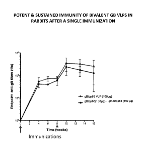
[0007] Figure 1 depicts exemplary EL1SA anti-gB antibody titers after
immunization
with bivalent gB virus-like particles (VLPs) (gB/pp65 and gB/pp65 + gH-
G/pp65). A potent and
sustained immunity is induced by bivalent gB VLPs in rabbits after a single
immunization.
CA 02867789 2014-09-18
WO 2013/144722 PCT/1B2013/001021
3
[0008] Figure 2 depicts exemplary FACS analysis of GFP expression in
fibroblast cells
indicative of neutralizing antibody response induced with gB/pp65 CMV VLPs in
rabbits.
Rabbits (n = 6/group) were immunized (IM) twice at weeks 0 and 8 and bled 2
weeks later. Sera
were pooled and tested at indicated dilutions in comparison to CytogamTM at
similar dilutions
against GFP-expressing CMV virus (TB40) in HFF fibroblasts. 100,000 cells were
collected
during flow cytometric analysis of infected (GFP) cells.
[0009] Figure 3 depicts exemplary FACS analysis of GFP expression in
fibroblast cells
indicative of neutralizing antibody response induced with bivalent gB + gH CMV
VLPs in
rabbits. Rabbits (n = 6/group) were immunized (IM) twice at weeks 0 and 8 and
bled 2 weeks
later. Sera were pooled and tested at indicated dilutions in comparison to
CytogamTM at similar
dilutions against GFP-expressing CMV virus (TB40) in HFF fibroblasts. 100,000
cells were
collected during flow cytometric analysis of infected (GFP) cells.
[0010] Figure 4 depicts exemplary percent neutralizations in HFF-1 cells.
Depicted are
neutralizations for 15RA09 group 7 (monovalent gB-G/monovalent gH-G adjuvanted
with alum)
pooled samples at P1Vd14, P1Vd28, P1Vd42, P1Vd55, and P2Vd14; and 15RA05 group
8
(empty MLV Gag) at P2Vd14, in presence of 10% guinea pig complement against
1:6 CMV-
GFP-TB40-010512 in HFF-1 cells.
[0011] Figure 5 depicts exemplary percent neutralizations in ARPE-19 cells.
Depicted
are neutralizations for 15RA09 group 7 (monovalent gB-G/monovalent gH-G
adjuvanted with
alum) pooled samples at P1Vd14, P1Vd28, P1Vd42, P1Vd55, and P2Vd14; and 15RA05
group
8 (empty MLV Gag) at P2Vd14, in presence of 2.5% rabbit complement against 1:3
CMV-GFP-
Towne-150612 in ARPE-19 cells.
Definitions
[0012] In order for the present disclosure to be more readily understood,
certain terms are
first defined below. Additional definitions for the following terms and other
terms are set forth
throughout the specification.
CA 02867789 2014-09-18
WO 2013/144722 PCT/IB2013/001021
4
[0013] Amino acid: As used herein, term "amino acid," in its broadest
sense, refers to any
compound and/or substance that can be incorporated into a polypeptide chain.
In some
embodiments, an amino acid has the general structure H2N¨C(H)(R)¨COOH. In some
embodiments, an amino acid is a naturally occurring amino acid. In some
embodiments, an
amino acid is a synthetic amino acid; in some embodiments, an amino acid is a
d-amino acid; in
some embodiments, an amino acid is an 1-amino acid. "Standard amino acid"
refers to any of the
twenty standard 1-amino acids commonly found in naturally occurring peptides.
"Nonstandard
amino acid" refers to any amino acid, other than the standard amino acids,
regardless of whether
it is prepared synthetically or obtained from a natural source. As used
herein, "synthetic amino
acid" encompasses chemically modified amino acids, including but not limited
to salts, amino
acid derivatives (such as amides), and/or substitutions. Amino acids,
including carboxy- and/or
amino-terminal amino acids in peptides, can be modified by methylation,
amidation, acetylation,
protecting groups, and/or substitution with other chemical groups that can
change the peptide's
circulating half-life without adversely affecting their activity. Amino acids
may participate in a
disulfide bond. Amino acids may comprise one or posttranslational
modifications, such as
association with one or more chemical entities (e.g., methyl groups, acetate
groups, acetyl
groups, phosphate groups, formyl moieties, isoprenoid groups, sulfate groups,
polyethylene
glycol moieties, lipid moieties, carbohydrate moieties, biotin moieties,
etc.). The term "amino
acid" is used interchangeably with "amino acid residue," and may refer to a
free amino acid
and/or to an amino acid residue of a peptide. It will be apparent from the
context in which the
term is used whether it refers to a free amino acid or a residue of a peptide.
[0014] Antigen: As used herein, the term "antigen" refers to a substance
containing one
or more epitopes (either linear, conformational or both) that are recognized
by antibodies. In
certain embodiments, an antigen is or comprises a virus or a viral
polypeptide. In some
embodiments, the term "antigen" refers to a subunit antigen (i.e., an antigen
which is separate
and discrete from a whole virus with which the antigen is associated in
nature; e.g., an antigen
which is associated with a virus-like particle). Alternatively or
additionally, in some
embodiments, the term "antigen" refers to killed, attenuated or inactivated
viruses. In certain
embodiments, an antigen is an "immunogen."
CA 02867789 2014-09-18
WO 2013/144722 PCT/IB2013/001021
[0015] Approximately or about: As used herein, the term "approximately" or
"about," as
applied to one or more values of interest, refers to a value that is similar
to a stated reference
value. In certain embodiments, the term "approximately" or "about" refers to a
range of values
that fall within 25%, 20%, 19%, 18%, 17%, 16%, 15%, 14%, 13%, 12%, 11%, 10%,
9%, 8%,
7%, 6%, 5%, 4%, 3%, 2%, 1%, or less in either direction (greater than or less
than) of the stated
reference value unless otherwise stated or otherwise evident from the context
(except where such
number would exceed 100% of a possible value).
[0016] Amelioration: As used herein, the term "amelioration" is meant the
prevention,
reduction or palliation of a state, or improvement of the state of a subject.
Amelioration
includes, but does not require complete recovery or complete prevention of a
disease, disorder or
condition (e.g., HCMV infection). The term "prevention" refers to a delay of
onset of a disease,
disorder or condition. Prevention may be considered complete when onset of a
disease, disorder
or condition has been delayed for a predefined period of time.
[0017] Dosage form: As used herein, the terms "dosage form" and "unit
dosage form"
refer to a physically discrete unit of a therapeutic agent for the patient to
be treated. Each unit
contains a predetermined quantity of active material calculated to produce the
desired therapeutic
effect. It will be understood, however, that the total dosage of the
composition will be decided
by the attending physician within the scope of sound medical judgment.
[0018] Dosing regimen: A "dosing regimen" (or "therapeutic regimen"), as
that term is
used herein, is a set of unit doses (typically more than one) that are
administered individually to a
subject, typically separated by periods of time. In some embodiments, a given
therapeutic agent
has a recommended dosing regimen, which may involve one or more doses. In some
embodiments, a dosing regimen comprises a plurality of doses each of which are
separated from
one another by a time period of the same length; in some embodiments, a dosing
regimen
comprises a plurality of doses and at least two different time periods
separating individual doses.
[0019] Expression: As used herein, "expression" of a nucleic acid sequence
refers to one
or more of the following events: (1) production of an RNA template from a DNA
sequence (e.g.,
by transcription); (2) processing of an RNA transcript (e.g., by splicing,
editing, 5' cap
CA 02867789 2014-09-18
WO 2013/144722 PCT/IB2013/001021
6
formation, and/or 3' end formation); (3) translation of an RNA into a
polypeptide or protein;
and/or (4) post-translational modification of a polypeptide or protein.
[0020] Fluorescence: As used herein, the term fluorescence refers to a
moiety that
luminesces. Typically fluorescent moieties contain electrons which can absorb
electromagnetic
energy at one wavelength and emit electromagnetic energy at a second
wavelength. Some
proteins or small molecules in cells are naturally fluorescent (e.g., NADH,
tryptophan,
endogenous chlorophyll, phycoerythrin, or green fluorescent protein (GFP)). It
will be
appreciated that various mutants of fluorescent proteins have been engineered
and may be used
in accordance with the present disclosure, such as EGFP, blue fluorescent
protein (EBFP,
EBFP2, Azurite, mKalamal), cyan fluorescent protein (ECFP, Cerulean, CyPet),
yellow
fluorescent protein (YFP, Citrine, Venus, YPet), redox sensitive GFP (roGFP),
and monomeric
GFP, among others. GFP and other fluorescent proteins can be expressed
exogenously in cells
alone or as a fusion protein. This approach permits fluorescent proteins to be
used as reporters
for any number of biological events, such as subcellular localization and
expression patterns.
[0021] Alternatively or additionally, specific or general proteins, nucleic
acids, lipids or
small molecules can be labeled with an extrinsic fluorophore, a fluorescent
dye which can be a
small molecule, protein or quantum dot. Exemplary fluorophores include, but
are not limited to,
1,5 IAEDANS; 1,8-ANS; 4- Methylumbelliferone; 5-carboxy-2,7-
dichlorofluorescein; 5-
Carboxyfluorescein (5-FAM); 5-Carboxynapthofluorescein; 5-
Carboxytetramethylrhodamine (5-
TAMRA); 5-Hydroxy Tryptamine (5-HAT); 5-ROX (carboxy-X-rhodamine); 6-
Carboxyrhodamine 6G; 6-CR 6G; 6-JOE; 7-Amino-4-methylcoumarin; 7-
Aminoactinomycin D
(7-AAD); 7-Hydroxy-4- I methylcoumarin; 9-Amino-6-chloro-2-methoxyacridine
(ACMA);
ABQ; Acid Fuchsin; Acridine Orange; Acridine Red; Acridine Yellow; Acriflavin;
Acriflavin
Feulgen SITSA; Aequorin (Photoprotein); AFPs--AutoFluorescent Protein--
(Quantum
Biotechnologies) see sgGFP, sgBFP; Alexa Fluor 350.TM.; Alexa Fluor 430.TM.;
Alexa Fluor
488.TM.; Alexa Fluor 532.TM.; Alexa Fluor 546.TM.; Alexa Fluor 568.TM.; Alexa
Fluor
594.TM.; Alexa Fluor 633.TM.; Alexa Fluor 647.TM.; Alexa Fluor 660.TM.; Alexa
Fluor
680.TM.; Alizarin Complexon; Alizarin Red; Allophycocyanin (APC); AMC, AMCA-S;
Aminomethylcoumarin (AMCA); AMCA-X; Aminoactinomycin D; Aminocoumarin; Anilin
Blue; Anthrocyl stearate; APC-Cy7; APTRA-BTC; APTS; Astrazon Brilliant Red 4G;
Astrazon
CA 02867789 2014-09-18
WO 2013/144722 PCT/IB2013/001021
7
Orange R; Astrazon Red 6B; Astrazon Yellow 7 GLL; Atabrine; ATTO-TAG.TM.
CBQCA;
ATTO-TAG.TM. FQ; Auramine; Aurophosphine G; Aurophosphine; BAO 9
(Bisaminophenyloxadiazole); BCECF (high pH); BCECF (low pH); Berberine
Sulphate; Beta
Lactamase; BFP blue shifted GFP (Y66H); Blue Fluorescent Protein; BFP/GFP
FRET; Bimane;
Bisbenzemide; Bisbenzimide (Hoechst); bis- BTC; Blancophor FFG; Blancophor SV;
BOBO.TM.-1; BOBO.TM.-3; Bodipy492/515; Bodipy493/503; Bodipy500/510; Bodipy;
505/515; Bodipy 530/550; Bodipy 542/563; Bodipy 558/568; Bodipy 564/570;
Bodipy 576/589;
Bodipy 581/591; Bodipy 630/650-X; Bodipy 650/665-X; Bodipy 665/676; Bodipy Fl;
Bodipy
FL ATP; Bodipy Fl-Ceramide; Bodipy R6G SE; Bodipy TMR; Bodipy TMR-X conjugate;
Bodipy TMR-X, SE; Bodipy TR; Bodipy TR ATP; Bodipy TR-X SE; BO-PRO.TM.-1; BO-
PRO.TM.-3; Brilliant Sulphoflavin FF; BTC; BTC-5N; Calcein; Calcein Blue;
Calcium
Crimson--; Calcium Green; Calcium Green-1 Ca2+ Dye; Calcium Green-2
Ca2+;
Calcium Green-5N Ca2+; Calcium Green-C18 Ca2+; Calcium Orange;
Calcofluor
White; Carboxy-X-rhodamine (5-ROX); Cascade Blue.TM.; Cascade Yellow;
Catecholamine;
CCF2 (GeneBlazer); CFDA; CFP (Cyan Fluorescent Protein); CFP/YFP FRET;
Chlorophyll;
Chromomycin A; Chromomycin A; CL-NERF; CMFDA; Coelenterazine; Coelenterazine
cp;
Coelenterazine f; Coelenterazine fcp; Coelenterazine h; Coelenterazine hcp;
Coelenterazine ip;
Coelenterazine n; Coelenterazine 0; Coumarin Phalloidin; C-phycocyanine; CPM I
Methylcoumarin; CTC; CTC Formazan; Cy2.TM.; Cy3.1 8; Cy3.5.TM.; Cy3.TM.; Cy5.1
8;
Cy5.5.TM.; Cy5.TM.; Cy7.TM.; Cyan GFP; cyclic AMP Fluorosensor (FiCRhR);
Dabcyl;
Dansyl; Dansyl Amine; Dansyl Cadaverine; Dansyl Chloride; Dansyl DHPE; Dansyl
fluoride;
DAPI; Dapoxyl; Dapoxyl 2; Dapoxyl 3'DCFDA; DCFH (Dichlorodihydrofluorescein
Diacetate);
DDAO; DHR (Dihydorhodamine 123); Di-4-ANEPPS; Di-8-ANEPPS (non-ratio); DiA (4-
Di
16-ASP); Dichlorodihydrofluorescein Diacetate (DCFH); DiD- Lipophilic Tracer;
DiD
(Dil C18(5)); DIDS; Dihydorhodamine 123 (DHR); Dil (Dil C18(3)); I
Dinitrophenol; Di0
(Di0C18(3)); DiR; DiR (Dil C18(7)); DM-NERF (high pH); DNP; Dopamine; DsRed;
DTAF;
DY-630-NHS; DY-635-NHS; EBFP; ECFP; EGFP; ELF 97; Eosin; Erythrosin;
Erythrosin ITC;
Ethidium Bromide; Ethidium homodimer-1 (EthD-1); Euchrysin; EukoLight;
Europium (111)
chloride; EYFP; Fast Blue; FDA; FeuIgen (Pararosaniline); FIF (Formaldehyd
Induced
Fluorescence); FITC; Flazo Orange; Fluo-3; Fluo-4; Fluorescein (FITC);
Fluorescein Diacetate;
Fluoro-Emerald; Fluoro-Gold (Hydroxystilbamidine); Fluor-Ruby; FluorX; FM 1-43
.TM.; FM
CA 02867789 2014-09-18
WO 2013/144722 PCT/IB2013/001021
8
4-46; Fura Red.TM. (high pH); Fura Red.TM./Fluo-3; Fura-2; Fura-2/BCECF;
Genacryl Brilliant
Red B; Genacryl Brilliant Yellow 10GF; Genacryl Pink 3G; Genacryl Yellow 5GF;
GeneBlazer;
(CCF2); GFP (S65T); GFP red shifted (rsGFP); GFP wild type' non-UV excitation
(wtGFP);
GFP wild type, UV excitation (wtGFP); GFPuv; Gloxalic Acid; Granular blue;
Haematoporphyrin; Hoechst 33258; Hoechst 33342; Hoechst 34580; HPTS;
Hydroxycoumarin;
Hydroxystilbamidine (FluoroGold); Hydroxytryptamine; Indo-1, high calcium;
Indo-1 low
calcium; Indodicarbocyanine (DiD); Indotricarbocyanine (DiR); Intrawhite Cf.
JC-1; JO J0-1;
JO-PRO-1; LaserPro; Laurodan; LDS 751 (DNA); LDS 751 (RNA); Leucophor PAF;
Leucophor
SF; Leucophor WS; Lissamine Rhodamine; Lissamine Rhodamine B; Calcein/Ethidium
homodimer; LOLO-1; LO-PRO-1; Lucifer Yellow; Lyso Tracker Blue; Lyso Tracker
Blue-
White; Lyso Tracker Green; Lyso Tracker Red; Lyso Tracker Yellow; LysoSensor
Blue;
LysoSensor Green; LysoSensor Yellow/Blue; Mag Green; Magdala Red (Phloxin B);
Mag-Fura
Red; Mag-Fura-2; Mag-Fura-5; Mag-lndo-1; Magnesium Green; Magnesium Orange;
Malachite
Green; Marina Blue; 1 MaxiIon Brilliant Flavin 10 GFF; MaxiIon Brilliant
Flavin 8 GFF;
Merocyanin; Methoxycoumarin; Mitotracker Green FM; Mitotracker Orange;
Mitotracker Red;
Mitramycin; Monobromobimane; Monobromobimane (mBBr-GSH); Monochlorobimane; MPS
(Methyl Green Pyronine Stilbene); NBD; NBD Amine; Nile Red;
Nitrobenzoxedidole;
Noradrenaline; Nuclear Fast Red; i Nuclear Yellow; Nylosan Brilliant lavin
E8G; Oregon
Green.TM.; Oregon Green.TM. 488; Oregon Green.TM. 500; Oregon Green.TM. 514;
Pacific
Blue; Pararosaniline (Feulgen); PBFI; PE-Cy5; PE-Cy7; PerCP; PerCP-Cy5.5; PE-
TexasRed
(Red 613); Phtoxin B (Magdala Red); Phorwite AR; Phorwite BKL; Phorwite Rev;
Phorwite
RPA; Phosphine 3R; PhotoResist; Phycoerythrin B [PE]; Phycoerythrin R [PE];
PKH26
(Sigma); PKH67; PMIA; Pontochrome Blue Black; POPO-1; POPO-3; P0-PRO-1; P0-1
PRO-
3; Primuline; Procion Yellow; Propidium lodid (P1); PyMPO; Pyrene; Pyronine;
Pyronine B;
Pyrozal Brilliant Flavin 7GF; QSY 7; Quinacrine Mustard; Resorufin; RH 414;
Rhod-2;
Rhodamine; Rhodamine 110; Rhodamine 123; Rhodamine 5 GLD; Rhodamine 6G;
Rhodamine
B; Rhodamine B 200; Rhodamine B extra; Rhodamine BB; Rhodamine BG; Rhodamine
Green;
Rhodamine Phallicidine; Rhodamine: Phalloidine; Rhodamine Red; Rhodamine WT;
Rose
Bengal; R-phycocyanine; R-phycoerythrin (PE); rsGFP; 565A; 565C; 565L; 565T;
Sapphire
GFP; SBFI; Serotonin; Sevron Brilliant Red 2B; Sevron Brilliant Red 4G; Sevron
I Brilliant Red
B; Sevron Orange; Sevron Yellow L; sgBFP.TM. (super glow BFP); sgGFP.TM.
(super glow
CA 02867789 2014-09-18
WO 2013/144722 PCT/IB2013/001021
9
GFP); SITS (Primuline; Stilbene Isothiosulphonic Acid); SNAFL calcein; SNAFL-
1; SNAFL-2;
SNARF calcein; SNARF1; Sodium Green; SpectrumAqua; SpectrumGreen;
SpectrumOrange;
Spectrum Red; SPQ (6-methoxy-N-(3 sulfopropyl)quinolinium); Stilbene;
Sulphorhodamine B
and C; Sulphorhodamine Extra; SYTO 11; SYTO 12; SYTO 13; SYTO 14; SYTO 15;
SYTO
16; SYTO 17; SYTO 18; SYTO 20; SYTO 21; SYTO 22; SYTO 23; SYTO 24; SYTO 25;
SYTO 40; SYTO 41; SYTO 42; SYTO 43; SYTO 44; SYTO 45; SYTO 59; SYTO 60; SYTO
61; SYTO 62; SYTO 63; SYTO 64; SYTO 80; SYTO 81; SYTO 82; SYTO 83; SYTO 84;
SYTO 85; SYTOX Blue; SYTOX Green; SYTOX Orange; Tetracycline;
Tetramethylrhodamine
(TRITC); Texas Red.TM.; Texas Red-X.TM. conjugate; Thiadicarbocyanine (DiSC3);
Thiazine
Red R; Thiazole Orange; Thioflavin 5; Thioflavin S; Thioflavin TON; Thiolyte;
Thiozole
Orange; Tinopol CBS (Calcofluor White); TIER; TO-PRO-1; TO-PRO-3; TO-PRO-5;
TOTO-1;
TOTO-3; TriColor (PE-Cy5); TRITC TetramethylRodaminclsoThioCyanate; True Blue;
Tru
Red; Ultralite; Uraninc B; Uvitcx SFC; wt GFP; WW 781; X-Rhodaminc; XRITC;
Xylene
Orange; Y66F; Y66H; Y66W; Yellow GFP; YFP; YO-PRO-1; YO- PRO 3; YOY0-1;YOY0-3;
Sybr Green; Thiazole orange (interchelating dyes); semiconductor nanoparticles
such as quantum
dots; or caged fluorophore (which can be activated with light or other
electromagnetic energy
source), or a combination thereof.
[0022] Fusion protein: As used herein, the term "fusion protein" generally
refers to a
polypeptide including at least two segments, each of which shows a high degree
of amino acid
identity to a peptide moiety that (1) occurs in nature, and/or (2) represents
a functional domain of
a polypeptide. Typically, a polypeptide containing at least two such segments
is considered to be
a fusion protein if the two segments are moieties that (1) are not included in
nature in the same
peptide, and/or (2) have not previously been linked to one another in a single
polypeptide, and/or
(3) have been linked to one another through action of the hand of man.
[0023] Gene: As used herein, the term "gene" has its meaning as understood
in the art.
It will be appreciated by those of ordinary skill in the art that the term
"gene" may include gene
regulatory sequences (e.g., promoters, enhancers, etc.) and/or intron
sequences. It will further be
appreciated that definitions of gene include references to nucleic acids that
do not encode
proteins but rather encode functional RNA molecules such as tRNAs, RNAi-
inducing agents,
etc. For the purpose of clarity we note that, as used in the present
application, the term "gene"
CA 02867789 2014-09-18
WO 2013/144722 PCT/1B2013/001021
generally refers to a portion of a nucleic acid that encodes a protein; the
term may optionally
encompass regulatory sequences, as will be clear from context to those of
ordinary skill in the
art. This definition is not intended to exclude application of the term "gene"
to non-protein¨
coding expression units but rather to clarify that, in most cases, the term as
used in this document
refers to a protein-coding nucleic acid.
[0024] Gene product or expression product: As used herein, the term "gene
product" or
"expression product" generally refers to an RNA transcribed from the gene (pre-
and/or post-
processing) or a polypeptide (pre- and/or post-modification) encoded by an RNA
transcribed
from the gene.
[0025] High-throughput: As used herein, the term "high-throughput" refers
broadly to
investigations with a large number of assays such that formatting of each
individual sample,
minimizing preparation steps and complications, and measuring of the assay
results either in
parallel or in rapid succession become important. High-throughput tests
generally do not include
manual, one-at-a-time assays, such as assays by a single individual in which
the preparation,
execution, measurement, and data collection for one assay are all completed
before the assay on
the next agent is done. High-throughput typically includes, for example, any
assays in which a
batch of samples (e.g., 24, 96, 384 or more test samples) are prepared and
measured. Formatting
the tests in such test samples is meant to accelerate the assay process by
enabling measurement
in parallel or in rapid succession, such as with the assistance of automation.
[0026] Immunogenic: As used herein, the term "immunogenic" means capable
of
producing an immune response in a host animal against a non-host entity (e.g.,
an HCMV
antigen). In certain embodiments, this immune response forms the basis of the
protective
immunity elicited by a vaccine against a specific infectious organism (e.g.,
an HCMV).
[0027] Immune response: As used herein, the term "immune response" refers
to a
response elicited in an animal. An immune response may refer to cellular
immunity, humoral
immunity or may involve both. An immune response may also be limited to a part
of the
immune system. For example, in certain embodiments, an immunogenic composition
may
induce an increased IFNy response. In certain embodiments, an immunogenic
composition may
induce a mucosal IgA response (e.g., as measured in nasal and/or rectal
washes). In certain
CA 02867789 2014-09-18
WO 2013/144722 PCT/1B2013/001021
11
embodiments, an immunogenic composition may induce a systemic IgG response
(e.g., as
measured in serum). In certain embodiments, an immunogenic composition may
induce virus-
neutralizing antibodies or a neutralizing antibody response.
[0028] Improve, increase, or reduce: As used herein, the terms "improve,"
"increase" or
"reduce," or grammatical equivalents, indicate values that are relative to a
baseline measurement,
such as a measurement in the same individual prior to initiation of the
treatment described
herein, or a measurement in a control individual (or multiple control
individuals) in the absence
of the treatment described herein.
[0029] Individual, subject, patient: As used herein, the terms "subject,"
"individual" or
"patient" refer to a human or a non-human mammalian subject. The individual
(also referred to
as "patient" or "subject") being treated is an individual (fetus, infant,
child, adolescent, or adult)
suffering from a disease, for example, HCMV infection. In some embodiments,
the subject is at
risk for HCMV infection. In some embodiments, the subject is an
immunosuppressed subject.
For example, in some embodiments, the immunosuppressed subject is selected
from the group
consisting of an HIV-infected subject, an AIDS patient, a transplant
recipient, a pediatric subject,
and a pregnant subject. In some embodiments, the subject has been exposed to
HCMV infection.
In some embodiments, the subject is a human.
[0030] Isolated: As used herein, the term "isolated" refers to a substance
and/or entity
that has been (1) separated from at least some of the components with which it
was associated
when initially produced (whether in nature and/or in an experimental setting),
and/or (2)
produced, prepared, and/or manufactured by the hand of man. Isolated
substances and/or entities
may be separated from about 10%, about 20%, about 30%, about 40%, about 50%,
about 60%,
about 70%, about 80%, about 90%, about 91%, about 92%, about 93%, about 94%,
about 95%,
about 96%, about 97%, about 98%, about 99%, or more than about 99% of the
other components
with which they were initially associated. In some embodiments, isolated
agents are about 80%,
about 85%, about 90%, about 91%, about 92%, about 93%, about 94%, about 95%,
about 96%,
about 97%, about 98%, about 99%, or more than about 99% pure. As used herein,
a substance is
"pure" if it is substantially free of other components. As used herein,
calculation of percent
CA 02867789 2014-09-18
WO 2013/144722 PCT/1B2013/001021
12
purity of isolated substances and/or entities should not include excipients
(e.g., buffer, solvent,
water, etc.).
[0031] Linker: As used herein, the term "linker" refers to, e.g., in a
fusion protein, an
amino acid sequence of an appropriate length other than that appearing at a
particular position in
the natural protein and is generally designed to be flexible and/or to
interpose a structure, such as
an a-helix, between two protein moieties. In general, a linker allows two or
more domains of a
fusion protein to retain 50%, 55%, 60%, 65%, 70%, 75%, 80%, 85%, 90%, 95% or
more of the
biological activity of each of the domains. A linker may also referred to as a
spacer.
[0032] Nucleic acid: As used herein, the term "nucleic acid," in its
broadest sense,
refers to any compound and/or substance that is or can be incorporated into an
oligonucleotide
chain. In some embodiments, a nucleic acid is a compound and/or substance that
is or can be
incorporated into an oligonucleotide chain via a phosphodiester linkage. In
some embodiments,
"nucleic acid" refers to individual nucleic acid residues (e.g., nucleotides
and/or nucleosides). In
some embodiments, "nucleic acid" refers to an oligonucleotide chain comprising
individual
nucleic acid residues. As used herein, the terms "oligonucleotide" and
"polynucleotide" can be
used interchangeably. In some embodiments, "nucleic acid" encompasses RNA as
well as single
and/or double-stranded DNA and/or cDNA. Furthermore, the terms "nucleic acid,"
"DNA,"
"RNA," and/or similar terms include nucleic acid analogs, i.e., analogs having
other than a
phosphodiester backbone. For example, the so-called "peptide nucleic acids,"
which are known
in the art and have peptide bonds instead of phosphodiester bonds in the
backbone, are
considered within the scope of the present disclosure. The term "nucleotide
sequence encoding
an amino acid sequence" includes all nucleotide sequences that are degenerate
versions of each
other and/or encode the same amino acid sequence. Nucleotide sequences that
encode proteins
and/or RNA may include introns. Nucleic acids can be purified from natural
sources, produced
using recombinant expression systems and optionally purified, chemically
synthesized, etc.
Where appropriate, e.g., in the case of chemically synthesized molecules,
nucleic acids can
comprise nucleoside analogs such as analogs having chemically modified bases
or sugars,
backbone modifications, etc. A nucleic acid sequence is presented in the 5' to
3' direction unless
otherwise indicated. The term "nucleic acid segment" is used herein to refer
to a nucleic acid
sequence that is a portion of a longer nucleic acid sequence. In many
embodiments, a nucleic
CA 02867789 2014-09-18
WO 2013/144722 PCT/1B2013/001021
13
acid segment comprises at least 3, 4, 5, 6, 7, 8, 9, 10, or more residues. In
some embodiments, a
nucleic acid is or comprises natural nucleosides (e.g., adenosine, thymidine,
guanosine, cytidine,
uridine, deoxyadenosine, deoxythymidine, deoxyguanosine, and deoxycytidine);
nucleoside
analogs (e.g., 2-aminoadenosine, 2-thiothymidine, inosine, pyrrolo-pyrimidine,
3-methyl
adenosine, 5-methylcytidine, C-5 propynyl-cytidine, C-5 propynyl-uridine, 2-
aminoadenosine,
C5-bromouridine, C5-fluorouridine, C5-iodouridine, C5-propynyl-uridine, C5-
propynyl-
cytidine, C5-methylcytidine, 2-aminoadenosine, 7-deazaadenosine, 7-
deazaguanosine, 8-
oxoadenosine, 8-oxoguanosine, 0(6)-methylguanine, and 2-thiocytidine);
chemically modified
bases; biologically modified bases (e.g., methylated bases); intercalated
bases; modified sugars
(e.g., 2'-fluororibose, ribose, 2'-deoxyribose, arabinose, and hexose); and/or
modified phosphate
groups (e.g., phosphorothioates and 5'-N-phosphoramidite linkages). In some
embodiments, the
present disclosure is specifically directed to "unmodified nucleic acids,"
meaning nucleic acids
(e.g., polynucleotides and residues, including nucleotides and/or nucleosides)
that have not been
chemically modified in order to facilitate or achieve delivery.
[0033] Pharmaceutically acceptable: The term "pharmaceutically acceptable"
as used
herein, refers to substances that, within the scope of sound medical judgment,
are suitable for use
in contact with the tissues of human beings and animals without excessive
toxicity, irritation,
allergic response, or other problem or complication, commensurate with a
reasonable benefit/risk
ratio.
[0034] Polypeptide: As used herein, a "polypeptide", generally speaking, is
a string of at
least two amino acids attached to one another by a peptide bond. In some
embodiments, a
polypeptide may include at least 3-5 amino acids, each of which is attached to
others by way of
at least one peptide bond. Those of ordinary skill in the art will appreciate
that polypeptides
sometimes include "non-natural" amino acids or other entities that nonetheless
are capable of
integrating into a polypeptide chain, optionally.
[0035] Substantial homology: The phrase "substantial homology" is used
herein to refer
to a comparison between amino acid or nucleic acid sequences. As will be
appreciated by those
of ordinary skill in the art, two sequences are generally considered to be
"substantially
homologous" if they contain homologous residues in corresponding positions.
Homologous
CA 02867789 2014-09-18
WO 2013/144722 PCT/1B2013/001021
14
residues may be identical residues. Alternatively, homologous residues may be
non-identical
residues will appropriately similar structural and/or functional
characteristics. For example, as is
well known by those of ordinary skill in the art, certain amino acids are
typically classified as
"hydrophobic" or "hydrophilic" amino acids., and/or as having "polar" or "non-
polar" side
chains Substitution of one amino acid for another of the same type may often
be considered a
"homologous" substitution.
[0036] As is well known in this art, amino acid or nucleic acid sequences
may be
compared using any of a variety of algorithms, including those available in
commercial computer
programs such as BLASTN for nucleotide sequences and BLASTP, gapped BLAST, and
PSI-
BLAST for amino acid sequences. Exemplary such programs are described in
Altschul, et al.,
Basic local alignment search tool, J. Mol. Biol., 215(3): 403-410, 1990;
Altschul, et al., Methods
in Enzymology; Altschul, et al., "Gapped BLAST and PSI-BLAST: a new generation
of protein
database search programs", Nucleic Acids Res. 25:3389-3402, 1997; Baxevanis,
et al.,
Bioinformatics : A Practical Guide to the Analysis of Genes and Proteins,
Wiley, 1998; and
Misener, et al., (eds.), Bioinformatics Methods and Protocols (Methods in
Molecular Biology,
Vol. 132), Humana Press, 1999. In addition to identifying homologous
sequences, the programs
mentioned above typically provide an indication of the degree of homology. In
some
embodiments, two sequences are considered to be substantially homologous if at
least 50%,
55%, 60%, 65%, 70%, 75%, 80%, 85%, 90%, 91%, 92%, 93%, 94%, 95%, 96%, 97%,
98%,
99% or more of their corresponding residues are homologous over a relevant
stretch of residues.
In some embodiments, the relevant stretch is a complete sequence. In some
embodiments, the
relevant stretch is at least 10, 15, 20, 25, 30, 35, 40, 45, 50, 55, 60, 65,
70, 75, 80, 85, 90, 95,
100, 125, 150, 175, 200, 225, 250, 275, 300, 325, 350, 375, 400, 425, 450,
475, 500 or more
residues.
[0037] Substantial identity: The phrase "substantial identity" is used
herein to refer to a
comparison between amino acid or nucleic acid sequences. As will be
appreciated by those of
ordinary skill in the art, two sequences are generally considered to be
"substantially identical" if
they contain identical residues in corresponding positions. As is well known
in this art, amino
acid or nucleic acid sequences may be compared using any of a variety of
algorithms, including
those available in commercial computer programs such as BLASTN for nucleotide
sequences
CA 02867789 2014-09-18
WO 2013/144722 PCT/1B2013/001021
and BLASTP, gapped BLAST, and PSI-BLAST for amino acid sequences. Exemplary
such
programs are described in Altschul, et al., Basic local alignment search tool,
J. Mol. Biol.,
215(3): 403-410, 1990; Altschul, et al., Methods in Enzymology; Altschul et
al., Nucleic Acids
Res. 25:3389-3402, 1997; Baxevanis et al., Bioinformatics : A Practical Guide
to the Analysis of
Genes and Proteins, Wiley, 1998; and Misener, et al., (eds.), Bioinformatics
Methods and
Protocols (Methods in Molecular Biology, Vol. 132), Humana Press, 1999. In
addition to
identifying identical sequences, the programs mentioned above typically
provide an indication of
the degree of identity. In some embodiments, two sequences are considered to
be substantially
identical if at least 50%, 55%, 60%, 65%, 70%, 75%, 80%, 85%, 90%, 91%, 92%,
93%, 94%,
95%, 96%, 97%, 98%, 99% or more of their corresponding residues are identical
over a relevant
stretch of residues. In some embodiments, the relevant stretch is a complete
sequence. In some
embodiments, the relevant stretch is at least 10, 15, 20, 25, 30, 35, 40, 45,
50, 55, 60, 65, 70, 75,
80, 85, 90, 95, 100, 125, 150, 175, 200, 225, 250, 275, 300, 325, 350, 375,
400, 425, 450, 475,
500 or more residues.
[0038] Suffering from: An individual who is "suffering from" a disease,
disorder, or
condition (e.g., HCMV infection) has been diagnosed with and/or exhibits one
or more
symptoms of the disease, disorder, or condition.
[0039] Susceptible to: An individual who is "susceptible to" a disease,
disorder, or
condition (e.g., HCMV infection) is at risk for developing the disease,
disorder, or condition. In
some embodiments, an individual who is susceptible to a disease, disorder, or
condition does not
display any symptoms of the disease, disorder, or condition. In some
embodiments, an
individual who is susceptible to a disease, disorder, or condition has not
been diagnosed with the
disease, disorder, and/or condition. In some embodiments, an individual who is
susceptible to a
disease, disorder, or condition is an individual who has been exposed to
conditions associated
with development of the disease, disorder, or condition (e.g., the individual
has been exposed to
HCMV).
[0040] Symptoms are reduced: According to the present disclosure, "symptoms
are
reduced" when one or more symptoms of a particular disease, disorder or
condition is reduced in
magnitude (e.g., intensity, severity, etc.) or frequency. For purposes of
clarity, a delay in the
CA 02867789 2014-09-18
WO 2013/144722 PCT/1B2013/001021
16
onset of a particular symptom is considered one form of reducing the frequency
of that symptom.
It is not intended that the present disclosure be limited only to cases where
the symptoms are
eliminated. The present disclosure specifically contemplates treatment such
that one or more
symptoms is/are reduced (and the condition of the subject is thereby
"improved"), albeit not
completely eliminated.
[0041] Therapeutically effective amount: As used herein, the term
"therapeutically
effective amount" refers to an amount sufficient to confer a therapeutic
effect on the treated
subject, at a reasonable benefit/risk ratio applicable to any medical
treatment. The therapeutic
effect may be objective (i.e., measurable by some test or marker) or
subjective (i.e., subject gives
an indication of or feels an effect). In particular, the "therapeutically
effective amount" refers to
an amount of a therapeutic protein or composition effective to treat,
ameliorate, or prevent a
desired disease or condition, or to exhibit a detectable therapeutic or
preventative effect, such as
by ameliorating symptoms associated with the disease, preventing or delaying
the onset of the
disease, and/or also lessening the severity or frequency of symptoms of the
disease. A
therapeutically effective amount is commonly administered in a dosing regimen
that may
comprise multiple unit doses. For any particular immunogenic composition, a
therapeutically
effective amount (and/or an appropriate unit dose within an effective dosing
regimen) may vary,
for example, depending on route of administration, on combination with other
pharmaceutical
agents. Also, the specific therapeutically effective amount (and/or unit dose)
for any particular
patient may depend upon a variety of factors including the disorder being
treated and the severity
of the disorder; the activity of the specific pharmaceutical agent employed;
the specific
composition employed; the age, body weight, general health, sex and diet of
the patient; the time
of administration, route of administration, and/or rate of excretion or
metabolism of the specific
immunogenic composition employed; the duration of the treatment; and like
factors as is well
known in the medical arts.
[0042] Treatment: As used herein, the term "treatment" (also "treat" or
"treating") refers
to any administration of an immunogenic composition that partially or
completely alleviates,
ameliorates, relieves, inhibits, delays onset of, reduces severity of and/or
reduces incidence of
one or more symptoms or features of a particular disease, disorder, and/or
condition (e.g.,
HCMV infection) or the predisposition toward the disease. Such treatment may
be of a subject
CA 02867789 2014-09-18
WO 2013/144722 PCT/1B2013/001021
17
who does not exhibit signs of the relevant disease, disorder and/or condition
and/or of a subject
who exhibits only early signs of the disease, disorder, and/or condition.
Alternatively or
additionally, such treatment may be of a subject who exhibits one or more
established signs of
the relevant disease, disorder and/or condition. In certain embodiments, the
term "treating"
refers to the vaccination of a patient.
[0043] Tropism: As used herein, the terms "tropism" or "host tropism" or
"cell tropism"
in the context of viruses and other pathogens generally refer to the ability
of the virus or
pathogen to infect a particular cell type. Tropism may refer to a way in which
the virus or
pathogen has evolved to preferentially target specific host species or
specific cell types within
those species. For example, HCMV can typically infect a remarkably broad cell
range within its
host, including parenchymal cells, connective tissue cells of virtually any
organ and various
hematopoietic cell types. Epithelial cells, endothelial cells, fibroblasts and
smooth muscle cells
are predominant targets for virus replication. However, the tropism for
various cells varies
greatly among different HCMV strains, e.g., from alterations within the UL128-
131 gene locus.
In some embodiments, an HCMV strain is able to infect fibroblasts, but not
epithelial and/or
endothelial cells. In some embodiments, an HCMV strain is able to infect
fibroblasts, epithelial
cells and endothelial cells.
[0044] Vaccination: As used herein, the term "vaccination" refers to the
administration
of a composition intended to generate an immune response, for example to a
disease-causing
agent (e.g., HCMV). For the purposes of the present disclosure, vaccination
can be administered
before, during, andlor after exposure to a disease-causing agent, and in
certain embodiments,
before, during, and/or shortly after exposure to the agent. In some
embodiments, vaccination
includes multiple administrations, appropriately spaced in time, of a
vaccinating composition.
[0045] Vector: As used herein, "vector" refers to a nucleic acid molecule
capable of
transporting another nucleic acid to which it is associated. In some
embodiments, vectors are
capable of extra-chromosomal replication and/or expression of nucleic acids to
which they are
linked in a host cell such as a eukaryotic and/or prokaryotic cell. Vectors
capable of directing
the expression of operatively linked genes are referred to herein as
"expression vectors."
CA 02867789 2014-09-18
WO 2013/144722 PCT/1B2013/001021
18
Detailed Description of Certain Embodiments
[0046] Among other things, the present disclosure provides methods useful
for
determining levels of HCMV infection in host cells and, by extension,
determining levels of
neutralizing antibodies present in a sample. The present disclosure
encompasses the recognition
that HCMV viruses that have a fluorescent moiety permit detection of viral
infection (e.g., by
assessing fluorescence in cells after contacting the host cell with the
virus). In some
embodiments, levels of HCMV infection are determined by fluorescence detection
where the
virus has been preincubated with a test sample (e.g., a serum sample) from a
subject. In some
embodiments, the subject has been administered a candidate HCMV vaccine.
I. HCMV Infection and Vaccines
[0047] Human cytomegalovirus (HCMV), a I3-herpesvirus, is a ubiquitously
occurring
pathogen. In general, entry of herpesviruses into cells is a complex process
initiated by
adsorption and receptor binding and followed by fusion of the virus envelope
with a cell
membrane. Fusion generally occurs at either the plasma membrane or an
endosomal membrane.
HCMV infects multiple cell types in vivo, including epithelial cells,
endothelial cells and
fibroblasts (Plachter B et al., 1996 Adv Virus Res 46:195-261). It fuses with
the plasma
membranes of fibroblasts (Compton T et al., 1992 Virology 191:387-395), but
enters retinal
pigmented epithelial cells and umbilical vein endothelial cells via
endocytosis (Bodaghi B et al.,
1999 J Immuno1162:957-964; Ryckman BJ et al., 2006 J Viro180:710-722). The
mechanism
by which herpesviruses choose their route of entry remains unclear. It is
generally assumed that
entry pathways are mainly determined by the host cell, but there is evidence
for tropic roles of
virion glycoproteins (Wang X et al., 1998 J Virol 72:5552-5558). HCMV encodes
two gH/gL
complexes: gH/gL/g0 and gH/gL/UL128/UL130/1JL131. The gO-containing complex is
sufficient for fibroblast infection, whereas the pUL128/UL130/UL131-containing
complex is
important for HCMV infection of endothelial and epithelial cells. As used
herein, the terms
"tropism" or "host tropism" or "cell tropism" in the context of viruses and
other pathogens
generally refer to the ability of the virus or pathogen to infect a particular
cell type. Tropism
may refer to a way in which the virus or pathogen has evolved to
preferentially target specific
host species or specific cell types within those species. In some embodiments,
an HCMV strain
CA 02867789 2014-09-18
WO 2013/144722 PCT/1B2013/001021
19
is able to infect fibroblasts, but not epithelial and/or endothelial cells. In
some embodiments, an
HCMV strain is able to infect fibroblasts, epithelial cells and endothelial
cells.
[0048] HCMV infects 50-85% of adults by 40 years of age (Gershon AA et al.,
1997 in
Viral Infections qf Humans, 4th edition, New York; Plenum Press:229-251). Most
healthy
individuals who acquire HCMV after birth develop few, if any, symptoms.
However, HCMV
disease is the cause of significant morbidity and mortality in
immunocompromised individuals,
such as recipients of hematopoietic cell transplants (HCT) and solid-organ
transplants (SOT)
(Pass RF 2001 Cytomegalovirus. In Fields Virology. zith edition, Philadelphia;
Lippincott
Williams & Wilkens:2675-2705). In SOT or HCT populations, HCMV disease can
occur either
from new infection transmitted from the donor organ or HCT, or can recur as a
result of
reactivation of latent virus in the recipient. In HIV-infected individuals,
HCMV infection
accelerates progression to AIDS and death, despite availability of
antiretroviral therapy (Deayton
JR et al., 2004 Lancet 363:2116-2121). In addition in the US, HCMV is the most
common
intrauterine infection and causes congenital abnormalities resulting in death
or severe birth
defects, including deafness and mental retardation, in approximately 8,000
infants each year
(Stagon S et al., 1986 JAMA 256:1904-1908).
[0049] Immune responses which control HCMV are incompletely understood. By
analogy to other human herpesviruses it can be assumed that both cellular and
humoral immune
responses play an important role (Kohl S 1992 Current topics in Microbiology
and Immunology
179:75-88). For murine CMV it was shown that either a cytotoxic T cell
response or the passive
transfer of neutralizing antibodies is sufficient to protect against a lethal
challenge (Rapp M et
al., 1993 Multidisciplinary Approach to Understanding Cytomegalovirus Disease
327-332;
Reddehase MJ et al., 198 J Virology 61:3102-3108).
[0050] Control of HCMV in immunocompromised persons is primarily associated
with
cellular immune responses; both CD8+ and CD4+ T lymphocytes appear to be
important for
protection against CMV disease (Gamadia LE et al., 2003 Blood 101:2686-2692;
Cobbold M et
al., 2005 J Exp Med 202:379-386). The cellular immune response to CMV includes
CD4+ helper
T-lymphocyte and CD8+ Cytotoxic T-lymphocyte responses to a number of
antigens, found in
the viral tegument, the region of the viral particle between the envelope and
capsid. A recent
CA 02867789 2014-09-18
WO 2013/144722 PCT/IB2013/001021
study of CMV-specific CD4+ and CD8+ T cells from healthy donors used
overlapping peptides
from a series of CMV open reading frames to identify antigens recognized after
CMV infection
(Sylwester AW et al., 2005 J Exp Med 202:673-685). The CMV tegument
phosphoprotein 65
(pp65) and surface glycoprotein gB were the antigens most frequently
recognized by CD4 T
cells, and pp65 was also one of the antigens most frequently recognized by CD8
T cells.
[0051] In contrast to the transplant setting, the maternal humoral immune
response
against the virus seems to be important in preventing HCMV disease in the
newborn. Antibodies
to surface glycoproteins, especially gB, appear to be critical for protection
against the maternal-
fetal transfer of HCMV (Fowler KB et al., 2003 JAMA 289:1008-1011). Moreover,
in an earlier
vaccination study it was shown that protection from re-infection is correlated
with neutralizing
antibodies (Adler SP et al., 1995 J Infectious Diseases 171:26-32). The
humoral immune
response to HCMV is dominated by responses to viral envelope glycoproteins
present in the
outer envelope of the virus particle (e.g., gB and gH).
[0052] In the case of HCMV, direct evaluation of immunological effector
functions is
difficult since the virus is strictly species specific and no animal model
system is available.
However, murinc CMV and guinea pig CMV have been used to evaluate vaccine
strategies in
these host species.
[0053] A CMV vaccine that induces both protective T cell and neutralizing
antibody
responses has the potential to prevent infection or ameliorate CMV disease due
to congenital
infection or transplantation.
[0054] The first live, attenuated HCMV vaccine candidate tested in humans
was based on
the laboratory-adapted AD169 strain. Subsequent trials with another laboratory-
adapted clinical
isolate, the Towne strain, confirmed that live attenuated vaccines could
elicit neutralizing
antibodies, as well as CD4+ and CD8+ T lymphocyte responses. The efficacy of
the Towne
vaccine was assessed in a series of studies in renal transplant recipients.
Although the Towne
vaccine did provide a protective impact on HCMV disease it failed to prevent
HCMV infection
after transplantation (Plotkin SA et al., 1984 Lancet 1:528-530). Towne
vaccine was also
evaluated in a placebo-controlled study of seronegative mothers who had
children attending
group daycare where it failed to prevent these women from acquiring infection
from their
CA 02867789 2014-09-18
WO 2013/144722 PCT/1B2013/001021
21
HCMV-infected children (Adler SP et al., 1995 J Infectious Diseases 171:26-
32). An
interpretation of these studies was that the Towne vaccine was overattenuated.
To explore this
possibility a series of genetic recombinants in which regions of the
unattenuated "Toledo" strain
of CMV were substituted for the corresponding regions of the Towne genome,
resulting in the
construction of Towne/Toledo "chimeras" that contain some, but not all, of the
mutations that
contribute to the Towne vaccine attenuation (Heineman TC et al. 2006 J Infect
Disease
193:1350-1360). The safety and tolerability of four Towne/Toledo "chimeras" is
being tested in
a Phase I trial. Long-term safety concerns about the potential risk of
establishing a latent HCMV
infection have hindered the further development of live, attenuated vaccines.
[0055] The leading subunit CMV vaccine candidate is based on the envelope
glycoprotein, gB, (purified recombinant gB vaccine is manufactured by Sanofi-
Pasteur Vaccines)
due to this protein's ability to elicit high-titer, virus-neutralizing
antibody responses during
natural infection. The recombinant gB vaccine elicits neutralizing antibody
responses and has an
excellent safety profile, however, it excludes other glycoprotein targets of
neutralizing antibody
response and more importantly T-Iymphocyte targets. The vaccine requires MF59
adjuvant to
optimize immunogenicity. In the most recent trial, this vaccine provided an
overall 50%
efficacy for prevention of CMV infection in a Phase 2 clinical trial in young
women (Pass RF et
al., 2009 N Engl J Med 360:1191-1199). Other viral proteins being evaluated as
subunit vaccine
candidates include pp65 and IE1, both of which elicit T-cell responses.
[0056] DNA vaccines elicit robust cellular and humoral immune responses in
animals
and are well suited to specificity and precision in vaccine design. DNA
vaccines have been
developed for CMV and have focused on gB, TEl and pp65 proteins as the
candidate target
immunogens. A bivalent CMV DNA vaccine candidate (Wloch MK, 2008 J Infectious
Diseases
297:1634-1642), using plasmid DNA encoding pp65 and gB and a trivalent vaccine
candidate
(Jacobson MA, 2009 Vaccine 27:1540-1548) that also includes a third plasmid
encoding the IE1
gene product have been developed by Vical Vaccines (US Patent No. 7,410,795).
The trivalent
DNA vaccine alone had minimal immunogenicity irrespective of route of
administration.
However the CMV DNA vaccine did appear to safely prime for a memory response
to CMV
antigens observed after administration of a live, attenuated CMV (Towne).
CA 02867789 2014-09-18
WO 2013/144722 PCT/IB2013/001021
22
[0057] In a vectored vaccine approach, the gene product of interest is
expressed in the
context of a non-replicating (usually viral) carrier. One example of this is a
canarypox vector
called ALVAC developed by Virogenetics and Sanofi-Pasteur Vaccines, which is
an attenuated
poxvirus that replicates abortively in mammalian cells. ALVAC expressing CMV
gB and
ALVAC expressing pp65 (US Patent No. 6,267,965) have been tested in clinical
trials. ALVAC-
CMV(gB) did not induce neutralizing antibodies but did prime for higher
neutralizing antibody
titers after subsequent infection with the Towne strain CMV (Adler SP et al.,
1999 J Infectious
Diseases 180:843-846), although it did not appear to boost neutralizing
antibody titers after
subsequent immunization with gB subunit/MF59 vaccine (Bernstein DI et al.,
2002 J Infectious
Diseases 185:686-690). A canarypox vector expressing pp65, ALVAC-CMV(pp64),
induced
long-lasting CTL responses in all originally seronegative volunteers, at
frequencies comparable
to naturally seropositive individuals (Berencsi K et al., 2001 J Infectious
Diseases 183:1171-
1179). Another approach used to express gB as a vectored vaccine is the use of
an alphavirus
replicon system by AlphaVax Inc (US Patent No. 7,419,674). This approach
involves a
propagation-defective single-cycle RNA replicon vector system derived from an
attenuated strain
of an alphavirus, Venezuelan Equine Encephalitis (VEE) virus, to produce virus-
like replicon
particles (VRPs) expressing pp65, TEl or gB protein (Berstein et al., 2010
Vaccine 28:484-493).
A two component alphavirus replicon vaccine was used to express the three CMV
proteins as a
soluble form of CMV gB (Towne strain) and a pp65/IE1 fusion protein (Reap EA
et al., 2007
Vaccine 25:7441-7449) was found to be safe and induced high levels of
neutralizing antibody
and polyfunctional CD4+ and CD8+ antigen-specific T cell responses. The
Geometric Mean
Titre (GMT) for the high dose group was about half the GMT in 12 naturally
infected, CMV
seropositive individuals tested in the assay.
[0058] A candidate for vaccination against HCMV currently in preclinical
development
is the "dense body" vaccine. Dense bodies (DBs) are enveloped, replication-
defective particles
formed during the replication of CMVs in cell culture. They contain both
envelope
glycoproteins and large quantities of pp65 protein. DBs are non-infectious and
immunogenic but
incapable of establishing latent HCMV infection in the vaccine recipient. DBs
have been shown
to be capable of inducing virus neutralizing antibodies and T-cell responses
in mice in the
absence of viral gene expression (Pepperl S et al., 2000 J Virol 74:6132-6146,
PCT Publication
No. WO 00/53729 and US Patent No. 6,713,070).
CA 02867789 2014-09-18
WO 2013/144722 PCT/IB2013/001021
23
[0059] Additional candidates contemplated for vaccination against HCMV are
virus like
particles (VLPs). Retroviruses are enveloped RNA viruses that belong to the
family
Retroviridae. After infection of a host cell by a retrovirus, RNA is
transcribed into DNA via the
enzyme reverse transcriptase. DNA is then incorporated into the host cell's
genome by an
integrase enzyme and thereafter replicates as part of the host cell's DNA. The
Retroviridae
family includes the following genus Alpharetrovirus, Betaretrovirus,
Ganunearetrovirus,
Deltaretrovirus, Epsilonretrovirus, Lentivirus and Spumavirus. The hosts for
this family of
retroviruses generally are vertebrates. Retroviruses produce an infectious
virion containing a
spherical nucleocapsid (the viral genome in complex with viral structural
proteins) surrounded
by a lipid bilayer derived from the host cell membrane.
[0060] Retroviral vectors can be used to generate enveloped virions that
are infectious
and either replication-competent or replication-defective. Replication-
competent infectious
retroviral vectors contain all of the necessary genes for virion synthesis and
continue to
propagate themselves once infection of the host cell occurs. Replication-
defective infectious
retroviral vectors do not spread after the initial infection. This is
accomplished by replacement
of most of the coding regions of the retrovirus with genes or nucleotide
sequences to be
transferred; so that the vector is incapable of making proteins required for
additional rounds of
replication.
[0061] Alternatively or additionally, retroviral vectors can be used to
generate virus-like
particles (VLPs) that lack a retrovirus-derived genome and are both non-
infectious and non-
replicating. Because of VLPs advantageous properties, VLPs may be utilized as
an antigen
delivery system. Furthermore, because VLPs are non-infectious, they can be
administered safely
as an immunogenic composition (e.g., a vaccine). VLPs are generally
structurally similar to
enveloped virions described above, but lack a retrovirus-derived genome,
making it unlikely that
viral replication will occur. Expression of capsid proteins (e.g., Gag) of
some viruses (e.g.,
murine leukemia viruses, such as Moloney Murine leukemia virus (MMLV)) leads
to self-
assembly into particles similar to the corresponding native virus, which
particles are free of viral
genetic material.
CA 02867789 2014-09-18
WO 2013/144722 PCT/IB2013/001021
24
[0062] A wide variety of VLPs have been prepared. For example, VLPs
including single
or multiple capsid proteins either with or without envelope proteins and/or
surface glycoproteins
have been prepared. In some cases, VLPs are non-enveloped and assemble by
expression of just
one major capsid protein, as shown for VLPs prepared from hepadnaviruses
(e.g., EngerixTM,
GSK and Recombivax HBTM, Merck), papillomaviruses (e.g., CervarixTM , GSK and
GardasilTM,
Merck), paroviruses, or polyomaviruses. In some embodiments, VLPs are
enveloped and can
comprise multiple antigenic proteins found in the corresponding native virus.
VLPs typically
resemble their corresponding native virus and can be multivalent particulate
structures. In some
embodiments, antigenic proteins may be presented internally within the VLP, as
a component of
the VLP structure, and/or on the surface of the VLP. In some embodiments,
presentation of an
antigen in the context of a VLP is advantageous for induction of neutralizing
antibodies against
the antigen as compared to other forms of antigen presentation, e.g., soluble
antigens not
associated with a VLP. Neutralizing antibodies most often recognize tertiary
or quaternary
structures; this often requires presenting antigenic proteins, like envelope
glycoproteins, in their
native viral conformation. Alternatively or additionally, VLPs may be useful
for presenting
antigens in a context which induces cellular immunity (e.g., T cell response).
In some
embodiments, use of antigen combinations in VLP systems can generate improved
immune
response.
Detectable HCMV
[0063] As described above, among other things, the present disclosure
provides methods
for determining levels of HCMV infection in host cells and, by extension,
determining levels of
neutralizing antibodies present in a sample. The present disclosure
encompasses the recognition
that HCMV viruses that have a fluorescent moiety permit detection of viral
infection (e.g., by
assessing fluorescence in cells after contacting the host cell with the
virus). In some
embodiments, levels of HCMV infection are determined by fluorescence detection
where the
virus has been preincubated with a test sample (e.g., a serum sample) from a
subject. In some
embodiments, the subject has been administered a candidate HCMV vaccine.
CA 02867789 2014-09-18
WO 2013/144722 PCT/IB2013/001021
[0064] Provided methods utilize an HCMV virus that includes a fluorescent
moiety. Any
HCMV virus capable of infecting a host cell described herein can be engineered
to include a
fluorescent moiety. In some embodiments, to infect fibroblasts, an HCMV virus
that includes all
or a portion of a gH/gL/g0 complex can be engineered to include a fluorescent
moiety. In some
embodiments, to infect endothelial cells and/or epithelial cells, an HCMV
virus that includes all
or a portion of a gH/gL/UL128/UL130/UL131 complex can be engineered to include
a
fluorescent moiety. Modified HCMV strains that are amenable to fluorescent
detection are
known in the art and may be used in accordance with the present disclosure.
For example,
UL32-EGFP-HCMV-TB40 is an in vitro recombination of HCMV strain TB40 with a
plasmid
carrying the TB40 UL32 gene fused to GFP (ATCC; VR-1578). The UL32-EGFP-HCMV-
TB40 recombinant strain gives rise to a recombinant HCMV virus with GFP fused
to the C
terminus of the tegument phosphoprotein pp150, the product of the UL32 gene
(Sampaio et al.,
2005 Journal of Virology 79(5):2754). Because GFP is associated with a viral
structural protein,
virus particles fluoresce green under appropriate illumination. The UL32-EGFP-
HCMV-TB40
strain has been demonstrated to have tropism for fibroblast cells (Sampaio et
al., 2005 Journal of
Virology 79(5):2754). An additional HCMV strain that is amenable to
fluorescent detection is
HB15-t178b, which contains the CMV strain AD169 genome and a GFP reporter
cassette
(Saccoccio et al., 2011 Vaccine 29(15):2705).
[0065] It is to be understood that the term fluorescence, as used herein,
refers to a moiety
that luminesces. Typically fluorescent moieties contain electrons which can
absorb
electromagnetic energy at one wavelength and emit electromagnetic energy at a
second
wavelength. Some proteins or small molecules in cells are naturally
fluorescent (e.g., NADH,
tryptophan, endogenous chlorophyll, phycoerythrin, or green fluorescent
protein (GFP)). It will
be appreciated that various mutants of fluorescent proteins have been
engineered and may be
used in accordance with the present disclosure, such as EGFP, blue fluorescent
protein (e.g.,
EBFP, EBFP2, Azurite, mKalamal), cyan fluorescent protein (e.g., ECFP,
Cerulean, CyPet),
yellow fluorescent protein (e.g., YFP, Citrine, Venus, YPet), redox sensitive
GFP (e.g., roGFP),
and monomeric GFP, among others. GFP and other fluorescent proteins can be
expressed
exogenously in cells alone or as a fusion protein. This approach permits
fluorescent proteins to
be used as reporters for any number of biological events, such as subcellular
localization and
expression patterns.
CA 02867789 2014-09-18
WO 2013/144722 PCT/IB2013/001021
26
[0066] Alternatively or additionally, specific or general proteins, nucleic
acids, lipids or
small molecules can be labeled with an extrinsic fluorophore, a fluorescent
dye which can be a
small molecule, protein or quantum dot. Exemplary fluorophores include, but
are not limited to,
1,5 IAEDANS; 1,8-ANS; 4- Methylumbelliferone; 5-carboxy-2,7-
dichlorofluorescein; 5-
Carboxyfluorescein (5-FAM); 5-Carboxynapthofluorescein; 5-
Carboxytetramethylrhodamine (5-
TAMRA); 5-Hydroxy Tryptamine (5-HAT); 5-ROX (carboxy-X-rhodamine); 6-
Carboxyrhodamine 6G; 6-CR 6G; 6-JOE; 7-Amino-4-methylcoumarin; 7-
Aminoactinomycin D
(7-AAD); 7-Hydroxy-4- I methylcoumarin; 9-Amino-6-chloro-2-methoxyacridine
(ACMA);
ABQ; Acid Fuchsin; Acridine Orange; Acridine Red; Acridine Yellow; Acriflavin;
Acriflavin
FeuIgen SITSA; Aequorin (Photoprotein); AFPs--AutoFluorescent Protein--
(Quantum
Biotechnologies) see sgGFP, sgBFP; Alexa Fluor 350; Alexa Fluor 430; Alexa
Fluor 488;
Alexa Fluor 532; Alexa Fluor 546; Alexa Fluor 568; Alexa Fluor 594; Alexa
Fluor 633;
Alexa Fluor 647; Alexa Fluor 660; Alexa Fluor 680; Alizarin Complexon;
Alizarin Red;
Allophycocyanin (APC); AMC, AMCA-S; Aminomethylcoumarin (AMCA); AMCA-X;
Aminoactinomycin D; Aminocoumarin; Anilin Blue; Anthrocyl stearate; APC-Cy7;
APTRA-
BTC; APTS; Astrazon Brilliant Red 4G; Astrazon Orange R; Astrazon Red 6B;
Astrazon Yellow
7 GLL; Atabrine; ATTO-TAGTm CBQCA; ATTO-TAGTm FQ; Auramine; Aurophosphine G;
Aurophosphine; BAO 9 (Bisaminophenyloxadiazole); BCECF (high pH); BCECF (low
pH);
Berberine Sulphate; Beta Lactamase; BFP blue shifted GFP (Y66H); Blue
Fluorescent Protein;
BFP/GFP FRET; Bimane; Bisbenzemide; Bisbenzimide (Hoechst); bis- BTC;
Blancophor FFG;
Blancophor SV; BOBOTm-1; BOBOTm-3; Bodipy492/515; Bodipy493/503;
Bodipy500/510;
Bodipy; 505/515; Bodipy 530/550; Bodipy 542/563; Bodipy 558/568; Bodipy
564/570; Bodipy
576/589; Bodipy 581/591; Bodipy 630/650-X; Bodipy 650/665-X; Bodipy 665/676;
Bodipy Fl;
Bodipy FL ATP; Bodipy Fl-Ceramide; Bodipy R6G SE; Bodipy TMR; Bodipy TMR-X
conjugate; Bodipy TMR-X, SE; Bodipy TR; Bodipy TR ATP; Bodipy TR-X SE; BO-
PROTm-1;
BO-PROTm-3; Brilliant Sulphoflavin FF; BTC; BTC-5N; Calcein; Calcein Blue;
Calcium
CrimsonTM; Calcium GreenTM; Calcium GreenTm-1 Ca2+ Dye; Calcium GreenTM2 Ca2-;
Calcium
GreenTm-5N Ca2'; Calcium GreenTm-C18 Ca2'; Calcium OrangeTM; Calcofluor White;
Carboxy-
X-rhodamine (5-ROX); Cascade Blue ; Cascade YellowTM; Catecholamine; CCF2
(GeneBlazer); CFDA; CFP (Cyan Fluorescent Protein); CFP/YFP FRET; Chlorophyll;
Chromomycin A; Chromomycin A; CL-NERF; CMFDA; Coelenterazine; Coelenterazine
cp;
CA 02867789 2014-09-18
WO 2013/144722 PCT/IB2013/001021
27
Coelenterazine f; Coelenterazine fcp; Coelenterazine h; Coelenterazine hcp;
Coelenterazine ip;
Coelenterazine n; Coelenterazine 0; Coumarin Phalloidin; C-phycocyanine; CPM I
Methylcoumarin; CTC; CTC Formazan; Cy2TM; Cy3.1 8; Cy3.5TM; Cy3TM; Cy5.1 8;
Cy5.5TM;
Cy5TM; Cy7TM; Cyan GFP; cyclic AMP Fluorosensor (FiCRhR); Dabcyl; Dansyl;
Dansyl Amine;
Dansyl Cadaverine; Dansyl Chloride; Dansyl DHPE; Dansyl fluoride; DAPI;
Dapoxyl; Dapoxyl
2; Dapoxyl 3'DCFDA; DCFH (Dichlorodihydrofluorescein Diacetate); DDAO; DHR
(Dihydorhodamine 123); Di-4-ANEPPS; Di-8-ANEPPS (non-ratio); DiA (4-Di 16-
ASP);
Dichlorodihydrofluorescein Diacetate (DCFH); DiD- Lipophilic Tracer; DiD
(Di1C18(5));
DIDS; Dihydorhodamine 123 (DHR); Dil (Di1C18(3)); I Dinitrophenol; Di0
(Di0C18(3)); DiR;
DiR (Dil C18(7)); DM-NERF (high pH); DNP; Dopamine; DsRed; DTAF; DY-630-NHS;
DY-
635-NHS; EBFP; ECFP; EGFP; ELF 97; Eosin; Erythrosin; Erythrosin ITC; Ethidium
Bromide;
Ethidium homodimer-1 (EthD-1); Euchrysin; EukoLight; Europium (111) chloride;
EYFP; Fast
Blue; FDA; Feuigen (Pararosanilinc); FIF (Formaldchyd Induced Fluorescence);
FITC; Flazo
Orange; Fluo-3; Fluo-4; Fluorescein (FITC); Fluorescein Diacetate; Fluoro-
Emerald; Fluoro-
Gold (Hydroxystilbamidine); Fluor-Ruby; FluorX; FM 1-43; FM 4-64; Fura RedTM
(high
pH); Fura RedTm/Fluo-3; Fura-2; Fura-2/BCECF; Genacryl Brilliant Red B;
Genacryl Brilliant
Yellow 10GF; Genacryl Pink 3G; Genacryl Yellow 5GF; GeneBlazer; (CCF2); GFP
(S65T);
GFP red shifted (rsGFP); GFP wild type' non-UV excitation (wtGFP); GFP wild
type, UV
excitation (wtGFP); GFPuv; Gloxalic Acid; Granular blue; Haematoporphyrin;
Hoechst 33258;
Hoechst 33342; Hoechst 34580; HPTS; Hydroxycoumarin; Hydroxystilbamidine
(FluoroGold);
Hydroxytryptamine; Indo-1, high calcium; Indo-1 low calcium;
Indodicarbocyanine (DiD);
Indotricarbocyanine (DiR); Intrawhite Cf; JC-1; JO J0-1; JO-PRO-1; LaserPro;
Laurodan; LDS
751 (DNA); LDS 751 (RNA); Leucophor PAF; Leucophor SF; Leucophor WS; Lissamine
Rhodamine; Lissamine Rhodamine B; Calcein/Ethidium homodimer; LOLO-1; LO-PRO-
1;
Lucifer Yellow; Lyso Tracker Blue; Lyso Tracker Blue-White; Lyso Tracker
Green; Lyso
Tracker Red; Lyso Tracker Yellow; LysoSensor Blue; LysoSensor Green;
LysoSensor
Yellow/Blue; Mag Green; Magdala Red (Phloxin B); Mag-Fura Red; Mag-Fura-2; Mag-
Fura-5;
Mag-Indo-1; Magnesium Green; Magnesium Orange; Malachite Green; Marina Blue; I
Maxilon
Brilliant Flavin 10 GFF; Maxilon Brilliant Flavin 8 GFF; Merocyanin;
Methoxycoumarin;
Mitotracker Green FM; Mitotracker Orange; Mitotracker Red; Mitramycin;
Monobromobimane;
Monobromobimane (mBBr-GSH); Monochlorobimane; MPS (Methyl Green Pyronine
Stilbene);
CA 02867789 2014-09-18
WO 2013/144722 PCT/IB2013/001021
28
NBD; NBD Amine; Nile Red; Nitrobenzoxedidole; Noradrenaline; Nuclear Fast Red;
i Nuclear
Yellow; Nylosan Brilliant lavin E8G; Oregon Green ; Oregon Green 488; Oregon
Green
500; Oregon Green 514; Pacific Blue; Pararosaniline (Feulgen); PBFI; PE-Cy5;
PE-Cy7;
PerCP; PerCP-Cy5.5; PE-TexasRed (Red 613); Phtoxin B (Magdala Red); Phorwite
AR;
Phorwite BKL; Phorwite Rev; Phorwite RPA; Phosphine 3R; PhotoResist;
Phycoerythrin B
[PE]; Phycoerythrin R [PE]; PKH26 (Sigma); PKH67; PMIA; Pontochrome Blue
Black; POPO-
1; POPO-3; P0-PRO-1; P0-1 PRO-3; Primuline; Procion Yellow; Propidium lodid
(P1);
PyMPO; Pyrene; Pyronine; Pyronine B; Pyrozal Brilliant Flavin 7GF; QSY 7;
Quinacrine
Mustard; Resorufin; RH 414; Rhod-2; Rhodamine; Rhodamine 110; Rhodamine 123;
Rhodamine 5 GLD; Rhodamine 6G; Rhodamine B; Rhodamine B 200; Rhodamine B
extra;
Rhodamine BB; Rhodamine BG; Rhodamine Green; Rhodamine Phallicidine;
Rhodamine:
Phalloidinc; Rhodamine Red; Rhodamine WT; Rose Bengal; R-phycocyanine; R-
phycoerythrin
(PE); rsGFP; S65A; S65C; S65L; S65T; Sapphire GFP; SBF1; Scrotonin; Scvron
Brilliant Red
2B; Scvron Brilliant Red 4G; Scvron 1 Brilliant Red B; Sevron Orange; Scvron
Yellow L;
sgBFPTM (super glow BFP); sgGFPTM (super glow GFP); SITS (Primuline; Stilbene
Isothiosulphonic Acid); SNAFL calcein; SNAFL-1; SNAFL-2; SNARF calcein;
SNARF1;
Sodium Green; SpectrumAqua; SpectrumGreen; SpectrumOrange; Spectrum Red; SPQ
(6-
methoxy-N-(3 sulfopropyl)quinolinium); Stilbene; Sulphorhodamine B and C;
Sulphorhodamine
Extra; SYTO 11; SYTO 12; SYTO 13; SYTO 14; SYTO 15; SYTO 16; SYTO 17; SYTO 18;
SYTO 20; SYTO 21; SYTO 22; SYTO 23; SYTO 24; SYTO 25; SYTO 40; SYTO 41; SYTO
42; SYTO 43; SYTO 44; SYTO 45; SYTO 59; SYTO 60; SYTO 61; SYTO 62; SYTO 63;
SYTO 64; SYTO 80; SYTO 81; SYTO 82; SYTO 83; SYTO 84; SYTO 85; SYTOX Blue;
SYTOX Green; SYTOX Orange; Tetracycline; Tetramethylrhodamine (TR1TC); Texas
Red();
Texas Red -X conjugate; Thiadicarbocyanine (DiSC3); Thiazine Red R; Thiazole
Orange;
Thioflavin 5; Thioflavin S; Thioflavin TON; Thiolyte; Thiozole Orange; Tinopol
CBS
(Calcofluor White); TIER; TO-PRO-1; TO-PRO-3; TO-PRO-5; TOTO-1; TOTO-3;
TriColor
(PE-Cy5); TRITC TetramethylRodaminelsoThioCyanate; True Blue; Tru Red;
Ultralite; Uranine
B; Uvitex SFC; wt GFP; WW 781; X-Rhodamine; XRITC; Xylene Orange; Y66F; Y66H;
Y66W; Yellow GFP; YFP; YO-PRO-1; YO- PRO 3; YOY0-1;YOY0-3; Sybr Green;
Thiazole
orange (interchelating dyes); semiconductor nanoparticles such as quantum
dots; or caged
CA 02867789 2014-09-18
WO 2013/144722 PCT/IB2013/001021
29
fluorophore (which can be activated with light or other electromagnetic energy
source), or a
combination thereof.
III. Infection Assays
[0067] Determination of infectious titer of HCMV typically includes
contacting a host
cell that is susceptible to infection by HCMV with serial dilutions of the
virus (e.g., HCMV that
includes a fluorescent moiety), under conditions that allow cell infection in
the absence of any
test substance. The number of target cells expressing a reporter gene
construct (e.g., a
fluorescent moiety, e.g., GFP) may be determined (e.g., by flow cytometry) to
calculate the
infectious titer of the virus preparation.
[0068] To assess the presence and/or activity of neutralizing antibodies in
serum of a
subject to whom a candidate vaccine has been administered, the serum may be
pre-incubated
with HCMV (e.g., HCMV that includes a fluorescent moiety, e.g., GFP) for a
period of time
sufficient for neutralizing antibodies to reduce infectivity of the HCMV
(e.g., at least 15 minutes,
at least 30 minutes, at least 1 hour, at least 2 hours, or more). Serial
dilutions of the pre-
incubated mixture of serum and HCMV (e.g., HCMV that includes a fluorescent
moiety) may
then be used to contact a host cell that is susceptible to infection by HCMV
under conditions that
allow infection. A person of ordinary skill in the art will be able to
determine appropriate
dilutions of scrum for infection assays. For example, in some embodiments,
dilutions tested are
1:6, 1:12, 1:24, 1:48, 1:96, 1:192, or combinations thereof.
[0069] It will be appreciated that conditions that allow infection may vary
depending on
a variety of factors, including viral strain, host cell type, temperature,
cell confluence, viral
concentration, among others. One of ordinary skill in the art would be able to
modify infection
conditions appropriately. In some embodiments, infection conditions include
incubation of the
host cell with virus for at least 1 hour, at least 2 hours, at least 3 hours,
at least 4 hours, at least 5
hours, at least 6 hours, at least 7 hours, at least 8 hours, or more. It will
also be appreciated that
monitoring for infection of cells may include visual cellular morphology
assessment (e.g., for
cellular swelling and rounding) and/or visual fluorescence assessment (e.g.,
detecting
CA 02867789 2014-09-18
WO 2013/144722 PCT/IB2013/001021
fluorescence on a device such as a fluorescence microscope). After infection,
host cells may be
collected (e.g., by washing and trypsinizing and/or scraping) and analyzed by
fluorescent
detection methods available in the art and described herein.
[0070] Any host cell susceptible to infection by HCMV can be used in the
methods
described herein. Exemplary host cells include, but are not limited to human
fibroblast cells,
such as human foreskin fibroblasts (HFF), and human epithelial cells such, as
retinal pigmented
epithelial cells (ARPE-19). Medium in which host cells are grown during the
infection assay
may vary with cell type. For example, in some embodiments, HFF infection
medium includes
MEM + 5% FBS + 1% PenStrep. In some embodiments, APRE infection medium
includes
DMEM:F-12 + 1% FBS.
IV. Fluorescence Detection and Neutralizing Antibody Assessment
[0071] Fluorescence may be detected using any appropriate method,
including, for
example, flow cytometry analysis, fluorescent activated cell sorting, or flow
microfluorometry.
It will be appreciated that the sensitivity of fluorescence detection
generally depends on the
number of copies of the fluorescent entity in the detection system, the
efficiency of the detection
instrument, and the fluorescence brightness of the fluorescent entity relative
to background
fluorescence that arises from endogenous biological fluorescent entities in
the sample and from
non-specific association of the fluorescent entity with the sample. The
brightness of the
fluorescent entity, in turn, depends on the quantum efficiency of the
fluorescent entity that
produces the fluorescence signal and the light absorbing capability
(quantified by the extinction
coefficient) of the fluorescent entity.
[0072] It is to be understood that cells can be identified and/or isolated
based on levels of
surface and/or intracellular fluorescence using reporter molecules such as
green fluorescent
protein (GFP). Generally, a correlation between fluorescence intensity and
protein production
(e.g., viral infection and production) will be observed. For example, in some
embodiments, high
levels of fluorescence intensity in a host cell correlates with high level of
viral infection of the
host cell.
CA 02867789 2014-09-18
WO 2013/144722 PCT/1B2013/001021
31
[0073] In some embodiments, cells are assessed for overall fluorescence
level (e.g.,
irrespective of subcellular localization of the fluorescence). In some
embodiments, cells are
assessed for fluorescence level in a particular subcellular location (e.g.,
the nucleus) of the cell.
In general, flow cytometry permits quantitative phenotyping of large numbers
of cells, however,
many flow cytometry applications do not include an ability to image cells as
they are quantitated.
In some embodiments, detection and quantitation of fluorescence of cells is
sufficient for
assessing infection. It will be appreciated that in some cases it is desirable
to determine the
localization of fluorescence within the cell. In some embodiments, flow
cytometry analysis of
cells may be combined with visual assessment of fluorescence. Dual analysis of
fluorescence
level and localization may be performed using multiple devices (e.g., flow
cytometry and
fluorescence microscopy) or on a single device. Such devices that permit
fluorescence analysis
and/or quantitation in conjunction with localization analysis are available in
the art. For
example, ImageStreamx (Amnis ) quantifies both intensity and localization of
fluorescence and
permits imaging of more than 50,000 cells per minute.
[0074] Cell fluorescence levels may be quantitated by any appropriate
method known in
the art. Cell fluorescence levels may be compared to a reference level. In
some embodiments,
the reference level is a predetermined or historical reference level. In some
embodiments, the
reference level is obtained by side-by-side comparison with a reference sample
(e.g., a positive
and/or negative control).
[0075] The advent of screening and selection methods that use flow
cytometry and cell
sorting considerably increase the number of cells that can be screened. For
example, several
million cells can be screened in a short time, and subpopulations and single
cells can be isolated
from within mixed-cell populations even when they are present at frequencies
as low as 10-6
within the population. It will be appreciated that one of the advantages of
provided methods is
that sample assessment can be achieved in a high-throughput manner. In some
embodiments,
provided methods increase throughput by 10%, 20%, 30%, 40%, 50%, 60%, 70%, or
more as
compared to known infection and/or neutralizing antibody detection methods
(e.g., staining of
viral proteins, fluorescence microscopy, ELISPOT, etc.). Provided methods may
be performed
in a multi-well plate format and are therefore particularly suitable for use
in mid-to-high
throughput screening. In some embodiments, the multi-well plates have 96
wells. In some
CA 02867789 2014-09-18
WO 2013/144722 PCT/IB2013/001021
32
embodiments, the multi-well plates have another number of wells, which include
but is not
limited to plates with 6, 12, 24, 384, 864 or 1536 wells. The terms "multi-
well plate" and
"microtiter plate" are used interchangeably.
[0076] In some embodiments, live cells are analyzed. In some embodiments,
cells are
fixed prior to analysis.
[0077] In some embodiments, the present disclosure provides methods for
measuring
anti-HCMV neutralizing antibodies by correlating fluorescence detection and/or
viral infection
of host cells. For example, an HCMV that includes a fluorescent moiety (e.g.,
GFP) may be pre-
incubated with serum from a subject immunized with an HCMV candidate vaccine.
Neutralizing
antibodies present in the serum will reduce HCMV (e.g., HCMV that includes a
fluorescent
moiety) from infecting a host cell that is normally susceptible to infection
by HCMV. The pre-
incubated mixture including HCMV (e.g., HCMV that includes a fluorescent
moiety) and serum
can then be used to contact such a host cell and fluorescence levels of the
host cell may be
assessed. Based on fluorescence levels in the host cell, a level of infection
of the host cell can be
determined. Level of infection of the host cell generally is inversely related
to the presence and
amount neutralizing antibody in the serum, which in turn, correlates with
efficacy of the
candidate vaccine to elicit a therapeutic response (e.g., a protective immune
response). For
example, the more efficacious a candidate vaccine at inducing neutralizing
antibodies in a
subject, the more neutralizing antibodies will be present in the serum, in
turn corresponding to a
decrease in ability of an HCMV that includes a fluorescent moiety to infect a
host cell after pre-
incubation with the serum, further corresponding to a decrease in fluorescence
detection in the
host cell after contacting it with an HCMV that includes a fluorescent moiety.
In some
embodiments, methods of the disclosure further include selecting and/or
identifying, based on a
correlation described herein, a candidate vaccine (e.g., an HCMV candidate
vaccine) as a
vaccine that induces neutralizing antibodies.
Examples
[0078] The following examples describe some exemplary modes of making and
practicing certain compositions that are described herein. It should be
understood that these
CA 02867789 2014-09-18
WO 2013/144722 PCT/IB2013/001021
33
examples are for illustrative purposes only and are not meant to limit the
scope of the
compositions and methods described herein.
Example 1: Immunization of Rabbits with Virus-Like Particles
[0079] This Example describes exemplary immunization of rabbits with virus-
like
particles containing various recombinant HCMV antigens.
[0080] HEK 293T cells (ATCC, CRL-11268) were transiently transfected using
calcium
phosphate methods with expression plasmids encoding various recombinant HCMV
antigens.
Expression of various HCMV antigens by the HEK 293 cells was confirmed by flow
cytometry.
After 48 to 72 hours of transfection, supernatants containing the VLPs were
harvested and
filtered through 0.45 pm pore size membranes and further concentrated and
purified by
ultracentrifugation through a 20% sucrose cushion in a SW32 Beckman rotor
(25,000 rpm, 2
hours, 4 C). Pellets were resuspended in sterile endotoxin-free PBS (GIBCO) to
obtain 500
times concentrated VLP stocks. Total protein was determined on an aliquot by a
Bradford assay
quantification kit (BioRad). Purified VLPs were stored at -80 C until used.
[0081] Rabbits were immunized intramuscularly at t = 0 and t = 8 weeks
with VLPs as
shown in Table 1 below. Serum was collected at t = 4, 6, 8, 10, 13 and 16
weeks.
Table 1.
Test Article # Dose Test Article Description
(n = 6/group)
1 100 lag gB/pp65 bivalent VLPs
6 100 lag/each gB/pp65 bivalent VLPs +
gH-G/pp65 bivalent VLPs
(1:1 ratio)
[0082] Enzyme-linked Immunosorbent Assay (ELISA) was performed to
determine
rabbit serum HCMV IgG content. Figure 1 shows potent and sustained immunity in
rabbits
immunized with gB/pp65 bivalent VLPs (upper) and gB/pp65 bivalent VLPs + gH-
G/pp65
bivalent VLPs (lower).
CA 02867789 2014-09-18
WO 2013/144722 PCT/IB2013/001021
34
Example 2: Flow Cytometry-Based Neutralization Activity
[0083] This Example describes assessment of neutralizing antibody
responses to HCMV
in scrum from immunized animals. The objectives of this Example included, but
were not
limited to: 1) confirming the immunogenicity of CMV clinical candidate
components selected
from other animal studies; 2) assessing dose and potential synergy and/or
antagonism with
administration routes (e.g., IM vs. IP administration); and 3) assessing
immunological boosting
and durability of immunity.
[0084] Serum samples from rabbits immunized with Test Article #6 from
Example 1
were collected at t = 0 (POV) and t = 2 weeks (P2Vd14) time points. Pooled,
heat inactivated
(HI) samples from this group were then tested for neutralizing antibody
activity as described
below.
[0085] Serum samples were diluted 1/6 to 1/96. HFF cells (P=9, used 24
hours post
seeded) and TB40-230212 freshly harvested virus were used. TB40-230212 virus
is descendant
of TB40-010212. TB40-010212 was obtained from ATCC (ATCC VR-1578; Human
herpesvirus 5; UL32-EGFP-HCMV-TB40). Virus infection duration was 15 days in a
T150
infection flask (Table 2). Microscopic observations indicated the virus was
infective. Infection
medium was TB40-HFF-1 infection medium (MEM+5%FBS+1%Pen/Strep) or VR1814-APRE-
19 infection medium (DMEM:F-12+1%FBS).
Table 2.
Virus infection
Virus Lot#
Microscope
Ascendant Duration Infection flask
observation
TB40-010212 T150
TB40 2:30212 15 days infective
(new ATCC)
[0086] An HFF seeded T150 flask was infected with CMV TB40-010212 (5
vials) + 20
ml of infectious media (MEM ¨Minimum Essential Medium Eagle from Sigma Cat.No.
M4655+
5% FBS + 1% P/S). After 15 days in CO2 incubator/37 C, the flask was well-
infected. Virus
was harvested by removing almost the entire supernatant from the flask and
keeping sterile in 50
mL Falcon tube. The remaining supernatant, around 5-7 nit, served to help
scraping the cells
with cell scraper (NUNC, Cat# 179707). After scraping, strong pipetting up and
down (up to
CA 02867789 2014-09-18
WO 2013/144722
PCT/IB2013/001021
10X) facilitated virus release from cells. Half of the original supernatant
volume from Falcon
tube was added (cca. 8 mL) in order to get more potent virus.
[0087] Five vials of TB40-010212 (ascendant) from liquid nitrogen was
infected in one
T150 flask, incubated at 37 C, 5%CO2 for 1 hour. Twelve mL of infection media
was added
after incubation. Virus was concentrated in 15 mL of supernatant.
[0088] CytogamTM
(CMV-IGW - Cytomegalovirus Immune Globulin Intravenous ¨
Human; CSL Behring; Commercial concentration 2.5 g/50 ml or 50 mg/ml) was used
as a
positive control. A 1/ 20 dilution was made as a base concentration (1 in 20
dilution: 100 AL of
CytogamTM 50 mg/mL + 1900 uL of infectious media) and later used for making
dilutions, the
same dilutions used for rabbit sera. CytogamTM was heat inactivated under the
same conditions
as rabbit sera (as described in Tables 3-5 below)
Table 3. Infection Assay
1/6 1/2 virus 2 6-well plate wells
1st plate
1/12 1/2 virus 2 6-well plate wells
Pooled HI POV
1124 1/2 virus 2 6-well plate wells
1/48 1/2 virus 2 6-well plate wells
1/96 1/2 virus 2 6-well plate wells
2nd plate
cells 2 6-well plate wells
1/6 1/2 virus 2 6-well plate wells
3rd plate
1/12 1/2 virus 2 6-well plate wells
Pooled HI P2V
1P4 1/2 virus 2 6-well plate wells
1/48 1/2 virus 2 6-well plate wells
1/96 1/2 virus 2 6-well plate wells 4t
plate
cells 2 6-well plate wells
1/6 1/2 virus 2 6-well plate wells
5th plate
1/12 1/2 virus 2 6-well plate wells
HI CytogarnTM
1/24 1/2 virus 2 6-well plate wells
(base 1/20)
1/48 1/2 virus 2 6-well plate wells
1/96 1/2 virus 2 6-well plate wells
6th plate
TB40-230212 1 in 2 diluted 2 6-well plate wells
Table 4. Pooled sera dilutions for POV, P2Vd14 and for CytogamTM
Used sera Used sera Media without Total Sera used in
Neat virus Final cone.
dilution FBS to make assay used in Sera
dilutions (u1) assay vs.
(u1) (u1) (u1) Virus
1/3 100 ul neat sera 200 300 130 u11/3 130
1/6
CA 02867789 2014-09-18
WO 2013/144722
PCT/1B2013/001021
36
vs.
1/2
1/12
1/6 150 ul 1/3 150 300 130 ul 1/6 130 vs.
1/2
1/24
1/12 150 ul 1/6 150 300 130 ul 1/12 130 vs.
1/2
1/48
1/24 150 ul 1/12 150 300 130 ul 1/24 130 vs.
1/2
1/96
1/48 150 ul 1/24 150 300 130 ul 1/48 130 vs.
1/2
Table 5. HFF-1 cell growth and infection media
Concentration
Reagent Supplier Cat# Lot# Exp Volume
of components
HFF-1 cells DMEM
growth (Dulbecco's
media Modified HyClone SH30243 .01 AWH17628 08/2012 80% 420m1
Eagle
Medium)
FBS (Fetal
(DMEM + bovine serum) HyClone SH30396.03 AVC67186 03/2015 15% 75m1
15% FBS +
1% Pen/Strep
(Penicillin P0781-
Pen/Strep) Sigma 031M0787 10/2012 1% 5m1
Streptomycin 100ML
Solution)
MEM
HFF (Minimum
infection Essential M4655-
media Medium Sigma
500m1 RNC0312 09/2012 94% 470m1
TB40 Eagle)
FBS (Fetal
Hyclone SH30396.03 AVC67186 03/2015 5% 25m1
bovine serum)
(MEM Pen/Strep
5% FBS + (Penicillin P0781-
1% Sigma 031M0787 10/2012 1% 5m1
Streptomycin 100ML
Pen/Strep) Solution)
[0089] Each particular concentration of sera or CytogamTM and 1/2 final
virus dilution
were combined and rotated at 37 C/1 hr. Ready to use 6-well plate with 95%-
100% confluent
HFF cells were carefully rinsed twice with warm PBS, then virus-sera mixture
was applied in
CA 02867789 2014-09-18
WO 2013/144722 PCT/IB2013/001021
37
duplicate as 100 lut mixture/well + 2001uL infection media to avoid drying
over 4 hrs.
Incubation was at 37 C/5% CO2/4 hrs (rocking was every 60 minutes). After a 4-
hour
incubation, contents were carefully aspirated from each well with a pipette
and 3 mL of fresh
infectious media were added. Plates were kept in the incubator at 37 C/5% CO2
for 10 days.
Daily checking of virus infection in cells was performed usually after 5 days
post-infection.
Swelling and rounding cells were visualized by light microscope or green
fluorescence detection
with a fluorescent microscope.
[0090] For sample collection, media was aspirated from each well. Each well
was rinsed
twice with PBS (HyClone DPBS/modified without Calcium and Magnesium;
Cat#5H30028.02)
and 100 uL of lx Trypsin-EDTA (Sigma; Cat#T4174-100 ml) and 100 ILIL of PBS
were added.
Samples were kept in CO2 incubator 2-3 minutes until cells were well
trypsinized. 1 mL/well of
PBS+5% FBS was added to stop trypsin. Samples were collected into transparent
flow intended
tubes (two wells for same sample/tube) (BD Falcon, 5 ml polystyrene round-
bottom, REF
352054). Plates were checked under the light microscope to confirm all cells
were collected (if
not add additional 500 AL PBS+5% FBS was used to collect the rest). Samples
were spun down
at 900 rpm for 10 minutes. Supernatant was discarded and the pellet kept. The
tube was
vortexed so that pellet was dispersed in the leftover buffer. The remaining
steps were performed
with minimal exposure to light. 200 jiL fixative (BD Biosciences, BD Cytofix,
Fixation buffer
Cat#554655, 100 ml) was added and incubated on ice for 15 minutes. 1 mL/tube
of mixture
PBS+5% FBS was added to stop Cytofix. Samples were spun down at 900 rpm for 10
minutes.
Supernatant was discarded and the pellet kept. The pellet was reconstituted in
5001aL of
PBS+5% FBS and vortexed vigorously before putting on flow cytometer.
[0091] Samples were subjected to flow cytometry and analyzed using
Cellquest software.
FSC and SSC parameters were set to "linear", while all other parameters were
set to "log."
100,000 cells were collected during flow cytometric analysis of infected
cells. Usual conditions
for flow using HFF cells and TB40 CMV were FSC E-1 and 4.83; SSC 325; FL1 427,
although
parameters can be slightly changed around existing conditions in order to get
more appropriate
dot plot.
CA 02867789 2014-09-18
WO 2013/144722 PCT/IB2013/001021
38
[0092] Figure 2 depicts exemplary FACS analysis of GFP expression in
fibroblast cells
indicative of neutralizing antibody response induced with gB/pp65 CMV VLPs in
rabbits.
Rabbits (n = 6/group) were immunized (IM) twice at weeks 0 and 8 and bled 2
weeks later. Sera
were pooled and tested at indicated dilutions in comparison to CytogamTM at
similar dilutions
against GFP-expressing CMV virus (TB40) in HFF fibroblasts. 100,000 cells were
collected
during flow cytometric analysis of infected (GFP) cells.
[0093] Figure 3 depicts exemplary FACS analysis of GFP expression in
fibroblast cells
indicative of neutralizing antibody response induced with bivalent gB + gH CMV
VLPs in
rabbits. Rabbits (n = 6/group) were immunized (IM) twice at weeks 0 and 8 and
bled 2 weeks
later. Sera were pooled and tested at indicated dilutions in comparison to
CytogamTM at similar
dilutions against GFP-expressing CMV virus (TB40) in HFF fibroblasts. 100,000
cells were
collected during flow cytometric analysis of infected (GFP) cells.
Example 3: Exemplary Micro-Neutralization Assay for Detection of Neutralizing
Antibodies
by Flow Cytometry
[0094] This Example describes detection of functional anti-CMV
neutralizing antibodies
by flow cytometry in sera samples from vaccinated animals.
Materials/Equipment
[0095] The following materials and equipment are used in this assay: Human
Foreskin
Fibroblasts (HFF-1) cells - ATCC# SCRC-1041; Human Arising Retinal Pigment
Epithelia ¨
(ARPE-19) ¨ ATCC#CRL-2302; Human herpesvirus 5 HCMV (UL32-EGFP-HCMV-TB40) -
ATCC# VR-1578; Human CMV-GFP-Towne TS15-rR (obtained from Dr. M. McVay, VCU-
Virginia); Goat Antiserum to Rabbit lgG (GAR) ¨ MP Cappel, Cat#: 55620;
Complement sera
from rabbit ¨ Sigma-Aldrich Cat#S7764-5 ml; Standard Guinea pig complement ¨
Cedarlane
Cat#CL-5000; Sterile distilled water ¨ Gibco, Cat#15230; Dulbecco's Modified
Eagle Medium
(DMEM) ¨ HyClone; Cat# SH30243.01 (growth media); Minimum Essential Medium
Eagle
(MEM) ¨ Sigma; Cat# M4655-500 ml (infectious media); Fetal bovine serum (FBS)
¨ HyClone;
Cat# SH30396.03; Penicillin Streptomycin Solution (Pen/Strep) ¨ Sigma; Cat#
P0781-100 ml;
CA 02867789 2014-09-18
WO 2013/144722 PCT/IB2013/001021
39
lx Trypsin-EDTA - Sigma; Cat#T4174-100 ml; Modified phosphate buffered saline
(DPBS -
without Calcium and Magnesium) ¨ HyClone; Cat# SH30028.02; Fixation buffer -
BD
Biosciences; BD Cytofix Cat#554655-100 ml; Cytogam (CMV-IGIV - Cytomegalovirus
Immune Globulin Intravenous/human- CSL Behring; Commercial concentration 2.5
g/50 ml;
Dimethyl sulfoxide (DMSO) ¨ Sigma-Aldrich Cat# D1435-500 ml; Biosafety
cabinet; Incubator
5% CO2, 37 C; Centrifuge; Vortex; Sample acquisition tubes for a Flow
cytometer (BD Falcon,
ml polystyrene round-bottom, REF 352054); 6-well plates; Multichannel
micropipette
reservoirs; 5 and 10 ml graduated pipettes; 10 ,1.1 to 1000 pi_ adjustable
single channel
micropipettes with disposable tips; Cell scraper ¨ NUNC; Cat# 179707; 50 ml
Falcon tubes ¨
BD 358206; Rotator ¨ for eppendorf tubes; Fluorescence microscope; FACSCAN
machine.
Obtaining and harvesting the virus
[0096] HFF-1 cells (95% confluent monolayer seeded in T150 flask) are
infected with
1.5 ml of UL32-EGFP-HCMV-TB40 ("TB40"). ARPE-19 cells are infected with HCMV-
GFP-
Towne-T515-rR ("Towne") in the same manner. Since these viruses are light
sensitive, cells are
infected with the light off The flasks are incubated in a CO2 incubator/37 C
for 30 minutes to
allow attachment between cells and virus.
[0097] 25 ml of infectious media is then added to the cells. (see Tables 6
and 7). Flasks
are kept in CO2 incubator/37 C until cells are well infected (in a case of
TB40, swelling and
rounding cells are seen by light microscope or green fluorescence with
fluorescent microscope;
for Towne, infection is detected by green fluorescence). Time necessary for
good infection
depends on the infectious capacity of the ascendant virus.
[0098] Harvesting is started by removing almost the entire supernatant from
the flask and
keeping it sterile in a 50 ml Falcon tube. Residual supernatant, around 5 ml,
facilitates scraping
of the cells with a cell scraper. Good scraping and strong pipetting up and
down (up to 10X)
enables virus to release from the cells. 10 ml of the original supernatant
volume from the Falcon
tube is added and spun down at 900 rpm for 10 minutes. The pellet is removed
and around 15 ml
of concentrated virus is kept. 5% of cryoprotectant (DMSO) in total amount of
virus is added. 1
ml aliquots are prepared, labeled, and kept at -80 C for short time or in
liquid nitrogen for long
period of time.
CA 02867789 2014-09-18
WO 2013/144722
PCT/1B2013/001021
Table 6. HFF-1 cells growth and infection media for CMV-GFP-TB40
Concentration
Reagent Supplier Cat# Volume
of components
DMEM
HFF-1 growth media
(Dulbecco's Modified HyClone SH30243.01 80% 420m1
Eagle Medium)
FBS (Fetal bovine
(DMEM + 15% FBS scrum) Hyclone SH30396.03 15% 75m1
1% Pen/Strep)
Pen/Slrep (Penicillin
Sigma P0781-100ML 1% 5m1
Streptomycin Solution)
MEM
HFF infectious
media for CMV- (Minimum Essential Sigma M4655-500m1 94%
470m1
GFP-TB40 virus
Medium Eagle)
FBS (Fetal bovine
Hyclone 5H30396.03 5% 25m1
serum)
(MEM + 5% FBS +
1% Pen/Strep)
PeniStrep (Penicillin
Sigma P0781-100ML 1% 5m1
Streptomycin Solution)
CA 02867789 2014-09-18
WO 2013/144722 PCT/1B2013/001021
41
Table 7. ARPE-19 cells growth & infection media for CMV-GFP-Towne
Concentration
Reagent Supplier Cat#
Volume
of components
DMEM : F-12
ARPE-19 growth (Dulbccco's Modified Eagle
media HyClone S H30023.01 80% 420m1
Medium nutrient mixture F-12
HAM)
(DMEM:F-12 +
15% FBS + FBS (Fetal bovine serum) Hyclone SH30396.03
15% 75m1
1% Pen/Strep)
Pen/Strep (Penicillin Streptomycin
Sigma P0781-100ML 1% 5m1
Solution)
DMEM : F-12
ARPE-19 (Dulbecco's Modified Eagle
HyClone S H30023.01 99%
470m1
infection media Medium nutrient mixture F-12
for Towne HAM)
growth
FBS (Fetal bovine serum) Hyclone SH30396.03 5%
25m1
( DMEM:F-12 +
5% FBS + 1%
PeMStrep)
Pen/Strep (Penicillin Streptomycin
Sigma P0781-100ML 1% 5m1
Solution)
Experimental set-up and method
[0099] Pre-
bleed and Post-immunized serum samples are heat inactivated at 56 C for 30
minutes prior to use. Commercial sera, Cytogam, that serves as positive
control is heat
inactivated, as well. Serum dilutions are made starting from 1/6 up to desired
dilutions in
duplicates. Cytogam is tested at comparable dilutions, a prior diluting the
stock reagent 1:20 to
adjust to the Ig content of human/rabbit sera (see Table 8).
CA 02867789 2014-09-18
WO 2013/144722 PCT/IB2013/001021
42
Table 8. Sera and virus dilutions (as an example lin2 virus dilution is shown)
Working Sera volume Media Total Sera volume Neat virus in Final
concentration
sera without FBS in assay assay Sera
dilution to make vs.
dilutions ( 1) Virus
1/3 100 [il neat sera 200 300 130 111 1/3 130 1/6
vs.
1/2
1/6 150 11 1/3 150 300 130 1.11 1/6 130 1/12
vs.
1/2
1/12 150 .1 1 1/6 150 300 130 1 1/12 130 1/24
vs.
1/2
1/24 150 [11 1/12 150 300 130p1 1/24 130 1/48
vs.
1/2
1/48 150 IA 1/24 150 300 130 p1 1/48 130 1/96
vs.
1/2
1/96 150 [11 1/48 150 300 130 1 1/96 130 1/192
vs.
1/2
1/192 (etc.) 150 1 1/96 150 300 130 [il
1/192 130 1/384
vs.
1/2(etc.)
[0100] Particular concentrations of sera (Cytogam) and virus are combined
and rotated at
37 C for 1 hr. In some assays, complement is included. For assays utilizing
complement and
HFF cells, 10% standard guinea pig complement is added to each particular sera
(Cytogam)/virus
mixture. Virus control is treated the same way. For assays utilizing
complement and ARPE-19
cells, 2.5% rabbit complement is added to each mix regardless which species is
used (rabbit or
sera). The sera and virus are rotated at 37 C for 1 hr.
[0101] 95% confluent cells (seeded in 6-well plates) are carefully rinsed
twice with warm
PBS prior to virus-sera mixture application, in duplicate. 100 1 of
mixture/well + 200 1
infection media is added to avoid drying over 4 hours of incubation. Cells are
incubated at
37 C/5% CO2/4 hrs (with rocking every 60 minutes). After a 4-hour incubation,
contents from
each well are carefully aspirated with a pipette or poured off, and 3 ml of
appropriate, fresh,
warm infectious media is added. Plates are incubated at 37 C/5% CO2 until well
infected. 5
days after incubation, cells are analyzed for virus infection. Infection of
HFF-1 cells is
CA 02867789 2014-09-18
WO 2013/144722 PCT/IB2013/001021
43
determined by swelling and rounding of cells, visualized by light microscope
or green
fluorescence with a fluorescent microscope. ARPE-19 cell infection is detected
as green
fluorescence with a fluorescent microscope.
Sample collection and preparation for flow cytometry
[0102] Before sample collection, the presence of good infection is
confirmed under a
light microscope and, if available, by analyzing GFP integrated into cells
using a fluorescent
microscope. Media is aspirated or poured off from each well. Cells are rinsed
twice with PBS
(HyClone DPBS/modified without Calcium and Magnesium; Cat#SH30028.02). To HFF-
1 cells
are added 1001,t1 of lx Trypsin-EDTA (Sigma; Cat#T4174-100 ml) and 200 ittl of
PBS. To
ARPE-19 cells are added 200 !al of lx Trypsin-EDTA and 100 pi of PBS. Cells
are kept in a
CO2 incubator 2-3 minutes until cells are well trypsinized. 1 ml/well of
PBS+5% FBS are added
to stop trypsin. Cells are collected into transparent flow intended tubes (two
wells for same
sample/tube) (BD Falcon, 5 ml polystyrene round-bottom, REF 352054). Wells are
visualized
under a light microscope and if cells remain, an additional 500 j.tl PBS+5%
FBS is added to
collect additional cells.
[0103] Falcon tubes are spun down at 900 rpm for 10 minutes. The
supernatant is
discarded and the pellet is kept. The tube is vortexed to disperse the pellet
in the residual buffer.
The remaining steps are performed in the dark. 200 ul Cytofix (BD Biosciences,
BD Cytofix,
Fixation buffer Cat#554655 100 ml) are added to each tube and samples arc
incubated on ice for
15 minutes. 1 ml of mixture PBS+5% FBS is added to each tube to stop Cytofix.
Tubes are
spun down at 900 rpm for 10 minutes. The supernatant is poured off and the
pellet is kept. The
pellet is reconstituted in 500 ul of PBS+5% FBS, vortexed vigorously, and flow
cytometry is
performed.
Example 4: Neutralization Activity of Exemplary VLPs in Rabbits
[0104] CHO cells were transfected at a cell density between 1.5E06 to
2.0E06 cells/mL
with plasmids of monovalent gB-G and monovalent gH-G (prepared as described in
PCT/U52012/64556). Stuffer DNA was added to make total DNA concentration up to
1 ps/mL
CA 02867789 2014-09-18
WO 2013/144722 PCT/IB2013/001021
44
cell culture. The plasmids used for transfection were first purified by
MaxiPrep or GigaPrep
plasmid purification kits (Qiagen). The PEIMAX used for transfection to
deliver DNA to the
cells was provided at a ratio of 6:1 (PEI: DNA wt/wt). The cell culture was
harvested 72 hours
post transfection by centrifuging at 4000 rpm for 20 minutes, using rotor JS-
4.2A by Beckman
Coulter, in 1 Litre bottles. The supernatant was filtered through 0.8/0.45 gm
filter (AcroPak 500
Capsule, Pall). The filtered supernatant was then concentrated by Tangential
Flow Filtration
(TFF for concentration of VLPs) and diafiltered against histidine-containing
buffer. 701.iL of
Benzonase (Novogen 99% purity (D00127703) containing 250 Units/L) was
suspended in
400juL of PBS that was supplemented with MgCl2 to have a 2 mM final
concentration. TFF
retentate portions were mixed with diluted Benzonase in PBS solution and kept
in a rotary shaker
(head-to-toe rotation) at room temperature for 1 hour and then the tubes were
placed at 4 C
overnight. The diafiltered benzonase treated material was then loaded onto an
anion exchange
chromatography column (AEX for reduction of DNA and host proteins) where the
flowthrough
was collected. The flowthrough was then sterile filtered through 0.45 gm and
aliquoted in
different volumes.
[0105] Monovalent gB-G and monovalent gH-G VLP compositions prepared as
described were mixed together, adjuvanted with alum and then tested in female
New Zealand
White rabbits 6-8 weeks old (minimum 5 animals per test group) for
neutralizing activity using
the microneutralization assay described in Example 3. Rabbits were immunized
intramuscularly
with 0.5 ml (250 ul in two sites of the proximal caudal hind thigh muscle) of
VLP compositions
three times, once on day 0 (Prime) and once on day 57 (week 8 Boost) and once
on day 162
(week 24 Boost). Rabbits were treated with 5011g of both monovalent gB-G and
monovalent
gH-G (mixed together at a 1:1 ratio) VLP composition. To assess humoral immune
responses in
rabbits, blood was collected from all rabbits in the study pre- 1st
immunization and then post- 1st
immunization days 28, 42 and 55 and post-2nd immunization at day 14.
[0106] Neutralizing antibody responses to HCMV were determined using a
microneutralization assay in fibroblast cells based on a GFP-expressing CMV
virus (TB40) and
flow cytometric analysis of infected (GFP+) HFF-1 cells as previously
described. Rabbit sera
collected pre- and post-immunizations as described were pooled and tested for
neutralizing
activity in the presence of guinea pig complement against HCMV expressing GFP
in HFF
CA 02867789 2014-09-18
WO 2013/144722 PCT/IB2013/001021
fibroblasts relative to a positive control CMV hyperglobulin, CytogamTM and a
negative control
consisting of empty Gag VLP (lacking antigenic proteins gB-G and/or gH-G).
[0107] Figure 4 shows the percent neutralization in HFF-1 cells incubated
with CMV-
GFP-TB40-010512 virus in presence of 10% Guinea Pig complement and rabbit
serum (group 7
pooled sera from 15RA09 was used as a positive data representative where
animals were treated
with monovalent gB-G and monovalent gH-G VLPs three times and also group 8
from 15RA05
study (empty Gag) was used as a negative control representative). As shown in
Figure 4, the
combination of monovalent gB-G and monovalent gH-G VLP composition elicited a
rapid,
synergistic sustained neutralizing antibody response in rabbits against
fibroblast cell infection,
while the empty Gag VLP showed no neutralizing antibody response. ("GFP-TB40-
010512"
denotes Human herpesvirus 5 HCMV (UL32-EGFP-HCMV-TB40) - ATCC# VR-1578
(described in Example 3) grown on May 1, 2012.)
[0108] Pooled rabbit sera was also tested for neutralizing antibody
responses to HCMV
using a microneutralization assay in epithelial cells based on a GFP-
expressing HCMV virus
(Towne TS15-rR) and flow cytometric analysis of infected (GFP+) ARPE-19 cells
as previously
described. Rabbit sera collected pre- and post-immunizations as described were
pooled and
tested for neutralizing activity in the presence of 2.5% rabbit complement
against HCMV
expressing GFP in ARPE-19 epithelial cells relative to a positive control CMV
hyperglobulin,
Cytogamrm and a negative control consisting of empty Gag VLP (lacking
antigenic proteins gB-
G and/or gH-G).
[0109] Figure 5 shows the percent neutralization in ARPE-19 cells incubated
with CMV-
GFP-Towne-150612 virus in presence of 2.5% rabbit complement and rabbit serum
(group 7
pooled sera from 15RA09 was used as a positive data representative where
animals were treated
with monovalent gB-G and monovalent gH-G VLP compositions three times and also
group 8
from 15RA05 study (empty Gag) was used as negative control representative). As
shown in
Figure 5, the combination of monovalent gB-G and monovalent gH-G VLP
composition elicited
a rapid, synergistic sustained neutralizing antibody response in rabbits
against epithelial cell
infection while the empty Gag VLP showed no neutralizing antibody response.
("GFP-Towne-
46
150612" denotes Human CMV-GFP-Towne TS15-rR (obtained from Dr. M. McVoy, VCU-
Virginia, and described in Example 3) grown on June 15, 2012.)
Other Embodiments
[0001] Other
embodiments of the disclosure will be apparent to those skilled in the art
from a consideration of the specification or practice of the disclosure
disclosed herein. It is
intended that the specification and examples be considered as exemplary only,
with the true
scope of the disclosure being indicated by the following claims.
CA 2867789 2018-11-02