Note: Descriptions are shown in the official language in which they were submitted.
CA 02867809 2014-09-18
WO 2013/134401
PCT/US2013/029396
ENZYMATIC NANOSENSOR COMPOSITIONS AND METHODS
CROSS REFERENCE TO RELATED APPLICATIONS
[0001] This application claims the benefit of U.S. Provisional Application
No.
61/607,173, filed March 6, 2012, the entire contents of which are hereby
incorporated by
reference herein.
STATEMENT REGARDING FEDERALLY SPONSORED
RESEARCH/DEVELOPMENT
[0002] This invention was made with government support by the Defense
Advanced
Research Projects Agency (DARPA) under award number W911NF-11-1-0025 and the
National Institute of General Medicine of the National Institutes of Health
under award
number RO1 GM084366. The government has certain rights in the invention.
BACKGROUND
[0003] Target detection is an important component in biotechnology,
analytical
chemistry, analysis of environmental samples, and medical diagnostics. Certain
types of
detection assays, such as fluorescence-based assays, are capable of providing
detailed
pictures of where fluorescent molecules are localized in tissues and cells. In
particular,
fluorescence-based assays exhibit exceptional sensitivity, detecting small
concentrations of
fluorescent molecules.
[0004] In addition, direct, minimally invasive monitoring of in vivo
physiological
conditions presents a route to determine health status in real time and
address needs as they
arise. Current in vivo monitoring system designs are limited by invasive
implantation
procedures and bio-fouling, limiting the utility of these tools for obtaining
physiologic data.
Traditional approaches using enzymes as recognition elements primarily rely on
the use of
electrodes to read out the signal changes after target detection. This imposes
a limitation for
non-invasive or non-contact monitoring, as the electrode must be physically
connected to
instrumentation to be measured. Former approaches to nanosensors have been
limited to
targets, such as ions or small molecules, that can be extracted into the core
of the
nanosensors. This approach does not allow detection of larger targets and has
limited
- 1 -
CA 02867809 2014-09-18
WO 2013/134401 PCT/US2013/029396
capabilities of being extended to additional targets without significant costs
to developing
new extraction chemicals.
[0005] Thus, there is a need for compositions and methods for the
inexpensive, sensitive,
and rapid detection of a diverse range of biochemical targets.
SUMMARY
[0006] Combining a catalytic agent with a fluorescent nanosensor that
measures the
effect of the enzymatic activity expands the range of detectable target
substrates. The
disclosed compositions and methods can be used in various contexts, including
in
biotechnology, analytical chemistry, analysis of environmental samples, and
medical
diagnostics. The disclosed methods and compositions can be used to detect
targets in
biological fluids, for cellular signaling, and for in vivo and in vitro
monitoring. One
application of the disclosed compositions and methods is to continuously track
bioanalytes in
vivo to enable clinicians and researchers to profile normal physiology and
discover early
markers for diseased states. Current in vivo monitoring system designs are
limited by
invasive implantation procedures and bio-fouling, which limit the utility of
these systems for
obtaining physiologic data. The disclosure allows measurement of a broad range
of target
substrates. Various combinations of fluorescent nanosensors and catalytic
agents can be used
to measure a wide range of target substrates both in vitro and in vivo.
[0007] According to aspects of the present disclosure, a composition
includes a catalytic
agent that catalyzes a reaction in which a target substrate and/or a co-
substrate is converted
into one or more products; and a nanosensor that is sensitive to an analyte
such that the
nanosensor emits a fluorescent signal upon detecting the analyte. The analyte
is the target
substrate, the co-substrate, or at least one of the one or more products.
[0008] In certain embodiments, the analyte includes oxygen, hydrogen,
ammonia, nitrate,
nitrite, and sulfate.
[0009] In further embodiments, the nanosensor is sensitive to oxygen. In
other
embodiments, the nanosenor includes a metal-centered dye, organic dye, or
biological
molecule. In other embodiments, the metal center of the metal-centered dye
includes
ruthenium (Ru(phen)3), platinum (Pt(II) meso-
Tetra(pentafluorophenyl)porphine), osmium,
rhenium, iridium, or mixtures thereof
[0010] In some embodiments, the nanosensor and the catalytic agent are
mixed together.
In other embodiments, the nanosensor and catalytic agent are operably linked.
- 2 -
CA 02867809 2014-09-18
WO 2013/134401 PCT/US2013/029396
[0011] In some embodiments, the catalytic agent is diamino oxidase,
acetylcholine
esterase, glucose oxidase, cholesterol oxidase, or glutamate dehydrogenase.
[0012] In further embodiments, the nanosensor and catalytic agent are
embedded in a
matrix. In particular embodiments, the matrix is a hydrogel that allows the
target substrate to
contact the catalytic agent.
[0013] In further embodiments, the nanosensor and catalytic agent are
attached to a
surface of a microfluidic device. In other embodiments, the nanosensor and
catalytic agent
are attached to the surface of the micro fluidic device through linkers.
[0014] In particular embodiments, the nanosensor and the catalytic agent
are attached to
the surface of a nanodevice. In some embodiments, the nanodevice includes a
polymer to
which the nanosensor and the catalytic agent are attached. In some
embodiments, the
polymer is polyvinyl chloride, polycaprolactone, polylactic acid, polylactic
co-glycolic acid,
poly(3-hydroxybutyrate), poly(carboxy phenoxy propane)-(sebacic acid),
polypropylene
fumarate, poly(alkyl cyanoacrylate, chitosan, alginate, polylysine, collagen,
or mixtures
thereof
[0015] Aspects of the methods disclosed herein provide methods of detecting
a target
substrate, including contacting a catalytic agent with a target substrate
and/or a co-substrate
such that the catalytic agent catalyzes conversion of the target substrate
and/or the co-
substrate into one or more products. The methods also include contacting a
nanosensor with
an analyte such that the nanosensor emits a fluorescent signal upon detecting
the analyte,
wherein the analyte is the target substrate, the co-substrate, or at least one
of the one or more
products. The methods further include measuring the concentration of the
target substrate
based on the fluorescent signal generated by the nanosensor.
[0016] In certain embodiments, the methods include using analytes such as
oxygen,
hydrogen, ammonia, nitrate, nitrite, and sulfate. In certain embodiments, the
methods include
using a nanosensor that is sensitive to oxygen.
[0017] In certain embodiments, the nanosensors used in the methods include
a metal-
centered dye, organic dye, or biological molecule. In certain embodiments, the
metal center
of the metal-centered dye include ruthenium (Ru(phen)3), platinum (Pt(II) meso-
Tetra(pentafluorophenyl)porphine), osmium, rhenium, iridium, or mixtures
thereof
[0018] In certain embodiments, the methods include using a nanosensor and a
catalytic
agent that are mixed together. In other embodiments, the nanosensor and the
catalytic agent
are operably linked.
- 3 -
CA 02867809 2014-09-18
WO 2013/134401 PCT/US2013/029396
[0019] In some embodiments, the catalytic agent used in the methods is
diamino oxidase,
acetylcholine esterase, glucose oxidase, cholesterol oxidase, or glutamate
dehydrogenase. In
other embodiments, the nanosensor and catalytic agent are embedded in a
matrix. In some
embodiments, the matrix is a hydrogel that allows the target substrate to
contact the catalytic
agent.
[0020] In further embodiments, the methods further include attaching the
nanosensor and
the catalytic agent to a surface of a microfluidic device. In further
embodiments, linkers are
used to attach the nanosensor and catalytic agent to the surface of the
microfluidic device.
[0021] In further embodiments, the methods include attaching the nanosensor
and the
catalytic agent to a surface of a nanodevice. In certain embodiments, the
nanodevice includes
a polymer to which the nanosensor and the catalytic agent are attached. In
further
embodiments, the polymer is polyvinyl chloride, polycaprolactone, polylactic
acid, polylactic
co-glycolic acid, poly(3-hydroxybutyrate), poly(carboxy phenoxy propane)-
(sebacic acid),
polypropylene fumarate, poly(alkyl cyanoacrylate, chitosan, alginate,
polylysine, collagen, or
mixtures thereof
SHORT DESCRIPTION OF THE FIGURES
[0022] The following figures are presented for the purpose of illustration
only, and are
not intended to be limiting.
[0023] Figure 1 shows a schematic of embodiments of the enzyme nanosensor
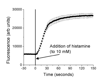
compositions. [0024] Figure 2 shows the enzyme nanosensor response to
histamine. While
fluorescence from the nanosensors is low in the absence of histamine, addition
of histamine
consumes oxygen and increases sensor fluorescence.
[0025] Figure 3 is a graphical representation of the enzyme nanosensor
system
responding rapidly and reversibly to histamine.
[0026] Figure 4A is the same as Figure 3 except that Figure 4 includes all
error bars,
while Figure 3 shows the errors bars from every five data points. Figure 4B
shows that
cycling histamine levels without continuous excitation shows full
reversibility. Figure 4C
shows that the nanosensors do not photobleach under continuous excitation in
the in vivo
animal imager. Figure 4D represents the fluorescence spectrum from enzyme
nanosensor
reversibility.
[0027] Figure 5 represents images from the in vitro calibration presented
in Figure 4.
- 4 -
CA 02867809 2014-09-18
WO 2013/134401 PCT/US2013/029396
[0028] Figures 6A-B are graphical representations showing that the enzyme
nanosensor
response is reproducible batch-to-batch. Figure 6A shows that the absolute
intensity of the
sensors change slightly (about 10%), but Figure 6B shows that the sensor
response to
histamine is not altered.
[0029] Figures 7A-B are graphical representations showing that altering the
ratio of
enzyme-to-nanosensor can control both the analyte response (Figure 7A) as well
as reaction
kinetics (Figure 7B).
[0030] Figure 8 represents fluorescence data using glucose oxidase as the
enzyme,
enabling detection of the catalytic agent glucose.
WM Figures 9A-C represent in vivo experimental results that demonstrate
the ability of
intradermal enzyme nanosensor to continuously monitor fluctuating histamine
levels.
loom Figures 10A-C represent fluorescence data for three animal
experiments that
demonstrate the ability of intradermal enzyme nanosensor to continuously
monitor
fluctuating histamine levels.
[0033] Figures 11A-B are graphical representations of all three histamine
response curves
(Figure 11A) and averaged data (Figure 11B, SD) for all three animal
experiments.
IOWA Figure 12 is a graphical representation showing that the enzyme
nanosensor
system responds rapidly to histamine concentrations in a dose-dependent
manner.
mig Figure 13 represents a one-compartment open model fit to the average
in vivo
data.
[0036] Figure 14 represents microscopic images of the enzyme nanosensor
composition
(pH nanosensors and acetylcholinesterase) encapsulated in a microdialysis
tube.
[0037] Figure 15 is a graphical representation of fluorescence ratio of the
nanosensors
versus acetylcholine concentration, and shows that the sensors respond to
acetylcholine in a
dose-dependent manner.
[0038] Figure 16 represents a calibration curve for oxygen nanosensors
(with Pt(II) mess-
Tetra (pentafluorophenyl)porphine as 02 sensor dye and octadecyl rhodamine as
the
reference dye) combined with the catalytic agent glucose oxidase to detect
glucose.
[0039] Figures 17A-B represent calibration curves similar to Figure 16
except no
reference dye was used and different catalytic agents were used. Glutamate
oxidase was used
for glutamate detection (Figure 17A), and tyrosinase for dopamine detection
(Figure 17B).
[0040] Figure 18 represents a calibration curve using oxygen-sensitive
ultrasmall
nanosensors with glutamate oxidase to detect glutamate.
- 5 -
CA 02867809 2014-09-18
WO 2013/134401 PCT/US2013/029396
DETAILED DESCRIPTION
[0041] The patent and scientific literature referred to herein establishes
knowledge that is
available to those of skill in the art. The issued U.S. patents, allowed
applications, published
foreign applications, and references that are cited herein are hereby
incorporated by reference
to the same extent as if each was specifically and individually indicated to
be incorporated by
reference.
[0042] Although compositions and methods similar or equivalent to those
described
herein can be used in the practice or testing of the present invention,
suitable compositions
and methods are described below.
Definitions
[0043] For convenience, certain terms employed in the specification,
examples and
claims are collected here. Unless defined otherwise, all technical and
scientific terms used in
this disclosure have the same meanings as commonly understood by one of
ordinary skill in
the art to which this disclosure belongs. The initial definition provided for
a group or term
provided in this disclosure applies to that group or term throughout the
present disclosure
individually or as part of another group, unless otherwise indicated.
[0044] In general, the compositions of the disclosure can be alternately
formulated to
comprise, consist essentially of, or consist of, any appropriate components
disclosed in this
disclosure. The compositions of the disclosure can additionally, or
alternatively, be
formulated so as to be devoid, or substantially free, of any components,
materials,
ingredients, adjuvants or species used in the prior art compositions or that
are otherwise not
necessary to the achievement of the function and/or objectives of the present
disclosure.
[0045] The articles "a" and "an" are used in this disclosure to refer to
one or more than
one (i.e., to at least one) of the grammatical object of the article. By way
of example, "an
element" means one element or more than one element.
[0046] The term "or" is used in this disclosure to mean, and is used
interchangeably with,
the term "and/or," unless indicated otherwise.
[0047] The present disclosure provides, in part, compositions that include
a nanosensor
that is sensitive to an analyte such that the nanosensor emits a fluorescent
signal upon
detecting the analyte; and a catalytic agent that catalyzes a reaction in
which a target substrate
is converted into one or more products, such that at least one of the one or
more products is
the analyte.
- 6 -
CA 02867809 2014-09-18
WO 2013/134401 PCT/US2013/029396
[0048] Aspects of the disclosed compositions comprise a catalytic agent and
a fluorescent
nanosensor. The fluorescent nanosensor measures the effect of the enzymatic
activity, and
expands the range of detectable target substrates. As disclosed herein, the
compositions and
methods are useful in biotechnology, analytical chemistry, analysis of
environmental
samples, and medical diagnostics. The disclosed compositions and methods can
be used to
detect targets in biological fluids, for cellular signaling, and for in vivo
and in vitro
monitoring. One application of the disclosed compositions and methods is to
continuously
track bioanalytes in vivo to enable clinicians and researchers to profile
normal physiology and
discover early markers for diseased states. In further embodiments, the
disclosed
compositions and methods detect analytes in environmental samples such as
water samples
(e.g., waste water, seawater, fresh water), soil samples, and samples from
industrial
production.
[0049] The disclosure allows measurement of a broad range of target
substrates. Various
combinations of fluorescent nanosensors and catalytic agents can be used to
measure a wide
range of target substrates both in vitro and in vivo.
[0050] Continuously monitoring in vivo substrate concentrations can be used
in a wide
range of applications, including but not limited to pharmacokinetic profiling
of novel drugs or
drug candidates and tracking biomarker concentrations during disease
progression, treatment,
or prevention. Current approaches rely on blood sampling followed by offline
analysis. This
process poses limitations when applied to common research models due to
limitations on the
amount and frequency of blood sampling.
[0051] In particular embodiments, the catalytic agent is an enzyme. Enzyme-
based
sensors can recognize a broad range of target substrates with high recognition
specificity, but
enzyme-based biosensors, including those for glucose, are still primarily
based on
electrochemical sensors. K. J. Cash, H. A. Clark, Trends Mol. Med 2010, 16.
584-593. In
certain embodiments, the enzyme is an oxidase. For example, glucose oxidase
catalytically
oxidizes glucose into gluconic acid, which lowers the pH, and the measured pH
change
correlates to glucose concentration. However, any enzyme that catalyzes the
reaction of one
or more substrates to a product can be used.
[0052] Fluorescent nanosensors are a modular family of sensors that can
continuously
monitor in vivo physiological parameters, including but not limited to oxygen,
pH, ammonia,
nitrate, nitrite, and sulfate. The sensors are approximately 100 nm in
diameter, and specific
nanosensor formulations that emit a reversible, concentration-dependent
fluorescent signal.
- 7 -
CA 02867809 2014-09-18
WO 2013/134401 PCT/US2013/029396
In the present disclosure, incorporating catalytic agents with the nanosensors
expands the
range of detectable biological targets and constitutes a significant advance
in the field of non-
invasive continuous target substrate monitoring. In some embodiments, surface
coatings
(with, e.g., PEG domains) can minimize protein fouling and safely prolong
nanoparticle
clearance, and biocompatible polymers (e.g. PLGA) can also be used. Amongst
other
applications, this disclosure enables straightforward, minimally-invasive
target substrate
monitoring.
Fluorescence Nanosensors
[0053] In the instant disclosure, a nanosensor is sensitive to an analyte
such that the
nanosensor emits a fluorescent signal upon detection of the analyte. In some
embodiments,
non-limiting examples of analytes include oxygen, hydrogen (pH), ammonia,
nitrate, nitrite,
and sulfate. Various fluorescent reports and derivatives thereof can be used
in the disclosed
compositions and methods. Nanosensors that are sensitive to oxygen include
metal-centered
dyes, organic dyes, and biological molecules. Metal-centered dyes include a
combination of
metals, ligand groups, or phorphyrin. Non-limiting examples of metal-centered
dyes include
dyes with the following metals: ruthenium (for example, Ru(phen)3), platinum
(for example,
Pt(II) meso-Tetra(pentafluorophenyl)porphine)), osmium, rhenium, iridium,
iridium, etc.
Ligand groups that can be included in metal-centered dyes include
phenanthroline; 2,2'-
bipyridine; 4,4'-dicarboxy-2,2'-bipyridine; 4,7-dipheny1-1,10-phenanthroline;
2,2'-bipyridy1-
4,4'-di-nonyl; 1,10-phenanthroline-5-amine; 1,10-phenanthroline-5-
isothiocyanate; and 1,10-
phenanthroline-5- N-hydroxysuccinimide ester. Porphyrin groups that can be
included in
metal-cenetered dyes include porphyrin, octaethylporphyrin ketone,
tetra(pentafluorophenyl)porphine, octaethyl porphyrin, and coproporphyrin.
Organic dyes
include any dye quenched by 02 and various fluorophores. Biological molecules
include but
are not limited to green fluorescent proteins (GFPs) and modified fluorescent
proteins (FPs).
[0054] For the analyte hydrogen (for pH), fluorescent nanosensors can
include
fluorescein, chromoionophores, BCECF, 6-JOE, Oregon green (488, 514), pHrodo,
SNARF
(1, 4F, 5F), phenol red, biological (GFP and GFP mutants), and nanomaterials
(QDs and
carbon nanotubes, including with or without chemical modifications). Examples
of
fluoresceins include FITC/conjugated fluorescein, F12, F16, F18 (hydrocarbon
tails), and
PLGFA-fluorescein. Suitable chromoionophores include Chromoionophore I (i.e.,
9-
(Diethylamino)-5-(octadecanoylimino)-5H-benzo[a]phenoxazine), Chromoionophore
II (i.e.,
- 8 -
CA 02867809 2014-09-18
WO 2013/134401 PCT/US2013/029396
9-Dimethylamino-5-[4-(16-buty1-2,14-dioxo-3,15-
dioxaeicosyl)phenylimino]benzo[a]phenoxazine), Chromoionophore III (i.e., 9-
(Diethylamino)-5-[(2-octyldecyl)imino]benzo[a]phenoxazine), Chromoionophore
VII (9-
Dimethylamino-5-[4-(15-buty1-1,13-dioxo-2,14-
dioxanonadecyl)phenylimino]benzo[a]phenoxazine), Chromoionophore IV (i.e. 5-
Octadecanoyloxy-2-(4-nitrophenylazo)phenol), Chromoionophore X (i.e. 4-
Dioctylamino-4'-
(trifluoroacetyl)stilbene), Chromoionophore VI (4',5'-Dibromofluorescein
octadecyl ester),
Chromoionophore VIII (3',3",5',5"-Tetrabromophenolphthaleinethyl ester
), Chromoionophore XVII (1-Hydroxy-4-[4-(2-
hydroxyethylsulfonyl)phenylazo]naphthalene-
2-sulfonic acid potassium salt), and Chromoionophore IX (4-Dibutylamino-4'-
(trifluoroacetyl)stilbene).
[0055] For the analyte NH3, fluorescent nanosensors can include the pH
sensors disclosed
herein, NH3 reactive complexes, and nanosensors with ammonium ionophore.
[0056] For the catalytic agent NADH/NADPH, fluorescent nanosensors can
include
quantum dots, other semiconductor dyes such as carbon dots, thionine,
methylene blue dyes,
and other redox dyes.
[0057] Electroactive dyes include but are not limited to metal-centered
dyes, methylene
blue, ferrocene, thionine, and cytodhrome. Dyes for membrane potential include
but are not
limited to RH237, RH414, RH421, RH795, Di-4-ANEPPS, Di-8-ANEPPS, Di-2-ANEPEQ,
Di-3-ANEPPDHQ, Di-12-ANEPPQ, and Di-4-ANEPPDHQ. Reference dyes can be used for
any fluorophore that does not respond to the analyte of interest, or any
fluorophore that has a
different response. Other potential readout mechanisms include color change
(absorbance),
photoacoustics, MRI, CT, ultrasound, and reflectance.
[0058] In addition, detection of fluorescence can be accomplished using
devices that can
be obtained commercially from, for example, Molecular Devices, LLC, Sunnyvale,
CA.
Encapsulation Methods
[0059] Encapsulation methods include but are not limited to using alginate
beads, other
hydrogel beads, polymer beads with double emulsion, and layer-by-layer
assembled shells.
[0060] In some embodiments, the nanosensors and catalytic agent are mixed
together
without using linkage chemistry. For instance, the nanosensors and catalytic
agents are
mixed in a polymeric matrix such that the catalytic agent and nanosensors are
embedded
- 9 -
CA 02867809 2014-09-18
WO 2013/134401 PCT/US2013/029396
within the matrix. In certain embodiments, a plurality of nanosensors and
catalytic agents are
embedded in a polymeric matrix such as polylactic acid and polylactic (co-
glycolic acid).
[0061] In other embodiments, the nanosensors and catalytic agent are
operably linked.
Linkage chemistries include using a wide range of available conjugation
techniques,
EDC/NHS, isothiocyanate, and click chemistry. Hermanson, Bioconjugate
Techniques (2nd
edition) (2008). In certain embodiments, the catalytic agent is linked to the
nanosensor. In
certain embodiments, the nanoparticle is immobilized within a polymeric matrix
that allows
the substrate of interest through the matrix to the nanoparticle. For example,
the nanoparticle
can be embedded within a polylactic acid matrix. The polylactic acid matrix is
then
functionalized with a linker group such as a maleimide group. See, e.g.,
Yamashiro et at.
(2008) Polymer Journal 40: 657-662. The maleimide group can then link the
nanoparticle to
the catalytic agent by, for instance, sulfhydryl crosslinking.
[0062] In addition, there are a variety of linker types that can be
utilized to link catalytic
agents and nanosensors. In some instances, photochemical/photolabile linkers,
thermolabile
linkers, and linkers that can be cleaved enzymatically can be used. Some
linkers are
bifunctional (i.e., the linker contains a functional group at each end that is
reactive with
groups located on the element to which the linker is to be attached). The
functional groups at
each end can be the same or different. Examples of suitable linkers that can
be used include
straight or branched-chain carbon linkers, heterocyclic linkers and peptide
linkers. A variety
of types of linkers are available from Pierce Chemical Company in Rockford,
Ill. and are
described in EPA 188,256; U.S. Pat. Nos. 4,671,958; 4,659,839; 4,414,148;
4,669,784;
4,680,338, 4,569,789 and 4,589,071, and by Eggenweiler, H.M, Pharmaceutical
Agent
Discovery Today 1998, 3, 552. NVOC (6 nitroveratryloxycarbonyl) linkers and
other NVOC-
related linkers are examples of suitable photochemical linkers (see, e.g., WO
90/15070 and
WO 92/10092). Peptides that have protease cleavage sites are discussed, for
example, in U.S.
Pat. No. 5,382,513.
[0063] In some embodiments, the compositions include the nanosensor and
catalytic
agent embedded in a matrix. In certain embodiments, the matrix is a polymer
selected from
the group consisting of poly(caprolactone) (PCL), ethylene vinyl acetate
polymer (EVA),
poly(lactic acid) (PLA), poly(L-lactic acid) (PLLA), poly(glycolic acid)
(PGA), poly(lactic
acid-co-glycolic acid) (PLGA), poly(L-lactic acid-co-glycolic acid) (PLLGA),
poly(D,L-
lactide) (PDLA), poly(L-lactide) (PLLA), poly(D,L-lactide-co-caprolactone),
poly(D,L-
lactide-co-caprolactone-co-glycolide), poly(D,L-lactide-co-PEO-co-D,L-
lactide), poly(D,L-
- 10 -
CA 02867809 2014-09-18
WO 2013/134401 PCT/US2013/029396
lactide-co-PPO-co-D,L-lactide), polyalkyl cyanoacralate, polyurethane, poly-L-
lysine (PLL),
hydroxypropyl methacrylate (HPMA), polyethyleneglycol, poly-L-glutamic acid,
poly(hydroxy acids), polyanhydrides, polyorthoesters, poly(ester amides),
polyamides,
poly(ester ethers), polycarbonates, silicones, polyalkylenes such as
polyethylene,
polypropylene, and polytetrafluoroethylene, polyalkylene glycols such as
poly(ethylene
glycol) (PEG), polyalkylene oxides (PEO), polyalkylene terephthalates such as
poly(ethylene
terephthalate), polyvinyl alcohols (PVA), and polyvinyl ethers. In certain
embodiments, the
polymer matrix has a shape. For example, the polymer matrix can be
rectangular, spherical,
tubular, oblong, elliptical, or irregular. Furthermore, the polymer matrix can
be any size
ranging from about 10 nm to about 100 mm.
[0064] In some embodiments, the matrix is a hydrogel that allows the target
substrate to
contact the catalytic agent. A "hydrogel" is a three-dimensional, semi-solid
network of one
or more polymers derived from monomers in which a relatively large amount of
water is
present in the wet state. A "gel" is a solvent-rich composition consisting of
a solvent
(imbibing solvent) in an insoluble, porous network comprising one or more
polymeric
organic molecules, where the solvent can be water, giving a "hydrogel," a
nonpolar organic
solvent, giving "nonpolar gel" or a polar organic solvent or a solution of
water and an organic
solvent, giving a "semipolar gel." One of ordinary skill in the art
understands how to make
and use hydrogels.
[0065] In certain aspects, the disclosed methods also include adding
hydrogels
comprising vinyl monomers, urea, formamide, polyethylene glycol, sugars,
oligosaccharides,
and polyvinylpyrolidone, and polyacrylamide. The gels can also include salts,
buffers, or
polypeptides to the pre-gelling solution, thereby regulating the viscosity,
vinyl monomer
diffusion during gel formation, interactions of the hydrogel polymer chains
during gel
formation, or degree of polymerization of the gelling solution.
[0066] In certain embodiments, the hydrogel can be given a particular
shape. For
instance, the hydrogel can be formed on a glass surface, and can be reacted
with
methacryloxypropyl trichlorosilane to bestow it with vinyl groups. In this
case, a gel is
formed in any particular shape, including but not limited to, rod, tube,
sheet, cone, sphere,
rectangle, square, or other shape allowed by a mold or environment. A gel can
be formed as
a sheet by pouring the gelling solution into a flat or curved mold, or between
two plates.
[0067] According to aspects of the present disclosure, the nanosensors have
a shape that
allows for accurate measurement of an analyte, that is, emission of an
accurate fluorescent
- 11 -
CA 02867809 2014-09-18
WO 2013/134401 PCT/US2013/029396
signal upon detecting the analyte. In some embodiments, the nanosensors have a
particular
shape that provides a high surface-to-volume ratio that allows for accurate
measurements. In
some embodiments, the nanosensors has an oblong or rectangular shape.
Exemplary shapes
include rectangles, elongated cylinders having a diameter shorter than the
length of the
cylinder, oblong structures, parallelepiped structures, rhomboid structures,
and elliptical
structures. Generally, any structure that provides a high aspect ratio for the
sensing agent is
within the scope of the invention. By "high aspect ratio," it is meant that
the structures
disclosed herein have lengths that are longer than their widths.
[0068] The disclosed nanosensors and catalytic agents can also be
immobilized within
multiwell plates. For example, the nanosensors can be conjugated to antibodies
coating the
surface of the multiwell plate. Tang et at. (2011) Biochemical Engineering
Journal 53(2):
223-228. The nanosensors can also be attached to the surface of the wells of
the multiwell
plate using technologies described herein. The catalytic agents can also be
attached to the
surface of the wells of the multiwell plate using antibodies or linking
technologies described
herein.
Catalytic Agents
[0069] Any catalytic agent that acts on a target substrate and changes the
concentration of
an analyte (for example, 02, pH, electron transfer, etc.) can be used in the
disclosed
compositions and methods. Non-limiting examples of catalytic agents include
diamino
oxidase, acetylcholine esterase, glucose oxidase, cholesterol oxidase,
monoamine oxidase,
glutamate dehydrogenase, alcohol dehydrogenase, urease, creatininase,
glutamate oxidase,
glucose dehydrogenase, lactate oxidase, tyrosinase, 3a-hydroxysteroid
dehydrogenase, and
1113-hydroxysteroid dehydrogenase.
Microfluidic Devices
[0070] In other embodiments, the enzyme nanosensors are incorporated into a
microfluidic device. Applications include using the device for sensing
analytes in biological
or non-biological fluids. In some embodiments, the nanosensor and catalytic
agent are
attached to a surface of a microfluidic device. In other embodiments, the
nanosensor and
catalytic agent are attached to the surface of the microfluidic device through
linkers. In other
embodiments, the nanosensor and the catalytic agent are attached to the
surface of a
nanodevice.
- 12 -
CA 02867809 2014-09-18
WO 2013/134401 PCT/US2013/029396
[0071] In other embodiments, the nanodevice includes a polymer to which the
nanosensor
and the catalytic agent are attached. Polymers useful in construction of the
microfluidic
device include but are not limited to polyvinyl chloride, polycaprolactone,
polylactic acid,
polylactic co-glycolic acid, poly(3-hydroxybutyrate), poly(carboxy phenoxy
propane)-
(sebacic acid), polypropylene fumarate, poly(alkyl cyanoacrylate, chitosan,
alginate,
polylysine, collagen, or mixtures thereof
[0072] In certain embodiments, the polymer includes poly(caprolactone)
(PCL), ethylene
vinyl acetate polymer (EVA), poly(lactic acid) (PLA), poly(L-lactic acid)
(PLLA),
poly(glycolic acid) (PGA), poly(lactic acid-co-glycolic acid) (PLGA), poly(L-
lactic acid-co-
glycolic acid) (PLLGA), poly(D,L-lactide) (PDLA), poly(L-lactide) (PLLA),
poly(D,L-
lactide-co-caprolactone), poly(D,L-lactide-co-caprolactone-co-glycolide),
poly(D,L-lactide-
co-PEO-co-D,L-lactide), poly(D,L-lactide-co-PPO-co-D,L-lactide), polyalkyl
cyanoacralate,
polyurethane, poly-L-lysine (PLL), hydroxypropyl methacrylate (HPMA),
polyethyleneglycol, poly-L-glutamic acid, poly(hydroxy acids), polyanhydrides,
polyorthoesters, poly(ester amides), polyamides, poly(ester ethers),
polycarbonates, silicones,
polyalkylenes such as polyethylene, polypropylene, and
polytetrafluoroethylene,
polyalkylene glycols such as poly(ethylene glycol) (PEG), polyalkylene oxides
(PEO),
polyalkylene terephthalates such as poly(ethylene terephthalate), polyvinyl
alcohols (PVA),
polyvinyl ethers, polyvinyl esters such as poly(vinyl acetate), polyvinyl
halides such as
poly(vinyl chloride) (PVC), polyvinylpyrrolidone, polysiloxanes, polystyrene
(PS),
polyurethanes, derivatized celluloses such as alkyl celluloses, hydroxyalkyl
celluloses,
cellulose ethers, cellulose esters, nitro celluloses, hydroxypropylcellulose,
carboxymethylcellulose, polymers of acrylic acids, such as
poly(methyl(meth)acrylate)
(PMMA), poly(ethyl(meth)acrylate), poly(butyl(meth)acrylate),
poly(isobutyl(meth)acrylate),
poly(hexyl(meth)acrylate), poly(isodecyl(meth)acrylate),
poly(lauryl(meth)acrylate),
poly(phenyl(meth)acrylate), poly(methyl acrylate), poly(isopropyl acrylate),
poly(isobutyl
acrylate), poly(octadecyl acrylate) (jointly referred to herein as
"polyacrylic acids"), and
copolymers and mixtures thereof, polydioxanone and its copolymers,
polyhydroxyalkanoates,
poly(propylene fumarate), polyoxymethylene, poloxamers, poly(ortho)esters,
poly(butyric
acid), poly(valeric acid), poly(lactide-co-caprolactone), trimethylene
carbonate,
polyvinylpyrrolidone, and the polymers described in Shieh et at., 1994, J.
Biomed. Mater.
Res., 28, 1465-1475, and in U.S. Patent No. 4,757,128, Hubbell et at., U.S.
Pat. Nos.
5,654,381; 5,627,233; 5,628,863; 5,567,440; and 5,567,435. Other suitable
polymers include
- 13 -
CA 02867809 2014-09-18
WO 2013/134401 PCT/US2013/029396
polyorthoesters (e.g., as disclosed in Heller et at., 2000, Eur. J. Pharm.
Biopharm., 50:121-
128), polyphosphazenes (e.g., as disclosed in Vandorpe et at., 1997,
Biomaterials, 18:1147-
1152), and polyphosphoesters (e.g., as disclosed in Encyclopedia of Controlled
Drug
Delivery, pp. 45-60, Ed. E. Mathiowitz, John Wiley & Sons, Inc. New York,
1999), as well
as blends and/or block copolymers of two or more such polymers. The carboxyl
termini of
lactide- and glycolide-containing polymers may optionally be capped, e.g., by
esterification,
and the hydroxyl termini may optionally be capped, e.g., by etherification or
esterification.
In certain embodiments, the polymer comprises or consists essentially of
polyvinyl chloride
(PVC), polymethyl methacrylate (PMMA) and decyl methacrylate or copolymers or
any
combination thereof
[0073] In certain embodiments, the polymer includes a biocompatible
polymer, e.g.,
selected from poly(caprolactone) (PCL), ethylene vinyl acetate polymer (EVA),
poly(ethylene glycol) (PEG), poly(vinyl acetate) (PVA), poly(lactic acid)
(PLA),
poly(glycolic acid) (PGA), poly(lactic-co-glycolic acid) (PLGA), polyalkyl
cyanoacrylate,
polyethylenimine, dioleyltrimethyammoniumpropane/dioleyl-sn-
glycerolphosphoethanolamine, polysebacic anhydrides, polyurethane, nylons, or
copolymers
thereof In polymers including lactic acid monomers, the lactic acid may be D-,
L-, or any
mixture of D- and L- isomers. The terms "biocompatible polymer" and
"biocompatibility"
when used in relation to polymers are art-recognized. For example,
biocompatible polymers
include polymers that are neither themselves toxic to the host (e.g., a cell,
an animal, or a
human), nor degrade (if the polymer degrades) at a rate that produces
monomeric or
oligomeric subunits or other byproducts at toxic concentrations in the host.
[0074] The polymer may include a plasticizer, such as dioctyl sebacate
(DOS), o-
nitrophenyl-octylether, dimethyl phthalate, dioctylphenyl-phosphonate, dibutyl
phthalate,
hexamethylphosphoramide, dibutyl adipate, dioctyl phthalate, diundecyl
phthalate, dioctyl
adipate, dioctyl sebacate, Citroflex A4, Citroflex A6, Citroflex B6, Citroflex
B4, or other
suitable plasticizers. In certain embodiments, the plasticizer is
poly(glycerol sebacate), PGS.
In certain embodiments, e.g., particularly where the polymer is biocompatible,
a
biocompatible plasticizer is used. The term "biocompatible plasticizer"
includes materials
that are soluble or dispersible in the relevant polymer, which increase the
flexibility of the
polymer matrix, and that, in the amounts employed, are biocompatible. Suitable
plasticizers
are well-known in the art and include those disclosed in U.S. Pat. Nos.
2,784,127 and
4,444,933. Specific plasticizers include, by way of example, acetyl tri-n-
butyl citrate (c. 20
- 14 -
CA 02867809 2014-09-18
WO 2013/134401 PCT/US2013/029396
weight percent or less), acetyltrihexyl citrate (c. 20 weight percent or
less), butyl benzyl
phthalate, dibutylphthalate, dioctylphthalate, n-butyryl tri-n-hexyl citrate,
diethylene glycol
dibenzoate (c. 20 weight percent or less) and the like.
[0075] Methods of fabricating microfluidic devices are known in the art.
For instance, a
microfluidic device can be made using soft lithography methods, microassembly,
bulk
micromachining methods, surface micro-machining methods, standard lithographic
methods,
wet etching, reactive ion etching, plasma etching, stereolithography and laser
chemical three-
dimensional writing methods, modular assembly methods, replica molding
methods, injection
molding methods, hot molding methods, laser ablation methods, combinations of
methods,
and other methods known in the art or developed in the future. A variety of
exemplary
fabrication methods are described in Fiorini and Chiu, 2005, "Disposable
microfluidic
devices: fabrication, function, and application" Biotechniques 38:429-46;
Beebe et al., 2000,
"Microfluidic tectonics: a comprehensive construction platform for
microfluidic systems."
Proc. Natl. Acad. Sci. USA 97:13488-13493; Rossier et al., 2002, "Plasma
etched polymer
microelectrochemical systems" Lab Chip 2:145-150; Becker et al., 2002,
"Polymer
microfluidic devices" Talanta 56:267-287; Becker et al., 2000, "Polymer
microfabrication
methods for microfluidic analytical applications" Electrophoresis 21:12-26;
U.S. Pat. No.
6,767,706 B2, e.g., Section 6.8 "Microfabrication of a Silicon Device"; Terry
et al., 1979, A
Gas Chromatography Air Analyzer Fabricated on a Silicon Wafer, IEEE Trans. on
Electron
Devices, v. ED-26, pp. 1880-1886; Berg et al., 1994, Micro Total Analysis
Systems, New
York, Kluwer; Webster et al., 1996, Monolithic Capillary Gel Electrophoresis
Stage with On-
Chip Detector in International Conference On Micro Electromechanical Systems,
MEMS 96,
pp. 491496; and Mastrangelo et al., 1989, Vacuum-Sealed Silicon Micromachined
Incandescent Light Source, in Intl. Electron Devices Meeting, IDEM 89, pp. 503-
506. Each
of these references are incorporated herein by reference for all purposes.
[0076] In additional embodiments, the device is fabricated using
elastomeric materials.
Fabrication methods using elastomeric materials and methods for design of
devices and their
components have been described in detail in the scientific and patent
literature. See, e.g.,
Unger et al., 2000, Science 288:113-16; U.S. Pat. No. 6,960,437 (Nucleic acid
amplification
utilizing microfluidic devices); U.S. Pat. No. 6,899,137 (Microfabricated
elastomeric valve
and pump systems); U.S. Pat. No. 6,767,706 (Integrated active flux
microfluidic devices and
methods); U.S. Pat. No. 6,752,922 (Microfluidic chromatography); U.S. Pat. No.
6,408,878
(Microfabricated elastomeric valve and pump systems); U.S. Pat. No. 6,645,432
- 15 -
CA 02867809 2014-09-18
WO 2013/134401 PCT/US2013/029396
(Microfluidic systems including three-dimensionally arrayed channel networks);
U.S. Patent
Application publication Nos. 2004/0115838, 2005/0072946; 2005/0000900;
2002/0127736;
2002/0109114; 2004/0115838; 2003/0138829; 2002/0164816; 2002/0127736; and
2002/0109114; PCT patent publications WO 2005/084191; WO 05030822A2; and WO
01/01025; Quake & Scherer, 2000, "From micro to nanofabrication with soft
materials"
Science 290: 1536-40; Xia et al., 1998, "Soft lithography" Angewandte Chemie-
International
Edition 37:551-575; Unger et al., 2000, "Monolithic microfabricated valves and
pumps by
multilayer soft lithography" Science 288:113-116; Thorsen et al., 2002,
"Microfluidic large-
scale integration" Science 298:580-584; Chou et al., 2000, "Microfabricated
Rotary Pump"
Biomedical Microdevices 3:323-330; Liu et al., 2003, "Solving the "world-to-
chip" interface
problem with a microfluidic matrix" Analytical Chemistry 75, 4718-23," Hong et
al, 2004,
"A nanoliter-scale nucleic acid processor with parallel architecture" Nature
Biotechnology
22:435-39; Fiorini and Chiu, 2005, "Disposable microfluidic devices:
fabrication, function,
and application" Biotechniques 38:429-46; Beebe et al., 2000, "Microfluidic
tectonics: a
comprehensive construction platform for microfluidic systems." Proc. Natl.
Acad. Sci. USA
97:13488-13493; Rolland et al., 2004, "Solvent-resistant photocurable "liquid
Teflon" for
microfluidic device fabrication" J. Amer. Chem. Soc. 126:2322-2323; Rossier et
al., 2002,
"Plasma etched polymer microelectrochemical systems" Lab Chip 2:145-150;
Becker et al.,
2002, "Polymer microfluidic devices" Talanta 56:267-287; Becker et al., 2000,
and other
references cited herein and found in the scientific and patent literature.
Each of these
references are incorporated herein by reference for all purposes.
[0077] In nanodevices, such as microelectromechanical systems (MEMS), the
compositions can be incorporated in the nanodevice such that the device has
surfaces coated
with a catatlytic agent that catalyzes the conversion of a target substrate
and/or co-substrate
into one or more products, and a nanosensor that is sensitive to an analyte
and produces a
fluorescent signal, where the analyte is a target substrate, a co-substrate,
or at least one of the
one or more products. In some embodiments, the nanosensors and catalytic agent
are
attached to the surface of a nanodevice. In other embodiments, the nanodevices
includes a
polymer to which the nanosensors and the catalytic agent are attached.
Methods of Detecting a Target Substrate
[0078] The present disclosure relates to methods of detecting a target
substrate. The
methods include first contacting a catalytic agent with a target substrate
and/or a co-substrate
such that the catalytic agent catalyzes conversion of the target substrate
and/or the co-
- 16 -
CA 02867809 2014-09-18
WO 2013/134401 PCT/US2013/029396
substrate into one or more products. Next, the method includes contacting a
nanosensor with
an analyte such that the nanosensor emits a fluorescent signal upon detecting
the analyte,
wherein the analyte is the target substrate, the co-substrate, or at least one
of the one or more
products. The method then includes measuring the concentration of the target
substrate based
on the fluorescent signal generated by the nanosensors.
[0079] The methods use the various compositions disclosed in detail herein.
The
disclosed methods can be used various contexts, including in biotechnology,
analytical
chemistry, analysis of environmental samples, and medical diagnostics. The
disclosed
methods can be used to detect targets in biological fluids, for cellular
signaling, and for in
vivo and in vitro monitoring. One application of the disclosed methods is to
continuously
track bioanalytes in vivo to enable clinicians and researchers to profile
normal physiology and
discover early markers for diseased states. Current in vivo monitoring system
designs are
limited by invasive implantation procedures and bio-fouling, which limit the
utility of these
systems for obtaining physiologic data. The disclosure allows measurement of a
broad range
of target substrates. Various combinations of fluorescent nanosensors and
catalytic agents
can be used to measure a wide range of target substrates both in vitro and in
vivo.
[0080] The following examples illustrate embodiments of the instant
disclosure, but are
not intended to limit the scope of the claimed invention. Alternative
materials and methods
may be utilized to obtain similar results.
EXAMPLES
[0081] This Example describes compositions and methods used to increase the
range of
measureable analytes by combining a catalytic agent with a fluorescent
nanosensor that
measures the effects of the catalytic agent. The enzyme nanosensor
compositions (for
example, the enzyme diamino oxidase and oxygen nanosensors) are used to
monitor in vivo
the concentration of the histamine dynamics as the concentration rapidly
increases and
decreases due to administration and clearance. The enzyme nanosensor
compositions
measured kinetics that match those reported from ex vivo measurements. This
Example
establishes a modular approach to in vivo nanosensor design for measuring a
broad range of
potential target analytes. Replacing the catalytic agent, or both the
catalytic agent and
nanosensor, can produce a composition that measures a wide range of specific
analytical
targets in vitro and in vivo.
- 17 -
CA 02867809 2014-09-18
WO 2013/134401
PCT/US2013/029396
[0082] Histamine is an important biochemical intermediary in allergy and
inflammation,
neurotransmission, gastric disorders, chronic myelogenous leukemia, and
bacterial signaling.
Histamine measurements predominantly rely on discrete microdialysis or blood
sampling
followed by offline measurements such as HPLC. Although this approach
functions
adequately for some experiments, it does impose limitations on the ability to
monitor
histamine concentrations in real-time or in the absence of clinical
laboratories for analysis,
and suffers some of the same implantation drawbacks of electrode sensors. Mou
et at.,
Biomaterials 2010, 31. 4530-4539. In vivo histamine concentrations vary over a
wide range,
from a resting plasma concentration as low as 4 nM (Bruce et at., Thorax 1976,
31. 724-729)
to 240 ILLM in diseased states (Gustiananda et at., Biosensors &
Bioelectronics 2012, 31. 419-
425) and as high as hundreds of mM inside mast cells. (Graham et at., The
Journal of
experimental medicine 1955, 102. 307-18). Compositions and methods that can
continuously
monitor systemic histamine levels can help delineate event progression in
basic biological
processes such as allergic response and neurobiology as well as the improved
developmental
testing of drugs targeting the histamine pathway.
[0083] In this Example, the disclosure together the approach of enzyme
recognition
biosensors with optical nanosensors to enable continuous histamine tracking in
vivo without
the need for blood sampling. To validate the system, we measured and modeled
histamine
pharmacokinetics and compared them with established values from offline
measurements.
The nanosensor-based measurements matched established pharmacokinetic
properties for in
vivo histamine clearance without the time, expense, or difficulty of
previously-used offline
methods. More importantly, the histamine sensor shows that a modular enzyme-
nanosensor
design can continuously track small biomolecules in vivo. The use of alternate
enzymes and
nanosensors is contemplated in the instant disclosure, such that various
sensors can be used
for additional target substrates, including but not limited to acetylcholine
and dopamine for in
vivo and in vitro applications.
Materials
[0084] Poly(vinyl chloride) (PVC), Bis(2-ethylhexyl) sebacate (DOS),
tetrahydrofuran
(THF), dichloromethane, Tris(4,7-dipheny1-1,10-phenanthroline)ruthenium(II)
dichloride
complex, and histamine dihydrochloride were purchased from Sigma Aldrich (St.
Louis,
MO). 5,10,15,20-Tetrakis(pentafluoropheny1)-21H,23H-porphine, platinum(II)
(PtTPFPP)
was purchased from Frontier Scientific (Logan, UT). 1,2-disteroyl-sn-glycero-3-
phosphoethanolamine-N-[methoxy(polyethylene glycol)-550] ammonium salt in
chloroform
- 18 -
CA 02867809 2014-09-18
WO 2013/134401 PCT/US2013/029396
(PEG-lipid) was purchased from Avanti Polar Lipids (Alabaster, AL). Diamine
oxidase
(DAO, 35 IU/mL) was purchased from Bio-Research Products Inc. (North Liberty,
IA).
Spectra/Por0 In Vivo Microdialysis Hollow Fibers (13 kDa MWCO, 200 gm inner
diameter)
was purchased from Spectrum Laboratories, Inc. (Rancho Dominguez, CA). Epoxy
(H2Hold)
was purchased from ITW Performance Polymers (Riviera Beach, FL) and phosphate
buffered
saline (PBS, pH=7.4) was purchased from Life Technologies (Grand Island, NY).
Animal Research
[0085] All animal experiments were approved by the institutional animal
care and usage
committee (IACUC) of Northeastern University as well as the US Army Medical
Research
and Materiel Command (USAMRMC) Animal Care and Use Review Office (ACURO). The
mice used in this research were male CD-1 Nude mice from Charles River
(Wilmington
MA). All experiments were carried out at Northeastern University.
Nanosensor Fabrication
[0086] Oxygen nanosensors (02N5) were fabricated using methods previously
reported
for ion sensitive nanosensors. Dubach et at., Journal of visualized
experiments : JoVE 2011;
Dubach et at., Proc Natl Acad Sci USA 2009, 106. 16145-50. In brief, this
process started
with formulation of an optode dissolved in 500 gL THF comprising 30 mg PVC, 60
gL DOS,
and 10.5 mg PtTPFPP. In a scintillation vial, 2 mg of PEG-lipid was dried and
then re-
suspended in 5 mL PBS with a probe tip sonicator for 30 seconds at 20%
intensity (Branson,
Danbury CT). 50 gL of the optode solution was diluted with 50 gL of
dichloromethane, and
the mixture was added to the PBS/PEG-lipid solution while under probe tip
sonication (3
minutes, 20% intensity). The nanosensor solution was filtered with 0.22 gm
syringe filter to
remove excess polymer (Pall Corporation, Port Washington, NY). Nanosensors
were sized
with a Brookhaven 90Plus (Holtsville, NY) and had an effective diameter of
approximately
100 nm. A rough estimate of particle concentration, based on Nanoparticle
Tracking Analysis
(NTA, Nanosight, Amesbury, UK) of a similar nanosensor preparation yields a
concentration
of ¨1.5 x 10¹² particles/mL. Enzyme nanosensor solution was prepared by mixing
oxygen
nanosensors with DA0 solution (35 IU/mL) in a 1:1 volume ratio.
In vitro characterization
[0087] Enzyme nanosensor solution was loaded into microdialysis tubing via
capillary
action. The ends of the microdialysis tube were sealed with epoxy, and adhered
to the bottom
of a culture dish with an optical glass bottom. The setup was submerged in PBS
for 1 hour to
allow the epoxy to harden. All images were taken using a Zeiss confocal
microscope (LSM
- 19 -
CA 02867809 2014-09-18
WO 2013/134401 PCT/US2013/029396
700) using 405 nm excitation and capturing emission above 612 nm using a 10X
air
objective. The histamine concentration was increased by addition of histamine
stock solution
(100 mM). Image analysis was performed using ImageJ. Intensity values were
extracted
from a three region of interest within the dialysis tubing which were averaged
together.
Figure 5 are example images from the in vitro calibration presented in Figure
4. Sensor
affinity was determined with a dose response curve using OriginPro software
(OriginLab,
Northampton, MA) and the Hilll fit. The limit of detection was determined as
the
concentration where the signal from the fit would be above 3 standard
deviations from the
blank signal. Reversibility cycling was conducted using a modified system with
the
microdialysis tubing affixed to a 20 mm glass coverslip loaded into a
perfusion system on the
microscope. Solutions of either 0 mM or 10 mM histamine were alternately
filled into the
system by gravity for a total of five cycles. This was repeated with three
separate dialysis
tubes in separate experiments. One region of interest was extracted from each
experiment and
these were averaged together. Figure 3 shows the error bars for every five
data points, while
the full dataset is presented in Figure 4.
[0088] Figure 3 is a graphical representation of the enzyme nanosensors
system
responding rapidly and reversibly to histamine. After an addition of histamine
(10 mM) to
the nanosensors, the fluorescence rapidly increases (top of the response).
Flushing the
system with fresh buffer reverses the fluorescence change of the nanosensors,
and is
repeatable for several cycles of histamine detection (bottom of the response).
[0089] Figure 4 represents further data and characterizations from the
enzyme
nanosensors system. Figure 4A is the same as Figure 3 except that Figure 4
includes all error
bars. Figure 4B shows that cycling histamine levels without continuous
excitation shows full
reversibility. Figure 4C shows that the nanosensors do not photobleach under
continuous
excitation in the in vivo animal imager. Figure 4D represents the fluorescence
spectrum from
enzyme nanosensors reversibility.
[0090] Additional in vitro characterizations, including photobleaching
(Figure 4), batch-
to-batch variability (Figures 6A-B), enzyme ratio tuning (Figures 7A-B), as
well as
accompanying methods are described below.
1. Spectrum Characterization of Enzymatic Nanosensor Response
[0091] Fluorescence spectrum characterization of response and reversibility
was
performed with a QuantaMaster 40 from Photon Technology International
(Birmingham, NJ).
1.8 mL PBS was mixed with 400 iut of oxygen nanosensors and 1 mL of DA0
solution
- 20 -
CA 02867809 2014-09-18
WO 2013/134401 PCT/US2013/029396
(enzyme nanosensor) in a stirred quartz cuvette which was heated to 37 C.
Fluorescence
spectra were obtained exciting at 395 nm (5 nm slit) and collecting emission
from 425-775
nm (5 nm slit) in 1 nm steps, at 0.1 sec integration/point and 3 scans per
point averaged.
1mM histamine was added and after the fluorescence peak signal had stabilized
(-60
minutes) another spectrum was obtained. Air was bubbled through the solution
to
reoxygenate the solution and determine sensor reversibility, and a final
spectrum was
obtained.
2. In vitro Photobleaching
[0092] 3 mL of oxygen nanosensors were placed in a sealed quartz cuvette
and placed in
the IVIS imager. They were exposed to continuous excitation and imaged every 2
minutes
for 2 hours using the same imaging parameters as in vivo experiments.
3. Batch Reproducibility
[0093] To determine the inter-batch variability three separate optode
solutions were
fabricated and used to create three batches of oxygen nanosensors using the
methods reported
in the manuscript. The sizes of each batch by DLS were nearly identical (144
nm, PDI 0.19;
150 nm, PDI 0.18; 150 nm, PDI 0.18). Response characteristics of enzyme
nanosensors
made with these three batches were tested using a 96 well optical bottom
plate. 200 L of the
enzyme nanosensor solution for each batch (DAO, oxygen nanosensors and PBS
(volume
1:1:1)) was added to each well. The wells were scanned every minute using a
Molecular
Devices Gemini EM (Sunnyvale, CA) exciting at 395 nm, emission at 650 nm and a
cutoff
filter at 630 nm. After 30 minutes, 50 L histamine stock solution was added
to each well to
raise the concentration to 0 nM, 20 nM, 200 nM, 2 M, 20 M, 200 M, 2 mM and
20 mM
(three wells at each concentration for each batch). The wells were then
scanned every minute
for 120 minutes using the same settings. Maximum intensity values were taken
as the average
response fluorescence (-12 minutes after histamine addition) and used to
generate the
calibration curves. Data is also presented with the intensity normalized to
the 20 mM data
point for each batch. In both cases the data is fit with the Hilll fit in
OriginPro.
[0094] Figure 6 shows that the enzyme nanosensors response is reproducible
batch-to-
batch. The absolute intensity of the sensors (Figure 6A) change slightly (-
10%), but the
sensor response to histamine is not altered (Figure 6B). The dissociation
constant (I(d) of the
three batches were measured as 0.54 mM, 0.51 mM, and 0.49 mM; the slight
difference
between these values and those in Figure 3 result from the different
configuration of the
-21 -
CA 02867809 2014-09-18
WO 2013/134401 PCT/US2013/029396
microscope measurement system. The sizes of the oxygen nanosensors by DLS were
144 nm,
150 nm, and 150 nm.
4. Ratio Tuning
[0095] To study the impact on sensing of the ratio of enzyme to nanosensor
on the
calibration and response time, we prepared three enzyme nanosensor solutions
at the
following ratios 1:0.5:1.5, 1:1:1, 1:2:0 (NS:DAO:PBS). These solutions were
then calibrated
as with the batch reproducibility above with the additional data point of time
to max
fluorescence recorded and presented below.
[0096] Figures 7A-B show that altering the ratio of enzyme-to-nanosensor
can control
both the analyte response (Figure 7A) as well as reaction kinetics (Figure
7B). Decreasing
the NS :enzyme ratio decreases the apparent Kd and the time to maximum
fluorescence after
histamine addition in an in vitro system.
5. Detection of Glucose with Enzymatic Nanosensors
[0097] As an example of the modular nature of the disclosed enzymatic
nanosensors
compositions and methods, we used glucose oxidase (G0x, Sigma) instead of the
DA0 to
detect glucose instead of histamine. Oxygen nanosensors were combined with GOx
(700
U/mL) in a 1:1 ratio and loaded into dialysis tubing and microsope perfusion
setup as
explained for histamine in the main methods section. Glucose solution (10 mM
in PBS, pH
7.4) was perfused into the imaging chamber during imaging followed by a rest
period, and
then a PBS rinse to regenerate the initial signal.
In vivo Studies
[0098] All in vivo studies were conducted using a Lumina II in vivo imaging
system
(IVIS) from Caliper Life Sciences (Hopkinton, MA). A customized light source
was used for
excitation of the nanosensors built from 4 high intensity LEDs emitting at 395
nm (Newark
Electronics, Chicago, IL) powered by a 9V battery. The IVIS was used in
bioluminescence
mode (no excitation light from the imager) with a 640 nm emission filter (20
nm bandpass)
and 4 second exposure.
[0099] The 02N5 were concentrated approximately 10-fold for in vivo
experiments using
Amicon Ultra centrifugal filters (0.5 mL volume, 10 kDa MWCO, Millipore
Corporation,
Billerica, MA). Enzyme nanosensor solutions were prepared using concentrated
02N5
nanosensors (25 L, ¨1013 particles) and DA0 (50 L, 1.75IU). As a control,
02N5
injections were made with concentrated nanosensors (25 L) diluted with 50 L
of PBS. This
- 22 -
CA 02867809 2014-09-18
WO 2013/134401 PCT/US2013/029396
control serves to measure changes in oxygen levels resulting from biological
effects of
histamine after injection (e.g. vasodilation, altered metabolism), and is
necessary to enable
specifically tracking histamine rather than a combination of histamine and
oxygen changes.
Mice were weighed, anesthetized with isoflurane (2% isoflurane, 98% oxygen),
and placed in
the IVIS imager. Two intradermal 30 iut injections of nanosensors were made
along the
midline of the back. Enzyme nanosensor was injected posterior to 02NS. After
injection the
animals are imaged every 30 seconds for 30 minutes. After that, one mouse was
administered
75 mg/kg histamine in PBS (i.p.) while the other mouse was administered PBS of
a matching
volume. The mice were imaged for an additional 45 minutes to 1 hour. All
animals were
sacrificed after the end of the experiment. Three separate experiments were
performed with
new mice and fresh batches of nanosensor solution. Sample images and
timecourse data from
all experiments are presented in the supplementary information.
[0100] For data analysis, a region of interest encompassing the injection
area was
selected and intensity was recorded. Each intensity value was normalized to
the same spot at
the first time point after injection of histamine. The difference in
normalized signals between
the enzymatic nanosensors and 02N5 was calculated for each mouse. This data
was also
averaged together across all three experiments using linear interpolation to
align time and
intensity points before averaging. Raw, normalized and averaged data is
presented in the
supplementary information. The average data was then fit to a single
compartment open
model: Equation(1)
k a
I = A * _______________ . [e- k 171(t - t fad _ e-k att¨ thzd]
(1)
Where I is the normalized fluorescent intensity difference, A is a scaling
parameter, ka and
ke are the absorption and elimination rate constants and tag is the lag time.
The parameters ka,
ke, and tag were fit using the method of residuals and A was fit using least
squares
minimization for plotting purposes.
Results and Discussion
[0101] The modular platform for continuous optical biomolecule monitoring
uses an
enzymatic recognition element and fluorescent nanosensors. To translate the
approach
- 23 -
CA 02867809 2014-09-18
WO 2013/134401 PCT/US2013/029396
established with glucose oxidase-based electrochemical sensors, we selected an
enzyme,
diamino oxidase (DAO), that consumes oxygen when it coverts histamine into
ammonia and
imidazole-4-acetaldehyde. As shown in Figure 1, when oxygen levels drop near
active DAO,
oxygen-responsive nanosensors (02NS) increase their fluorescence. In Figure 1,
the
enzymatic recognition of histamine by diamine oxidase (DAO) reduces local
oxygen
concentration, increasing the fluorescence of oxygen sensitive nanosensors
(02NS). A
decrease in histamine concentration allows oxygen to return, decreasing
fluorescence of the
nanosensor. This approach of combining 02NS with DAO detected histamine in
both in vitro
and in vivo experiments.
[0102] The 02NS for this platform is a plasticized polymer nanoparticle
core that
contains Pt(II) meso-Tetra(pentafluorophenyl)porphine (PtTPFPP), a hydrophobic
platinum
porphyrin dye. Meier et at., Angewandte Chemie-International Edition 2011, 50.
10893-
10896; Cywinski et at., Sensors and Actuators B-Chemical 2009, 135. 472-477;
Borisov et
at., Microchimica Acta 2009, 164. 7-15. These nanoparticles form through a
well-established
nanoemulsion technique, detailed in the methods section. Dubach et at.,
Journal of
visualized experiments : JoVE 2011; Dubach et at.,, Nano Lett 2007, 7. 1827-
31. PtTPFPP
produces a reversible, oxygen-dependent fluorescent signal, and its ¨250 nm
Stokes shift
minimizes interference from tissue autofluorescence in vivo. When 02N5 come
into contact
with oxygen, the oxygen quenches nanosensor fluorescence, and the nanosensors
recover
their fluorescence once oxygen is removed from the environment. To make 02N5
sensitive to
histamine, the sensor solution was mixed with a diamino oxidase (DAO) solution
to form the
enzyme nanosensor. In the absence of histamine, an air-saturated enzyme
nanosensor
solution emitted a low fluorescent signal, indicative of oxygen-induced
quenching (Figure 2).
Upon addition of histamine, DAO consumes oxygen according to the following
reaction:
Histamine + 02 + H20 imidazole-4-acetaldehyde + H202 + NH3
This reaction rapidly removes oxygen (t95% = 2.2 min, limited by mixing
system) from the
nanosensors, allowing the enzyme nanosensors to fluoresce. Figure 2 shows the
enzyme
nanosensor response to histamine. Fluorescence from the nanosensors is low in
the absence
of histamine. Addition of histamine consumes local oxygen, increasing sensor
fluorescence.
[0103] For longitudinal in vivo studies, enzyme nanosensor must change
their
fluorescence in a dose-dependent and reversible manner as histamine levels
fluctuate. We
demonstrated that enzyme nanosensors are reversible by encapsulating enzyme
nanosensors
in microdialysis tubing, washing through several cycles of histamine solutions
or histamine-
- 24 -
CA 02867809 2014-09-18
WO 2013/134401 PCT/US2013/029396
free buffer, and measuring the fluorescence with a confocal microscope. The
enzyme
nanosensor cannot diffuse across the tube walls, but small molecules such as
histamine and
oxygen can easily diffuse across the tube wall. Through 5 wash cycles and
nearly 75 minutes
of imaging, enzyme nanosensor reversed and settled to steady-state fluorescent
intensities at
each cycle (Figure 3). Although the continuous laser excitation on the
confocal microscope
induced some photobleaching, the weaker light source used for in vivo
experimentation did
not cause a discernible loss of fluorescence (Figure 4). In vivo, the
vasculature will
continuously supply oxygen to the nanosensors, ensuring that in the absence of
oxygen-
consuming enzymatic activity, enzyme nanosensor will reliably return to a
quenched state.
Furthermore, the enzyme nanosensor dose-response behavior in response to
histamine
solutions ranging from 1 to 50 mM, fit the Hill binding model well (Figure 4)
with a Kd of
3.4 mM and a lower limit of detection of 1.1 mM.
[0104] In Figure 12, the graphical data show that the enzyme nanosensor
system responds
rapidly to histamine concentrations in a dose-dependent manner. As histamine
concentration
is increased, the fluorescence from the nanosensors increases with an apparent
binding
constant of 3 mM.
[0105] In vivo testing is a common failure point for sensing platforms
because proteins
may adsorb and foul the sensor, similar biomolecules may produce false
positive signals, and
normal oxygen fluctuations may mask the sensor's response. For in vivo tests,
a whole animal
imaging system continuously measured the enzyme nanosensor fluorescence in
response to
changes in systemic histamine. Anesthetized mice received two injections along
the
centerline of their back; one site for enzyme nanosensor and one site for
enzyme-free 02NS.
The 02NS measured systemic oxygen and thus can account for any changes in
blood
oxygenation or skin optical density as a result of histamine-induced
vasodilation. Church et
al., Journal of Allergy and Clinical Immunology 1997, 99. 155-160. By
analyzing
fluorescent dynamics from both spots, an accurate histamine measurement is
possible even
with concurrent changes in oxygen concentration.
[0106] When the mice received an intraperitoneal histamine injection, the
enzyme
nanosensor implantation site fluoresced more brightly by a factor of 2.1 as it
responded to
histamine (Figure 9A, left mouse, lower spot). The 02NS implantation site
(upper spot) also
increased its fluorescence, although the increase was only ¨25% as large as
the increase from
the enzyme nanosensor spot. For control mice, who received saline rather than
histamine,
neither the enzyme nanosensor nor the 02NS injection spots changed throughout
the course
- 25 -
CA 02867809 2014-09-18
WO 2013/134401 PCT/US2013/029396
of the experiment. Figure 10C (Experiment 1) shows a normalized intensity plot
that corrects
for the effects of increased oxygen, measured by the 02NS, showing a clear
difference
between the control mouse and the histamine mouse that peaks after 12 minutes.
After
approximately 30 minutes, the enzyme nanosensor returned to basal fluorescence
and the two
signals from control (saline) and test (histamine) mice were equal (Figure
10C).
[0107] Figure 9 represent in vivo experimental results that demonstrate the
ability of
intradermal enzyme nanosensor to continuously monitor fluctuating histamine
levels. The
figures demonstrate the return to baseline fluorescence after histamine
clearance (rightmost
image). Figures 9A-C represent images from three animal experiments
demonstrating a
similar trend for histamine dynamics. Sensor injections and mouse position are
the same in
each of the three experiments. As histamine levels increase (via i.p.
injection), enzyme
nanosensor fluorescence drastically increases (left mouse, bottom injection),
while the 02N5
(top injection, controlling for oxygenation effects) shows a much smaller
increase. As
histamine levels decrease, the enzyme nanosensor fluorescence decreases as
well. No signal
change is seen from the control mouse (right mouse). The differential
fluorescence between
the two sensor sites (enzyme nanosensor and 02N5) demonstrates the response of
the
nanosensors to histamine levels (far right).
[0108] Figure 10 represents fluorescence data for all three animal
experiments. Figure
10A represents raw intensity values for each of the nanosensor injections
(EnzNS and 02N5
for both histamine and control mouse) in the three experiments. Figure 10B
represents
fluorescent intensity values for each of the nanosensor injections normalized
to the first data
point after histamine injection for the three experiments. Figure 10C
represent differential
fluorescence intensity values for the three experiments.
[0109] Figures 11A-B are graphical representations of all three histamine
response curves
(Figure 11A) and averaged data (Figure 11B, SD) for all three animal
experiments.
[0110] This kinetic profile agrees with off-line measurement studies that
have
documented rapid rates for histamine clearance. Petersen et at., Journal of
Allergy and
Clinical Immunology 1996, 97. 672-679; Pollock et al., Agents and Actions
1991, 32. 359-
365; Sakurai et al., Journal of Pharmacological and Toxicological Methods
1993, 29. 105-
109. Running this experiment in triplicate demonstrated the reproducibility
for detecting
histamine using this approach. All three experiments showed similar response
kinetics (see
supporting information Figures 9-11), with biological variation likely
accounting for
differences. Averaged data from the three experiments fit into a single
compartment open
- 26 -
CA 02867809 2014-09-18
WO 2013/134401 PCT/US2013/029396
model for pharmacokinetics (Equation (1), described in the methods) indicating
an
approximate absorption half-life of 2.8 minutes and an elimination half-life
of 7.6 minutes
(Figure 6). Other studies that measured histamine in humans using offline
techniques yield
elimination half-lives ranging from 4 minutes to 18 minutes. Petersen et at.,
Journal of
Allergy and Clinical Immunology 1996, 97. 672-679; Pollock et at., Agents and
Actions 1991,
32. 359-365; Middleton et at., J. Clin. Pharmacol. 2002, 42. 774-781. These
data indicates
that the enzyme nanosensor system accurately tracked histamine levels as it
was cleared from
the mice.
[0111] Figure 13 represents a one-compartment open model fit to the average
in vivo
data. The model parameters yield an elimination half-life of 7.6 minutes, an
absorption half-
life of 2.8 minutes and a lag time of 4.8 minutes. This data matches well with
available
literature values. Petersen et at., Journal of Allergy and Clinical Immunology
1996, 97. 672-
679; Pollock et at., Agents and Actions 1991, 32. 359-365; Sakurai et at.,
Journal of
Pharmacological and Toxicological Methods 1993, 29. 105-109; Middleton et at.,
Clin.
Pharmacol. 2002, 42. 774-781.
[0112] Traditional in vivo bio-analytical measurement systems have relied
on
electrochemical detection due to the robust and modular nature of enzyme
recognition
elements and the sensitivity of electrochemical measurement systems. These
systems are
useful for ex vivo measurements, but several factors will continue to confound
their
effectiveness in vivo. Primarily, electrode implantation produces local
inflammation and
induces a foreign body response with the eventual fate of fibrous capsule
formation. Frost et
at., Analytical Chemistry 2006, 78. 7370-7377. The fibrous capsule limits mass
transfer near
the electrode, changing measurement profiles, and every new electrode
implantation
introduces a new potential infection site. Although advances in wireless
communications
(Chang, et at., The Analyst 2012, 137. 2158-65; Vaddiraju, et at., Biosensors
&
Bioelectronics 2010, 25. 1553-1565) and supporting electronics may reduce the
risk for
infection, the foreign body response will still lead to capsule formation and
performance loss
in signal fidelity.
[0113] Nanoparticles implanted by subcutaneous injections minimize the
complications
from infection risk and capsule formation, and the Enzyme nanosensor
nanoparticles are
coated with poly(ethylene glycol) (PEG) to minimize protein fouling. Owens et
at., Int. J.
Pharm. 2006, 307. 93-102. This coating allows the nanosensors to provide a
continuous
signal with minimal side effects. Continuous, non-invasive physiological
monitoring is
-27 -
CA 02867809 2014-09-18
WO 2013/134401 PCT/US2013/029396
extremely beneficial for longitudinal analyte monitoring in patients with
chronic conditions
such as diabetes or renal failure as well as in laboratory research. This
monitoring is
especially valuable for experiments using transgenic mouse models where the
number of
potential blood samples is limited and the cost per animal is very high, which
precludes high
temporal resolution for tracking analyte concentrations. In a clinical
application, a patient
would receive a tattoo-like subdermal injection with a spatially-multiplexed
pattern so that
each spot would monitor one of several analytes important to maintaining a
positive
prognosis.
[0114] One of the biggest advantages of embodiments of the invention is the
modular
nature of the combination of nanosensor and enzyme. Previous optode-based
nanosensor
formulations relied on the range of available ionophores or boronic acids as
recognition
element limits. Until now, those nanosensors were limited in the breadth of
potential analytes
by the available recognition elements. In the instant embodiments, those same
nanosensors
detected an enzyme's activity, making the resulting optical signal
specifically responsive to
the enzyme's target substrate. Embodiments of the invention increase the
breadth of target
analytes, which can include many more molecules due to the specific
recognition capabilities
intrinsic to enzymes
Summary
[0115] Long-term physiologic monitoring requires continuously tracking in
situ
histamine levels, or those of any analyte, and this requires that the sensor
fluorescence and
response change only negligibly over the course of tracking. Nanosensors and
enzymes are
both sufficiently small to diffuse away from the injection site. The
nanosensors will not only
track histamine levels in vivo for long enough to observe a return to basal
levels, but also will
require the sensor system to stay at the injection site for extended lengths
of time. Rather than
using spherical sensors as with this work, high aspect ratio sensors show
significantly slower
diffusion rates and keep sensors near the injection site longer. Ozaydin-Ince
et at.,, Proc.
Natl. Acad. Sci. US. A. 2011, 108. 2656-2661.
[0116] The sensor lifetime and long-term biocompatibility are important for
prolonged
analyte monitoring. Directly conjugating the enzyme to the sensor surface or
co-
encapsulating the enzyme and nanosensor will keep the platform intact and
functional for a
longer period of time. This linkage may also increase the sensitivity of the
sensor system
through more localized oxygen depletion which will in turn lower the minimal
detection
limit. Many important biomolecules have substantially lower physiological
concentrations
- 28 -
CA 02867809 2014-09-18
WO 2013/134401 PCT/US2013/029396
than the mM levels in this study, and working in the nanomolar or low
micromolar range
would make detection of targets such as cortisol and other hormones feasible.
Another
important step towards longitudinal monitoring is the incorporation of a
reference
fluorophore that is not sensitive to oxygen concentrations, which will enable
ratiometric
measurements. The fluorescence ratio of the two fluorophores will change with
oxygen, or in
this case histamine, concentrations, but will not depend on sensor
concentration as the current
approach does. The current approach tracks changes in histamine levels, but
the use of
ratiometric measurement opens up the possibility of absolute quantification of
histamine
concentrations in vivo. The ratio of enzyme: sensor also contributes to the
platform's
sensitivity, and varying that ratio is an auxiliary factor to realize a highly
sensitive bio-
analytical sensor.
[0117] In summary, we produced optical, enzyme-based nanosensor systems to
monitor
target substrates, such as histamine, in vivo. The enzyme nanosensor platform
combined
enzymatic biorecognition by diamino oxidase with oxygen sensitive nanosensors
that produce
a fluorescent signal visible through the mouse's skin. A dose-response
calibration curve and
time-course imaging experiments showed that enzyme nanosensor are reversible
and
sensitive in a physiologically-relevant concentration range. We then were able
to
continuously monitor systemic histamine concentrations in live mice, observing
an increase
from the histamine dose and then return to normal levels as histamine cleared
the mice.
Measurements based on enzyme nanosensor fluorescence matched the known
elimination
kinetics for histamine, indicating that this system accurately tracks
histamine dynamics in
vivo. Future work will produce new sensors based on this modular platform by
replacing the
recognition enzyme or replacing both the enzyme and nanosensor as well as
directly
conjugating the enzymes and nanosensors together. These sensors will enable
simultaneous
and continuous physiologic measurements for a wide range of analytical
targets, and those
measurements can establish standards for basal and perturbed health conditions
which are
difficult to attain with current monitoring techniques.
[0118] This Example discloses histamine as an important biomolecule to
allergies and
anaphylaxis. However, it is contemplated that this modular platform can
quantifiably
monitor other biologically important small molecules such as but not limited
to lactate,
creatinine and urea. Any of these designs are achievable by replacing diamino
oxidase
enzyme with an oxidase enzyme for the desired target. Figure 8 demonstrates
this
embodiment with an alternate enzyme, for example, glucose oxidase. In Figure
8, using
- 29 -
CA 02867809 2014-09-18
WO 2013/134401 PCT/US2013/029396
glucose oxidase instead of diamino oxidase enables the detection of glucose
instead of
histamine. Figure 8 shows the detection of 10 mM glucose, as well as a
reversible
fluorescent signal.
[0119] If an oxidase enzyme is unavailable or ineffective for a desired
target, the platform
can support a pair of two complimentary enzymes along with the oxygen
nanosensors. In
such a case, a suitable primary enzyme to the target analyte would be coupled
with a
secondary oxidase enzyme that targets a breakdown product or co-substrate from
the primary
reaction. Nanosensors can be fabricated for a wide range of products to
measure based on
commercially-available ionophores including ammonium, nitrate, carbonate or
pH. This is
the first work to demonstrate in vivo the principle of enzyme coupled optical
nanosensors for
histamine detection, and to tune the nanosensors to match their dynamic range
to
physiological levels for in vivo detection.
[0120] Different catalytic agent/nanosensors combinations can be used to
detect various
target substrates. The compositions to detect histamine and glucose have been
discussed
herein. Other tested combinations are listed in Table 1 below.
Table 1 ¨ Catalytic Agent/Nanosensor Combinations to Detect Target Substrates
Target Substrate / Catalytic Agent / Nanosensor Analyte / Fluorophore
Histamine / DA0 / 02 / Ru
DAO: diamino oxidase
Ru: Tris(4,7,dipheny1-1,10-phenanthroline)Ru(II)C12
Histamine / DA0 / 02 / Pt
Pt: Pt(II) meso-Tetra(pentafluorophenyl)porphine
Glucose / GOx / 02/ Pt
GOx: glucose oxidase
Glucose / GOx / 02 / Pt (UNS)
UNS: ultrasmall nanosensors
UNS are nanosensors fabricated in a different method using surfactant micelles
to
template silica instead of plasticized polymer.
Acetylcholine / Acetylcholinesterase / pH / DAF & Rh 1 8
DAF: diamino fluorescein
Rhl 8: octadecyl rhodamine
- 30 -
CA 02867809 2014-09-18
WO 2013/134401
PCT/US2013/029396
ACh / AChE / pH / PLGA-F1, PLGA-Rh
PLGA with fluorescein or rhodamine attached
Cholesterol / COx / 02 / Ru
COx: cholesterol oxidase
Alcohol (&acetylaldehyde) / YADH / NADH
YADH: yeast alcohol dehydrogenase
Alcohol (& acetyaldehyde) / YADH / NADH / Thionine
Alcohol (& acetyaldehyde) / YADH / NADH / MB
MB: methylene blue
Alcohol (& acetyaldehyde) / YADH / NADH / Peredox/mcherry
Peredox/mcherry is a protein from another lab which senses NADH
concentrations.
Lactate / Lactate oxidase / 02 / Ru
Ru: Tris(4,7,dipheny1-1,10-phenanthroline)Ru(II)C12
Androsterone / 3AHSD / NADH / QDs
3alpha hydroxysteroid dehydrogenase
QD: quantum dots
Urea / Urease / pH / PLGA-F1,PLGA-Rh
Urea / Urease / pH / CHM/ Rh18
CHM: chromoionophore III
Creatinine / multiple / pH / PLGA-FL,PLGA-Rh
Multiple: creatininase, creatinase, urease
Creatinine / multiple / pH / CHIII/Rh18
Multiple: creatininase, creatinase, urease
Glutamate / glutamate oxidase / 02 / Pt (reg & UNS)
Dopamine / tyrosinase / 02 / Pt
Glucose / GOx / 02 / Pt & Rh18
Encapsulation
Alginate beads
Androsterone / 3AHSD / NADH
Double emulsion
Only Dextran-FITC (not enzyme or sensor)
Linkage Chemistry
Conjugate Ru to enzymes via NHS chemistry
Cholesterol oxidase ¨ Ruthenium
-31 -
CA 02867809 2014-09-18
WO 2013/134401 PCT/US2013/029396
Glucose oxidase ¨ Ruthenium
[0121] Figures 14-18 represent data for additional catalytic
agent/nanosensors
combinations used to detect target substrates. Figure 14 represent microscopic
images of the
enzyme nanosensors composition (pH nanosensors and acetylcholinesterase)
encapsulated in
a microdialysis tube to detect acetylcholine. From left to right, different
cycles of buffer
(top) and acetylcholine solution (bottom) are shown. Figure 14 shows that the
response is
reversible after acetylcholine exposure and can repeat for at least 5 cycles.
DAF and Rh18
were the fluorophores used. Detection of acetylcholine was based on a similar
methods to
that of histamine and idaminooxidase. In the presence of acetylcholine, the
enzyme degraded
to choline and acetic acid, lowering the pH. The nanosensors and enzyme were
encapsulated
in a microdialysis fiber and imaged using confocal microscopy. Addition of
acetylcholine
lowered the pH, changing the fluorescence. Replacing the solution with fresh
buffer
generated the initial signal. The process was repeated for five cycles,
showing that the
process was reversible for at least several cycles.
[0122] Figure 15 is a graphical representation of a calibration of
fluorescence ratio of the
nanosensors versus acetylcholine concentration, and shows that the sensors
respond to
acetylcholine in a dose-dependent manner. PLGA-FI and PLGA-Rh were the
fluorophores
used.
[0123] Figure 16 represents a calibration curve for oxygen nanosensors
(with Pt(II) mess-
Tetra (pentafluorophenyl)porphine as 02 sensor dye and octadecyl rhodamine as
the
reference dye) combined with the catalytic agent glucose oxidase to detect
glucose. The
polymer used was PVC plasticized with bis-2-ethylhexyl sebacate. Pt and RH 18
ref were
dyes used.
[0124] Figures 17A-B represent calibration curves similar to Figure 16
except with no
reference dye and different catalytic agents were used; glutamate oxidase was
used for
glutamate detection, and tyrosinase for dopamine detection.
[0125] Figure 18 represents a calibration curve using oxygen-sensitive
ultrasmall
nanosensors with glutamate oxidase to detect glutamate. The ultrasmall
nanosensors are
based on plutonic F127 polymer (PEG-block-PPG-block-PEG) and silica with
Pt(II) meso-
Tetra(pentafluorophenyl)porphine as the dye.
- 32 -
CA 02867809 2014-09-18
WO 2013/134401 PCT/US2013/029396
EQUIVALENTS
[0126] Those skilled in the art will recognize, or be able to ascertain,
using no more than
routine experimentation, numerous equivalents to the specific embodiments
described
specifically herein. Such equivalents are intended to be encompassed in the
scope of the
following claims.
- 33 -