Note: Descriptions are shown in the official language in which they were submitted.
81782381
SPECIFICATION
Title of the Invention: Use of non-steroidal anti-inflammatory drugs (NSAIDs)
and thiazolidinecliones in the treatment of giant cell tumors occuring in a
bone
or soft tissue or of chondrosarcoma
Technical Field
Wool]
The present invention relates to a prophylactic or therapeutic agent and the
like for giant cell tumors occurring in a bone and soft tissue or for
chondrosarcoma, in
which agent a non-steroidal anti-inflammatory agent or a thia2olidine
derivative is
used. The present invention also relates to a screening method for selecting a
substance as a prophylactic or therapeutic agent for giant cell tumors
occurring in. a
bone and soft tissue or for chondrosarcoma, which substance induces expression
of
PPARy, and thereby induces apoptosis or fat cell differentiation,.
Background Art
[00021
As giant cell tumors occurring in a bone and soft tissue, there are known
osteoclastoma occurring in a bone (giant cell tumor of bone), giant cell tumor
of tendon
sheath occurring in a soft tissue, pigmented villonodular synovitis, and the
like.
Osteoclastoma is a benign tumor frequently occurring in circumferences of the
knee joints of young to middle- or advanced-aged persons, and the patients'
man-and-
woman ratio is 1:1.3 to 1.5, which means higher morbidity of women.
Osteoclastoma
accounts for about 5% of the total bone tumors, and accounts for about 20% of
benign
bone tumors. Although it is a benign tumor, the recurrence rate thereof is as
high as
to 30%, and therapeutic treatment may sometimes be troubled by metastases to
lung or malignant transformation. The major pathological finding is
hyperplasia of
multinucleated giant cells and monocyte cells, and spindle cells are observed
among
them. It is considered that the body of the tumor is not these multinucleated
giant
cells, but fibroblast-like spindle cells present in the stroma. Further, since
it occurs in
the vicinity of a joint such as knee joint, shoulder joint, and wrist joint,
it is important
how the recurrence is prevented, and how a treatment is performed with
conserving
the joint function.
1
CA 2868311 2019-06-14
CA 02868311 2014-09-23
4St
Giant cell tumor of tendon sheath is a neoplastic disease occurring from
tendon sheath, joint, or synovial membrane of synovial bursa. It is classified
into the
100a1i7ed. type and diffused type on the basis of the growth type of a lesion,
and it is
generally considered that a lesion of the localized type is synonymous to the
giant cell
tumor of tendon sheath, and giant cell tumor of tendon sheath of the diffused
type is
synonymous to the pigmented villonodular synovitis. Giant cell tumor of tendon
sheath frequently occurs in women in their thirties to fifties, and in about
85% of
patients, it occurs in the vicinity of joints of fingers or on the flexor
tendon, and it
secondly frequently occurs in toes. It may sometimes infiltrate into bones. As
the
major pathological findings, there are mainly orbicular-ovate to spindle-
shaped
histiocyte-like monocytes, and multinucleated giant cells and foam cells are
accompanied by hemosiderosis. As the therapeutic treatment, tumor resection
(simple
enucleation) is performed as in the case of benign tumors, but the recurrence
rate is
reported to be 4 to 30%.
Pigmented villonodular synovitis occurs in relatively young adults not older
than 40, and slightly more frequently occurs in women. It most frequently
occurs in
the knee joint, and also occurs in the hip, leg, elbow, shoulder joints, and
the like.
Villus images and tubercle-like proliferation of the synovial membrane are
caused in
the joints, and hemarthrosis is often observed. Since it may infiltrate into
bones,
secondary osteoarthritis may occur. As the pathological findings, there are
intermingled synovial cell-like monocytes, multinucleated giant cells, foam
cells,
siderophores, inflammatory cells, and the like. As the therapeutic treatment,
the
tumor is surgically resected, but total resection is difficult, and therefore
the
recurrence rate is as high as 40 to 50%.
For all of osteoclastoma, giant cell tumor of tendon sheath, and pigmented
villonod-ular synovitis, any effective therapies have not been developed at
present,
except for surgical operation.
Chondrosarcoma accounts for about 20% of primary malignant bone tumors,
and shows the secondly highest occurrence frequency following that of bone
sarcoma.
It is histologically classified into conventional chondrosarcoma, periosteal
chondrosarcoma, mesenchymal chondrosarcoma, dedifferentiated chondrosarcoma,
clear-cell chondrosarcoma, extraskeletal myxoid chondrosarcoma, and the like.
Typical chondrosarcoma frequently occurs in thirties to fifties, and slightly
more
CA 02868311 2014-09-23
a frequently occurs in men. It most frequently occurs in the pelvic
bone, and secondly
frequently occurs in rib, proximal femur, proximal humerus, and distal femur.
Mesenchymal chondrosarcoma frequently occurs in persons from between the age
of
ten and nineteen to those in thirties, who are younger than those favorite for
conventional chondrosarcoma, and frequently occurs in jaw, spine, iliac bone,
rib, distal
part of femur, and the like. Dedifferentiated chondrosarcoma is a non-
cartilaginous
highly malignant tumor developed from a low malignancy conventional
chondrosarcoma, and they are developed in contiguity with each other, but with
a
definite boundary It occurs in persons in their fifties or sixties, and most
frequently
occurs in femur, and secondly frequently occurs in pelvis and humerus. Clear-
cell
chondrosarcoma frequently occurs in persons in their twenties to fifties, and
in about
2/3 of the patients, it occurs in humeral head or femoral head. It also occurs
in
cranical bone, spine, and bones of hand and foot. Extraskeletal myxoid
chondrosarcoma frequently occurs in persons in their forties and fifties, and
may occur
in deep part soft tissues of proximal parts of extremities such as thigh,
andof the
truncus, and soft tissues such as end parts of extremities, mediastinurn and
retroperitoneum. For these types of chondrosarcoma, any effective therapies
have not
been developed so far, except for surgical operation.
[0003]
The peroxisome proliferator-activated receptor y (PPARy) is a protein
belonging to the intranuclear receptor superfamily, and also functions as a
transcription factor. Although PPARy is mainly distributed over fat tissues,
and
participates in fat cell differentiation and the like, expression thereof is
also observed
in macrophages, vascular endothelial cells and the like. As the other
activities thereof,
it has been reported that the protein has an antidiabetic activity, anti-
arteriosclerotic
activity, bone metabolism related activity, antitumor activity, and anti-
inflammatory
activity For example, it has been reported that activation of PPARy induces
apoptosis
in various tumors (Non-patent documents 1 and 2), and expression of PPARy has
been
confirmed in various cancer cells, such as cells of breast cancer (Non-patent
document
3), colon cancer (Non-patent document 4), or lung cancer (Non-patent document
5).
Moreover, it has been reported that activation of PPARy is also induced by a
now
anti-inflammatory agent besides a PPARy ligand, resulting induction of cell
death (Non-patent document 6).
3
-
CA 02868311 2014-09-23
[0004]
However, neither expression of PPARy in giant cell tumor occurring in a bone
and soft tissue nor treatment targeting PPARy have been reported so far. There
has
only been disclosed for an anti-inflammatory activity and suppression of cell
proliferation of giant cell tumor of tendon sheath and pigmented villonodular
synovitis
which is a disease analogous to osteoclastoma, by suppression of IL-6
production
(Patent document 1).
Prior art references
Patent document
[0005]
Patent document 1: W02008/026729
Non-patent documents
[0006]
Non-patent document 1: Jpn. J. Cancer Res., 1999, 90:75-80
Non-patent document 2: FEBS Lett., 1999, 455:135-139
Non-patent document 3: Biochem. Pharmacol., 2011, 82:464-475
Non-patent document 4: Cancer Lett., 2010, 297:65-74
Non-patent document 5: Mol. Pharmacol., 2007, 72:674-685
Non-patent document 6: J. Pharmacol. Exp. Ther., 2002, 302:18-25
Summary of the Invention
Object to be Achieved by the Invention
[0007]
An object of the present invention is to provide a substance that induces
expression of PPARy in giant cell tumor occurring in a bone and soft tissue or
that in
chondrosarcoma, and induces apoptosis or fat cell differentiation mediated by
PPARy
as a prophylactic or therapeutic agent for giant cell tumors occurring in a
bone and soft
tissue or for chondrosarcoma. Another object of the present invention is to
provide a
screening method for selecting a substance as a prophylactic or therapeutic
agent for
giant cell tumors occurring in a bone and soft tissue or for chondrosarcoma,
which
substance induces expression of PPARy, and thereby induces apoptosis or fat
cell
differentiation.
4
CA 02868311 2014-09-23
Means for Achieving the Object
[0008]
The inventors of the present invention observed that PPARy was not usually
expressed in giant cell tumor occurring in a bone and soft tissue or
chondrosarcoma.
Whilst they also observed that PPARy was expressed in osteodastoma, which is
giant
cell tumor occurring in a bone and soft tissue, in a patient administered with
zaltoprofen which is one of non-steroidal a-nti-inflammatory agents and has a
PPARy-
agonistic activity, and that apoptosis or differentiation into fat cells were
induced.
The inventors of the present invention further conducted researches on the
basis of the aforementioned findings, and as a result, accomplished the
present
invention.
[0009]
The present invention thus provides the followings:
[1] A prophylactic or therapeutic agent for giant cell tumors occurring in a
bone and
soft tissue or for chondrosarcoma, which comprises a non-steroidal anti-
inflammatory
agent or a thiazolicline derivative as an active ingredient;
[2] The prophylactic or therapeutic agent according to [1] mentioned above,
wherein
the non-steroidal anti-inflammatory agent is selected from the group
consisting of
zaltoprofen, diclofenac, indomethacin, oxaprozin, acetaminophen, and
ketoprofen;
[3] The prophylactic or therapeutic agent according to [1] mentioned above,
wherein
the thiazolidine derivative is selected from the group consisting of
troglitazone,
rosiglitazone, pioglitazone, baraglitazone, and rivoglitazone;
[4] The prophylactic or therapeutic agent according to any one of [1] to [3]
mentioned
above, wherein the giant cell tumor occurring in a bone and soft tissue is
selected from
the group consisting of osteoclastoma, giant cell tumor of tendon sheath, and
pigmented villonodular synovitis;
[5] A method of screening a prophylactic or therapeutic agent for giant cell
tumors
occurring in a bone and soft tissue or for chondrosarcoma, which comprises the
following steps;
(1) the step of culturing a cell derived from giant cell tumors occurring in a
bone and
soft tissue or chondrosarcoma under conditions of presence and absence of a
test
substance,
(2) the step of measuring expression of PPARy gene, and also measuring
__________________________________________________________________ - -
,
CA 02868311 2014-09-23
4
(i) expression of an apoptosis-related gene, or
(ii) expression of a fat cell differentiation-related gene,
under both of the conditions, and
(3) the step of selecting a test substance that significantly changes
expression of the
PPARy gene, and also significantly changes
(i) expression of an apoptosis-related gene, or
(ii) expression of a fat cell differentiation-related gene,
compared with those observed under the condition of absence of the test
substance;
[6] The screening method according to [5] mentioned above, wherein the giant
cell
tumor occurring in a bone and soft tissue is selected from the group
consisting of
osteoclastoma, giant cell tumor of tendon sheath, and pigmented villonodular
synovitis;
[7] A method for prophylactic or therapeutic treatment of giant cell tumors
occurring in
a bone and soft tissue or of chondrosarcoma, which comprises administering an
effective amount of a non-steroidal anti-inflammatory agent or a thiazolidine
derivative
to a subject;
[8] The method for prophylactic or therapeutic treatment according to [7]
mentioned
above, wherein the non-steroidal anti-inflammatory agent is selected from the
group
consisting of zaltoprofen, diclofenac, indornethacin, oxaprozin,
acetaminophen, and
ketoprofen;
[9] The method for prophylactic or therapeutic treatment according to [7]
mentioned
above, wherein the thiazolidine derivative is selected from the group
consisting of
troglitazone, rosiglitazone, pioglitazorte, baraglitazone, and rivoglitazone;
[10] The method for prophylactic or therapeutic treatment according to any one
of [7] to
[9] mentioned above, wherein the giant cell tumor occurring in a bone and soft
tissue is
selected from the group consisting of osteoclastoma, giant cell tumor of
tendon sheath,
and pigmented villonodular synovitis;
[11] A non-steroidal anti-inflammatory agent or a thiazolidine derivative for
use in
prophylactic or therapeutic treatment of giant cell tumors occurring in a bone
and soft
tissue or of chondrosarcoma;
[12] The non-steroidal anti-inflammatory agent or thiazolidine derivative
according to
[11] mentioned above, wherein the non-steroidal anti-inflammatory agent is
selected
from the group consisting of zaltoprofen, diclofenac, indomethacin, oxaprozin,
acetaminophen, and ketoprofen;
6
81782381
[13] The non-steroidal anti-inflammatory agent or thiazolidine derivative
according
to [11] mentioned above, wherein the thiazolidine derivative is selected from
the
group consisting of troglitazone, rosiglitazone, pioglitazone, baraglitazone,
and
rivoglitazone; [14] The non-steroidal anti-inflammatory agent or thiazolidine
derivative according to any one of [11] to [131 mentioned above, wherein the
giant
cell tumor occurring in a bone and soft tissue is selected from the group
consisting of
osteoclastoma, giant cell tumor of tendon sheath, and pigmented villonodular
synovitis; and the like.
[0009a]
The present invention as claimed relates to:
- a use of a non-steroidal anti-inflammatory agent or a thiazolidine
derivative
or a pharmacologically acceptable salt thereof for prevention or treatment of
giant
cell tumor occurring in a bone or soft tissue, provided that a use of
diclofenac in
combination with prednisolone is excluded;
- a use of a non-steroidal anti-inflammatory agent or a thiazolidine
derivative
or a pharmacologically acceptable salt thereof for shrinking tumor of giant
cell tumor
occurring in a bone or soft tissue, provided that a use of diclofenac in
combination
with prednisolone is excluded;
- a use of a non-steroidal anti-inflammatory agent or a thiazolidine
derivative
or a pharmacologically acceptable salt thereof for suppressing proliferation
of giant
cell tumor occurring in a bone or soft tissue, provided that a use of
diclofenac in
combination with prednisolone is excluded;
- a use of a non-steroidal anti-inflammatory agent or a thiazolidine
derivative
or a pharmacologically acceptable salt thereof for improving knee-joint
excursion of a
patient with giant cell tumor occurring in a bone or soft tissue, provided
that a use of
diclofenac in combination with prednisolone is excluded;
- a use of a non-steroidal anti-inflammatory agent or a thiazolidine
derivative
or a pharmacologically acceptable salt thereof for inducing apoptosis or fat
cell
7
CA 2868311 2020-03-12
81782381
differentiation mediated by PPART of giant cell tumor occurring in a bone or
soft
tissue, provided that a use of diclofenac in combination with prednisolone is
excluded;
- a use of a non-steroidal anti-inflammatory agent or a thiazolidine
derivative
or a pharmacologically acceptable salt thereof for inducing expression of
PPART of
giant cell tumor occurring in a bone or soft tissue, provided that a use of
diclofenac in
combination with prednisolone is excluded;
- a use of zaltoprofen for prevention or treatment of chondrosarcoma;
- a use of zaltoprofen for shrinking tumor of chondrosarcoma;
- a use of zaltoprofen for suppressing proliferation of chondrosarcoma;
- a use of zaltoprofen for improving knee-joint excursion of a patient with
chondrosarcoma;
- a use of zaltoprofen for inducing apoptosis or fat cell differentiation
mediated by PPART of chondrosarcoma; and
- a use of zaltoprofen for inducing expression of PPART of chondrosarcoma.
Effect of the Invention
[00101
The prophylactic or therapeutic agent of the present invention can be
administered to a patient with giant cell tumor occurring in a bone and soft
tissue or
with chondrosarcoma as a chemotherapeutic drug to be administered before or
after
a surgical operation, or it can be administered to a person having a risk of
onset of
giant cell tumor occurring in a bone and soft tissue or that of chondrosarcoma
as a
prophylactic agent. Further, according to the present invention, by selecting
a test
substance that controls PPARy and apoptosis or fat cell differentiation, it
becomes
possible to retrieve a novel prophylactic or therapeutic agent for giant cell
tumors
occurring in a bone and soft tissue or for chondrosarcoma.
7a
CA 2868311 2020-03-12
81782381
Brief Description of the Drawings
[0011]
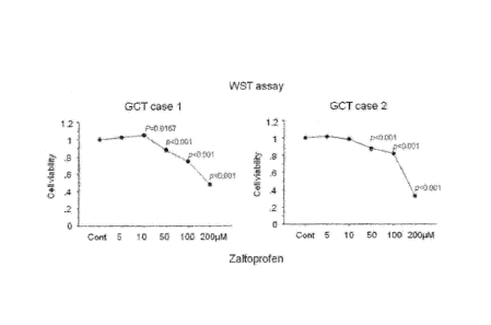
[Fig. 1] Graphs showing results of suppression of proliferation of GCT
cultured cells
that were cultured in a zaltoprofen-containing medium.
[Fig. 2] Photographs showing results of Tunel assay performed with GCT
cultured
cells that were cultured in a zaltoprofen-containing medium.
[Fig. 31 A graph showing ratios of Tunel-positive cells in GCT cultured cells
that
were cultured in a zaltoprofen-containing medium.
[Fig. 41 Photographs showing results of caspase 3 staining of GCT cultured
cells that
were cultured in a zaltoprofen-containing medium.
[Fig. 5] A graph showing ratios of caspase 3-positive cells in GCT cultured
cell
cultured in a zaltoprofen-containing medium.
[Fig. 6] Photographs showing results of PPARy staining of GCT cultured cells
that
were
7b
CA 2868311 2020-03-12
CA 02868311 2014-09-23
cultured in a zaltoprofen-conta i fling medium.
[Fig. 71 A graph showing ratios of PPARy-positive cells in OCT cultured cells
that were
cultured in a zaltoprofen-containing medium.
[Fig. 81 Photographs showing results of lipid staining of OCT cultured cells
that were
cultured in a zaltoprofen-containing medium.
[Fig. 9] A graph showing ratios of lipid-positive cells in GCT cultured cells
that were
cultured in a zaltoprofen-containing medium.
[Fig. 10] Photographs showing results of Panel assay and caspase 3 staining of
GCT
samples derived from a GCT patient administered with zaltoprofen.
[Fig. 11] Photographs showing results of PPARy staining of GCT samples derived
from
a GCT patient administered with zaltoprofen.
[Fig. 121 Graphs showing results of suppression of proliferation of GCT
cultured cells
that were cultured in an acetaminophen, indomethacin, diclofenac, or
troglitazone-
containing medium.
[Fig. 131 Graphs showing results of suppression of proliferation of GCT
cultured cells
that were cultured in an acetaminophen, indomethacin, diclofenac, or
troglitazone-
containing medium.
[Fig. 141 Graphs showing effect of PPARy siRNA on GCT cultured cells that were
cultured in a zaltoprofen or troglitazone-containing medium,
[Fig. 15] Graphs showing effect of PPARy siRNA on GCT cultured cells that were
cultured in a zaltoprofen or troglitazone-containing medium.
[Fig. 161 Graphs showing results of suppression of proliferation of cultured
cells
derived from giant cell tumor of tendon sheath or cultured cells derived from
pigmented vi]lonodular synovitis cultured in a zaltoprofen-containing medium.
[Fig. 17] Graphs showing results of suppression of proliferation of cultured
cells
derived from giant cell tumor of tendon sheath or cultured cells derived from
pigmented villonodular synovitis cultured in a troglitazone-containing medium.
[Fig. 181 Photographs showing results of Tunel assay of cultured cells derived
from
giant cell tumor of tendon sheath cultured in a zaltoprofen or troglitazone-
containing
medium.
[Fig. 19] Graphs showing ratios of Panel-positive cells in cultured cells
derived from
giant cell tumor of tendon sheath cultured in a zaltoprofen or troglitazone-
containing
medium.
8
_ _____________________________________________ _
CA 02868311 2014-09-23
[Fig. 201 Photographs showing results of Tunel assay of cultured cells derived
from
pigmented villonodular synovitis cultured in a zaltoprofen or troglitazone-
contai wing
medium.
[Fig. 21] Graphs showing ratios of Tunel-positive cells in cultured cells
derived from
pigmented villonodular synovitis cultured in a zaltoprofen or troglitazone-
containing
medium.
[Fig. 22] Photographs showing results of caspase 3 staining of cultured cells
derived
from giant cell tumor of tendon sheath cultured in a zaltoprofen or
troglitazone-
containing medium.
[Fig. 231 A graph showing ratios of caspase 3-positive cells in cultured cells
derived
from giant cell tumor of tendon sheath cultured in a zaltoprofen or
troglitazone-
containing medium.
[Fig. 24] Photographs showing results of caspase 3 staining of cultured cells
derived
from pigmented villonodular synovitis cultured in a zaltoprofen or
troglitazone-
containing medium.
[Fig. 25] Graphs showing ratios of caspase 3-positive cells in cultured cells
derived from
pigmented villonodular synovitis cultured in a zaltoprofen or troglitazone-
containing
medium.
[Fig. 26] Photographs showing results of PPARy staining of cultured cells
derived from
giant cell tumor of tendon sheath cultured in a zaltoprofen or troglitazone-
containing
medium.
[Fig. 27] Graphs showing ratios of PPARy-positive cells in cultured cells
derived from
giant cell tumor of tendon sheath cultured in a zaltoprofen or troglitazone-
containing
medium.
[Fig. 28] Photographs showing results of PPARy staining of cultured cells
derived from
pigmented villonodular synovitis cultured in a zaltoprofen or troglitazone-
containing
medium.
[Fig. 29] Graphs showing ratios of PPARy-positive cells in cultured cells
derived from
pigmented villonodular synovitis cultured in a zaltoprofen or troglitazone-
containing
medium.
[Fig. 30] Photographs showing results of lipid staining of cultured cells
derived from
giant cell tumor of tendon sheath or cultured cells derived from pigmented
villonodular
synovitis cultured in a zaltoprofen-containing medium.
9
CA 02868311 2014-09-23
[Fig. 311 Graphs showing ratios of lipid-positive cells in cultured cells
derived from
giant cell tumor of tendon sheath or cultured cells derived from pigmented
villonodular
synovitis cultured in a zaltoprofen-containing medium.
[Fig. 32] Graphs showing results of suppression of proliferation of GCT
cultured cells,
cultured cells derived from giant cell tumor of tendon sheath, and cultured
cells
derived from pigmented villonodular synovitis cultured in a pioglitazone-
containing
medium.
[Fig. 33] A photograph showing an X-ray image of the pelvic part of a patient
with
recurrence of osteoclastoma in the pelvic part obtained before the
administration of
zaltoprofen.
[Fig. 341 A photograph showing an MRI image of the pelvic part of a patient
with
recurrence of osteoclastoma in the pelvic part obtained before the
administration of
zaltoprofen.
[Fig. 35] A photograph showing an NMI image of the pelvic part of a patient
with
recurrence of osteoclastoma in the pelvic part obtained after two months of
the
administration of zaltoprofen. The arrows indicate a tumor necrosis region.
[Fig. 36] A photograph showing an MRI image of the pelvic part of a patient
with
recurrence of osteoclastoma in the pelvic part obtained after four months of
the
administration of zaltoprofen. The arrows indicate a tumor necrosis region.
[Fig. 371 A photograph showing a sagittal MRI image of the right knee part of
a patient
with recurrence of pigmented villonodular synovitis in the right knee part
obtained
before the administration of zaltoprofen.
[Fig. 381 A photograph showing a transversal MRI image of the right knee part
of a
patient with recurrence of pigmented villonodular synovitis in the right knee
part
obtained before the administration of zaltoprofen.
[Fig. 39] A photograph showing a sagittal MRI image of the right knee part of
a patient
with recurrence of pigmented villonodular synovitis in the right knee part
obtained
after three months of the administration of zaltoprofen. The arrows indicate
attenuation of the MRI imaging effect.
[Fig. 40] A photograph showing a transversal IVLRI image of the right knee
part of a
patient with recurrence of pigmented villonodular synovitis in the right knee
part
obtained after three months of the administration of zaltoprofen. The arrows
indicate
attenuation of the IVIRI imaging effect.
CA 02868311 2014-09-23
[Fig. 411 A photograph showing a coronal MRI image of the right knee part of a
patient
with recurrence of pigmented villonodular synovitis in the right knee part
obtained
before the administration of zaltoprofen.
[Fig. 4211 A photograph showing a transversal MRI image of the right knee part
of a
patient with recurrence of pigmented villonodular synovitis in the right knee
part
obtained before the administration of zaltoprofen.
[Fig. 431 A photograph showing a coronal MRI image of the right knee part of a
patient
with recurrence of pigmented villonodular synovitis in the right knee part
obtained
after two months of the administration of zaltoprofen. The arrows indicate
shrinkage
of tumor.
[Fig. 441 A photograph showing a transversal MRI image of the right knee part
of a
patient with recurrence of pigmented villonodular synovitis in the right knee
part
obtained after two months of the administration of zaltoprofen. The arrows
indicate
shrinkage of tumor.
[Fig. 45] Graphs showing results of suppression of proliferation of a
chondrosarcoma-
derived cell line cultured in a zaltoprofen, troglitazone, or pioglitazone-
containing
medium.
[Fig. 461 Photographs showing results of caspase 3 staining of a
chondrosarcoma-
derived cell line cultured in a zaltoprofen, troglitazone, or pioglitazone-
containing
medium.
[Fig. 471 Graphs showing ratios of caspase 3-positive cells in cells of a
chondrosarcoma-
derived cell line cultured in a zaltoprofen, troglitazone, or pioglitazone-
containing
medium.
[Fig. 481 Photographs showing results of PPARy staining of a chondrosarcoma-
derived
cell line cultured in a zaltoprofen, troglitazone, or pioglitazone-containing
medium.
[Fig. 49] Graphs showing ratios of PPARy-positive cells in cells of a
chondrosarcoma-
derived cell line cultured in a zaltoprofen, troglitazone, or pioglitazone-
contsining
medium.
Modes for Carrying out the Invention
[0012]
The inventors of the present invention found that PPARy was not expressed in
cells derived from patients with giant cell tumor occurring in a bone and soft
tissue,
11
CA 02868311 2014-09-23
and they also found that a specific substance having a PPARy-agonistic
activity
induced expression of PPARy in giant cell tumor occurring in a bone and soft
tissue,
and simultaneously induced apoptosis or fat cell differentiation. Therefore,
the
present invention provides a prophylactic or therapeutic agent for giant cell
tumors
occurring in a bone and soft tissue, which can induce apoptosis or fat cell
differentiation mediated by PPARy in giant cell tumor occurring in a bone au
d soft
tissue on the basis of the PPARy expression-inducing activity and the PPARy-
agonistic
activity The present invention also provides a prophylactic or therapeutic
agent for
chondrosarcoma, which can induce apoptosis or fat cell differentiation
mediated by
PPARy on the basis of the PPARy expression-inducing activity and the PPARy-
agonistic activity.
[0013]
The giant cell tumor occurring in a bone and soft tissue to which the
prophylactic or therapeutic agent of the present invention can be applied is
not
particularly limited, so far as giant cells developed in a bone and soft
tissue are
observed in the tumor. In particular, the agent of the present invention can
be
preferably applied to a tumor that generates giant cells in circumferences of
bone, joint,
or tendon sheath. The tumor in which giant cells developed in a bone and soft
tissue
are observed is preferably a benign tumor. Examples of such a tumor include,
for
example, osteoclastoma, giant cell tumor of tendon sheath, pigmented
villonodular
synovitis, and the like. Examples of benign giant cell tumor occurring in a
bone and
soft tissue also include chondroblastoma, nonossifying fibroma, osteoblastoma,
aneurysmal bone cyst, and the like.
Examples of the chondrosarcoma to which the prophylactic or therapeutic
agent of the present invention can be applied include conventional
chondrosarcoma,
periosteal chondrosarcoma, mesenchymal chondrosarcoma, dedifferentiated
chondrosarcoma, clear-cell chondrosarcoma, extraskeletal myxoid
chondrosarcoma, and
the like.
The prophylactic or therapeutic agent of the present invention can also be
used for a tumor in which expression of PPARy is observed. Examples of such a
tumor
in which expression of PPARy is observed include breast cancer, colon cancer,
lung
cancer, thyroid gland cancer, esophageal cancer, gastric cancer, pancreatic
cancer, liver
cancer, kidney cancer, vesical cancer, ovarian cancer, uterine cervix
carcinoma, prostate
12
CA 02868311 2014-09-23
cancer, malignant melanoma, leukemia, malignant lymphoma, liposarcoma,
leiomyosarcoma, bone sarcoma, and the like.
[00141
PPARy is a protein belonging to the intranuclear receptor superfamily, and it
recognizes the PPAR, response element (PPRE) when it has formed a heterodimer
with
the retinoici X receptor (RXR), which is one of the intranuclear receptors,
and activates
transcription of a gene downstream therefrom.
[0015]
In the present invention, examples of such a substance having a PPARy
expression-inducing activity and a PPARy-agonistic activity include non-
steroidal anti-
inflammatory agents. Therefore, the prophylactic or therapeutic agent for
giant cell
tumors occurring in a bone and soft tissue or chondrosarcoma that can induce
apoptosis or fat cell differentiation mediated by PPARy in giant cell tumors
occurring
in a bone and soft tissue or in chondrosarcoma on the basis of a PPARy
expression
inducing activity and a PPARy-agonistic activity is, according to an
embodiment of the
present invention, a prophylactic or therapeutic agent for giant cell tumors
occurring in
a bone and soft tissue or for chondrosarcoma, which comprises a non-steroidal
anti-
inflammatory agent as an active ingredient. The non-steroidal anti-
inflammatory
agent referred herein is not particularly limited. so long as it is a compound
that
inhibits the synthesis of prostaglandins and thromboxanes by inhibition of
cyclooxygenase, and thereby exhibits an anti-inflammatory activity Examples
include,
for example, zaltoprofen, diclofenac, indomethacin, oxaprozin, acetaminophen,
ketoprofen, and the like, and preferred examples include zaltoprofen,
diclofenac, and
indomethacin. Other than the non-steroidal anti-inflammatory agents, examples
of
the substance include, for example, thiazolidine derivatives. Therefore, the
prophylactic or therapeutic agent for giant cell tumors occurring in a bone
and soft
tissue or for chondrosarcoma that can induce apoptosis or fat cell
differentiation
mediated by PPARy in giant cell tumors occurring in a bone and soft tissue or
in
chondrosarcoma on the basis of a PPARy expression-inducing activity and a
PPARy
agonistic activity may also be a prophylactic or therapeutic agent for giant
cell tumors
occurring in a bone and soft tissue or for chondrosarcoma, which comprises a
thiazolidine derivative as an active ingredient. Thiazolidine derivatives are
known to
activate PPARy, and are PPARy agonists that promote transcription of the
adiponectin.
13
_ _____________________________________________ ,
CA 02868311 2014-09-23
A
gene. Examples of such thiazolidine derivatives include, for example,
troglitazone, =
rosigLitazone, pioglitazone, balaglitazone, rivoglitazone, and the like.
[0016]
In the present invention, the PPARy expression-inducing activity of the
substances having a PPARy expression-inducing activity and a PPARy-agonistic
activity, which substances include non-steroidal anti-inflammatory agents and
thiazolidine derivatives, can be confirmed by the known methods described
later. For
example, expression thereof can be confirmed by a method of extracting mRNA
from
giant cell tumors occurring in a bone and soft tissue or from chondrosarcoma
by an
ordinary method, and performing a reverse transcription reaction and PCR using
primers that enables amplification of a PPARy transcription product, or a
method of
fixing giant cell tumors occurring in a bone and soft tissue or fixing
choncLrosarcoma, or
extracting a cell disruption product, and confirming expression of a PPARy
translation
product by using an antigen-antibody reaction using antibodies directed to the
PPARy
translation product. The transcription product and translation product of
PPARy are
known. For example, as for the transcription product, nucleotide sequence
information is disclosed as GenBank accession No. B0006811, and as for the
translation product thereof, amino acid sequence information is disclosed as
GenBank
accession No. AAH06811. Further, the PPARy-agonistic activity can be confirmed
by,
for example, measuring expression level of a downstream gene, or expression
level of a
lipid. Examples of the gene locating downstream of PPARy include, for example,
an
apoptosis-related gene, fat cell differentiation-related gene,
arteriosclerosis-related
gene, anti-inflaramation-related gene, and the like. The apoptosis-related
gene may
be a gene responsible for an apoptosis signal, or may be a marker gene for
apoptosis.
Examples of the apoptosis-related gene include, for example, those of caspase
3, p53,
and the like. As for the transcription product of caspase 3, nucleotide
sequence
information is disclosed as GenBank accession No. BC016926, and as for the
translation product thereof, amino acid sequence information is disclosed as
GenBank
accession No. AAH16926. As for the transcription product of p53, nucleotide
sequence
information is disclosed as GenBank accession No. NM_000546, and as for the
translation product thereof, amino acid sequence information is disclosed as
GenBank
accession No. NP_000537. The fat cell differentiation-related gene may be a
gene that
induces differentiation into fat cells, or may be a marker gene for fat cell
differentiation.
14
CA 02868311 2014-09-23
A
Examples of the fat cell differentiation-related gene include, for example,
those of
Setd8 (SET domain containing (lysine methyItransferase) 8), Setdbl (SET
domain,
bifurcated 1), LPL (Lipoprotein Lipase), leptin, FABP4/aP2 (fatty acid-binding
protein-
4), acliponectin, a2Col6 (a chain 2 of type 6 collagen), and the like. As for
the
transcription product of Seta, nucleotide sequence information is disclosed as
GenBank accession No. NM_020382, and as for the translation product thereof,
amino
acid sequence information is disclosed as GenBank accession No, NP_065115. As
for
the transcription product of Setdbl, nucleotide sequence information is
disclosed as
GenBank accession No. BCO28671, and as for the translation product thereof,
amino
acid sequence information is disclosed as GenBank accession No. AA1128671. As
for
the transcription product of LPL, nucleotide sequence information is disclosed
as
GenBank accession No. BC011353, and as for the translation product thereof,
amino
acid sequence information is disclosed as GenBank accession No. AA1111353. As
for
the transcription product of leptin, nucleotide sequence information is
disclosed as
GenBank accession No. BC069452, and as for the translation product thereof,
amino
acid sequence information is disclosed as GenBank accession No. AAH69452. As
for
the transcription product of FABP4/aP2, nucleotide sequence information is
disclosed
as GenRank accession No. BC003672, and as for the translation product thereof,
amino
acid sequence information is disclosed as GenBank accession No. AA1103672. As
for
the transcription product of adiponectin, nucleotide sequence information is
disclosed
as GenBank accession No. BC096308, and as for the translation product thereof,
amino
acid sequence information is disclosed as GenBank accession No. AA1196308. As
for
the transcription product of a2Co16, nucleotide sequence information is
disclosed as
GenBank accession No. B0002483, and as for the translation product thereof,
amino
acid sequence information is disclosed as GenBank accession No. AA11002483.
Examples of the arteriosclerosis-related gene include, for example, those of
AT1R
(angiotensin II receptor I), and the like. As for the transcription product of
AT1R,
nucleotide sequence information is disclosed as GenBank accession No.
B0068494, and
as for the translation product thereof, amino acid sequence information is
disclosed as
GenBank accession No. AAH68494. Examples of the anti-inflammation-related gene
include, for example, those of NF-KB (nuclear factor kappa-light-chain-
enhancer of
activated B cells), and the like. As for the transcription product of NF-KB,
nucleotide
sequence information is disclosed as GenBank accession No. NM_003998, and as
for
CA 02868311 2014-09-23
the translation product thereof, amino acid sequence information is disclosed
as
GenBank accession No. NP_003989. The lipid is not particularly limited so long
as it
is a lipid contained in fat cells or 6RA-ties, and examples include
phospholipids,
glycolipids, lipoproteins, acylglycerols, ceramicles, and the like.
[00171
Specifically, the PPARy expression-inducing activity can be examined by
preparing an RNA (for example, total RNA, or mRNA) fraction from a cell, and
detecting the transcription product of the PPARy gene contained in the
fraction. Such
an RNA fraction can be prepared by a known method such as the guanidine-Csel
ultracentrifugation method and the AGPC method, and total RNA of high purity
can be
quickly and conveniently prepared from a small number of cells by using a
commercial
kit for RNA extraction (for example, RNeasy Mini Kit produced by QIAGEN and
the
like). Examples of the means for detecting a transcription product of a gene
in an
RNA fraction include, for example, a method of using hybridization (Northern
blotting,
dot blotting, DNA chip analysis, and the like), a method of using PCR (RT.PCR,
competitive PCR, real-time PCR, and the like), and the like. The quantitative
PCR
method as competitive PCR and real-time PCR is preferred, since it enables
quick and
convenient detection of change of expression of a gene from an extremely small
amount
of sample with good quantification ability.
[00181
When Northern blotting or dot blot hybridization is used, the PPARy gene can
be detected by, for example, using a nucleic acid probe that enables specific
detection of
a transcription product of the gene. The nucleic acid probe used for
measurement of
the PPARy expression-inducing activity is a polynucleoticle comprising a
contiguous
part or all of the gene containing about 15 or more nucleotides, preferably
about 18 to
500 nucleotides, more preferably about 18 to 200 nucleotides, still more
preferably
about 18 to 50 nucleotides, or a polynucleotide comprising a complementary
sequence
thereof Examples of the part or all of PPARy gene include a part or all of the
known
nucleotide sequence of PPARy mentioned above. The polynucleotid.e may be
either
DNA or RNA, or may be a DNAJRNA chimera. Preferably the polynucleotide
includes
DNA. Further, the polyuucleotide used as the probe may be a double-stranded or
single-stranded polynueleotide. The double-stranded polynucleotid.e may be a
double-
stranded DNA, double-stranded RNA, or a hybrid of DNA and RNA. In the case of
the
16
CA 02868311 2014-09-23
=
=
single-stranded polynucleotide, an antisense strand can be used.
1:00-191
The nucleic acid probe used for the measurement of the PPARy expression-
inducing activity is a polyrrucleotide containing a nucleotide sequence that
can
hybridize with a transcription product of the PPARy gene under stringent
conditions.
The hybridization can be performed by a method known per se or a method
similar to
such a known method, for example, the methods described in Molecular Cloning,
2nd
edition (J. Sambrook et at, Cold Spring Harbor Lab. Press, 1989), and the
like.
Examples of the stringent conditions include, for example, a hybridization
reaction at
45 C in 6 x SSC (sodium chloride/sodium citrate) followed by once or more
times of
washing in 0.2 x SSC/0.1% SDS at 65 C, and the like. Those skilled in the art
can
adjust the stringency to be a desired stringency by appropriately changing
salt
concentration of the hybridization solution, temperature for the hybridization
reaction,
concentration of the probe, length of the probe, number of mismatches, time of
the
hybridization reaction, salt concentration of the washing solution,
temperature for
washing, and the like.
[00201
The nucleic acid that functions as a probe capable of detecting expression of
the PPARy gene can be obtained by amplifying a nucleic acid of a desired
length by
PCR using a primer set that is capable of amplifying a part or all of a
transcription
product of the gene, and as the template, cDNA or genoraic DNA derived from
any of
cells [for example, hepatic cells, splenic cells, nerve cells, glia cells,
pancreatic B cells,
marrow cells, mesangial cells, Langerhans cells, epidermal cells, epithelial
cells, goblet
cells, endothelial cells, smooth muscle cells, fibroblasts, fibrocytes,
niyocytes, fat cells,
iramun.oc3rtes (for example, macrophage, T cell, B cell, natural killer cell,
mast cell,
neutrophile, basophile, eosinophile, monocyte), megakaryocytes, synovial
cells,
chondrocytes, osteocytes, osteoblasts, osteoclasts, breast cells, and
interstitial cells, or
precursor cells, stem cells, or cancer cells of these cells, and the like] or
any of tissues
in which the foregoing cells exist [for example, brain, parts of brain (for
example,
olfactory bulb, amygdaloid nucleus, basal ganglia, hippocampus, thalamus,
hypothalamus, cerebral cortex, oblongata, cerebellum), spinal cord, pituitary
gland,
stomach, pancreas, kidney, liver, gonad, thyroid, gallbladder, bone marrow,
suprarenal
gland, skin, lung, alimentary canal (for example, large intestine, small
intestine), blood
17
- - - -
CA 02868311 2014-09-23
vessel, heart, thymus gland, spleen, submaTillary gland, peripheral blood,
prostate
gland, testis, ovary, placenta, uterus, bone, joint, fat tissue, skeletal
muscle, and the
like] of an individual (for example, human, ape, mouse, rat, dog, bovine,
equine, swine,
ovine, goat, cat, rabbit, hamster, guinea pig and the like, preferably human),
or by
cloning the aforementioned gene or cDNA from cDNA or genomic DNA library
derived
from any of the cells and tissues mentioned above by colony or plaque
hybridization,
and if necessary, obtaining a fragment of an appropriate length by using a
restriction
enzyme and the like The hybridization can be performed by, for example, the
methods
described in Molecular Cloning, 2nd edition (mentioned above), and the like
Alternatively, the nucleic acid can be obtained by chemical synthesis of a
part or all of
the nucleotide sequence and/or a complementary sequence thereof based on the
nucleotide sequence information of the aforementioned PPARy gene, using a
commercial automatic DNA/RNA synthesizer. Further, by in situ (on chip)
synthesis
of the nucleic acid directly on a solid phase such as silicone or glass, a
chip on which
the nucleic acid is immobilized on a solid phase can also be prepared.
[0021]
Further, the nucleic acid that functions as a primer that is capable of
amplifying a part or all of the transcription product of the PPARy gene can be
obtained
by chemical synthesis of a part of the nucleotide sequence and a complementary
strand
sequence thereof based on the nucleotide sequence information of the
aforementioned
PPARy gene, using a commercial automatic DNA/RNA synthesizer.
[00221
The nucleic acid is preferably labeled with a labeling agent, in order to make
it
possible to detect and quantify the target nucleic acid. As the labeling
agent, for
example, a radioactive isotope, enzyme, fluorescent substance, luminescent
substance,
and the like are used. As the radioactive isotope, for example, [82P], [RH],
[ma and
the like are used. As the enzyme, a stable enzyme showing a high specific
activity is
preferred, and there are used, for example, 13-galactosidase, 8-glucosidase,
alkaline
phosphatase, peroxidase, malate dehydrogenase, and the like. As the
fluorescent
substance, for example, ftuorescamine, fluorescein isothiocyanate, and the
like are used.
As the luminescent substance, for example, luminol, luminol derivatives,
luciferin,
lucigenin, and the like are used. Furthermore, biotin-(strept)avidin can be
used for
the binding of the probe and the labeling agent.
18
CA 02868311 2014-09-23
=
[0023]
When Northern hybridization is performed, by subjecting an RNA fraction
prepared as described above to separation by gel electrophoresis, blotting RNA
to a
membrane such as cellulose nitrate, nylon, or polyvinylidene diftuoride
membrane,
allowing specific hybridization in a hybridization buffer containing a labeled
probe
prepared as described above, and then measuring amount of the labeling agent
binding
to the membrane for each band by an appropriate method, expression amount of
the
PPARy gene can be measured. When dot blotting is used, by similarly allowing a
hybridization reaction of the RNA fraction spotted on a membrane, and
measuring
amount of the labeling agent for each spot, expression amount of the gene can
be
measured.
[0024]
When DNA chip analysis is used, for example, cDNA introduced with an
appropriate promoter such as T7 promoter is synthesized from the RNA fraction
prepared as described above by a reverse transcription reaction, and cRNA is
further
synthesized by using an RNA polymerase (if a mononucleotide labeled with
biotin or
the like is used as a substrate in this synthesis, a labeled cRNA can be
obtained). By
contacting labeled cRNAs with the chip on which a probe is immobilized on a
solid
phase to allow a hybridization reaction, and measuring amounts of labeling
agents
binding to the probes, expression amount of the PPARy gene can be measured.
[0025]
According to another preferred embodiment, the quantitative PCR method is
used as the method for measuring expression amount of the PPARy gene. As the
quantitative PCR, for example, competitive PCR, real-time PCR, and the Rke are
available.
Examples of the set of oligonucleotides used as primers in PCR include, for
example, nucleic acid primers that enable specific detection of a
transcription product
of the aforementioned PPARy gene. In a preferred embodiment, as the set of
nucleic
acid primers used for the test method of the present invention, examples
include a set
of oligonucleotides comprising a polynucleotide complementary to a
polynucleotide
(sense strand) sequence having a length of a contiguous nucleotide sequence
containing
about 15 or more nucleotides, preferably about 15 to 50 nucleotides, more
preferably
about 15 to 30 nucleotides, still more preferably about 15 to 25 nucleotides,
and
19
_
. <
CA 02868311 2014-09-23
=
designed so that it can amplify a DNA fragment of about 100 bp to several kbp,
and a
polynucleotide that can hybridize with a polynucleotide (antisense strand)
having a
nucleotide sequence complementary to the aforementioned polynucleotide
sequence.
[00261
The competitive RT-PCR means a method of calculating amount of an objective
DNA by competitively performing amplification reactions in a reaction mixture
also
containing, as a competitor, a known amount of another template nucleic acid
which
can be amplified with a set of primers that can amplify the objective DNA, and
comparing amounts of amplified products. Therefore, when the competitive RT-
PCR
is used, there is used a known amount of a competitor nucleic acid that can be
amplified with the aforementioned primer set, of which amplification product
can be
distinguished (on the basis of, for example, difference of amplified sizes,
difference of
migration patterns of restriction enzyme-processed fragments observed in
electrophoresis, and the like) from the amplification product of the target
nucleic acid
(namely, transcription product of the nucleotide sequence information on the
PPARy
gene) after the amplification, in addition to the aforementioned primer set.
The target
nucleic acid and the competitor nucleic acid are competitively amplified,
since they use
the same primers, and therefore the ratio of the amounts of the amplification
products
reflects the ratio of the amounts of the original templates. The competitor
nucleic acid
may be DNA or RNA. When the competitor nucleic acid is DNA, after cDNA is
synthesized by a reverse transcription reaction from an RNA fraction prepared
as
described above, PCR can be performed in the presence of the aforementioned
primer
set and the competitor. When the competitor is RNA, after the competitor is
added to
an RNA fraction to perform a reverse transcription reaction, the
aforementioned
primer set can be added to perform PCR. In the latter case, efficiency of the
reverse
transcription reaction is also taken into consideration, and therefore the
absolute
amount of the original mRNA can be estimated.
[00271
The real-time PCR is a method of monitoring amount of amplification product
in real time by using a fluorescent reagent, and requires an apparatus
integrally
comprising a thermal cycler and a spectrophotofluorometer. Such an apparatus
is
marketed. Several methods are available in which different fluorescent reagent
is
used, including, for example, the intercalator method, TaqManTm probe method,
CA 02868311 2014-09-23
Molecular Beacon method, and the like. In these methods, after cDNA is
synthesized
by a reverse transcription reaction from an RNA fraction prepared as described
above,
the aforementioned primer set, SYBR Green I, a reagent that emits fluorescence
when
it binds with a double-stranded DNA (intercalator), such as ethiclium bromide,
and a
fluorescent regent (probe) comprising a nucleic acid that can be used as the
aforementioned probe (the nucleic acid can hybridize to a target nucleic acid
in a region
to be amplified), of which each end is modified with a fluorescent substance
(for
example, FA1V1, HEX, TET, FITC, and the like) or a quenching substance (for
example,
TAMRA, DABCYL, and the like), e.g. TaqManTm probe or Molecular Beacon probe,
are
added to a reaction system for PCR. Since the intercalator emits fluorescence
when it
binds with a synthesized double-stranded DNA and irradiated with an excitation
light,
production amount of the amplification product can be monitored by measuring
fluorescence intensity, and the amount of the original template cDNA can be
thereby
estimated. The TaqMannii probe is an oligonucleotide that can hybridize with
the
region of the target nucleic acid to be amplified, of which each end is
modified with a
fluorescent substance or a quenching substance. It hybridizes with the target
nucleic
acid, but does not emit fluorescence because of the presence of the quenching
substance
at the time of the annealing, and it is decomposed by exonuclease activity of
a DNA
polymerase at the time of the extension reaction to release a fluorescent
substance,
which emits fluorescence. Therefore, by measuring the fluorescence intensity,
the
production amount of the amplification product can be monitored, and the
amount of
the original template cDNA can be estimated therefrom. The Molecular Beacon
probe
is an oligonucleotide that can hybridize with a region of a target nucleic
acid to be
amplified and can have a hairpin secondary structure, of which both ends are
modified
with a fluorescent substance and a quenching substance, respectively. When it
has
the hairpin secondary structure, it does not emit fluorescence because of the
presence
of the quenching substance, but when it hybridizes with a target nucleic acid
at the
time of annealing, and thus the distance between the fluorescent substance and
the
quenching substance increases, it emits fluorescence. Therefore, by measuring
the
fluorescence intensity, production amount of the amplification product can be
monitored, and amount of the original template cDNA can be estimated
therefrom.
Since the real-time RT-PCR enables real time monitoring of the amount of the
product
amplified in PCR, it does not require electrophoresis, and thus enables
quicker analysis
21
CA 02868311 2014-09-23
of expression of the PPARy gene.
[0028]
Alternatively, expression of the PPARy gene in a cell can be investigated by
preparing a protein fraction from the cell and detecting a translation product
of the
gene (namely, PPARy) contained in the fraction. PPARy can be detected by an
immunoassay (for example, ELISA, FIA, RIA, Western blotting, and the like)
using
antibodies that specifically recognize the protein, or it can be detected by
measuring
the activity of the protein using a known method. Further alternatively, the
protein
can also be detected by using mass spectrometry such as MALDT-TOFMS.
[0029]
The antibody that specifically recognizes PPARy can be prepared by a general
available production method using a polypeptide constituting the protein,
total
sequence of a partial peptide of the polypeptide showing antigenicity, or a
partial
peptide thereof corresponding to an epitope as an immunogen. In this
specification,
examples of the antibody include naturally occurring antibodies such as
polyclonal
antibodies and monoclonal antibodies (mAb), chimeric antibodies that can be
produced
by using a gene recombination technique, humanized antibodies, single strand
antibodies, and bindable fragments thereof, but not limited to these examples.
The
antibody is preferably a polyclonal antibody, monoclonal antibodies, or a bind
able
fragment thereof. The bindable fragment means a partial region of any of the
aforementioned antibodies having a specific binding activity, and specific
examples
thereof include, for example, F(abl)2, Fab', Fab, Fv, sFy, dsFv, sdAb, and the
like (Exp.
Opin. Ther. Patents, Vol. 6, No. 5, pp.441456, 1996). A class of the antibody
is not
particularly limited, and the antibody may be any of those having any of
isotypes such
as IgG, IgM, IgA, IgD, or IgE. The antibody is preferably IgG or IgM, and it
is more
preferably IgG, if easiness of purification and the like are taken into
consideration.
[0030]
When each of the immunoassays is applied to the measurement of the PPARy
expression-inducing activity, setting of special conditions, operations, and
the like is
not required. A system for measuring PPARy may be constructed with ordinary
conditions and operation methods for each method with usual technical
consideration
of those skilled in the art. For the details of these general technical means,
review
articles, published books, and the like can be referred to. For example,
22
CA 02868311 2014-09-23
J.
"Radioimmunoassay", Edited by H. Irie (Kodansha, 1974), "Radioirnmunoassay,
Second
= Series", Edited by H. Irie (Kodansha, 1979), "Enzyme Immunoassay", Edited
by E.
Ishikawa (Igaku-Shoin, 1978), "Enzyme Immunoassay", 2nd Edition, Edited by E.
Ishikawa (Igaku-Shoin, 1982), "Enzyme Immunoassay", 3rd Edition, Edited by E.
Ishikawa (Igaku-Shoin, 1987), "Methods in ENZYMOLOGY", Vol. 70 (Immunochemical
Techniques (Part A)), ibid., Vol. 73 (Immunochemical Techniques (Part B)),
ibid., Vol. 74
(Immunochemical Techniques (Part C)), ibid., Vol. 84 (Immunochemical
Techniques
(Part D: Selected Immunoassays)), ibid., Vol. 92 (Immunochemical Techniques
(Part E:
Monoclonal Antibodies and General Immunoassay Methods)), ibid., Vol. 121
(Immunochemical Techniques (Part I: Hybridoma Technology and Monoclonal
Antibodies)) (these are published by Academic Press), and the like can be
referred to.
[0031]
Further, the PPARy-agonistic activity can be measured in the same manner as
that for the measurement of the PPARy expression-inducing activity mentioned
above,
i.e., by preparing an RNA fraction, a protein fraction, or a lipid fraction
from a cell, and
detecting a transcription product, or a translation product of a gene locating
downstream of the PPARy gene (for example, apoptosis-related gene, fat cell
differentiation-related gene, arteriosclerosis-related gene, anti-inflammation-
related
gene, and the like) or a lipid contained in the fraction. The methods for
preparing the
RNA fraction and the protein fraction and the methods for detecting them may
be the
same as the aforementioned methods explained for the measurement of the PPARy
expression-inducing activity. As for the preparation method of a lipid, it may
be
prepared by using a known method, and there can be used, for example, the
Folch
method in which a lipid is extracted from a sample containing the lipid by
adding
several-fold volume of a solvent such as a mixture of chloroform and methanol
to the
sample, the Bligh-Dyer method in which a lipid is extracted by adding several-
fold
volume of a solvent such as a mixture of chloroform, methanol and water, or
the like.
Further, as for the method for detecting the separated lipid, it can be
detected by using
a known method such as liquid chromatography (LC), gas chromatography (GC),
and
high performance liquid chromatography (HPLC). Alternatively, a method of
directly
detecting a lipid contained in a fat cell or tissue may also be used. The
reagents and
the like usable for such a method are marketed, and for example, HCS LipidTOX
Phospholipidosis and Steatosis Detection Kit (Invitrogen) and the like can be
used.
23
-
_
CA 02868311 2014-09-23
=
[0032]
=
Examples of person to whom the prophylactic or therapeutic agent of the
present invention for giant cell tumors occurring in a bone and soft tissue or
for
chondrosarcoma can be applied, which agent comprises a substance having a
PPAR,y
expression-inducing activity and has a PPAR.y-agonistic activity used in the
present
invention, preferably a non-steroidal anti-inflammatory agent or a
thiazolidine
derivative, as an active ingredient, include, for example, a person who has
clinically
diagnosed to have giant cell tumor occurring in a bone and soft tissue or have
chondrosarcoma, a person suspected to be that mentioned above, and a person
for
whom onset of such a disease as mentioned above is expected. Further, the
therapeutic agent of the present invention can also be preferably applied to a
metastatic cancer of which primary lesion is giant cell tumors occurring in a
bone and
soft tissue or chondrosarcoma. Examples of such metastatic cancer include
those of
lung cancer, colon cancer, breast cancer, prostate cancer, esophageal cancer,
gastric
cancer, liver cancer, biliary carcinoma, spleen carcinoma, renal carcinoma,
vesical
cancer, uterine cancer, ovarian cancer, testis cancer, thyroid gland cancer,
pancreatic
cancer, brain tumor, blood tumor, and the like.
[0033]
The prophylactic or therapeutic agent for giant cell tumors occurring in a
bone
and soft tissue or for chondrosarcoma of the present invention can be orally
used in the
form of a tablet, which may have a sugar coating as required, capsule, elixir,
microcapsule, or the like, or can be parenterally used in the form of an
injection such as
a sterile solution or suspension in water or another pharmacologically
acceptable
solvent. The prophylactic or therapeutic agent can be prepared by forming a
mixture
together with physiologically acceptable carrier, flavoring agent, excipient,
vehicle,
preservative, stabilizer, binder, and the like in the form of a generally
acceptable unit
dosage form required for implementation of pharmaceutical preparation. Amount
of
the active ingredient in such a preparation as mentioned above is
appropriately chosen
by taking a dose into consideration, which will be described later.
[0034]
As additives that can be mixed in tablets, capsules, and the like, there are
used, for example, binders such as gelatin, cornstarch, tragacanth, and gum
arabic,
excipients such as crystalline cellulose, bulking agents such as cornstarch,
gelatin, and
24
CA 02868311 2014-09-23
alginic acid, lubricants such as magnesium stearate, sweeteners such as
sucrose,
lactose, and saccharin, flavoring agents such as peppermint, Gaultheria
adenothrhy oil,
and cherry, and the like. When a unit dosage form is a capsule, liquid
carriers such as
oil or fat may further be added to the aforementioned materials. A sterilized
composition used for injection can be formulated by a common pharmaceutical
manufacturing method, such as a method of allowing the active ingredient to
exist in a
vehicle such as a distilled water for injection, or a method of dissolving or
suspending
the active ingredient in a naturally produced vegetable oil such as sesame oil
and
coconut oil.
[00351
Examples of an aqueous solution used for injection include physiological
saline,
an isotonic solution comprising glucose or other auxiliary agents (for
example, D-
sorbitol, D-mannitol, sodium chloride, and the like), and the like. Such
aqueous
solution may also be used in combination with appropriate solubilizing aids
including
alcohols (for example, ethanol, and the like), polyalcohols (for example,
propylene glycol,
polyethylene glycol, and the like), or nonionic surfactants (for example,
Polysorbate
80m, HCO-50, and the like). Examples of oily liquid include sesame oil and
soybean
oil, and such oily liquid may be used in combination with a solubili zing aid
such as
benzyl benzoate or benzyl alcohol. In addition, a buffer (for example,
phosphate buffer,
sodium acetate buffer, and the like), soothing agent (for example,
benzalkonium
chloride, procaine hydrochloride, and the like), stabilizer (for example,
human serum
albumin, polyethylene glycol, and the like), preservative (for example, benzyl
alcohol,
phenol, and the like), antioxidant, and the like may also be mixed. The
prepared
injection is usually filled into a suitable ampoule.
[0036]
The pharmaceutical preparation obtained as described above is safe with
reduced toxicity, and therefore it can be administered to, for example,
mammals (for
example, human, rat, mouse, guinea pig, rabbit, ovine, swine, bovine, equine,
cat, dog,
ape, and the like).
[00371
Although a dose of the prophylactic or therapeutic agent for giant cell tumors
occurring in a bone and soft tissue or for chondrosarcoma according to the
present
invention may differ depending on severity of the giant cell tumor occurring
in a bone
,
CA 02868311 2014-09-23
and soft tissue or that of chonclrosarcoma, an object of administration, an
administration route and the like, the dose may be, for example, in the case
of oral
administration, generally about 0.1 to 100 mg, preferably about 1.0 to 50 mg,
more
preferably about 1.0 to 20 mg, per day for an adult (body weight, 60 kg). In
the case of
parenteral administration, the dose of the prophylactic or therapeutic agent
may also
differ depending on an object of administration, severity, and the like. When
the
agent is administered to an adult (body weight, 60 kg) as an injection, the
dose may be,
for example, about 0.01 to 30 mg, preferably about 0.1 to 20 mg, more
preferably about
0.1 to 10 mg, per day. Also when the object of the administration is an
animal, not a
human, the agent can be administered at a dose determined from a dose per unit
body
weight calculated from the aforementioned dose per 60 kg of body weight.
[00381
So far, the mainstream of the method for therapeutic treatment of
osteoclastoma has been extraction of lesion by a surgical operation, and the
therapeutic
treatment is performed by curetting the tumor in a lesion, and filling a bone
lacking
part with an allogeneic bone, autogenous bone or artificial bone. However,
such a
method results in high recurrence rate of the t.Ameor, and suffers from risks
of
metastasis or malignant transformation in the process of repetition of
recurrence. By
adding the substance having a PPARy expression-inducing activity and a PPARy-
agonistic activity according to the present invention, preferably a non-
steroidal anti-
inflammatory agent or thiazolidine derivative, to artificial bone used for
restoration of
a bone lacking part, giant cell tumor recurrence-preventing effect can be
expected.
Therefore, the present invention provides an artificial bone containing a
substance
having a PPARy expression-inducing activity and a PPARy-agonistic activity,
preferably a non-steroidal anti-inflammatory agent or thiazolidine derivative.
[00391
As the substance having a PPARy expression-inducing activity and a PPARy
agonistic activity, contained in the artificial bone of the present invention,
preferably a
non-steroidal anti-inflammatory agent or thiazolidine derivative, those
mentioned
above can be used.
Examples of the material used for the artificial bone of the present invention
include known biomaterials for orthopedic surgery, for example, metallic
materials,
ceramic materials, polymer materials, protein materials, and composite
materials of
26
CA 02868311 2014-09-23
these.
Examples of the aforementioned metallic substances include, for example,
titanium, titanium alloy, stainless steel, cobalt-chrome alloy, and the like.
Examples of the aforementioned ceramic materials include, for example, bio-
inert ceramics such as alumina ceramics, single crystal alumina ceramics, and
zirconia
ceramics, and bio-active ceramics such as bioglass (Hench et al., Biomed.
Master.
Syrnp., 2, 117 (1972)), hydroxyapatite (Aoki et al., Ceramics, 10, 469
(1975)), apatite-
wollastonite (AW) glass (Bull. Inst. Chem, Res. KyotoUni., 60, 260 (1982)),
and TCP
ceramics (Ca3(PO4)2).
Examples of the aforementioned polymer materials include, for example,
poly(methyl methacrylate) (PMMA), high density polyethylene (HDP), silicone
rubber,
Teflon, polyester, polylactic acid, PVA hydrogel, and the like.
Examples of the aforementioned protein materials include, for example,
collagen, fibrin, chitin, chitosan, and the like.
[00401
Ratio of the substance having a PPARy expression-inducing activity and
having a PPARy-agonistic activity, preferably a non-steroidal anti-
inflammatory agent
or thiazolicline derivative, in the artificial bone of the present invention
consisting of a
mixture of the substance having a PPARy expression-inducing activity and
having a
PPARy-agonistic activity, preferably a non-steroidal anti-inflammatory agent
or
thiazo]idine derivative, and an artificial bone material, or an artificial
bone material
coated with the substance is about 1 to 20%, preferably about 15 to 20%.
[00411
As described above, the inventors of the present invention found that a
specific
compound having a PPARy-agonistic activity induced expression of PPARy in
giant cell
tumors occurring in a bone and soft tissue or chondrosarcoma, and
simultaneously
induced apoptosis or fat cell differentiation. Therefore, the present
invention also
provides a method of screening a prophylactic or therapeutic agent for giant
cell tumors
occurring in a bone and soft tissue or chondrosarcoma.
[00421
The method of screening a prophylactic or therapeutic agent for giant cell
tumors occurring in a bone and soft tissue or chondrosarcoma of the present
invention
comprises the following steps:
27
oo=- ,
CA 02868311 2014-09-23
(I) the step of culturing a cell derived from giant cell tumors occurring in a
bone and
soft tissue or chondrosarcoma under each condition of presence or absence of a
test
substance,
(2) the step of measuring expression of the PPARy gene, and also measuring
(i) expression of an apoptosis-related gene, or
(ii) expression of a fat cell differentiation-related gene under both the
conditions, and
(3) the step of selecting a test substance that significantly changes
expression of the
PPARy gene, and also significantly changes
(i) expression of an apoptosis-related gene, or
(ii) expression of a fat cell differentiation-related gene, compared with
those observed
under the condition of absence of the test substance.
[0043]
The cell derived from giant cell tumors occurring in a bone and soft tissue or
chondrosarcoma used in the screening method of the present invention may be a
primary cultured cell derived from a patient clinically diagnosed to have
giant cell
tumor occurring in a bone and soft tissue or chondrosarcoma, or a cell of an
already
established cell line. A cell line can be established according to a method
ordinarily
performed in this field, or according to a description of published
references. Further,
the cell derived from giant cell tumors occurring in a bone and soft tissue or
from
chondrosarcoma may consist of an arbitrary tissue (for example, synovial
membrane,
joint, cartilage, and the like) containing the cells, and such tissue, organ,
or the like
may be isolated from a living body and cultured. After a test substance is
administered to a living body per se, a biosa mple may be isolated after a
certain period
of time.
[00441
In the screening method of the present invention, when the cell derived from
giant cell tumors occurring in a bone and soft tissue or from chondrosarcoma
is a
cultured cell, the cell is cultured in the presence or absence of a test
substance.
According to the present invention, the test substance is not particularly
limited, and there are used, for example, a peptide, protein, non-peptide
compound,
synthetic compound, fermentation product, cell extract, plant extract, animal
tissue
extract, or the like is used, and these compounds may be novel compounds or
known
compounds. It is preferred and expected that the test substance is a substance
having
28
, , ______ ,
CA 02868311 2014-09-23
a PPARy expression-inducing activity and having a PPARy-agonistic activity.
Examples of such test substance include non-steroidal anti-inflammatory
agents.
Examples of the non-steroidal anti-inflammatory agents include zaltoprofen,
diclofenac,
indomethacin, and the like as mentioned above. Besides the non-steroidal anti-
inflammatory agents, examples include, for example, thiazolidine derivatives,
as
mentioned above. Examples of the thiazolidine derivatives include, for
example,
rosigIitazone, pioglitazone, troglitazone, balaglitazone, rivoglitazone, and
the like.
[00451
The culture can be performed on an appropriate cell culture base material,
and examples of the base material include, for example, flask, flask for
tissue culture,
dish, petri dish, dish for tissue culture, multidish, microplate, microwell
plate,
multiplate, multiwell plate, chamber slide, petri dish, tube, tray, culture
bag, and roller
bottle.
[00461
Cells derived from giant cell tumors occurring in a bone and soft tissue or
chondrosarcoma and suspended as monocells can be suspended in a medium, and
inoculated on the aforementioned cell culture base material. The cells are
usually
cultured in an incubator maintained at 5% CO2/95% air and 37 C until the cells
reach
con fluent.
[00471
When expressions of the PPARy gene and an apoptosis-related gene are
measured, a test substance and the cells can be contacted by, for example,
contacting a
medium suitable for apoptosis of the cells (for example, minimum essential
medium
(MEM), Dulbecco's modified Eagle's medium (DMEM), RPMI1640 medium, 199
medium, F12 medium, and the like containing about 5 to 20% of fetal bovine
serum
(FBS)) and any of various buffers (for example, HEPES buffer, phosphate
buffer,
phosphate buffered saline, Tris-HCl buffer, borate buffer, acetate buffer, and
the like)
containing the test substance with the cells for a certain period of time.
Although
concentration of the test substance to be added should be changed depending on
the
type of the compound (solubility, toxicity, and the like), the concentration
is
appropriately chosen to be in the range of, for example, about 1 to 1000 01,
preferably
about 5 to 500 p.M, more preferably about 50 to 200 M. Time of the contact
is, for
example, about 1 to 48 hours, preferably about 5 to 36 hours, more preferably
about 10
29
,
CA 02868311 2014-09-23
to 24 hours. Further, when expressions of the PPARy gene and a fat cell
differentiation-related gene or a lipid are measured, a test substance and the
cells can
be contacted for a certain period of time, for example, by adding the test
substance in a
serum free medium suitable for fat cell differentiation of the cells (for
example,
minimum essential medium (MEM), Dulbecco's modified Eagle's medium (DMEM),
RPMI1640 medium, 199 medium, F12 medium, and the like) and any of various
buffers
(for example, HEPES buffer, phosphate buffer, phosphate buffered saline, Tris-
HC1
buffer, borate buffer, acetate buffer, and the like). The aforementioned
medium may
optionally contain blood serum (for example, about 5 to 20% of fetal bovine
serum
(FBS)), and a fat cell inducing aid (for example, isobutyl-methylxanthine
(IBMX),
dexamethasone, insulin, and the like) as required. Furthermore, examples of
other
additives include transferrin, T3, eortisol, asc, calcium pantothenate,
biotin, and the
like. Further, a commercially available known kit for fat cell differentiation
may also
be used. Although concentration of the test substance to be added should be
changed
depending on the type of the compound (solubility, toxicity, and the like),
the
concentration is appropriately chosen to be in the range of, for example,
about 1 to
1000 RAI, preferably about 5 to 500 p.M, more preferably about 50 to 200 M.
Time of
the contact is, for example, about 1 to 48 hours, preferably about 5 to 36
hours, more
preferably about 10 to 24 hours. For inducing fat cell differentiation, the
cells may be
cultured in a fat cell differentiation-inducing medium, while they are
contacted with a
test substance, or the cells may be contacted with a test substance for a
certain period
of time, and then cultured in a fat cell differentiation-inducing medium.
[0048]
In the screening method of the present invention, together with expression of
the PPARy gene, (i) expression of an apoptosis-related gene, or (ii)
expression of a fat
cell differentiation-related gene is measured in the presence or absence of a
test
substance. Expression of the PPARy gene can be measured by the aforementioned
method for measuring the PPARy expression-inducing activity. Further,
expressions
of an apoptosis-related gene, a fat cell differentiation-related gene, and a
lipid can also
be measured by the aforementioned method for measuring the PPARy-agonistic
activity.
[0049]
As shown in the examples below, expression of PPARy was not observed in
giant cell tumor occurring in a bone and soft tissue or in chondrosarcoma.
Further,
CA 02868311 2014-09-23
when a non-steroidal anti-inflammatory agent or thiazolidine derivative having
a
PPARy-agonistic activity was administered or contacted, induced expression of
PPARy
could be confirmed. Furthermore, together with the induced expression,
expression of
caspase 3 or a lipid could be confirmed. Therefore, in the aforementioned
screening, a
prophylactic or therapeutic agent for giant cell tumors occurring in a bone
and soft
tissue or chondrosarcoma that can induce apoptosis or fat cell differentiation
mediated
by PPARy in giant cell tumors occurring in a bone and soft tissue or in
chondrosarcoma
can be selected.
[00501
When a relatively higher (or lower) expression level of the PPARy gene or a
lipid is observed in the cells administered with a test substance compared
with the
cells not administered with the test substance in the results of comparison of
expression levels of the PPARy gene, as well as 2r) apoptosis-related gene, a
fat cell
differentiation-related gene, and a lipid, the test substance can be selected
as a
prophylactic or therapeutic agent for giant cell tumors occurring in a bone
and soft
tissue or for chondrosarcoma, which exhibits PPARy-mediated efficacy thereof.
For
example, it is sufficient that the test substance provides a statistically
significant
difference compared with the result obtained in the absence of the test
substance, and
a test substance that increases (or decreases) the expression amount of the
PPARy gene,
an apoptosis-related gene, a fat cell differentiation-related gene, or a lipid
by about
20% or more, preferably about 30% or more, more preferably about 50% or more,
can be
chosen as a prophylactic or therapeutic agent for giant cell tumors occurring
in a bone
and soft tissue or for chondrosarcoma.
Examples
[00511
Hereafter, the present invention will be explained with reference to examples
and reference examples. However, the present invention is not limited by the
examples.
[0052]
Example 1: Analysis of suppression of cell proliferation and apoptosis of
cultured cells
of osteoclastoma (GCT) observed after addition of zaltoprofen
GCT cultured cells (case 1, patient with osteoclastoma in a distal part of the
31
CA 02868311 2014-09-23
right femur, in twenties; case 2, patient with osteoclastoma in a distal part
of the right
femur, in twenties) were cultured on a 96-well culture plate, zaltoprofen was
added to
the cells at various concentrations (5, 10, 50, 100, and 200 alVI), color
development was
attained with Cell Counting Kit-8 (CCK-8) (Dojindo) 24 hours thereafter, and
absorbance was measured at 450 nm 3 hours thereafter (Fig. 1). As a result,
zaltoprofen concentration-dependent suppression of the cell proliferation was
successfully observed.
Further, the aforementioned GCT cultured cells of the case 1 were cultured on
chamber cover slide glass, and zaltoprofen was added at different
concentrations (100,
and 200 p,M) 24 hours afterward. Then, 8 hours and 24 hours aftei ward, the
cells
were fixed with 4% paraformaldehyde, staining with caspase 3 and Tanel assay
were
performed, and presence or absence of apoptosis was analyzed. Observation was
performed with a fluorescence microscope (BZ-9000) of Keyence, and positive
images
for each concentration were quantitatively observed (Figs. 2 to 5). As a
result,
zaltoprofen concentration and administration time-dependent increases of the
Panel-
positive ratio and caspase 3-positive ratio were successfully observed.
[0053]
Example 2: PPARy immunostaining of cultured cells of osteoclastoma (GCT)
performed
after addition of zaltoprofen
The GCT cultured cells of the aforementioned case 1 were cultured on
chamber cover slide glass, and 24 hours thereafter, zaltoprofen was added at
various
concentrations (10, 50, 100, and 20004). After 24 hours, the cells were fixed
with 4%
paraformaldehyde, and staining of PPARy was performed. Observation was
performed with a fluorescence microscope (BZ-9000) of Keyence, and positive
images
for each concentration were quantitatively observed (Figs. 6 and 7). As a
result, it was
successfully observed that the expression of PPARy was about 15% for the
control, and
the expression of PPARy was increased in a zaltoprofen concentration-dependent
manner.
[0054]
Example 3: Analysis of fat cell differentiation of cultured cells of
osteoclastoma (GCT)
observed after addition of zaltoprofen
It has been reported that PPARy is a transcription factor indispensable for
fat
cell differentiation. Therefore, the GCT cultured cells of the aforementioned
case 1
32
CA 02868311 2014-09-23
were cultured on chamber cover slide glass, and when they reached confluent,
zaltoprofen was added to the cells at various concentrations (10, 50, and 100
iM).
From 24 hours thereafter, the cells were cultured in a fat cell
differentiation-inducing
medium (STREMPRO Adipogenesis Differentiation Kit, Invitrogen) for 7 to 14
days,
and differentiation into fat cells was analyzed with HCS LipidTOX Green
Neutral
Lipid Stain (Invitrogen) (Figs. 8 and 9). As a result, it was successfully
observed that
only a few positive images were obtained with HCS LipidTOX Green Neutral Lipid
Stain for the control, but positive images obtained with HCS LipidTOX Green
Neutral
Lipid Stain were increased in a zaltoprofen concentration-dependent manner.
[0055]
Example 4:Analysis of apoptosis of cells derived from patient with
osteoclastoma
(GCT) administered with zaltoprofen
An operational excision sample of a man in his 30's who had been
administered with 3 tablets per day of the zaltoprofen tablets, Soleton Tablet
80
(generic name: zaltoprofen, 80 mg, Nippon Chemiphar) for about 28 days for
pain due
to the tumor, and then subjected to the operation, and an operational excision
sample
of a patient with osteoclastoma not administered with zaltoprofen as a control
were
subjected to caspase 3 staining and Panel assay, and analyzed for the presence
or
absence of apoptosis. Observation was performed with a fluorescence microscope
(BZ-
9000) produced by Keyence (Fig. 10). As a result, almost no Tunel-positive
cells and
caspase 3-positive cells were observed among the cells derived from the GCT
patient
not administered with zaltoprofen, whilst Tunel-positive cells and caspase 3-
positive
cells were observed among the cells derived from the patient administered with
zaltoprofen.
[0056]
Example 5: PPARy immunostaining of cells derived from patient with
osteoclastoma
(GCT) administered with zaltoprofen
An operational excision sample of a man in his 30's who had been
administered with 3 tablets per day of the zaltoprofen tablets, Soleton Tablet
80
(generic name: zaltoprofen, 80 mg, Nippon Cherniphar) for about 28 days for
pain due
to the tumor, and then subjected to the operation, and an operational excision
sample
of a patient with osteoclastoma not administered with zaltoprofen as a control
were
subjected to staining of PPARy, and expression of PPARy was analyzed.
Observation
33
_ , ,
CA 02868311 2014-09-23
=
was performed with a fluorescence microscope (BZ-9000) of Keyence (Fig. 11).
As a
result, almost no PPARy-expressing cells were observed among the cells derived
from
the GCT patient not administered with zaltoprofen, whilst PPARy-expressing
cells
were observed among the cells derived from the patient administered with
zaltoprofen,
and in addition fat cell differentiation was observed.
[0057]
Example 6: Analysis of suppression of cell proliferation of cultured cells of
osteoclastoma (GCT) observed after addition of non-steroidal anti-inflammatory
agent
In the same manner as that of Example 1, the GCT cultured cells (case I, case
2) were cultured, a non-steroidal anti-inflammatory agent (acetaminophen,
indomethacin, or diclofenac) or troglitazone was added at various
concentrations, and
absorbance was measured at 450 nm (Figs. 12 and 13). As a result, it was
observed
that the proliferation of cells was suppressed in a non-steroidal anti-
inflammatory
agent concentration-dependent manner.
[0058]
Example 7: Effect of PPARy siRNA on cultured cells of osteoclastoma (GCT)
observed
after addition of non-steroidal anti-inflammatory agent
The cells (case 1, case 2) were cultured on a 96-well culture plate, allowed
to
react with PPARy siRNA, negative control siRNA, or only with the transfection
reagents (Thermo Scientific DharmaFECT, Thermo Scientific) for 48 hours, and
further
cultured in a usual culture medium for 48 hours. Then, 200 11/1 of
zaltoprofen or 60
p.M of troglitazone was added to the cells, 72 hours thereafter, color
development was
performed with Cell Counting Kit-8 (CCK-8, Dojindo), and 3 hours thereafter,
absorbance was measured at 450 nm (Figs. 14 and 15). As a result, it was
observed
that the effect of zaltoprofen was significantly suppressed in the PPARy siRNA
addition group.
[00591
Example 8: Analysis of suppression of cell proliferation and apoptosis of
cells derived
from giant cell tumor of tendon sheath (OCT of tendon sheath) or cells derived
from
pigmented villonodular synovitis (diffuse-type GCT) observed after addition of
now
anti-inflammatory agent or thiazolidine derivative
Cultured cells of giant cell tumor of tendon sheath (patient with giant cell
tumor of tendon sheath in the right knee, in 301s) and cultured cells of
pigmented
34
CA 02868311 2014-09-23
villonodular synovitis (patient with pigmented villonodular synovitis in the
left knee,
in 30's) were cultured in the same manner as that of Example 1, zaltoprofen or
troglitazone was added to the cells at various concentrations, and absorbance
was
measured (Figs. 16 and 17). As a result, it was observed that the cell
proliferation
was suppressed in a zaltoprofen or troglitazone concentration-dependent
manner.
Further, in the same manner as that of Example 1, the cultured cells of giant
cell tumor of tendon sheath and the cultured cells of pigmented villonodular
synovitis
were subjected to staining with caspase 3 and Panel assay, and presence or
absence of
apoptosis was analyzed (Figs. 18 to 25). As a result, it was observed that the
Tunel-
positive ratio and caspase 3-positive ratio increased in the cells added with
zaltoprofen
at a concentration of 400 gM or troglitazone at a concentration of 200 p_M
compared
with those observed for the control.
[0060]
Example 9: PPARy immunostaining of cells derived from giant cell tumor of
tendon
sheath (GCT of tendon sheath) or cells derived from pigmented villonodular
synovitis
(diffuse-type GCT) performed after addition of non-steroidal anti-inflammatory
agent
or thiazolidine derivative
In the same manner as that of Example 2, zaltoprofen or troglitazone was
added at concentration of 400 Al or 200 j.t.M, respectively, to the cells
derived from
giant cell tumor of tendon sheath or cells derived from pigmented villonodular
synovitis, and PPARy-positive images were observed (Figs. 26 to 29). As a
result,
expression of PPARy was successfully observed in the zaltoprofen or
troglitazone-added
cells.
[00611
Example 10: Analysis of fat cell differentiation of cells derived from giant
cell tumor of
tendon sheath (GCT of tendon sheath) or cells derived from pigmented
villonodular
synovitis (diffuse-type GCT) observed after addition of non-steroidal anti-
inflammatory
agent
As described in Example 3, it has been reported that PPARy is a transcription
factor indispensable for fat cell differentiation. Therefore, zaltoprofen was
added at a
concentration of 400 glVI to the cells derived from giant cell tumor of tendon
sheath or
the cells derived from pigmented villonodular synovitis, and differentiation
into fat
cells was analyzed (Figs. 30 and 31). As a result, it was successfully
observed that the
CA 02868311 2014-09-23
positive images obtained with HCS LipidTOX Green Neutral Lipid Stain increased
in
the zaltoprofen-added cells compared with the control.
[0062]
Example 11 Analysis of suppression of cell proliferation of cultured cells of
osteoclastoma (GCT) observed after addition of pioglitazone
In the same man.ner as that of Example 1, the GCT cultured cells (case 1, case
2), the cells derived from giant cell tumor of tendon sheath (GCT of tendon
sheath), or
the cells derived from pigmented villonodular synovitis (diffuse-type GCT)
were
cultured, and then pioglitazone, a thiazolidine derivative, was added at
various
concentrations, and absorbance was measured at 450 nm (Fig. 32). As a result,
it was
observed that the proliferation of cells was suppressed in a pioglitazone
concentration-
dependent manner.
[00631
Example 12: Analysis of NEM image of patient with osteoclastoma (GCT), patient
with
giant cell tumor of tendon sheath, or patient with pigmented villonodular
synovitis
administered with zaltoprofen
Soleton Tablet 80 (generic name: zaltoprofen, 80 mg, Nippon Chemiphar) was
administered to a patient with osteoclastoma, a patient with giant cell tumor
of tendon
sheath, or a patient with pigmented villonodular synovitis at a close of 3
tablets per day
(administered in morning, at noon, and in evening), and the turner size was
evaluated
by MRI every several months. As a typical case, in a case of recurrence of
osteoclastoma in the pelvic part (34 years old, female, Figs. 33 and 34),
gradual
shrinkage of the tumor was observed after two months (Fig. 35) and four months
(Fig.
36). Further, in a case of recurrence of pigmented villonodular synovitis in
the right
knee (26 years old, female, Figs. 37 and 38), attenuation of1VERI imaging
effect was
successfully observed after three months (Figs. 39 and 40), and improvement
was
observed for pain and knee-joint excursion. Furthermore, in another case of
recurrence of pigmented villonodular synovitis in the right knee (38 years
old, female,
Figs. 41 and 42), shrinkage of tumor and improvement of pain was successfully
observed after two months (Figs. 43 and 44).
[0064]
Example 13: Analysis of suppression of cell proliferation and apoptosis of
chondrosarcoma-derived cell line (H-EMC-SS) observed after addition of non-
steroidal
36
CA 02868311 2014-09-23
anti-inflammatory agent or thiazolidine derivative
In the same manner as that of Example 1, the cell line derived from
chondrosarcoma was cultured, troglitazone, pioglitazone, or zaltoprofen was
added at
various concentrations, and absorbance was measured (Fig. 45). As a result, it
was
observed that the cell proliferation was suppressed in a troglitazone,
pioglitazone, or
zaltoprofen concentration-dependent manner.
Further, in the same manner as that of Example 1, the chondrosarcoma-
derived cell line was subjected to caspase 3 staining, and presence or absence
of
apoptosis was analyzed (Figs. 46 and 47). As a result, it was successfully
observed
that the caspase 3-positive ratio increased in the cells added with
zaltoprofen at a
'concentration of 200 p11/1, troglitazone at a concentration of 100 !AM, or
pioglitazone at a
concentration of 200 )1M, compared with the control.
[0065]
Example 14: PPARy im.munostaining of chondrosarcoma-derived cell line (H-EMC-
SS)
performed after addition of non-steroidal anti-inflammatory agent or
thiazolidine
derivative
In the same manner as that of Example 2, to the chondrosarcoma-derived cell
line was added zaltoprofen at a concentration of 200 uM, troglitazone at a
concentration of 100 uM, or pioglitazone at a concentration of 200 uM, and the
PPARy-
positive images were measured (Figs. 48 and 49). As a result, expression of
PPARy
was successfully observed in the zaltoprofen, troglitazone, or pioglitazone-
added cells.
Industrial Applicability
[0066]
The prophylactic or therapeutic agent of the present invention is effective
for a
patient with giant cell tumor occurring in a bone and soft tissue or with
chondrosarcoma, or a person having a risk of onset of giant cell tumor
occurring in a
bone and soft tissue Or that of chondrosarcoma. Further, according to the
present
invention, research of a novel therapeutic agent for giant cell tumors
occurring in a
bone and soft tissue or chondrosarcoma is achievable by selecting a test
substance that
controls the PPARy gene and apoptosis, or fat cell differentiation.
This application claims Conventional priorities based on Japanese Patent
Application Nos. 2012-070351 (filing date: March 26, 2012) and 2012-235784
(filing
37
_ _ _
81782381
date: October 25, 2012) filed at the Japanese Patent Office.
38
CA 2868311 2019-06-14