Note: Descriptions are shown in the official language in which they were submitted.
CA 068610 21314-6
WO 2013/143558 PCT/EP2012/001357
Reinforcement implant for lamina with a cantilever bridge
part
The invention relates to a reinforcement Implant for
lamina with a cantilever bridge part.
The spinal columns of humans or animals are constructed
from a plurality of vertebrae arranged one above another.
They are interconnected both in a load-bearing manner and
also in an articulated manner. For this purpose, the
vertebrae have a structure with a solid vertebral body
with two osseous projections (pedicles) which protrude
laterally and to the rear and which, in their rear
region, are connected by an osseous arch. In the
connection area, the osseous arch is broadened (lamina)
and has, at its center, a rearwardly protruding spinous
process. The spinous process and two further transverse
processes on the side surfaces of the pedicles form
articulation points for muscles and ligaments. In the
area where the pedicles merge into the lamina, an upper
and a lower articulating process are arranged on each
side. These each form part of a facet joint with an
adjacent upper or lower vertebra. For load-bearing
connection to the adjacent upper and lower vertebra,
intervertebral disks are in each case provided which are
arranged at the bottom and/or top on relatively flat
cover surfaces of the vertebral body. The space bounded
by the rear side of the vertebral body and by the
vertebral arch forms a hollow space (spinal canal) in
which nerve fibers running parallel to the spinal column
are accommodated. It has been found that pressure is
exerted on the nerve fibers when they become pinched or
trapped, particularly on account of osseous growth in the
area of the spinal canal or on account of protrusions of
the intervertebral disk (so-called herniated disk), and
CA 068610 21314-6
WO 2013/143558 - 2 - PCT/EP2012/001357
that this may cause severe back pain.
For therapy, it is known to at least partially open the
vertebral arch in order to create an access route to the
spinal canal. There, the growths causing the problems are
removed by means of instruments known per se, and the
pressure is thus removed from the nerve fibers. The pain
induced by the pressure is in this way correspondingly
reduced. In this method, also known as laminectomy or
decompression, the access created in the lamina, that is
to say the opening present therein, is in most cases not
closed after the operation. It has been shown that this
weakens the mechanical stability of the vertebra.
It has been proposed by the applicant, in an earlier
patent application, to make available an implant set
comprising reinforcement implants in various sizes. They
have a rhombus-shaped filler body which is inserted into
and fills the opening created by the laminectomy. The
rhombus-shaped filler body bears with its two opposite
side surfaces on the resection surfaces of the lamina. In
this way, the laminar arch is again made complete by the
insertion of the filler body, such that it can again bear
loads and, in particular, does not collapse under
compressive loads. To be able to fill the resected area
as completely as possible and without expanding it, the
reinforcement implant has to be provided in a
considerable number of different sizes (at least seven)
per side (left or right). This means considerable
complexity of the implant set. Moreover, for the desired
function of transfer of pressure, it is important that
the lateral faces of the filler body lie as flat as
possible on the resection surfaces of the lamina. Since
the resection surfaces are often not quite plane in
practice, the transfer of pressure is impaired. Another
consideration is that the insertion of the filler body is
CA 02868610 2014-09-26
WO 2013/143558 - 3 - PCT/EP2012/001357
made difficult when the resection surfaces are not plane,
and this causes additional complications.
The object of the invention is to make available an
improved reinforcement implant that avoids these
disadvantages.
The solution according to the invention lies in the
features of the independent claims. Advantageous
developments form the subject matter of the dependent
claims.
A reinforcement implant for insertion into the lamina of
a vertebra, comprising a main body with bearing surfaces
on the vertebra and a fastening device, is provided,
according to the invention, with a cantilever part for
spanning a resected part, and also, at opposite ends of
the cantilever part, in each case with an anchoring part,
wherein a first anchoring part is designed with a
pressure surface for bearing on the spinous process of
the vertebra, and a second anchoring part is designed
with a transverse thrust surface for bearing on an outer
face of the lamina, and the pressure surface and the
transverse thrust surface enclose an obtuse angle,
wherein an anti-shear device, in particular a facet
screw, is arranged on the transverse thrust surface, and
one edge of the transverse thrust surface is adjoined by
a load-bearing area of the cantilever part for spanning
the resected part of the lamina.
The invention is based on the concept of using the
special anchoring parts to span the resected lamina
segment with a durable bridge that is robust in practice
and is also easy to implant. With the two bearing
surfaces oriented at an obtuse angle to each other,
namely the pressure surface on the one hand and the
CA 02868610 2014-09-26
WO 2013/143558 - 4 - PCT/EP2012/001357
transverse thrust surface on the other hand, a holding
arrangement is created that is secure in all spatial
dimensions and is free of constraint. This design avoids
static overdetermination, as is typical of implants
(especially designed as filler bodies) with two mutually
opposite pressure surfaces that lie substantially
parallel to each other. The natural elasticity in the
bone is taken up in this way and is thus preserved,
instead of being limited by constraint. The implant thus
behaves in a way that is more physiological. This is not
only favorable in terms of behavior, but also means an
increased useful life of the implant by avoiding
degeneration. It has indeed been found that very stiff
implants, which is what constraining implants are, easily
lead to degeneration of the now unstressed bone.
Moreover, the implant according to the invention is
easier to handle during the implantation itself. It does
not need to be inserted into the free space created by
the resection on the lamina, but is instead as it were
mounted in place from the outside in order thereby to
bridge the free space. For this purpose, the implant has,
on one side, a pressure surface that is placed against a
side face of the spinous process on the vertebra, and the
implant has, on its other side, a transverse thrust
surface that is placed on the outer face of the lamina
and is fixed there with an anti- shear device. The
implant does not therefore have to be pushed at all into
the free space. It has no load-bearing contact even with
the actual resection surfaces that were created by the
resection in the lamina. Unevenness in the resection
surface, which is in practice often unavoidable in
surgery, therefore has no influence on the position and
fastening of the implant.
The cantilever part of the reinforcement implant is
CA 02868610 2014-09-26
WO 2013/143558 - 5 - PCT/EP2012/001357
preferably designed such that its area that transfers
loading forces from the transverse thrust surface to the
pressure surface does not intersect a plane defined by
the transverse thrust surface. This means that the load-
bearing area of the cantilever part does not protrude
into the free space created by the resection on the
lamina; the bridge part is thus located completely
outside. It is thus possible to very largely avoid
irritations that are caused by transfer of force from the
transverse thrust surface to the pressure surface and
affect the particularly sensitive resected area of the
lamina.
The reinforcement implant is expediently designed such
that the anchoring parts are in the form of a first limb
and a second limb, which are connected via the cantilever
part. This limb structure makes it possible to reduce the
amount of material used and the space taken up by the
implant. The space-saving design minimizes the effect on
surrounding tissue and therefore the danger of
irritations caused by the implant. A pivot joint for a
fastening pin is preferably arranged on at least one of
the limbs. A fastening pin is understood in particular as
a screw or a bone nail. By means of this pivot joint, the
axis of the fastening pin can be freely adjusted within
certain limits. An adjustability through 15 in each
direction with respect to a center position ("normal
position") has proven suitable.
The pivot joint preferably has a cup-shaped receiving
seat and, mounted in the latter, a ring through which the
fastening pin is guided. The cup-shaped design provides a
stepless pivotability, which has low friction in the
relaxed state of the fastening pin and is self-locking in
the tensioned state of the fastening pin.
CA 02868610 2014-09-26
WO 2013/143558 - 6 - PCT/EP2012/001357
It is particularly preferable if the ring has a rotation
barrier, which holds it secure against rotation with
respect to the receiving seat of the pivot joint.
Undesired turning of the ring in the pivot joint is
prevented with a rotation barrier of this kind. Undesired
turning can customarily occur if the fastening pin is a
screw and the screw is to be tightened. In doing so, it
is unsuitable for the ring to turn too. With the rotation
barrier, the ring is prevented from turning about the
axis of the fastening pin, although the pivotability of
the ring is not restricted.
The pivot joints are expediently designed such that the
fastening pins are movable through at least 10 and at
most 20 in each direction about the normal position. It
has been found that a greater angle in the range of
adjustment can weaken the reliability of the fastening
and the accuracy of the positioning. By contrast, a
smaller range of adjustment often fails to satisfy the
requirements in respect of sufficient universality of the
reinforcement implant according to the invention.
The pivot joints in the two limbs are preferably designed
such that the fastening pins of the two limbs lie in one
plane in the normal position. In this way, a fastening
plane is covered that applies identically for both limbs.
By contrast, static overdetermination, as would be
present in a skewed arrangement of the fastening pins
outside a common plane, could lead to constraints. This
is effectively prevented by the arrangement in a common
plane.
The anti-shear device is preferably in the form of a
screw which is oriented such that, in its normal
position, it deviates from a perpendicular of the
transverse thrust surface by at most 30 , but preferably
CA 02868610 2014-09-26
WO 2013/143558 - 7 - PCT/EP2012/001357
by at least 100. It has been found that, with such an
arrangement, two objectives can be combined with each
other. One objective is to sufficiently secure the
reinforcement implant according to the invention against
undesired displacement relative to the lamina. The other
objective is to orient the screw in such a way that it
provides fastening in a mechanically robust part of the
bone, in the continuation of which part lies the facet
joint to the adjacent vertebra in the caudal direction
(i.e. toward the base of the spinal column). By using a
long screw, a so-called facet screw, which reaches into
the adjacent lower vertebra, it is thus possible not only
to achieve a fastening but at the same time also to fuse
the facet joint. The facet joint is thereby immobilized
on this side. If immobilization is not intended, a short
screw suffices that does not reach into the adjacent
lower vertebra.
On the cantilever part of the reinforcement implant, a
wing extension can be provided which protrudes from an
edge of the transverse thrust surface. The wing extension
is preferably oriented parallel to the pressure surface.
The wing extension is not itself load-bearing, and it
protrudes into the free space that has been created in
the lamina by the resection. It facilitates insertion of
the implant under difficult conditions. Depending on the
size of the wing extension, it also prevents penetration
of bone residues or other undesired material from outside
into the spinal canal of the vertebra. For this purpose,
the wing extension is preferably provided in various
sizes.
The wing extension is preferably designed such that it
has a plane outer face, directed away from the pressure
surface, and preferably a reinforcement rib on its inner
face directed toward the pressure surface. The outer face
CA 068610 21314-6
WO 2013/143558 - 8 - PCT/EP2012/001357
is designed to bear in the area of the lateral resection
surface of the lamina, there being no need for a force-
fit bearing on the resection surface of the lamina. The
smaller the gap located in between, the better the
protective action against entry of material. The wing
extension is expediently made in one piece with the
cantilever part. For further mechanical stiffening, the
reinforcement rib is provided on the inner face. In the
implanted state, this reinforcement rib is located in the
free space created by the resection and does not come
into contact with the lamina.
The wing extension is preferably arranged in the
transition area from the transverse thrust surface to the
cantilever part, specifically in such a way that the wing
extension extends over at most half the width of the
transverse thrust surface. In this way, a maximum
coverage by the wing extension is achieved without the
danger of the latter penetrating too far into the
resected space or into the spinal canal enclosed by the
lamina, with the nerve fibers running therein. The wing
extension is preferably configured such that its lower
edge has a diverging orientation with respect to an axis
of the anti-shear device. This means that the lower edge
moves further away in the downward direction the further
it is situated from the transverse thrust surface.
Optimal coverage is achieved by the extension piece
having a downwardly protruding configuration of this
kind.
It will be noted that the wing extension, by virtue of
its planar configuration on the outer face and by virtue
of the reinforcement rib preferably provided on the inner
face, can have an emergency bearing function. Should the
fastening via the bridge part come loose, for example
through failure of the anti-shear device, the lamina with
CA 068610 21314-6
WO 2013/143558 - 9 - PCT/EP2012/001357
its resection surface can then move only up to a point
where it bears on the plane outer face of the wing
extension and is supported there. This reliably avoids a
collapse of the vertebral arch and the ensuing dramatic
consequences for the patient.
The invention further relates to an implant set for
insertion into the lamina of a vertebra, comprising a
plurality of reinforcement implants of various sizes,
each comprising a main body with bearing surfaces on the
vertebra and a fastening device, wherein, according to
the invention, a cantilever part for spanning a resected
part is provided and also, at opposite ends of the
cantilever part, in each case an anchoring part, wherein
a first anchoring part is designed with a pressure
surface for bearing on the spinous process of the
vertebra, and a second anchoring part is designed with a
transverse thrust surface for bearing on an outer face of
the lamina, and the pressure surface and the transverse
thrust surface enclose an obtuse angle, wherein an anti-
shear device, in particular a facet screw, is arranged on
the transverse thrust surface, and one edge of the
transverse thrust surface is adjoined by a load-bearing
area of the cantilever part for spanning the resected
part of the lamina.
For a more detailed explanation and further optional
embodiments, reference is made to the above description
of the individual reinforcement implant.
The invention is explained in more detail below on the
basis of an illustrative embodiment and with reference to
the attached drawing, in which:
Fig. 1 shows a bottom view of an illustrative embodiment
of the reinforcement implant according to the invention;
CA 02868610 2014-09-26
WO 2013/143558 - 10 - PCT/EP2012/001357
Figs 2a and 2b show a plan view and a side view,
respectively, of the reinforcement implant with inserted
facet screws;
Fig. 3 shows an overview of various sizes of the
reinforcement implant and of two variants;
Figs 4a and 4b show a side view and a top view,
respectively, of a second embodiment of the reinforcement
implant;
Figs 5a and 5b show a side view and a top view,
respectively, of a third embodiment of the reinforcement
implant; and
Figs 6a to 6c show a vertebra with a lamina resection,
with and without inserted reinforcement implant according
to the second embodiment in Fig. 4.
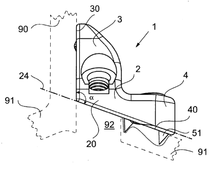
A first illustrative embodiment of a reinforcement
implant according to the invention is shown in Figure 1.
It is designated in its entirety by reference number 1.
It is substantially limb-shaped, with a first limb 3 and
a second limb 4, which are connected to each other by a
bridge part 2.
For a better understanding of the invention, there
follows a detailed explanation of the structure of the
vertebra and the nature of the interaction between the
reinforcement implant and the vertebra. Reference is made
in particular to Figures 6a to 6c. The vertebra 9 has a
solid vertebral body 98 with two laterally protruding
osseous projections 97 which, in their posterior region,
are connected by an osseous arch. The osseous arch
comprises a lamina 91 and, at the center thereof, a
CA 02868610 2014-09-26
WO 2013/143558 - 11 - PCT/EP2012/001357
rearwardly extending projection (spinous process) 90. In
the area of the transition into the lamina 91, upper and
lower articular projections are arranged on each side and
each form part of a facet joint 95, 95' to an adjacent
lower vertebra 9'. The vertebra 9 is also connected to
its adjacent lower vertebra by an intervertebral disk 99,
which is arranged in a load-bearing manner between a
lower cover surface of the vertebral body 98 the
corresponding upper cover surface of the lower adjacent
vertebra 9'. It will be seen from the rear view in Figure
6a that, in the area of the lamina 91, a free space 92 is
present to the right of the spinous process 90. This free
space was created by a resection, resulting in the
formation of corresponding resection surfaces 93, 94 on
the lamina 91 to the left and right of the free space 92.
The opening created by this free space 92 forms an access
to a spinal canal 96. It is closed and mechanically
stabilized with the reinforcement implant 1 according to
the invention.
As is shown in Figures 6a and 6b, the reinforcement
implant according to the invention is mounted in place on
the lamina 91 from the rear, i.e. from the posterior
direction, specifically in such a way that it lies with
its first limb 3 on the spinous process 90 and with its
second limb 4 on the posterior face of the area of the
lamina 91 directly to the right of the resection surface
94. A right-side implantation is shown in Figures 6a to
6c. It is equally possible to perform a left-side
implantation, using a reinforcement implant with a
suitable mirror-image configuration (compare Fig. 3).
To fasten the reinforcement implant 1 on the vertebra 9,
a pressure surface 30 is arranged on the outer face of
the first limb 3. The pressure surface 30 has a
substantially plane shape. A transverse thrust surface 40
CA 02868610 2014-09-26
WO 2013/143558 - 12 - PCT/EP2012/001357
is arranged on the outer face of the second limb 4 and is
designed to bear on the outer face of a lamina 91 of a
vertebra 9. An anti-shear device 5 is provided for the
transverse thrust surface 40. In the illustrative
embodiment shown, it comprises spikes 51 (although two
are shown, it is also possible to provide a smaller or a
greater number) and a facet screw 50 (see Figure 2). The
facet screw 50 is oriented such that, in its normal
position, its axis 55 forms an angle y of 30 with
respect to the perpendicular 67 of the transverse thrust
surface 40.
The facet screw 50 is provided with a head 52, a
threadless shaft 53, and a bone thread 54 at its outer
end. The length of the threadless shaft 53 is such that
the facet screw 50 comes to lie with the latter
completely within a near-side part of the facet joint 95,
while the part of the shaft with the bone thread 54 comes
to lie exclusively, in a part of the facet joint on the
other side, on the adjacent lower vertebra 9'. The effect
of this is that, when the screw 50 is tightened, the part
of the facet joint 95 on the other side is drawn toward
the head 52 of the screw under the force of the bone
thread 54 and is thus braced against the near-side part
of the facet joint 95. This ensures reliable
immobilization of the facet joint 95.
A second facet screw 50' is provided which is inserted
into the first limb 3. This facet screw 50' is oriented
such that it is aligned with the facet joint 95' located
on the other side of the vertebra. The structure of the
second facet screw 50' corresponds in principle to that
of the facet screw 50. It comprises a head 52', a
threadless shaft 53', and a bone thread 54'. The length
of the threadless shaft 53' is significantly greater than
the shaft 53, since the distance to the facet joint 95'
CA 02868610 2014-09-26
WO 2013/143558 - 13 - PCT/EP2012/001357
lying on the other side is significantly greater. This
second facet screw 50' is also referred to as a
translaminar screw 50'.
If the intention is simply to fix the reinforcement
implant 1, without immobilizing the facet joint 95, 95',
the screws 50, 50' are then shorter to the extent that
they are received completely within the vertebra 9, i.e.
they do not protrude into the part of the facet joint on
the other side on the adjacent lower vertebra 9' ("short
screw"). A special screw can also be provided that has a
thread along the entire length of the shaft.
The facet screws 50, 50' are not mounted rigidly in the
first and second limbs 3, 4, but instead are mounted such
that they are able to pivot relative to their screw axis,
specifically by an angle of 15 in each direction. For
this purpose, a pivot joint 6 is provided for each facet
screw 50, 50' in the limb 3 and also in the limb 4. The
pivot joint 6 comprises a cup-shaped seat 60, in which a
ring 61 provided with a spherical jacket surface is
fitted.
The two limbs 3, 4 are shaped such that they enclose an
obtuse angle a with their outer faces, and with the
pressure surface 30 and transverse thrust surface 40
arranged thereon. The angle a is preferably between 95
and 125 ; it is 1100 in the illustrative embodiment
shown. By virtue of this obtuse angle, the reinforcement
implant can be implanted from the dorsal direction, such
that it bridges the free space 92 created by the
resection on the lamina 91. For this purpose, the
reinforcement implant 1 lies with its second limb 4, and
with the transverse thrust surface 40 arranged thereon,
on the posterior face of the lamina 91. This forms one
anchoring part. The other anchoring part is formed by the
CA 02868610 2014-09-26
WO 2013/143558 - 14 - PCT/EP2012/001357
first limb 3, with the pressure surface 30 which is
arranged on the latter and which is pressed against a
side face of the spinous process 90 of the vertebra 9.
The cantilever part 2 located between the two limbs 3, 4
thus acts like a bridge spanning the free space 92
created by the resection. The force transfer lines
between the two limbs 3, 4 run through a load-bearing
area 20 of the cantilever part 2, specifically in such a
way that the load flow takes place completely outside the
free space 92. Structurally, this means that the force
transfer lines in the load-bearing area 20 run in such a
way that they do not intersect the plane 24 defined by
the transverse thrust surface 40, but instead run
exclusively outside this area (i.e. posteriorly).
In order to securely anchor the second limb 4 with its
transverse thrust surface 40 to the lamina 91, and in
particular to prevent an undesired shearing movement with
respect to the lamina 91, an anti-shear device 5 is
provided in the form both of the spikes 51 and also of
the facet screw 50 as fastening pin. Each of the two
devices mentioned is in itself sufficient to stop an
undesired shearing movement. In order to increase the
reliability of the fastening and to prevent lifting of
the transverse thrust surface 40 from the outer face of
the lamina 91, the facet screw 50 is provided. To prevent
the undesired shearing movement, it is not strictly
necessary that the screw 50 has the length shown in
Figure 2. A much shorter screw 50 is also sufficient, one
which is so short that it remains completely within the
vertebra 9. Only in those cases when the screw 50 is
additionally intended to provide the functionality of
immobilizing the facet joint 95 is the length of the
screw 50 made such that it protrudes with its thread 54
from the vertebra 9 and penetrates into the lower,
adjacent vertebral body 9', in order thereby to
CA 02868610 2014-09-26
WO 2013/143558 - 15 - PCT/EP2012/001357
immobilize the facet joint 95.
In a second embodiment and third embodiment of the
reinforcement implant according to the invention, as is
shown in Figure 4 and 5, a wing extension 7 is
additionally provided. Reference is made below to Figures
4a and 4b. The wing extension 7 protrudes from the
transverse thrust surface 40. More precisely, it is
arranged in the lower third of the transverse thrust
surface 40 in the area of the transition between the
second limb 4 and the cantilever part 2, i.e. in a
transition between transverse thrust surface 40 and
cantilever part 20. The wing extension 7 is oriented such
that it is parallel to the pressure surface 30 on the
first limb 3. The wing extension 7 has a plane surface on
its outer face 70 directed toward the transverse thrust
surface 40. On its opposite inner face oriented toward
the pressure surface 30, it is provided with a
reinforcement rib 71. The wing extension 7 comprises with
its lower area the second limb 4, such that as a whole it
protrudes obliquely downward (relative to the implanted
state of the reinforcement implant 1'). Its lower edge 72
is oriented such that it diverges outward with respect to
an axis 55 of the facet screw 50 mounted in the second
limb 4. The angle of divergence p is between 15 and 20 ,
in the illustrative embodiment shown about 18 .
Protruding obliquely downward as it does, the wing
extension 7 ensures that the spinal canal 96 bounded by
the lamina 91 is more effectively shielded from the
penetration of bone pieces that have formed particularly
during the resection of the free space 90. As far as the
patient is concerned, undesired penetration of bone
pieces of this kind would have the very adverse
consequence of once again inducing compressive loads on
the nerve fibers running in the spinal canal 96, as a
result of which the desired successful outcome of the
CA 02868610 2014-09-26
WO 2013/143558 - 16 - PCT/EP2012/001357
operation would no longer be achieved.
A further function of the wing extension 7 is that it
additionally serves for mechanical stiffening. On the one
hand, it gives the bridge part 20 greater mechanical
stability. The wing extension 7 is designed in one piece
with the bridge part 20. By virtue of the plane
configuration of its outer face 70, it is able to bear
flush on the resection surface 94, there being no need
for it to bear with a force fit. However, the smaller the
gap located in between, the better the protection against
penetration of material, in particular of pieces of bone
as has been explained above. The smallest possible gap
width also affords the advantage that the wing extension
7 can function for emergency bearing. Should the
fastening of the bridge part 20 on the anchor in the
second limb 4 come loose (for example if the anti-shear
device 5 fails as a result of the facet screw 50
breaking), the lamina 91 with its resection surface 94
can then only move up to a point where it bears on the
plane outer face 70 of the wing extension 7 and is then
supported by the latter. In this way, the lamina 91 is
further supported and its collapse is effectively
prevented.
Figures 5a and 5b show a third embodiment. Compared to
the second embodiment shown in Figures 4a and 4b, the
only real difference is that a larger wing extension 7'
is used. Otherwise, the explanations given above with
respect to the second embodiment apply accordingly.
The reinforcement implant 1 according to the invention is
preferably part of an implant set, as is shown in Figure
3. The various types, which differ in terms of their
size, are shown arranged in rows. For each size, the
reinforcement implant is provided both in a version for
CA 02868610 2014-09-26
WO 2013/143558 - 17 - PCT/EP2012/001357
right-side implantation (right-hand half of Figure 3) and
also in a version for left-side implantation (left-hand
half of Figure 3). There is in each case a version
without a wing extension, a version with a short wing
extension 7, and a version with a large wing extension
7'.