Note: Descriptions are shown in the official language in which they were submitted.
1
CHIMERIC ANTIGEN RECEPTORS TARGETING B-CELL MATURATION ANTIGEN
CROSS-REFERENCE TO RELATED APPLICATION AND STATEMENT OF
GOVERNMENT RIGHTS
[0001] This patent application claims the benefit of U.S. Provisional
Patent Application
No. 61/622,600 filed April 11, 2012. This invention was made with U.S.
Government
support under project number ZIABC011417 by the National Institutes of Health,
National
Cancer Institute. The U.S. Government has certain rights in the invention.
MATERIAL SUBMIT1ED ELECTRONICALLY
[0002] Submitted herein is a computcr-rcadable nucleotide/amino acid
sequence listing
submitted concurrently herewith and identified as follows: One 42,589 Byte
ASCII (Text) file named
"712361_ST25.TXT," created on March 14, 2013.
BACKGROUND OF THE INVENTION
[0003] Multiple myeloma (MM) is a malignancy characterized by an
accumulation of clonal
plasma cells (see, e.g., Palumbo et al., New England Med., 364(11): 1046-1060
(2011), and Lonial
etal., Clinical Cancer Res., 17(6): 1264-1277 (2011)). Current therapies for
MM often cause
remissions, but nearly all patients eventually relapse and die (see, e.g.,
Lonial et al., supra, and
Rajkumar, Nature Rev. Clinical Oncol., 8(8): 479-491 (2011)). Allogeneic
hematopoietic stem cell
transplantation has been shown to induce immune-mediated elimination of
myeloma cells; however,
the toxicity of this approach is high, and few patients are cured (see, e.g.,
Lonial et al., supra, and
Salit et al., Clin. Lymphoma, Myeloma, and Leukemia, 11(3): 247-252 (2011)).
Currently, there are
no clinically effective, FDA-approved monoclonal antibody or autologous T-cell
therapies for MM
(see, e.g., Richardson et al., British J. Haematology, /54(6): 745-754 (2011),
and Yi, Cancer Journal,
15(6): 502-510 (2009)).
10004] Adoptive transfer of T-cells genetically modified to recognize
malignancy-associated
antigens is showing promise as a new approach to treating cancer (see, e.g.,
Morgan et al., Science,
3/4(5796): 126-129 (2006); Brenner et al., Current Opinion in Immunology,
22(2): 251-257 (2010);
Rosenberg et al., Nature Reviews Cancer, 8(4): 299-308 (2008), Kershaw et al.,
Nature Reviews
Immunology, 5(12): 928-940 (2005); and Pule et al., Nature Medicine, 14(11):
1264-1270 (2008)). T-
cells can be genetically modified to express
CA 2869562 2019-07-24
2
chimeric antigen receptors (CARs), which are fusion proteins comprised of an
antigen
recognition moiety and T-cell activation domains (see, e.g., Kershaw et al.,
supra, Eshhar et
al., Proc. Natl. Acad. Sci. USA, 90(2): 720-724 (1993), and Sadelain et al.,
Curr. Opin.
Immunol., 21(2): 215-223 (2009)).
[0005] For B-cell lineage malignancies, extensive progress has been made in
developing
adoptive T-cell approaches that utilize anti-CD19 CARs (see, e.g., Jensen et
al., Biology of
Blood and Marrow Transplantation, 16: 1245-1256 (2010); Kochenderfer et al.,
Blood,
116(20): 4099-4102 (2010); Porter et al., The New England Journal of Medicine,
365(8):
725-733 (2011); Savoldo et al., Journal of Clinical Investigation, 121(5):
1822-1826 (2011),
Cooper et al., Blood, 101(4): 1637-1644 (2003); Brentjens et al., Nature
Medicine, 9(3): 279-
286 (2003); and Kalos et al., Science Translational Medicine, 3(95): 95ra73
(2011)).
Adoptively transferred anti-CD19-CAR-transduced T-cells have cured leukemia
and
lymphoma in mice (see, e.g., Cheadle et al., Journal of Immunology, 184(4):
1885-1896
(2010); Brentjens et al., Clinical Cancer Research, /3(18 Pt 1): 5426-5435
(2007); and
Kochenderfer et al., Blood, 116(19): 3875-3886 (2010)). In early clinical
trials, adoptively
transferred T-cells transduced with anti-CD19 CARs eradicated normal and
malignant B-cells
in patients with leukemia and lymphoma (see, e.g., Kochenderfer et I., Blood,
116(20): 4099-
4102 (2010); Porter et al., supra, Brentjens et al., Blood, 118(18): 4817-4828
(2011); and
Kochenderfer et al., Blood, December 8, 2011 (epublication ahead of print
(2012)). CD19,
however, is only rarely expressed on the malignant plasma cells of multiple
myeloma (see,
e.g., Gupta et al., Amer. J. Clin. Pathology, 132(5): 728-732 (2009); and Lin
et al., Amer. J.
Clin. Pathology, 121(4): 482-488 (2004)).
[0006] Thus, there remains a need for compositions that can be used in
methods to treat
multiple myeloma. This invention provides such compositions and methods.
BRIEF SUMMARY OF THE INVENTION
[0007] The invention provides an isolated or purified nucleic acid sequence
encoding a
chimeric antigen receptor (CAR), wherein the CAR comprises an antigen
recognition moiety
and a T-cell activation moiety, and wherein the antigen recognition moiety is
directed against
B-cell Maturation Antigen (BCMA).
CA 2869562 2018-04-11
3
BRIEF DESCRIPTION OF THE SEVERAL VIEWS OF THE DRAWING(S)
[0008] Figures 1A and 1B are graphs which depict experimental data
illustrating the
expression pattern of BCMA across a variety of human cell types, as determined
using
quantitative PCR. The results are expressed as the number of BCMA cDNA copies
per 105
actin cDNA copies.
[0009] Figures 2A-2L are graphs which depict experimental data illustrating
that cell-
surface BCMA expression was detected on multiple myeloma cell lines, but not
on other
types of cells, as described in Example 1. For all plots, the solid line
represents staining with
anti-BCMA antibodies, and the dashed line represents staining with isotype-
matched control
antibodies. All plots were gated on live cells.
[0010] Figure 3A is a diagram which depicts a nucleic acid construct
encoding an anti-
BCMA CAR. From the N-terminus to the C-terminus, the anti-BCMA CAR includes an
anti-
BCMA scFv, the hinge and transmembrane regions of the CD8a molecule, the
cytoplasmic
portion of the CD28 molecule, and the cytoplasmic portion of the CD3 molecule.
[0011] Figures 3B-3D are graphs which depict experimental data illustrating
that the anti-
bcma1 CAR, the anti-bcma2 CAR, and the SP6 CAR (described in Example 2) arc
expressed
on the surface of T-cells. Minimal anti-Fab staining occurred on untransduced
(UT) cells.
The plots are gated on CD3+ lymphocytes. The numbers on the plots are the
percentages of
cells in each quadrant.
[0012] Figures 4A-4C are graphs which depict experimental data illustrating
that T-cells
expressing anti-BCMA CARs degranulate T-cells in a BCMA-specific manner, as
described
Example 3. The plots are gated on live CD3+ lymphocytes. The numbers on the
plots are the
percentages of cells in each quadrant.
[0013] Figures 5A-5D are graphs which depict experimental data illustrating
that T-cells
expressing anti-BCMA CARs degranulate T-cells in a BCMA-specific manner, as
described
Example 3. The plots are gated on live CD3+ lymphocytes. The numbers on the
plots are the
percentages of cells in each quadrant.
[0014] Figures 6A-6C are graphs which depict experimental data illustrating
that T-cells
expressing anti-BCMA CARs produce the cytokines IFNy, IL-2, and TNF in a BCMA-
specific manner, as described Example 3. The plots are gated on live CD3+
lymphocytes.
The numbers on the plots are the percentages of cells in each quadrant.
CA 2869562 2018-04-11
4
[0015] Figure 7A is a graph which depicts experimental data illustrating
that T-cells
expressing the anti-bcma2 CAR proliferated specifically in response to BCMA.
Figure 6B is
a graph which depicts experimental data illustrating that T-cells expressing
the SP6 CAR did
not proliferate specifically in response to BCMA.
[0016] Figures 7C and 7D are graphs which depict experimental data
illustrating that T-
cells from Donor A expressing the anti-bcma2 CAR specifically killed the
multiple myeloma
cell lines H929 (Figure 6C) and RPMI8226 (Figure 6D) in a four-hour
cytotoxicity assay at
various effector:target cell ratios. T-cells transduced with the negative
control SP6 CAR
induced much lower levels of cytotoxicity at all effector:target ratios. For
all effector:target
ratios, the cytotoxicity was determined in duplicate, and the results are
displayed as the mean
+1- the standard error of the mean.
[0017] Figure 8A is a graph which depicts experimental data illustrating
that BCMA is
expressed on the surface of primary bone marrow multiple myeloma cells from
Myeloma
Patient 3, as described in Example 5. The plot is gated on CD38high CD56+
plasma cells,
which made up 40% of the bone marrow cells.
[0018] Figure 8B is a graph which depicts experimental data illustrating
that allogeneic
1-cells transduced with the anti-bcma2 CAR from Donor C produced IFNy after co-
culture
with the unmanipulated bone marrow cells of Myeloma Patient 3, as described in
Example 5.
Figure 7B also illustrates that T-cells from the same allogeneic donor
expressing the anti-
bcma2 CAR produced much less IFNy when they were cultured with peripheral
blood
mononuclear cell (PBMC) from Myeloma Patient 3. In addition, 1-cells from
Donor C
expressing the SP6 CAR did not specifically recognize the bone marrow of
Myeloma Patient
3.
[0019] Figure 8C is a graph which depicts experimental data illustrating
that a
plasmacytoma resected from Myeloma Patient 1 consisted of 93% plasma cells,
and these
primary plasma cells expressed BCMA, as revealed by flow cytometry for BCMA
(solid line)
and isotype-matched control staining (dashed line). The plot is gated on
plasma cells.
[0020] Figure 8D is a graph which depicts experimental data illustrating
that 1-cells from
Myeloma Patient 1 expressing the anti-bcma2 CAR produced IFNy specifically in
response to
autologous plasmacytoma cells.
[0021] Figure 8E s a graph which depicts experimental data illustrating
that 1-cells from
Myeloma Patient 1 expressing the anti-bcma2 CAR specifically killed autologous
plasmacytoma cells at low effector to target ratios. In contrast, 1-cells from
Myeloma Patient
CA 2869562 2018-04-11
5
1 expressing the SP6 CAR exhibited low levels of cytotoxicity against
autologous
plasmacytoma cells. For all effector: target ratios, the cytotoxicity was
determined in
duplicate, and the results are displayed as the mean+/-the standard error of
the mean.
[0022] Figure 9A is a graph which depicts experimental data illustrating
that T-cells
transduced with the anti-bcma2 CAR can destroy established multiple myeloma
tumors in
mice. Figure 9B is a graph which depicts the survival of tumor-bearing mice
treated with T-
cells expressing the anti-bcma2 CAR as compared to controls.
DETAILED DESCRIPTION OF THE INVENTION
[0023] The invention provides an isolated or purified nucleic acid sequence
encoding a
chimeric antigen receptor (CAR), wherein the CAR comprises an antigen
recognition moiety
and a T-cell activation moiety. A chimeric antigen receptor (CAR) is an
artificially
constructed hybrid protein or polypeptide containing an antigen binding domain
of an
antibody (e.g., a single chain variable fragment (scFv)) linked to T-cell
signaling or T-cell
activation domains. CARs have the ability to redirect T-cell specificity and
reactivity toward
a selected target in a non-MHC-restricted manner, exploiting the antigen-
binding properties
of monoclonal antibodies. The non-MHC-restricted antigen recognition gives T-
cells
expressing CARs the ability to recognize an antigen independent of antigen
processing, thus
bypassing a major mechanism of tumor escape. Moreover, when expressed in T-
cells, CARs
advantageously do not dimerize with endogenous T-cell receptor (TCR) alpha and
beta
chains.
[0024] "Nucleic acid sequence" is intended to encompass a polymer of DNA or
RNA,
i.e., a polynucleotide, which can be single-stranded or double-stranded and
which can contain
non-natural or altered nucleotides. The terms "nucleic acid" and
"polynucleotide" as used
herein refer to a polymeric form of nucleotides of any length, either
ribonucleotides (RNA) or
deoxyribonucleotides (DNA). These terms refer to the primary structure of the
molecule, and
thus include double- and single-stranded DNA, and double- and single-stranded
RNA. The
terms include, as equivalents, analogs of either RNA or DNA made from
nucleotide analogs
and modified polynucleotides such as, though not limited to methylated and/or
capped
polynucleotides.
[0025] By "isolated" is meant the removal of a nucleic acid from its
natural environment.
By "purified" is meant that a given nucleic acid, whether one that has been
removed from
nature (including genomic DNA and mRNA) or synthesized (including cDNA) and/or
CA 2869562 2018-04-11
6
amplified under laboratory conditions, has been increased in purity, wherein
"purity" is a
relative term, not "absolute purity." It is to be understood, however, that
nucleic acids and
proteins may be formulated with diluents or adjuvants and still for practical
purposes be
isolated. For example, nucleic acids typically are mixed with an acceptable
carrier or diluent
when used for introduction into cells.
[0026] The inventive nucleic acid sequence encodes a CAR which comprises an
antigen
recognition moiety that is directed against B-cell Maturation Antigen (BCMA,
also known as
CD269). BCMA is a member of the tumor necrosis factor receptor superfamily
(see, e.g.,
Thompson et al., J. Exp. Medicine, 192(1): 129-135 (2000), and Mackay et al.,
Annu. Rev.
ImmunoL, 21: 231-264 (2003)). BCMA binds B-cell activating factor (BAFF) and a
proliferation inducing ligand (APRIL) (see, e.g., Mackay et al., supra, and
Kalied et al.,
Immunological Reviews, 204: 43-54 (2005)). Among nonmalignant cells, BCMA has
been
reported to be expressed mostly in plasma cells and subsets of mature B-cells
(see, e.g., Laabi
et al., EMBO J., 11(11): 3897-3904 (1992); Laabi et al., Nucleic Acids Res.,
22(7): 1147-1154
(1994); Kalled et al., supra; O'Connor et al., J. Exp. Medicine, 199(4 91-97
(2004); and Ng
et al., J. ImmunoL, 173(2): 807-817 (2004)). Mice deficient in BCMA are
healthy and have
normal numbers of B-cells, but the survival of long-lived plasma cells is
impaired (see, e.g.,
O'Connor et al, supra; Xu et al., MoL Cell. Biol., 21(12): 4067-4074 (2001);
and Schiemann
et al., Science, 293(5537): 2111-2114 (2001)). BCMA RNA has been detected
universally in
multiple myeloma cells, and BCMA protein has been detected on the surface of
plasma cells
from multiple myeloma patients by several investigators (see, e.g., Novak et
al., Blood,
103(2): 689-694 (2004); Neri et al., Clinical Cancer Research, 13(19): 5903-
5909 (2007);
Bellucci et al., Blood, 105(10): 3945-3950 (2005); and Moreaux et al., Blood,
103(8): 3148-
3157 (2004)).
[0027] The inventive nucleic acid sequence encodes a CAR which comprises an
antigen
recognition moiety that contains a monoclonal antibody directed against BCMA,
or an
antigen-binding portion thereof. The term "monoclonal antibodies," as used
herein, refers to
antibodies that are produced by a single clone of B-cells and bind to the same
epitope. In
contrast, "polyclonal antibodies" refer to a population of antibodies that are
produced by
different B-cells and bind to different epitopes of the same antigen. The
antigen recognition
moiety of the CAR encoded by the inventive nucleic acid sequence can be a
whole antibody
or an antibody fragment. A whole antibody typically consists of four
polypeptides: two
identical copies of a heavy (H) chain polypeptide and two identical copies of
a light (L) chain
CA 2869562 2018-04-11
7
polypeptide. Each of the heavy chains contains one N-terminal variable (VH)
region and
three C-terminal constant (CHL C112 and CH3) regions, and each light chain
contains one N-
terminal variable (VL) region and one C-terminal constant (CL) region. The
variable regions
of each pair of light and heavy chains form the antigen binding site of an
antibody. The VH
and VL regions have the same general structure, with each region comprising
four framework
regions, whose sequences are relatively conserved. The framework regions are
connected by
three complementarity determining regions (CDRs). The three CDRs, known as
CDR1,
CDR2, and CDR3, form the "hypervariable region" of an antibody, which is
responsible for
antigen binding.
[0028] The terms
"fragment of an antibody," "antibody fragment," "functional fragment
of an antibody," and "antigen-binding portion" are used interchangeably herein
to mean one
or more fragments or portions of an antibody that retain the ability to
specifically bind to an
antigen (see, generally, Holliger et al., Nat. Biotech., 23(9): 1126-1129
(2005)). The antigen
recognition moiety of the CAR encoded by the inventive nucleic acid sequence
can contain
any BCMA-binding antibody fragment. The antibody fragment desirably comprises,
for
example, one or more CDRs, the variable region (or portions thereof), the
constant region (or
portions thereof), or combinations thereof. Examples of antibody fragments
include, but are
not limited to, (i) a Fab fragment, which is a monovalent fragment consisting
of the VL, VH,
CL, and CH I domains; (ii) a F(ab')2 fragment, which is a bivalent fragment
comprising two
Fab fragments linked by a disulfide bridge at the hinge region; (iii) a Fv
fragment consisting
of the VL and VH domains of a single arm of an antibody; (iv) a single chain
Fv (scFv),
which is a monovalent molecule consisting of the two domains of the Fv
fragment (i.e., VL
and VH) joined by a synthetic linker which enables the two domains to be
synthesized as a
single polypeptide chain (see, e.g., Bird et al., Science, 242: 423-426
(1988); Huston et al.,
Proc. Natl. Acad. Sci. USA, 85: 5879-5883 (1988); and Osbourn et al., Nat.
Biotechnol.,
778 (1998)) and (v) a diabody, which is a dimer of polypeptide chains, wherein
each
polypeptide chain comprises a VH connected to a VL by a peptide linker that is
too short to
allow pairing between the VH and VL on the same polypeptide chain, thereby
driving the
pairing between the complementary domains on different VH -VL polypeptide
chains to
generate a dimeric molecule having two functional antigen binding sites.
Antibody
fragments are known in the art and are described in more detail in, e.g., U.S.
Patent
Application Publication 2009/0093024 Al. In a preferred embodiment, the
antigen
CA 2869562 2018-04-11
8
recognition moiety of the CAR encoded by the inventive nucleic acid sequence
comprises an
anti-BCMA single chain Fv (scFv).
[0029] An antigen-binding portion or fragment of a monoclonal antibody can
be of any
size so long as the portion binds to BCMA. In this respect, an antigen binding
portion or
fragment of the monoclonal antibody directed against BCMA (also referred to
herein as an
"anti-BCMA monoclonal antibody") desirably comprises between about 5 and 18
amino
acids (e.g., about 5, 6, 7, 8, 9, 10, 11, 12, 13, 14, 15, 16, 17, 18, or a
range defined by any two
of the foregoing values).
[0030] In one embodiment, the inventive nucleic acid sequence encodes an
antigen
recognition moiety that comprises a variable region of an anti-BCMA monoclonal
antibody.
In this respect, the antigen recognition moiety comprises a light chain
variable region, a
heavy chain variable region, or both a light chain variable region and a heavy
chain variable
region of an anti-BCMA monoclonal antibody. Preferably, the antigen
recognition moiety of
the CAR encoded by the inventive nucleic acid sequence comprises a light chain
variable
region and a heavy chain variable region of an anti-BCMA monoclonal antibody.
Heavy and
light chain monoclonal antibody amino acid sequences that bind to BCMA are
disclosed in,
e.g., International Patent Application Publication WO 2010/104949.
[0031] In another embodiment, the inventive nucleic acid sequence encodes a
CAR
which comprises a signal sequence. The signal sequence may be positioned at
the amino
terminus of the antigen recognition moiety (e.g., the variable region of the
anti-BCMA
antibody). The signal sequence may comprise any suitable signal sequence. In
one
embodiment, the signal sequence is a human granulocyte-macrophage colony-
stimulating
factor (GM-CSF) receptor sequence or a CD8a signal sequence.
[0032] In another embodiment, the CAR comprises a hinge sequence. One of
ordinary
skill in the art will appreciate that a hinge sequence is a short sequence of
amino acids that
facilitates antibody flexibility (see, e.g., Woof et al., Nat. Rev. Immunol.,
4(2): 89-99 (2004)).
The hinge sequence may be positioned between the antigen recognition moiety
(e.g., an anti-
BCMA scFv) and the T-cell activation moiety. The hinge sequence can be any
suitable
sequence derived or obtained from any suitable molecule. In one embodiment,
for example,
the hinge sequence is derived from the human CD8oc molecule or a CD28
molecule.
[0033] The inventive nucleic acid sequence encodes a CAR comprising a T-
cell
activation moiety. The T-cell activation moiety can be any suitable moiety
derived or
obtained from any suitable molecule. In one embodiment, for example, the T-
cell activation
CA 2869562 2018-04-11
9
moiety comprises a transmembrane domain. The transmembrane domain can be any
transmembrane domain derived or obtained from any molecule known in the art.
For
example, the transmembrane domain can be obtained or derived from a CD8a
molecule or a
CD28 molecule. CD8 is a transmembrane glycoprotein that serves as a co-
receptor for the T-
cell receptor (TCR), and is expressed primarily on the surface of cytotoxic 1-
cells. The most
common form of CD8 exists as a dimer composed of a CD8a and CD8f3 chain. CD28
is
expressed on 1-cells and provides co-stimulatory signals required for 1-cell
activation.
CD28 is the receptor for CD80 (B7.1) and CD86 (B7.2). In a preferred
embodiment, the
CD8a and CD28 are human.
[0034] In addition to the transmembrane domain, the T-cell activation
moiety further
comprises an intracellular (i.e., cytoplasmic) 1-cell signaling domain. The
intercellular 1-
cell signaling domain can be obtained or derived from a CD28 molecule, a CD3
zeta (C)
molecule or modified versions thereof, a human Fe receptor gamma (FcRy) chain,
a CD27
molecule, an 0X40 molecule, a 4-1BB molecule, or other intracellular signaling
molecules
known in the art. As discussed above, CD28 is a 1-cell marker important in 1-
cell co-
stimulation. CD3 C associates with TCRs to produce a signal and contains
immunoreceptor
tyrosine-based activation motifs (ITAMs). 4-1BB, also known as CD137,
transmits a potent
costimulatory signal to 1-cells, promoting differentiation and enhancing long-
term survival
of T lymphocytes. In a preferred embodiment, the CD28, CD3 zeta, 4-1BB, 0X40,
and
CD27 are human.
[0035] The 1-cell activation domain of the CAR encoded by the inventive
nucleic acid
sequence can comprise any one of aforementioned transmembrane domains and any
one or
more of the aforementioned intercellular 1-cell signaling domains in any
combination. For
example, the inventive nucleic acid sequence can encode a CAR comprising a
CD28
transmembrane domain and intracellular 1-cell signaling domains of CD28 and
CD3 zeta.
Alternatively, for example, the inventive nucleic acid sequence can encode a
CAR
comprising a CD8a transmembrane domain and intracellular 1-cell signaling
domains of
CD28, CD3 zeta, the Fc receptor gamma (FcRy) chain, and/or 4-1BB.
[0036] In one embodiment, the inventive nucleic acid sequence encodes a CAR
which
comprises, from 5' to 3', a granulocyte-macrophage colony stimulating factor
receptor (GM-
CSF receptor) signal sequence, an anti-BCMA scFv, the hinge and transmembrane
regions of
the human CD8a molecule, the cytoplasmic T-cell signaling domain of the human
CD28
CA 2869562 2018-04-11
10
molecule, and T-cell signaling domain of the human CD3 molecule. In another
embodiment, the inventive nucleic acid sequence encodes a CAR which comprises,
from 5'
to 3', a human CD8ot signal sequence, an anti-BCMA scFv, the hinge and
transmembrane
regions of the human CD8a. molecule, the cytoplasmic T-cell signaling domain
of the human
CD28 molecule, and T-cell signaling domain of the human CD3 molecule. In
another
embodiment, the inventive nucleic acid sequence encodes a CAR which comprises,
from 5'
to 3', a human CD8a signal sequence, an anti-BCMA scFv, the hinge and
transmembrane
regions of the human CD8a molecule, the cytoplasmic T-cell signaling domain of
the human
4-1BB molecule and/or the cytoplasmic T-cell signaling domain of the human
0X40
molecule, and T-cell signaling domain of the human CD3 molecule. For example,
the
inventive nucleic acid sequence comprises or consists of the nucleic acid
sequence of SEQ ID
NO: 1, SEQ ID NO: 2, or SEQ ID NO: 3.
[0037] The invention further provides an isolated or purified chimeric
antigen receptor
(CAR) encoded by the inventive nucleic acid sequence.
[0038] The nucleic acid sequence of the invention can encode a CAR of any
length, i.e.,
the CAR can comprise any number of amino acids, provided that the CAR retains
its
biological activity, e.g., the ability to specifically bind to antigen, detect
diseased cells in a
mammal, or treat or prevent disease in a mammal, etc. For example, the CAR can
comprise
50 or more (e.g., 60 or more, 100 or more, or 500 or more) amino acids, but
less than 1,000
(e.g., 900 or less, 800 or less, 700 or less, or 600 or less) amino acids.
Preferably, the CAR is
about 50 to about 700 amino acids (e.g., about 70, about 80, about 90, about
150, about 200,
about 300, about 400, about 550, or about 650 amino acids), about 100 to about
500 amino
acids (e.g., about 125, about 175, about 225, about 250, about 275, about 325,
about 350,
about 375, about 425, about 450, or about 475 amino acids), or a range defined
by any two of
the foregoing values.
[0039] Included in the scope of the invention are nucleic acid sequences
that encode
functional portions of the CAR described herein. The term "functional
portion," when used
in reference to a CAR, refers to any part or fragment of the CAR of the
invention, which part
or fragment retains the biological activity of the CAR of which it is a part
(the parent CAR).
Functional portions encompass, for example, those parts of a CAR that retain
the ability to
recognize target cells, or detect, treat, or prevent a disease, to a similar
extent, the same
extent, or to a higher extent, as the parent CAR. In reference to a nucleic
acid sequence
CA 2869562 2018-04-11
11
encoding the parent CAR, a nucleic acid sequence encoding a functional portion
of the CAR
can encode a protein comprising, for example, about 10%, 25%, 30%, 50%, 68%,
80%, 90%,
95%, or more, of the parent CAR.
[0040] The inventive nucleic acid sequence can encode a functional portion
of a CAR
that contains additional amino acids at the amino or carboxy terminus of the
portion, or at
both termini, which additional amino acids are not found in the amino acid
sequence of the
parent CAR. Desirably, the additional amino acids do not interfere with the
biological
function of the functional portion, e.g., recognize target cells, detect
cancer, treat or prevent
cancer, etc. More desirably, the additional amino acids enhance the biological
activity of the
CAR, as compared to the biological activity of the parent CAR.
[0041] The invention also provides nucleic acid sequences encoding
functional variants
of the aforementioned CAR. The term "functional variant," as used herein,
refers to a CAR,
a polypeptide, or a protein having substantial or significant sequence
identity or similarity to
the CAR encoded by the inventive nucleic acid sequence, which functional
variant retains the
biological activity of the CAR of which it is a variant. Functional variants
encompass, for
example, those variants of the CAR described herein (the parent CAR) that
retain the ability
to recognize target cells to a similar extent, the same extent, or to a higher
extent, as the
parent CAR. In reference to a nucleic acid sequence encoding the parent CAR, a
nucleic acid
sequence encoding a functional variant of the CAR can be for example, about
10% identical,
about 25% identical, about 30% identical, about 50% identical, about 65%
identical, about
80% identical, about 90% identical, about 95% identical, or about 99%
identical to the
nucleic acid sequence encoding the parent CAR.
[0042] A functional variant can, for example, comprise the amino acid
sequence of the
CAR encoded by the inventive nucleic acid sequence with at least one
conservative amino
acid substitution. The phrase "conservative amino acid substitution" or
"conservative
mutation" refers to the replacement of one amino acid by another amino acid
with a common
property. A functional way to define common properties between individual
amino acids is
to analyze the normalized frequencies of amino acid changes between
corresponding proteins
of homologous organisms (Schulz, G. E. and Schirmer, R. H., Principles of
Protein
Structure, Springer-Verlag, New York (1979)). According to such analyses,
groups of amino
acids may be defined where amino acids within a group exchange preferentially
with each
other, and therefore resemble each other most in their impact on the overall
protein structure
(Schulz, G. E. and Schirmer, R. H., supra). Examples of conservative mutations
include
CA 2869562 2018-04-11
12
amino acid substitutions of amino acids within the sub-groups above, for
example, lysine for
arginine and vice versa such that a positive charge may be maintained;
glutamic acid for
aspartic acid and vice versa such that a negative charge may be maintained;
serine for
threonine such that a free -OH can be maintained; and glutamine for asparagine
such that a
free -NH2 can be maintained.
[0043] Alternatively or additionally, the functional variants can comprise
the amino acid
sequence of the parent CAR with at least one non-conservative amino acid
substitution.
"Non-conservative mutations" involve amino acid substitutions between
different groups, for
example, lysine for tryptophan, or phenylalanine for serine, etc. In this
case, it is preferable
for the non-conservative amino acid substitution to not interfere with, or
inhibit the biological
activity of, the functional variant. The non-conservative amino acid
substitution may
enhance the biological activity of the functional variant, such that the
biological activity of
the functional variant is increased as compared to the parent CAR.
[0044] The inventive nucleic acid sequence can encode a CAR (including
functional
portions and functional variants thereof) that comprises synthetic amino acids
in place of one
or more naturally-occurring amino acids. Such synthetic amino acids are known
in the art,
and include, for example, aminocyclohexane carboxylic acid, norleucine, a-
amino n-
decanoic acid, homoserine, S-acetylaminomethyl-cysteine, trans-3- and trans-4-
hydroxyproline, 4-aminophenylalanine, 4- nitrophenylalanine, 4-
chlorophenylalanine, 4-
carboxyphenylalanine, I3-phenylserine 13-hydroxypheny1alanine, phenylglycine,
a-
naphthylalanine, cyclohexylalanine, cyclohexylglycine, indoline-2-carboxylic
acid, 1,2,3,4-
tetrahydroisoquinoline-3-carboxylic acid, aminomalonic acid, aminomalonic acid
monoamide, N'-benzyl-N'-methyl-lysine, N',N'-dibenzyl-lysine, 6-hydroxylysine,
ornithine,
a-aminocyclopentane carboxylic acid, a-aminocyclohexane carboxylic acid, a-
aminocycloheptane carboxylic acid, a-(2-amino-2-norbornane)-carboxylic acid,
a,y-
diaminobutyric acid, a,[1-diaminopropionic acid, homophenylalanine, and a-tert-
butylglycine.
[0045] The inventive nucleic acid sequence can encode a CAR (including
functional
portions and functional variants thereof) which is glycosylated, amidated,
carboxylated,
phosphorylated, esterified, N-acylated, cyclized via, e.g., a disulfide
bridge, or converted into
an acid addition salt and/or optionally dimerized or polymerized, or
conjugated.
100461 In a preferred embodiment, the inventive nucleic acid sequence
encodes a CAR
that comprises or consists of the amino acid sequence of SEQ ID NO: 4, SEQ ID
NO: 5, SEQ
CA 2869562 2018-04-11
13
ID NO: 6, SEQ ID NO: 8, SEQ ID NO: 9, SEQ ID NO: 10, SEQ ID NO: 11, or SEQ ID
NO:
12.
[0047] The inventive nucleic acid sequence can be generated using methods
known in the
art. For example, nucleic acid sequences, polypeptides, and proteins can be
recombinantly
produced using standard recombinant DNA methodology (see, e.g., Sambrook et
al.,
Molecular Cloning: A Laboratory Manual, 3' ed., Cold Spring Harbor Press, Cold
Spring
Harbor, NY 2001; and Ausubel et al., Current Protocols in Molecular Biology,
Greene
Publishing Associates and John Wiley & Sons, NY, 1994). Further, a
synthetically produced
nucleic acid sequence encoding the CAR can be isolated and/or purified from a
source, such
as a plant, a bacterium, an insect, or a mammal, e.g., a rat, a human, etc.
Methods of isolation
and purification are well-known in the art. Alternatively, the nucleic acid
sequences
described herein can be commercially synthesized. In this respect, the
inventive nucleic acid
sequence can be synthetic, recombinant, isolated, and/or purified.
[0048] The invention also provides a vector comprising the nucleic acid
sequence
encoding the inventive CAR. The vector can be, for example, a plasmid, a
cosmid, a viral
vector (e.g., retroviral or adenoviral), or a phage. Suitable vectors and
methods of vector
preparation are well known in the art (see, e.g., Sambrook et al., supra, and
Ausubel et al.,
supra).
[0049] In addition to the inventive nucleic acid sequence encoding the CAR,
the vector
preferably comprises expression control sequences, such as promoters,
enhancers,
polyadenylation signals, transcription terminators, internal ribosome entry
sites (1RES), and
the like, that provide for the expression of the nucleic acid sequence in a
host cell.
Exemplary expression control sequences are known in the art and described in,
for example,
Goeddel, Gene Expression Technology: Methods in Enzymology, Vol. 185, Academic
Press,
San Diego, Calif. (1990).
[0050] A large number of promoters, including constitutive, inducible, and
repressible
promoters, from a variety of different sources are well known in the art.
Representative
sources of promoters include for example, virus, mammal, insect, plant, yeast,
and bacteria,
and suitable promoters from these sources are readily available, or can be
made synthetically,
based on sequences publicly available, for example, from depositories such as
the ATCC as
well as other commercial or individual sources. Promoters can be
unidirectional (i.e., initiate
transcription in one direction) or hi-directional (i.e., initiate
transcription in either a 3' or 5'
direction). Non-limiting examples of promoters include, for example, the T7
bacterial
CA 2869562 2018-04-11
14
expression system, pBAD (araA) bacterial expression system, the
cytomegalovirus (CMV)
promoter, the SV40 promoter, and the RSV promoter. Inducible promoters
include, for
example, the Tet system (U.S. Patents 5,464,758 and 5,814,618), the Ecdysone
inducible
system (No et al., Proc. Natl. Acad. Sci., 93: 3346-3351 (1996)), the T-REXIm
system
(Invitrogen, Carlsbad, CA), LACSWITCHTm System (Stratagene, San Diego, CA),
and the
Cre-ERT tamoxifen inducible recombinase system (Indra et al., Nue. Acid. Res.,
27: 4324-
4327 (1999); Nuc. Acid. Res., 28: e99 (2000); U.S. Patent 7,112,715; and
Kramer &
Fussenegger, Methods MoL Biol., 308: 123-144 (2005)).
[0051] The term "enhancer" as used herein, refers to a DNA sequence that
increases
transcription of, for example, a nucleic acid sequence to which it is operably
linked.
Enhancers can be located many kilobases away from the coding region of the
nucleic acid
sequence and can mediate the binding of regulatory factors, patterns of DNA
methylation, or
changes in DNA structure. A large number of enhancers from a variety of
different sources
are well known in the art and are available as or within cloned
polynucleotides (from, e.g.,
depositories such as the ATCC as well as other commercial or individual
sources). A number
of polynucleotides comprising promoters (such as the commonly-used CMV
promoter) also
comprise enhancer sequences. Enhancers can be located upstream, within, or
downstream of
coding sequences. The term "Ig enhancers" refers to enhancer elements derived
from
enhancer regions mapped within the immunoglobulin (Ig) locus (such enhancers
include for
example, the heavy chain (mu) 5' enhancers, light chain (kappa) 5' enhancers,
kappa and mu
intronic enhancers, and 3' enhancers (see generally Paul W.E. (ed),
Fundamental
Immunology, 3rd Edition, Raven Press, New York (1993), pages 353-363; and U.S.
Patent
5,885,827).
[0052] The vector also can comprise a "selectable marker gene." The term
"selectable
marker gene," as used herein, refers to a nucleic acid sequence that allows
cells expressing
the nucleic acid sequence to be specifically selected for or against, in the
presence of a
corresponding selective agent. Suitable selectable marker genes arc known in
the art and
described in, e.g., International Patent Application Publications WO
1992/08796 and WO
1994/28143; Wigler et al., Proc. Natl. Acad. Sci. USA, 77: 3567 (1980); O'Hare
et al., Proc.
NatL Acad. Sci. USA, 78: 1527 (1981); Mulligan & Berg, Proc. NatL Acad. Sci.
USA, 78:
2072 (1981); Colberre-Garapin et al., J. MoL Biol., 150: 1 (1981); Santerre et
al., Gene, 30:
147 (1984); Kent et al., Science, 237: 901-903 (1987); Wigler et al., Cell,
11: 223 (1977);
CA 2869562 2018-04-11
15
Szybalska & Szybalski, Proc. Natl. Acad. Sci. USA, 48: 2026 (1962); Lowy et
at., Cell, 22:
817 (1980); and U.S. Patents 5,122,464 and 5,770,359.
[0053] In some embodiments, the vector is an "episomal expression vector"
or
"episome," which is able to replicate in a host cell, and persists as an
extrachromosomal
segment of DNA within the host cell in the presence of appropriate selective
pressure (see,
e.g., Conese et al., Gene Therapy, 11: 1735-1742 (2004)). Representative
commercially
available episomal expression vectors include, but are not limited to,
episomal plasmids that
utilize Epstein Barr Nuclear Antigen 1 (EBNA1) and the Epstein Barr Virus
(EBV) origin of
replication (oriP). The vectors pREP4, pCEP4, pREP7, and pcDNA3.1 from
Invitrogen
(Carlsbad, CA) and pBK-CMV from Stratagene (La Jolla, CA) represent non-
limiting
examples of an episomal vector that uses T-antigen and the SV40 origin of
replication in lieu
of EBNA1 and oriP.
[0054] Other suitable vectors include integrating expression vectors, which
may
randomly integrate into the host cell's DNA, or may include a recombination
site to enable
the specific recombination between the expression vector and the host cell's
chromosome.
Such integrating expression vectors may utilize the endogenous expression
control sequences
of the host cell's chromosomes to effect expression of the desired protein.
Examples of
vectors that integrate in a site specific manner include, for example,
components of the flp-in
system from Invitrogen (Carlsbad, CA) (e.g., peDNATm5/FRT), or the cre-lox
system, such
as can be found in the pExchange-6 Core Vectors from Stratagene (La Jolla,
CA). Examples
of vectors that randomly integrate into host cell chromosomes include, for
example,
pcDNA3.1 (when introduced in the absence of T-antigen) from Invitrogen
(Carlsbad, CA),
and pCI or pFN1 OA (ACT) FLEXITM from Promega (Madison, WI).
[0055] Viral vectors also can be used. Representative viral expression
vectors include,
but are not limited to, the adenovirus-based vectors (e.g., the adenovirus-
based Per.C6 system
available from Crucell, Inc. (Leiden, The Netherlands)), lentivirus-based
vectors (e.g., the
lentiviral-based pLP1 from Life Technologies (Carlsbad, CA)), and retmviral
vectors (e.g.,
the pFB-ERV plus pCFB-EGSH from Stratagene (La Jolla, CA)). In a preferred
embodiment, the viral vector is a lentivirus vector.
[0056] The vector comprising the inventive nucleic acid encoding the CAR
can be
introduced into a host cell that is capable of expressing the CAR encoded
thereby, including
any suitable prokaryotic or eukaryotic cell. Preferred host cells are those
that can be easily
CA 2869562 2018-04-11
16
and reliably grown, have reasonably fast growth rates, have well characterized
expression
systems, and can be transformed or transfected easily and efficiently.
[0057] As used herein, the term "host cell" refers to any type of cell that
can contain the
expression vector. The host cell can be a eukaryotic cell, e.g., plant,
animal, fungi, or algae,
or can be a prokaryotic cell, e.g., bacteria or protozoa. The host cell can be
a cultured cell or
a primary cell, i.e., isolated directly from an organism, e.g., a human. The
host cell can be an
adherent cell or a suspended cell, i.e., a cell that grows in suspension.
Suitable host cells are
known in the art and include, for instance, DH5ot E. coli cells, Chinese
hamster ovarian cells,
monkey VERO cells, COS cells, HEK293 cells, and the like. For purposes of
amplifying or
replicating the recombinant expression vector, the host cell may be a
prokaryotic cell, e.g., a
DH5ot cell. For purposes of producing a recombinant CAR, the host cell can be
a
mammalian cell. The host cell preferably is a human cell. The host cell can be
of any cell
type, can originate from any type of tissue, and can be of any developmental
stage. In one
embodiment, the host cell can be a peripheral blood lymphocyte (PBL), a
peripheral blood
mononuclear cell (PBMC), or a natural killer (NK). Preferably, the host cell
is a natural
killer (NK) cell. More preferably, the host cell is a T-cell. Methods for
selecting suitable
mammalian host cells and methods for transformation, culture, amplification,
screening, and
purification of cells are known in the art.
[0058] The invention provides an isolated host cell which expresses the
inventive nucleic
acid sequence encoding the CAR described herein. In one embodiment, the host
cell is a T-
cell. The T-cell of the invention can be any T-cell, such as a cultured T-
cell, e.g., a primary
T-cell, or a T-cell from a cultured T-cell line, or a T-cell obtained from a
mammal. If
obtained from a mammal, the T-cell can be obtained from numerous sources,
including but
not limited to blood, bone marrow, lymph node, the thymus, or other tissues or
fluids. T-cells
can also be enriched for or purified. The T-cell preferably is a human T-cell
(e.g., isolated
from a human). The T-cell can be of any developmental stage, including but not
limited to, a
CD4+/CD8+ double positive T-cell, a CD4+ helper T-cell, e.g., Thi and Thz
cells, a CD8+ T-
cell (e.g., a cytotoxic T-cell), a tumor infiltrating cell, a memory T-cell, a
naive T-cell, and
the like. In one embodiment, the T-cell is a CD8+ T-cell or a CD4+ T-cell. T-
cell lines arc
available from, e.g., the American Type Culture Collection (ATCC, Manassas,
VA), and the
German Collection of Microorganisms and Cell Cultures (DSMZ) and include, for
example,
Jurkat cells (ATCC TIB-152), Sup-Ti cells (ATCC CRL-1942), RPMI 8402 cells
(DSMZ
ACC-290), Karpas 45 cells (DSMZ ACC-545), and derivatives thereof.
CA 2869562 2018-04-11
17
[0059] In another embodiment, the host cell is a natural killer (NK) cell.
NK cells are a
type of cytotoxic lymphocyte that plays a role in the innate immune system. NK
cells are
defined as large granular lymphocytes and constitute the third kind of cells
differentiated
from the common lymphoid progenitor which also gives rise to B and T
lymphocytes (see,
e.g., Immunobiology, .5111 ed., Janeway et al., eds., Garland Publishing, New
York, NY
(2001)). NK cells differentiate and mature in the bone marrow, lymph node,
spleen, tonsils,
and thymus. Following maturation, NK cells enter into the circulation as large
lymphocytes
with distinctive cytotoxic granules. NK cells are able to recognize and kill
some abnormal
cells, such as, for example, some tumor cells and virus-infected cells, and
are thought to be
important in the innate immune defense against intracellular pathogens. As
described above
with respect to T-cells, the NK cell can be any NK cell, such as a cultured NK
cell, e.g., a
primary NK cell, or an NK cell from a cultured NK cell line, or an NK cell
obtained from a
mammal. If obtained from a mammal, the NK cell can be obtained from numerous
sources,
including but not limited to blood, bone marrow, lymph node, the thymus, or
other tissues or
fluids. NK cells can also be enriched for or purified. The NK cell preferably
is a human NK
cell (e.g., isolated from a human). NK cell lines are available from, e.g.,
the American Type
Culture Collection (ATCC, Manassas, VA) and include, for example, NK-92 cells
(ATCC
CRL-2407), NK92MI cells (ATCC CRL-2408), and derivatives thereof.
[0060] The inventive nucleic acid sequence encoding a CAR may be introduced
into a
cell by "transfection," "transformation," or "transduction." "Transfection,"
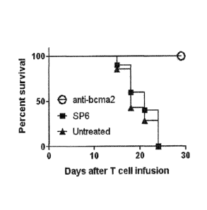
"transformation,"
or "transduction," as used herein, refer to the introduction of one or more
exogenous
polynucleotides into a host cell by using physical or chemical methods. Many
transfection
techniques are known in the art and include, for example, calcium phosphate
DNA co-
precipitation (see, e.g., Murray E.J. (ed.), Methods in Molecular Biology,
Vol. 7, Gene
Transfer and Expression Protocols, Humana Press (1991)); DEAE-dextran;
electroporation;
cationic liposome-mediated transfection; tungsten particle-facilitated
microparticle
bombardment (Johnston, Nature, 346: 776-777 (1990)); and strontium phosphate
DNA co-
precipitation (Brash et at., Mol. Cell Biol., 7: 2031-2034 (1987)). Phage or
viral vectors can
be introduced into host cells, after growth of infectious particles in
suitable packaging cells,
many of which are commercially available.
[0061] Without being bound to a particular theory or mechanism, it is
believed that by
eliciting an antigen-specific response against BCMA, the CARs encoded by the
inventive
nucleic acid sequence provide for one or more of the following: targeting and
destroying
CA 2869562 2018-04-11
18
BCMA-expressing cancer cells, reducing or eliminating cancer cells,
facilitating infiltration
of immune cells to tumor site(s), and enhancing/extending anti-cancer
responses. Thus, the
invention provides a method of destroying multiple myeloma cells, which
comprises
contacting one or more of the aforementioned isolated T-cells or natural
killer cells with a
population of multiple myeloma cells that express BCMA, whereby the CAR is
produced and
binds to BCMA on the multiple myeloma cells and the multiple myeloma cells are
destroyed.
As discussed herein, multiple myeloma, also known as plasma cell myeloma or
Kahler's
disease, is a cancer of plasma cells, which are a type of white blood cell
normally responsible
for the production of antibodies (Raab et al., Lancet, 374: 324-329 (2009)).
Multiple
myeloma affects 1-4 per 100,000 people per year. The disease is more common in
men, and
for yet unknown reasons is twice as common in African Americans as it is in
Caucasian
Americans. Multiple myeloma is the least common hematological malignancy (14%)
and
constitutes 1% of all cancers (Raab et al., supra). Treatment of multiple
myeloma typically
involves high-dose chemotherapy followed by hematopoietic stem cell
transplanatation
(allogenic or autologous); however, a high rate of relapse is common in
multiple myeloma
patients that have undergone such treatement. As discussed above, BCMA is
highly
expressed by multiple myeloma cells (see, e.g., Novak et al., supra; Neri et
al., supra;
Bellucci et at., supra; and Moreaux et al., supra).
[0062] One or more isolated T-cells expressing the inventive nucleic acid
sequence
encoding the anti-BCMA CAR described herein can be contacted with a population
of
multiple myeloma cells that express BCMA ex vivo, in vivo, or in vitro. "Ex
vivo" refers to
methods conducted within or on cells or tissue in an artificial environment
outside an
organism with minimum alteration of natural conditions. In contrast, the term
"in vivo" refers
to a method that is conducted within living organisms in their normal, intact
state, while an
"in vitro" method is conducted using components of an organism that have been
isolated
from its usual biological context. The inventive method preferably involves ex
vivo and in
vivo components. In this regard, for example, the isolated T-cells described
above can be
cultured ex vivo under conditions to express the inventive nucleic acid
sequence encoding the
anti-BCMA CAR, and then directly transferred into a mammal (preferably a
human) affected
by multiple myeloma. Such a cell transfer method is referred to in the art as
"adoptive cell
transfer (ACT)," in which immune-derived cells are passively transferred into
a new recipient
host to transfer the functionality of the donor immune-derived cells to the
new host.
Adoptive cell transfer methods to treat various types of cancers, including
hematological
CA 2869562 2018-04-11
19
cancers such as myeloma, are known in the art and disclosed in, for example,
Gattinoni et al.,
Nat. Rev. Immunol., 6(5): 383-393 (2006); June, CH, J. Clin. Invest., 117(6):
1466-76 (2007);
Rapoport et al., Blood, 117(3): 788-797 (2011); and Barber et al., Gene
Therapy, 18: 509-516
(2011)).
[0063] The invention also provides a method of destroying Hodgkin's
lymphoma cells.
Hodgkin's lymphoma (formerly known as Hodgkin's disease) is a cancer of the
immune
system that is marked by the presence of a multinucleated cell type called
Reed-Sternberg
cells. The two major types of Hodgkin's lymphoma include classical Hodgkin's
lymphoma
and nodular lymphocyte-predominant Hodgkin's lymphoma. Hodgkin's lymphoma
currently
is treated with radiation therapy, chemotherapy, or hematopoietic stem cell
transplantation,
with the choice of treatment depending on the age and sex of the patient and
the stage, bulk,
and histological subtype of the disease. BCMA expression has been detected on
the surface
of Hodgkin's lymphoma cells (see, e.g., Chiu et al., Blood, 109(2): 729-739
(2007)).
[0064] When T-cells or NK cells are administered to a mammal, the cells can
be
allogeneic or autologous to the mammal. In "autologous" administration
methods, cells (e.g.,
blood-forming stem cells or lymphocytes) are removed from a mammal, stored
(and
optionally modified), and returned back to the same mammal. In "allogeneic"
administration
methods, a mammal receives cells (e.g., blood-forming stem cells or
lymphocytes) from a
genetically similar, but not identical, donor. Preferably, the cells are
autologous to the
mammal.
[0065] The T-cells or NK cells desirably are administered to a human in the
form of a
composition, such as a pharmaceutical composition. Alternatively, the
inventive nucleic acid
sequence encoding the CAR, or a vector comprising the CAR-encoding nucleic
acid
sequence, can be formulated into a composition, such as a pharmaceutical
composition, and
administered to a human. The inventive pharmaceutical composition can comprise
a
population of T-cells of NK cells that express the inventive CAR. In addition
to the inventive
nucleic acid sequence, or host cells which express the inventive CAR, the
pharmaceutical
composition can comprise other pharmaceutically active agents or drugs, such
as
chemotherapeutic agents, e.g., asparaginase, busulfan, carboplatin, cisplatin,
daunorubicin,
doxorubicin, fluorouracil, gemcitabine, hydroxyurea, methotrexate, paclitaxel,
rituximab,
vinblastine, vincristine, etc. In a preferred embodiment, the pharmaceutical
composition
comprises an isolated T-cell or NK cell which expresses the inventive CAR,
more preferably
a population of T-cells or NK cells which express the inventive CAR.
CA 2869562 2018-04-11
20
[0066] The inventive T-cells or NK cells can be provided in the form of a
salt, e.g., a
pharmaceutically acceptable salt. Suitable pharmaceutically acceptable acid
addition salts
include those derived from mineral acids, such as hydrochloric, hydrobromic,
phosphoric,
metaphosphoric, nitric, and sulphuric acids, and organic acids, such as
tartaric, acetic, citric,
malic, lactic, fumaric, benzoic, glycolic, gluconic, succinic, and
arylsulphonic acids, for
example, p-toluenesulphonic acid.
[0067] The choice of carrier will be determined in part by the particular
inventive nucleic
acid sequence, vector, or host cells expressing the CAR, as well as by the
particular method
used to administer the inventive nucleic acid sequence, vector, or host cells
expressing the
CAR. Accordingly, there are a variety of suitable formulations of the
pharmaceutical
composition of the invention. For example, the pharmaceutical composition can
contain
preservatives. Suitable preservatives may include, for example, methylparaben,
propylparaben, sodium benzoate, and benzalkonium chloride. A mixture of two or
more
preservatives optionally may be used. The preservative or mixtures thereof are
typically
present in an amount of about 0.0001% to about 2% by weight of the total
composition.
[0068] In addition, buffering agents may be used in the composition.
Suitable buffering
agents include, for example, citric acid, sodium citrate, phosphoric acid,
potassium
phosphate, and various other acids and salts. A mixture of two or more
buffering agents
optionally may be used. The buffering agent or mixtures thereof are typically
present in an
amount of about 0.001% to about 4% by weight of the total composition.
[0069] Methods for preparing administrable (e.g., parenterally
administrable)
compositions are known to those skilled in the art and are described in more
detail in, for
example, Remington: The Science and Practice of Pharmacy, Lippincott Williams
&
Wilkins; 21st ed. (May 1, 2005).
[0070] The composition comprising the inventive nucleic acid sequence
encoding the
CAR, or host cells expressing the CAR, can be formulated as an inclusion
complex, such as
cyclodextrin inclusion complex, or as a liposome. Liposomes can serve to
target the host
cells (e.g., T-cells or NK cells) or the inventive nucleic acid sequence to a
particular tissue.
Liposomes also can be used to increase the half-life of the inventive nucleic
acid sequence.
Many methods are available for preparing liposomes, such as those described
in, for example,
Szoka et al., Ann. Rev. Biophys. Bioeng., 9: 467 (1980), and U.S. Patents
4,235,871,
4,501,728, 4,837,028, and 5,019,369.
CA 2869562 2018-04-11
21
[0071] The composition can employ time-released, delayed release, and
sustained release
delivery systems such that the delivery of the inventive composition occurs
prior to, and with
sufficient time to cause, sensitization of the site to be treated. Many types
of release delivery
systems are available and known to those of ordinary skill in the art. Such
systems can avoid
repeated administrations of the composition, thereby increasing convenience to
the subject
and the physician, and may be particularly suitable for certain composition
embodiments of
the invention.
[0072] The composition desirably comprises the host cells expressing the
inventive
nucleic acid sequence encoding a CAR, or a vector comprising the inventive
nucleic acid
sequence, in an amount that is effective to treat or prevent multiple myeloma
or Hodgkin's
lymphoma. As used herein, the terms "treatment," "treating," and the like
refer to obtaining a
desired pharmacologic and/or physiologic effect. Preferably, the effect is
therapeutic, i.e., the
effect partially or completely cures a disease and/or adverse symptom
attributable to the
disease. To this end, the inventive method comprises administering a
"therapeutically
effective amount" of the composition comprising the host cells expressing the
inventive
nucleic acid sequence encoding a CAR, or a vector comprising the inventive
nucleic acid
sequence. A "therapeutically effective amount" refers to an amount effective,
at dosages and
for periods of time necessary, to achieve a desired therapeutic result. The
therapeutically
effective amount may vary according to factors such as the disease state, age,
sex, and weight
of the individual, and the ability of the CAR to elicit a desired response in
the individual. For
example, a therapeutically effective amount of CAR of the invention is an
amount which
binds to BCMA on multiple myeloma cells and destroys them.
[0073] Alternatively, the pharmacologic and/or physiologic effect may be
prophylactic,
i.e., the effect completely or partially prevents a disease or symptom
thereof. In this respect,
the inventive method comprises administering a "prophylactically effective
amount" of the
composition comprising the host cells expressing the inventive nucleic acid
sequence
encoding a CAR, or a vector comprising the inventive nucleic acid sequence, to
a mammal
that is predisposed to multiple myeloma or Hodgkin's lymphoma. A
"prophylactically
effective amount" refers to an amount effective, at dosages and for periods of
time necessary,
to achieve a desired prophylactic result (e.g., prevention of disease onset).
[0074] A typical amount of host cells administered to a mammal (e.g., a
human) can be,
for example, in the range of one million to 100 billion cells; however,
amounts below or
above this exemplary range are within the scope of the invention. For example,
the daily
CA 2869562 2018-04-11
22
dose of inventive host cells can be about 1 million to about 50 billion cells
(e.g., about 5
million cells, about 25 million cells, about 500 million cells, about 1
billion cells, about 5
billion cells, about 20 billion cells, about 30 billion cells, about 40
billion cells, or a range
defined by any two of the foregoing values), preferably about 10 million to
about 100 billion
cells (e.g., about 20 million cells, about 30 million cells, about 40 million
cells, about 60
million cells, about 70 million cells, about 80 million cells, about 90
million cells, about 10
billion cells, about 25 billion cells, about 50 billion cells, about 75
billion cells, about 90
billion cells, or a range defined by any two of the foregoing values), more
preferably about
100 million cells to about 50 billion cells (e.g., about 120 million cells,
about 250 million
cells, about 350 million cells, about 450 million cells, about 650 million
cells, about 800
million cells, about 900 million cells, about 3 billion cells, about 30
billion cells, about 45
billion cells, or a range defined by any two of the foregoing values).
[0075] Therapeutic or prophylactic efficacy can be monitored by periodic
assessment of
treated patients. For repeated administrations over several days or longer,
depending on the
condition, the treatment is repeated until a desired suppression of disease
symptoms occurs.
However, other dosage regimens may be useful and are within the scope of the
invention.
The desired dosage can be delivered by a single bolus administration of the
composition, by
multiple bolus administrations of the composition, or by continuous infusion
administration
of the composition.
[0076] The composition comprising the host cells expressing the inventive
CAR-
encoding nucleic acid sequence, or a vector comprising the inventive CAR-
encoding nucleic
acid sequence, can be administered to a mammal using standard administration
techniques,
including oral, intravenous, intraperitoneal, subcutaneous, pulmonary,
transdermal,
intramuscular, intranasal, buccal, sublingual, or suppository administration.
The composition
preferably is suitable for parenteral administration. The term "parenteral,"
as used herein,
includes intravenous, intramuscular, subcutaneous, rectal, vaginal, and
intraperitoneal
administration. More preferably, the composition is administered to a mammal
using
peripheral systemic delivery by intravenous, intraperitoneal, or subcutaneous
injection.
[0077] The composition comprising the host cells expressing the inventive
CAR-
encoding nucleic acid sequence, or a vector comprising the inventive CAR-
encoding nucleic
acid sequence, can be administered with one or more additional therapeutic
agents, which can
be coadministered to the mammal. By "coadministering" is meant administering
one or more
additional therapeutic agents and the composition comprising the inventive
host cells or the
CA 2869562 2018-04-11
23
inventive vector sufficiently close in time such that the inventive CAR can
enhance the effect
of one or more additional therapeutic agents, or vice versa. In this regard,
the composition
comprising the inventive host cells or the inventive vector can be
administered first, and the
one or more additional therapeutic agents can be administered second, or vice
versa.
Alternatively, the composition comprising the inventive host cells or the
inventive vector and
the one or more additional therapeutic agents can be administered
simultaneously. An
example of a therapeutic agent that can be co-administered with the
composition comprising
the inventive host cells or the inventive vector is IL-2.
[0078] Once the composition comprising host cells expressing the inventive
CAR-
encoding nucleic acid sequence, or a vector comprising the inventive CAR-
encoding nucleic
acid sequence, is administered to a mammal (e.g., a human), the biological
activity of the
CAR can be measured by any suitable method known in the art. In accordance
with the
inventive method, the CAR binds to BCMA on the multiple myeloma cells, and the
multiple
myeloma cells are destroyed. Binding of the CAR to BCMA on the surface of
multiple
myeloma cells can be assayed using any suitable method known in the art,
including, for
example, ELISA and flow cytometry. The ability of the CAR to destroy multiple
myeloma
cells can be measured using any suitable method known in the art, such as
cytotoxicity assays
described in, for example, Kochenderfer et al., J. Immunotherapy, 32(7): 689-
702 (2009), and
Herman et al. J. Immunological Methods, 285(1): 25-40 (2004). The biological
activity of
the CAR also can be measured by assaying expression of certain cytokines, such
as CD107a,
IFNy, IL-2, and TNF.
[0079] One of ordinary skill in the art will readily appreciate that the
inventive CAR-
encoding nucleic acid sequence can be modified in any number of ways, such
that the
therapeutic or prophylactic efficacy of the CAR is increased through the
modification. For
instance, the CAR can be conjugated either directly or indirectly through a
linker to a
targeting moiety. The practice of conjugating compounds, e.g., the CAR, to
targeting
moieties is known in the art. See, for instance, Wadwa et al., J. Drug
Targeting 3: 111
(1995), and U.S. Patent 5,087,616.
[0080] The following examples further illustrate the invention but, of
course, should not
be construed as in any way limiting its scope.
EXAMPLE 1
[0081] This example demonstrates the expression pattern of BCMA in human
cells.
CA 2869562 2018-04-11
24
[0082] Quantitative polymerase chain reaction (qPCR) was performed on a
panel of
cDNA samples from a wide range of normal tissues included in the Human Major
Tissue
qPCR panel II (Origine Technologies, Rockville, MD) using a BCMA-specific
primer and
probe set (Life Technologies, Carlsbad, CA). cDNA from cells of a plasmacytoma
that was
resected from a patient with advanced multiple myeloma was analyzed as a
positive control.
RNA was extracted from the plasmacytoma cells with an RNeasy mini kit (Qiagen,
Inc.,
Valencia, CA), and cDNA was synthesized using standard methods. A standard
curve for the
BCMA qPCR was created by diluting a plasmid that encoded the full-length BCMA
cDNA
(Origine Technologies, Rockville, MD) in carrier DNA. The qPCR accurately
detected copy
numbers from 102 to 109 copies of BCMA per reaction. The number of I3-actin
cDNA copies
in the same tissues was quantitated with a Taqman I3-actin primer and probe
kit (Life
Technologies, Carlsbad, CA). A 13-actin standard curve was created by
amplifying serial
dilutions of a (3-actin plasmid. All qPCR reactions were carried out on the
Roche
LightCycler480 machine (Roche Applied Sciences, Indianapolis, IN).
[0083] The results of the qPCR analysis are depicted in Figures 1A and 1B.
93% percent
of the cells from the plasmacytoma sample were plasma cells as determined by
flow
cytometry. BCMA expression in the plasmacytoma sample was dramatically higher
than
BCMA expression in any other tissue. BCMA cDNA was detected in several
hematologic
tissues, such as peripheral blood mononuclear cells (PBMC), bone marrow,
spleen, lymph
node, and tonsil. Low levels of BCMA cDNA were detected in most
gastrointestinal organs,
such as duodenum, rectum, and stomach. BCMA expression in gastrointestinal
organs may
be the result of plasma cells and B-cells present in gut-associated lymphoid
tissues such as
the lamina propria and Peyer's Patches (see, e.g., Brandtzaeg, Immunological
Investigations,
39(4-5): 303-355 (2010)). Low levels of BCMA cDNA also were detected in the
testis and
the trachea. The low levels of BCMA cDNA detected in the trachea may be due to
the
presence of plasma cells in the lamina propria of the trachea (see, e.g.,
Soutar, Thorax,
31(2):158-166 (1976)).
[0084] The expression of BCMA on the surface of various cell types was
further
characterized using flow cytometry (see Figures 2A-2L), including multiple
myeloma cell
lines H929, U266, and RPMI8226. The multiple myeloma cell lines H929, U266,
and
RPMI8226 all expressed cell surface BCMA. In contrast, the sarcoma cell line
TC71, the T-
een leukemia line CCRF-CEM, and the kidney cell line 293T-17 did not express
cell surface
BCMA. Primary CD34+ hematopoietic cells, primary small airway epithelial
cells, primary
CA 2869562 2018-04-11
25
bronchial epithelial cells, and primary intestinal epithelial cells all lacked
cell surface BCMA
expression.
100851 The results of this example demonstrate that BCMA is expressed on
the surface of
multiple myeloma cells, and it has a restricted expression pattern in normal
tissues.
EXAMPLE 2
[0086] This example describes the construction of the inventive nucleic
acid sequence
encoding anti-BCMA chimeric antigen receptors (CARs).
[0087] Antibody sequences of two mouse-anti-human-BCMA antibodies
designated as
"C 12A3.2" and "ClID5.3" were obtained from International Patent Application
Publication
WO 2010/104949 (Kalled et al.). The amino acid sequences of the heavy chain
variable
regions and light chain variable regions of these antibodies were used to
design single chain
variable fragments (scFvs) having the following general structure:
light chain variable region ¨ linker ¨ heavy chain variable region.
[0088] The linker had the following amino acid sequence: GSTSGSGKPGSGEGSTKG
(SEQ ID NO: 7) (see, e.g., Cooper et al., Blood, 101(4): 1637-1644 (2003)).
[0089] DNA sequences encoding two chimeric antigen receptors were designed,
each of
which contained the following elements from 5' to 3': the CD8a signal
sequence, the
aforementioned anti-BCMA scFv, hinge and transmembrane regions of the human
CD8a
molecule, the cytoplasmic portion of the CD28 molecule, and the cytoplasmic
portion of the
CD3 molecule. A schematic of these CAR-encoding nucleic acid sequences is set
forth in
Figure 3A. The CARs incorporating variable regions from C12A3.2 and C11D5.3
were
designated anti-bcmal and anti-bcma2, respectively.
[0090] DNA sequences encoding five additional chimeric antigen receptors
based on the
above-described anti-bcma2 CAR were designed, each of which contained
different signal
sequences and 1-cell activation domains. In this respect, 8ss-anti-bcma2 CAR
contained the
following elements from 5' to 3: the CD8a signal sequence, scFv, hinge and
transmembrane
regions of the human CD8a molecule, the cytoplasmic portion of the CD28
molecule, and
the cytoplasmic portion of the CD3 molecule. The G-anti-bcma2 CAR contained
the
following elements from 5' to 3': the human GM-CSF receptor signal sequence,
scFv, hinge
and transmembrane regions of the human CD8a molecule, the cytoplasmic portion
of the
CD28 molecule, and the cytoplasmic portion of the CD3 molecule. The anti-bcma2-
BB
CA 2869562 2018-04-11
26
CAR contained the following elements from 5' to 3': the CD8a signal sequence,
scFv, hinge
and transmembrane regions of the human CD8a molecule, the cytoplasmic portion
of the 4-
1BB molecule, and the cytoplasmic portion of the CD3C molecule. The anti-bcma2-
0X40
CAR contained the following elements from 5' to 3': the CD8a signal sequence,
scFv, hinge
and transmembrane regions of the human CD8a molecule, the cytoplasmic portion
of the
0X40 molecule (see, e.g., Latza et al., European Journal of Immunology, 24:
677-683
(1994)), and the cytoplasmic portion of the CD3C molecule. The anti-bcma2-
BBOX40
contained the following elements from 5' to 3': the CD8a signal sequence,
scFv, hinge and
transmembrane regions of the human CD8a molecule, the cytoplasmic portion of
the 4-1BB
molecule, the cytoplasmic region of the 0X40 molecule, and the cytoplasmic
portion of the
CD31 molecule. The elements present in each of the seven CAR sequences are set
forth in
Table 1.
Table 1
Hinge and Intracellular
SEQ ID NO Signal
CAR
Transmembrane T-cell Signaling
(amino acid) Sequence
Regions Domain
anti-bcma1 4 Human CD8a Human CD8a
CD28
CD3C
CD28
anti-bcma2 5 Human CD8a Human CD8a
CD3C
GM-CSF CD28
G-Anti-bcma2 8 Human CD8a
receptor CD3C
8ss-anti-bcma2 9 Human CD8a CD28 Human CD8ct
CD3C
anti-bcma2-BB 10 Human CD8a Human CD8a 4-
1BB
CD3C
OX40
anti-bcma2-0X40 11 Human CD8a Human CD8a
CD3C
4-113B
anti-bcma2-BBOX40 12 Human CD8a Human CD8a
0X40
CD3C
[0091] The sequences used for CD8a, CD28, CD3, 4-1BB (CD137), and 0X40
(CD134) were obtained from the publicly available National Center for
Biotechnology
Information (NCBI) database.
[00921 The CAR-encoding nucleic acid sequences were generated using methods
known
in the art, such as those described in, for example, Kochenderfer et al., J.
Immunology, 32(7):
689-702 (2009), and Zhao et al., J. Immunology, 183(9): 5563-5574 (2009). The
nucleic acid
CA 2869562 2018-04-11
27
sequence encoding each CAR was codon optimized and synthesized using GeneArtTM
technology (Life Technologies, Carlsbad, CA) with appropriate restriction
sites.
[0093] The sequences encoding the anti-bcmal and anti-bcma2 CARs were
ligated into a
lentiviral vector plasmid designated pRRLSIN.cPPT.MSCV.coDMF5.oPRE (see, e.g.,
Yang
et al., J. Immunotherapy, 33(6): 648-658 (2010)). The coDMF5 portion of this
vector was
replaced with the CAR-encoding nucleic acid sequences using standard methods.
The two
resulting anti-BCMA CAR vectors were denoted pRRLSIN.cPPT.MSCV.anti-bcmaLoPRE
and pRRLSIN.cPPT.MSCV.anti-bcma2.oPRE. A negative-control CAR containing the
SP6
scFv that recognizes the hapten 2,4,6-trinitrophenyl also was constructed
(see, e.g., Gross et
al., Proc. Natl. Acad. Sci. USA, 86(24): 10024-10028 (1989)). This CAR was
referred to as
SP6. The SP6 CAR was cloned into the same lentiviral vector as the anti-BCMA
CARs and
contained the same signaling domains as anti-bcmal and anti-bcma2. Supernatant
containing
lentiviruses encoding each CAR was produced by the protocol described in Yang
et al.,
supra. Specifically, 293T-17 cells (ATCC CRL-11268) were transfected with the
following
plasmids: pMDG (encoding the vesicular stomatitis virus envelope protein),
pMDLg,/pRRE
(encoding HIV Gag and Pol proteins), pRSV-Rev (encoding RSV Rev protein), and
plasmids
encoding the anti-bcma CARs (see, e.g., Yang et al., supra).
[0094] The sequences encoding the G-anti-bcma2, 8ss-anti-bcma2, anti-bcma2-
BB, anti-
bcma2-0X40, and anti-bcma2-BBOX40 CARs were each ligated into a
gammaretroviral
vector plasmid designated MSGV (mouse stem cell virus-based splice-gag vector)
using
standard methods, such as those described in, e.g., Hughes et al., Human Gene
Therapy, 16:
457-472 (2005). After the CAR-encoding gammaretroviral plasmids were
generated,
replication incompetent retroviruses with the RD114 envelope were produced by
transient
transfection of 293-based packaging cells as described in Kochenderfer et al.,
J.
Immunotherapy, 32(7): 689-702 (2009).
[0095] The replication-incompetent lentiviruses and retroviruses encoding
the above-
described CARs were used to transduce human T-cells. For anti-bcma1 and anti-
bcma2, T-
cells were cultured as described previously (see, e.g., Kochenderfer et al.,
J. Immunotherapy,
32(7): 689-702 (2009)) and were stimulated with the anti-CD3 monoclonal
antibody OKT3
(Ortho-Biotech, Horsham, PA) in AIM VTM medium (Life Technologies, Carlsbad,
CA)
containing 5% human AB serum (Valley Biomedical, Winchester, VA) and 300
international
units (IU)/mL of interleukin-2 (Novartis Diagnostics, Emeryville, CA). Thirty-
six hours after
the cultures were started, the activated T-cells were suspended in lentiviral
supernatant with
CA 2869562 2018-04-11
28
protamine sulfate and 300 IU/mL IL-2. The cells were centrifuged for 1 hour at
1200xg. The
T-cells were then cultured for three hours at 37 C. The supernatant was then
diluted 1:1
with RPMI medium (Mediatech, Inc., Manassas, VA) +10% fetal bovine serum (Life
Technologies, Carlsbad, CA) and IL-2. The T-cells were cultured in the diluted
supernatant
overnight, and then returned to culture in AIM VTM medium (Life Technologies,
Carlsbad,
CA) plus 5% human AB serum with IL-2. T-cells were stained with biotin-labeled
polyclonal goat anti-mouse-F(ab)2 antibodies (Jackson Immunoresearch
Laboratories, Inc.,
West Grove, PA) to detect the anti-BCMA CARs. High levels of cell surface
expression of
the anti-bcma1 CAR, the anti-bcma2 CAR, and the SP6 CAR on the transduced T-
cells were
observed, as shown in Figures 3B-3D.
[0096] For the G-anti-bcma2, 8ss-anti-bcma2, anti-bcma2-BB, anti-bcma2-
0X40, and
anti-bcma2-BBOX40 CARs, peripheral blood mononuclear cells were suspended at a
concentration of 1x10⁶ cell per mL in 1-cell medium containing 50 ng,/mL of
the anti-CD3
monoclonal antibody OKT3 (Ortho, Bridgewater, NJ) and 300 IU/mL of IL-2.
RETRONECTINTm polypeptide (Takara Bio Inc., Shiga, Japan), which is a
recombinant
polypeptide of human fibronectin fragments that binds viruses and cell surface
proteins, was
dissolved at a concentration of 11 pig/mL in phosphate buffered saline (PBS)
solution, and
two mL of the RETRONECTIN TM polypeptide in PBS solution were added to each
well of
nontissue-culture-coated 6 well plates (BD Biosciences, Franklin Lakes, New
Jersey). The
plates were incubated for two hours at room temperature (RT). After the
incubation, the
RETRONECTIN TM solution was aspirated, and 2 mL of a blocking solution
consisting of
Hanks' balanced salt solution (HBSS) plus 2% bovine serum albumin (BSA) were
added to
each RETRONECTIN Tm-coated well. The plates were incubated for 30 minutes at
room
temperature (RI). The blocking solution was aspirated, and the wells were
rinsed with a
solution of HBSS+2.5% (4-(2-hydroxyethyl)-1-piperazineethanesulfonic acid )
(HEPES).
Retroviral supernatant was rapidly thawed and diluted 1:1 in T-cell media, and
two mL of the
diluted supernatant were then added to each RETRONECTIN TM-coated well. After
addition
of the supernatants, the plates were centrifuged at 2000xg for 2 hours at 32
C. The
supernatant was then aspirated from the wells, and 2x10⁶ T-cells that had been
cultured with
OKT3 antibody and IL-2 for 2 days were added to each well. When the T-cells
were added
to the retrovirus-coated plates, the T-cells were suspended at a concentration
of 0.5x10⁶ cells
per mL in T-cell medium plus 300 IU/mL of IL-2. After the T-cells were added
to each well,
the plates were centrifuged for 10 minutes at 1000xg. The plates were
incubated at 37 C
CA 2869562 2018-04-11
29
overnight. The transduction was repeated the next day. After an 18-24 hour
incubation, the
T-cells were removed from the plates and suspended in fresh T-cell medium with
300 IU/mL
of IL-2 at a concentration of 0.5x10⁶ cells per mL and cultured at 37 C and
5% CO2. High
levels of cell surface expression of anti-bcma2-BBOX40, anti-bcma2-BB, and 8ss-
anti-
bcma2 on the transduced T-cells were observed.
[0097] The results of this example demonstrate a method of producing the
inventive
CAR-encoding nucleic acid sequence, and methods of expressing the CAR on the
surface of
T-cells.
EXAMPLE 3
[0098] This example describes a series of experiments used to determine the
specificity
of the inventive CAR for BCMA.
Cells
[0099] NCI-H929, U266, and RPMI8226 are all BCMA+ multiple myeloma cell
lines
that were obtained from ATCC (ATCC Nos. CRL-9068, TIB-196, and CCL-155,
respectively). A549 (ATCC No. CCL-185) is a BCMA-negative lung cancer cell
line. TC71
is a BCMA-negative sarcoma cell line. CCRF-CEM is a BCMA-negative T-cell line
(ATCC
No. CCL-119). BCMA-K562 are K562 cells (ATCC No. CCL-243) that have been
transduced with a nucleic acid sequence encoding full-length BCMA. NGFR-K562
arc 1(562
cells that have been transduced with the gene encoding low-affinity nerve
growth factor (see,
e.g., Kochenderfer et al., J. Immunotherapy., 32(7):689-702 (2009)).
Peripheral blood
lymphocytes (PBL) from three patients with multiple myeloma (i.e., Myeloma
Patient 1
through 3) were used, as were PBL from three other subjects: Donor A, Donor B,
and Donor
C. Donors A through C all had melanoma. CD34+ primary cells were obtained from
three
normal healthy donors. A sample of plasmacytoma cells was obtained from
Myeloma patient
1, and a sample of bone marrow was obtained from Myeloma Patient 3. All of the
human
samples mentioned above were obtained from patients enrolled in IRB-approved
clinical
trials at the National Cancer Institute. The following primary human
epithelial cells were
obtained from Lonza, Inc. (Basel, Switzerland): small airway epithelial cells,
bronchial
epithelial cells, and intestinal epithelial cells.
CA 2869562 2018-04-11
30
Interferon-y and TNF ELISA
[0100] BCMA-postivie or BCMA-negative cells were combined with CAR-
transduced 1-
cells in duplicate wells of a 96 well round bottom plate (Corning Life
Sciences, Lowell, MA)
in AIM VTM medium (Life Technologies, Carlsbad, CA) + 5% human scrum. The
plates
were incubated at 37 C for 18-20 hours. Following the incubation, ELISAs for
IFNy and
TNF were performed using standard methods (Pierce, Rockford, IL).
[0101] T-cells transduced with the anti-bcmal or anti-bcma2 CARs produced
large
amounts of IFNy when they were cultured overnight with the BCMA-expressing
cell line
BCMA-K562, but the CAR-transduced T-cells only produced background levels of
IFNy
when they were cultured with the negative control cell line NGFR-K562, as
indicated in
Table 2 (all units are pg/mL IFNy).
Table 2
BCMA-Expressing Targets** BCMA-Negative Targets
1-cells
BCMA-K562 H929 RPMI-8226 NGFR-K562 CCRF-CEM A549 TC71 293T
Alone
Effector Cells*
anti-bcmal 15392 11306 5335 76 76 52 65 54
112
anti-bcma2 25474 23120 10587 62 67 32 31 28
41
SP6 32 60 149 27 28 21 361 73 27
Untransduced <12 <12 <12 <12 <12 <12 <12 12 <12
Targets Alone <12 <12 <12 <12 <12 <12 <12 13
* Effector cells were T-cells from a patient with multiple myeloma (Myeloma
Patient 2). The
T-cells were transduced with the indicated CAR or left untransduced.
** The indicated target cells were combined with the effector cells for an
overnight
incubation and an IFNy ELISA was performed.
[0102] T-cells expressing the 8ss-anti-bcma2, anti-bcma2-BB, and anti-bcma2-
0X40
CARs produced IFNy specifically in response to BCMA+ target cells when T-cells
and target
cells were cocultured overnight, as indicated in Table 3 (all units are pg/mL
of IFNy).
CA 2869562 2018-04-11
31
Table 3
BCMA-Positive Targets BCMA-Negative Targets
BCMA-K562 RPM1-8226 NGFR-K562 CCRF-CEM A549 T-cells
Alone
Effector Cells
anti-bcma2-0X40 17704 4875 42 44 24 40
anti-bcma2-BB 25304 8838 404 602 350 706
8ss-anti-bcma2 9671 2168 100 120 49 171
Untransduced <12 57 15 17 <12 20
[0103] T-cells
transduced with anti-BCMA CARs produced large amounts of IFNy when
they were cultured overnight with BCMA-expressing multiple myeloma cell lines.
In
contrast, the anti-BCMA CARs produced much lower amounts of IFNy when they
were
cultured with a variety of BCMA-negative cell lines. Compared with T-cells
transduced with
the anti-bcma1 CAR, T-cells transduced with the anti-bcma2 CAR and variants
thereof (i.e.,
8ss-anti-bcma2, anti-bcma2-BB, and anti-bcma2-0X40) produced more IFNy when
cultured
with BCMA-positive cells and less IFNy when cultured with BCMA-negative cells.
[0104] T-cells
transduced with the anti-bcma2 CAR variants produced TNF specifically
in response to BCMA+ target cells when T-cells and target cells were
cocultured overnight,
as indicated in Table 4 (all units are pg/mL of tumor necrosis factor (TNF)).
Table 4
BCMA-Positive Targets BCMA-Negative Targets
BCMA-K562 RPMI-8226 NGFR-K562 CCRF-CEM A549 T-cells
Alone
Effector Cells
anti-bcma2-0X40 4913 3406 <40 47 <40 74
anti-bcma2-BB 6295 2723 56 164 89 252
8ss-anti-bcma2 5340 1354 <40 121 <40 191
Untransduced <40 <40 47 <40 <40 <40
[0105] Because the T-cells transduced with the anti-bcma2 CAR and variants
thereof
exhibited slightly stronger and more specific recognition of BCMA-expressing
cells than 1-
cells transduced with the anti-bcma1 CAR, only the anti-bcma2 CAR and anti-
bcma2 CAR
variants were used in the following experiments.
CA 2869562 2018-04-11
32
CD107a assay
[0106] Two populations of 1-cells were prepared in two separate tubes. One
tube
contained BCMA-K562 cells, and the other tube contained NGFR-K562 cells. Both
tubes
also contained T-cells transduced with the anti-bcma2 CAR and anti-bcma2 CAR
variants, 1
mL of AIM V TM medium (Life Technologies, Carlsbad, CA) + 5% human serum, a
titrated
concentration of an anti-CD107a antibody (eBioscience, Inc., San Diego, CA;
clone
eBioH4A3), and 1 }IL of Golgi Stop (BD Biosciences, Franklin Lakes, NJ). All
tubes were
incubated at 37 C for four hours and then stained for expression of CD3, CD4,
and CD8.
[0107] CAR-transduced T-cells from three different subjects upregulated
CD107a
specifically in response to stimulation with BCMA-expressing target cells (see
Figures 4A-
4C). This indicates the occurrence of BCMA-specific degranulation of the T-
cells, which is a
prerequisite for perforin-mediated cytotoxicity (see, e.g., Rubio et al.,
Nature Medicine,
9(11): 1377-1382 (2003)). In addition, 1-cells expressing the anti-bcma2 CAR
variants 8ss-
anti-bcma2, anti-bcma2-BB, anti-bcma2-0X40 degranulated in a BCMA-specific
manner
when stimulated with target cells in vitro as shown in Figures 5A-5D.
Intracellular cytokine staining assay (ICCS)
[0108] A population of BCMA-K562 cells and a population of NGFR-K562 cells
were
prepared in two separate tubes as described above. Both tubes also contained T-
cells
transduced with the anti-bcma2 CAR from Myeloma Patient 2, 1 mL of AIM V
medium (Life
Technologies, Carlsbad, CA) + 5% human serum, and 1 ptL of Golgi Stop (BD
Biosciences,
Franklin Lakes, NJ). All tubes were incubated at 37 C for six hours. The
cells were surface-
stained with anti-CD3, anti-CD4, and anti-CD8 antibodies. The cells were
permeabilized,
and intracellular staining was conducted for IFNy (BD Biosciences, Franklin
Lakes, NJ, clone
B27), IL-2 (BD Biosciences, Franklin Lakes, NJ, clone MQ1-17H12), and TNF (BD
Biosciences, Franklin Lakes, NJ, clone MAb11) by following the instructions of
the
Cytofix/Cytoperm kit (BD Biosciences, Franklin Lakes, NJ).
[0109] Large populations of T-cells transduced with the anti-bcma2 CAR from
Myeloma
Patient 2 specifically produced the cytokines IFIµly, IL-2, and TNF in a BCMA-
specific
manner after the six-hour stimulation with BCMA-expressing target cells, as
shown in
Figures 6A-6C.
CA 2869562 2018-04-11
33
Proliferation assays
[01101 The ability of T-cells transduced with the anti-bcma2 CAR to
proliferate when
stimulated with BCMA-expressing target cells was assessed. Specifically,
0.5x10⁶ irradiated
BCMA-K562 cells or 0.5x10⁶ irradiated NGFR-K562 cells were co-cultured with
1x10⁶ total
T-cells that had been transduced with either the anti-bcma2 CAR or the SP6
CAR. The T-
cells were labeled with carboxyfluorescein diacetate succinimidyl ester (CFSE)
(Life
Technologies, Carlsbad, CA) as described in Mannering et al., J. Immunological
Methods,
283(1-2): 173-183 (2003). The medium used in the co-cultures was AIM VTM
medium (Life
Technologies, Carlsbad, CA) + 5% human AB serum. IL-2 was not added to the
medium.
Four days after initiation, the live cells in each co-culture were counted
with trypan blue for
dead cell exclusion. Flow cytometry was then performed by staining T-cells
with polyclonal
biotin-labeled goat-anti-human BCMA antibodies (R&D Systems, Minneapolis, MN)
followed by streptavidin (BD Biosciences, Franklin Lakes, NJ), anti-CD38
antibody
(eBioscience, Inc., San Diego, CA), and anti-CD56 antibody (BD Biosciences,
Franklin
Lakes, NJ). Flow cytometry data analysis was performed by using FlowJo
software (Tree
Star, Inc., Ashland, OR).
[01111 T-cells that expressed the anti-bcma2 CAR exhibited a greater
dilution of CFSE
when cultured with the BCMA-K562 cells than when cultured with negative
control NGFR-
K562 cells, as shown in Figure 7A. These results indicate that T-cells
transduced with the
anti-bcma2 CAR specifically proliferated when stimulated with BCMA-expressing
target
cells. In contrast, there was no significant difference in CFSE dilution when
T-cells
expressing the SP6 CAR were cultured with either BCMA-K562 target cells or
NGFR-K562
target cells (see Figure 7B), which demonstrates a lack of BCMA-specific
proliferation by T-
cells expressing the SP6 CAR.
[01121 At the beginning of the proliferation assays, 0.8x10⁶T-cells
expressing the anti-
bcma2 CAR were cultured with either BCMA-K562 cells or NGFR-K562 cells. After
4 days
of culture, 2.7x10⁶ T-cells expressing the anti-bcma2 CAR were present in the
cultures
containing BCMA-K562 cells while only 0.6x10⁶T-cells expressing the anti-bcma2
CAR
were present in the cultures containing NGFR-K562 cells. This BCMA-specific
increase in
the absolute number of T-cells expressing the anti-bcma2 CAR indicates that
these T-cells
proliferated in response to BCMA.
[0113] The results of this example demonstrate that T-cells expressing the
inventive CAR
exhibit BCMA-specific cytokine production, degranulation, and proliferation.
CA 2869562 2018-04-11
34
EXAMPLE 4
[0114] This example demonstrates that T-cells expressing the inventive anti-
BCMA CAR
can destroy multiple myeloma cell lines.
[0115] Cytotoxicity assays were performed to determine whether T-cells
transduced with
the anti-bcma2 CAR described in Examples 2 and 3 could destroy BCMA-expressing
multiple myeloma (MM) cell lines. Specifically, the cytotoxicity of target
cells was
measured by comparing the survival of BCMA-expressing target cells (i.e.,
multiple
myeloma cell lines H929 and RPMI8226) relative to the survival of negative
control CCRF-
CEM cells using an assay described in, e.g., Kochenderfer et al., J.
Immunotherapy, 32(7):
689-702 (2009), and Hermans et al., J. Immunological Methods, 285(1): 25-40
(2004).
[0116] Approximately 50,000 BCMA-expressing target cells and 50,000 CCRF-
CEM
cells were combined in the same tubes with different numbers of CAR-transduced
T-cells.
CCRF-CEM negative control cells were labeled with the fluorescent dye 5-(and-
6)-(((4-
chloromethyl)benzoyl)amino)tetramethylrhodamine (CMTMR) (Life Technologies,
Carlsbad, CA), and BCMA-expressing target cells were labeled with CFSE. In all
experiments, the cytotoxicity of effector T-cells that were transduced with
the anti-bcma2
CAR was compared to the cytotoxicity of negative control effector T-cells from
the same
subject that were transduced with the SP6 CAR. Co-cultures were established in
sterile 5 mL
test tubes (BD Biosciences, Franklin Lakes, NJ) in duplicate at the following
T-cell:target
cell ratios: 20.0:1, 7:1, 2:1, and 0.7:1. The cultures were incubated for four
hours at 37 C.
Immediately after the incubation, 7-amino-actinomycin D (7AAD; BD Biosciences,
Franklin
Lakes, NJ) was added. The percentages of live BCMA-expressing target cells and
live
CCRF-CEM negative control cells were determined for each T-cell/target cell co-
culture.
[0117] For each 1-cell/target cell co-culture, the percent survival of BCMA-
expressing
target cells relative to the CCRF-CEM negative control cells was determined by
dividing the
percent BCMA-expressing cells by the percent CCRF-CEM negative control cells.
The
corrected percent survival of BCMA-expressing target cells was calculated by
dividing the
percent survival of BCMA-expressing target cells in each T-cell/target cell co-
culture by the
ratio of the percent BCMA-expressing target cells:percent CCRF-CEM negative
control cells
in tubes containing only BCMA-expressing target cells and CCRF-CEM negative
control
cells without effector 1-cells. This correction was necessary to account for
variation in the
starting cell numbers and for spontaneous target cell death. Cytotoxicity was
calculated as
follows:
CA 2869562 2018-04-11
35
% cytotoxicity of BCMA-expressing target cells = 100-corrected % survival of
BCMA-
expressing target cells
[0118] The results of the cytotoxicity assay are shown in Figures 7C and
7D. T-cells
transduced with the anti-bcma2 CAR specifically killed the BCMA-expressing
multiple
myeloma cell lines 11929 and RPMI8226. In contrast, T-cells transduced with
the SP6 CAR
exhibited much lower levels of cytotoxicity against these cell lines.
[0119] The results of this example demonstrate that the inventive nucleic
acid sequence
encoding an anti-BCMA CAR can be used in a method of destroying multiple
myeloma cell
lines.
EXAMPLE 5
[0120] This example demonstrates that T-cells expressing the inventive anti-
BCMA CAR
can destroy primary multiple myeloma cells.
[0121] The primary multiple myeloma cells described in Example 2 were
evaluated for
BCMA expression, as well as BCMA-specific cytokine production, degranulation,
and
proliferation using the methods described above.
[0122] Cell surface BCMA expression was detected on four primary multiple
myeloma
samples, as well as on primary bone marrow multiple myeloma cells from Myeloma
Patient 3
(see Figure 8A). BCMA-expressing plasma cells made up 40% of the cells in the
bone
marrow sample from Mycloma Patient 3. Allogcneic T-cells transduced with the
anti-bcma2
CAR from Donor C produced IFNy after co-culture with the unmanipulated bone
marrow
cells of Myeloma Patient 3, as shown in Figure 8B. Anti-bcma2 CAR-transduced T-
cells
from the same allogeneic donor produced much less IFNy when they were cultured
with
peripheral blood mononuclear cell (PBMC) from Myeloma Patient 3. In addition,
SP6-CAR-
transduced T-cells from Donor C did not specifically recognize the bone marrow
of Myeloma
Patient 3. It has been previously reported that normal PBMC does not contain
cells that
express BCMA (see, e.g., Ng et al., J. Immunology, /73(2): 807-817 (2004)). To
confirm this
observation, PBMC of Patient 3 was assessed for BCMA expression by flow
cytometry.
PBMC of Patient 3 did not contain BCMA-expressing cells, aside from a small
population of
CD56+CD38high cells that made up approximately 0.75% of the PBMC. This
population
possibly consisted of circulating multiple myeloma cells.
[0123] A plasmacytoma resected from Myeloma Patient 1 consisted of 93%
plasma cells,
and these primary plasma cells expressed BCMA, as shown in Figure 8C. T-cells
from
CA 2869562 2018-04-11
36
Myeloma Patient 2 produced IFNy when cultured with the allogeneic,
unmanipulated
plasmacytoma cells of Myeloma Patient 1. T-cells from Myeloma Patient 2 did
not produce
significant amounts of IFNy when cultured with PBMC from Myeloma patient 1. T-
cells
from Myeloma Patient 2 that were transduced with the SP6 CAR did not produce
significant
amounts of IFNy when they were cultured with either plasmacytoma cells or PBMC
from
Myeloma Patient 1. The PBMC of Myeloma Patient 1 did not express BCMA as
measured
by flow cytometry.
[01241 T-cells of Myeloma Patient 1, who had received eight prior cycles of
myeloma
therapy, were successfully cultured and transduced with a lentivirus vector
encoding the anti-
bcma2 CAR. Eight days after the cultures were initiated, expression of the
anti-bcma2 CAR
was detected on 65% of the T-cells. The T-cells from Myeloma Patient 1
expressing the anti-
bcma2 CAR produced IFNy specifically in response to autologous plasmacytoma
cells
(Figure 8D). T-cells from Myeloma Patient 1 expressing the SP6 CAR did not
recognize
autologous plasmacytoma cells. T-cells expressing the anti-bcma2 CAR and T-
cells
expressing the SP6 CAR did not recognize autologous PBMC. T-cells from Myeloma
Patient
1 expressing the anti-bcma2 CAR also specifically killed autologous
plasmacytoma cells at
low effector to target ratios. In contrast, T-cells from Myeloma Patient 1
expressing the SP6
CAR exhibited low levels of cytotoxicity against autologous plasmacytoma cells
(Figure 8E).
[0125] The results of this example demonstrate that the inventive anti-BCMA
CAR can
be used in a method of destroying primary multiple myeloma cells.
EXAMPLE 6
[0126] This example demonstrates that T-cells expressing the inventive anti-
BCMA
CARs can destroy established tumors in mice.
[0127] Immunodeficient NSG mice (NOD.Cg-Prkdcscid Il2rgtm1Wjl/SzJ, Jackson
Laboratory) were injected intradermally with 8x10⁶ RPMI8226 cells. Tumors were
allowed
to grow for 17 to 19 days, and then the mice received intravenous infusions of
8x10⁶ human
T-cells that were transduced with either the anti-bcma2 CAR or the SP6 CAR.
Tumors were
measured with calipers every 3 days. The longest length and the length
perpendicular to the
longest length were multiplied to obtain the tumor size (area) in mm2. When
the longest
length reached 15 mm, mice were sacrificed. Animal studies were approved by
the National
Cancer Institute Animal Care and Use Committee.
CA 2869562 2018-04-11
37
[0128] The results of this example are shown in Figures 9A and 9B. At
around day 6,
mice treated with anti-bcma2-transduced T-cells showed a reduction in tumor
size, and
tumors were eradicated at day 15. In addition, all mice treated with anti-
bcma2-transduced
T-cells survived out to 30 days post T-cell infusion.
[0129] The results of this example demonstrate that the inventive anti-
BMCA CAR can
destroy multiple myeloma cells in vivo.
[0130] [Blank]
[0131] The use of the terms "a" and "an" and "the" and similar referents
in the context of
describing the invention (especially in the context of the following claims)
are to be
construed to cover both the singular and the plural, unless otherwise
indicated herein or
clearly contradicted by context. The terms "comprising," "having,"
"including," and
"containing" are to be construed as open-ended terms (i.e., meaning
"including, but not
limited to,") unless otherwise noted. Recitation of ranges of values herein
are merely
intended to serve as a shorthand method of referring individually to each
separate value
falling within the range, unless otherwise indicated herein, and each separate
value is
incorporated into the specification as if it were individually recited herein.
All methods
described herein can be performed in any suitable order unless otherwise
indicated herein or
otherwise clearly contradicted by context. The use of any and all examples, or
exemplary
language (e.g., "such as") provided herein, is intended merely to better
illuminate the
invention and does not pose a limitation on the scope of the invention unless
otherwise
claimed. No language in the specification should be construed as indicating
any non-claimed
element as essential to the practice of the invention.
[01321 Preferred embodiments of this invention are described herein,
including the best
mode known to the inventors for carrying out the invention. Variations of
those preferred
embodiments may become apparent to those of ordinary skill in the art upon
reading the
foregoing description. The inventors expect skilled artisans to employ such
variations as
appropriate, and the inventors intend for the invention to be practiced
otherwise than as
specifically described herein. Accordingly, this invention includes all
modifications and
equivalents of the subject matter recited in the claims appended hereto as
permitted by
CA 2869562 2019-07-24
38
applicable law. Moreover, any combination of the above-described elements in
all possible
variations thereof is encompassed by the invention unless otherwise indicated
herein or
otherwise clearly contradicted by context.
CA 2869562 2018-04-11