Note: Descriptions are shown in the official language in which they were submitted.
CA 02869607 2014-10-03
WO 2013/152177
PCT/US2013/035250
NONINVASIVE MEASUREMENT OF ANALYTE CONCENTRATION USING
A FIBERLESS TRANSFLECTANCE PROBE
[001] The present disclosure generally relates to the field of
biomedical testing. More specifically, the present disclosure relates to
methods and apparatus for noninvasive measurement of concentration of
analytes in body tissues.
Background
[002] Noninvasive diagnosis and measurement of blood glucose
concentration has attracted tremendous attention in the past two decades
because of the emergence of diabetes as an epidemic, particularly when
associated with an increased overall obesity of the population. Noninvasive
measurement of glucose offers the potential for increased frequency of
testing, and thus, enable tighter control of blood glucose concentrations
through concomitant adjustment of insulin doses. Noninvasive detection
techniques also offer the potential for a portable, closed-loop system for
monitoring and regulating insulin dosage. These prospective advantages have
led to considerable interest in the commercialization of noninvasive glucose
monitoring devices.
[003] Currently, all available portable end-user devices for measuring
blood glucose require puncturing the fingertip to obtain a blood sample. The
blood sample is then placed on a test strip that indicates the glucose
concentration. These devices are very compact and reasonably accurate, but
puncturing the fingertip to obtain a blood sample is inconvenient, painful,
and
CA 02869607 2014-10-03
WO 2013/152177
PCT/US2013/035250
poses a risk of infection. Noninvasive devices for measuring blood glucose
are not commercially available at present.
[004] A number of attempts have been made to measure blood
glucose concentration noninvasively by measuring tissue absorption of light
radiation in the near infrared energy spectrum-approximately 650 nm to 2700
nm. U.S. Patent No. 5,099,123 to Flarjunmaa et al., which is incorporated
herein in its entirety by reference, discloses a balanced differential (or
Optical
BridgeTM) method for measurement of analyte concentration in turbid
matrices, i.e. body fluids and tissue. The method utilizes two wavelengths- a
principle wavelength which is highly absorbed in the target analyte, and a
reference wavelength, selected using a balancing process, which is not (or
much less) absorbed in the target analyte. The two wavelengths are selected
to have substantially identical extinction coefficients in the background
matrix.
When a radiation beam comprising the two wavelengths in alternate
succession is applied to the sample tissue matrix, an alternating signal
synchronous with the wavelength alternation is registered in a signal detector
measuring the radiation transmitted or backscattered by the matrix. The
amplitude of the alternating signal is proportional to the concentration of
the
target analyte in the sample matrix. During the measurement, the Optical
Bridge balancing process is used to vary the two alternating wavelengths and
their relative intensities such that in the absence of analyte, the detector
signal is essentially zero. That is, the Optical Bridge uses the two near
infra-
red wavelengths to "null out'' the background absorption so that the analyte
concentration becomes much more visible.
2
CA 02869607 2014-10-03
WO 2013/152177
PCT/US2013/035250
[005] Subsequently, in U.S. Patent No. 5,178,142, which is
incorporated herein by reference, Harjunmaa et al. disclosed a method of
changing the extracellular to intracellular fluid ratio of the tissue matrix
by
varying the mechanical pressure on the tissue, and zeroing the
transmitted/reflected signal (balancing) when there is a minimum level of
analyte present in the sample.
[006] In U.S. Patent No. 7,003,337, which is incorporated herein by
reference, Harjunmaa et al. disclosed continuous estimation of the amount of
fluid containing the target analyte within the sample using another radiation
(such as green light which is absorbed by hemoglobin), and combining the
output of the sample detector with the fluid volume estimate to calculate the
analyte concentration. Further, in U.S. Application No, 11/526,564, which is
also incorporated herein by reference, Harjunmaa et al. disclosed a method of
producing a radiation beam using three fixed-wavelength laser diodes instead
of tuning the laser wavelengths during use.
[007] Other related patents include U.S. Pat, Nos. 5,112,124;
5,137,023; 5,183,042; 5,277,181 and 5,372,135, each of which is
incorporated by reference herein in its entirety.
Summary
[008] The present disclosure describes a method and apparatus for
noninvasively measuring the concentration of a target analyte in a sample
using a fiberless transflectance probe. A first aspect of the present
disclosure
is an illustrative apparatus for noninvasively interrogating a target region
for
measuring an amount of a target analyte, wherein the apparatus comprises a
source for generating a combined beam of electromagnetic radiation including
3
CA 02869607 2014-10-03
WO 2013/152177
PCT/US2013/035250
at least two repetitive periods of radiation having different wavelengths, at
least two of the wavelengths having different absorption coefficients for the
target analyte. The apparatus further comprises a detector arranged to detect
a portion of the radiation backscattered by the target region, the detector
generating an output signal proportional to the detected intensity of the
combined beam at each of the two repetitive periods of radiation, and a
fiberless transflectance probe for directing the beam of electromagnetic
radiation to the target region and conducting the backscattered light to the
detector, wherein the fiberless transflectance probe comprises a tapered
tubular housing with an inner reflective surface, a cylindrical optical rod
with
an outer reflective surface and a detection window through which the radiation
beam is transmitted to the target region.
[009] Another aspect of the present disclosure is an illustrative
transflectance probe for measuring a property of a sample, which includes a
detection window through which the sample is irradiated, an optical rod with
an outer reflective surface positioned perpendicular to the detection window,
a
tapered tubular housing with an inner reflective surface positioned around the
optical rod, at least one light source for irradiating the sample, and a
detector
positioned at the proximal end of the optical rod for detecting the light
backscattered by the sample,
[010] Yet another aspect of the present disclosure is an illustrative
method of noninvasively interrogating a target region for measuring an
amount of a target analyte, comprising the steps of providing a fiberless
transflectance probe comprising a tapered tubular housing with an inner
reflective surface, a detection window and an optical rod with an outer
4
CA 02869607 2014-10-03
WO 2013/152177
PCT/US2013/035250
reflective surface positioned perpendicular to the optical rod. The method
further includes providing at least two light sources operating at two
different
wavelengths for generating a radiation beam consisting of at least two time
multiplexed components, transmitting the radiation beam to the target region
by reflecting on the inner surface of the tubular housing and the outer
surface
of the optical rod, conducting the backscattered beam from the target region
to the detector by reflecting on the inner surface of the optical rod, and
providing a detector that detects the backscattered beam and produces an
output signal indicative of the differential absorption of the two wavelengths
by
the target region.
[011] It is to be understood that both the foregoing general
description and the following detailed description are exemplary and
explanatory only and are not restrictive of the invention, as claimed.
Brief Description of Drawings
[012] The accompanying drawings, which are incorporated in and
constitute a part of this specification, illustrate embodiments of the
invention
and together with the description, serve to explain the principles of the
various
aspects of the invention.
[013] FIG. 1 is a schematic diagram of an analyte testing device, in
accordance with an embodiment of the present disclosure;
[014] FIGS. 2A and 2B illustrate the operation of the Optical Bridge, in
accordance with an embodiment of the present disclosure;
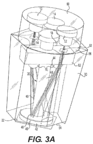
[015] FIG. 3A is a schematic diagram of an illustrative fiberless
transflectance probe embodiment;
CA 02869607 2014-10-03
WO 2013/152177
PCT/US2013/035250
[016] FIG. 3B is a schematic diagram of the distal end of the fiberless
transflectance probe embodiment illustrated in FIG. 3A; and
[017] FIG. 4 illustrates the distribution of the incident radiation beam
on a measurement site, in accordance with an embodiment of the present
disclosure.
Detailed Description
[018] Reference will now be made in detail to embodiments consistent
with the present disclosure, examples of which are illustrated in the
accompanying drawings. Wherever possible, the same reference numbers will
be used throughout the drawings to refer to the same or like parts.
[019] In an exemplary embodiment, an optical system comprising a
fiberless transflectance probe is used to measure the concentration of a
target
analyte in a fluid within a sample matrix. The analyte concentration is
measured and analyzed using a portable device developed using Optical
BridgeTM technology. In accordance with an embodiment of the present
disclosure and the Optical BridgeTM technology, noninvasive optical
measurements of the analyte concentration are performed using a beam of
electromagnetic radiation which alternates at a particular frequency between a
"principal" wavelength (A0), a "reference" wavelength (Ai) and an auxiliary
wavelength A2. A0 is selected to achieve high analyte absorption, and Ai is
selected to have minimal analyte absorption. During the Optical BridgeTM
balancing step, A1 is adjusted to have the same absorption in blood-less
tissue
as hp. The auxiliary wavelength )12 is selected to have high absorption in a
component of the fluid, and is used to provide an estimate of the fluid
content
6
CA 02869607 2014-10-03
WO 2013/152177
PCT/US2013/035250
of the sample matrix. In an exemplary embodiment of the present disclosure,
the fiberless transflectance probe is used to measure the concentration of
glucose (i.e. the target analyte) in blood (i.e. the fluid). In such as
embodiment, Ao is selected to be about 1620 nm and Al is selected to be
about 1380 nm, which are in the near infrared energy spectrum. The auxiliary
wavelength A, is selected to be about 525 nm, which is an isosbestic
wavelength for hemoglobin, and provides an excellent sensitivity to blood. In
one such embodiment, the three wavelengths, Ao, Al. and A2, are 1620 +/- 20
nm, 1380 +1- 20 nm, and 525 +/- 20 nm, respectively. The beam of
electromagnetic radiation consists of time multiplexed component of the three
different wavelengths (A0., A1 and A2) alternating at a frequency of 100 Hz.
In
another embodiment, some or all wavelengths are on at all times, i.e., they
are not alternating. In certain ernbodiments, the separation of the signal
into
its wavelength components is performed by the detector or processor.
[020] FIG. 1 shows a conceptual diagram of an analyte testing device
which utilizes the Optical Bridge TM technology for noninvasively measuring
the concentration of a target analyte (e.g. glucose) in a fluid (e.g. blood)
within
a sample matrix (e.g. a tissue matrix). The analyte testing device 10
comprises at least two laser diodes 12 and 14 operating at wavelengths Ao
and A1 respectively, a signal detector 18, and an optical transflectance probe
which interfaces the laser diodes with a measurernent site 22. In one
embodiment, the analyte testing device 10 further comprises at least one LED
16 operating at a wavelength A2. The beam through the optical probe 20
alternates between Ao, A1 and A2 at a preselected frequency. The wavelength
alternation is driven by the laser controller module 24. The measurement site
7
CA 02869607 2014-10-03
WO 2013/152177
PCT/US2013/035250
22 is chosen such that it is: 1) easily accessible, 2) well perfused with the
fluid
containing the target analyte; 3) small enough to fit in a sample port of a
portable instrument, 4) can be easily compressed/ uncompressed. In one
embodiment of the present disclosure, a subject's earlobe is used as the
measurement site 22. In another embodiment, the subject's finger is used as
the measurement site 22.
[021] In an exemplary embodiment, the extracellular-to-intracellular
fluid ratio of the measurement site 22 is changed during the measurement by
exerting varying mechanical pressure on the measurement site. In such an
embodiment, the amount of fluid in the measurement site 22 is modulated by
means of a linear actuator 26, as illustrated in FIG. 1. The linear actuator
compresses the measurement site 22 with a pressure sufficient to displace
fluid (with the target analyte) from the measurement site 22. In one such
embodiment, the linear actuator compresses the measurement site 22 with a
pressure three times systolic blood pressure. As the compressive force is
released, the displaced fluid returns to the measurement site. In one
embodiment, linear actuator 26 compresses measurement site 22 against
optical probe 20. In another embodiment, linear actuator 26 compresses
optical probe 20 against measurement site 22.
[022] The Optical BridgeTM technology exploits the principle that
compressed tissue has a relatively lower proportion of fluid with the target
analyte than uncompressed tissue, although some residual amount of analyte
remains in the measurement site 22 during the compression. In another
embodiment, the extracellular-to-intracellular fluid ratio is allowed to
change
as a result of natural pulsation due to heartbeat, and the measurement cycle
8
CA 02869607 2014-10-03
WO 2013/152177
PCT/US2013/035250
is synchronized with such pulsation. When the extracellular fluid volume in
the
measurement site is reduced either due to mechanical compression or natural
pulsation, the optical path of the radiation beam contains minimal fluid and
the
target analyte. The Optical Bridge TM balancing is performed at this position
at
the beginning of each rneasurement to achieve the maximum background
rejection. The balancing is performed by adjusting the light intensities at
the
two wavelengths Ao, A1, and also by modifying the reference wavelength
The variations in the background matrix structure are compensated for in the
balancing process. As indicated in FIG. 2A, the light intensities and the
wavelength A1 are adjusted such that the baseline absorption (indicated by the
Optical Bridge Signal 28) is essentially zero when there is minimal fluid and
analyte in the optical path, and the differential absorption of the
wavelengths
Ao and Al (indicated by the variation in Detector Output Voltage 30) is
minimum. The Optical Bridge Signal 28 is in effect the rectified Detector
Output Voltage 30.
[023] In an exemplary embodiment which utilizes the compression
mechanism, the pressure on the measurement site 22 is relaxed after the
Optical Bridge is balanced, allowing fluid to return to the site. The
attenuation
of the two wavelengths Ao and A1 is different at the uncompressed position, as
indicated by the larger variation in the Detector Output Voltage 30 in FIG.
2B.
At the uncompressed position, the Optical Bridge Signal 28 is higher (i.e.
there is more background absorption in the measurement site 22), as
indicated in FIG. 2B. Variations in the Detector Output Voltage 30 is
proportional to the changes in the amount of target analyte (e.g. glucose) in
the fluid. In order to accurately calculate the concentration of the analyte
in
9
CA 02869607 2014-10-03
WO 2013/152177
PCT/US2013/035250
the fluid, the variations of the amount of fluid in the measurement site must
also be measured. The wavelength A; which is highly absorbed by a
component of the fluid, and follows the same optical path as wavelengths Ao
and A1, is used to compensate for the changes in fluid volume in the
measurement site. Features extracted from the detected i12 signal are
processed to produce an estimate of the fluid volume, which is then combined
with the detected Ao and Ai signal output to produce an estimate of the
concentration of analyte in the blood.
[024] In one embodiment, an auxiliary radiation source 34, as
illustrated in FIG. 1, is used to detect pulse and to synchronize the
measurement with the inrush of blood into the measurement site 22. In one
embodiment, the auxiliary radiation source 34 is a LED operating at 525 nm
(an isosbestic wavelength for hemoglobin). The auxiliary radiation source 34
is directed at a portion of the sample matrix that maintains good circulation
at
all times. For example, the radiation source 34 may be directed at a portion
of
the sample matrix, outside the measurement site 22, which is not compressed
by the linear actuator 26. The radiation source 34 generates a pulse detection
beam which is scattered by the tissue, and a fraction of the original beam is
detected by the signal detector 18. The auxiliary radiation source 34 is
operated prior to the measurement step to synchronize the start of the
measurement process with a variation of the blood pressure.
[025] In one exemplary embodiment, optical probe 20 is configured for
transflectance measurements, wherein the radiation beam is inserted into the
measurement site 22 and the backscattered beam is detected by the signal
detector 18. The detector then generates a signal indicative of the
differential
CA 02869607 2014-10-03
WO 2013/152177
PCT/US2013/035250
absorption of the target analyte. An important consideration for such an
embodiment is that the light reflected from the surface of the measurement
site 22 should not reach the detector as it would overwhelm the backscattered
light.
[026] In one such embodiment, transflectance measurement is
performed using a bifurcated bundle of optical fibers, a first portion of
which is
adapted to receive light from the laser diodes operating at wavelengths ho and
Ai, and a second portion of which is adapted to conduct the backscattered
light to the signal detector. The fiber bundle passes through the optical
probe
20, and the common end of the fiber bundle is pressed against the
measurement site 22 for the transflectance measurements.
[027] In another embodiment, transflectance measurement is
performed using a fiberless transflectance probe 20, as illustrated in FIG.
3A.
The transflectance probe 20 interfaces laser diodes 12, 14, at least one LED
16 and the sample detector 18 with the measurement site 22. Transflectance
probe 20 comprises a cylindrical optical rod 40 having a polished outer
surface 45. In one embodiment, the optical rod 40 is made of fused quartz
and the outer surface 45 is coated with aluminum to increase the reflectivity
of the surface. In another embodiment, the optical rod 40 is a glass rod with
aluminum coating on the outer surface 45. Optical rod 40 is positioned
perpendicular to a round detection window 46. The distal end 42 of optical rod
40 is inserted into a circular opening 44 in the detection window 46, such
that
the distalmost end of the optical rod is axially aligned with the distal
surface
49 of the detection window, and is in direct contact with the surface of the
measurement site 22. In order to limit interaction between the incident light
11
CA 02869607 2014-10-03
WO 2013/152177
PCT/US2013/035250
and the backscattered light, the optical rod 40 is coated with aluminum
throughout its length, including the distal end 42 which is inserted into the
detection window 46. Additionally, the optical rod 40 and the detection window
46 are tightly coupled to ensure that a substantial portion of the radiation
backscattered from the measurement site 22 enters the optical rod 40.
[028] The electromagnetic radiation beam is transmitted to the
measurement site 22 through the detection window 46 during a measurement.
Thus, the detection window 46 acts as an interface between the sample
matrix and device hardware. The detection window 46 is also used to apply
mechanical pressure on the rneasurement site 22 during a
compression/decompression procedure, as described earlier. In one
embodiment consistent with the present disclosure, the detection window 46
is comprised of glass or quartz. In another embodiment, the detection window
46 is comprised of a thermoplastic polymer that has high transmittance in the
wavelength range consisting of Ao, Al, and A2, has low moisture absorbility
and
is suitable for injection molding. Example of such thermoplastic polymers
include, but is not limited to, cyclic polyolefins (COP),
polymethylmethacrylate
(PMMA), and polystyrene (PS).
[0291 The optical rod 40 is further surrounded by a tapered tubular
housing 50 having an inner reflective surface. In one embodiment, the inner
surface 52 is aluminized to increase the reflectivity of the surface. The
distal
end 54 of the tapered tubular housing 50 is coupled with the detection window
46, as shown in FIG. 3B. In one embodiment consistent with the present
disclosure, the tapered tubular housing 50 is made of quartz or glass. In
another embodiment, the tapered tubular housing 50 is made of a
12
CA 02869607 2014-10-03
WO 2013/152177
PCT/US2013/035250
thermoplastic polymer using injection molding, and the inner surface 52 is
coated with aluminum to increase the reflectivity of the surface. In yet
another
embodiment, the detection window 46 and the tapered tubular housing 50 are
injected molded together using the same thermoplastic polymer.
[030] The tapered tubular housing 50 also facilitates shaping of the
radiation beam emitted by the laser diodes and the LEDs. The shape of the
inner surface 52 and taper angle of the tubular housing guides the
distribution
of the emitted beam on the measurement site 22. In one preferred
embodiment consistent with the present disclosure, the tubular housing 50 is
configured as a truncated conical shell having a cone angle (angle between
the longitudinal angle and wall) of 7.5 . In another embodiment, the inner
surface of tapered tubular housing 50 is faceted in order to distribute the
incident light evenly on the measurement site 22. The number of facets in the
tubular housing corresponds to the number of laser diodes and LEDs used in
the optical probe 20. In one embodiment, the optical probe 20 includes four
laser diodes (two each for the wavelengths ho and Al), and two LEDs
operating at wavelength A2. In such an embodiment, the inner surface 52 of
the tapered tubular housing 50 has a faceted hexagonal shape, as shown in
FIGS. 3A and 3B. The facets on the inner surface 52 are in the form of a
convex cylinder, and the radius of curvature of each facet is optimized for
the
corresponding light source to provide an uniform distribution of light from
the
different sources on the measurement site 22. In an exemplary embodiment
of the present disclosure, the fiberless transflectance probe 20 is used in a
optical detection system to measure the concentration of glucose in blood. In
such as embodiment, ho is selected to be 1620 nm and 1\1 is selected to be
13
CA 02869607 2014-10-03
WO 2013/152177
PCT/US2013/035250
1380 nm, and the radii of curvature of the cylindrical facets associated with
the lasers operating at A0 and Al are 7.2 mm and 6.1 mm, respectively.
Additionally, the distance of the laser diodes 12, 14 from the central
longitudinal axis of the tubular housing guides the distribution of the
emitted
beam on the measurement site 22. In an embodiment for measuring the
concentration of glucose in blood, as discussed above, the distance of the
laser diodes from the central axis is 5.3 mm.
[031] The laser diodes 12, 14 are mounted on a heat sink 60 at the
proximal end 56 of tapered tubular housing 50 for temperature stability. In
one
embodiment, the LEDs 16 are also mounted on the heat sink adjacent to the
laser diodes. In another embodiment, the LEDs are mounted on a positioning
plate 62 below the heat sink 60, as shown in FIG. 3A, to maintain a stabilized
operating condition for the laser diodes.
[032] The radiation beam comprising the wavelengths Ao, Al, and )12, is
transmitted to the measurement site 22 by reflecting on the outer surface 45
of the optical rod 40 and the inner surface 52 of the tapered tubular housing
50. FIG. 4 shows the distribution of light from four laser diodes on the
measurement site 22. As shown in the figure, the light from the multiple
sources is distributed angularly uniformly on the measurement site, with the
area surrounding the optical rod 40 receiving rnore radiation than the area
around the edge of the tubular housing 50. Some of the light incident on the
measurement site 22 is backscattered by the sample, and a fraction of the
backscattered light reaches the interior of the optical rod 40 and is
conducted
to the signal detector 18 by reflecting on the inner surface of the optical
rod.
The sample detector 18 (not shown in FIG. 3A) is positioned at the proximal
.14
CA 02869607 2014-10-03
WO 2013/152177
PCT/US2013/035250
end 44 of the optical rod 40. When the backscattered light reaches detector
18, an alternating signal is generated which is proportional to the
differential
absorption of wavelengths AG, and Al by the fluid in the sample rnatrix. The
concentration of the target analyte in the fluid is then calculated from the
output signal using a signal processing algorithm.
[0331 In one exemplary embodiment consistent with the present
disclosure and the Optical BridgeTM technology, the analyte testing device 10
is a handheld unit. Referring again to FIG. 1, the handheld unit comprises a
screen 27 for graphic display of the measurement results, and the on-board
electronics consist of a processor 23 for operating the device and calculating
the target analyte concentration, and a control module 24 for driving the
laser
diodes 12, 14 and LEDs 16. The handheld unit may be powered from an
external power supply, rechargeable batteries, or through an USB port.
Additionally, the handheld analyte testing device 10 consists of a memory 25
which stores the measurement results. The memory 25 may further contain
interactive instructions for using and operating the device to be displayed on
the screen 27. The instructions may comprise an interactive feature-rich
presentation including a multimedia recording providing audio/video
instructions for operating the device, or alternatively simple text, displayed
on
the screen, illustrating step-by-step instructions for operating and using the
device. The inclusion of interactive instructions with the device eliminates
the
need for extensive training for use, allowing for patient self-testing and use
by
persons other than medical professionals. In an exemplary embodiment, the
memory 25 may also contain a reference database for statistical calibration of
the device. In another embodiment, the reference database may be accessed
CA 02869607 2014-10-03
WO 2013/152177
PCT/US2013/035250
from a remote storage device via a wireless or a wired connection. Similarly,
data collected from the subject by the analyte testing device 10 may be
recorded in the database for future reference.
[034] The analyte testing device 10 can be a standalone system or
can operate in conjunction with a mobile or stationary device to facilitate
display or storage of data, and to signal healthcare personnel when
therapeutic action is needed, if the device is used for continuous monitoring
of
a diagnostic parameter associated with a disease state. Mobile devices can
include, but are not limited to, handheld devices and wireless devices distant
from, and in communication with, the analyte testing device 10. Stationary
devices can include, but are not limited to, desktop computers, printers and
other peripherals that display or store the results of the test. In an
exemplary
embodiment, the analyte testing device 10 stores each patient file, which
includes a summary of the session and test results, on a removable memory
card 21, such as compact flash (CF) card. The user can then use the memory
card 21 to transfer patient information and procedural data to a computer, or
to produce a printout of the data and session summary. In another
embodiment, results from the processor 23 are transferred directly to an
external mobile or stationary device to facilitate display or storage of data.
For
example, the results from the processor 23 may be displayed or stored on a
PC 29 using a PC interface, such as an USB port, IRDA port, BLUETOOTH
or other wireless link. In yet another embodiment, the results can be
transmitted vvirelessly or via a cable to a printer 31 that prints the results
to be
used by attending medical personnel. Further, the analyte testing device 10
can transmit data to another mobile or stationary device to facilitate more
16
CA 02869607 2014-10-03
WO 2013/152177
PCT/US2013/035250
complex data processing or analysis. For example, the device, operating in
conjunction with PC 29, can send data to be further processed by the
computer.
[035] Although the Optical BridgeTM method and the analyte testing
device 10 are described here with a focus towards measuring the
concentration of glucose in blood, the method and device presented in this
disclosure may also be employed to detect the concentration of other
analytes, such as urea, cholesterol, nicotine, drugs, etc., in blood or other
fluids. Additionally, the fiberless transflectance probe 20 and its method of
use
may be utilized in any optical detection system operating in the infrared,
visible, or ultraviolet wavelength range.
[036] Other embodiments of the invention will be apparent to those
skilled in the art from consideration of the specification and practice of the
invention disclosed herein. It is intended that the specification and examples
be considered as exemplary only, with a true scope and spirit of the invention
being indicated by the following claims.
17