Note: Descriptions are shown in the official language in which they were submitted.
CA 02869732 2014-10-06
WO 2013/165615
PCT/US2013/032499
METHODS, SYSTEMS, AND DEVICES FOR DETECTING AND IDENTIFYING
MICROORGANISMS IN MICROBIOLOGICAL CULTURE SAMPLES
BACKGROUND OF THE INVENTION
Field of the Invention
The presently disclosed subject matter relates to methods, systems, and
devices
for detecting, identifying, and quantifying microorganisms in a culture
sample. More
particularly, the subject matter relates to the use of indicator particles to
detect and
identify one or more microorganisms in a biocontained sample capable of
supporting
growth of microorganisms.
BACKGROUND OF THE INVENTION
The ability to detect low levels of microorganisms, including pathogens, in a
microbiological culture in clinical samples (e.g., blood, stool, urine, etc.)
has gained
significant importance in recent years. Similarly, microbiologial culture is
important to
public health to detect microorganisms, including pathogens, in industrial
samples such
as food, cosmetics, and pharmaceuticals. The ability to detect such
microorganisms not
only provides techniques for treating those who have already been exposed, but
also to
instances where exposure can be prevented, such as when testing food samples.
Foodborne illnesses significantly impact society, not only with respect to
health,
but also health-care costs. The CDC has estimated that each year about 1 in 6
Americans (or 48 million people) gets sick, 128,000 are hospitalized, and
3,000 die of
foodborne diseases (see http://www.cdc.gov/foodsafety/facts.html). It has also
been
estimated that foodborne illnesses contribute to $152 billion in health-
related expenses
each year in the U.S., particularly for bacterial infections caused by
Campylobacterspp.,
Salmonella, Listeria monocyto genes and E.coli (see
http://www.producesafetyprojectorg/admin/assets/files/Health-Related-Foodbome-
Illness-Costs-Report.pdf-l.pdt).
The current level of food safety found in the U.S. is the result of Government
regulations combined with industry self-monitoring influenced by market
incentives, such
as legal liability, brand value, reputation, and the desire to sell more food
product. In the
1
CA 02869732 2019-10-06
WO 2013/165615
PCT/US2013/032499
U.S., the primary agencies responsible for food safety are the U.S Department
of
Agriculture (USDA) Food Safety and Inspection Services (FSIS), which is
responsible for
the safety of meat, poultry, and processed egg products, and the Food and Drug
Administration (FDA), which is responsible for virtually all other foods. In
1996, USDA's
FSIS promulgated the pathogen reduction hazard analysis critical control point
(PR/HACCP) rule, which, for example, mandates generic E. coil testing by
slaughter
plants. Other FSIS regulations enforce zero limits for two deadly pathogens ¨
Listeria
monocytogenes in ready-to-eat meat and poultry and E. coli 0157:H7 in ground
beef (see
http://www.ers.usda.gov/briefing/foodsafety/private.htm). Recently, the Food
Safety
Modernization Act was approved by Congress, the urgency for this legislation
being
underscored by continued outbreaks of foodborne illness over the last several
years--
from spinach to peppers to peanuts.
Food testing may occur on food samples themselves, either end product
materials, intermediates, or incoming raw materials. In addition, HACCP
(Hazard
Analysis and Critical Control Point) plans are implemented to control the
production
environment so as to minimize the risk of introduction of pathogens into the
food sample.
As part of many HACCP plans, environmental samples are acquired from surfaces,
floors, drains, and processing equipment and then analyzed for the presence
and
absence of pathogenic organisms. If a pathogen is detected, it may be isolated
and
subjected to further confirmatory testing.
Today, all food pathogen testing conducted entails a culture step to enrich
the
potentially low levels of microorganisms contained in a sample. Following
culture of the
sample, a portion is removed and tested for the presence of pathogens.
Pathogen testing
after culture can be done by immunoassays (e.g., bioMerieux's Vidase automated
ELISA
platform or SDIX's RapidChek lateral flow assays) or by PCR-based tests
(e.g., DuPont
Qualicon's BAXO system, Bio-Rad's iQ-CheckTM system). If a pathogen is present
in the
starting sample, the culture step can increase the concentration of the
pathogen as high
as 1.0E8 ¨ 1.0E9 cfu/mL, so that opening the sample after culture exposes both
the user
and the environment to a risk of contamination. This exposure inhibits many
food
producers from conducting pathogen testing on-site, instead choosing to send
samples to
external laboratories for testing. In addition, since it is unknown which
samples contain
pathogens and at what levels, food safety test protocols use lengthy culture
times to
ensure that the worst case scenario of one damaged pathogen is given
sufficient time to
grow to a detectable concentration. As a consequence, samples with higher
pathogen
loadings are cultured longer than may be strictly necessary, leading to a
delay in time to
results. There is thus a need in the field for pathogen test methods that
minimize time to
2
CA 02869732 2019-10-06
WO 2013/165615
PCT/US2013/032499
results and reduce the risk of exposure of the facility and personnel to
cultured
pathogens.
Similar concerns are present for clinical samples such as blood. Since the mid-
1980s, along with the expanding size of the immunocompromised patient
population, the
incidence of septicemia caused by opportunistic pathogens, such as yeast,
fungi, and
mycobacteria, has risen. Bacteremia, the presence of bacteria in the blood
stream, and
fungemia, the presence of fungi or yeasts in the blood stream, typically are
detected by
collecting a venous blood sample and disposing the blood sample in a blood
culture bottle
containing a growth medium suitable for promoting growth of the bacteria or
fungi of
interest. See generally, Reimer et al., "Update on Detection of Bacteremia and
Fungemia," Clinical Microbiology Reviews 10(3), 444-465 (1997). The blood
culture
sample can then be incubated for a period of time and checked intermittently
for an
indication of bacterial or fungal growth.
Instrumented methods known in the art for monitoring bacterial or fungal
growth in
blood culture bottles typically detect changes in the carbon dioxide and/or
oxygen
concentration in the blood culture bottle. These instruments detect the
presence and
absence of microorganisms but are not specific as to the particular type of
organism
present. For a nominally sterile sample such as blood, detection of a
microorganism in
the sample can be indicative of severe disease. However, the positive result
is
considered to be a partial or preliminary result and is typically not
actionable. As optimum
treatment of the disease relies on identification of the organism and
determination of its
antibiotic susceptibility, laboratory personnel must be available to advance
positive
cultures to full identification (ID) and antimicrobial susceptibility testing
(AST).
Identification of the organism requires accessing of the positive blood
culture sample by
laboratory personnel for further sample work-up.
Sample work-up following a positive blood culture result, i.e., a result
indicating
the presence but not identity of a microorganism, often includes
categorization of the
microorganism into one of two broad classes of organisms: Gram positive or
Gram
negative. Blood culture assays based on the detection of CO2 or 02 during the
culture
process cannot distinguish between pathogenic organisms, such as S. aureus,
and
contaminants, such as S. epidermidis since these methods are sensitive only to
growth
and absence of growth. Classification and identification of organisms is
performed
following the detection of growth in a blood culture sample. For example, kits
are
available for differentiating between Staphylococcus and Streptococcus species
and
other organisms. Kits also are available for differentiating between
organisms, such as S.
aureus and S. epidermidis. These kits, however, require removing at least an
aliquot of a
3
CA 02869732 2019-10-06
WO 2013/165615
PCT/US2013/032499
blood culture sample from the blood culture bottle and other procedures that
can
potentially expose the operator to the pathogen or destroy a portion of the
blood culture
that could be used for other analyses. They also typically require that
trained laboratory
staff are available to conduct the tests, potentially leading to a delay in
actionable clinical
results in the event that a blood culture sample goes positive when laboratory
personnel
are unavailable to conduct additional testing (e.g., in hospitals that operate
only a single
shift.)
While instruments exist today to detect the presence or absence of
microorganisms in blood (e.g., by use of a carbon dioxide or oxygen sensor),
these
instruments are not typically useful in non-sterile samples such as stool or
food samples.
For samples such as, for example food, there is expected to be a significant
concentration of benign microorganisms, and so detection of organisms by
carbon
dioxide or oxygen sensors is not inherently useful. For a food sample, it is
critical to
detect the presence of low levels of pathogenic organisms in a background of
high benign
microflora to avoid the spread of foodborne illnesses.
Therefore, there is a need for methods, systems, and devices for detecting not
only the presence or absence of organisms during the culture step of nominally
sterile
samples, but also identification of the organisms. For non-sterile samples,
such as stool
and food, there is also a need for methods, systems, and devices for
identifying
potentially harmful organisms in a culture in a biocontained manner. Such
methods,
systems and devices minimize user intervention, thereby minimizing time,
trained
personnel, plus potential exposure of personnel and environment to the
pathogen.
SUMMARY OF THE INVENTION
Embodiments of the presently disclosed subject matter provide methods,
systems,
and devices for detecting the presence, amount, and/or identity of specific
microorganisms in a microbiological culture. According to one embodiment, the
presently
disclosed assays can be performed within the culture vessel, so that detection
and/or
identification of specific microorganisms occur in conjunction with culture,
without the
need for user intervention. One or more microorganisms can be identified
within a single
culture. The culture vessel can be fully biocontained so that the growth of
the
microorganism and microorganism detection and identification can occur without
exposing either the user or the surrounding environment. Moreover, due to the
biocontainment of the culture, the analysis of the culture may occur without
the need for
the user to access the culture or wash the culture.
4
CA 02869732 2019-10-06
WO 2013/165615
PCT/US2013/032499
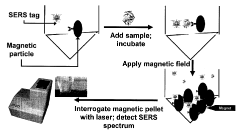
Optically active indicator particles, such as Surface Enhanced Raman
Scattering
(SERS)-active nanoparticles, each having associated therewith one or more
specific
binding members having an affinity for the one or more microorganisms of
interest, can
form a complex with specific microorganisms in the microbiological culture
sample. Thus,
the optically active indicator particles can be any particle capable of
producing an optical
signal that can be detected in a culture sample without wash steps. Further,
magnetic
capture particles, also having associated therewith one or more specific
binding members
having an affinity for the one or more microorganisms of interest, which can
be the same
or different from the specific binding members associated with the indicator
particles, can
be used to capture the microorganism-indicator particle complex and
concentrate the
complex in a localized area of an assay vessel for subsequent detection.
Importantly,
embodiments of the presently disclosed methods, systems, and devices allow
"real-time"
detection and identification of microorganisms in a sample in which active
growth of the
microorganism is occurring. Samples may include microbiological cultures
comprising a
growth medium and a clinical sample from a human or animal (domestic or stock)
such as
blood, stool, urine, or cerebral spinal fluid. Samples may also include
microbiological
cultures comprising a growth medium and an industrial sample such as food,
dairy,
beverage, water, environmental, agricultural products, personal care products
(including
cosmetics), biotechnology, or pharmaceuticals. Importantly, the assay can be
conducted
in a biocontained manner without exposure of the user or environment to the
sample
("closed system") and can provide automated, around the clock, detection and
identification of microorganisms by monitoring the assay signal over time as
the culture
progresses. The combination of detection and identification with
microbiological culture
can lead to earlier availability of actionable results.
Detection of microorganisms by the present invention can be performed either
directly or indirectly. For direct detection of micorganisms growing in
culture, the specific
binding members associated with the magnetic capture particles and indicator
particles
can have an affinity for the largely intact microorganism, e.g. by binding to
the surface of
bacteria or yeast. For indirect detection, the binding members associated with
the
magnetic capture particles and indicator particles may have an affinity for
byproducts of
the microorganism. Examples of byproducts could include but are not limited to
secreted
proteins, toxins, and cell wall components. Direct and indirect detection
modes made be
used alone or in combination.
According to another embodiment of the present invention, a vessel for
metering a
desired amount of culture sample is provided. The vessel includes a container
for
receiving a culture sample therein, wherein the container has an open end and
a closed
5
CA 02869732 2019-10-06
WO 2013/165615
PCT/US2013/032499
end. The vessel also includes a lid configured to engage the open end of the
container in
a fluid-tight connection. In addition, the vessel includes a basket coupled to
the lid and
including one or more reservoirs, wherein the basket is disposed between the
open end
and the closed end of the container. Where a plurality of reservoirs is used,
each
reservoir is configured to hold a different volume of culture sample.
Moreover, the vessel
includes one or more needle assemblies engaged with the lid, wherein the
needle
assembly includes a needle extending within a respective reservoir. Each
needle is
configured to selectively withdraw a sample contained in a respective
reservoir, wherein
each needle is further configured to engage a vial for a biocontained transfer
of the
sample from the reservoir to the vial. Thus, the vessel may be suitable for
metering a
desired amount of sample for two different assays (e.g., Salmonella or
Listeria) in a single
container, while facilitating transfer of the sample to a detection vial in a
biocontained
manner. In another embodiment of the present invention, the assay vial for
receiving a
sample is enclosed by a stopper or septum and cap configured to retain a
vacuum. Upon
connection of the assay vial cap with a compatible port containing a needle on
the
metering vessel, the sample is transferred in a biocontained fashion. The vial
cap
contains features to retain externally expressed fluid from the transfer and
protect the
user from contact with transfer surfaces.
Another embodiment of the present invention is directed to a system for
automatically processing a plurality of tubes containing a culture sample. The
system
includes an incubator for receiving a plurality of sample tubes therein,
wherein the
incubator is configured to incubate the sample tubes at a predetermined
temperature.
For example, the tubes may be positioned horizontally and adjacent to each
other. The
incubator may be configured to incubate different assays at different
temperatures
according to one embodiment. The system further includes a first translational
device
(e.g., a "Y-stage" for movement along a Y-axis) coupled to the tray and
configured to
move the sample tubes within the incubator, wherein the first translational
device is
further configured to move the sample tubes from the incubator to a detection
zone and to
agitate the sample tubes within the detection zone. For instance, the first
translational
device may move the samples tubes along their longitudinal axes. The system
also
includes a magnet assembly configured to apply a magnetic field to the
plurality of
sample tubes within the detection zone, as well as an optical device
configured to
interrogate each of the plurality of sample tubes within the detection zone
for detecting
one or more microorganisms. The system includes a second translational device
(e.g.,
an "X-stage" for movement along an X-axis) coupled to the optical device and
configured
to move the optical device within the detection zone for interrogating each of
the sample
6
CA 02869732 2019-10-06
WO 2013/165615
PCT/US2013/032499
tubes. The system may also include a third translational device (e.g., a "Z-
stage" for
movement along the Z-axis) coupled to the magnet assembly and the optical
device and
configured to move the magnet assembly and optical device within the detection
zone to
access another tray of tubes stacked vertically above the first tray. Thus,
the system
provides an automated and high-throughput system for processing a plurality of
samples
in real time during incubation of the culture tubes.
BRIEF DESCRIPTION OF THE DRAWINGS
Having thus described the presently disclosed subject matter in general terms,
reference will now be made to the accompanying drawings, which are not
necessarily
drawn to scale, and wherein:
Figure 1 is a schematic diagram showing a method of detecting and identifying
a
microorganism in a culture sample according to an embodiment of the invention.
Figure 2 is a schematic diagram showing an enrichment vessel and a detection
vial for containing and transferring a culture sample according to one
embodiment of the
present invention.
Figure 3 is a schematic diagram showing a method of intermittent detecting and
identifying of a microorganism in a culture sample according to an embodiment
of the
invention.
Figure 4 is a schematic diagram showing a method of real-time detecting and
identifying of a microorganism in a culture sample according to an embodiment
of the
invention.
Figures 5A-5E illustrate various views of an enrichment vessel according to
one
embodiment of the present invention.
Figure 6 is a cross-sectional view of an enrichment vessel according to one
embodiment of the present invention.
Figure 7 is an exploded view of an enrichment vessel according to one
embodiment of the present invention.
Figure 8 is a bottom view of lid for an enrichment vessel according to one
embodiment of the present invention.
Figures 9A-9C are various views of a basket for an enrichment vessel according
to one embodiment of the present invention.
7
CA 02869732 2019-10-06
WO 2013/165615
PCT/US2013/032499
Figures 10A and 10B illustrate a cap for a detection vial according to one
embodiment of the present invention.
Figures 11A and 11B illustrate a cap for a detection vial according to one
embodiment of the present invention.
Figure 12 is a cross-sectional view of a cap engaging a detection vial
according to
one embodiment of the present invention.
Figures 13A and 13B are a perspective view and an exploded view of a cap
engaging a detection vial according to an embodiment of the present invention.
Figures 14A and 14B are a perspective view and an exploded view of a cap
engaging a detection vial according an embodiment of the present invention.
Figure 15 is a cross-sectional view of detection vials engaging an enrichment
vessel according to an embodiment of the present invention.
Figures 16 and 17 are cross-sectional views of a detection vial engaging an
enrichment vessel according to an embodiment of the present invention.
Figure 18 is a schematic diagram showing a magnetic capture particle-
microorganism-SERS-active indicator particle complex within a culture bottle
according to
an embodiment the invention.
Figure 19 depicts a SERS-active indicator particle according to one embodiment
of the present invention.
Figure 20 depicts a SERS-active indicator particle according to one embodiment
of the present invention.
Figure 21 depicts a SERS-active indicator particle according to one embodiment
of the present invention.
Figure 22 shows a representative SERS spectrum of a SERS-active indicator
particle having associated therewith a 4,4'-dipyridyl (DIPY) Raman-active dye
according
to an embodiment of the invention.
Figure 23 shows a representative SERS signal plotted over culture time for
Salmonella according to an embodiment of the invention.
Figure 24 depicts a system for real-time monitoring of microorganism growth
according to an embodiment of the invention.
Figure 25 depicts a system for real-time monitoring of microorganism growth
according to another embodiment of the invention.
CA 02869732 2019-10-06
WO 2013/165615
PCT/US2013/032499
Figures 26-29 illustrate various views of a system for real-time monitoring of
microorganism growth according to an additional embodiment of the invention.
Figure 30 is a perspective view of a tray for holding sample tubes according
to an
embodiment of the present invention.
Figure 31 illustrates sequential steps for loading sample tubes into a tray,
loading
the tray into an incubator, and removing the trays from the incubator,
according to an
embodiment of the present invention.
Figure 32 is a perspective view of a tray for holding sample tubes according
to
another embodiment of the present invention.
Figures 33A-33C are partial views of trays for holding sample tubes according
to
various embodiments of the present invention.
Figure 34 is a perspective view of an incubator according to an embodiment of
the
present invention.
Figures 35-39 are various cross-sectional views of the system shown in Figures
26-29.
Figure 40 is an enlarged view of a rear door of an incubator according to an
embodiment of the present invention.
Figure 41 is a perspective view of an X-stage according to an embodiment of
the
present invention.
Figure 42 is a perspective view of a magnet assembly, an X-stage, and a Z-
stage
according to an embodiment of the present invention.
Figure 43 is a side view of a magnet assembly, a pelleting/read assembly, an X-
stage, a Y-stage, and a Z-stage in a lowered position according to an
embodiment of the
present invention.
Figure 44 is another perspective view of the system shown in Figures 26-29.
Figures 45 and 46 are partial perspective views of a magnet assembly, a
pelleting/read assembly, and an X-stage according to one embodiment of the
present
invention.
Figure 47 is partial perspective view of a magnet assembly and a Y-stage
according to one embodiment of the present invention.
9
CA 02869732 2019-10-06
WO 2013/165615
PCT/US2013/032499
Figures 48A-48B are perspective views of a system for real-time monitoring of
microorganism growth enclosed in a cabinet according to embodiments of the
present
invention.
Figure 49 illustrates a method for agitating and pelleting a culture sample
according to an embodiment of the present invention.
Figure 50 depicts a pelleting and optical system according to an embodiment of
the invention.
Figures 51 and 52 illustrate alternative magnet arrangements for pelleting a
culture sample according to embodiments of the present invention.
Figure 53 depicts a multiplexed detection of S. aureus and S. epidermidis
according to an embodiment of the invention.
Figure 54 shows the results of an experiment in which time to detection of E.
coli
growth was compared for blood culture samples with and without the SERS HNW
reagents suitable for use in the various embodiments of the invention.
Figure 55 shows a graph in which the growth of Salmonella enterica subspecies
enterica serovar Typhimurium, henceforth referred to as Salmonella Typhimurium
(or
other Salmon/la serovar name), was monitored in relation to the effect of
pelleting thereon
according to an embodiment of the invention.
Figure 56 shows a graph illustrating the effect of pelleting on microorganism
growth according to an embodiment of the invention.
Figure 57 illustrates an image of a SERS-magnetic bead precomplex (PC) in
water after pelleting with a fixed magnet according to one embodiment.
Figures 58A-58B are images of PC pellet formation in SDIX Salmonella secondary
media using a fixed magnet and different agitation frequencies according to
one
embodiment.
Figures 59A-59B are images of PC pellet formation in SDIX Salmonella secondary
media using a coupled magnet and different agitation frequencies according to
one
embodiment.
Figure 60 shows a graph in which time to detection of C. albicans in blood was
compared using a singleplex SERS detection according to an embodiment of the
invention.
Figure 61 shows a graph in which time to detection of C. albicans in blood was
compared using a multiplex SERS method according to an embodiment of the
invention.
CA 02869732 2019-10-06
WO 2013/165615
PCT/US2013/032499
Figure 62 shows a graph in which time to detection of E. coli and S.
epidermidis in
blood was compared using a multiplex SERS method according to an embodiment of
the
invention.
Figure 63 illustrates a graph of real-time detection of E. coil in blood with
aerobic
media and antibiotic absorbing resins according to an embodiment of the
present
invention.
Figure 64 shows a graph of the detection of E. coli in blood for different
sample
volumes according to an embodiment of the present invention.
Figure 65A shows a SERS curve with images captured at various times during
secondary enrichment of Salmonella Typhimurium according to one embodiment.
Figure 65B shows a SERS curve with images captured at various times during
secondary enrichment for a negative sample according to one embodiment.
Figure 65C shows a SERS curve with images captured at various times during
secondary enrichment of Salmonella Typhimurium according to one embodiment.
Figure 66 shows overlaying SERS curves for different agitation rates during
secondary enrichment of Salmonella Typhimurium according to one embodiment.
Figure 67 shows images of pellets for a positive sample and a negative sample,
respectively, according to an embodiment of the present invention.
Figures 68A-68C illustrate SERS curves for the real-time detection of E. coil
during culture in food samples according to embodiments of the present
invention.
Figure 69 illustrates SERS curves for the real-time detection of Salmonella
Enteritidis during culture in food samples according to an embodiment of the
present
invention.
Figure 70 illustrates SERS curves for the real-time detection of Listeria
swabbed
from stainless steel during culture according to an embodiment of the present
invention.
Figure 71 shows a flowchart of phases for the detection of Salmonella
Typhimurium using linear agitation according to an embodiment of the present
invention.
Figure 72 illustrates overlaying SERS curves during secondary enrichment for
Salmonella Typhimurium, Salmonella Enteritidis, and negative samples according
to an
embodiment of the present invention.
Figure 73 shows images of pellets formed during secondary enrichment of
Salmonella Typhimurium according to an embodiment of the present invention.
11
CA 02869732 2019-10-06
WO 2013/165615
PCT/US2013/032499
Figure 74 shows SERS curves obtained from rocking agitation and linear
agitation
during secondary enrichment of S. Enteritidis and S. Kentucky according to one
embodiment.
Figure 75 shows images of sample tubes containing S. aureus and S. epidermidis
in EDTA rabbit plasma, with and without SERS reagents, according to one
embodiment
of the present invention.
Figure 76 shows images of latex agglutination assays with S. aureus and S.
epidermidis, with and without SERS reagents, according to one embodiment of
the
present invention.
Figure 77 is a magnified image of gram staining of a mixture of magnetic
particles
and SERS tags according to one embodiment of the present invention.
Figure 78 is a magnified image of gram stained controls of S. aureus and E.
coli
with magnetic particles and SERS tags according to one embodiment of the
present
invention.
Figure 79 shows images of CHROMagar S. aureus plates streaked with a blood
culture of S. aureus and S. epidermdis with SERS reagents according to
embodiments of
the present invention.
Figure 80 is an image of an agar plate streaked with a blood culture of E.
coil with
SensidiscTM test discs according to an embodiment of the present invention.
Figure 81 is a table showing zone diameter measurements for E. coli, with and
without reagents, according to an embodiment of the present invention.
Figure 82 is a table showing a summary of the results of manual antibiotic
susceptibility testing using BD SensidiscsTM and various microorganisms with
and
without SERS reagents, according to one embodiment of the present invention.
Figure 83 shows images of agar plates streaked with a blood culture of E. coli
and
C. albicans, with and without reagents, overlaid with anti-fungal BD TaxoTm
discs,
according to an embodiment of the present invention.
Figure 84 is a table showing images of pellets formed in Salmonella secondary
media using different agitation frequencies and pelleting times, according to
an
embodiment of the present invention.
Figure 85 is a table showing the effect of agitation frequency on pellet
dispersion,
according to an embodiment of the present invention.
12
CA 02869732 2019-10-06
WO 2013/165615
PCT/US2013/032499
Figure 86 illustrates an enrichment vessel according to another embodiment of
the
present invention.
Figure 87 is a cross-sectional view of a syringe according to one embodiment
of
the present invention.
Figure 88 is a cross-sectional view of a syringe engaged with an enrichment
vessel according to one embodiment of the present invention.
Figures 89A-89C are enlarged cross-sectional views of a syringe engaged with
an
enrichment vessel according to various embodiments of the present invention.
Figures 90A and 90B are enlarged cross-sectional views of a syringe according
to
one embodiment of the present invention.
Figure 91 is a cross-sectional view of a syringe and a perspective view of a
plunger according to one embodiment of the present invention.
Figure 92 is a cross-sectional view of a syringe and a perspective view of a
plunger according to another embodiment of the present invention.
Figures 93-95 illustrate reconstitution stations according to various
embodiments
of the present invention.
Figure 96 is an image of fabricated fluorescent silica nanoparticles according
to
one embodiment of the present invention.
Figure 97 shows a graph depicting the signal intensity of fabricated
fluorescent
silica nanoparticles and conventional SERS tags according to one embodiment of
the
present invention.
Figure 98 shows a graph depicting the signal intensity over time of fabricated
fluorescent silica nanoparticles and conventional SERS tags for detecting the
presence of
Listeria in spinach according to one embodiment of the present invention.
Figure 99 shows a graph depicting the signal intensity over time of fabricated
fluorescent silica nanoparticles and conventional SERS tags for detecting the
presence
Listeria in cabbage according to one embodiment of the present invention.
Figure 100 is a perspective view of a container for an enrichment vessel
according
to one embodiment of the present invention.
Figure 101 is a top view of lid for an enrichment vessel according to one
embodiment of the present invention.
Figure 102 is a bottom view of the lid shown in Figure 101.
13
CA 02869732 2019-10-06
WO 2013/165615
PCT/US2013/032499
Figure 103 is a bottom perspective view of the lid shown in Figure 101.
Figure 104 is a side view of the lid shown in Figure 101.
Figure 105 is a cross-sectional view of the lid shown in Figure 101.
Figure 106 is a side view of a basket for an enrichment vessel according to
one
embodiment of the present invention.
Figure 107 is a top view of the basket shown in Figure 106.
Figure 108 is a bottom view of the basket shown in Figure 106.
Figure 109 is a perspective view of the basket shown in Figure 106.
14
CA 02869732 2019-10-06
WO 2013/165615
PCT/US2013/032499
DETAILED DESCRIPTION OF THE INVENTION
The presently disclosed subject matter now will be described more fully
hereinafter with reference to the accompanying Drawings, in which some, but
not all
embodiments of the presently disclosed subject matter are shown. Many
modifications
and other embodiments of the presently disclosed subject matter set forth
herein will
come to mind to one skilled in the art to which the presently disclosed
subject matter
pertains having the benefit of the teachings presented in the foregoing
descriptions and
the associated Drawings. Therefore, it is to be understood that the presently
disclosed
subject matter is not to be limited to the specific embodiments disclosed and
that
modifications and other embodiments are intended to be included within the
scope of the
appended claims. Although specific terms are employed herein, they are used in
a
generic and descriptive sense only and not for purposes of limitation.
The terms 'a," "an," and "the" refer to "one or more" when used in this
application,
including the claims. Thus, for example, reference to "a sample" includes a
plurality of
samples, unless the context clearly is to the contrary (e.g., a plurality of
samples), and so
forth.
Throughout this specification and the claims, the words "comprise,"
"comprises,"
and "comprising" are used in a non-exclusive sense, except where the context
requires
otherwise.
As used herein, the term "about," when referring to a value is meant to
encompass a specified value and variations thereof. Such variations may be, in
some
embodiments 100%, in some embodiments 50%, in some embodiments 20%, in
some embodiments 10%, in some embodiments 5%, in some embodiments 1%, in
some embodiments 0.5%, and in some embodiments 0.1% from the specified
amount, as such variations are appropriate to perform the disclosed methods or
employ
the disclosed compositions.
Further, when an amount, concentration, or other value or parameter is given
as
either a range, preferred range, or a list of upper preferable values and
lower preferable
values, this is to be understood as specifically disclosing all ranges formed
from any pair
of any upper range limit or preferred value and any lower range limit or
preferred value,
regardless of whether ranges are separately disclosed. Where a range of
numerical
values is recited herein, unless otherwise stated, the range is intended to
include the
endpoints thereof, and all integers and fractions within the range. It is not
intended that
the scope of the presently disclosed subject matter be limited to the specific
values
recited when defining a range.
CA 02869732 2019-10-06
WO 2013/165615
PCT/US2013/032499
The embodiments of the present invention provide systems and methods which
utilize indicator particles (e.g., surface enhanced Raman scattering (SERS)-
active
indicator particles), for detecting and/or identifying one or more
microorganisms in a
bacterial culture sample by a Homogeneous No Wash assay (HNW). More
specifically,
embodiments of the invention describe techniques for monitoring the
concentration of
microorganism in "real-time" as the microorganism level increases over time
within a
sample. The indicator particles have associated therewith one or more specific
binding
members having an affinity for the one or more microorganisms under test. When
contacted with a microbiological culture sample containing one or more
microorganisms
of interest, a complex, generally referred to herein as an indicator particle-
microorganism
complex, between the one or more microorganisms of interest and the indicator
particle
with associated specific binding member can be formed. The indicator particle-
microorganism complex can be captured by a magnetic capture particle and
concentrated
to form a pellet in a localized area (i.e., a "measurement zone') for
detection by
measuring the signal (e.g., SERS spectrum) and/or a visual inspection of an
image of the
pellet. The term "pellet", as used herein, is not meant to be limiting and in
one
embodiment, refers to a collection of a plurality of indicator particles and
magnetic
capture particles located in a localized area facilitated by application of a
magnetic field,
wherein the pellet is detectable using visual, optical, or other suitable
means. The pellet
may also include microorganisms captured therebetween, if present, and other
components and/or microorganisms may be non-specifically attached to the
magnetic
particles. The pellet may be temporarily formed in that the pellet may be
dispersed upon
removal of the magnetic field as discussed in greater detail below.
Furthermore, the various embodiments of the invention pertain to the ability
to
conduct the HNW assay repeatedly within the same microbiological culture
sample, by
forming, dispersing, and reforming the pellet over time. This enables the
concentration of
a particular analyte to be monitored real-time within a microbiological
culture sample and
is particularly valuable when the microorganism concentration is changing over
time, e.g.
in response to bacterial growth. More particularly, embodiments of the
invention pertain
to the ability to conduct the HNW assay within a microbiological culture
vessel, thereby
simultaneously detecting and identifying a microorganism as it grows. In
addition, the
technique can be used in conjunction with other methods of monitoring the
culture sample
(such as gas sensor or image analysis).
According to an embodiment of the invention, a microbiological culture of the
sample is conducted in a vessel that also contains the HNW reagents. The
culture vessel
is inserted into an instrument that allows incubation at a controlled
temperature and
16
CA 02869732 2019-10-06
WO 2013/165615
PCT/US2013/032499
contains optical devices (e.g., Raman optics, a Raman laser, and a
spectrometer). At
regular time intervals during the culture, a magnetic field is applied, and
the SERS signal
is read from the magnetic pellet. The pellet is dispersed between readings to
allow
continued interactions of the reagents with the sample. As the target organism
concentration increases throughout the enrichment process, detection and
identification
of the microorganism by the SERS technology occurs as soon as the
microorganism
concentration reaches the detection threshold of the technology. The ability
to
continuously monitor the SERS signal during culture ensures that the minimal
required
culture time is used and that the instrument can automatically alert the user
when a
microorganism is detected and identified.
A further embodiment uses a camera to monitor the formation and size of a
pellet
during a HNW assay which contains conjugated indicator particles and magnetic
beads
and the targeted pathogen within a culture vessel. Images show that pellet
size
increases, and in some cases the pellet disappears, from the camera view as
the HNW
assay progresses. The growth in pellet size and/or disappearance of the pellet
is an
indication of the presence of the targeted pathogen. Images captured during
analysis of
samples that contain conjugated indicator particles and magnetic beads with no
pathogen
show no change in pellet size and no pellet disappearance. This method of
detection can
be used alone or in conjunction with another detection method.
I. General Considerations for Detection and Identification of Microorganisms
in a
Microbiological Culture Sample
As used herein, the term "microbiological culture sample" refers to a
composition
comprising a "clinical" or an "industrial" sample with the potential of
containing
microorganisms that is disposed in, admixed, or otherwise combined with a
culture
medium, e.g., a blood culture broth, capable of supporting the growth of one
or more
microorganisms suspected of being present in the sample. More particularly,
embodiments of the presently disclosed subject matter provide methods,
systems, and
devices for detecting microorganisms in a microbiological culture sample
comprising a
media capable of supporting microorganism growth in either a clinical sample,
such as
blood, stool, urine or cerebral spinal fluid, or in an industrial product
sample, such as
food, environmental swabs or sponges, water, cosmetics, hygiene products,
pharmaceuticals, or other products intended for use or consumption by animals
or
humans.
17
CA 02869732 2019-10-06
WO 2013/165615
PCT/US2013/032499
Detecting and/or identifying microorganisms in microbiological culture
samples,
especially with optical or spectrometric methods, can present many challenges
due to the
complexity of the sample matrix. Clinical samples, particularly those such as
blood or
stool, are optically absorptive, making it difficult to detect optical or
spectral signals
without wash or lysis steps to remove optically interfering components of the
original
samples. Industrial samples, such as, for example food or cosmetic samples,
may be
optically absorptive, again requiring wash or lysis steps to remove optical
interferents in
the original sample. Although the application of SERS to detecting mammalian
cells and
microorganisms and the diagnostic application of SERS-active indicator
particles to
detecting a variety of analytes in the presence of blood and food samples has
been
reported, the application of SERS-active indicator particles to monitor
bacteria and fungi
concentrations in "real time" as the concentrations change due to
microorganism growth
has not been reported. As used herein, "real time" is not meant to be limiting
and may
refer to monitoring the culture sample continuously or in predetermined
increments of
time. For example, the culture sample may be tested repeatedly in
predetermined
increments of time (e.g., every 30 minutes, 1 hour, etc.) over a predetermined
incubation
period without opening the sample tube thereby maintaining biocontainment of
the
sample. "Biocontainment", as used herein, is also not meant to be limiting and
may refer
to the culture sample being in a closed system such that the surrounding
environment
outside of the container in which the culture sample is confined is not
exposed to the
microorganisms being cultured.
Further, the presently disclosed methods allow for the diagnostic use of
indicator
particles in microbiological cultures in a manner that does not inhibit the
growth of the
microorganism under detection.
Current methods of detecting the presence or absence of pathogens during
microbiological growth, e.g. blood culture cabinets, do not specifically
detect organisms,
but rather a non-specific product of metabolism (e.g., carbon dioxide).
Therefore, these
sensors can potentially be falsely triggered by carbon dioxide produced by
other
processes, such as oxidation, degradation, and respiration of the blood
culture cells (e.g.,
mammalian cells) that are normal flora in a blood sample. This significant
'blood
background' signal is an important noise source that complicates positivity
algorithms and
decreases overall analytical sensitivity. The signal generated from a specific
binding
event, as described in the presently disclosed methods, will be a clear
indicator that a
pathogen is present and will not likely be misinterpreted.
The various embodiments of this invention allow continuous growth, detection
and
identification all within the geometry of a single vial. The SERS HNW
technology
18
CA 02869732 2019-10-06
WO 2013/165615
PCT/US2013/032499
enables a culture system capable of providing round the clock (24 hours/7 days
a week)
alerts on growth positive samples along with additional identifying
information (e.g., gram
stain information or identification). In contrast to blood culture systems
currently on the
market which detect the absence or presence of growth, the SERS HNW assay can
provide identification of the microorganism or class of microorganisms.
Antibodies
conjugated to the SERS and magnetic particles can be selected to specifically
identify
gram positive versus gram negative bacteria. Importantly, the inherent
multiplexing
capabilities of the SERS technology are key for the blood culture and
industrial
applications.
Existing gas based sensors such as those used in blood culture cabinets are
unsuitable for detecting the presence of pathogenic microorganisms in samples
(e.g.
stool, food, or environmental samples) wherein there is an expected high level
of
background benign microorganisms. There are currently no known methods for
real-time
pathogen detection within a food or an environmental sample, because these
types of
samples typically have background (benign) microorganisms that also grow
during
culture, so a growth based sensor cannot distinguish between growth of the
background
organisms and growth of the target pathogen.
In addition, existing methods for microorganism identification require a
combination of sample preparation and/or wash steps to remove interfering
components,
minimize background signal, and/or generate a sample that is optically
transparent.
Because of the sample preparation and wash requirements, these methods cannot
be
applied within an ongoing culture.
The SERS-HNW assay overcomes the problems of the need for wash steps by
generating a Raman signal that can be read in a dirty or non-isolated sample.
It also
enables multiplexed detection and identification in complex matrices, thereby
making it
suitable for the multiplexed detection of blood stream infections or food
pathogens.
These attributes of the HNW assay have been previously disclosed. However, in
all
known previous disclosures, the HNW assay was applied a single time to a
single
sample, i.e., one pellet was formed and read to generate the "answer"
(identification +
detection). There has been no indication that the conduction of the HNW assay
would be
compatible with the specific requirements of real-time monitoring in culture,
specifically:
- The need to maintain viability of the culture (complex formation
with the
microorganism cannot inhibit growth);
19
CA 02869732 2019-10-06
WO 2013/165615
PCT/US2013/032499
- Ability to reliably and reproducibly disperse the magnetic pellet
once it has
been formed to enable the SERS and magnetic reagents to continue interacting
with the
sample;
- Ability of SERS HNW assay signal to increase and decrease over
time in
response to continuous changes in target concentration; and
- Ability to conduct the HNW assay on large volumes such as are
typically used
in blood culture and industrial applications, as one would have initially
expected that the
reagent volume requirements would have been cost prohibitive and/or that one
would be
unable to form a pellet that was representative of the entire volume. (Any
reasonable-
sized magnetic field would be expected to only pull magnetic particles from
the local
micro-environment.)
An HNW assay according to an embodiment of the invention can be used to
detect pathogens such as E. coil, Listeria, Salmonella, etc. growing in food
or
environmental samples. Since the presence of even a single damaged organism is
significant, samples are typically cultured in order to recover and
selectively grow the
pathogen to a detectable level. Because the initial sample may have a range of
pathogen
concentrations, varying levels of damage to the pathogen, and/or highly
variable
competing background microorganisms, the required culture time to reach the
limit of
detection for any given analytical method can vary wildly. For this reason,
detection
protocols are typically formulated for "worst case" scenarios i.e. the length
of culture time
is chosen to ensure that the single damaged pathogen is grown to a detectable
level.
Detection and identification of the pathogen (e.g., by immunoassay or PCR) is
then
performed at the completion of culture. Since the initial load of pathogen in
any given
sample cannot be known a priori, all samples are subjected to this long
culture protocol to
ensure that no pathogens are missed. However, it is likely that many samples
would
have yielded positive detection and identification after shorter culture
protocols, providing
earlier notification to the tester that there is a problem with the sample.
The combination
of the SERS-based HNW assay with culture allows real-time monitoring of the
pathogen
load in the sample throughout the culture, providing the significant advantage
that
samples with higher pathogen loads are detected as early as possible in the
culture
protocol.
CA 02869732 2019-10-06
WO 2013/165615
PCT/US2013/032499
II. Systems, Methods, and Devices for the Identification of Microorganisms in
a
Microbiological Culture Sample
Embodiments of the present invention are directed to methods, systems, and
devices for detecting and identifying microorganisms in a culture sample. With
reference
to Figure 1, the process generally includes providing a plurality of indicator
particles,
binding members, and magnetic capture particles in a vessel and adding a
sample that
potentially includes one or more microorganisms. The vessel may also include
culture or
growth media to aid in selectivity or additional growth of microorganisms. The
sample is
then incubated and agitated for a predetermined period of time. At selected
time points
or on a predetermined schedule over the course of incubation, a magnetic field
is applied
to the vessel so as to form a pellet. The pellet is then interrogated with a
light source to
produce a detectable signal (e.g., a SERS spectrum) that is detected and
analyzed. The
pellet may then be dispersed and the process repeated at the next determined
time point.
Figure 2 shows one embodiment of the methodology and devices that may be
used to detect and identify microorganisms in a culture sample. In this
regard, Figure 2
illustrates that a desired volume of an environmental sample (e.g., about 1 L
or less), a
food sample (e.g., about 25 g to 375 g resulting in a volume of about 250 mL
to 3 L), or a
clinical sample (e.g., about 100 mL or less) is obtained and placed in an
enrichment
vessel. In this instance, the enrichment vessel is configured to facilitate
analysis of
Salmonella or Listeria assays. The enrichment vessel is incubated for a
predetermined
period of time, after which a predetermined amount of sample is transferred to
a detection
vial in a biocontained manner, which will be explained in further detail
below. The
detection vial is then placed in a real-time SERS system for further
incubation and
automated analysis using SERS technology, which is also discussed in further
detail
below.
According to one embodiment, the SERS system is configured to accommodate a
plurality of detection vials and thereby provide a high throughput system. The
SERS
system may also be configured to facilitate an automated analysis of a
plurality of
different assays. For example, the SERS system may include dedicated zones for
handling and analysis of each assay.
The systems and methods according to the embodiments of the invention provide
real-time monitoring of microorganism growth in microbiological culture
samples. Figure
3 shows an embodiment of intermittent monitoring of microorganism growth or an
endpoint embodiment. In this embodiment, SERS HNW reagents 1 are added to the
vessel 2 where the culture occurs. The media 3 and sample 4 are added to the
vessel 2,
21
CA 02869732 2019-10-06
WO 2013/165615
PCT/US2013/032499
and the vessel 2 is placed into an incubator 5 so that the microorganism (e.g.
bacteria,
yeast, or cells) is allowed to grow. At user selected time points (either
during the culture
or at the end of a culture period) the vessel is removed from the incubator 5
and placed in
a SERS reader 6, which (after appropriate mixing of the sample) forms a
magnetic pellet
and reads the Raman signal. The vessel can then be reinserted into the
incubator 5 to
allow further growth time, if no Raman signal is detected.
Figure 4 shows an alternate embodiment in which the SERS signal is
continuously
monitored during bacterial growth. In this embodiment, the incubator and SERS
reader
are integrated into a single instrument 7 which, at prescribed time points,
forms the
magnetic pellet, reads the SERS signal, and disperses the reagents without
need for user
intervention.
A. Enrichment Vessel and Detection Vial
Microbiological culture bottles, tubes, syringes, vials, vessels, and the like
(e.g.,
enrichment vessels and, detection vials) suitable for use with the presently
disclosed
methods, systems, and devices can, in some embodiments, be made of glass or
plastic.
In some applications, a multilayered plastic is desirable to control gas
permeability. In
those embodiments wherein the microbiological culture vessel is made of
multilayered
plastic, the bottle may be injection or blow molded and have inner and outer
layers of
polyester, polypropylene, polyethylene, polyvinyl chloride, polycarbonate,
polyethylene
terephthalate (PET), cyclic olefin copolymer (COC), or any copolymer or
mixture thereof
separated by an intermediate layer of nylon, ethylene vinyl alcohol (EVOH),
polyethylene
vinyl alcohol, or copolymers or mixtures thereof. However, it is understood
that the
vessel may not be multilayered in other embodiments and formed using similar
techniques (e.g., injection or blow molding). In some applications, the vessel
components
may be treated with surface coating or chemical methods to control
vessel/sample
interactions or physical properties. In some embodiments, the vessel can be
transparent
to visible radiation, although, in particular embodiments, such transparency
is not
required. Additionally, in some embodiments, the presently disclosed vessels
can be
adaptable to sterilization. Further, in some embodiments, the vessel is
suitable for
aerobic or anaerobic culture. In one embodiment, the vessel is gas permeable.
In
addition, the vessel may include a constant wall thickness along its length
which may
enhance pelleting and optical analysis.
Figures 5A-5E and 7 depict an enrichment vessel 50 according to one
embodiment of the present invention. Optionally, the enrichment vessel 50 may
hold
dried or liquid culture media. The enrichment vessel generally includes a lid
52, a basket
22
CA 02869732 2019-10-06
WO 2013/165615
PCT/US2013/032499
54, needle assemblies 56, and a container 58. The lid 52 is engaged with the
basket 54
and is configured to engage and seal the container 58 in a fluid-tight
connection, such as
using a threaded or snap-fit attachment. In one example, the lid 52 may be
threaded onto
the container 58 but would include one or more back-off features to prevent
unscrewing
of the lid without the additional disengagement of the back-off feature (e.g.,
press down
and rotate the lid for removal). Thus, the lid 52, needle assemblies 56, and
basket 54
may be coupled together so as to be able to engage and disengage the container
58 as a
unit. For example, the lid 52 and basket 54 may be coupled together in a snap
fit or using
other suitable techniques such as adhesives, heat staking, or fasteners. In
this regard,
Figure 9C illustrates that the basket 54 may include fastener holes 60 for
engagement
with fasteners 62 to secure the lid and basket together (see also Figure 5A).
Figure 8
shows the bottom of the lid including a plurality of holes 65 that align with
respective
holes 60 on the basket (see Figure 9C) for receiving the fasteners 62
therethrough.
Likewise, the needle assemblies 56 may be attached to the lid 52 using similar
securement techniques, such as a force fit, threaded engagement, or adhesives.
The
container 58 is configured to hold a desired amount of sample therein and
thus, may be
various sizes and shapes as needed. For example, Figures 5A-5C, 7, and 100
illustrate
exemplary shapes of a container 58. In one embodiment, the basket 54 and
container 58
may be transparent or translucent to facilitate visibility within the
container and in
particular, visibility of the sample within the reservoirs 64, 66. In
addition, Figure 100
illustrates that the container 58 may include one or more volume lines 59 for
visualizing
the amount of sample contained in the container. Figure 7 also illustrates
that the vessel
50 may include a gasket 68 or other sealing member used to ensure a fluid-
tight
connection between the lid 52 and the container 58.
The enrichment vessel 50 includes a pair of needle assemblies 56 and
reservoirs
64, 66. However, it is understood that there may one or more needle assemblies
56 and
reservoirs 64, 66 in alternative embodiments. In the illustrated embodiment,
one needle
assembly 56 and reservoir 64 or 66 is configured for use with a particular
type of assay
(e.g., Salmonella or Listeria). Because different microorganisms are cultured
using
different media and sample sizes, the enrichment vessel facilitates use of a
single basket
for different assays.
The basket 54 is shown in more detail in Figures 9A-9C. The basket 54 includes
a pair of reservoirs 64, 66, with each reservoir configured to hold a
predetermined sample
volume. As shown, the reservoirs 64, 66 are spaced away from the bottom of the
container 58, wherein this space is configured to hold a desired sample. In
this regard,
the first reservoir 66 is configured to hold a larger volume than the second
reservoir 64.
23
CA 02869732 2019-10-06
WO 2013/165615
PCT/US2013/032499
In one specific embodiment, the first reservoir 66 is configured to hold about
5 mL and the
second reservoir 64 is configured to hold about 100 pL. As shown, the
reservoirs 64, 66
may be shaped to facilitate metering of the sample as well as alignment with a
respective
needle assembly 56. For example, Figures 5A-5C and 6 illustrate that each
needle 70 is
inserted within a reservoir 64, 66 and to the lowest position therein to
ensure that
substantially all of the metered sample is removed. Thus, the length of the
needle 70
may be adjusted depending on the size of the reservoir, as the needle
extending within
the first reservoir 66 is longer than the needle extending within the second
reservoir 64.
The shape of the reservoir 64, 66 may be any shape that is suitable to retain
the desired
amount of sample. For example, Figures 5A, 5B, 5C, 5E, and 9B show that the
second
reservoir 64 has a generally conical shape, while the first reservoir 66 has
surfaces that
extend along the needle and taper towards the base of the needle.
Figure 9C particularly illustrates that the basket 54 includes a number of
holes 72,
74 defined therethrough. Typically, the amount of sample in the container 58
would be
below the holes 72 when the container is in an upright position, but would be
at least
below the entrance to each reservoir 64, 66 so that a desired volume can be
metered.
Holes 72 may be defined within the basket adjacent the reservoirs, while holes
74 may be
defined through an upper surface 76 of the basket adjacent a lid-engaging
portion 78.
The holes 72 located adjacent the reservoirs 64, 66 are configured to drain
excess
sample within a respective reservoir. Namely, when the enrichment vessel 50 is
tilted
from an upright horizontal position to fill one of the reservoirs 64, 66,
returning the vessel
to the upright position results in the reservoir being over-filled with sample
and excess
sample will subsequently drain through the holes 72. As such, a desired volume
is
metered in a repeatable manner within each reservoir 64, 66. The holes 74
defined in the
upper surface 76 of the basket 54 may be used to allow sample to enter the
reservoirs
64, 66 while also preventing unwanted particulates in the sample from being
transferred
into the reservoirs. It is understood that the basket 54 may be modified
depending on the
amount of sample to be metered and the type of sample, such as by modifying
the size
and depth of the reservoir 64, 66, as well as the size and depth of the holes.
In this
regard, Figure 9C illustrates that the holes 72, 74 may be tapered in
different directions
from one another for aiding in draining, with the entrance to the holes 72
adjacent the
reservoir being larger than the entrance to the holes 74 in the upper surface
76 of the
basket. The smaller hole entrance may be used to filter any undesirable
particles from
entering the reservoirs. In addition, Figures 106-109 illustrate an embodiment
where the
basket 54 includes holes 72, 74 that are approximately the same size.
Moreover, the
basket 54 may also include a rib 75 or other raised surface that is configured
to aid in
24
CA 02869732 2019-10-06
WO 2013/165615
PCT/US2013/032499
draining of fluid through the basket. In particular, the rib 75 may be at the
center of the
basket 54 to facilitate venting during filtering and draining by offering a
surface capable of
draining excess fluid above the basket with different draining characteristics
than the
remainder of holes in the upper surface 76 of the basket. Figures 106 and 108
illustrate
an embodiment wherein a bottom surface of the reservoirs 64, 66 may include
one or
more protrusions 119 which aid draining by wicking fluid from the reservoir
and allowing
fluid from multiple drain holes 72 to coalesce.
As shown in Figure 9B, each reservoir 64, 66 is separated from an upper
surface
76 of the basket with a respective head space 80, 82. The head spaces 80, 82
allow the
sample to readily enter a respective reservoir 64, 66 when the container is
tilted. Thus,
when the container 58 is tilted, sample enters through the holes 74 defined in
the upper
surface 76 of the basket 54, into the head space 80 or 82, and enter the
reservoir 64 or
66. When the container 58 is returned to an upright position, the reservoir 64
or 66 is
overfilled due to excess sample located in the head space 80 or 82, wherein
the excess
sample then drains through the holes 72 defined adjacent the reservoir and
back into the
container. As shown in Figure 9B, the holes 72 defined adjacent the reservoirs
64, 66 are
located below the opening leading into the reservoir to facilitate draining
and metering the
desired amount of sample.
Each reservoir 64, 66 is aligned with a respective needle assembly 56 as shown
in Figures 5A-5C and 6. In one embodiment the lid 52 includes a lid-engaging
portion 78,
wherein the lid engaging portion is configured to couple the basket 54 and lid
together as
discussed above. The lid-engaging portion 78 and basket 54 have a smaller
outer
diameter than the inner diameter of the opening of the container 58 so as to
be
configured to be inserted within the container. The lid-engaging portion 78
may also
include openings for receiving respective needle assemblies 56 that extend
into the
reservoirs 64, 66. The needles 70 are located within the lid-engaging portion
78 so that
the needles are configured to engage a detection vial as discussed in further
detail below.
In this regard, the lid-engaging portion 78 includes a conical or tapered
surface 105
opposite a respective opening 102, 104 that is configured to receive and
engage with a
needle assembly 56. The needles 70 further extend through respective openings
defined
in the bottom of the conical surface 105 into the head space 80, 82 and within
a
respective reservoir 64, 66 (see Figures 5A-5C and 6). Figure 6 illustrates
that each
needle assembly 56 may be engaged with the lid engaging portion 78 such that
the
needles 70 extend proximate the reservoirs 64, 66. Figure 6 illustrates that
the lid 52 may
also include a vent 86 defined therein for allowing any nonhazardous, gaseous
byproducts to escape from the container to prevent pressure build up during
culture.
CA 02869732 2019-10-06
WO 2013/165615
PCT/US2013/032499
Figures 102-103 and 105 illustrate an alternative embodiment where a vent post
125
extends from a bottom surface of the lid 52. The vent post 125 defines an
opening
therethrough for receiving and engaging a filter for filtering any gaseous
byproducts
exiting the container 58. The vent post 125 aligns with vent 86. In this
regard, the vent
post 125 is configured to direct nonhazardous, gaseous byproducts through the
opening
in the vent post and through the vent 86 defined in an upper surface of the
lid 52.
Figures 102-103 and 105 also illustrate that the lid 52 may further include an
engagement post 127 extending outwardly from a bottom surface of the lid. The
engagement post 127 is configured to align with and engage a corresponding
engagement post 129 extending outwardly from an upper surface of the basket
54, as
shown in Figures 106, 107, and 109. As illustrated, the engagement post 127
has a
smaller diameter than engagement post 129, although the relative sizes of the
posts may
be reversed if desired. In addition, the engagement portion 78 shown in
Figures 103-105
is configured to engage a corresponding engagement portion 121 defined on an
upper
surface of the basket 54 (see Figures 106, 107, and 109). In particular, the
engagement
portion 78 may be sized and configured to overlie and encircle the engagement
portion
121. The outer periphery of the engagement 121 surface may define a plurality
of ribs
123. When the engagement surfaces 78 and 121 are brought into engagement with
one
another (e.g., by sliding and/or rotating with respect to one another), the
ribs may be
configured to compress the ribs 123. The compression may be sufficient to
create a
friction fit between the lid 52 and the basket 54. In one embodiment, the ribs
123 may be
crushed or otherwise deformed to create a friction fit.
Each needle assembly 56 is configured to engage a respective detection vial
100.
The detection vial 100 may include a particular cap configuration for mating
with a
respective opening 102, 104 defined in the lid 52. Thus, each cap may be
associated
with a specific type of sample so that the risk of using the wrong media for a
microorganism is minimized. For example, the lid 52 may include a keyed
opening 104
that only allows mating with the cap of the detection vial when the cap is
oriented to
engage the keyways 110 (see Figure 6). Figure 101 shows an alternative
embodiment of
a lid 52 where keyways 110 are defined along the length of the opening 104,
including
along conical surface 105. The keyways 110 may be defined on the inner surface
of the
opening, as shown in Figure 6, or on both the inner and outer surfaces of the
opening as
shown in Figures 101-103.
Figures 10A and 10B illustrate an exemplary embodiment of a cap 106 suitable
for
use with a detection vial. In this regard, the cap 106 includes a plurality of
ribs 108 that
are configured to engage respective keyways 110 defined in the opening 104 of
the lid 52
26
CA 02869732 2019-10-06
WO 2013/165615
PCT/US2013/032499
(see Figure 5D). Thus, in order for the cap 106 to be inserted within opening
104, ribs
108 would need to be radially aligned within the keyways 110. In addition, the
cap 106
includes a plurality of engagement features 112 that are configured to engage
the
detection vial 100 in a snap fit. The snap connection may minimize ovalization
of the
detection vial 100, as well as dislodgement of the cap 106 during handling. It
is
understood that the cap 106 and detection vial 100 may be secured together
using other
suitable techniques, such as a threaded or crimped sleeve/cap engagement,
adhesives,
ultrasonic welding, and/or heat staking. Figure 13A illustrates the cap 106
engaged with
a detection vial 100, according to one embodiment of the present invention.
As mentioned above, the cap 106 may have different configurations for
different
assays so that the risk of using the incorrect detection vial 100 is
eliminated. For
instance, Figures 11A and 11B illustrate an alternative cap 114 configuration,
while
Figure 14A shows the cap 114 engaged with a detection vial 100. As
illustrated, each
cap 106, 114 may include a plurality of ribs 108 and engagement features 112.
The cap
114 may be configured to be received within a respective opening 102, although
the
opening need not include corresponding keyways. Thus, the cap 114 may be
received
within the opening 102 regardless of its radial orientation, but the cap 114
would be
incapable of being inserted within opening 104. Thus, the ribs 108 of cap 106
may
prevent access to the opening 102 in the lid 52 just as the outer diameter of
cap 114 may
prevent access to opening 104.
Figures 12, 13B, and 14B illustrate additional features of the detection vial
100
and cap 114 according to one embodiment of the present invention. Namely, the
cap 114
includes a stopper 116 and an absorbent pad 118 disposed between the cap and
the
stopper. The absorbent pad 118 may be used to absorb any sample that exits the
detection vial 100 after transferring the sample from the enrichment vessel 50
into the
detection vial thereby minimizing exposure to the technician or environment.
The cap
114 may also have a finger stand-off 117 to prevent accidental contact of a
potentially
wetted pad. Moreover, the stopper 116 may be any suitable material that is
configured to
create a fluid-tight connection with the detection vial 100 (i.e., liquid and
gas), as well as
to be pierced by a needle to reseal to a fluid tight connection after being
pierced by a
needle and to engage the detection vial. For instance, the stopper 116 may be
a suitable
rubber or elastomeric material. Figure 12 also illustrates the engagement
between the
engagement features 112 and the protrusion 115 of the detection vial 100.
Thus, the
caps 106, 114 may engage with the detection vial 100 in a snap-fit, which
would prevent
unintentional removal or dislodgement of the cap. The shape of theprotrusion
115 can be
any shape that would allow for a retentive snap fit with the engagement
features 112.
27
CA 02869732 2019-10-06
WO 2013/165615
PCT/US2013/032499
The detection vials 100 may include the reagents and optionally a media, such
as
for example a specific growth media, depending on the microorganism that is
being
tested in the sample. The reagents and media may be present in the detection
vial in a
dried (e.g., dehydrated) format or in a wet (e.g., hydrated) format. For
example, the
media and reagent may be dried. The detection vials 100 may also hold a vacuum
when
the stopper 116 is engaged therewith. Thus, when the detection vial 100 is
inserted
within a respective opening 102, 104 in the lid 52, the stopper 116 and
absorbent pad 118
are pierced by the needle 70, and the sample within the reservoir 64 or 66 is
pulled
through the needle and into the detection vial (see Figures 15-17). In one
example, the
portion of the needle 70 extending into the opening 102 or 104 may include a
protective
sleeve 120 that is configured to be compressed as the detection vial 100 is
pushed
downwardly and into engagement with the needle. When the protective sleeve 120
is
compressed, the needle 70 is exposed and penetrates the stopper 116, allowing
the
vacuum to pull the portion of the sample contained within the reservoir 64 or
66. After
completion of the fluid transfer and removal of the detection vial 100, the
protective
sleeve 120 returns to its original shape covering the needle 70. As such, the
transfer of
sample between the enrichment vessel 50 and the detection vial 100 occurs in a
biocontained manner. The vacuum within each detection vial 100 may be any
amount
sufficient to pull a desired amount of sample from the reservoir (e.g., 8-10mL
draw
capacity for 5mL sample). Any further excess vacuum in the detection vial 100
is
exhausted by air following the fluid from the reservoir 64 or 66.
The detection vial 100 may be provided with reagents, with or without culture
or
growth media, stopper 116, pad 118, and cap 106 or 114 with vacuum or without
vacuum,
depending on the end-user (e.g., outsourced use versus in-house use). In this
vein, the
detection vial 100 may be assembled only to the stopper 116 for retention of
reagents
only, while the cap 106 or 114 is supplied separately for users who need to
access the
interior of the detection vial. Alternatively, the detection vial 100 can be
pre-assembled
with a stopper 116, pad 118, and cap 106, 114 combination as shown in Figure
12.
Furthermore, the media and reagent amount is determined by the detection vial
100, not
the initial enrichment volume, according to one embodiment.
As such, the configuration of the enrichment vessel 50 and detection vial 100
enable the sample to be contained and transferred in a biocontained manner,
thereby
limiting exposure to the technician or facility. The enrichment vessel 50 also
facilitates
accurate metering of a desired volume of sample, while also being configured
to
accommodate a plurality of types of samples. For example, this may be
particularly
useful for Salmonella and Listeria, where different assays, media, and amount
of sample
28
CA 02869732 2019-10-06
WO 2013/165615
PCT/US2013/032499
are utilized. The enrichment vessel 50 and detection vial 100 are also
configured to
reduce the risk that the incorrect vial will be used for testing by
incorporating mating
features between the enrichment vessel and the detection vial.
Figures 86 and 88 illustrate another embodiment of an enrichment vessel 125.
As
before, the vessel 125 includes a lid 126 engaged with a container 128 in a
fluid-tight
manner. As shown, the enrichment vessel 125 includes a longitudinal syringe
support
130 extending from the lid 126 and into the container 128. The syringe support
130 may
be attached to the lid 126 or may be integrally formed therewith. The syringe
support 130
includes an opening 132 configured to receive a syringe 134 therein, and is
generally
shaped in a mating relationship with the syringe 134. Engaged at the base of
the syringe
support 130 is a needle assembly including a hub 133 and a needle 135, wherein
the hub
is engaged with the syringe support, and the needle extends within the
container 128 and
is configured to draw sample therethrough. The needle 135 may include a
compressible
cover 141 that extends over the portion of the needle within the syringe 134.
As also
discussed above, the lid 126 may include a vent 131 for allowing nonhazardous,
gaseous
byproducts to escape from the container.
Figure 87 illustrates one embodiment of a syringe 134 that generally includes
a
handle 136, a plunger rod 137 coupled to the handle, a cap 138, a plunger 139
coupled to
the end of the plunger rod, a septum 140, and a seal 146. Figure 87 further
illustrates that
the syringe 134 is configured to engage the opening 132, such as with a twist-
lock
interface 142, for supporting the pull force applied while drawing the sample
out of the
container 128. Thus, the plunger 137 is longitudinally displaceable within the
syringe
134. The septum 140 is configured to be pierced by a needle and reseal upon
removal of
the needle. In other embodiments, the septum 140 also includes an absorbent
pad and a
stand-off feature to prevent contamination to the user due to any fluid that
may escape
through the septum. The seal 146 is engaged with the syringe 134 and cap 138
to create
a fluid-tight connection. The syringe 134 may also include a first larger bore
149
disposed within the opening 132 and a neck region 157 including a second
smaller bore
151. As shown in Figures 87 and 88, an extension 153 extends from the neck
region 157
and into the larger bore 149 such that a portion of the smaller bore 151
extends within the
larger bore 149. There may be one or more slots or openings 155 defined
between the
extension 153 and the base of the neck region 157, as discussed in greater
detail below.
Figures 89A-89C illustrate the progression of a start position, a "soft" stop,
and a
"hard" stop when removing sample from the container 128 and into the syringe
134. As
shown in Figure 89A, when the syringe 134 is engaged with the syringe support
130, the
plunger 139 is positioned adjacent to the needle 135 in the start position and
is
29
CA 02869732 2019-10-06
WO 2013/165615
PCT/US2013/032499
configured to compress the cover 141 in order to allow fluid communication
between the
container 128 and the syringe 134 via the needle. Figure 88 further
illustrates that the
needle 135 is configured to penetrate the septum 140 when the syringe 134 is
engaged
with the syringe support 130. As the plunger rod 137 is pulled outwardly from
the syringe
134, a portion 144 of the sample is pulled through the needle 135 and into the
bore 151,
and a first engagement feature 145 on the plunger rod engages the seal 146 to
stop
further withdrawal thereof. In this manner, the sample is withdrawn at a
desired rate
whereby the fluid is able to catch up with the vacuum so that underdrawing the
sample is
prevented.
In Figure 89C, the plunger rod 137 is withdrawn further from the syringe 134
whereby a second engagement feature 147 on the plunger rod 137 engages the
seal 146
to prevent further withdrawal of the plunger rod. In addition, the first
engagement feature
145 is engaged within a pocket 148 defined in the seal 146 that prevents the
plunger rod
137 from being displaced further out of the syringe 134. As shown in Figure
89C, the
plunger 139 may be disposed within the extension 153 of the syringe 134 such
that no
further vacuum may be pulled due to the plunger rod 137 and plunger 139 being
positioned so that the bore 151 is no longer closed. That is, when the plunger
139 is no
longer covering the openings 155, the bores 149 and 151 are in fluid
communication with
one another such that a vacuum is no longer being pulled. Moreover, the
engagement of
the plunger rod 137 and the seal 146 prevents the plunger rod from being
pulled back into
the syringe 134, while also preventing a user from further withdrawing the
plunger and
risking exposure to the sample.
Figures 90A and 90B illustrate another embodiment of a plunger rod 137. The
plunger rod 137 includes longitudinal ribs 159 that are configured to slide
within slots
defined within the cap 138. Moreover, Figure 90B shows that the plunger 137 is
able to
be twisted to disengage the ribs 159 from the slots and engage the cap 138 to
prevent
the plunger from returning into the syringe 134.
Figures 91 and 92 illustrate alternative plunger rods and plungers that may be
used for withdrawing different volumes from the container 128. In this regard,
Figure 91
corresponds to that described in connection with Figures 87, 88, and 89A-C.
Thus, the
plunger 139 is suitable for withdrawing smaller volumes into the smaller bore
151 (e.g.,
about 125 pL). Accordingly, the size of the plunger and length of the plunger
rod may be
varied as needed. In another embodiment, Figure 92 shows a plunger 161
suitable for
withdrawing sample into the larger bore 149 (e.g., about 5 mL). Thus, the
syringe 134 is
suitable for use with different plungers for withdrawing different volumes of
sample, which
is useful when testing for different microorganisms (e.g., Listeria and
Salmonella).
CA 02869732 2019-10-06
WO 2013/165615
PCT/US2013/032499
Moreover, the user may receive the syringe 134 and plunger/plunger rod pre-
assembled,
or the user may be able to add in reagents, reconstitution fluid, etc. and
then assemble
the plunger/plunger rod to the syringe.
A method for the detection and identification of one or more microorganisms in
a
microbiological culture sample according to an embodiment of the invention can
be
performed in a microbiological culture vessel. A microbiological culture
vessel can have
disposed therein one or more indicator particles and one or more magnetic
capture
particles each having associated therewith one or more binding members, e.g.,
an
antibody, having an affinity for the one or more microorganisms under test.
The indicator
particles and magnetic capture particles can be disposed in the
microbiological culture
vessel prior to, concurrent with, or subsequent to disposing therein a
clinical or industrial
sample suspected of containing the one or more microorganisms under test. The
culture
growth media can be disposed in the microbiological culture vessel prior to,
concurrent
with, or subsequent to addition of the clinical or industrial sample. Once the
indicator
particles, magnetic capture particles, culture media, and clinical or
industrial sample have
been introduced into the culture vessel, the culture vessel is then agitated
either
continuously or intermittently in order to mix the indicator particles and
magnetic capture
particles with the combined sample and culture medium. In preferred
embodiments
described herein the agitation profile (e.g., speed and/or displacement) may
be varied at
different stages of the culture or read cycle. When present in the clinical or
industrial
sample, the one or more microorganisms under test can bind with the one or
more =
binding members associated with the indicator particles and magnetic capture
particles to
form a magnetic capture particle-microorganism-indicator particle complex.
Where the indicator particle is SERS-active, Figure 18 shows an example of a
magnetic capture particle-microorganism-SERS-active indicator particle complex
within a
culture vessel 2. The SERS-active indicator particle 10, has associated
therewith one or
more specific binding members 11 having an affinity for one or more
microorganisms 12
under test. A magnetic capture particle 13 also has associated therewith one
or more
specific binding members 14 having an affinity for the one or more
microorganisms 12
under test. Magnetic capture particles 13 can bind to one or more
microorganisms 12,
which also can be bound to SERS-active particle 10, to form the magnetic
capture
particle-microorganism-SERS-active particle complex, which is also referred to
herein as
a sandwich complex, wherein the microorganism is bound simultaneously by more
than
one specific binding member. In this particular sandwich complex, at least one
specific
binding member 14 is attached to a magnetic capture particle 13 and at least
one other
specific binding member 11 is attached to a SERS-active indicator particle 10.
Thus, the
31
CA 02869732 2019-10-06
WO 2013/165615
PCT/US2013/032499
microorganism 12 is "sandwiched" between the magnetic capture particle 13 and
the
SERS-active indicator particle 10.
A magnetic field is applied to the sample via a magnet 15 to attract the
magnetic
capture particles 13 in order to localize the magnetic capture particle-
microorganism-
SERS-active indicator particle complexes into a pellet within the measurement
zone 9
inside of the culture vessel 2 for detecting the SERS signal. Radiation from
light source
16 can then be directed at the pellet and the SERS signal can be detected by
Raman
detector 17. Light source 16 and detector 17 are used to induce and measure,
respectively, the Raman signature produced by SERS-active indicator particle
10. The
localization of the magnetic capture particle-microorganism-SERS-active
indicator particle
complexes provides a SERS signal, the intensity of which is reflective of
microorganism
concentration, by localizing the SERS-active indicator particles that are
bound to
magnetic particles in the detection zone, thereby segregating them from the
unbound
SERS-active indicator particles remaining in solution.
In some embodiments, the measurement zone can be located along an inner
surface of a microbiological culture bottle or vessel. For example, with
respect to a bottle,
the measurement zone can be located along an inner surface within or adjacent
to the
bottle neck; an inner surface comprising the bottle mid-section; or an inner
surface along
the base of the bottle adjacent to, for example, a separate sensor, e.g., a
fluorescence-
based sensor or a colorimetric-based sensor, or in embodiments in which a
separate
sensor is not present, along an inner surface of the base, i.e., the bottom,
of the
microbiological culture bottle. In one preferred embodiment, the measurement
zone is
located along an inner surface generally at the mid-section of the culture
bottle or vessel.
Thus, the measurement zone may be located at or closer to the center of the
bottle or
vessel than the ends of the bottle or vessel (e.g., within the middle 50% of
the vessel).
The detection and/or identification of the one or more microorganisms of
interest
is accomplished only when the microorganism(s) is/are bound in the pellet as
part of a
binding member-microorganism-indicator particle complex. That is, no signal is
generated when the one or more microorganisms are not present in the
microbiological
culture sample or, if present, the microorganism does not have an epitope
recognized by
the binding member associated with the indicator particle. Under such
circumstances,
the indicator particles are not substantially present in the measurement zone.
If no significant SERS signal is observed upon application of a magnetic field
and
optical interrogation of the pellet, the magnetic particles pulled into the
pellet may be
dispersed back into solution in order to continue interacting with the sample.
If a
32
CA 02869732 2019-10-06
WO 2013/165615
PCT/US2013/032499
microorganism is present below the limit of detection of the technology, then
the
microorganism concentration can increase over time as the microorganism grows
in the
culture media so that the SERS signal is ultimately detected in the
measurement zone
upon future application of the magnetic field. In essence, a magnetic pellet
is formed,
optically interrogated, dispersed, allowed to interact with the sample, and
then reformed
at a specified frequency until either a signal is observed from the binding
member-
microorganism-indicator particle complex or the sample is determined to be
negative for
the microorganism of interest. Agitation of the culture vessel at various
stages throughout
this process may play a critical role. Agitation serves a variety of purposes.
First, it
ensures mixing of the SERS and magnetic particles with the sample and culture
media
allowing the formation of binding member-microorganism-indicator particle
complexes.
Second, it enables the dispersion of the magnetic particles back into solution
once the
pellet is formed. Third, in a preferred embodiment, agitation can occur while
the magnetic
field is applied. Agitation during application of the magnetic field brings
fluid from various
spatial points within the culture vessel into the region of the localized
magnetic field,
ensuring that magnetic particles are collected from regions of the sample
outside of the
localized magnetic field. Finally, in samples containing particulates (e.g.
resins,
charcoal, or calcium carbonate), agitation prior to and during pelleting can
limit the
number of these particulates from settling into the detection region and
interfering with the
optical signal. Different agitation rates and profiles may be optimum for each
of these
different functions.
For example, different agitation rates (i.e., frequency) and "throw" (i.e.,
vial
displacement along an axis) may be used in different phases of a measurement
cycle. In
one exemplary embodiment, a measurement cycle may include mixing, pre-pellet
dispersion, pelleting, reading, and dispersion, with each phase having a
particular
agitation rate and throw. In this regard, mixing includes the phase where
agitation occurs
during incubation, while pre-pellet dispersion occurs after mixing and prior
to pelleting.
Pelleting proceeds after pre-pellet dispersion and is followed by the reading
phase. The
reading phase corresponds to the interrogation of the vials by the read head,
while the
dispersion phase is provided for the pellet to be redispersed within the vial.
There may or
may not be delays between phases. In one embodiment, the agitation rate and
throw for
the phases may range from about 0 to 3 Hz and about 0 to 100 mm, respectively.
For
instance, the following agitation rates and throws may be used according to
embodiments
of the present invention: mixing ¨ about 0.5 to 1.5 Hz and 25 to 75 mm; pre-
pellet
dispersion ¨ about 1 to 2 Hz and 25 to 75 mm; pelleting ¨ about 0.5 to 2 Hz
and 25 to 75
mm; reading ¨ 0 Hz and 0 mm; and dispersion ¨ about 1 to 2 Hz and about 25 to
75 mm.
33
CA 02869732 2019-10-06
WO 2013/165615
PCT/US2013/032499
Moreover, the particular time period for each phase may also be varied. For
example, the
mixing phase may be significantly longer (e.g., about 5 to 60 min) than the
pre-pellet
dispersion, pelleting, reading, and dispersion phases (e.g., about 5 to 120
seconds per
phase).
Figure 23 shows one example of the time-dependent SERS signal intensity of
binding member-microorganism-indicator particle complexes captured by magnetic
capture particles for Salmonella. Generally, the beginning of the upslope of
the SERS
signal may be indicative of the presence of the microorganism. In this regard,
after about
6 hours of culture time, the presence of microorganism may be indicated, while
the peak
at about 9 hours indicates a higher concentration of microorganisms. However,
as also
shown in Figure 23, on the downslope of the SERS signal, microorganisms may
continue
to be present, such as at about 12 hours. In this instance, the downslope may
also
signify the presence of a microorganism, which may provide a useful means of
identifying
positivity in certain cases, such as when a large number of microorganisms are
present at
the beginning of incubation. As such, either an upslope or downslope in the
SERS signal
may be indicative of the presence of a microorganism in the culture sample. In
this
regard, readings may be taken periodically over time during the incubation
period in order
to identify such changes in the SERS signal.
B. Indicator Particles
"Indicator particles", as used herein, may be any particle that is capable of
producing a signal that can be detected directly in the culture sample without
removing
the sample, such as for performing wash steps. For example, the indicator
particles may
produce any optical signal (e.g., fluorescence or Raman or an optical image)
when
interrogated (e.g., with a light source). Examples of indicator particles
include SERS-
active particles, quantum dots, near-infrared fluorophores, or near-infrared
fluorescent
particles.
"Surface-enhanced Raman scattering" or "SERS" refers to the phenomenon that
occurs when the Raman scattering signal, or intensity, is enhanced when a
Raman-active
molecule is adsorbed on or in close proximity to, e.g., within about 50 A of,
the surface of
certain metals (e.g., gold or silver). Under such circumstances, the intensity
of the
Raman signal arising from the Raman-active molecule can be enhanced. "Surface-
enhanced resonance Raman scattering" or "SERRS" refers to an increased SERS
signal
that occurs when the reporter molecule in close proximity to a SERS-active
nanoparticle
surface is in resonance with the excitation wavelength. "Raman scattering"
generally
refers to the inelastic scattering of a photon incident on a molecule. Photons
that are
34
inelastically scattered have an optical frequency (vi), which is different
than the frequency
of the incident light (v0). The difference in energy (AE) between the incident
light and the
inelastically scattered light can be represented as (LE) = h1v0 - vii, wherein
h is Planck's
constant, and corresponds to energies that are absorbed by the molecule. The
incident
radiation can be of any frequency vO, but typically is monochromatic radiation
in the visible
or near-infrared spectral region. The absolute difference Iv0 - vii is an
infrared, e.g.,
vibrational, frequency. The frequency v1 of the "Raman scattered" radiation
can be
greater than or less than vO, but the amount of light with frequency v1 < v0
(Stokes
radiation) is greater than that with frequency v1 > v0 (anti-Stokes
radiation).
As used herein, the term "radiation" refers to energy in the form of
electromagnetic
radiation that can induce surface-enhanced Raman scattering in a sample under
test, e.g.,
a sample comprising a SERS-active nanoparticle having one or more SERS-active
reporter molecules associated therewith. More particularly, the term
"radiation" refers to
energy in the form of electromagnetic radiation that causes the surface of a
nanoparticle to
induce, emit, support, or otherwise cause light scattering, e.g., Raman
scattering, in a
reporter molecule proximate to the nanoparticle surface.
As used herein, a "reporter molecule" refers to any molecule or chemical
compound that is capable of producing a Raman spectrum when it is illuminated
with
radiation of a proper wavelength. A "reporter molecule" also can be referred
herein as a
"label," a "dye," a "Raman-active molecule," or "SERS-active molecule," each
of which can
be used interchangeably.
One of ordinary skill in the art would appreciate that a variety of molecules
can act
as SERS reporter molecules. For example, some fluorescent dye molecules also
can be
used as SERS reporter molecules. See, e.g., U.S. Patent Application No.
12/134,594 to
Thomas et al., filed June 6, 2008, and PCT International Patent Application
No.
PCT/US2008/066023 to Thomas et al., filed June 6, 2008. Generally, molecules
suitable
for use as SERS reporter molecules can be a small molecule, a large molecule,
or a
complex molecule, although the molecule does not need to be complex to act as
a SERS
reporter molecule. SERS reporter molecules, in some embodiments, can have at
least
one aromatic ring. Further, without wishing to be bound to any one particular
theory, a
change in polarizability of a bond is required for Raman activity. Also,
symmetric
molecules tend to exhibit specific and strong Raman signals. Advantageously, a
reporter
molecule exhibits a high Raman scattering cross section and a well-
characterized spectral
signature.
A SERS-active nanoparticle, as referred to herein, includes a nanoparticle
having a
surface that induces, causes, or otherwise supports surface-enhanced Raman
light
CA 2869732 2017-07-21 35
scattering (SERS) or surface-enhanced resonance Raman light scattering
(SERRS). A
number of surfaces are capable of producing a SERS signal, including roughened
surfaces, textured surfaces, and other surfaces, including smooth surfaces.
A SERS-active indicator particle suitable for use with the presently disclosed
assays includes a core, which induces the Raman effect, and can further
include one or
more layers and types of SERS-active materials located on the outer surface of
the core,
and optionally an encapsulant which partially or fully encapsulates the core
or the SERS
active materials.
Figures 19-21 show various examples of SERS-active indicator particles. Figure
19
shows a SERS-active indicator particle 20 with a single SERS-active
nanoparticle 21 as a
core, having a reporter molecule 22 located on the outer surface of the
nanoparticle core
and a layer of silica 23 fully encapsulating the core and reporter molecule.
Such SERS-
active indicator particles are described in U.S. Patent No. 6,514,767 to
Natan.
As used herein, the term "nanoparticle," refers to a particle having at least
one
dimension in the range of about 1 nm to about 1000 nm, including any integer
value
between 1 nm and 1000 nm (including about 1 , 2, 5, 10, 20, 50, 60, 70, 80,
90, 100, 200,
500, and 1000 nm). In some embodiments, the core of the SERS-active indicator
particle
is a metallic nanoparticle. In some embodiments, the SERS-active indicator
particle is a
spherical particle, or substantially spherical particle having a diameter
between about 2 nm
and about 200 nm (including about 2, 5, 10, 20, 50, 60, 70, 80, 90, 100, and
200 nm). In
some embodiments, the SERS-active indicator particle has a diameter between
about 2
nm and about 100 nm (including about 2, 5, 10, 20, 30, 40, 50, 60, 70, 80, 90,
and 100 nm)
and in some embodiments, between about 20 nm and 100 nm (including about 20,
21, 22,
23, 24, 25, 26, 27, 28, 29, 30, 31, 32, 33, 34, 35, 36, 37, 38, 39, 40, 41,
42, 43, 44, 45,
46, 47, 48, 49, 50, 51, 52, 53, 54, 55, 56, 57, 58, 59, 60, 61, 62, 63, 64,
65, 66, 67, 68,
69, 70, 71, 72, 73, 74, 75, 76, 77, 78, 79, 80, 81, 82, 83, 84, 85, 86, 87,
88, 89, 90, 91,
92, 93, 94, 95, 96, 97, 98, 99, and 100 nm).
SERS-active indicator particles suitable for use with the presently disclosed
assays
also can include a core comprising two or more nanoparticles. Figure 20 shows
a SERS-
active indicator particle 30 with a first SERS-active nanoparticle 31 and a
second SERS-
active nanoparticle 32 in the core, having a reporter molecule 33 located in
between the
first 31 and second 32 SERS-active nanoparticles. Such SERS-active indicator
particles
are described in U.S. Patent No. 6,861 ,263 to Natan. See also, for another
example, U.S.
Patent Application Publication No. 2003/0232388 to Kreimer et al., published
Dec. 18,
2003. Thus, the core a SERS-active indicator particle can include a single
nanoparticle or
CA 2869732 2017-07-21 36
can include multiple nanoparticles aggregated together. Such aggregates also
can be
encapsulated as further disclosed herein.
The core of a SERS-active indicator particle suitable for use with the
presently
disclosed methods typically comprises at least one metal, i.e., at least one
element
selected from the Periodic Table of the Elements that is commonly known as a
metal.
Suitable metals include Group 11 metals, such as Cu, Ag, and Au, or any other
metals
known by those skilled in the art to support SERS, such as alkali metals. In
some
embodiments, the core nanoparticle substantially comprises a single metal
element. For
example, the preparation of gold nanoparticles is described by Frens, G., Nat.
Phys. Sci.,
241 , 20 (1972). In other embodiments, the core nanoparticle comprises a
combination of
at least two elements, such as an alloy, for example, a binary alloy. In some
embodiments, the core nanoparticle is magnetic.
In other embodiments, the core of a SERS-active indicator particle includes
two
components in which a first material forms an inner core which surrounded by a
shell
formed from a second material, such as in an Au2S/Au core-shell particle.
Figure 21
shows such a SERS-active indicator particle 40 with an inner core 41 of Au2S
surrounded
by an outer shell 42 formed from Au as a core, having a reporter molecule
layer 43 located
on the outer surface of the core and a layer of silica 44 fully encapsulating
the core and
reporter molecule layer. Au2S/Au core-shell particles have been reported to
have widely
tunable near-IR optical resonance. See Averitt, R. D., et al., "Ultrafast
optical properties of
gold nanoshells," JOSA B, 16(10), 1824-1832 (1999). Further, Ag core/Au shell
particles,
such as those described by Cao, Y.W., et al., "DNA-modified core-shell Ag/Au
nanoparticles," J. Am. Chem. Soc, 123(32), 7961-7962 (2001), or Au core/Ag
shell
particles, or any core-shell combination involving SERS-active metals, can be
used. Other
combinations suitable for use in core-shell particles also are suitable for
use with the
presently disclosed subject matter, including Au- or Ag-functionalized
silica/alumina
colloids, Au- or Ag-functionalized TiO2 colloids, Au nanoparticle capped-Au
nanoparticles
(see, e.g., Mucic, et al., "DNA-directed synthesis of binary nanoparticle
network materials,"
J. Am. Chem. Soc, 120(48), 12674 (1998)); Au nanoparticle-capped TiO2
colloids; and
particles having a Si core with a metal shell (i.e., "nanoshells"), such as
silver-capped Si02
colloids or gold-capped Si02 colloids. See, e.g., Jackson, et al., Proc. Natl.
Acad. Sci.
U.S.A. 101 (52): 17930-5 (2004); see also U.S. Patent Nos. 6,344,272 and
6,685,986 to
Oldenburg et al.. The use of such nanoshells in biosensing applications has
been
described. See U.S. Patent No. 6,699,724 to West et al.
Another class of nanoparticles suitable for use as a core of a SERS-active
indicator
particle includes nanoparticles having an internal surface. Such nanoparticles
include
hollow particles and hollow nanocrystals or porous or semi-porous
nanoparticles. See,
CA 2869732 2017-07-21 37
e.g., U.S. Patent No. 6,913,825 to Ostafin et al. In some embodiments,
core/shell and
nanoparticles having an internal surface can exhibit an improved SERS signal.
While it is recognized that particle shape and aspect ratio can affect the
physical,
optical, and electronic characteristics of nanoparticles, the specific shape,
aspect ratio, or
presence/absence of internal surface area does not bear on the qualification
of a particle
as a nanoparticle. Accordingly, nanoparticles suitable for use as a core of a
SERS-active
indicator particle can have a variety of shapes, sizes, and compositions.
Further, the
nanoparticle core can be solid, or in some embodiments, as described
immediately
hereinabove, hollow. Non-limiting examples of suitable nanoparticles for use
as a core
include colloidal metal hollow or filled nanobars, magnetic, paramagnetic,
conductive or
insulating nanoparticles, synthetic particles, hydrogels (colloids or bars),
and the like. It will
be appreciated by one of ordinary skill in the art that nanoparticles can
exist in a variety of
shapes, including but not limited to spheroids, rods, disks, pyramids, cubes,
cylinders,
nanohelixes, nanosprings, nanorings, rod-shaped nanoparticles, arrow-shaped
nanoparticles, teardrop-shaped nanoparticles, tetrapod-shaped nanoparticles,
prism-
shaped nanoparticles, and a plurality of other geometric and non-geometric
shapes.
Further, nanoparticles suitable for use as a core of a SERS-active indicator
particle
can be isotropic or anisotropic. As referred to herein, anisotropic
nanoparticles have a
length and a width. In some embodiments, the length of an anisotropic
nanoparticle core is
the dimension parallel to the aperture in which the nanoparticle was produced.
In some
embodiments, the anisotropic nanoparticle core has a diameter (width) of about
350 nm or
less. In other embodiments, the anisotropic nanoparticle core has a diameter
(width) of
about 250 nm or less and in some embodiments, a diameter (width) of about 100
nm or
less. In some embodiments, the width of the anisotropic nanoparticle core is
between
about 15 nm to about 300 nm. Further, in some embodiments, the anisotropic
nanoparticle
core has a length, wherein the length is between about 10 nm and 350 nm.
Much of the SERS literature (both experimental and theoretical) suggests that
anisotropic particles (rods, triangles, prisms) can provide an increased
enhancement of the
Raman signal as compared to spheres. For example, the so-called "antenna
effect"
predicts that Raman enhancement is expected to be larger at areas of higher
curvature.
Many reports of anisotropic particles have been recently described, including
silver (Ag)
prisms and "branched" gold (Au) particles.
Anisotropic Au and Ag nanorods can be produced by electrodeposition into
preformed alumina templates, in a manner similar to the production of
Nanobarcodes
particles (Oxonica Inc., Mountain View, California). See, e.g., Nicewarner-
Pena, S. R., et
al., "Submicrometer metallic barcodes," Science, 294, 137-141 (2001); Walton,
I. D., et al.,
CA 2869732 2017-07-21 38
"Particles for multiplexed analysis in solution: detection and identification
of striped metallic
particles using optical microscopy," Anal. Chem. 74, 2240-2247 (2002). These
particles
can be prepared by the deposition of alternating layers of materials,
typically Au and Ag,
into preformed alumina templates, and can have a diameter of about 250 nm and
a length
of about 6 microns.
SERS-active indicator particles also suitable for use in the presently
disclosed
methods include composite nanostructures, e.g., satellite structures and core-
shell
structures, as disclosed in PCT International Patent Application No.
PCT/US2008/057700
to Weidemaier et al., filed March 20, 2008.
An advantage of the embodiments of SERS assays and devices for detecting
microorganisms in culture samples is the variety of SERS-active nanoparticles
that can be
prepared, each having a unique SERS signature. Representative SERS-active
indicator
particles useful for the presently disclosed methods include, but are not
limited to, SERS-
active indicator particles from Oxonica Inc. (Mountain View, California). Such
SERS-active
indicator particles include a nanoparticle core labeled with SERS reporter
molecules and
encapsulated in a glass shell.
Representative, non-limiting reporter molecules include 4,4'-dipyridyl (DIPY),
D8-
4,4'-dipyridyl (d8DIPY), trans-1 ,2-bis(4-pyridyI)-ethylene (BPE), and 2-
quinolinethiol
(QSH), each of which have been disclosed as useful Raman-active reporter dyes
in U.S.
Patent Publication No. 2006/0038979 to Natan et al., published February 23,
2006.
Additional non-limiting examples of suitable reporter molecules for the
presently disclosed
methods include 1 ,2-dil(4-pyridyl)acetylene (BPA), 4-azobis(pyridine) (4-
AZP)õ GM19, 1-
(4-pyridy1)-1-cyano-2-(2-fluoro-4-pyridy1)-ethylene (CNFBPE), 1-cyano-1-(4-
quinoliny1)-2-
(4-pyridy1)-ethylene (CQPE), dye 10, and 4-(4-hydroxyphenylazo)pyridine (136-
7). A
representative SERS spectrum of SERS-active nanoparticles labeled with 4,4'-
dipyridyl
(DIPY) is provided in Figure 22. As shown in Figure 22, the DIPY dye molecule
has a
dominant peak at about 1601 cm-1.
SERS-active indicator particles suitable for use with the presently disclosed
methods include, but are not limited to, nanoparticle cores comprising a
surface enhanced
Raman scattering (SERS)-active reporter molecule disclosed in U.S. Patent
Application
No. 12/134,594 to Thomas et al., filed June 6, 2008, and PCT International
Patent
Application No. PCT/US2008/066023 to Thomas et al., filed June 6, 2008, and
the variety
of SERS-active indicator particles disclosed in PCT International Patent
Application No.
PCT/US2008/057700 to Weidemaier et al., filed March 20, 2008.
In some embodiments, the SERS-active indicator particle comprises an
encapsulant. SERS-active nanoparticles have a tendency to aggregate in aqueous
solution
CA 2869732 2017-07-21 39
and once aggregated are difficult to re-disperse. Further, the chemical
composition of
some Raman-active molecules is incompatible with chemistries used to attach
other
molecules, such as proteins, to metal nanoparticles. These characteristics can
limit the
choice of Raman-active molecule, attachment chemistries, and other molecules
to be
attached to the metal nanoparticle. Accordingly, in some embodiments, the
presently
disclosed methods comprise SERS-active indicator particles in which the
reporter molecule
when affixed, e.g., either adsorbed or covalently attached to a nanoparticle
core, can be
coated or encapsulated, for example, in a shell, of a different material,
including a dielectric
material, such as a polymer, glass, metal, metal oxides, such as TiO2 and
Sn02, metal
sulfides or a ceramic material. Methods for preparing such SERS-active
indicator particles
are described in U.S. Patent No. 6,514,767 to Natan.
The thickness of the encapsulant can be varied depending on the physical
properties required of the SERS-active indicator particle. Depending on the
particular
combination of nanoparticle core, encapsulant, and dye, thick coatings of
encapsulant,
e.g., coatings on the order of one micron or more, could potentially attenuate
the Raman
signal. Further, a thin coating might lead to interference in the Raman
spectrum of the
associated microorganism by the molecules on the encapsulant surface. At the
same
time, physical properties, such as the sedimentation coefficient can be
affected by the
thickness of the encapsulant. In general, the thicker the encapsulant, the
more effective
CA 2869732 2017-07-21 40
CA 02869732 2019-10-06
WO 2013/165615
PCT/US2013/032499
the sequestration of the SERS-active dyes on the metal nanoparticle core from
the
surrounding solvent.
In embodiments wherein the encapsulant is glass, the thickness of the glass
typically can range from about 1 nm to about 70 nm. In exemplary, non-limiting
embodiments, the SERS-active indicator particles comprise gold nanoparticles
having a
diameter ranging from about 50 nm to about 100 nm encapsulated in a sphere of
glass
having a thickness ranging from about 5 nm to about 65 nm, in some
embodiments, from
about 10 nm to about 50 nm; in some embodiments, from about 15 nm to about 40
nm;
and, in some embodiments, about 35 nm. The optimization of the dimensions of
the
presently disclosed SERS-active indicator particles can be accomplished by one
of
ordinary skill in the art.
Further, SERS-active indicator particles comprising SERS-active dyes can be
functionalized with a molecule, such as a specific binding member of a binding
pair, which
can bind to a target microorganism. Upon binding the target microorganism, the
SERS
signal of the SERS-active reporter molecule changes in such a way that the
presence or
amount of the target microorganism can be determined. The use of a
functionalized
SERS-active indicator particle has several advantages over non-functionalized
indicator
particle. First, the functional group provides a degree of specificity to the
indicator particle
by providing a specific interaction with a target microorganism. Second, the
target
microorganism does not have to be Raman active itself; its presence can be
determined
by observing changes in the SERS signal of the Raman-active dye attached to
the
nanoparticle core. Such measurements are referred to herein as "indirect
detection," in
which the presence or absence of a target microorganism in a culture sample is
determined by detecting a SERS signal that does not directly emanate from the
microorganism of interest.
In other embodiments, the SERS-active indicator particle comprises a SERS-
active nanoparticle as a core, with no reporter molecule or encapsulant
present. The
surface of the core can be functionalized with a molecule, such as a specific
binding
member of a binding pair, which can bind to a target microorganism. Upon
binding the
target microorganism, the SERS spectrum of the target microorganism itself is
detected
to confirm the presence or amount of the target microorganism. Such
measurements are
referred to herein as "direct detection," in which the presence or absence of
a target
microorganism in a blood culture sample is determined by detecting a SERS
signal that
emanates directly from the microorganism of interest.
=
41
CA 02869732 2019-10-06
WO 2013/165615
PCT/US2013/032499
The SERS-active indicator particles can be functionalized to bind to a target
analyte in at least two different ways. In some embodiments, the SERS-active
reporter
molecule, i.e., the SERS-active dye, can be conjugated with a specific binding
member of
a binding pair, whereas in other embodiments, a specific binding member of a
binding
pair can be attached directly to the nanoparticle core. In embodiments in
which the
nanoparticle core is at least partially surrounded by an encapsulating shell,
the binding
member can be attached to an outer surface of the encapsulating shell.
C. Specific Binding Members
As used herein, the term "specific binding member," and grammatical
derivations
thereof, refers to a molecule for which there exists at least one separate,
complementary
binding molecule. A specific binding member is a molecule that binds,
attaches, or
otherwise associates with a specific molecule, e.g., a microorganism of
interest. When a
specific binding member of a particular type binds a particular type of
molecule, the
specific binding members are referred to as a "specific binding pair." For
example, an
antibody will specifically bind an antigen. Accordingly, "specific binding
pair" refers to two
different molecules, where one of the molecules through chemical or physical
means
specifically binds the second molecule. In this sense, a microorganism under
test is a
reciprocal member of a specific binding pair. Representative binding members
suitable
for use with particular microorganisms under test are provided herein below.
Further, specific binding pairs can include members that are analogs of the
original specific binding partners, for example, an analyte-analog having a
similar
structure to the analyte. By "similar" it is intended that, for example, an
analyte-analog
has an amino acid sequence that has at least about 60% or 65% sequence
identity, about
70% or 75% sequence identity, about 80% or 85% sequence identity, about 90%,
91%,
92%, 93%, 94%, 95%, 96%, 97%, 98%, 99% or greater amino acid sequence identity
compared to an analyte amino acid sequence using alignment programs and
standard
parameters well known in the art. An analog of an analyte also can have the
same
function as an analyte.
A specific binding member, when conjugated, for example, with a SERS-active
indicator particle, interacts with a specific microorganism under test in a
manner capable
of producing a detectable Raman signal differentiable from when a particular
microorganism is present or absent, or when a particular microorganism is
present in
varying concentrations over time.
The term "producing a detectable signal" refers to the ability to recognize
the
presence of a reported group or a change in a property of a reporter group,
e.g., SERS-
42
active reporter molecule, in a manner that enables the detection of the
binding member-
microorganism complex. Further, the producing of a detectable signal can be
reversible or
non-reversible. The signal-producing event includes continuous, programmed,
and
episodic means, including one-time or reusable applications. The reversible
signal-
producing event can be instantaneous or can be time-dependent, so long as a
correlation
with the presence or concentration of the analyte is established.
The binding, attachment, or association between the specific binding member
and,
for example, a microorganism, can be chemical or physical. The term "affinity"
refers to
the strength of the attraction between one binding member to another member of
a binding
pair at a particular binding site. The term "specificity" and derivations
thereof, refer to the
likelihood that a binding member will preferentially bind to the other
intended member of a
binding pair (the target as opposed to the other components in the sample).
Such binding
between one binding member, e.g., a binding protein, to another binding member
of a
binding pair, e.g., a ligand or analyte, can be reversible.
Further, as disclosed in U.S. Patent Application No. 12/134,594 to Thomas et
al.,
filed June 6, 2008, and PCT International Patent Application No.
PCT/US2008/066023 to
Thomas et al., filed June 6, 2008, in some embodiments, a polyethylene glycol
(PEG)
linker can be used to attach a specific binding member to a SERS-active
indicator particle,
a magnetic capture particle, or to a solid support. In the presently disclosed
methods, a
linker molecule, e.g., PEG, also can be used to attach a specific binding
member to a
SERS-active indicator particle, or a magnetic capture particle. The use of a
PEG linker
can reduce non-specific binding in the presently disclosed assays. Eliminating
non-
specific adsorption can be a significant challenge to assay performance. For
example, in
magnetic capture assays, non-specific binding can include the process in which
proteins or
other biomolecules from solution adhere to the surfaces of the magnetic
capture particle or
SERS-active indicator particle presenting binding members for the target
analyte or the
process by which the surfaces of the magnetic capture particle and SERS-active
nanoparticle adhere to one another via non-specific interactions. In some
embodiments,
the PEG linker comprises a bifunctional PEG molecule having a functional group
on either
terminal end of the linear molecule, separated by two or more ethylene glycol
subunits. In
some embodiments, the PEG molecule comprises between 2 and about 1000 ethylene
glycol subunits. In particular embodiments, the PEG linker comprises at least
12 ethylene
glycol subunits. Further, the PEG linker can be characterized by having a
molecular
weight of about 200 Da to about 100,000 Da.
CA 2869732 2017-07-21 43
CA 02869732 2019-10-06
WO 2013/165615
PCT/US2013/032499
Depending on the binding member, one of ordinary skill in the art would
recognize
upon review of the presently disclosed subject matter that linkers other than
PEG can be
used. For example, alkanethiols can be used as linkers for antibodies and
peptides.
Short chain alkanethiols, including, but not limited to, N-succinimidyl-S-
acetylthioacetate
(SATA) and N-succinimidyl-S-acetylthiopropionate (SATP) can be used as linkers
after
sulfhydryl deprotection. Other properties also can determine the choice of
linker, such as
the length of the linker chain. For example, PEG can be desirable in that it
also acts to
protect the surface of the reagent and is flexible, which can enhance the
ability of the
reagent to bind to the analyte of interest.
In some embodiments, the specific binding member is an immunoglobulin, also
referred to herein as an antibody, which comprises an antigen binding region
that binds to
antigens on the target microorganism or secreted thereby.
Antibodies and fragments thereof suitable for use in the presently disclosed
methods and devices may be naturally occurring or recombinantly derived and
can
include, but are not limited to, polyclonal, monoclonal, multispecific, human,
humanized,
primatized, or chimeric antibodies, single-chain antibodies, epitope-binding
fragments,
e.g., Fab, Fab' and F(ab')2, Fd, Fvs, single-chain Fvs (scFv), disulfide-
linked Fvs (sdFv),
fragments comprising either a variable light (VL) or variable heavy (VH)
domain,
fragments produced by a Fab expression library, and anti-idiotypic (anti-Id)
antibodies. In
all cases, the antibody or fragment thereof will have one or more
complementarity
determining regions (CDRs) specific for the target antigen. For purposes of
the invention,
a "complementarity determining region of an antibody" is that portion of an
antibody which
binds to an epitope, including any framework regions necessary for such
binding, and
which can be comprised of a subset of amino acid residues encoded by the human
heavy
chain V, D and J regions, the human light chain V and J regions, and/or
combinations
thereof.
Those skilled in the art are enabled to make any such antibody derivatives
using
standard art-recognized techniques. For example, Jones et al. (1986) Nature
321: 522-
525 discloses replacing the CDRs of a human antibody with those from a mouse
antibody. Marx (1985) Science 229: 455-456 discusses chimeric antibodies
having
mouse variable regions and human constant regions. Rodwell (1989) Nature 342:
99-100
discusses lower molecular weight recognition elements derived from antibody
CDR
information. Clackson (1991) Br. J. Rheumatol. 3052: 36-39 discusses
genetically
engineered monoclonal antibodies, including Fv fragment derivatives, single
chain
antibodies, fusion proteins chimeric antibodies and humanized rodent
antibodies.
Reichman et al. (1988) Nature 332: 323-327 discloses a human antibody on which
rat
44
CA 02869732 2019-10-06
WO 2013/165615
PCT/US2013/032499
hypervariable regions have been grafted. Verhoeyen et al. (1988) Science 239:
1534-
1536 teaches grafting of a mouse antigen binding site onto a human antibody.
D. Magnetic Capture Particles
Magnetic capture particles suitable for use with the presently disclosed
embodiments can comprise from about 15 % to about 100% magnetic material such
as,
for example, magnetite, including about 15% magnetite, about 20% magnetite,
about
25% magnetite, about 30% magnetite, about 35% magnetite, about 40% magnetite,
about 45% magnetite, about 50% magnetite, about 55% magnetite, about 60%
magnetite, about 65% magnetite, about 70% magnetite, about 75% magnetite,
about
80% magnetite, about 85% magnetite, about 90 % magnetite, about 95% magnetite,
and
any integer between about 15% and about 100%. Further, the magnetic capture
particles
can have a diameter ranging from about 100 nm to about 12 microns. In some
embodiments, the magnetic capture particles have a diameter ranging from about
400 nm
to about 8 microns. In other embodiments, the magnetic capture particles have
a
diameter ranging from about 800 nm to about 4 microns. In yet other
embodiments, the
magnetic capture particles have a diameter ranging from about 1.6 microns to
about 3.5
microns, including but not limited to, about 1.6, about 1.7, about 1.8, about
1.9, about 2.0,
about 2.1, about 2.2, about 2.3, about 2.4, about 2.5, about 2.6, about 2.7,
about 2.8,
about 2.9, about 3.0, about 3.1, about 3.2, and about 3.3, about 3.4, about
3.5, and about
4.5 microns. Representative particles suitable for use as magnetic capture
particles can
be obtained from Bangs Laboratories, Inc. (Fishers, Indiana), Life
Technologies
(Carlsbad, CA), or Polyscience Laboratories (Warrington, Pennsylvania).
Magnetic capture of the particles can be accomplished using any method known
in the art, including, but not limited to, placing a strong magnet or inducing
a magnetic
field at a localized area of the assay vessel. The localized magnetic field
can be induced,
for example, by one or more permanent magnets, electromagnets, and/or
materials (e.g.,
ferrous metals) to conduct, constrain, or focus a magnetic field. As depicted
in Figure 18,
which represents one embodiment, the magnet 15 is used to localize the
magnetic
capture particle-microorganism-SERS-active indicator particle complexes within
the
measurement zone 9. Incident radiation of a desired wavelength, e.g., a laser
beam, can
then be focused on the pellet of concentrated magnetic capture particle-
microorganism-
SERS-active indicator particle complexes and the SERS signal is obtained from
the
complexes.
CA 02869732 2019-10-06
WO 2013/165615
PCT/US2013/032499
E. Real Time System for Monitoring Growth in a Microbiological Culture Sample
Figure 24 depicts an embodiment of a real-time system 150 which provides real-
time monitoring of microorganism growth in microbiological culture samples for
the
automated detection of pathogens in clinical and industrial samples. The
system 150
includes a carousel 152 that holds a plurality of culture vessels 154 within a
temperature
controlled enclosure. For example, up to 25 microbiological culture vessels
may be used.
In this embodiment, the vessels 154 are placed around the periphery of a
carousel 152,
which rotates to present each vessel to a pelleting and read station 156 in
sequence at a
programmable frequency (e.g., one reading every 10 ¨ 60 minutes). The
pelleting and
read station 156 includes a magnet assembly to form a pellet along with an
optical read
head containing appropriate filters and lenses for epi-illumination and signal
collection.
For example, illumination may be provided by a 785 nm wavelength-stabilized
laser, and
collected signal is detected on a spectrometer appropriate to Raman
spectroscopy. After
pelleting and reading a given sample, the carousel 152 rotates to present the
next sample
in the carousel to the pelleting and read station 156. The sequence continues
until all
samples in the carousel 152 are read, at which point the carousel enters a
spinning mode
wherein the carousel rotates continuously until the next measurement cycle.
The
carousel and pelleting and read station are mounted to an arm which extends
from a
rocking platform. The resulting "offset rocking" motion provides agitation
through both
linear and rocking motion. The rocking platform operates at a selected
frequency for all
phases of a measurements cycle, for the duration of the experiment. The
agitation serves
multiple purposes. First, from the end of one measurement cycle to the start
of the next, it
ensures mixing of the SERS and magnetic particles with the sample and culture
media,
allowing the formation of binding member-microorganism-indicator particle
complexes.
Second, after the pellet has been read, it enables the dispersion of the
magnetic particles
back into solution. Third, during pelleting, agitation carries magnetic
particles in the fluid
from various spatial points within the sample vessel into the region of the
localized
magnetic field, ensuring that magnetic particles are collected from regions of
the sample
outside of the localized magnetic field. Finally, in samples containing
particulates (e.g.
resins, charcoal, or calcium carbonate), agitation prior to and during
pelleting can prevent
these particulates from settling into the detection region and interfering
with the optical
signal. As the target organism concentration increases throughout the
enrichment
process, detection and identification of the microorganism by optics, such as
SERS
technology, occurs as soon as the microorganism concentration reaches the
detection
threshold of the technology. The ability to continuously monitor the SERS
signal during
46
CA 02869732 2019-10-06
WO 2013/165615
PCT/US2013/032499
culture ensures that the minimal required culture time is used and that the
instrument can
automatically alert the user when a pathogen or microorganism is detected and
identified.
Figure 25 shows another embodiment of a system 200 configured to process a
plurality of samples. In this particular embodiment, each of the
microbiological culture
samples is processed in parallel and incubated within the same thermal zone.
In this
embodiment, the sample tubes are aligned in the same horizontal plane. The
system 200
may be placed within an enclosure that forms the thermal zone.
All samples agitate together for the reagent binding, pelleting, and pellet
dispersal
phases. In one embodiment, after pelleting, agitation stops for all samples,
and they are
read in succession. The parallel processing requires a different sample
arrangement than
the carousel used in the first system 150 configuration. Here, the sample
tubes 202 are
positioned adjacent to each other in a flat tray 204. In this embodiment,
agitation is by
linear reciprocation along the longitudinal axis of the tubes 202, which may
be
programmed for different frequencies and profiles throughout the assay. This
allows
different types and levels agitation for the pellet formation, pellet
dispersal, and reagent
binding phases. It also permits the agitation to be stopped for reading. The
programming
of different agitation at each phase is made possible by the parallel sample
processing
approach.
The system 200 shown in Figure 25 includes a magnet assembly 206 that is
configured to pivot into a position adjacent to the tubes 202 for pelleting
and then pivot
away from the sample tubes for interrogation. For the system 200 embodiment
shown in
Figure 25, the optical interrogation can occur with the magnet assembly 206
pivoted away
from the sample tubes 202 after pellet formation to allow access of the
optical read head
208 to the measurement zone of the sample tubes. Withdrawing the magnet
assembly
206 is possible in this configuration because the samples are not agitating
during reading.
Alternately, a pair of magnets may be arranged in such a way as to provide a
slot through
which the readings are taken by the read head 208, with the magnets maintained
in
position after pelleting. Thus, the read head 208 is configured to move along
the slot
between the magnets to interrogate each tube. This offers advantages by
removing the
need for moving the magnets between pelleting and reading of the tubes.
Figures 26-29 illustrate another embodiment of a system 250 for automated and
real-time monitoring of microorganism growth in culture samples. The system
250 is
configured to automatically process one or more different assays
simultaneously. In
general, the system 250 includes a plurality of incubators 252 serviced by a
single
pelleting/read assembly 254 that moves behind the incubators to service
(pellet and read)
47
CA 02869732 2019-10-06
WO 2013/165615
PCT/US2013/032499
each tray 256 one at a time in a detection zone. Each incubator 252 is
configured to
receive a tray 256 holding a plurality of sample tubes 258. Each tray 256 of
tubes is
configured to move from the incubator 252 to the pelleting/read assembly 254
where
pellets are formed and data collected in the detection zone. The
pelleting/read assembly
254 comprises a magnet assembly 260 to form pellets and the optical components
(e.g.,
Raman optics, laser, and spectrometer) to collect the optical signals.
In one embodiment, the system 250 includes a plurality of incubators 252.
Various assays may incubate culture samples at different temperatures. Thus,
the
system 250 may include a plurality of thermal zones 262 (incubation zones)
that can
operate at different temperatures, wherein each zone includes one or more
incubators.
The assays in each incubator 252 are processed simultaneously, wherein each
incubator
may include one or more trays 256 holding one or more sample tubes 258.
However, the
sample tubes 258 may not necessarily be processed in a batch. In this regard,
each
sample tube 258 can be placed in the incubator 252 at a different time,
thereby having a
different starting time for its test period. The sample tubes 258 may all be
exposed to the
same repeating test cycle during their test periods. The majority of sample
tubes 258 may
be introduced together in batches. As shown in Figures 26 and 27, the system
includes
four incubators 252 that are divided into two thermal zones 262. The thermal
zones 262
may be arranged vertically such that two incubators 252 are associated with a
respective
thermal zone. Identical thermal zones 262 are stacked vertically as shown in
Figure 27.
However, it is understood that the thermal zones 262 may be arranged
horizontally, or
side-by-side, if desired. Each zone 262 may include one or more incubators 252
maintained at identical incubator levels that operate at a common temperature.
In one
exemplary embodiment, the zones 262 are configured for processing Salmonella
and
Listeria assays, which are maintained at different temperatures (e.g., about
42 C and 30
C, respectively). Thus, the system 250 is configured to process different
sample tubes
258 (e.g., detection vials) regardless of the type of assay.
In one embodiment, each incubator 252 forms an enclosure suitable for
receiving
a tray 256 therein and maintaining a predetermined temperature necessary for
culturing a
particular sample. One example of an incubator 252 is shown in Figure 34. The
incubator 252 may include a base block 264, including the bottom and side
surfaces, front
and rear doors 266, 268, and a top surface 270. The incubator 252 may be
formed of a
variety of materials suitable for providing a temperature controlled
enclosure. For
example, the incubators 252 may be constructed from a single machined metal
base
block 264 (e.g., aluminum) that forms the bottom and sides of the temperature
controlled
region, while a metal plate (e.g., aluminum) forms the top surface 270. The
incubator 252
48
CA 02869732 2019-10-06
WO 2013/165615
PCT/US2013/032499
may also include a channel 272 on the left side of the base block that is
configured to
support a belt drive 274 for oscillating the tray 256 in a Y-direction under
control of a Y-
stage 278. The range of motion of the tray 256 extends beyond the heated zone
depth
to provide tray stroke into the pellet/read area behind the incubator 252.
Thus, the range
of motion of each tray 256 will be based on the amount of travel needed to
properly
agitate the tubes 258 as well as reposition the tubes outside of the incubator
252 for
pelleting and image analysis by the pelleting/read assembly 254.
In one embodiment, the incubator 252 includes a front door 266 and a rear door
268, wherein the doors cooperate with the top surface 270 and the base block
264 to
form an enclosure. The front door 266 is configured to be selectively opened
and closed
by an operator (see e.g., Figure 31). For example, the front door 266 may be
configured
to swing down so that the tray 256 can extend from the front of the incubator
252 a
minimum distance and the door will not obstruct tube access or visibility. The
front door
266 may be mounted using a variety of techniques to facilitate opening and
closing. For
instance, the front door 266 may be mounted on torsion spring loaded hinges
that close
the door upon tray withdrawal into the incubator 252. A pusher mechanism may
extend
from one or both sides of the front of the tray to aid in opening the door.
According to one
aspect, a flag is located inside of the front door 266 that is configured to
interrupt an
optical sensor mounted on the interior side wall of the incubator 252 to sense
when the
front door is closed.
Similarly, the rear door 268 may be configured to open and close upon the tray
256 exiting and reentering the rear of the incubator 252 (see Figure 40).
Thus, as the
tray 256 exits the incubator 252, the tray is configured to push the rear door
268 at least
partially open. Like the front door 266, the rear door 268 may be mounted on
torsion
spring loaded hinges, and a feature on the rear of the tray 256 may be
configured to push
the rear door open as the tray emerges from the rear of the incubator 252. A
sheath 276
is configured to receive the tray 256 upon exiting the incubator 252, and is
configured for
motion along a Z-axis as explained in further detail below. Once the rear door
268 is
partially open, the sheath 276 may be configured to fully open the rear door
as the sheath
rises along the Z-axis to align with the incubator 252. With the sheath 276
holding the
door open, the tray 256 is free to move in the pellet/read area without
interference from
the rear door 268. The rear door 268 is configured to close when the tray 256
is retracted
into the incubator 252 and the sheath 276 lowers. The incubator 252 may
include a
sensor for indicating that the tray 256 is properly positioned therein. For
instance, a flag
on the inside of the rear door 268 may be configured to interrupt an optical
sensor
mounted on the interior side wall of the incubator to sense when the rear door
is closed.
49
CA 02869732 2019-10-06
WO 2013/165615
PCT/US2013/032499
This sensor may also be used as a home indicator for the tray 256. Thus, the
rear door
sensor may establish the tray 256 position as in or out of the rear of the
incubator 252. A
second home sensor may locate home for each tray 256 relative to the pelleting
and
optical hardware during each excursion into the pelleting/read region.
Because each incubator 252 is temperature controlled, the incubator may be
wrapped by an insulating material (e.g., a closed cell foam insulation). Each
of the
thermal zones 262 may also be separated by an insulating material, which is
useful when
the zones are maintained at different temperatures. There may also be gaps
between
zones to limit cross talk between zones. In addition, the insulating material
may be used
to prevent thermal interaction when one incubator door 266 or 268 is open to
the front or
rear and the other is closed. Insulating spacers may separate incubators in a
zone 262.
Similarly, zones 262 may also be separated using spacers and insulating
material.
Each incubator 252 is heated using a heating element. For example, the heating
element may be configured to conduct heat through the base block 264 or
provide heated
air within the incubator. According to one embodiment, the heating element is
a flat
heating element adhered to or otherwise integrated with the bottom surface of
the base
block 264. The power distribution of the heating element may be tailored to
minimize
thermal gradients across the tubes 258 in the tray 256 to compensate for
thermal loss
through the Y drive 278 components on the left side of the incubator 252. Each
incubator
252 may be provided with one or more sensors for monitoring temperature
therein, such
as the temperature of the base block 264 and/or the air within the incubator.
As discussed above, each incubator 252 is configured to receive a respective
tray
256 therein. Figures 30-32 illustrate exemplary embodiments of trays 256
suitable for
positioning within a respective incubator. A tray 256 in each incubator 252 is
configured
to accommodate a plurality of tubes 258. In the illustrated embodiment, the
tray 256
holds 15 tubes 258 such that each thermal zone 262 holds 30 tubes, although
any
number of tubes may be used. The tubes 258 may be arranged horizontally in a
planar
array in each of the trays 256. In one embodiment shown in Figure 31, a
technician
manually places sample tubes 258 into removable sample trays 256. The
technician then
places the trays 256 into the incubators 252. An assay is complete when a
positive result
is determined or when the result remains negative for a predetermined period
of time.
When the assay is completed, the technician removes the trays 256 to be
refilled with
new tubes 258. Alternatively, the technician may remove and/or add tubes to
the tray
individually as needed without stopping assays already in progress within
other tubes in
the tray or adjacent incubators. Positive samples may be segregated for
further analysis.
In another embodiment, the trays 256 are not removable. Thus, the tubes 258
can be
CA 02869732 2019-10-06
WO 2013/165615 PCT/US2013/032499
placed individually into a non-removable tray 256 in each incubator 252. In
yet another
embodiment, the tray 256 may be unnecessary where the incubator 252 includes
suitable
means for holding the tubes 258 therein.
The arrangement of the tubes 258 horizontally and side-by-side in the trays
256
facilitates loading individual sample tubes or trays from the front of the
incubator 252.
Front loading avoids using bench space or isle space in front of the system,
as a top-
loaded tray would need to extend out the front of the system nearly the length
of a tube to
facilitate top loading. Further, the tray and sliding support would need to
withstand high
loads when a user exerts excessive pressure on the cantilevered extended tray.
Each tray 256 may be a variety of sizes and configurations for holding tubes
258
and facilitating placement within the incubator 252. For instance, Figure 31
illustrates that
the tubes 258 are inserted within respective slots 284 defined in the tray
256. The tubes
258 may be held in place using a force or an interference fit or biasing
elements disposed
within the tray 256. Where the trays 256 are removable from the incubator 252,
the trays
may include one or more gripping features 286 that allow a technician to hold
the trays
and manipulate the trays within the incubator, as shown in Figures 33A-33C.
In one embodiment, the tray 256 includes longitudinal slots 284 such that a
portion of the tubes 258 is visible through the tray. Longitudinal slots 284
also allow the
tubes 258 to protrude below the bottom surface of the tray 256 to provide a
contact area
with the pelleting magnets 288. To ensure sufficient contact with the magnets
across the
tray 256, each tube 258 may have vertical compliance in the tray. For example,
a spring
may hold the tube 258 down against the magnets as they rise to meet the tube.
The
spring may also retain the tubes 258 in the tray 256 by friction against the
oscillatory tray
motion.
According to one embodiment, the trays 256 are configured to oscillate
horizontally along a Y axis in each incubator 252 under the control of a Y-
stage 278 to
agitate tubes containing a sample, culture medium, and reagent. This
horizontal motion
may fulfill several functions:
a) Agitation for kinetic mixing in the incubator 252;
b) Extending the tubes out the front of the incubator 252 for operator
loading
and unloading;
C) Extending the tubes out the rear of the incubators 252 to the
pelleting/read
assembly 254;
d) Agitating the tubes 258 to disperse settled materials- e.g., solid
components
of media or samples;
e) Agitating the tubes 258 and magnets 288 in the pellet/read assembly 254
to
form pellets;
0 Positioning the tubes 258 over the read head 290 for data
collection;
51
CA 02869732 2019-10-06
WO 2013/165615
PCT/US2013/032499
g) Positioning tube labels over a bar code reader for sample ID;
h) Positioning the pellets over a camera to visualize pellets for internal
controls,
image-based detection methods, or remote diagnostics;
i) Agitating the tubes 258 to disperse pellets after reading;
j) Operating the incubator front door 266;
k) Operating the incubator rear door 268;
I) Circulating air in the incubator 252 to reduce temperature
gradients.
As shown in Figure 34, the system includes a Y-stage 278 for moving the trays
256 along a Y-axis, including for oscillating or agitating the trays and
moving the trays in
and out of the rear of the incubator 252. As shown in Figure 27, each
incubator 252 may
include a respective Y-stage 278 disposed along the Z-axis so as to be spaced
vertically
from one another. The tray 256 may be coupled to the Y-stage 278 using one or
more
carriages 282. For example, the tray 256 may be cantilevered from a carriage
282
mounted on a linear rail 292, wherein the linear rail is mounted to the
incubator base
block 264. The Y-stage 278 includes a motor 280 for driving a belt 274 around
timing
pulleys at both ends of the rail 292. It is understood that the Y-stage 278 is
configured to
agitate that trays 256 at a variety of frequencies and amplitudes depending on
the
particular assay and application. For example, the trays 256 may be oscillated
more
slowly while in the incubators 252 than when positioned in the pelleting/read
assembly
254.
The Y-stage 278 components may also be enclosed by an insulating material,
while the motor 280 driving the Y-stage is outside the insulated area. The
trays 256 and
carriage 282 may move through an opening in the insulating material.
The system 250 also includes a Z-stage 294 located behind the incubators 252
(see Figures 39, 42, and 43). The Z-stage 294 is configured to carry the
sheath 276,
magnet assembly 260, optical components (e.g., spectrometer 308, Raman probe
310,
etc.), and X-stage 296 along a Z-axis. The Z-stage 294 may include a carriage
298
configured to ride on a vertical linear rail 300. A motor 302 is configured to
raise and
lower the Z-stage in a Z direction (e.g., using a linear screw and nut
disposed within shaft
304 coupled to a Z-stage bracket 306). A sensor may be used to indicate when
the Z-
stage 294 is at the bottom of its travel in the Z-direction. The spectrometer
308, magnet
assembly 260, sheath 276, and X-stage 296 are mounted to the Z-stage bracket
306 and
all travel together in the Z-direction as a unit. The Z-stage 294 is
configured to move in
the Z-direction to accommodate each tray 256 that exits the incubator 252. The
Z-stage
294 is also configured to travel below the bottom incubator 252 sufficiently
to permit the
bottom incubator rear door 268 to close.
52
CA 02869732 2019-10-06
WO 2013/165615
PCT/US2013/032499
The system 250 also includes a magnet assembly 260, as shown in Figures 42,
45, and 46. The magnet assembly 260 may include one or more magnets 288
mounted
to a magnet frame 318 configured to apply a magnetic field to the tubes 258,
thereby
facilitating the formation of a pellet within each tube. For example, Figure
45 and 46
illustrate a pair of longitudinal magnets 288 spaced apart a sufficient
distance to permit
the read head 290 to extend therethrough to obtain a reading. Thus, the
magnets 288
may remain in position following pelleting and while the tubes 258 are being
read by the
read head 290. Each longitudinal magnet 288 may be a single magnet or a
collection of
a plurality of magnets arranged end to end.
Pellets may be formed when magnets 288 are brought into contact with, or
within
close proximity to, the bottom of the horizontally oriented tubes 258. The
tubes 258 and
magnets 288 gently oscillate during pellet formation to ensure the magnetic
particles in
suspension pass through the magnetic field and are attracted to a magnetic
field focal
point. According to one embodiment, the magnet assembly 260 is mounted to a
carriage
312 that rides in the Y-direction on a rail 314 affixed to the Z-stage bracket
306 (see
Figure 46). The rail 314 may be parallel to the Y-stage rail 292.
When the tray 256 extends out of the rear of the incubator 252 and into the
pelleting/read assembly 254, the Z-stage 294 may be raised from below along a
Z-axis.
As shown in Figures 42 and 47, a pin 316 extending outwardly from the magnet
frame
318 is configured to engage with a hole in the underside of the tray 256. Once
engaged,
the magnet frame 318 is coupled with the tray 256, and the magnet frame and
tray are
able to move together in the Y-direction along rail 314. Therefore, in one
embodiment,
the pelleting/read assembly 254 is configured to process one tray 256 at a
time. The
pelleting oscillation amplitude may vary depending on a number of factors
specific to a
particular assay. In one example, the oscillating amplitude may be up to about
50 mm.
The magnet frame 318 and tray 256 may be coupled at a position of full travel
of the Y-
stage in the Y-direction, while the center of the pelleting oscillations may
be located
forward from the coupling location to accommodate % the amplitude in the Y-
direction.
In one embodiment, a small amount of relative motion between the oscillating
tubes 258 and the magnets 288 allows the magnetic field to gather the magnetic
particles
into a tighter pellet. Thus, a loose coupling between the tray 256 and magnet
frame 318
may be desirable. Such a coupling may be implemented, for instance, by
mounting the
frame 318 on a block held in a slot in the frame 318 between two springs. As
the tray 256
oscillates fore and aft in the Y-direction, the frame 318 moves in relation to
the tray as the
springs alternately compress in a second oscillatory motion. The loose
coupling stroke
53
CA 02869732 2019-10-06
WO 2013/165615
PCT/US2013/032499
may be, for example, about 5 mm. The spring constants will be selected to
provide the
optimal oscillation frequency.
The magnet assembly 260 is configured to pellet each of the tubes 258 in the
tray
256 simultaneously, according to one embodiment of the present invention. The
magnets
288 may be configured to remain in place while the tubes 258 are being read by
the read
head 290. Alternatively, the pellets may be adequately persistent to permit
the magnet
288 or tubes 258 to be moved away from one another for reading.
Figure 41 shows one example of an X-stage 296 that is coupled to the Z-stage
294. Thus, the X-stage 296 is configured to be moved in the Z direction by the
Z-stage.
The X-stage 296 also facilitates motion of the read head 290 in the X-
direction for reading
each of the tubes 258. The X-stage 296 scans the read head 290 under the array
of
tubes 258 to collect assay data after pellets have been formed. During
pelleting, the X-
stage 296 is moved to an X-position that allows a coupled tray 256 to be moved
without
interference with the read head 290 and the magnet assembly 260. After
pelleting, with
the tray 256 held stationary, the X-stage 296 translates to each tube 258,
pausing to
collect data until all tubes are read. The X-stage 296 is then repositioned to
its start
position before the tray 256 is moved. In one exemplary embodiment, the X-
stage drive
(e.g., motor 320 and belt 322) is mounted in a channel 324 in the Z-stage
bracket 306
and utilizes a similar design as that of the Y-stage 278. The read head 290 is
mounted to
a carriage 326 that is configured to move along a rail 328 of the X-stage 296.
A flexible
sleeve 330 may be used to route the read head 290 electrical cables and
flexible fiber
optic cable from the Z-stage 294 to the moving read head. The flexible sleeve
330 is
configured to not only protect the fiber optic bundle but also allow the fiber
to flex in the X-
direction and provide a desired bend radius.
According to another embodiment, the X-stage 296 is configured to carry a bar
code reader (not shown) for reading a bar code or other identifier on each of
the tubes
258. For example, the bar code reader may be used to confirm that the tube 258
is in the
correct thermal zone 262, thereby preventing false negatives. The bar code
could include
other data, such as identification and assay information. As discussed above,
the tubes
258 may include longitudinal slots 284 that facilitate such reading by a bar
code reader.
Additionally, the bar code reader may provide imaging data on pellets for
internal
controls, image-based detection methods, and/or remote diagnostics.
As discussed above, a sheath 276 is configured to receive each tray 256 as the
tray exits the rear of the incubator 252. In particular, the insulated sheath
276 is carried
on the Z-stage 294 and is configured to align with each incubator 252 to
surround the tray
54
CA 02869732 2019-10-06
WO 2013/165615
PCT/US2013/032499
256 when it extends out the rear of the incubator into the pelleting/read
region 254. The
insulated sheath 276 minimizes the tray temperature change while the tray 256
is
extended out of the incubator 252. The sheath 276 both provides an insulated
sleeve
and blocks air flow from cooling the tray 256 and tubes 258 contained therein.
Also, the
sheath 276 is constructed from materials that minimize its thermal mass and
thus the
heat energy exchange with a tray 256 at a different temperature from than that
of the
preceding zone. For example, the sheath 276 may comprise a thin aluminum
structure
surrounding by an insulating material. In one specific embodiment, a tray at
about 30 C
entering a sheath at about 42 C will not increase in temperature by more than
about
0.5 C.
There may be a gap 332 defined between the incubator 252 and the aligned
sheath 276 such that the incubator cannot fully regulate the temperature in
the sheath. In
those instances where the temperature in the incubator 252 is higher than
ambient
temperature, the air surrounding the sheath 276 may be cooler than the tray
256, so any
thermal transfer from the sheath will be toward a lower tray temperature.
However, some
assays have an acceptable temperature tolerance should there be variations
resulting
from movement of the tray 256 from the incubator 252 into the sheath 276. For
example,
assays are more tolerant of brief negative temperature dips, e.g., about -2 C,
than
temperature rises, e.g., about +0.5 C for Salmonella at 42 C and Listeria at
30 C. Thus,
it may be unnecessary to form a good thermal seal with the incubator 252 as
long as
negative excursions are within acceptable tolerances.
In addition to surrounding the extended tray 256, the sheath 276 may also
enclose
the magnet assembly 260 as shown in Figure 42. Thus, the sheath 276 may be
sized to
enable the Z-stage 294 to move vertically with the magnet assembly 260 for
coupling and
uncoupling the tray 256, as discussed above. Moreover, the sheath 276 may
include
cutouts to provide clearance for the read head 290 along the bottom of the
sheath and for
the carriages 282 moving along the side via the Y-stage 278. The top of the
sheath 276
may also include cutout for the rear incubator door 268.
The aforementioned components of the system 250 may be enclosed in a cabinet
334, as shown in Figures 48A and 48B. A skin forms a cabinet 334 around the
incubator
252 and pelleting/read region 254 to control air flow and add thermal control.
The cabinet
334 also may also provide a safe enclosure in which the optical components
operate
(e.g., laser). The system 250 may further include one or more visible or
audible signals
for indicating the status of the assays. For example, one or more LED's 336
may be used
to indicate that the assay is in progress and when a positive result occurs.
Each tube 258
and/or tray 256 may include an associated LED 336 for such a purpose.
Different LED
CA 02869732 2019-10-06
WO 2013/165615
PCT/US2013/032499
colors may be used for different indications, wherein the colors may be
visible when the
front door 266 of the incubator 252 is open or closed.
The incubators 252 typically are maintained at a temperature that is higher
than
ambient. To aid in achieving this temperature difference and ensure excess
heat is not
delivered to trays 256 extending into the sheath 276, an air flow path may be
employed.
Fans with filters may also be used to pressurize the cabinet 334 to reduce
dust infiltration,
and other heat dissipation techniques may be used for components such as the
motors.
Other techniques, such as a thermal electric cooler may be used to further
cool the
cabinet 334.
Various electrical components may be used for interfacing with and controlling
the
system 250 as known to those of ordinary skill in the art. For example,
various motor
driver boards may be used to control the motors and provide the interface to
the other
devices such as sensors and encoders. Other boards may be used to provide
additional
functionality such as providing power and interface signals for the read head
290 and the
spectrometer 308 as well as driving heaters and reading the associated
thermistors.
Additionally, the system 250 may employ various other components such as a
microcontroller for controlling the system in an automated manner as known to
those of
ordinary skill in the art.
E. Agitation and Pelleting Techniques
Figure 49 depicts methods of agitation and pelleting according to an
embodiment
of the invention which have been developed that minimize the reagent volume
and obtain
a reading that is representative of a large volume (e.g., up to 250 mL). This
is
accomplished primarily by agitating the culture vessel along its longitudinal
axis during
the application of the magnetic field. The pellet is formed on the side of the
tube, along
the direction of the longitudinal axis. A combination of sample-to-tube volume
(e.g. 1:2),
tube length/width aspect ratio (e.g., 7:1), and agitation parameters results
may be
selected to optimize performance. Agitation promotes more efficient pellet
formation by
ensuring that particles from the larger volume are brought into close
proximity with the
magnet. In addition, agitation helps keep non-complexed cells, microorganisms,
and
loose solids (e.g. resin in blood culture samples) out of the pellet by
applying a force
away from the magnetic field direction.
Figure 50 depicts one embodiment of a device for forming and interrogating the
magnetic pellet. This device may be used in the carousel system 150 embodiment
shown in Figure 24. In the embodiment illustrated in Figure 50, the magnet
assembly
consists of a stack of ring magnets surrounding and collinear with the portion
of the
56
CA 02869732 2019-10-06
WO 2013/165615
PCT/US2013/032499
optical read head containing the objective lens. A hollow conical steel tip
focuses the
magnetic field at the tip of the cone, causing the pellet to be formed at the
focus of the
read head. This arrangement automatically aligns the pellet with the focus of
the read
head and relaxes the constraints on the alignment of tube, magnet, and read
head.
Consistent alignment of pellet, magnet, and read head are important for
consistent
measurements across all samples over the course of the assay, and this design
provides
reliable and repeatable pellet formation across sample tubes, over multiple
reads during
the course of an assay, and across instruments.
Figures 51 and 52 show alternate methods of forming the pellet that have been
used in accordance with the systems described above. The alignment of the
tubes in a
horizontal plane allows the optical signal to be read with the magnets either
withdrawn
after pelleting or maintained in the pelleting position. This allows various
magnet
geometries to be tested. Two have proven to be especially effective. One,
termed "North-
up" uses a bar magnet with magnetization directed normal to the tube (Figure
52). This
forms a symmetric circular pellet ideal for reading with the read head. The
other geometry
is termed "North-facing-North pair" (Figure 51), in which two bar magnets are
positioned
adjacent and parallel to each other. The magnetization is directed along the
line normal to
the magnets through the thickness of each magnet, such that the North poles
face each
other. With proper spacing between the magnets, the region of highest field
gradient is in
the region between the magnets, resulting in a pellet focused in the region
between the
magnets, which is easily accessible for reading with the magnets in place.
In another embodiment of the invention a camera is also added to the testing
station to monitor the formation and size of the pellet during SERS - HNW
assay which
contains conjugated SERS indicator particles and magnetic beads and the
targeted
pathogen within a culture vessel. The pellet size increases, and in some cases
the pellet
disappears, from the camera view as the HNW assay progresses. The growth in
pellet
size and/or disappearance of the pellet is an indication of the presence of
the targeted
pathogen. Images captured during analysis of samples that contain conjugated
SERS
indicator particles and magnetic beads with no pathogen show no change in
pellet size
and no pellet disappearance. This method of pathogen detection can be used
alone or in
conjunction with another detection method such as the previously described
SERS
analysis as a means of validation.
Embodiments of the presently disclosed methods can be conducted with any
suitable spectrometers or Raman spectrometer systems known in the art,
including, for
example, a Multimode Multiple Spectrometer Raman Spectrometer (Centice,
Morrisville,
North Carolina, United States of America), such as the Raman spectrometer
system
57
disclosed in U.S. Patent No. 7,002,679 to Brady et al. Other non-limiting
examples of
suitable spectrometers or Raman spectrometer systems include the Hamamatsu
C9405CA
and the lntevac ReporteR, and include both fiber-coupled and free-space
optical
configurations. Additional instrumentation suitable for use with the presently
disclosed
SERS-active indicator particles is disclosed in PCT International Patent
Application No.
PCT/US2008/057700 to Weidemaier et al., filed March 20, 2008.
Representative methods for conducting magnetic capture liquid-based SERS
assays are disclosed in PCT International Patent Application No.
PCT/US2008/057700 to
Weidennaier et al., filed March 20, 2008. Such methods can include referencing
and
control methods for compensating for variations in magnetic pellet size,
shape, or
positioning, and methods for generating improved Raman reference spectra and
spectral
analysis in magnetic pull-down liquid-based assays, as also disclosed in
PCT/US2008/057700. Further, multiple reporter molecules can be used to create
an
internal reference signal that can be used to distinguish background noise
from signal
detection, particularly in samples that exhibit or are expected to exhibit a
relatively weak
signal.
Further, as disclosed in U.S. Patent Application No. 12/134,594 to Thomas et
al.,
filed June 6, 2008, and PCT International Patent Application No.
PCT/US2008/066023 to
Thomas et al., filed June 6, 2008, dyes suitable for use as reporter molecules
in SERS-
active indicator particles typically exhibit relatively simple Raman spectra
with narrow line
widths. This characteristic allows for the detection of several different
Raman-active
species in the same sample volume. Accordingly, this feature allows multiple
SERS-active
indicator particles, each including different dyes, to be fabricated such that
the Raman
spectrum of each dye can be distinguished in a mixture of different types of
indicator
particles. This feature allows for the multiplex detection of several
different target species
in a small sample volume, referred to herein as multiplex assays.
Accordingly, in some embodiments, more than one type of binding member can be
attached to the SERS-active indicator particle. For example, the type of
binding member
attached to the SERS-active indicator particle can be varied to provide
multiple reagents
having different affinities for different target microorganisms. In this way,
the assay can
detect more than one microorganism of interest or exhibit different
selectivity's or
sensitivities for more than one microorganism. The SERS-active indicator
particle can
CA 2869732 2017-07-21 58
CA 02869732 2019-10-06
WO 2013/165615
PCT/US2013/032499
be tailored for culture samples in which the presence of one or more
microorganisms, or
the concentrations of the one or more microorganisms, can vary.
A SERS assay reagent can include more than one type of label, e.g., more than
one type of SERS-active reporter molecule, depending on the requirements of
the assay.
For example, SERS-active reporter molecules exhibiting a Raman signal at
different
wavelengths can be used to create a unique Raman "fingerprint" for a specific
microorganism of interest, thereby enhancing the specificity of the assay.
Different
reporter molecules can be attached to nanoparticle cores which have attached
thereto
different specific binding members to provide a reagent capable of detecting
more than
one microorganism of interest, e.g., a plurality of microorganisms of
interest.
In an embodiment of the invention, the multiplexing capabilities of the SERS
HNW
technology are used to identify six of the most common organisms causing blood
stream
infections. Six different types or "flavors" of SERS-active indicator
particles are present in
a blood culture bottle, each conjugated with antibodies specific to one of the
six
organisms to be detected. Also in the vessel are magnetic capture particles
capable of
forming sandwiches with the SERS-active indicator particles. The magnetic
capture
particles can be configured so that there is a common capture antibody or set
of
antibodies that sandwich multiple SERS-active indicator particles or
alternatively, there
could be six separate magnetic conjugates present in the vessel, with each
magnetic
conjugate uniquely capable of forming a sandwich with each of the six SERS-
active
indicator particles. When a magnetic pellet is formed and the SERS signal from
the pellet
is read, the measured Raman spectrum will be a contribution from each flavor
of SERS-
active indicator particle present in the pellet; the presence of a SERS-active
indicator
particle indicates the presence of the microorganism for which the SERS-active
indicator
particles is specific. Deconvolution algorithms can efficiently distinguish
the spectra of
the six individual SERS-active indicator particles from the measured aggregate
spectrum.
Figure 53 shows a schematic of a multiplexed embodiment using only two SERS-
active indicator particles. In a preferred embodiment, a standard gas sensor
(e.g.
BACTECTm) is retained, so that both the SERS signal and the pH sensor signals
are
simultaneously monitored. This enables efficient detection of any
microorganism that is
not recognized by the SERS HNW antibodies.
As further disclosed in PCT International Patent Application No.
PCT/US2008/057700 to Weidemaier et al., filed March 20, 2008, in the presently
disclosed assays involving SERS-active indicator particles, the SERS spectra
can be
amplified through the addition of a second aliquot of reporter molecules
capable of
59
CA 02869732 2019-10-06
WO 2013/165615
PCT/US2013/032499
generating a detectable signal and having associated therewith at least one
specific
binding member having an affinity for the at least one SERS-active reporter
molecule
associated with the one or more SERS-active indicator particles prior to,
concurrent with,
or subsequent to disposing the sample and/or the at least one SERS-active
reporter
molecules therein, wherein the second aliquot of reporter molecules is the
same as the at
least one SERS-active reporter molecules associated with the SERS-active
indicator
particles. In some embodiments, the second aliquot of reporter molecules
comprises a
SERS-active reporter molecule associated with a SERS-active indicator particle
capable
of producing a SERS signal. In those embodiments wherein a second aliquot of
reporter
molecules is disposed into the assay vessel, the specific binding member of
the second
aliquot of reporter molecules does not recognize the one or more specific
binding
members comprising the capture zone or attached to the magnetic capture
particles.
F. Workflow Examples
According to one exemplary embodiment, a culture sample for detecting and
identifying Salmonella may be provided in conjunction with the aforementioned
embodiments. In one embodiment, Salmonella is first cultured in a non-
selective media
within the enrichment vessel, followed by a biocontained transfer into a
detection vial
containing the detection reagents and a second, selective media. Generally,
the
Salmonella testing includes adding media with optional supplement into a media
preparation vessel. The media is then dispensed into the enrichment vessel and
a
sample is added into the enrichment vessel. Optionally, the sample is
homogenized
(e.g., by stomaching or blending) prior to addition to the enrichment vessel.
In this case,
the media from the media preparation vessel is added along with the sample to
the
homogenizer. Following homogenization, the sample is transferred into the
enrichment
vessel, and a lid is attached to the vessel. Once media and sample have been
added to
the enrichment vessel and the enrichment vessel lid has been attached, a bar
code on
the vessel may be read for chain of custody identification purposes. The
enrichment
vessel is then incubated for a predetermined period of time. Following
incubation, the
enrichment vessel and a detection vial may be scanned with a bar code reader.
The
detection vial includes a selective media and detection reagents that are
particular to
detecting Salmonella. In the case where the media in the detection vial is
dehydrated,
reconstitution fluid is added to the detection vial, and the vial is inverted
for mixing. The
enrichment container is then tilted to fill a respective reservoir with a
desired amount of
sample (e.g., 100 pL). The detection vial is inserted into the enrichment
vessel to engage
a needle within the opening for a biocontained transfer of the sample into the
detection
vial. The detection vial is then inserted within a real-time automated system
for
CA 02869732 2019-10-06
WO 2013/165615
PCT/US2013/032499
incubation and automated testing of the sample, including pelleting and
optical analysis of
the sample. Upon detection of a positive sample, the detection vial may be
removed,
scanned by a bar code scanner, and routed for further processing.
In an alternate exemplary embodiment, a culture sample for detecting and
identifying Listeria may be provided in conjunction with the aforementioned
embodiments.
In a preferred embodiment, culture of Listeria within the detection vial
occurs in the same
media that is used in the enrichment vessel, so that a single media is used
throughout the
workflow. Generally, the Listeria testing includes adding media with optional
supplement
into a media preparation vessel. The media is then dispensed into the
enrichment vessel
and a sample is added into the enrichment vessel. Optionally, the sample is
homogenized (e.g., by stomaching or blending) prior to addition to the
enrichment vessel.
In this case, the media from the media preparation vessel is added along with
the sample
to the homogenizer. Following homogenization, the sample is transferred into
the
enrichment vessel, and a lid is attached to the vessel. Once media and sample
have
been added to the enrichment vessel and the enrichment vessel lid has been
attached, a
bar code on the vessel may also be read for chain of custody identification
purposes.
The enrichment vessel is then incubated for a predetermined period of time.
Following
incubation, the enrichment vessel and a detection vial may be scanned with a
bar code
reader. The detection vial includes detection reagents that are particular to
detecting
Listeria. The enrichment container is then tilted to fill a respective
reservoir with a desired
amount of sample (e.g., 5 mL). The detection vial is inserted into the
enrichment vessel
to engage a needle within the port for a biocontained transfer of the sample
into the
detection vial. The detection vial is then inserted within a real-time
automated system for
incubation and automated testing of the sample, including pelleting and
optical analysis of
the sample. Upon detection of a positive sample, the detection vial may be
removed,
scanned by a bar code scanner, and routed for further processing.
G. Reconstitution Station
Figures 93-95 illustrate reconstitution stations that may be used in a
conjunction
with embodiments of the present invention. As discussed above, reconstitution
fluid may
be added to the detection vial where the media in the detection vial is
dehydrated. The
reconstitution stations facilitate metering of a desired volume and enable the
addition of
the reconstitution fluid without exhausting a vacuum retained within the
detection vial.
Figure 93 illustrates an embodiment where the reconstitution station 350
includes a
bladder 352 or similar secondary reservoir with a pinch valve 354. The
reconstitution
station 350 is gravity fed such that fluid is configured to travel from the
main reservoir,
through tubing 358, and into the bladder 352 when the valve 354 is open. For
example, a
61
CA 02869732 2019-10-06
WO 2013/165615
PCT/US2013/032499
lever 356 may be rotated clockwise to engage or pinch the tubing 358 to close
off the
tubing at the valve 354. A user is then able to insert the detection vial 360
into the access
port 362 so that a needle disposed within the access port engages the stopper
364 to
withdraw the reconstitution fluid into the tube. After the detection vial 360
is removed
from the access port 362, the stopper 364 is configured to seal the detection
vial to
maintain the vacuum therein. Moreover, the bladder 352 is configured to
automatically
refill such that the reconstitution station 350 is always primed.
Alternatively, Figure 94 illustrates a reconstitution station 375 that
includes a
rotary valve 376, according to another embodiment. In this regard, the
reconstitution fluid
is stored in reservoir 378 and is gravity fed into a second reservoir or
bladder. When the
rotary valve 376 is in a "closed" position, no fluid can escape from the
access port 380
due to leakage from the bladder. To transfer fluid, the rotary valve 376 may
be rotated to
an "open" position. A detection vial 382 is inserted within the access port
380 whereby
fluid can then be removed from the bladder when the stopper engages a needle
disposed
within the access port. Rotating the valve 376 to the open position also
closes off the
gravity line feeding into the bladder. When the valve 376 is rotated back to
the closed
position, the bladder is able to be automatically refilled. The rotary valve
376 may be
operated by a knob or other suitable mechanism for opening and closing the
valve,
although a rotary action is not required in order for effecting such opening
and closing of
the valve (e.g., a valve actuated through linear motion).
Figure 95 depicts a reconstitution station 400 that includes a syringe 402 in
fluid
communication with a rotary valve 404, according to another embodiment of the
present
invention. Unlike the prior embodiments, the reconstitution station 400 is not
gravity fed,
such that the reconstitution fluid is withdrawn from the reservoir 406 and
into the syringe
402 through actuation of the syringe. In this regard, the syringe 402 may be
configured to
withdraw a desired amount of reconstitution fluid into the syringe or a
bladder disposed
therein when the rotary valve 404 is in a closed position. Once the syringe is
filled,
rotating the valve 404 to an open position allows access to the fluid
contained within the
syringe by inserting a detection vial within the access port 408 and engaging
the needle
disposed in the access port 408. Rotating the valve 404 to the open position
closes off
the line feeding the bladder from the reservoir 406. Again, it is understood
that the rotary
valve 404 may be any suitable mechanism to facilitate opening and closing of
the valve.
Likewise, the syringe 402 may be any suitable device configured to withdraw a
desired
amount of reconstitution fluid from the reservoir 406.
The portrayed examples demonstrate reconstitution stations requiring no
external
power sources. One skilled in the art can also envision fluid metering systems
which are
62
CA 02869732 2019-10-06
WO 2013/165615
PCT/US2013/032499
powered.
Ill. Reoresentative Microorganisms
Embodiments of the present invention can be used to detect suspected blood
stream infections arising from bacteremia and fungemia. Multiple blood samples
typically
are collected from separate veins of a subject, e.g., a patient, at different
time intervals
depending on the symptoms of the subject, e.g., the observation of a fever, or
some other
initial diagnosis. A volume of the blood sample, e.g., about 3 mL to about 10
mL for
adults and about 1 mL for pediatric samples, can be disposed into a blood
culture growth
bottle after collection. Typically for each collection cycle, one sample is
disposed in a
blood culture growth bottle suitable for aerobic organisms and one sample is
disposed in
a blood culture growth bottle suitable for anaerobic organisms.
Unlike methods known in the art that detect an increase in gas production as a
measure of microbial growth in blood culture samples, the presently disclosed
methods
advantageously allow for the detection of intracellular pathogens (e.g.,
bacterial, viral).
Intracellular microorganisms or pathogens grow and reproduce within other
cells (e.g.,
eukaryotic cells) and therefore, cannot be detected using gas sensors known in
the art.
Representative intracellular microorganisms that can be detected with the
presently
disclosed methods include, but are not limited to, Chlamydia trachomatis and
Mycobacterium tuberculosis.
The microorganisms presented in Table 1 are commonly found in subjects as the
cause of bacteremia or septicemia and are ranked in the order in which they
are found in
subjects. Also annotated in Table 2 are those microorganisms which
collectively
represent 80% of all positive results in blood culture samples and those
microorganisms
which are considered to be under treated.
Table 1. Organisms by Occurrence of Bacterial Species or Group in 20021
Ranking Bacterial Species or Group ok Represents Under
80% of all BC Treated
positives
1 Coagulase-negative 42 X X
Staphylococcus (including S.
epidermidis)
2 S. aureus 16.5 X X
3 E. faecalis 8.3 X
4 E. coli 7.2 X X
63
CA 02869732 2019-10-06
WO 2013/165615
PCT/US2013/032499
Table 1. Organisms by Occurrence of Bacterial Species or Group in 20021
K. pneumoniae 3.6 X X
6 E. faecium 3.5 X
7 Streptococci viridans group 3.4
8 Psuedomanas aeruginosa 2.5 X
9 S. pneumoniae 2.3
Enterobacter cloacae 1.9
11 serratia marcescens 1.0
12 Acinetobacter baumannfi 0.9 X
13 Proteus mirabilis 0.9
14 Streptococcus agalactiae 0.8
Klebsiefia oxytoca 0/6
16 Enterobacter aerogenes 0.5
17 Stenotrophomonas maltophilia 0.3
18 Citrobacter freundii 0.3
19 Streptocuuocus pyogenes 0.3
Enterococcus avium 0.2
21 Others 3.4
Fungi > Yeast X
C. albicans
1Karlowsky, J. A. et al., "Prevalence and antimicrobial susceptibilities of
bacteria
isolated from blood cultures of hospitalized patients in the United States in
2002," Annals of Clinical Microbiology and Antimicrobials 3:7 (2004).
Food, water, cosmetic, pharmaceutical and environmental samples are commonly
screened for microorganisms including, but not limited to, enterotoxigenic
Escherichia coli
(ETEC), enteropathogenic Escherichia coli (EPEC), enterohemorrhagic
Escherichia coil
5 (EHEC), enteroinvasive Escherichia coli (EIEC), enteroaggregative
Escherichia coil
(EAEC), diffusely adherent Escherichia coli (DAEC), shiga toxin-producing
Escherichia
coli (STEC), E. coli 0157, E. coli 0157:H7, E. coli 0104, E. coli 026, E. coil
045, E. coli
0103, E. coli 0111, E. co/i0121 and E. coli 0145, Shigella species, Salmonella
species,
Salmonella bongori, Salmonella enterica, Cam pylobacter species, Yersinia
enterocolitica,
10 Yersinia pseudotuberculosis, Vibrio species, Vibrio cholerae, Listeria
species, Listeria
monocyto genes, Listeria grayi Listeria innocua, Listeria ivanovii, Listeria
seeligeri,
Listeria welshmeri, Staphylococcus species, Coagulase negative Staphylococcus
species, Staphylococcus aureus, Bacillus cereus, Bacillus subtilis,
Clostridium
perfringens, Clostridium botulinum, Clostridium tetani, Clostridium sporo
genes,
64
CA 02869732 2019-10-06
WO 2013/165615
PCT/US2013/032499
Cronobacter species, Cronobacter sakazakii (formally Enterobacter sakazakii),
Streptococcus species, S. pyogenes, Micrococcus species, Psuedomonas species,
P.
aeruginosa, P. fluorescens, P. putida, Legionella species, Serratia species,
K.
pneumoniae, Enterobacter species, Alcaligenes species, Achromobacter species,
yeast
and molds such as Aspergillus species, Penicillium species, Acremonium
species,
Cladosporium species, Fusarium species, Mucor species, Rhizopus species,
Stachybottys species, Trichoderma species, Altemaria species, Geotrichum
species,
Neurospora species, Rhizomucor species, Rhizopus species, Ustilago species,
Tolypocladium species, Mizukabi species, Spinellus species, Cladosporium
species,
Altemaria species, Botrytis species, Monilia species, Manoscus species,
Mortierella
species, Oidium species, Oosproa species, Thamnidium species, Candida species,
Saccharomyces species, Trichophyton species.
In addition, these samples are often screened for indicator organisms
including,
but not limited to, coliforms, fecal coliforms, E. coil, Enterobacteriaceae,
Enterococcus
species, coliphage or bacteriophage.
Additionally, some samples are screened for clinically significant antibiotic
resistant strains of microorganisms, including, but not limited to,
Methicillin-resistant S.
aureus and Vancomycin-resistant Enterococcus species.
Microorganisms that can be detected according to embodiments of the present
invention include, but are not limited to, Gram negative bacteria, Gram
positive bacteria,
acid-fast Gram positive bacteria, and fungi, including yeasts. Representative
bacterial
and fungal microorganisms, i.e., antigens, that are targets for the presently
disclosed
blood culture assays are provided immediately herein below, according to one
embodiment of the present invention. As noted elsewhere herein, antibodies
having
specificity for the antigens presented immediately herein below can include
but are not
limited to, polyclonal, monoclonal, Fab', Fab", recombinant antibodies, single
chain
antibodies (SCA), humanized antibodies, or chimeric antibodies. In all cases,
the
antibody will have one or more CDRs specific for the antigen listed
immediately herein
below. Antibodies are known in the art and are readily available for selected
antigens. In
some instances, the antigens are present on the cell surface. In other
instances, the
antigens are secreted from the cell and are present in the blood culture media
as "free
antigen." In yet other instances, both free and bound antigen can be measured
simultaneously to confirm a bacteremia or fungemia.
CA 02869732 2019-10-06
WO 2013/165615
PCT/US2013/032499
Regardless of the diagnostic information sought in the culture vessel, a
specific
binding member will often have broad specificity. The specific binding members
may be
pan-strain, pan-serogroup, pan-species or pan-genera.
The bacterial cell wall is a complex, semi-rigid structure, which defines the
shape
of the organism, surrounds the underlying fragile cytoplasmic membrane, and
protects
the bacterial cell from the external environment. The bacterial cell wall is
composed of a
macromolecular network known as peptidoglycan, comprising carbohydrates and
polypeptides that form a lattice around the bacterial cell. The bacterial cell
wall provides
the mechanical stability for the bacterial cell and prevents osmotic lysis.
Most relevant to
the present invention, it is the chemical composition of the cell wall that is
used to
differentiate the major species of bacteria.
The cell walls of different species of bacteria may differ greatly in
thickness,
structure and composition. However, there are two predominant types of
bacterial cell
wall, and whether a given species of bacteria has one or the other type of
cell wall can
generally be determined by the cell's reaction to certain dyes. Perhaps the
most widely-
used dye for staining bacteria is the Gram stain. When stained with this
crystal violet and
iodine stain, bacteria which retain the stain are called Gram positive, and
those that do
not are called Gram negative.
As used herein, by "Gram positive bacteria" is meant a strain, type, species,
or
genera of bacteria that, when exposed to Gram stain, retains the dye and is,
thus, stained
blue-purple.
As used herein, by "Gram negative bacteria" is meant a strain, type, species,
or
genera of bacteria that, when exposed to Gram stain does not retain the dye
and is, thus,
is not stained blue-purple. The ordinarily skilled practitioner will
recognize, of course, that
depending on the concentration of the dye and on the length of exposure, a
Gram
negative bacteria may pick up a slight amount of Gram stain and become stained
light
blue-purple. However, in comparison to a Gram positive bacteria stained with
the same
formulation of Gram stain for the same amount of time, a Gram negative
bacteria will be
much lighter blue-purple in comparison to a Gram positive bacteria.
Representative Gram negative bacteria include, but are not limited to,
bacteria in
the Enterobacteriaceae family. Representative Gram negative bacteria in the
Enterobacteriaceae family include, but are not limited to bacteria in the
Escherichia
genus, such as E. coif species (model). Suitable binding members, e.g.,
antibodies,
having an affinity for Gram negative bacteria in the Enterobacteriaceae family
include, but
are not limited to, those antibodies that specifically bind the
lipopolysaccharide (LPS) or
66
CA 02869732 2019-10-06
WO 2013/165615
PCT/US2013/032499
outer membrane protein (OMP). The LPS Lipid-A component, the LPS 0-Region, and
the LPS core having inner and outer core regions can serve as suitable
antigens for
specific binding members that have an affinity for Gram negative bacteria in
the
Escherichia genus.
Representative members of the Escherichia genus include: E. adecarboxylata, E.
albertii, E. blattae, E. coil, E. fergusonii, E. hermannii, and E. vulneris.
Another representative genus within the Enterobacteriaceae family is the
Klebsiella genus, including but not limited to, Klebsiella pneumoniae (model).
Suitable
binding members, e.g., antibodies, having an affinity for Gram negative
bacteria in the
Klebsiella genus include, but are not limited to, those that specifically bind
LPS, capsular
polysaccharide (CPS) or K antigens (high molecular weight capsular
polysaccharide with
a molecular weight of about 50 to about 70 kDa), or OMP.
Representative members of the Klebsiella genus include K. granulomatis, K.
mob//is, K. omithinolytica, K. oxytoca, K. ozaenae, K. planticola, K.
pneumoniae, K.
rhinoscleromatis, K. sin gaporensis, K. terrigena, K. trevisanii, and K.
varricola.
Gram negative bacteria also include bacteria belonging to the Chlamydiaceae
family. Representative Gram negative bacteria in the Chlamydiaceae family
include, but
are not limited to, bacteria in the Chlamydia genus, such as C. trachomatis
species
(model). Suitable binding members, e.g., antibodies, having an affinity for
Gram negative
bacteria in the Chlamydiaceae family include, but are not limited to, those
that specifically
bind lipopolysaccharide (LPS) or outer membrane protein (OMP), including major
outer
membrane protein (MOMP).
Representative members of the Chlamydia genus include: C. muridarum, C. suis,
and C. trachomatis.
Suitable Gram negative bacteria can also include those within the Pseudomonas
genus, including but not limited to P. aeruginosa (model), the
Stenotrophomonas genus,
including but not limited to, S. maltophilia (model), and the Acinetobacter
genus, including
but not limited to A. baumannii (model). Suitable antigens that are recognized
by specific
binding members with affinity for Gram negative bacteria within the
Pseudomonas genus
include, but are not limited to, LPS, OMP, iron-regulated membrane proteins
(IRMP),
flagella, mucoid exopolysaccharide (MEP), and outer membrane protein F (OprF).
Suitable antigens that are recognized by specific binding members with
affinity for Gram
negative bacteria within the Stenotrophomonas genus include, but are not
limited to, LPS,
flagella, major extracellular protease, OMP, the 30 kDa exposed protein that
binds to the
IgG Fc, and the 48.5 kDa membrane protein. Suitable antigens that are
recognized by
67
CA 02869732 2019-10-06
WO 2013/165615
PCT/US2013/032499
specific binding members with affinity for Gram negative bacteria within the
Acinetobacter
genus include, but are not limited to, LPS, LPS with D-rhamos, Bap (biofilm
associated
factor), capsular polysaccharide (CPS), and OMP.
Representative Gram positive bacteria include, but are not limited to,
bacteria in
the Micrococcaceae family. Gram positive bacteria in the Micrococcaceae family
include,
but are not limited to, bacteria in the Staphylococcus genus, including S.
epidermidis
species (model). Suitable binding members, e.g., antibodies, having an
affinity for Gram
positive bacteria include, but are not limited to, those that specifically
bind to Lipoteichoic
Acid (LTA), peptidoglycan, biofilm antigens, including 140/200-kDa biofilm
antigens and
20-kDa polysaccharide (PS), or Lipid S (glycerophospho-glycolipid). Other
suitable
binding members that have an affinity for Gram positive bacteria in the
Staphylococcus
genus, including but not limited to S. aureus, include those that specifically
bind teichoic
acid, microbial surface components recognizing adhesion matrix molecules
(MSCRAMMS), iron-responsive surface determinant A (IsdA), the 110 kDa, 98 kDa,
and
67 kDa proteins, RNAIII activating protein (RAP), target of RNAIII-activating
protein
(TRAP), alpha toxin, poly-n-succinyl beta-1-6-glucosamine (PNSG), lipase,
staphylolysin,
FnBPA, FnBPB, immunodominant staphylococcal antigen, capsular polysaccharide,
or
the cell surface antigen associated with methycillin resistance.
Representative members of the Staphylococcus genus include: S. aureus, S.
auricularis, S. capitis, S. caprae, S. cohnii, S. epidermidis, S. felis, S.
haemolyticus, S.
hominis, S. intermedius, S. lugdunensis, S. pettenkoferi, S. saprophyticus, S.
schleiferi, S.
simulans, S. vitulus, S. wameri, and S. xylosus.
Other representative Gram positive bacteria include bacteria in the
Enterococcus
genus, including but not limited to, E. faecalis (also known as Group D
Streptococcus)
and E. faecium. Suitable binding members, e.g., antibodies, having an affinity
for E.
faecalis include, but are not limited to, those that specifically bind to
lipoteichoic acid
(LTA), collagen binding surface antigen (CNA), aggregation substance (AS),
capsular
polysaccharide, teichoic acid-like capsular polysaccharide, Esp gene product,
Gls24, Epa
gene product, Ace (ECM binder), or peptidoglycan. Suitable binding members,
e.g.,
antibodies, having an affinity for E. faecalis include, but are not limited
to, those that
specifically bind to ACM protein (collagen binder) or SagA protein.
Representative acid-fast Gram positive bacteria include, but are not limited
to,
bacteria in the Mycobacteriaceae family. Acid-fast Gram positive bacteria in
the
Mycobacteriaceae family include, but are not limited to, bacteria in the
Mycobacterium
genus, such as M. bovis (model) species and M. tuberculosis species (model).
Suitable
68
CA 02869732 2019-10-06
WO 2013/165615
PCT/US2013/032499
binding members, e.g., antibodies, having an affinity for acid-fast Gram
positive bacteria
include but are not limited to, those that specifically bind to arabinomannon
(AM),
lipoarabinomannon (LAM) or the 38 kDa antigen.
Representative members of the Mycobacterium genus include: M. abscessus, M.
africanum, M. agri, M. aichiense, M. alvei, M. arupense, M. asiaticum, M.
aubagnense, M.
aurum, M. austroafricanum, Mycobacterium avium complex (MAC), including, M.
avium,
M. avium paratuberculosis, M. avium silvaticum, M. avium "hominissuis," M.
boenickei, M.
bohemicum, M. bolletii, M. botniense, M. bovis, M. branderi. M. brisbanense, M
brumae,
M. canariasense, M. caprae, M. celatum, M. chelonae, M. chimaera, M. chitae,
M.
chlorophenolicum, M. chubuense, M. colombiense, M. conceptionense, M. con
fluentis, M.
conspicuum, M. cookii, M. cosmeticum, M. diemhoferi, M. doricum, M. duvalii,
M.
elephantis, M. fallax, M. farcino genes, M. flavescens, M. florentinum, M.
fluoroanthenivorans, M. fortuitum, M. fortuitum subsp. acetamidolyticum, M.
frederiksbergense, M. gadium, M. gastri, M. genavense, M. gilvum, M. goodii,
M.
gordonae, M. haemophilum, M. hassiacum, M. heckeshomense, M. heidelbergense,
M.
hibemiae, M. hod/en, M. holsaticum, M. houstonense, M. immunogenum, M.
interjectum,
M. intermedium, M. intracellulare, M. kansasfi, M. komossense, M. kubicae, M.
kumamotonense, M. lacus, M. lentiflavum, M. leprae, M. lepraemurium, M.
madagascariense, M. mageritense, M. malmoense, M. marinum, M. massiliense, M.
microti, M. monacense, M. montefiorense, M. moriokaense, M. mucogenicum, M.
murale,
M. nebraskense, M. neoaurum, M. neworleansense, M. nonchromogenicum, M.
novocastrense, M. obuense, M. palustre, M. parafortuitum, M. parascrofulaceum,
M.
parmense, M. peregrinum, M. phlei, M. phocaicum, M. pinnipedii, M. porcinum,
M.
poriferae, M. pseudoshottsfi, M. puiveris, M. psychrotolerans, M.
pyrenivorans, M.
rhodesiae, M. saskatchewanense, M. scrofulaceum, M. senegalense, M. seoulense,
M.
septicum, M. shimoidei, M. shottsii, M. simiae, M. smegmatis, M. sphagni, M.
szulgai, M.
terrae, M. thermoresistibile, M. tokaiense, M. triplex, M. triviale,
Mycobacterium
tuberculosis complex (MTBC), including M. tuberculosis, M. bovis, M. bovis
BCG, M.
africanum, M. canetti, M. caprae, M. pinnipedii', M. tusciae, M. ulcerans, M.
vaccae, M.
vanbaalenfi, M. wolinskyi, and M. xenopi.
Representative fungi, including yeasts,, include, but are not limited to, the
Saccharomycetaceae family, including, the Candida genus, such as with C.
albicans
(model). Suitable binding members, e.g., antibodies, having an affinity for
fungi belonging
to the Candida genus include, but are not limited to, those that specifically
bind to
mannan, phosphomannan, annoprotein 58 (mp58), galactomannan, Beta-D-Glucan,
metalloabinitol, Cell Wall-associated glyceraldehyde-3-phosphate
dehydrogenase,
69
CA 02869732 2019-10-06
WO 2013/165615
PCT/US2013/032499
Enolase -(47/48kDa), Secreted-Aspartyl-Proteinase (SAP), or heat shock protein
90
(HSP-90).
Representative members of the Candida genus include: C. aaseri, C. alb/cans,
C.
amapae, C. anatomiae, C. ancudensis, C. antillancae, C. apicola, C. apis, C.
atlantica, C.
atmosphaerica, C. auringiensis, C. austromarina, C. azyma, C. beechii, C.
bertae, C.
berthetii, C. blankii, C. boidinii, C. boleticola, C. bombi, C. bomb/cola, C.
buinensis, C.
butyri, C. cantarellii, C. caseinolytica . C. castellii, C. castrensis, C.
catenulata, C.
chilensis, C. chiropterorum, C. chodatii, C. ciferrii, C. coipomoensis, C. con
globata, C.
cylindracea, C. dendrica, C. dendronema, C. deserticola, C. diddensiae, C.
diversa, C.
drimydis, C. dubliniensis, C. edax, C. entomophila, C. ergastensis, C. emobfi,
C.
ethanolica, C. euphorbiae, C. euphorbiiphila, C. fabianii, C. famata, C.
famata var.
famata, C. famata var. flareri, C. fennica, C. fermenticarens, C. firmetaria,
C. floricola, C.
fluviatilis, C. freyschussfi, C. friedrichfi, C. fructus, C. galacta, C.
geochares, C. glabrata,
C. glaebosa, C. glucosophila, C. gropengiesseri, C. guilliermondii, C.
guilliermondii var.
guilliermondii, C. guilliermondii var. membranaefaciens, C. haemulonfi, C.
homilentoma,
C. hum//is, C. incommunis, C. inconspicua, C. insectalens, C. insectamans, C.
insectorum, C. intermedia, C. ishiwadae, C. karawaiewii, C. kefyr, C. krissii,
C. kruisfi, C.
krusei, C. lactis-condensi, C. laureliae, C. lipolytica, C. fianquihuensis, C.
lodderae, C.
lusitaniae, C. lyxosophila, C. magnolia , C. maltosa, C. marls, C. maritima,
C.
melibiosica, C. membranifaciens, C. mesenterica, C. methanosorbosa, C.
mifieri, C.
mogfi, C. montana, C. multigemmis, C. musae, C. naeodendra, C. natalensis, C.
nemodendra, C. norvegensis, C. norvegica, C. odintsovae, C. oleophila, C. ore
gonensis,
C. ova/is, C. palmioleophila . C. paludigena, C. parapsilosis, C. pararugosa,
C.
pelliculosa, C. peltata, C. petrohuensis, C. pignaliae, C. pini, C. populi, C.
pseudointermedia, C. pseudolambica, C. psychrophila, C. pulcherrima, C.
quercitrusa, C.
quercuum, C. railenensis, C. reukaufii, C. rhagii, C. robusta, C.
rugopelliculosa, C.
rugosa, C. saitoana, C. sake, C. salida, C. salmanticensis, C. santamariae, C.
santjacobensis, C. savonica, C. schata vii, C. sequanensis, C. shehatae, C.
shehatae var.
lnsectosa, C. shehatae var. lignosa, C. shehatae var. shehatae, C. silvae, C.
silvanorum,
C. silvatica, C. silvicultrix, C. solani , C. sonorensis, C. sophiae-reginae,
C. sorbophila, C.
sorbosa, C. sorboxylosa, C. spandovensis, C. stellata, C. succiphila, C.
suecica, C.
tanzawaensis, C. tapae, C. techellsii, C. tenuis, C. torresii, C. tropicalis,
C. tsuchiyae, C.
uti/is, C. vaccinfi, C. valdiviana, C. valida, C. vanderwaltii, C.
vartiovaarae, C. versatilis, C.
vini, C. viswanathfi, C. wickerhamfi, C. xestobfi, and C. zeylanoides.
Therapeutic antibodies such as AurograbTM with specificity for the Methicillin-
resistant S. aureus (MRSA) strains also can be used on capture or indicator
surfaces.
Likewise the therapeutic monoclonal antibody (mab) MyograbTM (Efungumab) with
a
specificity for the Heat shock Protein HSP90 can be used for detection of C.
alb/cans.
The presently disclosed SERS-active indicator particles can be distinguished
from
the many other optically active materials that can be present in a culture
environment, such
as components of culture media used to support growth, whole blood, SPS
anticoagulant,
food particulates, and additives. Further, the specific SERS-active indicator
particles
exhibit the necessary signal intensity to allow detection of small quantities
of bacterial cells.
Additionally, a variety of SERS-active indicator particles, each having a
unique SERS
signature, allow blood culture samples to be interrogated for any one of a
plurality of
microorganisms (e.g., twenty) that can typically be found in mammalian, e.g.,
human,
blood. In such embodiments, the detection of each particular microorganism can
occur
simultaneously, which is referred to herein as a "multiplex assay."
According to one embodiment, for example, blood culture, the primary targets
for
the presently disclosed multiplex assays include: Coagulase-negative
Staphylococci, S.
aureus, E. faecalis, E. coli, K. pneumoniae, E. faecium, Viridans group
Streptococci,
Pseudomonas aeruginosa, S. pneumoniae, Enterobacter cloacae, Serratia
marcescens,
Acinetobacter baumannii, Proteus mirabilis, Streptococcus agalactie,
Klebsiella oxytoca,
Enterobacter aero genes, Stenotrophomonas maltophilia, Citrobacter freundii,
Streptococcus pyogenes, and Enterococcus avium. Such multiple targets can be,
in some
embodiments, be simultaneously detected by a presently disclosed multiplex
assay.
IV. Representative Culture Media
Representative culture media suitable for use with embodiments of the present
invention are provided immediately herein below. One of ordinary skill in the
art would
recognize that the presently disclosed formulations can be modified to meet
specific
performance requirements. Additionally, these formulations, depending on the
particular
application, can have disposed therein, 002, 02, N2, and combinations thereof,
to create
an environment suitable for aerobic, anaerobic, or microaerophilic growth.
Optionally,
some culture media contain adsorbents to isolate, i.e., remove, from the
culture medium,
interferents, such as antibiotics or immune elements that can be present in a
subject's
blood sample or metabolites produced during culture. See, e.g., U.S. Patent
No.
5,624,814. For example, the BD BACTECTm Media Plus Anaerobic/F, BD BACTECTm
Plus
Aerobic/F, and BD BACTECTm PEDS Plus/F, each of which is available from
Becton,
Dickinson, and Company, Franklin
71
CA 2869732 2017-07-21
CA 02869732 2019-10-06
WO 2013/165615
PCT/US2013/032499
Lakes, New Jersey, all contain resins for isolating antibiotics that otherwise
can inhibit
microbial growth in the blood culture medium. The resins are substantially
larger in
diameter than any component of blood and are more rigid than the mammalian
cells
found in blood. Another example of a culture absorbent is the precipitated
calcium
carbonate (1% - 2.5% w/v) found in various Tetrathionate Broth formulations
used for
selectively culturing Salmonella in food and environmental samples. The
calcium
carbonate particulates neutralize the sulfuric acid produced by the reduction
of
tetrathionate by growing Salmonella.
A. BD BACTECTm Myco/F Lytic Culture Vials
BD BACTECTm Myco/F Lytic Culture Vials support the growth and detection of
aerobic microorganisms. More particularly, BD BACTECTm Myco/F Lytic Culture
Vials are
non-selective culture media to be used as an adjunct to aerobic blood culture
media for
the recovery of mycobacteria from blood specimens and yeast and fungi from
blood and
sterile body fluids.
Mycobacterium tuberculosis (MTB) and mycobacteria other than tuberculosis
(MOTT), especially Mycobacterium avium complex (MAC), have become resurgent.
From 1985 to 1992, the number of MTB cases reported increased 18%. Between
1981
and 1987, AIDS case surveillances indicated that 5.5% of the patients with
AIDS had
disseminated nontuberculous mycobacterial infections, e.g., MAC. By 1990, the
increased cases of disseminated nontuberculous mycobacterial infections had
resulted in
a cumulative incidence of 7.6%. The incidence of fungemia also has steadily
increased
since the early 1980s. These increases have heightened the need for effective
diagnostic
procedures for fungemia and mycobacteremia.
Components of the presently disclosed formulations can include, but are not
limited to, ferric ammonium citrate or an equivalent that provides an iron
source for
specific strains of mycobacteria and fungi, saponin or an equivalent blood
lysing agent,
and specific proteins and sugars to provide nutritional supplements.
B. BD BACTECTm 128 Mycobacteria Culture Vials Middlebrook 7H12
The qualitative BACTECTm 12B Mycobacteria Medium can be used for the culture
and recovery of mycobacteria from clinical specimens, sputum, gastric, urine,
tissue,
mucopurulent specimens, other body fluids and other respiratory secretions,
differentiation of the Mycobacterium tuberculosis complex from other
mycobacteria, and
drug susceptibility testing of M. tuberculosis.
72
CA 02869732 2019-10-06
WO 2013/165615
PCT/US2013/032499
C. BACTECTm LYTIC/10 Anaerobic/F Culture Vials
The BACTECTm LYTIC/10 Anaerobic/FThe BACTECTm LYTIC/10 Anaerobic/F
medium is also suitable for embodiments of the present invention.
D. BACTECTm Plus Aerobic/F* and Plus Anaerobic/F* Culture Vials
Soybean-Casein Digest Broth
BACTECTm Plus Aerobic/F and Plus Anaerobic/F media provide a qualitative
procedure for the culture and recovery of microorganisms (bacteria and yeast)
from blood
and have been formulated to allow the addition of up to 10 mL of blood. The
addition of
these larger sample volumes results in overall higher detection rates and
earlier times to
detection.
E. BD BACTECTm Standard Anaerobic/F Culture Vials Soybean-Casein Digest
BD BACTECTm Standard Anaerobic/F Culture Vials Soybean-Casein Digest broth
provides a qualitative procedure for the culture and recovery of anaerobic
microorganisms from blood.
F. BD BACTECTm PEDS PLUSTm/F Culture Vials
BACTECIrm culture vials type PEDS PLUSTm/F (enriched Soybean-Casein Digest
broth with 002) are intended for use with aerobic cultures and provide for the
culture and
recovery of aerobic microorganisms (mainly bacteria and yeast) from pediatric
and other
blood specimens which are generally less than 3 mL in volume.
G. Standard/10 Aerobic/F Culture Vials
BACTECTm Standard/10 Aerobic/F culture vials (enriched Soybean-Casein Digest
broth with CO2) are intended for use in aerobic blood cultures and provide for
the culture
and recovery of aerobic microorganisms (bacteria and yeast) from blood.
73
CA 02869732 2019-10-06
WO 2013/165615
PCT/US2013/032499
H. BacT/ALERTTm Culture Vials
BacT/ALERTTm FAN, BacT/ALERTTm FN, and BacT/ALERTTm SN culture
vials (bioMerieux, Durham, NC) are intended for use in anaerobic blood
cultures and
provide for the culture and recovery of anaerobic microorganisms (bacteria and
yeast)
from blood.
I. Selective E. coli Culture Media
Modified Buffered Peptone water with pyruvate (mBPWp) and Acriflavin-
Cefsulodin-Vancomycin (ACV) Supplement is a media prescribed by the FDA
Bacteriological Analytical Manual (BAM) for enriching samples for the
detection of
diarrheagenic Escherichia co/i.
J. Selective Listeria Culture Media
Frasier Broth Base and Fraser Broth Supplement are used to selectively enrich
and detect Listeria species. The USDA Microbiological Laboratory Guidebook
(MLG)
recommends the use of Fraser Broth when testing for L. monocytogenes in red
meat,
poultry, egg and environmental samples (USDA MLG Chapter 8.07, revised
8/3/09).
K. Selective Salmonella Culture Media
Tetrathionate Base Broth, Hajna is a media designed for the selective
enrichment
of Salmonella. Tetrathionate is generated by the addition of iodine and
potassium iodide
just prior to enrichment. The USDA Microbiological Laboratory Manual
stipulates this
broth for the selective enrichment of Salmonella in meat, poultry, pasteurized
egg and
caffish products (USDA MLG Chapter 4.05, revised 1/20/11).
L. Salmonella Culture Media
In addition to the culture media listed above, there are several broths
commonly
known in the art to culture or sustain Salmonella, including, but not limited
to, Brain Heart
Infusion Broth, Brilliant Green Sulfa Enrichment (BD DifcoTm), modified
Brilliant Green
Broth (BD DifcoTm), Buffered Peptone Water (BD DifcoTm), Buffered Peptone
Casein
Water (BD DifcoTm), Dey-Engly Broth (BD DifcoTm), EE Broth Mossel Enrichment
(BD
DifcoTm), Gram Negative Broth (BD DifcoTm), Gram Negative Broth Hajna (BD
DifcoTm),
Lactose Broth (BD DifcoTm), Letheen Broth (BD DifcoTm), Lysine Decarboxylase
Broth, M
Broth (BD DifcoTm), Malonate Broth (BD DifcoTm), MR-VP Broth, Nutrient Broth,
One
Broth¨Salmonella (Oxoid), Phenol Red Carbohydrate Broth (BD BBLTm), Potassium
Cyanide Broth, Purple Carbohydrate Broth (BD BBLTm), RapidChek0 Salmonella
primary
74
CA 02869732 2019-10-06
WO 2013/165615
PCT/US2013/032499
media (SDIX), RapidChek SELECT TM Salmonella primary with supplement (SDIX),
RapidChek SELECT."' Salmonella secondary media (SDIX), Rappaport-Vassiliadis
Medium, modified Rappaport-Vassiliadis Medium, Rappaport-Vassiliadis R10 Broth
(BD
Difco Tm), Rappaport-Vassiliadis Salmonella (RVS) Soy Broth (BD DifcoTm),
Rappaport-
Vassiliadis Soya Peptone Broth, Selenite Broth (BD Difcorm), Selenite-F Broth
(BD
BBLTm), Selenite Cystine Broth (BD DifcoTm), Tetrathionate Broth,
Tetrathionate (Hajna)
Broth, Tryptone Broth, Tripticase Soy Broth, Tripticase Soy Broth with ferrous
sulfate,
Universal Preenrichment Broth, Universal Preenrichment Broth without ferric
ammonium
citrate, and Urea Broth.
M. Listeria Culture Media
In addition to the culture media listed above, there are several broths
commonly
known in the art to culture or sustain Listeria, including, but not limited
to, Brain Heart
Infusion (BHI) Broth, Buffered Listeria Enrichment Broth (BLEB), Nutrient
Broth, Purple
carbohydrate fermentation broth base (M13015), containing 0.5% solutions of
dextrose,
esculin, maltose, rhamnose, mannitol, and xylose, SIM medium, Trypticase soy
broth with
0.6% yeast extract, Tryptose Broth, Modified University of Vermont (UVM)
Broth,
Morpholinepropanesulfonic acid-buffered Listeria enrichment broth (MOPS-BLEB),
Demi-
Frasier, Fraser broth, Listeria enrichment broth (BD DifcoTM, Oxoid), One
Broth-Listeria
(Oxoid), RapidChek Listeria media with supplement (SDIX) and RapidChek
Listeria
F.A.S.T. TM media (SDIX).
V. Representative Samples
The amount of one or more microorganisms present in a sample under test can
be represented as a concentration. The concentration can be expressed as a
qualitative
value, for example, as a negative- or positive-type result, e.g., a "YES" or
"NO" response,
indicating the presence or absence of a microorganism, or as a quantitative
value.
Further, the concentration of a given microorganism in a culture sample can be
reported
as a relative quantity or an absolute quantity, e.g., as a "quantitative
value."
The quantity (i.e., concentration) of a microorganism can be equal to zero,
indicating the absence of the particular analyte sought or that the
concentration of the
particular analyte is below the detection limits of the assay. The quantity
measured can
be the signal, e.g., a SERS signal, without any additional measurements or
manipulations. Alternatively, the quantity measured can be expressed as a
difference,
percentage or ratio of the measured value of the particular microorganism to a
measured
value of another compound including, but not limited to, a standard or another
CA 02869732 2019-10-06
WO 2013/165615
PCT/US2013/032499
microorganism. The difference can be negative, indicating a decrease in the
amount of
measured microorganism(s). The quantities also can be expressed as a
difference or
ratio of the microorganism(s) to itself, measured at a different point in
time. The
quantities of microorganism can be determined directly from a generated
signal, or the
generated signal can be used in an algorithm, with the algorithm designed to
correlate the
value of the generated signals to the quantity of microorganism(s) in the
sample. As
discussed above, embodiments of the present invention are amenable for use
with
devices capable of measuring the concentrations of one or more microorganisms
in real
time.
EXAMPLES
The following Examples have been included to provide guidance to one of
ordinary skill in the art for practicing representative embodiments of the
presently
disclosed subject matter. In light of the present disclosure and the general
level of skill in
the art, those of skill can appreciate that the following Examples are
intended to be
exemplary only and that numerous changes, modifications, and alterations can
be
employed without departing from the scope of the presently disclosed subject
matter.
The following Examples are offered by way of illustration and not by way of
limitation.
Example 1
Effect of SERS HNW reagents on time to detection for E. Coll
Figure 54 shows the result of an experiment in which time to detection of E.
coli
growth was compared for blood culture samples with and without the SERS HNW
reagents suitable for use in the various embodiments of the invention.
In this example, unconjugated SERS-active indicator particles (SERS 440 tags)
and unconjugated magnetic capture particles (Dynal0 beads) were sterilized by
washing
with 70% ethanol. The sterilized SERS-active indicator particles and magnetic
capture
particles were then added to BACTECTm Standard/10 Aerobic/F Medium bottles
inoculated with E. coll. The time to detection for E. cofi growth by a
BACTECTm 9050
sensor was compared for bottles with and without the HNW assay reagents.
BACTEC TM
bottles without E. coli but with and without the HNW assay reagents were
included as
negative controls. As can be seen, the BACTECTm time-to-detection was
unaffected by
the presence of the SERS-active indicator particles and magnetic particles in
this
experiment. Thus the SERS HNW assay reagents do not significantly impact the
ability of
a microorganism to grow.
76
CA 02869732 2019-10-06
WO 2013/165615
PCT/US2013/032499
Example 2
Repeated Pelleting is Compatible with Microorganism Growth
Figure 55 shows the result of an experiment in which Salmonella Typhimurium
growth was monitored during the course of an experiment to determine if
pelleting
negatively affects organism growth.
In this example, S. Typhimurium (ATCC 14028) was grown in an overnight culture
in SDIX Salmonella Select Primary Media with supplement at 42 C. A 1:100
dilution was
made into SDIX Salmonella Secondary Media. The starting inoculation in
secondary
media was determined to be 1.8 x 10⁷ cfu/ml by plate count on Nutrient agar
plates. The
inoculated secondary media was then put into multiple tubes, all containing
SERS tags
and magnetic particles conjugated to SDIX Salmonella antibodies. The tubes
were
placed in the system 150 (see Figure 24) for monitoring during growth at 42 C.
The
tubes were pelleted and interrogated every 0.5 hour during growth. In this
experiment,
tubes were removed from the instrument after 1, 3, 6, 9 and 11 pelleting and
reading
cycles. These tubes were enumerated by plating dilutions onto Nutrient agar
plates. As
can be seen, the growth of S. Typhimurium is not compromised by the presence
of SERS
tags and magnetic particles, nor is it compromised by repeated pelleting and
interrogation
of the pellet by the laser.
Example 3
Effect of adjusting pelletinq frequency
In an experiment examining the effect of repeated pelleting on micro-organism
growth and assay performance, a single colony of Salmonella Kentucky (ATCC
9263)
was picked from a BD BBLTM Nutrient Agar streak plate and cultured overnight
at 42 C in
6 mL SDIX RapidChek Salmonella SELECTTm primary culture media with 60 pL
phage
supplement. Following the primary culture, 5 mL of a secondary culture medium
was
prepared, consisting of 90% secondary and 10% primary SDIX RapidCheke
Salmonella
SELECTTm media. In parallel, a 1:100 dilution of primary culture into the
primary medium
was prepared, and 125 pL of that dilution was inoculated into a BD MGITTm tube
containing the 5 mL of secondary medium, 16 pL of SERS tags, and 20 pL of
magnetic
beads. The resulting dilution of 1:4000 from the final concentration of the
primary culture
yielded an approximate inoculation concentration of 2.5 x 10⁵ CFU/mL. The
tubes were
then put into one of two carousel-based systems (see e.g., Figure 24) for 24
hours at
77
CA 02869732 2019-10-06
WO 2013/165615
PCT/US2013/032499
42 C at a linear agitation speed of - 1 Hz. All experimental parameters except
read
frequency were kept the same between the two instruments. The read frequency
for each
instrument was set to either 5 or 2 reads/hour.
Figure 56 shows a representative set of data from the experiment. The data
from
the two systems are shown, with intensity axes scaled for comparison. (Note
that the
absolute intensities of tag weights between instruments should not be compared
due to
differences in optical efficiencies.) The shapes of the growth curves are
nearly identical,
indicating that the increased number of cycles of pellet formation,
measurement, and
dispersal did not impede growth or detection. The only significant difference
is that the
curve with more frequent readings provides better resolution on the growth
kinetics.
Example 4
Effect of relative motion of sample tube and magnets
Reproducible pellet formation is a critical step to achieve reproducible assay
signal. This example pertains to two distinct ways to form a pellet. In the
first (fixed
magnet), the magnet is held fixed in place, while the tube is moved over the
magnet for
the full extent of the agitation throw. In the second preferred configuration
(coupled),
shown in Figure 49, the magnet moves along with the tube. In a series of
experiments,
SERS tags were covalently linked to tosyl-activated magnetic particles to form
a SERS-
magnetic bead pre-complex (PC). Pre-complexed beads are prepared by covalent
linkage of SERS particles to Dynabeads0 M-280 Tosyl-activated magnetic
particles
through reaction of thiol(-SH) groups on the SERS surface with tosyl (Tos)
groups on the
surface of the magnetic particles PC acts as a model system for pellet
formation testing
where the pellet can be interrogated for SERS signal. Pellet formation of PC
in water as
well as in a commercial secondary media for Salmonella (SDIX RapidChek0
Salmonella
SELECTTm) was compared for the fixed magnet and coupled geometries. These
tests
were performed using a flat-bed system configuration (see e.g., Figure 25).
Figure 57 shows an image of PC in water after pelleting with a fixed magnet.
PC
in water was pelleted for 1.5 minutes by moving the tube over a fixed bar
magnet at 0.5
Hz agitation frequency and 25 mm throw. Agitation was stopped and the magnet
was
allowed to persist for 30 seconds before moving the bar away from the tubes.
As shown
in Figure 57, a single pellet was formed. This image highlights the ability to
drag the
magnetic complexes with the magnet through water.
78
CA 02869732 2019-10-06
WO 2013/165615
PCT/US2013/032499
In contrast, Figures 58A and 58B show PC pellet formation in SDIX Salmonella
secondary media using a fixed magnet and two different agitation frequencies.
PC in
SDIX secondary media was pelleted for 3 minutes by moving the tube over a
fixed bar
magnet at either 2 Hz (21A) or 0.5 Hz (21B) agitation frequency and 25 mm
throw.
Agitation was stopped and the magnet was allowed to persist for 30 seconds
before
moving the magnet bar away from the tubes. As shown in Figure 58A, two pellets
of
magnetic complexes were pulled to the bottom of the tube, located at the
limits of the
relative motion between the tube and the magnet. For the slower agitation
(Figure 58B),
two pellets were formed at the ends of the magnet travel along with an ill-
defined line
connecting the pellets. As reproducible SERS signal is best obtained with a
dense,
reproducibly placed pellet, Figures 58A and 58B highlight the disadvantages of
the fixed
magnet configuration, which appears unable to drag the magnetic beads through
SDIX
Salmonella secondary media, presumably due to the solid particulates present
in this
media.
Figures 59A shows a preferred embodiment using the coupled magnet
configuration. PC in SDIX Salmonella secondary media was pelleted for 1.5
minutes by
moving the tube coupled with a bar magnet at '1.5 Hz agitation frequency and
25 mm
throw. Agitation was stopped and the magnet was allowed to persist for 30
seconds
before moving the bar magnet away from the tubes. A single dense pellet was
formed
where the bar magnet contacts the sample tube. Compared to the pellets formed
using
the fixed magnet configuration, the pellet formed using coupled magnets is
more compact
and dense, as shown in Figure 59A. Figure 59B shows a similar experiment with
a
coupled magnet, only using a 0.8 Hz agitation frequency. Although a single
pellet was
formed, settled media interferes with the ability to pull a dense pellet, as
evidenced by the
diffuse particles in the center of the pellet.
The results illustrated in Figures 57 - 59 show that for a throw of 25 mm, the
fixed
magnet pelleting approach failed to form a single dense pellet in the presence
of SDIX
Salmonella secondary media using a variety of agitation frequencies. This
media
contains solid particulates that settle rapidly and interfere with the ability
of the magnet to
drag the magnetic complexes along the bottom of the tube. Although fast
agitation will
keep solid media from settling, two pellets are formed at the limits of the
relative motion
between the tube and the magnet. As agitation slows to a stop, these pellets
cannot be
dragged through the media to form a single pellet. Because the magnetic
complexes can
be dragged through water, the fixed magnet approach can be used to pellet PC
in water.
The coupled magnet pelleting approach forms a single dense pellet in the
presence of SDIX Salmonella secondary media at a variety of agitation
frequencies.
79
CA 02869732 2019-10-06
WO 2013/165615
PCT/US2013/032499
Coupling magnets to the tube for pelleting does not require magnetic complexes
to drag
along the bottom of the tube because they are pulled to a common point to form
a single
pellet.
Using coupled magnets, fast agitation forms a denser pellet compared to slow
agitation. This is likely due to the solid media settling using slow agitation
and interfering
with pellet formation. Using fast agitation, the solid is suspended in
solution and
magnetic complexes can be pulled into a pellet with less interference from the
media.
Example 5
Singleplex detection of C. albicans in human blood
Figure 60 shows an example of detection and identification of microorganisms
within a blood culture sample (spiked blood) for a singleplex format. In this
experiment,
Candida albicans (ATCC 10231) was grown in an overnight culture in Sabouraud
Dextrose Broth from a single colony at 30 C in a shaking culture. The culture
was diluted
down and inoculated into human blood at 3 cfu/ml or 0 cfu/ml as a negative
control.
Positive and negative samples were inoculated into BACTECTm Std 10 Aerobic/F
bottles
without detection reagents as well as into tubes containing BACTECTm Std 10
Aerobic/F
media and the detection reagents (SERS tags and magnetic particles conjugated
with
Virostat 6411 anti-Candida albicans antibody). The overall blood to media
ratio was 1:8.
The inocula were plated on BBLTM CHROMagarTm for enumeration. Detection tubes
were
inserted into the carousel system 150 (see Figure 24) and BACTECTm bottles
were
inserted into the BACTECTm FX instrument for real time monitoring during
growth at 35 C.
The positive SERS tube was detected at 18 hours and the BACTECTm bottle was
positive
at 30 hours. SERS signal provides detection and ID at least 12 hours before
BACTECTm
FX for this singleplex assay in human blood.
Example 6
Detection of C. albicans in a 4-pex assay format
Figure 61 shows an example of detection and identification of microorganisms
within a blood culture sample (spiked blood) for a multiplex format. In this 4-
plex assay
for the detection of C. albicans, E. coil 0157, K. pneumoniae, and S. aureus,
SERS tags
with four distinct Raman reporters were each conjugated to antibodies for one
of the four
organisms. (Antibodies conjugated to SERS tags Virostat 6411 polyclonal anti-
C.
albicans, Biodesign MAV119-499 monoclonal anti-E. coil 0157:H7, Biodesign
C55573M
CA 02869732 2019-10-06
WO 2013/165615 PCT/US2013/032499
monoclonal anti-Staphylococcus and Santa Cruz Biotechnology sc-80861
monoclonal
anti-K. pneumoniae). All four SERS tag types were present in the assay
mixture, along
with magnetic beads conjugated with capture antibodies for the four
microorganisms.
The magnetic beads present in the assay were a pool of magnetic beads
consisting of
Dynal anti-E. coil 0157 magnetic particles (Life Technologies catalog #710-
03), Dynal
M280 beads conjugated to Virostat 6411 polyclonal anti-C. albicans, Dynal
M280 beads
conjugated with Biodesign C55573M monoclonal anti- Staphylococcus, and Dynal
M280
bead conjugated with Affinity Bioreagents PA1-7226 polyclonal anti- K.
pneumoniae.
In the experiment depicted in Figure 61, C. albicans (ATCC 10231) was grown in
an overnight culture in Sabouraud Dextrose Broth from a single colony at 30 C
in a
shaking culture. The culture was diluted down and inoculated into human blood
at 3
cfu/ml or 0 cfu/ml as a negative control. Positive and negative samples were
inoculated
into BACTECTm Std 10 Aerobic/F bottles as well as sample tubes containing
BACTECTm
Std 10 Aerobic/F media with the detection reagents. The blood to media ratio
in the final
sample was 1:8. The C. albicans inocula were plated on BBLTM CHROMagarTm for
enumeration. The sample tubes containing the SERS reagents were inserted into
a
carousel system (see Figure 24), while the BACTECTm bottles without detection
reagents
were inserted into the BACTECTm FX instrument for real time monitoring during
growth at
35 C.
C. albicans was detected by SERS at 16.6 hours, while the BACTECTm gas
sensor gave positive detection at 28 hours. Furthermore, detection by SERS was
accompanied by identification of the microorganism as C. albicans, whereas the
BACTECTm instrument provided no identification information. As can be seen in
Figure
61, the detection of C. albicans by SERS in a multiplexed format resulted in
no significant
SERS signal from the other (non-C. albicans) SERS tags.
Example 7
Detection of E. coil and S. epidermis Coninfection in Rabbit Blood
Figure 62 shows an example of multiplexed detection and identification of
microorganisms within a blood culture sample (spiked blood) for a model co-
infection. E.
coli 0157:H7 (ATCC 700728) and S. epidermidis (ATCC 55133) were separately
grown
in overnight cultures in BD Nutrient Broth from a single colony at 37 C in a
shaking
culture. The cultures were diluted down and co-inoculated into rabbit blood at
2.6 cfu/ml
for E. coli 0157:H7 and 12.5 cfu/ml for S. epidermidis. Positive and negative
samples
81
CA 02869732 2019-10-06
WO 2013/165615
PCT/US2013/032499
were inoculated into BACTECTm Std 10 Aerobic/F bottles (no SERS reagents) as
well as
tubes containing BACTECTm Std 10 Aerobic/F media and the detection reagents
described in Example 6. (SERS tags conjugated to Virostat 6411, Biodesign
MAV119-
499, Biodesign C55573M and Santa Cruz Biotechnology sc-80861, as well as
Dynal0
anti-E. coil 0157 magnetic particles and Dynale M280 particles conjugated to
Virostat
6411, Biodesign C55573M and Affinity Bioreagents PA1-7226.) The blood was
diluted
1:8 in BACTECTm media. The inocula were plated on BBLTM CHROMagarTm for
enumeration. Detection tubes containing the SERS reagents were inserted into
the
carousel system 150 (see Figure 24), while BACTECTm bottles without SERS
reagents
were inserted into the BACTECTm FX instrument for real time monitoring during
growth at
35 C. E. coil 0157:H7 was detected and identified by SERS at 7.9 hours, while
S.
epidermidis was detected and identified by SERS at 11.4 hours. The BACTECTm
bottle
was positive at 10.4 hours, but provided no level of identification.
Example 8
Real-time SERS detection in samples containino particulates
In this example, E. coli 0157:H7 (ATCC 700728) was thawed from a glycerol
stock and inoculated into rabbit blood diluted into BACTECTm Plus Aerobic/F
Media at a
ratio of 1:8. BACTECTm Plus Aerobic/F Media contains resin particles (17% w/v)
to
enhance the recovery of organisms without the need for special processing. The
inoculated blood plus media was enumerated by plate counts to confirm an
inoculation of
5 cfu/ml. The sample was placed in three replicate tubes containing SERS and
magnetic
bead conjugates (Biodesign MAV119-499 and G5V119-500 antibodies). Detection
tubes
were inserted into the carousel system 150 (see Figure 24) for real time
monitoring during
growth at 35 C. Results are shown in Figure 63. The carousel system was able
to
efficiently form and interrogate a pellet in the presence of the resin.
Example 9
Detection in large volumes
The agitation provided during pelleting allows magnetic beads to be captured
efficiently, even in large sample volumes or at low magnetic bead
concentrations.
In one example, assays were conducted with SERS and magnetic particle reagent
volumes held constant, while varying sample volumes to achieve a range of
reagent
82
CA 02869732 2019-10-06
WO 2013/165615
PCT/US2013/032499
concentrations. Samples of 5, 10, 20, 30, 40, and 50 mL of a 1:10 dilution of
rabbit blood
in BD BACTECTm Standard 10 Aerobic/F blood culture medium were tested in 50 mL
Falcon TM tubes on a carousel-based assay system modified for large sample
volumes
(see e.g., Figure 24). E. coli 0157 was thawed from a frozen stock and spiked
into each
sample at 104 cfu/mL. Over a course of six days, each volume was tested in
triplicate,
with only one sample of a given volume tested per day.
In each tube, a master mix typically used for 5 mL samples was created by
combining 125 pL of SERS tags and 80 pL of magnetic particles in 795 pL of
1:10 blood
and media. The resulting 1 mL master mix was added to each test sample. SERS
tags
conjugated with Biodesign MAV119-499 anti-E. coil antibodies, and Dynabeads0
Anti-E.
coil 0157 (710-04) magnetic particles from Life Technologies TM,were used.
Samples were placed in a carousel-based assay system (see e.g., Figure 24) at
35 C, with pelleting for 60 sec, an incident laser power of 50 mW, a 5 sec
CCD
integration time, rocker operating at ¨ 0.5 cycles/sec, and a read frequency
of 5/hour.
Results for a representative sample of each volume are shown in Figure 64.
Although the signal strength is reduced with lower reagent concentrations, the
system is
able to effectively form pellets and detect growth even at a concentration 10x
lower than
the standard. As can be seen, the assay effectively detects growth for a
variety of
volumes.
Example 10
Failure to Pellet Using Fast Agitation in Carousel System
In the carousel system (see e.g., Figure 24), the samples are agitated while
the
magnetic pellet is being formed to ensure that magnetic complexes from the
full fluid
volume pass through the localized magnetic field. A camera captures images of
the
pellet during laser interrogation to monitor pellet formation throughout the
assay.
Figure 65A shows an example in which a pellet fails to form within a few hours
of
secondary enrichment of 107 CFU/mL Salmonella Typhimurium (ATCC 14028) at fast
agitation frequencies in the carousel system 150 (see Figure 24). Salmonella
Typhimurium (ATCC 14028) was cultured overnight in SDIX RapidCheke Salmonella
SELECTTm Primary Media with supplement at 42 C. A 1:100 dilution of the
culture with
SDIX Salmonella Secondary Media was inoculated into a secondary container with
conjugated magnetic particles and SERS tags and placed into the carousel
system 150.
The starting inoculation in secondary media was determined to be 1 x 10⁷
CFU/mL by
83
CA 02869732 2019-10-06
WO 2013/165615
PCT/US2013/032499
plate count on Nutrient agar plates. The instrument read 2 times per hour,
pelleted for 30
seconds, and agitated at 2 Hz with 25 mm throw. Figure 65A shows the resulting
SERS
curve with images captured at various times during secondary enrichment. The
first
pellet image contains a yellow circle to highlight the area where the pellet
should form.
This figure shows the pellet grows in size by 2 hours and fails to form near 3
hours of
secondary enrichment time.
For very high loads of Salmonella, the pellet becomes particularly large
because
there is a lot of pathogen present in the pellet. When agitation is too fast,
the magnetic
field is unable to overcome the fluid dynamics, and the pellet fails to form.
Figure 65B shows the SERS curve and corresponding images during secondary
enrichment for a negative sample (conjugated magnetic beads and conjugated
SERS
tags in media). This data shows a consistently low Raman signal and consistent
pellet
size during the assay.
By slowing the agitation frequency to 1 Hz during secondary enrichment of 107
CFU/mL of Salmonella Typhimurium (ATCC 14028), the pellet consistently formed
throughout the assay. Salmonella Typhimurium (ATCC 14028) was cultured
overnight in
SDIX RapidChek0 Salmonella SELECTTm Primary Media with supplement at 42 C. A
1:100 dilution of the culture with SDIX Salmonella Secondary Media was
inoculated into a
secondary container with conjugated magnetic particles and SERS tags and
placed into
the carousel system 150. The starting inoculation in secondary media was
determined to
be 1 x 10⁷ CFU/mL by plate count on Nutrient agar plates. The instrument read
2 times
per hour, pelleted for 30 seconds, and agitated at 1 Hz with 25 mm throw.
Figure 65C
shows the resulting SERS curve with images captured at various times during
secondary
enrichment. The first pellet image contains a yellow circle to highlight the
area where the
pellet should form. Figure 65C shows the pellet is retained throughout the
experiment.
Figure 66 shows the impact of the agitation rate on pellet persistence by
overlaying the
SERS curves from the 2 Hz and 1 Hz agitation rates (Figures 65A and 65C,
respectively).
With slower agitation, the signal decay is much slower and the peak is much
broader than
when agitation is fast. Furthermore, real-time monitoring of the pellet
through an in-line
camera indicates that the loss of signal for fast agitation is due to the
absence of the
pellet, while for the slow agitation the pellet is always formed. The pellet
fails to form
near 3 hours at 2 Hz agitation, but is always present using 1 Hz agitation.
Consistent
formation of the pellet at high organism load leads to longer persistence of
the SERS
signal. This persistence in the SERS signal may be advantageous if, for
example, there
is a delay between when the sample is added to the detection reagents and when
the
sample is placed into the instrument.
84
CA 02869732 2019-10-06
WO 2013/165615
PCT/US2013/032499
Example 11
Determination of the presence of a targeted pathogen using visual inspection
of the pellet in sandwich immuno-assays.
In this example a method for detection of microorganisms within a
microbiological
sample that can eliminate the need for laser, optics, and spectrometer
according to an
embodiment of the invention is described. This method involves the use of a
camera to
capture images during reads in order to monitor the formation of a pellet
during the
course of a SERS-HNW assay.
In the experiment described in example 11 and shown in Figure 65A, 65B, and
65C, the presence of the microorganism causes the pellet to grow in size
(Figures 65A
and 65C). In contrast, when the microorganism is absent, both the pellet size
and the
SERS signal remain stable. Without the presence of the targeted pathogen, no
sandwiches can be formed, resulting in no Raman signal and no increase in
pellet size.
Figure 67 shows the pellet size for a negative sample compared to the pellet
size for a
positive sample after 3 hours of secondary enrichment in a carousel system
(see e.g.,
Figure 24). This figure shows a larger pellet formed for the positive sample
compared to
the negative sample.
During secondary enrichment of a sample which contains conjugated SERS tags
and magnetic beads and the targeted pathogen, images show that pellet size
increases,
and in some cases, fails to form as the assay progresses. The growth in pellet
size
and/or disappearance of the pellet is an indication of the presence of the
targeted
pathogen. Images captured during reads of samples that contain conjugated SERS
tags
and magnetic beads with no pathogen show no change in pellet size and no
pellet
disappearance. Using image analysis to monitor pellet size may present a
method of
detecting microorganisms in the assay. This method of detection can be used
alone or in
conjunction with another detection method.
Example 12
Real-time detection of E. coli 0157:H7 during culture in food samples
Figures 68A, 68B and 680 show representative data acquired with a carousel
system (see e.g., Figure 24) for the detection of E. coli 0157 in stomached
ground beef,
spinach rinsate, and milk solids.
CA 02869732 2019-10-06
WO 2013/165615
PCT/US2013/032499
Raw ground beef was prepared according to the USDA Microbiology Laboratory
Guidebook (MLG Chapter 5). 25 g samples of ground beef were diluted with 225
ml
mTSB with Novobiocin in a stomacher bags. Each stomacher bag was then
stomached
in a Seward Stomacher 400 for 2 minutes. 5 ml aliquots of the stomached
ground beef
were transferred to tubes containing SERS tag and magnetic particle
conjugates. E. coli
0157:H7 (ATCC 43888) was grown in an overnight culture in Nutrient Broth from
a single
colony at 37 C in a shaking culture. The culture was serially diluted down to
approximately 102 - 104 in Nutrient Broth. A 0.05 ml aliquot was added to each
positive
tube and a 0.05 ml aliquot of Nutrient Broth was added to negative control
tubes.
The spinach rinsate sample was prepared according to the FDA Bacteriological
Analytical Manual (BAM Chapter 4A). An equal weight of Butterfield's phosphate
buffer
was added to spinach leaves in a re-sealable plastic bag and agitated by hand
for 5
minutes. The spinach rinsate was then added to an equal volume of double
strength (x2)
mBPWp. E. coli 0157:H7 (ATCC 43888) was grown in an overnight culture in
Nutrient
Broth from a single colony at 37 C in a shaking culture. The culture was
serially diluted
and inoculated into the spinach rinsate + (x2) mBPWp at a concentration of 103
or 0
cfu/ml. 5 ml aliquots of these samples were added to tubes containing SERS tag
and
magnetic particle conjugates.
The milk sample was prepared according to the FDA Bacteriological Analytical
Manual (BAM Chapter 4A). Whole milk was centrifuged for 10 minutes at 10,000 x
g.
The supernatant layer was poured off and the pellet was resuspended in mBPWp
at
1.125 times the original milk volume. E. coil 0157:H7 (ATCC 43888) was grown
in an
overnight culture in Nutrient Broth from a single colony at 37 C in a shaking
culture. The
culture was diluted down to 5000 cfu/ml in Nutrient Broth. 50 ul aliquots of
the diluted E.
coil 0157:H7 culture or Nutrient Broth (negative control) was added to 5 ml
tubes of the
resuspended milk culture plus assay reagents.
All inocula were plated for enumeration on BD BBLTM CHROMagarTm plates.
Tubes were inserted into the carousel system for real time monitoring during
growth at
C for 8 hours.
Example 13
Real-time detection of Salmonella during culture in food samples
Figure 69 shows an example in which heat stressed S. Enteritidis (ATCC 13076)
was detected during real-time growth in ground beef plus culture media.
Salmonella
86
CA 02869732 2019-10-06
WO 2013/165615
PCT/US2013/032499
Enteritidis was grown in an overnight culture in Nutrient Broth from a single
colony at
37 C in a shaking culture. The culture was diluted down 1:100 in Nutrient
Broth and heat
stressed for 20 minutes at 54 C. The heat stressed sample was further diluted
down to
200 cfu/ml in Nutrient Broth. Two 25 g samples of raw ground beef were
inoculated with
1 ml aliquots of the diluted, heat stressed culture of S. Enteritidis. The
inoculum was
plated for enumeration on Nutrient Agar plates. The inoculated ground beef
samples
were hand massaged in a stomacher bag for 2 minutes to thoroughly mix the
inoculum.
225 ml of SDIX RapidCheke Salmonella SELECTTm Primary Media with supplement
were added to the inoculated samples in the stomacher bag. Negative control
samples
were prepared with 25 g of raw ground beef in 225 ml of SDIX RapidCheke
Salmonella
SELECTTm Primary Media with supplement. Each stomacher bag was then stomached
in
a Seward Stomacher 400 for 2 minutes. The stomacher bags were then placed in
a
42 C incubator for approximately 22 hours. 100 ul samples of the enriched
primary
cultures were then added to tubes containing 4.5 ml SDIX Salmonella Secondary
Media,
0.4 ml SDIX RapidCheke Salmonella SELECTTm Primary Media and assay reagents.
The tubes were then inserted into the carousel system (see Figure 24) for real
time
monitoring during growth at 42 C for 8 hours. Positive samples were detected
within
approximately 2 hours, while negative samples resulted in flat detection
curves.
Example 14
Detection of Listeria in an Environmental Sample
Figure 70 shows the detection of Listeria monocytogenes (ATCC 19115) on a
stainless steel coupon. L. monocyto genes (ATCC 19115) was grown in an
overnight
culture in Brain Heart Infusion Broth from a single colony at 30 C in a
shaking culture.
The culture was diluted down to 5 x 10⁴ or 0 cfu/ml in PBS + 5% milk. A 0.1 ml
aliquot
was placed on a 1" x 1" stainless steel coupon and allowed to air dry
overnight. The next
day, cotton tipped swabs, wet with DIE neutralization broth, were swiped
across the
surface several times. The swabs were then added to 5 ml of SDIX Listeria
media with
supplement, SERS tags and magnetic particles in tubes. The tubes were then
inserted
into the carousel system (see Figure 24) for real time monitoring during
growth at 30 C.
Positive samples were detected between approximately 19 and 27 hours, while
negative
samples resulted in flat detection curves.
87
CA 02869732 2019-10-06
WO 2013/165615
PCT/US2013/032499
Example 15
Detection of Salmonella using flat-bed system
In this example Salmonella was detected using linear agitation and a flat-bed
system (see e.g., Figure 25). Salmonella Typhimurium (ATCC 14028) and
Salmonella
Enteritidis (ATCC 13076) were grown separately in overnight cultures in SDIX
RapidChek Salmonella SELECTTm Primary Media with supplement at 42 C. A 1:100
dilution of each strain was made into separate SDIX Salmonella Secondary Media
lots.
The starting inoculation for each strain in secondary media was determined to
be 1 x 10⁷
CFU/mL by plate count on Nutrient agar plates. Each strain was inoculated in
duplicates
into tubes containing SERS tags and magnetic particles conjugated with anti-
Salmonella
antibodies (Virostat 0701). These tubes, along with two negative samples (SDIX
secondary media and conjugated SERS tags and magnetic particles with no
Salmonella)
were placed in the instrument for monitoring during secondary enrichment at 42
C.
The system used in this example was a flat-bed configuration (see e.g., Figure
25). In this configuration, agitation is by linear reciprocation along the
axis of the tubes,
which may be programmed for different frequencies and profiles throughout the
assay.
Each cycle consists of the following phases: mixing, pre-pellet dispersion,
pelleting,
reading, and dispersing. The magnet configuration used in this example was a
single bar
magnet with N-pole facing the samples. Once a pellet was formed, the bar was
moved
away from the samples to allow reading. Figure 71 shows the agitation
frequency and
throw used for each phase. The experiment was run for -19 hours with a cycle
repeating
every -20 minutes.
The pre-pellet dispersion phase is intended to re-suspend settled solid in the
SDIX
secondary media prior to pelleting. Settled solid from the media is known to
interfere with
pelleting of magnetic complexes. The single bar magnet is brought in contact
with the
tubes during agitation and the samples are pelleted for 60 seconds. Agitation
is stopped
for 5 seconds and the magnet is moved away from the sample tubes to allow the
optics
engine to interrogate each pellet. A camera also captures images of each
pellet. The
agitation resumes to disperse the pellet and the cycle repeats.
Figure 72 shows the SERS curves during secondary enrichment of S.
Typhimurium (ATCC 14028), S. Enteriditis (ATCC 13076), and negative samples.
As can
be seen, SERS curves are smooth and can easily be distinguished from the
negatives.
Figure 73 shows images of the pellets formed at various times during secondary
enrichment of S. Typhimurium. As can be seen, round, dense pellets are
consistently
formed throughout the assay using the flatbed instrument.
88
CA 02869732 2019-10-06
WO 2013/165615
PCT/US2013/032499
Example 16
Linear versus rocking agitation
In this example, identical Salmonella assays were run on two carousel systems
(see e.g., Figure 24) using different agitation methods: a linear
reciprocation along the
axis of the tubes and a rocking oscillation. Salmonella Enteritidis (ATCC
13076) and
Salmonella Kentucky (ATCC 9263) were grown separately in overnight cultures in
SDIX
RapidChek0 Salmonella SELECT"' Primary Media with supplement at 42 C. A 1:100
dilution of each strain was made into separate SDIX Salmonella Secondary Media
lots.
The starting inoculation for each strain in secondary media was determined to
be 1 x 10⁷
CFU/mL by plate count on Nutrient agar plates. Each strain was inoculated in
duplicate
into tubes containing SERS tags and magnetic particles conjugated with anti-
Salmonella
antibodies from Virostat (0701). These tubes were placed in the carousel
systems for
monitoring during secondary enrichment at 42 C. Each instrument read 2 times
per hour,
pelleted for 30 seconds, and agitated at 1 Hz.
Figure 74 shows SERS curves obtained from the rocking agitation and linear
agitation carousel system during secondary enrichment of S. Enteriditis and S.
Kentucky.
It can be seen that good results are obtained with both methods.
In this example, linear agitation resulted in some advantages over the rocking
motion. Pelleting performance was better using linear agitation compared to
rocking
because the pellet was always formed at the center of the read head using
linear
agitation. The rocking agitation system does not oscillate symmetrically about
the rocker
arm, causing the wheel of tubes to favor the forward motion. This asymmetric
fluid
motion causes the fluid force on the pellet to favor the front side of the
tubes. Due to its
mechanical simplicity compared to rocking agitation, linear agitation is a
preferred method
of agitation.
Example 17
Compatibility of real time No Wash assay with subsequent sample processing
In this example, the compatibility of the SERS-based real time assay with
sample
processing tests that are typically performed following detection of a
positive blood culture
sample by conventional gas sensors was tested. These tests may be used to
provide
organism identification out of a positive blood culture bottle. These tests
include
standard tube coagulase assays, latex agglutination assays, gram staining,
chromogenic
89
CA 02869732 2019-10-06
WO 2013/165615
PCT/US2013/032499
media development, manual antibiotic susceptibility testing and anti-fungal
inhibition on
plated cultures.
The standard tube coagulase assays were performed by separately selecting
several colonies of S. aureus or S. epidermidis from a streak plate and
emulsifying them
into BACTECTm media. A 50 pl sample of emulsified bacteria with or without
SERS
reagents (at assay concentrations) was added to 500 pl of EDTA rabbit plasma
and
incubated at 37 C. The S. aureus samples with and without SERS reagents both
coagulated the plasma within 4 hours (Figure 75). The S. epidermidis samples
did not
coagulate the rabbit plasma within 4 hours. Therefore, the presence of SERS
reagents
does not impede the ability to distinguish S. aureus from S. epidermidis via
coagulase
activity, even at relatively high SERS reagent concentrations.
A latex agglutination test for S. aureus identification was also evaluated for
any
interference caused by the SERS assay particles. S. aureus and S. epidermidis
samples
with and without SERS reagents (at assay concentrations) were prepared as
described
above. One drop of BD BBLTM Staphyloslide TM test latex was then added to the
assay
card, as was one drop of control latex. To each type of latex, 10 pl samples
of 1) S.
epidermidis with SERS reagents, 2) S. epidermidis without SERS reagents, 3) S.
aureus
with SERS reagents, and 4) S. aureus without SERS reagents were added. The
solutions were mixed and rocked for ¨20 sec. Figure 76 shows the cards with S.
epidermidis (left card) and S. aureus type 8 (right card). Bacterial samples
with SERS
reagents were added to the top row, while samples without SERS reagents were
added
to the bottom row. The results are identical for samples with and without
reagents, with
only the S. aureus samples showing agglutination. The only samples to show
agglutination were the S. aureus samples with and without SERS reagents,
demonstrating the SERS reagents do not impede latex agglutination in the
presence of S.
aureus, do not falsely agglutinate control latex, and do not falsely
agglutinate test latex in
the presence of S. epidermidis.
Gram staining with assay reagents was also performed as a test of downstream
processing compatibility. SERS tags and magnetic particles in buffer were
added to BD
BBL Control Gram Slides containing Staphylococcus aureus ATCC 25923 (gram
positive
cocci) and Escherichia coli ATCC 25922 (gram negative rods) and imaged using a
100X
oil immersion objective, as is typically used in the clinic. Figure 77 shows
the magnetic
particles and SERS tags without the control organisms. The magnetic particles
are
clearly visible as large brown spheres. The magnetic particles are also
uniform in color
and size, effectively serving as an internal size standard (-3 pm) for
microscopy. The
SERS tags, which are 0.1 ¨ 0.2 pm in diameter, are not visible. Figure 78
shows a
CA 02869732 2019-10-06
WO 2013/165615
PCT/US2013/032499
magnetic particle in the presence of a mixture of S. aureus (purple cocci) and
E. coli (pink
rods) imaged at 100X. Magnetic particles are clearly unstained and easily
distinguishable
from the microorganisms in this image.
CHROMagarTm chromogenic media allows identification, differentiation and
separation of single pathogen by a single color developed in the solid media.
Samples
from overnight blood cultures of S. aureus type 8 and S. epidermidis
containing SERS
reagents were streaked onto CHROMagarTm plates. The results we obtained
(Figure 79)
indicate that the SERS reagents do not impact the ability to obtain single
colonies and do
not impede the species-specific CHROMagarTm color development, wherein S.
aureus is
shown on the left and S. epidermidis is shown on the right. As expected, S.
aureus
colonies are mauve while S. epidermidis colonies are white when streaked on BD
BBLTM
CHROMagarTm Staph aureus plates.
Manual antibiotic testing using the agar disc diffusion method (BD Sensi-
discTM)
was also tested in the presence of SERS reagents. Overnight blood cultures of
E. colt
0157 with SERS reagents were streaked on BD BBLTM Mueller Hinton II Agar
plates and
three BD BBLTM Sensi-disc TM test discs were placed on top and the culture
allowed to
grow at 37 C overnight. The next day, the zones of inhibition (Figure 80) were
measured
(in mm) and compared to the SensidiscTM Zone Diameter Interpretive Chart for
the
determination of sensitive, inhibitory or resistant isolates. Figure 80 shows
Ampicillin-10
(top left), Levofloxacin-5 (top right), Vancomycin-30 (bottom)). The zone
diameter
measurements did not vary more than 1-2 mm between the E. colt culture with
reagents
and the culture without reagents (Figure 81). This process was repeated with
other blood
culture bacteria and yeast (Table 82), which shows that the ability to
determine the
antibiotic susceptibility of a microorganism using the disc diffusion method
is not impacted
by the presence of SERS reagents.
For testing yeast, Nystatin TaxoTm discs were used. These discs are not used
for
susceptibility testing, but for differentiation and isolation of bacteria from
specimens with
both bacteria and yeast. Therefore, a slightly different method was tested.
Mixed blood
cultures of E. colt and C. albicans with and without SERS reagents were
streaked onto
TSA ll plates. The Nystatin TaxoTm discs were placed on top and the cultures
were grown
at 37 C overnight. In both samples with (left image of Figure 83) and without
(right image
of Figure 83) assay reagents, the Nystatin inhibition of C. albicans growth
resulted in
areas of isolated E. coil colonies.
91
CA 02869732 2019-10-06
WO 2013/165615
PCT/US2013/032499
Example 18
Effect of agitation frequency and pelleting time on pellet formation
This example pertains to pelleting using a configuration where the tube is
coupled
to the magnet (see e.g., Figure 49) such that the magnet moves with the tube
and is held
in the same relative position to the tube throughout the agitation. In a
series of
experiments, SERS tags were covalently linked to tosyl-activated magnetic
particles to
form a SERS-magnetic bead precomplex (PC). Pre-complexed beads are prepared by
covalent linkage of SERS particles to Dynabeads M-280 Tosyl-activated
magnetic
particles through reaction of thiol(-SH) groups on the SERS surface with tosyl
(Tos)
groups on the surface of the magnetic particles PC acts as a model system for
pellet
formation testing where the pellet can be interrogated for SERS signal. In
this example,
pellet formation of PC in a commercial secondary media for Salmonella (SDIX
RapidChek0 Salmonella SELECTTm) was compared using a variety of agitation
frequencies and pelleting times. These tests were performed using a flat-bed
system
configuration (see e.g., Figure 25) with a single bar magnet with the N-pole
facing the
tubes (see e.g., Figure 52).
Figure 84 shows a table with images of pellets formed using PC in SDIX
Salmonella secondary media using three different agitation frequencies and
three
different pelleting times. PC in SDIX secondary media was pelleted by
agitating the tubes
coupled with the bar magnet at varying agitation frequencies at 50 mm throw
(amplitude).
Agitation was stopped and the magnet was allowed to persist for 5 seconds
before
moving the bar magnet away from the tubes. Images of the pellets were captured
once
the bar magnet was moved away from the tubes.
As shown in Figure 84, fast agitation forms a denser pellet compared to slow
agitation. This is likely due to the solid media settling using slow agitation
and interfering
with pellet formation. Using fast agitation, the solid is suspended in
solution and
magnetic complexes can be pulled into a pellet with less interference from the
media. It
can be seen in the pellets formed using 0.7 Hz agitation frequency with 30,
60, and 90
second pelleting times that settled media interferes with the ability to pull
a dense pellet,
as evidenced by the diffuse pellet.
Example 19
Effect of agitation frequency on pellet dispersion
This example pertains to measuring the time required to fully disperse a
pellet
using a variety of agitation throws (amplitude) and frequencies. In each case,
a pellet
92
CA 02869732 2019-10-06
WO 2013/165615
PCT/US2013/032499
was formed using a configuration in which the tube is coupled to the magnet
(see e.g.,
Figure 49) such that the magnet moves with the tube and is held in the same
relative
position to the tube throughout the agitation. The magnet used in this example
was a
single bar magnet with the N-pole facing the sample tubes, such as shown in
Figure 52.
In this example, the tube was manually shaken before each test to thoroughly
mix
the media (SDIX RapidChek Salmonella SELECT-ft') and PC. The sample was
loaded
into the flatbed instrument and a pellet was formed by agitating at 1.8 Hz and
25 mm
throw for 90 seconds. Agitation was stopped and the magnet was allowed to
persist for 5
seconds before moving the magnet bar away from the tubes. Various agitation
frequencies and throws were used in separate tests to disperse the pellet.
Pellet
dispersal was monitored by visual inspection and the time required to fully
disperse the
pellet was measured. Data with an asterisk indicates that no settled media was
observed.
As shown in Figure 85, fast agitation disperses the pellet in less time
compared to
slow agitation. Also, for a given agitation frequency, the pellet disperses
quicker with a
longer agitation throw. Based on observations in this example, solid media in
the
secondary media remains suspended in solution at agitation frequencies above
2.5 Hz at
mm throw and above 1.5 Hz at 50 mm throw.
20 Example 20
Fluorescence HNW Assay Feasibility Testing in the Presence of Food
This example demonstrates the feasibility of conducting a homogeneous no wash
assay in conjunction with culture using near infrared ("NIR") fluorescent
particles instead
25 of SERS tags. In this example, fluorescent silica nanoparticles were
fabricated using a
modified Stober growth technique incorporating both a silane-NIR dye conjugate
(to
provide the fluorescent signal) and a thiolated silane (to provide a chemical
handle for
antibody conjugation). Particles were characterized by transmission electron
microscopy
("TEM"), UV/Vis extinction spectroscopy, and fluorescence spectroscopy, and
found to be
relatively monodisperse and bright. Figure 96 illustrates a TEM image of NIR
fluorescent
silica nanoparticles (scale bar is 200 nm), while Figure 97 illustrates a
fluorescence
spectrum of NIR fluorescent nanoparticles (OD 0.5) and Raman spectrum of
standard
ES/HB SERS tags (OD 1.2). Fluorescent nanoparticles were conjugated with
Listeria Ab
using a standard conjugation protocol which was modified to account for
differences in
fluorescent nanoparticle concentration, surface area, and mass relative to
SERS tags.
93
Conjugated fluorescent silica nanoparticles were tested in a Listeria HNW
assay on a
carousel system 150 (See Figure 24) using 10`)/0 w/v blended samples of
spinach and
cabbage. Control tests were performed using SERS tags with both spinach and
cabbage
samples.
Fluorescent tags were able to successfully detect Listeria in both food
samples.
Figure 98 depicts the spinach data collected using NIR fluorescent
nanoparticle tags and
SERS tags, while Figure 99 depicts the cabbage data collected using NIR
fluorescent
nanoparticle tags and SERS tags. Both signal and background were found to be
higher for
fluorescent tags than for SERS tags, however, detection was successful with
relatively
high signal to background ratios of ¨4:1.
It will be understood that, although a number of patent applications, patents,
and
other references are referred to herein, such reference does not constitute an
admission
that any of these documents forms part of the common general knowledge in the
art.
Although the foregoing subject matter has been described in some detail by way
of
illustration and example for purposes of clarity of understanding, it will be
understood by
those skilled in the art that certain changes and modifications can be
practiced within the
scope of the appended claims and equivalents thereof.
The foregoing description is intended to be exemplary of various embodiments
of
the invention. It will be understood by those skilled in the art that various
changes and
modifications to the disclosed embodiments can be made without departing from
the
purview and spirit of the invention as defined in the appended claims.
CA 2869732 2017-07-21 94