Note: Descriptions are shown in the official language in which they were submitted.
SYSTEMS FOR EXPANDING TISSUE PRIOR TO ENERGY ABLATION
[001] TECHNICAL FIELD
[002] The embodiments disclosed herein relate generally to systems, devices
and methods
for expanding tissue, particularly one or more layers of gastrointestinal
tissue.
BACKGROUND OF THE INVENTION
[003] The field of gastrointestinal endoscopy has for many years focused on
diagnostic
and therapeutic techniques to observe, modify and remove tissues located in
the digestive
tract. For example, prior to a procedure to remove or otherwise modify tissue,
a method
referred to in the art as "lift and cut" involves the injection of saline or
other biocompatible
solution beneath the submucosa in an attempt to elevate and/or expand the
submucosa,
thereby changing the geometry to make it suitable for treatment, for example
resection of
tissue. In some cases, an injection catheter is used to deliver the fluid
within the submucosal
layer, which does not readily dissipate, throughout the target area, and once
the target
resection area has been elevated and/or expanded, the tissue can be treated.
[004] However, the current devices, systems and methods for expanding
submucosal and
other tissue layers are cumbersome, inaccurate, and have a limited effected
tissue area.
Therefore, there is a need for improved devices, systems and methods for
expanding
submucosal and other tissue layers that provide simplified use, larger
expansion areas, and
reduced procedure time.
- 1 -
CA 2869904 2018-10-19
CA 02869904 2014-10-07
WO 2013/159066
PCT/US2013/037485
SUMMARY OF THE INVENTION
[005] According to one aspect of the present inventive concepts, a device
for expanding
tissue comprises at least one fluid delivery tube comprising a proximal end, a
distal end and a
lumen therebetween; and at least one fluid delivery element in fluid
communication with the
at least one fluid delivery tube lumen where the device is configured to
expand one or more
tissue layers, such as a to perform a near full circumferential expansion of
luminal wall
tissue.
[006] In some embodiments, the device is configured to perform near full
circumferential
expansion of luminal wall tissue. The near circumferential expansion can be
performed with
a single operator step of fluid delivery, for example where the at least one
fluid delivery
element comprises two or more fluid delivery elements and fluid delivery from
the two or
more fluid delivery elements occurs simultaneously or sequentially.
Alternatively, the near
circumferential expansion can be performed with multiple operator steps of
fluid delivery.
[007] In some embodiments, the device is configured to narrow a lumen
surrounded by
luminal wall tissue, for example narrowed to a diameter 85% or less of the
diameter prior to
luminal wall tissue expansion, or in some cases 75% or less.
[008] In some embodiments, the device is configured to smooth the inner
surface of
luminal wall tissue, for example plicae of the gastrointestinal tract.
[009] In some embodiments, the device is configured to deliver a pre-
determined volume
of fluid into tissue. The volume of fluid delivered can range from
approximately 0.5 ml to
4.0 ml which can be delivery between 2 and 10 times. The volume of fluid
delivered can
range from approximately 1.0 ml to 3.0 ml which can be delivered between 2 and
10 times.
[010] In some embodiments, the device is configured to provide pressure-
controlled
delivery of fluid into tissue. The device can deliver fluid until a maximum
pressure is
reached, or until the pressure is above a minimum level.
[011] In some embodiments, the device is configured to expand a first layer
of tissue
while avoiding expansion of a second, deeper layer of tissue. Conversely, the
device can be
configured to expand a first layer of tissue while avoiding expansion of a
second, more
shallow layer of tissue.
[012] In some embodiments, the device is configured cause the at least one
fluid delivery
element to initially penetrate the plicae of the gastrointestinal tract.
[013] The one or more tissue layers to be expanded can comprise luminal
wall tissue. The
one or more tissue layers to be expanded can comprise submucosal tissue, for
example
duodenal submucosal tissue. The device can be configured to avoid expansion of
tissue
selected from the group consisting of: mucosal layer tissue; muscularis layer
tissue; serosal
-2-
CA 02869904 2014-10-07
WO 2013/159066
PCT/US2013/037485
layer tissue; and combinations of these. Other types of tissue that can be
expanded by the
device are selected from the group consisting of: a gastrointestinal tissue
layer; a duodenal
tissue layer; an esophageal tissue layer; a jejunum tissue layer; an ileum
tissue layer; a colon
tissue layer; a stomach tissue layer; a bladder tissue layer; an oral cavity
tissue layer; a uterus
tissue layer; and combinations of these.
[014] The at least one fluid delivery element can comprise two or more
fluid delivery
elements, for example a first and a second fluid delivery element. The first
and the second
fluid delivery elements can be similar or dissimilar. The first fluid delivery
element and the
second fluid delivery element can be configured to deliver fluid
simultaneously and/or
sequentially. The at least one fluid delivery tube can comprise a single fluid
delivery tube
where the first fluid delivery element and the second fluid delivery element
are fluidly
connected to the single fluid delivery tube. Alternatively, the at least one
fluid delivery tube
can comprise at least two fluid delivery tubes where the first fluid delivery
element is fluidly
connected to a first fluid delivery tube and the second fluid delivery element
is fluidly
connected to a second fluid delivery tube.
[015] The at least one fluid delivery element can comprise three or more
fluid delivery
elements positioned in a relatively circumferential array. in some
embodiments, the device
comprises a support assembly where the three or more fluid delivery elements
are positioned
on and/or in the support assembly. The support assembly can comprise a support
structure
selected from the group consisting of: at least one balloon; two or more
support arms; a
radially deployable structure; and combinations of these. The support assembly
can comprise
two or more support arms where a first fluid delivery element is positioned
proximate a first
support arm and wherein a second fluid delivery element is positioned
proximate a second
support arm. The at least one delivery element can comprise at least four
fluid delivery
elements where the support assembly comprises at least four support arms, and
where a fluid
delivery element is positioned proximate each of the four support arms. The
support
assembly can comprise a radially expandable support assembly that is
expandable via a
retractable shaft. The support assembly can comprise a support assembly
configured to be
biased in a radially expanded state, and also configured to be radially
compacted. The
support assembly can comprise two or more tubes where each of the two or more
tubes
surrounds a fluid delivery element, for example where the two or more tubes
slidingly engage
a fluid delivery element. Each of the two or more tubes can comprise an exit
port through
which a fluid delivery element can be advanced. Each of the two or more tubes
can comprise
a vacuum port configured to apply tension to tissue. Each of the two or more
tubes can
comprise an entry port configured to allow tissue to pass through. The support
assembly can
-3-
CA 02869904 2014-10-07
WO 2013/159066 PCT/US2013/037485
comprise two or more exit ports through which a fluid delivery element can be
advanced and
a vacuum can be applied to the two or more exit ports. The support assembly
can comprise at
least one vacuum port.
[016] The device can further comprise at least one exit port where the at
least one fluid
delivery element is configured to be operably advanced out of the at least one
exit port. The
device can be configured to apply a vacuum to the at least one exit port. The
device can
further comprise an elongate shaft which slidingly receives the at least one
fluid delivery
tube, and where the at least one exit port is positioned at a side portion of
the elongate shaft,
and the device can be configured to allow an operator to adjust the trajectory
of the at least
one fluid delivery element out of the at least one exit port.
[017] The at least one fluid delivery element can comprise an advanceable
fluid delivery
element. For example, the fluid delivery element can be advanced by an
operator. The
device can comprise a control configured to advance the at least one fluid
delivery element
where the control can be positioned on a handle of the device. The control can
be configured
to allow an operator to modify the advancement of the at least one fluid
delivery element.
The device can comprise a guide surface configured to cause and/or maintain a
predetermined trajectory for the at least one fluid delivery element. The
device can comprise
a surface positioned such as to limit the advancement of the at least one
fluid delivery
element. The at least one fluid delivery element can be advanced a fixed
distance, for
example a distance set by an operator. The distance can range from
approximately lmm to
10mm, or from approximately 3mm to 7mm. The device can comprise an elongate
shaft
comprising a recess portion surrounding the at least one fluid delivery tube
where the at least
one fluid delivery element is configured to advance into the recess portion.
The device can
comprise an elongate shaft with a distal end surrounding the at least one
fluid delivery tube
where the at least one fluid delivery element is configured to advance out of
the shaft distal
end. The at least one fluid delivery element can be resiliently biased in a
retracted state, for
example via a spring element. The at least one fluid delivery element can
comprise a first
fluid delivery element and a second fluid delivery element where each of the
fluid delivery
elements are biased in a retracted state.
[018] The at least one fluid delivery element can comprise at least two
fluid delivery
elements, each comprising advanceable fluid delivery elements. For example, a
first fluid
delivery element can be independently advanceable from a second fluid delivery
element.
Alternatively, the first and second fluid delivery elements can be advanced
simultaneously.
[019] The device can further comprise a spring-loaded fluid delivery
advancement
assembly. The assembly can be configured to be activated by an operator. The
assembly can
-4-
CA 02869904 2014-10-07
WO 2013/159066 PCT/US2013/037485
be configured to advance multiple fluid delivery elements, and in some cases,
the multiple
fluid delivery elements can be advanced independently of one another.
[020] The at least one fluid delivery element can be configured to move
laterally as the
tissue is expanded.
[021] The at least one fluid delivery element can comprise at least one
element selected
from the group consisting of; a needle; a water jet; an iontophoretic element;
a porous
element; and combinations of these. In an embodiment where the at least one
fluid delivery
comprises a needle, the device can comprise an elongate shaft with a recess
where the needle
is constructed and arranged to be maintained within the elongate shaft recess.
The needle
diameter can range from approximately 20 to 35 gauge, for example from
approximately 23
to 27 gauge. The needle can comprise a solid tip needle comprising an exit
port selected
from the group consisting of: at least one side hole; a porous section; and
combinations of
these. The needle can comprise at least one side hole. The needle can be
configured to
penetrate through mucosal tissue and into submucosal tissue but not penetrate
muscularis
tissue. The needle can be configured to penetrate through mucosal tissue and
into
submucosal tissue but not penetrate serosal tissue. The needle can comprise an
exposed
length of less than or equal to lOmm, for example an exposed length less than
or equal to
7mm The needle can extend from an expandable support, for example an
expandable
support selected from the group consisting of: a balloon; a cage; one or more
radially
extending arms; and combinations of these.
[022] The at least one fluid delivery element can comprise a sharpened
distal end. The at
least one fluid delivery element can comprise a beveled distal end, for
example where the
bevel angle ranges from 100 and 60 such as a bevel angle of approximately 30
.
[023] The at least one fluid delivery element can comprise a water jet
where the water jet
can comprise a nozzle configured to cause fluid to penetrate one or more
tissue layers.
[024] The device can further comprise fluid configured to be delivered to
the tissue
through the at least one fluid delivery element. The fluid can be a fluid
selected from the
group consisting of: a liquid; a gas; and combinations of these. For example,
the fluid can be
selected from the group consisting of: water; saline such as hypertonic
saline; air; CO2; one
or more hydrogels; epinephrine; hypertonic dextrose water; hyaluronic acid;
glycerol
solutions; and combinations of these. The fluid can be one that provides a
visual image
corresponding to the amount of tissue expansion, for example a fluid selected
from the group
consisting of: methylene blue; dye; radiopaque fluid; MR visualizable fluid;
ultrasonically
visualizable fluid; and combinations of these. The fluid can comprise a
magnetic fluid. The
fluid can change color as the fluid temperature changes. The fluid can
comprise at least two
-5-
CA 02869904 2014-10-07
WO 2013/159066
PCT/US2013/037485
fluids, for example a first fluid with a first reflectance color and a second
fluid with a second
reflectance color where the device is configured to deliver the first fluid
through a first fluid
delivery element and the second fluid through a second fluid delivery element.
The fluid can
comprise a fluid that is heated prior to delivery into tissue. The fluid can
comprise a fluid
configured to change viscosity after delivery into tissue, for example the
fluid can increase or
decrease in viscosity after delivery into tissue. The fluid can comprise a
fluid of similar
osmolarity to the tissue. The fluid can comprise a fluid configured as an
insulator. The fluid
can comprise glycerol and saline, for example heated glycerol and saline. The
fluid can be
configured to provide a bioactive function, for example a function selected
from the group
consisting of: sclerosant; an anti-inflammatory agent; an antimicrotubule or
other mitotic
inhibitors; an alkylating agent; an antimetabolite; an anthracycline; a plant
alkaloids; a
topoisomerase inhibitor; an anti-proliferative; and combinations of these.
[025] The device can further comprise a manipulating assembly configured to
manipulate
one or more of: tissue; fluid; delivered fluid; and combinations of these. The
manipulating
assembly can comprise a vacuum port. The vacuum port can comprise a width that
is less
than or equal to 2.0mm, or less than or equal to 1.5mm, or less than or equal
to 1.0mm. The
vacuum port can comprise a length that is less than or equal to 5.0mm, or less
than or equal to
4.0mm, or less than or equal to 3.0mm. The vacuum port can comprise a width of
approximately 1.5mrn and a length of approximate 4.0mm. The device can further
comprise
a lumen in fluid communication with the vacuum port. The device can further
comprise a
vacuum generator in fluid communication with the vacuum port. The vacuum port
can be
configured to move the tissue toward the at least one fluid delivery element.
The device can
be configured to apply a vacuum of approximately 5cmHg to 45cmHg below
atmospheric
pressure to the vacuum port, for example a vacuum of approximately 5cmHg to
20cmHg
below atmospheric pressure to the vacuum port. The device can be configured to
allow an
operator to adjust the pressure applied at the vacuum port. The manipulating
assembly can be
configured to prevent motion of a portion of tissue as the at least one fluid
delivery element
penetrates into that portion of tissue. The manipulating assembly can be
configured to
prevent motion of a portion of tissue as the at least one fluid delivery
element delivers fluid
into that portion of tissue. The manipulating assembly can be configured to
move fluid
previously delivered into tissue, for example via a vacuum and/or via the
application of a
translating force across the tissue. The manipulating assembly can be
configured to direct the
flow of fluid being delivered into tissue. The manipulating assembly can
comprise one or
more components selected from the group consisting of: a balloon; an
expandable ring; a
-6-
CA 02869904 2014-10-07
WO 2013/159066
PCT/US2013/037485
vacuum port; a grasper such as a pair of articulating jaws; a radially
expandable cage; a
radially deployable arm; and combinations of these.
[026] The device can further comprise a luminal sealing element configured
to at least
partially occlude the lumen of the at least one fluid delivery tube surrounded
by the tissue.
For example, the luminal sealing element can comprise a balloon positioned
proximal to or
distal to the at least one fluid delivery element.
[027] The device can further comprise a pressure monitoring assembly
configured to
monitor pressure prior to, during and/or after expansion of the tissue.
[028] The device can further comprise a diagnostic assembly configured to
perform an
assessment of the tissue expansion. For example, the diagnostic assembly can
assess the
amount of tissue expansion; the thickness of one or more tissue layers; the
penetration of the
at least one fluid delivery element into tissue; and combinations of these.
The diagnostic
assembly can comprise a visualization assembly. The visualization assembly can
be
configured to monitor the color density of fluid delivered into tissue. The
visualization
assembly can comprise a component selected from the group consisting of: a
visible light
camera; an ultrasound imager; an OCT device; an OCDR device; confocal
cndomicroscopy
via either scanning or structured illumination; and combinations of these. The
visualization
assembly can further comprise a light emitting source configured to monitor
the depth of
penetration of the at least one fluid delivery element into tissue. The
diagnostic assembly can
comprise a tissue analyzer, for example an ultrasonic tissue analyzer
configured to provide
tissue thickness information. The diagnostic assembly can comprise an
impedance
measurement element. The diagnostic assembly can be configured to deliver
heated and/or
chilled fluid and to assess tissue expansion based on a measured change in
temperature.
[029] The device can further comprise at least one sensor. The at least one
sensor can
comprises a sensor selected from the group consisting of: temperature sensor;
impedance
sensor; optical sensor; pressure sensor; strain gauge; force sensor; and
combinations of these.
The sensor can be configured to perform a function selected from the group
consisting of:
quantify or otherwise assess one or more of: amount of tissue expansion;
current tissue
thickness (e.g. pre, during and/or post expansion); tissue layer thickness;
penetration distance
of a fluid delivery element; color density of a delivered fluid; impedance of
tissue;
temperature of tissue such as temperature of tissue that has received a heated
or chilled fluid
via needle; and combinations of these.
[030] The device can further comprise an expanding element. The expanding
element can
be configured to minimize migration of fluid delivered to tissue. For example,
the expanding
element can comprise a balloon. The expanding element can comprise a first
balloon and a
-7-
CA 02869904 2014-10-07
WO 2013/159066
PCT/US2013/037485
second balloon where the at least one fluid delivery element is positioned
between the first
and the second balloon. The expanding element can comprise a tapered profile.
The
expanding element can comprise a dog-bone profile. The expanding element can
comprise at
least one recess. The at least one fluid delivery element can be configured to
be positioned
and/or advanced in the at least one recess. A vacuum port can be positioned in
the at least
one recess.
[031] The device can further comprise an elongate shaft surrounding the at
least one fluid
delivery tube.
[032] The at least one fluid delivery element can comprise a first fluid
delivery element
and a second fluid delivery element where the at least one fluid delivery tube
comprises a
first fluid delivery tube in fluid communication with the first fluid delivery
tube and a second
fluid delivery tube in fluid communication with the second fluid delivery
tube. The device
can further comprise a shaft surrounding the first fluid delivery tube and the
second fluid
delivery tube and wherein the first fluid delivery tube and the second fluid
delivery tube are
positioned in a side-by-side arrangement.
[033] The at least one fluid delivery element can comprise at least three
fluid delivery
elements. The at least one fluid delivery tube can comprise at least three
fluid delivery tubes
singly connected to the at least three fluid delivery elements. Alternatively,
the at least one
fluid delivery tube can comprise a single fluid delivery tube where the device
further
comprises a manifold configured to operably connect the single fluid delivery
tube to the first
fluid delivery element, the second fluid delivery element and the third fluid
delivery element.
[034] The at least one fluid delivery element can comprise at least four
fluid delivery
elements. The at least one fluid delivery tube can comprise at least four
fluid delivery tubes
singly connected to the at least four fluid delivery elements. Alternatively,
the at least one
fluid delivery tube can comprise a single fluid delivery tube where the device
further
comprises a manifold configured to operably connect the single fluid delivery
tube to the first
fluid delivery element, the second fluid delivery element, the third fluid
delivery element and
the fourth fluid delivery element.
[035] In some embodiments, the device is configured to be inserted through
an endoscope.
In some embodiments, the device is configured to be inserted through a lumen
of 13mm or
less, or a lumen of 8mm or less, or a lumen of 6mm or less.
[036] In some embodiments, the device comprises a workable insertion length
of at least
25cm, or at least 35cm, or at least 100cm, or at least 140cm.
[037] In some embodiments, the device is configured for over-the-wire
delivery into the
gastrointestinal tract. The device can comprise a lumen configured to
slidingly receive a
-8-
CA 02869904 2014-10-07
WO 2013/159066
PCT/US2013/037485
guidewire. Additionally or alternatively, the device can comprise a sidecar
configured to
rapid exchange delivery over a guidewire.
[038] The device can further comprise an elongate shaft surrounding the at
least one fluid
delivery tube wherein the at least one fluid delivery element is configured to
be advanced
from the elongate shaft, for example where the elongate shaft comprises an
endoscope shaft.
[039] The device can further comprise an elongate shaft surrounding the at
least one fluid
delivery tube and comprising a distal portion and an opening positioned in the
distal portion.
The at least one fluid delivery element can be positioned in the distal
portion opening. The at
least one fluid delivery element can be configured to be advanceable into the
distal portion
opening. The device can be configured to apply a vacuum to the distal portion
opening. The
distal portion opening can comprise a recess in the elongate shaft distal
portion.
[040] According to another aspect of the present inventive concepts, a
method comprises
providing a tissue expansion device comprising at least one fluid delivery
tube comprising a
proximal end, a distal end, and a lumen therebetween; and at least one fluid
delivery element
in fluid communication with the at least one fluid delivery tube lumen; and
delivering fluid
through the at least one fluid delivery element into a first tissue location
to expand one or
more layers of tissue.
[041] Delivering fluid through the at least one fluid delivery element into
a first tissue
location to expand one or more layers of tissue can comprise delivering the
fluid via at least
two fluid delivery elements simultaneously.
[042] The one or more layers of tissue can be expanded to move an inner
layer of tissue
toward a treatment element.
[043] The method can further comprise delivering a second volume of fluid.
The second
volume of fluid can be delivered to the first tissue location, or to a second,
different tissue
location.
[044] The method can further comprise moving delivered fluid residing in
the tissue. The
fluid residing in the tissue can be moved as fluid is being delivered through
the fluid delivery
element.
[045] The method can further comprise applying a force to tissue prior to
and/or during
the delivering of fluid. The force can be applied by two expandable elements,
for example
two expandable balloons.
[046] The method can further comprise manipulating the first tissue
location and/or tissue
proximate the first tissue location prior to delivering the fluid into the
first tissue location.
The manipulating can comprise applying a vacuum. The at least one fluid
delivery element
can be advanced into the vacuum manipulated tissue, for example where the at
least one fluid
-9-
CA 02869904 2014-10-07
WO 2013/159066
PCT/US2013/037485
delivery element comprises a needle. Alternatively or additionally, the
manipulating can
comprise grasping the tissue with a tool.
[047] The method can further comprise monitoring the expansion of tissue.
For example,
the monitoring can comprise monitoring tissue expansion for sufficiency.
[048] The method can further comprise ablating tissue proximate the
expanded tissue.
BRIEF DESCRIPTION OF THE DRAWINGS
[049] The advantages of the technology described above, together with
further
advantages, may be better understood by referring to the following description
taken in
conjunction with the accompanying drawings. The drawings are not necessarily
to scale,
emphasis instead generally being placed upon illustrating the principles of
the technology.
[050] Fig. 1 is a side view of a tissue expanding device including multiple
fluid delivery
elements in a retracted state, consistent with the present inventive concepts.
[051] Fig. IA is a side view of the tissue expanding device of Fig. 1, with
the multiple
fluid delivery elements advanced, consistent with the present inventive
concepts.
[052] Fig. 2 is a flow chart of a method for tissue expansion, consistent
with the present
inventive concepts.
[053] Figs. 3A, 3B and 3C are a series of sectional side and end views of a
segment of
luminal wall tissue, prior to, during and after full circumferential tissue
expansion,
respectively, consistent with the present inventive concepts.
[054] Fig. 4 is a side view of a distal portion of a tissue expansion
device, including a
manually deployable expandable assembly, consistent with the present inventive
concepts.
[055] Fig 4A is a side view of the tissue expansion device of Fig. 4, after
radial expansion
of the deployable assembly, consistent with the present inventive concepts.
[056] Fig. 4B is a side view of the tissue expansion device of Figs. 4 and
4A, after radial
expansion of the deployable assembly and advancement of fluid delivery
elements, consistent
with the present inventive concepts.
[057] Fig. 5 is a side and end view of a distal portion of a tissue
expansion device,
including a self-expanding assembly, consistent with the present inventive
concepts.
[058] Fig. 5A is a side sectional view of a segment of a support arm of a
tissue expansion
device, including a support member for a fluid delivery element, consistent
with the present
inventive concepts.
[059] Fig. 5B is a top view of an opening of a support arm of a tissue
expansion device,
consistent with the present inventive concepts.
-10-
CA 02869904 2014-10-07
WO 2013/159066
PCT/US2013/037485
[060] Fig. 5C is a perspective view of an alternative opening of a support
arm, consistent
with the present inventive concepts.
[061] Fig. 6 is a side sectional view of a distal portion of a fluid
delivery element
comprising a penetrator and an atraumatic surrounding tube and positioned in a
body lumen,
consistent with the present inventive concepts.
[062] Fig. 6A is a side sectional view of the fluid delivery element of
Fig. 6 after the tube
has been advanced over the penetrator and into tissue, consistent with the
present inventive
concepts.
[063] Fig. 6B is a side sectional of the fluid delivery element of Figs. 6
and 6A after fluid
has been injected into a layer of tissue, consistent with the present
inventive concepts.
[064] Fig. 7 is a side sectional view of a fluid delivery element
comprising a needle with
an internal lumen and positioned in a body lumen, consistent with the present
inventive
concepts.
[065] Fig. 8 is a side sectional view of a fluid delivery element
comprising a water jet
including a nozzle and an internal lumen and positioned in a body lumen,
consistent with the
present inventive concepts.
[066] Fig. 9 is a side sectional view of a fluid delivery element
comprising an
iontophoretic fluid delivery assembly and positioned in a body lumen,
consistent with the
present inventive concepts.
[067] Fig. 10 is a side sectional view of a distal portion of a tissue
expansion device
comprising a side recess portion and protected needle exit port, consistent
with the present
inventive concepts.
[068] Fig. 10A is a side sectional view of the tissue expansion device of
Fig. 10 after the
device has been positioned proximate tissue, consistent with the present
inventive concepts.
[069] Fig. 10B is a side sectional view of the tissue expansion device of
Figs. 10 and 10A
after a needle has been axially advanced into the tissue, consistent with the
present inventive
concepts.
[070] Fig. 11 is a side sectional view of a distal portion of a tissue
expansion device
comprising an end recess portion and protected needle exit port, consistent
with the present
inventive concepts.
[071] Fig. 12 is a side sectional view of the distal portion of a tissue
expansion device
comprising an endoscope and an advanceable needle and positioned in a body
lumen,
consistent with the present inventive concepts.
-11-
CA 02869904 2014-10-07
WO 2013/159066 PCT/US2013/037485
[072] Fig. 13 is a side view of the distal portion of a tissue expansion
device comprising
multiple needles and a fluid dispersion manifold, consistent with the present
inventive
concepts.
[073] Fig. 13A is a magnified view of the fluid dispersion manifold of Fig
13, consistent
with the present inventive concepts.
[074] Fig. 13B is a magnified sectional view of a support arm of Fig. 13,
consistent with
present inventive concepts.
[075] Fig. 14 is a side sectional view of a distal portion of a tissue
expansion device
comprising a spring-loaded needle injector, consistent with the present
inventive concepts.
[076] Fig. 14A is a side sectional view of the distal portion of the tissue
expansion device
of Fig. 14, after advancement of the needle by the spring-loaded injector,
consistent with the
present inventive concepts.
[077] Fig. 15 is a side sectional view of a distal portion of a tissue
expansion device
comprising a needle biased in a retracted state, consistent with the present
inventive concepts.
[078] Fig. 15A is a side sectional view of the distal portion of the tissue
expansion device
of Fig. 15, after advancement of the needle, consistent with the present
inventive concepts.
[079] Fig. 16 is a side sectional view of a distal portion of a tissue
expansion device
comprising a luminal occlusion assembly and a fluid delivery element
comprising a needle,
each positioned in a body lumen, consistent with the present inventive
concepts.
[080] Fig. 16A is a side sectional view of the luminal occlusion assembly
and fluid
delivery element of Fig. 16, after the luminal occlusion assembly has been
brought into
contact with tissue, consistent with the present inventive concepts.
[081] Fig. 16B is a side sectional view of the luminal occlusion assembly
and fluid
delivery element of Figs. 16 and 16A, after the needle has been advanced into
tissue,
consistent with the present inventive concepts.
[082] Fig. 16C is a side sectional view of the luminal occlusion assembly
and fluid
delivery element of Figs. 16, 16A and 16B, after a fluid has been advanced
through an
opening in the needle and into the tissue, consistent with the present
inventive concepts.
[083] Fig. 17 is a side sectional view of a distal portion of a tissue
expansion device
including a fluid delivery element with an operator adjustable needle
trajectory guide,
consistent with the present inventive concepts.
[084] Fig. 17A is the tissue expansion device of Fig. 17 after the
adjustable guide has
been rotated to cause the trajectory taken by the needle to tend toward a
distal end of the
device, consistent with the present inventive concepts.
-12-
CA 02869904 2014-10-07
WO 2013/159066 PCT/US2013/037485
[085] Fig. 17B is the tissue expansion device of Fig. 17 after the
adjustable guide has been
rotated to cause the trajectory taken by the needle to tend toward a proximal
end of the
device, consistent with the present inventive concepts.
[086] Fig. 18 is a side sectional view of a fluid delivery element
comprising a needle with
a side hole and positioned with the needle penetrating into a second tissue
layer of a body
lumen, consistent with the present inventive concepts.
[087] Fig. 18A is a side sectional view of the fluid delivery element of
Fig. 18 after
injected fluid has expanded the second layer of tissue, consistent with the
present inventive
concepts.
[088] Fig. 18B is a side sectional view of the fluid delivery element of
Figs. 18 and 18A,
after the introduction into the body lumen of a tissue manipulating assembly
which has been
brought into contact with a luminal wall proximate the injection site,
consistent with the
present inventive concepts.
[089] Fig. 18C is a side sectional view of the fluid delivery element and
tissue
manipulating assembly of Fig. 18B, after a force has been applied to the
luminal wall causing
modification to the tissue expansion, consistent with the present inventive
concepts.
[090] Fig. 19 is a system for expanding tissue as well as for ablating or
otherwise treating
target tissue, consistent with the present inventive concepts.
DETAILED DESCRIPTION OF THE INVENTION
[091] Reference will now be made in detail to the present embodiments of
the inventive
concepts, examples of which are illustrated in the accompanying drawings.
Wherever
practical, the same reference numbers will be used throughout the drawings to
refer to the
same or like parts.
[092] It is an object of the present inventive concepts to provide devices,
systems, and
methods to safely and effectively expand an area of tissue, such as one or
more layers of a
portion of tubular or solid tissue, such as tissue of an organ or tissue of
the gastrointestinal
tract of a patient. The devices and systems of the present inventive concepts
include one or
more fluid delivery elements, such as needles or water jets configured to
deliver one or more
fluids to the tissue to be expanded. Needles may comprise hollow or partially
hollow
needles, such as needles with one or more openings at the distal end and/or at
a side wall
location. One or more visualization assemblies may be included, such as to
allow an operator
to visualize or otherwise assess the tissue expansion procedure. One or more
tissue
manipulation assemblies may be included, such as to apply a force to enhance
or otherwise
modify the tissue expansion.
-13-
CA 02869904 2014-10-07
WO 2013/159066 PCT/US2013/037485
[093] In some embodiments, a vacuum or other negative pressure may be used
to
manipulate tissue and/or to maintain proximity between a portion of a tissue
expansion
device or assembly, and tissue. This vacuum or other negative pressure can
comprise a
pressure below another pressure, such as a pressure below the environment of
the patient,
hereinafter referred to as a "vacuum" or "vacuum pressure". The vacuum may be
provided
by one or more vacuum sources, such as via one or more operator adjustable
vacuum sources.
[094] In some embodiments, the tissue expansion is performed prior to
treatment of tissue,
such as ablation of a target volume of tissue. The devices and systems of the
present
invention may further include one or more ablation devices, such as ablation
devices
configured to treat a layer of tissue above a previously expanded tissue
layer, such as to
prevent damage to one or more tissue layers below the expanded tissue layer.
In these
embodiments, the expanded tissue layer acts as a safety volume of tissue,
reducing the
specificity of the ablation and/or protecting the underlying tissue from
damage.
[095] Referring now to Fig. 1, a side view of a device for expanding tissue
is illustrated,
including multiple fluid delivery elements, consistent with the present
inventive concepts.
Device 100 includes handle 110, which is fixedly attached to a hollow tube,
outer sheath 109,
typically a flexible tube made of one or more biocompatible materials. Sheath
109 surrounds
and slidingly receives inner shaft 101, also typically a flexible tube made of
one or more
biocompatible materials. Inner shaft 101 includes distal end 102. In some
alternative
embodiments, device 100 does not include sheath 109, and inner shaft 101 is
fixedly attached
to handle 110. Attached on a distal portion of shaft 101 is expandable
assembly 130,
typically a radially expandable and/or radially compressible assembly such as
an inflatable
balloon, a flexible basket or cage, or a series of radially deployable arms.
In alternative
embodiments, assembly 130 can be directed or otherwise brought to tissue
through
deflection, advancement or other manipulation, with or without expansion, such
as is
described in reference to Figs. 6, 7, 8, 9, 10, 11, 12, 14, 15, 16, 17 and 18
herebelow.
Expandable assembly 130 is configured to allow one or more fluid delivery
elements to be
brought in proximity to tissue, such as to penetrate tissue or otherwise be
positioned to allow
fluid to be delivered to tissue and cause one or more layers of tissues to
expand. Expandable
assembly 130 can include one or more openings 131, such as openings 131a and
13 lb
through 131n as shown. Assembly 130 can be constructed and arranged to apply
force to
tissue. Assembly 130 can be constructed and arranged to orient fluid delivery
elements 140
and/or openings 131, such as to position openings 131 relatively perpendicular
to luminal
wall tissue. The tissue may comprise one or more locations within a patient's
body, such as
-14-
tissue comprising a body lumen such as one or more portions of the
gastrointestinal tract.
Typical tissue locations are described in detail in reference to Fig. 3A, 3B
and 3C herebelow.
[096] Handle 110 can include a varied number of controls and/or groups of
controls
configured to advance, deploy or otherwise activate one or more assemblies or
components of
device 100. Typical controls include one or more mechanical and/or electrical
controls such
as knobs, levers, switches, solenoids and the like. Controls may be connected
to electrical
wires such as to deliver power to an assembly or component of device 100.
Controls may be
connected to one or more mechanical linkages such as linkages including
advanceable and
retractable shafts or cables, cams and pivots. Controls can be configured to
activate a
hydraulic or pneumatic supply.
[097] Knob 114 is a control configured to be rotated to advance and/or
retract inner shaft
101 within outer sheath 109. In Fig. 1, inner shaft 101 has been advanced such
that
expandable assembly 130 has exited sheath 109. Expandable assembly 130 may
comprise an
assembly that is resiliently biased in the radially expanded condition shown,
such as an
assembly comprising a Nitinordage biased in the radially expanded condition
shown that
expands at it exits sheath 109. In these embodiments, retraction of shaft 101
can be
performed to draw expandable assembly 130 within sheath 109, expandable
assembly 130
being radially compressed during its insertion into sheath 109. Alternatively,
expandable
assembly 130 may be deployable to the radially expanded condition after
exiting sheath 109,
such as when expandable assembly 130 comprises a balloon that can be inflated
or a
deployable cage or array of arms that can be deployed by retraction of a
shaft.
[098] Handle 110 can include one or more controls 111, such as controls
Illa and 111b
through 111n as shown, such as to electrically and/or mechanically activate
one or more
components or assemblies of device 100, such as to activate flow of fluid
and/or application
of a vacuum, such as by activating one or more fluid valves as described in
reference to Fig.
19 herebelow. Handle 110 can include an array of knobs 112 and receiving slots
113, such as
knobs 112a and 112b through 112n and receiving slots 113a and 113b through
113n. Knobs
112a and 112b through 112n are operably connected to one or more linkages, not
shown but
configured to individually or collectively control the advancement and
retraction of one or
more fluid delivery elements 140, such as fluid delivery elements 140a and
140b through
14011 that advance through openings 131a and 13 lb through 131n, respectively.
Alternatively
or additionally, one or more fluid delivery elements 140 may be constructed
and arranged to
penetrate tissue by entering into an opening 131, without exiting opening 131,
such as when a
vacuum is applied to the opening 131 and tissue is drawn into opening 131, as
is described
herebelow. A numerous range of vacuum pressure levels can be applied, such as
a vacuum of
-15-
CA 2869904 2018-10-19
CA 02869904 2014-10-07
WO 2013/159066 PCT/US2013/037485
to 45 cmHg below atmospheric pressure, such as a vacuum between 5 and 20 cmHg
below
atmospheric pressure. Fluid delivery elements 140 can be of numerous forms
configured to
deliver fluid to tissue, including but not limited to: a needle; a water jet
comprising a nozzle;
an iontophoretic element; a porous element; and combinations of these. Knobs
112 can be
configured to allow axial travel of fluid delivery elements 140 through a
range of distances,
such as distances between lmm and lOmm, or distances between 3mm and 7mm. In
some
embodiments, fluid delivery extension is limited to a maximum of lOmm or 7mm.
Fluid
delivery elements 140 can be advanced axially and/or radially. In some
embodiments, fluid
delivery elements 140 are advanced axially and radially, such as to radially
advance to be
proximate and/or within (e.g. penetrating into) tissue. Alternatively, fluid
delivery elements
140 may be advanced into a protective recess, such as the openings 131
described in
reference to Figs. 5, 5A, 5B, 5C, 10 and 11 herebelow, such as after tissue
that has been
drawn via a vacuum into the recess.
[099] In some embodiments, one or more adjustable mechanical stops may be
included,
such as adjustable stop 118, configured to allow an operator to limit the
advancement of knob
112 to the right of the page as shown. Handle 110 may include one or more
markings
corresponding to the travel of fluid delivery elements 140 through advancement
of knobs
112, markings not shown. Magnitude of advancement of fluid delivery elements
140, both
linear distance as well as radial displacement from a central axis, may be
configured to
expand a first tissue layer, while avoiding expansion of a second, deeper
tissue layer. The
fluid delivery elements 140 may be constructed and arranged, and positioned,
such as to
expand a first tissue layer, while avoiding expansion of a second, more
shallow tissue layer.
The fluid delivery elements 140 may be configured to penetrate (e.g. when in
the form of a
needle) and/or to cause fluid to penetrate (e.g. when in the form of a water
jet) tissue of
various properties and shapes. In some embodiments, a fluid delivery element
140 is
configured to penetrate the plicae of the gastrointestinal tract.
[0100] Fluid delivery elements 140 may be of similar or dissimilar types, such
as in an
embodiment in which fluid delivery element 140a is a needle and fluid delivery
element 140b
is a water jet. Multiple fluid delivery elements 140 may be configured to
deliver fluid
simultaneously and/or sequentially. Multiple fluid delivery elements 140 may
be connected
to individual supplies of fluid, such as fluid delivery tubes 121a and121b
through 121n, or
one or more fluid delivery elements 140 may be attached to a single supply of
fluid, such as
is described in reference to Fig. 13 herebelow.
[0101] Fluid delivery elements 140 may comprise a symmetric circumferential
array of
fluid delivery elements, such as an array of 2, 3, 4, 5, 6, 7, 8, 9 or 10
fluid delivery elements
-16-
CA 02869904 2014-10-07
WO 2013/159066 PCT/US2013/037485
140. In some embodiments, fluid delivery elements 140 can comprise a linear or
axial array
of fluid delivery elements, such as an array of 2, 3, 4, 5, 6, 7, 8, 9 or 10
fluid delivery
elements 140. In some embodiments, multiple fluid delivery elements 140 can be
in an
asymmetric pattern, along a single circumference or at varied axial locations
along device
100. Fluid delivery elements 140 may be positioned singly, on or within two or
more support
arms of expandable assembly 130, not shown but such as the support arms
described in detail
in reference to Figs. 4 and 5 herebelow. Alternatively, multiple fluid
delivery elements can
be positioned on or within a single support arm. Arrays of multiple fluid
delivery elements
140 may be arranged in a spiral pattern, and can comprise a pre-deployment
and/or post-
deployment spiral pattern of fluid delivery elements 140 that may be similar
or different.
Spiral patterns of fluid delivery elements 140 can be positioned to allow
efficient compacting
of an expandable assembly 130, such as to be insertable into a small lumen of
a body access
device such as an endoscope. Arrays of multiple fluid delivery elements 140
can be
configured to deliver fluid simultaneously or sequentially. Fluid injections
may comprise a
single injection in a single location; multiple injections in a single
location (e.g. multiple
injections without repositioning assembly 130); or multiple injections in
multiple locations.
Repositioning of assembly 130 between injections can comprise axial
advancement or
retraction, as well as rotation.
[0102] In some embodiments, a vacuum is applied to openings 131, such as via a
vacuum
pump or other negative pressure source fluidly attached to openings 131, such
as via vacuum
source 340 connected to one or more internal components of handle 110 via
connection 341
as shown. A vacuum can be applied to one or more lumens of handle 110 and/or
shaft 101,
not shown but lumens that are fluidly connected to one or more lumens of
connection 341,
and then travel distally to fluidly connect to one or more openings 131.
Vacuum applied to
openings 131, or another opening of expandable assembly 130 or another
component of
device 100, can be used to maintain contact with tissue and/or to manipulate
tissue. In some
embodiments, the applied vacuum is constructed and arranged to cause tissue to
be drawn
into openings 131, such as is described in reference to Figs. 5 and 10
herebelow.
Alternatively or additionally, vacuum source 340 can apply a vacuum to one or
more fluid
delivery elements 140, such as vacuum intermittently applied to one or more
needles between
fluid delivery periods. Vacuum source 340 can provide a fixed vacuum and/or it
may provide
a vacuum whose pressure or other performance parameter is adjustable by an
operator. In
some embodiments, one or more of controls 111 may comprise a control
configured to
connect vacuum source 340 to one or more of openings 131. In a particular
embodiment, one
or more of controls 111 comprises a hole or other opening that is fluidly
connected to a
-17-
CA 02869904 2014-10-07
WO 2013/159066 PCT/US2013/037485
lumen that fluidly connects vacuum source 340 and one or more openings 131.
This opening
of control 111 prevents any significant vacuum pressure from reaching the one
or more
connected openings 131. However, covering of the opening of control 111, such
as by the
finger of an operator, causes the vacuum pressure to increase at the one or
more associated
openings 131, such as to cause tissue to be withdrawn into these one or more
openings 131.
[0103] In some embodiments, assembly 130, one or more fluid delivery elements
140,
and/or another component of device 100 comprise a flexibility and radial
support that allow
flexing without luminal collapse, such that one or more fluid delivery
elements 140 can
automatically translate radially (e.g. toward the center of a lumen) as one or
more tissue
layers expand. Alternatively or additionally, assembly 130, one or more fluid
delivery
elements 140, and/or another component of device 100 may be configured to
manually be
translated and/or radially compacted to similarly translate radially as one or
more tissue
layers expand.
[0104] In some embodiments, fluid delivery is performed during advancement
and/or
retraction of one or more fluid delivery elements 140. Alternatively or
additionally, fluid
delivery is performed after one or more fluid delivery elements 140 are
positioned at a target
tissue location. Fluid delivery elements 140 can comprise a component selected
from the
group consisting of: a needle; a water jet; an iontophoretic element; and
combinations of
these, such as those described in reference to Figs. 7, 8 and 9 and other
figures described
herebelow. Fluid delivery elements 140 are shown in a retracted state in Fig.
1. The multiple
fluid delivery elements 140 and associated openings 131 may be distributed
evenly along a
relatively singular axial location of shaft 101. For example, two fluid
delivery elements 140
may be separated by 180 , three fluid delivery elements 140 may be separated
by 120 , four
fluid delivery elements 140 may be separated by 90 , five fluid delivery
elements 140 by be
separated by 72 , and so on. In alternative embodiments, one or more fluid
delivery elements
140 and associated openings 131 can be separated by different separation
angles, and can be
positioned at a single axial position (i.e. a single circumferential pathway),
or along multiple
axial locations.
[0105] Handle 110 may include or be attached to one or more sources of fluid,
such as
reservoirs 125 including reservoir 125a and 125b through 125n as shown.
Reservoirs 125
may comprise a supply of fluid, such as a liquid filled chamber, or they may
comprise a port,
such as a luer, for attachment to a supply of fluid, such as a fluid filled
syringe. Fluid
delivery elements 140a and 140b through 140n are fluidly connected to fluid
delivery tubes
121a and 121b through 121n respectively, such that fluid can be delivered from
each
reservoir 125, through each associated fluid delivery tube 121 to each
respective fluid
-18-
CA 02869904 2014-10-07
WO 2013/159066 PCT/US2013/037485
delivery element 140. While fluid delivery tubes 121a and 121b through 121n
are shown
exiting the side of handle 110, alternative exit points can be used including
exiting the distal
end of handle 110 such as to ease in rotation of handle 110.
[0106] Numerous forms of one or more fluids can be delivered through fluid
delivery
elements 140 to expand tissue. The fluid may comprise a liquid, a gas, or a
combination of
one or more liquids and gases. In some embodiments, the injected fluid is
selected from the
group consisting of: water; saline such as hypertonic saline; air; CO2; one or
more hydrogels;
epinephrine; hypertonic dextrose water; hyaluronic acid; glycerol solutions;
and
combinations of these. In some embodiments, the injected fluid comprises a
colorant or is
otherwise configured to be visible during injection, such as via an endoscope
camera or other
visualization device such that the tissue expansion can be quantified or
otherwise assessed.
Typical fluids to be visualized include but are not limited to: methylene
blue; dye; radiopaque
fluid; MR visualizable fluid; ultrasonically visualizable fluid; and
combinations of these. The
injected fluid may comprise a fluid selected from the group consisting of: a
magnetic fluid; a
hydrogel; a fluid configured to increase in viscosity after injection; a fluid
configured to
decrease in viscosity after injection; a fluid that is heated prior to
injection such as a mixture
of glycerol and saline that is heated prior to injection; a fluid with a
similar osmolarity to the
tissue in which it is being injected; a fluid configured to act as an thermal
or electrical
insulator; and combinations of these. Colored (e.g. non-clear) fluids or
fluids that change
color may be injected. In some embodiments, a liquid changes color due to a
temperature
change of the fluid, such as to assess the presence or quantity of tissue
expansion. In some
embodiments, a first color fluid is injected during a first injection, and a
second color fluid is
injected during a second injection, such as with the same or a different fluid
delivery element.
In some embodiments, an injected fluid provides a bioactive function, such as
a bioactive
function selected form the group consisting of: sclerosant; an anti-
inflammatory agent; an
antimicrotubule or other mitotic inhibitors; an alkylating agent; an
antimetabolite; an
anthracycline; a plant alkaloids; a topoisomerase inhibitor; an anti-
proliferative; and
combinations of these.
[0107] Handle 110 may include or be attached to a functional element, such as
functional
element 119 shown, which comprises a functional element or assembly selected
from the
group consisting of: a vacuum source; a hydraulic source; a pneumatic source;
a source of
electrical energy such as a battery or a radiofrequency energy generator; a
rotating drive
mechanism such as a drive mechanism configured to rotate an imaging element
such as an
ultrasound crystal or an optical fiber; and combinations of these. Functional
element 119
may be fluidly, electrically or otherwise operably connected to one or more
components of
-19-
CA 02869904 2014-10-07
WO 2013/159066
PCT/US2013/037485
device 100, such as an operably connection to fluid delivery elements 140,
expandable
assembly 130, openings 131, and/or another component of device 100.
[0108] In some embodiments, device 100 comprises one or more sensors 135, such
as one
or more sensors selected from the group consisting of: a pressure sensor; a
force sensor; a
strain gauge; an electrode; an impedance sensor; a visualization sensor such
as an ultrasound
crystal, an optical visible light, OCT or OCDR fiber; a light sensor array
such as a CCD; a
physiologic sensor; a magnetic sensor; a light sensor; and combinations of
these. In some
embodiments, a pressure sensor is included, such as to monitor pressure of
tissue expansion.
Sensor 135 may be used to perform a diagnostic, such as in a diagnostic
assembly in
combination with one or more components integral to or external to handle 110,
such as one
or more electronic components configured to analyze a signal received from
sensor 135 and
produce a diagnostic output. Sensor 135 can be used to quantify or otherwise
assess one or
more of: amount of tissue expansion; current tissue thickness (e.g. pre,
during and/or post
expansion); tissue layer thickness; penetration distance of a fluid delivery
element; color
density of an injected fluid; impedance of tissue; temperature of tissue such
as temperature of
tissue that has received a heated or chilled fluid via a needle such as needle
141 of Figs. 4-
4B; and combinations of these. Alternatively or additionally, sensor 135 may
comprise a
transducer, such as a transducer selected from the group consisting of: a heat
transducer; a
cooling transducer; a source of light such as an LED; and combinations of
these.
[0109] Device 100 may be configured to be advanced through a separate body
introduction
device, such as an endoscope in which device 100 is introduced through a lumen
also known
as a "working channel" of the endoscope. In these embodiments, device 100 may
not include
outer sheath 109, and shaft 101 may be fixedly attached to handle 110.
Expandable assembly
130 can be expanded automatically or manually, as it exits or after it exits,
respectively, the
distal end of the endoscope. Device 100 is introduced such that fluid delivery
elements 140
are in proximity to one or more tissue layers to be expanded, such as the
tissue described in
reference to Fig. 3A, 3B and 3C herebelow. Shaft 101 may comprise a diameter
configured
for insertion through lumen of a limited size, such as a shaft with a maximum
diameter or
otherwise configured to be inserted through a lumen with a diameter less than
or equal to
6mm. In some embodiments, shaft 101 is inserted into a patient's anatomy along
the side of
an endoscope, such as when shaft 101 has a relatively continuous diameter of
approximately
8mm or less. In other embodiments, shaft 101 is inserted into the patient
anatomy void of an
endoscope, such as when shaft 101 has a relatively continuous diameter of
approximately
13mm or less.
-20-
CA 02869904 2014-10-07
WO 2013/159066 PCT/US2013/037485
[0 1 1 0] Shaft 101 may comprise an insertable or "working" length configured
to provide
access to one or more body locations such as one or more gastrointestinal body
locations. In
some embodiments, device 100 is configured to expand tissue in the esophagus
and shaft 101
is configured to be inserted through the mouth and have a working length of
greater than or
equal to approximately 25cm. In some embodiments, device 100 is configured to
expand
tissue in the stomach and shaft 101 is configured to be inserted through the
mouth and have a
working length of greater than or equal to approximately 35cm. In some
embodiments,
device 100 is configured to expand tissue in the duodenum and shaft 101 is
configured to be
inserted through the mouth and have a working length of greater than or equal
to
approximately 100cm. In some embodiments, device 100 is configured to expand
tissue in
the jejunum and shaft 101 is configured to be inserted through the mouth and
have a working
length of greater than or equal to approximately 140cm. In some embodiments,
device 100 is
configured to expand tissue in the ileum and shaft 101 is configured to be
inserted through
the mouth and have a working length of less than or equal to approximately
300cm. Device
100 may be configured for delivery over a guidewire, such as via a lumen along
the majority
of length of shaft 101 (such as is described in reference to Fig. 4
herebelow), or via a sidecar
lumen configured for rapid exchange guidewire delivery, such as is described
in reference to
Fig. 10 herebelow. Device 100 can include one or more markers, not shown, but
typically
comprising one or more markers selected from the group consisting of:
radiopaque markers;
electromagnetic markers; ultrasonically visible markers; and combinations of
these.
[0111] Referring now to Fig. 1A, knobs 112 have each been advanced to the
right of the
page as shown, such as to individually cause fluid delivery elements 140a and
140b through
140n to exit openings 131a and 131b through 131n, respectively. In an
alternative
embodiment, one or more knobs 112 are configured to advance two or more fluid
delivery
elements 140. The amount of extension of each fluid delivery element 140 may
be controlled
manually and/or automatically by the amount of advancement of knob 112, such
as to control
depth of penetration of fluid delivery element 140 into tissue. Handle 110 can
include one or
more markings, not shown but delineated to indicate axial advancement and/or
radial
displacement of each fluid delivery element 140. One or more needle stops can
be included
to ensure precise advancement of each fluid delivery element 140, needle stops
not shown but
such as those described in reference to Fig. 10 herebelow.
[0112] In some embodiments, sheath 109, shaft 101, expandable assembly 130
and/or
another component of device 100 is constructed and arranged to be displaced as
tissue is
expanded, such as a radial displacement toward the center of a lumen such as a
lumen of the
duodenum. Alternatively or additionally, expandable assembly 130 and/or
another
-21-
CA 02869904 2014-10-07
WO 2013/159066 PCT/US2013/037485
component of device 100 may be constructed and arranged to radially compress
as tissue is
expanded.
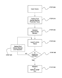
[0113] Referring now to Fig. 2, a flow chart of a method for tissue expansion
is
illustrated, consistent with the present inventive concepts. In STEP 200, a
tissue expansion
device of the present inventive concepts is inserted into a patient, such as a
patient receiving a
gastrointestinal diagnostic or therapeutic procedure. The tissue expansion
device may be
inserted through a lumen of a body access device, such as an endoscope.
Alternatively or
additionally, the tissue expansion device may be inserted over a guidewire,
such as a
guidewire passing through a lumen of the device, or a rapid exchange segment
near the distal
end of the tissue expansion device.
[0114] In STEP 210, one or more fluid delivery elements of the tissue
expansion device are
positioned in proximity to tissue to be expanded. This positioning may be
performed using a
visualization apparatus, such as a visualization apparatus selected from the
group consisting
of: an imaging device integral to or inserted through an endoscope; an imaging
assembly
integral to the tissue expansion device; an imaging device external to the
patient such as a
fluoroscope, a CT scanner, an MR scanner; an ultrasound imager; an imaging
device inserted
into the patient, such as a visual camera and/or an ultrasound probe or
catheter; and
combinations of these.
[0115] In STEP 220, an optional step is performed in which one or more fluid
delivery
elements of the tissue expansion device are advanced, such as an advancement
in which the
one or more fluid delivery elements make contact with tissue and/or penetrate
an outer layer
of tissue. In some embodiments, the one or more fluid delivery elements
penetrate the
mucosal layer of the gastrointestinal tract and enter the submucosal layer,
such as in a
segment of the duodenum. In some embodiments, an expandable assembly including
one or
more fluid delivery elements may be expanded, typically during or prior to the
performance
of STEP 220, such as to contact luminal wall tissue such as luminal wall
tissue of the
duodenum. The expandable assembly can be resiliently biased in a radially
expanded state,
such as a resiliently biased basket or cage supporting one or more fluid
delivery elements and
attached fluid delivery tubes. Alternatively or additionally, STEP 220 may
include a tissue
manipulation step in which tissue is moved, such as a movement toward a fluid
delivery
element and/or into an opening. In some embodiments, vacuum is applied to a
port or other
opening, such as to draw tissue into the opening, such as is described in
reference to Figs. 10,
10A and 10B herebelow. Once positioned in the opening, a fluid delivery
element can be
advanced and/or fluid delivered to the captured tissue. The applied vacuum and
opening size
can be constructed and arranged to preferentially move certain tissue into the
opening, such
-22-
CA 02869904 2014-10-07
WO 2013/159066 PCT/US2013/037485
as to preferentially move one or more inner layers of tissue into the opening
while avoiding
one or more deeper layers being moved into the opening. In some embodiments,
mucosal
and submucosal tissue layers are drawn into the opening while the muscularis
layer remains
outside the opening or is otherwise positioned to avoid being expanded by the
fluid delivery
element. After application of the vacuum, one or more other tissue
manipulations may be
performed (e.g. to "tent" the tissue), such as via advancement, retraction
and/or rotation of
the tissue expansion device and/or a component of the device.
[0116] In STEP 230, one or more fluids are delivered by the one or more fluid
delivery
elements, into tissue, to cause one or more layers of the tissue to expand. In
some
embodiments, one or more fluid delivery elements are moved (e.g. advanced or
retracted),
during the fluid delivery of STEP 230. Fluid is delivered through one or more
fluid delivery
tubes of the tissue expansion device, to the one or more fluid delivery
elements. The one or
more fluid delivery tubes can be attached to one or more sources of fluids,
such as one or
more syringes, pumping assemblies and/or reservoirs of fluids.
[0117] In STEP 240, an optional step of assessing tissue expansion is
performed. The
tissue expansion assessment can be performed using one or more visualization
devices as has
been described above, such as a device used in a visualization procedure
performed at a time
after fluid injection, such as 10, 20 or 30 seconds after fluid injection has
initiated or ceased.
In some instances, a visualization procedure may be performed at a time
immediately prior to
the performance of an ablation procedure, such as 15, 30, or 45 minutes after
fluid injection
has initiated or ceased. If insufficient expansion is achieved, an optional
STEP 245 may be
performed, in which one or more fluid delivery elements are retracted, and one
or more
portions of the tissue expansion device is repositioned. STEP 245 may include
various
repositioning maneuvers including but not limited to: rotating a shaft of the
fluid delivery
device and/or a support structure containing one or more fluid delivery
elements; advancing
one or more fluid delivery elements axially and/or radially; retracting one or
more fluid
delivery elements axially and/or radially; and combinations of these. STEP 245
may further
include advancing fluid delivery elements, such as the advancement described
in reference to
STEP 220 hereabove, such as when one or more fluid delivery elements were
previously
retracted during STEP 245. STEP 230 is subsequently repeated, with or without
the
retraction and/or repositioning of STEP 245, in which one or more fluids are
injected into
tissue to cause expansion of one or more layers of tissue. The optional step
of STEP 240 can
be subsequently repeated, assessing the sufficiency of tissue expansion.
[0118] STEP 250 is performed after the injection of fluid into tissue in STEP
230, with or
without the assessment performed in STEP 240 and/or the repositioning
performed in STEP
-23-
CA 02869904 2014-10-07
WO 2013/159066
PCT/US2013/037485
245. In STEP 250, the fluid delivery device can be removed, remain in place
for subsequent
tissue expansion at a later time, or relatively immediately be advanced to a
new tissue
expansion location, such as by returning to STEP 210 and repeating STEPS 210
through 250
as illustrated.
[0119] The tissue expansion methods of the present inventive concepts may
comprise a
single step of injecting fluid, such as from one or more fluid delivery
elements.
Alternatively, the tissue expansion may be performed with multiple fluid
injection steps, such
as a first injection at a first location, followed by a second injection at a
different location.
The tissue expansion devices and their assemblies are typically configured to
be rotated, such
as to inject at multiple tissue locations along a relatively uniform
circumference of luminal
wall tissue. Fluid may be injected by multiple fluid delivery elements
simultaneously and/or
sequentially.
[0120] The fluid injected to cause tissue expansion may be of a pre-determined
volume,
such as a pre-determined volume per injection and/or cumulative volume of
multiple
injections delivered to a single site (e.g. a single injection of a needle or
an amount of fluid
delivered by a water jet's nozzle to a single location). In some embodiments,
this pre-
determined volume of fluid per injection and/or site comprises a volume of
0.5m1 to 4.0m1, or
1.0m1 to 3.0m1. These pre-determined volumes may be injected at different
sites, such as
between 2 to 10 sites along a relative circumference of luminal wall tissue.
Complete tissue
expansion can comprise one or more axial and/or circumferential injections,
performed
simultaneously and/or sequentially. Injections may be performed by one or more
fluid
delivery elements, such as two or more fluid delivery elements delivering
fluid
simultaneously and/or sequentially. Between injections, the tissue expansion
device can be
axially advanced and/or retracted, and it can be rotated. In some embodiments,
fluid is
delivered at a first location causing tissue expansion in a first expansion
location. A second
injection can be performed proximate the first expansion location, such as
proximate an edge
of the first expansion location. Repeated injections proximate previously
expanded locations
can be used to ease injection as well as reduce likelihood of perforation or
failed tissue
expansion.
[0121] Referring now to Figs. 3A, 3B and 3C, sectional side and end views of a
segment
of luminal wall tissue are illustrated, prior to, during and after full
circumferential tissue
expansion, respectively, consistent with the present inventive concepts. In
Fig. 3A, a side
and end sectional view of a segment of luminal wall tissue includes inner
layer Ll, mid layer
L2 and outer layer L3, prior to any expansion by a tissue expansion device of
the present
inventive concepts. In Fig. 3B, a tissue expansion has occurred at a single
location toward
-24-
CA 02869904 2014-10-07
WO 2013/159066 PCT/US2013/037485
the top of the page as shown, within tissue layer L2. In Fig. 3C, a tissue
expansion has
occurred for a full 3600 segment of layer L2. In some embodiments, a full or
near full
circumferential expansion (e.g. greater than approximately 300 of tissue
expansion, greater
than approximately 320 of tissue expansion, or greater than approximately 330
of tissue
expansion), is performed in a relatively single step, such as from multiple
fluid delivery
elements. In other embodiments, a full or near full circumferential expansion
is performed in
multiple steps, such as from one or more fluid delivery elements that are
configured to inject
fluid in a first step and be rotated in one or more subsequent steps, each
rotation followed by
an injection of fluid into tissue.
[0122] The expansion of a tissue layer, such as layer L2 of Figs. 3A through
3C, may be
performed to cause a reduction in cross sectional area of the lumen, such as a
reduction to
85% of the pre-expansion cross sectional area (e.g. a 35mm lumen reduced to a
30mm
lumen), or a reduction to 75% of the pre-expansion cross-sectional area. Some
body lumens
comprise an inner layer including a non-smooth surface, such as the lining of
the duodenum
or jejunum including one or more folds known as the plicac. In some
embodiments, the
tissue expansion causes folds such as plicae to be smoothed and/or widened.
This
modification can be useful in subsequent treatments of the lumen's inner
lining, such as to
improve the results of one or more ablation procedures.
[0123] Numerous forms and locations of patient tissue can be expanded by the
devices,
systems and methods of the present inventive concepts. In some embodiments,
the tissue to
be expanded comprises submucosal tissue, such as submucosal tissue of the
duodenum. The
devices systems and methods of the present inventive concepts may be
constructed and
arranged to avoid expanding one or more layers of tissue, such as when the
muscularis or
serosal layer of the duodenum is prevented from being expanded. Applicable
tissue may
comprise luminal wall tissue or other tissue layers. Applicable tissue
locations to be
expanded can include luminal wall tissue selected from the group consisting
of: a
gastrointestinal tissue layer; a duodenal tissue layer; an esophageal tissue
layer; a jejunal
tissue layer; an ileal tissue layer; a colonic tissue layer; and combinations
of these.
Alternatively or additionally, tissue to be expanded may comprise tissue
selected from the
group consisting of: a stomach tissue layer; a bladder tissue layer; an oral
cavity tissue layer;
a uterine tissue layer; and combinations of these.
[0124] Referring now to Fig. 4, a side view of a distal portion of a tissue
expansion device
is illustrated, including a manually deployable expandable assembly,
consistent with the
present inventive concepts. A tissue expansion device, such as a tissue
expansion device
similar to device 100 of Fig. 1, includes an expandable assembly 130 in a pre-
deployment
-25-
CA 02869904 2014-10-07
WO 2013/159066 PCT/US2013/037485
(e.g. prior to radial expansion) state. Expandable assembly 130 is shown to
have been axially
advanced to exit sheath 109, such as via one or more controls on a handle
mounted on the
proximal end of sheath 109. In some embodiments, sheath 109 comprises an
endoscope,
such as when expandable assembly 130 is advanced through a working channel of
the
endoscope. Expandable assembly 130 comprises at least two support arms, arms
132a and
132b typically a hollow or partially hollow tube such as a metal tube or a
plastic tube. Arms
132a and 132b include openings 131a and 131b, respectively. The distal ends of
arms 132a
and 132b are attached to a control cable, cable 103, typically a metal or non-
metallic cable
which travels proximally and attaches to one or more controls on a proximal
handle, such as
those described in reference to handle 110 of Fig. 1 and configured to advance
or retract
cable 103. Cable 103 can comprise a hollow tube, such as a cable including a
guidewire
lumen 105, configured to allow over-the-wire delivery of expandable assembly
130 and
sheath 109.
[0125] Two fluid delivery elements, needles 141a and 141b are shown positioned
within
arms 132a and 132b, respectively. Needles 141a and 141b typically comprise
metal needles,
such as needles with a gauge between 20 and 35 gauge, or between 23 and 27
gauge.
Needles 141a and/or 141b may comprise a beveled end, such as an end with a
bevel angle
between 10 and 60 , such as a bevel angle of approximately 30 . Needles 141a
and 141b are
fluidly attached to one or more fluid delivery tubes, such as fluid delivery
tubes 121
described in reference to Fig. 1, such that one or more fluids can be
delivered to needles 141a
and 141b via the fluid delivery tubes. Needles 141a and/or 141b may comprise a
particular
sharpness or other penetration characteristic such as to preferably penetrate
one form of
tissue, such as the submucosa, while avoiding or minimizing penetration of a
deeper layer of
tissue, such as the muscularis or serosal layers. Needles 141a and/or 141b may
be
constructed and arranged to be advanced to an exposed length of less than
lOmm, such as less
than 7mm. Needles 141a and 141b may be constructed and arranged to remain
within
openings 131a and 13 lb, respectively, as is described in reference to Figs. 5
and 10
herebelow. Vacuum can be applied to openings 131a and 131b such as to draw
tissue toward
and/or into openings 131a and 131b. Alternatively or additionally, needles
141a and 141b
may be constructed and arranged to advance out of openings 131a and 131b,
respectively, as
is described in reference to Fig. 4B herebelow.
[0126] Referring now to Fig. 4A, cable 103 has been retracted such that the
mid portions
of arms 132a and 132b extend radially from the axis of sheath 109. Openings
131a and 131b
correspondingly extend radially as shown. Needles 141a and 141b remain in the
pre-
deployed position shown.
-26-
[0127] Referring now to Fig. 4B, needles 141a and 141b have been advanced to
exit
openings 13Ia and 131b, such that when expandable assembly 130 is positioned
against
tissue, such as luminal wall tissue, needles 141a and 141b penetrate into one
or more tissue
layers. Advancement of needles 141a and 14 lb may be done in combination or
independently, such as via one or more controls on a proximal handle, not
shown but such as
is described in reference to handle 110 of Fig. 1. In alternative embodiments,
needles 141a
and 141b do not exit openings 131a and 131b, respectively, during advancement,
such as is
described in reference to the tissue expansion devices of Figs 5 and 10
herebelow. In these
embodiments, a vacuum is applied to draw tissue into openings 131a and 131b
and needles
141a and 141b penetrate the captured tissue when advanced.
[0128] Referring now to Fig. 5, a side and end view of a distal portion of a
tissue
expansion device arc illustrated, including a self-expanding assembly,
consistent with the
present inventive concepts. A tissue expansion device, such as a tissue
expansion device
similar to device 100 of Fig. 1, includes an expandable assembly 130 in a
deployed (e.g. a
radially expanded) state. Expandable assembly 130 is attached to the distal
end of shaft 101,
which has been axially advanced to exit sheath 109, such as via one or more
controls on a
handle mounted on the proximal end of sheath 109. In some embodiments, sheath
109
comprises an endoscope, such as when expandable assembly 130 is advanced
through a
working channel of the endoscope. Expandable assembly 130 comprises at least
three
support arms, such as three support arms comprising proximal segments 133a,
133b and
133c, and attached distal segments 134a, 134b and 134c respectively. Proximal
segments
133a, 133b and 133c are typically a hollow or partially hollow tube such as a
metal tube or a
plastic tube, and configured to slidingly receive a fluid delivery element and
include openings
131a, 131b and 131c, respectively. Distal segments 134a, 134b and 134c are
typically
resiliently biased in the orientation shown in Fig. 5, such as to cause
expandable assembly
130 to be in a radially expanded condition when not surrounded by a
compressing tube, such
as sheath 109. Expandable assembly may be constructed of a resiliently biased
metal, such as
stainless steel and/or Nitinoilat is resiliently biased in an expanded or
contracted geometry.
In some embodiments, assembly 130 is configured to transition to a radially
compacted state,
such as when inserted within a lumen of a tube such as sheath 109 and/or a
working channel
of an endoscope. Distal segments 134a, 134b and 134c can comprise an elastic,
biocompatible material such as Nitinofmor stainless steel formed in a curved
orientation.
Distal segments 134a, 134b and 134c may comprise a flat sheet material or a
round tube. The
distal end of distal segments 134a, 134b and 134c are attached to tip 139
which surrounds
and maintains the position of the distal end of segments 134a, 134b and 134e.
In alternative
-27-
CA 2869904 2018-10-19
embodiments, distal segments 134a, 134b and 134c may be fabricated of a single
sheet or
otherwise fabricated such that their distal ends are connected, with or
without a tip 139. Tip
139 may be covered by an atraumatic material, such as silicone or other
biocompatible
polymer.
[01291 Each proximal segment 133a, 133b and 133c contains a single fluid
delivery
element, needle 141a, 141b and 141c, respectively. Needles 141a, 141b and 141c
are each
attached to a single fluid delivery tube, 121a, 121b and 121c, respectively.
In some
embodiments, each fluid delivery tube travels proximally to a handle, such as
to be attached
to individual supplies of fluid for injection into tissue, such as reservoirs
125a, 125b and
125c, respectively described in reference to Fig. 1. In other embodiments, one
or more of
fluid delivery tubes 121a, 12 lb and/or 121c fluidly attach to each other,
such as to attach to a
source of fluid, such that fluid is simultaneously injected through one or
more of fluid
delivery tubes 121a, 121b and/or 121c. In some embodiments, a vacuum is
applied, such as
via a proximal handle port, such as a vacuum applied at one or more of
openings 131a, 13 lb
and 131c, around needles 141a, 141b and 141c, respectively, such as through a
vacuum
delivery tube, such as is described in reference to Figs. 5A and 5C herebelow.
Applied
vacuum may be configured to maintain tissue position during penetration of
tissue by needles
141a, 141h and/or 141c, and/or to manipulate tissue into openings 131a, 131b
and 131c for
subsequent needle advancement. In some embodiments, a second opening is
provided on one
or more of support arms 133a, 133b and/or 133c, openings not shown but
configured to be
fluidly attached to a source of vacuum and to apply one or more forces to
tissue.
[01301 Referring now to Fig. 5A, a side sectional view of a segment of a
support arm of a
tissue expansion device is illustrated, consistent with the present inventive
concepts. A fluid
delivery element is shown in an advanced position and a support member
surrounds the fluid
delivery element. A segment of a support arm 133 is shown, such as a segment
of proximal
support arm 133a, 133b and/or 133c of Fig. 5. Support arm 133 comprises two
lumens,
lumen 107 and lumen 108, and may be constructed of a rigid or flexible
material, such as a
metal material such as stainless steel or NitinolT,mor a plastic material such
as a PebaxTM with a
durometer between 50D and 80D. Lumen 108 slidingly receives needle 141. The
distal end
of fluid delivery tube 121 is fluidly and mechanically attached to the
proximal end of needle
141, such as via a sealed bond and/or frictionally engaging interface (e.g.
the interface
between a proximal outer diameter portion of needle 141 and a distal inner
diameter portion
of tube 121). This attachment provides a fluid seal yet allows fluid to pass
through fluid
delivery tube 121 into needle 141. Needle 141 is constructed and arranged to
be advanced
into a recess in support arm 133, opening 131, such as to the advanced
position shown.
-28-
CA 2869904 2018-10-19
CA 02869904 2014-10-07
WO 2013/159066
PCT/US2013/037485
Needle 141 can comprise a needle between 25 and 30 gauge, such as a 27 gauge
stainless
steel needle with a beveled tip. Fluid delivery tube 121 can comprise a
flexible shaft, such as
a shaft comprising a plastic material including a braid, such as a polyimide
shaft including a
stainless steel braid.
[0131] On its proximal end, lumen 107 is fluidly attached to an operator
activatable supply
of vacuum, not shown but such as vacuum source 340 of Fig. 1. Lumen 107 is
fluidly
attached on its distal end to opening 131. Opening 131 can comprise sloped
side walls 231,
such as to cause tissue drawn into opening 131 to have a preferred shape
and/or a preferred
array of tensional force vectors imparted on the tissue. In use, needle 141 is
in a retracted
state (e.g. not entering opening 131), and a vacuum can be applied to opening
131. After
tissue is drawing into opening 131, needle 141 is advanced (to the right of
the page) to the
position shown in Fig. 5A, penetrating tissue and ready for fluid to be
delivered through fluid
delivery tube 121 to expand one or more tissue layers drawing the tissue into
opening 131.
[0132] A support and/or guiding element, ferrule 149 may be included to
provide support
to needle 141 as it penetrates tissue. Ferrule 149 can be configured to
prevent undesired
rotation, bending and or twisting to needle 141, such as when needle 141 is
advanced into
tissue. Ferrule 149 can comprise a round tube that is bonded to or
frictionally engages a
distal portion of needle 141. Ferrule 149 can comprise an outer diameter that
approximates
the inner diameter of lumen 108, such as when ferrule 149 comprises an outer
diameter
between 0.020" and 0.036" (e.g. a diameter approximating 0.028"), and lumen
108 comprises
a diameter between 0.027" and 0.043" (e.g. a diameter approximating 0.035").
Ferrule 149
can comprise a tubular construct with a length between 1.5mm and 2.5mm, such
as a length
approximating 2.0mm. Ferrule 149 can comprise a rigid material such as a metal
such as
stainless steel. Ferrule 149 can be axially positioned on needle 141 such that
a majority of
ferrule 149 remains within lumen 108 as the distal end of needle 141 travels
axially to the
position shown in Fig. 5A.
[0133] Referring now to Fig. 5B, a top view of an opening of a support arm of
a tissue
expansion device is illustrated, with a fluid delivery element in an advanced
position,
consistent with the present inventive concepts. A segment of a support arm 133
is shown,
such as a segment of proximal support arm 133a, 133b and/or 133c of Fig. 5,
and/or a
segment of support arm 133 of Fig. 5A. Support arm 133 slidingly receives
fluid delivery
tube 121 and needle 141. Needle 141 is constructed and arranged to be advanced
into a
recess in support arm 133, opening 131, such as to the advanced position
shown. Opening
131 can comprise sloped walls 231 such as to cause tissue drawn into opening
131 to have a
preferred shape and/or a preferred array of tensional force vectors imparted
on the tissue.
-29-
Needle 141 and support shaft 133 can be constructed and arranged to maintain
the relatively
aligned position shown in Fig. 5B (e.g. aligned to a central axis of support
arm 133 and/or
opening 131), such as when needle 141 is advanced through tissue drawn into
opening 131.
Alignment can be achieved through a needle support and/or aligning component,
such as
ferrule 149 of Fig. 5A. In some embodiments, one or more components of the
tissue
expansion device, such as one or more of needle 141 and support arm 133, are
constructed
and arranged such that when needle 141 is fully advanced, the opening in the
distal end of
needle 141 is centered in opening 131, as is shown in Fig. 5B. Control of this
positioning can
be accomplished through the use of a needle stop, such as is described in
reference to Fig. 10
herebelow and/or via one or more controls integral to a handle, such as handle
110 and
associated controls described in reference to Fig. 1 hereabove.
[0134] In some embodiments, opening 131 comprises an axial length of
approximately
4mm, and needle 141 is constructed and arranged such that 3mm of length
resides in opening
131 when needle 141 is fully advanced, and the opening in the end of needle
141 is centered
in opening 131 as shown. In some embodiments, opening 131 comprises an axial
length up
to 5mm. In some embodiments, opening 131 comprises a width up to 2mm.
[0135] Referring now to Fig. SC, a perspective view of an alternative opening
of a support
arm is illustrated, consistent with the present inventive concepts. A segment
of a support arm
133 is shown, such as a segment of proximal support arm 133a, 133b and/or 133c
of Fig. 5.
Support arm 133 includes lumen 108 which is configured to slidingly receive a
fluid delivery
tube and a fluid delivery element such as a needle, fluid delivery tube and
fluid delivery
element removed for illustrative clarity. Support arm 133 can comprise a
diameter between
0.070" and 0.100", such as a diameter approximating 0.090". Support arm 133
may be
constructed of a rigid or flexible material, such as a metal material such as
stainless steel or
Nitind,mor a plastic material such as a Pebaxml material. Lumen 108 can
comprise a circular
cross section, as is shown in Fig. 5C, such as a circular cross section with a
diameter between
0.020" and 0.040", or approximately 0.035". Support arm 133 further includes
lumen 107,
which is configured to be fluidly attached to a supply of vacuum pressure such
as vacuum
pressure supplied by an operator adjustable vacuum source, such as vacuum
source 340 of
Fig. I. Lumen 107 fluidly applies the vacuum to a recess or opening in support
arm 133,
such as opening 131', such as to cause tissue to be drawn into opening 131'.
Lumen 107 may
comprise a crescent shaped cross section, as is shown in Fig. 5C. Opening 131'
is further
constructed and arranged to receive a fluid delivery element such as a needle.
[0136] Opening 131' comprises projections 232 along the side walls of opening
131' such
as projections configured to limit the amount of tissue drawn into opening
131' when a
-30-
CA 2869904 2018-10-19
CA 02869904 2014-10-07
WO 2013/159066 PCT/US2013/037485
vacuum is applied to opening 131' via lumen 107. In some embodiments,
projections 232 are
constructed and arranged to allow sufficient tissue to be drawn into opening
131' such that
one or more fluids delivered by a fluid delivery element such as a needle into
tissue, causes
tissue expansion to occur in a submucosal layer of gastrointestinal tissue,
while avoiding
expansion of deeper layers such as the muscularis or serosal layers.
Additionally, opening
131' comprises sloped walls 231 such as to cause tissue drawn into opening
131' to have a
preferred shape and/or a preferred array of tensional force vectors imparted
on the tissue.
[0137] Referring now to Fig. 6, a side sectional view of a fluid delivery
element
comprising a penetrator and an atraumatic surrounding tube is illustrated,
consistent with the
present inventive concepts. Fluid delivery element 141 has been positioned in
a lumen of
tissue, such as a lumen of the duodenum or other gastrointestinal lumen. The
tissue
comprises multiple layers, such as innermost layer Li, deeper layer L2 and yet
deeper layer
L3. In some embodiments, Li comprises a mucosal layer, L2 comprises a
submucosal layer,
and L3 comprises a muscular layer with or without an overlying serosal layer.
Fluid delivery
element 141 comprises a hollow tube 144 that includes a rounded or otherwise
atraumatic
distal end 145, which slidingly receives a sharpened tube, penetrator 143,
typically a hollow
or solid tube, such as a metallic hypotube with a sharpened distal end. Tube
144 includes a
hole in its wall, opening 142, positioned relatively proximate end 145. Tube
144 and
penetrator 143 are sized such that fluid can be delivered in the space between
tube 144 and
penetrator 143, such as via one or more fluid delivery tubes, not shown but
traveling
proximally and fluidly connected to a supply of injectable fluid, such as is
described in
reference to Fig. 1 hereabove. Fluid can be delivered to tissue out of the
distal end of tube
144 and/or via opening 142, such as when the distal end of tube 144 is
occluded, such as by a
less expansive layer of deeper tissue.
[0138] Fluid delivery element 141 is configured to allow initial penetration
into tissue by
penetrator 143, after which tube 144 can be advanced into tissue, as is
illustrated and
described in reference to Figs. 6A and 6B herebelow. In Fig. 6, penetrator 143
has been
advanced through tissue layer Li and into tissue layer L2. Injection of fluid
during
advancement of tube 144 while penetrator 143 is also advanced (as in Figure 6)
can be
performed, with fluid preferentially exiting through port 142, causing tissue
expansion of
layer L2 during advancement.
[0139] Referring now to Fig. 6A, tube 144 has been advanced, over penetrator
143,
through tissue layer Ll and into tissue layer L2. Penetrator 143 is
positioned, via
advancement of tube 144 and/or retraction of penetrator 143, such that the
distal end of
-31-
penetrator 143 is contained within tube 144, such as to prevent further
advancement of fluid
delivery element 141 into deeper layers of tissue such as into tissue layer
L3.
[0140] Referring now to Fig. 6B, fluid has been injected in the space between
penetrator
143 and tube 144, such as to exit the distal end 145 of tube 144 andJor
through opening 142,
causing tissue layer L2 to expand.
[0141] Referring now to Fig. 7, a side sectional view of a fluid delivery
element
comprising a needle with an internal lumen is illustrated, consistent with the
present inventive
concepts. Fluid delivery element 140' has been positioned in a lumen of
tissue, such as a
lumen of the duodenum or other gastrointestinal lumen. The tissue comprises
multiple
layers, such as innermost layer Li, deeper layer L2, and yet deeper layer L3.
Fluid delivery
element 140' comprises a needle 141, including a lumen 147. Needle 141
comprises a
sharpened distal tip 146, typically comprising a beveled end, and may be
configured to be
operably advanceable from shaft 101, such as via one or more controls on a
proximal handle,
as is described in reference to Fig. 1 hereabove. Needle 141 can be
constructed and arranged
to be rotated (e.g. when retracted), such as to perform multiple fluid
delivery events around a
circumference of tissue, such as to create a full or near full circumferential
tissue expansion.
Lumen 147 is fluidly connected to one or more fluid delivery tubes, not shown
but traveling
proximally and fluidly connected to a supply of injectable fluid. Fluid can be
delivered to
tissue out of distal end 146 of needle 141, such as via one or more controls
on a proximal
handle or on a device connected to a proximal handle, such as is described in
reference to
Fig. 1 hereabove.
[0142] Referring now to Fig. 8, a side sectional view of a fluid delivery
element
comprising a water jet including a nozzle and an internal lumen is
illustrated, consistent with
the present inventive concepts. Fluid delivery element 140" has been
positioned in a lumen
of tissue, such as a lumen of the duodenum or other gastrointestinal lumen.
The tissue
comprises multiple layers, such as innermost layer Li, deeper layer L2 and yet
deeper layer
L3. Fluid delivery element 140" comprises a nozzle 148, fluidly connected to
lumen 147.
Nozzle 148 can be configured to allow a high-pressure delivery of fluid,
injectate 150, shown
in a collimated stream comprising sufficient pressure to penetrate one or more
tissue surfaces,
such as to deliver tissue to a deeper layer of tissue, such as a water nozzle
as is used in the
Erbejet immanufactured by Erbe Elektromedizin GmbH of Tubingen, Germany.
Nozzle 148
can be configured to be operably advanceable from shaft 101, such as via one
or more
controls on a proximal handle, as is described in reference to Fig. 1
hereabove. Nozzle 148
can be constructed and arranged to be rotated, such as to perform multiple
fluid delivery
events around a circumference of tissue, such as to create a full or near full
circumferential
-32-
CA 2869904 2018-10-19
CA 02869904 2014-10-07
WO 2013/159066 PCT/US2013/037485
tissue expansion. While nozzle 148 is shown in an orientation along the axis
of shaft 101,
nozzle 148 can be oriented off-axis, such as at an angle between 1 and 179 ,
typically
between 100 and 170 . While nozzle 148 is shown as a single nozzle, multiple
nozzles can be
employed. Lumen 147 is fluidly connected to one or more fluid delivery tubes,
not shown
but traveling proximally and fluidly connected to a supply of injectable
fluid. Fluid can be
delivered to tissue from nozzle 148, such as via one or more controls on a
proximal handle or
on a device connected to a proximal handle, such as is described in reference
to Fig. 1
hereabove.
[0143] Referring now to Fig. 9, a side sectional view of a fluid delivery
element
comprising an iontophoretic fluid delivery assembly is illustrated, consistent
with the present
inventive concepts. Fluid delivery element 140" has been positioned in a lumen
of tissue,
such as a lumen of the duodenum or other gastrointestinal lumen. The tissue
comprises
multiple layers, such as innermost layer Li, deeper layer L2 and yet deeper
layer L3. Fluid
delivery element 140" ' comprises an iontophoretic delivery element comprising
reservoir
151 and electrode 152. Reservoir 151 is fluidly connected to lumen 147.
Reservoir 151 and
electrode 152 can be configured to be operably advanceable from shaft 101,
such as via one
or more controls on a proximal handle, as is described in reference to Fig. 1
hereabove.
Reservoir 151 can be constructed and arranged to be rotated, such as to
perform multiple
fluid delivery events around a circumference of tissue, such as to create a
full or near full
circumferential tissue expansion. Lumen 147 is fluidly connected to one or
more fluid
delivery tubes, not shown but traveling proximally and fluidly connected to a
supply of
injectable fluid. Electrode 152 is connected to one or more wires, not shown
but traveling
proximally to a control unit configured to cause electrode 152 to apply an
electric field in
and/or around reservoir 151. While electrode 152 is shown as a single
electrode, multiple
electrodes may be employed. Injectate 150 comprises an ionic fluid capable of
being driven
into at least tissue layers Li and L2, by the electrical fields created by
electrode 152, such as
via iontophoretic delivery well known to those of skill in the art. Activation
of electrode 152
can be accomplished via one or more controls on a proximal handle or on a
device connected
to a proximal handle, such as is described in reference to Fig. 1 hereabove.
[0144] Referring now to Fig. 10, a side sectional view of a distal portion of
a tissue
expansion device comprising a side recess portion and protected needle exit
port is illustrated,
consistent with the present inventive concepts. Device 100 includes shaft 101
with integral
side car 106, positioned at or near distal end 102 of shaft 101. Side car 106
comprises a
guidewire lumen 106', such that shaft 101 can be advanced and/or exchanged
over a
guidewire via rapid exchange delivery, as is known to those of skill in the
art. Shaft 101
-33-
CA 02869904 2014-10-07
WO 2013/159066 PCT/US2013/037485
further comprises two lumens, a first lumen 108 which can slidingly receive a
fluid delivery
element, needle 141, and a second lumen 107, configured to carry a vacuum.
Shaft 101
comprises a recess in its side wall, recess 155, relatively proximate distal
end 102 of shaft
101. Recess 155 can be positioned in a lumen of tissue, such as a lumen of the
duodenum or
other gastrointestinal lumen, such as to expand one or more layers of tissue,
such as the
submucosal layer of the duodenum. An opening 158 is positioned between lumen
107 and
recess 155, such that an applied vacuum can be introduced to recess 155 via
lumen 107, such
as to draw tissue into recess 155 as shown as described in reference to Figs.
10A and 10B
herebelow.
[0145] Needle 141 includes lumen 147, which is fluidly connected to one or
more fluid
delivery tubes, not shown but such as one or more fluid delivery tubes in
fluid
communication with a supply of fluid, such as is described in reference to
device 100 of Fig.
1 hereabove. Needle 141 is configured to be operably advanceable along the
axis of shaft
101 and lumen 108, such as via one or more controls on a proximal handle, also
as is
described in reference to Fig. 1 hereabove, such as to have its distal end
exit lumen 108 and
enter recess 155. A mechanical stop 157 is positioned within lumen of 108. A
collar 156 is
attached to needle 141, such that advancement of needle 141 is limited when
collar 156
makes contact with mechanical stop 157. In some embodiments, the position of
collar 156
and/or stop 157 can be adjusted, such as via one or more controls positioned
on a proximal
handle, such as to adjust the permitted travel of needle 141. In some
embodiments, the length
of needle 141 is chosen such that it is longer than the axial length of recess
155, such as to
prevent needle 141 from exiting device 100 if needle 141 were to become
detached.
[0146] In some embodiments, device 100 includes an imaging component,
visualization
element 165 which is connected to cables 166. Imaging component 165 is
configured to
provide an image to the operator, such as via one or more visual displays, not
shown but
connected to cables 166 and positioned in view of one or more operators of
device 100.
Imaging component 165 may comprise an imaging element selected from the group
consisting of: an ultrasound imager; an optical coherence domain reflectometry
(OCDR)
imager; an optical coherence tomography (OCT) imager; confocal endomicroscopy
via either
scanning or structured illumination; and combinations of these. Cables 166 may
comprise
electrical wires, optical fibers and/or one or more rotating shafts, such as
to provide power or
otherwise enable imaging component 165 to provide an image. Images provided by
imaging
component 165 can be used to determine sufficiency or otherwise assess the
tissue expansion
caused by delivering fluid to one or more tissue layers via needle 141.
-34-
CA 02869904 2014-10-07
WO 2013/159066 PCT/US2013/037485
[0147] Referring now to Fig. 10A, the distal portion of shaft 101 and recess
155 have been
positioned proximate tissue, such as gastrointestinal tissue, and a vacuum has
been applied to
recess 155 via port 158 and lumen 107, such as via a vacuum source within
and/or attached to
a proximal handle. As the vacuum is applied, tissue proximate recess 155 is
drawn into
recess 155 as illustrated in Fig. 10A. In some embodiments, recess 155
comprises a
geometry and size to precisely cause tissue expansion of specific layers of
the intestinal
anatomy. For example, recess 155 may comprise a geometry and size to cause the
mucosal
layer (<1mrn thick) to enter recess 155 without including the muscularis
layer. In these
embodiments, the spongy submucosal layer stretches and enlarges as tissue is
drawn into
recess 155, creating a larger target for needle 141 insertion. Minimizing the
recess 155 width
can be used to prevent drawing the full thickness of tissue into recess 155 by
vacuum applied
through lumen 107. In some embodiments, recess 155 comprises a width less than
2.0mm,
such as a width less than 1.5mm or less than 1.0mm. Minimizing recess 155
axial length can
be used to improve needle 141 penetration into tissue drawn into recess 155,
such as when
the distal end of recess 155 provides a normal force in reaction to needle 141
tissue
penetration. In some embodiments, recess 155 length is less than 5.0mm, such
as a length
less than 4.0mm or less than 3.0mm. In some embodiments, recess 155 comprises
a width
approximating 1.5mm and a length approximating 4.0mm. In some embodiments,
lumen 107
and needle 141 are each contained in a single tube, such as the dual lumen
tube described in
reference to Fig. 5C hereabove.
[0148] Referring now to Fig. 10B, while maintaining a vacuum in lumen 107,
needle 141
has been axially advanced into the tissue, as illustrated. Advancement of
needle 141 can be
accomplished by one or more controls on the proximal end of shaft 101, such as
controls
integral to a handle such as are described in reference to handle 110 of Fig.
1. After insertion
of needle 141 into the tissue contained within recess 155, such as via fluid
delivery tubes
which travel proximally to one or more sources of fluid on the proximal end of
shaft 101. In
some embodiments, recess 155 is sized and configured to limit the excursion
distance
between the location where the tip of needle 141 enters recess 155 and the
location where
needle 141 first penetrates tissue. Minimizing this distance can prevent
bunching or
stretching of tissue or otherwise improve needle 141 penetration into tissue
captured within
recess 155.
[0149] Shaft 101 and recess 155 of Figs. 10, 10A and 10B can be constructed
and arranged
to be rotated. In these embodiments, multiple fluid delivery events can be
performed around
a circumference of tissue, such as to draw a series of tissue sections via
vacuum into recess
-35-
CA 02869904 2014-10-07
WO 2013/159066
PCT/US2013/037485
155, followed by multiple advancements of needle 141 and delivery of fluid,
such as to create
a full or near full circumferential tissue expansion.
[0150] Referring now to Fig. 11, a side sectional view of a distal portion of
a tissue
expansion device comprising an end recess portion and protected needle exit
port is
illustrated, consistent with the present inventive concepts. Shaft 101' and
recess portion 155'
are similar to shaft 101 and recess 155 of Fig. 10 except recess portion 155'
is positioned at
the distal end 102 of shaft 101'.
[0151] Shaft 101' further comprises a first lumen 108 which slidingly receives
fluid
delivery tube 121 and a fluid delivery element, needle 141. Shaft 101' further
comprises a
second lumen 107, configured to carry a vacuum. Recess 155' and lumen 107 are
constructed and arranged to withdraw tissue into recess 155' and apply tension
to this tissue
to resist forces encountered during penetration of the tissue by needle 141.
[0152] Recess 155' can be positioned in a lumen of tissue, such as a lumen of
the
duodenum or other gastrointestinal lumen, such as to expand one or more layers
of tissue,
such as the submucosal layer of the duodenum. An opening 158' is positioned
between
lumen 107 and recess 155', such that an applied vacuum can be introduced to
recess 155' via
lumen 107, such as to draw tissue into recess 155' as shown as described in
reference to Figs.
10A and 10B hereabove.
[0153] Needle 141 includes lumen 147, which is fluidly connected to fluid
delivery tube
121 which attaches at its proximal end to one or more supplies of fluid, such
as is described
in reference to device 100 of Fig. 1 hereabove. Needle 141 is configured to be
operably
advanceable along the axis of shaft 101' and lumen 108, such as via one or
more controls on
a proximal handle, also as is described in reference to Fig. 1 hereabove, such
as to exit lumen
108 and enter recess 155'. In typically use, distal end 102 of shaft 101 is
positioned
proximate tissue, vacuum is applied via opening 158', after which needle 141
is advanced
into the capture tissue and one or more fluids are injected into the tissue
via lumen 108 and
fluid delivery tube 121, causing one or more tissue layers to expand.
[0154] Referring now to Fig. 12, a side sectional view of the distal portion
of a tissue
expansion device comprising an endoscope and an advanceable needle is
illustrated,
consistent with the present inventive concepts. Endoscope 170 has been
advanced through
luminal tissue, such as through the gastrointestinal tract such as to a
location in the
duodenum. The tissue comprises multiple layers, such as innermost layer Li,
deeper layer
L2 and yet deeper layer L3. Endoscope 170 has had its distal end deflected,
via one or more
steering controls common to endoscope devices, such that needle 141 can be
axially
advanced to penetrate layer Li and L2 of tissue, while avoiding penetration of
layer L3, such
-36-
CA 02869904 2014-10-07
WO 2013/159066
PCT/US2013/037485
as to penetrate a mucosal and submucosal layer of the gastrointestinal tract,
while avoid
penetrating deeper layers such as the serosa.
[0155] Needle 141, including a lumen 147, may be configured to be operably
advanceable
from working channel 172 of endoscope 170, such as via one or more controls.
Endoscope
170 can be constructed and arranged to be rotated (e.g. when needle 141 is
retracted), such as
to perform multiple fluid delivery events around a circumference of tissue,
such as to create a
full or near full circumferential tissue expansion. Needle 141 and lumen 147
are fluidly
connected to fluid delivery tube 121 (e.g. a hypotube) via bond joint 122.
Lumen 147 is
fluidly attached to lumen 147' of fluid delivery tube 121. In some
embodiments, lumen 147'
is a larger diameter than lumen 147 as shown in Fig. 12, such as to reduce the
pressure
required to deliver fluid through fluid delivery tube 121. Fluid delivery tube
121 travels
proximally and fluidly connected to a supply of injectable fluid, such as a
syringe or pumping
assembly. Fluid can be delivered to tissue out of distal end of needle 141,
such as via one or
more controls on a proximal handle or on a device connected to a proximal
handle, such as is
described in reference to Fig. 1 hereabove.
[0156] In some embodiments, needle 141 and/or fluid delivery tube 121 comprise
a
flexibility and radial support that allow flexing without luminal collapse,
such that needle 141
can translate toward the center of the lumen as tissue layer L2 expands.
[0157] Endoscope 170 includes a camera 171, positioned to allow an operator to
visualize
penetration of needle 141 into tissue, as well as the expansion of one or more
tissue layers
such as layer L2 shown. In some embodiments, the injected fluid comprises a
dye or other
visualizable colorant that can be used to quantify or otherwise assess the
amount of tissue
expansion (e.g. the deeper the color visualized at a location, the thicker the
expansion at that
location). Alternatively or additionally, endoscope 170 may comprise another
visualization
device, such as a device selected from the group consisting of: an ultrasound
imager; an
optical coherence domain reflectometry (OCDR) imager; an optical coherence
tomography
(OCT) imager; and combinations of these. Endoscope 170 can further comprise a
source of
light, such as LED 173, such as to deliver visible light and/or infrared
light. Endoscope 170
can further comprise a second working channel 174, such as a working channel
sized to
slidingly receive a tissue manipulating device, such as a tissue manipulating
device described
in reference to Fig. 18 herebelow.
[0158] Referring now to Fig. 13, a side view of the distal portion of a tissue
expansion
device comprising multiple needles and a fluid dispersion manifold is
illustrated, consistent
with the present inventive concepts. Expandable assembly 130 is shown distal
to distal end
102 of shaft 101. Expandable assembly 130 comprises at least three support
arms, such as
-37-
CA 02869904 2014-10-07
WO 2013/159066 PCT/US2013/037485
support arms 132a, 132b and 132c shown. Support arms 132a, 132b and 132c can
be
resiliently biased in the radially expanded condition shown in Fig. 13. In
some embodiments,
expandable assembly 130 is slidingly received by shaft 101, such that
retraction of
expandable assembly 130 into shaft 101 causes expandable assembly 130 to
radially
compress. In other embodiments, expandable assembly 130 may be fixed relative
to shaft
101, such as when expandable assembly 130 is inserted through an endoscope
whose working
channel radially compresses expandable assembly. Tip 139 can connect and/or
surround the
distal ends of support arms 132a, 132b and 132c, and tip 139 may include an
atraumatic
covering such as an elastomer or other relatively soft material. In an
alternative embodiment,
expandable assembly 130 comprises two support arms, such as two support arms
132a and
132b positioned 180 from each other.
[0159] Support arms 132a, 132b and 132c each comprise a radially outward
facing
opening, openings 131a, 131b and 132c respectively. A fluid delivery element,
such as
needles 141a, 141b and 141c, are slidingly received by arms 132a, 132b and
132c,
respectively. Needles 141a, 141b and 141c are constructed and arranged to be
operably
advanced out of openings 131a, 131b and 131c, respectively, such as has been
described in
detail hereabove.
[0160] Needles 141a, 141b and 141c are each attached to a fluid delivery tube,
fluid
delivery tubes 121a, 121b and 121c respectively. Fluid delivery tubes 121a,
121b and 121c
are fluidly attached to a fluid dispersion manifold, valve assembly 160, which
in turn is
fluidly attached to a single fluid delivery tube, lumen 108. Lumen 108 travels
proximally and
is fluidly connected to one or more sources of injectable fluid, such as has
been described in
detail hereabove.
[0161] Referring now to Fig. 13A, a magnified sectional view of the valve
assembly 160
of Fig. 13 is illustrated, consistent with the present inventive concepts.
Valve assembly 160
is connected at its proximal end to lumen 108. Valve assembly 160 comprises
three sets of
solenoids and pistons, including solenoids 161a, 161b and 161c, which advance
and retract
pistons 162a, 162b and 162c, respectively. Pistons 162a, 162b and 162c are
positioned in
fluid delivery tubes 121a, 121b and 121c, such as to cause a flow path between
each tube and
lumen 108 to be open or closed. A cable of wires 163 is attached on its distal
end to
solenoids 161a, 161b and 161c. Wires 163 travel proximally, such as to a
control circuit
included in a handle and configured to allow an operator to independently
cause fluid to flow
to any or all of fluid delivery tubes 121a, 121b and 121c.
[0162] Referring now to Fig. 13B, a magnified sectional view of a support arm
of Fig. 13
is illustrated, consistent with the present inventive concepts. Support arm
132a comprises
-38-
proximal segment 133a, configured to slidingly receive a fluid delivery
clement, needle 141a,
and includes opening 131a, Needle 141a and attached fluid delivery tube 121a
have been
advanced, such as via one or more controls on a proximal handle, such as
handle 110 of
Fig.1, configured to allow an operator to advance needle 141a, In some
embodiments, as
needle 141a is advanced, it makes contact with ramp 136a, positioned proximate
opening
131a and typically a hard surface configured to direct needle 141a at a pre-
determined
trajectory, such as to penetrate one or more layers of tissue, such as one or
more layers of
gastrointestinal tissue.
[0163] Referring now to Fig. 14, a side sectional view of a distal portion of
a tissue
expansion device comprising a spring-loaded needle injector is illustrated,
consistent with the
present inventive concepts. Shaft 101 includes distal end 102 and surrounds a
fluid delivery
element, such as needle 141. An injection assembly 190 is included and
configured to allow
an operator to cause a spring-force driven advancement of needle 141, such as
an automated
advancement of a predetermined distance and/or pre-determined force into
tissue. An
optional mechanical stop may be included, projection 197, attached to an inner
wall of shaft
101.
[0164] Injection assembly 190 includes a spring 191, which is attached on one
end to
needle 141 and on its other end to an inner wall of shaft 101. Spring 191 is
positioned to
exert an advancing force (i.e. to the left of the page as shown) on needle 141
when needle 141
is in the retracted state shown in Fig. 14. Injection assembly 190 further
includes a latching
assembly 193 comprising control rod 194 which is attached to one end of
biasing spring 192.
The other end of biasing spring 192 is attached to an inner wall of shaft 101,
such as to create
a biasing force on rod 194 toward the left of the page as shown. Control rod
194 travels
proximally and is typically attached to an advancement and retraction control
on a handle on
the proximal end of shaft 101, such as has been described in detail in
reference to Fig. 1
hereabove. Control rod 194 operably engages a pivoting latch 195, which
releasably engages
a radial extending portion of needle 141, projection 196. When positioned as
shown in Fig.
14, pivoting latch 195 applies a force to needle 141 via projection 196 that
prevents spring
191 from advancing needle 141 out of the distal end 102 of shaft 101.
[0165] Referring now to Fig. 14A, control rod 194 has been retracted, causing
pivoting
latch 195 to pivot and release engagement with projection 196. The force
applied by spring
191 causes needle 141 to advance to the left of the page as shown. In some
embodiments, a
mechanical stop is included, projection 197, causing needle 141 to advance a
maximum
distance, such as to a target tissue layer and/or target tissue depth. After
advancement, one or
-39-
CA 2869904 2018-10-19
CA 02869904 2014-10-07
WO 2013/159066 PCT/US2013/037485
more fluids can be delivered to one or more tissue layers as has been
described in detail
hereabove.
[0166] In some embodiments, projection 197 can be moved along the axis of
shaft 101,
such as when slidingly received by a slot and securement mechanism, each not
shown but
configured to allow operator adjustment of the position of projection 197.
Subsequent
retraction of needle 141, such as by one or more controls on a proximal handle
as has been
described in detail hereabove, will cause injection assembly 190 and latching
assembly 193 to
reset to the condition shown in Fig. 14, ready for repositioning of the
device, and additional
spring-loaded advancement of needle 141 into tissue to support tissue
expansion via fluid
delivery. Injection assembly 190 may be included in one or more of the tissue
expansion
devices described herein, such as to allow automated fluid delivery
advancement, e.g.
advancement of needle 141 and/or to allow controlled force of tissue
penetration and/or
controllable advancement distance.
[0167] Referring now to Fig. 15, a side sectional view of a distal portion of
a tissue
expansion device comprising a needle biased in a retracted state is
illustrated, consistent with
the present inventive concepts. Shaft 101 includes distal end 102 and
surrounds a fluid
delivery element, such as needle 141. A biasing assembly 198 includes a spring
199
connected on one end to needle 141 and on its other end to an inner wall of
shaft 101. Spring
199 is attached and oriented to provide a biasing force tending needle 141 to
be in a retracted
state, such as the retracted state shown in Fig. 15.
[0168] Referring now to Fig. 15A, needle 141 has been advanced (to the left of
the page as
shown), such as via one or more controls included in a proximal handle as has
been described
in detail hereabove. Spring 199 is extended, placing a biasing force tending
to cause needle
141 to retract. Biasing assembly 198 may be included in one or more of the
tissue expansion
devices described herein, such as to prevent an operator from inadvertently
leaving a fluid
delivery element, such as needle 141, in an advanced position.
[0169] Referring now to Fig. 16, a side sectional view of a luminal occlusion
assembly
and fluid delivery element comprising a needle is illustrated, consistent with
the present
inventive concepts. A fluid delivery element, needle 141 has been positioned
in a lumen of
tissue, such as a lumen of the duodenum or other gastrointestinal lumen. The
tissue
comprises multiple layers, such as innermost layer Li, deeper layer L2 and yet
deeper layer
L3. Needle 141, including a side exit port, opening 142 which is fluidly
attached to lumen
147, can be configured to be operably advanceable from shaft 101, such as via
one or more
controls on a proximal handle, as is described in reference to Fig. 1
hereabove. Shaft 101 can
be constructed and arranged to be rotated (e.g. when needle 141 retracted),
such as to perform
-40-
CA 02869904 2014-10-07
WO 2013/159066 PCT/US2013/037485
multiple fluid delivery events around a circumference of tissue, such as to
create a full or near
full circumferential tissue expansion. Lumen 147 is fluidly connected to one
or more fluid
delivery tubes, not shown but traveling proximally and fluidly connected to a
supply of
injectable fluid. Fluid can be delivered to tissue out of the distal end of
needle 141, such as
via one or more controls on a proximal handle or on a device connected to a
proximal handle,
such as is described in reference to Fig. 1 hereabove.
[0170] An assembly for fully or partially occluding a lumen, occlusion
assembly 180 is
positioned relatively proximate needle 141, such as to occlude flow of one or
more fluids in
the lumen surrounded by layer Li (e.g. insufflation fluids), and/or to occlude
flow of fluid
within one or more of tissue layers Li, L2 and/or L3 (e.g. the fluid injected
by needle 141,
blood and/or other fluids within layers Li, L2 and/or L3). Occlusion assembly
180 includes
an expandable device, such as balloon 182 which can be operably expanded, such
as via the
delivery of one or more fluids such as air, CO2 and/or saline into balloon 182
via inflation
tube 181. Inflation tube 181 travels proximally and connects to an inflation
port or other
supply of fluids, such as on a handle as has been described hereabove. In Fig.
16, balloon
182 has yet to be expanded, and needle 141 has not yet penetrated tissue layer
Ll. In
alternative embodiments, occlusion assembly 180 may comprise another
expandable element,
such as an expandable cage or basket configured to apply force to one or more
layers of
tissue.
[0171] Referring now to Fig. 16A, balloon 182 is inflated to contact tissue
layer Li, such
as with a full or partial circumferential contact. The level of expansion may
be chosen to
compress one or more tissue layers such as Li, L2 and/or L3, as shown.
[0172] Referring now to Fig. 16B, needle 141 has been advanced through tissue
layer Li
and into tissue layer L2, positioning opening 142 in tissue layer L2. Balloon
182 has been
maintained in its inflated state.
[0173] Referring now to Fig. 16C, fluid has been advanced through opening 142
into
tissue layer L2, causing tissue layer L2 to expand as shown. Maintenance of
balloon 182 in
the inflated state reduces and/or prevents migration of fluid beyond the
position of balloon
182, such as to direct the expansion of tissue circumferentially and/or in a
direction to the
right of balloon 182 as shown on the page.
[0174] While balloon 182 is shown positioned distal to the penetration site of
needle 141
into tissue, in alternative methods balloon 182 can be placed proximally to
needle 141.
While occlusion assembly 180 is illustrated have a single balloon 182, in
alternative
embodiments, multiple balloons or other inflatable elements may be included,
such as to be
positioned distal and/or proximal to the penetration site of needle 141. In
these multiple
-41-
CA 02869904 2014-10-07
WO 2013/159066 PCT/US2013/037485
balloon embodiments, a fluid delivery element such as needle 141 may be
configured to be
deployed between a first balloon and a second balloon.
[0175] Referring now to Fig. 17, the distal portion of a tissue expansion
device including
a fluid delivery element with an operator adjustable needle trajectory guide
is illustrated,
consistent with the present inventive concepts. Shaft 101 includes distal end
102 and an
opening 131 proximate distal end 102. Shaft 101 surrounds a fluid delivery
element, needle
141, configured to be advanced into one or more tissue layers to support fluid
delivery to
expand the one or more tissue layers. Needle 141 passes through a needle guide
assembly
115. Needle guide assembly 115 comprises needle guide 117, typically a metal
or other rigid
material configured to slidingly receive needle 141 and direct a distal
portion of needle 141
into tissue. Needle guide assembly 115 further comprises a pivot point, pin
116, which is
slidingly received by guide 117 and about which needle guide 117 can pivot.
Needle guide
117 is attached to cable 104, which travels proximally to a control, not shown
but typically an
operator control on a handle configured to advance and/or retract cable 104.
Cable 104 can
be advanced and/or retracted to cause needle guide 117 to pivot, such as to
change the
trajectory that needle 141 exits opening 131 of shaft 101. In Fig. 17, cable
104 and needle
guide 117 are positioned such that needle 141 exits shaft 101 at approximately
90 .
[0176] Referring now to Fig. 17A, cable 104 has been retracted, causing the
trajectory
taken by needle 141 to tend toward distal end 102 of shaft 101.
[0177] Referring now to Fig. 17B, cable 104 has been advanced, causing the
trajectory
taken by needle 141 to tend away from distal end 102 of shaft 101 (e.g. toward
the proximal
end of shaft 101.
[0178] Referring now to Fig. 18, a side sectional view of a fluid delivery
element
comprising a needle with a side hole is illustrated, consistent with the
present inventive
concepts. The needle has been advanced to penetrate into a second layer of a
body lumen. A
fluid delivery element comprises needle 141, which includes a solid tip, a
lumen 147, and a
side hole, opening 142 as shown. Lumen 147 is fluidly connected to one or more
fluid
delivery tubes, not shown but traveling proximally to connect to a fluid
delivery assembly,
such as fluid delivery assembly 330 described herein. The fluid delivery
assembly 330 is
configured to deliver one or more fluids, as has been described hereabove.
Shaft 101
includes lumen 108, through which needle 141 is slidingly advanced. In some
embodiments,
lumen 108 and/or another lumen of shaft 101 is fluidly attached to a source of
vacuum, such
as vacuum source 340 described herein. The vacuum source 340 may be set to a
pre-
determined vacuum pressure and/or it may be operator adjustable. In Fig. 18,
needle 141 and
shaft 101 have been inserted into a body lumen comprising layers Li, L2 and
L3, such as
-42-
CA 02869904 2014-10-07
WO 2013/159066 PCT/US2013/037485
through the working channel of an endoscope or other body access device, or
over a
guidewire, neither shown but described in reference to other embodiments
herein. Needle
141 has been advanced into tissue layer L2. A vacuum may have been applied to
lumen 108
by vacuum source 340 as needle 141 was advanced into tissue.
[0179] Referring now to Fig. 18A, fluid has been injected from a fluid
delivery assembly,
through lumen 147 and opening 142 to cause expansion of tissue layer L2.
During injection,
a vacuum may have been applied to lumen 108 via vacuum source 340.
[0180] Referring now to Fig. 18B, a tissue manipulating assembly 175 has been
introduced to a site proximate the tissue penetration site of needle 141.
Tissue manipulating
assembly 175 comprises an elongate tube, shaft 176 through which probe 177 has
been
advanced such that its distal end, including opening 178, is in contact with
tissue. A vacuum
is applied by vacuum source 340 through lumen 179 to opening 178. A vacuum may
have
simultaneously been applied to lumen 108 of shaft 101.
[0181] Referring now to Fig. 18C, tissue manipulating assembly 175 has been
repositioned, while a vacuum remains applied via lumen 179 and opening 178,
causing a
force to be applied to the contacted tissue. The applied force causes the
geometry of the
expanded tissue and/or the fluid contained within the expanded tissue, to be
operator
adjusted. In some embodiments, tissue manipulation is performed during
injection of fluid
into tissue via needle 141, such as to direct the flow of fluid within the
tissue. The applied
force can be used to cause the tissue to "tent", such as to adjust the
expanded tissue area
and/or to create a greater target for penetration by needle 141.
[0182] In some embodiments, a visualization device, such as a camera integral
to or
inserted through an endoscope, such as the visualization element described in
reference to
Fig. 10 hereabove, is used to adjust the expanded tissue geometry. In some
embodiments,
fluid is continuously or intermittently injected as various forces are applied
to tissue by tissue
manipulating assembly 175. In some embodiments, a vacuum is applied to lumen
108 of
shaft 101 to provide a second tissue manipulating probe.
[0183] While the tissue manipulating assembly 175 of Fig. 18B and 18C
comprises a
vacuum assisted device, numerous forms and configurations of devices that can
apply a force
to tissue are to be considered within the spirit and scope of the present
inventive concepts. In
some embodiments, the tissue manipulator comprises one or more of: a balloon;
an
expandable ring; a vacuum port; a grasper such as a pair of articulating jaws;
a radially
expandable cage; a radially deployable arm; and combinations of these.
[0184] Referring now to Fig. 19, a system for expanding tissue as well as for
ablating or
otherwise treating target tissue is illustrated, consistent with the present
inventive concepts.
-43-
System 300 is constructed and arranged to treat target tissue 10, including
one or more tissue
portions. System 300 may include one or more ablation devices or ablation
elements, such as
those described in International PCT Application Serial Number
PCT/US2012/01739,
entitled "Devices and Methods for the Treatment of Tissue", filed January 18,
2012; and
International PCT Application Serial Number PCT/US2013/28082, entitled "Heat
Ablation
Systems, Devices and Methods for the Treatment of Tissue", filed February 27,
2013.
In the
embodiment of Fig. 19, system 300 includes a multiple filament elongate device
301
comprising shafts 311a and 311b. In some embodiments, device 301 comprises a
flexible
portion with a diameter less than 6mm and a length of 100cm or longer, such as
a length of
up to 300cm or other length configured to allow treatment of gastrointestinal
tissue including
the duodenum, the jejunum and/or the ileum. Shaft 311a has a distal end 312.
Shafts 311a
and 311b are sized and configured such that shaft 311a is slidingly received
by shaft 311b.
Shafts 311a and 311b have been inserted through a working channel (e.g. a 6mm
working
channel), lumen 351, of endoscope 350. Shafts 311a and 311b may be inserted
over a
guidewire, such as guidewire 371 shown exiting distal end 312.
[0185] Device 301 includes fluid delivery assembly 130, which may comprise an
expandable or other fluid delivery assembly comprising one or more fluid
delivery elements
such as has been described hereabove. Fluid delivery assembly 130 includes at
least one
fluid delivery element, needle 141, constructed and arranged to deliver fluid
to expand one or
more layers of tissue. Alternatively or additionally, fluid delivery assembly
130 can
comprise additional or alternative fluid delivery elements, such as water jets
or other fluid
delivery elements described hereabove. In some embodiments, needle 141 is
constructed and
arranged to deliver fluid to tissue by exiting opening 131 and penetrating
tissue, such as has
been described in reference to Fig. 4B hereabove. In other embodiments, a
vacuum can be
applied to opening 131, such as to draw tissue into opening 131 allowing
needle 141 to
penetrate tissue without exiting opening 131, such as is described in
reference to Figs. 5 and
hereabove. System 300 can include source of vacuum, such as vacuum source 340
which
can be fluidly attached to an opening or recess of fluid delivery assembly
130, such as a
vacuum applied by vacuum source 340 to opening 131.
[0186] Fluid delivery assembly 130 can include one or more support arms, such
as the
various support arms included in the tissue expansion devices of Figs. 4, 5
and 13 described
hereabove. Fluid delivery assembly 130 can comprises a resiliently biased cage
or other
assembly biased in a radially expanded condition by radially compactable, such
as to be
inserted through a lumen of an endoscope. Alternatively, fluid delivery
assembly 130 may be
-44-
CA 2869904 2018-10-19
CA 02869904 2014-10-07
WO 2013/159066
PCT/US2013/037485
expandable from a radially compact state to a radially expanded state. Fluid
delivery
assembly 130 can include an expandable balloon such as a balloon used to
position one or
more fluid delivery elements proximate tissue to be expanded.
[0187] Device 301 further includes an expandable tissue treatment element,
expandable
treatment element 322b, mounted to shaft 311b. Treatment element 322b may be
configured
in various forms to treat the target tissue, such as in one or more of the
treatment element
forms, such as a balloon configured to contain a hot or cold fluid, an array
of electrodes
configured to deliver RF energy, or other treatment forms. In one embodiment,
element 322b
comprises an expandable balloon, such as one or more of: a compliant balloon;
a non-
compliant balloon; a balloon with a pressure threshold; a balloon with
compliant and non-
compliant portions; a balloon with a fluid entry port; a balloon with a fluid
exit port; and
combinations of these. In another embodiment, treatment element 322b comprises
one or
more of an abrasive element configured for abrading tissue; and an energy
delivery element
such as an energy delivery element configured to deliver RF energy. Shafts
311a and 311b
may include one or more lumens passing therethrough, and may comprise wires or
optical
fibers for transfer of data and/or energy. Shaft 311b may comprise one or more
shafts, such
as one or more concentric shafts configured to deliver and/or recirculate hot
fluid through
treatment delivery element 322b, such as to deliver a bolus of hot fluid
energy or other
thermal dose. Device 301 may comprise multiple treatment elements, such as two
or more
treatment elements configured to deliver similar and/or dissimilar forms of
energy or other
treatment. In an alternative embodiment, fluid delivery assembly 130 is not
expandable,
simply comprising a fluid delivery element capable of delivering fluid to
expand one or more
layer of tissue.
[0188] Endoscope 350 may be a standard endoscope, such as a standard
gastrointestinal
endoscope, or a customized endoscope, such as an endoscope including sensor
353
configured to provide information related to the tissue treatment of the
present inventive
concepts. Sensor 353 and the other sensors of system 300 may be a sensor
selected from the
group consisting of: heat sensors such as thermocouples; impedance sensors
such as tissue
impedance sensors; pressure sensors; blood sensors; optical sensors such as
light sensors;
sound sensors such as ultrasound sensors; electromagnetic sensors such as
electromagnetic
field sensors; and combinations of these. Sensor 353 may be configured to
provide
information to one or more components of system 300, such as to monitor the
treatment of
target tissue 10 and/or to treat target tissue 10 in a closed loop fashion.
Energy delivery may
be modified by one or more sensor readings. In one embodiment, an algorithm
processes one
-45-
CA 02869904 2014-10-07
WO 2013/159066 PCT/US2013/037485
or more sensor signals to modify amount of energy delivered, power of energy
delivered
and/or temperature of energy delivery.
[0189] A sensor such as a chemical detection sensor may be included, such as
to confirm
proper apposition of treatment element 322b, fluid delivery assembly 130
and/or needle 141.
In this configuration, a chemical sensor such as a carbon dioxide sensor can
be placed distal
to treatment element 322b and/or fluid delivery assembly 130, and a fluid such
as carbon
dioxide gas is introduced proximal to the treatment element 322b and/or fluid
delivery
assembly 130. Detection of the introduced fluid may indicate inadequate
apposition of
treatment element 322b, fluid delivery assembly 130 and/or needle 141, such as
to prevent
inadequate transfer of energy to the target tissue and/or to prevent
inadequate tissue
expansion.
[0190] Endoscope 350 may include camera 352, such as a visible light,
ultrasound and/or
other visualization device used by the operator of system 300 prior to, during
or after the
treatment of target tissue 10, such as during insertion or removal of
endoscope 350 and/or
shafts 311a and 311b. Camera 352 may provide direct visualization of internal
body spaces
and tissue, such as the internal organs of the gastrointestinal tract.
Endoscope 350 may be
coupled with or otherwise include a guidewire, such as to allow insertion of
endoscope 350
into the jejunum.
[0191] System 300 may be configured to perfoim insufflation of the body lumen.
The
body lumen may be pressurized, such as by using one or more standard
insufflation
techniques. Insufflation fluid can be introduced through lumen 354 of
endoscope 350.
Lumen 354 travels proximally and connects to a source of insufflation liquid
or gas, not
shown, but typically a source of air, CO2 and/or water. Alternatively or
additionally,
insufflation fluid may be delivered by device 301, such as through shaft 311a
and/or 311b, or
through a port in treatment element 322a and/or 322b, ports not shown but
fluidly attached to
a source of insufflation liquid or gas, also not shown. Alternatively or
additionally, a separate
device, configured to be inserted through endoscope 350 or to be positioned
alongside
endoscope 350, may have one or more lumens configured to deliver the
insufflation fluid.
System 300 may include one or more occlusive elements or devices, such as
expandable
treatment element 322b, fluid delivery assembly 130, or another expandable
device, not
shown but configured to radially expand such as to fully or partially occlude
the body lumen,
such that insufflation pressure can be achieved and/or maintained over time
(e.g. reduce or
prevent undesired migration of insufflation fluid). The one or more occlusive
elements or
devices may be positioned proximal to and/or distal to the luminal segment to
be insufflated.
-46-
CA 02869904 2014-10-07
WO 2013/159066 PCT/US2013/037485
[0192] The treatment elements and fluid delivery assemblies of the present
inventive
concepts, such as treatment element 322b and fluid delivery assembly 130,
respectively, of
FIG. 19, may have a fixed diameter or they may be expandable. Expandable
elements may
comprise inflatable balloons, expandable cages, radially deployable arms, and
the like.
Treatment elements may include an energy delivery element or arrays of
elements, such as an
array of balloon lobes for delivery of thermal energy from a hot fluid. Energy
delivery
elements may be configured to deliver one or more different forms of energy.
Energy may be
delivered in constant or varied magnitudes or other energy levels. Energy may
be continuous
or pulsed, and may be delivered in a closed-loop fashion. Energy delivery may
be varied
from a first tissue location to a second location, such as a decrease in
energy from a first
treated location to a second treated location when the second treated location
is thinner than
the first treated location. Alternatively or additionally, energy delivery may
be varied during
a single application to a single tissue location, such as by adjusting the
amount of energy
delivered, or by moving a portion of the energy delivery element, such as by
deflating an
energy delivery element as has been described in detail hereabove.
[0193] Treatment element 322b may be configured to cause the complete or
partial
destruction of the target tissue, such as the complete or partial destruction
of the duodenal
mucosa. Treatment element 322b may be configured to remove previously treated
and/or
untreated tissue. Pressure maintained within treatment element 322b can be set
and/or varied
to adjust the treatment being performed such as to: adjust the depth of
treatment; adjust the
force applied by a mechanical abrasion device; adjust the amount of energy
applied during
thermal energy delivery (e.g. by changing tissue contact); and combinations of
these.
[0194] Treatment element 322b may include one or more sensors 316b. Sensor
316b may
be one or more sensors as described hereabove. Sensor 316b may be a sensor
configured to
provide information related to the tissue treatment performed by treatment
element 322b,
such as a visualization sensor mounted to treatment element 322b that is
configured to
differentiate tissue types that are proximate treatment element 322b, such as
to differentiate
mucosal and submucosal tissue. Alternatively or additionally, sensor 316b may
be a sensor
configured to provide information related to the tissue treatment performed by
treatment
element 322b, such as a temperature sensor mounted to treatment element 322b
and
configured to monitor the temperature of treatment element 322b and/or tissue
proximate
treatment element 322b.
[0195] Energy Delivery and Fluid Transport Unit (EDU) 330 may be configured to
deliver
and extract one or more fluids from treatment element 322b, as well as deliver
one or more
forms of energy to target tissue. In one embodiment, EDU 330 is configured to
deliver one
-47-
CA 02869904 2014-10-07
WO 2013/159066 PCT/US2013/037485
or more supplies of hot fluid, such as hot water or saline to a balloon
treatment element. In
these embodiments, EDU 330 typically includes one or more fluid pumps, such as
one or
more peristaltic, displacement or other fluid pumps; as well as one or more
heat exchangers
or other fluid heating elements internal or external to device 301. EDU 330
may be
constructed and arranged to rapidly deliver and/or withdraw fluid to and/or
from treatment
element 322b with one or more fluid transport means. Fluid transport means may
include a
pump configured to deliver fluid at a flow rate of at least 50 ml/min and/or a
pump or vacuum
source configured to remove fluid at a flow rate of at least 50 ml/min. A pump
or vacuum
source may be configured to continuously exchange hot fluid and/or to perform
a negative
pressure priming event to remove fluid from one or more fluid pathways of
device 301. EDU
330 and/or device 301 may include one or more valves in the fluid delivery
and/or fluid
withdrawal pathways in fluid communication with treatment element 322b. Valves
may be
configured to control entry of fluid into an area and/ or to maintain pressure
of fluid within an
area. Valves may be used to transition from a heating fluid, such as a fluid
of 90 C
maintained in a treatment element for approximately 12 seconds, to a cooling
fluid, such as a
fluid between 4 C and 10 C maintained in the treatment element for
approximately 30 to 60
seconds. Typical valves include but are not limited to: duck-bill valves; slit
valves;
electronically activated valves; pressure relief valves; and combinations of
these. EDU 330
may be configured to rapidly inflate and/or deflate treatment element 322b.
EDU 330 may be
configured to purge the fluid pathways of device 301 with a gas such as air,
such as to
remove cold or hold fluid from device 301 and/or to remove gas bubbles from
device 301.
[0196] In another embodiment, EDU 330 is configured to deliver at least
radiofrequency
(RF) energy, and system 300 includes ground pad 332 configured to be attached
to the patient
(e.g. on the back of the patient), such that RF energy can be delivered in
monopolar delivery
mode. Alternatively or additionally, EDU 330 may be configured to deliver
energy in a
bipolar RF mode, such as when treatment element 322b is configured to deliver
RF energy
and/or system 300 includes a second energy delivery element, not shown but
typically
including one or more electrodes or electrically conductive surfaces.
[0197] Alternatively or additionally, EDU 330 may be constructed and arranged
to deliver
fluid to tissue, such as fluid delivered to one or more fluid delivery
elements such as needle
141, to cause expansion of one or more tissue layers, such as one or more
layers of
submucosal layers of the gastrointestinal tract. Fluid can be delivered
simultaneously and/or
sequentially to multiple fluid delivery elements. EDU may provide fluid in a
controlled
matter, such as at a controlled pressure or flow rate, or at a pre-determined
volume, such as at
a pre-determined volume per injection.
-48-
CA 02869904 2014-10-07
WO 2013/159066
PCT/US2013/037485
[0198] System 300 may include controller 360, which typically includes a
graphical user
interface, not shown but configured to allow one or more operators of system
300 to perform
one or more functions such as entering of one or more system input parameters
and
visualizing and/or recording of one or more system output parameters. Typical
system input
parameters include but are not limited to: temperature of a fluid to be
delivered to a treatment
element such as a balloon; temperature of a cooling fluid to be delivered;
flow rate of a hot
fluid to be delivered; volume of a hot fluid to be delivered; type of energy
to be delivered
such as RF energy, thermal energy and/or mechanical energy; quantity of energy
to be
delivered such as a cumulative number ofjoules of energy to be delivered or
peak amount of
energy to be delivered; types and levels of combinations of energies to be
delivered; energy
delivery duration; pulse width modulation percentage of energy delivered;
number of
reciprocating motions for an abrasive device to transverse; temperature for a
treatment
element such as target temperature or maximum temperature; insufflation
pressure;
insufflation duration; fluid flow rate for tissue expansion; flow volume for
tissue expansion;
vacuum duration for capture into a recess such as recess 155 of Fig. 10;
vacuum pressure
level such as vacuum level applied to a recess such as recess 155 of Fig. 10;
and
combinations of these. System input parameters may include information based
on patient
anatomy or conditions such as pre-procedural or pen-procedural parameters
selected from the
group consisting of: mucosal density and/or thickness; mucosal "lift" off of
submueosa after
a submucosal injection; longitudinal location of target tissue within the GI
tract; tissue layer
thickness such as thickness of a layer pre-expansion, during expansion and/or
after expansion
by a fluid delivery element such as needle 141; and combinations of these.
Typical system
output parameters include but are not limited to: temperature information such
as tissue
and/or treatment element temperature information; pressure information such as
balloon
pressure information or insufflation pressure information; force information
such as level of
force applied to tissue information; patient information such as patient
physiologic
information recorded by one or more sensors; and combinations of these.
[0199] Controller 360 and/or one or more other components of system 300 may
include an
electronics module, such as an electronics module including a processor,
memory, software,
and the like. Controller 360 is typically configured to allow an operator to
initiate, modify
and cease treatment of tissue by the various components of system 300, such as
by energy
delivery unit 330 and/or vacuum source 340. Controller 360 may be configured
to adjust the
temperature, flow rate and/or pressure of fluid delivered to expandable
treatment element
322b and/or one or more fluid delivery elements, such as needle 141.
Controller 360 may be
configured to initiate insufflation and/or to adjust insufflation pressure.
Controller 360 may
-49-
CA 02869904 2014-10-07
WO 2013/159066
PCT/US2013/037485
be configured to deliver energy (e.g. from EDU 330) or other tissue treatment
in a closed-
loop fashion, such as by modifying one or more tissue treatment parameters
based on signals
from one or more sensors of system 300. Controller 360 may be programmable
such as to
allow an operator to store predetermined system settings for future use.
System 300, EDU
330 and/or controller 360 may be constructed and arranged to modify the
temperature, flow
rate and/or pressure of a fluid delivered to one or more treatment elements
and/or to one or
more fluid delivery elements based a parameter selected from the group
consisting of: one or
more measured properties of delivered fluid; one or more measured properties
of the
treatment element; one or more properties of the fluid delivery element; one
or more
measured properties of tissue to be treated; one or more measured properties
of tissue to be
expanded; and combinations of these.
[0200] Controller 360 and EDU 330 may be configured to deliver energy in
constant,
varied, continuous and discontinuous energy delivery profiles. Pulse width
modulation
and/or time division multiplexing (TDM) may be incorporated to achieve
precision of energy
delivery, such as to ensure ablation of target tissue while leaving non-target
tissue intact.
[0201] System 300 may include a mechanism configured to apply motion to
treatment
element 322b and/or fluid delivery assembly 130, such as motion transfer
element 335.
Motion transfer element 335 may be configured to rotate and/or axially
translate shafts 311a
and/or 311b such that treatment element 322b and/or fluid delivery assembly
130,
respectively, are rotated and/or translated. Motion transfer element 335 may
be configured to
rotate treatment element 322b and fluid delivery assembly 130 independently or
in unison.
Motion transfer element 335 may include one or more rotational or linear drive
assemblies,
such as those including rotational motors, magnetic and other linear
actuators, and the like
which are operably connected to shaft 311a and/or 311b. Shafts 311a and/or
311b are
constructed with sufficient column strength and/or torque transfer properties
to sufficiently
rotate and/or translate treatment element 322b and/or fluid delivery assembly
130,
respectively, during associated tissue treatment and/or tissue expansion.
Motion transfer
element 335 may be in communication with controller 360, such as to activate,
adjust and/or
otherwise control motion transfer element 335 and thus the motion of treatment
element 322b
and/or fluid delivery assembly 130. Motion transfer element 335 may be
manually driven
and/or automatically (e.g. motor) driven. Alternatively or additionally,
motion transfer
element 335 may be used to advance or retract treatment element 322b and/or
fluid delivery
assembly 130 from a first position to treat or expand a first portion of
target tissue, to a
second position to treat or expand a second portion of target tissue. In this
embodiment,
-50-
CA 02869904 2014-10-07
WO 2013/159066
PCT/US2013/037485
repositioning of treatment element 322b and/or fluid delivery assembly 130 may
be
configured to provide overlapping treatments and/or tissue expansions.
[0202] Controller 360 may be configured to control energy delivery, such as
controlling
energy delivery to treatment element 322b. For example, if treatment element
322b is an RF
electrode array, and EDU 330 comprises an RF generator, controller 360 may be
programmed to provide a specific amount of RF energy for a defined period of
time. In
another example, if treatment element 322b is a heated saline balloon, then
controller 360 can
be configured to provide and withdraw heated saline to treatment element 322b,
such as
through an energy transfer tube not shown, at a desired temperature and for a
desired time
period. Controller 360 may be configured for manual control, so that the
operator first
initiates the energy delivery, then allows the treatment element 322b to
ablate the tissue for
some time period, after which the operator terminates the energy delivery.
[0203] System 300 may further include one or more imaging devices, such as
imaging
device 370. Imaging device 370 may be configured to be inserted into the
patient and may
comprise a visual light camera; an ultrasound imager; an optical coherence
domain
rcflectometry (OCDR) imager; and/or an optical coherence tomography (OCT)
imager, such
as when integral to, attached to, contained within and/or proximate to shaft
311a and/or 311b.
Imaging device 370 may be inserted through a separate working channel of
endoscope 350,
lumen not shown. In one embodiment, imaging device 370 is an ultrasound
transducer
connected to a shaft, not shown but surrounded by shaft 311a and typically
rotated and/or
translated to create a multi-dimensional image of the area surrounding imaging
device 370.
Alternatively or additionally, imaging device 370 may be external to the
patient, such as an
imaging device selected from the group consisting of: an X-ray; a fluoroscope;
an ultrasound
image; an MRI; a PET Scanner; and combinations of these.
[0204] System 300 may further include protective cap 380, configured to be
positioned
proximate tissue to prevent damage to certain tissue during energy delivery or
other tissue
treatment event. Protective cap 380 may be delivered with endoscope 350 or
another
elongate device such that cap 380 can be placed over and then positioned to
protect the
Ampulla of Vater. In a typical embodiment, protective cap 380 is removed
within 24 hours
of placement, such as by being removed during the procedure after treatment of
the target
tissue.
[0205] In addition to or as an alternative to fluid delivery assembly 130,
system 300 may
further include tissue expansion device 390, configured to expand the target
tissue area, such
as sub-mucosal tissue expanding device, such one or more tissue expansion
devices 100 of
Fig. I or another tissue expansion device described herein in reference to
Figs. 2 through 18.
-51-
CA 02869904 2014-10-07
WO 2013/159066 PCT/US2013/037485
Tissue expansion device 390 may be inserted through endoscope 350 and/or
alongside
endoscope 350. Tissue expansion can greatly alleviate the need for precision
of treatment,
such as precision of energy delivery, due to the increased size (e.g.
increased depth) of the
target tissue and an associated safety zone of tissue to which treatment
causes no significant
adverse event (e.g. an expanded submucosal layer prior to a mucosal layer
ablation).
[0206] System 300 may further include one or more pharmaceutical or other
agents 500,
such as an agent configured for systemic and/or local delivery to a patient.
These agents may
be delivered, pre-procedurally, pen-procedurally and/or post-procedurally. The
agents may
be configured to improve healing, such as agents selected from the group
consisting of:
antibiotics, steroids, mucosal cytoprotective agents such as sucralfate,
proton pump inhibitors
or other acid blocking drugs; and combinations of these. Alternative or in
addition to these
agents, pre-procedural and/or post-procedural diets may be employed. Pre-
procedural diets
may include food intake that is low in carbohydrates and/or low in calories.
Post-procedural
diets may include food intake that comprise a total liquid diet or a diet that
is low in calories
and/or low in carbohydrates. In some embodiments, a diuretic or other fluid
reducing agent
may be delivered to the patient, such as a diuretic delivered after completion
of a tissue
expansion procedure.
[0207] In a typical embodiment, system 300 does not include a chronically
implanted
component or device, only body inserted devices that are removed at the end of
the clinical
procedure or shortly thereafter, such as devices removed within 8 hours of
insertion, within
24 hours of insertion and/or within one week of insertion. In an alternative
embodiment,
implant 510 may be included. Implant 510 may comprise one or more of: a stent;
a sleeve;
and a drug delivery device such as a coated stent, a coated sleeve and/or an
implanted pump.
In embodiments including an implant, such as implant 510, tissue expansion
such as
submucosal tissue expansion can be performed to enhance the anchoring of the
implant such
as to the luminal wall of the gastrointestinal tract.
[0208] Each of the components of system 300 may be removably attached to
another
component, particularly controller 360, energy delivery unit 330, vacuum
source 340, motion
transfer element 335, ground pad 332 and endoscope 350 and device 301.
[0209] While the preferred embodiments of the devices and methods have been
described
in reference to the environment in which they were developed, they are merely
illustrative of
the principles of the inventions. Modification or combinations of the above-
described
assemblies, other embodiments, configurations, and methods for carrying out
the invention,
and variations of aspects of the invention that are obvious to those of skill
in the art are
intended to be within the scope of the claims. In addition, where this
application has listed
-52-
CA 02869904 2014-10-07
WO 2013/159066 PCT/US2013/037485
the steps of a method or procedure in a specific order, it may be possible, or
even expedient
in certain circumstances, to change the order in which some steps are
performed, and it is
intended that the particular steps of the method or procedure claim set forth
herebelow not be
construed as being order-specific unless such order specificity is expressly
stated in the claim.
-53-