Note: Descriptions are shown in the official language in which they were submitted.
CA 02869976 2016-06-09
COHESIVE ROBOT-ULTRASOUND PROBE FOR PROSTATE BIOPSY
FIELD OF THE INVENTION
[0002] The present invention relates generally to diagnostic screening. More
particularly, the present invention relates to a device for performing a
prostate biopsy.
BACKGROUND OF THE INVENTION
[0003] Prostate cancer (PCa) is the most frequently diagnosed non-skin
malignancy
for men in the United States. Studies have shown that it is necessary to treat
48 men to
prevent one death from PCa, suggesting that significant overtreatment exists.
On the other
hand, in 2010 more than 32,000 men died of PCa. Surgery and radiation therapy
can achieve
excellent cancer control, but both surgery and radiation therapy are
associated with adverse
effects and an increased burden to our healthcare system. Alternative
management options
evolved, such as active surveillance (watchful waiting) and focal ablations.
Yet these
alternative management options rely heavily on biopsy. The current transrectal
ultrasound-
guided (TRUS) prostate biopsy, however, has significant shortcoming and high
false-negative
rates, largely related to targeting inaccuracies.
[0004] Many biopsy navigation devices for TRUS and 3D probes have been
proposed to guide the biopsy. These biopsy navigation devices address several
targeting error
components but are commonly affected by prostate deformations, which are
difficult to
account for and to quantify. These errors may be relatively large. In fact,
these errors can be
larger than the radius of a "clinically significant" PCa tumor, and therefore,
these errors alone
can deteriorate the targeting plan.
CA 02869976 2014-10-08
WO 2013/155156
PCT/US2013/035928
[0005] Several technologies have been proposed to improve prostate biopsy.
These
technologies include 3D sonography, probe position tracking, image-fusion, and
robotics.
Using 3D ultrasound not only provides images of the prostate region, but also
helps the
physician's mnemonic perception of the anatomy and potentially improves
his/her ability to
sample the prostate more uniformly. For 3D ultrasound, multi-plane images are
acquired by
sensor arrays or using a mechanism that moves a sensor within the probe.
Sensor array probes
produce faster 3D acquisition but image quality and resolution tend to be
lower because of
space constraints, limiting the ability to guide the biopsy, because anatomic
landmarks are
more difficult to distinguish. Internal motion scanning probes preserve 2D
image quality but
have longer acquisition times, making it difficult to guide the intervention
in real-time.
[0006] Several methods of tracking (continuously measuring) the location of
TRUS
probes have been proposed. Probe location is first used to render in 3D the
prostate volume
scanned by the images and then use these images to provide navigation to guide
the biopsy.
Optical and electromagnetic position trackers have been adapted to measure the
location of
the TRUS probe.
[0007] However, these systems generally require the use of a transperineal
biopsy
path or cause deflections of the prostate. The transperineal path is rarely
used for biopsy
because it causes more discomfort for the patients. This approach uses
numerous biopsy
cores, up to 100, and is performed in the operating room under anesthesia.
However, to date
this remains the most comprehensive way of biopsy because it gives more
control on biopsy
localization. But, for most of the biopsy patient population-at-large, the
transperineal biopsy
is logistically and economically unfeasible. Thus, there is a critical need
for more accurate
devices to perform the common TRUS-guided transrectal biopsy.
[0008] Current freehand TRUS-guided prostate biopsy inherently moves and
2
CA 02869976 2014-10-08
WO 2013/155156
PCT/US2013/035928
deforms the gland (displacement + deformation deflection). Typically, the TRUS
probe
causes variable deflection of the gland while imaging. The resulting images
are distorted, and
the volume is skewed and not entirely reliable if rendered in 3D. Deflections
are very
difficult to quantify and correct.
[0009] Several common TRUS imaging planes and biopsy paths are included in
FIGS. 1A-1E. These schematics aim to explain the types of prostate
deflections. FIG. lA
shows the current standard of care. A 2D TRUS probe typically uses an end-fire
sector
ultrasonic sensor. A needle-guide adapter is attached parallel to the probe to
guide the needle
within the plane of the sensor, so that needle insertion can be seen in
ultrasound. The probe is
freehanded by the physician, who first moves the probe to understand the 3D
anatomy, and
second aligns the probe for each biopsy based on a mnemonic plan. Alignment is
held with
one hand while the other inserts the needle and triggers the biopsy. This is a
very common
but difficult task with subjective planning, navigation, and quality control.
Among the
problems is that prostate deflections are inexorable because the sensor must
keep in contact
with the rectum for the sonic waves to propagate. Pressing against the rectum
deflects the
prostate, more or less depending on the ultrasound coupling gel and
physician's handling
abilities. The schematic shows a simple indentation, but in reality
deflections may be
complex. While freehanded, it is nearly impossible to maintain the state of
deflection while
imaging and taking the biopsy.
[0010] With 3D and/or tracked TRUS the probe is still freehanded. Navigation
and/or 3D are very helpful for the physician, but prostate deflections and
derived skewed
image problems remain. This is also the case for TRUS-MRI fusion based on
freehanded
TRUS, deflections further deteriorating fusion accuracy.
[0011] For the accuracy of image-guided biopsy targeting it is essential that
the
3
CA 02869976 2014-10-08
WO 2013/155156
PCT/US2013/035928
scanned volumetric images are geometrically accurate, and at the time when the
biopsy is
targeted the prostate has not geometrically changed from its initially imaged
state, based on
which the biopsy plan was made. If a certain level of compression is necessary
for sound
wave propagation, the same level must be maintained throughout. Several
systems achieve
this requirement for imaging and/or transperineal biopsy and brachytherapy.
[0012] Brachytherapy stabilizers were the first devices to support the probe.
Here,
probes use primarily a transverse ultrasound sensor, as illustrated in FIG.
1B. Images are
scanned by stepping the probe in and out. As illustrated, this may also induce
deflections at
the end of the probe, but these should be somewhat repeatable at the same
depth. The needle
is passed transperineally through a needle-guide template of parallel holes.
Transrectal needle
access is unfeasible.
[0013] An essential advance was the addition of a fixed protective tubular
cover,
illustrated in FIG. 1C. The stepper now moves the probe within the cover so
that the state of
prostate deflection is preserved. This approach was first used by the
TargetScan system and
scan motion was motorized. The BioXbot adds robotic motion for the needle as
well. Both
work on the transperineal path.
[0014] TargetScan has also made a transrectal needle-guide adapter, but the
only
way to target the prostate was to bend the needle, as illustrated in FIG. 1D.
However, that
was problematic because core biopsy needles have internally moving parts which
may jam if
bent. But perhaps more difficult to account are targeting errors. When a
needle is curved the
amount of resistance encountered at the needle point contributes to its
curvature making
targeting uncertain when passing the heterogeneous tissues.
100151 Recently, an alternative to the protective tubular cover was
implemented on
the University of Western Ontario robot by using a lateral-fire probe and with
a purely rotary
4
CA 02869976 2014-10-08
WO 2013/155156
PCT/US2013/035928
scan, as shown in FIG. 1E. Prostate deflections are preserved because the
probe is round and
well lubricated by the ultrasound coupling gel. This approach can be used for
TRUS-guided
prostatectomy. Most recently this approach was also used in a robot for
prostatectomy
navigation and elastography at the University of British Columbia, Vancouver,
Canada.
[0016] An apparent problem with the lateral-fire rotary scan approach is that
it can't
be used for transrectal biopsy. However, it was observed that an oblique
trajectory of the
needle relative to the probe allows a straight needle to target the prostate,
as shown in FIG.
IF. For this approach however, the needle must cross the shaft of the probe,
which is not
possible with current probes. There appears to be only one probe that presents
a lateral slot on
the side of the shaft, the BK Medical 8818. However, this probe does not have
a lateral-fire
sensor. Moreover, the needle-guide is locked to the probe thus inducing
deformations when
aligning for biopsy. Another possible approach is to pass the needle oblique
but on the probe
side. However, this is problematic because the needle may injure the anus and
cause
significant discomfort, and because the needle will lie outside the ultrasound
plane making it
difficult to monitor.
[0017] It would therefore be advantageous to provide a new TRUS probe for
imaging
the prostate with minimal deflection and an oblique path for the needle to
follow for biopsy,
thus allowing for an accurate transrectal biopsy path.
SUMMARY OF THE INVENTION
10018] The foregoing needs are met, to a great extent, by the present
invention,
wherein in one aspect a system for performing biopsy of a prostate includes an
ultrasound
wand. The ultrasound wand has a probe having a housing and a proximal end and
a distal
end, the housing further defining a channel, and the channel takes an oblique
path though the
5
CA 02869976 2014-10-08
WO 2013/155156
PCT/US2013/035928
probe. The probe includes a lateral fire sensor positioned within the housing
of the probe. A
handle is operatively coupled to the probe, such that the probe rotates about
a longitudinal
axis of the handle. Additionally, a biopsy needle can be configured for
insertion through the
channel, such that the prostate can be biopsied.
[0019] In accordance with another aspect of the present invention, the handle
includes
a first motor to rotate the probe about the longitudinal axis of the handle.
The probe can also
include a needle guide pivot disposed adjacent to the channel, which provides
the biopsy
needle with a first degree-of-freedom of movement. A second motor is
operatively coupled
to the needle guide pivot to pivot the biopsy needle within the channel. A
spacer can also be
.. included, such that the depth of the needle is adjustable thus providing a
second degree-of-
freedom to the biopsy needle.
[0020] In accordance with yet another aspect of the present invention, an
ultrasound
wand includes a probe having a housing and a proximal end and a distal end,
the housing
further defining a channel configured to accept a biopsy needle, wherein the
channel is
defined by the housing to have an oblique path though the probe. The probe
also includes a
lateral fire sensor positioned within the housing of the probe. A handle is
operatively coupled
to the probe, such that the probe rotates about a longitudinal axis of the
handle.
[0021] In accordance with still another aspect of the present invention, the
handle
includes a first motor to rotate the probe about the longitudinal axis of the
handle. The probe
can further include a needle guide disposed adjacent to the channel. The
needle guide
provides the biopsy needle with a first degree-of-freedom of movement, and a
second motor
is operatively coupled to the needle guide to pivot the biopsy needle within
the channel. A
spacer can also be included, such that the depth of the needle is adjustable
thus providing a
second degree-of freedom to the biopsy needle.
6
100221 In accordance with yet another aspect of the present invention, a
method for
performing a biopsy of a prostate includes imaging the prostate using an
ultrasound wand having a
lateral fire sensor. The method also includes inserting a biopsy needle
through a channel defined by
a probe of the ultrasound wand, wherein the channel defines an oblique path
though the probe and
pivoting the biopsy needle within the channel in order to direct the biopsy
needle to a predetermined
point on the prostate. The method further includes adjusting the depth of the
biopsy needle within
the channel in order to direct the biopsy needle to the predetermined point on
the prostate, and
taking the biopsy of the prostate at the predetermined point.
In accordance with another aspect, there is provided a system for performing
biopsy of a
prostate comprising: an ultrasound wand comprising: a probe having a housing
and a proximal end
and a distal end, the housing further defining a straight channel, wherein the
straight channel takes
an oblique path through a width of the probe, wherein the straight channel
comprises a first opening
on a first side of the probe and a second opening on a second side of the
probe, wherein the oblique
path of the straight channel extends between the first opening and the second
opening, and wherein
the oblique path is oblique relative to a longitudinal axis of the probe; a
needle guide disposed
oblique to the probe and adjacent to the straight channel; a lateral fire
sensor positioned within the
housing of the probe; and a handle operatively coupled to the probe, such that
the probe rotates
about a longitudinal axis of the handle, wherein the handle includes a first
motor to rotate the probe
about the longitudinal axis of the handle; a straight biopsy needle configured
for insertion obliquely
into the straight channel through the first opening, through the channel, and
exiting the channel
through the second opening, and through the needle guide such that the
prostate can be biopsied;
and a second motor operatively coupled to the needle guide to pivot the
straight biopsy needle when
it is positioned within the straight channel.
In accordance with another aspect, there is provided an ultrasound wand
comprising: a probe
having a housing and a proximal end and a distal end, the housing further
defining a straight
channel configured to accept a straight biopsy needle, wherein the straight
channel is defined by the
housing to have an oblique path through a width of the probe, wherein the
straight channel
comprises a first opening on a first side of the probe and a second opening on
a second side of the
probe, wherein the oblique path of the straight channel extends between the
first opening and the
second opening, and wherein the oblique path is oblique relative to a
longitudinal axis of the probe;
a needle guide disposed oblique to the probe adjacent to the straight channel;
a lateral fire sensor
positioned within the housing of the probe; a handle operatively coupled to
the probe, such that the
7
CA 2869976 2019-02-01
probe rotates about a longitudinal axis of the handle, wherein the handle
includes a first motor to
rotate the probe about the longitudinal axis of the handle; and a second motor
operatively coupled to
the needle guide to pivot the straight biopsy needle when it is positioned
within the straight channel.
In accordance with another aspect, there is provided a use of a system for
performing a
biopsy of a prostate, the system comprising an ultrasound wand comprising: a
lateral fire sensor for
imaging the prostate and a straight biopsy needle which is insertable through
a straight channel
defined by a probe of the ultrasound wand, the straight channel defining an
oblique path through a
width of the probe, wherein the straight channel comprises a first opening on
a first side of the
probe and a second opening on a second side of the probe, wherein the oblique
path of the straight
channel extends between the first opening and the second opening, and wherein
the oblique path is
oblique relative to a longitudinal axis of the probe; and a handle operatively
coupled to the probe,
such that the probe rotates about a longitudinal axis of the handle, wherein
the handle includes a
first motor to rotate the probe about the longitudinal axis of the handle;
wherein the straight biopsy
needle is pivotable within the straight channel in order to direct the
straight biopsy needle to a
predetermined point on the prostate using a second motor; and wherein the
depth of the straight
biopsy needle is adjustable within the straight channel in order to direct the
straight biopsy needle to
the predetermined point on the prostate; for taking the biopsy of the prostate
at the predetermined
point.
BRIEF DESCRIPTION OF THE DRAWINGS
[0023] The accompanying drawings provide visual representations which will be
used to
more fully describe the representative embodiments disclosed herein and can be
used by those
skilled in the art to better understand them and their inherent advantages. In
these drawings, like
reference numerals identify corresponding elements and:
[0024] FIGS. 1A-1E illustrate common TRUS imaging planes and biopsy paths.
[0025] FIG. 1 illustrates an imaging plane and biopsy path according to an
embodiment of
the present invention.
[0026] FIGS. 2A and 2B illustrate a schematic diagram of a system for
performing a
prostate biopsy, according to an embodiment of the invention.
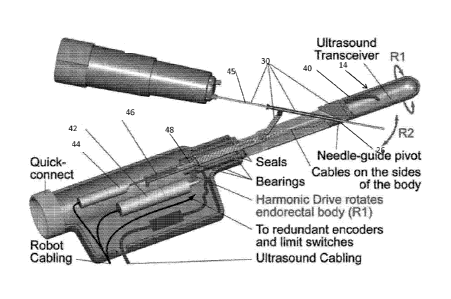
[0027] FIG. 3 illustrates a sectional view of an ultrasound wand and robotics
unit according
to an aspect of the present invention.
[0028] FIGS. 4A and 4B illustrate a CAD simulation of prostate targeting. FIG.
4A is a
sagittal view and FIG. 4B is a coronal view.
7a
CA 2869976 2019-02-01
CA 02869976 2014-10-08
WO 2013/155156
PCT/US2013/035928
[0029] FIG. 5 is a perspective view of the probe of an ultrasound wand
according to
an aspect of the present invention.
[0030] FIGS. 6A-6E illustrate a rotary slice and rendered prostate of a
prostate
mockup.
DETAILED DESCRIPTION
[0031] The presently disclosed subject matter now will be described more fully
hereinafter with reference to the accompanying Drawings, in which some, but
not all
embodiments of the inventions are shown. Like numbers refer to like elements
throughout.
The presently disclosed subject matter may be embodied in many different forms
and should
not be construed as limited to the embodiments set forth herein; rather, these
embodiments
are provided so that this disclosure will satisfy applicable legal
requirements. Indeed, many
modifications and other embodiments of the presently disclosed subject matter
set forth
herein will come to mind to one skilled in the art to which the presently
disclosed subject
matter pertains having the benefit of the teachings presented in the foregoing
descriptions and
the associated Drawings. Therefore, it is to be understood that the presently
disclosed subject
matter is not to be limited to the specific embodiments disclosed and that
modifications and
other embodiments are intended to be included within the scope of the appended
claims.
[0032] An embodiment in accordance with the present invention provides a
device
and method for a transrectal ultrasound (TRUS) guided prostate biopsy. The
device includes
an ultrasound wand equipped with a lateral fire ultrasound sensor that fits
the entire prostate
in parasagittal views. A purely rotary and controlled motion about the probe
axis is used to
scan the entire prostate. The ultrasound wand also defines a channel having an
oblique path
though the wand. The channel accommodates a biopsy needle, which can be
pivoted relative
to the probe, as well as inserted through the channel to different depths in
order to perform a
biopsy of the prostate. The device therefore uses only three degrees-of-
freedom (DoE) of
8
CA 02869976 2014-10-08
WO 2013/155156
PCT/US2013/035928
movement in order to obtain the biopsy samples. The wand can also be used in
conjunction
with robotic control. Additionally, the ultrasound wand can be used to
generate a three-
dimensional image of the prostate.
[0033] FIGS. 2A and 2B illustrate, a schematic of the proposed biopsy system
and
setup. As shown in FIGS. 2A and 2B, a system 10 includes a robotic-TRUS (R-
TRUS) wand
12 having a probe 14 that can be inserted into a rectum 16 of a patient 18.
The R-TRUS
wand 12 is held in place by an arm 20. The R-TRUS wand 12 is also connected to
a standard
two-dimensional ultrasound 21 and controller 22. Further, as illustrated in
FIGS. 2A and 2B,
a biopsy needle 24 can be inserted through a channel 26 defined by the probe
14. The biopsy
needle 24 is inserted obliquely relative to the probe 14 of the R-TRUS wand
12. The channel
26 can further include a needle guide 28 for limiting the motion of the biopsy
needle 24. A
needle spacer 30 can be used for controlling the depth to which the biopsy
needle 24 is
inserted.
[0034] The R-TRUS wand 12 can also include a handle 32 as illustrated in FIGS.
2A
and 2B. The handle 32 contains the robotics for controlling the R-TRUS wand 12
and needle
guide 28. These robotics will be discussed in further detail below, but
overall the R-TRUS
wand 12 has 3 degrees-of-freedom. Two degrees-of-freedom are implemented on
the probe
and a third, remote degree of freedom is implemented on a small needle-spacer
that attaches
to the needle. The needle can take the form of a standard 18Ga biopsy needle.
Alternately, the
needle can take the form of any other needle or biopsy device known to or
conceivable by
one of skill in the art.
[0035] By way of example, the procedure can proceed as follows, however, the
procedure discussed below is not to be considered limited to only the
following methodology.
After confirming that appropriate antibiotics and fleet enema are given to
minimize infection,
9
CA 02869976 2014-10-08
WO 2013/155156
PCT/US2013/035928
the patient is placed in a lateral decubitus position on the examination table
in the urology
clinic. Soft blocks around lower extremities are used to maintain the
patient's position and
local anesthesia is administered with a periprostatic Lidocaine block to
minimize patient
discomfort and motion. The probe 14 of the R-TRUS wand 12, detached from the
support
arm 20 is inserted manually. The probe is adjusted, such that the lateral-fire
ultrasound shows
the entire sagittal view of the prostate shown in FIG. 2B, using an image from
an MRI scan.
The R-TRUS wand 12 can also include a quick-connect coupler 34 to attach the
wand 12 to
the support arm 20. The physician uses a joystick 35 to rotate the probe 14
about its axis
confirming that the entire prostate is visible, side-to-side. An automated
rotary scan is
performed to uniformly sweep the entire gland and record image-position pairs.
Pixels of the
2D image slices are mapped in 3D using volume rendering. This 3D mapping can
be
executed immediately.
10036] The biopsy plan is overlaid as described further, herein. The R-TRUS
wand 12
automatically rotates and angles its oblique needle-guide 28 to point to the
planned locations
of biopsy, one after another. Automatically, the active spacer 30 of the
needle adjusts its
length to match the depth of the target. Real-time B-mode images are presented
to the
physician. The planned needle trajectory and target may be superimposed as
needed. If
necessary, the physician can choose to correct the alignment using the
joystick. Manually,
he/she then inserts the needle, fires the biopsy, collects, and labels the
sample. The oblique
needle-guide 28 is in the central lane of the sensor, enabling the physician
to monitor the
insertion. Ultrasound images are also recorded to document the actual biopsy
locations. The
actual locations are calculated in 3D based on the images and the
corresponding angles of the
probe. The biopsy map with matching sample labeling accompanies the
histopathologic
biopsy report.
CA 02869976 2014-10-08
WO 2013/155156
PCT/US2013/035928
[0037] The body of the R-TRUS wand 12 is fixed with a support arm 20 through a
quick-connect coupler 34, as illustrated in FIG. 2A. A 7-DoA (degrees of
adjustment)
passive arm can be used with a coupler, such as a threaded coupler, a
frictional coupler, or a
magnetic coupler, or any other suitable form of coupler. Disconnecting the
wand 12 from the
arm 20 facilitates placement of probe 14 in the rectum 16. It can also make it
easy to remove
the probe 14 for cleaning, and enhance safety as a quick disconnect.
[0038] FIG. 3 illustrates a sectional view of an ultrasound wand and robotics
unit
according to an aspect of the present invention. The endorectal shaft 40 of
the probe 14 spins
about an axis (R1) controlled by the R1 motor 42 through a transmission,
preferably a
harmonic drive. The R2 motor 44 pushes a rod 46 through a ball-screw 48, which
acts upon
the 3-bar mechanism of the needle-guide, adjusting the angle of the guide, R2.
A central
through shaft opening 26 is made to accommodate the needle-guide components
28. The
ultrasound transceiver is hermetically closed at the end of the probe. Its
cables are passed on
the sides of the shaft into the body of the probe, and mounted loose to allow
the shaft to
rotate. For this, R1 is limited to approximately 90 . Two bearings support
the shaft for
smooth motion, and another on the rod decouples the R1 and R2. The shaft and
rod are round
so 0-rings are used to seal the body hermetically.
[0039] For example, the CAD scene built with a sagittal pelvic MRI illustrated
in
FIG. 3 shows that the needle 24 can occupying the space taken by the
ultrasound sensor and
target the prostate without injuring the anus. FIGS. 4A and 4B show a CAD
simulation of
prostate targeting. FIG. 4A is a sagittal view and FTG. 4B is a coronal view.
As shown, the
needlepoint density plot overlaps the prostate in the two orthogonal views
showing that the 3-
DoF are properly selected and dimensioned to target the gland.
[0040] Further, as illustrated in FIG. 3, a spacer 30 with adjustable length
is placed
11
CA 02869976 2014-10-08
WO 2013/155156
PCT/US2013/035928
over a shaft 45 of the needle 24. The length of this spacer 30 is adjusted
automatically under
image-guided control. When the needle 24 and spacer 30 are fully inserted into
the needle-
guide 26 and fired, the center of the biopsy core matches the depth of the
biopsy target. Any
biopsy needle or biopsy device known to or conceivable to one of skill in the
art can be used.
For example, an 18Ga biopsy needle is common for the prostate, but any
suitable biopsy
needle can be used. The spacer 30 can be connected with a flexible cable to
the robot
controller, as illustrated in FIGS. 3, 4A, and 4B.
[0041] The present invention also includes cleaning and sterilization
features, as
illustrated in FIG. 5. The needle-guide 26 includes parts that come in contact
with the needle
that should be sterile. To enable cleaning, an opening 50 extends through the
shaft 40 and 0-
rings 52 seal the base. The sterile components of the needle guide 26 clip
easily to the probe
shaft 40. The sterile components of the needle guide 26 are only three simple
parts that could
be re-sterilized or made disposable together with the needle 24. These
components can be
manufactured separately or together with the biopsy needle 24.
[0042] A PC based robot controller for the R-TRUS wand can use an onboard
processor motion control card. Software developments can be made in Visual C++
(Microsoft
Corp, Redmond, WA), or any other suitable programming language. A special
emergency,
watchdog, and relays hardware board is included to mitigate control software
malfunctions.
The watchdog monitors several safety functions of the robot and disables its
power, should a
critical situation occur. A series of principles for medical robot safety can
also be included.
The motors of the robot use redundant position sensors. Among other safety
checks, the
watchdog verifies that the outputs of the sensors are in agreement. Having
reliable sensors, in
turn, enables the watchdog to reliably trace following-errors for all axes.
The motors are
calculated and chosen so that they are not oveipowered, limiting their
potential force/torque
12
CA 02869976 2014-10-08
WO 2013/155156
PCT[1JS2013/035928
in case of malfunction.
[0043] Axis level motion control can use libraries (MCI) of the motion control
card.
Direct and inverse kinematics of the robot are calculated based on kinematic
parameters from
the design. These map the robot to joint coordinates. In addition, robot-to-
image registration
is also required to control the robot based on the images. Registration is
typically required for
every intervention. For the R-TRUS wand however, the registration is
maintained because of
the cohesive structure. A calibration/registration procedure is performed by
imaging a
geometric calibration rig with the R-TRUS wand in a water tank. The mapping of
the image
to the robot spaces can then be calculated by comparing the 3D imaged geometry
of the rig
with its known geometric shape, as is known to those of skill in the art.
[0044] Image processing, visualization, robotic navigation, and biopsy
planning use
custom Visual C++ modules in the Amira Visualization platform (Visage Imaging
Inc, San
Diego, CA). This is a powerful image processing and visualization platform
which offers
open customization. Images are acquired from the standard 2D ultrasound
machine with an
acquisition card. Concurrently, the orientation of the image frames are
available from the
robot. During the rotary scan, these image-orientation pairs are saved in
DICOM format
including the pixel spacing (scaling), image position, and image orientation
standard tags, in
a volumetric DICOM form. An R-TRUS probe enables a standard 2D ultrasound to
acquire
3D images. Pixels of the 2D images are mapped in 3D using volume rendering.
Compared to
.. prostate segmentation, which takes time, volume rendering can be displayed
immediately.
FIGS. 5A-5E illustrate a rotary slice and rendered prostate of a CIRS 053
(Norfolk, VA)
prostate mockup. Once images of the prostate are shown, biopsy locations can
readily be
selected by the urologist in the original B-mode parasagittal slices, re-
sliced transverse slices,
or other display ways as needed.
13
CA 02869976 2014-10-08
WO 2013/155156
PCT/US2013/035928
[0045] Software to implement a new way of defining the biopsy plan quickly and
consistently is also included. Defining the plan involves the following 3
steps, illustrated in
FIGS. 6B-6D: 1) after the scan, the central sagittal slice through the
prostate is selected, as
illustrated in FIG. 6B. This is one of the original B-mode rotary slices; 2)
in this slice
illustrated in FIG. 6C the physician outlines the general direction of the
urethra, thus defining
the urethra plane normal and through this line. The software then re-slices
the rendered
volume with the urethra plane as shown in FIG. 6D; 3) in this para-coronal
slice through the
urethra, the urologist places a preformatted template which already has the
classic
distribution of the 12-cores. This can be quickly dragged, rotated, and scaled
to match the
gland, by grabbing the white and green controls shown in FIG. 6D. The plan may
be shown
in B-mode, re-sliced views, volumetrically, shown in FIG. 6E, or any
combination. It may
also be adjusted by the physician as needed by editing locations independently
or adjusting
step 3 above.
[0046] Repeat treatment of patients with this device would be more effective
because
a repeat treatment could take advantage of the documented biopsy map produced.
To register
the old to the current images, a segmentation of the previous prostate is
performed ahead of
time. At the time of the intervention, this segmented prostate is aligned to
the volume
rendered gland. This allows registering the old biopsy sites to the new
images. The physician
can then decide the best approach for the new biopsy plan, either to retarget
a known location
of cancer in case of an active surveillance patient, or choose other locations
in case of a
negative previous biopsy.
[0047] While this system has been described for use in prostate biopsy, it
need not be
limited to this application and could be used for different imaging and biopsy
procedures
known to one of skill in the art. The many features and advantages of the
invention are
14
CA 02869976 2014-10-08
WO 2013/155156
PCT/US2013/035928
apparent from the detailed specification, and thus, it is intended by the
appended claims to
cover all such features and advantages of the invention which fall within the
true spirit and
scope of the invention. Further, since numerous modifications and variations
will readily
occur to those skilled in the art, it is not desired to limit the invention to
the exact
construction and operation illustrated and described, and accordingly, all
suitable
modifications and equivalents may be resorted to, falling within the scope of
the invention.