Note: Descriptions are shown in the official language in which they were submitted.
CA 02870126 2014-10-09
¨ 1 -
Description
Title of Invention: ANTI-FGFR2 ANTIBODY
Technical Field
The present invention relates to a novel antibody, a
nucleotide comprising a nucleotide sequence encoding the
amino acid sequence of the antibody, a vector having an
insert of the nucleotide, a cell comprising the
nucleotide or the vector, a method for producing the
antibody, comprising the step of culturing the cell, a
pharmaceutical composition comprising the antibody, a
composition for diagnosis comprising the antibody, a
functional fragment of the antibody, a modified form of
the antibody, etc.
Background Art
Fibroblast growth factors (FGFs) are known to play
an important role in embryogenesis, tissue homeostasis,
and metabolism via FGF receptor (FGFR) signals (Non
Patent Literature 1). In humans, 22 FGFs (FGF1 to FGF14
and FGF16 to FGF23) and 4 FGF receptors (FGFR1 to FGFR4;
hereinafter, collectively referred to as "FGFRs") having
a tyrosine kinase domain are found. These FGFRs are each
constituted by an extracellular region comprising a
ligand binding site composed of 2 or 3 immunoglobulin-
like domains (IgD1 to IgD3), a single-pass transmembrane
FP1320s/(PN814663)/Eng1ish translation of PCT specification (September 2014)
5106289-1-MCARVALHO
CA 02870126 2014-10-09
- 2 -
region, and an intracellular region comprising the
tyrosine kinase domain. FGFR1, FGFR2, and FGFR3 each
have two splicing variants called IIIb and IIIc. These
isoforms differ in the sequence of approximately 50 amino
acids in the latter half of IgD3 and exhibit distinctive
tissue distribution and ligand specificity. It is
generally known that the IIIb isoform is expressed in
epithelial cells, while the IIIc isoform is expressed in
mesenchymal cells. Upon binding of FGFs to FGFRs, these
FGFRs are dimerized and phosphorylated at their
particular tyrosine residues. This phenomenon promotes
the recruiting of important adaptor proteins such as FGFR
substrate 2a (FRS2a) and induces the activation of many
signaling pathways including MAPK and PI3K/Akt pathways.
As a result, FGFs and their corresponding receptors
control a wide range of cell functions including growth,
differentiation, migration, and survival.
The abnormal activation of FGFRs is known to
participate in particular types of malignant tumor
development in humans (Non Patent Literature 1 and 2).
Particularly, findings such as the overexpression of
FGFR2 and its ligand, receptor mutations or gene
amplification, and isoform switching, have been made as
to the association of FGFR2 signal abnormality with
cancer. Specifically, a single nucleotide polymorphism
(SNP) in intron 2 of the FGFR2 gene reportedly correlates
with the risk of breast cancer progression caused by the
FP1320s/(PN814663)/English translation of PCT specification (September 2014)
5106289-1-MCARVALHO
CA 02870126 2014-10-09
- 3 -
high expression of FGFR2 (Non Patent Literature 3 and 4).
Missense mutations that constitutively activate FGFR2
have been reported in endometrial cancer, ovary cancer,
breast cancer, lung cancer, and stomach cancer (Non
Patent Literature 2, 3, and 5). Also, the amplification
or overexpression of the FGFR2 gene has been reported in
stomach cancer and breast cancer (Non Patent Literature 2,
3, and 5). In addition, class switch from FGFR2 IIIb to
FGFR2 IIIc is also known to occur during the progression
of prostate cancer or kidney cancer and correlate with
poor prognosis (Non Patent Literature 6 and 7).
As mentioned above, the association of FGFR2
overexpression or mutations or switching from IIIb to
IIIc, with many cancer types suggests the possibility of
FGFR2 as an excellent therapeutic target for cancer. In
fact, monoclonal antibodies against FGFR2 have been
obtained and are under evaluation for their antitumor
effects in preclinical trials in order to reveal the role
of FGFR2 in oncogenesis and determine the possibility of
FGFR2 as a therapeutic target for cancer (Non Patent
Literature 8 and 9). All of these antibodies have been
shown to have a neutralizing effect that inhibits
signaling derived from a ligand for FGFR2 IIIb.
Unfortunately, there has been no report on a functional
antibody having effector effects such as ADCC or a
neutralizing effect on IIIc.
FP1320s/(PN814663)/English translation of PCT specification (September 2014)
5106289-1-MCARVALHO
CA 02870126 2014-10-09
- 4 -
Citation List
Non Patent Literature
Non Patent Literature 1: Eswarakumar, V.P., et al., J.
Cytokine Growth Factor Rev., Apr. 2005, Vol. 16 (No. 2),
p. 139-149, published online on Feb 1, 2005, Review
Non Patent Literature 2: Turner, N. and Grose, R., Nat.
Rev. Cancer, Feb. 2010, Vol. 10 (No. 2), p. 116-129,
Review
Non Patent Literature 3: Easton, D.F., et al., Nature,
Jun. 28, 2007, Vol. 447 (No. 7148), p. 1087-1093
Non Patent Literature 4: Hunter DJ, et al., Nat. Genet.,
Jul. 2007, Vol. 39 (No. 7), p. 870-874, published online
on May 27, 2007
Non Patent Literature 5: Katoh, Y. and Katoh, M., Int. J.
Mol. Med., Mar. 2009, Vol. 23 (No. 3), p. 307-311, Review
Non Patent Literature 6: Chaffer, C.L., et al.,
Differentiation, Nov. 2007, Vol. 75 (No. 9), p. 831-842,
published online on Aug. 14, 2007, Review
Non Patent Literature 7: Carstens, R.P., et al., Oncogene,
Dec. 18, 1997, Vol. 15 (No. 25), p. 3059-3065
Non Patent Literature 8: Zhao, W.M., et al., Clin. Cancer
Res., Dec. 1, 2010, Vol. 16 (No. 23), p. 5750-5758,
published online on Jul. 29, 2010
Non Patent Literature 9: Bai, A., et al., Cancer Res.,
Oct. 1, 2010, Vol. 70 (No. 19), p. 7630-7639, published
online on Aug. 13, 2010
FP1320s/(PN814663)/English translation of PCT specification (September 2014)
5106289-1-MCARVALHO
CA 02870126 2014-10-09
- 5 -
Summary of Invention
Technical Problem
An object of the present invention is to provide an
antibody against FGFR2.
Another object of the present invention is to
provide a pharmaceutical composition, etc. comprising an
anti-FGFR2 antibody having an anticancer effect.
An alternative object of the present invention
includes a nucleotide encoding the amino acid sequence of
the antibody, a vector having an insert of the nucleotide,
a cell comprising the nucleotide or the vector, a method
for producing the antibody, comprising the step of
culturing the cell, etc.
A further alternative object of the present
invention is to provide a method for treating cancer
using the antibody.
Solution to Problem
The present inventors have conducted diligent
studies to attain the objects and consequently completed
the present invention by developing a novel anti-FGFR2
antibody and have found that the antibody has an
anticancer effect.
The present invention relates to:
(1) An antibody or a functional fragment thereof, which
has antibody dependent cellular cytotoxic activity and
binds to a fibroblast growth factor receptor (FGFR);
FP1320s/(PN814663)/Eng1ish translation of PCT specification (September 2014)
5106289-1-MCARVALHO
CA 02870126 2014-10-09
- 6 -
(2) The antibody or functional fragment thereof according
to (1), wherein the fibroblast growth factor receptor
(FGFR) is human FGFR;
(3) The antibody or functional fragment thereof according
to (1) or (2), wherein the fibroblast growth factor
receptor (FGFR) is FGFR2;
(4) The antibody or functional fragment thereof according
to any one of (1) to (3), wherein the antibody or
functional fragment thereof binds to human fibroblast
growth factor receptor 2 (human FGFR2) IIIb and/or human
fibroblast growth factor receptor 2 (human FGFR2) IIIc;
(5) The antibody or functional fragment thereof according
to any one of (1) to (4), wherein the antibody or
functional fragment thereof binds to human fibroblast
growth factor receptor 2 (human FGFR2) IIIb and human
fibroblast growth factor receptor (human FGFR2) IIIc;
(6) The antibody or functional fragment thereof according
to any one of (1) to (5), wherein the antibody or
functional fragment thereof binds to one or two or more
immunoglobulin-like domains of the human fibroblast
growth factor receptor 2;
(7) The antibody or functional fragment thereof according
to any one of (1) to (6), wherein the antibody or
functional fragment thereof binds to immunoglobulin-like
domain 2 of the human fibroblast growth factor receptor
2;
FP1320s/(PN814663)/English translation of PCT specification (September 2014)
5106289-1-MCARVALHO
CA 02870126 2016-05-04
- 7 -
(8) The antibody or functional fragment thereof according
to any one of (1) to (4) and (6), wherein the antibody or
functional fragment thereof binds to immunoglobulin-like
domain 3 of the human fibroblast growth factor receptor
2;
(9) The antibody or functional fragment thereof according
to any one of (1) to (8), wherein the antibody or
functional fragment thereof has neutralizing activity
against the human fibroblast growth factor receptor 2
(human FGFR2) IIIb and/or the human fibroblast growth
factor receptor 2 (human FGFR2) IIIc;
(10) The antibody or functional fragment thereof
according to any one of (1) to (9), wherein the antibody
or functional fragment thereof has neutralizing activity
against the human fibroblast growth factor receptor 2
(human FGFR2) IIIb and the human fibroblast growth factor
receptor 2 (human FGFR2) IIIc;
(11) The antibody or functional fragment thereof
according to any one of (1) to (10), wherein the antibody
or functional fragment thereof has antitumor activity;
(12) The antibody or functional fragment thereof
according to (11), wherein the antibody or functional
fragment thereof exhibits antitumor activity in vivo;
(12.1) The antibody or functional fragment thereof
according to any one of (1) to (12), wherein the antibody
is a humanized antibody;
CA 02870126 2016-05-04
- 7a -
(12.2) The antibody or functional fragment thereof
according to (12.1), wherein the antibody comprises a
light chain comprising CDRL1 consisting of amino acid
positions 44 to 54 of the amino acid sequence represented
by SEQ ID NO: 73 (Figure 81) of the Sequence Listing,
CDRL2 consisting of amino acid positions 70 to 76 of the
amino acid sequence represented by SEQ ID NO: 73 (Figure
81) of the Sequence Listing, and CDRL3 consisting of
amino acid positions 109 to 118 of the amino acid
sequence represented by SEQ ID NO: 73 (Figure 81) of the
Sequence Listing;
(12.3) The antibody or functional fragment thereof
according to (12.2), wherein the antibody comprises a
heavy chain selected from the following (i) to (v):
(i) a heavy chain comprising CDRH1 consisting of amino
acid positions 50 to 54 of the amino acid sequence
represented by SEQ ID NO: 97 (Figure 105) of the
Sequence Listing, CDRH2 consisting of amino acids
positions 69 to 85 of the amino acid sequence
represented by SEQ ID NO: 97 (Figure 105) of the
Sequence Listing, and CDRH3 consisting of amino acid
positions118 to 126 of the amino acid sequence
represented by SEQ ID NO: 97 (Figure 105) of the
Sequence Listing;
(ii) a heavy chain comprising CDRH1 consisting of amino
acid positions 50 to 54 of the amino acid sequence
represented by SEQ ID NO: 89 (Figure 97) of the
CA 02870126 2016-05-04
- 7b -
Sequence Listing, CDRH2 consisting of amino acids
positions 69 to 85 of the amino acid sequence
represented by SEQ ID NO: 89 (Figure 97) of the
Sequence Listing, and CDRH3 consisting of amino acid
positions 118 to 126 of the amino acid sequence
represented by SEQ ID NO: 89 (Figure 97) of the
Sequence Listing;
(iii) a heavy chain comprising CDRH1 consisting of amino
acid positions 50 to 54 of the amino acid sequence
represented by SEQ ID NO: 91 (Figure 99) of the
Sequence Listing, CDRH2 consisting of amino acids
positions 69 to 85 of the amino acid sequence
represented by SEQ ID NO: 91 (Figure 99) of the
Sequence Listing, and CDRH3 consisting of amino acid
positions 118 to 126 of the amino acid sequence
represented by SEQ ID NO: 91 (Figure 99) of the
Sequence Listing;
(iv) a heavy chain comprising CDRH1 consisting of amino
acid positions 50 to 54 of the amino acid sequence
represented by SEQ ID NO: 95 (Figure 103) of the
Sequence Listing, CDRH2 consisting of amino acids
positions 69 to 85 of the amino acid sequence
represented by SEQ ID NO: 95 (Figure 103) of the
Sequence Listing, and CDRH3 consisting of amino acid
positions 118 to 126 of the amino acid sequence
represented by SEQ ID NO: 95 (Figure 103) of the
Sequence Listing; and
CA 02870126 2016-05-04
- 7c -
(v) a heavy chain comprising CDRH1 consisting of amino
acid positions 50 to 54 of the amino acid sequence
represented by SEQ ID NO: 83 (Figure 91) of the
Sequence Listing, CDRH2 consisting of amino acids
positions 69 to 85 of the amino acid sequence
represented by SEQ ID NO: 83 (Figure 91) of the
Sequence Listing, and CDRH3 consisting of amino acid
positions 118 to 126 of the amino acid sequence
represented by SEQ ID NO: 83 (Figure 91) of the
Sequence Listing;
(12.4) The antibody or functional fragment thereof
according to (12.1), wherein the antibody is selected
from the following (i) to (xix):
(i) a humanized antibody (hFR2-14_H19/L1) comprising a
light chain comprising amino acid positions 21 to 235
of the amino acid sequence represented by SEQ ID NO:
73 (Figure 81), and a heavy chain comprising amino
acid positions 20 to 467 of the amino acid sequence
represented by SEQ ID NO: 97 (Figure 105);
(ii) a humanized antibody (hFR2-14_H12/L1) comprising a
light chain comprising amino acid positions 21 to 235
of the amino acid sequence represented by SEQ ID NO:
73 (Figure 81), and a heavy chain comprising amino
acid positions 20 to 467 of the amino acid sequence
represented by SEQ ID NO: 97 (Figure 105);
(iii) a humanized antibody (hFR2-14_H8/L1) comprising a
light chain comprising amino acid positions 21 to 235
CA 02870126 2016-05-04
- 7d -
of the amino acid sequence represented by SEQ ID NO:
73 (Figure 81), and a heavy chain comprising amino
acid positions 20 to 467 of the amino acid sequence
represented by SEQ ID NO: 89 (Figure 97);
(iv) a humanized antibody (hFR2-14_H9/L1) comprising a
light chain comprising amino acid positions 21 to 235
of the amino acid sequence represented by SEQ ID NO:
73 (Figure 81), and a heavy chain comprising amino
acid positions 20 to 467 of the amino acid sequence
represented by SEQ ID NO: 91 (Figure 99);
(v) a humanized antibody (hFR2-14 H11/L1) comprising a
light chain comprising amino acid positions 21 to 235
of the amino acid sequence represented by SEQ ID NO:
73 (Figure 81), and a heavy chain comprising amino
acid positions 20 to 467 of the amino acid sequence
represented by SEQ ID NO: 95 (Figure 103);
(vi) a humanized antibody (hFR2-14_H5/L1) comprising a
light chain comprising amino acid positions 21 to 235
of the amino acid sequence represented by SEQ ID NO:
73 (Figure 81), and a heavy chain comprising amino
acid positions 20 to 467 of the amino acid sequence
represented by SEQ ID NO: 83 (Figure 91);
(vii) a humanized antibody (hFR2-14_Hl/L1) comprising a
light chain comprising amino acid positions 21 to 235
of the amino acid sequence represented by SEQ ID NO:
73 (Figure 81), and a heavy chain comprising amino
CA 02870126 2016-05-04
- 7e -
acid positions 20 to 467 of the amino acid sequence
represented by SEQ ID NO: 75 (Figure 83);
(viii) a humanized antibody (hFR2-14 H2/L1) comprising
a light chain comprising amino acid positions 21 to
235 of the amino acid sequence represented by SEQ ID
NO: 73 (Figure 81), and a heavy chain comprising
amino acid positions 20 to 467 of the amino acid
sequence represented by SEQ ID NO: 77 (Figure 85);
(ix) a humanized antibody (hFR2-14_H3/L1) comprising a
light chain comprising amino acid positions 21 to 235
of the amino acid sequence represented by SEQ ID NO:
73 (Figure 81), and a heavy chain comprising amino
acid positions 20 to 467 of the amino acid sequence
represented by SEQ ID NO: 79 (Figure 87);
(x) a humanized antibody (hFR2-14_H4/L1) comprising a
light chain comprising amino acid positions 21 to 235
of the amino acid sequence represented by SEQ ID NO:
73 (Figure 81), and a heavy chain comprising amino
acid positions 20 to 467 of the amino acid sequence
represented by SEQ ID NO: 81 (Figure 89);
(xi) a humanized antibody (hFR2-14_H6/L1) comprising a
light chain comprising amino acid positions 21 to 235
of the amino acid sequence represented by SEQ ID NO:
73 (Figure 81), and a heavy chain comprising amino
acid positions 20 to 467 of the amino acid sequence
represented by SEQ ID NO: 85 (Figure 93);
CA 02870126 2016-05-04
- 7f -
(xii) a humanized antibody (hFR2-14 H7/L1) comprising a
light chain comprising amino acid positions 21 to 235
of the amino acid sequence represented by SEQ ID NO:
73 (Figure 81), and a heavy chain comprising amino
acid positions 20 to 467 of the amino acid sequence
represented by SEQ ID NO: 87 (Figure 95);
(xiii) a humanized antibody (hFR2-14 H10/L1) comprising
a light chain comprising amino acid positions 21 to
235 of the amino acid sequence represented by SEQ ID
NO: 73 (Figure 81), and a heavy chain comprising
amino acid positions 20 to 467 of the amino acid
sequence represented by SEQ ID NO: 93 (Figure 101);
(xiv) a humanized antibody (hFR2-14 H13/L1) comprising
a light chain comprising amino acid positions 21 to
235 of the amino acid sequence represented by SEQ ID
NO: 73 (Figure 81), and a heavy chain comprising
amino acid positions 20 to 467 of the amino acid
sequence represented by SEQ ID NO: 99 (Figure 107);
(xv) a humanized antibody (hFR2-14 H14/L1) comprising a
light chain comprising amino acid positions 21 to 235
of the amino acid sequence represented by SEQ ID NO:
73 (Figure 81), and a heavy chain comprising amino
acid positions 20 to 467 of the amino acid sequence
represented by SEQ ID NO: 101 (Figure 109);
(xvi) a humanized antibody (hFR2-14 H15/L1) comprising
a light chain comprising amino acid positions 21 to
235 of the amino acid sequence represented by SEQ ID
CA 02870126 2016-05-04
- 7g -
NO: 73 (Figure 81), and a heavy chain comprising
amino acid positions 20 to 467 of the amino acid
sequence represented by SEQ ID NO: 103 (Figure 111);
(xvii) a humanized antibody (hFR2-14_H16/L1) comprising
a light chain comprising amino acid positions 21 to
235 of the amino acid sequence represented by SEQ ID
NO: 73 (Figure 81), and a heavy chain comprising
amino acid positions 20 to 467 of the amino acid
sequence represented by SEQ ID NO: 105 (Figure 113);
(xviii) a humanized antibody (hFR2-14_H17/L1)
comprising a light chain comprising amino acid
positions 21 to 235 of the amino acid sequence
represented by SEQ ID NO: 73 (Figure 81), and a heavy
chain comprising amino acid positions 20 to 467 of
the amino acid sequence represented by SEQ ID NO: 107
(Figure 115); and
(xix) a humanized antibody (hFR2-14_H18/L1) comprising a
light chain comprising amino acid positions 21 to 235
of the amino acid sequence represented by SEQ ID NO:
73 (Figure 81), and a heavy chain comprising amino
acid positions 20 to 467 of the amino acid sequence
represented by SEQ ID NO: 109 (Figure 117);
(12.5) The antibody or functional fragment thereof
according to (12.1), wherein the antibody comprises a
light chain variable region comprising of amino acid
positions 21 to 130 of the amino acid sequence
represented by SEQ ID NO: 73 (Figure 81) and a heavy
CA 02870126 2016-05-04
- 7h -
chain variable region comprising of one of the following
(i) to (v):
(i) amino acid positions 20 to 137 of the amino acid
sequence represented by SEQ ID NO: 97 (Figure 105);
(ii) amino acid positions 20 to 137 of the amino acid
sequence represented by SEQ ID NO: 89 (Figure 97);
(iii) amino acid positions 20 to 137 of the amino acid
sequence represented by SEQ ID NO: 91 (Figure 99);
(iv) amino acid positions 20 to 137 of the amino acid
sequence represented by SEQ ID NO: 95 (Figure 103);
or
(v) amino acid positions 20 to 137 of the amino acid
sequence represented by SEQ ID NO: 83 (Figure 91);
(12.6) The antibody or functional fragment thereof
according to (12.3), wherein the heavy chain is item (i),
according to (12.4), wherein the humanized antibody is
item (i), or according to (12.5), wherein the humanized
antibody is item (i) and wherein the antibody is
defucosylated;
(13) The antibody or functional fragment thereof
according to any one of (1) to (4), (6), (8), (9), (11)
and (12), wherein the antibody consists of a heavy chain
comprising CDRH1 consisting of the amino acid sequence
CA 02870126 2014-10-09
- 8 -
represented by SEQ ID NO: 52 (Figure 60) of the Sequence
Listing or an amino acid sequence derived from the amino
acid sequence by the substitution of one or two amino
acids, CDRH2 consisting of the amino acid sequence
represented by SEQ ID NO: 53 (Figure 61) of the Sequence
Listing or an amino acid sequence derived from the amino
acid sequence by the substitution of one or two amino
acids, and CDRH3 consisting of the amino acid sequence
represented by SEQ ID NO: 54 (Figure 62) of the Sequence
Listing or an amino acid sequence derived from the amino
acid sequence by the substitution of one or two amino
acids, and a light chain comprising CDRL1 consisting of
the amino acid sequence represented by SEQ ID NO: 61
(Figure 69) of the Sequence Listing or an amino acid
sequence derived from the amino acid sequence by the
substitution of one or two amino acids, CDRL2 consisting
of the amino acid sequence represented by SEQ ID NO: 62
(Figure 70) of the Sequence Listing or an amino acid
sequence derived from the amino acid sequence by the
substitution of one or two amino acids, and CDRL3
consisting of the amino acid sequence represented by SEQ
ID NO: 63 (Figure 71) of the Sequence Listing or an amino
acid sequence derived from the amino acid sequence by the
substitution of one or two amino acids, and binds to
human FGFR2;
(14) The antibody or functional fragment thereof
according to any one of (1) to (7) and (9) to (12),
FP1320s/(PN814663)/Eng1ish translation of PCT specification (September 2014)
5106289-1-MCARVALHO
CA 02870126 2014-10-09
- 9 -
wherein the antibody consists of a heavy chain comprising
CDRH1 consisting of the amino acid sequence represented
by SEQ ID NO: 55 (Figure 63) of the Sequence Listing or
an amino acid sequence derived from the amino acid
sequence by the substitution of one or two amino acids,
CDRH2 consisting of the amino acid sequence represented
by SEQ ID NO: 56 (Figure 64) of the Sequence Listing or
an amino acid sequence derived from the amino acid
sequence by the substitution of one or two amino acids,
and CDRH3 consisting of the amino acid sequence
represented by SEQ ID NO: 57 (Figure 65) of the Sequence
Listing or an amino acid sequence derived from the amino
acid sequence by the substitution of one or two amino
acids, and a light chain comprising CDRL1 consisting of
the amino acid sequence represented by SEQ ID NO: 64
(Figure 72) of the Sequence Listing or an amino acid
sequence derived from the amino acid sequence by the
substitution of one or two amino acids, CDRL2 consisting
of the amino acid sequence represented by SEQ ID NO: 65
(Figure 73) of the Sequence Listing or an amino acid
sequence derived from the amino acid sequence by the
substitution of one or two amino acids, and CDRL3
consisting of the amino acid sequence represented by SEQ
ID NO: 66 (Figure 74) of the Sequence Listing or an amino
acid sequence derived from the amino acid sequence by the
substitution of one or two amino acids, and binds to
human FGFR2;
FP1320s/(PN814663)/English translation of PCT specification (September 2014)
5106289-1-MCARVALHO
CA 02870126 2014-10-09
- 10 -
(15) The antibody or functional fragment thereof
according to any one of (1) to (7) and (9) to (12),
wherein the antibody consists of a heavy chain comprising
CDRH1 consisting of the amino acid sequence represented
by SEQ ID NO: 58 (Figure 66) of the Sequence Listing or
an amino acid sequence derived from the amino acid
sequence by the substitution of one or two amino acids,
CDRH2 consisting of the amino acid sequence represented
by SEQ ID NO: 59 (Figure 67) of the Sequence Listing or
an amino acid sequence derived from the amino acid
sequence by the substitution of one or two amino acids,
and CDRH3 consisting of the amino acid sequence
represented by SEQ ID NO: 60 (Figure 68) of the Sequence
Listing or an amino acid sequence derived from the amino
acid sequence by the substitution of one or two amino
acids, and a light chain comprising CDRL1 consisting of
the amino acid sequence represented by SEQ ID NO: 67
(Figure 75) of the Sequence Listing or an amino acid
sequence derived from the amino acid sequence by the
substitution of one or two amino acids, CDRL2 consisting
of the amino acid sequence represented by SEQ ID NO: 68
(Figure 76) of the Sequence Listing or an amino acid
sequence derived from the amino acid sequence by the
substitution of one or two amino acids, and CDRL3
consisting of the amino acid sequence represented by SEQ
ID NO: 69 (Figure 77) of the Sequence Listing or an amino
acid sequence derived from the amino acid sequence by the
FP1320s/(PN814663)/English translation of PCT specification (September 2014)
5106289-1-MCARVALHO
CA 02870126 2014-10-09
¨ 11 -
substitution of one or two amino acids, and binds to
human FGFR2;
(16) The antibody or functional fragment thereof
according to (15), wherein the CDRH3 consists of an amino
acid sequence derived from the amino acid sequence
represented by SEQ ID NO: 60 (Figure 68) of the Sequence
Listing by the substitution of one or two amino acids;
(17) The antibody or functional fragment thereof
according to any one of (1) to (16), wherein the antibody
is a monoclonal antibody;
(18) The antibody or functional fragment thereof
according to any one of (1) to (17), wherein the antibody
is a chimeric antibody;
(19) The antibody or functional fragment thereof
according to any one of (1) to (18), wherein the antibody
is a humanized antibody;
(20) The antibody or functional fragment thereof
according to (19), wherein the antibody is selected from
the following (i) to (xix):
(i) a humanized antibody (hFR2-14 H19/L1) comprising a
light chain comprising amino acid positions 21 to 235 of
the amino acid sequence represented by SEQ ID NO: 73
(Figure 81), and a heavy chain comprising amino acid
positions 20 to 467 of the amino acid sequence
represented by SEQ ID NO: 97 (Figure 105);
(ii) a humanized antibody (hFR2-14 H12/L1) comprising a
light chain comprising amino acid positions 21 to 235 of
FP1320s/(PN814663)/English translation of PCT specification (September 2014)
5106289-1-MCARVALHO
CA 02870126 2014-10-09
- 12 -
the amino acid sequence represented by SEQ ID NO: 73
(Figure 81), and a heavy chain comprising amino acid
positions 20 to 467 of the amino acid sequence
represented by SEQ ID NO: 97 (Figure 105);
(iii) a humanized antibody (hFR2-14_H8/L1) comprising a
light chain comprising amino acid positions 21 to 235 of
the amino acid sequence represented by SEQ ID NO: 73
(Figure 81), and a heavy chain comprising amino acid
positions 20 to 467 of the amino acid sequence
represented by SEQ ID NO: 89 (Figure 97);
(iv) a humanized antibody (hFR2-14_H11/L1) comprising a
light chain comprising amino acid positions 21 to 235 of
the amino acid sequence represented by SEQ ID NO: 73
(Figure 81), and a heavy chain comprising amino acid
positions 20 to 467 of the amino acid sequence
represented by SEQ ID NO: 95 (Figure 103);
(v) a humanized antibody (hFR2-14_H5/L1) comprising a
light chain comprising amino acid positions 21 to 235 of
the amino acid sequence represented by SEQ ID NO: 73
(Figure 81), and a heavy chain comprising amino acid
positions 20 to 467 of the amino acid sequence
represented by SEQ ID NO: 83 (Figure 91);
(vi) a humanized antibody (hFR2-14 Hl/L1) comprising a
light chain comprising amino acid positions 21 to 235 of
the amino acid sequence represented by SEQ ID NO: 73
(Figure 81), and a heavy chain comprising amino acid
FP1320s/(PN814663)/English translation of PCT specification (September 2014)
5106289-14OCARVALHO
CA 02870126 2014-10-09
- 13 -
positions 20 to 467 of the amino acid sequence
represented by SEQ ID NO: 75 (Figure 83);
(vii) a humanized antibody (hFR2-14_H2/L1) comprising a
light chain comprising amino acid positions 21 to 235 of
the amino acid sequence represented by SEQ ID NO: 73
(Figure 81), and a heavy chain comprising amino acid
positions 20 to 467 of the amino acid sequence
represented by SEQ ID NO: 77 (Figure 85);
(viii) a humanized antibody (hFR2-14 H3/L1) comprising a
light chain comprising amino acid positions 21 to 235 of
the amino acid sequence represented by SEQ ID NO: 73
(Figure 81), and a heavy chain comprising amino acid
positions 20 to 467 of the amino acid sequence
represented by SEQ ID NO: 79 (Figure 87);
(ix) a humanized antibody (hFR2-14_H4/L1) comprising a
light chain comprising amino acid positions 21 to 235 of
the amino acid sequence represented by SEQ ID NO: 73
(Figure 81), and a heavy chain comprising amino acid
positions 20 to 467 of the amino acid sequence
represented by SEQ ID NO: 81 (Figure 89);
(x) a humanized antibody (hFR2-14_H6/L1) comprising a
light chain comprising amino acid positions 21 to 235 of
the amino acid sequence represented by SEQ ID NO: 73
(Figure 81), and a heavy chain comprising amino acid
positions 20 to 467 of the amino acid sequence
represented by SEQ ID NO: 85 (Figure 93);
FP1320s/(PN814663)/English translation of PCT specification (September 2014)
5106289-1-MCARVALHO
CA 02870126 2014-10-09
- 14 -
(xi) a humanized antibody (hFR2-14_H7/L1) comprising a
light chain comprising amino acid positions 21 to 235 of
the amino acid sequence represented by SEQ ID NO: 73
(Figure 81), and a heavy chain comprising amino acid
positions 20 to 467 of the amino acid sequence
represented by SEQ ID NO: 87 (Figure 95);
(xii) a humanized antibody (hFR2-14_H9/L1) comprising a
light chain comprising amino acid positions 21 to 235 of
the amino acid sequence represented by SEQ ID NO: 73
(Figure 81), and a heavy chain comprising amino acid
positions 20 to 467 of the amino acid sequence
represented by SEQ ID NO: 91 (Figure 99);
(xiii) a humanized antibody (hFR2-14_H10/L1) comprising a
light chain comprising amino acid positions 21 to 235 of
the amino acid sequence represented by SEQ ID NO: 73
(Figure 81), and a heavy chain comprising amino acid
positions 20 to 467 of the amino acid sequence
represented by SEQ ID NO: 93 (Figure 101);
(xiv) a humanized antibody (hFR2-14_H13/L1) comprising a
light chain comprising amino acid positions 21 to 235 of
the amino acid sequence represented by SEQ ID NO: 73
(Figure 81), and a heavy chain comprising amino acid
positions 20 to 467 of the amino acid sequence
represented by SEQ ID NO: 99 (Figure 107);
(xv) a humanized antibody (hFR2-14 H14/L1) comprising a
light chain comprising amino acid positions 21 to 235 of
the amino acid sequence represented by SEQ ID NO: 73
FP13205/(PN814663)/English translation of PCT specification (September 2014)
5106289-1-MCARVALHO
CA 02870126 2014-10-09
- 15 -
(Figure 81), and a heavy chain comprising amino acid
positions 20 to 467 of the amino acid sequence
represented by SEQ ID NO: 101 (Figure 109);
(xvi) a humanized antibody (hFR2-14 H15/L1) comprising a
light chain comprising amino acid positions 21 to 235 of
the amino acid sequence represented by SEQ ID NO: 73
(Figure 81), and a heavy chain comprising amino acid
positions 20 to 467 of the amino acid sequence
represented by SEQ ID NO: 103 (Figure 111);
(xvii) a humanized antibody (hFR2-14 H16/L1) comprising a
light chain comprising amino acid positions 21 to 235 of
the amino acid sequence represented by SEQ ID NO: 73
(Figure 81), and a heavy chain comprising amino acid
positions 20 to 467 of the amino acid sequence
represented by SEQ ID NO: 105 (Figure 113);
(xviii) a humanized antibody (hFR2-14 H17/L1) comprising
a light chain comprising amino acid positions 21 to 235
of the amino acid sequence represented by SEQ ID NO: 73
(Figure 81), and a heavy chain comprising amino acid
positions 20 to 467 of the amino acid sequence
represented by SEQ ID NO: 107 (Figure 115); and
(xix) a humanized antibody (hFR2-14_H18/L1) comprising a
light chain comprising amino acid positions 21 to 235 of
the amino acid sequence represented by SEQ ID NO: 73
(Figure 81), and a heavy chain comprising amino acid
positions 20 to 467 of the amino acid sequence
represented by SEQ ID NO: 109 (Figure 117);
FP1320s/(PN814663)/English translation of PCT specification (September 2014)
5106289-1-MCARVALHO
CA 02870126 2014-10-09
- 16 -
(21) The antibody or functional fragment thereof
according to any one of (1) to (12), wherein the antibody
comprises heavy and light chains comprising amino acid
sequences having 95% or higher identity to the amino acid
sequences of the heavy and light chains, respectively, of
an antibody according to (20), and binds to human FGFR2;
(22) The antibody or functional fragment thereof
according to any one of (1) to (12), wherein the antibody
or functional fragment thereof binds to a site on an
antigen recognized by an antibody according to any one of
(13) to (16) and (20);
(23) The antibody or functional fragment thereof
according to any one of (1) to (12), wherein the antibody
or functional fragment thereof competes with an antibody
according to any one of (13) to (16) and (20) for binding
to human FGFR2;
(24) The antibody or functional fragment thereof
according to any one of (1) to (12), wherein the antibody
or functional fragment thereof binds to an epitope on
human FGFR2, the epitope being constituted by tyrosine
(Tyr) at residue 155, threonine (Thr) at residue 157,
lysine (Lys) at residue 176, alanine (Ala) at residue 181,
glycine (Gly) at residue 182, glycine (Gly) at residue
183, asparagine (Asn) at residue 184, proline (Pro) at
residue 185, methionine (Met) at residue 186, threonine
(Thr) at residue 188, glutamine (Gin) at residue 200,
glutamic acid (Glu) at residue 201, glycine (Gly) at
FP1320s/(PN814663)/English translation of PCT specification (September 2014)
5106289-1-MCARVALHO
CA 02870126 2014-10-09
- 17 -
residue 205, glycine (Gly) at residue 206, lysine (Lys)
at residue 208, valine (Val) at residue 209, arginine
(Arg) at residue 210, asparagine (Asn) at residue 211,
glutamine (Gin) at residue 212, histidine (His) at
residue 213, tryptophan (Trp) at residue 214, and
isoleucine (Ile) at residue 217 in the amino acid
sequence represented by SEQ ID NO: 70 (Figure 78) or SEQ
ID NO: 71 (Figure 79);
(25) The antibody or functional fragment thereof
according to any one of (1) to (12), wherein the antibody
or functional fragment thereof has an interaction
distance with each of tyrosine (Tyr) at residue 155,
threonine (Thr) at residue 157, lysine (Lys) at residue
176, alanine (Ala) at residue 181, glycine (Gly) at
residue 182, glycine (Gly) at residue 183, asparagine
(Asn) at residue 184, proline (Pro) at residue 185,
methionine (Met) at residue 186, threonine (Thr) at
residue 188, glutamine (Gin) at residue 200, glutamic
acid (Glu) at residue 201, glycine (Gly) at residue 205,
glycine (Gly) at residue 206, lysine (Lys) at residue 208,
valine (Val) at residue 209, arginine (Arg) at residue
210, asparagine (Asn) at residue 211, glutamine (Gin) at
residue 212, histidine (His) at residue 213, tryptophan
(Trp) at residue 214, and isoleucine (Ile) at residue 217
in the amino acid sequence represented by SEQ ID NO: 70
(Figure 78) or SEQ ID NO: 71 (Figure 79);
FP1320s/(PN814663)/English translation of PCT specification (September 2014)
5106289-44NCARVALHO
CA 02870126 2014-10-09
- 18 -
(26) The antibody or functional fragment thereof
according to (25), wherein the interaction distance is 6
angstroms or shorter;
(27) The antibody or functional fragment thereof
according to (25) or (26), wherein the interaction
distance is 4 angstroms or shorter;
(28) The antibody or functional fragment thereof
according to any one of (1) to (12) and (21) to (27),
wherein the antibody is a human antibody;
(29) The antibody or functional fragment thereof
according to any one of (1) to (28), wherein the antibody
or functional fragment thereof inhibits the binding of
FGF to human FGFR2;
(30) The antibody or functional fragment thereof
according to any one of (1) to (29), wherein the antibody
or functional fragment thereof has antibody dependent
cellular cytotoxic activity and/or antibody dependent
cell phagocytosis activity;
(31) A nucleotide of any one of the following (i) to
(iii):
(i) a nucleotide comprising a nucleotide sequence
encoding a partial or whole amino acid sequence of the
heavy or light chain of an antibody according to any one
of (1) to (30);
(ii) a nucleotide consisting of a nucleotide sequence
comprising the nucleotide sequence encoding a partial or
FP1320s/(PN814663)/English translation of PCT specification (September 2014)
5106289-1-MCARVALHO
CA 02870126 2014-10-09
- 19 -
whole amino acid sequence of the heavy or light chain of
an antibody according to any one of (1) to (30); and
(iii) a nucleotide consisting of the nucleotide sequence
encoding a partial or whole amino acid sequence of the
heavy or light chain of an antibody according to any one
of (1) to (30);
(32) A recombinant vector having an insert of a
nucleotide according to (31);
(33) A recombinant cell comprising a nucleotide according
to (31) or a recombinant vector according to (32);
(34) A cell producing an antibody or a functional
fragment thereof according to any one of (1) to (30);
(35) A method for producing an antibody or a functional
fragment thereof according to any one of (1) to (30),
comprising the following steps (i) and (ii):
(i) culturing a cell according to (33) or (34); and
(ii) recovering the antibody or functional fragment
thereof according to any one of (1) to (30) from the
cultures obtained in the step (i);
(36) The antibody or functional fragment thereof
according to any one of (1) to (12) which is obtained by
a method according to (35);
(37) The antibody or functional fragment thereof
according to any one of (1) to (30) and (36), wherein 1
to 5 amino acids are deleted from the amino terminus or
carboxyl terminus of the heavy or light chain;
FP13205/(PN814663)/English translation of PCT specification (September 2014)
5106289A-MCARVALHO
CA 02870126 2016-05-04
- 20 -
(38) A modified form of an antibody or a functional
fragment thereof according to any one of (1) to (30),
(36), and (37);
(39) The modified form according to (38), wherein a sugar
chain modification is regulated;
(40) The modified form according to (39), wherein the
antibody is selected from antibodies (i) to (xix) of
(20);
(41) A pharmaceutical composition comprising an antibody
or a functional fragment thereof according to any one of
(1) to (30), (36), and (37), or a modified form according
to any one of (38) to (40) as an active ingredient;
(42) The pharmaceutical composition according to (41),
wherein the pharmaceutical composition is an anticancer
agent;
(43) The pharmaceutical composition according to (42),
wherein the cancer is FGFR2-positive;
(44) A composition for testing or diagnosis of cancer
comprising an antibody or a functional fragment thereof
according to any one of (1) to (30), (36), and (37), or a
modified form according to any one of (38) to (40);
(45) A composition comprising an antibody or a functional
fragment thereof as defined in (1) which has human FGFR2
IIIb selectivity or a modified form of the antibody or
the functional fragment;
(46) The composition according to (45), wherein the
antibody comprises a heavy chain comprising CDRH1 to
CA 02870126 2014-10-09
- 21 -
CDRH3 and a light chain comprising CDRL1 to CDRL3
according to (13);
(47) The composition according to (46), wherein the
antibody comprises a heavy chain variable region having
the amino acid sequence represented by SEQ ID NO: 12
(Figure 20) and a light chain variable region having the
amino acid sequence represented by SEQ ID NO: 21 (Figure
29);
(48) The composition according to (46) or (47), wherein
the antibody is a chimeric antibody or a rat antibody;
(49) The composition according to any one of (45) to (48),
wherein the composition is for detection or assay of
human FGFR2 IIIb;
(50) A method for detecting or assaying human FGFR2 IIIb,
comprising the step of contacting a test sample with a
composition according to any one of (45) to (48);
(51) A method for detecting or assaying human FGFR2 IIIc,
comprising the following steps (i) to (iii):
(i) contacting a test sample with a composition
comprising an antibody or a functional fragment thereof
which selectively binds to human FGFR2 IIIb and human
FGFR2 IIIc, or a modified form of the antibody or the
functional fragment to detect or assay human FGFR2 IIIb
and human FGFR2 IIIc in the test sample;
(ii) contacting the test sample with a composition
according to any one of (45) to (49) to detect or assay
the human FGFR2 IIIb in the test sample; and
FP1320s/(PN814663)/English translation of PCT specification (September 2014)
5106289-1-MCARVALHO
CA 02870126 2014-10-09
- 22 -
(iii) comparing the results of detection or assay in the
step (i) with the results of detection or assay in the
step (ii) or subtracting the results of detection or
assay in the step (ii) from the results of detection or
assay in the step (i) to obtain detection or assay
results or a value of the human FGFR2 IIIc in the test
sample;
(52) The composition according to any one of (44) to (49)
or the method according to (50) or (51), wherein the
composition or the method is for diagnosis or testing of
a human FGFR2-positive cancer;
(53) A method for identifying a recipient individual for
a pharmaceutical composition according to any one of (41)
to (43), comprising the following steps (i) and (ii):
(i) contacting an individual-derived sample with a
composition according to any one of (44) to (49); and
(ii) determining the individual to be positive when human
FGFR2 is detected in the sample;
(54) The method according to (53), wherein the human
FGFR2 is human FGFR2 IIIb;
(55) The method according to (53), wherein the human
FGFR2 is human FGFR2 IIIc and human FGFR2 IIIb;
(56) The composition according to any one of (44) to (49),
wherein the composition is used in a method according to
any one of (53) to (55);
FP1320s/(PN814663)/Eng1ish translation of PCT specification (September 2014)
5106289-1-MCARVALHO
CA 02870126 2016-05-04
- 23 -
(57) The method according to any one of (53) to (55) or
the composition according to (56), wherein the individual
has cancer or is at risk thereof;
(58) The pharmaceutical composition according to any one
of (41) to (43), wherein the pharmaceutical composition
is administered to an individual identified to be
positive by a method according to any one of (53) to
(55);
(59) A reagent comprising an antibody or a functional
fragment thereof according to any one of (1) to (30),
(36), and (37), or a modified form according to any one
of (38) to (40);
(60) A method for identifying an antibody or a functional
fragment thereof according to (4), comprising the
following steps (i) to (iii):
(i) contacting a test antibody or a functional fragment
thereof with a protein comprising tyrosine (Tyr) at amino
acid position 155 to isoleucine (Ile) at amino acid
position 217 in the amino acid sequence represented by SEQ
ID NO: 70 (Figure 78) or SEQ ID NO: 71 (Figure 79);
(ii) measuring or determining the distance between the
antibody or functional fragment thereof and each of
tyrosine (Tyr) at residue 155, threonine (Thr) at residue
157, lysine (Lys) at residue 176, alanine (Ala) at residue
181, glycine (Gly) at residue 182, glycine (Gly) at
residue 183, asparagine (Asn) at residue 184, proline
(Pro) at residue 185, methionine (Met) at residue 186,
CA 02870126 2016-10-12
- 24 -
threonine (Thr) at residue 188, glutamine (Gin) at residue
200, glutamic acid (Glu) at residue 201, glycine (Gly) at
residue 205, glycine (Gly) at residue 206, lysine (Lys) at
residue 208, valine (Val) at residue 209, arginine (Arg)
at residue 210, asparagine (Asn) at residue 211, glutamine
(Gin) at residue 212, histidine (His) at residue 213,
tryptophan (Trp) at residue 214, and isoleucine (Ile) at
residue 217 in the amino acid sequence represented by SEQ
ID NO: 70 or SEQ ID NO: 71 in the protein; and
(iii) determining the antibody or functional fragment thereof
to be positive when the antibody or functional fragment
thereof has an interaction distance with each of the residues;
(61) The method according to (60), further comprising the
following step (iv):
(iv) assaying the antitumor activity of the antibody or
functional fragment thereof;
(62) The method according to (60) or (61), wherein the
antibody or functional fragment thereof is a modified
form of the antibody or the functional fragment;
(63) A method for producing an antibody or functional
fragment thereof determined to be positive in step (iii)
according to (60) or (61), comprising preparing the
antibody or functional fragment thereof by a step
including gene recombination, peptide synthesis, or in
vitro translation;
(64) The pharmaceutical composition according to any one
of (41) to (43) and (58), further comprising an
additional drug;
CA 02870126 2014-10-09
- 25 -
(65) The antibody or functional fragment thereof
according to any one of (1) to (30), (36), and (37), or
the modified form according to any one of (38) to (40),
wherein the antibody, the functional fragment, or the
modified form is conjugated with an additional compound;
and
(66) The pharmaceutical composition according to any one
of (41) to (43), (58), and (64), wherein the
pharmaceutical composition comprises an antibody, a
functional fragment, or a modified form according to (65),
etc.
Advantageous Effects of Invention
Use of the antibody provided by the present
invention enables treatment or prevention of various
cancers and testing or diagnosis of various cancers.
Brief Description of Drawings
Figure 1 is a diagram showing results of testing the
binding activity of rat anti-FGFR2 antibodies (FR2-10,
FR2-13, and FR2-14) against human FGFR2 by flow cytometry.
The vertical axis represents a relative value of the
average fluorescence intensity assayed by flow cytometry.
Figure 2 is a diagram showing results of testing for
epitopes on human FGFR2 to which the rat anti-FGFR2
antibodies (FR2-10, FR2-13, and FR2-14) bind by flow
cytometry. The vertical axis represents a relative value
FP1320s/(PN814663)/Eng1ish translation of PCT specification (September 2014)
5106289-1-MCARVALHO
CA 02870126 2014-10-09
- 26 -
of the average fluorescence intensity assayed by flow
cytometry.
Figure 3A is a diagram showing the signal-
neutralizing activity of the rat anti-FGFR2 antibodies
(FR2-10, FR2-13, and FR2-14) against human FGFR2 IIIb by
Elkl trans-reporter assay.
Figure 3B is a diagram showing the signal-
neutralizing activity of the rat anti-FGFR2 antibodies
(FR2-10, FR2-13, and FR2-14) against human FGFR2 IIIc by
Elkl trans-reporter assay.
Figure 4 is a diagram showing the signal inhibitory
effect of the rat anti-FGFR2 antibody FR2-10 on FGFR2 by
Western blotting. This diagram illustrates that the
addition of the rat FR2-10 antibody inhibited FGFR2, FRS2,
and ERK phosphorylation induced by the addition of FGF7
to a human stomach cancer cell line SNU-16.
Figure 5 is a diagram showing results of testing the
binding activity of human chimeric anti-FGFR2 antibodies
(cFR2-10, cFR2-13, and cFR2-14) against human FGFR2 by
Cell-ELISA.
Figure 6A is a diagram showing the signal-
neutralizing activity of the human chimeric anti-FGFR2
antibodies (cFR2-10, cFR2-13, and cFR2-14) against human
FGFR2 IIIb by Elkl trans-reporter assay.
Figure 6B is a diagram showing the signal-
neutralizing activity of the human chimeric anti-FGFR2
FP1320s/(PN814663)/English translation of PCT specification (September 2014)
5106289-1-MCARVALHO
CA 02870126 2014-10-09
- 27 -
antibodies (cFR2-10, cFR2-13, and cFR2-14) against human
FGFR2 IIIc by Elkl trans-reporter assay.
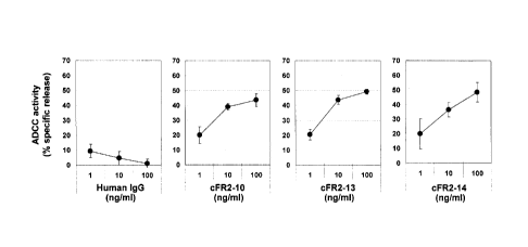
Figure 7 is a diagram showing the ADCC activity of
the human chimeric anti-FGFR2 antibodies (cFR2-10, cFR2-
13, and cFR2-14). 293T-lacZ cells expressing human FGFR2
IIIb were used as target cells, and human PBMC was used
as effector cells.
Figure 8 is a diagram showing the in vivo antitumor
activity of the human chimeric anti-FGFR2 antibodies
(cFR2-10, cFR2-13, and cFR2-14) against human stomach
cancer cell line SNU-16-transplanted nude mice. Figure
8A) shows the results for the cFR2-10 antibody. Figure
83) shows the results for the cFR2-13 antibody. Figure
80) shows the results for the cFR2-14 antibody.
Figure 9 shows the N-terminal amino acid sequence of
a band corresponding to the heavy chain of the rat anti-
FGFR2 antibody FR2-10 (SEQ ID NO: 1 of the Sequence
Listing).
Figure 10 shows the N-terminal amino acid sequence
of a band corresponding to the light chain of the rat
anti-FGFR2 antibody FR2-10 (SEQ ID NO: 2 of the Sequence
Listing).
Figure 11 shows the N-terminal amino acid sequence
of a band corresponding to the heavy chain of the rat
anti-FGFR2 antibody FR2-13 (SEQ ID NO: 3 of the Sequence
Listing).
FP1320s/(PN814663)/English translation of PCT specification (September 2014)
5106289-1-MCARVALHO
CA 02870126 2014-10-09
- 28 -
Figure 12 shows the N-terminal amino acid sequence
of a band corresponding to the light chain of the rat
anti-FGFR2 antibody FR2-13 (SEQ ID NO: 4 of the Sequence
Listing).
Figure 13 shows the N-terminal amino acid sequence
of a band corresponding to the heavy chain of the rat
anti-FGFR2 antibody FR2-14 (SEQ ID NO: 5 of the Sequence
Listing).
Figure 14 shows the N-terminal amino acid sequence
of a band corresponding to the light chain of the rat
anti-FGFR2 antibody FR2-14 (SEQ ID NO: 6 of the Sequence
Listing).
Figure 15 shows a primer for gene amplification of a
rat heavy chain (SEQ ID NO: 7 of the Sequence Listing).
Figure 16 shows a sequencing primer for the heavy
chain of FR2-10 (SEQ ID NO: 8 of the Sequence Listing).
Figure 17 shows a sequencing primer for the heavy
chain of FR2-13 (SEQ ID NO: 9 of the Sequence Listing).
Figure 18 shows a sequencing primer for the heavy
chain of FR2-14 (SEQ ID NO: 10 of the Sequence Listing).
Figure 19 shows the nucleotide sequence of a cDNA
encoding the heavy chain variable region of the rat anti-
FGFR2 antibody FR2-10 (SEQ ID NO: 11 of the Sequence
Listing).
Figure 20 shows the amino acid sequence of the heavy
chain variable region of the rat anti-FGFR2 antibody FR2-
(SEQ ID NO: 12 of the Sequence Listing).
FP1320s/(PN814663)/English translation of PCT specification (September 2014)
5106289-1-M CARVALHO
CA 02870126 2014-10-09
- 29 -
Figure 21 shows the nucleotide sequence of a cDNA
encoding the heavy chain variable region of the rat anti-
FGFR2 antibody FR2-13 (SEQ ID NO: 13 of the Sequence
Listing).
Figure 22 shows the amino acid sequence of the heavy
chain variable region of the rat anti-FGFR2 antibody FR2-
13 (SEQ ID NO: 14 of the Sequence Listing).
Figure 23 shows the nucleotide sequence of a cDNA
encoding the heavy chain variable region of the rat anti-
FGFR2 antibody FR2-14 (SEQ ID NO: 15 of the Sequence
Listing).
Figure 24 shows the amino acid sequence of the heavy
chain variable region of the rat anti-FGFR2 antibody FR2-
14 (SEQ ID NO: 16 of the Sequence Listing).
Figure 25 shows a primer for gene amplification of a
rat light chain (SEQ ID NO: 17 of the Sequence Listing).
Figure 26 shows a sequencing primer for a rat light
chain (SEQ ID NO: 18 of the Sequence Listing).
Figure 27 shows a sequencing primer for the light
chain of FR2-10 (SEQ ID NO: 19 of the Sequence Listing).
Figure 28 shows the nucleotide sequence of a cDNA
encoding the light chain variable region of the rat anti-
FGFR2 antibody FR2-10 (SEQ ID NO: 20 of the Sequence
Listing).
Figure 29 shows the amino acid sequence of the light
chain variable region of the rat anti-FGFR2 antibody FR2-
(SEQ ID NO: 21 of the Sequence Listing).
FP1320s/(PN814663)/Eng1ish translation of PCT specification (September 2014)
5106289A-MCARVALHO
CA 02870126 2014-10-09
- 30 -
Figure 30 shows a primer for gene amplification of
the rat FR2-13 or FR2-14 light chain (SEQ ID NO: 22 of
the Sequence Listing).
Figure 31 shows the nucleotide sequence of a cDNA
encoding the light chain variable region of the rat anti-
FGFR2 antibody FR2-13 (SEQ ID NO: 23 of the Sequence
Listing).
Figure 32 shows the amino acid sequence of the light
chain variable region of the rat anti-FGFR2 antibody FR2-
13 (SEQ ID NO: 24 of the Sequence Listing).
Figure 33 shows the nucleotide sequence of a cDNA
encoding the light chain variable region of the rat anti-
FGFR2 antibody FR2-14 (SEQ ID NO: 25 of the Sequence
Listing).
Figure 34 shows the amino acid sequence of the light
chain variable region of the rat anti-FGFR2 antibody FR2-
14 (SEQ ID NO: 26 of the Sequence Listing).
Figure 35 shows a DNA fragment comprising a DNA
sequence encoding the amino acids of a human K chain
secretory signal sequence and a human K chain constant
region (SEQ ID NO: 27 of the Sequence Listing).
Figure 36 shows a primer F for a light chain
expression vector (SEQ ID NO: 28 of the Sequence Listing).
Figure 37 shows a primer R for a light chain
expression vector (SEQ ID NO: 29 of the Sequence Listing).
Figure 38 shows a DNA fragment comprising a DNA
sequence encoding the amino acids of a human heavy chain
FP1320s/(PN814663)/English translation of PCT specification (September 2014)
5106289-1-MCARVALHO
CA 02870126 2014-10-09
- 31 -
signal sequence and a human IgG1 constant region (SEQ ID
NO: 30 of the Sequence Listing).
Figure 39 shows the nucleotide sequence of the light
chain of human chimeric FR2-10 (cFR2-10) (SEQ ID NO: 31
of the Sequence Listing). In this sequence, nucleotide
positions 1 to 60 represent a signal sequence, which is
usually not contained in the nucleotide sequences of most
of mature cFR2-10 light chains.
Figure 40 shows the amino acid sequence of the light
chain of human chimeric FR2-10 (cFR2-10) (SEQ ID NO: 32
of the Sequence Listing). In this sequence, amino acid
positions 1 to 20 represent a signal sequence, which is
usually not contained in the amino acid sequences of most
of mature cFR2-10 light chains.
Figure 41 shows a primer set F for the light chain
of human chimeric FR2-10 (SEQ ID NO: 33 of the Sequence
Listing).
Figure 42 shows a primer set R for the light chain
of human chimeric FR2-10 (SEQ ID NO: 34 of the Sequence
Listing).
Figure 43 shows the nucleotide sequence of the heavy
chain of human chimeric FR2-10 (cFR2-10) (SEQ ID NO: 35
of the Sequence Listing). In this sequence, nucleotide
positions 1 to 57 represent a signal sequence, which is
usually not contained in the nucleotide sequences of most
of mature cFR2-10 heavy chains.
FP1320s/(PN814663)/English translation of PCT specification (September 2014)
5106289-1-MCARVALHO
CA 02870126 2014-10-09
- 32 -
Figure 44 shows the amino acid sequence of the heavy
chain of human chimeric FR2-10 (cFR2-10) (SEQ ID NO: 36
of the Sequence Listing). In this sequence, amino acid
positions 1 to 19 represent a signal sequence, which is
usually not included in the amino acid sequence of most
of mature cFR2-10 heavy chains.
Figure 45 shows a primer set F for the heavy chain
of human chimeric FR2-10 (SEQ ID NO: 37 of the Sequence
Listing).
Figure 46 shows a primer set R for the heavy chain
of human chimeric FR2-10 (SEQ ID NO: 38 of the Sequence
Listing).
Figure 47 shows the nucleotide sequence of the light
chain of human chimeric FR2-13 (cFR2-13) (SEQ ID NO: 39
of the Sequence Listing). In this sequence, nucleotide
positions 1 to 60 represent a signal sequence, which is
usually not contained in the nucleotide sequences of most
of mature cFR2-13 light chains.
Figure 48 shows the amino acid sequence of the light
chain of human chimeric FR2-13 (cFR2-13) (SEQ ID NO: 40
of the Sequence Listing). In this sequence, amino acid
positions 1 to 20 represent a signal sequence, which is
usually not contained in the amino acid sequences of most
of mature cFR2-13 light chains.
Figure 49 shows a primer F for the light chain of
human chimeric FR2-13 (SEQ ID NO: 41 of the Sequence
Listing).
FP1320s/(PN814663)/English translation of PCT specification (September 2014)
5106289-1-MCARVALHO
CA 02870126 2014-10-09
- 33 -
Figure 50 shows a primer R for the light chain of
human chimeric FR2-13 (SEQ ID NO: 42 of the Sequence
Listing).
Figure 51 shows the nucleotide sequence of the heavy
chain of human chimeric FR2-13 (cFR2-13) (SEQ ID NO: 43
of the Sequence Listing). In this sequence, nucleotide
positions 1 to 57 represent a signal sequence, which is
usually not contained in the nucleotide sequences of most
of mature cFR2-13 heavy chains.
Figure 52 shows the amino acid sequence of the heavy
chain of human chimeric FR2-13 (cFR2-13) (SEQ ID NO: 44
of the Sequence Listing). In this sequence, amino acid
positions 1 to 19 represent a signal sequence, which is
usually not contained in the amino acid sequences of most
of mature cFR2-13 heavy chains.
Figure 53 shows a primer F for the heavy chain of
human chimeric FR2-13 (SEQ ID NO: 45 of the Sequence
Listing).
Figure 54 shows a primer R for the heavy chain of
human chimeric FR2-13 (SEQ ID NO: 46 of the Sequence
Listing).
Figure 55 shows the nucleotide sequence of the light
chain of human chimeric FR2-14 (cFR2-14) (SEQ ID NO: 47
of the Sequence Listing). In this sequence, nucleotide
positions 1 to 60 represent a signal sequence, which is
usually not contained in the nucleotide sequences of most
of mature cFR2-14 light chains.
FP1320s/(2N814663)/English translation of PCT specification (September 2014)
5106289-1-MCARVALHO
CA 02870126 2014-10-09
- 34 -
Figure 56 shows the amino acid sequence of the light
chain of human chimeric FR2-14 (cFR2-14) (SEQ ID NO: 48
of the Sequence Listing). In this sequence, amino acid
positions 1 to 20 represent a signal sequence, which is
usually not contained in the amino acid sequences of most
of mature cFR2-14 light chains.
Figure 57 shows a primer for the light chain of
human chimeric FR2-14 (SEQ ID NO: 49 of the Sequence
Listing).
Figure 58 shows the nucleotide sequence of the heavy
chain of human chimeric FR2-14 (cFR2-14) (SEQ ID NO: 50
of the Sequence Listing). In this sequence, nucleotide
positions 1 to 57 represent a signal sequence, which is
usually not contained in the nucleotide sequences of most
of mature cFR2-14 heavy chains.
Figure 59 shows the amino acid sequence of the heavy
chain of human chimeric FR2-14 (cFR2-14) (SEQ ID NO: 51
of the Sequence Listing). In this sequence, amino acid
positions 1 to 19 represent a signal sequence, which is
usually not contained in the amino acid sequences of most
of mature cFR2-14 heavy chains.
Figure 60 shows the amino acid sequence of the heavy
chain CDR1 of the rat anti-FGFR2 antibody FR2-10 (SEQ ID
NO: 52 of the Sequence Listing).
Figure 61 shows the amino acid sequence of the heavy
chain CDR2 of the rat anti-FGFR2 antibody FR2-10 (SEQ ID
NO: 53 of the Sequence Listing).
FP1320s/(PN814663)/English translation of PCT specification (September 2014)
5106289-1-MCARVALHO
CA 02870126 2014-10-09
- 35 -
Figure 62 shows the amino acid sequence of the heavy
chain CDR3 of the rat anti-FGFR2 antibody FR2-10 (SEQ ID
NO: 54 of the Sequence Listing).
Figure 63 shows the amino acid sequence of the heavy
chain CDR1 of the rat anti-FGFR2 antibody FR2-13 (SEQ ID
NO: 55 of the Sequence Listing).
Figure 64 shows the amino acid sequence of the heavy
chain CDR2 of the rat anti-FGFR2 antibody FR2-13 (SEQ ID
NO: 56 of the Sequence Listing).
Figure 65 shows the amino acid sequence of the heavy
chain CDR3 of the rat anti-FGFR2 antibody FR2-13 (SEQ ID
NO: 57 of the Sequence Listing).
Figure 66 shows the amino acid sequence of the heavy
chain CDR1 of the rat anti-FGFR2 antibody FR2-14 (SEQ ID
NO: 58 of the Sequence Listing).
Figure 67 shows the amino acid sequence of the heavy
chain CDR2 of the rat anti-FGFR2 antibody FR2-14 (SEQ ID
NO: 59 of the Sequence Listing).
Figure 68 shows the amino acid sequence of the heavy
chain CDR3 of the rat anti-FGFR2 antibody FR2-14 (SEQ ID
NO: 60 of the Sequence Listing).
Figure 69 shows the amino acid sequence of the light
chain CDR1 of the rat anti-FGFR2 antibody FR2-10 (SEQ ID
NO: 61 of the Sequence Listing).
Figure 70 shows the amino acid sequence of the light
chain CDR2 of the rat anti-FGFR2 antibody FR2-10 (SEQ ID
NO: 62 of the Sequence Listing).
FP1320s/(PN814663)/English translation of PCT specification (September 2014)
5106289-1-MCARVALHO
CA 02870126 2014-10-09
- 36 -
Figure 71 shows the amino acid sequence of the light
chain CDR3 of the rat anti-FGFR2 antibody FR2-10 (SEQ ID
NO: 63 of the Sequence Listing).
Figure 72 shows the amino acid sequence of the light
chain CDR1 of the rat anti-FGFR2 antibody FR2-13 (SEQ ID
NO: 64 of the Sequence Listing).
Figure 73 shows the amino acid sequence of the light
chain CDR2 of the rat anti-FGFR2 antibody FR2-13 (SEQ ID
NO: 65 of the Sequence Listing).
Figure 74 shows the amino acid sequence of the light
chain CDR3 of the rat anti-FGFR2 antibody FR2-13 (SEQ ID
NO: 66 of the Sequence Listing).
Figure 75 shows the amino acid sequence of the light
chain CDR1 of the rat anti-FGFR2 antibody FR2-14 (SEQ ID
NO: 67 of the Sequence Listing).
Figure 76 shows the amino acid sequence of the light
chain CDR2 of the rat anti-FGFR2 antibody FR2-14 (SEQ ID
NO: 68 of the Sequence Listing).
Figure 77 shows the amino acid sequence of the light
chain CDR3 of the rat anti-FGFR2 antibody FR2-14 (SEQ ID
NO: 69 of the Sequence Listing).
Figure 78 shows the amino acid sequence of human
FGFR2 IIIb (SEQ ID NO: 70 of the Sequence Listing).
Figure 79 shows the amino acid sequence of human
FGFR2 IIIc (SEQ ID NO: 71 of the Sequence Listing).
Figure 80 shows the nucleotide sequence of hFR2-
14 Ll (SEQ ID NO: 72 of the Sequence Listing). In this
FP1320s/(PN814663)/English translation of PCT specification (September 2014)
5106289-1-MCARVALHO
CA 02870126 2014-10-09
- 37 -
sequence, nucleotide positions 1 to 60 represent a signal
sequence, which is usually not included in the nucleotide
sequence of most of mature light chains hFR2-14_L1.
Figure 81 shows the amino acid sequence of hFR2-
14 Ll (SEQ ID NO: 73 of the Sequence Listing). In this
sequence, amino acid positions 1 to 20 represent a signal
sequence, which is usually not included in the amino acid
sequence of most of mature light chains hFR2-14_L1.
Figure 82 shows the nucleotide sequence of hFR2-
14 H1 (SEQ ID NO: 74 of the Sequence Listing). In this
sequence, nucleotide positions 1 to 57 represent a signal
sequence, which is usually not included in the nucleotide
sequence of most of mature heavy chains hFR2-14_H1.
Figure 83 shows the amino acid sequence of hFR2-
14 H1 (SEQ ID NO: 75 of the Sequence Listing). In this
sequence, amino acid positions 1 to 19 represent a signal
sequence, which is usually not included in the amino acid
sequence of most of mature heavy chains hFR2-14_Hl.
Figure 84 shows the nucleotide sequence of hFR2-
14 H2 (SEQ ID NO: 76 of the Sequence Listing). In this
sequence, nucleotide positions 1 to 57 represent a signal
sequence, which is usually not included in the nucleotide
sequence of most of mature heavy chains hFR2-14_H2.
Figure 85 shows the amino acid sequence of hFR2-
14 H2 (SEQ ID NO: 77 of the Sequence Listing). In this
sequence, amino acid positions 1 to 19 represent a signal
FP13205/(PN814663)/English translation of PCT specification (September 2014)
5106289-1-MCARVALHO
CA 02870126 2014-10-09
- 38 -
sequence, which is usually not included in the amino acid
sequence of most of mature heavy chains hFR2-14_H2.
Figure 86 shows the nucleotide sequence of hFR2-
14 H3 (SEQ ID NO: 78 of the Sequence Listing). In this
sequence, nucleotide positions 1 to 57 represent a signal
sequence, which is usually not included in the nucleotide
sequence of most of mature heavy chains hFR2-14_H3.
Figure 87 shows the amino acid sequence of hFR2-
14 H3 (SEQ ID NO: 79 of the Sequence Listing). In this
sequence, amino acid positions 1 to 19 represent a signal
sequence, which is usually not included in the amino acid
sequence of most of mature heavy chains hFR2-14_H3.
Figure 88 shows the nucleotide sequence of hFR2-
14 H4 (SEQ ID NO: 80 of the Sequence Listing). In this
sequence, nucleotide positions 1 to 57 represent a signal
sequence, which is usually not included in the nucleotide
sequence of most of mature heavy chains hFR2-14_H4.
Figure 89 shows the amino acid sequence of hFR2-
14 H4 (SEQ ID NO: 81 of the Sequence Listing). In this
sequence, amino acid positions 1 to 19 represent a signal
sequence, which is usually not included in the amino acid
sequence of most of mature heavy chains hFR2-14_H4.
Figure 90 shows the nucleotide sequence of hFR2-
14 H5 (SEQ ID NO: 82 of the Sequence Listing). In this
sequence, nucleotide positions 1 to 57 represent a signal
sequence, which is usually not included in the nucleotide
sequence of most of mature heavy chains hFR2-14_H5.
FP1320s/(PN814663)/English translation of PCT specification (September 2014)
5106289-1-MCARVALHO
CA 02870126 2014-10-09
- 39 -
Figure 91 shows the amino acid sequence of hFR2-
14 H5 (SEQ ID NO: 83 of the Sequence Listing). In this
sequence, amino acid positions 1 to 19 represent a signal
sequence, which is usually not included in the amino acid
sequence of most of mature heavy chains hFR2-14 H5.
Figure 92 shows the nucleotide sequence of hFR2-
14 H6 (SEQ ID NO: 84 of the Sequence Listing). In this
sequence, nucleotide positions 1 to 57 represent a signal
sequence, which is usually not included in the nucleotide
sequence of most of mature heavy chains hFR2-14 H6.
Figure 93 shows the amino acid sequence of hFR2-
14 H6 (SEQ ID NO: 85 of the Sequence Listing). In this
sequence, amino acid positions 1 to 19 represent a signal
sequence, which is usually not included in the amino acid
sequence of most of mature heavy chains hFR2-14_H6.
Figure 94 shows the nucleotide sequence of hFR2-
14 H7 (SEQ ID NO: 86 of the Sequence Listing). In this
sequence, nucleotide positions 1 to 57 represent a signal
sequence, which is usually not included in the nucleotide
sequence of most of mature heavy chains hFR2-14_H7.
Figure 95 shows the amino acid sequence of hFR2-
14 H7 (SEQ ID NO: 87 of the Sequence Listing). In this
sequence, amino acid positions 1 to 19 represent a signal
sequence, which is usually not included in the amino acid
sequence of most of mature heavy chains hFR2-14_H7.
Figure 96 shows the nucleotide sequence of hFR2-
14 H8 (SEQ ID NO: 88 of the Sequence Listing). In this
FP1320s/(PN814663)/English translation of PCT specification (September 2014)
5106289-1-MCARVALHO
CA 02870126 2014-10-09
- 40 -
sequence, nucleotide positions 1 to 57 represent a signal
sequence, which is usually not included in the nucleotide
sequence of most of mature heavy chains hFR2-14_H8.
Figure 97 shows the amino acid sequence of hFR2-
14 H8 (SEQ ID NO: 89 of the Sequence Listing). In this
sequence, amino acid positions 1 to 19 represent a signal
sequence, which is usually not included in the amino acid
sequence of most of mature heavy chains hFR2-14_H8.
Figure 98 shows the nucleotide sequence of hFR2-
14 H9 (SEQ ID NO: 90 of the Sequence Listing). In this
sequence, nucleotide positions 1 to 57 represent a signal
sequence, which is usually not included in the nucleotide
sequence of most of mature heavy chains hFR2-14_H9.
Figure 99 shows the amino acid sequence of hFR2-
14 H9 (SEQ ID NO: 91 of the Sequence Listing). In this
sequence, amino acid positions 1 to 19 represent a signal
sequence, which is usually not included in the amino acid
sequence of most of mature heavy chains hFR2-14_H9.
Figure 100 shows the nucleotide sequence of hFR2-
14 H10 (SEQ ID NO: 92 of the Sequence Listing). In this
sequence, nucleotide positions 1 to 57 represent a signal
sequence, which is usually not included in the nucleotide
sequence of most of mature heavy chains hFR2-14_H10.
Figure 101 shows the amino acid sequence of hFR2-
14 H10 (SEQ ID NO: 93 of the Sequence Listing). In this
sequence, amino acid positions 1 to 19 represent a signal
FP1320s/(PN814663)/Eng1ish translation of PCT specification (September 2014)
5106289-1-MCARVALHO
CA 02870126 2014-10-09
- 41 -
sequence, which is usually not included in the amino acid
sequence of most of mature heavy chains hFR2-14_H10.
Figure 102 shows the nucleotide sequence of hFR2-
14 H11 (SEQ ID NO: 94 of the Sequence Listing). In this
sequence, nucleotide positions 1 to 57 represent a signal
sequence, which is usually not included in the nucleotide
sequence of most of mature heavy chains hFR2-14_H11.
Figure 103 shows the amino acid sequence of hFR2-
14 H11 (SEQ ID NO: 95 of the Sequence Listing). In this
sequence, amino acid positions 1 to 19 represent a signal
sequence, which is usually not included in the amino acid
sequence of most of mature heavy chains hFR2-14_H11.
Figure 104 shows the nucleotide sequence of hFR2-
14 H12 or hFR2-14 H19 (SEQ ID NO: 96 of the Sequence
Listing). In this sequence, nucleotide positions 1 to 57
represent a signal sequence, which is usually not
included in the nucleotide sequence of most of mature
heavy chains hFR2-14_H12 or hFR2-14_H19.
Figure 105 shows the amino acid sequence of hFR2-
14 H12 or hFR2-14 H19 (SEQ ID NO: 97 of the Sequence
Listing). In this sequence, amino acid positions 1 to 19
represent a signal sequence, which is usually not
included in the amino acid sequence of most of mature
heavy chains hFR2-14_H12 or hFR2-14_H19.
Figure 106 shows the nucleotide sequence of hFR2-
14 H13 (SEQ ID NO: 98 of the Sequence Listing). In this
sequence, nucleotide positions 1 to 57 represent a signal
FP1320s/(PN814663)/English translation of PCT specification (September 2014)
5106289-1-MCARVALHO
CA 02870126 2014-10-09
- 42 -
sequence, which is usually not included in the nucleotide
sequence of most of mature heavy chains hFR2-14_H13.
Figure 107 shows the amino acid sequence of hFR2-
14 H13 (SEQ ID NO: 99 of the Sequence Listing). In this
sequence, amino acid positions 1 to 19 represent a signal
sequence, which is usually not included in the amino acid
sequence of most of mature heavy chains hFR2-14_H13.
Figure 108 shows the nucleotide sequence of hFR2-
14 H14 (SEQ ID NO: 100 of the Sequence Listing). In this
sequence, nucleotide positions 1 to 57 represent a signal
sequence, which is usually not included in the nucleotide
sequence of most of mature heavy chains hFR2-14 H14.
Figure 109 shows the amino acid sequence of hFR2-
14 H14 (SEQ ID NO: 101 of the Sequence Listing). In this
sequence, amino acid positions 1 to 19 represent a signal
sequence, which is usually not included in the amino acid
sequence of most of mature heavy chains hFR2-14_H14.
Figure 110 shows the nucleotide sequence of hFR2-
14 H15 (SEQ ID NO: 102 of the Sequence Listing). In this
sequence, nucleotide positions 1 to 57 represent a signal
sequence, which is usually not included in the nucleotide
sequence of most of mature heavy chains hFR2-14_H15.
Figure 111 shows the amino acid sequence of hFR2-
14 H15 (SEQ ID NO: 103 of the Sequence Listing). In this
sequence, amino acid positions 1 to 19 represent a signal
sequence, which is usually not included in the amino acid
sequence of most of mature heavy chains hFR2-14_H15.
FP1320s/(PN814663)/English translation of PCT specification (September 2014)
5106289-1-MCARVALHO
CA 02870126 2014-10-09
- 43 -
Figure 112 shows the nucleotide sequence of hFR2-
14 H16 (SEQ ID NO: 104 of the Sequence Listing). In this
sequence, nucleotide positions 1 to 57 represent a signal
sequence, which is usually not included in the nucleotide
sequence of most of mature heavy chains hFR2-14_H16.
Figure 113 shows the amino acid sequence of hFR2-
14 H16 (SEQ ID NO: 105 of the Sequence Listing). In this
sequence, amino acid positions 1 to 19 represent a signal
sequence, which is usually not included in the amino acid
sequence of most of mature heavy chains hFR2-14 H16.
Figure 114 shows the nucleotide sequence of hFR2-
14 H17 (SEQ ID NO: 106 of the Sequence Listing). In this
sequence, nucleotide positions 1 to 57 represent a signal
sequence, which is usually not included in the nucleotide
sequence of most of mature heavy chains hFR2-14 H17.
Figure 115 shows the amino acid sequence of hFR2-
14 H17 (SEQ ID NO: 107 of the Sequence Listing). In this
sequence, amino acid positions 1 to 19 represent a signal
sequence, which is usually not included in the amino acid
sequence of most of mature heavy chains hFR2-14_H17.
Figure 116 shows the nucleotide sequence of hFR2-
14 H18 (SEQ ID NO: 108 of the Sequence Listing). In this
sequence, nucleotide positions 1 to 57 represent a signal
sequence, which is usually not included in the nucleotide
sequence of most of mature heavy chains hFR2-14_H18.
Figure 117 shows the amino acid sequence of hFR2-
14 H18 (SEQ ID NO: 109 of the Sequence Listing). In this
FP1320s/(PN814663)/English translation of PCT specification (September 2014)
5106289-1-MCARVALHO
CA 02870126 2014-10-09
- 44 -
sequence, amino acid positions 1 to 19 represent a signal
sequence, which is usually not included in the amino acid
sequence of most of mature heavy chains hFR2-14_H18.
Figure 118 shows a primer VH3A-F for an hFR2-14_H2
type heavy chain (SEQ ID NO: 110 of the Sequence Listing).
Figure 119 shows a primer VH3A-R for an hFR2-14_H2
type heavy chain (SEQ ID NO: 111 of the Sequence Listing).
Figure 120 shows a primer D23fw for gene
amplification of D2 (SEQ ID NO: 112 of the Sequence
Listing).
Figure 121 shows a primer D23ry for gene
amplification of D2 (SEQ ID NO: 113 of the Sequence
Listing).
Figure 122 is a diagram showing results of assaying
the binding activity of 4 types of humanized anti-FGFR2
antibodies (hFR2-14 Hl/L1 to hFR2-14 H4/L1) and the human
chimeric anti-FGFR2 antibody (cFR2-14) against each human
FGFR2 variant protein using Biacore. Each antibody was
expressed in 293F cells and purified for use in the assay.
Figure 123 is a diagram showing results of assaying
the binding activity of 15 types of humanized anti-FGFR2
antibodies (hFR2-14 H3/L1 and hFR2-14 H5/L1 to hFR2-
_
14 H18/L1) against a human FGFR2 IIIc variant protein
using Biacore. Each antibody was expressed in 293F cells,
and its culture supernatant was used in the assay.
Figure 124 is a diagram showing results of testing
the human FGFR2-selective binding activity of 3 types of
FP13202/(PN814663)/English translation of PCT specification (September 2014)
5106289-1-MCARVALHO
CA 02870126 2014-10-09
- 45 -
humanized anti-FGFR2 antibodies (hFR2-14 H3/L1, hFR2-
_
14 H8/L1, and hFR2-14 H12/L1) by Cell-ELISA.
Figure 125A is a diagram showing the thermograms of
types of humanized anti-FGFR2 antibodies.
Figure 125B is a diagram showing the thermograms of
5 types of humanized anti-FGFR2 antibodies.
Figure 125C is a diagram showing the Tm values of 10
types of humanized anti-FGFR2 antibodies.
Figure 126 is a diagram showing the KD values of
hFR2-14 Hl/L1, hFR2-14 H2/L1, hFR2-14 H3/L1, hFR2-
_
14 H4/L1, hFR2-14 H5/L1, hFR2-14 H8/L1, hFR2-14 H9/L1,
hFR2-14 H11/L1, hFR2-14 H12/L1, and hFR2-14 H19/L1
antibody analytes for an antigen before and after
degradation.
Figure 127A is a diagram showing the signal-
neutralizing activity of the humanized anti-FGFR2
antibodies (hFR2-14 Hl/L1, hFR2-14 H2/L1, hFR2-14 H3/L1,
and hFR2-14 H4/L1) and the human chimeric anti-FGFR2
antibody (cFR2-14) against human FGFR2 IIIb by Elkl
trans-reporter assay.
Figure 127B is a diagram showing the signal-
neutralizing activity of the humanized anti-FGFR2
antibodies (hFR2-14 Hl/L1, hFR2-14 H2/L1, hFR2-14 H3/L1,
and hFR2-14 H4/L1) and the human chimeric anti-FGFR2
antibody (cFR2-14) against human FGFR2 IIIc by Elkl
trans-reporter assay.
FP1320s/(PN814663)/English translation of PCT specification (September 2014)
5106289-1-MCARVALHO
CA 02870126 2014-10-09
- 46 -
Figure 128A is a diagram showing the signal-
neutralizing activity of the humanized anti-FGFR2
antibodies (hFR2-14 H3/L1, hFR2-14 H5/L1, hFR2-14 H6/L1,
hFR2-14 H7/L1, and hFR2-14 H8/L1) against human FGFR2
IIIb by Elkl trans-reporter assay.
Figure 128B is a diagram showing the signal-
neutralizing activity of the humanized anti-FGFR2
antibodies (hFR2-14 H9/L1, hFR2-14 H10/L1, hFR2-14 H11/L1,
hFR2-14 H12/L1, and hFR2-14 H13/L1) against human FGFR2
IIIb by Elkl trans-reporter assay.
Figure 1280 is a diagram showing the signal-
neutralizing activity of the humanized anti-FGFR2
antibodies (hFR2-14 H14/L1, hFR2-14 H15/L1, hFR2-
_
14 H16/L1, hFR2-14 H17/L1, and hFR2-14 H18/L1) against
human FGFR2 IIIb by Elkl trans-reporter assay.
Figure 129A is a diagram showing the signal-
neutralizing activity of the humanized anti-FGFR2
antibodies (hFR2-14 H12/L1 and hFR2-14 H19/L1) against
human FGFR2 IIIb by Elkl trans-reporter assay.
Figure 129B is a diagram showing the signal-
neutralizing activity of the humanized anti-FGFR2
antibodies (hFR2-14 H12/L1 and hFR2-14 H19/L1) against
human FGFR2 IIIc by Elkl trans-reporter assay.
Figure 130A is a diagram showing the ADCC activity
of the humanized anti-FGFR2 antibodies (hFR2-14 Hl/L1 and
hFR2-14 H2/L1). 293T-lacZ cells expressing human FGFR2
FP1320s/(PN814663)/English translation of PCT specification (September 2014)
5106289-1-MCARVALHO
CA 02870126 2014-10-09
- 47 -
IIIb were used as target cells, and human PBMC was used
as effector cells.
Figure 1303 is a diagram showing the ADCC activity
of the humanized anti-FGFR2 antibodies (hFR2-14 H3/L1 and
hFR2-14 H4/L1) and the human chimeric anti-FGFR2 antibody
(cFR2-14). 293T-lacZ cells expressing human FGFR2 IIIb
were used as target cells, and human PBMC was used as
effector cells.
Figure 131 is a diagram showing the ADCC activity of
the humanized anti-FGFR2 antibodies (hFR2-14 H3/L1, hFR2-
_
14 H8/L1, and hFR2-14 H12/L1). NCI-H716 cells expressing
human FGFR2 were used as target cells, and human PBMC was
used as effector cells.
Figure 132 is a diagram showing the ADCC activity of
the humanized anti-FGFR2 antibodies (hFR2-14 H12/L1 and
hFR2-14 H19/L1). NCI-H716 cells (Figure 132A)), SNU-16
cells (Figure 132B)), or KATO III cells (Figure 132C))
expressing human FGFR2 were used as target cells, and
human PBMC was used as effector cells.
Figure 133 is a diagram showing the ADCP activity of
the humanized anti-FGFR2 antibodies (hFR2-14 H12/L1 and
hFR2-14 H19/L1). NCI-H716 cells (Figure 133A)) or KATO
III cells (Figure 133B)) expressing human FGFR2 were used
as target cells, and macrophage-like cells differentiated
from human PBMC were used as effector cells.
Figure 134A is a diagram showing the in vivo
antitumor activity of the humanized anti-FGFR2 antibody
FP1320s/(PN814663)/English translation of PCT specification (September 2014)
5106289-14MCARVALHO
CA 02870126 2014-10-09
- 48 -
hFR2-14 Hl/L1 against human stomach cancer cell line SNU-
_
16-transplanted nude mice.
Figure 134B is a diagram showing the in vivo
antitumor activity of the humanized anti-FGFR2 antibody
hFR2-14 H2/L1 against human stomach cancer cell line SNU-
_
16-transplanted nude mice.
Figure 1340 is a diagram showing the in vivo
antitumor activity of the humanized anti-FGFR2 antibody
hFR2-14 H3/L1 against human stomach cancer cell line SNU-
_
16-transplanted nude mice.
Figure 134D is a diagram showing the in vivo
antitumor activity of the humanized anti-FGFR2 antibody
hFR2-14 H4/L1 against human stomach cancer cell line SNU-
_
16-transplanted nude mice.
Figure 135A is a diagram showing the in vivo
antitumor activity of the humanized anti-FGFR2 antibody
hFR2-14 H5/L1 against human stomach cancer cell line SNU-
_
16-subcutaneously transplanted models.
Figure 1355 is a diagram showing the in vivo
antitumor activity of the humanized anti-FGFR2 antibody
hFR2-14 H8/L1 against human stomach cancer cell line SNU-
16-subcutaneously transplanted models.
Figure 1350 is a diagram showing the in vivo
antitumor activity of the humanized anti-FGFR2 antibody
hFR2-14 H9/L1 against human stomach cancer cell line SNU-
_
16-subcutaneously transplanted models.
FP1320s/(PN814663)/English translation of PCT specification (September 2014)
5106289-1-MCARVALHO
CA 02870126 2014-10-09
- 49 -
Figure 135D is a diagram showing the in vivo
antitumor activity of the humanized anti-FGFR2 antibody
hFR2-14 H11/L1 against human stomach cancer cell line
SNU-16-subcutaneously transplanted models.
Figure 135E is a diagram showing the in vivo
antitumor activity of the humanized anti-FGFR2 antibody
hFR2-14 H12/L1 against human stomach cancer cell line
SNU-16-subcutaneously transplanted models.
Figure 135F is a diagram showing the in vivo
antitumor activity of the humanized anti-FGFR2 antibody
hFR2-14 H19/L1 against human stomach cancer cell line
SNU-16-subcutaneously transplanted models.
Figure 136 is a diagram showing the in vivo
antitumor activity of the humanized anti-FGFR2 antibodies
(hFR2-14 H12/L1 and hFR2-14 H19/L1) against human
colorectal cancer cell line NCI-H716 tumor block models.
Figure 136(A) shows the results for hFR2-14 H12/L1.
Figure 136(B) shows the results for hFR2-14 H19/L1.
Figure 137 is a diagram showing the in vivo
antitumor activity of the humanized anti-FGFR2 antibodies
(hFR2-14 H12/L1 and hFR2-14 H19/L1) against human
colorectal cancer cell line NCI-H716-luc-peritoneally
disseminated models. Figure 137(A) shows the results
showing luciferase activity. Figure 137(B) shows the
results showing survival rates.
Figure 138 is a diagram showing a ribbon model of an
FGFR2D2/H3L1Fab complex. FGFR2D2 is shown at the lower
FP1320s/(2N814663)/English translation of PCT specification (September 2014)
5106289A-MCARVALHO
CA 02870126 2014-10-09
- 50 -
left. The H chain (dark gray) of H3L1Fab is shown from
the center to the upper right. The L chain of H3L1Fab is
shown at the right thereof.
Figure 139 is a diagram showing the superposition of
FGFR2/FGF1 and FGFR2D2/H3L1Fab. FGFRD3 (IgD3) is shown
at the lower center. FGF1 (dark gray) is shown above
FGFRD3. FGFRD2 is shown above FGF1. The H chain (dark
gray) and L chain of H3L1Fab are each shown at the upper
right of FGFRD2.
Figure 140 is a diagram showing results of
immunostainingblocks of 293a cells forced to express each
molecule of the FGFR family, using the rat antibody FR2-
10.
Figure 141 is a diagram showing results of
immunostaining blocks of SNU-16 cells (Figures 141A and
141D), KATO III cells (Figures 141B and 141E), and NCI-
H716 cells (Figures 141C and 141F). Figures 141A to 141C
show the results obtained using the rat antibody FR2-10.
Figures 141D to 141F show the results obtained using a
commercially available antibody.
Figure 142 is a diagram showing results of
immunostaining xenograft tumor samples of SNU-16 cells
(Figure 142A), KATO III cells (Figure 142B), and NCI-H716
cells (Figure 1420) using the rat antibody FR2-10.
Figure 143 is a diagram showing the activity of
inhibiting the binding of a ligand FGF7 to its receptor
FP1320s/(PN814663)/English translation of PCT specification (September 2014)
5106289-14MCARVALHO
CA 02870126 2014-10-09
- 51 -
FGFR2 by the cFR2-10, hFR2-14_H12/L1, and hFR2-14_H19/L1
antibodies.
Description of Embodiments
1. Definitions
In the present invention, the term "gene" means a
nucleotide comprising a nucleotide sequence encoding the
amino acids of a protein, or its complementary strand.
The "gene" is meant to include, for example, a
polynucleotide, an oligonucleotide, DNA, mRNA, cDNA, and
cRNA as the nucleotide comprising a nucleotide sequence
encoding the amino acids of a protein, or its
complementary strand. Such a gene is a single-stranded,
double-stranded, or triple or more stranded nucleotide.
The "gene" is also meant to include an association of DNA
and RNA strands, a mixture of ribonucleotides (RNAs) and
deoxyribonucleotides (DNAs) on one nucleotide strand, and
a double-stranded or triple or more stranded nucleotide
comprising such a nucleotide strand. Examples of the
"FGFR2 gene" of the present invention can include DNA,
mRNA, cDNA, and cRNA comprising a nucleotide sequence
encoding the amino acid sequence of the FGFR2 protein.
In the present invention, the term "nucleotide" has
the same meaning as a "nucleic acid" and is also meant to
include, for example, DNA, RNA, a probe, an
oligonucleotide, a polynucleotide, and a primer. Such a
nucleotide is a single-stranded, double-stranded, or
FP1320s/(PN814663)/English translation of PCT specification (September 2014)
5106289-14MCARVALHO
CA 02870126 2014-10-09
- 52 -
triple or more stranded nucleotide. The "nucleotide" is
also meant to include an association of DNA and RNA
strands, a mixture of ribonucleotides (RNAs) and
deoxyribonucleotides (DNAs) on one nucleotide strand, and
an associate of two strands or three or more strands
comprising such a nucleotide strand.
In the present invention, the terms "polypeptide",
"peptide", and "protein" have the same meaning.
In the present invention, the term "antigen" has the
same meaning as "immunogen".
In the present invention, the term "cell" also
includes, for example, various cells derived from
individual animals, subcultured cells, primary cultured
cells, cell lines, recombinant cells, and microbial cells.
In the present invention, antibodies recognizing
FGFR2, FGFR2 IIIb, FGFR2 IIIc, FGFR3, FGFRs, and the like
are also referred to as an "anti-FGFR2 antibody", an
"anti-FGFR2 IIIb antibody", an "anti-FGFR2 IIIc antibody",
an "anti-FGFR3 antibody", and an "anti-FGFRs antibody",
respectively. These antibodies include chimeric
antibodies, humanized antibodies, human antibodies, and
the like.
In the present invention, the term "functional
fragment of the antibody" means an antibody fragment that
exerts at least a portion of functions exerted by the
original antibody. Examples of the "functional fragment
of the antibody" can include, but are not limited to, Fab,
FP1320s/(PN814663)/English translation of PCT specification (September 2014)
5106289-1-MCARVALHO
CA 02870126 2014-10-09
- 53 -
F(ab')2, scFv, Fab', and single chain immunoglobulin.
Such a functional fragment of the antibody may be
obtained by treating a full-length molecule of the
antibody protein with an enzyme such as papain or pepsin
or may be a recombinant protein produced in an
appropriate host cell using a recombinant gene.
In the present invention, the "site" to which an
antibody binds, i.e., the "site" recognized by an
antibody, means a partial peptide or partial conformation
on an antigen bound or recognized by the antibody. In
the present invention, such a site is also referred to as
an epitope or an antibody binding site. Examples of the
site on the FGFR2 protein bound or recognized by the
anti-FGFR2 antibody of the present invention can include
a partial peptide or partial conformation on the FGFR2
protein.
The heavy and light chains of an antibody molecule
are known to each have three complementarity determining
regions (CDRs). The complementarity determining regions
are also called hypervariable domains. These regions are
located in the variable regions of the antibody heavy and
light chains. These sites have a particularly highly
variable primary structure and are usually separated at
three positions on the respective primary structures of
heavy and light chain polypeptide strands. In the
present invention, the complementarity determining
regions of the antibody are referred to as CDRH1, CDRH2,
FP1320s/(PN814663)/English translation of PCT specification (September 2014)
5106289-1-MCARVALHO
CA 02870126 2014-10-09
- 54 -
and CDRH3 from the amino terminus of the heavy chain
amino acid sequence for the complementarity determining
regions of the heavy chain and as CDRL1, CDRL2, and CDRL3
from the amino terminus of the light chain amino acid
sequence for the complementarity determining regions of
the light chain. These sites are proximal to each other
on the three-dimensional structure and determine
specificity for the antigen to be bound.
In the present invention, the term "antibody mutant"
means a polypeptide that has an amino acid sequence
derived from the amino acid sequence of the original
antibody by the substitution, deletion, addition, and/or
insertion (hereinafter, collectively referred to as a
"mutation") of amino acid(s) and binds to the FGFR2
protein of the present invention. The number of mutated
amino acids in such an antibody mutant is 1 to 2, 1 to 3,
1 to 4, 1 to 5, 1 to 6, 1 to 7, 1 to 8, 1 to 9, 1 to 10,
1 to 12, 1 to 15, 1 to 20, 1 to 25, 1 to 30, 1 to 40, or
1 to 50. Such an antibody mutant is also encompassed by
the "antibody" of the present invention.
In the present invention, the term "several" in "1
to several" refers to 3 to 10.
Examples of activities or properties exerted by the
antibody of the present invention can include biological
activities or physicochemical properties and can
specifically include various biological activities,
binding activity against an antigen or an epitope,
FP1320s/(PN814663)/English translation of PCT specification (September 2014)
5106289-1-MCARVALHO
CA 02870126 2014-10-09
- 55 -
stability during production or storage, and thermal
stability.
In the present invention, the phrase "hybridizing
under stringent conditions" means hybridization under
conditions involving hybridization at 65 C in a solution
containing 5 x SSC, followed by washing at 65 C for 20
minutes in an aqueous solution containing 2 x SSC-0.1%
SDS, at 65 C for 20 minutes in an aqueous solution
containing 0.5 x SSC-0.1% SDS, and at 65 C for 20 minutes
in an aqueous solution containing 0.2 x SSC-0.1% SDS, or
hybridization under conditions equivalent thereto. SSC
means an aqueous solution of 150 mM NaCl-15 mM sodium
citrate, and n x SSC means SSC with an n-fold
concentration.
In the present invention, the term "cytotoxicity"
refers to some pathological change brought about to cells
and means not only direct trauma but every structural or
functional damage to cells, including DNA cleavage,
formation of base dimers, chromosomal break, damage on
mitotic apparatus, and reduction in the activities of
various enzymes.
In the present invention, the term "cytotoxic
activity" means activity that causes the cytotoxicity
mentioned above.
In the present invention, the term "antibody
dependent cellular cytotoxic activity", also called "ADCC
activity", means the effect or activity of damaging
PP1320s/(PN814663)/English translation of PCT specification (September 2014)
5106289-1-MCARVALHO
CA 02870126 2014-10-09
- 56 -
target cells such as tumor cells by NK cells via
antibodies.
In the present invention, the term "antibody
dependent cell phagocytosis activity", also called "ADCP
activity", means the effect or activity of englobing
target cells such as tumor cells by monocyte or
macrophage cells via antibodies. This activity is also
referred to as "antibody dependent phagocytic effect or
activity".
In the present invention, the term "complement
dependent cytotoxic activity", also called "CDC activity",
means the effect or activity of damaging target cells
such as tumor cells by complement via antibodies.
In the present invention, the term "cancer" has the
same meaning as "tumor".
In the present invention, the term
"immunohistochemistry (IHC)" means a histological
(histochemical) approach of detecting an antigen in a
tissue preparation. The immunohistochemistry is
synonymous with an "immune antibody method" and has the
same meaning as "immunostaining".
2. Antigenic protein
(2-1) Properties
FGFRs are receptor proteins that bind to fibroblast
growth factors (FGFs). In the present invention, FGFRs
are derived from vertebrates, preferably mammals, more
FP1320s/(PN814663)/English translation of PCT specification (September 2014)
5106289-1-MCARVALHO
CA 02870126 2014-10-09
- 57 -
preferably humans. Human FGFs and FGFRs are classified
into 22 FGFs (FGF1 to FGF14 and FGF16 to FGF23) and 4
FGFRs (FGFR1 to FGFR4) having a tyrosine kinase domain,
respectively. These FGFRs are each composed of an
extracellular region comprising a ligand binding site
composed of 2 or 3 immunoglobulin-like domains (IgD1 to
IgD3), a single-pass transmembrane region, and an
intracellular region comprising the tyrosine kinase
domain. Of them, FGFR1, FGFR2, and FGFR3 each have two
splicing variants called IIIb and IIIc. These isoforms
differ in the sequence of approximately 50 amino acids in
the latter half of IgD3 and exhibit distinctive tissue
distribution and ligand specificity. FGFRs have the
following activities: (1) binding to FGFs; (2) this
binding dimerizes the FGFRs; (3) this dimerization
phosphorylates the FGFRs at their particular tyrosine
residues; (4) this phosphorylation promotes the
recruitment of adaptor proteins such as FGFR substrate 2a
(FRS2a); and (5) this transduces signals generated by FGF
stimulation to cells or tissues expressing the FGFRs or
activates signal transduction.
The FGFR2 protein according to the present invention
has the following properties:
(i) Binding to FGF.
The FGFR2 IIIb protein typically binds to one or two
or more FGFs selected from the group consisting of FGF1,
FGF3, FGF7 (KGF), FGF10, FGF22, and FGF23. The FGFR2
FP1320s/(PN814663)/English translation of PCT specification (September 2014)
5106289-1-MCARVALHO
CA 02870126 2014-10-09
- 58 -
IIIb protein may bind to other FGFs and may not bind to
mutated forms of the FGFs included in the above group.
The FGFR2 IIIc protein typically binds to one or two
or more FGFs selected from the group consisting of FGF1,
FGF2, FGF4, FGF6, FGF9, FGF17, FGF18, FGF21, and FGF23.
The FGFR2 IIIc protein may bind to other FGFs and may not
bind to mutated forms of the FGFs included in the above
group.
(ii) Transducing signals generated by FGF
stimulation into FGFR2-expressing cells or tissues
Examples of the transduction of signals generated by
FGF stimulation can include, but are not particularly
limited to, FGFR2 autophosphorylation, recruitment of
FGFR substrates and promotion thereof, and activation of
signaling pathways such as MAPK, PI3K, Akt, and
extracellular signal-regulated kinase (ERK) pathways via
these events. Examples of the FGFR substrates can
include FGFR substrate 2a (FRS2a).
Testing methods for evaluating the activation of
this signal transduction and the inhibition thereof are
not particularly limited and can be arbitrarily selected
from methods known in the art. Examples thereof can
include evaluation systems for ERK signal transduction,
and Elkl luciferase reporter assay described later.
(iii) The FGFR2 IIIb protein according to the
present invention comprises an amino acid sequence
described in any one of the following (a) to (d)
FP1320s/(PN814663)/Snglish translation of PCT specification (September 2014)
5106289-1-MCARVALHO
CA 02870126 2014-10-09
- 59 -
(hereinafter, referred to as an "FGFR2 IIIb amino acid
sequence"), consists of an amino acid sequence comprising
the FGFR2 IIIb amino acid sequence, or consists of the
FGFR2 IIIb amino acid sequence:
(a) the amino acid sequence represented by SEQ ID NO: 70
(Figure 78) of the Sequence Listing;
(b) an amino acid sequence that exhibits 80% or higher,
82% or higher, 84% or higher, 86% or higher, 88% or
higher, 90% or higher, 92% or higher, 94% or higher, 96%
or higher, 98% or higher, or 99% or higher, sequence
identity to the amino acid sequence represented by SEQ ID
NO: 70 (Figure 78) of the Sequence Listing and is carried
by a polypeptide having FGF binding activity;
(c) an amino acid sequence that is derived from the amino
acid sequence represented by SEQ ID NO: 70 (Figure 78) of
the Sequence Listing by the substitution, deletion,
addition, or insertion of 1 to 50, 1 to 45, 1 to 40, 1 to
35, 1 to 30, 1 to 25, 1 to 20, 1 to 15, 1 to 10, 1 to 8,
1 to 6, 1 to 5, 1 to 4, 1 to 3, 1 or 2, or 1 amino
acid(s) and is carried by a polypeptide having FGF
binding activity; and
(d) an amino acid sequence that is encoded by the
nucleotide sequence of a nucleotide hybridizing under
stringent conditions to a nucleotide having a nucleotide
sequence complementary to a nucleotide sequence encoding
the amino acid sequence represented by SEQ ID NO: 70
FP13205/(PN814663)/English translation of PCT specification (September 2014)
51062894-MCARVALHO
CA 02870126 2014-10-09
- 60 -
(Figure 78) of the Sequence Listing and is carried by a
polypeptide having FGF binding activity.
The polypeptide described in any one of (b) to (d)
may have FGFR2 activities other than the FGF binding
activity.
The FGFR2 IIIc protein according to the present
invention comprises an amino acid sequence described in
any one of the following (a) to (d) (hereinafter,
referred to as an "FGFR2 IIIc amino acid sequence"),
consists of an amino acid sequence comprising the FGFR2
IIIc amino acid sequence, or consists of the FGFR2 IIIc
amino acid sequence:
(a) an amino acid sequence represented by SEQ ID NO: 71
(Figure 79) of the Sequence Listing;
(b) an amino acid sequence that exhibits 80% or higher,
82% or higher, 84% or higher, 86% or higher, 88% or
higher, 90% or higher, 92% or higher, 94% or higher, 96%
or higher, 98% or higher, or 99% or higher, sequence
identity to the amino acid sequence represented by SEQ ID
NO: 71 (Figure 79) of the Sequence Listing and is carried
by a polypeptide having FGF binding activity;
(c) an amino acid sequence that is derived from the amino
acid sequence represented by SEQ ID NO: 71 (Figure 79) of
the Sequence Listing by the substitution, deletion,
addition, or insertion of 1 to 50, 1 to 45, 1 to 40, 1 to
35, 1 to 30, 1 to 25, 1 to 20, 1 to 15, 1 to 10, 1 to 8,
1 to 6, 1 to 5, 1 to 4, 1 to 3, 1 or 2, or 1 amino
FP1320s/(PN814663)/Eng1ish translation of PCT specification (September 2014)
5106289-1-MCARVALHO
CA 02870126 2014-10-09
- 61 -
acid(s) and is carried by a polypeptide having FGF
binding activity; and
(d) an amino acid sequence that is encoded by the
nucleotide sequence of a nucleotide hybridizing under
stringent conditions to a nucleotide having a nucleotide
sequence complementary to a nucleotide sequence encoding
the amino acid sequence represented by SEQ ID NO: 71
(Figure 79) of the Sequence Listing and is carried by a
polypeptide having FGF binding activity.
The polypeptide described in any one of (b) to (d)
may have FGFR2 activities other than the FGF binding
activity.
Examples of the amino acid sequences of human FGFR2
IIIb and human FGFR2 IIIc can include the amino acids (a)
to (d) as well as the amino acids published under
NP 075259 and NP 000132, respectively.
(iv) The FGFR2 protein of the present invention can
be obtained from FGFR2-expressing cells, tissues, or
cancer tissues, cells derived from the tissues, cultures
of the cells, and the like, of a vertebrate, preferably
of a mammal, more preferably of a rodent such as a mouse
or a rat and a human, even more preferably of a human, a
rat, or a mouse.
Examples of the normal tissues highly expressing
FGFR2 can include the brain, the large intestine, thyroid
glands, the uterine, the gallbladder, and the skin. Gene
amplification is found in some cancers highly expressing
FP1320s/(PN814663)/English translation of PCT specification (September 2014)
5106289-1-MCARVALHO
CA 02870126 2014-10-09
- 62 -
FGFR2, such as stomach cancer and breast cancer, while
overexpression is found in some cancers highly expressing
FGFR2, such as pancreatic cancer and ovarian cancer.
Examples of the cultured cell lines highly expressing
FGFR2 IIIb can include stomach cancer cell lines and
breast cancer cell lines. Examples of the cultured cell
lines highly expressing FGFR2 IIIb can include colorectal
(cecal) cancer cell lines. Examples of cancer tissues
expressing FGFR2 IIIc can include tissues with uterine
cervix cancer and non-small cell lung cancer. Of these
cancers, uterine cervix cancer highly expresses FGFR2
IIIc.
The FGFR2 protein of the present invention may be a
native (non-recombinant) or recombinant protein. The
FGFR2 protein is also meant to include fusion products
with another peptide or protein such as a carrier or a
tag. The FGFR2 protein is further meant to include forms
provided with chemical modification including the
addition of a polymer such as PEG and/or with biological
modification including sugar chain modification.
Moreover, the FGFR2 protein of the present invention is
meant to include an FGFR2 protein fragment. An FGFR2
protein fragment possessing the properties described
above in (i) and/or (ii) is referred to as a functional
fragment of the FGFR2 protein.
(2-2) Antigen gene
FP1320s/(PN814663)/English translation of PCT specification (September 2014)
5106289-1-MCARVALHO
CA 02870126 2014-10-09
- 63 -
The FGFR2 IIIb gene according to the present
invention comprises a nucleotide sequence described in
any one of the following (a) to (c) (hereinafter,
referred to as an "FGFR2 IIIb gene sequence"), consists
of a nucleotide sequence comprising the FGFR2 gene
sequence, or consists of the FGFR2 gene sequence:
(a) a nucleotide sequence encoding the amino acid
sequence represented by SEQ ID NO: 70 (Figure 78) of the
Sequence Listing;
(b) a nucleotide sequence that hybridizes under stringent
conditions to a nucleotide consisting of a nucleotide
sequence complementary to the nucleotide sequence
encoding the amino acid sequence represented by SEQ ID
NO: 70 (Figure 78) of the Sequence Listing and encodes
the amino acid sequence of a polypeptide having FGF
binding activity; and
(c) a nucleotide sequence that encodes an amino acid
sequence derived from the amino acid sequence represented
by SEQ ID NO: 70 (Figure 78) of the Sequence Listing by
the substitution, deletion, addition, or insertion of 1
to 50, 1 to 45, 1 to 40, 1 to 30, 1 to 25, 1 to 20, 1 to
15, 1 to 10, 1 to 8, 1 to 6, 1 to 5, 1 to 4, 1 to 3, 1 or
2, or 1 amino acid(s) and encodes the amino acid sequence
of a polypeptide having FGF binding activity.
The polypeptide having the amino acid sequence
encoded by the nucleotide sequence (b) or (c) may have
FGFR2 activities other than the FGF binding activity.
CA 02870126 2014-10-09
- 64 -
The FGFR2 IIIc gene according to the present
invention comprises a nucleotide sequence described in
any one of the following (a) to (c) (hereinafter,
referred to as an "FGFR2 IIIc gene sequence"), consists
of a nucleotide sequence comprising the FGFR2 gene
sequence, or consists of the FGFR2 gene sequence:
(a) a nucleotide sequence encoding the amino acid
sequence represented by SEQ ID NO: 71 (Figure 79) of the
Sequence Listing;
(b) a nucleotide sequence that hybridizes under stringent
conditions to a nucleotide consisting of a nucleotide
sequence complementary to the nucleotide sequence
encoding the amino acid sequence represented by SEQ ID
NO: 71 (Figure 79) of the Sequence Listing and encodes
the amino acid sequence of a polypeptide having FGF
binding activity; and
(c) a nucleotide sequence that encodes an amino acid
sequence derived from the amino acid sequence represented
by SEQ ID NO: 71 (Figure 79) of the Sequence Listing by
the substitution, deletion, addition, or insertion of 1
to 50, 1 to 45, 1 to 40, 1 to 30, 1 to 25, 1 to 20, 1 to
15, 1 to 10, 1 to 8, 1 to 6, 1 to 5, 1 to 4, 1 to 3, 1 or
2, or 1 amino acid(s) and encodes the amino acid sequence
of a polypeptide having FGF binding activity.
The polypeptide having the amino acid sequence
encoded by the nucleotide sequence (b) or (c) may have
FGFR2 activities other than the FGF binding activity.
CA 02870126 2014-10-09
- 65 -
The expression and expression level of the FGFR2
gene may be assayed with either an FGFR2 gene transcript
or the FGFR2 protein as an index. The former index can
be determined by RT-PCR, Northern blot hybridization, or
the like, while the latter index can be determined by,
for example, immunoassay such as enzyme-linked
immunosorbent assay (hereinafter, referred to as "ELISA"),
Western blotting, or immunohistological staining.
(2-3) Preparation of antigenic protein
The FGFR2 protein of the present invention can be
prepared by purification or isolation from animal tissues
(including body fluids), cells derived from the tissues,
or cultures of the cells, gene recombination, in vitro
translation, chemical synthesis, etc.
(2-3-1) Purification or isolation of non-recombinant
FGFR2
The non-recombinant FGFR2 protein can be purified or
isolated from FGFR2-expressing cells, normal tissues, or
cancer tissues, or cells derived therefrom. Examples of
the FGFR2-expressing normal tissues, cancer tissues, or
cancer cells can include those described in (iv) of
paragraph (2-1), though the origin of the non-recombinant
FGFR2 protein is not limited thereto.
The purification or isolation from such tissues,
cells, cell cultures, or the like, can be performed by
the combination of approaches well known by those skilled
CA 02870126 2014-10-09
- 66 -
in the art, such as fractionation and chromatography.
Such approaches include, but are not limited to, salting
out, gel filtration, ion-exchange chromatography,
affinity chromatography, hydrophobic chromatography,
normal-phase or reverse-phase chromatography, and the
like. A column for affinity chromatography can be
prepared by packing the column with an affinity gel
cross-linked with an anti-FGFR2 monoclonal antibody. A
crude or partially purified fraction containing the FGFR2
protein is applied to this column. Subsequently, non-
specifically adsorbed substances are removed with
sterilized phosphate-buffered saline (PBS), and a buffer
solution for elution can then be applied thereto to
thereby selectively recover the FGFR2 protein. The
solution containing the FGFR2 protein can be subjected to
gel filtration or to buffer replacement and/or
concentration using a concentrator such as Centriprep.
(2-3-2) Preparation of recombinant FGFR2 protein
The FGFR2 protein of the present invention can also
be prepared in a recombinant form. Specifically, host
cells are transfected with a gene encoding the amino acid
sequence of the FGFR2 protein or an FGFR2 protein
fragment, and the FGFR2 protein can be recovered from
cultures of the cells. For example, the FGFR2 gene or
its fragment is inserted into an expression vector.
Subsequently, prokaryotic or eukaryotic host cells are
FP1320s/(PN814663)/Eng1ish translation of PCT specification (September 2014)
5106289-1-MCARVALHO
CA 02870126 2014-10-09
- 67 -
transfected with the resulting recombinant vector, and
the obtained recombinant cells can be incubated to
thereby express the FGFR2 protein. An expression pattern
known in the art, such as secretion expression,
intracellular expression of soluble forms, or expression
in inclusion body forms can be used. Also, the FGFR2
protein can be expressed not only as a molecule having
the same amino terminus (N terminus) and/or carboxy
terminus (C terminus) as native ones, but also as a
fusion protein with a secretory signal, an intracellular
localization signal, a tag for affinity purification, or
a partner peptide. The FGFR2 protein can be purified or
isolated from such recombinant cell cultures by an
appropriate combination of methods such as fractionation
and chromatography described in (2-3-1)
Purification or isolation of non-recombinant FGFR2
protein.
The FGFR2 gene or its fragment can be prepared by a
method well known by those skilled in the art.
Examples thereof can include: polymerase chain
reaction (hereinafter, referred to as "PCR"; Saiki, R.K.,
et al., Science (1988) 239, p. 487-489) with a cDNA
library prepared from FGFR2-expressing cells, tissues, or
the like as a template using one set of primers capable
of specifically amplifying the sequence; reverse
transcription PCR (hereinafter, referred to as "RT-PCR")
FP1320s/(PN814663)/English translation of PCT specification (September 2014)
5106289-1-MCARVALHO
CA 02870126 2014-10-09
- 68 -
with an mRNA fraction prepared from FGFR2-expressing
cells, tissues, or the like as a template using a primer
capable of reverse-transcribing the sequence and one set
of primers capable of specifically amplifying the
sequence; expression cloning using immunoassay; and cDNA
cloning using the partial amino acid sequence of purified
FGFR2 protein.
(2-3-3) In vitro translation
The FGFR2 protein of the present invention can also
be prepared by in vitro translation. Such a translation
method is not particularly limited as long as the method
employs a cell-free translation system involving enzymes
necessary for transcription and translation, substrates,
and energy substances. Examples thereof can include a
method using Rapid Translation System (RTS) manufactured
by Roche Diagnostics K.K.
(2-3-4) Chemical synthesis
The FGFR2 protein of the present invention can also
be prepared by chemical synthesis. Examples of the
chemical synthesis method can include solid-phase peptide
synthesis methods such as Fmoc and Boc synthesis methods.
3. Antibody
(3-1) Classification of antibody
FP1320s/(PN814663)/English translation of PCT specification (September 2014)
5106289-1-MCARVALHO
CA 02870126 2014-10-09
- 69 -
The antibodies of the present invention may be
either monoclonal or polyclonal antibodies. Examples of
the monoclonal antibody of the present invention can
include non-human animal-derived antibodies (non-human
animal antibodies), human-derived antibodies (human
antibodies), chimeric antibodies, and humanized
antibodies.
Examples of the non-human animal antibody can
include antibodies derived from vertebrates such as
mammals and birds. Examples of the mammal-derived
antibody can include rodent-derived antibodies such as
mouse antibodies and rat antibodies. Examples of the
bird-derived antibody can include chicken antibodies.
Examples of the anti-human FGFR2 rat monoclonal antibody
can include FR2-10, FR2-13, and FR2-14.
Examples of the chimeric antibody can include, but
are not limited to, an antibody comprising non-human
animal antibody-derived variable regions bound with human
antibody (human immunoglobulin) constant regions.
Examples of the chimeric antibody comprising non-human
animal antibody-derived variable regions bound with human
antibody constant regions can include cFR2-10, cFR2-13,
and cFR2-14 having heavy and light chain variable regions
derived from the rat monoclonal antibody FR2-10, FR2-13,
or FR2-14 mentioned above, and human heavy and light
chain constant regions.
FP1320s/(PN814663)/English translation of PCT specification (September 2014)
5106289A-MCARVALHO
CA 02870126 2014-10-09
- 70 -
Examples of the humanized antibody can include, but
are not limited to, a human antibody (human
immunoglobulin variable regions) grafted with CDRs in the
variable regions of a non-human animal antibody, a human
antibody grafted with the CDRs as well as with partial
sequences of framework regions of a non-human animal
antibody, and an antibody having human antibody amino
acid(s) substituted for one or two or more non-human
animal antibody-derived amino acid(s) in any of these
humanized antibodies. Examples of the CDRs in the
variable regions of a non-human animal antibody can
include CDRH1 to CDRH3 in the heavy chain variable region
and CDRL1 to CDRL3 in the light chain variable region
derived from FR2-10, FR2-13, or FR2-14 mentioned above.
The human antibody is not particularly limited as
long as the antibody recognizes the antigen of the
present invention. Examples thereof can include a human
antibody binding to the same site, as in the case of an
antibody having the CDRs of the antibody of the present
invention, and a human antibody binding to the same site
on FGFR2 as in the case of FR2-10, FR2-13, or FR2-14
mentioned above.
The antibody according to the present invention may
be comprised of portions derived from a plurality of
different antibodies as long as the antibody has FGFR2
binding activity. Examples of such an antibody can
include an antibody comprising heavy and/or light chains
FP1320s/(PN814663)/English translation of PCT specification (September 2014)
5106289-14ACARVALHO
CA 02870126 2014-10-09
- 71 -
exchanged among a plurality of different antibodies, an
antibody comprising full-length heavy and/or light chains
exchanged thereamong, an antibody comprising variable or
constant regions exchanged thereamong, and an antibody
comprising all or some CDRs exchanged thereamong. The
heavy and light chain variable regions of the chimeric
antibody may be derived from different antibodies of the
present invention. CDRH1 to CDRH3 and CDRL1 to CDRL3 in
the heavy and light chain variable regions of the
humanized antibody may be derived from two or more
different antibodies of the present invention. CDRH1 to
CDRH3 and CDRL1 to CDRL3 in the heavy and light chain
variable regions of the human antibody may be a
combination of CDRs carried by two or more different
antibodies of the present invention. Such an antibody
comprised of portions derived from a plurality of
different antibodies may have one or two or more of the
activities described in paragraphs (3-3) to (3-6).
Examples of the isotype of the monoclonal antibody
of the present invention can include, but are not
particularly limited to, IgG such as IgGl, IgG2, IgG3,
and IgG4, IgM, IgA such as IgAl and IgA2, IgD, and IgE
and can preferably include IgG and IgM. The isotype and
subclass of the monoclonal antibody can be determined by,
for example, an Ouchterlony test, ELISA, or radio
immunoassay (hereinafter, referred to as "RIA"). A
commercially available kit for identification (e.g.,
FP1320s/(PN814663)/English translation of PCT specification (September 2014)
5106289-1-MCARVALHO
CA 02870126 2014-10-09
- 72 -
Mouse Typer Kit; Bio-Rad Laboratories, Inc., and RAT
MONOCLONAL ANTIBODY ISOTYPING TEST KIT: AbD Serotec) may
be used.
(3-2) Binding specificity of antibody
The antibody of the present invention recognizes the
FGFR2 protein. In other words, the antibody of the
present invention binds to the FGFR2 protein. Such an
antibody is referred to as an "anti-FGFR2 antibody".
Preferably, the antibody of the present invention
specifically recognizes the FGFR2 protein. In other
words, preferably, the antibody of the present invention
specifically binds to the FGFR2 protein. More preferably,
the antibody of the present invention specifically binds
to the FGFR2 IIIb protein and/or the FGFR2 IIIc protein.
Even more preferably, the antibody of the present
invention specifically binds to the immunoglobulin-like
domain (hereinafter, referred to as "hg-like domain") of
the FGFR2 IIIb protein and/or the FGFR2 IIIc protein.
Examples of such an hg-like domain can include Ig-like
domain 2 and Ig-like domain 3.
In the present invention, the "specific recognition",
i.e., "specific binding", means binding which is not non-
specific adsorption. Examples of criteria for
determination of whether binding is specific or not can
include a dissociation constant (hereinafter, referred to
as "KD"). Preferably, the antibody of the present
FP1320s/(PN814663)/English translation of PCT specification (September 2014)
5106289-1-MCARVALHO
CA 02870126 2014-10-09
- 73 -
invention has a KD value of 1 x 10⁻⁵ or lower, 5 x 10⁻⁶ or
lower, 2 x 10⁻⁶ or lower, or 1 x 10⁻⁶ or lower, more
preferably 5 x 10⁻⁷ or lower, 2 x 10⁻⁷ or lower, or 1 x
10-7 or lower, even more preferably 5 x 10⁻⁸ or lower, 2 x
10-8 or lower, or 1 x 10⁻⁸ or lower, further more
preferably 5 x 10⁻⁹ or lower, 2 x 10⁻⁹ or lower, or 1 x
10-9 or lower, most preferably 5 x 10⁻¹⁰ or lower, 2 x 10-
or lower, or 1 x 10⁻¹⁰ or lower for the FGFR2 protein.
In the present invention, the binding of the
antibody to the antigen can be assayed or determined by
ELISA, RIA, surface plasmon resonance (hereinafter,
referred to as "SPR") analysis, or the like. Examples of
equipment used in the SPR analysis can include
BIAcore(TM) (manufactured by GE Healthcare Bio-Sciences
Corp.), ProteOn(TM) (manufactured by Bio-Rad Laboratories,
Inc.), SPR-Navi(TM) (manufactured by BioNavis Oy Ltd.),
Spreeta(TM) (manufactured by Texas Instruments Inc.),
SPRi-Plex II(TM) (manufactured by Horiba, Ltd.), and
Autolab SPR(TM) (manufactured by Metrohm Japan Ltd.).
The binding of the antibody to the antigen expressed on
cell surface can be assayed by flow cytometry, Cell-ELISA,
or the like.
(3-3) Antitumor activity of antibody
The antibody of the present invention has antitumor
activity. Preferably, the antibody of the present
invention has antitumor activity in vivo. In the present
FP1320s/(PN814663)/English translation of PCT specification (September 2014)
5106289-1-MCARVALHO
CA 02870126 2014-10-09
- 74 -
invention, the "antitumor activity" has the same meaning
as "anti-cancer activity".
In the present invention, the antitumor activity
means the activity of inhibiting the growth, malignant
transformation, invasion, or metastasis of tumor tissues
and/or tumor cells, increase in tumor size or weight, etc.
The antitumor activity can be evaluated according to
a standard method. The in vivo antitumor activity can be
evaluated as an effect on human tumor, for example, by
use of human cancer tissue- or cancer cell-transplanted
non-human animal models (xenografts). Examples of the
non-human animal used for the xenografts can include mice
such as nude mice, and rats.
Alternatively, the antitumor activity may be
evaluated as suppressive or inhibitory activity against
the growth of cancer cells.
(3-4) Cytotoxic activity of antibody
The anti-FGFR2 antibody of the present invention may
have antibody dependent cellular cytotoxic (ADCC)
activity and/or complement dependent cytotoxic (CDC)
activity and/or antibody dependent cell phagocytosis
(ADCP) activity. Preferably, the antibody of the present
invention has ADCC activity. More preferably, the
antibody of the present invention has ADCC activity
against FGFR2-expressing cells. The ADCC activity, the
FP1320s/(PN814663)/English translation of PCT specification (September 2014)
5106289-1-MCARVALHO
CA 02870126 2014-10-09
- 75 -
CDC activity, and the ADCP activity can be assayed by a
method known in the art.
Cells expressing the antigen of interest (target
cells) and effector cells capable of killing the target
cells are used in the ADCC activity assay. The effector
cells recognize the Fc regions of antibodies bound with
the target cells via Fcy receptors. The effector cells
kill the target cells by signals transduced from the Fcy
receptors. In the case of assaying the ADCC activity of
an antibody having a human-derived Fc region, human NK
cells are used as the effector cells. The human NK cells
can be prepared from human peripheral blood mononuclear
cells (PBMCs) by a method known in the art.
Alternatively, PBMCs may be used directly as the effector
cells.
Cells expressing the antigen of interest (target
cells) and effector cells (e.g., monocytes or
macrophages) capable of englobing the target cells are
used in the ADCP activity assay. These effector cells
can be prepared by inducing, by a method known in the art,
differentiation from monocyte fractions to macrophages,
wherein said monocyte fractions have been separated from
human peripheral blood mononuclear cells (PBMCs) by a
method known in the art.
(3-5) Effect of antibody on signal transduction
FP1320s/(PN814663)/English translation of PCT specification (September 2014)
5106289-1-MCARVALHO
CA 02870126 2014-10-09
- 76 -
The biological activities and properties of the
anti-FGFR2 antibody of the present invention can also be
evaluated through FGFR2-mediated FGF signals. Examples
of the FGFR2-mediated signal transduction by FGF
stimulation can include, but are not particularly limited
to, FGFR2 autophosphorylation, recruitment of FGFR
substrates and promotion thereof, and activation of
signaling pathways such as MAPK, PI3K, Akt, and
extracellular signal-regulated kinase (ERK) pathways via
these events. Examples of the FGFR substrates can
include FGFR substrate 2a (FRS2a). Testing methods for
evaluating the activation of this signal transduction and
the inhibition thereof are not particularly limited and
can be arbitrarily selected from methods known in the art.
Examples thereof can include evaluation systems for ERK
signal transduction, and Elkl luciferase reporter assay
described later.
Preferably, the antibody of the present invention
also has neutralizing activity against FGFR2. More
preferably, the antibody of the present invention has
neutralizing activity against FGFR2 IIIb and/or FGFR2
IIIc. The neutralizing activity means the activity of
inhibiting or suppressing the activation of FGFR2 by an
FGFR2 ligand. For example, an antibody inhibiting FGF
dependent FGFR2-mediated signals, signal transduction, or
the like can be confirmed to have such neutralizing
FP1320s/(PN814663)/English translation of PCT specification (September 2014)
5106289-1-MCARVALHO
CA 02870126 2014-10-09
- 77 -
activity. Exemplary assay of the neutralizing activity
is shown in 2)-3 of Example 2 and Example 11.
(3-6) Activity of inhibiting receptor-ligand binding
by antibody
Preferably, the antibody of the present invention
inhibits the binding of FGFR2 to its ligand. More
preferably, the antibody of the present invention
inhibits the binding of FGFR2 IIIb and/or FGFR2 IIIc to
FGF. This inhibition of receptor-ligand binding may be
any of competitive inhibition and noncompetitive
inhibition. Examples of the ligands of FGFR2 IIIb and
FGFR2 IIIc can include FGF1, FGF3, FGF7, FGF10, FGF22,
and FGF23, and FGF1, FGF2, FGF4, FGF6, FGF9, FGF17, FGF18,
FGF21, and FGF23, respectively.
(3-7) Monoclonal antibody
The present invention provides a monoclonal antibody.
The monoclonal antibody includes, for example, non-human
animal-derived monoclonal antibodies such as rat, mouse,
rabbit, chicken, and fish antibodies, chimeric antibodies,
humanized antibodies, human antibodies, functional
fragments thereof, and modified forms of these antibodies
or functional fragments. Of them, examples of the rat
monoclonal antibody can include the FR2-10, FR2-13, and
FR2-14 antibodies.
FP1320s/(PN814663)/English translation of PCT specification (September 2014)
5106289-1-MCARVALHO
CA 02870126 2014-10-09
- 78 -
FR2-10 is an anti-human FGFR2 rat monoclonal
antibody obtained by the method described in Example 1.
The nucleotide sequence of the heavy chain variable
region of FR2-10 is described in SEQ ID NO: 11 (Figure
19) of the Sequence Listing, and its amino acid sequence
is described in SEQ ID NO: 12 (Figure 20). The
nucleotide sequence of the light chain variable region of
FR2-10 is described in SEQ ID NO: 20 (Figure 28) of the
Sequence Listing, and its amino acid sequence is
described in SEQ ID NO: 21 (Figure 29). The amino acid
sequence of CDRH1 of FR2-10 is described in SEQ ID NO: 52
(Figure 60). The amino acid sequence of CDRH2 thereof is
described in SEQ ID NO: 53 (Figure 61). The amino acid
sequence of CDRH3 thereof is described in SEQ ID NO: 54
(Figure 62). The amino acid sequence of CDRL1 thereof is
described in SEQ ID NO: 61 (Figure 69). The amino acid
sequence of CDRL2 thereof is described in SEQ ID NO: 62
(Figure 70). The amino acid sequence of CDRL3 thereof is
described in SEQ ID NO: 63 (Figure 71).
FR2-13 is an anti-human FGFR2 rat monoclonal
antibody obtained by the method described in Example 1.
The nucleotide sequence of the heavy chain variable
region of FR2-13 is described in SEQ ID NO: 13 (Figure
21) of the Sequence Listing, and its amino acid sequence
is described in SEQ ID NO: 14 (Figure 22). The
nucleotide sequence of the light chain variable region of
FR2-13 is described in SEQ ID NO: 23 (Figure 31) of the
FP1320s/(PN814663)/English translation of PCT specification (September 2014)
5106289-1-MCARVALHO
CA 02870126 2014-10-09
- 79 -
Sequence Listing, and its amino acid sequence is
described in SEQ ID NO: 24 (Figure 32). The amino acid
sequence of CDRH1 of FR2-13 is described in SEQ ID NO: 55
(Figure 63). The amino acid sequence of CDRH2 thereof is
described in SEQ ID NO: 56 (Figure 64). The amino acid
sequence of CDRH3 thereof is described in SEQ ID NO: 57
(Figure 65). The amino acid sequence of CDRL1 thereof is
described in SEQ ID NO: 64 (Figure 72). The amino acid
sequence of CDRL2 thereof is described in SEQ ID NO: 65
(Figure 73). The amino acid sequence of CDRL3 thereof is
described in SEQ ID NO: 66 (Figure 74).
FR2-14 is an anti-human FGFR2 rat monoclonal
antibody obtained by the method described in Example 1.
The nucleotide sequence of the heavy chain variable
region of FR2-14 is described in SEQ ID NO: 15 (Figure
23) of the Sequence Listing, and its amino acid sequence
is described in SEQ ID NO: 16 (Figure 24). The
nucleotide sequence of the light chain variable region of
FR2-14 is described in SEQ ID NO: 25 (Figure 33) of the
Sequence Listing, and its amino acid sequence is
described in SEQ ID NO: 26 (Figure 34). The amino acid
sequence of CDRH1 of FR2-14 is described in SEQ ID NO: 58
(Figure 66). The amino acid sequence of CDRH2 thereof is
described in SEQ ID NO: 59 (Figure 67). The amino acid
sequence of CDRH3 thereof is described in SEQ ID NO: 60
(Figure 68). The amino acid sequence of CDRL1 thereof is
described in SEQ ID NO: 67 (Figure 75). The amino acid
FP1320s/(PN814663)/English translation of PCT specification (September 2014)
5106289-1-MCARVALHO
CA 02870126 2014-10-09
- 80 -
sequence of CDRL2 thereof is described in SEQ ID NO: 68
(Figure 76). The amino acid sequence of CDRL3 thereof is
described in SEQ ID NO: 69 (Figure 77).
The antibody mutant of the present invention
preferably exhibits, for example, reduced sensitivity to
protein degradation or oxidation, an improved biological
activity, an improved ability to bind to the antigen, or
physicochemical or functional properties imparted thereto.
Examples of such an antibody mutant can include an
antibody having an amino acid sequence derived from the
amino acid sequence of the original antibody by
conservative amino acid substitution. The conservative
amino acid substitution is a substitution that occurs in
an amino acid group related to amino acid side chains.
Preferred amino acid groups are as follows: an
acidic group including aspartic acid and glutamic acid; a
basic group including lysine, arginine, and histidine; a
nonpolar group including alanine, valine, leucine,
isoleucine, praline, phenylalanine, methionine, and
tryptophan; and an uncharged polar family including
glycine, asparagine, glutamine, cysteine, serine,
threonine, and tyrosine. Other preferred amino acid
groups are as follows: an aliphatic hydroxy group
including serine and threonine; an amide-containing group
including asparagine and glutamine; an aliphatic group
including alanine, valine, leucine, and isoleucine; and
an aromatic group including phenylalanine, tryptophan,
FP1320s/(PN814663)/English translation of PCT specification (September 2014)
5106289-14ACARVALHO
CA 02870126 2014-10-09
- 81 -
and tyrosine. Such amino acid substitution in the
antibody mutant is preferably performed without reducing
the antigen binding activity of the original antibody.
Aspartic acid contained in a protein is easily
converted to isoaspartic acid by isomerization when an
amino acid linked thereto on the C terminal side has a
small side chain. On the other hand, asparagine is
easily converted to aspartic acid by deamidation and may
be further converted to isoaspartic acid by isomerization.
The progression of such isomerization or deamidation may
influence the stability of the protein. Accordingly,
aspartic acid or asparagine in the protein or, for
example, an amino acid adjacent thereto, can be
substituted by a different amino acid in order to
circumvent such isomerization or deamidation. Preferably,
an antibody mutant having such amino acid substitution
maintains the antigen binding activity of the original
antibody.
The present invention also encompasses, for example:
an antibody mutant having an amino acid sequence derived
from the amino acid sequence of FR2-10, FR2-13, or FR2-14
of the present invention by conservative amino acid
substitution; and a mouse antibody, a rat antibody, a
chimeric antibody, a humanized antibody, or a human
antibody comprising a CDR having an amino acid sequence
in which a conservative amino acid mutation occurs in the
FP13203/(PN814663)/English translation of PCT specification (September 2014)
5106289-1-MCARVALHO
CA 02870126 2014-10-09
- 82 -
amino acid sequence of any of CDRH1 to CDRH3 and CDRL1 to
CDRL3 derived from FR2-10, FR2-13, or FR2-14.
The mutant of the antibody of the present invention
encompasses a human FGFR2-binding antibody mutant
comprising CDRH1 to CDRH3 and CDRL1 to CDRL3 having amino
acid sequences derived from the amino acid sequences of
any one or two or more of CDRH1 to CDRH3 and CDRL1 to
CDRL3 derived from FR2-10, FR2-13, or FR2-14 of the
present invention by the substitution of 1 to several,
preferably 1 to 3, more preferably 1 or 2, most
preferably 1 amino acid(s) by different amino acid(s).
Preferred examples of the mutant of FR2-14 can
include an antibody comprising CDRH3 having an amino acid
sequence derived from the CDRH3 amino acid sequence by
the substitution of amino acid(s) by different amino
acid(s).
Preferred examples of the mutant of FR2-14 in which
amino acid(s) of heavy chain CDRH3 is substituted can
include: a mutant with the first amino acid aspartic acid
(amino acid at position 118 of SEQ ID NO: 51 (Figure 59))
substituted by glutamic acid; a mutant with the second
amino acid glycine (amino acid at position 119 of SEQ ID
NO: 51 (Figure 59)) substituted by tyrosine, alanine,
tryptophan, valine, arginine, asparagine, methionine,
leucine, lysine, isoleucine, histidine, phenylalanine, or
glutamic acid; and a mutant with the 8th amino acid
threonine substituted by alanine.
FP1320s/(PN814663)/English translation of PCT specification (September 2014)
5106289-1-MCARVALHO
CA 02870126 2014-10-09
- 83 -
The antibody mutant also includes an antibody having
CDRH1 to CDRH3 and CDRL1 to CDRL3 derived from a
plurality of antibodies. Examples of such a mutant can
include an antibody mutant comprising CDRH3 derived from
a certain antibody and CDRH1, CDRH2, and CDRL1 to CDRL3
derived from another antibody.
The "antibody" according to the present invention
also encompasses these antibody mutants.
The constant regions of the antibody of the present
invention are not particularly limited. Preferably,
constant regions derived from a human antibody are used
in the antibody of the present invention for the
treatment or prevention of a disease in a human.
Examples of the heavy chain constant region of the human
antibody can include Cyl, Cy2, Cy3, Cy4, C , C6, Cal, Ca2,
and CE. Examples of the light chain constant region of
the human antibody can include CK and Ck.
(3-8) Chimeric antibody
The anti-FGFR2 chimeric antibody of the present
invention or a functional fragment thereof has antitumor
activity. Preferably, the anti-FGFR2 chimeric antibody
of the present invention or the functional fragment
thereof has antitumor activity in vivo. Also preferably,
such a chimeric antibody or a functional fragment thereof
specifically binds to the FGFR2 IIIb protein and/or the
FGFR2 IIIc protein. More preferably, the chimeric
FP1320s/(PN814663)/English translation of PCT specification (September 2014)
5106289-14ACARVALHO
CA 02870126 2014-10-09
- 84 -
antibody or functional fragment thereof binds to the Ig-
like domains of these proteins. Preferably, such a
chimeric antibody further has ADCC activity and/or ADCP
activity. The chimeric antibody of the present invention
or the functional fragment thereof also has neutralizing
activity against FGFR2. Preferably, the chimeric
antibody of the present invention or the functional
fragment thereof has neutralizing activity against FGFR2
IIIb and/or FGFR2 IIIc. More preferably, the chimeric
antibody of the present invention or the functional
fragment thereof has neutralizing activity against FGFR2
IIIb and FGFR2 IIIc. Preferably, the chimeric antibody
of the present invention or the functional fragment
thereof further inhibits the binding of FGFR2 to its
ligand.
The nucleotide sequence and amino acid sequence of
the light chain of cFR2-10 exemplified as the rat-human
chimeric antibody of the present invention and the
nucleotide sequence and amino acid sequence of the heavy
chain thereof are shown in SEQ ID NOs: 31, 32, 35, and 36
(Figures 39, 40, 43, and 44), respectively, of the
Sequence Listing. Likewise, the nucleotide sequence and
amino acid sequence of the light chain of cFR2-13 and the
nucleotide sequence and amino acid sequence of the heavy
chain thereof are shown in SEQ ID NOs: 39, 40, 43, and 44
(Figures 47, 48, 51, and 52), respectively, of the
Sequence Listing. The nucleotide sequence and amino acid
FP1320s/(PN814663)/English translation of PCT specification (September 2014)
5106289-14ACARVALHO
CA 02870126 2014-10-09
- 85 -
sequence of the light chain of cFR2-14 and the nucleotide
sequence and amino acid sequence of the heavy chain
thereof are shown in SEQ ID NOs: 47, 48, 50, and 51
(Figures 55, 56, 58, and 59), respectively, of the
Sequence Listing. Nucleotide positions 1 to 60 in the
nucleotide sequences of the light chains and amino acid
positions 1 to 20 in the amino acid sequences of the
light chains represent a signal sequence, which is
usually not contained in the nucleotide sequences and
amino acid sequences of most of mature light chains,
respectively. Likewise, nucleotide positions 1 to 57 in
the nucleotide sequences of the heavy chains and amino
acid positions 1 to 19 in the amino acid sequences of the
heavy chains represent a signal sequence, which is
usually not contained in the nucleotide sequences and
amino acid sequences of most of mature heavy chains,
respectively.
(3-9) Functional fragment of antibody
According to one aspect, the present invention
provides a functional fragment of the anti-FGFR2 antibody
of the present invention. The functional fragment of the
antibody means a fragment that maintains at least a
portion of the functions of the antibody. Examples of
such functions of the antibody can generally include
antigen binding activity, antigen activity-regulating
activity, antibody dependent cellular cytotoxic (ADCC)
FP1320s/(PN814663)/English translation of PCT specification (September 2014)
5106289-1-MCARVALHO
CA 02870126 2014-10-09
- 86 -
activity, and antibody dependent cell phagocytosis (ADCP)
activity. Examples of the functions of the anti-FGFR2
antibody of the present invention can include FGFR2
protein binding activity, ADCC activity, ADCP activity,
neutralizing activity against FGFR2, in vivo antitumor
activity, and the activity of inhibiting the binding of
FGFR2 to its ligand.
The functional fragment of the antibody is not
particularly limited as long as the fragment of the
antibody maintains at least a portion of the activities
of the antibody. Examples thereof can include, but are
not limited to, Fab, F(ab')2, Fv, single chain Fv (scFv)
comprising heavy and light chain Fvs linked via an
appropriate linker, diabodies, linear antibodies,
multispecific antibodies formed from antibody fragments,
and Fab', which is a monovalent fragment of antibody
variable regions obtained by the treatment of F(ab')2
under reducing conditions. The functional fragment of
the antibody of the present invention is also meant to
include a molecule comprising the fragment of the
antibody of the present invention as well as other
portions, such as scEv retaining a linker portion.
A molecule that is derived from the antibody protein
by the deletion of 1 to several or more amino acid(s) at
its amino terminus and/or carboxy terminus and maintains
at least a portion of the functions of the antibody is
also encompassed in the meaning of the functional
FP1320s/(PN814663)/English translation of PCT specification (September 2014)
5106289-1-MCARVALHO
CA 02870126 2014-10-09
- 87 -
fragment of the antibody. For example, the heavy chain
of an antibody produced by cultured mammalian cells is
known to lack a lysine residue at the carboxy terminus
(Journal of Chromatography A, 705: 129-134 (1995)). Also,
the heavy chain of such an antibody is known to lack two
amino acid residues (glycine and lysine) at the carboxy
terminus and instead have an amidated proline residue at
the carboxy terminus (Analytical Biochemistry, 360: 75-83
(2007)). The deletion and the modification in these
heavy chain sequences, however, do not influence the
ability of the antibody to bind to the antigen or its
effector functions (complement activation, antibody
dependent cytotoxic effects, etc.). Such a modified form
of the functional fragment of the antibody is also
encompassed by the antibody of the present invention or
the functional fragment thereof, or a modified form
(described later) of the antibody or functional fragment.
The antibody of the present invention or the
functional fragment thereof may be a multispecific
antibody having specificity for at least 2 types of
different antigens. The multispecific antibody is not
limited to a bispecific antibody, which binds to 2 types
of different antigens, and an antibody having specificity
for 3 or more types of different antigens is also
encompassed in the meaning of the "multispecific
antibody" of the present invention.
FP13208/(PN814663)/English translation of PCT specification (September 2014)
5106289-1-MCARVALHO
CA 02870126 2014-10-09
- 88 -
The multispecific antibody of the present invention
may be a full-length antibody or a functional fragment
thereof (e.g., bispecific F(abf)2 antibody). The
bispecific antibody can also be prepared by linking the
heavy and light chains (HL pairs) of two types of
antibodies. Alternatively, the bispecific antibody may
be obtained by fusing two or more types of monoclonal
antibody-producing hybridomas to prepare bispecific
antibody-producing fusion cells (Millstein et al., Nature
(1983) 305, p. 537-539). The multispecific antibody can
also be prepared in the same way as above.
According to one aspect, the antibody of the present
invention is a single chain antibody (single chain Fv;
hereinafter, referred to as "scFv"). The scFv is
obtained by linking the heavy and light chain V regions
of the antibody via a polypeptide linker (Pluckthun, The
Pharmacology of Monoclonal Antibodies, 113, Rosenburg and
Moore, ed., Springer Verlag, New York, p. 269-315 (1994);
and Nature Biotechnology (2005), 23, p. 1126-1136). Also,
bi-scFv comprising two soFvs linked via a polypeptide
linker can be used as a bispecific antibody.
Alternatively, multi-scFv comprising three or more scFvs
may be used as a multispecific antibody.
The present invention includes a single chain
immunoglobulin comprising full-length heavy and light
chain sequences of the antibody linked via an appropriate
linker (Lee, H-S, et al., Molecular Immunology (1999), 36,
FP13205/(PN814663)/English translation of PCT specification (September 2014)
5106289-1-MCARVALHO
CA 02870126 2014-10-09
- 89 -
p. 61-71; and Shirrmann, T. et al., mAbs (2010), 2 (1) p.
1-4). Such a single chain immunoglobulin can be
dimerized to thereby maintain a structure and activities
similar to those of the antibody, which is originally a
tetramer. Also, the antibody of the present invention
may be an antibody that has a single heavy chain variable
region and has no light chain sequence. Such an antibody,
called a single domain antibody (sdAb) or a nanobody, has
been reported to maintain the ability to bind to an
antigen (Muyldemans S. et al., Protein Eng. (1994), 7 (9),
1129-35; and Hamers-Casterman C. et al., Nature (1993),
363 (6428), 446-8). These antibodies are also
encompassed in the meaning of the functional fragment of
the antibody according to the present invention.
(3-10) Humanized antibody and human antibody
According to one aspect, the present invention
provides a humanized antibody or a functional fragment
thereof.
The anti-FGFR2 humanized antibody of the present
invention or a functional fragment thereof has antitumor
activity. Preferably, the anti-FGFR2 humanized antibody
of the present invention or the functional fragment
thereof has antitumor activity in vivo. Also preferably,
such a humanized antibody or a functional fragment
thereof specifically binds to the FGFR2 IIIb protein
and/or the FGFR2 IIIc protein. More preferably, the
FP1320s/(PN814663)/English translation of PCT specification (September 2014)
5106289-14OCARVALHO
CA 02870126 2014-10-09
- 90 -
humanized antibody or functional fragment thereof binds
to the Ig-like domains of these proteins. Preferably,
such a humanized antibody or a functional fragment
thereof further has ADCC activity and/or ADCP activity.
The humanized antibody of the present invention or the
functional fragment thereof also has neutralizing
activity against FGFR2. Preferably, the humanized
antibody of the present invention or the functional
fragment thereof has neutralizing activity against FGFR2
IIIb and/or FGFR2 IIIc. More preferably, the humanized
antibody of the present invention or the functional
fragment thereof has neutralizing activity against FGFR2
IIIb and FGFR2 IIIc. Preferably, the humanized antibody
of the present invention or the functional fragment
thereof further inhibits the binding of FGFR2 to its
ligand.
Preferred examples of the humanized antibody of the
present invention can include humanized antibodies having
the heavy chain CDRH1 to CDRH3 and light chain CDRL1 to
CDRL3 of the rat FR2-10 antibody, the rat FR2-13 antibody,
or the rat FR2-14 antibody as described below in A to C.
(A. Humanized antibody having heavy chain CDRH1 to
CDRH3 and light chain CDRL1 to CDRL3 of rat FR2-10
antibody)
Examples of the anti-FGFR2 humanized antibody of the
present invention or a functional fragment thereof can
include a humanized antibody that consists of a heavy
FP1320s/(PN814663)/English translation of PCT specification (September 2014)
5106289-1-MCARVALHO
CA 02870126 2014-10-09
- 91 -
chain having a variable region comprising CDRH1
consisting of the amino acid sequence represented by SEQ
ID NO: 52 (Figure 60) of the Sequence Listing, CDRH2
consisting of the amino acid sequence represented by SEQ
ID NO: 53 (Figure 61) of the Sequence Listing, and CDRH3
consisting of the amino acid sequence represented by SEQ
ID NO: 54 (Figure 62) of the Sequence Listing, and a
light chain having a variable region comprising CDRL1
consisting of the amino acid sequence represented by SEQ
ID NO: 61 (Figure 69) of the Sequence Listing, CDRL2
consisting of the amino acid sequence represented by SEQ
ID NO: 62 (Figure 70) of the Sequence Listing, and CDRL3
consisting of the amino acid sequence represented by SEQ
ID NO: 63 (Figure 71) of the Sequence Listing, and that
recognizes the FGFR2 protein of the present invention,
and a fragment of the antibody that maintains the FGFR2
protein binding activity of the antibody, and mutants of
the antibody or the fragment.
(B. Humanized antibody having heavy chain CDRH1 to
CDRH3 and light chain CDRL1 to CDRL3 of rat FR2-13
antibody)
Alternative examples of the anti-FGFR2 humanized
antibody or a functional fragment thereof can include a
humanized antibody that consists of a heavy chain having
a variable region comprising CDRH1 consisting of the
amino acid sequence represented by SEQ ID NO: 55 (Figure
63) of the Sequence Listing, CDRH2 consisting of the
FP13205/(PN814663)/English translation of PCT specification (September 2014)
5106289-1-MCARVALHO
CA 02870126 2014-10-09
- 92 -
amino acid sequence represented by SEQ ID NO: 56 (Figure
64) of the Sequence Listing, and CDRH3 consisting of the
amino acid sequence represented by SEQ ID NO: 57 (Figure
65) of the Sequence Listing, and a light chain having a
variable region comprising CDRL1 consisting of the amino
acid sequence represented by SEQ ID NO: 64 (Figure 72) of
the Sequence Listing, CDRL2 consisting of the amino acid
sequence represented by SEQ ID NO: 65 (Figure 73) of the
Sequence Listing, and CDRL3 consisting of the amino acid
sequence represented by SEQ ID NO: 66 (Figure 74) of the
Sequence Listing, and that recognizes the FGFR2 protein
of the present invention, and a fragment of the antibody
that maintains the FGFR2 protein binding activity of the
antibody, and mutants thereof.
(C. Humanized antibody having heavy chain CDRH1 to
CDRH3 and light chain CDRL1 to CDRL3 of rat FR2-14
antibody)
Alternative examples of the anti-FGFR2 humanized
antibody or a functional fragment thereof can include a
humanized antibody that consists of a heavy chain having
a variable region comprising CDRH1 consisting of the
amino acid sequence represented by SEQ ID NO: 58 (Figure
66) of the Sequence Listing, CDRH2 consisting of the
amino acid sequence represented by SEQ ID NO: 59 (Figure
67) of the Sequence Listing, and CDRH3 consisting of the
amino acid sequence represented by SEQ ID NO: 60 (Figure
68) of the Sequence Listing, and a light chain having a
FP1320s/(PN814663)/English translation of PCT specification (September 2014)
5106289-1-MCARVALHO
CA 02870126 2014-10-09
- 93 -
variable region comprising CDRL1 consisting of the amino
acid sequence represented by SEQ ID NO: 67 (Figure 75) of
the Sequence Listing, CDRL2 consisting of the amino acid
sequence represented by SEQ ID NO: 68 (Figure 76) of the
Sequence Listing, and CDRL3 consisting of the amino acid
sequence represented by SEQ ID NO: 69 (Figure 77) of the
Sequence Listing, and that recognizes the FGFR2 protein
of the present invention, and a fragment of the antibody
that maintains the FGFR2 protein binding activity of the
antibody, and mutants thereof.
The preferred humanized antibody of the present
invention is not limited to those described above in A to
C. The humanized antibody is more preferably a humanized
FR2-14 antibody and its mutants. Examples thereof
include, but are not limited to, hFR2-14 Hl/L1 to hFR2-
_
14 H19/L1. The more preferred humanized antibody of the
present invention also includes, for example, an antibody
comprising a heavy chain comprising the heavy chain
variable region of any one of the humanized antibodies
hFR2-14 H1/L1 to hFR2-14 H19/L1, and a light chain
comprising the light chain variable region of any one of
the humanized antibodies hFR2-14 Hl/L1 to hFR2-14 H19/L1.
hFR2-14 Hl/L1 is a humanized antibody obtained in
Example 8. The nucleotide sequence of the light chain of
this antibody comprises nucleotide positions 61 to 705 of
SEQ ID NO: 72 (Figure 80), and its amino acid sequence
comprises amino acid positions 21 to 235 of SEQ ID NO: 73
FP1320s/(2N814663)/English translation of PCT specification (September 2014)
5106289A-MCARVALHO
CA 02870126 2014-10-09
- 94 -
(Figure 81). The nucleotide sequence of the heavy chain
of this antibody comprises nucleotide positions 58 to
1401 of SEQ ID NO: 74 (Figure 82), and its amino acid
sequence comprises amino acid positions 20 to 467 of SEQ
ID NO: 75 (Figure 83). The antibody was excellent in
conformational stability because of its high Tm value and
had high binding activity against FGFR2, FGFR2 ligand
dependent FGFR2 signal-neutralizing activity, ADCC
activity, and in vivo antitumor activity (see Examples).
hFR2-14 H2/L1 is a humanized antibody obtained in
Example 8. The nucleotide sequence of the light chain of
this antibody comprises nucleotide positions 61 to 705 of
SEQ ID NO: 72 (Figure 80), and its amino acid sequence
comprises amino acid positions 21 to 235 of SEQ ID NO: 73
(Figure 81). The nucleotide sequence of the heavy chain
of this antibody comprises nucleotide positions 58 to
1401 of SEQ ID NO: 76 (Figure 84), and its amino acid
sequence comprises amino acid positions 20 to 467 of SEQ
ID NO: 77 (Figure 85). The antibody was excellent in
conformational stability because of its high Tm value and
had high binding activity against FGFR2, FGFR2 ligand
dependent FGFR2 signal-neutralizing activity, ADCC
activity, and in vivo antitumor activity (see Examples).
hFR2-14 H3/L1 is a humanized antibody obtained in
Example 8. The nucleotide sequence of the light chain of
this antibody comprises nucleotide positions 61 to 705 of
SEQ ID NO: 72 (Figure 80), and its amino acid sequence
FP1320s/(PN814663)/English translation of PCT specification (September 2014)
5106289-14OCARVALHO
CA 02870126 2014-10-09
- 95 -
comprises amino acid positions 21 to 235 of SEQ ID NO: 73
(Figure 81). The nucleotide sequence of the heavy chain
of this antibody comprises nucleotide positions 58 to
1401 of SEQ ID NO: 78 (Figure 86), and its amino acid
sequence comprises amino acid positions 20 to 467 of SEQ
ID NO: 79 (Figure 87). The antibody was excellent in
conformational stability because of its high Tm value and
had high binding activity against FGFR2, FGFR2 ligand
dependent FGFR2 signal-neutralizing activity, ADCC
activity, and in vivo antitumor activity (see Examples).
hFR2-14 H4/L1 is a humanized antibody obtained in
Example 8. The nucleotide sequence of the light chain of
this antibody comprises nucleotide positions 61 to 705 of
SEQ ID NO: 72 (Figure 80), and its amino acid sequence
comprises amino acid positions 21 to 235 of SEQ ID NO: 73
(Figure 81). The nucleotide sequence of the heavy chain
of this antibody comprises nucleotide positions 58 to
1401 of SEQ ID NO: 80 (Figure 88), and its amino acid
sequence comprises amino acid positions 20 to 467 of SEQ
ID NO: 81 (Figure 89). The antibody was excellent in
conformational stability because of its high Tm value and
had high binding activity against FGFR2, FGFR2 ligand
dependent FGFR2 signal-neutralizing activity, ADCC
activity, and in vivo antitumor activity (see Examples).
hFR2-14 H5/L1 is a humanized antibody obtained in
Example 8. The nucleotide sequence of the light chain of
this antibody comprises nucleotide positions 61 to 705 of
FP13208/(PN814663)/English translation of PCT specification (September 2014)
5106289-1-MCARVALHO
CA 02870126 2014-10-09
- 96 -
SEQ ID NO: 72 (Figure 80), and its amino acid sequence
comprises amino acid positions 21 to 235 of SEQ ID NO: 73
(Figure 81). The nucleotide sequence of the heavy chain
of this antibody comprises nucleotide positions 58 to
1401 of SEQ ID NO: 82 (Figure 90), and its amino acid
sequence comprises amino acid positions 20 to 467 of SEQ
ID NO: 83 (Figure 91). The antibody was excellent in
conformational stability because of its high Tm value,
had high binding activity against FGFR2, excellent
thermal stability, FGFR2 ligand dependent FGFR2 signal-
neutralizing activity, and in vivo antitumor activity,
and maintained its high antigen binding activity even
when exposed to severe conditions (see Examples).
hFR2-14 H6/L1 is a humanized antibody obtained in
Example 8. The nucleotide sequence of the light chain of
this antibody comprises nucleotide positions 61 to 705 of
SEQ ID NO: 72 (Figure 80), and its amino acid sequence
comprises amino acid positions 21 to 235 of SEQ ID NO: 73
(Figure 81). The nucleotide sequence of the heavy chain
of this antibody comprises nucleotide positions 58 to
1401 of SEQ ID NO: 84 (Figure 92), and its amino acid
sequence comprises amino acid positions 20 to 467 of SEQ
ID NO: 85 (Figure 93). The antibody was excellent in
conformational stability because of its high Tm value and
had high binding activity against FGFR2 and FGFR2 ligand
dependent FGFR2 signal-neutralizing activity (see
Examples).
FP1320s/(PN814663)/English translation of PCT specification (September 2014)
5106289-1-MCARVALHO
CA 02870126 2014-10-09
- 97 -
hFR2-14 H7/L1 is a humanized antibody obtained in
Example 8. The nucleotide sequence of the light chain of
this antibody comprises nucleotide positions 61 to 705 of
SEQ ID NO: 72 (Figure 80), and its amino acid sequence
comprises amino acid positions 21 to 235 of SEQ ID NO: 73
(Figure 81). The nucleotide sequence of the heavy chain
of this antibody comprises nucleotide positions 58 to
1401 of SEQ ID NO: 86 (Figure 94), and its amino acid
sequence comprises amino acid positions 20 to 467 of SEQ
ID NO: 87 (Figure 95). The antibody was excellent in
conformational stability because of its high Tm value and
had high binding activity against FGFR2 and FGFR2 ligand
dependent FGFR2 signal-neutralizing activity (see
Examples).
hFR2-14 H8/L1 is a humanized antibody obtained in
Example 8. The nucleotide sequence of the light chain of
this antibody comprises nucleotide positions 61 to 705 of
SEQ ID NO: 72 (Figure 80), and its amino acid sequence
comprises amino acid positions 21 to 235 of SEQ ID NO: 73
(Figure 81). The nucleotide sequence of the heavy chain
of this antibody comprises nucleotide positions 58 to
1401 of SEQ ID NO: 88 (Figure 96), and its amino acid
sequence comprises amino acid positions 20 to 467 of SEQ
ID NO: 89 (Figure 97). The antibody was excellent in
conformational stability because of its high Tm value,
had high binding activity against FGFR2, excellent
thermal stability, FGFR2 ligand dependent FGFR2 signal-
F213205/(2N814663)/English translation of PCT specification (September 2014)
5106289-1-MCARVALHO
CA 02870126 2014-10-09
- 98 -
neutralizing activity, ADCC activity, and in vivo
antitumor activity, and maintained its high antigen
binding activity even when exposed to severe conditions
(see Examples).
hFR2-14 H9/L1 is a humanized antibody obtained in
Example 8. The nucleotide sequence of the light chain of
this antibody comprises nucleotide positions 61 to 705 of
SEQ ID NO: 72 (Figure 80), and its amino acid sequence
comprises amino acid positions 21 to 235 of SEQ ID NO: 73
(Figure 81). The nucleotide sequence of the heavy chain
of this antibody comprises nucleotide positions 58 to
1401 of SEQ ID NO: 90 (Figure 98), and its amino acid
sequence comprises amino acid positions 20 to 467 of SEQ
ID NO: 91 (Figure 99). The antibody was excellent in
conformational stability because of its high Tm value and
had high binding activity against FGFR2, excellent
thermal stability, FGFR2 ligand dependent FGFR2 signal-
neutralizing activity, and in vivo antitumor activity
(see Examples).
hFR2-14 H10/L1 is a humanized antibody obtained in
Example 8. The nucleotide sequence of the light chain of
this antibody comprises nucleotide positions 61 to 705 of
SEQ ID NO: 72 (Figure 80), and its amino acid sequence
comprises amino acid positions 21 to 235 of SEQ ID NO: 73
(Figure 81). The nucleotide sequence of the heavy chain
of this antibody comprises nucleotide positions 58 to
1401 of SEQ ID NO: 92 (Figure 100), and its amino acid
FP1320s/(PN814663)/English translation of PCT specification (September 2014)
5106289-1-MCARVALHO
CA 02870126 2014-10-09
- 99 -
sequence comprises amino acid positions 20 to 467 of SEQ
ID NO: 93 (Figure 101). The antibody was excellent in
conformational stability because of its high Tm value and
had high binding activity against FGFR2 and FGFR2 ligand
dependent FGFR2 signal-neutralizing activity (see
Examples).
hFR2-14 H11/L1 is a humanized antibody obtained in
Example 8. The nucleotide sequence of the light chain of
this antibody comprises nucleotide positions 61 to 705 of
SEQ ID NO: 72 (Figure 80), and its amino acid sequence
comprises amino acid positions 21 to 235 of SEQ ID NO: 73
(Figure 81). The nucleotide sequence of the heavy chain
of this antibody comprises nucleotide positions 58 to
1401 of SEQ ID NO: 94 (Figure 102), and its amino acid
sequence comprises amino acid positions 20 to 467 of SEQ
ID NO: 95 (Figure 103). The antibody was excellent in
conformational stability because of its high Tm value,
had high binding activity against FGFR2, excellent
thermal stability, FGFR2 ligand dependent FGFR2 signal-
neutralizing activity, ADCC activity, and in vivo
antitumor activity, and maintained its high antigen
binding activity even when exposed to severe conditions
(see Examples).
hFR2-14 H12/L1 is a humanized antibody obtained in
Example 8. The nucleotide sequence of the light chain of
this antibody comprises nucleotide positions 61 to 705 of
SEQ ID NO: 72 (Figure 80), and its amino acid sequence
FE1320s/(PN814663)/English translation of PCT specification (September 2014)
5106289-1-MCARVALHO
CA 02870126 2014-10-09
- 100 -
comprises amino acid positions 21 to 235 of SEQ ID NO: 73
(Figure 81). The nucleotide sequence of the heavy chain
of this antibody comprises nucleotide positions 58 to
1401 of SEQ ID NO: 96 (Figure 104), and its amino acid
sequence comprises amino acid positions 20 to 467 of SEQ
ID NO: 97 (Figure 105). The antibody was excellent in
conformational stability because of its high Tm value,
had high binding activity against FGFR2, excellent
thermal stability, FGFR2 ligand dependent FGFR2 signal-
neutralizing activity, ADCC activity, ADCP activity, the
activity of inhibiting the binding of an FGFR2 ligand to
FGFR2, and in vivo antitumor activity, and maintained its
high antigen binding activity even when exposed to severe
conditions (see Examples).
hFR2-14 H13/L1 is a humanized antibody obtained in
Example 8. The nucleotide sequence of the light chain of
this antibody comprises nucleotide positions 61 to 705 of
SEQ ID NO: 72 (Figure 80), and its amino acid sequence
comprises amino acid positions 21 to 235 of SEQ ID NO: 73
(Figure 81). The nucleotide sequence of the heavy chain
of this antibody comprises nucleotide positions 58 to
1401 of SEQ ID NO: 98 (Figure 106), and its amino acid
sequence comprises amino acid positions 20 to 467 of SEQ
ID NO: 99 (Figure 107). The antibody was excellent in
conformational stability because of its high Tm value and
had high binding activity against FGFR2 and FGFR2 ligand
FP1320s/(PN814663)/English translation of PCT specification (September 2014)
5106289-1-M CARVALHO
CA 02870126 2014-10-09
¨ 101 -
dependent FGFR2 signal-neutralizing activity (see
Examples).
hFR2-14 H14/L1 is a humanized antibody obtained in
Example 8. The nucleotide sequence of the light chain of
this antibody comprises nucleotide positions 61 to 705 of
SEQ ID NO: 72 (Figure 80), and its amino acid sequence
comprises amino acid positions 21 to 235 of SEQ ID NO: 73
(Figure 81). The nucleotide sequence of the heavy chain
of this antibody comprises nucleotide positions 58 to
1401 of SEQ ID NO: 100 (Figure 108), and its amino acid
sequence comprises amino acid positions 20 to 467 of SEQ
ID NO: 101 (Figure 109). The antibody was excellent in
conformational stability because of its high Tm value and
had high binding activity against FGFR2 and FGFR2 ligand
dependent FGFR2 signal-neutralizing activity (see
Examples).
hFR2-14 H15/L1 is a humanized antibody obtained in
Example 8. The nucleotide sequence of the light chain of
this antibody comprises nucleotide positions 61 to 705 of
SEQ ID NO: 72 (Figure 80), and its amino acid sequence
comprises amino acid positions 21 to 235 of SEQ ID NO: 73
(Figure 81). The nucleotide sequence of the heavy chain
of this antibody comprises nucleotide positions 58 to
1401 of SEQ ID NO: 102 (Figure 110), and its amino acid
sequence comprises amino acid positions 20 to 467 of SEQ
ID NO: 103 (Figure 111). The antibody was excellent in
conformational stability because of its high Tm value and
FP1320s/(PN814663)/English translation of PCT specification (September 2014)
5106289-1-MCARVALHO
CA 02870126 2014-10-09
- 102 -
had high binding activity against FGFR2 and FGFR2 ligand
dependent FGFR2 signal-neutralizing activity (see
Examples).
hFR2-14 H16/L1 is a humanized antibody obtained in
Example 8. The nucleotide sequence of the light chain of
this antibody comprises nucleotide positions 61 to 705 of
SEQ ID NO: 72 (Figure 80), and its amino acid sequence
comprises amino acid positions 21 to 235 of SEQ ID NO: 73
(Figure 81). The nucleotide sequence of the heavy chain
of this antibody comprises nucleotide positions 58 to
1401 of SEQ ID NO: 104 (Figure 112), and its amino acid
sequence comprises amino acid positions 20 to 467 of SEQ
ID NO: 105 (Figure 113). The antibody was excellent in
conformational stability because of its high Tm value and
had high binding activity against FGFR2 and FGFR2 ligand
dependent FGFR2 signal-neutralizing activity (see
Examples).
hFR2-14 H17/L1 is a humanized antibody obtained in
Example 8. The nucleotide sequence of the light chain of
this antibody comprises nucleotide positions 61 to 705 of
SEQ ID NO: 72 (Figure 80), and its amino acid sequence
comprises amino acid positions 21 to 235 of SEQ ID NO: 73
(Figure 81). The nucleotide sequence of the heavy chain
of this antibody comprises nucleotide positions 58 to
1401 of SEQ ID NO: 106 (Figure 114), and its amino acid
sequence comprises amino acid positions 20 to 467 of SEQ
ID NO: 107 (Figure 115). The antibody was excellent in
FP1320s/(PN814663)/English translation of PCT specification (September 2014)
5106289-1-MCARVALHO
CA 02870126 2014-10-09
- 103 -
conformational stability because of its high Tm value and
had high binding activity against FGFR2 and FGFR2 ligand
dependent FGFR2 signal-neutralizing activity (see
Examples).
hFR2-14 H18/L1 is a humanized antibody obtained in
Example 8. The nucleotide sequence of the light chain of
this antibody comprises nucleotide positions 61 to 705 of
SEQ ID NO: 72 (Figure 80), and its amino acid sequence
comprises amino acid positions 21 to 235 of SEQ ID NO: 73
(Figure 81). The nucleotide sequence of the heavy chain
of this antibody comprises nucleotide positions 58 to
1401 of SEQ ID NO: 108 (Figure 116), and its amino acid
sequence comprises amino acid positions 20 to 467 of SEQ
ID NO: 109 (Figure 117). The antibody was excellent in
conformational stability because of its high Tm value and
had high binding activity against FGFR2, FGFR2 ligand
dependent FGFR2 signal-neutralizing activity, ADCC
activity, and in vivo antitumor activity (see Examples).
hFR2-14 H19/L1 is a humanized antibody with
regulated sugar chain modification obtained in Example 9.
The nucleotide sequence of the light chain of this
antibody comprises nucleotide positions 61 to 705 of SEQ
ID NO: 72 (Figure 80), and its amino acid sequence
comprises amino acid positions 21 to 235 of SEQ ID NO: 73
(Figure 81). The nucleotide sequence of the heavy chain
of this antibody comprises nucleotide positions 58 to
1401 of SEQ ID NO: 96 (Figure 104), and its amino acid
FP1320s/(PN814663)/English translation of PCT specification (September 2014)
5106289-14ACARVALHO
CA 02870126 2014-10-09
- 104 -
sequence comprises amino acid positions 20 to 467 of SEQ
ID NO: 97 (Figure 105). The antibody was excellent in
conformational stability because of its high Tm value,
had high binding activity against FGFR2, excellent
thermal stability, FGFR2 ligand dependent FGFR2 signal-
neutralizing activity, ADCC activity, ADCP activity, the
activity of inhibiting the binding of an FGFR2 ligand to
FGFR2, and in vivo antitumor activity, and maintained its
high antigen binding activity even when exposed to severe
conditions (see Examples).
These humanized FR2-14 antibodies were not found to
cause weight loss or other significant toxic events, when
administered to mice. The hFR2-14 H12/L1 antibody and
the hFR2-14 H19/L1 antibody were administered at a single
dose of approximately 150 mg/kg to each cynomolgus monkey
and observed 14 days later. As a result, significant
clinical findings, hematological change, weight loss, or
other significant toxic events were not observed. Thus,
the humanized antibody of the present invention possesses
excellent safety as a pharmaceutical composition for
treatment or prevention of a disease.
Among the more preferred humanized antibodies hFR2-
14 Hl/L1 to hFR2-14 H19/L1 of the present invention, the
antibody is even more preferably hFR2-14_H5/L1, hFR2-
14 H11/L1, hFR2-14 H8/L1, hFR2-14 H12/L1, or hFR2-
_
14 H19/L1, further more preferably hFR2-14 H12/L1 or
hFR2-14 H19/L1.
FP13205/(PN814663)/English translation of PCT specification (September 2014)
5106289-1-MCARVALHO
CA 02870126 2016-05-04
- 105 -
The present invention also encompasses an antibody
that comprises a heavy or light chain comprising an amino
acid sequence having 80% or higher, 82% or higher, 84% or
higher, 86% or higher, 88% or higher, 90% or higher, 92%
or higher, 94% or higher, 96% or higher, 98% or higher,
or 99% or higher identity to the amino acid sequence of
the heavy or light chain of any one of the rat FR2-10,
FR-10FR, and FR2-14 antibodies, the chimeric cFR2-10,
cFR2-13, and cFR2-14 antibodies, and the humanized hFR2-
14 Hl/L1 to hFR2-14 H19/L1 antibodies of the present
invention and binds to FGFR2, or a functional fragment
thereof. Such sequence identity is preferably 94% or
higher, more preferably 96% or higher, even more
preferably 98% or higher, most preferably 99% or higher.
Preferably, these antibodies have one or more of the
activities described in paragraphs (3-3) to (3-6).
The identity or homology between two types of amino
acid sequences can be determined using the default
parameter of Blast algorithm version 2.2.2 (Altschul,
Stephen F., Thomas L. Madden, Alejandro A. Schaffer,
Jinghui Zhang, Zheng Zhang, Webb Miller, and David J.
Lipman (1997), "Gapped BLAST and PSI-BLAST: a new
generation of protein database search programs", Nucleic
Acids Res. 25: 3389-3402). The Blast algorithm is also
available online.
CA 02870126 2014-10-09
- 106 -
The present invention also encompasses an antibody
that comprises a heavy or light chain comprising an amino
acid sequence derived from the amino acid sequence of the
heavy or light chain of any one of the rat FR2-10, FR-
10FR, and FR2-14 antibodies, the chimeric cFR2-10, cFR2-
13, and cFR2-14 antibodies, and the humanized hFR2-
14 Hl/L1 to hFR2-14 H19/L1 antibodies of the present
invention by the substitution, deletion, addition, or
insertion of 1 to 50, 1 to 45, 1 to 40, 1 to 35, 1 to 30,
1 to 25, 1 to 20, 1 to 15, 1 to 10, 1 to 8, 1 to 6, 1 to
5, 1 to 4, 1 to 3, 1 or 2, or 1 amino acid(s) and binds
to the FGFR2 protein, or a functional fragment thereof.
Such an amino acid mutation is preferably substitution.
The number of mutated amino acids is preferably 1 to 5,
more preferably 1 to 4, even more preferably 1 to 3,
further more preferably 1 or 2, most preferably 1.
Preferably, these antibodies have one or more of the
activities described in paragraphs (3-3) to (3-6).
The present invention also encompasses an antibody
that comprises a heavy or light chain comprising an amino
acid sequence encoded by the nucleotide sequence of a
nucleotide hybridizing under stringent conditions to a
nucleotide having a nucleotide sequence complementary to
a nucleotide sequence encoding the amino acid sequence of
the heavy or light chain of any one of the rat FR2-10,
FR-10FR, and FR2-14 antibodies, the chimeric cFR2-10,
cFR2-13, and cFR2-14 antibodies, and the humanized hFR2-
FP1320s/(PN814663)/English translation of PCT specification (September 2014)
5106289-14ACARVALHO
CA 02870126 2014-10-09
- 107 -
14 Hl/L1 to hFR2-14 H19/L1 antibodies of the present
invention and binds to the FGFR2 protein, or a functional
fragment thereof. Preferably, these antibodies have one
or more of the activities described in paragraphs (3-3)
to (3-6).
According to an alternative aspect, the present
invention provides a human antibody or a functional
fragment thereof. The human antibody of the present
invention is not particularly limited as long as the
antibody is derived from a human and binds to FGFR2. The
human antibody of the present invention or a functional
fragment thereof has antitumor activity, preferably in
vivo antitumor activity. Also preferably, such a human
antibody or a functional fragment thereof specifically
binds to the FGFR2 IIIb protein and/or the FGFR2 IIIc
protein. More preferably, the human antibody or
functional fragment thereof binds to the Ig-like domains
of these proteins. Preferably, such a human antibody or
a functional fragment thereof further has ADCC activity
and/or ADCP activity. The human antibody of the present
invention or the functional fragment thereof also has
neutralizing activity against FGFR2. Preferably, the
human antibody of the present invention or the functional
fragment thereof has neutralizing activity against FGFR2
IIIb and/or FGFR2 IIIc. More preferably, the human
antibody of the present invention or the functional
fragment thereof has neutralizing activity against FGFR2
FP1320s/(PN814663)/English translation of PCT specification (September 2014)
5106289-1-MCARVALHO
CA 02870126 2014-10-09
- 108 -
IIIb and FGFR2 IIIc. Preferably, the human antibody of
the present invention or the functional fragment thereof
further inhibits the binding of FGFR2 to its ligand.
(3-11) Antibody binding to epitope
An "antibody binding to the same site" as in the
case of the antibody provided by the present invention is
also included in the antibody of the present invention.
The "antibody binding to the same site" as in the case of
a certain antibody means another antibody that binds to a
site on an antigen molecule recognized by the antibody.
If a second antibody binds to a partial peptide or a
partial three-dimensional structure on an antigen
molecule bound by a first antibody, the first and second
antibodies are determined as binding to the same site.
Alternatively, the first and second antibodies are
determined as binding to the same site by confirming that
the second antibody competes with the first antibody for
binding to the antigen, i.e., the second antibody
interferes with the binding of the first antibody to the
antigen, even if the peptide sequence or three-
dimensional structure of the specific binding site is not
determined. When the first and second antibodies bind to
the same site and the first antibody has an effect
characteristic of one aspect of the antibody of the
present invention, such as an antitumor activity, the
second antibody also has an exceedingly high probability
FP1320s/(PN814663)/English translation of PCT specification (September 2014)
5106289-1-MCARVALHO
CA 02870126 2014-10-09
- 109 -
of having the same activity thereas. Thus, if a second
anti-FGFR2 antibody binds to a site bound by a first
anti-FGFR2 antibody, the first and second antibodies are
determined as binding to the same site on the FGFR2
protein. Alternatively, the first and second anti-FGFR2
antibodies are determined as binding to the same site on
the FGFR2 protein by confirming that the second anti-
FGFR2 antibody competes with the first anti-FGFR2
antibody for binding to the FGFR2 protein.
The present invention also encompasses an antibody
binding to a site on the FGFR2 protein recognized by the
monoclonal antibody FR2-10, FR2-13, or FR2-14 of the
present invention.
The antibody binding site can be determined by a
method well known by those skilled in the art, such as
immunoassay. For example, a series of peptides are
prepared by appropriately sequentially cleaving the amino
acid sequence of the antigen from its C terminus or N
terminus, and the reactivity of the antibody thereto is
studied to roughly determine a recognition site. Then,
shorter peptides are synthesized, and the reactivity of
the antibody to these peptides can be studied to thereby
determine the binding site. The antigen fragment
peptides can be prepared using a technique such as gene
recombination or peptide synthesis.
When the antibody binds to or recognizes the partial
conformation of the antigen, the binding site for the
FP132 s/(PN814663)/English translation of PCT specification (September 2014)
5106289-1-MCARVALHO
CA 02870126 2014-10-09
¨ 110 -
antibody can be determined by identifying amino acid
residues on the antigen adjacent to the antibody using X-
ray structural analysis. For example, the antibody or
its fragment and the antigen or its fragment can be bound
to each other and crystallized, followed by structural
analysis to identify each amino acid residue on the
antigen having an interaction distance with the antibody.
The interaction distance is 8 angstroms or shorter,
preferably 6 angstroms or shorter, more preferably 4
angstroms or shorter. One or more such amino acid
residues having an interaction distance with the antibody
can constitute a site (epitope) on the antigen to which
the antibody binds. Two or more such amino acid residues
may not be adjacent to each other on the primary sequence.
The Fab fragment of the rat, chimeric, or humanized
FR2-14 antibody and the D2 fragment (peptide consisting
of amino acid positions 128 to 249 of SEQ ID NO: 70
(Figure 78)) of human FGFR2 IIIb are bound to each other
and crystallized under conditions involving 1.1 to 2.1 M
ammonium sulfate-0.15 M Tris-HCl buffer solution (pH 6.5
to 8.5) to obtain crystals in the tetragonal system with
a space group of P41212 and unit cells of a = b = 60.57
angstroms and c = 331.2 angstroms. A molecular
replacement method can be performed using the three-
dimensional structure coordinates thereof to determine a
phase (see Example 15).
FP1320s/(PN814663)/English translation of PCT specification (September 2014)
5106289-1-MCARVALHO
CA 02870126 2014-10-09
- 111 -
The rat, chimeric, or humanized FR2-14 antibody
recognizes partial conformation on human FGFR2 IIIb. The
epitope for this antibody is constituted by tyrosine
(Tyr) at residue 155, threonine (Thr) at residue 157,
lysine (Lys) at residue 176, alanine (Ala) at residue 181,
glycine (Gly) at residue 182, glycine (Gly) at residue
183, asparagine (Asn) at residue 184, proline (Pro) at
residue 185, methionine (Met) at residue 186, threonine
(Thr) at residue 188, glutamine (Gin) at residue 200,
glutamic acid (Glu) at residue 201, glycine (Gly) at
residue 205, glycine (Gly) at residue 206, lysine (Lys)
at residue 208, valine (Val) at residue 209, arginine
(Arg) at residue 210, asparagine (Asn) at residue 211,
glutamine (Gin) at residue 212, histidine (His) at
residue 213, tryptophan (Trp) at residue 214, and
isoleucine (Ile) at residue 217 in the amino acid
sequence (SEQ ID NO: 70; Figure 78) of human FGFR2 IIIb
or the amino acid sequence (SEQ ID NO: 71; Figure 79) of
human FGFR2 IIIc. In other words, this antibody has an
interaction distance with each of tyrosine (Tyr) at
residue 155, threonine (Thr) at residue 157, lysine (Lys)
at residue 176, alanine (Ala) at residue 181, glycine
(Gly) at residue 182, glycine (Gly) at residue 183,
asparagine (Asn) at residue 184, proline (Pro) at residue
185, methionine (Met) at residue 186, threonine (Thr) at
residue 188, glutamine (Gin) at residue 200, glutamic
acid (Glu) at residue 201, glycine (Gly) at residue 205,
PP13203/(PN814663)/English translation of PCT specification (September 2014)
5106289-1-MCARVALHO
CA 02870126 2014-10-09
- 112 -
glycine (Gly) at residue 206, lysine (Lys) at residue 208,
valine (Val) at residue 209, arginine (Arg) at residue
210, asparagine (Asn) at residue 211, glutamine (Gin) at
residue 212, histidine (His) at residue 213, tryptophan
(Trp) at residue 214, and isoleucine (Ile) at residue 217
in the amino acid sequence (SEQ ID NO: 70; Figure 78) of
human FGFR IIIb or the amino acid sequence (SEQ ID NO:
71; Figure 79) of human FGFR2 IIIc. The epitope site for
this antibody is also found in the amino acid sequence of
human FGFR2 IIIc. The antibody of the present invention
or the functional fragment thereof, or a modified form of
the antibody or functional fragment also encompasses an
antibody binding to this epitope or having an interaction
distance with these amino acid residues, a functional
fragment thereof, or a modified form of the antibody or
functional fragment.
(3-12) Modified form of antibody
The present invention provides a modified form of
the antibody or functional fragment thereof. The
modified form of the antibody of the present invention or
the functional fragment thereof means an antibody of the
present invention or a functional fragment thereof
provided with chemical or biological modification. The
chemically modified form includes, for example, a form
having an amino acid skeleton conjugated with a chemical
moiety, and a form having a chemically modified N-linked
FP1320s/(PN814663)/English translation of PCT specification (September 2014)
5106289-1-MCARVALHO
CA 02870126 2014-10-09
- 113 -
or 0-linked carbohydrate chain. The biologically
modified form includes, for example, a form that has
undergone post-translational modification (e.g., N-linked
or 0-linked glycosylation, N-terminal or C-terminal
processing, deamidation, isomerization of aspartic acid,
or oxidation of methionine), and a form containing a
methionine residue added to the N-terminus by expression
using prokaryotic host cells. Such a modified form is
also meant to include a form labeled to permit detection
or isolation of the antibody or the antigen of the
present invention, for example, an enzyme-labeled form, a
fluorescently labeled form, or an affinity-labeled form.
Such a modified form of the antibody of the present
invention or the functional fragment thereof is useful
for improvement of the stability or blood retention of
the original antibody of the present invention or the
original functional fragment thereof, reduction in
antigenicity, detection or isolation of the antibody or
the antigen, etc.
Examples of the chemical moiety contained in the
chemically modified form can include water-soluble
polymers such as polyethylene glycol, ethylene
glycol/propylene glycol copolymers,
carboxymethylcellulose, dextran, and polyvinyl alcohol.
Examples of the biologically modified form can
include a form modified by enzymatic treatment, cell
treatment, or the like, a form fused with another peptide,
FP1320s/(PN814663)/English translation of 2CT specification (September 2014)
5106289-1-MCARVALHO
CA 02870126 2014-10-09
- 114 -
such as a tag, added by gene recombination, and a form
prepared from host cells expressing an endogenous or
exogenous sugar chain-modifying enzyme.
The antibody dependent cellular cytotoxic activity
of the antibody of the present invention or the
functional fragment thereof may be enhanced by regulating
the modification (glycosylation, defucosylation, etc.) of
the sugar chain bound with the antibody or functional
fragment. For example, methods described in W099/54342,
W000/61739, and W002/31140 are known as such a technique
of regulating the sugar chain modification of the
antibody, though this technique is not limited thereto.
The modified form of the antibody of the present
invention also includes an antibody that has undergone
the sugar chain modification thus regulated.
Such a modification may be made at an arbitrary
position or a desired position in the antibody or
functional fragment thereof. Alternatively, the same or
two or more different modifications may be made at one or
two or more positions therein.
In the present invention, the "modified form of the
antibody fragment" is also meant to include even a
"fragment of the modified form of the antibody".
In the present invention, the modified form of the
antibody or the modified form of the functional fragment
thereof is also simply referred to as an "antibody" or a
"functional fragment of the antibody".
FP1320s/(2N814663)/English translation of PCT specification (September 2014)
5106289-1-MCARVALHO
CA 02870126 2014-10-09
- 115 -
hFR2-14 H19/L1 is a humanized antibody with
regulated sugar chain modification obtained in Example 9.
The nucleotide sequence of the light chain of this
antibody comprises nucleotide positions 61 to 705 of SEQ
ID NO: 72 (Figure 80), and its amino acid sequence
comprises amino acid positions 21 to 235 of SEQ ID NO: 73
(Figure 81). The nucleotide sequence of the heavy chain
of this antibody comprises nucleotide positions 58 to
1401 of SEQ ID NO: 96 (Figure 104), and its amino acid
sequence comprises amino acid positions 20 to 467 of SEQ
ID NO: 97 (Figure 105). Such a humanized antibody is
also encompassed by the antibody of the present invention
or the modified form of the antibody of the present
invention.
4. Method for producing antibody
(4-1) Method using hybridoma
In order to prepare the anti-FGFR2 antibody of the
present invention, anti-FGFR2 antibody-producing cells
are isolated from the spleens of animals immunized with
the FGFR2 protein or its soluble form according to the
method of Kohler and Milstein (Kohler and Milstein,
Nature (1975), 256, p. 495-497; and Kennet, R. ed.,
Monoclonal Antibodies, p. 365-367, Plenum Press, N.Y.
(1980)). The cells are fused with myeloma cells to
thereby establish hybridomas. Monoclonal antibodies can
be obtained from cultures of these hybridomas.
FP1320s/(PN814663)/English translation of PCT specification (September 2014)
5106289-14ACARVALHO
CA 02870126 2014-10-09
,
- 116 -
(4-1-1) Preparation of antigen
The antigen for the preparation of the anti-FGFR2
antibody can be obtained according to, for example, the
method for preparing a native or recombinant FGFR2
protein described in other paragraphs of the present
specification. Examples of the antigen that may be thus
prepared can include the FGFR2 protein and an FGFR2
protein fragment comprising a partial sequence with at
least 6 consecutive amino acids of the FGFR2 protein, and
their derivatives further comprising an arbitrary amino
acid sequence or carrier added thereto (hereinafter,
collectively referred to as an "FGFR2 antigen").
The recombinant FGFR2 antigen can be prepared by
transfecting host cells with a gene comprising a
nucleotide sequence encoding the amino acid sequence of
the FGFR2 antigen, and recovering the antigen from
cultures of the cells. Such a recombinant antigen may be
a fusion protein with another protein such as an
immunoglobulin Fc region. An FGFR2 antigen obtained in a
cell-free in vitro translation system from a gene
comprising a nucleotide sequence encoding the amino acid
sequence of the FGFR2 antigen is also included in the
recombinant FGFR2 antigen. The non-recombinant FGFR2
antigen can be purified or isolated from FGFR2-expressing
normal tissues, cancer tissues, or cancer cells, cultures
of the cancer cells, or the like described in (iv) of
paragraph (2-1).
CA 02870126 2014-10-09
- 117 -
(4-1-2) Production of anti-FGFR2 monoclonal antibody
The monoclonal antibody is typically produced
through the following steps:
(a) preparing an antigen,
(b) preparing antibody-producing cells,
(c) preparing myeloma cells (hereinafter, referred to as
"myelomas"),
(d) fusing the antibody-producing cells with the myelomas,
(e) screening for a hybridoma group producing the
antibody of interest, and
(f) obtaining single cell clones (cloning).
This production method further involves (g) a step
of culturing the hybridomas, a step of raising hybridoma-
transplanted animals, etc., and (h) a step of assaying or
determining the biological activity of the monoclonal
antibody, etc., if necessary.
Hereinafter, the method for preparing the monoclonal
antibody will be described in detail with reference to
these steps. However, the method for preparing the
antibody is not limited to those steps, and, for example,
antibody-producing cells other than spleen cells and
myelomas may be used.
(a) Purification of antigen
This step is performed according to the method for
preparing the FGFR2 protein described above in (2-3).
(b) Step of preparing antibody-producing cell
FP1320s/(PN814663)/English translation of PCT specification (September 2014)
5106289-1-MCARVALHO
CA 02870126 2014-10-09
- 118 -
The antigen obtained in step (a) is mixed with an
adjuvant such as a complete or incomplete Freund's
adjuvant or potassium aluminum sulfate, and laboratory
animals are immunized with the resulting immunogen. Any
laboratory animal used in a hybridoma preparation method
known in the art can be used without limitations.
Specifically, for example, mice, rats, goats, sheep,
cattle, or horses can be used. From the viewpoint of
readily available myeloma cells to be fused with isolated
antibody-producing cells, etc., the animals to be
immunized are preferably mice or rats.
The strain of mice or rats actually used is not
particularly limited. In the case of mice, for example,
A, AKR, BALB/c, BALB/cAnNCrj, BDP, BA, CE, C3H, 57BL,
C573L, C57L, DBA, FL, 11TH, HT1, LP, NZB, NZW, RF, R III,
SJL, SWR, WB, or 129 can be used. In the case of rats,
for example, Wistar, Low, Lewis, Sprague-Dawley, ACI, BN,
or Fischer can be used.
These mice and rats are available from laboratory
animal breeders or distributors, for example, CLEA Japan,
Inc. or Charles River Laboratories Japan Inc.
Of those mice and rats, a BALB/c mouse strain or
Wistar and Low rat strains are particularly preferred as
animals to be immunized in consideration of fusion
compatibility with the myeloma cells described later.
Also, in consideration of the homology between human
and mouse antigens, mice whose biological mechanism to
FP1320s/(PN814663)/English translation of PCT specification (September 2014)
5106289A-MCARVALHO
CA 02870126 2014-10-09
- 119 -
remove autoantibodies has been reduced, i.e., autoimmune
disease mice, are also preferably used.
In this context, these mice or rats are preferably 5
to 12 weeks old, more preferably 6 to 8 weeks old, at the
time of immunization.
The animals can be immunized with the FGFR2 protein
using, for example, the method of Weir, D. M., Handbook
of Experimental Immunology Vol. I. II. III., Blackwell
Scientific Publications, Oxford (1987), Kabat, E. A. and
Mayer, M. M., Experimental Immunochemistry, Charles C
Thomas Publisher Spigfield, Illinois (1964).
Examples of methods for determining antibody titers
can include, but are not limited to, immunoassay such as
RIA and ELISA.
Antibody-producing cells derived from spleen cells
or lymphocytes separated from the immunized animals, can
be prepared according to a method known in the art, for
example, Kohler et al., Nature (1975) 256, p.495,; Kohler
et al., Eur. J. Immnol. (1977) 6, p.511,; Milstein et al.,
Nature (1977), 266, p.550,; Walsh, Nature, (1977) 266,
p.495.
In the case of spleen cells, a general method can be
adopted, which involves chopping the spleens, filtering
cells through a stainless mesh, and then floating the
resulting cells in an Eagle's minimum essential medium
(MEM) or the like, to separate antibody-producing cells.
(c) Step of preparing myeloma
FP1320s/(PN814663)/English translation of PCT specification (September 2014)
5106289-1-MCARVALHO
CA 02870126 2014-10-09
- 120 -
The myeloma cells used in cell fusion are not
particularly limited and can be selected appropriately
for use from cell lines known in the art. For example, a
hypoxanthine-guanine phosphoribosyl transferase (HGPRT)-
deficient line, i.e., mouse-derived X63-Ag8 (X63), NS1-
ANS/1 (NS1), P3X63-Ag8.U1 (P3U1), X63-Ag8.653 (X63.653),
SP2/0-Ag14 (SP2/0), MPC11-45.6TG1.7 (45.6TG), FO,
S149/5XXO, or BU.1, rat-derived 210.RSY3.Ag.1.2.3 (Y3),
or human-derived U266AR (SKO-007), GM1500-GTG-Al2
(GM1500), UC729-6, LICR-LOW-HMy2 (HMy2), or 8226AR/NIP4-1
(NP41), whose screening procedures have already been
established, is preferably used in consideration of
convenience in the selection of hybridomas from fusion
cells. These HGPRT-deficient lines are available from,
for example, American Type Culture Collection (ATCC).
These cell lines are subcultured in an appropriate
medium, for example, an 8-azaguanine medium [RPMI-1640
medium supplemented with glutamine, 2-mercaptoethanol,
gentamicin, and fetal bovine serum (hereinafter, referred
to as "FBS") and further supplemented with 8-azaguanine],
an Iscove's modified Dulbecco's medium (hereinafter,
referred to as "IMDM"), or a Dulbecco's modified Eagle
medium (hereinafter, referred to as "DMEM") and
subcultured in a normal medium [e.g., ASF104 medium
(manufactured by Ajinomoto Co., Inc.) containing 10% FBS]
3 to 4 days before cell fusion to secure that the number
FP1320s/(PN814663)/English translation of PCT specification (September 2014)
5106289-1-MCARVALHO
CA 02870126 2014-10-09
- 121 -
of cells is equal to or greater than 2 x 10⁷ cells on the
day of cell fusion.
(d) Step of fusing antibody-producing cell with
myeloma cell
The antibody-producing cells can be fused with the
myeloma cells under conditions that prevent cell
viability from being exceedingly reduced, according to
any method known in the art (e.g., Weir, D.M., Handbook
of Experimental Immunology Vol. I. II. III., Blackwell
Scientific Publications, Oxford (1987), Kabat, E. A. and
Mayer, M. M., Experimental Immunochemistry, Charles C
Thomas Publisher Spigfield, Illinois (1964)). For
example, a chemical method which involves mixing
antibody-producing cells with myeloma cells in a high-
concentration solution of a polymer such as polyethylene
glycol, or a physical method using electric stimulation
can be used.
(e) Step of screening for hybridoma group producing
antibody of interest
A method for selection from the hybridomas obtained
by cell fusion is not particularly limited, and a
hypoxanthine-aminopterin-thymidine (HAT) selection method
(Kohler et al., Nature (1975) 256, p.495; Milstein et al.,
Nature (1977) 266, p.550) is typically used. This method
is effective for obtaining hybridomas using an HGPRT-
deficient myeloma cell line, which cannot survive in the
presence of aminopterin. Specifically, unfused cells and
FP13203/(PN814663)/English translation of PCT specification (September 2014)
5106289-14ACARVALHO
CA 02870126 2014-10-09
- 122 -
hybridomas can be cultured in a HAT medium to thereby
allow only hybridomas resistant to aminopterin to
selectively live and grow.
(f) Step of obtaining single cell clone (cloning)
The hybridomas can be cloned using any method known
in the art, for example, a methylcellulose, soft agarose,
or limiting dilution method (see e.g., Barbara, B.M. and
Stanley, M.S.: Selected Methods in Cellular Immunology,
W.H. Freeman and Company, San Francisco (1980)). The
limiting dilution method is preferred.
(g) Step of culturing hybridoma and step of raising
hybridoma-transplanted animal
The selected hybridomas can be cultured to thereby
produce monoclonal antibodies. Preferably, the desired
hybridomas are cloned and then subjected to antibody
production.
The monoclonal antibody produced by such a hybridoma
can be recovered from cultures of the hybridoma. Also, a
recombinant antibody can be recovered from cultures of
cells transfected with the monoclonal antibody gene.
Alternatively, the hybridoma may be injected
intraperitoneally to mice of the same strain (e.g.,
BALB/cAnNCrj described above) or Nu/Nu mice and allowed
to grow. Then, the monoclonal antibody can be recovered
from their ascites.
(h) Step of assaying or determining biological
activity of monoclonal antibody
FP1320s/(PN814663)/English translation of PCT specification (September 2014)
5106289-1-MCARVALHO
CA 02870126 2014-10-09
- 123 -
Various biological tests can be selected and applied
thereto according to the purpose.
(4-2) Cell immunization method
Cells expressing the native FGFR2 protein, cells
expressing the recombinant FGFR2 protein or its fragment,
or the like, can be used as immunogens to thereby prepare
an anti-FGFR2 antibody by the hybridoma method described
above.
Examples of the cells expressing the native FGFR2
protein can include FGFR2-expressing cells, cell lines
derived from FGFR2-expressing tissues or cancer, and cell
lines derived from cancer tissues in which switching from
FGFR2 IIIb to FGFR2 IIIc expression is seen. Cancer
highly expressing FGFR2 includes: cancers found to have
gene amplification, such as stomach cancer and breast
cancer; and cancers found to have overexpression, such as
pancreatic cancer and ovary cancer. Examples of cultured
cell lines highly expressing FGFR2 IIIb can include
stomach cancer cell lines and breast cancer cell lines.
Examples of cultured cell lines highly expressing FGFR2
IIIc can include colorectal (cecal) cancer cell lines.
Examples of the cancer tissues in which switching from
FGFR2 IIIb to FGFR2 IIIc expression is seen can include
tissues of prostate cancer, urinary bladder cancer, and
breast cancer. Examples of the cancer tissues expressing
FGFR2 IIIc can include tissues of uterine cervix cancer
FP1320s/(PN814663)/English translation of PCT specification (September 2014)
5106289-1-MCARVALHO
CA 02870126 2014-10-09
- 124 -
and non-small cell lung cancer. Of these cancers,
uterine cervix cancer highly expresses FGFR2 IIIc.
Examples of the normal tissues highly expressing FGFR2
can include the brain, the large intestine, thyroid
glands, the uterine, the gallbladder, and the skin.
These FGFR2-expressing cells are used in an amount
of 1 x 10⁵ to 1 x 10⁹ cells, preferably 1 x 10⁶ to 1 x 10⁸
cells, more preferably 0.5 to 2 x 10⁷ cells, even more
preferably 1 x 10⁷ cells, per immunization shot. The
number of cells used for immunization can be changed
according to the expression level of the FGFR2 protein.
The immunogens are generally administered
intraperitoneally and may be administered through an
intradermal route or the like. The hybridomas can be
prepared by the application of the method described in
paragraph (4-1-2).
(4-3) Gene recombination
In order to prepare the antibody of the present
invention, a nucleotide (heavy chain nucleotide)
comprising a nucleotide sequence encoding the amino acid
sequence of its heavy chain and a nucleotide (light chain
nucleotide) comprising a nucleotide sequence encoding the
amino acid sequence of its light chain, or a vector
having an insert of the heavy chain nucleotide and a
vector having an insert of the light chain nucleotide are
introduced into host cells, and then the cells are
FP1320s/(PN814663)/English translation of PCT specification (September 2014)
5106289-1-MCARVALHO
CA 02870126 2014-10-09
- 125 -
cultured, and the antibody can be recovered from the
cultures. The heavy chain nucleotide and the light chain
nucleotide may be inserted in one vector.
Prokaryotic or eukaryotic cells can be used as the
host cells. In the case of using host eukaryotic cells,
animal cells, plant cells, or eukaryotic microbes can be
used.
Examples of the animal cells can include mammal-
derived cells, i.e., monkey-derived COS cells (Gluzman, Y.
Cell (1981), 23, p. 175-182, ATCC CRL-1650), mouse
fibroblast NIH3T3 (ATCC No. CRL-1658), a mouse NSO cell
line (ECACC), Chinese hamster ovary cells (CHO cells,
ATCC CCL-61), dihydrofolate reductase-deficient lines
thereof (CHO"; Urlaub, G. and Chasin, L.A. Proc. Natl.
Acad. Sci. U.S.A. (1980), 77, p. 4126-4220), CHOK1SV
(Lonza Biologics), cells derived from birds such as
chickens, and cells derived from insects.
Also, cells modified to regulate the sugar chain
modification of proteins such as antibodies can be used
as the hosts. For example, CHO cells modified such that
sugar chains with fucose bound to N-acetylglucosamine at
their reducing ends are reduced or removed among complex-
type N-glycoside-linked sugar chains binding to the Fc
region of the antibody, may be used in antibody
expression to thereby prepare a defucosylated antibody
(also referred to as a modified form of the antibody)
(W000/61739, W002/31140, etc.).
FP1320s/(PN814663)/English translation of PCT specification (September 2014)
5106289-1-MCARVALHO
CA 02870126 2014-10-09
- 126 -
Examples of the eukaryotic microbes can include
yeasts.
Examples of the prokaryotic cells can include E.
coli and Bacillus subtilis.
A signal peptide for the secretion of the antibody
of the present invention (monoclonal antibody derived
from each animal, rat antibody, mouse antibody, chimeric
antibody, humanized antibody, human antibody, etc.) is
not limited to the secretory signal of an antibody of the
same species, the same type, and the same subtype as the
antibody of the present invention or to the antibody of
the present invention's own secretory signal. Any
secretory signal of an antibody of different type or
subtype therefrom or any secretory signal of a protein
derived from a different eukaryotic species therefrom or
a prokaryotic species can be selected and used.
(4-4) Methods for designing and preparing humanized
antibody
Examples of the humanized antibody can include, but
are not limited to, a human-derived antibody having CDRs
replaced with the CDRs of a non-human animal antibody
(see Nature (1986), 321, p. 522-525), a human antibody
grafted with the CDR sequences and with some amino acid
residues of framework regions by CDR grafting (see
W090/07861 and US6972323), and an antibody having human
antibody amino acid(s) replaced for one or two or more
FP1320s/(PN814663)/English translation of PCT specification (September 2014)
5106289-1-MCARVALHO
CA 02870126 2014-10-09
- 127 -
non-human animal antibody-derived amino acid(s) in any of
these humanized antibodies.
(4-5) Method for preparing human antibody
Further examples of the antibody of the present
invention can include a human antibody. The anti-FGFR2
human antibody means an anti-FGFR2 antibody consisting of
the amino acid sequence of a human-derived antibody. The
anti-FGFR2 human antibody can be obtained by a method
using human antibody-producing mice carrying human
genomic DNA fragments comprising human antibody heavy and
light chain genes (see e.g., Tomizuka, K.et al., Nature
Genetics (1997) 16, p.133-143,; Kuroiwa, Y.et.al., Nuc.
Acids Res. (1998) 26, p.3447-3448; Yoshida, H. et. al.,
Animal Cell Technology: Basic and Applied Aspects vol.10,
p.69-73 (Kitagawa, Y., Matuda, T. and Iijima, S. eds.),
Kluwer Academic Publishers, 1999.; Tomizuka, K.et.al.,
Proc. Natl. Acad. Sci. USA (2000) 97, p.722-727).
Specifically, such human antibody-producing animals
may be any of recombinant animals that are obtained by
disrupting the endogenous immunoglobulin heavy and light
chain gene loci of non-human mammals and instead
introducing thereto human immunoglobulin heavy and light
chain gene loci via yeast artificial chromosome (YAC)
vectors or the like, and recombinant animals that are
created by crossing these animals.
FP1320s/(PN814663)/English translation of PCT specification (September 2014)
5106289-1-MCARVALHO
CA 02870126 2014-10-09
- 128 -
Alternatively, eukaryotic cells may be transfected
with cDNAs encoding the heavy and light chains,
respectively, of such a human antibody, preferably with
vectors comprising the cDNAs, by a gene recombination
technique. The transfected cells producing a recombinant
human monoclonal antibody can be cultured. This antibody
can be obtained from the culture supernatant.
In this context, for example, eukaryotic cells,
preferably mammalian cells such as CHO cells, lymphocytes,
or myelomas, can be used as the hosts.
Also, a method for obtaining a phage display-derived
human antibody selected from a human antibody library
(see e.g., Wormstone, I. M. et. al, Investigative
Ophthalmology & Visual Science. (2002) 43 (7), p.2301-
2308; Carmen, S. et. al., Briefings in Functional
Genomics and Proteomics (2002), 1 (2), p.189-203;
Siriwardena, D.et.al., Opthalmology (2002) 109 (3),
p.427-431) is known.
For example, a phage display method (Nature
Biotechnology (2005), 23, (9), p. 1105-1116) can be used,
which involves allowing the variable regions of a human
antibody to be expressed as a single chain antibody
(scFv) on phage surface and selecting a phage binding to
the antigen.
The phage selected on the basis of its ability to
bind to the antigen can be subjected to gene analysis to
FP1320s/(PN814663)/Eng1ish translation of PCT specification (September 2014)
5106289-1-MCARVALHO
CA 02870126 2014-10-09
- 129 -
thereby determine DNA sequences encoding the variable
regions of the human antibody binding to the antigen.
If the DNA sequence of scFv binding to the antigen
is determined, an expression vector having this sequence
can be prepared and introduced to appropriate hosts to
allow them to express the human antibody (W092/01047,
W092/20791, W093/06213, W093/11236, W093/19172,
W095/01438, W095/15388, Annu. Rev. Immunol (1994) 12,
p.433-455, Nature Biotechnology (2005) 23 (9), p.1105-
1116).
(4-6) Method for preparing functional fragment of
antibody
The method for preparing a single chain antibody is
well known in the art (see e.g., US. Patent Nos.
4,946,778, 5,260,203, 5,091,513, and 5,455,030). In this
scFv, a heavy chain variable region and a light chain
variable region are linked via a linker that prevents
them from forming a conjugate, preferably a polypeptide
linker (Huston, J.S. et al., Proc. Natl. Acad. Sci. U.S.A.
(1988), 85, p. 5879-5883). The heavy chain variable
region and the light chain variable region in scFv may be
derived from the same antibody or may be derived from
different antibodies.
For example, an arbitrary single chain peptide
consisting of 12 to 19 residues is used as the
polypeptide linker that links these variable regions.
FP1320s/(PN814663)/English translation of PCT specification (September 2014)
5106289A-MCARVALHO
CA 02870126 2014-10-09
- 130 -
In order to obtain scFv-encoding DNA, of the
sequences of DNA encoding the heavy chain or heavy chain
variable region of the antibody and DNA encoding the
light chain or light chain variable region thereof, each
DNA portion encoding the whole or desired amino acid
sequence is used as a template and amplified by PCR using
a primer pair flanking both ends of the template.
Subsequently, DNA encoding the polypeptide linker moiety
is further amplified in combination with a primer pair
flanking both ends of the DNA so that the obtained
fragment can be linked at its ends to the heavy and light
chain DNAs, respectively.
The scFv-encoding DNA can be used to thereby prepare,
according to a routine method, an expression vector
containing the DNA and host cells transformed with the
expression vector. In addition, the host cells can be
cultured, and the scFv can be recovered from the cultures
according to a routine method.
Also in order to obtain any other functional
fragment of the antibody, a gene encoding the functional
fragment is obtained according to the method described
above and introduced into cells. The functional fragment
of interest can be recovered from cultures of the cells.
The antibody of the present invention may be
multimerized to thereby enhance its affinity for the
antigen. In this case, antibodies of the same type may
be multimerized, or a plurality of antibodies recognizing
FP1320s/(PN814663)/English translation of PCT specification (September 2014)
5106289-1-MCARVALHO
CA 02870126 2014-10-09
- 131 -
a plurality of epitopes, respectively, of the same
antigen may be multimerized. Examples of methods for
multimerizing these antibodies can include the binding of
two scFvs to an IgG 0H3 domain, the binding thereof to
streptavidin, and the introduction of a helix-turn-helix
motif.
The antibody of the present invention may be a
mixture of plural types of anti-FGFR2 antibodies
differing in amino acid sequence, i.e., a polyclonal
antibody. Examples of the polyclonal antibody can
include a mixture of plural types of antibodies differing
in a portion or the whole of CDRs. Such a polyclonal
antibody can be recovered from cultures of mixed-cultured
different antibody-producing cells (W02004/061104).
Alternatively, separately prepared antibodies may be
mixed. Antiserum, which is one aspect of the polyclonal
antibody, can be prepared by immunizing animals with the
desired antigen and recovering serum from the animals
according to a standard method.
Antibodies conjugated with various molecules such as
polyethylene glycol (PEG) can also be used as modified
forms of the antibody.
The antibody of the present invention may further be
any of conjugates formed by these antibodies with other
drugs (immunoconjugates). Examples of such an antibody
can include the antibody conjugated with a radioactive
FP1320s/(2N814663)/English translation of PCT specification (September 2014)
5106289-1-MCARVALHO
CA 02870126 2014-10-09
- 132 -
material or a compound having a pharmacological action
(Nature Biotechnology (2005), 23, p. 1137-1146).
(4-7) Purification of antibody
The obtained antibody can be purified until
homogeneous. Usual protein separation and purification
methods can be used for the separation and purification
of the antibody.
The antibody can be separated and purified by
appropriately selected or combined approach(es), for
example, chromatography columns, filters, ultrafiltration,
salting out, dialysis, preparative polyacrylamide gel
electrophoresis, and/or isoelectric focusing (Strategies
for Protein Purification and Characterization: A
Laboratory Course Manual, Daniel R. Marshak et al. eds.,
Cold Spring Harbor Laboratory Press (1996); Antibodies: A
Laboratory Manual. Ed Harlow and David Lane, Cold Spring
Harbor Laboratory (1988)) though the separation and
purification method is not limited thereto.
Examples of chromatography include affinity
chromatography, ion-exchange chromatography, hydrophobic
chromatography, gel filtration, reverse-phase
chromatography, and adsorption chromatography.
These chromatography approaches can be performed
using liquid-phase chromatography such as HPLC or FPLC.
Examples of columns used in affinity chromatography
can include protein A, protein G, and antigen columns.
FP1320s/(PN814663)/English translation of PCT specification (September 2014)
5106289-1-MCARVALHO
CA 02870126 2016-05-04
- 133 -
Examples of the protein A columns include Protein A
Ceramic HyperDim (manufactured by Pall Corp.), POROSTM
(manufactured by Applied Biosystems, Inc.), and SepharoseTM
F.F. (manufactured by GE Healthcare Bio-Sciences Corp.).
Also, the antibody may be purified using its binding
activity against the antigen using an antigen-immobilized
carrier.
(4-8) Nucleotides encoding antibody, recombinant
vector, and recombinant cell
The present invention provides a nucleotide(s)
encoding the antibody of the present invention or the
functional fragment thereof, or the modified form of the
antibody or functional fragment (hereinafter, this
nucleotide is referred to as an "antibody gene"), a
recombinant vector having an insert of the gene, a cell
comprising the gene or the vector (hereinafter, this cell
is referred to as an "antibody gene-transfected cell"),
and a cell producing the antibody of the present
invention or the functional fragment thereof, or the
modified form of the antibody or functional fragment
(hereinafter, this cell is referred to as an "antibody-
producing cell").
Preferably, the antibody gene of the present
invention comprises a nucleotide sequence described in
any one of the following (a) to (e) (hereinafter,
referred to as an "antibody gene sequence"), consists of
CA 02870126 2014-10-09
- 134 -
a nucleotide sequence comprising the antibody gene
sequence, or consists of the antibody gene sequence:
(a) a combination of a nucleotide sequence encoding the
heavy chain amino acid sequence of any one of the rat
FR2-10, FR2-13, and FR2-14, chimeric cFR2-10, cFR2-13,
and cFR2-14, and humanized hFR2-14_Hl/L1 to hFR2-
14 H19/L1 antibodies and a nucleotide sequence encoding
the light chain amino acid sequence of any one thereof;
(b) a combination of a nucleotide sequence encoding the
amino acid sequence of a heavy chain comprising CDRH1 to
CDRH3 of any one of the rat FR2-10, FR2-13, and FR2-14,
chimeric cFR2-10, cFR2-13, and cFR2-14, and humanized
hFR2-14 Hl/L1 to hFR2-14 H19/L1 antibodies and a
nucleotide sequence encoding the amino acid sequence of a
light chain comprising CDRL1 to CDRL3 of any one thereof;
(c) a combination of a nucleotide sequence encoding a
heavy chain amino acid sequence comprising the amino acid
sequence of the heavy chain variable region of any one of
the rat FR2-10, FR2-13, and FR2-14, chimeric cFR2-10,
cFR2-13, and cFR2-14, and humanized hFR2-14_Hl/L1 to
hFR2-14 H19/L1 antibodies and a nucleotide sequence
encoding a light chain amino acid sequence comprising the
amino acid sequence of the light chain variable region of
any one thereof;
(d) a nucleotide sequence that hybridizes under stringent
conditions to a nucleotide consisting of a nucleotide
sequence complementary to any one of the nucleotide
FP1320s/(PN814663)/English translation of PCT specification (September 2014)
5106289-1-MCARVALHO
CA 02870126 2014-10-09
- 135 -
sequences (a) to (c) and encodes the amino acid sequence
of an antibody binding to FGFR2; and
(e) a nucleotide sequence that encodes an amino acid
sequence derived from any one of the amino acid sequences
(a) to (c) by the substitution, deletion, addition, or
insertion of 1 to 50, 1 to 45, 1 to 40, 1 to 30, 1 to 25,
1 to 20, 1 to 15, 1 to 10, 1 to 8, 1 to 6, 1 to 5, 1 to 4,
1 to 3, 1 or 2, or 1 amino acid(s) and encodes the amino
acid sequence of an antibody binding to FGFR2.
The antibody having the amino acid sequence encoded
by the nucleotide sequence (d) or (e) may have one or two
or more of the activities described in paragraphs (3-3)
to (3-6), in addition to FGFR2 binding activity.
However, the antibody gene of the present invention
is not limited to those described in (a) to (e).
The present invention provides, as described in
paragraph (4-3), a method for producing the antibody of
the present invention or the functional fragment thereof,
or the modified form of the antibody or functional
fragment, comprising the steps of: culturing the antibody
gene-transfected cell of the present invention and
recovering the antibody, the functional fragment, or the
modified form from the cultures. The antibody or
functional fragment thereof, or the modified form of the
antibody or functional fragment obtained by this
production method is also included in the present
invention.
FP1320s/(PN814663)/English translation of PCT specification (September 2014)
5106289-1-MCARVALHO
CA 02870126 2014-10-09
- 136 -
5. Pharmaceutical composition
The present invention provides a pharmaceutical
composition comprising the anti-FGFR2 antibody or
functional fragment thereof, or the modified form of the
antibody or functional fragment.
The pharmaceutical composition of the present
invention is useful in the treatment or prevention of
various diseases that are initiated or exacerbated by
abnormal or increased FGFR2 signals due to overexpression
of FGFR2 or its ligand or FGFR2 mutations or gene
amplification, or by isoform switching of FGFR2
(hereinafter, these diseases are referred to as "FGFR2-
related diseases"), particularly, various cancers.
Examples of causes of the initiation or exacerbation
of such cancers to be treated or prevented can include
single nucleotide polymorphism (SNP) in an intron of the
FGFR2 gene, high expression of FGFR2, missense mutations
that constitutively activate FGFR2, amplification or
overexpression of the FGFR2 gene, and switching from
FGFR2 IIIb to FGFR2 IIIc.
Examples of such cancer types can include breast
cancer, endometrial cancer, ovary cancer, lung cancer
(e.g., non-small cell lung cancer), stomach cancer,
prostate cancer, kidney cancer, liver cancer, pancreatic
cancer, colorectal cancer, esophageal cancer, urinary
bladder cancer, uterine cervix cancer, blood cancer,
FP1320s/(PN814663)/English translation of PCT specification (September 2014)
5106289-1-MCARVALHO
CA 02870126 2014-10-09
- 137 -
lymphoma, and malignant melanoma. Preferred examples
thereof can include these cancers expressing the FGFR2
protein.
In the present invention, the treatment or
prevention of a disease includes, but is not limited to,
the prevention of the onset of the disease, preferably
the disease in an individual expressing the FGFR2 protein,
the suppression or inhibition of exacerbation or
progression thereof, the alleviation of one or two or
more symptoms exhibited by an individual affected with
the disease, the suppression or remission of exacerbation
or progression thereof, the treatment or prevention of a
secondary disease, etc.
The pharmaceutical composition of the present
invention can comprise a therapeutically or
prophylactically effective amount of the anti-FGFR2
antibody or the functional fragment of the antibody and a
pharmaceutically acceptable diluent, vehicle, solubilizer,
emulsifier, preservative, and/or additive.
The "therapeutically or prophylactically effective
amount" means an amount that exerts therapeutic or
prophylactic effects on a particular disease by means of
a particular dosage form and administration route and has
the same meaning as a "pharmacologically effective
amount".
The pharmaceutical composition of the present
invention may comprise materials for changing,
FP1320s/(PN814663)/English translation of PCT specification (September 2014)
5106289-1-MCARVALHO
CA 02870126 2014-10-09
- 138 -
maintaining, or retaining pH, osmotic pressure, viscosity,
transparency, color, tonicity, sterility, or the
stability, solubility, sustained release, absorbability,
permeability, dosage form, strength, properties, shape,
etc., of the composition or the antibody comprised
therein (hereinafter, referred to as "pharmaceutical
materials"). The pharmaceutical materials are not
particularly limited as long as the materials are
pharmacologically acceptable. For example, no or low
toxicity is a property preferably possessed by these
pharmaceutical materials.
Examples of the pharmaceutical materials can include,
but are not limited to, the following: amino acids such
as glycine, alanine, glutamine, asparagine, histidine,
arginine, and lysine; antimicrobial agents; antioxidants
such as ascorbic acid, sodium sulfate, and sodium
bisulfite; buffers such as phosphate, citrate, or borate
buffers, sodium bicarbonate, and Tris-HCl solutions;
fillers such as mannitol and glycine; chelating agents
such as ethylenediaminetetraacetic acid (EDTA);
complexing agents such as caffeine, polyvinylpyrrolidine,
P-cyclodextrin, and hydroxypropyl-P-cyclodextrin; bulking
agents such as glucose, mannose, and dextrin; other
hydrocarbons such as monosaccharides, disaccharides,
glucose, mannose, and dextrin; coloring agents;
corrigents; diluents; emulsifiers; hydrophilic polymers
such as polyvinylpyrrolidine; low-molecular-weight
FP1320s/(PN814663)/English translation of PCT specification (September 2014)
5106289-1-MCARVALHO
CA 02870126 2016-05-04
- 139 -
polypeptides; salt-forming counterions; antiseptics such
as benzalkonium chloride, benzoic acid, salicylic acid,
thimerosal, phenethyl alcohol, methylparaben,
propylparaben, chlorhexidine, sorbic acid, and hydrogen
peroxide; solvents such as glycerin, propylene glycol, and
polyethylene glycol; sugar alcohols such as mannitol and
sorbitol; suspending agents; surfactants such as PEG,
sorbitan ester, polysorbates such as polysorbate 20 and
polysorbate 80, tritonTM, tromethamine, lecithin, and
cholesterol; stability enhancers such as sucrose and
sorbitol; elasticity enhancers such as sodium chloride,
potassium chloride, mannitol, and sorbitol; transport
agents; diluents; excipients; and/or pharmaceutical
additives.
The amount of these pharmaceutical materials added
is 0.001 to 1000 times, preferably 0.01 to 100 times,
more preferably 0.1 to 10 times the weight of the anti-
FGFR2 antibody or functional fragment thereof, or the
modified form of the antibody or functional fragment.
An immunoliposome comprising the anti-FGFR2 antibody
or functional fragment thereof, or the modified form of
the antibody or functional fragment encapsulated in a
liposome, or a modified antibody form comprising the
antibody conjugated with a liposome (U.S. Patent No.
6214388, etc.) is also included in the pharmaceutical
composition of the present invention.
CA 02870126 2014-10-09
- 140 -
The excipients or vehicles are not particularly
limited as long as they are liquid or solid materials
usually used in injectable water, saline, artificial
cerebrospinal fluids, and other preparations for oral or
parenteral administration. Examples of saline can
include neutral saline and serum albumin-containing
saline.
Examples of buffers can include a Tris buffer
adjusted to bring the final pH of the pharmaceutical
composition to 7.0 to 8.5, an acetate buffer adjusted to
bring the final pH thereof to 4.0 to 5.5, a citrate
buffer adjusted to bring the final pH thereof to 5.0 to
8.0, and a histidine buffer adjusted to bring the final
pH thereof to 5.0 to 8Ø
The pharmaceutical composition of the present
invention is a solid, a liquid, a suspension, or the like.
Another example of the pharmaceutical composition of the
present invention can include freeze-dried preparations.
The freeze-dried preparations can be formed using an
excipient such as sucrose.
The administration route of the pharmaceutical
composition of the present invention may be any of
enteral administration, local administration, and
parenteral administration. Examples thereof can include
intravenous administration, intraarterial administration,
intramuscular administration, intradermal administration,
hypodermic administration, intraperitoneal administration,
FP1320s/(PN814663)/English translation of PCT specification (September 2014)
5106289-1-MCARVALHO
CA 02870126 2014-10-09
- 141 -
transdermal administration, intraosseous administration,
intraarticular administration, and the like.
The composition of a pharmaceutical composition can
be determined according to the administration method, the
binding affinity of the antibody for the FGFR2 protein,
etc. The anti-FGFR2 antibody of the present invention or
the functional fragment thereof, or the modified form of
the antibody or functional fragment having higher
affinity (lower KD value) for the FGFR2 protein can exert
its pharmaceutical efficacy at a lower dose.
The dose of the anti-FGFR2 antibody of the present
invention is not limited as long as the dose is a
pharmacologically effective amount. The dose can be
appropriately determined according to the species of an
individual, the type of disease, symptoms, sex, age, pre-
existing conditions, the binding affinity of the antibody
for the FGFR2 protein or its biological activity, and
other factors. A dose of usually 0.01 to 1000 mg/kg,
preferably 0.1 to 100 mg/kg, can be administered once
every day to every 180 days or twice or three or more
times a day.
Examples of the form of the pharmaceutical
composition can include injections (including freeze-
dried preparations and drops), suppositories, transnasal
absorption preparations, transdermal absorption
preparations, sublingual formulations, capsules, tablets,
ointments, granules, aerosols, pills, powders,
FP1320s/(PN814663)/English translation of PCT specification (September 2014)
5106289-1-MCARVALHO
CA 02870126 2014-10-09
- 142 -
suspensions, emulsions, eye drops, and biological implant
formulations.
The pharmaceutical composition comprising the anti-
FGFR2 antibody or functional fragment thereof, or the
modified form of the antibody or functional fragment as
an active ingredient can be administered concurrently
with or separately from an additional drug. For example,
the pharmaceutical composition comprising the anti-FGFR2
antibody or functional fragment thereof as an active
ingredient may be administered after administration of
the additional drug, or the additional drug may be
administered after administration of the pharmaceutical
composition. Alternatively, the pharmaceutical
composition and the additional drug may be administered
concurrently. Examples of the additional drug can
include various anticancer agents such as
chemotherapeutics and radiation therapy. These use
approaches are collectively referred to as "combined use
of the additional drug" with the antibody of the present
invention. The present invention also encompasses a
pharmaceutical composition comprising the antibody of the
present invention or the functional fragment thereof, or
the modified form of the antibody or functional fragment
and further comprising an additional drug.
The present invention provides a method for treating
or preventing FGFR-related diseases such as cancer, use
of the antibody of the present invention for preparing a
FP13208/(PN814663)/English translation of PCT specification (September 2014)
5106289A-MCARVALHO
CA 02870126 2014-10-09
- 143 -
pharmaceutical composition for treatment or prevention of
the diseases, and use of the antibody of the present
invention for treating or preventing the diseases. The
present invention also encompasses a kit for treatment or
prevention comprising the antibody of the present
invention.
6. Composition for diagnosis
The present invention provides a composition for
testing or diagnosis (hereinafter, referred to as a
"composition for diagnosis") comprising the anti-FGFR2
antibody of the present invention or the functional
fragment thereof, or the modified form of the antibody or
functional fragment.
The composition for diagnosis of the present
invention is useful in the testing or diagnosis of FGFR2-
related diseases such as cancer or of FGFR2 expression.
In the present invention, the testing or the diagnosis
includes, for example, the determination or measurement
of a risk of developing a disease, the determination of
the presence or absence of a disease, the measurement of
the degree of progression or exacerbation of a disease,
the measurement or determination of the effect of drug
therapy using the pharmaceutical composition comprising
the anti-FGFR2 antibody or the like, the measurement or
determination of the effect of therapy other than drug
therapy, the measurement of a risk of recurrence of a
FP13208/(PN814663)/English translation of PCT specification (September 2014)
5106289-1-MCARVALHO
CA 02870126 2014-10-09
- 144 -
disease, and the determination of the presence or absence
of recurrence of a disease. However, the testing or the
diagnosis according to the present invention is not
limited to these, and any approach can be used.
The composition for diagnosis of the present
invention is useful in the identification of a recipient
individual for the antibody of the present invention or
the functional fragment thereof, or the modified form of
the antibody or functional fragment, a composition
comprising the same, or a pharmaceutical composition
comprising the same.
The composition for diagnosis can comprise a pH
buffer, an osmoregulator, salts, a stabilizer, an
antiseptic, a color developer, a sensitizer, an
aggregation inhibitor, and the like.
The present invention provides a method for testing
or diagnosing FGFR2-related diseases such as cancer, use
of the antibody of the present invention for preparing a
composition for diagnosis of the diseases, and use of the
antibody of the present invention for testing or
diagnosing the diseases. The present invention also
encompasses a kit for testing or diagnosis comprising the
antibody of the present invention.
The desirable testing or diagnosis method involving
the antibody of the present invention is sandwich ELISA.
Any usual detection method using antibodies, such as
ELISA, RIA, enzyme-linked immunospot (ELISPOT) assay, dot
FP1320s/(PN814663)/English translation of PCT specification (September 2014)
5106289-1-MCARVALHO
CA 02870126 2014-10-09
- 145 -
blotting, Ouchterlony test, or
counterimmunoelectrophoresis (CIE), may be used. The
antibodies can be labeled by a method using biotin or by
any other labeling method that can be carried out in
biochemical analysis using a labeling material such as
HRP, alkaline phosphatase, or FITC. A chromogenic
substrate such as TMB (3, 3', 5, 5'-tetramethylbenzidine),
BCIP (5-bromo-4-chloro-3-indoly1 phosphate), p-NPP (p-
nitrophenyl phosphate), OPD (o-Phenylenediamine), ABTS
(3-Ethylbenzothiazoline-6-sulfonic acid), and SuperSignal
ELISA Pico Chemiluminescent Substrate (Thermo Fisher
Scientific Inc.), a fluorescent substrate QuantaBlu(TM)
Fluorogenic Peroxidase Substrate (Thermo Fisher
Scientific Inc.), and a chemiluminescent substrate can be
used in detection using enzymatic labeling. Samples
derived from humans or non-human animals as well as
artificially treated samples such as recombinant proteins
can be subjected to this assay. Examples of test samples
derived from individual organisms can include, but are
not limited to, blood, synovial fluids, ascites, lymph,
cerebrospinal fluids, tissue homogenate supernatants, and
tissue sections.
The sandwich ELISA kit for testing or diagnosis
comprising the antibody of the present invention may
comprise a solution of FGFR2 protein standards, a
coloring reagent, a buffer solution for dilution, an
antibody for solid phase, an antibody for detection, and
FP13208/(PN814663)/English translation of PCT specification (September 2014)
5106289-1-MCARVALHO
CA 02870126 2014-10-09
- 146 -
a washing solution, and the like. Preferably, the amount
of the antibody bound to the antigen can be measured by
the application of a method such as an absorbance,
fluorescence, luminescence, or radioisotope (RI) method.
Preferably, an absorbance plate reader, a fluorescence
plate reader, a luminescence plate reader, an RI liquid
scintillation counter, or the like is used in the
measurement.
The present invention provides an antibody useful
for immunohistochemistry (IHC) analysis or a functional
fragment thereof, and a modified form of the antibody or
functional fragment, and a composition comprising the
same. Such a composition is also encompassed by the
"composition for diagnosis" of the present invention.
The immunohistochemistry is not particularly limited
as long as this approach involves reacting a tissue
section with an antigen-binding antibody (primary
antibody) and detecting the primary antibody bound with
the antigen.
Preferably, the tissue section is treated with
paraffin. The paraffin-treated tissue section is
deparaffinized, followed by antigen retrieval treatment
and nonspecific reaction inhibition treatment. Examples
of methods for the antigen retrieval treatment can
include heat treatment and enzymatic treatment using
trypsin or the like. Heat treatment is preferred. The
heat treatment is usually performed under preferred
FP1320s/(PN814663)/English translation of PCT specification (September 2014)
5106289-1-MCARVALHO
CA 02870126 2014-10-09
- 147 -
conditions involving a temperature of 90 to 110 C, pH 8
to 10, and a treatment time ranging from 20 to 60 minutes.
A Tris-EDTA buffer solution (e.g., a 10 mM Tris buffer
solution containing 1 mM EDTA) or the like can be used in
pH adjustment. A method for inactivating an endogenous
enzyme having the same or similar catalytic activity as
an enzyme used in color development is usually used as
the nonspecific reaction inhibition treatment. For color
development through peroxidase reaction, endogenous
peroxidase present in tissues is preferably inhibited in
advance using H202 or the like. A solvent such as water
or methanol can be used for H202. The concentration of
H202 is 0.1 to 3%, preferably 0.3 to 3%. The H202
solution can be supplemented with sodium azide. Also, a
blocking method using serum or casein can be used as the
nonspecific reaction inhibition treatment. Tissues can
be treated with serum or casein before the primary
antibody reaction. Alternatively, serum or casein may be
contained in a solvent for diluting the primary antibody.
The reaction conditions for the primary antibody are
not particularly limited and involve a temperature of 20
to 50 C, preferably 25 to 42 C, more preferably 37 C. The
reaction time is 5 minutes to all night and all day,
preferably 10 minutes to 6 hours, more preferably 30
minutes to 2 hours.
Preferably, an antibody (secondary antibody) capable
of being visualized and binding to the primary antibody
FP1320s/(PN814663)/English translation of PCT specification (September 2014)
5106289-1-MCARVALHO
CA 02870126 2014-10-09
- 148 -
can be used in the detection of the primary antibody.
Preferably, the secondary antibody can be visualized by
use of a method involving binding an enzyme such as
peroxidase or alkaline phosphatase to the secondary
antibody or adding biotin or the like to the secondary
antibody and binding thereto streptavidin or the like
conjugated with the enzyme, followed by reaction with a
chromogenic substrate compatible with the enzyme.
Examples of the method involving binding an enzyme to the
secondary antibody can include a method using a reagent
comprising a dextrin polymer or an amino acid polymer to
which multiple molecules of the enzyme and the secondary
antibody are attached (polymer method). A chromogenic
substrate such as DAB can be used in the method involving
reacting a biotinylated secondary antibody with
peroxidase-labeled streptavidin (LSAB method).
The immunohistochemistry procedure can be performed
automatically using an immunological apparatus programmed
with a reaction solution, reaction conditions, the number
of washing runs, etc.
The antibody or functional fragment thereof, or the
modified form of the antibody or functional fragment
comprised in the composition for diagnosis of the present
invention is preferably an antibody binding to FGFR2,
i.e., an antibody having FGFR2 selectivity or a
functional fragment thereof, or a modified form of the
antibody or functional fragment, more preferably an
FP13205/(PN814663)/English translation of PCT specification (September 2014)
5106289-1-MCARVALHO
CA 02870126 2014-10-09
- 149 -
antibody having human FGFR2 IIIb selectivity or
functional fragment thereof or a modified form of the
antibody or functional fragment. More preferably,
according to another aspect, the antibody or functional
fragment thereof, or the modified form of the antibody or
functional fragment contained in the composition for
diagnosis of the present invention more has selectivity
for both human FGFR2 IIIb and human FGFR2 IIIc.
Examples of the antibody having human FGFR2 IIIb
selectivity can include an antibody comprising a heavy
chain comprising the heavy chain CDRH1 to CDRH3 of the
rat FR2-10 antibody and a light chain comprising the
light chain CDRL1 to CDRL3 thereof, an antibody
comprising the heavy and light chain variable regions of
the rat FR2-10 antibody, and an antibody comprising the
heavy and light chains of the rat FR2-10 antibody.
Examples of such antibodies can include, but are not
limited to, the rat FR2-10 antibody, the chimeric cFR2-10
antibody, and the humanized FR2-10 antibodies.
Examples of the antibody having selectivity for both
human FGFR2 IIIb and human FGFR2 IIIc can include an
antibody comprising a heavy chain comprising the heavy
chain CDRH1 to CDRH3 of the rat FR2-13 antibody and a
light chain comprising the light chain CDRL1 to CDRL3
thereof, an antibody comprising a heavy chain comprising
the heavy chain CDRH1 to CDRH3 of the rat FR2-14 antibody
and a light chain comprising the light chain CDRL1 to
FP1320s/(PN814663)/English translation of PCT specification (September 2014)
5106289-1-MCARVALHO
CA 02870126 2014-10-09
- 150 -
CDRL3 thereof, an antibody comprising the heavy and light
chain variable regions of the rat FR2-13 antibody, an
antibody comprising the heavy and light chain variable
regions of the rat FR2-14 antibody, an antibody
comprising the heavy and light chains of the rat FR2-13
antibody, and an antibody comprising the heavy and light
chains of the rat FR2-14 antibody. Examples of such
antibodies can include, but are not limited to, the rat
FR2-13 antibody, the chimeric cFR2-13 antibody, the
humanized FR2-13 antibodies, the rat FR2-14 antibody, the
chimeric cFR2-14 antibody, and the humanized FR2-14
antibodies.
According to a preferred aspect, the composition for
diagnosis of the present invention is for detection or
assay of FGFR2, more preferably for detection or assay of
human FGFR2 IIIb and/or human FGFR2 IIIc, even more
preferably for detection or assay of human FGFR2 IIIb or
human FGFR2 IIIb and human FGFR2 IIIc.
The present invention provides a method for
detecting or assaying human FGFR2 IIIb in a test sample.
Alternatively, human FGFR2 IIIc in a test sample can
be detected or assayed by: (i) detecting or assaying
human FGFR2 IIIb and human FGFR2 IIIc in the test sample;
(ii) detecting or assaying human FGFR2 IIIb in the
sample; and (iii) comparing the results of detection or
assay in the step (i) with the results of detection or
assay in the step (ii) or subtracting the results of
FP13208/(PN814663)/English translation of PCT specification (September 2014)
5106289-1-MCARVALHO
CA 02870126 2014-10-09
- 151 -
detection or assay in the step (ii) from the results of
detection or assay in the step (i). Such a method for
detecting or assaying human FGFR2 IIIc is also
encompassed in the present invention.
The composition for diagnosis of the present
invention can be used in these detection or assay methods.
The present invention also encompasses such an assay
method and a composition for diagnosis which are intended
for diagnosis or testing of human FGFR2-positive cancer,
preferably, human FGFR2 IIIb- and/or human FGFR2 IIIc-
positive cancer.
The present invention also encompasses a method for
identifying a recipient individual for the pharmaceutical
composition of the present invention. This
identification method involves assaying human FGFR2 in a
sample derived from the individual. The individual can
be determined to be positive when human FGFR2 is detected
in the sample or when human FGFR2 is detected in a larger
amount than that of human FGFR2 detected in a sample
derived from a healthy individual. The human FGFR2 in
the identification method is preferably human FGFR2 IIIb
and/or human FGFR2 IIIc, more preferably human FGFR2 IIIb
or human FGFR2 IIIb and human FGFR2 IIIc.
The composition for diagnosis of the present
invention can be used in this method.
FP13205/(PN814663)/English translation of PCT specification (September 2014)
5106289-1-MCARVALHO
CA 02870126 2014-10-09
- 152 -
According to a preferred aspect, the individual in
the identification method has cancer or is at risk
thereof.
According to one aspect, the pharmaceutical
composition of the present invention can be administered
to an individual determined to be positive by the
identification method.
7. Reagent
The antibody of the present invention or the
functional fragment thereof, or the modified form of the
antibody or functional fragment is also useful as a
reagent. Such a reagent is used for testing or diagnosis
as mentioned above, for research, and for any other use.
8. Screening method
The present invention provides a method for
identifying a substance having FGFR2-neutralizing
activity. This method involves identifying a substance
binding to a site on the antigen to which the antibody of
the present invention binds. For example, a test
substance is contacted with the human FGFR2 IIIb protein
or its fragment. Subsequently, the distance is measured
between the substance and amino acid residues
constituting the epitope on the human FGFR IIIb to which
any one of the rat FR2-14 antibody, the chimeric cFR2-14
antibody, and the humanized hFR2-14 Hl/L1 to hFR2-
FP13205/(PN814663)/English translation of PCT specification (September 2014)
5106289-1-MCARVALHO
CA 02870126 2014-10-09
- 153 -
14 H19/L1 antibodies binds. The substance can be
determined to be positive when the substance has an
interaction distance with each of the residues.
Such an identification method is also useful as a
method for identifying a substance having antitumor
activity. The antitumor activity of the substance
confirmed to be positive by the method may be further
assayed.
The test substance is preferably a peptide or an
antibody or a functional fragment thereof, or a modified
form of the antibody or functional fragment.
The peptide or the antibody or functional fragment
thereof, or the modified form of the antibody or
functional fragment confirmed to be positive by the
method can also be prepared by gene recombination,
peptide synthesis, or in vitro translation. The present
invention also encompasses a method for producing such a
peptide or an antibody, or the like.
Examples
Hereinafter, the present invention will be described
further specifically with reference to the Examples.
However, the present invention is not intended to be
limited to them.
Procedures related to gene manipulation in the
Examples below were performed according to the methods
described in "Molecular Cloning" (Sambrook, J., Fritsch,
PP1320s/(PN814663)/Engiish translation of PCT specification (September 2014)
5106289-1-MCARVALHO
CA 02870126 2014-10-09
- 154 -
E.F. and Maniatis, T., Cold Spring Harbor Laboratory
Press, 1989) or the methods described in other
experimental manuals used by those skilled in the art, or
using commercially available reagents or kits according
to the instruction manuals, unless otherwise specified.
Example 1. Preparation of rat anti-human FGFR2 antibody
1)-1 Immunization
Eight-week-old female WKY/Izm rats (Japan SLC, Inc.)
and 7-week-old female Crlj:WI rats (Charles River
Laboratories Japan Inc.) were used. At day 0, a mixture
of 50 g of Recombinant Human FGFR2P (IIIb)/Fc Chimera
(manufactured by R&D Systems, Inc.) and Freund's Complete
Adjuvant (manufactured by Wako Pure Chemicals Industries,
Ltd.) (volume ratio: 1:2) was administered to the tail
base of each WKY/Izm rat. At day 21, 50 g of
Recombinant Human FGFR2P (IIIb)/Fc Chimera was
administered to the tail base of eachrat. At day 35, the
lymph node or the spleen was collected from the rat and
used in hybridoma preparation. At day 0, a mixture of 50
g of FGFR2P (IIIb)/Fc Chimera and Freund's Complete
Adjuvant (volume ratio: 1:1) was subcutaneously or
intradermally administered to each Crlj:WI rat. At days
7, 14, and 21, a mixture of 50 g of FGFR2P (IIIb)/Fc
Chimera and Freund's Incomplete Adjuvant (volume ratio:
1:1) was subcutaneously or intradermally administered to
the rat. At day 38, 50 g of FGFR23 (IIIb)/Fc Chimera
FP1320s/(2N814663)/English translation of PCT specification (September 2014)
5106289-1-MCARVALHO
CA 02870126 2014-10-09
- 155 -
was administered into the tail vein of the rat. At day
42, the lymph node or the spleen was collected from the
rat and used in hybridoma preparation.
1)-2 Hybridoma preparation
The lymph node cells or the spleen cells were
electrically fused with mouse myeloma SP2/0-ag14 cells
using Hybrimune Hybridoma Production System (manufactured
by Cyto Pulse Sciences, Inc.). The fused cells were
diluted with ClonaCell-HY Selection Medium D
(manufactured by StemCell Technologies Inc.) and cultured.
Hybridoma colonies that appeared were recovered to
prepare monoclonal hybridomas. Each hybridoma colony
thus recovered was cultured, and the obtained hybridoma
culture supernatant was used to screen for an anti-FGFR2
antibody-producing hybridoma.
1)-3 Construction of expression vector for screening
for antigen-binding antibody
1)-3-1 Construction of human FGFR2 IIIb and FGFR2
IIIc expression vectors (pcDNA-DEST4O-FGFR2 IIIb and
pcDNA-DEST4O-FGFR2 IIIc)
cDNAs encoding a human FGFR2 IIIb variant protein
(isoform 2: NP 075259) and a human FGFR2 IIIc variant
protein (isoform 1: NP 000132) were cloned into pcDNA-
DEST40 vectors to construct vectors pcDNA-DEST4O-FGFR2
FP1320s/(PN814663)/English translation of PCT specification (September 2014)
5106289A-MCARVALHO
CA 02870126 2014-10-09
- 156 -
IIIb and pcDNA-DEST4O-FGFR2 IIIc for expression of each
variant protein, respectively.
1)-3-2 Construction of Ig-like domain-deficient
FGFR2 IIIb expression vector
Vectors for expression of a mutant deficient in a
region of amino acid positions 54 to 110 in the full-
length amino acid sequence (1 to 822) of FGFR2 IIIb
(hereinafter, referred to as an "IgD1-deletion mutant"),
a mutant deficient in a region of amino acid positions
154 to 246 therein (hereinafter, referred to as an "IgD2-
deletion mutant"), or a mutant deficient in a region of
amino acid positions 263 to 358 therein (hereinafter,
referred to as an "IgD3-deletion mutant") were
constructed by PCR with pcDNA-DEST4O-FGFR2 IIIb as a
template.
1)-4 Antibody screening by Cell-ELISA
1)-4-1 Preparation of antigen gene-expressing cell
for Cell-ELISA
HEK293 cells were adjusted to 7.5 x 10⁵ cells/ml in
a DMEM medium containing 10% FBS. pcDNA-DEST4O-FGFR2
IIIb or a control pcDNA-DEST40 was transfected thereto
using Lipofectamine 2000 (manufactured by Life
Technologies Corp.). The resulting cells were dispensed
in an amount of 50 l/well to a 96-well half area plate
(manufactured by Corning Inc.) and cultured overnight at
FP1320s/(PN814663)/English translation of PCT specification (September 2014)
5106289-1-MCARVALHO
CA 02870126 2014-10-09
- 157 -
37 C under 5% CO2 conditions in a DMEM medium containing
10% FBS. The obtained transfected cells were used in the
attached state in Cell-ELISA.
1)-4-2 Cell-ELISA
After removal of the culture supernatant from the
expression vector-transfected HEK293 cells prepared in
Example 1)-4-1, each hybridoma culture supernatant was
added to the pcDNA-DEST4O-FGFR2 IIIb- or pcDNA-DEST40-
transfected HEK293 cells, and the plate was left standing
at 4 C for 1 hour. The cells in the wells were washed
once with PBS containing 5% FBS. Then, Anti-Rat IgG-
Peroxidase antibody produced in rabbit (manufactured by
Sigma-Aldrich Corp.) diluted 500-fold with PBS containing
5% FBS was added thereto, and the plate was left standing
at 4 C for 1 hour. The cells in the wells were washed 5
times with PBS containing 5% FBS. Then, an OPD
chromogenic solution (OPD solution (o-phenylenediamine
dihydrochloride (manufactured by Wako Pure Chemicals
Industries, Ltd.) and H202 dissolved at concentrations of
0.4 mg/ml and 0.6% (v/v), respectively, in 0.05 M
trisodium citrate and 0.1 M disodium hydrogen phosphate
dodecahydrate, pH 4.5)) was added thereto at a
concentration of 25 1/well. Color reaction was
performed with occasional stirring and stopped by the
addition of 1 M HC1 at a concentration of 25 l/well.
Then, the absorbance was measured at 490 nm using a plate
FP1320s/(PN814663)/English translation of PCT specification (September 2014)
5106289-1-MCARVALHO
CA 02870126 2014-10-09
- 158 -
reader (ENVISION; PerkinElmer, Inc.). In order to select
a hybridoma producing an antibody specifically binding to
FGFR2 expressed on cell membrane surface, hybridomas that
yielded a culture supernatant exhibiting higher
absorbance for the pcDNA-DEST4O-FGER2 IIIb expression
vector-transfected HEK293 cells compared with the control
pcDNA-DEST40-transfected HEK293 cells were selected as
anti-FGFR2 antibody production-positive hybridomas.
1)-5 Antibody screening by flow cytometry
1)-5-1 Preparation of antigen gene-expressing cell
for flow cytometry analysis
HEK293T cells were inoculated at a density of 5 x
104 cells/cm2 to a 225-cm2 flask (manufactured by Sumitomo
Bakelite Co., Ltd.) and cultured overnight at 37 C under
5% CO2 conditions in a DMEM medium containing 10% FBS.
On the next day, the pcDNA-DEST4O-FGER2 IIIb IgD1-
deletion mutant deficient in the N-terminal IgD1 domain
or a control pcDNA-DEST40 was transfected to the HEK293T
cells using Lipofectamine 2000, and the cells were
further cultured overnight at 37 C under 5% CO2
conditions. On the next day, the expression vector-
transfected HEK293T cells were treated with TrypLE
Express (manufactured by Life Technologies Corp.), washed
with DMEM containing 10% FBS, and then suspended in PBS
containing 5% FBS. The obtained cell suspension was used
in flow cytometry analysis.
FP1320s/(2N814663)/English translation of PCT specification (September 2014)
5106289-1-MCARVALHO
CA 02870126 2014-10-09
- 159 -
1)-5-2 Flow cytometry analysis
The FGFR2 IIIb binding specificity of the antibody
produced by each hybridoma determined to be positive by
Cell-ELISA in Example 1)-4 was further confirmed by flow
cytometry. Each HEK293T cell suspension prepared in
Example 1)-5-1 was centrifuged to remove a supernatant.
Then, the pcDNA-DEST4O-FGFR2 IIIb IgD1-deletion mutant-
transfected HEK293T cells or the pcDNA-DEST40-transfected
HEK293T cells were suspended by the addition of the
hybridoma culture supernatant and left standing at 4 C
for 1 hour. The cells were washed twice with PBS
containing 5% PBS, then suspended by the addition of
Anti-Rat IgG FITC conjugate (manufactured by Sigma-
Aldrich Corp.) diluted 320-fold with PBS containing 5%
PBS, and left standing at 4 C for 1 hour. The cells were
washed 3 times with PBS containing 5% FBS and then
resuspended in PBS containing 5% PBS and 2 g/ml 7-
aminoactinomycin D (manufactured by Molecular Probes,
Inc.), followed by detection using a flow cytometer
(F0500; manufactured by Beckman Coulter Inc.). The data
was analyzed using Flowjo (manufactured by Tree Star
Inc.). After removal of 7-aminoactinomycin D-positive
dead cells by gating, the FITC fluorescence intensity of
live cells was plotted to a histogram. Hybridomas that
yielded a sample exhibiting a shift to stronger
fluorescence intensity in the histogram of the pcDNA-
FP1320s/(PN814663)/English translation of PCT specification (September 2014)
5106289-1-MCARVALHO
CA 02870126 2014-10-09
- 160 -
DEST4O-FGFR2 IIIb IgD1-deletion mutant-transfected
HEK293T cells compared with the fluorescence intensity
histogram of the control pcDNA-DEST40-transfected HEK293T
cells were obtained as anti-FGFR2 IIIb antibody-producing
hybridomas.
1)-6 Screening based on signal-neutralizing effect
In order to evaluate the signal-neutralizing effect
of the anti-FGFR2 antibody produced by each obtained
hybridoma, an Elkl luciferase reporter gene assay system
for detecting ERK (extracellular signal-regulated kinase)
activation induced by FGFR2 activation via ligand FGF7
stimulation was constructed by a method shown below and
used in the evaluation of the obtained antibody for its
effect.
1)-6-1 Construction of vector for reporter assay
First, a pFR-Luc2CP vector was constructed. pFR-Luc
(Stratagene #219005) was cleaved with HindIII, blunt-
ended using T4 DNA polymerase, and then cleaved with
BamHI to isolate a 140-bp fragment comprising five GAL4
binding elements and a TATA box. Next, pGL4.12[1uc2CP]
(Promega #E6661) was cleaved with EcoICRI and BglII,
dephosphorylated, and then ligated with the 140-bp
fragment to prepare a pFR-Luc2CP vector.
1)-6-2 Elkl luciferase reporter gene assay
FP1320s/(PN814663)/English translation of POT specification (September 2014)
5106289-1-MCARVALHO
CA 02870126 2014-10-09
- 161 -
The Elkl luciferase reporter gene assay was carried
out using each hybridoma culture supernatant selected in
Example 1)-5. A cell line 293a, which was HEK293 cells
stably transfected with integrin av and integrin P3
expression vectors, was transiently cotransfected with
pcDNA-DEST4O-FGFR2 IIIb or pcDNA-DEST40, pFA2-Elkl
(manufactured by Stratagene Corp.), pFR-Luc2CP, and
pGL4.74[hRluc/TK] (manufactured by Promega Corp.)
according to transfection procedures using Lipofectamine
2000 (manufactured by Invitrogen Corp.), inoculated to a
white 96-well cell culture plate (manufactured by Corning
Inc.), and cultured overnight at 37 C under 5% CO2
conditions. On the next day, the culture supernatant was
removed, and the hybridoma culture supernatant diluted 5-
fold with DMEM containing 5% FBS was added at a
concentration of 50 l/well to the plate. After culture
at 37 C for 1 hour under 5% CO2 conditions, a ligand
human FGF7 (manufactured by R&D systems, Inc.) was added
at a final concentration of 10 ng/ml to each well. After
incubation for 6 hours, cell lysates were prepared and
assayed for firefly luciferase activity (specific signal)
and Renilla luciferase activity (signal for
normalization) using Dual-luciferase reporter assay
system (manufactured by Promega Corp.). The
firefly/Reni//a ratio was calculated to normalize data on
each well. Hybridomas FR2-10, FR2-13, and FR2-14 were
selected which produced anti-FGFR2 antibodies that
FP1320s/(PN814663)/English translation of PCT specification (September 2014)
5106289-1-MCARVALHO
CA 02870126 2016-05-04
- 162 -
reduced ligand FGF7 dependent reporter activation to a
ligand-free level in the FGFR2-expressing HEK293 reporter
cells.
1)-7 Isotyping of antibody
The anti-FGFR2 antibody-producing hybridomas FR2-10,
FR2-13, and FR2-14 were isotyped using Rat monoclonal
isotyping test kit (manufactured by AbD Serotec). As a
result, their isotypes were confirmed to be IgG2a and K
chains for 5R2-10 and IgG1 and K chains for FR2-13 and
FR2-14.
1)-8 Preparation of monoclonal antibody
Each monoclonal antibody was purified from the
ascites (hereinafter, referred to as an "antibody
purification material") of a hybridoma-transplanted mouse.
The mouse ascites was prepared as follows: first, 7-
to 8-week-old BALB/cAJcl-nu/nu (Japan SLC, Inc.) was
treated with pristane (manufactured by Sigma-Aldrich
Corp.). Approximately 1 to 4 weeks later, each hybridoma
washed with PBS was intraperitoneally transplanted in an
amount of 1 to 2 x 10⁷ cells per mouse. One to 2 weeks
later, ascites accumulated intraperitoneally was
collected, sterilized through a 0.22- m filter, and used
as an antibody purification material.
Each antibody was purified using HitrapTM MabSelectTM
SuRe (manufactured by GE Healthcare Bio-Sciences Corp.).
CA 02870126 2014-10-09
- 163 -
The antibody purification material was applied to a
column, which was then washed with PBS, followed by
elution with 2 M arginine-HC1 (pH 4.0). The eluted
antibody solution was neutralized and then buffer-
replaced with PBS. The concentration of the purified
antibody was determined by the elution of the antibody
bound with POROS G 20 gm Column PEEK, 4.6 mm x 50 mm,
0.83 ml (manufactured by Applied Biosystems, Inc.) and
the subsequent measurement of the absorbance (0.D. 280
nm) of the eluate. Specifically, the antibody sample
diluted with PBS was applied to POROS G 20 gm
equilibrated with an equilibration buffer (30.6 mM sodium
dihydrogen phosphate dodecahydrate, 19.5 mM monopotassium
phosphate, and 0.15 M NaC1, pH 7.0). The column was
washed with an equilibration buffer, followed by the
elution of the antibody bound with the column using an
eluent (0.1% (v/v) HC1 and 0.15 M NaC1). The peak area
of the absorbance (0.D. 280 nm) of the eluate was
measured, and the concentration was calculated according
to the following expression: Antibody sample
concentration (mg/ml) = (Peak area of the antibody
sample) / (Peak area of a standard (human IgG1)) x
Concentration (mg/ml) of the standard x Dilution ratio of
the sample. Also, the concentration of endotoxin
contained in the obtained antibody was measured using
Limulus ES-II Single Test Wako (Wako Pure Chemicals
Industries, Ltd. 295-51030; containing control standard
FP1320s/(2N814663)/Englisn translation of PCT specification (September 2014)
510628914MCARVALHO
CA 02870126 2014-10-09
- 164 -
endotoxin) and Toxinometer (Wako Pure Chemicals
Industries, Ltd. ET-301 or ET-5000) and confirmed to be 1
EU/mg or lower. The antibody was used in the subsequent
experiments.
Example 2. In vitro evaluation of rat anti-human FGFR2
antibodies (FR2-10, FR2-13, and FR2-14)
2)-1 Study on selective binding activity of obtained
rat anti-FGFR2 antibodies (FR2-10, FR2-13, and FR2-14)
against human FGFR2
2)-1-1 Construction of human FGFR1 IIIc, human FGFR3
IIIb, human FGFR3 IIIc, and human FGFR4 expression
vectors
cDNAs encoding a human FGFR1 IIIc variant protein
(isoform 1: NP 075598), a human FGFR3 IIIb variant
protein (isoform 3: NP_001156685), a human FGFR3 IIIc
variant protein (isoform 1: NP 000133), and a human FGFR4
protein (isoform 1: NP 002002) were cloned into pcDNA-
DEST40 vectors to construct vectors pcDNA-DEST4O-FGFR1
IIIc, pcDNA-DEST4O-FGFR3 IIIb, pcDNA-DEST4O-FGFR3 IIIc,
and pcDNA-DEST4O-FGFR4 for expression of each variant
protein, respectively.
2)-1-2 Flow cytometry analysis
Each human FGFR expression vector constructed in
Examples 1)-3-1 and 2)-1-1 was transfected to HEK293T
cells by the method shown in Example 1)-5-1. The cell
FP1320s/(PN814663)/English translation of PCT specification (September 2014)
5106289-1-MCARVALHO
CA 02870126 2014-10-09
- 165 -
suspension was centrifuged to remove the supernatant.
Then, these various human FGFR expression vector-
transfected HEK293T cells and the pcDNA-DEST40-
transfected HEK293T cells were separately suspended by
the addition of the hybridoma culture supernatant
containing the FR2-10, FR2-13, or FR2-14 antibody and
left standing at 4 C for 1 hour. The cells were washed
twice with PBS containing 5% FBS, then suspended by the
addition of Anti-Rat IgG FITC conjugate (manufactured by
Sigma-Aldrich Corp.) diluted 320-fold with PBS containing
5% FBS, and left standing at 4 C for 1 hour. The cells
were washed 3 times with PBS containing 5% FBS and then
resuspended in PBS containing 5% FBS and 2 g/ml 7-
aminoactinomycin D (manufactured by Molecular Probes,
Inc.), followed by detection using a flow cytometer
(FC500; manufactured by Beckman Coulter Inc.). The data
was analyzed using Flowjo (manufactured by Tree Star
Inc.). After removal of 7-aminoactinomycin D-positive
dead cells by gating, the FITC fluorescence intensity of
live cells was plotted to a histogram to calculate
average fluorescence intensity (MFI). As seen from
Figure 1, the rat FR2-10 antibody was shown to
selectively bind to human FGFR2 IIIb, while the rat FR2-
13 and FR2-14 antibodies were shown to selectively bind
to both human FGFR2 IIIb and FGFR2 IIIc.
FP13208/(PN814663)/English translation of PCT specification (September 2014)
5106289-1-MCARVALHO
CA 02870126 2014-10-09
- 166 -
2)-2 Identification of epitope for obtained rat
anti-FGFR2 antibodies (FR2-10, FR2-13, and FR2-14)
Epitopes bound by the obtained rat anti-FGFR2
antibodies were identified using vectors for expression
of mutants lacking any one of the three Ig-like domains
present in the FGFR2 extracellular region. The FGFR2
IIIb IgD1, IgD2, or IgD3-deletion mutant expression
vector constructed in Example 1)-3-2 was transfected to
HEK293T cells by the method shown in Example 1)-5-1. The
cell suspension was centrifuged to remove a supernatant.
Then, these various Ig-like domain-deficient FGFR2 IIIb
expression vector-transfected HEK293T cells and the
pcDNA-DEST40-transfected HEK293T cells were separately
suspended by the addition of the hybridoma culture
supernatant containing the FR2-10, FR2-13, or FR2-14
antibody and left standing at 4 C for 1 hour. The cells
were washed twice with PBS containing 5% PBS, then
suspended by the addition of Anti-Rat IgG FITC conjugate
(manufactured by Sigma-Aldrich Corp.) diluted 320-fold
with PBS containing 5% PBS, and left standing at 4 C for
1 hour. The cells were washed 3 times with PBS
containing 5% PBS and then resuspended in PBS containing
5% PBS and 2 pg/m1 7-aminoactinomycin D (manufactured by
Molecular Probes, Inc.), followed by detection using a
flow cytometer (F0500; manufactured by Beckman Coulter
Inc.). The data was analyzed using Flowjo (manufactured
by Tree Star Inc.). After removal of 7-aminoactinomycin
FP1320s/(PN814663)/English translation of PCT specification (September 2014)
5106289-1-MCARVALHO
CA 02870126 2014-10-09
- 167 -
D-positive dead cells by gating, the FITC fluorescence
intensity of live cells was plotted to a histogram to
calculate average fluorescence intensity (MFI). As seen
from Figure 2, the rat FR2-10 antibody was shown to bind
to the Ig-like domain 3 of human FGFR2 IIIb. By contrast,
the rat FR2-13 and FR2-14 antibodies, which also
exhibited binding activity against human FGFR2 IIIc, were
shown to bind to the common Ig-like domain 2 of human
FGFR2 IIIb and FGFR2 IIIc.
2)-3 Signal-neutralizing effects of obtained rat
anti-FGFR2 antibodies (FR2-10, FR2-13, and FR2-14)
In order to evaluate the signal-neutralizing effects
of the obtained antibodies by the Elkl luciferase
reporter gene assay, 293a cells were cotransfected with
pcDNA-DEST4O-FGFR2 IIIb or pcDNA-DEST4O-FGFR2 IIIc
constructed in Example 1)-3-1, pFA2-Elk1 (manufactured by
Stratagene Corp.), pFR-Luc2CP, and pGL4.74[hRluc/TK]
(manufactured by Promega Corp.) by the method shown in
Example 1)-6-2, and cultured overnight at 37 C under 5%
CO2 conditions. On the next day, the culture supernatant
was removed, and the cells were then preincubated for 1
hour with the rat FR2-10, FR2-13, or FR2-14 antibody
diluted with DMEM containing 2% FBS. Subsequently, a
ligand human FGF7 (manufactured by R&D systems, Inc.) or
human FGF9 (manufactured by PeproTech Inc.) was added at
a final concentration of 10 ng/ml to each well. After
FP1320s/(2N814663)/English translation of PCT specification (September 2014)
5106289-1-MCARVALHO
CA 02870126 2014-10-09
- 168 -
incubation for 6 hours, firefly luciferase activity
(specific signal) and Renilla luciferase activity (signal
for normalization) were assayed using Dual-Glo Luciferase
Assay System (manufactured by Promega Corp.). The
firefly/Reni//a ratio was calculated to normalize data on
each well. As shown in Figure 3A, the rat FR2-10, FR2-13,
and FR2-14 antibodies inhibited ligand FGF7 dependent
reporter activation in the FGFR2 IIIb-expressing cells.
As shown in Figure 3B, the rat FR2-13 and FR2-14
antibodies inhibited ligand FGF9 dependent reporter
activity in the FGFR2 IIIc-expressing cells. These
results demonstrated that these antibodies have the
effect of inhibiting the activation of FGFR2 by its
ligand.
2)-4 FGFR2 signal inhibitory effect of obtained rat
anti-FGFR2 antibody (FR2-10) on human cancer cell line
The FGFR2 signal inhibitory effect of the obtained
antibody was tested using a stomach cancer cell line SNU-
16 endogenously expressing FGFR2. SNU-16 cells (3 x 10⁶)
suspended in an RPMI medium containing 0.1% bovine serum
albumin were seeded onto a 12-well plate and incubated
overnight. The rat FR2-10 antibody was added thereto,
and the cells were incubated at 37 C for 1 hour. Then,
ng/ml FGF7 (manufactured by R&D systems, Inc.) was
added thereto, and the cells were further incubated for
10 minutes. Subsequently, the cells were lysed with an
FP1320s/(PN814663)/English translation of PCT specification (September 2014)
5106289-1-MCARVALHO
CA 02870126 2016-05-04
- 169 -
RIPA buffer (1% NP-40, 0.5% sodium deoxycholate, and 0.1%
SDS in PBS) containing Complete Mini (manufactured by
Roche Applied Science) and a phosphatase inhibitor
(manufactured by Nacalai Tesque Inc.). The lysates were
centrifuged to obtain the cell lysis solution, and the
protein concentration was determined using BCA protein
assay (manufactured by Pierce Biotechnology, Inc.). The
lysates were resuspended in a DTT-containing buffer and
denatured at 99 C for 5 minutes. The protein (20
g/lane) was separated by SDS-PAGE on a 5 to 20% gel.
The protein was blotted onto a PVDF membrane
(manufactured by Bio-Rad Laboratories, Inc.). The
membrane was blocked with TBS-T (2 mM Tris, 50 mM NaC1,
and 0.1% Tween'-20 (pH 7.4)) containing 5% skimmed milk
(MEGMILK SNOW BRAND Co., Ltd.) at room temperature for 1
hour. Then, antibodies against FGFR2, phosphorylated
FGFR2 (p-FGFR2), FRS2, phosphorylated FRS2 (p-FRS2), ERK,
or phosphorylated ERK (p-ERK) were added thereto,
followed by incubation overnight at 4 C. After washing,
the membrane was incubated with a horseradish peroxidase-
conjugated anti-rabbit secondary antibody (Amersham
Biosciences Corp.). Immunoreactive bands were visualized
with X-ray films using ECL plus substrate (GE Healthcare
Bio-Sciences Corp.). As shown in Figure 4, the ligand
FGF7 stimulation increased the phosphorylation of FGFR2,
FRS2, and ERK, whereas the rat FR2-10 antibody inhibited
CA 02870126 2014-10-09
- 170 -
the increase in the phosphorylation of these molecules in
a concentration dependent manner.
Example 3. Sequencing of cDNAs encoding variable regions
of rat anti-human FGFR2 antibodies (FR2-10, FR2-13, and
FR2-14)
3)-1 Identification of N-terminal amino acid
sequences of heavy and light chains of rat FR2-10, FR2-13,
and FR2-14 antibodies
In order to identify the N-terminal amino acid
sequences of the heavy and light chains of the rat FR2-10,
FR2-13, and FR2-14 antibodies, the rat FR2-10, FR2-13,
and FR2-14 antibodies purified in Example 1)-8 were each
separated by SDS-PAGE. After the separation, each
protein in the gel was transferred from the gel to Sequi-
Blot PVDF membrane (Bio-Rad Laboratories, Inc.). The
membrane was washed with a washing buffer (25 mM NaCl and
mM sodium borate buffer, pH 8.0), then stained by
dipping in a staining solution (50% methanol, 20% acetic
acid, and 0.05% Coomassie brilliant blue) for 5 minutes,
and then decolorized with 90% methanol. Bands
corresponding to the heavy chain (band with a smaller
mobility) and light chain (band with a larger mobility)
of each antibody visualized on the PVDF membrane were
excised, and their respective N-terminal amino acid
sequences were identified by the automatic Edman method
(see Edman et al. (1967) Eur. J. Biochem. 1, 80) using
FP1320s/(PN814663)/English translation of PCT specification (September 2014)
5106289-1-MCARVALHO
CA 02870126 2014-10-09
- 171 -
Procise cLC protein sequencer Model 492cLC (Applied
Biosystems, Inc.). As a result, the N-terminal amino
acid sequence of the band corresponding to the heavy
chain of FR2-10 was EVQLVESGGGLV (SEQ ID NO: 1 of the
Sequence Listing; Figure 9), and the N-terminal amino
acid sequence of the band corresponding to the light
chain thereof was DIQMTQSPSSLSA (SEQ ID NO: 2 of the
Sequence Listing; Figure 10).
The N-terminal amino acid sequence of the band
corresponding to the heavy chain of FR2-13 was QVKLL (SEQ
ID NO: 3 of the Sequence Listing; Figure 11), and the N-
terminal amino acid sequence of the band corresponding to
the light chain thereof was DIQMTQSPASLSASLGE (SEQ ID NO:
4 of the Sequence Listing; Figure 12).
The N-terminal amino acid sequence of the band
corresponding to the heavy chain of FR2-14 was QVKLL (SEQ
ID NO: 5 of the Sequence Listing; Figure 13), and the N-
terminal amino acid sequence of the band corresponding to
the light chain thereof was DIQMTQSPASLSASLGE (SEQ ID NO:
6 of the Sequence Listing; Figure 14).
3)-2 Preparation of mRNA from FR2-10-, FR2-13-, and
FR2-14-producing hybridomas
In order to amplify cDNAs encoding the variable
regions of FR2-10, FR2-13, and FR2-14, mRNAs were
prepared from each of the FR2-10-, FR2-13-, and FR2-14-
F21320s/(PN814663)/Engllsh translation of PCT specification (September 2014)
5106289-1-MCARVALHO
CA 02870126 2014-10-09
- 172 -
producing hybridoma cells using mRNA Isolation kit (Roche
Applied Science).
3)-3 Synthesis of cDNA (5'-RACE-Ready cDNA)
cDNAs (5'-RACE-Ready cDNAs) were synthesized using
70 ng of each mRNA prepared in Example 3)-2, PrimeScript
Reverse Transcriptase (Takara Bio Inc.), and SMART RACE
cDNA Amplification Kit (Clontech Laboratories, Inc.).
3)-4 5'-RACE PCR amplification and sequencing of
cDNAs encoding FR2-10, FR2-13, and FR2-14 heavy chain
variable regions
Since the isotype of the FR2-10 heavy chain was
IgG2a and the isotypes of the FR2-13 and FR2-14 heavy
chains were IgG1 (Example 1)-7), the primers used for PCR
amplification of the variable region-encoding cDNA of
each heavy chain gene were oligonucleotides having the
nucleotide sequences of UPM (Universal Primer A Mix;
attached to SMART RACE cDNA Amplification Kit) and 5'-
GAGTTACTTTTGAGAGCAGTTCCAGGAG-3' (RG1R1: SEQ ID NO: 7 of
the Sequence Listing; Figure 15). The UPM used was
attached to SMART RACE cDNA Amplification Kit (Clontech
Laboratories, Inc.), while RG1R1 was designed from the
sequences of rat heavy chain (IgG2a and IgG1) constant
regions registered in the database.
cDNAs encoding the heavy chain variable regions of
FR2-10, FR2-13, and FR2-14 were each amplified by 5'-RACE
FP1320s/(PN814663)/English translation of PCT specification (September 2014)
5106289-1-MCARVALHO
CA 02870126 2014-10-09
- 173 -
PCR using this primer set and the cDNAs (5'-RACE-Ready
cDNAs) synthesized in Example 3)-3 as templates. This
PCR was carried out on the Touchdown PCR program
according to the manual of SMART RACE cDNA Amplification
Kit (Clontech Laboratories, Inc.) using polymerase KOD-
Plus- (Toyobo Co., Ltd.).
Each heavy chain variable region-encoding cDNA
amplified by 5'-RACE PCR was purified using MinElute PCR
Purification Kit (Qiagen N.V.) and then analyzed by
sequencing.
The sequencing primer used was an oligonucleotide
having the nucleotide sequence 5'-
GAGTTACTTTTGAGAGCAGTTCCAGGAG-3' (RG1R1: SEQ ID NO: 7 of
the Sequence Listing; Figure 15).
On the basis of the results of this sequencing
analysis, a sequencing primer for a complementary strand
of each cDNA was further designed as shown below and used
in sequencing analysis.
Sequencing primer for FR2-10
5'-GGTTCTCCCACTCAGTAATC-3' (10HF: SEQ ID NO: 8 of the
Sequence Listing; Figure 16)
Sequencing primer for FR2-13
5'-CATATGATCAGTGTCCTCTC-3' (13HF: SEQ ID NO: 9 of the
Sequence Listing; Figure 17)
Sequencing primer for FR2-14
FP1320s/(PN814663)/English translation of PCT specification (September 2014)
51062891-MCARVALHO
CA 02870126 2016-05-04
- 174 -
5'-ATATGATCAGTGTCCTCTCC-3' (14HF: SEQ ID NO: 10 of the
Sequence Listing; Figure 18)
The sequencing analysis was carried out using a gene
sequence analyzer ("ABI PRISMI'm 3700 DNA Analyzer; Applied
Biosystems, Inc." or "Applied Biosystems 3730x1 Analyzer;
Applied Biosystems, Inc."). GeneAmpTM 9700 (Applied
Biosystems, Inc.) was used in sequencing reaction.
The determined nucleotide sequences of the cDNAs
encoding the heavy chain variable regions of FR2-10, FR2-
13, and FR2-14 and the amino acid sequences of these
variable regions are shown in SEQ ID NOs: 11, 13, and 15
and SEQ ID NOs: 12, 14, and 16 (Figures 19, 21, and 23
and Figures 20, 22, and 24), respectively, of the
Sequence Listing.
The amino acid sequences of the FR2-10, FR2-13, and
FR2-14 heavy chain variable regions determined on the
basis of their nucleotide sequences were consistent with
the N-terminal amino acid sequences determined in Example
3)-1.
3)-5 5'-RACE PCR amplification and sequencing of
cDNA encoding FR2-10 light chain variable region
Since the isotype of the FR2-10 light chain was K
(Example 1)-7), the primers used for PCR amplification of
the variable region-encoding cDNA of the light chain gene
were oligonucleotides having the nucleotide sequences of
CA 02870126 2014-10-09
- 175 -
UPM (Universal Primer A Mix; attached to SMART RACE cDNA
Amplification Kit) and 5'-TTCATGAGGCACACGACTGAGGCACCTCC-
3' (RKR3: SEQ ID NO: 17 of the Sequence Listing; Figure
25). The UPM used was attached to SMART RACE cDNA
Amplification Kit (Clontech Laboratories, Inc.), while
RKR3 was designed from the sequences of rat light chain
constant regions registered in the database.
A cDNA encoding the light chain variable region of
FR2-10 was amplified by 5'-RACE PCR using this primer set
and the cDNA (5'-RACE-Ready cDNA) synthesized in Example
3)-3 as a template. This PCR was carried out on the
Touchdown PCR program according to the manual of SMART
RACE cDNA Amplification Kit (Clontech Laboratories, Inc.)
using polymerase KOD-Plus- (Toyobo Co., Ltd.). The light
chain variable region-encoding cDNA amplified by 5'-RACE
PCR was purified using MinElute PCR Purification Kit
(Qiagen N.V.) and then analyzed by sequencing.
The sequencing primer used was an oligonucleotide
having the nucleotide sequence 5'-TCCAGTTGCTAACTGTTCCG-3'
(sqRK: SEQ ID NO: 18 of the Sequence Listing; Figure 26)
designed from the sequences of rat light chain constant
regions registered in the database.
On the basis of the results of this sequencing
analysis, a sequencing primer for a complementary strand
of the cDNA was further designed as shown below and used
in sequencing analysis.
FP1320s/(PN814663)/English translation of PCT specification (September 2014)
5106289-1-MCARVALHO
CA 02870126 2014-10-09
- 176 -
Sequencing primer for FR2-10
5'-CAGTGGTATCAACGCAGAG-3' (10LF: SEQ ID NO: 19 of the
Sequence Listing; Figure 27)
Sequencing analysis and sequencing reaction were
performed as mentioned above.
The determined nucleotide sequence of the cDNA
encoding the light chain variable region of FR2-10 and
the amino acid sequence of this variable region are shown
in SEQ ID NO: 20 and SEQ ID NO: 21 (Figures 28 and 29),
respectively, of the Sequence Listing.
The amino acid sequence of the FR2-10 light chain
variable region determined on the basis of its nucleotide
sequence was consistent with the N-terminal amino acid
sequence determined in Example 3)-1.
3)-6 5'-RACE PCR amplification and sequencing of
cDNAs encoding FR2-13 and FR2-14 light chain variable
regions
Since the isotypes of the FR2-13 and FR2-14 light
chains were K (Example 1)-7), the primers used for PCR
amplification of the variable region-encoding cDNA of
each light chain gene were oligonucleotides having the
nucleotide sequences of UPM (Universal Primer A Mix;
attached to SMART RACE cDNA Amplification Kit) and 5'-
TACGTGCTGTCTTTGCTGTCCTGATCAG-3' (RKR6: SEQ ID NO: 22 of
the Sequence Listing; Figure 30). The UPM used was
FP132 s/(PN814663)/English translation of PCT specification (September 2014)
5106289-1-MCARVALHO
CA 02870126 2014-10-09
- 177 -
attached to SMART RACE cDNA Amplification Kit (Clontech
Laboratories, Inc.), while RKR6 was designed from the
sequences of rat light chain constant regions registered
in the database.
Variable region-encoding cDNAs of the light chain
genes of FR2-13 and FR2-14 were each amplified by 5'-RACE
PCR using this primer set and the cDNAs (5'-RACE-Ready
cDNAs) synthesized in Example 3)-3 as templates. This
PCR was carried out on the Touchdown PCR program
according to the manual of SMART RACE cDNA Amplification
Kit (Clontech Laboratories, Inc.) using polymerase KOD-
Plus- (Toyobo Co., Ltd.).
Each light chain variable region-encoding cDNA
amplified by 5'-RACE PCR was purified using MinElute PCR
Purification Kit (Qiagen N.V.) and then cloned using Zero
Blunt TOPO PCR Cloning Kit (Invitrogen Corp.). The
cloned cDNAs encoding the light chain variable regions
were analyzed by sequencing.
The sequencing primers used were an oligonucleotide
having the nucleotide sequence 5'-
TTCATGAGGCACACGACTGAGGCACCTCC-3' (RKR3: SEQ ID NO: 17 of
the Sequence Listing; Figure 25) designed from the
sequences of rat light chain constant regions registered
in the database, and NUP (Nested Universal Primer A:
attached to SMART RACE cDNA Amplification Kit).
Sequencing analysis and sequencing reaction were
performed as mentioned above.
FP1320s/(PN814663)/English translation of PCT specification (September 2014)
5106289-14MCARVALHO
CA 02870126 2014-10-09
- 178 -
The determined nucleotide sequences of the cDNAs
encoding the light chain variable regions of FR2-13 and
FR2-14 and the amino acid sequences of these variable
regions are shown in SEQ ID NOs: 23 and 25 (Figures 31
and 33) and SEQ ID NOs: 24 and 26 (Figures 32 and 34),
respectively, of the Sequence Listing.
The amino acid sequences of the FR2-13 and FR2-14
light chain variable regions determined on the basis of
their nucleotide sequences were consistent with the N-
terminal amino acid sequences determined in Example 3)-1.
Example 4. Preparation of human chimeric anti-FGFR2
antibodies (cFR2-10, cFR2-13, and cFR2-14)
4)-1 Construction of chimeric and humanized light
chain expression vector pCMA-LK
A plasmid pcDNA3.3-TOPO/LacZ (Invitrogen Corp.) was
digested with restriction enzymes XbaI and PmeI. The
obtained fragment of approximately 5.4 kb was ligated
with a DNA fragment comprising a DNA sequence (shown in
SEQ ID NO: 27 (Figure 35) of the Sequence Listing)
encoding a human K chain secretory signal and a human K
chain constant region using In-Fusion Advantage PCR
cloning kit (Clontech Laboratories, Inc.) to prepare
pcDNA3.3/LK.
PCR was performed with pcDNA3.3/LK as a template
using a primer set shown below. The obtained fragment of
approximately 3.8 kb was phosphorylated and then self-
FP1320s/(PN814663)/English translation of PCT specification (September 2014)
5106289-1-MCARVALHO
CA 02870126 2014-10-09
- 179 -
ligated to construct a chimeric and humanized light chain
expression vector pCMA-LK having a signal sequence, a
cloning site, and the nucleotide sequence encoding the
human K chain constant region, downstream of the CMV
promoter.
Primer set
5'-tataccgtcgacctctagctagagcttggc-3' (3.3-Fl: SEQ ID NO:
28 of the Sequence Listing; Figure 36)
5'-gctatggcagggcctgccgccccgacgttg-3' (3.3-R1: SEQ ID NO:
29 of the Sequence Listing; Figure 37)
4)-2 Construction of chimeric and humanized IgG1
type heavy chain expression vector pCMA-G1
pCMA-LK was digested with XbaI and PmeI. The
obtained DNA fragment except for the DNA sequence
encoding the K chain secretory signal and the human K
chain constant region was ligated with a DNA fragment
comprising a DNA sequence (shown in SEQ ID NO: 30 (Figure
38) of the Sequence Listing) encoding the amino acids of
a human heavy chain signal sequence and a human IgG1
constant region using In-Fusion Advantage PCR cloning kit
(Clontech Laboratories, Inc.) to construct a chimeric and
humanized IgG1 type heavy chain expression vector pCMA-G1
having a signal sequence, a cloning site, and the
nucleotide sequence encoding the human IgG1 heavy chain
constant region, downstream of the CMV promoter.
FP1320s/(PN814663)/Eng1ish translation of PCT specification (September 2014)
5106289-1-MCARVALHO
CA 02870126 2014-10-09
- 180 -
4)-3 Construction of human chimeric FR2-10 light
chain expression vector
A DNA fragment comprising a light chain variable
region-encoding cDNA was amplified using the FR2-10 light
chain variable region-encoding cDNA obtained in Example
3) as a template, KOD-Plus- (Toyobo Co., Ltd.), and a
primer set shown below, and inserted to the restriction
enzyme BsiWI-cleaved site of the general-purpose vector
pCMA-LK for chimeric and humanized antibody light chain
expression using In-Fusion Advantage PCR cloning kit
(Clontech Laboratories, Inc.) to construct a human
chimeric FR2-10 light chain expression vector. The
obtained expression vector was designated as "pCMA-
LK/cFR2-10". The nucleotide sequence of the human
chimeric FR2-10 light chain and the amino acid sequence
of this light chain are shown in SEQ ID NOs: 31 and 32
(Figures 39 and 40), respectively, of the Sequence
Listing.
Primer set for human chimeric FR2-10 light chain
5'-atctccggcgcgtacggcgacatccagatgacccagtctccatcttcc-3'
(c10-LF: SEQ ID NO: 33 of the Sequence Listing; Figure
41)
5'-ggagggggcggccacagcccgttttatttccaacttcgtccctg-3' (c10-
LR: SEQ ID NO: 34 of the Sequence Listing; Figure 42)
FP1320s/(PN814663)/English translation of PCT specification (September 2014)
5106289-1-MCARVALHO
CA 02870126 2014-10-09
- 181 -
4)-4 Construction of human chimeric FR2-10 heavy
chain expression vector
A DNA fragment comprising a heavy chain variable
region-encoding cDNA was amplified using the FR2-10 heavy
chain variable region-encoding cDNA obtained in Example
3) as a template, KOD-Plus- (Toyobo Co., Ltd.), and a
primer set shown below, and inserted to the restriction
enzyme BlpI-cleaved site of the chimeric and humanized
IgG1 type heavy chain expression vector pCMA-G1 using In-
Fusion Advantage PCR cloning kit (Clontech Laboratories,
Inc.) to construct a human chimeric FR2-10 heavy chain
expression vector. The obtained expression vector was
designated as "pCMA-G1/cFR2-10". The nucleotide sequence
encoding the human chimeric FR2-10 heavy chain and the
amino acid sequence of this heavy chain are shown in SEQ
ID NOs: 35 and 36 (Figures 43 and 44), respectively, of
the Sequence Listing.
Primer set for human chimeric FR2-10 heavy chain
5'-ccagatgggtgctgagcgaggtgcagctggtggagtctgggggaggc-3'
(c10-HF: SEQ ID NO: 37 of the Sequence Listing; Figure
45)
5'-cttggtggaggctgagctgacagtgactgaagttccttgaccccaggc-3'
(c10-HR: SEQ ID NO: 38 of the Sequence Listing; Figure
46)
FP1320s/(2N814663)/English translation of PCT specification (September 2014)
5106289-1-MCARVALHO
CA 02870126 2014-10-09
- 182 -
4)-5 Construction of human chimeric FR2-13 light
chain expression vector
A DNA fragment comprising a light chain variable
region-encoding cDNA was amplified using the FR2-13 light
chain variable region-encoding cDNA obtained in Example
3) as a template, KOD-Plus- (Toyobo Co., Ltd.), and a
primer set shown below, and inserted to the restriction
enzyme BsiWI-cleaved site of the general-purpose vector
pCMA-LK for chimeric and humanized antibody light chain
expression using In-Fusion Advantage PCR cloning kit
(Clontech Laboratories, Inc.) to construct a human
chimeric FR2-13 light chain expression vector. The
obtained expression vector was designated as "pCMA-
LK/cFR2-13". The nucleotide sequence encoding the human
chimeric FR2-13 light chain and the amino acid sequence
of this light chain are shown in SEQ ID NOs: 39 and 40
(Figures 47 and 48), respectively, of the Sequence
Listing.
Primer set for human chimeric FR2-13 light chain
5'-atctccggcgcgtacggcgacatccagatgacacagtctccagcttcc-3'
(c13-LF: SEQ ID NO: 41 of the Sequence Listing; Figure
49)
5'-ggagggggcggccacagcccgtttcagttccagcttggtcccaac-3' (c13-
LR: SEQ ID NO: 42 of the Sequence Listing; Figure 50)
FP1320s/(PN814663)/English translation of PCT specification (September 2014)
5106289-14NCARVALHO
CA 02870126 2014-10-09
- 183 -
4)-6 Construction of human chimeric FR2-13 heavy
chain expression vector
A DNA fragment comprising a heavy chain variable
region-encoding cDNA was amplified using the FR2-13 heavy
chain variable region-encoding cDNA obtained in Example
3) as a template, KOD-Plus- (Toyobo Co., Ltd.), and a
primer set shown below, and inserted to the restriction
enzyme BlpI-cleaved site of the chimeric and humanized
IgG1 type heavy chain expression vector pCMA-G1 using In-
Fusion Advantage PCR cloning kit (Clontech Laboratories,
Inc.) to construct a human chimeric FR2-13 heavy chain
expression vector. The obtained expression vector was
designated as "pCMA-G1/cFR2-13". The nucleotide sequence
encoding the human chimeric FR2-13 heavy chain and the
amino acid sequence of this heavy chain are shown in SEQ
ID NOs: 43 and 44 (Figures 51 and 52), respectively, of
the Sequence Listing.
Primer set for human chimeric FR2-13 heavy chain
5'-ccagatgggtgctgagccaggttaagctgctgcagtctggggctgag-3'
(c13-HF: SEQ ID NO: 45 of the Sequence Listing; Figure
53)
5'-cttggtggaggctgagctgacagtgaccagagtgccttggccccag-3'
(c13-HR: SEQ ID NO: 46 of the Sequence Listing; Figure
54)
FP1320s/(PN814663)/English translation of PCT specification (September 2014)
5106289-1-MCARVALHO
CA 02870126 2014-10-09
- 184 -
4)-7 Construction of human chimeric FR2-14 light
chain expression vector
A DNA fragment comprising a light chain variable
region-encoding cDNA was amplified using the FR2-14 light
chain variable region-encoding cDNA obtained in Example
3) as a template, KOD-Plus- (Toyobo Co., Ltd.), and a
primer set shown below, and inserted to the restriction
enzyme BsiWI-cleaved site of the general-purpose vector
pCMA-LK for chimeric and humanized antibody light chain
expression using In-Fusion Advantage PCR cloning kit
(Clontech Laboratories, Inc.) to construct a human
chimeric FR2-14 light chain expression vector. The
obtained expression vector was designated as "pCMA-
LK/cFR2-14". The nucleotide sequence encoding the human
chimeric FR2-14 light chain and the amino acid sequence
of this light chain are shown in SEQ ID NOs: 47 and 48
(Figures 55 and 56), respectively, of the Sequence
Listing.
Primer set for human chimeric FR2-14 light chain
5'-atctccggcgcgtacggcgacatccagatgacacagtctccagcttcc-3'
(c13-LF: SEQ ID NO: 41 of the Sequence Listing; Figure
49)
5'-ggagggggcggccacagcccgtttcagttccagcttggtcccagc-3' (c14-
LR: SEQ ID NO: 49 of the Sequence Listing; Figure 57)
FP1320s/(PN814663)/English translation of PCT specification (September 2014)
5106289-1-MCARVALHO
CA 02870126 2014-10-09
- 185 -
4)-8 Construction of human chimeric FR2-14 heavy
chain expression vector
A DNA fragment comprising a heavy chain variable
region-encoding cDNA was amplified using the FR2-14 heavy
chain variable region-encoding cDNA obtained in Example
3) as a template, KOD-Plus- (Toyobo Co., Ltd.), and a
primer set shown below, and inserted to the restriction
enzyme BlpI-cleaved site of the chimeric and humanized
IgG1 type heavy chain expression vector pCMA-G1 using In-
Fusion Advantage PCR cloning kit (Clontech Laboratories,
Inc.) to construct a human chimeric FR2-14 heavy chain
expression vector. The obtained expression vector was
designated as "pCMA-G1/cFR2-14". The nucleotide sequence
encoding the human chimeric FR2-14 heavy chain and the
amino acid sequence of this heavy chain are shown in SEQ
ID NOs: 50 and 51 (Figures 58 and 59), respectively, of
the Sequence Listing.
Primer set for human chimeric FR2-14 heavy chain
5'-ccagatgggtgctgagccaggttaagctgctgcagtctggggctgag-3'
(c13-HF: SEQ ID NO: 45 of the Sequence Listing; Figure
53)
5'-cttggtggaggctgagctgacagtgaccagagtgccttggccccag-3'
(c13-HR: SEQ ID NO: 46 of the Sequence Listing; Figure
54)
FP1320s/(PN814663)/English translation of PCT specification (September 2014)
5106289-1-MCARVALHO
CA 02870126 2014-10-09
- 186 -
4)-9 Preparation of human chimeric anti-FGFR2
antibody
4)-9-1 Production of human chimeric anti-FGFR2
antibody
FreeStyle 293F cells (Invitrogen Corp.) were
subcultured and cultured according to the manual.
1.2 x 10⁹ FreeStyle 293F cells (Invitrogen Corp.) in
the logarithmic growth phase were inoculated to a 3-L
Fernbach Erlenmeyer Flask (Corning Inc.), adjusted to 1.0
x 106 cells/ml by dilution with FreeStyle 293 expression
medium (Invitrogen Corp.), and then shake-cultured at 90
rpm at 37 C for 1 hour in an 8% CO2 incubator. 3.6 mg of
polyethyleneimine (Polysciences #24765) was dissolved in
20 ml of Opti-Pro SFM medium (Invitrogen Corp.). Next,
each H chain expression vector (0.4 mg) and each L chain
expression vector (0.8 mg) prepared using PureLink HiPure
Plasmid kit (Invitrogen Corp.) were suspended in 20 ml of
Opti-Pro SFM medium (Invitrogen Corp.). 20 ml of the
expression vector/Opti-Pro SFM mixed solution was added
to 20 ml of the polyethyleneimine/Opti-Pro SFM mixed
solution, and the mixture was gently stirred, left for 5
minutes, and then added to the FreeStyle 293F cells. The
cells were shake-cultured at 90 rpm at 37 C for 7 days in
an 8% CO2 incubator, and the obtained culture supernatant
was filtered through Disposable Capsule Filter (ADVANTEC
#CCS-045-E1H).
FP1320s/(PN814663)/English translation of PCT specification (September 2014)
5106289-1-MCARVALHO
CA 02870126 2014-10-09
- 187 -
The human chimeric FR2-10 antibody obtained by the
combination of pCMA-G1/cFR2-10 and pCMA-LK/cFR2-10 was
designated as "cFR2-10". The human chimeric FR2-13
antibody obtained by the combination of pCMA-G1/cFR2-13
and pCMA-LK/cFR2-13 was designated as "cFR2-131!. The
human chimeric FR2-14 antibody obtained by the
combination of pCMA-G1/cFR2-14 and pCMA-LK/cFR2-14 was
designated as "cFR2-14".
4)-9-2 Purification of human chimeric anti-FGFR2
antibody
Each culture supernatant obtained in Example 4)-9-1
was purified by two steps using rProtein A affinity
chromatography (at 4 to 6 C) and ceramic hydroxyapatite
(at room temperature). Buffer replacement steps after
the rProtein A affinity chromatography purification and
after the ceramic hydroxyapatite purification were
carried out at room temperature. First, 1100 to 1200 ml
of the culture supernatant was applied to MabSelect SuRe
(manufactured by GE Healthcare Bio-Sciences Corp., HiTrap
column; volume 1 ml x 2 connected) equilibrated with PBS.
After entry of the whole culture solution into the column,
the column was washed with 15 to 30 ml of PBS. Next,
antibody-containing fractions were collected by elution
with a 2 M arginine hydrochloride solution (pH 4.0). The
fractions were buffer-replaced with 5 mM sodium phosphate,
50 mM MES, and 20 mM NaC1 (pH 6.5) using a desalting
FP1320s/(2N814663)/Eng1ish translation of PCT specification (September 2014)
5106289-1-MCARVALHO
CA 02870126 2014-10-09
- 188 -
column (manufactured by GE Healthcare Bio-Sciences Corp.,
HiTrap Desalting column; volume 5 ml x 2 connected). The
buffer-replaced antibody solution was further applied to
a ceramic hydroxyapatite column (Bio-Rad Laboratories,
Inc., Bio-Scale CHT2-1 Hydroxyapatite Column; volume 2
ml) equilibrated with a buffer of 5 mM NaPi, 50 mM MES,
and 20 mM NaC1 (pH 6.5). Antibody-containing fractions
were collected by linear concentration gradient elution
using sodium chloride. The fractions were buffer-
replaced with CBS (10 mM citrate buffer solution and 140
mM sodium chloride, pH 6.0) using a desalting column
(manufactured by GE Healthcare Bio-Sciences Corp., HiTrap
Desalting column; volume 5 ml x 2 connected). Finally,
the fractions were concentrated and adjusted to an IgG
concentration of 1.0 mg/ml or higher using Centrifugal UF
Filter Device VIVASPIN 20 (molecular weight cutoff: 30K,
Sartorius Japan K.K., at 4 C), and used as a purified
sample.
Example 5. In vitro activity of human chimeric anti-FGFR2
antibodies (cFR2-10, cFR2-13, and cFR2-14)
5)-1 Antigen binding activity of human chimeric
anti-FGFR2 antibodies (cFR2-10, cFR2-13, and cFR2-14)
293a cells (described in Example 1)-6) were adjusted
to 5 x 10⁵ cells/ml in a DMEM medium containing 10% FBS.
pcDNA-DEST4O-FGFR2 IIIb or pcDNA-DEST4O-FGFR2 IIIc was
transfected thereto using Lipofectamine 2000
FP1320s/(PN814663)/English translation of PCT specification (September 2014)
5106289-1-MCARVALHO
CA 02870126 2014-10-09
- 189 -
(manufactured by Invitrogen Corp.). The resulting cells
were dispensed in an amount of 100 l/well to a 96-well
plate (manufactured by Corning Inc.) and cultured
overnight at 37 C under 5% CO2 conditions in a DMEM
medium containing 10% FBS. The obtained transfected
cells were used in the attached state in Cell-ELISA.
After removal of the culture supernatant, the cFR2-10,
cFR2-13, or cFR2-14 antibody was added at a final
concentration of 2 g/ml to the pcDNA-DEST4O-FGFR2 IIIb-
or pcDNA-DEST4O-FGFR2 IIIc-transfected cells, and the
plate was left standing at 4 C for 1 hour. The cells in
the wells were washed once with PBS containing 5% FBS.
Then, Anti-Human IgG-Peroxidase antibody produced in goat
(manufactured by Sigma-Aldrich Corp.) diluted 500-fold
with PBS containing 5% FBS was added thereto, and the
plate was left standing at 4 C for 1 hour. The cells in
the wells were washed 5 times with PBS containing 5% FBS.
Then, an OPD chromogenic solution (OPD solution (o-
phenylenediamine dihydrochloride (manufactured by Wako
Pure Chemicals Industries, Ltd.) and H202 dissolved at
concentrations of 0.4 mg/ml and 0.6% (v/v), respectively,
in 0.05 M trisodium citrate and 0.1 M disodium hydrogen
phosphate dodecahydrate, pH 4.5)) was added thereto at a
concentration of 100 l/well. Color reaction was
performed with occasional stirring and stopped by the
addition of 1 M HCl at a concentration of 100 l/well.
Then, the absorbance was measured at 490 nm using a plate
FP13208/(PN814663)/English translation of PCT specification (September 2014)
5106289-1-MCARVALHO
CA 02870126 2014-10-09
- 190 -
reader (ARVO; PerkinElmer, Inc.). As shown in Figure 5,
the cFR2-10 antibody selectively bound to FGFR2 IIIb,
while the cFR2-13 and cFR2-14 antibodies bound to both
FGFR2 IIIb and FGFR2 IIIc.
5)-2 Signal-neutralizing effects of human chimeric
anti-FGFR2 antibodies (cFR2-10, cFR2-13, and cFR2-14)
In order to evaluate the signal-neutralizing effects
of the obtained antibodies by the Elkl luciferase
reporter gene assay, 293a cells were transfected with
pcDNA-DEST4O-FGFR2 IIIb or pcDNA-DEST4O-FGFR2 IIIc
constructed in Example 1)-3-1 by the method shown in
Example 1)-6-2, and cultured overnight at 37 C under 5%
CO2 conditions. On the next day, the culture supernatant
was removed, and the cells were then preincubated for 1
hour with the cFR2-10, cFR2-13, or cFR2-14 antibody
(final concentration: 0.05 to 5 g/ml) diluted with DMEM
containing 2% FBS. Subsequently, a ligand human FGF7
(manufactured by R&D systems, Inc.) or human FGF9
(manufactured by PeproTech Inc.) was added at a final
concentration of 10 ng/ml to each well. After incubation
for 6 hours, cell lysates were prepared and assayed for
firefly luciferase activity (specific signal) and Renilla
luciferase activity (signal for normalization) using
Dual-luciferase reporter assay system (manufactured by
Promega Corp.). The firefly/Reni//a ratio was calculated
to normalize data on each well. As shown in Figure 6A,
F213208/(PN814663)/English translation of PCT specification (September 2014)
5106289-1-MCARVALHO
CA 02870126 2014-10-09
- 191 -
cFR2-10, cFR2-13, and cFR2-14 inhibited ligand FGF7
dependent reporter activation in the FGFR2 IIIb-
expressing cells. As shown in Figure 6B, cFR2-13 and
cFR2-14 inhibited ligand FGF9 dependent reporter
activation in the FGFR2 IIIc-expressing cells. These
results demonstrated that these antibodies have the
effect of inhibiting the activation of FGFR2 by its
ligand.
5)-3 ADCC activity of human chimeric anti-FGFR2
antibodies (cFR2-10, cFR2-13, and cFR2-14)
5)-3-1 Preparation of target cell
293FT cells (Invitrogen Corp.) were cotransfected
with pLenti6/V5-GW/lacZ and ViraPower(TM) Packaging Mix
(Invitrogen Corp.) according to the attached protocols to
prepare a recombinant lentivirus expressing the p-
galactosidase gene. 293T cells were infected by the
obtained recombinant lentivirus according to the protocol
of ViraPower Lentiviral Expression Systems (Invitrogen
Corp.). Virus-infected cells were selected using 10
g/ml Blasticidin (Invitrogen Corp.) to obtain a line
stably expressing P-galactosidase. These 293T cells
stably expressing P-galactosidase were used as target
cells in the assay of ADCC activity.
5)-3-2 Preparation of target cell
FP1320s/(PN814663)/English translation of PCT specification (September 2014)
5106289-1-MCARVALHO
CA 02870126 2014-10-09
- 192 -
The stably P-galactosidase-expressing 293T cells
(hereinafter, referred to as 293T-lacZ) obtained in
Example 5)-3-1 were inoculated to a 225-cm2 flask in an
amount of 1 x 10⁷ cells/ml in a DMEM medium containing
10% FBS. After overnight culture at 37 C, pcDNA-DEST40-
FGFR2 IIIb was transfected to the cells using
Lipofectamine 2000 (manufactured by Invitrogen Corp.),
and the cells were cultured at 37 C for 2 days under 5%
CO2 conditions in a DMEM medium containing 10% FBS and
then dissociated and recovered from the flask using
TrypLE Express (manufactured by Invitrogen Corp.). The
cells were washed twice with phenol red-free RPMI1640
containing 5% FBS (hereinafter, referred to as a "medium
for ADCC"). The number of live cells was counted by the
trypan blue dye exclusion test. The cells were
resuspended to 1 x 10⁵ cells/ml in a medium for ADCC and
used as target cells.
5)-3-3 Preparation of effector cell
Uncharacterized Cryopreserved PBMC (manufactured by
Cellular Technology Ltd.) was suspended in a phenol red-
free RPMI1640 medium (manufactured by Invitrogen Corp.)
containing 10% FBS, centrifuged, and then resuspended.
The number of live cells was counted by the trypan blue
dye exclusion test. After centrifugation, the medium was
removed, and the cells were suspended and adjusted to a
FP1320s/(PN814663)/English translation of PCT specification (September 2014)
5106289.14MCARVALHO
CA 02870126 2014-10-09
- 193 -
live cell density of 2.3 x 10⁶ cells/ml in a medium for
ADCC and used as effector cells.
5)-3-4 ADCC assay
The 293T-lacZ cells prepared in Example 5)-3-2 were
added at a concentration of 50 l/well to a 96-well U-
bottomed microplate. The cFR2-10, cFR2-13, cFR2-14, or
human control antibody (hIgG) diluted to 1 to 100 ng/ml
(final concentration) with a medium for ADCC was added
thereto at a concentration of 50 l/well, and the plate
was left standing at 4 C for 1 hour. The effector cells
prepared in Example 5)-3-3 were further added thereto at
a concentration of 75 l/well. The plate was centrifuged
at 1200 rpm at room temperature for 5 minutes, followed
by overnight culture at 37 C under 5% CO2 conditions. On
the next day, 50 1 of the supernatant in each well was
recovered into a white plate (manufactured by Corning
Inc.). A solution of P-Glo assay system (manufactured by
Promega Corp.) was added thereto at a concentration of 50
l/well. The luminescence intensity was measured using a
plate reader (ENVISION; manufactured by PerkinElmer,
Inc.). The percentage of cells lysed by ADCC activity
was calculated according to the following expression:
Percentage of cells lysed (%) = (A - B) / (C - B) x
100
FP1320s/(PN814663)/English translation of PCT specification (September 2014)
5106289-1-MCARVALHO
CA 02870126 2016-05-04
- 194 -
A: Count of sample well
B: Average of spontaneous release (wells
supplemented with neither the antibody nor the effector
cells) counts (n = 3). The same operation as in the
sample well was performed except that 50 1 and 75 1 of
a medium for ADCC were added instead of the antibody and
the effector cells, respectively.
C: Average of maximum release (wells containing
target cells lysed in a surfactant) counts (n = 3). 50
1 and 75 1 of a medium for ADCC were added instead of
the antibody and the effector cells, respectively. For
the assay, 175 1 of the P-Glo assay system solution was
added to each well containing the target cells and mixed
therewith. A 100 1 aliquot thereof was added to a white
plate to carry out the assay.
As shown in Figure 7, cFR2-10, cFR2-13, and cFR2-14
had ADCC activity against the FGFR2 IIIb-expressing cells.
Example 6. In vivo antitumor activity of human chimeric
anti-FGFR2 antibodies (cFR2-10, cFR2-13, and cFR2-14)
x 106 cells of a human stomach cancer line SNU-16
(purchased from ATCC) were suspended in 50% MatrigelTM
(purchased from Nippon Becton Dickinson Company, Ltd.)
and subcutaneously transplanted to the axillary region of
each nude mouse (CAnN.Cg-Foxnlnu/Cr1Crlj, purchased from
Charles River Laboratories Japan Inc.). The mice were
CA 02870126 2014-10-09
- 195 -
grouped according to their tumor volumes. Seven, 11, 14,
and 18 days after transplantation, each human chimeric
anti-FGFR2 antibody (cFR2-10, cFR2-13, or cFR2-14) was
intraperitoneally administered at a dose of 1.5 or 15
mg/kg to the cancer-bearing mice (n = 8). The major axis
and minor axis of the transplanted tumor were measured
twice a week using an electronic digital caliper
(manufactured by Mitsutoyo Corp.). The tumor volume was
calculated according to the following expression:
Tumor volume (mm3) = 1/2 x Minor axis (mm) x Minor
axis (mm) x Major axis (mm)
The results on the cFR2-10 antibody are shown in
Figure 8-A. The percentage of tumor growth inhibition at
21 days after transplantation (final assay day) was 55%
for the 1.5 mg/kg administration group and 35% for the 15
mg/kg administration group.
The results on the cFR2-13 antibody are shown in
Figure 8-B. The percentage of tumor growth inhibition at
21 days after transplantation (final assay day) was 57%
for the 1.5 mg/kg administration group and 46% for the 15
mg/kg administration group.
The results on the cFR2-14 antibody are shown in
Figure 8-C. The percentage of tumor growth inhibition at
21 days after transplantation (final assay day) was 34%
FP1320s/(PN814663)/English translation of PCT specification (September 2014)
5106289-14ACARVALHO
CA 02870126 2014-10-09
- 196 -
for the 1.5 mg/kg administration group and 53% for the 15
mg/kg administration group.
Example 7. Design of humanized version (hFR2-14) of human
chimeric anti-FGFR2 antibody (cFR2-14)
7)-1 Molecular modeling of FR2-14 variable region
The molecular modeling of the cFR2-14 variable
regions was carried out by a method generally known as
homology modeling (Methods in Enzymology, 203, 121-153,
(1991)). The variable regions of FR2-14 determined above
were compared with the primary sequences (three-
dimensional structures derived from X-ray crystal
structures are available) of human immunoglobulin
variable regions registered in Protein Data Bank (Nuc.
Acid Res. 28, 235-242 (2000)). As a result, 1ZAN was
selected because it had the highest sequence homology to
the light chain variable region of cFR2-14. Also, 1CT8
was selected because of its highest sequence homology to
the heavy chain variable region of cFR2-14. The three-
dimensional structures of framework regions were prepared
as a "framework model" by combining the coordinates of
1ZAN and 10T8 corresponding to the light and heavy chains
of cFR2-14. The CDRs of cFR2-14 were assigned as
clusters 11A, 7A, 10A, 10A, and 10A to CDRL1, CDRL2,
CDRL3, CDRH1, and CDRH2, respectively, according to the
classification of Thornton et al. (J. Mol. Biol., 263,
800-815, (1996)). Its CDRH3 was classified into k(6)-
FP1320s/(PN814663)/English translation of PCT specification (September 2014)
5106289-1-MCARVALHO
CA 02870126 2014-10-09
- 197 -
according to the H3 rule (FEBS letter, 399, 1-8 (1996)).
Subsequently, the typical conformation of each CDR was
incorporated into the framework model.
Finally, energy calculation for excluding
disadvantageous interatomic contact was conducted in
order to obtain possible molecular models of the cFR2-14
variable regions in terms of energy. These procedures
were performed using a commercially available protein
three-dimensional structure prediction program Prime and
conformation search program MacroModel (Schrodinger, LLC).
7)-2 Design of amino acid sequence of humanized FR2-
14
The humanized FR2-14 antibody was constructed by a
method generally known as CDR grafting (Proc. Natl. Acad.
Sci. USA 86, 10029-10033 (1989)). An acceptor antibody
was selected on the basis of the homology of amino acids
in framework regions.
The sequences of the cFR2-14 framework regions were
compared with the sequences of all human frameworks
registered in the Kabat database (Nuc. Acid Res., 29,
205-206 (2001)) of antibody amino acid sequences. As a
result, an FV/IL-2'CL antibody was selected as an
acceptor due to its 72% sequence homology as to framework
regions. The amino acid residues of the framework
regions in FV/IL-2'CL were aligned with the amino acid
residues of the cFR2-14 framework regions to identify the
FP1320s/(2N814663)/English translation of PCT specification (September 2014)
5106289-1-MCARVALHO
CA 02870126 2014-10-09
- 198 -
positions of amino acids that did not match there between.
The positions of these residues were analyzed using the
three-dimensional model of cFR2-14 constructed above.
Then, the donor residues to be grafted onto the acceptor
were selected according to the criteria provided by Queen
et al. (Proc. Natl. Acad. Sci. USA 86, 10029-10033
(1989)).
Some donor residues thus selected were transferred
to the acceptor antibody to construct the humanized FR2-
14 sequence as described in Examples below.
In addition, 1 to 3 amino acid residues in each CDR
of cFR2-14 were substituted by different amino acid
residues to construct a humanized FR2-14 sequence
containing modified CDRs as described in Examples below.
7)-3 Humanization of FR2-14 light chain
7)-3-1 hFR2-14 Li type light chain:
A humanized FR2-14 light chain designed by the
replacement of amino acid positions 29 (alanine), 35
(leucine), 37 (glutamic acid), 38 (threonine), 42
(glutamic acid), 62 (asparagine), 90 (glutamine), 92
(serine), 94 (lysine), 96 (asparagine), 100 (serine), 103
(valine), 105 (serine), 107 (phenylalanine), 121
(alanine), 125 (leucine), 127 (leucine), and 130
(alanine) in the cFR2-14 light chain shown in SEQ ID NO:
48 of the Sequence Listing with serine, valine, aspartic
acid, arginine, threonine, lysine, aspartic acid,
FP1320s/(PN814663)/English translation of PCT specification (September 2014)
51062894-MCARVALHO
CA 02870126 2014-10-09
- 199 -
threonine, threonine, serine, proline, phenylalanine,
threonine, tyrosine, glutamine, valine, isoleucine, and
threonine, respectively, was designated as an "hFR2-14_L1
type light chain".
A nucleotide sequence encoding the hFR2-14_Ll type
light chain is shown in SEQ ID NO: 72 of the Sequence
Listing. Nucleotide positions 61 to 705 encode a mature
light chain produced by the cleavage of the signal
sequence. Also, the amino acid sequence of the hFR2-
14 Ll type light chain is shown in SEQ ID NO: 73 of the
Sequence Listing. Amino acid positions 21 to 235
represent a mature light chain produced by the cleavage
of the signal sequence. The sequences of SEQ ID NOs: 72
and 73 are further described in Figures 80 and 81,
respectively.
7)-4 Humanization of FR2-14 heavy chain
7)-4-1 hFR2-14 H1 type heavy chain:
A humanized FR2-14 heavy chain designed by the
replacement of amino acid positions 22 (lysine), 24
(leucine), 30 (leucine), 31 (valine), 39 (leucine), 43
(threonine), 56 (leucine), 57 (lysine), 59 (valine), 62
(proline), 63 (serine), 64 (threonine), 67 (isoleucine),
86 (lysine), 87 (alanine), 94 (phenylalanine), 95
(serine), 101 (aspartic acid), 106 (threonine), 110
(alanine) and 114 (phenylalanine) in the cFR2-14 heavy
chain shown in SEQ ID NO: 51 of the Sequence Listing with
FP1320s/(PN814663)/English translation of PCT specification (September 2014)
5106289-1-M CARVALHO
CA 02870126 2014-10-09
- 200 -
glutamine, valine, valine, lysine, valine, alanine,
valine, arginine, alanine, glutamine, glycine, leucine,
methionine, arginine, valine, serine, threonine, glutamic
acid, arginine, threonine and tyrosine, respectively, was
designated as an "hFR2-14_Hl type heavy chain".
A nucleotide sequence encoding the hFR2-14_Hl type
heavy chain is shown in SEQ ID NO: 74 of the Sequence
Listing. Nucleotide positions 58 to 1401 encode a mature
heavy chain produced by the cleavage of the signal
sequence. Also, the amino acid sequence of the hFR2-
14 H1 type heavy chain is shown in SEQ ID NO: 75 of the
Sequence Listing. Amino acid positions 20 to 467
represent a mature heavy chain produced by the cleavage
of the signal sequence. The sequences of SEQ ID NOs: 74
and 75 are further described in Figures 82 and 83,
respectively.
7)-4-2 hFR2-14 H2 type heavy chain:
A humanized FR2-14 heavy chain designed by the
replacement of amino acid positions 22 (lysine), 24
(leucine), 30 (leucine), 31 (valine), 39 (leucine), 43
(threonine), 56 (leucine), 57 (lysine), 59 (valine), 62
(proline), 63 (serine), 64 (threonine), 67 (isoleucine),
86 (lysine), 87 (alanine), 91 (valine), 94
(phenylalanine), 95 (serine), 101 (aspartic acid), 106
(threonine), 110 (alanine) and 114 (phenylalanine) in the
cFR2-14 heavy chain shown in SEQ ID NO: 51 of the
FP13203/(PN814663)/English translation of PCT specification (September 2014)
5106289-1-MCARVALHO
CA 02870126 2014-10-09
- 201 -
Sequence Listing with glutamine, valine, valine, lysine,
valine, alanine, valine, arginine, alanine, glutamine,
glycine, leucine, methionine, arginine, valine, alanine,
serine, threonine, glutamic acid, arginine, threonine and
tyrosine, respectively, was designated as an "hFR2-14 H2
type heavy chain".
A nucleotide sequence encoding the hFR2-14_H2 type
heavy chain is shown in SEQ ID NO: 76 of the Sequence
Listing. Nucleotide positions 58 to 1401 encode a mature
heavy chain produced by the cleavage of the signal
sequence. Also, the amino acid sequence of the hFR2-
14 H2 type heavy chain is shown in SEQ ID NO: 77 of the
Sequence Listing. Amino acid positions 20 to 467
represent a mature heavy chain produced by the cleavage
of the signal sequence. The sequences of SEQ ID NOs: 76
and 77 are further described in Figures 84 and 85,
respectively.
7)-4-3 hFR2-14 H3 type heavy chain:
A humanized FR2-14 heavy chain designed by the
replacement of amino acid positions 22 (lysine), 24
(leucine), 30 (leucine), 31 (valine), 39 (leucine), 43
(threonine), 56 (leucine), 57 (lysine), 59 (valine), 67
(isoleucine), 86 (lysine), 87 (alanine), 94
(phenylalanine), 95 (serine), 101 (aspartic acid), 106
(threonine), 110 (alanine) and 114 (phenylalanine) in the
cFR2-14 heavy chain shown in SEQ ID NO: 51 of the
FP1320s/(PN814663)/Eng1ish translation of PCT specification (September 2014)
5106289-1-MCARVALHO
CA 02870126 2014-10-09
- 202 -
Sequence Listing with glutamine, valine, valine, lysine,
valine, alanine, valine, arginine, alanine, methionine,
arginine, valine, serine, threonine, glutamic acid,
arginine, threonine and tyrosine, respectively, was
designated as an "hFR2-14_H3 type heavy chain".
A nucleotide sequence encoding the hFR2-14_H3 type
heavy chain is shown in SEQ ID NO: 78 of the Sequence
Listing. Nucleotide positions 58 to 1401 encode a mature
heavy chain produced by the cleavage of the signal
sequence. Also, the amino acid sequence of the hFR2-
14 H3 type heavy chain is shown in SEQ ID NO: 79 of the
Sequence Listing. Amino acid positions 20 to 467
represent a mature heavy chain produced by the cleavage
of the signal sequence. The sequences of SEQ ID NOs: 78
and 79 are further described in Figures 86 and 87,
respectively.
7)-4-4 hFR2-14 H4 type heavy chain:
A humanized FR2-14 heavy chain designed by the
replacement of amino acid positions 22 (lysine), 24
(leucine), 30 (leucine), 31 (valine), 39 (leucine), 43
(threonine), 56 (leucine), 57 (lysine), 59 (valine), 64
(threonine), 67 (isoleucine), 86 (lysine), 87 (alanine),
94 (phenylalanine), 95 (serine), 101 (aspartic acid), 106
(threonine), 110 (alanine), 114 (phenylalanine) and 125
(threonine) in the cFR2-14 heavy chain shown in SEQ ID
NO: 51 of the Sequence Listing with glutamine, valine,
FP1320s/(PN814663)/English translation of PCT specification (September 2014)
5106289-1-MCARVALHO
CA 02870126 2014-10-09
- 203 -
valine, lysine, valine, alanine, valine, arginine,
alanine, isoleucine, methionine, arginine, valine, serine,
threonine, glutamic acid, arginine, threonine, tyrosine
and alanine, respectively, was designated as an "hFR2-
14 H4 type heavy chain".
A nucleotide sequence encoding the hFR2-14_H4 type
heavy chain is shown in SEQ ID NO: 80 of the Sequence
Listing. Nucleotide positions 58 to 1401 encode a mature
heavy chain produced by the cleavage of the signal
sequence. Also, the amino acid sequence of the hFR2-
14 H4 type heavy chain is shown in SEQ ID NO: 81 of the
Sequence Listing. Amino acid positions 20 to 467
represent a mature heavy chain produced by the cleavage
of the signal sequence. The sequences of SEQ ID NOs: 80
and 81 are further described in Figures 88 and 89,
respectively.
7)-4-5 hFR2-14 H5 type heavy chain:
A humanized FR2-14 heavy chain designed by the
replacement of amino acid positions 22 (lysine), 24
(leucine), 30 (leucine), 31 (valine), 39 (leucine), 43
(threonine), 56 (leucine), 57 (lysine), 59 (valine), 67
(isoleucine), 86 (lysine), 87 (alanine), 94
(phenylalanine), 95 (serine), 101 (aspartic acid), 106
(threonine), 110 (alanine), 114 (phenylalanine) and 118
(aspartic acid) in the cFR2-14 heavy chain shown in SEQ
ID NO: 51 of the Sequence Listing with glutamine, valine,
FP1320s/(PN814663)/English translation of POT specification (September 2014)
5106289-1-MCARVALHO
CA 02870126 2014-10-09
- 204 -
valine, lysine, valine, alanine, valine, arginine,
alanine, methionine, arginine, valine, serine, threonine,
glutamic acid, arginine, threonine, tyrosine and glutamic
acid, respectively, was designated as an "hFR2-14_H5 type
heavy chain".
A nucleotide sequence encoding the hFR2-14_H5 type
heavy chain is shown in SEQ ID NO: 82 of the Sequence
Listing. Nucleotide positions 58 to 1401 encode a mature
heavy chain produced by the cleavage of the signal
sequence. Also, the amino acid sequence of the hFR2-
14 H5 type heavy chain is shown in SEQ ID NO: 83 of the
Sequence Listing. Amino acid positions 20 to 467
represent a mature heavy chain produced by the cleavage
of the signal sequence. The sequences of SEQ ID NOs: 82
and 83 are further described in Figures 90 and 91,
respectively.
7)-4-6 hFR2-14 H6 type heavy chain:
A humanized FR2-14 heavy chain designed by the
replacement of amino acid positions 22 (lysine), 24
(leucine), 30 (leucine), 31 (valine), 39 (leucine), 43
(threonine), 56 (leucine), 57 (lysine), 59 (valine), 67
(isoleucine), 86 (lysine), 87 (alanine), 94
(phenylalanine), 95 (serine), 101 (aspartic acid), 106
(threonine), 110 (alanine), 114 (phenylalanine) and 119
(glycine) in the cFR2-14 heavy chain shown in SEQ ID NO:
51 of the Sequence Listing with glutamine, valine, valine,
FP13208/(PN814663)/English translation of PCT specification (September 2014)
5106289-1-MCARVALHO
CA 02870126 2014-10-09
- 205 -
lysine, valine, alanine, valine, arginine, alanine,
methionine, arginine, valine, serine, threonine, glutamic
acid, arginine, threonine, tyrosine and alanine,
respectively, was designated as an "hFR2-14 H6 type heavy
chain".
A nucleotide sequence encoding the hFR2-14_H6 type
heavy chain is shown in SEQ ID NO: 84 of the Sequence
Listing. Nucleotide positions 58 to 1401 encode a mature
heavy chain produced by the cleavage of the signal
sequence. Also, the amino acid sequence of the hFR2-
14 H6 type heavy chain is shown in SEQ ID NO: 85 of the
Sequence Listing. Amino acid positions 20 to 467
represent a mature heavy chain produced by the cleavage
of the signal sequence. The sequences of SEQ ID NOs: 84
and 85 are further described in Figures 92 and 93,
respectively.
7)-4-7 hFR2-14 H7 type heavy chain:
A humanized FR2-14 heavy chain designed by the
replacement of amino acid positions 22 (lysine), 24
(leucine), 30 (leucine), 31 (valine), 39 (leucine), 43
(threonine), 56 (leucine), 57 (lysine), 59 (valine), 67
(isoleucine), 86 (lysine), 87 (alanine), 94
(phenylalanine), 95 (serine), 101 (aspartic acid), 106
(threonine), 110 (alanine), 114 (phenylalanine) and 119
(glycine) in the cFR2-14 heavy chain shown in SEQ ID NO:
51 of the Sequence Listing with glutamine, valine, valine,
FP1320s/(PN814663)/English translation of PCT specification (September 2014)
5106289-1-MCARVALHO
CA 02870126 2014-10-09
- 206 -
lysine, valine, alanine, valine, arginine, alanine,
methionine, arginine, valine, serine, threonine, glutamic
acid, arginine, threonine, tyrosine and glutamic acid,
respectively, was designated as an "hFR2-14_H7 type heavy
chain".
A nucleotide sequence encoding the hFR2-14_H7 type
heavy chain is shown in SEQ ID NO: 86 of the Sequence
Listing. Nucleotide positions 58 to 1401 encode a mature
heavy chain produced by the cleavage of the signal
sequence. Also, the amino acid sequence of the hFR2-
14 H7 type heavy chain is shown in SEQ ID NO: 87 of the
Sequence Listing. Amino acid positions 20 to 467
represent a mature heavy chain produced by the cleavage
of the signal sequence. The sequences of SEQ ID NOs: 86
and 87 are further described in Figures 94 and 95,
respectively.
7)-4-8 hFR2-14 H8 type heavy chain:
A humanized FR2-14 heavy chain designed by the
replacement of amino acid positions 22 (lysine), 24
(leucine), 30 (leucine), 31 (valine), 39 (leucine), 43
(threonine), 56 (leucine), 57 (lysine), 59 (valine), 67
(isoleucine), 86 (lysine), 87 (alanine), 94
(phenylalanine), 95 (serine), 101 (aspartic acid), 106
(threonine), 110 (alanine), 114 (phenylalanine) and 119
(glycine) in the cFR2-14 heavy chain shown in SEQ ID NO:
51 of the Sequence Listing with glutamine, valine, valine,
FP13208/(PN814663)/English translation of PCT specification (September 2014)
5106289-1-MCARVALHO
CA 02870126 2014-10-09
- 207 -
lysine, valine, alanine, valine, arginine, alanine,
methionine, arginine, valine, serine, threonine, glutamic
acid, arginine, threonine, tyrosine and phenylalanine,
respectively, was designated as an "hFR2-14_H8 type heavy
chain".
A nucleotide sequence encoding the hFR2-14_H8 type
heavy chain is shown in SEQ ID NO: 88 of the Sequence
Listing. Nucleotide positions 58 to 1401 encode a mature
heavy chain produced by the cleavage of the signal
sequence. Also, the amino acid sequence of the hFR2-
14 H8 type heavy chain is shown in SEQ ID NO: 89 of the
Sequence Listing. Amino acid positions 20 to 467
represent a mature heavy chain produced by the cleavage
of the signal sequence. The sequences of SEQ ID NOs: 88
and 89 are further described in Figures 96 and 97,
respectively.
7)-4-9 hFR2-14 H9 type heavy chain:
A humanized FR2-14 heavy chain designed by the
replacement of amino acid positions 22 (lysine), 24
(leucine), 30 (leucine), 31 (valine), 39 (leucine), 43
(threonine), 56 (leucine), 57 (lysine), 59 (valine), 67
(isoleucine), 86 (lysine), 87 (alanine), 94
(phenylalanine), 95 (serine), 101 (aspartic acid), 106
(threonine), 110 (alanine), 114 (phenylalanine) and 119
(glycine) in the cFR2-14 heavy chain shown in SEQ ID NO:
51 of the Sequence Listing with glutamine, valine, valine,
FP13203/(PN814663)/English translation of PCT specification (September 2014)
5106289-1-MCARVALHO
CA 02870126 2014-10-09
- 208 -
lysine, valine, alanine, valine, arginine, alanine,
methionine, arginine, valine, serine, threonine, glutamic
acid, arginine, threonine, tyrosine and histidine,
respectively, was designated as an "hFR2-14_H9 type heavy
chain".
A nucleotide sequence encoding the hFR2-14_H9 type
heavy chain is shown in SEQ ID NO: 90 of the Sequence
Listing. Nucleotide positions 58 to 1401 encode a mature
heavy chain produced by the cleavage of the signal
sequence. Also, the amino acid sequence of the hFR2-
14 H9 type heavy chain is shown in SEQ ID NO: 91 of the
Sequence Listing. Amino acid positions 20 to 467
represent a mature heavy chain produced by the cleavage
of the signal sequence. The sequences of SEQ ID NOs: 90
and 91 are further described in Figures 98 and 99,
respectively.
7)-4-10 hFR2-14 H10 type heavy chain:
A humanized FR2-14 heavy chain designed by the
replacement of amino acid positions 22 (lysine), 24
(leucine), 30 (leucine), 31 (valine), 39 (leucine), 43
(threonine), 56 (leucine), 57 (lysine), 59 (valine), 67
(isoleucine), 86 (lysine), 87 (alanine), 94
(phenylalanine), 95 (serine), 101 (aspartic acid), 106
(threonine), 110 (alanine), 114 (phenylalanine) and 119
(glycine) in the cFR2-14 heavy chain shown in SEQ ID NO:
51 of the Sequence Listing with glutamine, valine, valine,
FP1320s/(PN814663)/English translation of PCT specification (September 2014)
5106289-1-MCARVALHO
CA 02870126 2014-10-09
- 209 -
lysine, valine, alanine, valine, arginine, alanine,
methionine, arginine, valine, serine, threonine, glutamic
acid, arginine, threonine, tyrosine and isoleucine,
respectively, was designated as an "hFR2-14 H10 type
heavy chain".
A nucleotide sequence encoding the hFR2-14_H10 type
heavy chain is shown in SEQ ID NO: 92 of the Sequence
Listing. Nucleotide positions 58 to 1401 encode a mature
heavy chain produced by the cleavage of the signal
sequence. Also, the amino acid sequence of the hFR2-
14 H10 type heavy chain is shown in SEQ ID NO: 93 of the
Sequence Listing. Amino acid positions 20 to 467
represent a mature heavy chain produced by the cleavage
of the signal sequence. The sequences of SEQ ID NOs: 92
and 93 are further described in Figures 100 and 101,
respectively.
7)-4-11 hFR2-14 Hll type heavy chain:
A humanized FR2-14 heavy chain designed by the
replacement of amino acid positions 22 (lysine), 24
(leucine), 30 (leucine), 31 (valine), 39 (leucine), 43
(threonine), 56 (leucine), 57 (lysine), 59 (valine), 67
(isoleucine), 86 (lysine), 87 (alanine), 94
(phenylalanine), 95 (serine), 101 (aspartic acid), 106
(threonine), 110 (alanine), 114 (phenylalanine) and 119
(glycine) in the cFR2-14 heavy chain shown in SEQ ID NO:
51 of the Sequence Listing with glutamine, valine, valine,
FP13205/(PN814663)/English translation of PCT specification (September 2014)
5106289-1-MCARVALHO
CA 02870126 2014-10-09
- 210 -
lysine, valine, alanine, valine, arginine, alanine,
methionine, arginine, valine, serine, threonine, glutamic
acid, arginine, threonine, tyrosine and lysine,
respectively, was designated as an "hFR2-14_H11 type
heavy chain".
A nucleotide sequence encoding the hFR2-14_H11 type
heavy chain is shown in SEQ ID NO: 94 of the Sequence
Listing. Nucleotide positions 58 to 1401 encode a mature
heavy chain produced by the cleavage of the signal
sequence. Also, the amino acid sequence of the hFR2-
14 H11 type heavy chain is shown in SEQ ID NO: 95 of the
Sequence Listing. Amino acid positions 20 to 467
represent a mature heavy chain produced by the cleavage
of the signal sequence. The sequences of SEQ ID NOs: 94
and 95 are further described in Figures 102 and 103,
respectively.
7)-4-12 hFR2-14 H12 type heavy chain:
A humanized FR2-14 heavy chain designed by the
replacement of amino acid positions 22 (lysine), 24
(leucine), 30 (leucine), 31 (valine), 39 (leucine), 43
(threonine), 56 (leucine), 57 (lysine), 59 (valine), 67
(isoleucine), 86 (lysine), 87 (alanine), 94
(phenylalanine), 95 (serine), 101 (aspartic acid), 106
(threonine), 110 (alanine), 114 (phenylalanine) and 119
(glycine) in the cFR2-14 heavy chain shown in SEQ ID NO:
51 of the Sequence Listing with glutamine, valine, valine,
FP13208/(PN814663)/English translation of PCT specification (September 2014)
5106289-1-MCARVALHO
CA 02870126 2014-10-09
- 211 -
lysine, valine, alanine, valine, arginine, alanine,
methionine, arginine, valine, serine, threonine, glutamic
acid, arginine, threonine, tyrosine and leucine,
respectively, was designated as an "hFR2-14_H12 type
heavy chain".
A nucleotide sequence encoding the hFR2-14 H12 type
heavy chain is shown in SEQ ID NO: 96 of the Sequence
Listing. Nucleotide positions 58 to 1401 encode a mature
heavy chain produced by the cleavage of the signal
sequence. Also, the amino acid sequence of the hFR2-
14 H12 type heavy chain is shown in SEQ ID NO: 97 of the
Sequence Listing. Amino acid positions 20 to 467
represent a mature heavy chain produced by the cleavage
of the signal sequence. The sequences of SEQ ID NOs: 96
and 97 are further described in Figures 104 and 105,
respectively.
7)-4-13 hFR2-14 H13 type heavy chain:
A humanized FR2-14 heavy chain designed by the
replacement of amino acid positions 22 (lysine), 24
(leucine), 30 (leucine), 31 (valine), 39 (leucine), 43
(threonine), 56 (leucine), 57 (lysine), 59 (valine), 67
(isoleucine), 86 (lysine), 87 (alanine), 94
(phenylalanine), 95 (serine), 101 (aspartic acid), 106
(threonine), 110 (alanine), 114 (phenylalanine) and 119
(glycine) in the cFR2-14 heavy chain shown in SEQ ID NO:
51 of the Sequence Listing with glutamine, valine, valine,
FP1320s/(PN814663)/English translation of PCT specification (September 2014)
5106289-1-MCARVALHO
CA 02870126 2014-10-09
- 212 -
lysine, valine, alanine, valine, arginine, alanine,
methionine, arginine, valine, serine, threonine, glutamic
acid, arginine, threonine, tyrosine and methionine,
respectively, was designated as an "hFR2-14_H13 type
heavy chain".
A nucleotide sequence encoding the hFR2-14_H13 type
heavy chain is shown in SEQ ID NO: 98 of the Sequence
Listing. Nucleotide positions 58 to 1401 encode a mature
heavy chain produced by the cleavage of the signal
sequence. Also, the amino acid sequence of the hFR2-
14 H13 type heavy chain is shown in SEQ ID NO: 99 of the
Sequence Listing. Amino acid positions 20 to 467
represent a mature heavy chain produced by the cleavage
of the signal sequence. The sequences of SEQ ID NOs: 98
and 99 are further described in Figures 106 and 107,
respectively.
7)-4-14 hFR2-14 H14 type heavy chain:
A humanized FR2-14 heavy chain designed by the
replacement of amino acid positions 22 (lysine), 24
(leucine), 30 (leucine), 31 (valine), 39 (leucine), 43
(threonine), 56 (leucine), 57 (lysine), 59 (valine), 67
(isoleucine), 86 (lysine), 87 (alanine), 94
(phenylalanine), 95 (serine), 101 (aspartic acid), 106
(threonine), 110 (alanine), 114 (phenylalanine) and 119
(glycine) in the cFR2-14 heavy chain shown in SEQ ID NO:
51 of the Sequence Listing with glutamine, valine, valine,
FP13205/(PN814663)/English translation of PCT specification (September 2014)
5106289-14ACARVALHO
CA 02870126 2014-10-09
- 213 -
lysine, valine, alanine, valine, arginine, alanine,
methionine, arginine, valine, serine, threonine, glutamic
acid, arginine, threonine, tyrosine and glutamine,
respectively, was designated as an "hFR2-14 H14 type
heavy chain".
A nucleotide sequence encoding the hFR2-14_H14 type
heavy chain is shown in SEQ ID NO: 100 of the Sequence
Listing. Nucleotide positions 58 to 1401 encode a mature
heavy chain produced by the cleavage of the signal
sequence. Also, the amino acid sequence of the hFR2-
14 H14 type heavy chain is shown in SEQ ID NO: 101 of the
Sequence Listing. Amino acid positions 20 to 467
represent a mature heavy chain produced by the cleavage
of the signal sequence. The sequences of SEQ ID NOs: 100
and 101 are further described in Figures 108 and 109,
respectively.
7)-4-15 hFR2-14 H15 type heavy chain:
A humanized FR2-14 heavy chain designed by the
replacement of amino acid positions 22 (lysine), 24
(leucine), 30 (leucine), 31 (valine), 39 (leucine), 43
(threonine), 56 (leucine), 57 (lysine), 59 (valine), 67
(isoleucine), 86 (lysine), 87 (alanine), 94
(phenylalanine), 95 (serine), 101 (aspartic acid), 106
(threonine), 110 (alanine), 114 (phenylalanine) and 119
(glycine) in the cFR2-14 heavy chain shown in SEQ ID NO:
51 of the Sequence Listing with glutamine, valine, valine,
FP13205/(PN814663)/English translation of PCT specification (September 2014)
5106289-1-MCARVALHO
CA 02870126 2014-10-09
- 214 -
lysine, valine, alanine, valine, arginine, alanine,
methionine, arginine, valine, serine, threonine, glutamic
acid, arginine, threonine, tyrosine and arginine,
respectively, was designated as an "hFR2-14_H15 type
heavy chain".
A nucleotide sequence encoding the hFR2-14_H15 type
heavy chain is shown in SEQ ID NO: 102 of the Sequence
Listing. Nucleotide positions 58 to 1401 encode a mature
heavy chain produced by the cleavage of the signal
sequence. Also, the amino acid sequence of the hFR2-
14 H15 type heavy chain is shown in SEQ ID NO: 103 of the
Sequence Listing. Amino acid positions 20 to 467
represent a mature heavy chain produced by the cleavage
of the signal sequence. The sequences of SEQ ID NOs: 102
and 103 are further described in Figures 110 and 111,
respectively.
7)-4-16 hFR2-14 H16 type heavy chain:
A humanized FR2-14 heavy chain designed by the
replacement of amino acid positions 22 (lysine), 24
(leucine), 30 (leucine), 31 (valine), 39 (leucine), 43
(threonine), 56 (leucine), 57 (lysine), 59 (valine), 67
(isoleucine), 86 (lysine), 87 (alanine), 94
(phenylalanine), 95 (serine), 101 (aspartic acid), 106
(threonine), 110 (alanine), 114 (phenylalanine) and 119
(glycine) in the cFR2-14 heavy chain shown in SEQ ID NO:
51 of the Sequence Listing with glutamine, valine, valine,
FP1320s/(PN814663)/English translation of PCT specification (September 2014)
5106289-1-MCARVALHO
CA 02870126 2014-10-09
- 215 -
lysine, valine, alanine, valine, arginine, alanine,
methionine, arginine, valine, serine, threonine, glutamic
acid, arginine, threonine, tyrosine and valine,
respectively, was designated as an "hFR2-14_H16 type
heavy chain".
A nucleotide sequence encoding the hFR2-14 H16 type
heavy chain is shown in SEQ ID NO: 104 of the Sequence
Listing. Nucleotide positions 58 to 1401 encode a mature
heavy chain produced by the cleavage of the signal
sequence. Also, the amino acid sequence of the hFR2-
14 H16 type heavy chain is shown in SEQ ID NO: 105 of the
Sequence Listing. Amino acid positions 20 to 467
represent a mature heavy chain produced by the cleavage
of the signal sequence. The sequences of SEQ ID NOs: 104
and 105 are further described in Figures 112 and 113,
respectively.
7)-4-17 hFR2-14 H17 type heavy chain:
A humanized FR2-14 heavy chain designed by the
replacement of amino acid positions 22 (lysine), 24
(leucine), 30 (leucine), 31 (valine), 39 (leucine), 43
(threonine), 56 (leucine), 57 (lysine), 59 (valine), 67
(isoleucine), 86 (lysine), 87 (alanine), 94
(phenylalanine), 95 (serine), 101 (aspartic acid), 106
(threonine), 110 (alanine), 114 (phenylalanine) and 119
(glycine) in the cFR2-14 heavy chain shown in SEQ ID NO:
51 of the Sequence Listing with glutamine, valine, valine,
FP1320s/(PN814663)/English translation of PCT specification (September 2014)
5106289-1-MCARVALHO
CA 02870126 2014-10-09
- 216 -
lysine, valine, alanine, valine, arginine, alanine,
methionine, arginine, valine, serine, threonine, glutamic
acid, arginine, threonine, tyrosine and tryptophan,
respectively, was designated as an "hFR2-14 H17 type
heavy chain".
A nucleotide sequence encoding the hFR2-14 H17 type
heavy chain is shown in SEQ ID NO: 106 of the Sequence
Listing. Nucleotide positions 58 to 1401 encode a mature
heavy chain produced by the cleavage of the signal
sequence. Also, the amino acid sequence of the hFR2-
14 H17 type heavy chain is shown in SEQ ID NO: 107 of the
Sequence Listing. Amino acid positions 20 to 467
represent a mature heavy chain produced by the cleavage
of the signal sequence. The sequences of SEQ ID NOs: 106
and 107 are further described in Figures 114 and 115,
respectively.
7)-4-18 hFR2-14 H18 type heavy chain:
A humanized FR2-14 heavy chain designed by the
replacement of amino acid positions 22 (lysine), 24
(leucine), 30 (leucine), 31 (valine), 39 (leucine), 43
(threonine), 56 (leucine), 57 (lysine), 59 (valine), 67
(isoleucine), 86 (lysine), 87 (alanine), 94
(phenylalanine), 95 (serine), 101 (aspartic acid), 106
(threonine), 110 (alanine), 114 (phenylalanine) and 119
(glycine) in the cFR2-14 heavy chain shown in SEQ ID NO:
51 of the Sequence Listing with glutamine, valine, valine,
FP1320s/(PN814663)/English translation of PCT specification (September 2014)
5106289-1-MCARVALHO
CA 02870126 2014-10-09
- 217 -
lysine, valine, alanine, valine, arginine, alanine,
methionine, arginine, valine, serine, threonine, glutamic
acid, arginine, threonine, tyrosine and tyrosine,
respectively, was designated as an "hFR2-14_H18 type
heavy chain".
A nucleotide sequence encoding the hFR2-14 H18 type
heavy chain is shown in SEQ ID NO: 108 of the Sequence
Listing. Nucleotide positions 58 to 1401 encode a mature
heavy chain produced by the cleavage of the signal
sequence. Also, the amino acid sequence of the hFR2-
14 H18 type heavy chain is shown in SEQ ID NO: 109 of the
Sequence Listing. Amino acid positions 20 to 467
represent a mature heavy chain produced by the cleavage
of the signal sequence. The sequences of SEQ ID NOs: 108
and 109 are further described in Figures 116 and 117,
respectively.
Example 8. Obtainment and expression of humanized
antibody (hFR2-14) of rat anti-human FGFR2 antibody FR2-
14
8)-1 Construction of light chain expression vector
for humanized antibody (hFR2-14) of rat anti-human FGFR2
antibody FR2-14
8)-1-1 Construction of hFR2-14 Li type light chain
expression vector
A DNA comprising a gene encoding the hFR2-14 Li type
light chain variable region shown in amino acid positions
FP1320s/(2N814663)/English translation of PCT specification (September 2014)
5106289-1-MCARVALHO
CA 02870126 2014-10-09
- 218 -
21 to 130 of SEQ ID NO: 73 was synthesized and cleaved
with a restriction enzyme BsiWI. The resulting DNA
fragment was inserted to the restriction enzyme BsiWI-
cleaved site of the general-purpose vector (pCMA-LK) for
chimeric and humanized antibody light chain expression to
construct an hFR2-14 Li type light chain expression
vector. The obtained expression vector was designated as
"pCMA-LK/hFR2-14_L1".
8)-2 Construction of heavy chain expression vector
for humanized antibody (hFR2-14) of rat anti-human FGFR2
antibody FR2-14
8)-2-1 Construction of hFR2-14_H1, hFR2-14_H3, and
hFR2-14 H4 type heavy chain expression vectors
A DNA comprising a gene encoding each of the hFR2-
14 H1, hFR2-14 H3, and hFR2-14 H4 type heavy chain
variable regions shown in amino acid positions 20 to 137
of SEQ ID NO: 75, amino acid positions 20 to 137 of SEQ
ID NO: 79, and amino acid positions 20 to 137 of SEQ ID
NO: 81, respectively, of the Sequence Listing was
synthesized and cleaved with a restriction enzyme BlpI.
The resulting DNA fragment was inserted to the
restriction enzyme BlpI-cleaved site of the general-
purpose vector (pCMA-G1) for humanized antibody heavy
chain expression to construct hFR2-14_H1, hFR2-14_H3, and
hFR2-14 H4 type heavy chain expression vectors. The
obtained expression vectors were designated as "pCMA-
FP1320s/(EN814663)/English translation of PCT specification (September 2014)
5106289-1-MCARVALHO
CA 02870126 2014-10-09
- 219 -
G1/hFR2-14 H1", "pCMA-G1/hFR2-14 H3", and "pCMA-G1/hFR2-
_
14 H4", respectively.
8)-2-2 Construction of hFR2-14 H2 type heavy chain
expression vector
An hFR2-14 H2 type heavy chain expression vector was
constructed using pCMA-G1/hFR2-14_Hl constructed in
Example 8)-2-1 as a template, a primer set shown below,
and QuikChange XL Site-Directed Mutagenesis Kit (Agilent
Technologies, Inc.). The obtained expression vector was
designated a "pCMA-Gl/hFR2-14 H2". The nucleotide
sequence of the hFR2-14_H2 type heavy chain is shown in
SEQ ID NO: 76 of the Sequence Listing, and its amino acid
sequence is shown in SEQ ID NO: 77.
Primer set
5'-ggcagagtgaccctgaccgccgacaagagcaccagcacc-3' (VH3A-F:
SEQ ID NO: 110 of the Sequence Listing)
5'-ggtgctggtgctcttgtcggcggtcagggtcactctgcc-3' (VH3A-R:
SEQ ID NO: 111 of the Sequence Listing)
8)-2-3 Construction of hFR2-14 H5 type heavy chain
expression vector
The hFR2-14 H3 type heavy chain expression vector
pCMA-Gl/hFR2-14 H3 prepared in Example 8)-2-1 was used as
a template and mutated using a primer set described below
and KOD -Plus- Mutagenesis Kit (Toyobo Co., Ltd.) to
construct an hFR2-14 H5 type heavy chain expression
FP1320s/(PN814663)/English translation of PCT specification (September 2014)
5106289-1-MCARVALHO
CA 02870126 2014-10-09
- 220 -
vector. The obtained expression vector was designated as
"pCMA-Gl/hFR2-14_H5".
Primer set:
5'-GAGGGCTACGGCGACTGGTTCACATAC-3' (H5-F)
5'-GGTGGCGCAGTAGTACACGGCGGT-3' (H-R, which is used as a
common primer in the description below)
8)-2-4 Construction of hFR2-14_H6 type heavy chain
expression vector
The hFR2-14 H3 type heavy chain expression vector
pCMA-Gl/hFR2-14 H3 prepared in Example 8)-2-1 was used as
a template and mutated using a primer set described below
and KOD -Plus- Mutagenesis Kit (Toyobo Co., Ltd.) to
construct an hFR2-14 H6 type heavy chain expression
vector. The obtained expression vector was designated as
"pCMA-Gl/hFR2-14_H6".
Primer set:
5'-GACGCCTACGGCGACTGGTTCACATAC-3' (H6-F)
5'-GGTGGCGCAGTAGTACACGGCGGT-3' (H-R)
8)-2-5 Construction of hFR2-14_H7 type heavy chain
expression vector
The hFR2-14 H3 type heavy chain expression vector
pCMA-Gl/hFR2-14_H3 prepared in Example 8)-2-1 was used as
a template and mutated using a primer set described below
and KOD -Plus- Mutagenesis Kit (Toyobo Co., Ltd.) to
construct an hFR2-14 H7 type heavy chain expression
FP1320s/(PN814663)/English translation of PCT specification (September 2014)
5106289-1-MCARVALHO
CA 02870126 2014-10-09
- 221 -
vector. The obtained expression vector was designated as
"pCMA-G1/hFR2-14_H7".
Primer set:
5'-GACGAGTACGGCGACTGGTTCACATAC -3' (H7-F)
5'-GGTGGCGCAGTAGTACACGGCGGT-3' (H-R)
8)-2-6 Construction of hFR2-14 H8 type heavy chain
expression vector
The hFR2-14 H3 type heavy chain expression vector
pCMA-G1/hFR2-14 H3 prepared in Example 8)-2-1 was used as
a template and mutated using a primer set described below
and KOD -Plus- Mutagenesis Kit (Toyobo Co., Ltd.) to
construct an hFR2-14 H8 type heavy chain expression
vector. The obtained expression vector was designated as
"pCMA-G1/hFR2-14 H8".
Primer set:
5'-GACTTCTACGGCGACTGGTTCACATAC-3' (H8-F)
5'-GGTGGCGCAGTAGTACACGGCGGT-3' (H-R)
8)-2-7 Construction of hFR2-14 H9 type heavy chain
expression vector
The hFR2-14 H3 type heavy chain expression vector
pCMA-G1/hFR2-14 H3 prepared in Example 8)-2-1 was used as
a template and mutated using a primer set described below
and KOD -Plus- Mutagenesis Kit (Toyobo Co., Ltd.) to
construct an hFR2-14 H9 type heavy chain expression
FP13202/(PN814663)/Eng1ish translation of PCT specification (September 2014)
5106289-1-MCARVALHO
CA 02870126 2014-10-09
- 222 -
vector. The obtained expression vector was designated as
"pCMA-Gl/hFR2-14_H9".
Primer set:
5'-GACCACTACGGCGACTGGTTCACATAC-3' (H9-F)
5'-GGTGGCGCAGTAGTACACGGCGGT-3' (H-R)
8)-2-8 Construction of hFR2-14_H10 type heavy chain
expression vector
The hFR2-14 H3 type heavy chain expression vector
pCMA-Gl/hFR2-14_H3 prepared in Example 8)-2-1 was used as
a template and mutated using a primer set described below
and KOD -Plus- Mutagenesis Kit (Toyobo Co., Ltd.) to
construct an hFR2-14 H10 type heavy chain expression
vector. The obtained expression vector was designated as
"pCMA-Gl/hFR2-14 H10".
Primer set:
5'-GACATCTACGGCGACTGGTTCACATAC-3' (H1O-F)
5'-GGTGGCGCAGTAGTACACGGCGGT-3' (H-R)
8)-2-9 Construction of hFR2-14 Hll type heavy chain
expression vector
The hFR2-14 H3 type heavy chain expression vector
pCMA-Gl/hFR2-14_H3 prepared in Example 8)-2-1 was used as
a template and mutated using a primer set described below
and KOD -Plus- Mutagenesis Kit (Toyobo Co., Ltd.) to
construct an hFR2-14 H11 type heavy chain expression
FP1320s/(PN814663)/English translation of PCT specification (September 2014)
5106289-1-MCARVALHO
CA 02870126 2014-10-09
,
- 223 -
vector. The obtained expression vector was designated as
"pCMA-Gl/hFR2-14_H11".
Primer set:
5'-GACAAGTACGGCGACTGGTTCACATAC-3' (H11-F)
5'-GGTGGCGCAGTAGTACACGGCGGT-3' (H-R)
8)-2-10 Construction of hFR2-14_H12 type heavy chain
expression vector
The hFR2-14 H3 type heavy chain expression vector
pCMA-Gl/hFR2-14_H3 prepared in Example 8)-2-1 was used as
a template and mutated using a primer set described below
and KOD -Plus- Mutagenesis Kit (Toyobo Co., Ltd.) to
construct an hFR2-14 hH12 type heavy chain expression
vector. The obtained expression vector was designated as
"pCMA-Gl/hFR2-14 H12".
Primer set:
5'-GACCTGTACGGCGACTGGTTCACATAC-3' (H12-F)
5'-GGTGGCGCAGTAGTACACGGCGGT-3' (H-R)
8)-2-11 Construction of hFR2-14 H13 type heavy chain
expression vector
The hFR2-14 H3 type heavy chain expression vector
_
pCMA-Gl/hFR2-14_H3 prepared in Example 8)-2-1 was used as
a template and mutated using a primer set described below
and KOD -Plus- Mutagenesis Kit (Toyobo Co., Ltd.) to
construct an hFR2-14 H13 type heavy chain expression
CA 02870126 2014-10-09
- 224 -
vector. The obtained expression vector was designated as
"pCMA-G1/hFR2-14_H13".
Primer set:
5'-GACATGTACGGCGACTGGTTCACATAC-3' (H13-F)
5'-GGTGGCGCAGTAGTACACGGCGGT-3' (H-R)
8)-2-12 Construction of hFR2-14_H14 type heavy chain
expression vector
The hFR2-14 H3 type heavy chain expression vector
pCMA-G1/hFR2-14_H3 prepared in Example 8)-2-1 was used as
a template and mutated using a primer set described below
and ROD -Plus- Mutagenesis Kit (Toyobo Co., Ltd.) to
construct an hFR2-14 H14 type heavy chain expression
vector. The obtained expression vector was designated as
"pCMA-Gl/hFR2-14_H14".
Primer set:
5'-GACCAGTACGGCGACTGGTTCACATAC-3' (H14-F)
5'-GGTGGCGCAGTAGTACACGGCGGT-3' (H-R)
8)-2-13 Construction of hFR2-14_H15 type heavy chain
expression vector
The hFR2-14 H3 type heavy chain expression vector
pCMA-G1/hFR2-14_H3 prepared in Example 8)-2-1 was used as
a template and mutated using a primer set described below
and KOD -Plus- Mutagenesis Kit (Toyobo Co., Ltd.) to
construct an hFR2-14 H15 type heavy chain expression
CA 02870126 2014-10-09
- 225 -
vector. The obtained expression vector was designated as
"pCMA-Gl/hFR2-14_H15".
Primer set:
5'-GACCGGTACGGCGACTGGTTCACATAC-3' (H15-F)
5'-GGTGGCGCAGTAGTACACGGCGGT-3' (H-R)
8)-2-14 Construction of hFR2-14_H16 type heavy chain
expression vector
The hFR2-14 H3 type heavy chain expression vector
pCMA-Gl/hFR2-14_H3 prepared in Example 8)-2-1 was used as
a template and mutated using a primer set described below
and KOD -Plus- Mutagenesis Kit (Toyobo Co., Ltd.) to
construct an hFR2-14 H16 type heavy chain expression
vector. The obtained expression vector was designated as
"pCMA-Gl/hFR2-14_H16".
Primer set:
5'-GACGTGTACGGCGACTGGTTCACATAC-3' (H16-F)
5'-GGTGGCGCAGTAGTACACGGCGGT-3' (H-R)
8)-2-15 Construction of hFR2-14 H17 type heavy chain
expression vector
The hFR2-14 H3 type heavy chain expression vector
pCMA-Gl/hFR2-14_H3 prepared in Example 8)-2-1 was used as
a template and mutated using a primer set described below
and KOD -Plus- Mutagenesis Kit (Toyobo Co., Ltd.) to
construct an hFR2-14 H17 type heavy chain expression
CA 02870126 2014-10-09
- 226 -
vector. The obtained expression vector was designated as
"pCMA-Gl/hFR2-14_H17".
Primer set:
5'-GACTGGTACGGCGACTGGTTCACATAC-3' (H17-F)
5'-GGTGGCGCAGTAGTACACGGCGGT-3 (H-R)
8)-2-16 Construction of hFR2-14_H18 type heavy chain
expression vector
The hFR2-14 H3 type heavy chain expression vector
pCMA-Gl/hFR2-14_H3 prepared in Example 8)-2-1 was used as
a template and mutated using a primer set described below
and KOD -Plus- Mutagenesis Kit (Toyobo Co., Ltd.) to
construct an hFR2-14 H18 type heavy chain expression
vector. The obtained expression vector was designated as
"pCMA-Gl/hFR2-14_H18".
Primer set:
5'-GACTACTACGGCGACTGGTTCACATAC-3' (H18-F)
5'-GGTGGCGCAGTAGTACACGGCGGT-3' (H-R)
8)-3 Preparation of humanized FR2-14 antibody
(FreeStyle 293F cell)
8)-3-1 Production of humanized FR2-14 antibody
FreeStyle 293F cells (Invitrogen Corp.) were
subcultured and cultured according to the manual.
1.2 x 10⁹ FreeStyle 293F cells (Invitrogen Corp.) in
the logarithmic growth phase were inoculated to a 3-L
Fernbach Erlenmeyer Flask (Corning Inc.), adjusted to 1.0
CA 02870126 2014-10-09
- 227 -
x 106 cells/mL by dilution with FreeStyle 293 expression
medium (Invitrogen Corp.), and then shake-cultured at 90
rpm at 37 C for 1 hour in an 8% CO2 incubator. 3.6 mg of
polyethyleneimine (Polysciences #24765) was dissolved in
20 ml of Opti-Pro SFM medium (Invitrogen Corp.). Next,
each H chain expression vector (0.4 mg) and the L chain
expression vector (0.8 mg) prepared using PureLink HiPure
Plasmid kit (Invitrogen Corp.) were suspended in 20 ml of
Opti-Pro SFM medium (Invitrogen Corp.). 20 ml of the
expression vector/Opti-Pro SFM mixed solution was added
to 20 ml of the polyethyleneimine/Opti-Pro SFM mixed
solution, and the mixture was gently stirred, further
left for 5 minutes, and then added to the FreeStyle 293F
cells. The cells were shake-cultured at 90 rpm at 37 C
for 7 days in an 8% CO2 incubator, and the obtained
culture supernatant was filtered through Disposable
Capsule Filter (ADVANTEC #CCS-045-E1H).
The humanized FR2-14 antibodies obtained by the
combination of pCMA-G1/hFR2-14_H5, pCMA-G1/hFR2-14 H6,
pCMA-G1/hFR2-14_H7, pCMA-G1/hFR2-14_H8, pCMA-G1/hFR2-
14 H9, pCMA-G1/hFR2-14 H10, pCMA-Gl/hFR2-14 H11, pCMA-
_
Gl/hFR2-14 H12, pCMA-Gl/hFR2-14 H13, pCMA-G1/hFR2-14 H14,
pCMA-G1/hFR2-14 H15, pCMA-G1/hFR2-14_H16, pCMA-Gl/hFR2-
14 H17 and pCMA-Gl/hFR2-14 H18 with pCMA-LK/hFR2-14 L1
were designated as "hFR2-14_H5/L1", "hFR2-14_H6/L1",
"hFR2-14 H7/L1", "hFR2-14 H8/L1", "hFR2-14 H9/L1", "hFR2-
_
14 H10/L1", "hFR2-14 H11/L1", "hFR2-14 H12/L1", "hFR2-
_
FP1320s/(PN814663)/English translation of PCT specification (September 2014)
5106289-14OCARVALHO
CA 02870126 2014-10-09
- 228 -
14 H13/L1", "hFR2-14 H14/L1", "hFR2-14 H15/L1", "hFR2-
_
14 H16/L1", "hFR2-14 H17/L1" and "hFR2-14 H18/L1,
respectively.
8)-3-2 Purification of humanized FR2-14 antibody
Each antibody was purified from the culture
supernatant obtained in Example 8)-3-1 by two steps using
rProtein A affinity chromatography (at 4 to 6 C) and
ceramic hydroxyapatite (at room temperature). Buffer
replacement steps after the rProtein A affinity
chromatography purification and after the ceramic
hydroxyapatite purification were carried out at 4 to 6 C.
First, the culture supernatant was applied to MabSelect
SuRe (manufactured by GE Healthcare Bio-Sciences Corp.,
HiTrap column) equilibrated with PBS. After entry of the
whole culture solution in the column, the column was
washed with PBS in an amount at least twice the column
volume. Next, antibody-containing fractions were
collected by elution with a 2 M arginine hydrochloride
solution (pH 4.0). The fractions were buffer-replaced
with PBS by dialysis (Thermo Fisher Scientific Inc.,
Slide-A-Lyzer Dialysis Cassette) and then diluted 5-fold
with a buffer of 5 mM sodium phosphate and 50 mM MES (pH
7.0). The resulting antibody solution was applied to a
ceramic hydroxyapatite column (Bio-Rad Laboratories, Inc.,
Bio-Scale CHT Type-I Hydroxyapatite Column) equilibrated
with a buffer of 5 mM NaPi, 50 mM MES, and 30 mM NaCl (pH
FP1320s/(PN814663)/English translation of PCT specification (September 2014)
5106289-1-MCARVALHO
CA 02870126 2014-10-09
- 229 -
7.0). Antibody-containing fractions were collected by
linear concentration gradient elution using sodium
chloride. The fractions were buffer-replaced with HBSor
(25 mM histidine and 5% sorbitol, pH 6.0) by dialysis
(Thermo Fisher Scientific Inc., Slide-A-Lyzer Dialysis
Cassette). Finally, the fractions were concentrated and
adjusted to an IgG concentration of 25 mg/ml or higher
using Centrifugal UF Filter Device VIVASPIN 20 (molecular
weight cutoff: UF10K, Sartorius Japan K.K., at 4 C), and
used as a purified sample.
Example 9. Preparation of humanized FR2-14 antibody with
regulated sugar chain modification
The humanized antibody comprising the heavy chain
comprising amino acid positions 20 to 467 of the amino
acid sequence represented by SEQ ID NO: 97 (Figure 105),
and the light chain comprising amino acid positions 21 to
235 of the amino acid sequence represented by SEQ ID NO:
73 (Figure 81) was defucosylated according to a method
known in the art to regulate the sugar chain modification
of the antibody protein. The obtained antibody was
designated as hFR2-14_H19/L1. This modified form was
subjected to mass spectrometry. As a result, the peak of
a fucose-containing H chain was equal to or lower than
the detection limit. In the present invention, the
antibody with regulated sugar chain modification, such as
FP1320s/(PN814663)/English translation of PCT specification (September 2014)
5106289-14MCARVALHO
CA 02870126 2014-10-09
- 230 -
hFR2-14 H19/L1, is also referred to as an "antibody" or a
"modified form of the antibody".
Example 10. Physical property evaluation of humanized
anti-human FGFR2 antibody (hFR2-14)
10)-1 Biacore assay of antigen binding activity of
humanized anti-human FGFR2 antibody (hFR2-14)
The antibody was assayed for its dissociation
constant for an antigen (rhFGFR2 alpha (IIIb) Fc chimera
or rhFGFR2 alpha (IIIc) Fc chimera) using Biacore 3000
(GE Healthcare Bio-Sciences Corp.) by the capture method,
which involves capturing the antibody as a ligand onto an
immobilized anti-human IgG(Fab) antibody and assaying the
antigen as an analyte. Approximately 5000 RU of the
anti-human IgG(Fab) antibody (Human Fab capture kit, GE
Healthcare Bio-Sciences Corp.) was covalently bound to a
sensor chip CM5 (BIAcore, Inc.) by the amine coupling
method. Similarly, this antibody was immobilized onto a
reference cell. The running buffer used was HBS-EP+ (10
mM HEPES (pH 7.4), 0.15 M NaC1, 3 mM EDTA, and 0.05%
Surfactant P20). A 1 g/mL antibody solution of the
purified antibody was added onto the anti-human IgG(Fab)
antibody-immobilized chip, at a flow rate of 10 L/min
for 60 seconds or the culture supernatant containing the
antibody was added for 60 seconds. Then, serial
dilutions (0.3 to 500 nM) of the antigen were added
thereto at a flow rate of 30 1/min for 180 seconds.
FP1320s/(PN814663)/English translation of PCT specification (September 2014)
5106289-1-MCARVALHO
CA 02870126 2014-10-09
- 231 -
Subsequently, the dissociation phase was monitored for
300 seconds. 10 mM Gly-HC1 (pH 2.1) was added twice
thereto as a regenerating solution at a flow rate of 10
1/min for 60 seconds. The data was analyzed using the
Bivalent binding model of analytical software
(BIAevaluation software, version 4.1) to calculate an
association rate constant kon, a dissociation rate
constant koff, and a dissociation constant (KD; KD =
koff/kon).
10)-1-1 Binding activity evaluation of 4 types of
humanized anti-FGFR2 antibodies (hFR2-14 Hl/L1 to hFR2-
_
14 H4/L1) and human chimeric anti-FGFR2 antibody (cFR2-
_
14)
Four types of humanized anti-FGFR2 antibodies (hFR2-
14 Hl/L1 to hFR2-14 H4/L1) and the human chimeric anti-
_
FGFR2 antibody (cFR2-14) were expressed and purified by
the methods of Examples 8)-3 and 4)-9 and evaluated for
their binding activity against each human FGFR2 variant
protein by the method shown in Example 10)-1. The
Biacore assay results are shown in Figure 122.
10)-1-2 Binding activity evaluation of 15 types of
humanized anti-FGFR2 antibodies (hFR2-14 H3/L1 and hFR2-
_
14 H5/L1 to hFR2-14 H18/L1)
Fifteen types of humanized anti-FGFR2 antibodies
(hFR2-14 H3/L1 and hFR2-14 H5/L1 to hFR2-14 H18/L1) were
FP1320s/(2N814663)/English translation of PCT specification (September 2014)
5106289-1-MCARVALHO
CA 02870126 2014-10-09
- 232 -
expressed by the method of Example 8)-3, and the culture
supernatant containing each antibody was used in the
evaluation of binding activity against the FGFR2 IIIc
protein by the method shown in Example 10)-1. The
Biacore assay results are shown in Figure 123.
10)-2 Study on selective binding activity of
humanized anti-human FGFR2 antibodies (hFR2-14 H3/L1,
hFR2-14 H8/L1, and hFR2-14 H12/L1) against human FGFR2
10)-2-1 Construction of human FGFR1 IIIb expression
vector (pcDNA-DEST4O-FGFR1 IIIb)
A cDNA encoding a human FGFR1 IIIb variant protein
(protein comprising the amino acid sequence of the FGFR1
IIIb domain (AAB19502) between an amino acid sequence of
positions 1 to 310 and an amino acid sequence of
positions 359 to 820 of isoform 2 (NP 056934)) was cloned
into a pcDNA-DEST40 vector to construct pcDNA-DEST40-
FGFR1 IIIb.
10)-2-2 Cell-ELISA
Various human FGFR expression vectors constructed in
Examples 1)-3-1, 2)-1-1, and 10)-2-1 were separately
transfected to 293a cells (described in Example 1)-6)
using Lipofectamine 2000 (manufactured by Life
Technologies Corp.). The resulting cells were dispensed
in an amount of 100 1/we1l to a 96-well plate
(manufactured by Corning Inc.) and cultured overnight at
FP1320s/(PN814663)/English translation of PCT specification (September 2014)
5106289-1-MCARVALHO
CA 02870126 2014-10-09
- 233 -
37 C under 5% CO2 conditions in a DMEM medium containing
10% FBS. After removal of the culture supernatant, a
solution of hFR2-14 H3/L1, hFR2-14 H8/L1, or hFR2-
_
14 H12/L1 diluted to 3 g/ml with PBS containing 5% PBS
was added at a concentration of 50 l/well to the plate,
and the plate was left standing at 4 C for 1 hour. The
cells in the wells were washed twice with PBS containing
5% PBS. Then, Anti-human IgG-Peroxidase conjugate
antibody produced in goat (manufactured by Sigma-Aldrich
Corp.) and diluted 2000-fold with PBS containing 5% PBS
was added thereto, and the plate was left standing at 4 C
for 1 hour. The cells in the wells were washed 3 times
with PBS containing 5% PBS. Then, an OPD chromogenic
solution (OPD solution (o-phenylenediamine
dihydrochloride (manufactured by Wako Pure Chemicals
Industries, Ltd.) and H202 dissolved at concentrations of
0.4 mg/ml and 0.6% (v/v), respectively, in 0.05 M
trisodium citrate and 0.1 M disodium hydrogen phosphate
dodecahydrate, pH 4.5)) was added thereto at a
concentration of 100 l/well. Color reaction was
performed with occasional stirring and stopped by the
addition of 1 M HCl at a concentration of 100 l/well.
Then, the absorbance was measured at 490 nm using a plate
reader (ARVO; PerkinElmer, Inc.). As seen from Figure
124, the hFR2-14 H3/L1, hFR2-14 H8/L1, and hFR2-14 H12/L1
antibodies were shown to specifically bind to both human
FGFR2 IIIb and FGFR2 IIIc.
FP1320s/(2N814663)/Eng1ish translation of PCT specification (September 2014)
5106289-1-MCARVALHO
CA 02870126 2014-10-09
- 234 -
10)-3 Thermal stability assay of humanized anti-
human FGFR2 antibody (hFR2-14) using differential
scanning calorimetry (DSC)
The thermal stability was assayed using differential
scanning calorimetry (DSC). Each sample was dissolved at
a concentration of 0.5 mg/mL in an HBSor buffer solution
(prepared to contain 25 mM histidine (pH 6.0) and 5%
sorbitol), and 400 L of the sample solution was used in
the DSC assay. The DSC assay conditions were set as
follows: an initial temperature of 20 C; a final
temperature of 100 C; a rate of temperature rise of
200 C/hour; a filtering time of 2 seconds; and a feedback
mode of Low. The reference solution used was HBSor. VP-
Capillary DSC Platform manufactured by GE Healthcare Bio-
Sciences Corp. (USA) was used as a DSC assay apparatus in
all experiments. Baseline correction was conducted by
the subtraction of the baseline (scan curve obtained from
the reference solution also charged into a sample cell)
from a scan curve obtained from the sample solution.
Next, concentration calibration was conducted using molar
concentration calculated from the molecular weight of
each sample. Figures 125A and 125B show the thermograms
of various humanized FGFR2 antibodies. The thermal
denaturation midpoint Tm is defined as the temperature at
which the maximum peak in each thermogram exhibited a
peak top. As shown in Figure 1250, the hFR2-14 Hl/L1
FP13205/(PN814663)/English translation of PCT specification (September 2014)
5106289-1-MCARVALHO
CA 02870126 2014-10-09
- 235 -
antibody had a Tm value of 87.6 C. The hFR2-14_H2/L1
antibody had a Tm value of 87.2 C. The hFR2-14_H3/L1
antibody had a Tm value of 79.5 C. The hFR2-14_H4/L1
antibody had a Tm value of 81.6 C. The hFR2-14_H5/L1
antibody had a Tm value of 77.2 C. The hFR2-14_H8/L1
antibody had a Tm value of 81.0 C. The hFR2-14 H9/L1
antibody had a Tm value of 78.8 C. The hFR2-14_H11/L1
antibody had a Tm value of 80.3 C. The hFR2-14_H12/L1
antibody had a Tm value of 82.2 C. The hFR2-14 H19/L1
antibody had a Tm value of 82.2 C.
10)-4 Binding stability test of humanized anti-human
FGFR2 antibody (hFR2-14) using Biacore
The humanized anti-human FGFR2 antibody (hFR2-14)
was evaluated for its antigen binding stability by a
method described below.
Various humanized anti-FGFR2 antibodies (hFR2-
14 Hl/L1 to hFR2-14 H5/L1, hFR2-14 H8/L1, hFR2-14 H9/L1,
hFR2-14 H11/L1, hFR2-14 H12/L1, and hFR2-14 H19/L1) and
the human chimeric anti-FGFR2 antibody (cFR2-14) were
expressed and purified by the methods of Examples 8)-3,
9), and 4)-9 and each dissolved at a concentration of 20
mg/mL in an HBSor buffer solution (prepared to contain 25
mM histidine (pH 6.0) and 5% sorbitol). The solutions
were heated to 40 C for 4 weeks to prepare degraded
analytes. The analytes were assayed for their binding
activity before and after degradation by the method shown
FP1320s/(PN814663)/English translation of PCT specification (September 2014)
5106289A-MCARVALHO
CA 02870126 2014-10-09
- 236 -
in Example 10)-1 using Biacore. The Biacore assay
results are shown in Figure 126.
Example 11. Signal-neutralizing effect of humanized anti-
human FGFR2 antibody (hFR2-14)
11)-1 Signal-neutralizing effects of humanized anti-
FGFR2 antibodies (hFR2-14 Hl/L1, hFR2-14 H2/L1, hFR2-
14 H3/L1, and hFR2-14 H4/L1) and human chimeric anti-
_
FGFR2 antibody (cFR2-14)
In order to evaluate the signal-neutralizing effects
of the humanized antibodies by the Elkl luciferase
reporter gene assay, 293a cells were cotransfected with
pcDNA-DEST4O-FGFR2 IIIb or pcDNA-DEST4O-FGFR2 IIIc, pFA2-
Elk1 (manufactured by Stratagene Corp.), pFR-Luc2CP, and
pGL4.74[hRluc/TK] (manufactured by Promega Corp.) by the
method shown in Example 1)-6-2, and cultured overnight at
37 C under 5% CO2 conditions. On the next day, the
culture supernatant was removed, and the cells were then
preincubated for 1 hour with the hFR2-14 Hl/L1, hFR2-
14 H2/L1, hFR2-14 H3/L1, hFR2-14 H4/L1, or cFR2-14
antibody diluted with DMEM containing 2% FBS.
Subsequently, a ligand (human FGF7 or human FGF9,
manufactured by PeproTech Inc.) was added at a final
concentration of 10 ng/ml to each well. After incubation
for 6 hours, firefly luciferase activity (specific
signal) and Renilla luciferase activity (signal for
normalization) were assayed using Dual-Glo Luciferase
FP1320s/(PN814663)/English translation of PCT specification (September 2014)
5106289-1-MCARVALHO
CA 02870126 2014-10-09
- 237 -
Assay System (manufactured by Promega Corp.). The
firefly/Reni//a ratio was calculated to normalize data on
each well. As shown in Figure 127A, hFR2-14 Hl/L1, hFR2-
14 H2/L1, hFR2-14 H3/L1, hFR2-14 H4/L1, and cFR2-14
inhibited ligand FGF7 dependent reporter activation in
the FGFR2 IIIb-expressing cells. As shown in Figure 127B,
hFR2-14 Hl/L1, hFR2-14 H2/L1, hFR2-14 H3/L1, hFR2-
_
14 H4/L1, and cFR2-14 inhibited ligand FGF9 dependent
reporter activity in the FGFR2 IIIc-expressing cells.
These results demonstrated that these antibodies have the
effect of inhibiting the activation of FGFR2 by its
ligand.
11)-2 Signal-neutralizing effects of humanized anti-
FGFR2 antibodies (hFR2-14 H3/L1, hFR2-14 H5/L1, hFR2-
_
14 H6/L1, hFR2-14 H7/L1, hFR2-14 H8/L1, hFR2-14 H9/L1,
hFR2-14 H10/L1, hFR2-14 H11/L1, hFR2-14 H12/L1, hFR2-
_
14 H13/L1, hFR2-14 H14/L1, hFR2-14 H15/L1, hFR2-14 H16/L1,
hFR2-14 H17/L1, and hFR2-14 H18/L1)
In order to evaluate the signal-neutralizing effects
of the humanized antibodies by the Elkl luciferase
reporter gene assay, 293a cells were cotransfected with
pcDNA-DEST4O-FGFR2 IIIb or pcDNA-DEST4O-FGFR2 IIIc, pFA2-
Elkl (manufactured by Stratagene Corp.), pFR-Luc2CP, and
pGL4.74[hRluc/TK] (manufactured by Promega Corp.) by the
method shown in Example 1)-6-2, and cultured overnight at
37 C under 5% CO2 conditions. On the next day, the
FP1320s/(PN814663)/English translation of PCT specification (September 2014)
5106289-1-MCARVALHO
CA 02870126 2014-10-09
- 238 -
culture supernatant was removed, and the cells were then
preincubated for 1 hour with the culture supernatant of
the 293 FreeStyle cells (manufactured by Invitrogen
Corp.) producing the antibodies hFR2-14_H3/L1, hFR2-
14 H5/L1, hFR2-14 H6/L1, hFR2-14 H7/L1, hFR2-14 H8/L1,
hFR2-14 H9/L1, hFR2-14 H10/L1, hFR2-14 H11/L1, hFR2-
_
14 H12/L1, hFR2-14 H13/L1, hFR2-14 H14/L1, hFR2-14 H15/L1,
hFR2-14 H16/L1, hFR2-14 H17/L1, and hFR2-14 H18/L1
(prepared in Example 8)-3-1) diluted with DMEM containing
2% FBS. Subsequently, a ligand (human FGF7, manufactured
by R&D systems, Inc.) was added at a final concentration
of 10 ng/ml to each well. After incubation for 6 hours,
firefly luciferase activity (specific signal) and Renilla
luciferase activity (signal for normalization) were
assayed using Dual-Glo Luciferase Assay System
(manufactured by Promega Corp.). The firefly/Reni//a
ratio was calculated to normalize data on each well. As
shown in Figures 128A, 128B, and 128C, hFR2-14_H3/L1,
hFR2-14 H5/L1, hFR2-14 H6/L1, hFR2-14 H7/L1, hFR2-
_
14 H8/L1, hFR2-14 H9/L1, hFR2-14 H10/L1, hFR2-14 H11/L1,
hFR2-14 H12/L1, hFR2-14 H13/L1, hFR2-14 H14/L1, hFR2-
_
14 H15/L1, hFR2-14 H16/L1, hFR2-14 H17/L1, and hFR2-
_
14 H18/L1 inhibited ligand FGF7 dependent reporter
activation in the FGFR2 IIIb-expressing cells.
11)-3 Signal-neutralizing effects of humanized anti-
FGFR2 antibodies (hFR2-14 H12/L1 and hFR2-14 H19/L1)
FP1320s/(PN814663)/English translation of PCT specification (September 2014)
5106289-14ACARVALHO
CA 02870126 2014-10-09
- 239 -
In order to evaluate the signal-neutralizing effects
of the humanized antibodies by the Elkl luciferase
reporter gene assay, 293a cells were cotransfected with
pcDNA-DEST4O-FGFR2 IIIb or pcDNA-DEST4O-FGFR2 IIIc, pFA2-
Elk1 (manufactured by Stratagene Corp.), pFR-Luc2CP, and
pGL4.74[hRluc/TK] (manufactured by Promega Corp.) by the
method shown in Example 1)-6-2, and cultured overnight at
37 C under 5% CO2 conditions. On the next day, the
culture supernatant was removed, and the cells were then
preincubated for 1 hour with the hFR2-14_H12/L1 or hFR2-
14 H19/L1 antibody (prepared in Examples 8) and 9))
diluted with DMEM containing 2% PBS. Subsequently, a
ligand human FGF7 (manufactured by R&D systems, Inc.) or
human FGF9 (manufactured by PeproTech Inc.) was added at
a final concentration of 10 ng/ml to each well. After
incubation for 6 hours, firefly luciferase activity
(specific signal) and Renilla luciferase activity (signal
for normalization) were assayed using Dual-Glo Luciferase
Assay System (manufactured by Promega Corp.). The
firefly/Reni//a ratio was calculated to normalize data on
each well. As shown in Figure 129A, hFR2-14_H12/L1 and
hFR2-14 H19/L1 inhibited ligand FGF7 dependent reporter
activation in the FGFR2 IIIb-expressing cells. As shown
in Figure 129B, hFR2-14 H12/L1 and hFR2-14_H19/L1
inhibited ligand FGF9 dependent reporter activity in the
FGFR2 IIIc-expressing cells. These results demonstrated
FP1320s/(PN814663)/English translation of PCT specification (September 2014)
5106289-1-MCARVALHO
CA 02870126 2014-10-09
- 240 -
that these antibodies have the effect of inhibiting the
activation of FGFR2 by its ligand.
Example 12. ADCC activity of humanized anti-human FGFR2
antibody (hFR2-14)
12)-1 ADCC activity of humanized anti-FGFR2
antibodies (hFR2-14 H1/L1, hFR2-14 H2/L1, hFR2-14 H3/L1,
and hFR2-14 H4/L1) and human chimeric anti-FGFR2 antibody
(cFR2-14) against FGFR2-overexpressing cell
The FGFR2 IIIb-expressing 293T-lacZ cells prepared
by the method of Example 5)-3-2 were added at a
concentration of 50 l/well to a 96-well U-bottomed
microplate. hFR2-14_Hl/L1, hFR2-14 H2/L1, hFR2-14 H3/L1,
hFR2-14 H4/L1, cFR2-14, or human IgG diluted to 1 to 100
ng/ml (final concentration) with a medium for ADCC
described in Example 5)-3-2 was added thereto at a
concentration of 50 l/well, and the plate was left
standing at 4 C for 1 hour. The effector cells of
Example 5)-3-3 were further added thereto at a
concentration of 75 l/well. The plate was centrifuged
at 1200 rpm at room temperature for 5 minutes, followed
by overnight culture at 37 C under 5% CO2 conditions. On
the next day, 50 1 of the supernatant in each well was
recovered into a white plate (manufactured by Corning
Inc.). A solution of P-Glo assay system (manufactured by
Promega Corp.) was added thereto at a concentration of 50
l/well. The luminescence intensity was measured using a
FP1320s/(PN814663)/English translation of PCT specification (September 2014)
5106289-1-MCARVALHO
CA 02870126 2014-10-09
- 241 -
plate reader (ENVISION; manufactured by PerkinElmer,
Inc.). The percentage of cells lysed by ADCC activity
was calculated according to the following expression:
Percentage of cells lysed (%) = (A - B) / (C - B) x
100
A: Count of sample well
B: Average of spontaneous release (wells
supplemented with neither the antibody nor the effector
cells) counts (n = 3). The same operation as in the
sample well was performed except that 50 1 and 75 1 of
a medium for ADCC were added instead of the antibody and
the effector cells, respectively.
C: Average of maximum release (wells containing
target cells lysed in a surfactant) counts (n - 3). 50
1 and 75 1 of a medium for ADCC were added instead of
the antibody and the effector cells, respectively. For
the assay, 175 1 of the P-Glo assay system solution was
added to each well containing the target cells and mixed
therewith. A 100 1 aliquot thereof was added to a white
plate to carry out the assay.
As shown in Figures 130A and 130B, hFR2-14_Hl/L1,
hFR2-14 H2/L1, hFR2-14 H3/L1, hFR2-14 H4/L1, and cFR2-14
had ADCC activity against the FGFR2 IIIb-expressing cells.
FP1320s/(PN814663)/English translation of PCT specification (September 2014)
5106289-1-MCARVALHO
CA 02870126 2014-10-09
- 242 -
12)-2 ADCC activity of humanized anti-FGFR2
antibodies (hFR2-14 H3/L1, hFR2-14 H8/L1, and hFR2-
_
14 H12/L1) against FGFR2-expressing cancer cell line
12)-2-1 Preparation of target cell
KATO III, NCI-H716, or SNU16 cells were washed twice
with a medium for ADCC and passed through a cell strainer
(manufactured by Becton Dickinson and Company). Then,
the number of live cells was counted by the trypan blue
dye exclusion test. The cells were resuspended to 1 x
105 cells/ml and used as target cells.
12)-2-2 Preparation of PBMC cell
25 ml of healthy human blood was gradually layered
over 20 ml of Lymphosepar I (manufactured by Immuno-
Biological Laboratories Co., Ltd.), followed by
centrifugation at 1500 rpm at room temperature for 30
minutes. A cell layer located between plasma and
Lymphosepar I (manufactured by Lmmuno-Biological
Laboratories Co., Ltd.) was recovered using a dropper and
suspended in 20 ml of a phenol red-free RPMI1640 medium
(manufactured by Life Technologies Corp.) containing 10%
PBS. The suspension was centrifuged at 1500 rpm for 5
minutes. After removal of the supernatant, the cells
were washed twice by the addition of 20 ml of a medium
for ADCC. The number of live cells was counted by the
trypan blue dye exclusion test. After centrifugation,
FP1320s/(PN814663)/Eng1ish translation of PCT specification (September 2014)
5106289-1-MCARVALHO
CA 02870126 2014-10-09
- 243 -
the medium was removed, and the cells were suspended in a
medium for ADCC and used as effector cells.
12)-2-3 Evaluation of ADCC activity
The NCI-H716 cells prepared by the method of Example
12)-2-1 were added at a concentration of 50 1/well to a
96-well U-bottomed microplate. hFR2-14 H3/L1, hFR2-
14 H8/L1, hFR2-14 H12/L1, or human IgG diluted to 1 to
1000 ng/ml (final concentration) with a medium for ADCC
was added thereto at a concentration of 50 l/well, and
the plate was left standing at 4 C for 1 hour. The PBMC
cells (13.4 x 10⁶ cells/ml) of Example 12)-2-2 were
further added thereto at a concentration of 75 l/well.
The plate was centrifuged at 1200 rpm at room temperature
for 5 minutes, followed by overnight culture at 37 C
under 5% CO2 conditions. On the next day, 10 x Lysis
Solution attached to CytoTox 96 Non-Radioactive
Cytotoxicity Assay (manufactured by Promega Corp.) kit
was added at a concentration of 17.5 l/well to the wells
containing only the target cells and the medium and
stirred, and the plate was then left standing at 37 C for
45 minutes under 5% CO2 conditions. After centrifugation
at 200 g at room temperature for 4 minutes, 50 1 of the
supernatant in each well was recovered into a 96-well
flat-bottomed microplate (manufactured by Corning Inc.).
Substrate Mix was added thereto at a concentration of 50
1/well, and the plate was left standing at room
FP1320s/(PN814663)/Eng1ish translation of PCT specification (September 2014)
5106289-1-MCARVALHO
CA 02870126 2014-10-09
- 244 -
temperature for 30 minutes while shielded from light.
Stop Solution was further added thereto at a
concentration of 50 1/well. The absorbance was measured
at 490 nm using a plate reader (ARVO; manufactured by
PerkinElmer, Inc.). The percentage of cells lysed by
ADCC activity was calculated according to the following
expression:
Cytotoxicity (%) = (A - B - C) / (D - C) x 100
A: Count of sample well
B: Average of spontaneous release (correction of
culture solution-containing wells from effector cell-
containing wells) counts from effector cells (n = 3).
C: Average of spontaneous release (correction of
culture solution-containing wells from target cell-
containing wells) counts from target cells (n = 3).
D: Average of maximum release (correction of wells
containing a culture solution lysed in a surfactant from
wells containing target cells lysed in a surfactant)
counts (n = 3).
As shown in Figure 131, hFR2-14_H3/L1, hFR2-14_H8/L1,
and hFR2-14 H12/L1 had ADCC activity against the FGFR2-
_
expressing cancer cell line NCI-H716 cells.
FP1320s/(PN814663)/English translation of PCT specification (September 2014)
5106289-1-MCARVALHO
CA 02870126 2014-10-09
- 245 -
12)-3 ADCC activity of humanized anti-FGFR2
antibodies (hFR2-14 H12/L1 and hFR2-14 H19/L1) against
FGFR2-expressing cancer cell line
The KATO III, NCI-H716, or SNU16 cells prepared by
the method of Example 12)-2-1 were added at a
concentration of 50 l/well to a 96-well U-bottomed
microplate. hFR2-14 H12/L1 or hFR2-14 H19/L1 (prepared
in Examples 8) and 9)) or human IgG diluted to 1 to 1000
ng/ml (final concentration) with a medium for ADCC was
added thereto at a concentration of 50 l/well, and the
plate was left standing at 4 C for 1 hour. The effector
cells (20 x 106 cells/ml) of Example 12)-2-2 were further
added thereto at a concentration of 75 l/well. The
plate was centrifuged at 1200 rpm at room temperature for
minutes, followed by overnight culture at 37 C under 5%
CO2 conditions. On the next day, 10 x Lysis Solution
attached to CytoTox 96 Non-Radioactive Cytotoxicity Assay
(manufactured by Promega Corp.) kit was added at a
concentration of 17.5 p1/well to the wells containing
only the target cells and the medium and stirred, and the
plate was then left standing at 37 C for 45 minutes under
5% CO2 conditions. After centrifugation at 200 g at room
temperature for 4 minutes, 50 1 of the supernatant in
each well was recovered into a 96-well flat-bottomed
microplate (manufactured by Corning Inc.). Substrate Mix
was added thereto at a concentration of 50 l/well, and
the plate was left standing at room temperature for 30
FP1320s/(PN814663)/English translation of PCT specification (September 2014)
5106289-1-MCARVALHO
CA 02870126 2014-10-09
- 246 -
minutes while shielded from light. Stop Solution was
further added thereto at a concentration of 50 l/well.
The absorbance was measured at 490 nm using a plate
reader (ARVO; manufactured by PerkinElmer, Inc.). The
percentage of cells lysed by ADCC activity was calculated
according to the calculation method shown in Example 12)-
2-3.
As shown in Figures 132A, 132B, and 1320, hFR2-
14 H12/L1 and hFR2-14 H19/L1 had ADCC activity against
the FGFR2-expressing cancer cell lines NCI-H716, SNU-16,
and KATO III, and this activity was shown to be higher in
hFR2-14 H19/L1.
Example 13. ADCP activity of humanized anti-human FGFR2
antibody (hFR2-14)
13)-1 ADCP activity of humanized anti-FGFR2
antibodies (hFR2-14 H12/L1 and hFR2-14 H19/L1) against
FGFR2-expressing cancer cell line
13)-1-1 Preparation of target cell
KATO III or NCI-H716 cells were recovered and washed
3 times with PBS. Then, the number of live cells was
counted by the trypan blue dye exclusion test. 1 x 10⁶
cells were separated, centrifuged, and then suspended in
200 1 of Diluent C attached to PKH26 Red Fluorescent
Cell Linker Kit for General Cell Membrane Labeling
(manufactured by Sigma-Aldrich Corp.). 1 mM PKH26 Linker
was diluted as a labeling solution to 10 RM with Diluent
FP1320s/(PN814663)/English translation of PCT specification (September 2014)
5106289-1-MCARVALHO
CA 02870126 2014-10-09
- 247 -
C. Immediately thereafter, the cell suspension was mixed
with an equal volume of the PKH26 Linker solution, and
the mixture was left standing at room temperature for 5
minutes. The cells were washed twice by the addition of
ml of an RPMI1640 medium (manufactured by Life
Technologies Corp.) containing 10% FBS, then resuspended
to 5 x 10⁵ cells/ml, and used as target cells.
13)-1-2 Preparation of PBMC cell
25 ml of healthy human blood was gradually layered
over 20 ml of Lymphosepar I (manufactured by Immuno-
Biological Laboratories Co., Ltd.), followed by
centrifugation at 1500 rpm at room temperature for 30
minutes. A cell layer located between plasma and
Lymphosepar I (manufactured by Immuno-Biological
Laboratories Co., Ltd.) was recovered using a dropper and
suspended in 20 ml of an RPMI1640 medium (manufactured by
Life Technologies Corp.) containing 10% FBS. The
suspension was centrifuged at 1500 rpm for 5 minutes.
After removal of the supernatant, the cells were washed
twice by the addition of 20 ml of an RPMI1640 medium
(manufactured by Life Technologies Corp.) containing 10%
FBS. The number of live cells was counted by the trypan
blue dye exclusion test. The resulting cells were used
as effector cells.
13)-1-3 Preparation of effector cell
FP1320s/(PN814663)/English translation of PCT specification (September 2014)
5106289-1-MCARVALHO
CA 02870126 2016-05-04
- 248 -
The PBMC cells prepared in Example 13)-1-2 was
adjusted to 5 x 10⁷ cells/ml with RoboSep buffer
(manufactured by StemCell Technologies Inc.). 50 1 of
EasySepTM human Monocyte enrichment cocktail attached to
Human monocyte Enrichment Kit Without CD16 Depletion
(manufactured by StemCell Technologies Inc.) was added
per ml of the PBMC cell suspension. After reaction at
4 C for 10 minutes, 50 1 of EasySep Magnetic Particles
was added per ml of the PBMC cell suspension. After
reaction at 4 C for 5 minutes, RoboSep buffer
(manufactured by StemCell Technologies Inc.) was added
thereto up to 2.5 ml, and the reaction mixture was loaded
to EasySep Magnet. After 2 minutes and 30 seconds, the
supernatant was recovered and then centrifuged at 1200
rpm for 5 minutes to separate monocyte fractions. The
fractions were washed once by the addition of an RPMI1640
medium (manufactured by Life Technologies Corp.)
containing 10% FBS. Then, an RPMI1640 medium
(manufactured by Life Technologies Corp.) containing 10%
FBS, 10 ng/ml GM-CSF (manufactured by PeproTech Inc.),
and 10 ng/ml M-CSF (manufactured by PeproTech Inc.) was
added thereto, and the mixture was inoculated to a 225-
cm2 flask for suspension culture (manufactured by
Sumitomo Bakelite Co., Ltd.). The cells were cultured at
37 C for 14 days under 5% CO2 conditions. During the
culture period, the medium was replaced every 3 to 4 days
with an RPMI1640 medium (manufactured by Life
CA 02870126 2014-10-09
- 249 -
Technologies Corp.) containing 10% FBS, 10 ng/ml GM-CSF
(manufactured by PeproTech Inc.), and 10 ng/ml M-CSF
(manufactured by PeproTech Inc.). Fourteen days later,
0.05% trypsin-EDTA (manufactured by Life Technologies
Corp.) was added to macrophages differentiated therefrom
by induction. After reaction at 37 C for 40 minutes, the
cells were dissociated from the flask. The cells were
washed twice by the addition of an RPMI1640 medium
(manufactured by Life Technologies Corp.) containing 10%
FBS, then resuspended to 5 x 10⁵ cells/ml in an RPMI1640
medium (manufactured by Life Technologies Corp.)
containing 10% FBS, 10 ng/ml M-CSF (manufactured by
PeproTech Inc.), and 250 U/ml IFN-y (manufactured by
PeproTech Inc.), and used as effector cells.
13)-1-4 Evaluation of ADCP activity
The target cells prepared by the method of Example
13)-1-1 were added at a concentration of 100 l/well to
Ultra-Low Attachment 96-well U-bottomed microplate
(manufactured by Corning Inc.). hFR2-14 H12/L1 or hFR2-
14 H19/L1 (prepared in Examples 8) and 9)) or human IgG
diluted to 0.5 to 500 ng/ml (final concentration) with an
RPMI1640 medium (manufactured by Life Technologies Corp.)
containing 10% FBS was added thereto at a concentration
of 100 l/well, and the plate was left standing at 4 C
for 30 minutes. After centrifugation at 1200 rpm at room
temperature for 5 minutes and removal of the supernatant,
FP1320s/(PN814663)/English translation of PCT specification (September 2014)
5106289-1-MCARVALHO
CA 02870126 2014-10-09
- 250 -
the cells were suspended in 100 1t1/well of an RPMI1640
medium (manufactured by Life Technologies Corp.)
containing 10% FBS. The effector cells (5 x 10⁵
cells/ml) prepared in Example 13)-1-3 were added thereto
at a concentration of 100 l/well, and the plate was then
left standing at 37 C for 3 hours under 5% CO2 conditions.
After centrifugation at 1200 rpm at 4 C for 5 minutes and
removal of the supernatant, the cells were washed with
200 gl/well of PBS containing 5% FBS. 45 l/well of PBS
containing 5% FBS and 5 l/well of APC human CD1lb
(manufactured by Becton Dickinson and Company) were added
to the cells, and the plate was left standing at 4 C for
15 minutes. The cells were washed twice with 200 l/well
of PBS containing 5% FBS. The cells were suspended in
200 l/well of PBS containing 1% paraformaldehyde, and
the plate was left overnight at 4 C. On the next day,
the cells were assayed by flow cytometry (FACS Canto II;
manufactured by Becton Dickinson and Company). The data
was analyzed using Flowjo (manufactured by Tree Star
Inc.). After development on FSC (forward scatter)/SSC
(side scatter), the number of PE-positive cells (A) and
the number of cells positive for both APC and PE (B) were
calculated. The cells positive for both APC and PE (B)
mean that the macrophages englobed the target cells. The
percentage of cells phagocytosed by ADCP activity was
calculated according to the following expression:
FP1320s/(PN814663)/English translation of PCT specification (September 2014)
5106289-1-MCARVALHO
CA 02870126 2014-10-09
- 251 -
Percentage of cells phagocytosed (%) = B / (A + B) x
100
As shown in Figures 133A and 133B, hFR2-14_H12/L1
and hFR2-14 H19/L1 had ADCP activity against the FGFR2-
_
expressing cancer cell lines NCI-H716 and KATO III.
Example 14. In vivo antitumor activity of humanized anti-
FGFR2 antibody (hFR2-14)
14)-1 In vivo antitumor activity of humanized anti-
FGFR2 antibodies (hFR2-14 Hl/L1, hFR2-14 H2/L1, hFR2-
_
14 H3/L1, and hFR2-14 H4/L1) against human stomach cancer
cell line SNU-16-subcutaneously transplanted model
x 106 cells of a human stomach cancer line SNU-16
(purchased from ATCC) were suspended in 50% Matrigel
(purchased from Nippon Becton Dickinson Company, Ltd.)
and subcutaneously transplanted to the axillary region of
each nude mouse (CAnN.Cg-Foxnlnu/Cr1Crlj, purchased from
Charles River Laboratories Japan Inc.). The mice were
grouped according to their tumor volumes. Seven, 10, 14,
17, and 21 days after transplantation, each humanized
anti-FGFR2 antibody (hFR2-14_Hl/L1, hFR2-14_H2/L1, hFR2-
14 H3/L1, or hFR2-14 H4/L1) prepared in Example 8)-3 was
intraperitoneally administered at a dose of 1.5 or 15
mg/kg to the cancer-bearing mice (n - 8). The major axis
and minor axis of the transplanted tumor were measured
twice a week using an electronic digital caliper
FP13202/(PN814663)/English translation of PCT specification (September 2014)
5106289-1-MCARVALHO
CA 02870126 2014-10-09
- 252 -
(manufactured by Mitsutoyo Corp.). The tumor volume was
calculated according to the following expression:
Tumor volume (mm3) = 1/2 x Minor axis (mm) x Minor
axis (mm) x Major axis (mm)
The results for the hFR2-14 Hl/L1 antibody are shown
in Figure 134A. The percentage of tumor growth
inhibition at 21 days after transplantation (final assay
day) was 58% for the 1.5 mg/kg administration group and
53% for the 15 mg/kg administration group.
The results for the hFR2-14 H2/L1 antibody are shown
in Figure 134B. The percentage of tumor growth
inhibition at 21 days after transplantation (final assay
day) was 58% for the 1.5 mg/kg administration group and
58% for the 15 mg/kg administration group.
The results for the hFR2-14 H3/L1 antibody are shown
in Figure 134C. The percentage of tumor growth
inhibition at 21 days after transplantation (final assay
day) was 64% for the 1.5 mg/kg administration group and
41% for the 15 mg/kg administration group.
The results for the hFR2-14 H4/L1 antibody are shown
in Figure 134D. The percentage of tumor growth
inhibition at 21 days after transplantation (final assay
day) was 70% for the 1.5 mg/kg administration group and
39% for the 15 mg/kg administration group.
14)-2 In vivo antitumor activity of humanized anti-
FGFR2 antibodies (hFR2-14 H5/L1, hFR2-14 H8/L1, hFR2-
_
FP1320s/(PN814663)/English translation of PCT specification (September 2014)
5106289-1-MCARVALHO
CA 02870126 2014-10-09
- 253 -
14 H9/L1, hFR2-14 H11/L1, hFR2-14 H12/L1, and hFR2-
_
14 H19/L1) against human stomach cancer cell line SNU-16-
_
subcutaneously transplanted model
x 106 cells of a human stomach cancer line SNU-16
(purchased from ATCC) were suspended in 50% Matrigel
(purchased from Nippon Becton Dickinson Company, Ltd.)
and subcutaneously transplanted to the axillary region of
each nude mouse (CAnN.Cg-Foxnlnu/Cr1Crlj, purchased from
Charles River Laboratories Japan Inc.). The mice were
grouped according to their tumor volumes. Seven, 10 (or
11), 14, and 17 (or 18) days after transplantation, each
humanized antibody (hFR2-14 H5/L1, hFR2-14_H8/L1, hFR2-
14 H9/L1, hFR2-14 H11/L1, or hFR2-14 H12/L1) prepared in
Example 8)-3 or the humanized antibody (hFR2-14_H19/L1)
prepared in Example 9) was intraperitoneally administered
at a dose of 2 or 20 mg/kg to the cancer-bearing mice (n
= 8 or 9). The major axis and minor axis of the
transplanted tumor were measured twice a week using an
electronic digital caliper (manufactured by Mitsutoyo
Corp.). The tumor volume was calculated according to the
following expression:
Tumor volume (mm3) = 1/2 x Minor axis (mm) x Minor
axis (mm) x Major axis (mm)
The results on hFR2-14 H5/L1 are shown in Figure
135A. The percentage of tumor growth inhibition at 21
days after transplantation (final assay day) was 41% for
FP1320s/(PN814663)/English translation of PCT specification (September 2014)
5106289-14ACARVALHO
CA 02870126 2014-10-09
- 254 -
the 2 mg/kg administration group and 51% for the 20 mg/kg
administration group.
The results for hFR2-14 H8/L1 are shown in Figure
135B. The percentage of tumor growth inhibition at 21
days after transplantation (final assay day) was 30% for
the 2 mg/kg administration group and 30% for the 20 mg/kg
administration group.
The results for hFR2-14 H9/L1 are shown in Figure
1350. The percentage of tumor growth inhibition at 21
days after transplantation (final assay day) was 1% for
the 2 mg/kg administration group and 23% for the 20 mg/kg
administration group.
The results for hFR2-14 H11/L1 are shown in Figure
135D. The percentage of tumor growth inhibition at 21
days after transplantation (final assay day) was 61% for
the 2 mg/kg administration group and 42% for the 20 mg/kg
administration group.
The results for hFR2-14 H12/L1 are shown in Figure
135E. The percentage of tumor growth inhibition at 21
days after transplantation (final assay day) was 36% for
the 2 mg/kg administration group and 58% for the 20 mg/kg
administration group.
The results for hFR2-14 H19/L1 are shown in Figure
135F. The percentage of tumor growth inhibition at 21
days after transplantation (final assay day) was 70% for
the 2 mg/kg administration group and 40% for the 20 mg/kg
administration group.
FP1320s/(PN814663)/English translation of PCT specification (September 2014)
5106289-1-MCARVALHO
CA 02870126 2014-10-09
- 255 -
14)-3 In vivo antitumor activity of humanized anti-
FGFR2 antibodies (hFR2-14 H12/L1 and hFR2-14 H19/L1)
against human colorectal cancer cell line NCI-H716 block-
transplanted model
A tumor block (5 x 5 x 5 mm.3) of a human colorectal
cancer line NCI-H716 (purchased from ATCC) was
subcutaneously transplanted to the axillary region of
each nude mouse (CAnN.Cg-Foxnlnu/Cr1Crlj, purchased from
Charles River Laboratories Japan Inc.). On the
transplantation day and 3, 7, 10, 14, 17, 21, and 24 days
after transplantation, each humanized antibody (hFR2-
14 H12/L1 or hFR2-14 H19/L1) prepared in Example 8) or 9)
was intraperitoneally administered at a dose of 20 mg/kg
to the cancer-bearing mice (n = 11). The major axis and
minor axis of the transplanted tumor were measured twice
a week using an electronic digital caliper (manufactured
by Mitsutoyo Corp.). The tumor volume was calculated
according to the following expression:
Tumor volume (mm3) = 1/2 x Minor axis (mm) x Minor
axis (mm) x Major axis (mm)
The results for hFR2-14 H12/L1 are shown in Figure
136A. The percentage of tumor growth inhibition at 28
days after transplantation (final assay day) was 99%.
The results for hFR2-14 H19/L1 are shown in Figure
136B. The percentage of tumor growth inhibition at 28
days after transplantation (final assay day) was 99%.
FP1320s/(PN814663)/English translation of PCT specification (September 2014)
5106289-1-MCARVALHO
CA 02870126 2014-10-09
- 256 -
14)-4 In vivo antitumor activity of humanized anti-
FGFR2 antibodies (hFR2-14 H12/L1 and hFR2-14 H19/L1)
against human colorectal cancer cell line NCI-H716-luc-
peritoneally disseminated model
3 x 10⁶ cells of luciferase gene-expressing human
colorectal cancer line NCI-H716-luc (NCI-H716 (purchased
from ATCC) transfected with the luciferase gene) were
suspended in saline (purchased from Otsuka Pharmaceutical
Co., Ltd.) and intraperitoneally transplanted to each NOG
mouse (NOD/Shi-SCID, IL-2Rynull, purchased from Central
Institute for Experimental Animals). One day after
transplantation, the mice were grouped according to their
weights. One, 5, 8, 12, 16, 19, and 26 days after
transplantation, each humanized antibody (hFR2-14_H12/L1
or hFR2-14 H19/L1) prepared in Examples 8) and 9) was
intraperitoneally administered at a dose of 20 mg/kg to
the cancer-bearing mice (n = 11). The date of death of a
mouse was recorded, and the survival rate (%) was
calculated. Thirty-seven days after transplantation,
VivoGlo Luciferin (purchased from Promega Corp.) was
administered at a dose of 150 mg/kg to the tail veins of
the cancer-bearing mice. Ten minutes after
administration, the luciferase activity of the cancer-
bearing mice was assayed using IVIS (manufactured by
Caliper, A PerkinElmer Company). The luciferase activity
FP1320s/(PN814663)/English translation of PCT specification (September 2014)
5106289-1-MCARVALHO
CA 02870126 2014-10-09
- 257 -
(p/s/cm2/sr) was quantified using Living Image
(manufactured by Caliper, A PerkinElmer Company).
Figure 137(A) shows the tumor growth inhibitory
effects of the humanized antibodies. The percentage of
tumor growth inhibition relative to the non-administered
group, with the luciferase activity as an index, at 37
days after transplantation was 98% for the hFR2-
14 H12/L1-administered group and 98% for the hFR2-
_
14 H19/L1-administered group.
Figure 137(3) shows the life prolonging effects of
the humanized antibodies. The survival rate at 93 days
after transplantation was 0% for the non-administered
group, whereas the survival rate was 64% for the hFR2-
14 H12/L1 antibody-administered group and 91% for the
hFR2-14 H19/L1 antibody-administered group.
Example 15. X-ray structural analysis of complex of
humanized anti-FGFR2 antibody (hFR2-14_H3/L1) and FGFR2
protein
15)-1 Preparation of FGFR2 protein for
crystallization
15)-1-1 Preparation of FGFR2 protein expression
vector for crystallization
In order to construct a vector for expression of a
region consisting of an amino acid sequence of amino acid
positions 148 to 249 in the common portion of human FGFR2
IIIb and IIIc (Figure 78; SEQ ID NO: 70 and Figure 79;
FP1320s/(PN814663)/English translation of PCT specification (September 2014)
5106289-1-MCARVALHO
CA 02870126 2014-10-09
- 258 -
SEQ ID NO: 71) (hereinafter, this region is referred to
as "D2"), FOR reaction was performed using a primer set
shown below and a vector plasmid comprising amino acid
positions 126 to 313 of human FGFR2 (Figure 78; SEQ ID
NO: 70 and Figure 79; SEQ ID NO: 71) as a template.
Primer set for gene amplification of D2:
D23fw: 5v-CTGTTTCAAGGT000AGCAATAACAAACGTGCACCGTATTGG-3'
(Figure 120; SEQ ID NO: 112) and
D23rv: 5'-CGCAAGCTTGTCGACTCAAACAACATCCAGATGATAGGTATG-3'
(Figure 121; SEQ ID NO: 113).
The obtained FOR product was inserted to pET24b(+)
(manufactured by Merck KGaA (Novagen)) preliminarily
containing nucleotide sequences encoding a His tag and an
HRV3c protease cleavage site using In-Fusion HD Cloning
Kit (manufactured by Takara Bio Inc.) (hereinafter, the
resulting vector is referred to as "pET24b(+)-D2", and
hereinafter and in the drawings, the recombinant protein
expressed by "pET24b(+)-D2" is referred to as FGFR2D2).
15)-1-2 Preparation of FGFR2 protein (FGFR2D2) for
crystallization
E. coil Origami 2 (DE3) (manufactured by Merck KGaA
(Novagen)) was transformed with the expression plasmid
pET24b(+)-D2 and precultured overnight at 200 rpm at 37 C
in 50 mL of Terrific medium (manufactured by ForMedium
Ltd.) supplemented with 25 g/ml kanamycin (manufactured
by Wako Pure Chemicals Industries, Ltd.) and 12 g/ml
FP1320s/(PN814663)/English translation of PCT specification (September 2014)
5106289-1-MCARVALHO
CA 02870126 2016-05-04
- 259 -
tetracycline (manufactured by Wako Pure Chemicals
Industries, Ltd.). 50 mL of the precultured solution was
added to 1 L of Terrific medium also supplemented with 25
g/ml kanamycin and 12 g/ml tetracycline, and cultured at
250 rpm at 37 C for 1 hour. Then, the temperature was
lowered to 16 C, and the expression of FGFR2D2 was induced
by the addition of 1 mM IPTG, followed by culture for 26
hours. The bacterial cells were collected by
centrifugation at 4500 rpm for 30 minutes, suspended in a
binding buffer (50 mM Tris-HC1 (pH 8.0), 400 mM NaC1, and
20 mM imidazole) containing Inhibitor Cocktail
(manufactured by Roche Applied Science) dissolved therein,
and then sonicated on ice. After centrifugation at 25000
rpm for 20 minutes, the supernatant was recovered and
applied to HisTrap FF crude column (manufactured by GE
Healthcare Bio-Sciences Corp.). The column was washed
with the binding buffer, followed by gradient elution
with an elution buffer (50 mM Tris-HC1 (pH 8.0), 400 mM
NaCl, and 500 mM imidazole) to collect fractions
containing the protein of interest. The collected sample
was diluted with a buffer (50 mM Tris-HC1 (pH 7.5) and
0.1 mM EDTA) and then applied to HiTrap SP HP, followed
by gradient elution with a buffer (50 mM Tris-HC1 (pH
7.5), 1 M NaC1, and 0.1 mM EDTA) to collect fractions
containing the protein of interest. The obtained sample
was applied to a gel filtration column (HiLoadTM 16/600
SuperdexTM 75 pg; manufactured by GE Healthcare Bio-
CA 02870126 2016-05-04
- 260 -
Sciences Corp.) equilibrated with a buffer (25 mM HEPES
(pH 7.5), 300 mM NaCl, and 0.1 mM EDTA) to collect
fractions containing the protein of interest. The
obtained FGFR2D2 was concentrated into 15 mg/mL using
AmiconTM Ultra-4 (manufactured by Merck Millipore).
15)-2 Preparation of Fab fragment of humanized anti-
FGFR2 antibody (hFR2-14_H3/L1)
hFR2-14 H3/L1 was dialyzed against 20 mM phosphate
and 10 mM EDTA (pH 7.0) and then concentrated into 24
mg/ml using Amicon Ultra-15 MWCO 10K (manufactured by
Merck Millipore) to prepare 1.9 ml of a concentrate. 5.5
ml of cysteine (manufactured by Sigma-Aldrich Corp.)
adjusted to 0.005 mM with 20 mM phosphate and 10 mM EDTA
(pH 7.0), and 0.19 ml of papain (manufactured by Sigma-
Aldrich Corp.) diluted 1/100 with 20 mM phosphate and 10
mM EDTA (pH 7.0) were added to the concentrate and
reacted at 37 C for 18 hours. After 18 hours, the
reaction was stopped by the addition of 2.53 ml of N-
ethylmaleimide (manufactured by Tokyo Chemical Industry
Co., Ltd.) dissolved at a concentration of 120 mM in 20
mM phosphate and 10 mM EDTA (pH 7.0). The reaction
solution was applied to MabSelect SuRe 5 ml (manufactured
by GE Healthcare Bio-Sciences Corp.) equilibrated with
PBS to recover 18 ml of a flow-through fraction
corresponding to the Fab fragment. The fraction was
concentrated using Amicon Ultra-15 MWCO 10K (manufactured
CA 02870126 2014-10-09
- 261 -
by Merck Millipore) and applied to Superdex 200 16/60
(manufactured by GE Healthcare Bio-Sciences Corp.)
equilibrated with 50 mM Tris-HC1 and 20 mM NaCl (pH 7.5)
to recover 18 ml of a fraction corresponding to the Fab
fragment (hereinafter, the resulting fragment is referred
to as an "H3L1Fab fragment").
15)-3 Preparation of H3L1Fab fragment/FGFR2D2
complex sample
0.5 mL of 15 mg/mL FGFR2D2 was mixed per mL of 12.5
mg/mL H3L1Fab fragment, and the mixture was left
overnight at 4 C and then applied to HisTrap FF crude
equilibrated with a binding buffer (50 mM Tris-HC1 (pH
8.0), 400 mM NaCl, and 20 mM imidazole). The column was
washed with the binding buffer, followed by elution with
an elution buffer (50 mM Tris-HC1 (pH 8.0), 400 mM NaC1,
and 500 mM imidazole). The eluted sample was applied to
a gel filtration column (HiLoad 16/600 Superdex 200 pg;
manufactured by GE Healthcare Bio-Sciences Corp.)
equilibrated with a buffer (25 mM Tris-HC1 (pH 7.5) and
50 mM NaCl) to recover 11 mL of a fraction corresponding
to the complex.
15)-4 Crystallization and structural analysis of
H3L1Fab fragment/FGFR2D2 complex
The obtained complex of H3L1Fab and FGFR2D2 was
concentrated into 22 mg/mL and used in crystallization.
FP1320s/(PN814663)/English translation of PCT specification (September 2014)
5106289-1-MCARVALHO
CA 02870126 2014-10-09
- 262 -
The vapor diffusion method was used for the
crystallization. To 0.5 to 0.7 L of the protein
solution, an equal amount of a precipitant solution
(1.225 M ammonium sulfate and 0.15 M Tris-HC1, pH 8.5)
was added, and the resulting solution was placed in a
sealed container containing 0.45 mL of a precipitant
solution such that these solutions had no contact with
each other. The container was left standing at 20 C.
Three days later, 0.2 mm x 0.2 mm x 0.2 mm single
crystals were obtained.
The obtained crystals were dipped in a precipitant
solution supplemented with 30% (v/v) ethylene glycol and
subsequently frozen under nitrogen stream of -180 C. X-
ray diffraction data was collected under nitrogen stream
of 95 K using BL5A of Photon Factory, Institute of
Materials Structure Science, High Energy Accelerator
Research Organization. Diffraction intensity was
digitized from the obtained diffraction image using
software HKL2000 (manufactured by HKL Research Inc.) to
determine crystal structure factors. The crystals were
in the tetragonal system with a space group of P41212 and
unit cells of a = b = 60.57 angstroms and c = 331.2
angstroms.
The molecular replacement method was performed using
the obtained structure factors and the three-dimensional
structure coordinates of FGFR2 (the portion concerned was
extracted from PDB code: 30J2) and Fab (the antibody
FP1320s/(2N814663)/English translation of PCT specification (September 2014)
5106289A-MCARVALHO
CA 02870126 2014-10-09
- 263 -
structure previously subjected to crystal structural
analysis was utilized) to determine a phase. Software
phaser (CCP4: Collaborative Computational Project No. 4)
was used in calculation. The crystals each contained 1
complex in the asymmetric unit.
Structure refinement was performed using software
refmac5 (CCP4), and model correction was performed using
software coot. This operation was repetitively performed
to obtain a final R factor of 21.8% and a free R factor
of 25.8% with a resolution of 2.3 angstroms. The model
is composed of 1 complex and contains amino acid residues
1 to 215 of the H3L1Fab L chain, amino acid residues 1 to
221 of the H3L1Fab H chain, amino acid residues 150 to
249 of FGFR2D2, 1 sulfate ion, and 285 water molecules.
The C-terminal 1 residue of the H3L1Fab L chain, the C-
terminal 4 residues of the H3L1Fab H chain, and N-
terminal 20 residues comprising the His tag and protease
cleavage site of FGFR2D2 were not included in the model
because of their obscure electric density.
The determined amino acid residues of FGFR2D2 within
4 angstroms from H3L1Fab are as follows: Tyr155, Thr157,
Lys176, Ala181, G1y182, Gly183, Asn184, Pro185, Met186,
Thr188, G1n200, G1u201, G1y205, G1y206, Lys208, Va1209,
Arg210, Asn211, G1n212, His213, Trp214, and 11e217.
Figure 138 shows a ribbon model of the whole complex.
Figure 139 shows a diagram showing the superposition of
the D2 region in the FGFR2D2/H3L1Fab complex onto the
FP1320s/(PN814663)/English translation of PCT specification (September 2014)
5106289-14ACARVALHO
CA 02870126 2016-05-04
- 264 -
corresponding region of an FGFR2/FGF1 complex structure
(PDB code: 30J2).
Example 16. Immunostaining using rat anti-FGFR2 antibody
FR2-10
16)-1 Preparation of sample for immunostaining
16)-1-1 Preparation of cell line expressing each
molecule of FGFR family
293a cells (described in Example 1)-6) were adjusted
to 6 x 10⁶ cells/225-cm2 flask (manufactured by Sumitomo
Bakelite Co., Ltd.) in a DMEM medium containing 10% FBS
and cultured overnight at 37 C under 5% CO2 conditions.
The cells were transfected with the FGFR1 IIIb, FGFR1
IIIc, FGFR2 IIIb, FGFR2 IIIc, FGFR3 IIIb, FGFR3 IIIc, or
FGFR4 expression vector constructed in Examples 1)-3-1,
2)-1-1, and 10)-2-1 or an empty vector, i.e., pcDNA-
DEST4O-FGFR1 IIIb, pcDNA-DEST4O-FGFR1 IIIc, pcDNA-DEST40-
FGFR2 IIIb, pcDNA-DEST4O-FGFR2 IIIc, pcDNA-DEST4O-FGFR3
IIIb, pcDNA-DEST4O-FGFR3 IIIc, pcDNA-DEST4O-FGFR4, or
pcDNA-DEST40 using FuGENETM 6 (manufactured by Roche
Diagnostics K.K.) and cultured for two nights at 37 C
under 5% CO2 conditions. The obtained cells were
recovered using TrypLE Express (manufactured by Life
Technologies Corp.) and centrifuged to obtain a pellet,
which was then washed once with PBS and centrifuged. The
resulting pellet was fixed in 20% neutral buffered
formalin.
CA 02870126 2014-10-09
- 265 -
16)-1-2 Preparation of FGFR2-expressing cancer cell
line
A human stomach cancer line SNU-16 and a human
colorectal cancer line NCI-H716 (purchased from ATCC)
cultured in RPMI containing 10% FBS were each recovered
and centrifuged to obtain pellet, which was then fixed in
20% neutral buffered formalin. A human stomach cancer
line KATO III (purchased from ATCC) cultured in DMEM
containing 10% FBS was recovered and centrifuged to
obtain a pellet, which was then fixed in 20% neutral
buffered formalin.
16)-1-3 Preparation of tumor sample of FGFR2-
expressing cancer cell line xenograft model
x 106 cells of SNU-16 were suspended in 50%
Matrigel (manufactured by Nippon Becton Dickinson Company,
Ltd.) and subcutaneously transplanted to the axillary
region of each nude mice (CAnN.Cg-Foxnlnu/Cr1Crlj,
purchased from Charles River Laboratories Japan Inc.).
Twenty days after transplantation, tumor was recovered
and fixed in Mildform (manufactured by Wako Pure
Chemicals Industries, Ltd.).
3 x 10⁵ cells of KATO III were suspended in 100%
Matrigel (manufactured by Nippon Becton Dickinson Company,
Ltd.) and subcutaneously transplanted to the axillary
region of each SCID mouse (CB17/1cr-Prkdc5cid/Cr1Crlj,
FP1320s/(PN814663)/English translation of PCT specification (September 2014)
5106289-1-MCARVALHO
CA 02870126 2014-10-09
- 266 -
purchased from Charles River Laboratories Japan Inc.).
Thirty days after transplantation, tumor was recovered
and fixed in Mildform (manufactured by Wako Pure
Chemicals Industries, Ltd.).
2.5 x 10⁶ cells of NCI-H716 were suspended in 100%
Matrigel (manufactured by Nippon Becton Dickinson Company,
Ltd.) and subcutaneously transplanted to the axillary
region of each nude mice (CAnN.Cg-Foxnlnu/Cr1Crlj,
purchased from Charles River Laboratories Japan Inc.).
Twenty-one days after transplantation, tumor was
recovered.
16)-2 Paraffin embedding and sectioning
Paraffin embedding and sectioning are general
approaches, and any tool or instrument can be used
without particular limitations.
The cells of each line prepared in Examples 16)-1-1
and 16)-1-2 were recovered into a 15-mL tube and
centrifuged at 1500 rpm for 5 minutes to remove a
supernatant. 3 mL of 20% neutral buffered formalin
(manufactured by Wako Pure Chemicals Industries, Ltd.)
was layered over the cell pellet and left standing at
room temperature for 30 minutes or longer for fixation.
Then, 5 mL of chloroform was added thereto. Immediately
thereafter, the tube was centrifuged at 1000 rpm for 10
minutes, and the formalin layer was immediately removed.
Then, the cell pellet formed between the formalin layer
FP1320s/(PN814663)/English translation of PCT specification (September 2014)
5106289-1-MCARVALHO
CA 02870126 2014-10-09
- 267 -
and the chloroform layer was recovered. The cell pellet
was put in a nylon mesh bag, which was then placed in a
cassette for tissue preparation (Unicassette Standard,
manufactured by Sakura Finetek Japan Co., Ltd.). The
cell pellet, together with the cassette, was dipped in
ethanol to wash off the chloroform. Each xenograft
tissue prepared in Example 16)-1-3 was fixed in Mildform
(purchased from Wako Pure Chemicals Industries, Ltd.),
then trimmed at the cutout portion, and placed in a
cassette.
The cell pellet and the xenograft tissue were
paraffin-embedded by a conventional method. Dehydration,
delipidation, and paraffin impregnation were performed
using an automatic fixation and embedding apparatus
(Tissue-Tek VIP5 Jr.; manufactured by Sakura Finetek
Japan Co., Ltd.). The cassette was taken out of the
automatic fixation and embedding apparatus and
transferred to the paraffin bath of a paraffin-embedded
block preparation apparatus (Tissue-Tek TEC Plus;
manufactured by Sakura Finetek Japan Co., Ltd.). A small
amount of melted paraffin was injected into an embedding
dish loaded to this apparatus. The cell pellet or the
tissue was separated with tweezers from the cassette
container or the nylon mesh taken out of the paraffin
bath, and loaded into the paraffin in this embedding dish.
Subsequently, a cassette was placed as an embedding frame
on the embedding dish, and melted paraffin was poured
FP1320s/(PN814663)/English translation of PCT specification (September 2014)
5106289-1-MCARVALHO
CA 02870126 2014-10-09
- 268 -
over the cell pellet or the tissue within the cassette.
The embedding dish containing the embedding frame
integrated with the cells or the tissue was placed on a
cooling unit and cooled. After solidification of
paraffin, the embedded block was taken out of the
embedding dish and subjected to sectioning. The
sectioning was performed by the slicing of the embedded
block thus prepared into sections with a thickness of 3
gm using a microtome (IVS-410; manufactured by Sakura
Finetek Japan Co., Ltd.). Each section thus obtained was
applied to an antistripping glass slide (Platinum;
manufactured by Matsunami Glass Ind., Ltd.). The glass
slide was dried overnight on a paraffin stretcher
(manufactured by Sakura Finetek Japan Co., Ltd.) at 50 C,
accommodated in a slide case, and stored in a desiccator.
16)-3 Staining
Each sample was stained using an automatic staining
apparatus (Discovery Ultra; manufactured by Ventana
Medical Systems, Inc.). The reaction temperature during
the staining process was set to 37 C, unless otherwise
specified. The amounts of various reagents added were
all set to one drop. The sample was deparaffinized by 3
incubation runs each involving 68 C for 4 minutes using
fresh EZ buffer (manufactured by Ventana Medical Systems,
Inc.). The sample was washed with EZ buffer. Cell
conditioning was carried out by 4 runs each involving
FP1320s/(PN814663)/English translation of PCT specification (September 2014)
5106289-1-MCARVALHO
CA 02870126 2014-10-09
- 269 -
95 C using fresh CC1 buffer (manufactured by Ventana
Medical Systems, Inc.) (a total of 52 minutes). The
sample was washed 4 times with a reaction buffer
(manufactured by Ventana Medical Systems, Inc.). The rat
anti-FGFR2 antibody FR2-10 and a commercially available
antibody Anti-Human K-sam Rabbit IgG Affinity Purify
(manufactured by IBL Co., Ltd.) were diluted to 10 g/mL
and 210 g/mL, respectively, with an antibody diluent
dedicated to Discovery (manufactured by Ventana Medical
Systems, Inc.), and reacted with the sample for 1 hour.
After washing 4 times with a reaction buffer, a solution
of biotinylated goat anti-rat IgG (manufactured by
Jackson ImmunoResearch Laboratories, Inc.) diluted 500-
fold with an antibody diluent dedicated to Discovery and
Discovery Universal Secondary Antibody (manufactured by
Ventana Medical Systems, Inc.) were reacted therewith for
32 minutes. The sample was washed twice with a reaction
buffer. Inhibitor D (DAB Map kit; manufactured by
Ventana Medical Systems, Inc.) was reacted therewith for
8 minutes, and the sample was then washed twice with a
reaction buffer. SA-HRP D (DAB Map kit; manufactured by
Ventana Medical Systems, Inc.) was reacted therewith for
16 minutes, and the sample was washed twice with a
reaction buffer. DAB D (DAB Map kit; manufactured by
Ventana Medical Systems, Inc.) was reacted therewith for
4 minutes. Then, DAB H202 (DAB Map kit; manufactured by
Ventana Medical Systems, Inc.) was added thereto,
FP1320s/(2N814663)/Eng1ish translation of PCT specification (September 2014)
5106289-1-MCARVALHO
CA 02870126 2014-10-09
- 270 -
followed by reaction with 8 minutes. The sample was
washed twice with a reaction buffer. Copper-D (DAB Map
kit; manufactured by Ventana Medical Systems, Inc.) was
reacted therewith for 4 minutes, and the sample was
washed 3 times with a reaction buffer. Hematoxylin
nuclear staining reagent II (manufactured by Ventana
Medical Systems, Inc.) was reacted therewith for 4
minutes, and the sample was washed twice with a reaction
buffer. A lithium carbonate reagent (manufactured by
Ventana Medical Systems, Inc.) was reacted therewith for
4 minutes, and the sample was washed once with a reaction
buffer.
The completely stained preparations were dehydrated
with ethanol series, cleared with xylene series, and then
mounted on glass covers together with mounting agents.
The preparations were observed under an optical
microscope and evaluated for brown stains representing
positive reaction products.
As shown in Figure 140, the rat anti-FGFR2 antibody
FR2-10 exhibited very strong stains only on some cells in
the blocks of cells forced to express FGFR2 IIIb. No
positive stain was observed in other forcedly expressing
cells or empty vector-transfected cells. Thus, it was
concluded that the rat anti-FGFR2 antibody FR2-10 is
capable of specifically staining FGFR2 IIIb.
As shown in Figure 141, the commercially available
anti-FGFR2 antibody exhibited clear positive stains on
FP13208/(PN814663)/English translation of PCT specification (September 2014)
5106289-1-MCARVALHO
CA 02870126 2014-10-09
- 271 -
many cells in the blocks of SNU-16 cells (Figure 141-D),
KATO III cells (Figure 141-E), and NCI-H716 cells (Figure
141-F), demonstrating that these cell lines expressed the
FGFR2 protein. On the other hand, the rat anti-FGFR2
antibody FR2-10 exhibited clear positive stains on many
cells in the blocks of SNU-16 cells (Figure 141-A) and
KATO III cells (Figure 141-B), but exhibited no positive
stain on the NCI-H716 cells (Figure 141-C), demonstrating
that the NCI-H716 cells did not express the FGFR2 IIIb
protein.
As shown in Figure 142, the rat anti-FGFR2 antibody
FR2-10 exhibited clear positive stains on many cells in
the xenograft tumors derived from SNU-16 cells (Figure
142-A) and KATO III cells (Figure 142-B), but exhibited
no stain on the xenograft tumor derived from NCI-H716
cells (Figure 142-C).
Example 17. Inhibitory activity of humanized anti-human
FGFR2 antibody (hFR2-14) against ligand-receptor binding
The binding of the ligand FGF7 to the antigen (C-
terminally His-tagged rhFGFR2 alpha (IIIb)) was detected
by ELISA. rhFGFR2 alpha (IIIb) was diluted to 2 g/ml
with PBS and added at a concentration of 100 l/well to
96 well Clear Polystyrene High Bind Stripwell Microplate
(manufactured by Corning Inc.), and the plate was left
overnight at 4 C. On the next day, the solution was
removed using an aspirator, and the contents in the wells
FP13205/(2N814663)/English translation of PCT specification (September 2014)
5106289-1-MCARVALHO
CA 02870126 2014-10-09
- 272 -
were washed 3 times with PBS containing 0.05% Tween-20
(manufactured by Bio-Rad Laboratories, Inc.). Then, PBS
containing 1% BSA (manufactured by Sigma-Aldrich Corp.)
was added thereto at a concentration of 200 g1/well, and
the plate was left at room temperature for 1 hour. After
removal of the solution, the contents in the wells were
washed 3 times with PBS containing 0.05% Tween-20. FGF7
diluted to 9 ng/ml with PBS containing 1% BSA, heparin
(manufactured by Sigma-Aldrich Corp.) diluted to 300
gg/ml, and the cFR2-10 (prepared in Example 4)), hFR2-
14 H12/L1 (prepared in Example 8)), or hFR2-14 H19/L1
antibody (prepared in Example 9)) diluted to 0.3 to 30
2g/m1 were each added thereto at a concentration of 50
gl/well, and the plate was left at room temperature for 2
hours. After removal of the solution, the contents in
the wells were washed 3 times with PBS containing 0.05%
Tween-20. A biotinylated anti-FGF-7 antibody (Human
KGF/FGF-7 DuoSet, manufactured by R&D Systems, Inc.)
diluted 180-fold with PBS containing 1% BSA was added
thereto at a concentration of 100 gl/well, and the plate
was left at room temperature for 2 hours. After removal
of the solution, the contents in the wells were washed 3
times with PBS containing 0.05% Tween-20. Streptavidin-
HRP (Human KGF/FGF-7 DuoSet, manufactured by R&D Systems,
Inc.) diluted 200-fold with PBS containing 1% BSA was
added thereto at a concentration of 100 gl/well, and the
plate was left at room temperature for 20 minutes while
FP1320s/(PN814663)/English translation of PCT specification (September 2014)
5106289-1-MCARVALHO
CA 02870126 2014-10-09
- 273 -
shielded from light. After removal of the solution, the
contents in the wells were washed 3 times with PBS
containing 0.05% Tween-20. A substrate solution
containing Reagent A and Reagent B mixed in equal amounts
(Substrate Reagent Pack, manufactured by R&D Systems,
Inc.) was added thereto at a concentration of 100 l/well,
and the plate was left at room temperature for 20 minutes
for color reaction while shielded from light. The
reaction was stopped by the addition of Stop solution
(manufactured by R&D Systems, Inc.) at a concentration of
50 1/well. Then, the absorbance was measured at 450 nm
and 570 nm using a plate reader. The measurement value
at 570 nm was subtracted from the measurement value at
450 nm to determine a value. As shown in Figure 143, the
cFR2-10, hFR2-14 H12/L1, and hFR2-14 H19/L1 antibodies
all had the activity of inhibiting the binding of the
ligand to the receptor.
Industrial Applicability
Use of the antibody provided by the present
invention enables treatment or prevention of various
cancers and testing or diagnosis of various cancers.
Sequence Listing Free Text
SEQ ID NO: 1: N-terminal amino acid sequence of a band
corresponding to the heavy chain of a rat anti-FGFR2
antibody FR2-10 (Figure 9).
FP1320s/(PN814663)/English translation of PCT specification (September 2014)
5106289-1-MCARVALHO
CA 02870126 2014-10-09
- 274 -
SEQ ID NO: 2: N-terminal amino acid sequence of a band
corresponding to the light chain of a rat anti-FGFR2
antibody FR2-10 (Figure 10).
SEQ ID NO: 3: N-terminal amino acid sequence of a band
corresponding to the heavy chain of a rat anti-FGFR2
antibody FR2-13 (Figure 11).
SEQ ID NO: 4: N-terminal amino acid sequence of a band
corresponding to the light chain of a rat anti-FGFR2
antibody FR2-13 (Figure 12).
SEQ ID NO: 5: N-terminal amino acid sequence of a band
corresponding to the heavy chain of a rat anti-FGFR2
antibody FR2-14 (Figure 13).
SEQ ID NO: 6: N-terminal amino acid sequence of a band
corresponding to the light chain of a rat anti-FGFR2
antibody FR2-14 (Figure 14).
SEQ ID NO: 7: Primer for gene amplification of a rat
heavy chain (Figure 15).
SEQ ID NO: 8: Sequencing primer for the heavy chain of
FR2-10 (Figure 16).
SEQ ID NO: 9: Sequencing primer for the heavy chain of
FR2-13 (Figure 17).
SEQ ID NO: 10: Sequencing primer for the heavy chain of
FR2-14 (Figure 18).
SEQ ID NO: 11: Nucleotide sequence of a cDNA encoding the
heavy chain variable region of a rat anti-FGFR2 antibody
FR2-10 (Figure 19).
FP1320s/(PN814663)/English translation of PCT specification (September 2014)
5106289-1-MCARVALHO
CA 02870126 2014-10-09
- 275 -
SEQ ID NO: 12: Amino acid sequence of the heavy chain
variable region of the rat anti-FGFR2 antibody FR2-10
(Figure 20).
SEQ ID NO: 13: Nucleotide sequence of a cDNA encoding the
heavy chain variable region of a rat anti-FGFR2 antibody
FR2-13 (Figure 21).
SEQ ID NO: 14: Amino acid sequence of the heavy chain
variable region of the rat anti-FGFR2 antibody FR2-13
(Figure 22).
SEQ ID NO: 15: Nucleotide sequence of a cDNA encoding the
heavy chain variable region of a rat anti-FGFR2 antibody
FR2-14 (Figure 23).
SEQ ID NO: 16: Amino acid sequence of the heavy chain
variable region of the rat anti-FGFR2 antibody FR2-14
(Figure 24).
SEQ ID NO: 17: Primer for gene amplification of a rat
light chain (Figure 25).
SEQ ID NO: 18: Sequencing primer for a rat light chain
(Figure 26).
SEQ ID NO: 19: Sequencing primer for the light chain of
FR2-10 (Figure 27).
SEQ ID NO: 20: Nucleotide sequence of a cDNA encoding the
light chain variable region of a rat anti-FGFR2 antibody
FR2-10 (Figure 28).
SEQ ID NO: 21: Amino acid sequence of the light chain
variable region of the rat anti-FGFR2 antibody FR2-10
(Figure 29).
FP1320s/(PN814663)/English translation of PCT specification (September 2014)
5106289-1-MCARVALHO
CA 02870126 2014-10-09
- 276 -
SEQ ID NO: 22: Primer for gene amplification of the rat
FR2-13 or FR2-14 light chain (Figure 30).
SEQ ID NO: 23: Nucleotide sequence of a cDNA encoding the
light chain variable region of a rat anti-FGFR2 antibody
FR2-13 (Figure 31).
SEQ ID NO: 24: Amino acid sequence of the light chain
variable region of the rat anti-FGFR2 antibody FR2-13
(Figure 32).
SEQ ID NO: 25: Nucleotide sequence of a cDNA encoding the
light chain variable region of a rat anti-FGFR2 antibody
FR2-14 (Figure 33).
SEQ ID NO: 26: Amino acid sequence of the light chain
variable region of the rat anti-FGFR2 antibody FR2-14
(Figure 34).
SEQ ID NO: 27: DNA fragment comprising a DNA sequence
encoding the amino acids of a human K chain secretory
signal sequence and a human K chain constant region
(Figure 35).
SEQ ID NO: 28: Primer F for a light chain expression
vector (Figure 36).
SEQ ID NO: 29: Primer R for a light chain expression
vector (Figure 37).
SEQ ID NO: 30: DNA fragment comprising a DNA sequence
encoding the amino acids of a human heavy chain signal
sequence and a human IgG1 constant region (Figure 38).
SEQ ID NO: 31: Nucleotide sequence of the light chain of
human chimeric FR2-10 (cFR2-10) (Figure 39). In this
FP1320s/(PN814663)/English translation of PCT specification (September 2014)
5106289-1-MCARVALHO
CA 02870126 2014-10-09
- 277 -
sequence, nucleotide positions 1 to 60 represent a signal
sequence, which is usually not contained in the
nucleotide sequences of most of mature cFR2-10 light
chains.
SEQ ID NO: 32: Amino acid sequence of the light chain of
human chimeric FR2-10 (cFR2-10) (Figure 40). In this
sequence, amino acid positions 1 to 20 represent a signal
sequence, which is usually not contained in the amino
acid sequences of most of mature cFR2-10 light chains.
SEQ ID NO: 33: Primer set F for the light chain of human
chimeric FR2-10 (Figure 41).
SEQ ID NO: 34: Primer set R for the light chain of human
chimeric FR2-10 (Figure 42).
SEQ ID NO: 35: Nucleotide sequence of the heavy chain of
human chimeric FR2-10 (cFR2-10) (Figure 43). In this
sequence, nucleotide positions 1 to 57 represent a signal
sequence, which is usually not contained in the
nucleotide sequences of most of mature cFR2-10 heavy
chains.
SEQ ID NO: 36: Amino acid sequence of the heavy chain of
human chimeric FR2-10 (cFR2-10) (Figure 44). In this
sequence, amino acid positions 1 to 19 represent a signal
sequence, which is usually not contained in the amino
acid sequences of most of mature cFR2-10 heavy chains.
SEQ ID NO: 37: Primer set F for the heavy chain of human
chimeric FR2-10 (Figure 45).
FP1320s/(PN814663)/English translation of PCT specification (September 2014)
51062894-MCARVALHO
CA 02870126 2014-10-09
- 278 -
SEQ ID NO: 38: Primer set R for the heavy chain of human
chimeric FR2-10 (Figure 46).
SEQ ID NO: 39: Nucleotide sequence of the light chain of
human chimeric FR2-13 (cFR2-13) (Figure 47). In this
sequence, nucleotide positions 1 to 60 represent a signal
sequence, which is usually not contained in the
nucleotide sequences of most of mature cFR2-13 light
chains.
SEQ ID NO: 40: Amino acid sequence of the light chain of
human chimeric FR2-13 (cFR2-13) (Figure 48). In this
sequence, amino acid positions 1 to 20 represent a signal
sequence, which is usually not contained in the amino
acid sequences of most of mature cFR2-13 light chains.
SEQ ID NO: 41: Primer F for the light chain of human
chimeric FR2-13 (Figure 49).
SEQ ID NO: 42: Primer R for the light chain of human
chimeric FR2-13 (Figure 50).
SEQ ID NO: 43: Nucleotide sequence of the heavy chain of
human chimeric FR2-13 (cFR2-13) (Figure 51). In this
sequence, nucleotide positions 1 to 57 represent a signal
sequence, which is usually not contained in the
nucleotide sequences of most of mature cFR2-13 heavy
chains.
SEQ ID NO: 44: Amino acid sequence of the heavy chain of
human chimeric FR2-13 (cFR2-13) (Figure 52). In this
sequence, amino acid positions 1 to 19 represent a signal
FP1320s/(PN814663)/English translation of PCT specification (September 2014)
5106289-1-MCARVALHO
CA 02870126 2014-10-09
- 279 -
sequence, which is usually not contained in the amino
acid sequences of most of mature cFR2-13 heavy chains.
SEQ ID NO: 45: Primer F for the heavy chain of human
chimeric FR2-13 (Figure 53).
SEQ ID NO: 46: Primer R for the heavy chain of human
chimeric FR2-13 (Figure 54).
SEQ ID NO: 47: Nucleotide sequence of the light chain of
human chimeric FR2-14 (cFR2-14) (Figure 55). In this
sequence, nucleotide positions 1 to 60 represent a signal
sequence, which is usually not contained in the
nucleotide sequences of most of mature cFR2-14 light
chains.
SEQ ID NO: 48: Amino acid sequence of the light chain of
human chimeric FR2-14 (cFR2-14) (Figure 56). In this
sequence, amino acid positions 1 to 20 represent a signal
sequence, which is usually not contained in the amino
acid sequences of most of mature cFR2-14 light chains.
SEQ ID NO: 49: Primer for the light chain of human
chimeric FR2-14 (Figure 57).
SEQ ID NO: 50: Nucleotide sequence of the heavy chain of
human chimeric FR2-14 (cFR2-14) (Figure 58). In this
sequence, nucleotide positions 1 to 57 represent a signal
sequence, which is usually not contained in the
nucleotide sequences of most of mature cFR2-14 heavy
chains.
SEQ ID NO: 51: Amino acid sequence of the heavy chain of
human chimeric FR2-14 (cFR2-14) (Figure 59). In this
FP1320s/(PN814663)/English translation of PCT specification (September 2014)
5106289-1-MCARVALHO
CA 02870126 2014-10-09
- 280 -
sequence, amino acid positions 1 to 19 represent a signal
sequence, which is usually not contained in the amino
acid sequences of most of mature cFR2-14 heavy chains.
SEQ ID NO: 52: Amino acid sequence of the heavy chain
CDR1 of the rat anti-FGFR2 antibody FR2-10 (Figure 60).
SEQ ID NO: 53: Amino acid sequence of the heavy chain
CDR2 of the rat anti-FGFR2 antibody FR2-10 (Figure 61).
SEQ ID NO: 54: Amino acid sequence of the heavy chain
CDR3 of the rat anti-FGFR2 antibody FR2-10 (Figure 62).
SEQ ID NO: 55: Amino acid sequence of the heavy chain
CDR1 of the rat anti-FGFR2 antibody FR2-13 (Figure 63).
SEQ ID NO: 56: Amino acid sequence of the heavy chain
CDR2 of the rat anti-FGFR2 antibody FR2-13 (Figure 64).
SEQ ID NO: 57: Amino acid sequence of the heavy chain
CDR3 of the rat anti-FGFR2 antibody FR2-13 (Figure 65).
SEQ ID NO: 58: Amino acid sequence of the heavy chain
CDR1 of the rat anti-FGFR2 antibody FR2-14 (Figure 66).
SEQ ID NO: 59: Amino acid sequence of the heavy chain
CDR2 of the rat anti-FGFR2 antibody FR2-14 (Figure 67).
SEQ ID NO: 60: Amino acid sequence of the heavy chain
CDR3 of the rat anti-FGFR2 antibody FR2-14 (Figure 68).
SEQ ID NO: 61: Amino acid sequence of the light chain
CDR1 of the rat anti-FGFR2 antibody FR2-10 (Figure 69).
SEQ ID NO: 62: Amino acid sequence of the light chain
CDR2 of the rat anti-FGFR2 antibody FR2-10 (Figure 70).
SEQ ID NO: 63: Amino acid sequence of the light chain
CDR3 of the rat anti-FGFR2 antibody FR2-10 (Figure 71).
FP1320s/(PN814663)/English translation of PCT specification (September 2014)
5106289-1-MCARVALHO
CA 02870126 2014-10-09
- 281 -
SEQ ID NO: 64: Amino acid sequence of the light chain
CDR1 of the rat anti-FGFR2 antibody FR2-13 (Figure 72).
SEQ ID NO: 65: Amino acid sequence of the light chain
CDR2 of the rat anti-FGFR2 antibody FR2-13 (Figure 73).
SEQ ID NO: 66: Amino acid sequence of the light chain
CDR3 of the rat anti-FGFR2 antibody FR2-13 (Figure 74).
SEQ ID NO: 67: Amino acid sequence of the light chain
CDR1 of the rat anti-FGFR2 antibody FR2-14 (Figure 75).
SEQ ID NO: 68: Amino acid sequence of the light chain
CDR2 of the rat anti-FGFR2 antibody FR2-14 (Figure 76).
SEQ ID NO: 69: Amino acid sequence of the light chain
CDR3 of the rat anti-FGFR2 antibody FR2-14 (Figure 77).
SEQ ID NO: 70: Amino acid sequence of human FGFR2 IIIb
(Figure 78).
SEQ ID NO: 71: Amino acid sequence of human FGFR2 IIIc
(Figure 79).
SEQ ID NO: 72: Nucleotide sequence of a humanized FR2-14
light chain (hFR2-14 L1) (Figure 80). In this sequence,
nucleotide positions 1 to 60 represent a signal sequence,
which is usually not contained in the nucleotide
sequences of most of mature light chains hFR2-14 Ll.
SEQ ID NO: 73: Amino acid sequence of the humanized FR2-
14 light chain (hFR2-14_L1) (Figure 81). In this
sequence, amino acid positions 1 to 20 represent a signal
sequence, which is usually not contained in the amino
acid sequences of most of mature light chains hFR2-14_L1.
FP13200/(PN814663)/English translation of PCT specification (September 2014)
5106289-14ACARVALHO
CA 02870126 2014-10-09
- 282 -
SEQ ID NO: 74: Nucleotide sequence of a humanized FR2-14
heavy chain (hFR2-14_H1) (Figure 82). In this sequence,
nucleotide positions 1 to 57 represent a signal sequence,
which is usually not contained in the nucleotide
sequences of most of mature heavy chains hFR2-14_H1.
SEQ ID NO: 75: Amino acid sequence of the humanized FR2-
14 heavy chain (hFR2-14 H1) (Figure 83). In this
sequence, amino acid positions 1 to 19 represent a signal
sequence, which is usually not contained in the amino
acid sequences of most of mature heavy chains hFR2-14 Hl.
SEQ ID NO: 76: Nucleotide sequence of a humanized FR2-14
heavy chain (hFR2-14_H2) (Figure 84). In this sequence,
nucleotide positions 1 to 57 represent a signal sequence,
which is usually not contained in the nucleotide
sequences of most of mature heavy chains hFR2-14_H2.
SEQ ID NO: 77: Amino acid sequence of the humanized FR2-
14 heavy chain (hFR2-14_H2) (Figure 85). In this
sequence, amino acid positions 1 to 19 represent a signal
sequence, which is usually not contained in the amino
acid sequences of most of mature heavy chains hFR2-14 H2.
SEQ ID NO: 78: Nucleotide sequence of a humanized FR2-14
heavy chain (hFR2-14_H3) (Figure 86). In this sequence,
nucleotide positions 1 to 57 represent a signal sequence,
which is usually not contained in the nucleotide
sequences of most of mature heavy chains hFR2-14_H3.
SEQ ID NO: 79: Amino acid sequence of the humanized FR2-
14 heavy chain (hFR2-14_H3) (Figure 87). In this
FP13203/(PN814663)/English translation of PCT specification (September 2014)
5106289-1-MCARVALHO
CA 02870126 2014-10-09
- 283 -
sequence, amino acid positions 1 to 19 represent a signal
sequence, which is usually not contained in the amino
acid sequences of most of mature heavy chains hFR2-14 H3.
SEQ ID NO: 80: Nucleotide sequence of a humanized FR2-14
heavy chain (hFR2-14_H4) (Figure 88). In this sequence,
nucleotide positions 1 to 57 represent a signal sequence,
which is usually not contained in the nucleotide
sequences of most of mature heavy chains hER2-14_H4.
SEQ ID NO: 81: Amino acid sequence of the humanized FR2-
14 heavy chain (hFR2-14 H4) (Figure 89). In this
sequence, amino acid positions 1 to 19 represent a signal
sequence, which is usually not contained in the amino
acid sequences of most of mature heavy chains hFR2-14_H4.
SEQ ID NO: 82: Nucleotide sequence of a humanized FR2-14
heavy chain (hFR2-14_H5) (Figure 90). In this sequence,
nucleotide positions 1 to 57 represent a signal sequence,
which is usually not contained in the nucleotide
sequences of most of mature heavy chains hFR2-14_H5.
SEQ ID NO: 83: Amino acid sequence of the humanized FR2-
14 heavy chain (hFR2-14 H5) (Figure 91). In this
sequence, amino acid positions 1 to 19 represent a signal
sequence, which is usually not contained in the amino
acid sequences of most of mature heavy chains hFR2-14 H5.
SEQ ID NO: 84: Nucleotide sequence of a humanized FR2-14
heavy chain (hFR2-14 H6) (Figure 92). In this sequence,
nucleotide positions 1 to 57 represent a signal sequence,
FP1320s/(PN814663)/English translation of PCT specification (September 2014)
5106289-1-MCARVALHO
CA 02870126 2014-10-09
- 284 -
which is usually not contained in the nucleotide
sequences of most of mature heavy chains hFR2-14_H6.
SEQ ID NO: 85: Amino acid sequence of the humanized FR2-
14 heavy chain (hFR2-14_H6) (Figure 93). In this
sequence, amino acid positions 1 to 19 represent a signal
sequence, which is usually not contained in the amino
acid sequences of most of mature heavy chains hFR2-14 H6.
SEQ ID NO: 86: Nucleotide sequence of a humanized FR2-14
heavy chain (hFR2-14 H7) (Figure 94). In this sequence,
nucleotide positions 1 to 57 represent a signal sequence,
which is usually not contained in the nucleotide
sequences of most of mature heavy chains hFR2-14_H7.
SEQ ID NO: 87: Amino acid sequence of the humanized FR2-
14 heavy chain (hFR2-14_H7) (Figure 95). In this
sequence, amino acid positions 1 to 19 represent a signal
sequence, which is usually not contained in the amino
acid sequences of most of mature heavy chains hFR2-14 H7.
SEQ ID NO: 88: Nucleotide sequence of a humanized FR2-14
heavy chain (hFR2-14_H8) (Figure 96). In this sequence,
nucleotide positions 1 to 57 represent a signal sequence,
which is usually not contained in the nucleotide
sequences of most of mature heavy chains hFR2-14 H8.
SEQ ID NO: 89: Amino acid sequence of the humanized FR2-
14 heavy chain (hFR2-14 H8) (Figure 97). In this
sequence, amino acid positions 1 to 19 represent a signal
sequence, which is usually not contained in the amino
acid sequences of most of mature heavy chains hFR2-14 H8.
FP1320s/(PN814663)/English translation of PCT specification (September 2014)
5106289-1-MCARVALHO
CA 02870126 2014-10-09
- 285 -
SEQ ID NO: 90: Nucleotide sequence of a humanized FR2-14
heavy chain (hFR2-14 H9) (Figure 98). In this sequence,
nucleotide positions 1 to 57 represent a signal sequence,
which is usually not contained in the nucleotide
sequences of most of mature heavy chains hFR2-14_H9.
SEQ ID NO: 91: Amino acid sequence of the humanized FR2-
14 heavy chain (hFR2-14_H9) (Figure 99). In this
sequence, amino acid positions 1 to 19 represent a signal
sequence, which is usually not contained in the amino
acid sequences of most of mature heavy chains hFR2-14_H9.
SEQ ID NO: 92: Nucleotide sequence of a humanized FR2-14
heavy chain (hFR2-14_H10) (Figure 100). In this sequence,
nucleotide positions 1 to 57 represent a signal sequence,
which is usually not contained in the nucleotide
sequences of most of mature heavy chains hFR2-14_H10.
SEQ ID NO: 93: Amino acid sequence of the humanized FR2-
14 heavy chain (hFR2-14_H10) (Figure 101). In this
sequence, amino acid positions 1 to 19 represent a signal
sequence, which is usually not contained in the amino
acid sequences of most of mature heavy chains hFR2-14 H10.
SEQ ID NO: 94: Nucleotide sequence of a humanized FR2-14
heavy chain (hFR2-14_H11) (Figure 102). In this sequence,
nucleotide positions 1 to 57 represent a signal sequence,
which is usually not contained in the nucleotide
sequences of most of mature heavy chains hFR2-14_H11.
SEQ ID NO: 95: Amino acid sequence of the humanized FR2-
14 heavy chain (hFR2-14 H11) (Figure 103). In this
FP1320s/(PN814663)/English translation of PCT specification (September 2014)
5106289-1-MCARVALHO
CA 02870126 2014-10-09
- 286 -
sequence, amino acid positions 1 to 19 represent a signal
sequence, which is usually not contained in the amino
acid sequences of most of mature heavy chains hFR2-14_H11.
SEQ ID NO: 96: Nucleotide sequence of a humanized FR2-14
heavy chain (hFR2-14_H12 or hFR2-14_H19) (Figure 104).
In this sequence, nucleotide positions 1 to 57 represent
a signal sequence, which is usually not contained in the
nucleotide sequences of most of mature heavy chains hFR2-
14 H12 or hFR2-14 H19.
SEQ ID NO: 97: Amino acid sequence of the humanized FR2-
14 heavy chain (hFR2-14 H12 or hFR2-14_H19) (Figure 105).
In this sequence, amino acid positions 1 to 19 represent
a signal sequence, which is usually not contained in the
amino acid sequences of most of mature heavy chains hFR2-
14 H12 or hFR2-14 H19.
SEQ ID NO: 98: Nucleotide sequence of a humanized FR2-14
heavy chain (hFR2-14 H13) (Figure 106). In this sequence,
nucleotide positions 1 to 57 represent a signal sequence,
which is usually not contained in the nucleotide
sequences of most of mature heavy chains hFR2-14 H13.
SEQ ID NO: 99: Amino acid sequence of the humanized FR2-
14 heavy chain (hFR2-14_H13) (Figure 107). In this
sequence, amino acid positions 1 to 19 represent a signal
sequence, which is usually not contained in the amino
acid sequences of most of mature heavy chains hFR2-14 H13.
SEQ ID NO: 100: Nucleotide sequence of a humanized FR2-14
heavy chain (hFR2-14_H14) (Figure 108). In this sequence,
FP13205/(PN814663)/English translation of PCT specification (September 2014)
5106289-1-MCARVALHO
CA 02870126 2014-10-09
- 287 -
nucleotide positions 1 to 57 represent a signal sequence,
which is usually not contained in the nucleotide
sequences of most of mature heavy chains hFR2-14_H14.
SEQ ID NO: 101: Amino acid sequence of the humanized FR2-
14 heavy chain (hFR2-14_H14) (Figure 109). In this
sequence, amino acid positions 1 to 19 represent a signal
sequence, which is usually not contained in the amino
acid sequences of most of mature heavy chains hFR2-14_H14.
SEQ ID NO: 102: Nucleotide sequence of a humanized FR2-14
heavy chain (hFR2-14_H15) (Figure 110). In this sequence,
nucleotide positions 1 to 57 represent a signal sequence,
which is usually not contained in the nucleotide
sequences of most of mature heavy chains hFR2-14 H15.
SEQ ID NO: 103: Amino acid sequence of the humanized FR2-
14 heavy chain (hFR2-14_H15) (Figure 111). In this
sequence, amino acid positions 1 to 19 represent a signal
sequence, which is usually not contained in the amino
acid sequences of most of mature heavy chains hFR2-14_H15.
SEQ ID NO: 104: Nucleotide sequence of a humanized FR2-14
heavy chain (hFR2-14 H16) (Figure 112). In this sequence,
nucleotide positions 1 to 57 represent a signal sequence,
which is usually not contained in the nucleotide
sequences of most of mature heavy chains hFR2-14_H16.
SEQ ID NO: 105: Amino acid sequence of the humanized FR2-
14 heavy chain (hFR2-14_H16) (Figure 113). In this
sequence, amino acid positions 1 to 19 represent a signal
FP1320s/(PN814663)/English translation of PCT specification (September 2014)
5106289-1-MCARVALHO
CA 02870126 2014-10-09
- 288 -
sequence, which is usually not contained in the amino
acid sequences of most of mature heavy chains hFR2-14_H16.
SEQ ID NO: 106: Nucleotide sequence of a humanized FR2-14
heavy chain (hFR2-14 H17) (Figure 114). In this sequence,
nucleotide positions 1 to 57 represent a signal sequence,
which is usually not contained in the nucleotide
sequences of most of mature heavy chains hFR2-14_H17.
SEQ ID NO: 107: Amino acid sequence of the humanized FR2-
14 heavy chain (hFR2-14_H17) (Figure 115). In this
sequence, amino acid positions 1 to 19 represent a signal
sequence, which is usually not contained in the amino
acid sequences of most of mature heavy chains hFR2-14_H17.
SEQ ID NO: 108: Nucleotide sequence of a humanized FR2-14
heavy chain (hFR2-14 H18) (Figure 116). In this sequence,
nucleotide positions 1 to 57 represent a signal sequence,
which is usually not contained in the nucleotide
sequences of most of mature heavy chains hFR2-14_H18.
SEQ ID NO: 109: Amino acid sequence of the humanized FR2-
14 heavy chain (hFR2-14_H18) (Figure 117). In this
sequence, amino acid positions 1 to 19 represent a signal
sequence, which is usually not contained in the amino
acid sequences of most of mature heavy chains hFR2-14 H18.
SEQ ID NO: 110: Primer VH3A-F for an hFR2-14 H2 type
heavy chain (Figure 118).
SEQ ID NO: 111: Primer VH3A-R for an hFR2-14 H2 type
heavy chain (Figure 119).
FP1320s/(PN814663)/English translation of PCT specification (September 2014)
5106289-1-MCARVALHO
CA 02870126 2014-10-09
- 289 -
SEQ ID NO: 112: Primer D23fw for gene amplification of D2
(Figure 120).
SEQ ID NO: 113: Primer D23ry for gene amplification of D2
(Figure 121).
FP1320s/(PN814663)/English translation of PCT specification (September 2014)
5106289-1-MCARVALHO