Note: Descriptions are shown in the official language in which they were submitted.
CA 02870203 2014-10-09
WO 2013/155359
PCT/US2013/036259
UNDER THE PATENT CO-OPERATION TREATY
PCT PATENT APPLICATION FOR:
CARTILAGE REPAIR, PRESERVATION AND GROWTH BY STIMULATION
OF BONE-CHONDRAL INTERPHASE AND DELIVERY SYSTEM AND
RELATED METHODS THEREFOR
CROSS-REFERENCE TO RELATED APPLICATIONS
The present application claims priority to United States Provisional Patent
Applications No. 61/686,835, filed on April 11,2012, and 61/800,574, filed on
March 15, 2013, each entitled "CARTILAGE REPAIR, PRESERVATION AND
GROWTH BY STIMULATION OF BONE-CHONDRAL INTERPHASE AND
DELIVERY SYSTEM AND METHODS THEREFOR", and each of which is
hereby incorporated herein by reference.
TECHNICAL FIELD:
The present invention relates to various novel treatments for degenerative
joints
and discs, and improved devices and therapies for the delivery of therapeutic
agents to hard to reach anatomical areas with minimal trauma so as to better
implement such novel treatments.
BACKGROUND OF THE INVENTION:
Conventional Therapies For Degenerative Disc And Other Cartilage Disease
Considering knee degeneration or osteoarthritis ("OA") as an example, pain in
knee OA, defined as loss of articular cartilage in the knee, is thought to be
caused by increased pressure on the subchondral bone. Thus, there are
changes in subchondral bone marrow that can be seen at the earliest stages of
the onset of OA (Lorieg et al, Rheum 7: 43-49, 2011).
-1-
CA 02870203 2014-10-09
WO 2013/155359
PCT/US2013/036259
Current technologies for treating knee OA include non-steroidal anti-
inflammatory
drugs (Nsaids) including the newer Cox-2 inhibitors. Although these
medications
decrease inflammation and pain, their prolonged use (i) is thought to have an
adverse impact on cartilage and (ii) comes with complications of increased
risk of
hypertension, coronary artery disease, renal failure (especially in diabetics)
and
peptic ulcer disease.
Hyaluronic acid (HA) has been shown to have some positive impact on cartilage.
However, it has limited success rates in treating knee OA. Thus, while some
studies show good success rates, others show rather poor ones. Furthermore,
success rates decrease substantially in those patients with moderate to severe
knee OA.
Microfracture has been used for a very small subset of knee OA patients with
small cartilage defects. This technique has seen limited success rates. The
technique functions by creating fibrocartilage. However, if done excessively,
microfracture can sometimes even accelerate the rate of cartilage loss.
Finally, total and partial knee replacements have been used. These procedures
have significant complication rates of blood clots and infections, are
expensive,
require hospital stays, have the associated liability of inserting metal in
the body,
and come with markedly increased healthcare costs.
Conventionally, when delivering a therapeutic agent to a hard to reach
anatomical area, such as, for example, the bone-chondral interphase (BC!), a
drill is used to create a pathway. Generally, a device with a central cannula
is
used, which is initially provided with a miniature drill shaft and drill bit
within it.
The practitioner drills into the bone, and then removes the drill shaft and
bit from
the central cannula. Then a stylet is inserted, thus isolating the bone tissue
from
the outside environment. Finally, the stylet is removed and one of various
appropriate therapies (e.g., drug, biologic or therapeutic) can be delivered
via a
syringe or other delivery device.
This conventional procedure thus twice exposes the internal tissue to ambient
air. Once when the drill shaft and bit are removed and replaced with a stylet,
and
again when the stylet is removed to introduce a therapeutic agent. Each time
-2-
CA 02870203 2014-10-09
WO 2013/155359
PCT/US2013/036259
internal tissues are exposed in this way the risk of infection increases.
Furthermore, this is technically more challenging and time consuming with
increased risk of complications.
What is thus needed in the art are exemplary devices and methods to reach
internal anatomical areas which at the same time decreases the exposure of
internal tissues to ambient air and reduces trauma. What are further needed in
the art are therapeutics and methods of treatment to address loss of
cartilage,
and devices that enable simpler delivery of such therapeutics in less time,
with
reduced trauma, so as to reduce the risk of complications.
BRIEF DESCRIPTION OF THE DRAWINGS
It is noted that the application file contains at least one drawing executed
in color.
Copies of this patent application with color drawings will be provided by the
U.S.
Patent Office upon request and payment of the necessary fee.
Figs. A and B, appearing at the beginning of the set of figures, are a set of
two
images entitled "Example A Pre-operative" and "Example A Post-operative" from
an example test case according to an exemplary method of the present
invention, described below as "Example A" under the Experimental Results
portion of this disclosure. These images relate to the case described in the
independent radiologist's report provided in Appendix A.
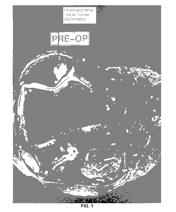
Figs. 1-2 are preoperative scans of an individual's knee;
Figs 3-4 are corresponding scans of the individual's knee after treatment
according to the methods of exemplary embodiments of the present invention;
Fig. 5 is a side-by-side comparison of another individual's knee before and
after
treatment according to the methods of exemplary embodiments of the present
invention;
Fig. 6 is an exemplary distal end of an exemplary bone-chondral interphase
("BC1") delivery device according to exemplary embodiments of the present
invention;
-3-
CA 02870203 2014-10-09
WO 2013/155359
PCMS2013/036259
Fig. 7 is an exploded view of an exemplary delivery device according to
exemplary embodiments of the present invention (top panel), and a magnified
view of an exemplary proximal portion of the exemplary delivery device (bottom
panel);
Fig. 8 is a further magnified view of an exemplary proximal portion of the
exemplary delivery device of Figs. 6-8;
Fig. 9 depicts an alternate exemplary embodiment of the exemplary delivery
device of Fig. 6;
Fig. 10 depicts an exemplary delivery device which may be known as a
"Percutaneous Intradiscal Annular Repair System" (PIARES"), directed to
percutaneous intradiscal annular repair according to exemplary embodiments of
the present invention;
Fig. 11 depicts a variant embodiment of the exemplary delivery device of Fig.
10;
Fig. 12 depicts detailed views of an exemplary delivery device according to an
embodiment of the present invention directed to bone-cartilage interfaces of
peripheral joints and spine;
Fig. 13 depicts an exemplary delivery device being inserted into the bone
above
and below a right knee according to exemplary embodiments of the present
invention;
Fig. 14 depicts a magnified view of the knee joint, and adjacent tibia and
femur
from the drawing shown in Fig. 13;
Fig. 15 depicts details of the distal portion of the exemplary delivery device
of
Figs. 13-14;
Figs. 16-20 are detailed design drawings of an alternate improved exemplary
"PecaBoo" delivery device tool according to exemplary embodiments of the
present invention;
Fig. 21 depicts an elongated form of the exemplary PecaBoo device of Figs. 16-
20, such as may be used for hip procedures;
Fig. 22 illustrates the exemplary PecaBoo device (hip length) as inserted into
the
superior and inferior compartments adjacent to an exemplary hip joint; and
-4-
CA 02870203 2014-10-09
WO 2013/155359
PCT/US2013/036259
Figs. 23-48 depict an exemplary prototype of the PecaBoo tool shown in Figs.
16-20 as used on a patient in an exemplary procedure on the knee, with
corresponding high quality X-Ray images of various stages of the exemplary
procedure.
SUMMARY OF THE INVENTION
Novel therapeutics and methods of treatment to repair, preserve and grow
cartilage are presented. In addition, systems and methods for delivering a
therapeutic to a hard to reach anatomical area, such as, for example, the BCI,
are presented. In exemplary embodiments of the present invention, a cannulated
delivery device provided with a cutting tip and threads on its distal exterior
can be
provided which has considerable advantages over conventional devices. These
include, for example, (i) ease of manufacture, (ii) use of the exemplary
device
being faster than conventional approaches, with both less table time and less
steps, (iii) lesser exposure of internal tissues to ambient air, and thus less
risk of
infection, and (iv) lesser technical complexity leading to lesser
complications.
Using such an exemplary device, various novel therapies for joint and
cartilage
repair, preservation and generation can be implemented. Various versions of
such a device are disclosed. Alternatively, for disc repair, a delivery device
directed to percutaneous intradiscal annular repair, or "PIARES" device can be
used to introduce therapeutics intradiscally. The device is a two-needle
device,
with a first cannula/needle, with a finger grip at its distal end, and a
longer inner
needle, which can then penetrate through the outer needle into the disc, and
can
then, for example, be used to introduce therapeutics, for example, via a
syringe.
When provided with a septum at the inner needle's proximal end, the PIARES
device becomes a completely closed system, and its use minimizes trauma.
Thus, in exemplary embodiments of the present invention, a surgical hand tool
can
be provided, used for the non-invasive placement and delivery of therapeutics,
to a
targeted site. This can be done through minimally invasive skin incision, or
without
any incision, as maybe desired. The delivery and placement of the therapeutic
can
be controlled and does not need a powered drill or guide wire.
-5-
CA 02870203 2014-10-09
WO 2013/155359
PCT/US2013/036259
An exemplary device can have a closed pointed end, a threaded portion, and be
provided with thread cutting/forming features, such as flute(s), and can have
a shaft
perforations to the central lumen at a distal end to deliver therapeutics or
other
preparations. At the proximal end, means can be provided to attach a syringe
in
communication with the shaft's central lumen, and there may be a keyed
engagement feature for attachment of a hand grip. The delivery device can be
made of sufficient length to reach bone on either side of a desired or
targeted joint,
and to easily penetrate soft tissue and cortical bone to reach a targeted site
in
cancellous bone adjacent to a cartilage defect.
The device's main shaft or drill portion can be made of hardened stainless
steel, or
the like, such as, for example, 400 series or 17-40 stainless steel, for
example.
The device can have, for example, an attachable/removable hand grip for ease
of
placement of the drill bit to a site, with a solid proximal end with which to
tap or
hammer, and with a grip for torquing the device through cortical bone and to
guide
a threaded shaft to a targeted site in cancellous bone, for example. The grip
can
have an ergonomic form for ease of use, such as a tri-lobe handle, which
mimics
the natural turn of a wrist in 120 degree increments.
The device can have an impact cap to (i) provide impact anvil surface to
protect a
proximal luer during impaction, as well as to (ii) close the luer opening to a
shaft
lumen.
DETAILED DESCRIPTION OF THE INVENTION:
While the exact cause of knee OA remains unknown, it is strongly believed by
the inventors that alterations in the bone-cartilage interface ("BCI") are
present at
the earliest stages of knee OA. Therefore, therapies for treating knee OA must
target the BC!. This approach can further be extended to other areas where
cartilage has been damaged or lost.
As described below, methods according to exemplary embodiments of the
present invention address the BCI where early alterations can accelerate knee
OA. In exemplary embodiments of the present invention, methods are provided
-6-
CA 02870203 2014-10-09
WO 2013/155359
PCT/US2013/036259
to stimulate the subchondral bone marrow and expose the mesenchymal stem
cells (MSCS) that come out of the bone marrow as a result, to growth factors
from platelet-rich plasma (PRP) and very small embryonic like cells (VSELs).
VSELs are known to be released after an injury resulting in enhanced repair in
the animal stroke model (Kucia et al: Cell Tissue Research 331: 125-134,
(2008). This enhances cartilage repair and possible regeneration. As is known,
MSCSs exposed to PRP differentiate into chondrocytes (Mishra, et al: Tissue
Eng. Methods 15: 431-435 (2009)).
Methods according to exemplary embodiments of the present invention have a
very low risk of infection, are significantly less expensive than major
surgical
procedures, and avoid the liability of metal implants or NSAID medications.
Furthermore there is very little down time for patients undergoing this
procedure
inasmuch as it is performed on an outpatient basis with a quick return to
work.
I. Exemplary
"Ground Up" Methodology For Cartilage Repair-
In exemplary embodiments of the present invention, cartilage issues can thus
be
treated from the "ground up." Such an approach is analogous to how in
agriculture plants are often treated by accessing their roots. Thus, in
exemplary
embodiments of the present invention, technologies can be used that target the
bone-cartilage interphase (BCD to treat cartilage issues, as opposed to
conventional "top down" approaches such as, for example, the current
undesirable practice of microfracture. As noted above, microfracture creates
fibrocartilage with very limited success in patients with cartilage defects.
Moreover, microfracture can only be used for a very small subset of knee
osteoarthritis ("OA") patients ¨ only those having small cartilage defects. If
it is
done excessively it itself can even lead to accelerated cartilage loss.
Thus, in exemplary embodiments of the present invention, novel methodologies
for the treatment of degenerative joints and discs can be utilized. This can
be
applied, for example, to the knee, to treat medial joint knee degenerative
disc
disease ("DJD") using the following protocol, for example:
-7-
CA 02870203 2014-10-09
WO 2013/155359
PCT/US2013/036259
Day 1:
350 mcg of Granulocyte Colony-Stimulating Factors ("GCSFs") injected
subcutaneoulsly;
Day 2:
350 mcg of Granulocyte Colony-Stimulating Factors, or GCSFs injected
subcutaneoulsly (this second GCSF injection is optional);
Draw blood and spin it down to obtain 4cc of Platelet Rich Plasma ("PRP")
in total;
Put 1cc of the PRP into the tibial medial compartment by drilling and
injecting at the bone-cartilage interphase ("BCI"), followed by injection of
0.1 cc of 10% calcium chloride solution or thrombin to form a clot.
Wait two minutes before reverse drilling out the delivery device so that a
clot may form and keep the PRP from leaking out. Alternatively, bone wax
can be injected to keep the PRP in place;
Put 1cc of PRP into femoral medial or lateral compartment by drilling and
injecting at the bone-cartilage interphase, followed by injection of 0.1 cc of
10% calcium chloride solution or thrombin to form a clot.
Wait two minutes before reverse drilling out the delivery device so that a
clot may form and keep the PRP from leaking out;
Put remaining 2cc PRP into the knee joint;
MRI is done pre-treatment and at 3 months post-treatment.
It is understood that these are exemplary values only. Variations of the
quantities of therapeutics can also be used, such as, for example, a range of
1-3
cc of PRP injected each above and below the relevant joint, a range of 0.1 ¨
0.3
cc of calcium chloride used afterwards, and a range of 2-6 cc of PRP injected
into the joint. Additionally, one can wait between 2-4 minutes following
delivery
of the PRP and clotting agent, for example.
-8-
CA 02870203 2014-10-09
WO 2013/155359
PCT/US2013/036259
It is noted that GCSFs have not heretofore been used for cartilage repair. An
exemplary GCSF that may be used can be, for example, Neupogen.
In exemplary embodiments of the present invention, the PRP can be delivered
via one syringe, and either CaCI, thrombin or bone wax, for example, can be
delivered via another syringe. Alternatively, a skilled, dexterous and quick
practitioner may, for example, load both the PRP and CaCI into one syringe, if
she can deliver the dose quickly enough so that no clotting occurs. In
exemplary
embodiments of the present invention this method can be used, and the
inventors have successfully done it in experimental cases.
In exemplary embodiments of the present invention the procedure can, and
preferably should, be performed under fluoroscopy or ultrasound guidance to
insure proper positioning of the delivery devices at the BCI and to further
insure
that there is no penetration through the cartilage, which would cause damage.
Following injection of the therapeutic, the delivery device should be left in
place
for approximately 2 minutes to make sure a clot is formed. Alternatively, bone
wax or the equivalent can be used, for example, to seal the entry instead of
calcium chloride.
Exemplary Sterile Kit
In exemplary embodiments of the present invention, an exemplary kit can
contain, for example, two disposable delivery devices, to be used to inject at
the
BC! in the superior and inferior locations to a joint, as shown, for example,
in
Figs. 23-48. Making them disposable minimizes the risk of infection. Such an
exemplary kit can also be provided with a vial of bone wax and a 2cc vial of
10%
calcium chloride solution, for example. Calcium chloride activates platelets
and
also forms a clot. In general, the CaCI can be provided in a 1110th ratio to
the
biologic, thus for 1ccof stem cells and PRP, a 0.1 cc volume of CaCI may be
used. Alternatively, in exemplary embodiments of the present invention, the
delivery device maybe reusable, and sterilizable, such as a version of the
exemplary PecaBoo device described below. Still alternatively, a device can
have a disposable hub and drill portion (including impact cap ¨ see Fig. 17),
and
a reusable handle, for example.
-9-
CA 02870203 2014-10-09
WO 2013/155359
PCT/US2013/036259
Rationale ¨ Expose Mesenchymal Cells to PRP to Generate New Cartilage
The rationale behind the inventive protocol is that bone marrow may be
stimulated by the GCSFs to produce mesenchynnal cells (MSC). As these MSC
cells come out of the bone marrow and make their way towards the BCI, they are
exposed to the PRP (or, for example, PRP and VSELs) before reaching the
bone-cartilage interphase. There is good evidence that exposure to PRP (or
PRP and VSELs) induces the MSCs to become cartilage, or more granularly, the
MSCs and VSELs (Very Small Embryonic Like stem cells), when exposed to
PRP or hyaluronic acid develop into chondrocytes, which in turn create the
cartilage matrix. This is believed to be the key factor that has led to the
success
seen in the knee treated and described in Appendix A, where an approximate
doubling of cartilage size (relative to the pre-treatment MRI result) was seen
in a
post-operative MRI three months following treatment, with markedly reduced
pain. Furthermore, VSELs are released into the peripheral blood following
stimulation with GCSF which, as noted above, helps with cartilage repair.
Thus,
both PRP and VSELs ma, for example, be delivered to the femoral and tibial
compartments, for example, in an exemplary knee procedure. Sometimes just
drilling is sufficient to stimulate cartilage growth, or to resolve an
ischemia. Thus,
in various exemplary embodiments of the present invention, the following
various
approaches can be used in treating affected joints; in all cases a drill
delivery
device according to the present invention may be used:
1. Drill alone;
2. Drill + PRP + bone wax;
3. Drill + PRP + bone wax;
4. 300 mcg GCSF in 1-3 injections, then followed by drill, then followed by
VSEL
+ PRP + {bone wax or CaC1};
5. Bone marrow aspirate + CaCI or bone wax;
6. Culture expanded autologous stem cells, from stem cell bank;
7. Autologous embryonic stem cells, from cord blood;
-10-
CA 02870203 2014-10-09
WO 2013/155359
PCT/US2013/036259
8. embryonic stem cells, from a cell bank; and
9. Autologous preserved cells.
Thus, in exemplary embodiments of the present invention, there can be a
significant reduction in the number of knee replacements being done with a
concomitantly large reduction in health care costs, inasmuch as the inventive
technology does not involve hospitalization or expensive artificial joints. In
exemplary embodiments of the present invention, if partial results are seen
with
one treatment, there is the option to repeat the treatment from three months
to
two years later for additional therapeutic benefit. It is noted that the
treatment
can be, for example, repeated indefinitely as long as there is therapeutic
benefit.
Thus, from some patients the treatment can be repeated over decades if
helpful.
It is noted that in exemplary embodiments of the present invention the bone
marrow can be, for example, chemically stimulated with GCSF, while the bone
marrow can also be stimulated mechanically by creating microtrauma above the
bone marrow ("above" in the sense of a direction towards the knee joint). Such
a
microtrauma stimulates the bone marrow to produce more MSCS and VSEL
cells, and also increases the blood supply to the bone-cartilage interface to
allow
for better repair.
Experimental Results
Example A
In an experimental trial of treatment methods according to an exemplary
embodiment of the present invention, a 50 year old female with advanced
degenerative osteoarthritic disease of the knee was treated. Prior to the
treatment, a pre-operative MRI was done. Post treatment a three month follow-
up
MRI was performed on the patient and read by an independent radiologist.
Provided hereto as Appendix A is the independent radiologist's report. As
noted
in the report, the post operative MRI showed an increase in cartilage matrix
from
1.8 mm pre-treatment to 3.7 mm at the follow-up MRI. Thus, the inventive
technique shows early promise for cartilage repair and possible regeneration.
-11-
CA 02870203 2014-10-09
WO 2013/155359
PCT/US2013/036259
Example B
Imaging data from another experimental case are provided in Figs. 1-4. As can
be seen therein, Figs. 1 and 2 are respectively axial and sagittal images of a
patient's knee from a preoperative scan. Fig. 1 depicts an area of decreased
blood flow due to an ischemia, as shown by the red arrow, also seen in Fig. 2
pointed to by the red and green arrows. Injections similarly performed
according
to the above described protocol, to the femoral and tibial compartments by
drilling and injecting at the bone-cartilage interface, followed by injections
into the
knee joint. Figs. 3 and 4 are corresponding axial and sagittal images form a
MRI
taken three months following the treatment. As can be seen, significant new
cartilage has grown, and the ischemia has been essentially resolved, as shown
in Fig. 3.
Example C
Fig. 5 depicts side by side comparisons of sagittal images of a knee of a
third
patient. The left panel is an image form a preoperative scan, and the right
panel
a corresponding image from a post operative MRI. As shown in Fig. 5, the post
operative MRI showed an increase in cartilage matrix from 1.60 mm to 1.87 mm
at the follow-up MRI.
In exemplary embodiments of the present invention, the therapeutic protocol
described above can similarly be used for osteoarthritis and avascular
necrosis,
as well as for treating meniscal and labral injuries in the joints.
Exemplary Variations of the Protocol
In exemplary embodiments of the present invention, variations on the above-
described protocols can be used for other anatomical areas. Examples of these
are next described.
Joints - for joints, the step of injecting GCSF for stimulating bone marrow
may
be skipped. The drilling/twisting alone of the delivery device (as described
-12-
CA 02870203 2014-10-09
WO 2013/155359
PCT/US2013/036259
below) will stimulate bone marrow combined with PRP injection or injection of
other biologics such as, for example, stem cells as an alternative.
Joint Arthritis - there is an alternative method for treating joint arthritis
by using
adipose tissue derived stem cells that can be injected intravenously combined
with intra-articularly without drilling into the bone-cartilage interface. If
that does
not work then an alternative method is adipose derived stem cells injected
intravenously, intra-articularly and into the bone-cartilage interface. This
combination of systemic and local therapy is believed to be the next big step
in
biologic interventions for joint issues.
Spine ¨ in similar fashion as was described above for the knee, for the spine
one
can inject GCSF on days 1 and 2, followed by extracting PRP on day 2. The
PRP can then be drilled into vertebral bodies above and below the affected
disc
along with intra-discal injection and epidural injection of the PRP.
Alternatively, one can skip the GCSF and just drill into vertebral bodies
followed
by injection of PRP into the vertebral body followed by thrombin or calcium
chloride to form a clot (so that the PRP does not leak out), and then
injecting the
PRP, followed by thrombin or calcium chloride, intradiscally. It is noted that
for
injecting the vertebral body the novel BCI device described below (Figs. 6-8
and
12) can be used. For an intradiscal injection, standard existing spinal
needles
can be used, or for example, a variation of the novel PIARES delivery device
as
shown in Figs. 10 and 11.
Finally, another alternative method for treating disc or stenosis issues of
the
spine can be, for example, to use adipose tissue derived stem cells which
first
can be given intravenously along with caudal epidural injection. If this does
not
give results, then the adipose stem cells can, for example, be given
intravenously
along with intradiscal injection and caudal epidural, using, for example, a
standard
spinal needle, or, for example, a variation of the novel PIARES delivery
device as
shown in Figs. 10 and 11.
-13-
CA 02870203 2014-10-09
WO 2013/155359
PCT/US2013/036259
II. Exemplary Delivery Devices
In exemplary embodiments of the present invention, the therapeutic methods
described above can be delivered in a safe and efficient manner using various
novel delivery devices according to various exemplary embodiments of the
present invention, as next described.
Exemplary Delivery Device
Figs. 6-9 depict an exemplary delivery device according to exemplary
embodiments of the present invention. Fig. 6 depicts an exemplary distal end
of
an exemplary delivery device according to exemplary embodiments of the
present invention. As can be seen therein, the device is essentially a hollow
cannula with threads on the outside of it. The threads allow for controlled
insertion and removal of the device. It has a cutting point at its distal end,
and
immediately proximal to the cutting tip (i.e., above it) a series of holes are
provided to dispense various therapeutics. As shown in Fig. 6, the solid slug
at
the tip of the device can be laser welded in place, for example, and the
various
holes in the cannula laser cut, for example. Exemplary dimensions are shown in
Fig. 6, but are understood to be merely exemplary, and not limiting.
Given the solid cutting tip, a user first presets the device with hammer taps,
and
then can screw in the device a desired length. This can be done manually, or
via
a drill interface provided at the distal end of the device, for example. As
described below, one can, for example, tap with a hammer to set the device
into
place into dense cortical bone, and then subsequently twist (or drill) to
advance
the delivery device into spongy bone (interior cancellous bone).
Fig. 7 is an exploded view of an exemplary embodiment of the delivery device
according to the present invention (top panel), and a magnified view of an
exemplary proximal portion of the exemplary delivery device (bottom panel).
With reference thereto, the top panel of Fig. 7 shows how the device has a
cannula/needle portion, a needle hub, and a cap with twist grip and a surface
at
its end for tapping with a hammer. The cap and needle hub can be connected
via keys, which thus insure that the cap and needle hub do not move relative
to
-14-
CA 02870203 2014-10-09
WO 2013/155359
PCT/US2013/036259
one another as a user drills, for example, or manually screws/twists in, for
example, the device.
Provided at the top of the needle hub can be a needle pierce septum, which
allows a sterile syringe to be introduced into the cannula to inject
therapeutics or
PRP rich blood, as described above, after removing the cap, once the device is
in the proper position. Thus, using such a septum, the tissue exposed to the
distal end of the delivery device need never contact open air, and the
delivery
system is thus totally closed. The septum can be made of silicone, for
example,
or other appropriate materials.
Fig. 8 is a further magnified view of the distal portion of the exemplary
device of
Fig.7. Fig. 9 depicts an alternate exemplary embodiment of an exemplary
delivery device of Fig. 7, where the cap is screwed on to a luer provided at
the
distal end of the cannula/needle.
Exemplary Delivery Device For Discs - PIARES
Figs. 10-12 depict an exemplary delivery device directed to percutaneous
intradiscal annular repair according to exemplary embodiments of the present
invention. This device is known as a "PIARES" device by the inventors, and is
used for introducing therapeutics intradiscally, as described above. Such a
device is inserted by hand, in most cases. The device is a two-needle device,
and can have, for example, a first cannula/needle, with a finger grip and luer
hub
at its distal end. The cannula can be, for example, 16 gauge, and be
approximately 3.5 inches long, for example, but such dimensions are exemplary
and not limiting. There can be provided a stylette, to fit within the cannula,
of, for
example, 21 gauge (for a 16 gauge cannula). The stylette can lock onto the
distal end of the luer, at a luer lock hub. The stylette can remain in the
outer
needle as a user inserts the device near a disc (but not all the way to the
disc),
then be removed so as to allow the insertion of the longer inner needle, which
can then penetrate into the disc, and can then, for example, be used to
introduce
therapeutics, for example, via a syringe.
-15-
CA 02870203 2014-10-09
WO 2013/155359
PCT/US2013/036259
Fig. 10 thus also shows, at the bottom of the figure, the second, or inner
needle
of the device. This inner needle fits inside the first needle/cannula, and
protrudes
from it into the disc. The inner needle can be, for example, 5 inches in
length,
where the bottom 20 mm or so have perforations out of which the therapeutic
agents can diffuse into the patient. Such a device can have, for example, a
cannula of 21-25 gauge. It can have a similar luer and finger grip, and can
similarly accept a syringe which can lock on its luer lock hub, to deliver the
therapeutics, as described above. There is no stylette for this inner needle,
obviously.
Fig. 11 depicts a variant embodiment of the exemplary PIARES delivery device
of Fig. 10, where instead of a luer lock hub at the proximal end of the inner
needle, a septum is provided, thus completely isolating the delivery device
and
the disc into which the inner needle protrudes from exposure to the ambient
space. To introduce therapeutic agents into the inner needle and thus out the
distal holes into the disc, a user inserts a needle into the septum, in
similar
fashion as shown in Fig. 12 for the peripheral joint and spine embodiment of
the
delivery device.
Thus, in operation, a user first inserts the outer needle, with stylette
inside. This
is done under imaging guidance, such as, for example, fluoroscopy or
ultrasound. The outer needle is placed near, but not all the way towards, the
relevant disc. The stylette is then removed, and the inner needle inserted
inside
the outer needle. Thus, as shown, the outer needle can be 16 gauge, and the
inner needle from 21 to 25 gauge, for example. Because it is longer than the
outer needle, for example, 5 inches versus 3.5 inches, as shown in Figs. 10-
11,
the inner needle protrudes out the end of the outer needle, and can be guided
into the disc itself. Now at this point the distal end of the inner needle
touches
the disc, but if the septum embodiment of Fig. 11 is used on the inner needle,
the
system is completely closed. Once intradiscal, therapeutic can be introduced
via
the inner needle.
Thus, the PIARES device has a number of novel advantages: (i) it provides a
fully and completely closed system when the septum is used on the inner
-16-
CA 02870203 2014-10-09
WO 2013/155359
PCT/US2013/036259
needle's proximal end; (ii) therapeutic can be delivered simultaneously to the
nucleus and annulus of the disc, thus to deliver therapeutic to where the tear
is;
and (iii) by using the outer needle for initial positioning, and then
granularly
positioning the longer inner needle, which is then fully set up to deliver
therapeutic agents, trauma to the disc is minimized, as opposed to
conventional
approaches where needles are moved in and out. Less trauma means quicker
healing and better disc repair.
Fig. 12 depicts detailed views of an exemplary delivery device according to an
embodiment of the present invention directed to delivering therapeutics to
bone-
cartilage interfaces of peripheral joints and spine. It is a more detailed
drawing of
the exemplary device shown in exploded view at the top panel of Fig. 7, with a
perspective view. As seen in Fig. 12, there can be a cutting tip, and
proximally
from it external screw threads between which are interspersed perforations.
Thus the grip is first tapped with a hammer for initially setting it into
place into
dense cortical bone, and then subsequently twisted by a user to advance the
delivery device into spongy bone (interior cancellous bone). The cannula can
be
from 14 to 16 gauge, for example, and at the proximal tip of the device there
can
be a needle pierceable septum seal, for example, or a luer lock with removable
cap such that a syringe can be attached, as shown in various other embodiments
and as described above.
While the shown version has a flat distal surface for tapping a hammer for the
initial setting, in other exemplary embodiments an interface can be provided
in
the center of the end of the cap, to interface with commonly used drills, for
example.
Illustrations of Delivery Device As Used in Knee Procedures
Figs. 13-15 illustrate exemplary use of the device of Figs. 6-9 in knee
procedures. Fig. 13 depicts an exemplary delivery device being inserted into
the
bone above and below an exemplary right knee according to exemplary
embodiments of the present invention. Because this is a peripheral joint, the
device of Fig. 12 would be used. As can be seen in Fig. 13, the device is
-17-
CA 02870203 2014-10-09
WO 2013/155359
PCT/US2013/036259
generally inserted above and below the affected joint, and is inserted so as
to be
close to the interior edge of the cortical bone, above and below the cartilage
of
the affected joint. In the case of the knee joint depicted, one delivery
device is
inserted above and below the articular cartilage of the knee. As described
above, using the protocol described above, the therapeutic introduced by the
practitioner or user diffuses from the holes in the distal end of the cannula,
and
the bone marrow is stimulated by a GCSF to produce mesenchymal cells (MSC).
As these cells come out of the bone marrow and make their way towards the BCI
they get exposed to PRP before reaching the bone-cartilage interphase. The
exposure to PRP is believed to thus induce the MSCs to become cartilage.
Fig. 14 depicts a magnified view of the knee joint, and adjacent tibia and
femur
as shown in Fig. 13, further illustrating the diffusion of therapeutic(s)
uniformly
away from the cannula. Fig. 15 depicts details of the distal portion of the
exemplary delivery device of Figs. 13-14, showing the threads and the holes
interspersed between them, at various rotational orientations of the delivery
device.
Alternate Exemplary Delivery Device ¨ "PecaBoo"
Figs. 16-21 are detailed design drawings of an alternate improved exemplary
delivery device tool according to an exemplary embodiment of the present
invention. Variations of this device ma, for example, be used in knee, hip and
other joint procedures. This alternate delivery device is next described.
An exemplary prototype of the tool of Figs. 16-20 was fabricated, and tested
on
various patients with DJD of the knee with excellent results. The exemplary
tool
may be known and/or marketed under the trade name "PecaBoo."
Figs. 16-21, next described, illustrate two versions of an exemplary delivery
device according to exemplary embodiments of the present invention. With
reference to Fig. 16(a), this is an overall view of the device. The device has
three primary parts: a drill portion, an impact cap and an ergonomic tri-lobe
handle. The tri-lobe handle is shown with its top and bottom views,
respectively,
in Fig. 16(b). As can be seen on the top of the tri-lobe handle there is a
built in
metal pad shown, for example, in Fig. 16(b), at the far right image, which
-18-
CA 02870203 2014-10-09
WO 2013/155359
PCT/1JS2013/036259
occupies almost all of the space of the top portion of the tri-lobe handle.
This
metal portion can be used once taken off the tool and turned around as a kind
of
a hammer, mallet or tapping device to push in the drill, when covered by the
impact cap, into a patient's bone. The impact cap prevents damage to the hub
in
such a use. It is noted that the exemplary dimensions provided in Figs. 16-21
are exactly that, exemplary, and this is one of many prototypes that can be
built
according to various exemplary embodiments of the present invention. The
dimensions are left for illustrative purposes only to provide exemplary aspect
ratios, as well as exemplary dimensions, for a tool that has been found to be
convenient to certain practitioners.
With reference to Fig. 16(c), which is a longitudinal cross section of the
exemplary tool, there is a tight slip fit between the tri-lobe handle and the
drill
portion of the tool, such that the handle tightly fits upon the tool such that
it can
be turned and manipulated. Also seen in the cross section is the impact cap
which covers the luer at the proximal end of the drill as illustrated in more
detail
in the following figures. It is noted that in these figures the drill is
referred to as
"drill 16." The "16" refers to an internal design identifier.
With reference to Fig. 17, there is seen the drill with integral hub at 17(a),
an 0-
ring which slips over the hub at 17(b), the impact cap referred to above, at
17(c)
and the tri-lobe handle at 17(d). These fit together as shown, where the 0-
ring is
slipped over the top of the drill with integral hub so that it sits as shown
in Fig.
16(c). This then creates the tight slip fit of the tri-lobe handle on the hub.
Alternatively, C clip rings can be used instead of an 0-ring -- which would
need
to be replaced after some time ¨ or, for example, other attachment mechanisms
as may exist in the art. The impact cap shown in Fig. 17(c) covers a female
luer
lock such that the drill is totally closed and not exposed to the air any more
than
absolutely necessary. The impact cap allows the tri-lobe handle, as shown at
17(d), to be removed from the remainder of the tool and still allow the tool
to be a
completely closed system. Moreover, a practitioner can, upon removing the tri-
lobe handle as noted above, turn it around such that the metal place built
into the
-19-
CA 02870203 2014-10-09
WO 2013/155359
PCT/US2013/036259
top of it can be used to tab on the impact cab shown as Fig. 17(c) without
damaging or affecting the rest of the tool, namely the drill with integral hub
shown
at Fig. 17(a). In exemplary embodiments of the present invention the drill may
be
disposable and the handle reusable, or the entire device autoclavable and
reusable.
Fig. 18 illustrates the exemplary delivery tool of Figs. 16 and 17 in various
longitudinal views and longitudinal cross section. With reference to Fig.
18(a) the
female locking luer is shown at the far right as well as the integral hub upon
which, for example, an 0-ring (as shown at Fig. 17(b)) can be placed to
provide a
tight slip fit. Additionally, Fig. 18(a) shows, for example, a 2.0 pitch helix
to the
cutting threads and illustrates further that the tip of the drill can be
coated with a
coating such as, for example, titanium nitride, or TiN. TiN is an extremely
hard
ceramic material which is often used as a coating on titanium allows, steel,
carbide and aluminum components to improve the substrate surface properties.
Because of these hardening properties, such a coating can be used to protect
cutting and sliding surfaces of medical devices, and it is also used as a non-
toxic
exterior for medical implants, making it ideal to improve the hardness and
cutting
ability of the tip of the exemplary delivery tool and at the same time be
medically
inured to the patient's tissues. As before, exemplary dimensions are provided
in
Fig. 18, and they are, of course, simply illustrative and not intended to bind
or
limit the invention in any way. Fig. 18(b) illustrates the 0-ring gland, and
an
exemplary laser weld if the drill and hub are decided to be made in two
pieces.
Alternatively, they can be made in one piece and machined. Fig. 18(b) also
illustrates how the drill can be made of 455 stainless steel cannulated bar
stock,
for example. Other metals and stainless steel grades are also usable, in
various
exemplary embodiments.
Fig. 18(c), the longitudinal cross section, again shows exemplary diameters,
which are only illustrative. This figure also illustrates that the drill with
integrated
hub can be fabricated as one piece and that the tip of the drill has a
straight
portion, as well as a tapered portion occupying the most distal portion of the
-20-
CA 02870203 2014-10-09
WO 2013/155359
PCT/US2013/036259
delivery device, to make it more easily insertable into a patient. There are
also
seen in the depictions of Fig. 18 at the tip of the device some grooves
slightly
distal to the actual tip which are shown in greater detail in Fig. 19.
Finally, the tip
can be laser welded to the cannulated drill portion, as shown in Fig. 18(c).
Fig. 18A is a copy of Fig. 18 somewhat magnified, with further explanatory
notes
by the present inventors. With reference to Fig. 18A(a) one can see that there
are perforations in the distal tip of the exemplary delivery device, as called
out by
the inventor notes. These perforations are the holes by which the fluid is
delivered. In a variation from that shown in the device of Figs. 13 and 14, in
this
exemplary embodiment there are only a few holes. Actually two full
perforations
across the entire diameter of the distal tip of the exemplary device, making
four
holes in total, located only at the most distal portion. Of course, they have
to be
proximal to the point at which the spade-type cutting tip, as shown in Figs.
18A(b)
and 18A(c), but they can be immediately proximal of that as shown in Fig.
18A(c). It is found that a smaller number of holes placed at the extreme
distal
portion of the exemplary tool or delivery device allow the therapeutics to be
delivered closest to the bone chondral interphase. Thus, once a practitioner
has
screwed in the device as far as he needs it to be (and this is done under
fluoroscopy as described above, and as shown in the photographs of actual
procedures described below) he or she can then unscrew the device slightly,
moving it back, and dispense the medication into the cavity created at the tip
of
the device by slightly moving back the device or unscrewing the device. The
physician can, for example, unscrew slightly and deliver medication, and
repeat
this process a few times, and thereby fill up a section of the channel
created.
This ensures that the medication goes to where it is most useful, and does not
leak out the back of the device.
Also shown in Fig. 18A(a) there are thread cutting flutes which are shown in a
magnified depiction immediately beneath Fig. 18A(a). These are, as described
above, used to cut the bone as the drill is turned by a user as it is
protruding into
the patient's bone. Additionally, one can see the taper of the distal tip as
shown
-21-
CA 02870203 2014-10-09
WO 2013/155359
PCT/US2013/036259
in the drawing below Fig. 18A(a), and the fact that the tip itself can be
laser
welded all around to make sure that it is well fastened within the cannula.
Fig.
18A(b) also depicts an exemplary internal thread diameter --or minor diameter -
-
of the threaded portion of the tool, as well as the external thread diameter --
also
known as the major diameter. The tapered thread portion of the distal portion
of
the tip is illustrated in Fig. 18A(b).
Finally, Figs. 18A(a) and 18A(b) illustrate the engagement key by which the
hub
may be attached to the handle, and further depict the threaded luer lock
engagement by which that occurs. Moreover, in Fig. 18A(c) the various portions
proceeding from the proximal to the distal end of the exemplary drill are
shown,
from right to left, beginning with the female luer, the hub, the cannulated
shaft,
the center lumen, the threads, and the "spade" type cutting tip. The
attachment
of the hub to the cannulated shaft, if it is two pieces, can be by welding all
around
or in two places, or, for example, the combination of hub and drill, i.e. the
cannulated shaft, can be fabricated as one piece and formed by machining.
Fig. 19 illustrates an overall exemplary dimensional relationship between the
drill
with integrated hub and the threaded distal portion thereof. For example, the
threads can occupy approximately 30% of the overall length of the drill with
integrated hub in one example. Again, as noted above, these dimensions are
purely illustrative and various other dimensions and dimensional relationships
can be implemented in various exemplary embodiments of the present invention,
all within the scope of the present invention.
Fig. 19(a) illustrates the drill with integrated hub without the impact cab
and
without the handle. Within Fig. 19(a), a section of the tip is labeled as "D"
and
that is presented in Fig. 19(b) in a greatly magnified view. This tip contains
both
the fluid side ports by means of which therapeutics and/or liquids are
dispensed
into a patient using the exemplary delivery device, as well as various cutting
features. There is a spade drill point which has essentially a flat surface on
two
sides and cutting edges at the tip. This makes for much easier cutting than a
-22-
CA 02870203 2014-10-09
WO 2013/155359
PCT/US2013/036259
fully cylindrical shape. Once the spade point is inserted, when the user turns
it
creates a cylindrical bore. As can be understood, the exemplary delivery
device
of Figs. 16-19 is, as noted, capable of being slightly pounded or tapped into
the
bone of a patient above and below, for example, the knee joint or above and
below, for example, a hip joint. As opposed a simply non-tapered cylinder, it
is
much easier to penetrate the bone and then cut the bone as the device is
turned.
This easily creates a pathway for the drill to proceed through the bone to a
point
close the bone chondral interface. Therefore the combination of (i) the spade
drill point, (ii) the thread cutting flutes, and (iii) the tapering of the
drill tip, all in
combination allow for easy cutting of the bone surrounding the tip as a
practitioner turns the drill such as, for example, by holding the tri-lobe
handle
shown in Fig. 17(d), or, if she has sufficient strength, by simply twisting
the
integral hub or the impact cap. In alternate exemplary embodiments some or all
of these features can be provided, but it need not always be necessary to have
all of them.
Along those lines, Fig. 20 illustrates exemplary details of the impact cap. As
can
be seen, it can be made of 455 stainless steel bar stock, it can have a
straight
plunge cut so as to be able to be fastened on the female locking luer as shown
in
Fig. 18 as well as in Fig. 17(a), and it can have exemplary dimensions as
shown,
for example, or various other dimensions -- the ones shown being completely
exemplary and illustrative.
It is noted that the exemplary delivery tool of Figs. 16-20 has been found
useful
in treatment of weakened knee joints. As can further be well understood, for
treating the hip joint, the same therapies described above can be used;
however,
to deliver the medicines, namely the PRP, the bone wax or calcium chloride and
stem cells, if used, a slightly longer drill would need to be created to
penetrate
through the fat and muscle to get to the hip joint. Therefore, Fig. 21
illustrates an
elongated version of the exemplary drill with integral hub as shown in Fig.
18(c)
for example. As can be seen with reference to Fig. 19 (top image), the overall
length of the exemplary knee drill delivery tool is 104mm, but in the case of
the
-23-
CA 02870203 2014-10-09
WO 2013/155359
PCT/US2013/036259
exemplary hip embodiment shown in Fig. 21(d), the overall length is 205mm, for
example. Other relative dimensions are well within the scope of the present
invention, it being generally understood that for most patients it takes a
somewhat longer device to reach the hip than it does to reach the knee joint.
Other exemplary dimensions of the hip-type drill, shown in Figs. 21 and 22 as
"Drill 17", are shown in Figs. 21(a), 21(b), 21(c) and 21(d). The device is
essentially the same or similar to the exemplary knee version of Fig. 18,
except
for the length of the drill itself, and in particular the length of the
portion
proceeding the tapers that is not threaded. The threaded portion, as shown in
Fig. 21(d), can be, for example, the same as "Drill 16" which is the project
name
for the exemplary knee delivery tool shown in Figs. 16-20. ("Drill 17" being
the
project name for the exemplary drill for hip joints).
Fig. 22 is a drawing of the exemplary hip delivery device, namely the "Drill
17"
device super imposed on a coronal section of a human left hip joint and
showing
surrounding muscles and tissues. The numbers referred to in Fig. 22 are
provided in the following Table for background and ease of locating where the
exemplary drill is to be placed in exemplary embodiments of the present
invention. As can be seen in the drawing, although this would not be done in
practice, for ease of illustration, there is one drill shown in the proper
position for
the superior portion of the joint and one for the inferior portion of the
joint,
although obviously in practice these would generally be done sequentially and
not at the same time.
Table of Anatomical Areas For Fig. 22
1. External iliac artery
2. Psoas major
3. Iliacus
4. Iliac crest
5. Gluteus medius
6. Gluteus minimus
-24-
CA 02870203 2014-10-09
WO 2013/155359
PCMIS2013/036259
7. Greater trochanter
8. Vastus lateralis
9. Shaft of femur
10.Vastus medialis
11.Profunda femoris vessels
12.Adductor longus
13. Pectineus
14. Medial circumflex femoral vessels
15. Capsule of hip joint
16. Neck of femur
17.Zona orbicularis of capsule
18.Head of femur
19.Acetabular labrum
20. Rim of acetabulum
21. Hyaline cartilage of head
22. Hyaline cartilage of acetabulum
Exemplary Clinical Use of PecaBoo Device
Next described are Figs. 23-48, which are photographs of exemplary actual
procedures on human knees performed using the exemplary PecaBoo device
described above. Procedures were done under fluoroscopic guidance, as noted
above, and therefore both photographs of the patient's knees as well as some
of
the images from the fluoroscopy will be provided.
With reference thereto, Fig. 23 shows a practitioner initially inserting the
PecaBoo device into a patient's knee close to the BCI, as described above.
Similarly, Fig. 24 shows the same patient where the practitioner has pushed
the
device significantly into the patient and is obviously inserting into the
bone.
Fig. 25 shows an even further protrusion of the device into the bone and that
is
its stopping place as shown in Fig. 26. Fig. 27 shows that the exemplary
-25-
CA 02870203 2014-10-09
WO 2013/155359
PCT/US2013/036259
PecaBoo device has been screwed into the bone near the BC! below the knee
joint itself. Fig. 28 is a close up of the image shown in Fig. 27 showing the
same
thing.
As noted above, after the device has been inserted into the bone near the BCI,
the practitioner may, for example, remove the handle, remove the impact cap,
and attach a syringe to the female luer which protrudes from the exposed
portion
of the hub after the impact cab has been removed. This is shown in Fig. 29.
Additionally seen in Fig. 29 is the red ring of the exemplary 0-ring remaining
on
the hub as shown in the expanded view of Fig. 17, except here in Fig. 29 the 0-
ring is placed securely onto the hub which allows the tight fit of the yellow
tri-lobe
handle seen in Figs. 23 and 26. In the configuration of Fig. 29, the set-up is
ready for injection as per one of the above described protocols. Fig. 30 shows
a
close up of the view of Fig. 29.
Fig. 31 now shows the syringe, which had been attached in the views of Figs.
29
and 30, being removed. As noted above, when a practitioner injects the
medication into the patient in the set-up of Figs. 29 and 30, he or she will
often
back out the exemplary PecaBoo delivery device so that the medication can be
injected into the cavity left behind. This backing up and injecting may, for
example, be repeated numerous times. Therefore, at the end of an injection,
the
exemplary delivery device will protrude less into the bone than it did at the
beginning of the injection. This is shown in Fig. 32 which shows the position
of
the protrusion of the device as shown in Fig. 31 into the bone which is less
of a
protrusion than that shown in Figs. 27 and 29, after the initial screwing in
of the
delivery tool, as can readily be seen by comparison.
Fig. 33 shows a view from the other end of the patient, i.e., looking upwards
from
the area of the patient's foot. This is a different patient than shown in the
previous figures. As seen in Fig. 33, the practitioner has just begun
inserting an
exemplary delivery device near the patient's knee joint in similar fashion as
-26-
CA 02870203 2014-10-09
WO 2013/155359
PCT/US2013/036259
shown above. In this case, however, the exemplary delivery device is being
inserted superior to the patient's knee on the femoral side.
Fig. 34 shows a little bit of advance of the device and Fig. 35 shows it
having
been pushed in all the way such that the therapeutics can now be delivered
after
removal of the tri-lobe handle and the impact cap. This situation is seen in
Fig.
36, where syringes are being attached to the female luer of the device.
Fig. 37 shows the protrusion of the exemplary device into the affected area on
the femoral side of the joint. It is also noted in Fig. 37 that the patient
has
already had bone screw and other hardware inserted from prior procedures.
Fig. 38 shows once again the device being inserted into the knee of a patient
as
described before, and Fig. 39 shows it having been protruded quite some
distance into the patient's body which was necessary given the patient's
tissue
width. Fig. 40 shows the device under fluoroscopy into the bone superior to
the
knee joint corresponding with the view of Fig. 39. Fig. 41 shows the same
patient now being made ready for the injection, and Fig. 42 shows the
injection
using a syringe inserted into the female lure of the exemplary device. Fig. 43
is
another image obtained from the fluoroscopic guidance as the practitioner was
performing the injection.
Fig. 44 depicts another view of similar to that of Fig. 42 but from a
different angle
showing the device with the hub shown. Fig. 45 illustrates an injection into
that
same patient as does Fig. 46 when the injection has essentially been
completed.
Fig. 47 shows the beginning of an exemplary procedure where the device is
first
inserted into a patient, and Fig. 48 shows the fluoroscopic guidance where the
distal tip of the drill is just penetrating the position inferior to the knee
joint on the
top of the tibia.
It will be understood that the images of Figs. 23-48 are merely exemplary and
illustrate one example of the use of an exemplary delivery device according to
-27-
CA 02870203 2014-10-09
WO 2013/155359
PCT/US2013/036259
the present invention with regard to patients with degenerated cartilage, or
for
example, edema resulting from an ischemia, in the knee joint.
Thus, in exemplary embodiments of the present invention, for joints, such as,
the
knee, for example, there may be female luer locks to minimize air exposure.
For
PIARES, for example, the intradiscal system, the same system may be used.
Alternatively, in each case ¨ PIARES, PecaBoo or the device of Figs. 6-9, in
some
exemplary embodiments there can be either a fully closed system, using a
septum,
or, for example, one with female and male luer locks, minimizing air exposure.
Thus, in some embodiments a knee device, such as PecaBoo, may have female
and male luer locks, and PIARES for intradiscal use may have a septum and be a
fully closed system. In others all of PIARES, PecaBoo and other systems may be
totally closed and use septums or the like.
Kits may be provided with each type, or with one type, either closed system or
male
and female luer locks, or a given kit may mix and match. Exemplary delivery
devices may also be sold separately.
As noted above, in exemplary embodiments of the present invention, the drill
and
hub, as shown in Fig. 18, for example, may be preferably fabricated in one
piece
without seams, but may also be provided in two pieces, as shown, with two
pieces
with a continuous 360 degree welded seam fully sealing around where the drill
passes through the hub. One piece is often preferred for reasons of cost as
well. It
is noted that even when fabricated in one piece, in some exemplary embodiments
the hardened tip may still need to be made separately and welded onto the
distal
end drill, as noted above.
Thus, in exemplary embodiments of the present invention, a surgical hand tool
can
be provided, used for the non-invasive placement and delivery of therapeutics,
to a
targeted site. This can be done through minimally invasive skin incision, or
without
any incision, as maybe desired. The delivery and placement of the therapeutic
can
be controlled and does not need a powered drill or guide wire.
-28-
CA 02870203 2014-10-09
WO 2013/155359
PCT/US2013/036259
An exemplary device can have a closed pointed end, a threaded portion, and be
provided with thread cutting/forming features, such as flute(s), and can have
a shaft
perforations to the central lumen at a distal end to deliver therapeutics or
other
preparations. At the proximal end, means can be provided to attach a syringe
in
communication with the shaft's central lumen, and there may be a keyed
engagement feature for attachment of a hand grip. The delivery device can be
made of sufficient length to reach bone on either side of a desired or
targeted joint,
and to easily penetrate soft tissue and cortical bone to reach a targeted site
in
cancellous bone adjacent to a cartilage defect.
The device's main shaft or drill portion can be made of hardened stainless
steel, or
the like, such as, for example, 400 series or 17-40 stainless steel, for
example.
The device can have, for example, an attachable/removable hand grip for ease
of
placement of the drill bit to a site, with a solid proximal end with which to
tap or
hammer, and with a grip for torquing the device through cortical bone and to
guide
a threaded shaft to a targeted site in cancellous bone, for example. The grip
can
have an ergonomic form for ease of use, such as a tri-lobe handle, which
mimics
the natural turn of a wrist in 120 degree increments.
The device can have an impact cap to (i) provide impact anvil surface to
protect a
proximal luer during impaction, as well as to (ii) close the luer opening to a
shaft
lumen.
The above-presented description and figures are intended by way of example
only,
and are not intended to limit the present invention in any way except as set
forth in
the following claims. It is particularly noted that persons skilled in the art
can
readily combine various technical aspects of the elements of the various
exemplary embodiments described above in numerous other ways, all of which
are considered to be within the scope of the invention.
-29-