Note: Descriptions are shown in the official language in which they were submitted.
CA 02870406 2014-11-07
REMOVABLE LOCALIZING WIRE
Background of the Invention
Field of the Invention
The invention relates generally to localizing wires and more particularly to a
localizing wire which comprises an anchor portion having collapsible
properties enabling
the anchor portion to be retracted into a cannula lumen for repositioning or
removal in a
tissue mass. In another aspect, the invention relates to a localizing wire
configured such
that an externally extending portion of the wire can lie against the exterior
of the tissue
mass. In yet another aspect, the invention relates to a rnethod of using the
localizing wire.
In one other aspect, the invention relates to a localizing wire that can be
removed from a
tissue mass without there-introduction of a cannula.
Description of the Related Art
Localizing wires are well-known devices for marking areas, such as lesions, in
a
tissue mass, frequently breast tissue. When such a lesion is identified with a
medical
imaging technique, such as radiography or ultrasonography, it is often
desirable to position
a localizing wire or other type of imaging marker near the lesion to
facilitate locating the
lesion during later procedures, such as biopsy or surgery. Alternatively, a
localizing wire,
tissue marker or staple can be placed in the tissue mass after a biopsy has
been performed.
In the latter case, the localizing wire marks the location of the biopsy
cavity for future
procedures.
Localizing wires typically comprise an anchor portion implanted at the tissue
site
of interest, with a wire portion extending from the anchor portion to exit
through the skin.
A practitioner can then use the wire as a visual and tactile guide to the
lesion rather than
solely relying on imaging techniques, which currently provide good 2-D images
but not 3-
D images. During surgery, surgeons typically prefer a localizing wire to
locate the lesion
because it leads them straight to the biopsy site.
To implant a localizing wire, a needle, or cannula, is inserted into the
tissue mass
and, with guidance from an imaging system, is positioned with its tip at a
selected location
at or near the lesion. Once the needle is in place, the localizing wire is
extended through
the needle and out the tip into or adjacent the lesion where the hook on the
end of the wire
1
CA 02870406 2014-11-07
engages the tissue mass. Thereafter, the needle is removed from the tissue
mass, and the
localizing wire remains anchored in place by the hook.
It is critical that the localizing wire be accurately placed at the desired
location
within the tissue and remain in the desired location. Movement of the
localizing wire after
it is properly located and implanted is very undesirable as it will not
properly identify the
lesion or the biopsy site if a follow-up surgery is required.
However, there is often a need to reposition the localizing wire after the
initial
implantation. For a variety of reasons, such as, for example, the nature of
the instrument
used for implanting, the initial implantation may not always be located at the
desired site.
Under such circumstances, the localizing wire will need to be repositioned.
Thus, a
contemporary localizing wire must perform the conflicting functions of keeping
the
localizing wire anchored at the desired implantation site while permitting the
repositioning
of the localizing wire.
Prior localizing wires accomplished these conflicting functions by the anchor
having a pointed, hook shape and being formed of a memory metal, such as
Nitinol.
When the localizing wire was stored in the cannula, the anchor was
substantially straight
and took on the hook shape only as it was extended exteriorly of the cannula.
As the
anchor was extended from the cannula, it pierced the surrounding tissue and
formed the
hook shape to anchor the localizing wire to the tissue. The localizing wire
could be
repositioned by withdrawing the anchor back into the cannula to straighten out
the hook.
The cannula would then be repositioned and the anchor once again extended to
anchor the
wire. The curvature of the hook shape was great enough that the anchor would
not defect
in response to an external pulling on the wire.
While the prior localizing wire adequately accomplished the conflicting
functions,
it does have certain known disadvantages. One such disadvantage is that the
tissue is
pierced each time the localizing wire is repositioned, which causes additional
trauma. It is
better to minimize the trauma to the surrounding tissue for reduced recovery
time and the
patient's comfort. Thus, there remains a need for a device that can reimplant
or remove a
localizing wire in a tissue mass after initial implantation with minimal
discomfort to a
patient.
Another disadvantage of current localizing wires is that, after implantation,
a
portion of the localizing wire extends exteriorly of the tissue. This
exteriorly extending
2
CA 02870406 2014-11-07
portion of the localizing wire projects away from the surface of the tissue
mass.
While the projecting of the exterior portion of the wire is useful for the
surgeon in
locating the localizing wire during surgery, it creates the risk that the
patient or
someone else might accidentally catch the exteriorly extending portion and
pull or tug
on the localizing wire, resulting in the possible repositioning of the
localizing wire
inside the tissue. Such an accidental repositioning is very undesirable in
that the
localizing wire will no longer properly locate the lesion and it can be
painful for the
patient. This is one of the reasons why localizing wires are typically
inserted just
prior to the surgery and are not intended to be left in the tissue mass for an
extended
period.
In practice, because of the several hour or several day delay between the time
that a biopsy is taken and the results of the tissue analysis is received, it
is a common
practice for an internal imaging marker, such as that disclosed in U.S. Patent
No.
6,575,991, to be placed at the biopsy site. If the analysis of the tissue
indicates that
follow-up surgery is required, then a localizing wire is placed within the
tissue at the
site of the internal imaging marker prior to surgery and the surgeon uses the
localizing
wire to locate the biopsy site. It is desirable to have a localizing wire that
can be used
instead of the internal imaging marker to mark the biopsy site, left within
the tissue
for an extended period of time, and used to guide the surgeon if surgery is
required or
easily removed if surgery is not required.
Summary of the Invention
According to an aspect of the invention, there is provided a use of a
localizing
wire and cannula, for marking a lesion in a tissue mass, said cannula having
at least
one lumen, and being insertable into a tissue; said localizing wire being
installed
within the cannula, and comprising a localizing thread and a localizing
anchor, said
localizing anchor having a collapsed shape delineating a first area in a lumen
of the
cannula and an expanded shape delineating a second area greater than the first
area
outside the at least one lumen of the cannula; the localizing anchor being
movable
outside the at least one lumen at a first selected location relative to the
lesion in the
tissue mass, said anchor being expandable from the collapsed shape to the
expanded
shape to exert expansive force against the tissue mass.
3
CA 02870406 2014-11-07
Further, according to another aspect of the invention, there is provided a use
of
a localizing wire and cannula, for marking a lesion in a tissue mass, said
cannula
having at least one lumen, and being insertable into a tissue mass; said
localizing wire
being installed within the cannula, and comprising a localizing thread and a
localizing
anchor, said localizing anchor having a collapsed shape delineating a first
area in a
lumen of the cannula and an expanded shape delineating a second area outside
the at
least one lumen of the cannula; the localizing wire and cannula being movable
so as
to position the localizing anchor outside the at least one lumen at a first
selected
location relative to the lesion in the tissue mass, for exerting an expansive
force
against the tissue mass when the anchor is transitioned from the collapsed
shape to the
expanded shape.
According to yet another aspect of the invention, there is provided a use of a
localizing wire for positioning in a tissue mass, said localizing wire
comprising a
thread and an anchor coupled to the thread, said wire being insertable into
the tissue
mass so that the anchor is received within the tissue mass, and an external
portion of
the thread extends beyond the exterior of the tissue mass; said external
portion being
capable of laying substantially flat against the exterior of the tissue mass.
A further aspect of the invention provides a use of a localizing wire having a
reconfigurable anchor for implanting in a tissue mass, said anchor being
configurable
between an anchoring configuration to anchor the localizing wire in the tissue
mass,
and a release configuration to allow release and withdrawal of the localizing
wire
from the tissue mass.
A further aspect of the invention provides a use of a localizing wire and a
tube
for marking a lesion in a tissue mass, the tube comprising at least one lumen,
and
being insertable into a tissue; the localizing wire being installed within the
tube, and
comprising a localizing thread and a localizing anchor, the localizing anchor
having a
collapsed shape delineating a first area in the at least one lumen, and an
expanded
shape delineating a second area outside the at least one lumen, the second
area being
greater than the first area; the localizing wire and tube being movable to
place the
localizing anchor outside the at least one lumen at a first selected location
relative to
the lesion, the anchor being expandable from the collapsed shape to the
expanded
shape to exert expansive force against the tissue mass.
3a
CA 02870406 2014-11-07
A further aspect of the invention provides a use of a localizing wire and a
tube,
for marking a lesion in a tissue mass, said tube having at least one lumen,
and being
insertable into a tissue mass; said localizing wire being installed within the
tube, and
comprising a localizing thread and a localizing anchor, the localizing anchor
having a
collapsed shape delineating a first area in the at least one lumen and an
expanded
shape delineating a second area outside the at least one lumen; the localizing
wire and
tube being movable to position the localizing anchor outside the at least one
lumen at
a first selected location relative to the lesion, for exerting an expansive
force against
the tissue mass when the localizing anchor is transitioned from the collapsed
shape to
the expanded shape.
Herein is described a method of marking a lesion in a tissue mass with a
localizing wire. The method comprises installing a localizing wire into a
cannula
having at least one lumen. The localizing wire comprising a localizing thread
and a
localizing anchor having a collapsed shape delineating a first area in the at
least one
lumen and an expanded shape delineating a second area greater than the first
area
outside the at least one lumen. The cannula is inserted into the tissue mass.
One of
the localizing wire and the cannula is moved to place the localizing anchor
outside the
at least one lumen at a first selected location relative to the lesion so that
the
localizing anchor expands from the first to the second shape to exert an
expansive
force against the tissue mass.
The method can further comprise retracting the localizing anchor into the at
least one lumen to return the localizing anchor from the expanded shape to the
collapsed shape.
3b
CA 02870406 2014-11-07
The cannula can then be repositioned to relocate the localizing member at a
second
selected location relative to the lesion. The cannula can be withdrawn from
the tissue
mass with the localizing anchor remaining in the selected location and the
localizing
thread extending exterior of the tissue.
The method can further comprise reinserting the cannula into the tissue mass
with
the localizing thread extending through the at least one lumen followed by
retracting the
localizing anchor into the at least one lumen to return the localizing anchor
from the
expanded shape to the collapsed shape, and then withdrawing the cannula with
the
localizing wire therein to remove the localizing wire from the tissue mass.
The cannula can comprise multiple lumens, with an imaging marker installed
into
one of the lumens. The imaging marker or the cannula can then be moved to
place the
imaging marker at a selected location relative to the lesion.
The invention ...................................................... also
relates to a localizing wire for marking the location of a lesion
in a tissue mass. The localizing wire is adapted for placement through at
least one cannula
lumen relative to the lesion. The localizing wire comprises a localizing
anchor for holding
the localizing wire at a selected location within the tissue mass relative to
the lesion. A
localizing thread connects to the localizing anchor and is sized to extend
outside the tissue
mass when the localizing anchor is held at the selected location. The
localizing anchor has
a collapsed shape delineating a first area when the localizing anchor is in
the at least one
lumen and an expanded shape delineating a second area larger than the first
area when the
localizing anchor is outside the at least one lumen.
The localizing thread can be made of wire. The anchor can have many different
shapes when expanded. Some of the expanded shapes include a square, triangle,
and
circle. The anchor can also be disk shaped.
The localizing anchor and the localizing thread can be made of the same piece
or
can be separate pieces connected together.
The localizing anchor can be withdrawn into the at least one lumen after the
localizing anchor has been placed at the selected location relative to the
lesion by
changing the anchor from the expanded shape to the collapsed shape. The shape
of the
localizing anchor can be changed from the expanded shape to the collapsed
shape by
pulling on the localizing thread, whether the localizing wire is extending
through the
cannula or not.
4
CA 02870406 2014-11-07
The at least one lumen comprises multiple lumens. An imaging marker can be
installed in one of the multiple lumens.
The localizing anchor is made from resilient material and inherently expands
from
the collapsed shape to the expanded shape to exert a force against the tissue
mass, with the
localizing anchor displacing but not puncturing the tissue mass when the
localizing anchor
expands from the collapsed to the expanded shape.
The invention relates to a method of marking a lesion in a tissue mass with a
localizing wire. The method comprises installing a localizing wire into a
cannula having
at least one lumen. The localizing wire comprising a localizing thread and a
localizing
anchor having a collapsed shape delineating a first area in the at least one
lumen and an
expanded shape delineating a second area greater than the first area outside
the at least one
lumen. The cannula is inserted into the tissue mass. One of the localizing
wire and the
cannula is moved to place the localizing anchor outside the at least one lumen
at a first
selected location relative to the lesion so that the localizing anchor exerts
an expansive
force against the tissue mass at the selected location relative to the lesion
when the
localizing anchor transitions from the collapsed shape to the expanded shape.
The invention also relates to a localizing wire for insertion in a tissue
mass,
comprising an anchor for at least temporarily fixing the localizing wire in
the tissue mass,
and a thread coupled to the anchor and being configured such that an exterior
portion of
the thread extending exteriorly of the tissue mass can lie substantially flat
against the
exterior of the tissue mass.
The thread can be configured to lie substantially flat against the exterior of
the
tissue mass by: the thread bending without plastic deformation; the thread
bending under
its own weight; and/or selecting the cross-sectional area and/or the Young's
Modulus of
the thread. The thread can lie substantially flat against the tissue mass such
that there is no
gap between the tissue mass and the external portion of the thread near the
insertion point
into the tissue mass. While all of the external portion can lie flat against
the tissue mass,
at least a portion of the localizing wire near the insertion point is
configured to lie
substantially flat against the exterior of the tissue mass.
At least the portion of the thread near the insertion point can be made from
annealed steel to provide the desired Young's Modulus.
CA 02870406 2014-11-07
The invention further relates to a method of positioning a localizing wire in
a tissue
mass, with the localizing wire comprising a thread and an anchor coupled to
the thread.
The method comprises inserting the localizing wire into the tissue mass such
that the
anchor is received within the tissue mass and a portion of the thread extends
beyond the
exterior of the tissue mass, and laying the external portion of the thread
such that it is
substantially flat against the exterior of the tissue mass.
The laying step can be accomplished by bending the external portion of the
thread
without any plastic deformation of the thread. The bending of the external
portion can be
effected by the weight of the external portion. The laying can be such that
there is
substantially no gap between the exterior of the tissue mass and the portion
of the external
portion near the insertion point of the localizing wire into the tissue mass.
Also, the entire
external portion can lie substantially flat against the exterior of the tissue
mass.
The method can further comprise fixing the external portion to the exterior of
the
tissue mass. The fixing can be accomplished by taping the external portion to
the exterior
of the tissue mass.
In another aspect, the invention relates to a localizing wire for locating a
site within
a tissue mass and implantable within the tissue mass using an introducer. The
localizing
wire comprising a thread to which is mounted an anchor and an actuator. The
anchor has
an alterable configuration that is alterable between an anchor configuration
and a release
configuration. The actuator is mounted to the localizing wire for relative
movement
therewith between an anchor position, where the anchor assumes the anchor
configuration,
and a removal position, where the anchor assumes the release configuration.
The actuator
is accessible exteriorly of the tissue mass after the localizing wire is
implanted to permit
the release of the anchor and the withdrawal of the localizing wire without a
subsequent
surgical procedure.
The actuator can comprise a shroud that is slidably mounted to the localizing
wire,
with the shroud at least partially covering the hook when the actuator is in
the removal
position. The shroud can be of a length such that a portion of the shroud
extends beyond
the exterior of the tissue mass after the localizing wire is implanted.
The shroud can further comprise an elongated sheath defining a hollow interior
in
which the thread is slidably received. The sheath can be flexible. The sheath
can have
proximal and distal ends, wherein when the localizing wire is moved to the
release
6
CA 02870406 2014-11-07
position, the proximal end bears against the anchor to move the anchor to the
release
configuration. When the localizing wire is implanted the distal end extends
beyond the
exterior of the tissue mass.
The thread and sheath are configured such that the sheath can be slidably
removed
from the thread. The sheath can be of many forms and is especially a tube or a
coil spring.
The tube can made of plastic, which can be transparent.
The combination of the sheath and the thread can be configured such that a
portion
of the combined thread and sheath extends exteriorly of the tissue mass and
can lie
substantially flat against the exterior of the tissue mass. The portion of the
combined
thread and sheath can have a bending portion that bends without plastic
deformation so
that the portion of the combined thread and sheath can lie substantially flat
against the
exterior of the tissue mass. The bending portion can be configured to bend
under its own
weight. The portion of the combined thread and sheath can be configured to lie
substantially flat against the tissue mass by selecting at least one of the
cross sectional area
and the Young's Modulus of the combined portion of the thread and sheath. The
combined thread and sheath can be configured to lie substantially flat against
the tissue
mass such that there is no gap between the tissue mass and the portion of the
combined
thread and sheath near the insertion point into the tissue mass.
The invention further relates to a method of implanting and removing, into a
tissue
mass, a localizing wire having a reconfigurable anchor. The method comprises:
inserting
the localizing wire into the tissue mass; configuring the anchor into an
anchoring
configuration where the anchor anchors the localizing wire in the tissue mass;
reconfiguring the anchor from the anchoring configuration to a release
configuration,
where the anchor does not anchor the localizing wire in the tissue mass; and
withdrawing
the localizing wire with the anchor in the release configuration from the
tissue mass.
The insertion of the localizing wire can comprise inserting an introducer with
a
hollow interior into the tissue mass and inserting the localizing wire into
the hollow
interior of the introducer. The introducer can then be drawn away from the
anchor after
the inserting of the localizing wire in the tissue mass to expose the anchor.
The
withdrawing of the introducer comprises completely removing the introducer
from the
tissue mass. The withdrawing of the introducer effects the reconfiguring of
the anchor
from the anchoring configuration to a release configuration.
7
CA 02870406 2014-11-07
The inserting of the localizing wire into the introducer can occur prior to or
after the
inserting of the introducer into the tissue mass. The configuring of the
anchor into the
anchoring configuration can be effected by relatively moving the introducer
and the
localizing wire. The relative movement of the introducer and the localizing
wire comprises
moving the localizing wire relative to the introducer.
The configuring of the anchor into the anchor position need not occur after
the
insertion of the localizing wire. The anchor can be in the anchoring
configuration prior to the
insertion of the localizing wire.
The method can further comprise sliding a sheath on the localizing wire to
configure
the anchor. The sheath can be slid toward the anchor to place the anchor in
the release
configuration. Additionally, the sheath can be slid away from the anchor to
place the anchor
in the anchoring configuration. The anchor can be at least partially received
within the sheath
when the anchor is in the release configuration.
The method can also comprise laying a portion of the localizing wire extending
beyond the exterior of the tissue mass against the exterior of the tissue
mass. The exterior
portion of the localizing wire can be secured to the exterior of the tissue
mass. The securing
can be effected by taping the exterior portion of the localizing wire to the
exterior of the
tissue mass.
The withdrawing of the introducer can comprise completely removing the
introducer
from the tissue mass. The withdrawing of the introducer also can effect the
reconfiguring the
anchor from the anchoring configuration to a release configuration.
According to other aspects, the present invention relates to a localizing wire
for
marking a location of a lesion in a tissue mass, the localizing wire adapted
for placement
through at least one cannula lumen relative to the lesion, the localizing wire
comprising: a
localizing anchor adapted to hold the localizing wire at a selected location
within the tissue
mass relative to the lesion; and a localizing thread connected to the
localizing anchor to
define an enclosed variable area, the localizing thread extending along an
axis, the localizing
thread being: (i) sized to extend outside the tissue mass and (ii) disengaged
and separated
8
CA 02870406 2014-11-07
from the cannula lumen when the localizing anchor is held at the selected
location, the
localizing thread and the localizing anchor being formed from the same piece;
wherein the
localizing anchor comprises a resilient material adapted to automatically
transition from a
collapsed shape that bounds a first area when the localizing anchor is in the
at least one
lumen, to an expanded shape that bounds a second area larger than the first
area when the
localizing anchor is outside the at least one lumen, wherein the localizing
anchor displaces
but does not puncture the tissue mass when the localizing anchor expands from
the collapsed
shape to the expanded shape, and wherein the axis of the localizing thread
intersects the
expanded shape of the localizing anchor at approximately a center point of an
opening in the
localizing anchor, and wherein the expanded shape of the localizing anchor is
generally a
square.
According to other aspects, the present invention relates to a localizing wire
for
marking a location of a lesion in a tissue mass, the localizing wire adapted
for placement
through at least one cannula lumen relative to the lesion, the localizing wire
comprising; a
localizing anchor adapted to hold the localizing wire at a selected location
within the tissue
mass relative to the lesion; and a localizing thread connected to the
localizing anchor to
define an enclosed variable area, the localizing thread extending along an
axis, the localizing
thread being (i) sized to extend outside the tissue mass and (ii) disengaged
and separated
from the cannula lumen when the localizing anchor is held at the selected
location, the
localizing thread and the localizing anchor being formed from the same piece;
wherein the
localizing anchor comprises a resilient material adapted to automatically
transition from a
collapsed shape that bounds a first area when the localizing anchor is in the
at least one
lumen, to an expanded shape that bounds a second area larger than the first
area when the
localizing anchor is outside the at least one lumen, wherein the localizing
anchor displaces
but does not puncture the tissue mass when the localizing anchor expands from
the collapsed
shape to the expanded shape, and wherein the axis of the localizing thread
intersects the
expanded shape of the localizing anchor at approximately a center point of an
opening in the
localizing anchor, and wherein the expanded shape of the localizing anchor is
generally
triangular.
According to other aspects, the present invention relates to a localizing wire
for
8a
CA 02870406 2014-11-07
marking a location of a lesion in a tissue mass, the localizing wire adapted
for placement
through at least one cannula lumen relative to the lesion, the localizing wire
comprising: a
localizing anchor adapted to hold the localizing wire at a selected location
within the tissue
mass relative to the lesion; and a localizing thread connected to the
localizing anchor to
define an enclosed variable area, the localizing thread extending along an
axis, the localizing
thread being (i) sized to extend outside the tissue mass and (ii) disengaged
and separated
from the eannula lumen when the localizing anchor is held at the selected
location, the
localizing thread and the localizing anchor being formed from the same piece;
wherein the
localizing anchor comprises a resilient material adapted to automatically
transition from a
collapsed shape that bounds a first area when the localizing anchor is in the
at least one
lumen, to an expanded shape that bounds a second area larger than the first
area when the
localizing anchor is outside the at least one lumen, wherein the localizing
anchor displaces
but does not puncture the tissue mass when the localizing anchor expands from
the collapsed
shape to the expanded shape, and wherein the axis of the localizing thread
intersects the
expanded shape of the localizing anchor at approximately a center point of an
opening in the
localizing anchor, wherein the at least one lumen comprises multiple lumens,
and further
comprising an imaging marker installed in one of the multiple lumens.
According to other aspects, the present invention relates to a localizing wire
for
insertion in a tissue mass, comprising: an anchor adapted to at least
temporarily fix the
localizing wire in the tissue mass; and a thread coupled to the anchor to
define an enclosed
variable area and being configured such that (i) an interior portion of the
thread extending
within the tissue mass extends along an axis and (ii) substantially all of an
exterior portion of
the thread extending exteriorly of the tissue mass can lie substantially flat
against the exterior
of the tissue mass, the thread and the anchor being formed from the same
piece; wherein the
anchor comprises a resilient material adapted to automatically transition from
a collapsed
shape that bounds a first area when lateral forces applied to the anchor are
higher, to an
expanded shape that bounds a second area larger than the first area when
lateral forces
applied to the anchor are lower, wherein the anchor displaces but does not
puncture the tissue
mass when the anchor expands from the collapsed shape to the expanded shape,
and wherein
the axis of the interior portion of the thread intersects the expanded shape
of the anchor at
approximately a center point of an opening in the anchor when in the expanded
shape, and
8b
CA 02870406 2014-11-07
wherein the thread has a bending portion that bends without plastic
deformation so that
substantially all of the exterior portion of the thread can lie substantially
flat against the
exterior of the tissue mass.
According to other aspects, the present invention relates to a localizing wire
for
insertion in a tissue mass, comprising: an anchor adapted to at least
temporarily fix the
localizing wire in the tissue mass; and a thread coupled to the anchor to
define an enclosed
variable area and being configured such that (i) an interior portion of the
thread extending
within the tissue mass extends along an axis and (ii) substantially all of an
exterior portion of
the thread extending exteriorly of the tissue mass can lie substantially flat
against the exterior
of the tissue mass, the thread and the anchor being formed from the same
piece; wherein the
anchor comprises a resilient material adapted to automatically transition from
a collapsed
shape that bounds a first area when lateral forces applied to the anchor are
higher, to an
expanded shape that bounds a second area larger than the first area when
lateral forces
applied to the anchor are lower, wherein the anchor displaces but does not
puncture the tissue
mass when the anchor expands from the collapsed shape to the expanded shape,
and wherein
the axis of the interior portion of the thread intersects the expanded shape
of the anchor at
approximately a center point of an opening in the anchor when in the expanded
shape, and
wherein the thread is configured to lie substantially flat against the tissue
mass such that
there is no gap between the tissue mass and the exterior portion of the thread
near the
insertion point into the tissue mass.
According to other aspects, the present invention relates to a localizing wire
for
insertion in a tissue mass, comprising: an anchor adapted to at least
temporarily fix the
localizing wire in the tissue mass; and a thread coupled to the anchor to
define an enclosed
variable area and being configured such that (i) an interior portion of the
thread extending
within the tissue mass extends along an axis and (ii) substantially all of an
exterior portion of
the thread extending exteriorly of the tissue mass can lie substantially flat
against the exterior
of the tissue mass, the thread and the anchor being formed from the same
piece; wherein the
anchor comprises a resilient material adapted to automatically transition from
a collapsed
shape that bounds a first area when lateral forces applied to the anchor are
higher, to an
expanded shape that bounds a second area larger than the first area when
lateral forces
8c
CA 02870406 2014-11-07
applied to the anchor are lower, wherein the anchor displaces but does not
puncture the tissue
mass when the anchor expands from the collapsed shape to the expanded shape,
and
wherein the axis of the interior portion of the thread intersects the expanded
shape of the
anchor at approximately a center point of an opening in the anchor when in the
expanded
shape, and wherein at least a portion of the localizing wire near the
insertion point is
configured to lie substantially flat against the exterior of the tissue mass.
According to other aspects, the present invention relates to the use of a
localizing
wire and a tube having at least one lumen for marking a lesion in a tissue
mass, wherein the
localizing wire comprises: a localizing thread and a localizing anchor, the
localizing anchor
defming a single enclosed perimeter having a collapsed enclosed shape
delineating a first
completely bounded area in the at least one lumen and an expanded enclosed
shape
delineating a second completely bounded area greater than the first completely
bounded area
outside the at least one lumen, the expanded enclosed shape resulting from a
change in shape
of the single enclosed perimeter from the collapsed enclosed shape, the
localizing wire
having a shape suitable for installation into a lumen of the at least one
lumen of the tube;
wherein movement of one of the localizing wire and the tube allows to place
the localizing
anchor outside the at least one lumen at a first selected location relative to
the lesion so that
the localizing anchor expands from the first completely bounded area to the
second
completely bounded area and allows to exert an expansive force against the
tissue mass
without puncturing the tissue mass.
According to other aspects, the present invention relates to an apparatus for
locating a
site within a tissue mass, comprising: an introducer cannula; and a localizing
wire configured
for insertion into the introducer cannula to facilitate insertion of the
localizing wire into the
tissue mass, the localizing wire including: an anchor adapted to anchor the
localizing wire
within the tissue mass, the anchor having an alterable configuration formed
from a resilient
material that is alterable between an anchor configuration and a release
configuration,
wherein the anchor inherently assumes the anchor configuration when lateral
forces applied
to the anchor are reduced; a thread coupled to the anchor; and a sheath having
a hollow
interior for receiving the thread to slidably mount the sheath to the thread
to facilitate relative
movement between the sheath and the thread between an anchor position and a
removal
8d
CA 02870406 2014-11-07
position, the sheath having proximal and distal ends, wherein when the
localizing wire is
moved to the removal position, the proximal end bears against the anchor to
move the anchor
to the release configuration, wherein the sheath is a coil spring, and wherein
the anchor, the
sheath and the thread are configured to be inserted into the tissue mass using
the introducer
cannula and wherein both the sheath and the thread remain coupled to the
anchor and are
accessible exteriorly of the tissue mass after the localizing wire is
implanted in the tissue
mass, and after the introducer cannula used to implant the localizing wire has
been removed
from the tissue mass, to facilitate a transition of the anchor from the
release configuration to
the anchor configuration and to facilitate a transition of the anchor from the
anchor
configuration to the release configuration.
According to other aspects, the present invention relates to an apparatus for
locating a
site within a tissue mass, comprising: an introducer cannula; and a localizing
wire configured
for insertion into the introducer cannula to facilitate insertion of the
localizing wire into the
tissue mass, the localizing wire including: a single hook-shaped anchor
adapted to anchor the
localizing wire within the tissue mass, the hook-shaped anchor having an
alterable
configuration formed from a resilient material that is alterable between an
anchor
configuration and a release configuration, wherein the hook-shaped anchor
inherently
assumes the anchor configuration when lateral forces applied to the hook-
shaped anchor are
reduced; a thread coupled to the hook-shaped anchor; and a coil spring having
a proximal
end and a distal end, and having a hollow interior for receiving the thread to
slidably mount
the coil spring to the thread, and wherein the anchor, the coil spring and the
thread are
configured to be inserted into the tissue mass using the introducer cannula
and wherein both
the coil spring and the thread are accessible exteriorly of the tissue mass
after the introducer
cannula used to implant the localizing wire in the tissue mass has been
removed from the
tissue mass, the coil spring and the thread being adapted for relative
movement therebetween
to facilitate deployment of the hook-shaped anchor to the anchor
configuration, and to
facilitate the retraction of the hook-shaped anchor to the release
configuration, without a
subsequent surgical procedure.
8e
CA 02870406 2014-11-07
Brief Description of the Drawings
In the drawings:
Fig. 1 is a side view of a localizing wire introducer having a cannula
containing a first
embodiment of a localizing wire comprising a thread and an anchor, with the
anchor
extending from the cannula and in an expanded condition.
Fig. 2 is a close-up perspective view of the end of the cannula illustrated in
Fig. 1.
Fig. 3 is a sectional view taken along view line 3-3 of Fig. 2 illustrating
the anchor
received within the cannula and in a collapsed condition.
Fig. 4 is an enlarged side view of the localizing thread and localizing anchor
illustrated in Fig. 2 in the expanded condition.
8f
CA 02870406 2014-11-07
Fig. 5 is an enlarged side view of a second embodiment of the localizing
thread
and localizing anchor illustrated in Fig. 4.
Fig. 6 is an enlarged side view of a third embodiment of the localizing thread
and
localizing anchor, with the anchor shown in the expanded condition.
Fig. 6A is an enlarged view of the third embodiment shown in a collapsed
condition within the cannula.
Fig. 7 is an enlarged side view of a fourth embodiment of the localizing
thread and
localizing anchor.
Fig. 7A is an enlarged view of the fourth embodiment shown in a collapsed
condition within the cannula.
Fig. 8 is an enlarged perspective view of a sixth embodiment of the locali7ing
thread and localizing anchor.
Fig. 8A is an enlarged view of the sixth embodiment shown in a collapsed
condition within the cannula.
Fig. 9 is an enlarged perspective view of an alternate embodiment of a cannula
having a first lumen for receiving the localizing wire and a second lumen.
Figs. 10A-E are side elevation views illustrating a process of placing the
localizing
wire at a selected location in a tissue of interest.
Fig. 11 is a drawing similar to Fig. 10E but using a second embodiment of the
localizing wire that is configured to lay against the exterior of the tissue
mass after
insertion.
Fig. 12 is a side view of one embodiment of a localizing wire configured to
lie
substantially flat against the exterior of the tissue mass.
Fig. 13 is a sectional view taken along line 13-13 of Fig. 12 and illustrating
the
cross-sectional area of the localizing wire.
Fig. 14 is a longitudinal sectional view of another embodiment of a
repositionable
and removable localizing wire comprising a localizing wire with a
reconfigurable anchor
and an actuator in the form of a sheath for reconfiguring the anchor, with the
anchor
shown in an anchoring configuration and a release configuration (phantom
lines).
Fig. 15 is a sectional view taken along line 15-15 of Fig. 14.
Fig. 16 is a longitudinal sectional view similar to Fig. 14, except that the
sheath is
moved relative to the localizing wire to effect the reconfiguring of the
anchor from the
9
CA 02870406 2014-11-07
anchoring configuration to the release configuration, with the anchor being
retracted
within the sheath.
Fig. 17 is a longitudinal sectional view similar to Fig. 14 and illustrating
an
alternative sheath in the form of a coil spring.
Description of an Embodiment of the Invention
Referring now to the figures, and particularly to Fig. 1, an embodiment of the
invention is illustrated comprising a localizing wire 10 operably
communicating with a
well-known introducer 12 having a cannula 14. The cannula 14 comprises a
distal end 30
having an insertion tip 34 and a proximal end 32. As best seen in Fig. 2, the
cannula 14
defmes a lumen 16 through which the localizing wire 10 is placed.
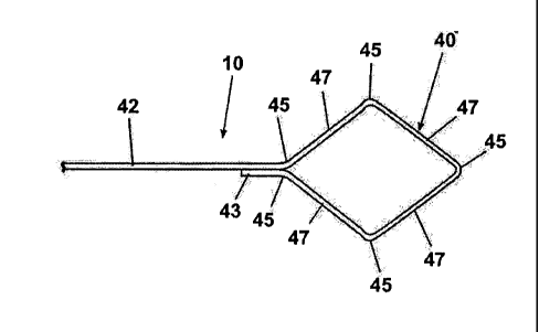
Referring to Figs. 1-4, the localizing wire 10 comprises a localizing anchor
40 and
a localizing thread 42. When mounted in the introducer 12 prior to
implantation (Fig. 3),
the anchor 40 is contained within the lumen 16 and a portion of the localizing
thread
extends exteriorly from the rear of the introducer (Fig. 1). However, it is
not necessary for
the thread to extend exteriorly of the introducer. The thread can be contained
within the
interior of the introducer.
The localizing anchor 40 is fabricated of a resilient, physiologically inert
material
such as stainless steel or titanium wire, which can assume a first collapsed
configuration in
the lumen 16 as illustrated in Fig. 3, and a second expanded configuration
outside the
lumen 16 as illustrated in Figs. 1, 2 and 4.
Referring to Fig. 4, the anchor 40 of the localizing wire 10 is shown in
greater
detail. As illustrated, the anchor 40 is formed from the same wire as the
thread 42. In
other words, the anchor 40 is a continuation of the localizing thread 42, with
an end 43 of
the anchor 40 being connected to the thread 42 to complete the anchor 40. The
end 43 can
be bonded or welded to the thread 42 to form the connection.
The anchor 40 has a diamond or square shape when it is in the expanded
condition
as illustrated in Fig. 4. The diamond shape is formed by providing multiple
bends 45
along the portion of the wire forming the anchor 40, which define therebetween
corresponding segments 47. The bends 45 function as hinges about which the
segments
can move to permit the anchor to transition between the collapsed (Fig. 3) and
expanded
(Fig. 4) conditions.
CA 02870406 2014-11-07
While the anchor 40 is shown as a continuation of the thread 42 in Fig. 4,
Fig. 5
illustrates an alternative where the localizing anchor 40 and the localizing
thread 42 comprise
separate elements, with the localizing anchor 40 attached to the localizing
thread 42 through
a suitable connector 44, such as a crimped or interference-fit collar, a weld,
or the like. With
a separate localizing anchor 40 and localizing thread 42, different materials
can be used for
each element. For example, the localizing anchor 40 could be formed of a
material having a
resiliency that would be suitable for the shape-changing properties described
herein, but
unsuitable for the localizing thread 42. Conversely, the localizing thread 42
could comprise a
material having a stiffness that would be unsuitable for the localizing anchor
40.
The localizing anchor 40 can be formed with different shapes which can be
selected
based upon, for example, the density of the tissue into which the localizing
anchor 40 is to be
placed, the size of the lesion of interest, the anchoring force required, and
the like. In addition
to the diamond shape of Fig. 4, the localizing anchor 40 can have a generally
triangular shape
in the expanded condition as shown in Fig. 6. The triangular shape is shown in
the collapsed
condition in Fig. 6A. Figs. 7 and 7A illustrate another shape for the
localizing anchor 40,
which has the shape of a ring 48 in the expanded condition.
The anchor shapes illustrated in Figs. 3-7A are similar in that they are of a
thread type
structure that encloses an area. When the anchor 40 is in the expanded
condition, the
enclosed area is much greater than when in the collapsed condition. These
shapes have no
sharp edges that would penetrate the surrounding tissue, yet they still anchor
the localizing
wire. The lack of penetrating edges reduces the trauma to the patient during
repositioning.
Figs. 8 and 8A illustrate another localizing anchor 40 comprising a disc 54,
preferably
formed with regularly-spaced fold lines 56 for collapsing the disc 54, similar
to an umbrella.
As illustrated in Fig. 9, a dual-lumen cannula 60 can be utilized comprising a
primary
lumen 62 and a secondary lumen 64. The primary lumen 62 carries the localizing
wire 10,
while the secondary lumen 64 can carry a conventional imaging marker 66 such
as that
disclosed in U.S. Patent No. 6,575,991, or can be used for the introduction of
dye, irrigating
fluid, pharmaceuticals, and the like, such as that disclosed in co-pending
U.S. Patent
Application Publication No. 2004/0106891, filed August 28, 2003.
11
CA 02870406 2014-11-07
Referring now to Figs. 10A-F, the localizing wire 10 is utilized as follows.
The
localizing wire 10 is first loaded into the lumen 16 for introduction of the
cannula 14 into the
tissue of interest 18. As illustrated in Fig. 10A, the cannula 14 is
introduced into the tissue 18
so that the insertion tip 34 is at the location of interest, for example
adjacent to or penetrating
the lesion 20. As shown in Fig. 10B, the localizing wire 10 is then placed at
the location of
interest by relatively moving the localizing wire 10 and the cannula 14 to
expose the anchor
40 beyond the cannula 14. The relative movement is traditionally accomplished
by advancing
the localizing wire 10 relative to the cannula 14. However, the cannula can be
retracted
relative to the localizing wire 10 as disclosed in U.S. Patent 8,131,346,
filed
November 17, 2003, entitled Apparatus And Method For Implanting A Preloaded
Localization Wire.
As the localizing anchor 40 exits the end of the cannula, it expands from the
collapsed
to the expanded condition. As it expands into the surrounding tissue, the
anchor 40
compresses and/or displaces the adjacent tissue sufficiently to imbed the
localizing anchor 40
in the tissue at the location of interest, but does not puncture the tissue as
with a hook-type
anchor. If the localizing anchor 40 is not satisfactorily placed at the
selected location, the
localizing wire 10 can be retracted into the lumen 16 as shown in Fig. 10C by
pulling on the
localizing thread 42 and drawing the anchor 40 back into the cannula. As the
anchor 40
contacts the cannula 12, the interference between the cannula 12 and the
anchor collapses the
anchor 40 from the expanded to the collapsed condition.
As shown in Figs. 10D-E, the cannula 14 can then be repositioned and the
localizing
wire 10 ejected from the lumen 16 into the new location. The cannula 14 can
then be
removed from the tissue 18, leaving the localizing wire 10 in place with the
localizing anchor
40 at the location of interest, and the localizing thread 42 extending outside
the tissue 18 as
with a conventional localizing wire.
Removal of the localizing wire 10 can be accomplished by passing the cannula
14
over the localizing thread 42 and inserting the cannula 14 into the tissue 18
to the localizing
anchor 40. The localizing anchor 40 can then be retracted into the lumen 16,
followed by
removal of the cannula 14 from the tissue 18. Alternatively, the localizing
anchor 40 can be
12
CA 02870406 2014-11-07
repositioned as discussed above. The localizing wire 10 can also be removed
without the
reintroduction of the cannula 14 by merely pulling the localizing thread 42
away from the
tissue 18. The localizing anchor 40 will be urged into a collapsed
12a
CA 02870406 2014-11-07
configuration by the tissue 18 to facilitate removal of the localizing anchor
40 from the
tissue 18.
Prior localizing wires using a hook-shaped anchor that pierced the tissue
could not
be removed from the tissue without causing substantial tissue trauma unless a
cannula was
used. The localizing wire 12 can be removed without a cannula with a
sufficient force, but
will not cause the same trauma to the surrounding tissue as the prior art
devices since the
anchor 40 does not rely on piercing the tissue for anchoring.
The localizing wire described herein has the advantage of being readily
repositionable through retraction of the localizing wire into the lumen of a
cannula after
the localizing wire has been expelled from the lumen. Unlike prior art
localizing wires
having a hook-like configuration, the localizing wire does not puncture the
tissue, whereas
the prior art localizing wires puncture the tissue, complicating, if not
precluding, removal
of the localizing wire from the tissue. Furthermore, the localizing wire
described herein
can be removed from the tissue without the necessity of reinserting a cannula
into the
tissue, thereby minimizing injury and discomfort to the patient.
It should be noted that while all of the embodiments disclose an anchor with a
completely bounded area that varies in size as the anchor is reconfigured from
the release
to the anchor configuration, it is within the scope of the invention for the
anchors not to
completely bound an area. For example, the end 43 need not be attached to the
thread 42.
Instead, the end 43 could be unattached and be shaped to follow the leg 47 or
ring 48. The
end 43 could even be excluded and the leg 47 or ring 48 could just terminate
prior to the
thread 42. In either of these configurations, the area would be effectively
bounded, not
actually bounded and the bound area would be an effectively bound area. Thus,
the term
area as used in this application includes both an actually bound area and an
effectively
bound area.
Fig. 11 illustrates an alternative embodiment localizing wire 110 comprising
an
anchor 140 and thread 142, with at least the thread 142 being configured such
that the
exterior portion 143 of the thread 142 will lie substantially flat against the
exterior of the
tissue 18. The ability of the thread 142 to lie against and not project
substantially above
the tissue 18 significantly reduces the likelihood that the thread 142 will be
accidentally
caught or hooked on a person, machine or other object. This significantly
reduces the
likelihood that the anchor 140 will be dislodged from the position selected by
the medical
13
CA 02870406 2014-11-07
professional. If the anchor 140 was moved from its initial position, it would
reduce the
efficacy of the wire 110 in marking the biopsy site or lesion. The ability of
the thread 142
to lie substantially flat against the tissue 18 also reduces the likelihood of
trauma to the
tissue 18 caused by the jerking of the wire 110. Tape 146 or another similar
material can
be used to hold the thread 142 in place against the exterior of the tissue 18.
Two factors are believed to be most relevant to configure the localizing wire
110
such that the thread 142 lies substantially flat against the exterior of the
tissue 18: the
second moment of area, I, and the Young's Modulus, E, of the material. The
second
moment of area, I, is a physical property of the wire. It is representative of
the distribution
of the mass of the object relative to the objects geometric axis. The greater
the mass is
distributed from the geometric axis, the greater the value of I, and the more
resistant the
object is to bending about the geometric axis.
The Young's Modulus essentially defines the stiffness of the material. All
things
being equal, the greater the Young's Modulus of a material, the greater the
material will
resist deflection.
In the context of a localizing wire, the Young's Modulus provides more room
for
adjustment to get the wire to lay flat against the tissue mass. This is
because the cross-
sectional area of the localizing wire has become somewhat standardized along
with the
cannula. Thus, the Young's Modulus is the best candidate for ensuring that the
external
portion of the localizing wire will lie substantially flat against the tissue.
Regardless of which factor provides the most room for adjustment, ultimately
what
is required is that the second moment of area and the Young's Modulus are
selected such
that the exterior portion of the localizing wire can be bent over and held
against the tissue
mass by a suitable fastener, such as tape. Preferably, the second moment of
area and the
Young's Modulus are selected such that the bending does not result in plastic
deformation
of the localizing wire as such deformation is more likely to cause the portion
of the
localizing wire within the tissue mass to move, which might negate the marking
functionality of the localizing wire. It is more preferred that the second
moment of area
and the Young's Modulus are selected such that the external portion of the
localizing wire
bends as needed under its own weight.
While it is preferred that the bending occur immediately at or around the
insertion
point 145 of the localizing wire into the tissue mass, it is not necessary. If
the localizing
14
CA 02870406 2014-11-07
wire does not bend near the insertion point 145 before the localizing wire
lies flat against
the tissue, a gap will form between the localizing wire and the tissue thereby
effectively
creating a small loop on which something could catch. The smaller this loop,
the less
likely an object will catch it. Thus, this loop should be minimized, but it
should be done is
such a way that does not cause the shifting of the internal portion of the
localizing wire,
which would negate the marking functionality.
The "lying flat" functionality can be applied to any type of localizing wire
and not
just the localizing wires shown in Figs. 1-10F. For example, Figs. 12 and 13
illustrate a
localizing wire 210 configured to lie flat against the exterior of the tissue
mass and
comprising a straight thread 242 and a hook-shaped anchor 240. The thread has
a
generally circular cross section 244. Other cross sections can be used, such
as oval,
square, polygonal, and they can be either hollow or solid.
The circular cross section 244 as illustrated is typical of current localizing
wires
that do not have the lie flat functionality. Thus, to achieve the lie flat
functionality, the
localizing wire is made from a material that provides a Young's Modulus that
when
combined with the cross section will permit the thread 242 to lie flat against
the tissue 18.
For the described cross section, a suitable material is annealed steel. The
annealing of the
steel reduces the stiffness of the material, which reduces the Young's
Modulus, to permit
the bending of the thread 242 as needed to lie flat.
It is most preferred that localizing wire 242 bends as needed to lie flat
under its
own weight. Thus, the portion of the thread 242 extending exteriorly of the
tissue
preferably has sufficient mass to effect the bending. For the example
illustrated, the
length of the thread preferably takes into account a suitable safety margin to
ensure that
there is enough thread 242 exterior of the tissue mass to effect the bending.
It is worth noting that not the entire length of the thread need be configured
to
effect the lying flat of the thread on the exterior of the tissue mass. This
portion of the
thread can be referred to as the bending portion, which may extend along all
or only a
portion of the thread. It is anticipated that only that portion of the thread
generally near
the insertion point 145 into the tissue mass need be so configured if the
bending is to
minimize any gap between the exterior of the tissue and the thread. However,
it is
anticipated that for manufacturing simplicity, the entire thread will be so
configured,
especially if the material is treated to select the desired Young's Modulus.
CA 02870406 2014-11-07
Figs. 14-16 illustrate another embodiment of a localizing wire 310 that can be
repositioned like the 'localizing wire 10. Additionally, the localizing wire
310 can be
withdrawn from the tissue mass 18 after the localizing wire 310 is implanted
and after the
removal of the cannula 14, without the reintroduction of the cannula 14.
The localizing wire 310 comprises a configurable anchor 340 and a thread 342.
An
actuator for reconfiguring the configurable anchor in the form of a sheath 350
is slidably
mounted on the thread 342. As illustrated, the anchor 340 is made of a
resilient material,
such as Nitenol, that permits the anchor 340 to be configured between an
anchoring
configuration (Fig. 13), where it has a hook-like shape for anchoring in the
tissue mass,
and a release configuration (shown in phantom in Fig. 14), where the anchor is
generally
straight relative to the thread 342 to release the anchor 340 from the tissue
mass 18.
It should be noted that while the anchor 340 illustrated in Figs. 14-16 is
known in
the art, other configurable anchors, such as those shown in Figs. 1-9, can
also be used.
The sheath 350 has a proximal end 352 near the anchor 340 and an opposing
distal
end 354. The sheath 350 defines a hollow interior 356 in which the thread 342
is received
to slidably couple the sheath 350 and thread 342 for relative slidable
movement. The
sheath 350 functions as an actuator for moving the anchor between the
anchoring and
release configurations. When the sheath 350 is withdrawn from the anchor 340
(Fig. 14),
the anchor 340 because of its resiliency will inherently assume the anchoring
configuration. To effect a change in the configuration of the anchor 340, the
sheath 350 is
advanced toward the anchor 340 by the relative movement of the thread 342 and
sheath
350. As the proximal end 352 of the sheath 350 is advanced toward the anchor
340, the
proximal end 352 comes into contact with the anchor 340. The continued
advancement of
the sheath 350 causes the anchor 340 to straighten as it is received within
the interior 356
of the sheath 350 and thereby effect the reconfiguring of the anchor 340 into
the release
configuration.
It should be noted that the anchor 340 need not be completely received within
the
interior 356 of the sheath 350 for the anchor 340 to be in the release
configuration. The
anchor 340 need only be straightened enough that the anchor will release from
the tissue.
The complete receipt of the anchor 340 in the interior 356 of the sheath 350
is preferred as
that ensures that the anchor 340 is straight enough and it will cause the
least amount of
tissue damage when the localizing wire is moved or withdrawn.
16
CA 02870406 2014-11-07
The sheath 340 is illustrated as being transparent, but it can have any
desired
degree of opacity. The sheath 340 is preferably made from a suitable plastic.
The sheath
340 can also have any suitable type of imageable markings that permit the
location of the
sheath 340, especially the proximal end 352, which would permit the user to
view the
relative location of the proximal end 352 and the anchor 340, which can also
have such
imageable markings. Such imageable markings are well known in the art and vary
on the
type of imaging technique being used.
The implanting of the localizing wire 310 is essentially identical to that
described
for the localizing wire 10. In short, an introducer, typically a cannula 14,
is inserted into
the tissue mass 18. The localizing wire is inserted through the lumen of the
cannula and
out the open end of the cannula, where the anchor 340 can anchor in the tissue
mass 18.
Once the localizing wire is properly positioned, the cannula 14 is withdrawn.
The localizing wire 310 can be inserted along with the cannula 14 or after the
cannula 14 is inserted. The localizing wire 310 can be inserted with the
anchor 340 either
in the anchoring configuration or the release configuration. If it is inserted
in the release
configuration, it will, of course, need to be put in the anchoring
configuration to anchor.
After the withdrawal of the cannula 14, the localizing wire 310 can be
repositioned
or withdrawn without the need for reinserting the cannula 14, as is now
required. The
localizing wire 310 can be repositioned or withdrawn by relatively moving the
thread 342
and sheath 350 such that the anchor is reconfigured from the anchoring
configuration to
the release configuration. In the release configuration, the localizing wire
can be
repositioned or withdrawn. If repositioned, the sheath 350 and thread 342 are
relatively
moved to configure the anchor in the anchoring configuration and re-anchor the
localizing
wire. If withdrawn, the user merely pulls on the exterior portion of the
localizing wire
310.
The reconfiguring of the anchor 340 after the implanting of the localizing
wire
310, can be done by manipulating the portions of the sheath 350 and thread 342
(sliding
them relative to each other) that extend exteriorly of the tissue mass. This
prevents the
need for reinserting the cannula 14 as is currently done.
The ability to remove the localizing wire 310 without the reintroduction of
the
cannula 14 is very beneficial. Often the localizing wire 310 will be inserted
in a tissue
mass at the location of a biopsy site. The localizing wire 310 is left in
while analysis is
17
CA 02870406 2014-11-07
run on the biopsy specimen. It can take from a few hours to a few days to
complete the
analysis. This is too long of a time to leave the cannula 14 inserted in the
patient as the
rigid cannula 14 is uncomfortable, if not painful, when left in. It can also
increase the risk
of infection since the lumen of the cannula 14 creates an open air pathway
into the tissue
mass 18. In cases where the analysis confirms that no follow-up surgical
procedure need
be done, such as a tissue removal, the localizing wire can easily be removed
without
reintroducing the cannula 14, eliminating additional tissue damage and
discomfort for the
patient, not to mention the cost of the cannula insertion procedure.
It should be noted that the cannula 14 need not be completely withdrawn from
the
tissue mass for the sheath 340 to be used to effect the reconfiguring of the
anchor. The
cannula 14 need only be withdrawn away from the anchor a sufficient amount
such that
the cannula 14 does not interfere with the reconfiguring of the anchor. It is
expected that
in most cases the cannula will be complete removed because of the increased
risk of
infection and accidental tissue damage if it is left in placed, especially
since the cannula
will no longer be needed for the removal of the localizing wire because of the
sheath.
It should also be noted that during insertion the anchor could extend beyond
the
end of the sheath but would still be constrained in the release configuration
by the cannula,
and the withdrawal of the cannula away from the anchor would effect the
reconfiguration
of the anchor to the anchor configuration. The sheath could then be used to
reconfigure
the anchor from the anchor configuration to the release configuration when it
is desired to
remove the localizing wire.
Fig. 17 illustrates an alternative localizing wire 410 having an alternative
design
for the sheath in the form of a coil spring 450. The coil spring 450 is wound
such that it
defines a hollow interior 456 for receiving the thread 342. The coil spring
450 has a
proximal end 452 and a distal end 454. The operation of the localizing wire
410 with the
coil spring 450 is identical to that previously described.
The localizing wires 310 and 410 can be configured to lay flat against the
exterior
of the tissue mass 18 as previously described. With the localizing wires 310,
410, the
characteristics of both the thread 342, 442 and the sheath 350, 450 must be
taken into
account to achieve the laying flat functionality.
While the invention has been specifically described in connection with certain
specific embodiments thereof, it is to be understood that this is by way of
illustration and
18
CA 02870406 2014-11-07
not of limitation. Reasonable variation and modification are possible within
the scope of
the forgoing disclosure and drawings without departing from the spirit of the
invention
which is defined in the appended claims.
19