Note: Descriptions are shown in the official language in which they were submitted.
DELIVERY SYSTEM FOR OCULAR IMPLANT
[0001] Continue to next paragraph.
BACKGROUND
[0002] This disclosure relates generally to methods and devices for
use in
delivering devices for treating glaucoma.
[0003] The mechanisms that cause glaucoma are not completely known.
It
is known that glaucoma results in abnormally high pressure in the eye, which
leads to
optic nerve damage. Over time, the increased pressure can cause damage to the
optic
nerve, which can lead to blindness. Treatment strategies have focused on
keeping the
intraocular pressure down in order to preserve as much vision as possible over
the
remainder of the patient's life.
1
CA 2870549 2019-07-08
CA 02870549 2014-10-15
WO 2013/158919 PCT/US2013/037234
[0004] Pursuant to such strategies, one or more implants can be
delivered into the eye for shunting fluid out of the anterior chamber in order
to
regulate pressure in the eye. Accurate placement of an implant in the angle
of the eye is critical for the targeted effect of reducing intraocular
pressure
(10P). Placing an implant too distally into the eye, such as too distally into
the
supraciliary space, may leave no portion of the implant remaining in the
anterior chamber. This may inhibit aqueous outflow, as the fluid will not have
a direct communication with the flow target location if there is no opening to
the anterior chamber.
[0005] Conversely if the implant is placed too proximally in the
supraciliary space such that a significant portion of the implant remains in
the
anterior chamber, damage to the corneal endothelium may result from
implants that protrude upwards and touch the cornea. Implants placed too
proximally may also touch the iris resulting in increased amounts of pigment
dispersion in the eye, which can increase outflow resistance and intraocular
pressure by clogging the trabecular meshwork. Correct placement of the
implant is desired for a safe and successful surgical outcome.
[0006] .. In view of the foregoing, there is a need for improved
delivery systems for delivering implants into the eye such as by way of an ab
intemo procedure.
SUMMARY
2
CA 02870549 2014-10-15
WO 2013/158919 PCT/US2013/037234
[0007] There is a need for improved delivery systems, devices
and methods for the treatment of eye diseases such as glaucoma.
[0008] In a first embodiment, disclosed herein is a delivery device
for delivering an ocular implant into an eye. The delivery device can include
a
proximal handle portion and a distal delivery portion coupled to a distal end
of
the handle portion and configured to releasably hold an ocular implant. In
addition, the delivery portion can include a sheath positioned axially over a
guidewire. The delivery device can further include an actuator coupled to a
mechanism that releases the ocular implant from the delivery portion upon
actuation of the actuator.
[0009] Also described herein are methods of delivering an ocular
implant to a target location within an eye. In an embodiment, disclosed is a
method including loading the ocular implant onto a distal delivery portion of
a
delivery system. The delivery system can include a proximal handle portion
with the delivery portion coupled to a distal end of the handle portion. In
addition, the delivery portion can be configured to releasably hold the ocular
implant. The delivery portion can further include a sheath positioned axially
over a guidewire. Additionally, the delivery device can include an actuator
coupled to a mechanism that releases the ocular implant from the delivery
portion upon actuation of the actuator. The method can further include
inserting the distal delivery portion and the ocular implant into the eye
through
a corneal incision and positioning the ocular implant into the target location
within the eye by way of an ab-interno procedure. Furthermore, the method
can include actuating the actuator and releasing the ocular implant into the
target location.
3
CA 02870549 2014-10-15
WO 2013/158919 PCT/US2013/037234
[0010] Other features and advantages should be apparent from
the following description of various embodiments, which illustrate, by way of
example, the principles of the described subject matter.
BRIEF DESCRIPTION OF THE DRAWINGS
[0011] These and other aspects will now be described in detail
with reference to the following drawings.
[0012] FIG. 1 shows an example cross-sectional view of a portion
of the human eye.
[0013] FIG. 2 shows and an example partial cross-sectional view
of the eye showing a part of the anterior and posterior chambers of the eye
and an ocular implant implanted in the eye.
[0014] FIG. 3 shows a perspective view of an embodiment of a
delivery device having a proximal handle component and a distal delivery
component with an ocular implant loaded onto the distal delivery component.
[0015] FIG. 4 shows a close up view of the distal end of the
delivery component of FIG. 3 which illustrates the implant loaded onto a
guidewire of the delivery system.
[0016] FIG. 5 shows a partial cross section view of the delivery
system of FIG. 3 showing a distal portion of the handle component, including
4
CA 02870549 2014-10-15
WO 2013/158919 PCT/US2013/037234
the spring-loaded actuator in a compressed configuration, and the distal
delivery component.
[0017] FIG. 6 shows the partial cross section view of the delivery
system of FIG. 5 with the spring-loaded actuator shown in a decompressed
configuration which releases the implant from the distal delivery component.
[0018] FIG. 7 shows an embodiment of the guidewire of the
delivery system having a curved configuration.
[0019] FIG. 8 shows an embodiment of the guidewire of the
delivery system having a sinusoidal configuration.
[0020] FIG. 9 shows an embodiment of the guidewire of the
delivery system having a length sufficient to extend from the supraciliary
space down to the sub-retinal space.
[0021] FIG. 10 shows an enlarged view of the anterior region of
the eye with the implant approaching the supraciliary space or suprachoroidal
space from the anterior chamber.
[0022] FIG. 11 shows a perspective view of an embodiment of a
direct visualization (DV) system.
[0023] FIG. 12 shows an enlarged view of a distal end of the DV
system including a part of a DV wire 12 and stopper tube 16.
CA 02870549 2014-10-15
WO 2013/158919 PCT/US2013/037234
[0024] FIG. 13 shows a cross-sectional view of a portion of the
DV system shown in FIG. 11.
[0025] FIG. 14 shows the distal end of the DV system shown in
FIG. 11 inserted into an eye.
[0026] FIG. 15 shows the DV wire of the DV system aligned
alongside an implant delivery applier showing the corresponding indicators.
[0027] FIG. 16 shows the DV wire inserted into the eye for
measuring anatomical features of the eye.
[0028] FIG. 17 shows the distal end of the DV wire abutting the
base of the angle of the eye and the stopper tube in an advanced position
along the DV wire.
[0029] FIG. 18 shows the implant delivery applier implanting an
ocular implant through the same incision the DV system used in FIGS. 8 and
9.
[0030] FIG. 19 shows the indicators on the implant delivery
applier being used to determine the proper insertion depth of the implant.
[0031] FIG. 20 shows the implant in an implanted state and
providing fluid communication between the anterior chamber and the
suprachoroidal or supraciliary space.
6
CA 02870549 2014-10-15
WO 2013/158919 PCT/US2013/037234
[0032] FIGS. 21A shows an embodiment of the implant delivery
applier having a feedback mechanism.
[0033] FIGS. 21B shows the feedback mechanism of the implant
delivery applier shown in FIG. 21A in a retracted state.
[0034] FIG. 22 shows a cross-section view of an embodiment of a
pencap implant loader configured to house an implant and releaseably couple
to a delivery device.
[0035] FIG. 23 shows a perspective view of the pencap implant
loader of FIG. 22.
[0036] FIG. 24 shows a cross-section view of another
embodiment of a pencap implant loader configured to house an implant and
releaseably couple to a delivery device.
[0037] FIG. 25 shows a perspective view of the pencap implant
loader of FIG. 24.
[0038] Like reference symbols in the various drawings indicate
like elements.
DETAILED DESCRIPTION
[0039] FIG. 1 is a cross-sectional, perspective view of a portion of
the eye showing the anterior and posterior chambers of the eye. A schematic
7
CA 02870549 2014-10-15
WO 2013/158919 PCT/US2013/037234
representation of an implant 105 is positioned inside the eye such that a
proximal end 110 is located in the anterior chamber 115 and a distal end 120
communicates with and/or is located in or near the supraciliary space or
suprachoroidal space (sometimes referred to as the perichoroidal space). It
should be appreciated that FIG. 1 and other figures herein are schematic and
are not necessarily to scale with respect to size and relative positions of
actual eye tissue.
[0040] The implant 105 provides a fluid pathway between the
anterior chamber 115 into the supraciliary space and toward the
suprachoroidal space. The implant 105 has a distal end120 that may be
positioned in the supraciliary space or the suprachoroidal space. The implant
105 may be positioned at least partially between the ciliary body and the
sclera or it may be at least partially positioned between the sclera and the
choroid. The distal end 120 of the implant 105 is not necessarily positioned
between the choroid and the sclera.
[0041] In an embodiment, the implant 105 is an elongate element
having one or more internal lumens through which aqueous humor can flow
from the anterior chamber 115 into the supraciliary space. The implant 105
can have a substantially uniform internal diameter along its entire length,
although the shape of the implant 105 can vary along its length (either before
or after insertion of the implant), as described below. Moreover, the implant
105 can have various cross-sectional shapes (such as a circular, oval or
rectangular shape) and can vary in cross-sectional shape moving along its
length. The cross-sectional shape can be selected to facilitate easy insertion
8
into the eye. The following applications describe exemplary implants: U.S.
Patent
Publication Nos. 2007-0191863 and 2009-0182421.
[0042] FIG. 2 is a cross-sectional view of a portion of the human eye. The
eye is generally spherical and is covered on the outside by the sclera S. The
retina (not
shown) lines the inside posterior half of the eye. The retina registers the
light and sends
signals to the brain via the optic nerve. The bulk of the eye is filled and
supported by
the vitreous body, a clear, jelly-like substance. The elastic lens L is
located near the
front of the eye. The lens L provides adjustment of focus and is suspended
within a
capsular bag from the ciliary body CB, which contains the muscles that change
the focal
length of the lens. A volume in front of the lens L is divided into two by the
iris I, which
controls the aperture of the lens and the amount of light striking the retina.
The pupil is
a hole in the center of the iris I through which light passes. The volume
between the iris
I and the lens L is the posterior chamber PC. The volume between the iris I
and the
cornea is the anterior chamber AC. Both chambers are filled with a clear
liquid known
as aqueous humor.
[0043] The ciliary body CB continuously forms aqueous humor in the
posterior chamber PC by secretion from the blood vessels. The aqueous humor
flows
around the lens L and iris I into the anterior chamber and exits the eye
through the
trabecular meshwork, a sieve-like structure situated at the corner of the iris
I and the
wall of the eye (the corner is known as the iridocorneal angle). Some of the
aqueous
humor filters through the trabecular
9
3247716
CA 2870549 2019-07-08
CA 02870549 2014-10-15
WO 2013/158919 PCT/US2013/037234
meshwork near the iris root into Schlemm's canal, a small channel that drains
into the ocular veins. A smaller portion rejoins the venous circulation after
passing through the ciliary body and eventually through the sclera (the
uveoscleral route).
[0044] The internal lumen of the implant 105 serves as a
passageway for the flow of aqueous humor through the implant 105 directly
from the anterior chamber toward or into the supraciliary or suprachoroidal
space. In addition, the internal lumen of the implant 105 can be used as an
access location to mount the implant 105 onto a delivery device, as described
in more detail below. The internal lumen can also be used as a pathway for
flowing fluid, such as an irrigation fluid or a visco-elastic substance(s),
into the
eye for flushing or to maintain pressure in the anterior chamber, or using the
fluid to assist in dissection, visualization or hydraulic creation of a
dissection
plane into or within the suprachoroidal space.
[0045] .. Fluid can be flowed toward or into the supraciliary or
suprachoroidal space, for example via a delivery cannula or through the
internal lumen of the shunt. The fluid can be flowed into the eye with a
pressure sufficient to form a dissection plane into or within the supraciliary
suprachoroidal space. The fluid can accumulate within the eye so as to form
a lake. In general, hydro-dissection or the injection of fluids such as a
visco-
elastic substance(s) can be used to separate the ciliary body from the sclera
to enlarge an area of detachment of the ciliary body from the sclera with or
without insertion of a device.
CA 02870549 2014-10-15
WO 2013/158919 PCT/US2013/037234
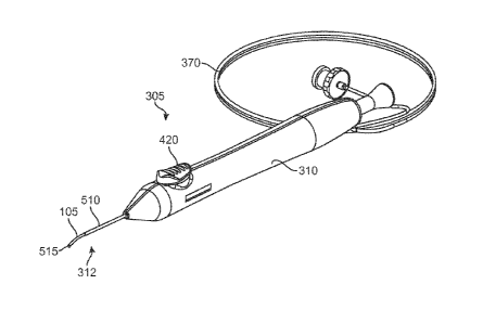
[0046] FIG. 3 shows an embodiment of a delivery system 305 that
can be used to deliver the implant 105 into the eye. In some embodiments,
the implant 105 can provide fluid communication between the anterior
chamber toward the suprachoroidal or supraciliary space while in an
implanted state. It should be appreciated that these delivery systems 305 are
exemplary and that variations in the structure, shape and actuation of the
delivery system 305 are possible. The delivery system 305 can include a
proximal handle component 310 and a distal delivery component 312. The
proximal handle component 310 can include an actuator 420, such as a
button, to control the release of an implant from the delivery component 312
into a target location in the eye. The actuator 420 can vary in structure and
is
not limited to a button.
[0047] An embodiment of the delivery component 312 includes an
elongate applier in the form of a guidewire 515 and a "stopper" or sheath 510
positioned axially over the guidewire 515. The guidewire 515 can insert
longitudinally through the internal lumen of the implant 105 and can assist in
inserting and positioning the implant 105 into the target location. The sheath
510 can aid in the release of the implant 105 from the delivery component 312
into the target location in the eye. In addition, the actuator 420 can be used
to
control movement or relative movement of the guidewire 515 and/or the
sheath 510. For example, the sheath 510 can be fixed relative to the handle
component 310 and act as a stopper which can impede the implant 105 from
moving in a proximal direction as the guidewire 515 is withdrawn proximally
from the implant 105 upon actuation of the actuator 420.
11
CA 02870549 2014-10-15
WO 2013/158919 PCT/US2013/037234
[0048] For example, in a first state, the guidewire 515 can be
extended distally relative to a distal end of the sheath 510. Actuation of the
actuator 420, such as by pressing the actuator 420, can cause the guidewire
515 to slide proximally or retract into the sheath 510. This can effectively
disengage the implant 105 off the distal end of the guidewire 515 and
releases the implant 105 in a controlled fashion into the target location.
Controlled disengagement of the implant 105 off the distal end of the
guidewire 515 can assist in ensuring that positioning of the implant 105
within
the target location is maintained.
[0049] FIG. 4 shows an embodiment of the implant 105 mounted
on the delivery component 312 of the delivery system 305. More specifically,
the implant 105 can be mounted on the distal region of the guidewire 515, as
shown in FIG. 4. In addition, the sheath 510 can be sized and shaped to
receive or abut a portion of the proximal end of the implant 105. In this
embodiment, upon actuation of the actuator 420, the guidewire 515 can slide
in a proximal direction (arrow P) into the sheath 510 which can allow the
proximal end of the implant 105 to abut the distal end of the sheath 510 and
prevent the implant 105 from sliding in the proximal direction. This can
effectively disengage the implant 105 off the distal end of the guidewire 515
and controllably releases the implant 105 into the target location within the
eye.
[0050] In some embodiments, the actuator 420 can be a push-
button that is coupled to a spring-activated mechanism. Upon applying a
force onto the actuator 420, the spring mechanism can retract the guidewire
515 toward and/or into the sheath 510 which can release the implant 105 from
12
CA 02870549 2014-10-15
WO 2013/158919 PCT/US2013/037234
the guidewire 515. The mechanism by which the guidewire 515 can be
withdrawn into the sheath 510 can be a spring activated assembly or any of a
variety of mechanisms that allow the guidewire to retract upon activation of
an
actuator.
[0051] FIG. 5 shows an embodiment of a portion of the delivery
system 305 in cross-section with the implant 105 loaded onto the guidewire
515. The delivery system 305 can include a front spring 550 which can assist
in positioning the guidewire 515. For example, the front spring 550 can be
compressed or charged which can allow the guidewire 515 to be positioned in
an extended state relative to the handle 310. When the guidewire 515 is in an
extended state, the guidewire 515 can be loaded with the implant 105, as
shown in FIG. 5.
[0052] The delivery system 305 can include a variety of
mechanisms for assisting in the positioning of the guidewire 515. For
example, the delivery system 305 can include a feature which can interact
with the actuator 420 in order to allow the actuator to assist in positioning
the
guidewire 515. For example, the guidewire 515 can be attached at a proximal
end to a piston 560 having a de-tent latch 555. The de-tent latch 555 can
interact with the actuator 420 such that upon actuation of the actuator 420,
the
555 latch can release the piston 560 from a locked position and allow the
piston 560 to move. For example, once the piston 560 is allowed to move, the
front spring 550 can force the piston to move in a direction, such as in a
proximal direction, thus causing the guidewire 515 to move in a proximal
direction. Movement of the guidewire 515 in a proximal direction can allow
13
CA 02870549 2014-10-15
WO 2013/158919 PCT/US2013/037234
the implant 105 loaded on the distal end of the guidewire 515 to be released
from the guidewire 515.
[0053] In some embodiments, the actuator 420 can be configured
such that when actuated or depressed by the user, the detent latch 555 of the
piston 560 is flexed downward thereby allowing the front spring 550 to
release. As the piston 560 moves proximally with the guidewire 515, the
implant 105 can abut the distal end of the stopper tube 510 and release from
the guidewire 515. FIG. 6 shows an embodiment of the delivery system 305
in a retracted state where the front spring 550 is in a decompressed state
with
the implant 105 fully released from the guidewire 515.
[0054] The travel of the piston 560 can be defined such that the
guidewire 515 reaches a complete stop in the proximal direction only after the
implant 105 is fully released. In addition, the force of the front spring 550
can
allow withdrawal of the guidewire 515 from the implant 105 when the implant
105 is positioned in a variety of angles relative to the stopper tube 510. For
example, the force of the front spring 550 can allow the withdrawal of the
guidewire 515 from the implant 105 when the implant 105 is at a 45 degree
angle relative to the stopper tube 510, such as what may be encountered
when the implant 105 is being deployed to the supraciliary space.
[0055] In some embodiments, for example, the front spring 550
can provide approximately 1.0 to 2.0 lbf at the compressed or charged
configuration which can allow the guidewire 515 to withdraw from the implant
105, including when the implant 105 is positioned at an approximate 45
degree angle relative to the stopper tube 510. However, the front spring 550
14
CA 02870549 2014-10-15
WO 2013/158919 PCT/US2013/037234
can provide any of a variety of spring force which allows the guidewire 515 to
release the implant 105 positioned at a variety of angles relative to at least
the
stopper tube 510.
[0056] In some embodiments, the front spring 550 can create
approximately 2.0 to 10.0 lbf. For example, a greater spring force of the
front
spring 550 can allow the guidewire 515 to retract in a variety of conditions.
In
addition, a lower force of the front spring, such as 0.10 to 1.0 lbf, may
reduce
the speed of the retraction and reduce the force required to reload the
system.
Any of a variety of front springs 550 can be implemented in the delivery
system 350.
[0057] A dampening element, such as grease 565, may be placed
between the piston 560 and inside wall of the handle 310 which can assist in
providing a slower retraction of the guidewire 515. A slower retraction of the
guidewire 515 can prevent or lessen any jerking motion of the delivery system
350 in the user's hands, including at the end of the piston 560 travel. This
dampening grease 565 can be a silicone grease such that grease is
unaffected by production level e-beam sterilization dose of 25 -50 kGy. In
addition, other dampening elements aside from grease 565 may be used.
Alternate dampening grease such as low, medium, or high viscosity
fluorocarbons may be used to alter the dampening and speed of deployment.
These materials may have a larger acceptable e-beam sterilization range.
[0058] In some embodiments, the spring-activated retraction of
the guidewire 515 can improve the delivery of supraciliary and suprachoroidal
implants. For example, some current tools for implanting ocular implants
CA 02870549 2014-10-15
WO 2013/158919 PCT/US2013/037234
require a sliding motion of the user's finger, such as in the range of
approximately 0.280" inches of travel, in order to release the implant. The
sliding motion can be difficult for surgeons to achieve while simultaneously
holding the distal end of the delivery tool steady. In contrast, the spring-
activated mechanism of the present disclosure, including the spring activated
push-button mechanism, allows for smaller and more ergonomic motion of the
users finger to activate guidewire 515 retraction which also allows the user
to
maintain the distal end of the delivery device 312 in a steady position. In
addition, the spring-activated mechanism of the present disclosure can allow
implantation to occur more quickly and with less unwanted distal movement of
the implant 105 during the guidewire retention.
[0059] The outer diameter of the guidewire 515 can be smaller
than the inner diameter of the implant 105 (i.e. the fluid channel) such that
the
implant 105 can be loaded onto the guidewire 515 by sliding the guidewire
515 into and through an internal lumen of the implant 105. In some
embodiments, the guidewire 515 can include a retention feature that can act
to retain the implant 105 on the guidewire 515. For example, the guidewire
515 can include a retention feature which can assist in retaining the implant
105 on the guidewire 515 during blunt dissection and implantation in order to
prevent the implant 105 from inadvertently sliding off the guidewire 515.
[0060] Before the implant 105 has been released from the
guidewire 515 and implanted into the target location within the eye, the
implant 105 can be moved either distally or proximally in order to adjust its
placement. This can exert axial forces on the implant 105 which may cause it
to slip off the guidewire 515 if it is not well retained on the guidewire 515.
16
CA 02870549 2014-10-15
WO 2013/158919 PCT/US2013/037234
Therefore, in some embodiments, the guidewire 515 can include features
which can assist in retaining the implant 105 onto the guidewire 515 during
positioning of the implant 105, including positioning the implant 105 within
the
target location.
[0061] FIG. 7 shows an embodiment of a guidewire 515 which
has at least one retention feature including a curved configuration 520 along
a
length of the guidewire 515. In some embodiments, the curved configuration
520 of the guidewire 515 can assist in facilitating entry of the implant 105
into
the supracilliary space. In addition, the curvature of the guidewire 515 can
change the shape of the implant 105 due to the implant 105 conforming to the
curved shape of the guidewire 515 which can facilitate placement of the
implant 105 into the supraciliary space as it curves along the scleral wall.
The
curvature radius or arc, including the curved configuration 520 of the
guidewire 515, can vary and can be in the range of approximately .425" to
about .525" with a central angle of approximately 20 degrees to approximately
40 degrees.
[0062] Additionally, any part of the guidewire 515 can have the
curved configuration 520, including either the distal end or the entire length
of
the guidewire 515. Furthermore, the guidewire 515 can alternate between
having a variety of configurations, including both straight and curved
configurations. For example, the guidewire 515 can have a curved
configuration in its natural state but can conform to a straight passageway,
such as through the handle 310 of the delivery system 305. Therefore, the
guidewire 515 can conform to a straight passageway and return to a curved
configuration after having passed through the straight passageway.
17
CA 02870549 2014-10-15
WO 2013/158919 PCT/US2013/037234
[0063] In some embodiments, the guidewire 515 can have one or
more cut patters along a length of the guidewire 515 which can allow the
guidewire 515 to be more flexible than the material comprising the guidewire
515 can allow. For example, the distal end or tip of the guidewire 515 can
include a spiral cut pattern which allows the tip of the guidewire 515 to
deflect
or bend in one or more of a variety of directions relative to a longitudinal
axis
of the guidewire 515. Furthermore, the spiral cut pattern can allow the distal
end or tip of the guidewire 515 to deflect or bend to a greater degree than
what the guidewire could achieve without the spiral cut pattern. These cut
patterns may additionally serve as fluid conduits which can provide a
passageway for substances injected into the guidewire 515 to be released to
an area surrounding the guidewire, including either the implant or the eye.
[0064] FIG. 8 shows an embodiment of the guidewire 515 having
at least one retention feature including a sinusoidal or S-curve configuration
along a length of the guidewire 515. The sinusoidal or S-curve configuration
can assist in retaining the implant 105 onto the guidewire 515, such as by at
least one curved region 524 along a length of the guidewire 515. The at least
one curved feature can include a protrusion, bump, etc. For example, the
curved feature 524 can be configured to provide an interference fit between
the guidewire 515 and the inner lumen of the implant 105.
[0065] In some embodiments, the retention feature can include an
S-shaped curve along a length of the guidewire 515 which can have one or
more rounded curved features 524, including bends or peaks, as shown in
FIG. 8. Furthermore, each retention feature, such as curved feature 524, can
form a point of contact between the inner lumen of the implant 105 and the
18
CA 02870549 2014-10-15
WO 2013/158919 PCT/US2013/037234
guidewire 515. The curved features 524 of the guidewire S-curve can also
reduce the risk of damaging the inner lumen of the implant 105 as the
guidewire 515 is released from the implant 105. In addition, the retention
features can provide a gentle interaction and retention between the guidewire
515 and the implant 105, including during removal of the guidewire 515 from
the implant 105. Alternatively, the guidewire 515 retention features can be
stamped, bent or shape-set, including in the shape of swells or other
formations along at least a part of the length of the guidewire 515.
[0066] In an embodiment, an amount of retention force can be
defined by the peak-to-peak distance between two or more retention features
or curved features 524 of the implant 105. For example, larger peak-to-peak
distances between the two or more curved features 524 can produce higher
retention forces and smaller peak-to-peak distances can produce lower
retention forces. In some embodiments, a peak-to-peak distance that is too
large can cause damage to the implant 105, such as due to the guidewire 515
scraping away material along the inner lumen during removal. For example,
the peak-to-peak distance may be in the range of approximately 0.0100" to
approximately 0.0200", or in the range of approximately 0.0120" to
approximately 0.0150". In addition, at least one retention force acting upon
the implant 105, such as a polyimide implant, by the guidewire 515 of
approximately .050 - .200 lbf can be sufficient to retain the implant 105
along
the guidewire 515 during manipulation of the implant 105 prior to implantation
into the target location.
[0067] In alternate embodiments, the material of the guidewire
515 can be made out of one or more flexible materials, such as metals
19
CA 02870549 2014-10-15
WO 2013/158919 PCT/US2013/037234
including stainless steel or elgiloy, and polymers such as Pebax, silicones,
urethanes, including a variety of combinations of materials. In some
embodiments, the guidewire 515 can have a radius of curvature or arc which
is less than 0.425", such as in order to provide a small curvature of the
implant 105 during insertion. This configuration can be advantageous when
access between the incision and the target location requires the implant 105
to be introduced into the target location by way of a small radius, such as
less
than 0.425".
[0068] .. Alternatively, the radius of curvature or arc of the guidewire
515 can be larger than 0.525". Any of a variety of radius of curvature or arcs
of the guidewire 515 can be implemented into any of the delivery systems 305
in order to best accommodate insertion of the implant 105 into the designated
target location. For example, the radius of curvature or arc of the guidewire
515 may be such that it can allow the implant 105 to bend against the scleral
wall during insertion into the supraciliary space. In addition, the retention
features of the guidewire 515 can vary and can include one or more of a
variety of shapes and sizes along a length of the guidewire 515. For example,
the retention features can be configured to include spiral shapes, triangle
peaks or the like. Additionally, the retention features can extend along one
or
more of a variety of planes, including more than one retention feature
extending in planes positioned perpendicular relative to each other.
[0069] .. In addition, any number of retention features can be
positioned along a length of the guidewire 515. For example, at least two,
including more than five or more than ten retention features can be positioned
along a length of the guidewire 515. In addition, each retention feature can
CA 02870549 2014-10-15
WO 2013/158919 PCT/US2013/037234
provide the same or a variety of different amounts of retention forces for
securing the implant 105 in a position along the guidewire 515. In some
embodiments, the peak-to-peak distance between the retention features can
be larger than the inner diameter of the implant 105 and can be a
dimensioned larger than .0150" such that it does not damage the implant 105.
[0070] In some embodiments of the delivery system 305, instead
of using the guidewire 515 to provide retention of the implant 105, an
additional feature of the delivery system 305 or device can be used in order
to
provide the necessary retention of the implant 105 onto the guidewire 515.
This may include, for example, a Pebax material which can be coupled onto a
part of the guidewire 515 in order to create at least a width along the
guidewire 515 that is larger than the inner diameter of the implant 105. For
example, the Pebax material can be crimped to the guidewire and can retain
the implant 105 relative to the guidewire 515 until the implant 150 is
released
from the delivery system 305, such as after actuation of the actuator 420.
[0071] As shown in FIGS. 3 and 4, the delivery system 305 can
include at least one fluid delivery feature which can be configured to deliver
fluid into at least one of the implant or the eye, including during or after
implantation of the implant 105. The delivered fluid can vary and may include
a viscoelastic, drugs, stem cells, or a combination thereof. In addition, the
delivery may be in combination with retinal or macula therapy.
[0072] The at least one fluid delivery feature can include an
elongated tube 370 having at least one inner lumen. The elongated tube 370
can extend outward from the handle 310. In addition, the elongated tube 370
21
CA 02870549 2014-10-15
WO 2013/158919 PCT/US2013/037234
can extend through the handle 310. Additionally, the elongated tube 370 can
have an internal lumen which communicates with an internal lumen of the
guidewire 515.
[0073] In some embodiments, the guidewire 515 can include one
or more outlet openings, such as slots 541 (FIG. 4), which can be located
along a length of the guidewire 515, including along a distal region of the
guidewire 515. The slots 541 can allow fluid communication between the
internal lumen of the guidewire 515 and an area surrounding the guidewire
515. In addition, the outlet openings or slots 541 can also be in fluid
communication with at least one inner lumen of the elongated tube 370.
[0074] In some embodiments, the elongated tube 370 can be
connected at a proximal end to a source of fluid (such as via a Luer
connection). The source of fluid can provide fluid into at least one inner
lumen of the elongated tube 370 which can be delivered to a variety of places
either within at least one of the delivery system 305, the implant 105 or the
eye. For example, some of the fluid provided by the fluid source can be
passed through the elongated tube 370 and exit the guidewire 515 via the
slots 541 for delivery into the eye.
[0075] The size of the at least one inner lumens of the elongated
tube 370 and guidewire 515 may vary. In an embodiment, the inner lumen of
either the elongated tube 370 or guidewire 515 can be within a range of
approximately .001" to approximately .010" in diameter, or approximately
.005" to approximately .009" in diameter. In addition, the size of the inner
22
CA 02870549 2014-10-15
WO 2013/158919 PCT/US2013/037234
lumen can depend on the size constraints of the outer diameter of either the
elongated tube 370 or the guidewire 515.
[0076] In some embodiments, the distal slots 541 of the guidewire
515 can allow fluid from at least the fluid source to be delivered to a distal
end
of the implant 105, including during or after implantation of the implant 105.
In
addition, fluid from the fluid source can be delivered to an area adjacent the
distal end of the implant in order to create an aqueous lake or create a
tenting
effect around at least a part of or adjacent the implant 105. The size and
location of the slots 541 can be sized, shaped and positioned along the
guidewire 515 in order to create a variety of fluid delivery effects. For
example, at least two slots 541 can be configured symmetrically relative to
the
distal end of the guidewire 515 which can allow the fluid to be delivered
symmetrically around or near the distal end of the implant.
[0077] In an embodiment, the flow rate of the fluid from the fluid
source can be within a range of approximately lmg/sec to 10mg/sec, or
approximately 2mg/sec to 5 mg/sec. In addition, the burst pressure of the
delivery system 305, including the fluid delivery features, can be large
enough
to withstand the pressure of injecting a fluid through the lumens of the
delivery
system 305 and implants 105.
[0078] In some embodiments, the burst pressure of the delivery
system 305 can be larger than the pressure required for the fluid to flow from
the fluid source through at least the delivery system 305. For example, the
burst pressure can be approximately 400 psi to approximately 1500 psi, or
approximately 600 psi to approximately 1200 psi. In addition, the burst
23
CA 02870549 2014-10-15
WO 2013/158919 PCT/US2013/037234
pressure required for viscoelastic flow of Healon 5 can be approximately 100
psi to approximately 500 psi, or approximately 200 psi to approximately 300
psi.
[0079] In some embodiments, fluid from the fluid source can be
delivered to one or more sections along the axial length of the implant 105.
For example, one or more holes along the length of the implant 105 (as
shown in FIG. 4) can be configured to be sufficiently large such that a fluid
may be delivered from the guidewire 515. For example, one or more slits 514
positioned along the length of the guidewire 515, such as below a loaded
implant 105, can allow fluid to travel through the at least one hole along the
length of the implant 105 and into the eye. For example, the fluid can flow
out
from the one or more holes along the length of the implant and into the
supraciliary or suprachoroidal space surrounding the body of the implant 105
(depending on where the implant is positioned and the length of the implant).
The release of fluid through the at least one hole along the length of the
implant 105 can assist in creating additional space surrounding the implant
105 which can improve tenting.
[0080] One or more drugs can be delivered to the inner lumen of
the implant 105 through the one or more holes or slits 514 along the axial
length of the guidewire 515. Alternatively or in addition, drugs can be
delivered through the guidewire 515 slots 541 positioned at or near the distal
end of the guidewire 515 which can dispense fluid either before or during
retraction of the guidewire 515. In some instances, this can reduce the
fibrotic
response of the surrounding tissue to the implant 105. Additionally, the
delivery of fluids may be administered through separate components that do
24
CA 02870549 2014-10-15
WO 2013/158919 PCT/US2013/037234
not retain the implant 105. For example, separate tubes may be inserted into
the eye alongside of the implant 105 which can deliver drugs or viscoelastic
to, for example, the distal end of the implant 105.
[0081] The system may also be used for the ab-intemo delivery of
fluids to other locations in the eye. FIG. 9, for example, shows the guidewire
515 having a length sufficient to extend from the supraciliary space down to
the sub-retinal space. Fluid delivery in the subretinal portion of the eye may
be advantageous because it can allow for direct delivery of drugs to the
macula for diseases such as age related macular degeneration (AMD) or
diabetic retinopathy, or the like. A variety of drugs can be delivered to the
sub-
retinal space, including anti-VEGF treatments or the like. Alternatively other
fluids containing a stem cell therapeutic may be delivered through the
guidewire 515 and into the sub-retinal or sub-macula space. These could be
used to treat disease such as glaucoma, AMD, and diabetic retinopathy.
[0082] .. Additionally, fluid may be delivered to various anatomical
structures comprising the eye. For example, fluid can be delivered to
anatomical structures such as the Schlemm's Canal. By way of further
example, the guidewire 515 can be passed through the Trabecular Meshwork,
such as via an ab interno procedure, and into the Schlemm's Canal where
viscoelastic substances can then be injected. The viscoelastic substances
can then travel circumferentially around the eye for a number of hours which
can dilate the Schlemm's Canal. In another embodiment, the guidewire 515
may be inserted through the sclera with the tip of the guidewire 515 just
below
the conjunctiva. Fluids such as viscoelastic may then be injected to create a
sub-conjunctiva space which can form a filtration bleb.
CA 02870549 2014-10-15
WO 2013/158919 PCT/US2013/037234
[0083] A guidewire 515 assembly having an increased stiffness,
such as one made from Nitinol, can be appropriately sized and delivered
through an ab-interno approach. Alternate materials such as flexible
polymers including Pebax, silicone, and urethane, can also be used. The ab-
interno procedure can offer a patient significant reductions in complications
and risks that are associated with the current ab-externo procedures,
including conjunctivitis.
[0084] An example method of delivering and implanting the ocular
implant 105 in the eye can include loading one or more implants 105 on a
delivery system 305 and implanting the implants 105 by way of an ab interno
procedure. The implant 105 can be implanted such that it can provide fluid
communication between the anterior chamber and the supraciliary or
suprachoroidal space. The implant 105 can then be secured in the eye so that
it provides permanent fluid communication between the anterior chamber and
the supraciliary space or suprachoroidal space.
[0085] The guidewire 515 can be positioned on the delivery
system 305 such that the distal tip of the guidewire 515, the implant 105 and
sheath 510 can penetrate through a small corneal incision in order to access
the anterior chamber, such as along the limbus of the cornea. In an
embodiment, the incision can be very close to the limbus, such as either at
the level of the limbus or within 2 mm of the limbus in the clear cornea. The
guidewire 515 can be used to make the incision or a separate cutting device
can be used. For example, a knife-tipped device or diamond knife can be
used to initially enter the cornea.
26
CA 02870549 2014-10-15
WO 2013/158919 PCT/US2013/037234
[0086] The corneal incision can have a size that is sufficient to
permit passage of at least the implant 105. In an embodiment, the incision
can be approximately 1 mm in size. In another embodiment, the incision can
be no greater than approximately 2.85 mm in size. In another embodiment,
the incision is no greater than approximately 2.85 mm and can be greater
than approximately 1.5 mm.
[0087] After insertion through the incision, the guidewire 515 can
be advanced into the anterior chamber along a pathway that enables the
implant 105 to be delivered to a position such that the implant 105 provides a
flow passageway from the anterior chamber toward the suprachoroidal space.
The guidewire 515 can be advanced further into the eye such that the blunt
distal tip of the guidewire 515 and/or the implant 105 seats with and can
penetrate the iris root IR or a region of the ciliary body CB or the iris root
part
of the ciliary body near its tissue border with the scleral spur.
[0088] The guidewire 515 can approach the iris root from the
same side of the anterior chamber as the deployment location such that the
guidewire 515 does not have to be advanced across the iris. Alternately, the
guidewire 515 can approach the location from across the anterior chamber
such that the guidewire 515 is advanced across the iris and/or the anterior
chamber toward the opposite iris root. The guidewire 515 can approach the
eye and the iris root along a variety of pathways. For example, the guidewire
515 can be advanced through the anterior chamber such that it does not
intersect the optical axis of the eye. In other words, the corneal incision
and
the location where the implant 105 is implanted at the iris root can be in the
27
CA 02870549 2014-10-15
WO 2013/158919 PCT/US2013/037234
same quadrant (if the eye is viewed from the front and divided into four
quadrants).
[0089] FIG. 10 shows an enlarged view of the anterior region of
the eye showing the anterior chamber AC, the cornea C, the iris I, and the
sclera S. In addition, FIG. 10 shows the implant 105 loaded onto a guidewire
515 and approaching the supraciliary space or suprachoroidal space from the
anterior chamber AC. The implant 105 mounted on the guidewire 515 can
move along a pathway such that the dissection entry point of the distal tip of
the guidewire 515 can penetrate the iris root IR near its junction with the
scleral spur SSp or the iris root portion of the ciliary body CB or other
desired
location. The surgeon can rotate or reposition the handle 310 of the delivery
system 305 in order to obtain a proper approach trajectory for the distal tip
of
the guidewire 515, as described in further detail below.
[0090] The guidewire 515 with the implant 105 positioned
thereupon can be advanced from a region of the anterior chamber which can
be viewed through a transparent zone of the cornea to a region of the anterior
chamber that may be obscured by an opaque zone of the cornea. The
guidewire 515 and implant 105 can be advanced through the cornea C until
resistance is felt and the delivery device can be seated at a location near
the
iris root IR, the ciliary body or the iris root portion of the ciliary body.
The
guidewire 515 can then be advanced further such that the guidewire 515 and
implant 105 loaded thereon can penetrate an area of fibrous attachment
between the scleral spur SSP and the ciliary body CB. This area of fibrous
attachment can be approximately 1 mm in length. Once the distal tip of the
guidewire 515 penetrates and is urged past this fibrous attachment region, the
28
CA 02870549 2014-10-15
WO 2013/158919 PCT/US2013/037234
guidewire 515 can then more easily cause the sclera S to peel away or
otherwise separate from the ciliary body CB and possibly the choroid as the
guidewire 515 follows the inner curve of the sclera S and enters the
supraciliary space. A combination of the guidewire's tip shape, material,
material properties, diameter, flexibility, compliance, coatings, pre-
curvature
etc. can make it more inclined to follow an implantation pathway which mirrors
the curvature of the inner wall of the sclera and between tissue layers such
as
between the sclera and the ciliary body, and between the sclera and the
choroid.
[0091] The dissection plane of the guidewire 515 and implant 105
can follow the curve of the inner scleral wall such that the implant 105
mounted on the guidewire 515 can bluntly dissect the boundary between the
scleral spur SSp and the ciliary body CB such that a distal region of the
implant extends into the supraciliary space. For example, the dissection
plane can be formed by the guidewire 515 and implant 105 after either the
guidewire 515 or implant 105 penetrates the iris root or the iris root portion
of
the ciliary body. In an embodiment, the implant 105 can be positioned such
that it does not extend anteriorly past the scleral spur SSP far enough to
reach or otherwise contact the choroid. In addition, in some embodiments,
the distal end of the implant 105 does not reach and cannot contact the
choroid. In another embodiment, the implant 105 can extend sufficiently past
the scleral spur SSP such that it can be positioned between the tissue
boundaries of the sclera and the choroid (the suprachoroidal space).
[0092] In some embodiments, at least approximately 1 mm to
approximately 2 mm of the implant (along the length) remains in the anterior
29
CA 02870549 2014-10-15
WO 2013/158919 PCT/US2013/037234
chamber AC. The implant 105 can be positioned so that a portion of the
implant 105 is sitting on top of the ciliary body CB. The ciliary body CB may
act as a platform off of which the implant 105 can cantilever towards or into
the suprachoroidal space SChS although the implant may not actually enter
the suprachoroidal space. The implant 105 can lift or "tent" the sclera S
outward such that a tented chamber is formed around the distal end of the
implant 105. It should be appreciated that the actual contour of the tented
region of tissue may differ in the actual anatomy. In some embodiments, the
distal end of the implant 105 does not extend far enough to reach the choroid.
In another embodiment, the distal end of the implant 105 reaches the choroid
and can contact the choroid.
[0093] Once properly positioned, the implant 105 can then be
released from the guidewire 515. The implant 105 can be released for
example by withdrawing the guidewire 515 such that the implant 105 is
effectively disengaged in a controlled manner from the tip of the guidewire
515 with the assistance of the sheath 510, as described above.
[0094] The implant 105 can include one or more structural
features near its proximal region that aid to anchor or retain the implant 105
in
the target location in the eye. The structural features can include flanges,
protrusions, wings, tines, or prongs, and the like which can lodge into
surrounding eye anatomy in order retain the implant 105 in place and prevent
the implant 105 from moving further into the suprachoroidal space SchS.
[0095] The delivery system 305 can be used in combination with
any number of devices and systems in order to complete a variety of
CA 02870549 2014-10-15
WO 2013/158919 PCT/US2013/037234
procedures. For example, the delivery system 305 can be used with a direct
visualization (DV) system which is configured and adapted to measure one or
more anatomical features of the eye, including the iridocomeal angle of the
eye. The DV system can, for example, provide a user with measurements
which can allow the user to determine an appropriately sized implant for
implantation into the eye. In addition, the measurements can assist the user
in properly positioning and implanting the implant into the eye, including the
implant 105 described above. More specifically, the delivery system 305 can
utilize the one or more measurements taken by the DV system in order to
assist the delivery system 305 in properly positioning and implanting an
appropriately sized implant.
[0096] FIG. 11 shows a perspective view of an embodiment of a
DV system 10 which can be comprised of a hand-held tool having a DV wire
12 that is movably coupled to an elongated handle 14. At least a portion of
the DV wire 12 can be slidably and axially-positioned in a stopper tube 16
affixed to a distal end 19 of the handle 14. Both the stopper tube 16 and the
DV wire 12 can extend outward from the distal end 19 of the handle 14. The
handle 14 can be sized and shaped to be held in a single hand of a user. In
addition, the handle 14 can be configured such that the DV system 10 can be
operated single handedly. Furthermore, the handle 14 can have one or more
gripping features 18, such as ridges and cutouts, for improved ergonomics
and ease of holding.
[0097] The DV wire 12 can be coupled to a spring 30 inside the
handle 14 which can allow the DV wire 12 to move inward and outward along
a longitudinal axis of the DV system 10 and relative to the handle 14 and
31
CA 02870549 2014-10-15
WO 2013/158919 PCT/US2013/037234
stopper tube 16. The spring 30 can provide a spring force that can assist in
allowing the DV wire 12 to retract proximally into the handle 14 upon an
applied force against the distal end of the DV wire 12. The spring constant of
the spring 30 can be relatively low such that the DV wire 12 moves relatively
easily when a force is applied. In one aspect, the spring constant can be
sufficiently low such that the DV wire 12 will yield and ocular tissue is not
damaged when the distal tip of the DV wire 12 is pressed against ocular
tissue. In addition, a handle plug 32 (as shown in FIG. 13) inside the handle
14 can provide a hard stop for the DV wire 12 which can limit the distance
that
the DV wire 12 can retract into the stopper tube 16 and handle 14.
[0098] In some embodiments the stopper tube 16 can extend
straight out of and along the same longitudinal axis as the handle 14.
However, in some embodiments the stopper tube 16 can be curved or extend
in a variety of other configurations. For example, the stopper tube 16 may be
curved which can provide easier access to particular anatomical parts of the
eye, such as the base of the iridocorneal angle. The distal end of the stopper
tube 16 can have rounded edges which can assist in preventing damage to
ocular tissue during use. In addition, the stopper tube 16 can be
manufactured out of a variety of materials, such as stainless steel, titanium,
plastics, or other equivalent materials, including any number of medical grade
materials.
[0099] FIG. 12 shows an enlarged view of the DV wire 12 and
distal region of the stopper tube 16. The DV wire 12 can have a distal contact
tip 20 that can be configured to be pressed against ocular tissue. The contact
tip 20 may be rounded or blunt to eliminate or reduce tissue damage by the
32
CA 02870549 2014-10-15
WO 2013/158919
PCT/US2013/037234
contact tip 20 when pressed against ocular tissue. In addition, one or more
indicators or marks 22 can be positioned along a length of the DV wire 12. In
some embodiments, one or more indicators 22 can be positioned along a
length of either the DV wire 12 or stopper tube 16. The indicators 22 can
assist a user in acquiring measurements of one or more anatomical features
of the eye. For example, the distal end of the DV wire 12 can be placed
against the base of the angle of the eye such that the user can then determine
the depth of the angle.
[00100] The indicators 22 can be arranged along the DV wire 12
such that they correspond to a standard form of measurement, i.e.,
millimeters, fractions of an inch, etc. In such an embodiment, a user can use
the DV wire 12 to make specific measurements, including measurements of
particular anatomical features of the eye. In some embodiments, the
indicators 22 do not correlate with a standard form of measurement and are
simply reference points along the DV wire 12. In either embodiment, a user
can position the DV wire 12 in the eye and use any of the indicators 22 as
reference points relative to various anatomical features in the eye. As will
be
discussed in greater detail below, the referenced indicators 22 can assist the
user in subsequent procedures, including determining an appropriately sized
implant for the eye as well as assisting in correctly inserting the implant
into
the eye, including with the delivery system 305.
[00101] The DV wire 12 can be manufactured out of a variety of
materials, such as stainless steel, titanium, plastics, or other equivalent
materials, including any number of medical grade materials. In addition, the
DV wire 12 can be at least partially tubular or hollow in order to allow one
or
33
CA 02870549 2014-10-15
WO 2013/158919
PCT/US2013/037234
more components, such as the measuring features discussed below, to be
contained within the DV wire 12, including within the contact tip 20.
[00102] The contact tip 20 can be configured to provide sufficient
surface area so as to not be traumatic to ocular tissues and/or create
accidental cyclodialysis. The indicators 22 can be visible to the physician
through the cornea when the DV wire 12 is extended from the stopper tube
16. In addition, the indicators 22 can be visible through the cornea so that a
gonio lens is not needed in order to determine the depth of the iridocorneal
angle. Furthermore, the DV system 10 can perform sufficient measurements
such that a gonio lens is not necessary to perform a procedure. By relieving
the need for a gonio lens to conduct a procedure, both procedure time and
efficiency can be improved.
[00103] The DV wire 12 can be stamped, chemically etched, or
marked with any number of patterning techniques in order to provide
indicators 22 that can be seen by a user while inserted in the eye. The
indicators 22 may exhibit any combination of numbering and or patterning
features, with varying degrees of darkness, contrast, size, shape and color.
[00104] In some embodiments, the contact tip 20 can include a
loop 24 which can provide additional damping when the contact tip 20 is in
contact with ocular tissue. In addition, the contact tip 20 can be made out of
a
material that allows the loop 24 to deform, such as a soft or flexible
material,
in order to provide a damping effect. The loop 24 can be made out of the
same or different material than the rest of the DV wire 12, or the loop 24 can
be coated with a material, such as a flexible or soft material.
34
CA 02870549 2014-10-15
WO 2013/158919
PCT/US2013/037234
[00105] In some embodiments, deformation of the contact tip 20 or
loop 24 can assist in providing a visual cue to the user that the distal end
of
the DV wire 12, such as the contact tip 20 or loop 24, is in contact with
tissue.
For example, the contact tip 20 can include one or more features having a
spiral cut or any number of a variety of looped patterns which can allow for
visually identifiable movements at low forces. Furthermore, deformation of
the loop 24 can act as a deformable element which can provide visual cues to
the user, such as when the loop 24 is in contact with tissue.
[00106] The cross section of the DV wire 12 can be rectangular,
although the shape may vary. For example, the DV wire 12 can have a
circular, elliptical or any one or more of a variety of cross sections along
the
length of the DV wire 12. In addition, the edges of the DV wire 12 can be
smooth and free of sharp edges to avoid damage to tissue. The proximal end
of the DV wire 12 can have ridges for holding the spring 30 in place as well
as
a hard stop to prevent the spring 30 from sliding off the proximal end.
[00107] FIG. 13 shows a cross-sectional view of a part of the DV
system 10, including the distal end 19 of the handle 14. The DV wire 12 of
the DV system 10 can be coupled to a spring 30 at a proximal region which
can bias the DV wire 12 toward a distally outward direction relative to the
handle 14. In addition, the spring 30 can resist movement of the DV wire 12 in
a proximal direction (i.e., into the handle 14) and urge the DV wire in a
distal
direction (i.e., out of the handle 14).
[00108] The spring 30 can be a low force spring (i.e., a spring
constant in the range of .001 to .100 Newtons). The spring 30 may be made
CA 02870549 2014-10-15
WO 2013/158919
PCT/US2013/037234
of Nitinol, stainless steel, titanium, plastics, or other equivalent
materials, and
may exhibit strain induced deformation. Additionally, the spring 30 may be at
least one of a tension spring, compression spring, torsion spring, leaf
spring,
Belleville washer, constant force spring, or urethane spring. The spring 30
may be an ultra-low force spring (i.e., less than .001 Newtons) for greater
sensitivity, or a higher force spring (i.e., greater than .100 Newtons) for
overcoming frictional viscous forces of aqueous fluids.
[00109] One or more features may be added or removed from the
DV system 10 based on its intended use (i.e., disposable, re-usable, etc.).
For example, one or more holes through the handle 14 and handle plug 32
may be included in the system in order to allow for sterilization and re-use
of
the DV system 10. Other features can be implemented for special or
improved use of the DV system 10.
[00110] FIG. 14 shows an example of a part of the distal region of
the DV system 10 inserted in an eye. The DV system 10 can be inserted into
the anterior chamber 115 of the eye via a corneal or limbal incision such that
the DV wire 12 can pass across the anterior chamber 15 (pursuant to an ab-
interno approach) toward the base of the angle, such as below the scleral
spur 124 and above the iris 122. The distal end of the DV wire 12, such as
the contact tip 20, can be pressed against ocular tissue, as shown by way of
example in FIG. 14.
[00111] The DV wire 12 can apply a force against ocular tissue
while the handle 14 and stopper tube 16 continue to advance in the direction
of the eye. The spring 30 can allow the proximal end of the DV wire 20 to
36
CA 02870549 2014-10-15
WO 2013/158919
PCT/US2013/037234
travel towards the handle plug 32 while the handle 14 and stopper tube 16
continue to travel in the direction of the eye. In some embodiments, the DV
wire 20 can continue to retract into the handle 14 until the proximal end of
the
DV wire 20 abuts the handle plug 32. Retraction of the DV wire 20 into the
stopper tube 16 and handle 14 can indicate to the user that the contact tip 20
of the DV wire 12 is properly positioned, such as the distal end of the DV
wire
12 is positioned against the base of the angle. This can assist in at least
minimizing damage to the ocular tissue by preventing the user from applying
more force than is necessary when attempting to properly position the DV
wire 12 in the eye.
[00112] Once the surgeon becomes aware that the DV wire 20 is
properly positioned, the surgeon can then take appropriate measurements,
such as of the iridocomeal angle of the eye. Measurements can be made by,
for example, referencing the indicators 22 along the DV wire 12 relative to
one
or more anatomical features of the eye. After measurements have been
taken, the surgeon can then retract the distal end of the DV system 10 from
the eye. Any number of procedures can follow the removal of the DV system
10, including the insertion of an ocular implant, such as with the delivery
system 305 described above.
[00113] FIG. 15 shows the distal end of the DV system 20,
including the DV wire 12, aligned alongside a distal end of an implant
delivery
applier 30. The implant delivery applier 30 can have an elongated body 32
with an adaption feature 34 at a distal end 36 of the elongated body 32. The
adaptation feature 34 can be configured to adapt one or more ocular implants
50 to the distal end 36 of the implant delivery applier 30, as shown in FIG.
15.
37
CA 02870549 2014-10-15
WO 2013/158919
PCT/US2013/037234
The body 32 of the implant delivery applier 30 can include indicators or marks
38 which correspond with the indicators 22 along the DV wire 12, as also
shown in FIG. 15. The corresponding indicators along the implant delivery
applier 30 and DV wire 12 can allow measurements and positioning of the DV
wire 12 relative to anatomical features of the eye to be easily replicated
with
the implant delivery applier 30, as will be discussed in greater detail below.
[00114] In addition, the delivery system 305 described above can
include one or more features of the implant delivery applier 30 such that the
delivery system 305 can be used similarly to the implant delivery applier 30
as
described herein. For example, the delivery system 305 can include one or
more indicators or marks which correspond with the one or more indicators 22
along the length of the DV wire 12. However, any function or feature
disclosed or suggested herein relating to the implant delivery applier 30 can
be included in the delivery system 305. Similarly, any function or feature
disclosed or suggested herein relating to the delivery system 305 can be
included in the implant delivery applier 30.
[00115] FIGS. 16-19 show an example method of use of the
implant delivery applier 30 and DV wire 12 of the DV system 10 having
corresponding marks 38 and 22, respectively, for properly inserting an implant
in the eye. The method shown can be used, for example, to at least acquire
one or more measurements of the eye, determine a properly sized implant
and implant the properly sized implant, such as the implant 105 described
above, into the eye. Furthermore, this method can be completed without the
use of a gonio lens which can improve the time and efficiency of the
procedure.
38
CA 02870549 2014-10-15
WO 2013/158919
PCT/US2013/037234
[00116] As shown in FIG. 16, a user can first insert the distal end of
the DV wire 12 through a corneal or limbal incision along the eye and advance
the distal end of the DV wire 12 across the anterior chamber of the eye
(pursuant to an ab-interno approach). Viscoelastic substances or balanced
saline solutions may be used to maintain the anterior chamber of the eye and
open a space comprising a part of the angle of the eye. The incision can be
approximately .08mm to 2.0mm in length and can be either created by the DV
wire 12 or a separate instrument. Additionally, the incision can be
approximately 1.2mm to 1.7mm in length.
[00117] The user can advance the DV system 10 and position the
distal end of the DV wire 12 against ocular tissue, such as between the
scleral
spur 124 and iris 122 in order to measure the depth of the iridocorneal angle.
The spring loaded feature of the DV wire 12 can assist the user in determining
when the distal end, such as the loop 24 or contact tip 20, of the DV wire 12
is
in contact with ocular tissue. For example, the user can continue to advance
the DV system 10 into the eye until the user begins to observe the stopper
tube 16 travel over the DV wire 12. Movement of the stopper tube 16 relative
to the DV wire 12 can alert the user that the distal end of the DV wire 12 is
positioned against ocular tissue within the eye.
[00118] Once the user has determined that the distal end of the DV
wire 12 is positioned against the base of the angle of the eye, such as
between the scleral spur 124 and iris 122, the user can take measurements of
the eye using the DV wire 12. For example, the user can use the indicators
22 along the DV wire 12 to take measurements of certain anatomical features
of the eye, including the depth of the angle of the eye. As shown in FIG. 17,
39
CA 02870549 2014-10-15
WO 2013/158919
PCT/US2013/037234
the user can view the DV wire 12 along a generally vertical line of sight 40
in
order to observe which indicator 22 is aligned with one or more anatomical
features of the eye when the distal end of the DV wire 12 is positioned
against
the base of the angle. For example, the user can view the DV wire 12 along
the vertical line of sight 40 and observe which indicator 22 is aligned with,
for
example, the inner edge of the iris 122. Any number of anatomical features
can be measured using the indicators 22 along the DV wire 12 without
departing from the scope of this disclosure.
[00119] In addition, the user can advance a feature of the DV
system 10, such as the stopper tube 16, in order to assist the user in
determining which indicator 22 is aligned with certain anatomical features of
the eye. FIG. 17 shows an example of the stopper tube 16 being used to
assist the user in determining which indicator 22 or part of the DV wire 12
aligns with the inner edge of the iris 122 when the distal end of the DV wire
12
is placed against the base of the iridocorneal angle in order to measure the
depth of the angle. The stopper tube 16 can be advanced across the DV wire
12 by simply continuing to advance the DV system 10 after the distal end of
the DV wire 12 is positioned against ocular tissue within the angle of the
eye,
as discussed above.
[00120] Once the user has obtained appropriate measurements,
the user can remove the DV wire 12 from the eye. The implant 50 coupled to
the implant delivery applier 30, or delivery system 305, can then be inserted
into the eye. The same incision that was used to insert the DV wire 12 can be
used to insert the implant delivery applier 30 and implant 50. In addition,
the
implant 50 can be advanced across the eye along the same or similar
CA 02870549 2014-10-15
WO 2013/158919
PCT/US2013/037234
trajectory such that the distal end of the implant 50 contacts generally the
same area of ocular tissue between the sclera! spur 124 and iris 122 that the
distal end of the DV wire 12 had previously contacted while taking
measurements.
[00121] As shown in FIGS. 18 and 19, the implant delivery applier
30 can be advanced in order to allow the implant 50 to be inserted into the
suprachoroidal or supraciliary space. The user can continue to advance the
implant 50 into the suprachoroidal or supraciliary space until one or more
indicator 38 along the implant delivery applier 30 aligns with one or more
anatomical features of the eye. In particular, the user can advance the
implant delivery applier 30 until the same indicator 38 along the implant
delivery applier 30 is aligned with the iris 122 as was along the DV wire 12
when the distal end of the DV wire 12 was in contact with the base of the
angle (see, for example, FIGS. 17 and 19).
[00122] As shown in FIG. 19, the user can advance the implant
delivery applier 30 until the user observes a particular anatomical feature of
the eye align with an indicator 38 along the implant delivery applier 30 which
corresponds to an indicator 22 along the DV wire 12 which had previously
been aligned with the same particular anatomical feature, such as when the
distal end of the DV wire 12 was in contact with the base of the angle. When
this corresponding indicator 38 on the implant delivery applier 30 is aligned
with the particular anatomical feature of the eye, the user can determine that
the implant 50 is properly positioned in the eye for permanent implantation.
For example, proper positioning in the eye for permanent implantation
includes positioning the implant so that it can provide fluid communication
41
CA 02870549 2014-10-15
WO 2013/158919
PCT/US2013/037234
between the anterior chamber and the suprachoroidal or supraciliary space
without discomfort or irritation to the eye. Therefore, once the user has
aligned the appropriate indicator 38 along the implant delivery applier 30
with
the particular anatomical feature, the user can then release the implant 50
from the implant delivery applier 30 and remove the implant delivery applier
30 from the eye. As shown in FIG. 20, the implant 50 can then remain in the
implanted position permanently or for a desired length of time.
[00123] The DV wire can be aligned with the implant delivery
applier such that the distal end of the DV wire aligns with a position along
the
length of the head of the implant 50 when the implant 50 is coupled to the
implant delivery applier 30. The alignment of the distal end of the DV wire 12
relative to the head of the implant 50 coupled to the implant delivery applier
30 can vary depending on the desired placement of the head relative to the
anterior chamber of the eye when the implant 50 is in its permanently
implanted position. For example, and shown by way of example in FIG. 20, it
may be beneficial to have at least a portion of the head of the implant 50
extend into the anterior chamber of the eye. This can assist in ensuring that
the implant 50 provides a fluid pathway between the anterior chamber and
supraciliary or suprachoroidal space.
[00124] FIGS. 21A-21B shows an embodiment of a feedback
mechanism 52 coupled to or comprising the implant delivery applier 30. The
feedback mechanism 52 can include a sheath 54 coupled to a spring 56 at a
proximal end of sheath 52. In such an embodiment, the spring loaded sheath
54 can be used to indicate depth or acknowledge when a certain landmark
has been reached. For example, the sheath 54 can be positioned such that
42
CA 02870549 2014-10-15
WO 2013/158919
PCT/US2013/037234
the distal end of the sheath 54 extends a distance over the implant 50
attached to the distal end of the implant delivery applier 30. Upon
implantation of the implant 50 within the eye, the sheath 54 can be pushed in
the proximal direction, or retracted, when the implant 54 has been implanted
to a preferred depth within the eye. Retraction of the sheath 54 can indicate
to a user that the sheath 54 has hit a hard stop, such as ocular tissue, and
the
implant 50 has been properly implanted. The implant 50 can then be released
for permanent implantation once proper implant positioning has been
determined.
[00125] In addition, the feedback mechanism 52 can assist the
user in positioning the implant 50 such that the proximal end of the implant
50
is in direct communication with the anterior chamber of the eye in an
implanted state. This can ensure that the implant 50 can provide a fluid path
from the anterior chamber of the eye to another part of the eye, such as to
the
suprachoroidal or supraciliary space, and improve fluid flow within the eye.
[00126] Furthermore, the DV system 10 can be used for a variety
of surgical procedures. For example, the DV system can be used to
accurately locate and take measurements relating to a variety of anatomical
structures, such as the trabecular meshwork and the Schlemm's Canal. The
various measurements taken with the DV system 10 can be used for
accurately positioning implants into one or more anatomical structures,
including at least the trabecular meshwork and Schlemm's Canal.
[00127] Furthermore, in some embodiments, the distal end of the
DV system 10, such as the distal end of the DV wire 12, can include non-
43
CA 02870549 2014-10-15
WO 2013/158919
PCT/US2013/037234
contact measuring features for determining one or more of a measurement or
a distance within the eye. For example, the distal end of the DV wire 12 can
include one or more measuring features which can include ultrasound,
infrared, optical coherence tomography, or the like. In some embodiments,
the measuring features can assist in measuring the relative distance of an
anatomical feature of the eye relative to a part of the DV wire 12, such as
the
distal end. Additionally, the DV wire 12 can include various other features
which can assist in providing information to a user, such as pressure and
temperature sensors.
[00128] In some embodiments, the handle can include a display
which can indicate to a user one or more parameters measured by the DV
system 10, such as by a measuring feature of the DV system 10. Information
displayed on the display can include, for example, at least one or more of a
distance measured between the distal end of the DV wire 12 and an
anatomical feature, a measurement of an anatomical feature, a pressure
exerted by the distal end of the DV wire 12 against tissue, pressure within
the
eye or temperature.
[00129] At least some optical implants are small, such as having
lengths and widths on the order of millimeters, which can make it difficult
for a
user to manipulate the implant. In particular, it can be difficult for the
user to
prepare the implant for loading as well as loading the implant onto a delivery
device, such as the implant delivery applier 30 and delivery system 305
described above. Therefore, it can be beneficial to have a device which can
assist in protecting the implant, including the implants 105 and 50 disclosed
herein, prior to loading onto a delivery device. In addition, it can be
beneficial
44
CA 02870549 2014-10-15
WO 2013/158919
PCT/US2013/037234
to have a device which can assist in loading the implant onto the delivery
device. Furthermore, it can be beneficial to have a device which assists in
ensuring that the implant is properly loaded onto the delivery device without
damaging the implant.
[00130] The present disclosure includes a pencap implant loader
which can assist in protecting the implant, including during storage and
loading the implant onto a delivery device. In addition, the pencap implant
loader can assist in loading the implant onto the delivery device and ensure
that the implant is properly loaded onto the delivery device without damaging
the implant. Therefore, the pencap implant loader embodiments disclosed
herein can improve surgery time, streamline surgery procedures, and at least
minimize implant loading related complications.
[00131] FIGS. 22 and 23 illustrate an embodiment of a pancap
implant loader 200 which includes an implant housing 202 and a delivery
device adapter 204. The implant housing 202 can be configured to house an
implant at least either prior to or during loading of the implant onto the
delivery
device. In addition, the implant housing 202 can be configured to house a
variety of one or more implants, including the implants 105 and 50 disclosed
herein.
[00132] The delivery device adapter 204 can be configured to
adapt to any number of implant delivery devices, including the implant
delivery
applier 30 and delivery system 305 described above. In some embodiments,
the delivery device adapter 204 can include a pair of retention arms 206 which
can have retention features 208 which can grasp and secure the delivery
CA 02870549 2014-10-15
WO 2013/158919
PCT/US2013/037234
device in a position relative to the pencap implant loader 200. For example,
the retention arms 206 and retention features 208 can secure the delivery
device relative to the pencap implant loader 200 such that the delivery device
can effectively and efficiently load the implant contained in the implant
housing 202 onto the delivery device. Furthermore, the retention arms 206
and retention features 208 can secure the delivery device such that an
implant loading feature of the delivery device is aligned with the implant
housing 202 in order to allow the implant loading feature, such as a
guidewire,
to load the implant correctly and without damage to the implant.
[00133] In some embodiments, the delivery device adapter 204 can
include at least one spring loaded retention arm 206, as shown in FIG. 22.
The spring loaded retention arm 206 can allow a user to squeeze the
retention arms 206 in order to couple or decouple the delivery device from the
pencap implant loader 200. In addition, the retention features 208 can
securely mate with features along the delivery device in order to secure the
coupling between the pencap implant loader 200 and the delivery device.
[00134] The retention arms 206 can be made out of a variety of
materials, including materials that provide the retention arms 206 with spring-
loading for coupling and decoupling the delivery device. However, the
retention arms 206 can be made out of one or more of a variety of materials.
In addition, the retention arms 206 can include one or more gripping features
222, including ridges along a length of the handle. The gripping features 222
can assist a user in grasping and manipulating the pencap implant loader 200.
46
CA 02870549 2014-10-15
WO 2013/158919
PCT/US2013/037234
[00135] In some embodiments, the pencap implant loader 200 can
include a relief 220 which can assist in allowing a distal end of the delivery
device to mate with the pencap implant loader, such as without becoming
jammed in the pencap implant loader 200. In addition, the relief 220 can
assist in allowing the retention arms 206 to couple and decouple the delivery
device, such as by allowing additional movement of the retention arms 206.
[00136] The pencap implant loader 200 can be made out of any
number of a variety of materials, including stainless steel, titanium,
plastics, or
any medical grade or similar materials. In addition, the pencap implant loader
200 can include a passageway 210 which extends through at least a part of
the pencap implant loader 200. The passageway 210 can allow the implant to
load into the implant housing 202 as well as allow a guidewire or other
component of the delivery device to advance into the pencap implant loader
200 in order to load the implant onto the delivery device.
[00137] In some embodiments, the passageway 210 can include
more than one inner diameter. For example, a distal segment 212 of the
passageway can have the smallest diameter along the length of the
passageway 210 which can be sized and shaped to allow a guidewire to pass
through. In addition, the implant housing 202 can comprise a middle second
segment of the passageway 210 which can be sized and shaped to allow the
implant and guidewire to be inserted. However, the distal segment 212 can
be sized and shaped to only allow the guidewire to pass through and prevent
the implant from passing through. This can ensure that the implant is properly
contained within the implant housing 202 and cannot travel more distal than
the implant housing 202.
47
CA 02870549 2014-10-15
WO 2013/158919
PCT/US2013/037234
[00138] In addition, the passageway 210 can include a proximal
third segment 214 which can have a larger diameter than either the distal
segment 212 or the implant housing 202 in order to allow at least the implant
and guidewire to pass through. The third segment 214 can also be sized and
shaped to allow a distal part of the delivery device to insert at least a
distance
into the third segment 214. For example, the third segment 214 can allow at
least a part of the stopper tube 510 of the implant delivery applier 30 to
insert
a distance within the third segment 214.
[00139] As will be discussed below, some embodiments of the
pencap implant loader 200 can be configured to be coupled to the delivery
device, such as the implant delivery applier 30, including during storage of
the
pencap implant loader 200 and delivery device. Therefore, at least one
segment of the passageway can be configured to allow at least a part of the
delivery device to couple to the pencap implant loader 200 in order to allow
the delivery device to releasably couple to the pencap implant loader 200 for
an extended period of time.
[00140] An example method of use of the pencap implant loader
200 includes coupling the pencap implant loader 200 having at least one
implant contained in the implant housing 202 to the delivery device. The
coupled configuration of the pencap loader 200 to the delivery device is then
packaged and stored for later use by a user. Upon use, the user can remove
the coupled configuration of the pencap implant loader 200 and the delivery
device from the packaging and decouple the pencap implant loader 200 from
the delivery device. Upon decoupling, the delivery device includes at least
48
CA 02870549 2014-10-15
WO 2013/158919
PCT/US2013/037234
one implant that was housed in the implant housing loaded onto the delivery
device for implantation into an eye.
[00141] In some embodiments, prior to decoupling the pencap
implant loader 200 from the delivery device, the user can cause an implant
delivery feature, such as a guidewire, to extend into the implant housing 202
in order to load the at least one implant onto the implant delivery feature.
Once the implant delivery feature has sufficiently advanced into the implant
housing such that the implant is loaded onto the implant delivery feature, the
pencap implant loader 200 can be decoupled from the delivery device.
[00142] In some embodiments, the pencap implant loader 200 can
be loaded with one or more implants and stored prior to use without being
coupled to a delivery device. Therefore, upon use, the user can manually
couple the pencap implant loader 200 to the delivery device in order to load
the at least one implant contained in the pencap implant loader 200 onto the
delivery device.
[00143] FIGS. 24 and 25 illustrate another embodiment of the
pencap implant loader 300 including an implant housing 202 and a delivery
device adapter 204. The pencap implant loader 300 can include one or more
of the same features as discussed above with regards to the pencap implant
loader 200, including a distal first segment 212 and a proximal third segment
214. In addition, the delivery device adapter 204 includes a twisting
retention
feature 230 which provides a similar function as the retention arms 206
discussed above.
49
CA 02870549 2014-10-15
WO 2013/158919
PCT/US2013/037234
[00144] The twisting retention feature 230 can include at least one
retaining pin feature 232 which can assist in coupling the pencap implant
loader 300 to the delivery device. In some embodiments, in order to decouple
the pencap implant loader 300 from the delivery device, the user can twist the
pencap implant loader 300, such as in the direction of the arrow 234 shown in
FIGS. 24 and 25. Upon twisting of the pencap implant loader 300, the
retaining pin feature 232 can assist in relieving the twisting retention
feature
from securing the coupling between the pencap implant loader 300 and the
delivery device.
[00145] In some embodiments, the pencap implant loader 200 and
300 can include a feature that either prevents or allows re-capping of the
pencap implant loader onto the delivery device. In addition, the pencap
implant loader 200 and 300 can include a springing feature which can bias the
implant proximally and bias the implant towards a correct position during
loading.
[00146] While this specification contains many specifics, these
should not be construed as limitations on the scope of an invention that is
claimed or of what may be claimed, but rather as descriptions of features
specific to particular embodiments. Certain features that are described in
this
specification in the context of separate embodiments can also be
implemented in combination in a single embodiment. Conversely, various
features that are described in the context of a single embodiment can also be
implemented in multiple embodiments separately or in any suitable sub-
combination. Moreover, although features may be described above as acting
in certain combinations and even initially claimed as such, one or more
CA 02870549 2014-10-15
WO 2013/158919
PCT/US2013/037234
features from a claimed combination can in some cases be excised from the
combination, and the claimed combination may be directed to a sub-
combination or a variation of a sub-combination. Similarly, while operations
are depicted in the drawings in a particular order, this should not be
understood as requiring that such operations be performed in the particular
order shown or in sequential order, or that all illustrated operations be
performed, to achieve desirable results. Only a few examples and
implementations are disclosed. Variations, modifications and enhancements
to the described examples and implementations and other implementations
may be made based on what is disclosed.
51