Note: Descriptions are shown in the official language in which they were submitted.
CA 02870554 2014-10-15
WO 2013/158613
PCT/US2013/036734
1
VALVE REPLACEMENT SYSTEMS AND METHODS
BACKGROUND OF THE DISCLOSURE
[0001] The present disclosure relates to medical interventional systems and
methods
and more particularly, to valve replacement systems and methods. The long-term
clinical
effect of valve regurgitation is well recognized as a significant contributor
to
cardiovascular related morbidity and mortality. In particular, there are two
basic
classifications of mitral regurgitation ("MR"), primary and secondary. Primary
MR results
when there is either direct tissue pathology of the valve structures or there
is structural
damage/alteration of one or more valve structures (leaflets, chordae).
Secondary MR
results from damage to the myocardium and left ventricle resulting in left
ventricular
dilatation, and secondary alteration of mitral valve geometry and functional
loss of valve
competence. Whether valvular in origin leading to a ventricular problem or of
ventricular/muscle origin leading to the valvular problem, the effect of high
levels of MR
is significant on cardiopulmonary physiology, resulting in significantly
elevated left atrial
pressures and pulmonary pressures, pulmonary congestion, and volume and energy
overload effects on the myocardium. This physiology creates significant heart
failure
symptoms of shortness of breath and decreased physical endurance, ultimately
leading to
death.
[0002] The decision to intervene on a regurgitant mitral valve relates to
the level of
mitral regurgitation, the symptoms of the patient as an indicator of
progressive negative
physiologic effect, and the functional status of the left ventricle,
specifically ejection
fraction. The risk of intervention is weighed against the benefit of MR
treatment.
[0003] The mitral valve is a therapeutic target of intervention/surgery
early in the
disease process of primary valvular disease because of MR's deleterious
effects on
heart/ventricular function if left untreated. For patients with moderate-
severe or severe
levels of MR combined with even a modest decrease in ejection fraction ("EF"),
or the
development of symptoms, surgical correction is indicated. In this situation,
the risk of
surgery in what is an otherwise healthy patient is far outweighed by the
beneficial effects
of eliminating the long-term negative effects of MR.
[0004] A more difficult question has been the patient with secondary or
functional
mitral regurgitation. In this situation, the patient has pre-existing LV
dysfunction
combined with heart failure symptoms, and a developing/worsening level of MR.
The
CA 02870554 2014-10-15
WO 2013/158613
PCMJS2013/036734
2
risks of intervention in this scenario are much greater. The net benefit of
surgically
intervening to eliminate the MR has not been demonstrated. Symptomatic benefit
has been
seen, but not a net mortality benefit. Therefore, it is usually contemplated
or applied
concomitantly when a patient is undergoing coronary artery bypass graft CABG
revascularization.
[0005] The classification of mitral regurgitation as primary or secondary
is a useful to
differentiate between the underlying disease processes that led to the
incompetent valve.
These provide a starting point that can direct the type and timing of an
intervention.
However, classification is not sufficient to fully describe the issues that
direct a
therapeutic approach. Because the mitral valve is complex structurally,
mechanically, and
physiologically, a more detailed description and understanding of the
abnormalities
associated with mitral regurgitation is needed to direct existing therapies,
as well as
develop new options for therapy.
[0006] Pathologic abnormality of the mitral valve tissue is a common cause
of primary
mitral regurgitation. Typical pathologies that occur include rheumatic,
myxomatous,
endocarditis, and Marfan's or other collagen based tissue diseases.
Calcification and
leaflet thickening are also abnormalities associated with direct tissue level
changes in the
valve. These can be either part of a primary tissue based disease or result
from a
long-standing insult to the valve, including regurgitant jetting across the
leaflets.
[0007] Congenital and acquired structural abnormalities like ruptured
chordae, leaflet
prolapse, fenestrations, and clefts can also be forms of primary valve disease
leading to
mitral regurgitation.
[0008] Functional MR results from myocardial damage leading to ventricular
functional loss and geometric changes that impact the valve coaptation through
associated
annular dilatation and papillary muscle displacement. In pure functional MR,
the valve
structures are not pathologic nor have structural defects, but the geometric
alteration leads
to a loss of coaptation of the mitral valve leaflets, often in the central
A2/P2 segment of
the valve.
[0009] As with many multi-factorial clinical problems, one etiologic
element (tissue
pathology, structural alterations, functional/geometric changes) may lead to
others
resulting in a "mixed" picture. This is especially true with mitral
regurgitation. In the case
CA 02870554 2014-10-15
WO 2013/158613
PCMJS2013/036734
3
of primary MR of either tissue or structural origin, volume overload of the LV
can create
failure and LV dilatation creating a component of functional MR if the valve
is left
untreated. In the case of long standing functional MR, tissue changes can be
seen such as
calcification and thickening caused by the regurgitant jet and high leaflet
stresses.
Muscle/tissue damage to the myocardium in and around the sub-valvular
apparatus can
create structural alteration such as ruptured papillary muscles/chordae and
prolapse.
Excessive tenting of the leaflets associated with significant functional MR
can also stress
the chords causing rupture.
[0010] The net result is that MR is a spectrum disorder with many patients
having a
mixed picture of valve abnormalities. This is an important factor in the
decisions
surrounding a mitral valve therapeutic approach, specifically repair or
replacement.
[0011] The primary goal of any therapy of the mitral valve is to
significantly reduce or
eliminate the regurgitation. By eliminating the regurgitation, the destructive
volume
overload effects on the left ventricle are attenuated. The volume overload of
regurgitation
relates to the excessive kinetic energy required during isotonic contraction
to generate
overall stroke volume in an attempt to maintain forward stroke volume and
cardiac output.
It also relates to the pressure potential energy dissipation of the leaking
valve during the
most energy-consuming portion of the cardiac cycle, isovolumic contraction.
Additionally,
successful MR reduction should have the effect of reducing the elevated
pressures in the
left atrium and pulmonary vasculature reducing pulmonary edema (congestion)
and
shortness of breath symptomatology. It also has a positive effect on the
filling profile of
the left ventricle and the restrictive LV physiology that can result with MR.
These
pathophysiologic issues indicate the potential benefits of MR therapy, but
also indicates
the complexity of the system and the need for a therapy to focus beyond the MR
level or
grade.
[0012] It is also desirable to prevent new deleterious physiology or
function of the
valve. The procedure and system used to fix the mitral valve ideally should
avoid
worsening other (non-MR) existing pathologic conditions or creating new
pathologic
conditions as a result of the treatment of the critical factors to be managed
is
Stenosis/gradient. That is, if a valve system is used that does not allow for
sufficient LV
inflow without elevated filling pressures, then critical benefits of MR
reduction are
dissipated or lost. Moreover, atrial fibrillation is to be avoided as it can
result if elevated
CA 02870554 2014-10-15
WO 2013/158613
PCMJS2013/036734
4
pressures are not relieved by the therapy, or are created by the system (high
pressure
results in atrial stress leading to dilatation ultimately leading to
arrhythmias). Also, if the
procedure results in damage to atrial tissue at surgery, it can result in the
negative
physiologic effect of atrial fibrillation. Further, one should be aware of the
possibility of
increased LV Wall Stress (LV geometry). Due to the integral relationship of
the mitral
valve with LV geometry through the papillary and chordal apparatus, LV wall
stress levels
can be directly affected resulting in alterations of LV filling and
contraction mechanics.
Accordingly, a system that does not preserve or worsens the geometry of the LV
can
counter the benefits of MR reduction because of the alteration of contractile
physiology.
[0013] It has been generally agreed that it is preferable if the valve can
be repaired.
Repair of valve elements that target the regurgitant jet only allows for
minimal alteration
to the valve elements/structures that are properly functioning allowing for
the least
potential for negatively effecting the overall physiology while achieving the
primary goal.
Native valve preservation can be beneficial because a well repaired valve is
considered to
have a better chance of having long standing durability versus a replacement
with an
artificial valve that has durability limits. Also, while current surgical
artificial valves
attempt chord sparing procedures, the LV geometric relationship may be
negatively
altered if not performed or performed poorly leading to an increase in LV wall
stress due
to an increase in LV diameter. Thus, while preferred and possible for
technically
competent surgeons, the relatively high recurrence rate of MR due to
inadequate repair,
the invasiveness of the surgery especially in sick or functional MR patients,
and the
complexities of a repair for many surgeons lead to a high percentage of mitral
operations
being replacement.
[0014] Conventionally, surgical repair or replacement of the mitral valve
is performed
on cardiopulmonary bypass and is usually performed via an open median
sternotomy
resulting in one of the most invasive high risk cardiac surgical operations
performed,
especially in subpopulations such as functional MR. Therefore, a key
improvement to
mitral valve operations is to significantly lower the risk and invasiveness,
specifically
utilizing a percutaneous or minimally invasive technique.
[0015] While there have been attempts to replicate existing surgical repair
via less
invasive surgical or percutaneous methods, given the complexity of repairing
the valve
surgically, the efforts have largely been deemed lacking in achieving adequate
efficacy
CA 02870554 2014-10-15
WO 2013/158613
PCT[US2013/036734
and have not altered the risk benefit ratio sufficiently to warrant ongoing
investment,
approval, or adoption. In particular, there has been a general technology
failure due to the
complexity of anatomy to percutaneously manage with an implant or implantable
procedure. The broad spectrum of mitral disease directly influences outcomes
with a
resulting inability to match technology with pathology. There has also been
observed
inadequate efficacy with poor surgical replication and safety results. It has
also been
recognized that percutaneous approaches successful to certain valve
procedures, such as
aortic valve replacement associated with a single pathology and a relatively
circular rigid
substrate, mitral valves often suffer from multiple pathologies and a flexible
or elastic
annular with multiple structures.
[0016] Accordingly, what is needed is an effective long lasting MR
reduction without
creating negative physiologic consequences to the cardio-pulmonary system
(heart, lungs,
peripheral vasculature) including stenosis, LV wall stress and atrial
fibrillation. It is also
desirable to be able to perform the operation in a reliable, repeatable, and
easy to perform
procedure and to have a broadly applicable procedure for both patients and
physicians,
while employing a significantly less invasive method.
[0017] The present disclosure addresses these and other needs.
CA 02870554 2014-10-15
WO 2013/158613
PCMJS2013/036734
6
SUMMARY
[0018] Briefly and in general terms, the present disclosure is directed
towards valve
replacement and repair systems and methods. In one particular aspect, the
present
disclosure describes a percutaneous or minimally invasive mitral valve
replacement
system that eliminates MR, provides adequate physiologic inflow, and preserves
and/or
improves LV geometry in a reliable, repeatable, and easy to perform procedure.
[0019] In one aspect, there is provided a mitral valve replacement system
including an
anchoring structure and an artificial valve configured to treat a native
heart. In another
aspect, there is provided a method of replacing a valve including providing
anchor
structure, advancing a valve delivery catheter into a heart, advancing an
artificial valve out
of the delivery catheter and into the heart, and positioning the artificial
valve to treat a
native heart.
[0020] In one approach, the mitral valve replacement system addresses a
number of
basic functional requirements. One requirement is the valve function itself,
the occlusion
of flow during systole, and open to flow during diastole. Another requirement
is the seal
between the artificial replacement valve frame/structure and the tissue to
prevent/minimize
any peri-valvular leaks or flow. A further requirement is the anchoring or
securement
function to hold the functioning valve in position and withstand the
substantial and
variable cyclical load placed on the valve during systolic pressurization of
the valve
surface. It is intended that each of these is met in the durable,
therapeutically, and
physiologically appropriate mitral valve replacement system disclosed herein.
[0021] The presently disclosed system may utilize a staged approach to the
functional
elements of the system, starting with the anchoring or securement functional
element.
Additionally, the staging can be performed within a single procedure or in
multiple, time
separated procedures. By staging and separating functional elements, the
individual
elements will be simpler in design and simpler to deploy and implant. This
staging of the
anchor implantation of the present invention provides a stable, reliable,
consistent,
substrate to deliver a replacement valve into the mitral position.
[0022] A mitral valve replacement system according to the present disclosure
includes
an anchor element, a sealing element, and a valve element, and utilizes an
anchor delivery
system, and a valve delivery system. More than one element may be incorporated
into a
CA 02870554 2014-10-15
WO 2013/158613
PCMJS2013/036734
7
structure, for example, an anchor element also may comprise a sealing
structure, or a valve
element may comprise a sealing structure. In accordance with the present
teachings, the
elements of the valve replacement system may be implanted in staged
procedures, for
example, an anchor element may be implanted during a first procedure and a
valve
element may be implanted during a second procedure. As disclosed herein, the
processes,
systems used for implantation, and timing of implantation may vary. The
present
disclosure further contemplates that the anchor element (and in some cases
sealing
element) of the disclosed mitral valve replacement system may be used with
existing valve
technologies, as discussed further below. Similarly, delivery systems may
include those
disclosed herein, but the present disclosure also contemplates that existing
delivery
systems may be used to deliver prior art valve structures.
[0023] Thus, in various approaches, a stable, reliable, consistent substrate
is created by
implanting an anchor structure to secure a valve without disruption of native
valve
function until an artificial valve is operational. Further, an anchor
structure that
predictably accepts an artificial valve and will seal the tissue and an
implant interface is
provided as is an anchor delivery system that can accurately, simply, and
reliably deliver
anchor substrate structure while maintaining native valve function. In one
particular
aspect, a supra-annular ring with commissural anchors is provided, two
commissural
anchors sized and shaped to correspond to valve commissures and a third anchor
for
placement at a second anchor location. Anchor delivery can involve individual,
releasable
control elements such that in situ access to each anchoring location is
provided in order to
deploy tissue penetrating structures for securement. Catheter/tube access is
contemplated
as is over-the-wire access.
[0024] It is also contemplated that current valve technologies can be
leveraged. A valve
to anchor interface can involve a geometric interlock, to thereby allow the
flexibility for
adaptation to a broad spectrum of valve technology. In this regard, a valve to
native valve
interface preserves sub-valvular structure relationships.
[0025] Moreover, the valve anchor approach can fundamentally alter the
complexity of
performing a completely percutaneous mitral replacement by creating a reliable
and
consistent substrate. Thus, it is intended that the implant design exploit the
geometry/mechanics of the commissures to create sufficient holding capability.
Further,
design and delivery approaches that maintain native valve function providing
the ability to
81780157
8
completely separate and stage the implantation of the system functional
components is
contemplated as are delivery methods that have potential for quick
fluoroscopic delivery,
positioning, and deployment. Consequently, there is an optimal valve
performance
opportunity due to maximal design flexibility and technology leveraging, and a
delivery
capability to achieve precise positioning prior to valve deployment. The same
creates desired
tissue/implant sealing and maintains sub-valvular structural relationships.
[0026] Accordingly, employing the present system and method facilitates
effective long
lasting MR reduction without creating negative physiologic consequences to the
cardiopulmonaiy system (heart, lungs, peripheral vasculature) including
stenosis, LV wall
stress, and atrial fibrillation. The method can involve performance of the
operation in a
reliable, repeatable, and easy to perform procedure and is a broadly
applicable procedure for
both patients and physicians. A significantly less invasive method results,
one which can be
fully percutaneous from the start.
[0026a] According to an embodiment, there is provided a mitral heart valve
assembly system
implantable via endovascular access to a native mitral valve annulus located
between a left
atrium and a left ventricle of a heart, comprising: an anchor assembly to
secure adjacent to the
native mitral valve annulus, the anchor assembly including: a supra-annular
structure defining
a first mating face at an upper portion of the anchor assembly configured to
reside above the
native mitral valve annulus; a plurality of wire structures that extend
downwardly away from
the supra-annular structure and are sized and shaped to extend through
commissures between
native mitral valve leaflets and into a sub-annular position; and a plurality
of sub-annular
anchor projections extending from the wire structures and being sized and
shaped to anchor
into a sub-annular gutter along an underside of the native mitral valve
annulus; and a valve
assembly configured to expand into mechanical engagement with the anchor
assembly,
wherein the valve assembly includes: an occluder component having a plurality
of leaflets
positioned to occlude blood flow during systole and open to blood flow during
diastole; a
support component to maintain a portion of the occluder component in a supra-
annular
position in the left atrium above the native mitral valve annulus and further
having a second
mating face sized and shaped to be positioned inwardly of and directly mate
with the first
Date recue/Received date 2020-04-08
81780157
8a
mating face of the supra-annular structure of the anchor assembly; and a lower
peripheral
portion that is sized and shaped to extend into the left ventricle below the
native mitral valve
annulus while the support component maintains the portion of the occluder
component in the
supra-annular position above the valve annulus.
[0027] Other features and advantages of the present disclosure will become
apparent from
the following detailed description, taken in conjunction with the accompanying
drawings,
which illustrate, by way of example, the principles of the invention.
Date recue/Received date 2020-04-08
CA 02870554 2014-10-15
WO 2013/158613
PCMJS2013/036734
9
BRIEF DESCRIPTION OF THE DRAWINGS
[0028] FIGS. IA and IB are graphical representations, depicting
characteristics of
potential patient populations;
[0029] FIG. 2A is a schematic drawing of the mitral valve anatomy at the
level of the
mitral annulus;
[0030] FIG. 2B is a side view, depicting a portion of the schematic from
FIG. 2A;
100311 FIG. 2C is a schematic section view of the mitral commissural area,
showing
the region of possible anchor and/or anchor projection tissue engagement;
[0032] FIG. 2D is a vertical cross section through the aorta and the A2/P2
segment of
the mitral valve, depicting possible locations for attachment of the anchor to
the valve
tissue or anatomy;
[0033] FIG. 2E is a transverse (short axis) cross section of the heart at
the mitral valve
annular level, depicting the commissural and posterior leaflet cleft locations
as possible
attachment locations for the anchor;
[0034] FIG. 3 is a vertical cross-section of the heart, depicting the
posterior wall of LV
with an exemplary anchor embodiment;
[0035] FIG. 4 is a transverse (short axis) cross section of the heart,
depicting the mitral
valve annular level of the exemplary embodiment of FIG. 3, showing the
circular anchor
structure;
[0036] FIG. 5 is a vertical cross section through the aorta and the A2/P2
segment of
the mitral valve, depicting the anchor of FIG. 3;
[0037] FIG. 6 is a vertical cross-section of the heart looking at the
posterior wall of
LV, depicting another anchor structure;
[0038] FIG. 7 is a cross section view of the anchor structure of FIG. 6
taken at the
natural cleft, depicting capture of the anterior and posterior leaflet;
[0039] FIG. 8 is a cross section view of an anchor, depicting the P2
segment of the
posterior leaflet;
CA 02870554 2014-10-15
WO 2013/158613
PCT[US2013/036734
[0040] FIG. 9 is a transverse (short axis) cross section of the heart at
the mitral valve
annular level, depicting the anchor structure of FIGS. 6 and 7.
[0041] FIG. 10 is a vertical cross section through the aorta and the A2/P2
segment of
the mitral valve, depicting the anchor of FIGS. 6 and 7.
[0042] FIG. 11 is sectional view of an anchor structure and the heart at
the
commissural location;
[0043] FIG. 12 is a sectional view to FIG. 11, with penetrating projections
and a
flattened structure at tip to create a mechanical hold;
[0044] FIG. 13 is cross section of an anchor structure and the heart at the
commissural
location, showing the anchor structure that has geometric interference to the
wall beneath
the leaflet;
[0045] FIG. 14 is a transverse (short axis) sectional view at the mitral
valve annular
level, showing the anchor of FIG. 13 at the anterior commissural locations;
[0046] FIG. 15 is a transverse (short axis) sectional view, depicting
another
embodiment of an anchor;
[0047] FIG. 16 shows a top view of an embodiment of an anchor structure in
the
non-deployed or delivered state prior;
[0048] FIG. 17 is an magnified partial view, depicting a penetrating
structure of FIG.
16 taken from between points A and B in FIG. 16;
[0049] FIG. 18 is a top view of the structure of FIG. 16, depicting a
deployed
configuration;
[0050] FIG. 19 is a magnified partial top view, depicting an alternative
penetrating
structure;
[0051] FIG. 20 is a front view, depicting an alternative approach to a
penetrating
structure;
[0052] FIG. 21 is a side view, depicting structure of Figure 20;
CA 02870554 2014-10-15
WO 2013/158613
PCMJS2013/036734
11
[0053] FIG. 22 is a transverse view, depicting another anchor wire frame
structure;
[0054] FIG. 23 is a transverse view, depicting a wire frame anchoring
structure
[0055] FIG. 24 is a transverse view, depicting another anchor wire frame;
[0056] FIG. 25 is a transverse view, depicting yet another anchor
structure;
[0057] FIG. 26 is a cross-sectional view, depicting still yet another
anchor wire frame;
[0058] FIG. 27 is a transverse view, depicting the anchor wire frame of
FIG. 26
[0059] FIG. 28 is a transverse view at the level of the mitral annulus,
depicting an
anchor structure that has a interconnecting cross member;
[0060] FIG. 29 is a transverse view, depicting the anchor of FIG. 28;
100611 FIG. 30 is a vertical cross-section of the heart looking at the
posterior wall of
LV, depicting an exemplary anchor wire frame;
[0062] FIG. 31 is a view from above the annulus, depicting the anchor frame
of FIG.
30.
[0063] FIG. 32 is a vertical cross section through the aorta and the A2/P2
segment of
the mitral valve, showing the anchor frame of FIGS. 30 and 31.
[0064] FIG. 33 is a vertical cross-section of the heart looking at the
posterior wall of
LV, depicting an anchor wire that includes a cross member;
[0065] FIG. 34 is a transverse (short axis) cross section of the heart at
the mitral valve
annular level, depicting the anchor frame of FIG. 33;
[0066] FIG. 35 is a vertical cross section through the aorta and the A2/P2
segment of
the mitral valve, showing a view of the anchor frame of FIGS. 33 and 34;
[0067] FIG. 36 is a vertical cross-section of the heart looking at the
posterior wall of
LV, depicting another anchor wire frame;
[0068] FIG. 37 is a transverse (short axis) cross section of the heart at
the mitral valve
annular level, depicting the anchor frame of FIG. 36;
CA 02870554 2014-10-15
WO 2013/158613
PCMJS2013/036734
12
[0069] FIG. 38 is a vertical cross section through the aorta and the A2/132
segment of
the mitral valve, showing a view of the anchor frame of FIGS. 36 and 37;
[0070] FIG. 39 is a side view, depicting an anchor structure with a saddle
shape;
[0071] FIG. 40 is a view of an anchor structure that has an arc section;
[0072] FIG. 41 is a side view, depicting an exemplary anchor structure that
has a
serpentine wire frame;
[0073] FIG. 42 is a top view, depicting an anchor structure that has a
serpentine wire
frame;
[0074] FIG. 43 is a cross-sectional view, depicting an adjustable anchor
wire frame;
[0075] FIG. 44 is a section view of the commissural region, depicting
structure for
direct mechanical load support of the anchor;
[0076] FIG. 45A depicts a section view at the region of the fibrous trigone
structure to
provide direct mechanical load support to an anchor;
[0077] FIG. 45B depicts an anchor structure to create a dimensional
interference;
[0078] FIG. 45C depicts the anchor structure and illustrates shear loading
of the
anchor/tissue interface;
[0079] FIGS. 45D-45F depict various anchor configurations that abut the LV
wall;
[0080] FIG. 46 is a partial section view, depicting the deployed
configuration of an
anchor;
[0081] FIG. 47 is a side view, depicting the anchor of FIG. 46;
[0082] FIG. 48 is a section view, depicting the anchor of FIGS. 46 and 47;
[0083] FIG. 49 is a cross-sectional view, depicting the anchor of FIGS. 46-
48 in a
predeployed state;
[0084] FIG. 50 depicts a deployed leaflet clip;
CA 02870554 2014-10-15
WO 2013/158613
PCMJS2013/036734
13
[0085] FIG. 51 is a section view, depicting the deployed leaflet clip of
FIG. 50;
[0086] FIG. 52 is a top view, depicting an anchor structure for attachment
to the
fibrous region of the trigone;
[0087] FIG. 53 is a side view of the mitral annulus, depicting a portion of
the anchor
structure of FIG. 52;
[0088] FIG. 54 shows a penetrating anchor securement element that utilizes
a helical
screw;
[0089] FIG. 55 shows a penetrating anchor securement element that utilizes
a wire
brush;
[0090] FIG. 56 shows the securement element of FIG. 55 in position in a
section view
of the mitral annulus;
[0091] FIG. 57 shows a penetrating anchor securement element that utilizes
a helical
screw;
[0092] FIG. 58 shows an exemplary penetrating anchor securement element;
[0093] FIG. 59 shows another penetrating anchor securement element;
[0094] FIG. 60 shows yet another penetrating anchor securement element;
[0095] FIG. 61 shows a generic penetrating securement element that is
placed in a
position further down into the LV;
[0096] FIG. 62 is a transverse (short axis) cross section view of the heart
at the mitral
valve annular level, depicting an embodiment of a circular anchor structure;
[0097] FIG. 63 is a vertical cross section through the aorta and the A2/132
segment of
the mitral valve, depicting the structure of FIG. 62;
[0098] FIG. 64 is a vertical cross section of the heart looking at the
posterior wall of
LV and the mitral valve, depicting an embodiment of a sealing skirt structure;
CA 02870554 2014-10-15
WO 2013/158613
PCMJS2013/036734
14
[0099] FIG. 65 is a transverse (short axis) cross section of the heart at
the mitral valve
annular level, depicting the sealing skirt of FIG. 64;
[00100] FIG. 66 is a vertical cross section through the aorta and the A2/P2
segment of
the mitral valve, depicting the sealing skirt of FIG. 64 and 65;
[00101] FIG. 67 is a collapsed view of the valve and sealing skirt of FIGS. 64
and 65;
[00102] FIG. 68 is a side view, depicting an embodiment of a sealing
structure;
[00103] FIG. 69 is a side view of a sealing structure of FIG. 68;
[00104] FIG. 70 is a transverse (short axis) cross section of the heart at
the mitral valve
annular level, depicting the sealing structure of FIG. 69;
[00105] FIG. 71 is a vertical cross section looking at the posterior wall of
LV and the
mitral valve, depicting the sealing structure of FIGS. 69 and 70;
[00106] FIG. 72 is a vertical cross section through the aorta and the A2/P2
segment of
the mitral valve, depicting the sealing structure of FIGS. 69 and 70;
[00107] FIG. 73 is a vertical cross section looking at the posterior wall of
LV and the
mitral valve, depicting an embodiment of a sealing structure that has a frame;
[00108] FIG. 74 is a transverse (short axis) cross section of the heart at
the mitral valve
annular level, depicting the sealing structure of FIG. 73;
[00109] FIG. 75 is a vertical cross section through the aorta and the A2/P2
segment of
the mitral valve, depicting the sealing structure of FIGS. 73 and 74;
[00110] FIG. 76 is a vertical cross section looking at the posterior wall of
LV and the
mitral valve, depicting another embodiment of a sealing structure;
[00111] FIG. 77 is a transverse (short axis) cross section of the heart at
the mitral valve
annular level, depicting the sealing structure of FIG. 76;
[00112] FIG. 78 is a vertical cross section through the aorta and the A2/P2
segment of
the mitral valve, depicting the sealing structure of FIGS. 76 and 77;
CA 02870554 2014-10-15
WO 2013/158613
PCMJS2013/036734
[00113] FIG. 79 is a cross-sectional view, depicting the sealing structure
of FIG. 76;
[00114] FIG. 80 is a top view of the exemplary assembly of FIG. 79 showing the
bi-leaflet valve;
[00115] FIG. 81 is a top view, depicting the assembly of FIG. 80;
[00116] FIG. 82 is a cross section, depicting the structure of FIG. 81;
[00117] FIG. 83 is a top view, depicting a sealing structure frame with fabric
covered
wire mesh;
[00118] FIG. 84 is a top view, depicting an expandable metal mesh sealing
structure;
[00119] FIG. 85 is a vertical cross section looking at the posterior wall of
LV and the
mitral valve, depicting a sealing structure that has a flexible sealing skirt;
[00120] FIG. 86 is a transverse view at the mitral level, depicting the
sealing structure
of FIG. 85;
[00121] FIG. 87 is a section view of the sealing structure of FIG. 85 in a
vertical cross
section through the aorta and the A2/P2 segment of the mitral valve;
[00122] FIG. 88 is a vertical cross section through the aorta and the A2/P2
segment of
the mitral valve, depicting yet another embodiment of a sealing structure;
1001231 FIG. 89 is a vertical section view of the structure of FIG. 88;
[00124] FIG. 90 is a top view of the structure of FIGS. 87 and 88;
[00125] FIG. 91 is a side view, depicting an anchor/valve interface structure;
[00126] FIG. 92 is a cross section of the structure in FIG. 91;
[00127] FIG. 93 is an alternative profile to the cross section of FIG. 92;
[00128] FIG. 94 is a side view, depicting sealing structure in the form of a
folded and
balloon expanded metal frame;
[00129] FIG. 95 is a cross-sectional view, depicting the device of FIG. 93;
CA 02870554 2014-10-15
WO 2013/158613
PCMJS2013/036734
16
[00130] FIG. 96 is a side view, depicting an embodiment of an anchor/valve
engagement structure comprising a slotted tubular metal frame;
[00131] FIG. 97 is a side view, depicting the frame of FIG. 96;
[00132] FIG. 98 is a side view, depicting a slotted tubular metal frame;
[00133] FIG. 99 is a side view, depicting the anchor engagement structure of
FIG. 98;
[00134] FIG. 100 is a sectional view of the heart, depicting showing an
embodiment of
a mitral valve replacement system;
[00135] FIG. 101 is a top view, depicting the system of FIG. 100;
[00136] FIG. 102 is a section view with the heart in a cross section along the
aorta and
A2/P2 section of the mitral valve, depicting the structure of FIGS. 100 and
101;
[00137] FIG. 103 is a cross-section view, depicting a structural
relationship of an
expanded valve and an anchor;
[00138] FIG. 104 is a side view, depicting a valve replacement system
including a
valve;
[00139] FIG. 105 is the view of the posterior wall of the valve, depicting the
system of
FIG. 104;
[00140] FIG. 106 is a top view, depicting the exemplary system of FIGS. 104
and 105;
[00141] FIG. 107 shows a cross-sectional view, depicting an alternative
embodiment
for anchoring the system of FIG. 104;
[00142] FIG. 108 is a top view, depicting the valve and hook structure of FIG.
107;
[00143] FIG. 109 is a cross sectional view, depicting a sealing mechanism
comprising a
membrane perimeter;
[00144] FIG. 110 is a side view, depicting the sealing structure of FIG.
109;
1001451 FIG. 111 is a top view, depicting the structure of FIGS. 109 and
110;
CA 02870554 2014-10-15
WO 2013/158613
PCT/US2013/036734
17
[00146] FIG. 112 is a side view, depicting a wire frame structure attached to
the
artificial valve;
[00147] FIG. 113 is a sectional view, depicting sealing structure for the
artificial valve
frame to native leaflets;
[00148] FIG. 114 is a sectional view, depicting wire structure that can be
used to secure
the leaflets;
[00149] FIG. 115 is a sectional view, depicting the structure of 114;
[00150] FIG. 116 is side view, depicting a guidewire placed in the LV;
[00151] FIG. 117 shows the placement an intraventricular guide catheter used
for the
anchor delivery and orientation of the tip toward the mitral orifice;
[00152] FIGS. 118 and 119 show the placement of a guidewire across the mitral
orifice
in a long axis and short axis heart views, respectively;
[00153] FIG. 120 shows the retraction of an expanded wire cage structure back
through
the mitral orifice;
[00154] FIG. 121 shows a transverse cross section, depicting the cage;
[00155] FIGS. 122 and 123 show long axis and short axis views, depicting the
advancement of an anchor delivery catheter over the previously tracked wire;
[00156] FIG. 124 shows the advancement and unfolding of an anchor in the left
atria;
[00157] FIG. 125 is a transverse short axis view, depicting the unfolded
anchor,
delivery wires and connections to frame, and the delivery catheter of FIG.
124;
[00158] FIG. 126 is a vertical long axis view, depicting the anchor in
position after the
delivery catheter has been pulled beneath the valve;
[00159] FIG. 127 is a transverse section view, depicting the anchor of FIG.
126;
[00160] FIG. 128 is a transverse cross section depicting a shaft separator;
[00161] FIG. 129 is a transverse cross section, depicting a shaft
separator;
CA 02870554 2014-10-15
WO 2013/158613
PCMJS2013/036734
18
[00162] FIG. 130 is a vertical long axis, depicting the shaft separator of
FIG. 128;
[00163] FIG. 131 is a transverse cross section at the mitral leaflet level,
depicting the
shaft separator of FIGS. 128 and 129;
[00164] FIG. 132 is a perspective view, depicting the shaft separator of
FIG. 128;
[00165] FIGS. 133 and 134 are cross-sectional views, depicting the delivery
catheter
arrangement for securement elements;
[00166] FIG. 135 is a perspective view, depicting an alternative structure
for releasing
the anchor structure;
[00167] FIG. 136 depicts the first stage of an exemplary procedure for
percutaneous
delivery of the artificial mitral valve;
[00168] FIG. 137 depicts the transvenous, trans-septal access catheter in
position that is
used to deliver the valve into the anchor structure;
[00169] FIG. 138 depicts the next stage in the deployment of a percutaneously
delivered, generalized artificial mitral valve of the present disclosure into
the anchor
structure previously positioned;
[00170] FIG. 139 shows radiopaque markers on both the anchor structure
(rectangles)
and the artificial valve (circles);
[00171] FIG. 140 depicts the structures of Figure 138 with the valve having
been
advanced into proper axial location;
[00172] FIG. 141 depicts markers used to facilitate rotational alignment of
the valve;
[00173] FIG. 142 depicts the structures of FIG. 141 with the valve deployed;
[00174] FIG. 143 is a side view, depicting a less invasive delivery of a
mechanical
valve into the mitral position via a trans-atrial approach;
[00175] FIG. 144 is a side view, depicting the system of FIG. 143 showing the
mechanical valve rotated inside the left atrium;
CA 02870554 2014-10-15
WO 2013/158613
PCT/US2013/036734
19
[00176] FIG. 145 is a side view, depicting the system of FIGS. 143 and 144;
[00177] FIG. 146 is a side view, depicting the mechanical valve deployed in
position;
[00178] FIG. 147 is a side view, depicting a less invasive delivery of a
mechanical
valve;
[00179] FIG. 148 is a side view, depicting the system of FIG. 147;
[00180] FIG. 149 is a side view, depicting the system of FIGS. 147 and 148;
and
[00181] FIG. 150 is a side view, depicting the mechanical valve deployed in
position.
CA 02870554 2014-10-15
WO 2013/158613
PCMJS2013/036734
DETAILED DESCRIPTION OF THE PREFERRED EMBODIMENTS
[00182] Referring now to the drawings, which are provided by way of background
and
example, and not limitation, the present disclosure relates to medical
interventional
procedures and devices. In various aspects, heart valve repair is addressed
and in
particular, mitral valve replacement approaches are presented.
[00183] With reference to FIGS. 1A-B, there is shown a graphical
representation of a
potential patient population suffering from MR. Patients are classified by
valve
abnormality versus the severity of symptoms (i.e. ejection fraction). A
decision to be
made involves whether to replace or repair the subject valve. However, it has
been found
that a majority of patients with MR are left untreated. This is especially
true with
functional MR. It has been determined that such patients can be treated using
a
percutaneous mitral valve replacement approach.
[00184] In open surgical valve replacement, the valve is implanted in its
functional
configuration and size. Additionally, conventional artificial surgical valves
have a sewing
ring around their perimeter that is directly attached to the valve annulus
tissue with
multiple sutures to provide both the securement and sealing functions. The
surgical
approach requires sternotomy, the heart to be stopped (cardiopulmonary bypass)
and the
atrium to be opened.
[00185] For less invasive, beating heart approaches to valve replacement,
(such as is
performed in the aortic valve) whether trans-apical access or endovascular
access
(venousiantegrade, arterial/retrograde), the valve is not in a functional
configuration and is
in a compressed state to aid deployment. This requires the valve to be
deployed by some
means to achieve its functional configuration and size. These procedural
operations of
deploying a functional valve, a tissue sealing structure, and a load bearing
anchor structure
that is solidly secured and sealed to the native anatomic location must be
performed
quickly and remotely to accommodate the desired less invasive and beating
heart
implantation. This combination of multiple deployable elements with multiple
functional
requirements of the composite system dramatically increases the complexity of
the system
and procedure.
[00186] In general, the most difficult of the three functions to reliably
achieve can be
the anchoring function due to the variable and cyclical load requirements and
the
CA 02870554 2014-10-15
WO 2013/158613
PCMJS2013/036734
21
complexity of the anatomic structures of the native mitral valve. The sealing
function of
the system is similarly difficult because of the pressure requirements and
again, the
complexity of the anatomic structures of the native mitral valve. The simplest
is the
deployable valve functional element, as the TAVI experience provides a basis
for the
starting point design structures and mechanisms.
1001871 It is desirable to have a simple and repeatable procedure to deliver a
highly
functional and long lasting valve system requires a different approach than
currently being
pursued by others in the field.
1001881 In particular, a mitral valve replacement system according to the
present
disclosure includes an anchor element, a sealing element, and a valve element,
and utilizes
an anchor delivery system, and a valve delivery system. More than one element
may be
incorporated into a structure, for example, an anchor element also may
comprise a sealing
structure, or a valve element may comprise a sealing structure. In accordance
with the
present teachings, the elements of the valve replacement system may be
implanted in
staged procedures, for example, an anchor element may be implanted during a
first
procedure and a valve clement may be implanted during a second procedure. As
disclosed
herein, the processes, systems used for implantation, and timing of
implantation may vary.
The present disclosure further contemplates that the anchor element (and in
some cases
sealing element) of the disclosed mitral valve replacement system may be used
with
existing valve structures, as discussed further below. Similarly, delivery
systems may
include those disclosed herein, but the present disclosure also contemplates
that existing
delivery systems may be used to deliver prior art valve structures.
1001891 It should be noted that in planned percutaneous structural heart
interventions
(TAVI, mitral repair, mitral replacement) (i.e. pereutaneous), there are at
least two
procedures performed for each individual patient. The first procedure includes
a diagnostic
assessment and possible PCl/stenting of the patient's coronary arteries and
often includes
a right heart cath for cardiac physiology assessment. Valve implantation and
or repair is
not performed prior to knowing the patient has been previously completely
revascularized
if necessary.
1001901 As mentioned, generally the most difficult and most significant
requirement for
a less invasive valve system is the anchoring attachment of the system. The
presently
CA 02870554 2014-10-15
WO 2013/158613
PCMJS2013/036734
22
disclosed mitral valve replacement system staging of the anchor implantation
allows
exploitation of various anatomic valve and ventricular structures to achieve
the required
holding force of the anchor system. When performed in two time separated
procedures,
staging the implantation of the anchor separately from other system elements
provides
time for tissue ingrowth into the anchor structure and resultant strengthening
of the overall
holding force of the anchor structure in the anatomy.
[00191] Staging of anchor implantation allows for maintaining native valve
function
until artificial valve element(s) are in place.
[00192] Anchor element embodiments disclosed herein may utilize and exploit
anatomic structures and geometry to attain the required mechanical holding
forces whether
engaged acutely or chronically with the addition of tissue ingrowth of the
anchor.
[00193] As noted above, the sealing element (non-valvular) can either be a
structure
distinct from the primary tissue anchor or valve elements, in combination with
the anchor,
or in combination with the valve. When provided in combination with the anchor
structure, a possibility is that the sealing and anchoring functions can both
benefit from
tissue ingrowth and incorporation of the anchor implant structure. This would
allow for a
sealed tissue/anchor implant interface that could be engaged by the valve
structure element
without the need for additional structures/elements to seal between the valve
and tissue.
[00194] This situation provides the stable, predictable substrate to receive
and deploy
an artificial valve into the mitral position. The predictable substrate
significantly alters and
reduces the requirements placed on the valve for both delivery and deployment,
making it
more analogous to the aortic percutaneous valves that utilize the generally
circular, tubular
and solid (calcified) aortic root to attach and seal. It may even provide the
benefit of
having a more reliable substrate due to the lack of calcified deposits that
affect valve shape
and function in the current TAVI valves that can lead to peri-valvular leaks.
[00195] Yet another aspect of staging is the ability to stage the actual
valve/occluder
function. In this approach, a non-functional valve structure could be deployed
in the same
procedure as that of the implantation of anchor and sealing structures, but
since the valve
is non-functional, the loads encountered by the system would be significantly
less than
those encountered by a fully functional valve, reducing the load placed on the
anchor
element. As the anchor and sealing structures grow into and are incorporated
in the
CA 02870554 2014-10-15
WO 2013/158613
PCMJS2013/036734
23
tissue/existing anatomy, the holding capability of these structures increases
until such time
as the valve/occluder function is deployed, either automatically (e.g., suture
dissolving
over time) or by some trigger mechanism or actuation during a second
procedure. This
actuation could be achieved remotely without invading the body (e.g., RF or
ultrasound-like actuation).
[00196] The valve replacement system according to the present disclosure
allows for
valve delivery flexibility without, or only minor non-critical, alteration of
the final
implant. Specifically, tissue valves can be delivered either via a fully
percutaneous
procedure or a minimally invasive surgical delivery of the valve without
modification to
the valvc.,s implant to accommodate the alternative route.
[00197] Another aspect of staged implantation of anchor and valve structures
is that
previously developed technology for deployable valves in the aortic position
may be able
to be extensively leveraged for use in the mitral position, i.e., minimal
modification of
existing valve structures may permit their use in the mitral space.
[00198] Yet another aspect of having a stable consistent anchor platform for
receiving a
valve structure is that it allows for valve sizing that is appropriate for the
patient
population (FMR, structural, mixed) and even specific to the patient being
treated. In other
words, it allows for the largest valve possible in every patient rather than
compromising
size (smaller than physiologically desired) to accommodate technology
limitations in
systems that must combine multiple (increase complexity) valve, attachment,
sealing and
delivery structures.
[00199] The system according to the present teachings also allows for
therapeutic
flexibility of the artificial valve. The presently disclosed system allows for
beating heart
implantation of both tissue and mechanical valves. As disclosed herein,
delivery systems
are provided that allow implantation of mechanical valves via either a trans-
apical or
trans-atrial thorascopic route.
[00200] Overall, the present disclosure describes a system including a
platform anchor,
valve, and delivery technology that allows therapeutic flexibility (mitral
replacement with
either tissue or mechanical valves), implantation flexibility via either fully
percutaneous or
minimally invasive (trans-apical, trans-atrial) procedures, minimized delivery
complexity
CA 02870554 2014-10-15
WO 2013/158613
PCMJS2013/036734
24
to allow a simple to perform procedure, and a patient population that is not
restricted by
the underlying pathology.
[00201] It is contemplated that the structural substrate of the mitral annular
be
managed. Also, the mitral annulus is typically nonplanar, non-circular in
shape, flexible
and distensible. These all contribute to a complex substrate to effectively
attach an
artificial valve, and specifically the anchor structure. Complex
valve/ventricle structural
relationships should be managed. The apparatus of the mitral valve includes
multiple
leaflets with multiple lines of coaptation all connected via chordae tendinae
at the leaflet
tips to the LV wall or papillary muscles. This creates possible of
entanglement of system
elements during implantation and if the subvalvular apparatus is not
maintained or is
damaged, the LV geometry may be negatively altered increasing LV wall stress
and
reducing overall cardiac function in spite of the artificial valve eliminating
MR.
Moreover, the load requirement are contemplated to be managed. The static
functional
load on the implanted artificial valve may be calculated by Valve area x Trans-
valvular
(LV pressure ¨ left atrium pressure) pressure. This is generally approximately
3 pounds
with a range of 1-4 pounds. Because the mitral valve is in a cyclical flowing
system, the
requirements of handling the pressure load is accentuated by a closure or
impact load
created by stopping the momentum effect of the LV pressurized blood. The blood
that
starts to flow back towards the atrium during systole must be decelerated. And
diverted to
the aortic outflow.
[00202] Another aspect is consideration of the anchor implant is the load
distribution or
force per unit of area of anchor attachment. This can be at a level that does
not allow the
anchor structure(s) to pull out of the tissue once attached. One mechanism to
minimize is
to have a relatively rigid anchor frame such to help distribute the valve load
across the
entire anchor surface in contact or attached with the tissue. Another
mechanism is to have
multiple points of attachment along the anchor. The tissue anchor geometry is
another
structural design consideration in order to prevent tissue migration or pull
through due to
excessive local forces or tissue necrosis that can be encountered when the
tissue is
overcompressed. To maximize acute mechanical hold in the tissue, the profile
geometry
of the anchor tissue element can be designed to maximize the breadth and depth
of tissue
engagement as well as the surface width and geometry of the penetrating
element. The
tissue used to provide the holding force for the anchor can be exploited such
that certain
CA 02870554 2014-10-15
WO 2013/158613
PCMJS2013/036734
regions of the mitral valve have greater intrinsic tensile strength (e.g.
trigone region) or
utilize tissue that has a response that enhances the extent (thickness, area)
of ingrowth (LV
muscle wall). The tissue collagen orientation in certain regions needs to be
accounted for
if it is small chain, non-oriented fibers or can be used to maximize hold if
it is larger chain
and oriented collagen.
[00203] Due to the continuous and cyclical loads and motion of the system,
anchor
device biostability can be required, specifically fatigue resistance,
corrosion resistance and
overall mechanical durability. One of the system elements is intended to
interface with
tissue to form a seal. This can be the anchor forming the seal and the valve
seals to the
anchor, or the anchor holds valve and a valve element seals to the tissue. The
implanted
valve interface to anchor can provide sufficient and stable holding capability
with a
transfer of the valve load effectively onto the anchor. This may be
accomplished by a
frictional fit via expansion (balloon, self) of the valve into the anchor
and/or tissue or a
mechanical interlock mechanism between the anchor and valve. Further, the
anchor
implant structure can be a biocompatible device, including specific
biocompatibility for
blood contact and tissue contact.
[00204] The specific anatomic locations that may provide mechanical and
structural
attachment of the anchor is another area of consideration. The anchor may be
designed to
incorporate one or more of a commissural location such as the anterior trigone
region or
the posterior leaflet cleft. An attachment location could also be the anterior
portion of an
atrial wall, or at an annular region/surface (posterior or anterior). Leaflet
capture is also
contemplated such as at the sub-posterior leaflet or the sub commissural
leaflet.
Attachment can also be at or within the left ventricle (endocardial) such as
to the posterior
wall (including posterior leaflet capture or a papillary space wedge), the
apical/sub-papillary, the anterior/posterior wall bridge, or transmurally
(septal, free wall,
apex).
[00205] The anchor itself can include various approaches to support the
skeletal
structure. In one approach, the structure can be a supra-valvular structure
with
commissural feet. The commissural feet/projections can be structures which are
multi-functional elements that can provide mechanical/geometric anchoring,
penetration
(needle/barb like) securement, and tissue based incorporation (in-growth)
including
subvalvular/sub-leaflet structures that extend into the LV wall, all of which
do not
CA 02870554 2014-10-15
WO 2013/158613
PCMJS2013/036734
26
interrupt leaflet, chordae or native valve function. Also, they can provide a
positioning
basis for the entire anchor because of their engagement with the commissural
clefts in the
anterior and posterior leaflets while still avoiding interaction or disruption
of the chordae
or native leaflets. More detail on specific methods of the anchor/tissue
interface are
described below.
[00206] The ring or top structure can be designed to provide a relatively
circular,
non-distensible, non-elongating homogeneous frame substrate that the
artificial valve can
engage and attach to during its deployment. This can be adapted to function
much like the
calcified aortic root for TAVI without the in-homogeneity or need for pre-
dilatation. This
structure may be continuous or interrupted, and completely around annulus or
only
partially around annular circumference. In particular, it can be sinusoidal in
plane of valve
leaflets trying to create continuous attachment around entire circumference
(each sinusoid
comes in and out of plane) or sinusoidal perpendicular to valve bridging from
point to
point creating, multiple attachment points, thereby allowing for tissue
ingrowth between
sinusoidal points of native leaflet or annulus tissue contact/engagement. The
anchor can
be malleable with points of attachment between commissures, a single wire or
multiple
connected wire components, or be formed into a saddle configuration to
approximate
natural saddle geometry of valve (may be based off of 3d echo or CT to
determine
geometry).
[00207] There may further be a covering of the skeletal frame. The covering of
the
anchor skeleton can provide opportunity for facilitating collagen tissue
ingrowth into or
onto the implant structure and/or covering in locations such as on top (atrial
side) of leaflet
or annulus, at side of leaflets or annulus, at a ventricular wall at sub-
valvular level, or
underneath (ventricular side) of the leaflet or commissures.
[00208] A superstructure above the valve annulus may provide options for valve
attachment to the anchor or even an alternative therapy such as mitral repair
via a septal
lateral cinch. Various superstructures above the annulus can include A2 P2
points of
attachment, two circles to allow for double aortic valves, or use of the
atrial wall behind
A2 or P2.
[00209] Materials for components used in multiple combinations and
configurations,
may include metals, especially for the anchor skeleton or frame structures
such as Nitinol
81780157
27
because of its superelasticity and ability to be compressed into a deliverable
shape/state
and then deployed into a functional state, titanium due to its strength and
biocompatibility,
SST: hardened for its strength or malleable to aid in conforming to shape,
cobalt/chromium alloy for strength and known valve component implant history;
or
composites to provide multiple properties based on anatomic location. Tissue
elements
also may be incorporated on the anchor implant to aid overall function of
holding or tissue
engagement and sealing including pericardial (bovine, ovine, porcine) tissue
or valve
tissue (bovine, ovine, porcine). Further synthetic polymers can be used as
biocompatible
elements in implants and on the anchor due to their know tissue and blood
compatibility
properties. These can include Elast-Eorr(a silicone and urethane copolymer),
ePTFE,
urethane, silicone, PEEK, polyester (PET), or UHMWP.
(00210) The anchor implant can use one or more mechanisms to achieve the
stable,
reliable, and consistent holding forces necessary for the overall system. The
anterior
conunissural/trigoneal region has been found to be a consistent and
predictable anatomic
feature across multiple patient populations. The projections or feet placed in
this area will
have minimal or no impact on native leaflet and valve functions. It is also an
area that
accommodates the anchor structure to have contact with the supra, luta, and
sub valvular
structures including the LV wall beneath and behind the comraissural leaflet.
The tissue
substrate of this area is also very advantageous as the trigone/annulus
consists of highly
organized and strong collagen and the well perfused muscle tissue provides a
good
ingrowth substrate for added chronic stability.
[00211) Geometrichnechanical holding force for anchor that exploits the
geometry/
configuration of anatomic structures (relative to force vector) to achieve the
necessary
holding force required by a deployed artificial valve or other therapeutic
element is further
contemplated. The force vector encountered by the anchor structure's
comrnissural
projections are substantially under shear loading verses a perpendicular load
relative to the
tissue. Conunissural projections or foot elements that are able to deploy
behind the
anterior and posterior leaflets in the cul de sac where the leaflet meets the
annulus
provides for direct mechanical holding capability. The commissural projections
of the
anchor structure connected and bridged to each other provide an ability to
create a
mechanical wedge structure to resist the force and hold the valve in position.
LV wall
projections of the commissural feet can provide for the ability to develop
deep tissue
CA 2870554 2019-07-29
=
81780157
28
penetration elements into the muscle, wider elements to increase surface area
of
contact/attachment, and longer projections to increase capability. Moreover,
because the
projections can be placed such that they are Supra annular and Sub-annular, a
C like
structure in cross section can be utilized That is either connected or
clamped, With regard
to tissue penetration based securement, direct mechanical holding force is
contemplated
for an anchor that utilizes the natural strength of the LV and leaflet tissues
to hold onto
anchor structure. These elements can be configured to either be inserted into
the tissue and
resist pull out (barb like), or they may go into and out of tissue to provide
a tissue "bite"
like a stitch, or both elements can be employed. The structure can be located
posterior
annulus or entire annular perimeter, or adjacent leaflet tissue, the
trigone/anterior annulus,
an endocardial LV surface or LV Muscle tissue. Further, the tissue penetration
securement elements can be linear (staple or nail like), helical (rotation
axis is
perpendicular to tissue interface or rotation axis is parallel to tissue
interface
(in/out/in/out)), curved and or curled, or bent (L shaped or S shaped).
[00212] It is also contemplated to use chronic ingrowth to provide long term
stable
implantation of the artificial valve and proper sealing function. In addition,
chronic
ingrowth of implant structural elements can serve as a fundamental mechanism
to achieve
the necessary holding force of the anchor functional element of the system. It
exploits the
natural healing response to foreign bodies placed into tissue and the blood
stream to
develop a strong collagen based tissue connection between the implant surface
structures
and the native valve tissue with a possible endothelial surface. This can be
achieved while
still managing the response to prevent unwanted damage to anatomic structures,
damage to
blood elements, or creation of thromboeinboli.
(002131 More areas of consideration are the surface composition elements,
specifically
the material choice and texture to promote tissue reaction and device
incorporation with
maximal force holding capability. These elements can also be incorporated onto
the tissue
penetration elements to further increase the bolding force by incorporation
deep into tissue
rather than just at the surface. The anchor can have a gross surface
modification (barbs,
slits), a surface texture/ pores to promote ingrowth and mechanical hold, a
fabric material
covering (DacronTmvelour, double velour, ePTFE), a wire brush (multiple short
wire
elements) or an adhesive. There can further be a single or multiple points of
attachment,
planar attachment or by way of a confluent surface. Moreover, the
tissue/anchor interface
CA 2870554 2019-07-29
CA 02870554 2014-10-15
WO 2013/158613
PCMJS2013/036734
29
can be rigid or flexible and can include a wire frame structure that puts a
compressive
force onto surface contact interface to promote increased response. Also,
tissue surface
modification can include an abrasive, a chemical irritant to promote
inflammatory
response or application of heat.
[00214] In current conventional approaches to valvular intervention, a
diagnostic
echocardiograph is initially performed to assess valve function followed by
two
percutaneous procedures. First, a diagnostic angiography is performed with or
without a
right heart catheterization to assess, for example, whether they might also
require
revascularization first, prior to valve intervention. Here, patients do not
receive valve
therapy without the patient being fully revascularized. Thereafter, at a
different time and
place, valve replacement therapy is performed involving fixation/attachment,
accomplishing a tissue sealing interface, and valve deployment and then
release. In
contrast, the presently described approach, however, can include an assessment
involving
a diagnostic echocardiography followed by a unique percutaneous valve
procedure
sequencing. First, a diagnostic angiography (+ / - right heart cath) can be
performed along
with anchor fixation/attachment and anchor/tissue sealing. Subsequently,
either later or
during the same interventional procedure, valve replacement therapy can occur
involving
valve deployment and release. Thus, since the anchor implant allows the native
valve to
remain functional, the anchor implantation procedure could be added to the end
of the
angio (+ / - PCI), and not require a separate interventional procedure. A
quick, simple,
and reliable anchor deployment could permit a fully ingrown structure that
significantly
enhances the holding force of a subsequently implanted replacement valve.
Tissue
ingrowth of the entire anchor perimeter, or at key positions thereon, can in
fact provide
the necessary tissue seal in advance of valve deployment. Moreover, the anchor
design
could be simplified due to less required acute holding force. Therefore, a
tissue
incorporated and healed anchor provides a structure to perform several methods
of annular
adjustment, including plication, reduction annuloplasty, and septal-lateral
cinching.
[00215] There are certain desirable anchoring locations for an anchor implant.
Direct
attachment to tissue is contemplated at locations adjacent the mitral valve,
as are locations
for placement of anchor projections at leaflet cleft locations. Again, it is
intended that
there be low or no impact to native leaflet function as a result of the
implantation of an
anchor implant, so as to maintain the pre-existing native valve function until
a replacement
CA 02870554 2014-10-15
WO 2013/158613
PCMJS2013/036734
valve is implanted. At the mitral valve 50 (See FIGS. 2A-2E), there is of
course the mitral
annulus 52 defining structure from which the anterior leaflet 54 and posterior
position
leaflet 56 extend and articulate. Between the anterior and posterior leaflets
54, 56 are
commissural leaflets 58. The trigones 60 are positioned at a perimeter of the
anterior
leaflet 54 and adjacent the commissural leaflet 58. Commissures 62 are the
openings or
slits dividing the anterior leaflet 54 form the commissural leaflets, and
positioned near the
trigones 60. Such structure defines consistent and predictable anatomical
features across
patients. Notably, the high collagen annular trigone 60 generally can be
relied upon to
present a strong anchoring location. The muscle tissue in this area also
provides a good
ingrowth substrate for added stability. There is also a potential for sub-
leaflet attachment
for more stability (See FIG. 2C). Accordingly, primary anchoring locations 62,
64 for an
anchor implant are included in FIGS. 2D and 2E.
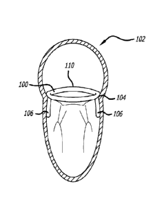
[00216] Turning now to FIGS. 3-5, there is shown one embodiment of an anchor
implant 100 configured for atrial anchoring and implantation within the heart
102 at the
mitral valve annulus 104. The anchor implant defines a supra-annular ring
sized and
shaped to be placed at the annulus, and includes commissural projections 106.
As shown
in FIG. 3, the projections 106 can be placed at an anterior commissural
trigone 108. As
described above, the commissural projections 106 are configured to extend
between
leaflets 109 without interfering with their functions (See FIG. 4). Moreover,
as shown, the
implant 100 includes a generally circular body 110 which can be formed from a
wire or
other structure, and the projections 106 arc loops extending away from a plane
defined by
the circular body 110. It is to be further recognized that the body 110
includes a pair of
bends 112 configured on opposite sides of the projections 106 to thereby
provide
necessary stress relief and clearance for the placement of the projections
between leaflets
109. Furthermore as noted previously, the anchor 100 can be covered with
various
materials, such as PET and ePTFE, so as to present a desired biocompatible
surface to
body tissue.
[00217] As shown in FIGS. 6-10, various other approaches to the anchor implant
are
contemplated. As before, the anchor 120 can be placed at the mitral valve
annulus 104
with projections extending beyond and between the leaflets 109. The
projections 125 can
be one or more of an expanding structure deployed through the coaptation line
and below
the leaflet 109 thereby capturing the anterior and posterior leaflet adjacent
the
CA 02870554 2014-10-15
WO 2013/158613
PCMJS2013/036734
31
commissures (See FIG. 7) or can define a piercing anchor 126 (See FIG. 8). In
a further
aspect, the piercing anchor 126 can be deployed in the P2 segment 128 of the
posterior
mitral valve leaflet, for example (See FIG. 9), so that the leaflet is
punctured and captured
by the anchor 120. Thus, a further secure attachment in anatomy can be
achieved by way
of expanding anchor or piercing anchor structure.
[00218] Two additional approaches to penetrating projections for use in
connection
with an anchor implant are shown in FIGS. 11 and 12. In one approach (FIG.
11), a
projection 130 can form a hook-like member with a barb 132 at its terminal
end. Such
structure defines a geometric interference with wall anatomy below a leaflet
and the
barbed end 132 penetrates the tissue of the LV to provide a secure attachment.
Alternatively, a projection 134 can be configured to penetrate commissural
anatomy and
terminate with a T-bar 136 which engages an external wall of the LV to thereby
provide a
secure attachment.
[00219] Non-penetration or non-piercing projections are also contemplated. As
shown
in FIG. 13, a projection 140 can be contoured to match a profile of the wall
beneath a
leaflet, and further include a foot pad 142 for engaging tissue. As shown in
FIGS. 14 and
15, the anchor implant can include a plurality of projections 144 having a
looped shape
and including webbing 146 for tissue ingrowth. Here, the looped structure of
the
projections 144 include a neck sized to fit between commissural slits and
about
commissural leaflets, the loop structures residing below the leaflet and
against the LV wall
to provide a secure engagement.
[00220] In another approach (See FIGS. 16-19), the anchor implant 150 can
define an
expandable body. In a compressed or contracted state (FIG. 16), the anchor
implant 150 is
smaller for delivering to an implantation site, whereupon the anchor 150 is
expanded (FIG.
18) to securely engage tissue. The implant can include a first convex side
member 152
configured between a pair of concave members 154. The junctions between the
members
can be looped to provide desired stress relief and a platform against which
the convex
member 154 can expand or open. Temporary restraining bands 156 are placed
about the
looped structure at the junction between the convex member 152 and each of the
concave
members 154. Further as shown in FIGS. 17 and 19, external surfaces of the
member 152,
154 can be equipped with tissue penetrating structure such as arrow-like barbs
158 or fish
hooks 160. One approach to converting the anchor implant 150 from its
contracted state
CA 02870554 2014-10-15
WO 2013/158613
PCT/US2013/036734
32
to its expanded state is to employ an expandable member such as that of a
balloon catheter
(not shown). At an implantation site (See FIG. 18), the expandable member is
placed
within an interior of the members 152, 154 and expanded to facilitate the
conversion of the
concave members 154 to convex members. Such action overcomes the restraining
bands
156 and facilitates the advancement of the tissue penetrating structure with
tissue.
[00221] Another approach to structure for penetrating tissue is shown in FIGS.
20 and
21. Here, the penetration structure is embodied in a staple-like structure 162
including a
pair of spaced arms 164 joined at a U 166. Angled barbs 168 are further
provided on
lateral sides of the arm 164 to provide a further secure engagement to tissue.
In one
approach, the staples 162 are deployed about an anchor implant such that the U
structure
166 captures the anchor implant and the arms with barbs secure the anchor in
place. A
threaded base 170 is further provided to connect the staple 162 to a push rod
or wire
delivery system (not shown). In this way, one or a plurality of staples 162
can be
advanced to the implantation site to help securely set an anchor implant.
[00222] Various other approaches to an anchor implant are shown in FIGS. 22-
38.
FIG. 22 depicts an implant 180 embodying a generally figure 8 shape connected
at its
middle by a connection cord 182. A pair of commissural projections or feet 184
are
further provided and spaced along the implant to reflect contouring of
commissures
(points A and B) of a heart valve. The feet 184 can define loops held in shape
by a
retaining band 186. Another elongate loop 187 is formed at one end of the
connector cord
182 and is configured to extend laterally. The assembly can be further
provided with
webbing 188 for tissue ingrowth, the contour of the figure 8-shape and the
elongate loop
187 providing structure across which the webbing extends to define a generally
circular
overall implant body structure, with the feet 189 extending therefrom.
[00223] The approach to the anchor implant 190 shown in FIG. 23 also is
embodied in
an assembly having a generally round or circular profile. The implant 190
includes a
generally circular wire frame 192 having a covering and a plurality of feet,
two of which
are intended to pass through valve commissures and a third positioned at a P2
location. A
flexible fabric web 194 is configured across the wire frame 192, the web being
centered
about center points 196 of the valve and including a pair of triangular
sections 197 joined
by a middle band web section 198.
CA 02870554 2014-10-15
WO 2013/158613
PCMJS2013/036734
33
[00224] An anchor implant having a generally T-shaped frame 200 is shown in
FIG. 24.
At a base of the T-frame is a commissural foot 202 configured for P2
attachment, whereas
the ends of the T-bar of the frame include commissural feet 204. An expandable
web
fabric membrane 206 extends between the curvilinear members defining the T-
shape
frame 200. As shown in FIG. 25, the T-shaped frame 200 can further include a
pair of
limited elongation flexible cords 208 each extending from the commissural feet
202 at the
base of the T-shape frame 200 to one end of the T-bar of the frame.
[00225] In yet another approach (FIGS. 26-27), an anchor implant 210 can be
embodied
in structure designed to prevent turning of an artificial valve attached to
the implant 210,
and to help in proper seating. Commissural feet 212 are configured as before,
that is to
reside between and below natural valve commissures. The wire frame of the
implant 210
includes further larger loops 214 of varying sizes and angled in a manner to
engage
anterior and posterior walls of the left atrium to thereby provide lever point
support
against rotation and structure including the frame from sliding through a
valve orifice.
Flexible cords 216 are further provided to retain the shape of the large loops
during
subsequent implantation of a replacement valve.
[00226] Bar-like anchors are also contemplated (See FIGS. 28-38). In one
approach as
shown in FIGS. 28 and 29, the anchor implant 220 can be embodied in a cross-
member
222 sized to span a full area of a commissural leaflet. The ends of the cross-
member are
provided with broad pads 224, a top portion 226 of which covers the
commissural leaflet
and can act to minimize leaking in this area. A bottom portion 228 of the
broad pads 224
can include a projection through a commissural of the leaflet, thus providing
an anchor
function.
[00227] As shown in FIGS. 30-32, the anchor implant 230 can include an intra-
annular
commissural anchoring frame 232 sized and shaped to receive an artificial
valve, or can
alternatively be employed to hold another anchor implant in place during a
healing and
tissue ingrowth stage. Opposite ends of the implant 230 are curved members 234
which
are intended to conform to local anatomy, and extend from above the annulus
(intra-atrial),
through valve commissures, and to within the LV. Texturing or tissue fabric
can be
associated or configured upon the member 234. The cross-member 236 connecting
the
ends of the implant 230 also defines a curved member designed to reside in the
atrium and
CA 02870554 2014-10-15
WO 2013/158613
PCMJS2013/036734
34
further includes a band 238 providing both strain relief and support structure
to the cross-
member 236.
[00228] Another anchoring frame 240 is shown in FIGS. 33-35. Here, opposite
ends of
the implant are curved members 242 which are sized and shaped to conform to
local
anatomy and extend from above the annulus, through valve commissures, and to
within
the LV. The cross-member 244 extends from both ends of the curved end members,
and
thus is intended to reside below valve annulus. Cross member 244 resides
between the
chordal tent of the anterior leaflet chordae and posterior leaflet chordae
with the lateral
ends extending between the individual chordae out to the LV wall. Because the
cross
member 244 is a loop like structure it does not entangle in the chordae, stays
between the
respective anterior and posterior leaflet chords, and helps orient the cross
member parallel
to the commis sure to commis sure line.
[00229] In an alternative approach (FIGS. 36-38), a bar-like, anchor frame 250
has a
generally omega profile sized to span the heart atrium. The ends of the frame
252 are
shaped to conform to anatomy, and also extend through valve commissures. The
curved
bar 254 connecting the shaped ends is sized and shaped to reside above the
valve annulus.
[00230] Moreover, as shown in FIGS. 39-43, it is further contemplated that a
ring frame
of an anchor implant can reside in multiple planes. That is, the frame can
embody a saddle
shape 260 configured to accommodate the curvature of a valve annulus such as
of the
mitral valve (FIG. 39). The frame can further include an arc section 262
configured to
accommodate the curvature of the inter-trigone anterior leaflet (See FIG. 40).
Moreover,
the anchor frame can include a serpentine shape 264 (FIG. 41) intended to
accommodate
dimensional and shape variations of the native annulus. The curves of the
serpentine
pattern can be perpendicular to the native annulus as shown in FIG. 41, or
they can extend
horizontally with respect to the annulus as shown in FIG. 42, or of course a
combined
approach to undulations may be desirable.
[00231] With reference to FIG. 43, it is also contemplated that a wire frame
anchor
implant can include adjustable sub-structure for accommodating perimeter
variations of a
valve annulus. Here, a delivery tube 270 can be configured to advance or
retract sections
of a wire implant to best fit it to anatomy. Excess length would be severed
and removed.
In one approach, lengths of posterior frame wire can be so adjusted, leaving
unchanged an
CA 02870554 2014-10-15
WO 2013/158613
PCMJS2013/036734
anterior frame portion 272, and commissural extension 274. Other areas of an
anchor
implant can also be likewise adjusted as desired.
[00232] Next, various approaches to anchor attachments are presented. As shown
in
FIG. 44, a direct mechanical load can be placed behind a leaflet 109 to
provide structural
support to an anchor. As previously stated, it is further contemplated that
the robust
anatomy of collagen annulus or a trigone can be relied upon to receive
piercing anchors
(See FIGS. 45A-45B) such as fish-hook 286 or arrowhead-like 288 structure. It
has been
found that muscle attachment provides excellent ingrowth for stable long term
anchoring.
Shear loading is depicted in FIG. 45C and deeper (FIG. 45D), wider (FIG. 45E)
and longer
areas (FIG. 45F) of anchor penetration are also contemplated. By taking one or
more of
these approaches, desired and increased holding capability is achieved.
[00233] As shown in FIGS. 46-49, one specific approach to a commissural
projection
of an anchor implant can assume a pair of loops 290. Such loops are intended
to be sized
so that they can reside under and behind a leaflet 109. Moreover, it is
contemplated that
the anchor projection be made from flexible and elastic material and be able
to be inserted
in a straightened configuration within a delivery tube 292. A delivery tube
292 can be
employed for each anchor projection 290, and a connection delivery wire 294 is
further
provided to control positioning. Thus, the delivery wire 294 can be withdrawn
or
otherwise disengaged from the anchor projection 290, and to thereby permit
release to be
reconfigured into its looped configuration. This can be done either before or
after ejection
from the anchor projection 290 from within the delivery tube 292. Moreover,
individual,
releasable control is provided by employing a delivery wire approach. That is,
through
multiple connections of a plurality of delivery wires to an anchor, desirable
control is
facilitated. Catheter/tube access or over-the-wire access approaches are also
contemplated
for providing in situ access to each anchoring location to deploy both anchor
portions and
tissue penetrating structures.
[00234] As shown in FIGS. 50 and 51, the anchor projection 30 can also include
a
leaflet clip configuration 302 attached to its terminal end. The clip 302 can
also be
delivered as described above using a delivery tube and connection delivery
wire 294, so
that it is properly positioned, such as behind a leaflet 109. A flat terminal
end 304 of the
clip 302 presents the valve anatomy with an atraumatic surface, as well as a
robust
engagement.
CA 02870554 2014-10-15
WO 2013/158613
PCMJS2013/036734
36
[00235] In another approach (FIGS. 52-53), an anchor implant 310 can include
commissural projections 312 that extend down and anteriorly to hook under the
fibrous
region of the trigone 60, and beyond the annulus. A penetrating securement
element in the
form of a helical screw 314 (See FIG. 54) can be further deployed through a
loop
presented by the commissural projections 312 to further seat and securely
attach the
anchor implant 310 in place. It is noted that a proximal winding 316 of the
helical fastener
314 can have a larger profile for positioning on an outside wall of local
anatomy (such as
the LV wall). A terminal end 318 of the helical element is sized to retain the
structures
into engagement.
[00236] Other approaches to fasteners are also contemplated (FIGS. 55-61). As
shown
in FIGS. 55-56, one device can embody a fastener with a wire brush body 320
and a nail
head terminal end 322 as retaining structure. Other approaches involving a
variable coil
(FIG. 57), a longitudinally directed coil 324 (FIG. 58) configured parallel to
a
commissural projection, or a curved penetrating ribbon securement markers 326,
328
(FIGS. 59-60) can also be useful approaches to providing secure retention of
parts against
anatomy. Moreover, a commissural projection 330 (FIG. 61) can be equipped with
a cord
extension through which a penetration sccurement element 332 can be inserted
and
advanced into body anatomy to accomplish a necessary attachment function.
[00237] It is intended that approaches to sealing may need to provide a
contiguous seal
between the overall implant (including the implanted replacement valve) and
the native
valve tissue/structures to prevent regurgitant para-valvular flow.
Additionally,
contemplated sealing configurations are intended to provide for tissue
engagement and
stable incorporation at the tissue and sealing structure interface. These
sealing structures
may also provide a staged interface to accommodate alternative type valve
implant
systems, such as the dual parallel valve approaches where the geometry for
valve interface
is not the native annulus or the anchor implant structure. Sealing structures
may include a
frame portion, made of metal or other suitable material in a wire or laser cut
configuration.
The frame may be covered with a material to promote tissue ingrowth.
Additionally or
alternatively, the sealing structure may utilize an expandable member. The
expandable
member can be balloon inflation, a self-expanding metal frame from a
compressed
delivered state, or a self-expanding material such as foam or a hydrogel. The
expanding
member may be used to directly form the seal or it may be used as a deployment
CA 02870554 2014-10-15
WO 2013/158613
PCMJS2013/036734
37
mechanism of another structure. Besides using the direct tissue engagement
forces
designed into the structure and or deployment of the valve assembly, the
system can
exploit the pressurization of the LV during systole to create the forces
needed to seal
between the valve assembly and the tissue.
[00238] As previously noted, the seal may be incorporated into the anchor
itself,
incorporated into the valve, or may be a separate structure. Incorporation of
the tissue
sealing mechanism onto the anchor assembly can either be achieved acutely or
utilize the
chronic ingrowth of the anchor into the tissue to generate a seal allowing for
a secondary
seal between the valve and the anchor during its deployment. The secondary
seal can
utilize the stability and consistency of the anchor structure to complete the
overall seal for
the complete valve assembly.
[00239] In the situation where the tissue seal is incorporated onto a
valve/occluder
assembly, the anchor structure is primarily utilized to provide the load
bearing function
Where a separate implant structure creates substrate for sealing, the implant
engages the
anchor and valve and the seal is created by one of the following; sealing
directly to the
tissue around the anchor implant creating the primary tissue seal with a
secondary seal to
the valve, sealing to the anchor and the valve as secondary seals with the
anchor as the
primary tissue seal, or structurally bridging the anchor to the valve where
the valve creates
the primary tissue seal
[00240] Furthermore, to develop an acute seal between the native tissue and
the specific
element of the overall valve assembly, direct force between the valve assembly
and the
tissue can be used. Alternatively, the pressurization of the LV during
ventricular systole
can be used to "inflate" or pressurize an element on the valve assembly such
that it
engages the native tissue to create a seal.
[00241] In one approach, for the interface between the anchor and the tissue,
simple
surface contact between the surfaces can facilitate sealing through tissue
ingrowth.
Additionally the anchor skeleton can provide an expansion force to create a
compressive
interface. In certain embodiments of the anchor, dilatory or pinching like
forces can be
created at certain regions of the anchor.
[00242] Mechanisms to create engagement between the anchor and valve can
include
balloon expansion of valve frame into anchor structure, or self-expansion of
valve frame
CA 02870554 2014-10-15
WO 2013/158613
PCMJS2013/036734
38
into anchor structures. Additionally, a hook-like engagement where the valve
frame
clasps or hooks onto anchor or anchor and tissue can be used as can a
frictional fit between
the structures, the same being created via balloon or self-expansion of the
valve frame into
the anchor. Interlock mechanisms where the valve frame engages the anchor ring
can be
employed as can conformable balloon or material interface.
[00243] For a tissue/valve interface, dilation of the valve or valve frame
elements can be
utilized. Also contemplated is a simple surface contact to facilitate ingrowth
of tissue onto
valve structures, compression and expansion elements on the valve frame for
directly
engaging the tissue, inflation of a valve structure via the LV pressure, and
then
deployment of hooks or structural frame elements enhance or create tissue
engagement. It
is further noted that the various sealing structures disclosed can be adapted
to either be
part of the anchor structure, part of the valve structure, or be an
independently delivered
structure. Moreover, all disclosed features can be utilized individually or in
any
combination. Surface composition elements, specifically material choice and
texture to
promote tissue reaction and device incorporation with maximal sealing
capability, may
have a significant impact on sealing capabilities. Specific sealing
modifications can
include surface texture (pores to promote ingrowth and mechanical hold),
material choice
(Dacron velour, double velour, ePTFE), abrasive surfaces, and/or a chemical
irritant to
promote inflammatory response. Further, tissue surface modification can
involve abrasive
chemical irritant to promote inflammatory response, and the use of heat.
[00244] Accordingly, as shown in FIGS. 62 and 63, an anchor implant 450 can
include
an internal ring 452 surrounded by a covering 454 adapted for tissue ingrowth.
As stated,
tissue ingrowth cooperates with the engagement of the anchor implant 410 to
create a seal
against heart tissue. As before, the anchor implant is sized and shaped to fit
the heart
valve annulus.
[00245] Sealing can also be accomplished by employing petals 460 arranged
about a
circumference of an expandable frame 462 (See FIGS. 64-67). As shown in FIG.
67, the
valve assembly can assume a compressed configuration for delivery and then
expanded
upon implantation. Selected such petals 460 can be arranged to engage a valve
annulus or
alternatively can be configured to engage the leaflets themselves. A foldable
fabric 464
can further be provided about the petals to facilitate sealing and a
continuous engagement
about a perimeter of a valve.
CA 02870554 2014-10-15
WO 2013/158613
PCMJS2013/036734
39
[00246] As shown in FIGS. 68-72, an expandable ventricular ring 470 can also
be used
to accomplish desired sealing. Here, a pair of longitudinally spaced circular
frames 472,
474 support a fabric covering 476. The lower frame 474 expands outwardly to
engage
tissue and provide the sealing function against tissue. It is contemplated
that the first
frame 472 resides above the valve annulus and the second frame 474 engages
anatomy
below the annulus. In this situation, the seal between the native
tissue/leaflet with frame
474 is facilitated by the LV pressurization of the internal surface of the
frame 474 and
fabric 476 as well as the underside of the leaflets pushing the tissue and
frame 474 against
each other.
[00247] With reference to FIGS. 73-75, a fluid filled balloon 480 can also be
incorporated into sealing structure. The balloon 480 can define a ring-shaped
structure
which when expanded engages and seals against varying heart anatomy. The
assembly
can further include commissural engagement anchors 482, the same being defined
by a
wire frame. A downwardly projecting frame 484 can also be included to aid in
absorbing
forces tending rotate the assembly when implanted at the native valve.
[00248] With reference to FIGS. 76-78, a sealing assembly 490 can also embody
an
anchor ring 492 partially enclosed by a ring balloon 494. The anchor ring 492
engages
tissue at the valve annulus and the circular balloon 494 forms a "C" about the
anchor ring
492 when viewed in cross-section, thereby presenting two additional contact
points with
anatomy about the perimeter of a valve. Such structure provides a seal between
the
assembly and heart anatomy as well as between the anchor ring 492 and ring
balloon 494
through an interlocking engagement. A circular band 496 placed at the junction
between
the balloon 494 and anchor ring 492 can limit expansion of the balloon 494 and
facilitate
its surrounding the anchor ring 492.
[00249] As shown in FIGS. 79-82, a sealing assembly 500 can alternatively
embody a
circular frame 502 including a plurality of vertically arranged, expandable
wire struts 504
spaced about a periphery of the frame 502. Configured between a central frame
structure
506 and the expandable struts 502 is an expandable circular balloon 508. Upon
expansion
of the balloon 508, the expandable struts 504 expand outwardly and thus can
engage and
bend around an anchor implant 509 placed at the interventional site. Such
bending forces
created by the expanded balloon operate to seal the assembly 500 against the
anchor 509
and valve anatomy.
CA 02870554 2014-10-15
WO 2013/158613
PCMJS2013/036734
[00250] FIGS. 83 and 84 depict an additional two approaches to sealing
structures. In
each, a frame is contoured with a fabric covering 510. The frame can be formed
from a
wire mesh 512 (FIG. 83) or can be defined by an expandable metal frame 514
(FIG. 84).
Again, such structure is intended to sealingly engage within heart anatomy and
when
desired, about an anchor implant.
[00251] Moreover, as shown in FIGS. 85-87, sealing can be created by a
flexible
polyester skirt 520 which expands outwardly in response to pressures within
the heart.
The expandable skirt 520 can be configured about an extremity of a frame of a
heart valve
that is additionally supported by commissural anchors 522 extending to within
the LV.
Over time, tissue over growth covers the skirt 520 providing further
attachment and
sealing.
[00252] Various approaches to expandable strut structure are also contemplated
for use
as sealing structures (See FIGS. 88-100). In a first approach (FIGS. 88-90),
an expandable
frame 530 can include a plurality of rows of cells, a middle row of cells
include members
which expand outwardly to engage an anchor implant 532 about a periphery of an
annulus.
In another approach (FIGS. 91-93), a sealing frame 540 is embodied in a
braided and
folded wire mesh structure. An internal portion of the fold 542 attaches to a
valve
structure 543, whereas a portion of an outer section 544 of the folded wire
mesh engages
and forms about an anchor implant frame 546. In yet another approach, the
sealing frame
can be defined by folded metal wire forming structure 550 including indented
geometry
552 for engaging an anchor frame 554 and an inner layer which can support
valve leaflets
556 (See FIGS. 94-95). Other expandable frame designs are shown in FIGS. 96-
99. One
such design 560 (FIGS. 96-97) embodies a frame 562 which expands into a
tubular shape
with a mid-level indentation 564 formed by smaller slits and sized to engage
an anchor
implant, and a second design 570 (FIGS. 98-99) expands into a tube with a
series of upper
and lower projections 572 spaced longitudinally providing a space to receive
an anchor.
[00253] In still yet another approach (FIGS. 100-102) a compressible material
580 can be
employed to create or facilitate sealing. The compressible material can define
a ring
structure about a valve 582 and placed into engagement with an anchor 584. The
compressible material can be made from a foam or hydrogel and thus act as an
interlocking element.
CA 02870554 2014-10-15
WO 2013/158613
PCMJS2013/036734
41
[00254] Additionally, the sealing device can be embodied in a ring 590
including
commissural projections 592 including barbs 594 (FIG. 103). Placement of a
valve
assembly with the ring 590 helps maintain the barbs 594 in place. Moreover, a
sealing
ring 600 can conform about and beyond the annulus and between papillary
muscles and
cooperate with projections 602 extending from an anchor to accomplish secure
implantation (See FIGS. 104-106). Furthermore, as shown in FIGS. 107-108, a
sealing
device 610 can cooperate with a plurality of projections 612 extending from a
valve, as
well as a hooked attachment member 614 that is inserted into tissue as an
anchoring
structure.
[00255] Various other tubular sealing assemblies are shown in FIGS. 109-115.
Turning
in particular to FIGS. 109-111, a tubular sealing assembly 620 can include an
expandable
tubular frame 622 and a membrane perimeter 624 extending about a midsection
thereof.
Upon expansion of the tubular frame 622, the midsection of the frame 622 opens
to the
profile defined by the membrane 624 to thereby present a sealing interface
structure.
Alternatively, an expandable wire frame 630 can be unconstrained by a membrane
and
extend to match body anatomy (FIG. 112) or it can be a sealing device 640
including a
compressible material band 642 about its midsection as well as an additional
sealing ring
644 (See FIG. 113) attached to its leading edge.
[00256] In yet another approach (See FIGS. 114-115), the sealing device 650
can include
a pair of wire extensions 652 sized and shaped to extend within the LV between
papillary
muscles. A wire 654 with a sheath 656 at is tip can be used during delivery of
the sealing
device 650 to the implantation site. Once placed as desired, the structure can
be
disengaged from the wire extensions 652, permitting them to engage supporting
anatomy
within the LV. In order to provide a surface to capture anterior and posterior
leaflets, the
wire extensions can assume a coiled configuration when deployed.
[00257] The delivery system and method used to deliver the anchor system can
depend
on both the structure and type of materials used for the anchor, as well as
the desired route
of access for implantation, and the type of deployment of the anchor. An
anchor delivery
system can generally include a guide catheter and an anchor delivery catheter,
either as
separate components, or integrated together. The guide catheter may include
specific
curves to facilitate navigation into the atrium or ventricle and may also
include a steerable
torquable shaft to aid with anchor positioning or orientation. The guide
catheter may
CA 02870554 2014-10-15
WO 2013/158613
PCMJS2013/036734
42
further include a deflectable tip region. The anchor delivery catheter can
house or hold the
collapsed anchor during delivery and deployment and may include delivery tubes
or wires
that are releasably connected to the anchor. Other elements such as shaft
dividers may be
utilized to help with managing multiple connection shafts as well as
orientation of the
anchor during deployment. Additional components inside the connection shaft or
wires, or
deliverable through or over, may include tissue penetrating elements to aid
with overall
securement and anchoring. A proximal hub can be configured to function to
selectively
manipulate, seal, and deploy certain elements. It is contemplated that
structures can be
incorporated onto the anchor to allow for a percutaneous delivery and include
the use of
super elastic Nitinol for the primary skeleton of the anchor or the use of a
malleable SST
or a similar material that could be folded down inside the delivery catheter
but then
balloon expanded against the tissue interface and would conform mitral tissue
interface.
The use of heat set small radii in certain locations of the anchor structure
can allow for
folding to fit inside delivery catheter where the strain limit of the material
is not exceeded.
Also, the use of ribbon at certain locations within the anchor skeletal
structure can allow
for tight bends in the thinner dimension for bending inside catheter, but
still achieve the
structural rigidity required if the broader section of ribbon is properly
oriented when
deployed. Smaller and larger diameter wire can also be used to vary the
configuration to
allow for bending/collapse in the catheter while still having the necessary
structural
strength and interface when deployed.
1002581 It is further contemplated that the anchor structures allow for
arterial
(aorta-retrograde), venous (via transatrial septum - antegrade), trans-apical
(LV), or
trans-atrial via a right thoracotomy access into the left atrium. Because of
the relatively
small size of the anchor, the ability to compress or fold the anchor into a
small delivery
configuration (especially with Nitinol or malleable stainless), and the
separation of the
anchor from the valve, the arterial route is feasible and may be especially
useful in the
situation where the anchor is deployed at the time of a diagnostic angiogram
that is in
advance of the actual valve therapy (separate procedures), as the arterial
groin access has
already been created. Routes of access can include arterial, venous and/or
thorax/apical.
1002591 To deploy the anchor into the heart, both catheter sheath retraction
and anchor
push out of sheath are contemplated approaches. Also, to have the anchor
achieve the
CA 02870554 2014-10-15
WO 2013/158613
PCMJS2013/036734
43
desired configuration inside the heart, either a self-expanding anchor, or use
of balloon
expansion to expand the anchor, or components of both are contemplated.
[00260] For anchors that are supra-annular with commissural feet, delivery
system
connections to the tips of the commissural feet or projections can facilitate
positioning and
proper orientation and seating into the mitral orifice. In this regard, the
anchor and
connections would exit the delivery catheter while the catheter tip was
residing in the left
atrium. The delivery sheath could then be pulled down beneath the level of the
mitral
annulus allowing the shaft connections to the feet to orient and align with
the commissural
clefts of the anterior annulus between the anterior and posterior native
leaflets. As the
shaft connections are pulled, the commissural feet would move toward the edge
and bring
the feet into position next to the LV wall within the natural leaflet cleft.
Once in this
position, additional features of the implants could be deployed or delivered
via the shaft
connections used to aid in attaching the feet to the wall/leaflet tissue, e.g.
staple, barbed
hooks or nails. Similar feet could be utilized for orientation along the
clefts seen naturally
on the P1 and P3 regions of the posterior leaflet.
[00261] According to one aspect, an exemplary embodiment of the delivery
system
utilizes an outer delivery or guide catheter that has a pre-formed curve that
positions the
anchor delivery catheter into the proper orientation toward the mitral valve
from the LV.
For retrograde access to the LV from the aorta, curves ranging from 90 to 200
degrees
may be used. A pre-formed shaft separator or a shaft separator with
prespecified bending
moments within the guide catheter can also aid in orienting and positioning
the anchor and
associated connection shafts.
[00262] According to another aspect, an exemplary embodiment of a delivery
system
may include connection shafts to connect to and control movement of the
commissural
feet of the anchor being deployed. The connection shafts can be tubular or
wire structures
or combinations that can release ably disengage from the anchor after
positioning. These
connection shafts can allow for independent manipulation of the anchor at each
individual
point of attachment.
[00263] The anchor securement elements can be deployed utilizing the two basic
structures of the shaft connections to the anchor frame and/or projections,
namely the
tubular shaft and the wire connections providing the temporary securement. The
tubular
CA 02870554 2014-10-15
WO 2013/158613
PCMJS2013/036734
44
shaft can be retracted to deploy an expanding frame element housed inside the
shaft during
frame delivery. Also, the tubular shaft can be used as a conduit to deliver a
separate
structure to the attachment location and/or to expose or deploy a securement
element and
then used to either actuate or drive the element by re-advancing the tube.
Further, a wire
element can be used as a conduit or rail to deliver a separate structure to
the attachment
location. The wire element can also be used to deploy or push out an element
loaded/housed inside the tube/wire structure and/or the wire structure can be
used by
rotating an element connected at its tip to deliver the securement element.
[00264] With reference to FIGS. 116-127, one delivery system and method is
presented.
In a first step (FIG. 116), a j-tip guidewire 700 is advanced within the heart
through the
aortic valve AV. Conventional methods including those outlined above are
employed to
gain access to the aorta. Next, an intraventricular guide catheter 702 is
advanced over or
along the guidewire 700 (FIG. 117). The distal tip 704 of the guide catheter
702 is
oriented toward the mitral valve MV, with a curve plane of the catheter being
parallel to
the A2/P2 orientation. The guidewire 700 (or a second guidewire) is then
advanced
through the orifice of the mitral valve MV, thereby providing a platform for
placing
structure across the mitral valve MV (See FIGS. 118-119).
[00265] With the guidewire 700 across to the mitral valve MV, a balloon or
expandable
cage 710 is configured within the orifice of the mitral valve MV (See FIGS.
120-121). It
is to be recognized that the guidewire 700 can be equipped with the balloon or
expandable
cage 710, or a separate device can be advanced over the guidewire 700, and the
balloon or
cage 710 placed within the orifice of the mitral valve MV. In one embodiment,
structure
is provided to withdraw a distal portion of the balloon or cage 710 relative
to proximal
structure to thereby expand the balloon or cage 710, or a sleeve structure can
be advanced
relative to a distal portion of the balloon or cage to accomplish the
expansion. It is also
contemplated that the balloon or cage 710 be expanded in the left atrium LA
and then
withdrawn within the orifice of the mitral valve 710 to ensure there is no
entanglement
with cords 712 supporting the mitral valve with any portion or components of
the delivery
system. The balloon or cage 710 is then contracted and removed from within the
orifice of
the mitral valve MV, or otherwise covered with other structure of the delivery
system.
[00266] If the wire cage meets a restriction, the cage can be collapsed and it
and wire can
be withdrawn back into guide and non-entangled wire access attempted again.
CA 02870554 2014-10-15
WO 2013/158613
PCMJS2013/036734
Alternatively, the expanded wire cage could be advanced first until it passes
mitral orifice
without restriction. The size of the cage is large enough to fit through
orifice but not
between chordae attached to the same papillary muscle, and traverses between
chordal tent
and anterior and posterior leaflets. The wire is then advanced into atria to
provide anchor
system delivery that does not entangle with subvalv-ular mitral apparatus.
Alternatively, a
balloon tipped catheter can be utilized instead of a cage. Once the wire is
successfully
placed, the wire cage or balloon system is removed from body leaving wire
access for the
next steps of anchor delivery.
[00267] As shown in FIGS. 122 and 123, an anchor delivery catheter 720 is next
advanced over the guidewire 700 and within the guide catheter 702. A length of
the
anchor delivery catheter 720 is placed across the orifice of the mitral valve
MV.
Subsequently (See FIGS. 124 and 125), the anchor implant 730 is advanced out
of a
terminal end of the anchor delivery catheter 720 and into the left atrium LA.
Proper
orientation of the anchor implant 730 is provided by a plurality of connection
wires 740.
Such wires 740 are each connected to a single commissural projection 744 of
the anchor
implant 740, so that one commissural projection 744 is aligned with each
trigone T, and
one commissural 744 is aligned with the posterior leaflet segment P2.
Connection
between wires 740 and commissural projection 744 is maintained until proper
positioning
is ensured, and so that reorientation and retrieval is possible.
[00268] Once the positioning of the anchor implant 740 is verified, the anchor
delivery
catheter 720 and connection wires 742 are withdrawn to place the anchor
implant 740
within the annulus of the mitral valve MV. When placed as desired, a
commissural
projection 744 is placed at each the trigone T, and one at P2 (See FIGS. 126-
127). As can
be best appreciated from FIG. 125, a shaft 750 having three or more
longitudinal bores
formed therein can be employed to push the anchor implant 720 out from an
interior of the
anchor delivery catheter 720. The longitudinal bores provide conduits for the
connection
wires 742 used to orient the anchor implant 730. A central longitudinal bore
(not shown)
can be further provided to receive the guidewire 700; however, in the event
the shaft 750
does not include a central bore, the guidewire 700 is removed from the
interventional site
prior to the advancement of the anchor delivery catheter 720 within the guide
catheter 702.
[00269] In an alternative approach to the anchor delivery catheter (Sec FIGS.
128-132
shown without an anchor), a longitudinal shaft separator 760 can be employed
in place of
CA 02870554 2014-10-15
WO 2013/158613
PCMJS2013/036734
46
the above-described shaft. Thus, rather than have a plurality of longitudinal
bores sized to
receive the connection wires 742, the shaft separator 760 includes a plurality
of splines
762 extending from a central core to define spaces for the connection wires
747. A
central bore 764 is further provided to slidingly receive the guidewire 700.
As shown in
FIGS. 128 and 129, the splines 762 can be placed at varying locations to
facilitate proper
orientation of structure within the anchor delivery catheter 720 such as by
auto-orienting
connection wires to a plane of the oriented guide catheter. For example, one
spline 766
can be sized and positioned to orient along an inner radius 768 of a curve of
the delivery
system (See FIG. 132). The curve of the shaft separator 760 aligns and
maintains
rotational orientation of the separator splines relative to the curve of the
anchor delivery
catheter 720 or the guide catheter 768.
[00270] Moreover, with reference to FIGS. 133-135, various alternatives are
presented
regarding releasable connections between commissural projections and structure
of the
anchor delivery system 720. In one approach (See FIGS. 133-134), a commissural
projection 770 of an anchor implant can include a deployable staple 772. The
deployable
staple 772 is configured within the anchor delivery catheter or a separate
sheath 774 of the
anchor delivery system. A positioning rod 776 with a threaded terminal end is
joined to
internal threads formed within the deployable staple 772 to define a threaded
connection
778 between the parts. Further, a retainer cord 780 is placed through the
sheath 774, the
same including a loop 782 configured about the positioning rod 776 and a
terminal end
784 engaging the commissural projection 770. In this way, distinct point
controls are
provided to separately position and deploy the staple 772 and the commissural
projection
770. That is, the retainer cord 780 can be withdrawn from engagement with the
commissural projection 700 to facilitate its implantation separately from
deploying the
staple by rotating the position rod 770 from engagement with the staple 771.
[00271] In an alternative approach (See FIG. 135), a threaded connection 790
can be
provided between a threaded receiver 792 formed on the anchor itself, and a
threaded
connection wire 794. Here, a delivery connector tube 796 includes a first
large bore 797
sized to receive the commissural projection 798, and a second smaller bore
sized to
slidingly receive the connector wire 794. Again, distinct point control is
provided in that
the commissural projection 798 can be released independently of disengagement
of the
threaded connection 790.
CA 02870554 2014-10-15
WO 2013/158613
PCMJS2013/036734
47
[00272] Percutaneous, or minimally invasive trans-apical, valve delivery
systems
typically can be over the wire systems with the valve assembly compressed or
crimped
into the delivery state. To expose the valve, the outer catheter structure or
sheath can either
be withdrawn or the implant pushed or expelled from the outer catheter. The
tip of the
valve delivery system can also include a tapered and flexible tip section to
facilitate
navigation and tracking of the system within the vasculature or heart. Once
exposed the
valve is either self-expanding or balloon expanding. Some releasable
connection shafts or
wires to the valve frame may also be incorporated to facilitate positioning
and orientation.
[00273] Various loading methods and structures are contemplated. Tools such as
crimping devices can be utilized for compressing the valve down onto the
delivery
catheter shaft and into a deliverable configuration and size. Moreover, a
primary route of
access for a replacement mitral valve can be via a venous trans-septal
antegrade approach.
It is also anticipated a transapical approach can be utilized. A trans-atrial
approach via a
right thoracotomy to gain access to the left atrium can also be used and may
be useful
when utilizing a mechanical valve for implantation. Thus, routes of access can
include
arterial, venous and/or thorax/apical.
[00274] Various deployment methods are also contemplated. The deployment of
the
valve can utilize any of the current techniques being employed for
percutaneous pulmonic
or aortic implantation. This includes retraction of a sheath or advancement of
the valve
inside the sheath to have the valve exit the delivery catheter. Once exited,
either partially
or completely, the final valve deployment could include self-expansion or
balloon
expansion. With either of these final deployment techniques, a nondeployed
interlock
structure/mechanism on the perimeter of the artificial valve could provide a
temporary
space for flow communication of the atria with the ventricle during diastole
during
artificial valve expansion. Upon completion of artificial valve expansion, it
would now be
functional and the interlock mechanism could now be deployed to complete the
anchoring
and sealing of the artificial valve. This particular embodiment can eliminate
the
conventional need for rapid pacing during valve deployment; there is flow
allowed during
diastole while valve is deployed. Therefore, each of retraction, push, self-
expanding, and
balloon approaches are contemplated.
[00275] With respect to orientation/positioning methods, utilizing a
separately implanted
anchor substrate is the ability to utilize a fluoroscopic alignment technique
to mesh the
CA 02870554 2014-10-15
WO 2013/158613
PCMJS2013/036734
48
anchor with the valve. In this scenario, the x-ray fluoroscopic camera could
be adjusted so
a radiopaque (complete or interrupted around perimeter) anchor structure would
be
visualized in a relatively straight line (camera orientation ¨ line connecting
emitter with
intensifier- is perpendicular to anchor circular axis, or parallel to plane of
anchor ring).
The valve frame structure could similarly have a radiopaque perimeter at the
point at or
near the interlock region with the anchor. When the anchor was viewed in the
manner
described, the valve axial orientation could be adjusted so the radiopaque
perimeter was
also a line (without moving camera position) meaning the two cylindrical axes
of the
anchor and valve were now parallel. Subsequently, the valve line could be
appropriately
positioned above, below, or at the interlock region. This linear alignment of
the two
radiopaque structures would be even more visually pronounced as the valve
frame was
being expanded/deployed, whether by balloon or self-expanding. This could
additionally
allow for fine tuning or adjustment prior to final engagement of the valve
with the anchor
structure.
[00276] With references to FIGS. 136-142, various steps to placement of an
artificial
mitral valve are presented. FIG. 136 shows the anchor delivery system 800
placed within
the heart subsequent to implantation of an anchor implant 802. In one
approach, the
anchor implant 802 is left to heal in place prior to deployment of the
artificial valve.
Alternatively, as stated above, valve implantation can be conducted along with
or just
subsequent to the placement of the anchor implant 802. Using a transarterial
approach, as
shown in FIGS. 137-138, an artificial valve delivery system 810 is advanced in
a
transvenous, trans-septal approach to position an artificial valve 820 into
engagement with
the anchor implant 802. In order to properly orient the anchor implant 802 and
valve 810
relative to each other, each of the anchor implant and valve can include
radiopaque
markers. Anchor radiopaque markers 822 and valve radiopaque markers 824 can
thus be
aligned both longitudinally or axially (See FIGS. 139-140) as well as
rotationally (See
FIGS. 141-142). In this context, a fluoroscopic camera (not shown) can be
employed to
guide the relatively positional relationship between anchor 822 and valve 824
markers. It
is to be further noted that radiopaque markers residing on a commissural
projection of an
anchor implant can further be used to ensure that they arc seated as desired
between
commissures, and markers located on a ring portion of an implant can be used
to locate
such structure at a valve annulus.
CA 02870554 2014-10-15
WO 2013/158613
PCMJS2013/036734
49
[00277] Turning now to FIGS. 143-146 and FIGS. 147-150, an alternative trans-
atrial
approach and a trans-apical approach to artificial valve delivery are
depicted. Here, a
valve delivery system includes an introducer tool 828 which is insertable
through a portal
assembly 830. The portal assembly 830 has a generally flat, oval cross-
sectional profile
intended to present a less invasive structure to heart tissue. The introducer
tool 828 further
includes an articulating terminal end portion 832 adapted to releasably hold
an artificial
valve 834. The terminal end portion 832 is configured to retain a tilted valve
834 (See
FIGS. 143-144 and 147-148) for insertion into an interior of the heart through
the portal
assembly 830, and is articulatable so that the artificial heart valve 834 can
be turned and
placed into engagement with an anchor implant 840 previously delivered at the
native
valve (See FIGS. 145-146 and 149-150). Notably, the portal assembly 830
further
includes purse string sutures 842 configured about an exterior surface of a
portion of the
assembly located at the point of heart insertion. Upon removing the portal
assembly 830
from the interventional site, the purse strings are intended to remain in
place on the heart
and are thus available to close the access point employed for the valve
insertion. In this
way, the implantation procedure is completed expeditiously with a repaired
access point.
[00278] Further modifications and alternative embodiments will be apparent to
those of
ordinary skill in the art in view of the disclosure herein. For example, the
systems and the
methods may include additional components or steps that were omitted from the
diagrams
and description for clarity of operation. Moreover, those of ordinary skill in
the art will
appreciate that aspects and/or features disclosed with respect to one
embodiment in some
case may be incorporated in other embodiments even if not specifically
described with
respect to such other embodiments. It is to be understood that the various
embodiments
shown and described herein are to be taken as exemplary. Elements and
materials, and
arrangements of those elements and materials, may be substituted for those
illustrated and
described herein, parts and processes may be reversed, and certain features of
the present
teachings may be utilized independently, all as would be apparent to one
skilled in the art
after having the benefit of the description herein. Changes may be made in the
elements
described herein without departing from the spirit and scope of the present
teachings and
following claims. Accordingly, this description is to be construed as
illustrative only and is
for the purpose of enabling those skilled in the art the general manner of
carrying out the
present teachings. It is to be understood that the particular examples and
embodiments set
forth herein are nonlimiting, and modifications to structure, dimensions,
materials, and
CA 02870554 2014-10-15
WO 2013/158613
PCT/US2013/036734
methodologies may be made without departing from the scope of the present
teachings.
Other embodiments in accordance with the present disclosure will be apparent
to those
skilled in the art from consideration of the specification and practice of the
invention
disclosed herein. It is intended that the specification and examples be
considered as
exemplary only, with a true scope and spirit being indicated by the following
claims.
[00279] Thus, it will be apparent from the foregoing that, while particular
forms of the
invention have been illustrated and described, various modifications can be
made without
parting from the spirit and scope of the invention.