Note: Descriptions are shown in the official language in which they were submitted.
CA 02870702 2014-10-15
WO 2013/158540
PCT/US2013/036575
CAPTURE REACTIONS
RELATED APPLICATIONS
The present patent application claims the benefit of and priority to U.S.
Nonprovisional
Patent Application Serial No. 13/448,961, filed on April 17, 2012, and U.S.
Provisional Patent
Application Serial No. 61/624,778, filed on April 16, 2012. The entirety of
each is herein
incorporated by reference.
FIELD OF INVENTION
The invention generally relates to methods for improving performance of
capture
reactions.
BACKGROUND
Routine sequencing of whole genomes is not economically feasible, and as an
alternative,
it is often necessary to select genomic areas of interest for capture prior to
sequencing.
Numerous techniques have been developed for capturing target nucleic acids for
subsequent
detection and analysis that are compatible for use with massively parallel
sequencing platforms.
Such exemplary techniques include multiplex PCR capture with primer pairs and
array-based or
solution-based hybrid capture. Often, capture-based technologies are designed
to provide a
mechanism to analyze complex genomes by selecting genomic areas of interest
prior to
sequencing or detection. By analyzing the area of interest, the genome can be
studied with
significantly reduced costs and reduced time as compared with the task of
sequencing large
numbers of complex genomes in their entireties.
A problem with nucleic acid capture techniques is their inability to capture
multiple loci
with substantially uniform efficiencies. Such efficiencies define the amount
of sequencing
required to adequately cover the targets. Turner et al., Annu. Rev. Genomics
Hum. Genet. 2009
10:263-84. Generally, the distribution of abundances of capture reaction
products is rather wide,
with the most and least frequent species spanning multiple orders of
magnitude. Such a wide
CA 02870702 2014-10-15
WO 2013/158540
PCT/US2013/036575
distribution in abundance means that a large number of sequencing reactions
must be performed
to generate an effective coverage of the target, increasing costs and time to
results.
SUMMARY
The invention recognizes that capture reactions performed on whole genomic
nucleic
acids result in poor uniformity due to, for example, the folding and melting
temperature of high
molecular weight genomic nucleic acids. The massive length of genomic nucleic
acids in base
pairs, approximately 3.3 x 10⁹ bp for humans, and the natural folding of
genomic nucleic acids
prohibit the ability of capture moieties introduced to the genomic nucleic
acid from being
exposed to the targets for hybridization, leading to failed or inefficient
capture of the target.
The invention further recognizes that fragmenting nucleic acid prior to
performing a
capture reaction allows for greater exposure of a target site to a capture
moiety, reducing failed
capture, and increasing the percentage of capture moieties that hybridize to
targets within the
genome. Accordingly, methods of the invention lead to a product that is
substantially more
uniform than products obtained from capture reactions in which a fragmenting
step has not been
performed prior to the capture reaction. This advantageously yields a target
abundance
distribution that is significantly more uniform than if a native high
molecular weight genomic
nucleic acid is used. Such products are more suited for use in a number of
applications,
particularly in clinical diagnostics.
In certain aspects, methods of the invention involve obtaining a nucleic acid,
fragmenting
the nucleic acid into nucleic acid fragments, and capturing a target on a
nucleic acid fragment.
Methods of the invention work well with any capture technique and are
particularly suited for
capture techniques using molecular inversion probes. The nucleic acid can be
genomic DNA,
genomic RNA, or a whole genome amplification (WGA) product. Performance of the
method
with a WGA product is useful in situations where input DNA is limiting, e.g.
limiting amounts of
tumor tissue, fetal cells circulating in maternal blood, etc.
In embodiments that utilize molecular inversion probes (MIP), any molecular
inversion
probe may be used. An exemplary MIP is a single-stranded probe about 70
nucleotides in
length, composed of a universal core of 30 nucleotides that is flanked by
specific 20-nucleotide
targeting sequences on each side, i.e. targeting arms. However, the length and
composition of
the probe can vary to most adequately capture the desired target sequence. The
targeting arms are
designed to hybridize to specific genomic regions upstream and downstream of a
target sequence
2
CA 02870702 2014-10-15
WO 2013/158540
PCT/US2013/036575
of interest located on the nucleic acid fragment. After the target sequence of
interest is isolated
between the target arms, the target sequence can be analyzed. Although each
MIP captures one
target of interest for analysis, multiple probes can be combined into a single
vessel containing the
fragmented nucleic acids for a multiplexed assay that simultaneously examines
multiple target
loci.
Fragmenting the nucleic acid can be accomplished by any technique known in the
art.
Exemplary techniques include mechanically fragmenting, chemically fragmenting,
and/or
enzymatically fragmenting. Mechanical nucleic acid fragmentation can be, for
example,
sonication, nebulization, and hydro-shearing (e.g., point-sink shearing).
Enzymatic nucleic acid
fragmenting includes, for example, use of nicking endonucleases or restriction
endonucleases.
The nucleic acid can also be chemically fragmented by performing acid
hydrolysis on the nucleic
acid or treating of the nucleic acid with alkali or other reagents.
The fragment length can be adjusted based on the sizes of the nucleic acid
targets to be
captured. The nucleic acid fragments can be of uniform length or of a
distribution of lengths. In
certain embodiments, the nucleic acid is fragmented into nucleic acid
fragments having a length
of about 10 kb or 20 kb. In addition, the nucleic acid fragments can range
from between 1 kb to
kb, with various distributions.
In certain embodiments, the nucleic acid is also denatured, which may occur
prior to,
during, or after the fragmenting step. The nucleic acid can be denatured using
any means known
20 in the art, such as pH-based denaturing, heat-based denaturing,
formamide or urea, exonuclease
degradation, or endonuclease nicking. In certain embodiments, the use of pH,
such as in acid
hydrolysis, alone or in combination with heat fragments and either partially
or fully denatures the
nucleic acid. This combined fragmenting and denaturing method can be used to
fragment the
nucleic acid for MIP capture or to fragment captured target nucleic acids or
whole genomic DNA
for shotgun library preparation.
BRIEF DESCRIPTION OF THE DRAWINGS
FIG. 1 illustrates a non-limiting embodiment of a tiled probe layout;
FIG. 2 illustrates a non-limiting embodiment of a staggered probe layout; and
FIG. 3 illustrates a non-limiting embodiment of an alternating staggered probe
layout.
3
CA 02870702 2014-10-15
WO 2013/158540
PCT/US2013/036575
DETAILED DESCRIPTION
This invention generally relates to improving performance of molecular
inversion probe
capture reactions. In certain embodiments, methods of the invention include
the steps of
obtaining a genomic nucleic acid, fragmenting the genomic nucleic acid,
optionally denaturing
the genomic nucleic acid or fragmented genomic nucleic acid, and performing a
capture reaction
on the fragmented genomic nucleic acid. Methods of the invention work well
with any capture
technique and are particularly suited for capture techniques using molecular
inversion probes.
Fragmenting genomic nucleic acids prior to performing capture advantageously
improves the
uniformity of the product of the capture reaction.
Nucleic acids suitable for use in aspects of the invention include but are not
limited to
genomic DNA, genomic RNA, synthesized nucleic acids, whole or partial genome
amplification
product, and high molecular weight nucleic acids, e.g. individual chromosomes.
Genomic DNA
and genomic RNA constitute the total genetic information of an organism.
Genomic nucleic
acids molecules are generally large, and in most organisms are organized into
DNA¨protein
complexes called chromosomes, which the exception of viruses that have RNA
genomes.
Genomic RNA also includes, for example, RNA transcribed from DNA, unprocessed
transcripts,
mRNAs, and cDNAs. Sometimes the quality and quantity of genomic nucleic acids
obtained
from samples precludes their usefulness in large scale genotyping studies. To
overcome this
problem, use of whole genome amplification products and partial genome
amplification products
allows for characterization of the genome of a sample even if the quantity and
quality of the
genomic nucleic acid is limited.
Obtaining a Nucleic Acid
Target nucleic acid is obtained from a sample using methods known in the art.
Samples
include, but are not limited to: biological samples, such as tissue and bodily
fluid. For example,
samples are obtained from, e.g., blood, urine, serum, lymph, saliva, anal and
vaginal secretions,
perspiration and semen, skin, organs and the like. Samples are also obtained
from the
environment (e.g., air, agricultural, water and soil); and research samples
(e.g., products of a
nucleic acid amplification reaction, or purified genomic DNA, RNA, proteins,
etc.).
Isolation, extraction or derivation of genomic nucleic acids is performed by
methods
known in the art. Isolating nucleic acid from a biological sample generally
includes treating a
biological sample in such a manner that genomic nucleic acids present in the
sample are
4
CA 02870702 2014-10-15
WO 2013/158540
PCT/US2013/036575
extracted and made available for analysis. Any isolation method that results
in extracted/isolated
genomic nucleic may be used in the practice of the present invention.
Nucleic acids may be obtained by methods known in the art. Generally, nucleic
acids are
extracted using techniques, such as those described in Sambrook, J., Fritsch,
E.-F., and Maniatis,
T, 1989) Molecular Cloning: A Laboratory Man k.I al.2nd ed. Cold Spring
Harbor, NY:Cold
Spring Harbor Laboratory.), the contents of which are incorporated by
reference herein. Other
methods include: salting out DNA extraction (P. Sunnucks et al., Genetics,
1996, 144: 747-756;
S. M. Aljanabi and I. Martinez, Nucl. Acids Res. 1997, 25: 4692-4693),
trimethylammonium
bromide salts DNA extraction (S. Gustincich et al., BioTechniques, 1991, 11:
298-302) and
guanidinium thiocyanate DNA extraction (J. B. W. Hammond et al., Biochemistry,
1996, 240:
298-300). Several protocols have been developed to extract genomic DNA from
blood.
There are also numerous kits that can be used to extract DNA from tissues and
bodily
fluids and that are commercially available from, for example, BD Biosciences
Clontech (Palo
Alto, Calif.), Epicentre Technologies (Madison, Wis.), Gentra Systems, Inc.
(Minneapolis,
Minn.), MicroProbe Corp. (Bothell, Wash.), Organon Teknika (Durham, N.C.),
Qiagen Inc.
(Valencia, Calif.), Autogen (Holliston, MA); Beckman Coulter (Brea, CA),
(AutoGenFlex STAR
robot with Qiagen FlexiGene chemistry. For example, Autogen manufactures
FlexStar
automated extraction kits used in combination with Qiagen FlexiGene Chemistry,
and
Beckeman Coulter manufactures Agencourt GenFind kits for bead-based extraction
chemistry.
User Guides that describe in detail the protocol(s) to be followed are usually
included in all these
kits, for example, Qiagen's literature for their PureGene extraction chemistry
entitled "Qiagen
PureGene Handbook" 3rd Edition, dated June 2011.
After cells have been obtained from the sample, it is preferable to lyse cells
in order to
isolate genomic nucleic acid. Cellular extracts can be subjected to other
steps to drive nucleic
acid isolation toward completion by, e.g., differential precipitation, column
chromatography,
extraction with organic solvents and the like. Extracts then may be further
treated, for example,
by filtration and/or centrifugation and/or with chaotropic salts such as
guanidinium
isothiocyanate or urea or with organic solvents such as phenol and/or HCC13 to
denature any
contaminating and potentially interfering proteins. The genomic nucleic acid
can also be
resuspended in a hydrating solution, such as an aqueous buffer. The genomic
nucleic acid can be
suspended in, for example, water, Tris buffers, or other buffers. In certain
embodiments the
5
CA 02870702 2014-10-15
WO 2013/158540
PCT/US2013/036575
genomic nucleic acid can be re-suspended in Qiagen DNA hydration solution, or
other Tris-
based buffer of a pH of around 7.5.
Depending on the type of method used for extraction, the genomic nucleic acid
obtained
can vary in size. The integrity and size of genomic nucleic acid can be
determined by pulse-field
gel electrophoresis (PFGE) using an agarose gel.
In addition to genomic nucleic acids, whole genome amplification product and
partial
genomic amplification products can be used in aspects of the invention.
Methods of obtaining
whole genome amplification product and partial genome amplification product
are described in
detail in Pinter et al. U.S. Patent Publication Number 2004/0209299, and
include, for example,
generally ligation mediated PCR.TM., random primed PCR.TM., strand
displacement mediated
PCR.TM., and cell immortalization.
Fragmenting the Nucleic Acid
Nucleic acids, including genomic nucleic acids, can be fragmented using any of
a variety
of methods, such as mechanical fragmenting, chemical fragmenting, and
enzymatic fragmenting.
Methods of nucleic acid fragmentation are known in the art and include, but
are not limited to,
DNase digestion, sonication, mechanical shearing, and the like (J. Sambrook et
al., "Molecular
Cloning: A Laboratory Manual", 1989, 2nd Ed., Cold Spring Harbour
Laboratory Press:
New York, N.Y.; P. Tijssen, "Hybridization with Nucleic Acid Probes--
Laboratory Techniques
in Biochemistry and Molecular Biology (Parts I and II)", 1993, Elsevier; C. P.
Ordahl et al.,
Nucleic Acids Res., 1976, 3: 2985-2999; P. J. Oefner et al., Nucleic Acids
Res., 1996, 24: 3879-
3889; Y. R. Thorstenson et al., Genome Res., 1998, 8: 848-855). U.S. Patent
Publication
2005/0112590 provides a general overview of various methods of fragmenting
known in the art.
Genomic nucleic acids can be fragmented into uniform fragments or randomly
fragmented. In certain aspects, nucleic acids are fragmented to form fragments
having a
fragment length of about 5 kilobases or 100 kilobases. In a preferred
embodiment, the genomic
nucleic acid fragments can range from 1 kilobases to 20 kilobases. Preferred
fragments can vary
in size and have an average fragment length of about 10 kilobases. However,
desired fragment
length and ranges of fragment lengths can be adjusted depending on the type of
nucleic acid
targets one seeks to capture and the design and type of MIP probes. The
particular method of
fragmenting is selected to achieve the desired fragment length. Numerous non-
limiting
examples are provided below.
6
CA 02870702 2014-10-15
WO 2013/158540 PCT/US2013/036575
Chemical fragmentation of genomic nucleic acids can be achieved using a number
of
different methods. For example, hydrolysis reactions including base and acid
hydrolysis are
common techniques used to fragment nucleic acid. Hydrolysis is facilitated by
temperature
increases, depending upon the desired extent of hydrolysis. Fragmentation can
be accomplished
by altering temperature and pH as described below. The benefit of pH-based
hydrolysis for
shearing is that it can result in single-stranded products. Additionally,
temperature can be used
with certain buffer systems (e.g. Tris) to temporarily shift the pH up or down
from neutral to
accomplish the hydrolysis, then back to neutral for long-term storage etc.
Both pH and
temperature can be modulated to effect differing amounts of shearing (and
therefore varying
length distributions).
In one aspect, a nucleic acid is fragmented by heating a nucleic acid immersed
in a buffer
system at a certain temperature for a certain period to time to initiate
hydrolysis and thus
fragment the nucleic acid. The pH of the buffer system, duration of heating,
and temperature can
be varied to achieve a desired fragmentation of the nucleic acid. In one
embodiment, after a
genomic nucleic acid is purified, it is resuspended in a Tris-based buffer at
a pH between 7.5 and
8.0, such as Qiagen's DNA hydrating solution. The resuspended genomic nucleic
acid is then
heated to 65 C and incubated overnight (about 16-24 hours) at 65 C. Heating
shifts the pH of
the buffer into the low- to mid- 6 range, which leads to acid hydrolysis. Over
time, the acid
hydrolysis causes the genomic nucleic acid to fragment into single-stranded
and/or double-
stranded products. The above method of fragmenting can be modified by
increasing the
temperature and reducing the heating time. For example, a nucleic acid is
fragmented by
incubating the nucleic acid in the Tris-based buffer at a pH between 7.5 and
8.0 for 15 minutes at
92 C. In addition to adjusting the temperature and the duration of heating,
the pH of the Tris-
based buffer can be adjusted to achieve a desired nucleic acid fragmentation.
Other methods of hydrolytic fragmenting of nucleic acids include alkaline
hydrolysis,
formalin fixation, hydrolysis by metal complexes (e.g., porphyrins), and/or
hydrolysis by
hydroxyl radicals. RNA shears under alkaline conditions, see, e.g. Nordhoff et
al.,Nucl. Acid.
Res., 21 (15):3347-57 (2003), whereas DNA can be sheared in the presence of
strong acids or
strong bases.
An exemplary acid/base hydrolysis protocol for producing genomic nucleic acid
fragments is described in Sargent et al. (1988) Methods Enzymol., 152:432.
Briefly, 1 g of
7
CA 02870702 2014-10-15
WO 2013/158540
PCT/US2013/036575
purified DNA is dissolved in 50 mL 0.1 N NaOH. 1.5 mL concentrated HC1 is
added, and the
solution is mixed quickly. DNA will precipitate immediately, and should not be
stirred for more
than a few seconds to prevent formation of a large aggregate. The sample is
incubated at room
temperature for 20 minutes to partially depurinate the DNA. Subsequently, 2 mL
10 N NaOH
([0H-] concentration to 0.1 N) is added, and the sample is stirred until the
DNA redis solves
completely. The sample is then incubated at 65 C for 30 minutes in order to
hydrolyze the DNA.
Resulting fragments typically range from about 250-1000 nucleotides but can
vary lower or
higher depending on the conditions of hydrolysis.
Chemical cleavage can also be specific. For example, selected nucleic acid
molecules can
be cleaved via alkylation, particularly phosphorothioate-modified nucleic acid
molecules (see,
e.g., K. A. Browne, "Metal ion-catalyzed nucleic Acid alkylation and
fragmentation," J. Am.
Chem. Soc. 124(27):7950-7962 (2002)). Alkylation at the phosphorothioate
modification renders
the nucleic acid molecule susceptible to cleavage at the modification site.
See I. G. Gut and S.
Beck, "A procedure for selective DNA alkylation and detection by mass
spectrometry," Nucl.
Acids Res. 23(8):1367-1373 (1995).
Methods of the invention also contemplate chemically shearing nucleic acids
using the
technique disclosed in Maxam-Gilbert Sequencing Method (Chemical or Cleavage
Method),
Proc. Natl. Acad. Sci. USA. 74:560-564. In that protocol, the genomic nucleic
acid can be
chemically cleaved by exposure to chemicals designed to fragment the nucleic
acid at specific
bases, such as preferential cleaving at guanine, at adenine, at cytosine and
thymine, and at
cytosine alone.
Mechanical shearing of nucleic acids into fragments can occur using any method
known
in the art. For example, fragmenting nucleic acids can be accomplished by
hydroshearing,
trituration through a needle, and sonication. See, for example, Quail, et al.
(Nov 2010) DNA:
Mechanical Breakage. In: eLS. John Wiley & Sons, Chichester.
doi:10.1002/9780470015902.a0005 333.pub2.
The nucleic acid can also be sheared via nebulization, see (Roe, BA, Crabtree.
JS and
Khan, AS 1996); Sambrook & Russell, Cold Spring Harb Protoc 2006. Nebulizing
involves
collecting fragmented DNA from a mist created by forcing a nucleic acid
solution through a
small hole in a nebulizer. The size of the fragments obtained by nebulization
is determined
chiefly by the speed at which the DNA solution passes through the hole,
altering the pressure of
8
CA 02870702 2014-10-15
WO 2013/158540
PCT/US2013/036575
the gas blowing through the nebulizer, the viscosity of the solution, and the
temperature. The
resulting DNA fragments are distributed over a narrow range of sizes (700-1330
bp). Shearing of
nucleic acids can be accomplished by passing obtained nucleic acids through
the narrow
capillary or orifice (Oefner et al., Nucleic Acids Res. 1996; Thorstenson et
al., Genome Res.
1995). This technique is based on point¨sink hydrodynamics that result when a
nucleic acid
sample is forced through a small hole by a syringe pump.
In HydroShearing (Genomic Solutions, Ann Arbor, Mich., USA), DNA in solution
is
passed through a tube with an abrupt contraction. As it approaches the
contraction, the fluid
accelerates to maintain the volumetric flow rate through the smaller area of
the contraction.
During this acceleration, drag forces stretch the DNA until it snaps. The DNA
fragments until
the pieces are too short for the shearing forces to break the chemical bonds.
The flow rate of the
fluid and the size of the contraction determine the final DNA fragment sizes.
Sonication is also used to fragment nucleic acids by subjecting the nucleic
acid to brief
periods of sonication, i.e. ultrasound energy. A method of shearing nucleic
acids into fragments
by sonification is described in U.S. Patent Publication 2009/0233814. In the
method, a purified
nucleic acid is obtained placed in a suspension having particles disposed
within. The suspension
of the sample and the particles are then sonicated into nucleic acid
fragments.
An acoustic-based system that can be used to fragment DNA is described in U.S.
Pat.
Nos. 6,719,449, and 6,948,843 manufactured by Covaris Inc. U.S. Pat. No.
6,235,501 describes a
mechanical focusing acoustic sonication method of producing high molecular
weight DNA
fragments by application of rapidly oscillating reciprocal mechanical energy
in the presence of a
liquid medium in a closed container, which may be used to mechanically
fragment the DNA.
Another method of shearing nucleic acids into fragments uses ultrasound energy
to
produce gaseous cavitation in liquids, such as shearing with Diagonnode's
BioRuptor .
Cavitation is the formation of small bubbles of dissolved gases or vapors due
to the alteration of
pressure in liquids. These bubbles are capable of resonance vibration and
produce vigorous
eddying or microstreaming. The resulting mechanical stress can lead to
shearing the nucleic acid
in to fragments.
Enzymatic fragmenting, also known as enzymatic cleavage, cuts nucleic acids
into
fragments using enzymes, such as endonucleases, exonucleases, ribozymes, and
DNAzymes.
Such enzymes are widely known and are available commercially, see Sambrook, J.
Molecular
9
CA 02870702 2014-10-15
WO 2013/158540
PCT/US2013/036575
Cloning: A Laboratory Manual, 3rd (2001) and Roberts RJ (January 1980).
"Restriction and
modification enzymes and their recognition sequences," Nucleic Acids Res. 8
(1): r63¨r80.
Varying enzymatic fragmenting techniques are well-known in the art, and such
techniques are
frequently used to fragment a nucleic acid for sequencing, for example,
Alazard et al, 2002;
Bentzley et al, 1998; Bentzley et al, 1996; Faulstich et al, 1997; Glover et
al, 1995; Kirpekar et
al, 1994; Owens et al, 1998; Pieles et al, 1993; Schuette et al, 1995; Smirnov
et al, 1996; Wu &
Aboleneen, 2001; Wu et al, 1998a.
The most common enzymes used to fragment nucleic acids are endonucleases. The
endonucleases can be specific for either a double-stranded or a single
stranded nucleic acid
molecule. The cleavage of the nucleic acid molecule can occur randomly within
the nucleic acid
molecule or can cleave at specific sequences of the nucleic acid molecule.
Specific fragmentation
of the nucleic acid molecule can be accomplished using one or more enzymes in
sequential
reactions or contemporaneously.
Restriction endonucleases recognize specific sequences within double-stranded
nucleic
acids and generally cleave both strands either within or close to the
recognition site in order to
fragment the nucleic acid. Naturally occurring restriction endonucleases are
categorized into
four groups (Types I, II III, and IV) based on their composition and enzyme
cofactor
requirements, the nature of their target sequence, and the position of their
DNA cleavage site
relative to the target sequence. Bickle TA, Kruger DH (June 1993). "Biology of
DNA
restriction". Microbiol. Rev. 57 (2): 434-50; Boyer HW (1971). "DNA
restriction and
modification mechanisms in bacteria". Annu. Rev. Microbiol. 25: 153-76; Yuan R
(1981).
"Structure and mechanism of multifunctional restriction endonucleases". Annu.
Rev. Biochem.
50: 285-319. All types of enzymes recognize specific short DNA sequences and
carry out the
endonucleolytic cleavage of DNA to give specific fragments with terminal 5'-
phosphates. The
enzymes differ in their recognition sequence, subunit composition, cleavage
position, and
cofactor requirements. Williams RJ (2003). "Restriction endonucleases:
classification,
properties, and applications". Mol. Biotechnol. 23 (3): 225-43.
Where restriction endonucleases recognize specific sequencings in double-
stranded
nucleic acids and generally cleave both strands, nicking endonucleases are
capable of cleaving
only one of the strands of the nucleic acid into a fragment. Nicking enzymes
used to fragment
CA 02870702 2014-10-15
WO 2013/158540
PCT/US2013/036575
nucleic acids can be naturally occurring or genetically engineered from
restriction enzymes. See
Chan et al., Nucl. Acids Res. (2011) 39 (1): 1-18.
Denaturing the Nucleic Acid
Methods of the invention also provide for denaturing nucleic acid to render
the nucleic
acid single stranded for hybridization to a capture probe, such as a MIP
probe. Denaturation can
result from the fragmentation method chosen, as described above. For example,
one skilled in
the art recognizes that a genomic nucleic acid can be denatured during pH-
based shearing or
fragmenting via nicking endonucleases. Denaturation can occur either before,
during, or after
fragmentation. In addition, the use of pH or heat during the fragmenting step
can result in
denatured nucleic acid fragments. See, for example, McDonnell, "Antisepsis,
disinfection, and
sterilization: types, action, and resistance," pg. 239 (2007).
Heat-based denaturing is the process by which double-stranded deoxyribonucleic
acid
unwinds and separates into single-stranded strands through the breaking of
hydrogen bonding
between the bases. Heat denaturation of a nucleic acid of an unknown sequence
typically uses a
temperature high enough to ensure denaturation of even nucleic acids having a
very high GC
content, e.g., 95 C-98 C. in the absence of any chemical denaturant. It is
well within the abilities
of one of ordinary skill in the art to optimize the conditions (e.g., time,
temperature, etc.) for
denaturation of the nucleic acid. Temperatures significantly lower than 95C
can also be used if
the DNA contains nicks (and therefore sticky overhangs of low Tm) or sequence
of sufficiently
low Tm.
Denaturing nucleic acids with the use of pH is also well known in the art, and
such
denaturation can be accomplished using any method known in the art such as
introducing a
nucleic acid to high or low pH, low ionic strength, and/or heat, which
disrupts base-pairing
causing a double-stranded helix to dissociate into single strands. For methods
of pH-based
denaturation see, for example, Dore et al. Biophys J. 1969 November; 9(11):
1281-1311; A. M.
Michelson The Chemistry of Nucleosides and Nucleotides, Academic Press, London
and New
York (1963).
Nucleic acids can also be denatured via electro-chemical means, for example,
by
applying a voltage to a nucleic acid within a solution by means of an
electrode. Varying
11
CA 02870702 2014-10-15
WO 2013/158540
PCT/US2013/036575
methods of denaturing by applying a voltage are discussed in detail in U.S.
Patent Nos.
6,197,508 and U.S. Patent No. 5,993,611.
Molecular Inversion Probe Capture
Molecular inversion probe technology is used to detect or amplify particular
nucleic acid
sequences in complex mixtures. Use of molecular inversion probes has been
demonstrated for
detection of single nucleotide polymorphisms (Hardenbol et al. 2005 Genome Res
15:269-75)
and for preparative amplification of large sets of exons (Porreca et al. 2007
Nat Methods 4:931-
6, Krishnakumar et al. 2008 Proc Natl Acad Sci USA 105:9296-301). One of the
main benefits
of the method is in its capacity for a high degree of multiplexing, because
generally thousands of
targets may be captured in a single reaction containing thousands of probes.
In certain embodiments, molecular inversion probes include a universal portion
flanked
by two unique targeting arms. The targeting arms are designed to hybridize
immediately
upstream and downstream of a specific target sequence located on a genomic
nucleic acid
fragment. The molecular inversion probes are introduced to nucleic acid
fragments to perform
capture of target sequences located on the fragments. According to the
invention, fragmenting
aids in capture of target nucleic acid by MIP probes. After capture of the
target sequence of
interest, the captured target may further be subjected to an enzymatic gap-
filling and ligation
step, such that a copy of the target sequence is incorporated into a circle.
Capture efficiency of
the MIP to the target sequence on the nucleic acid fragment can be improved by
lengthening the
hybridization and gap-filing incubation periods. (See, e.g., Turner EH, et
al., Nat Methods. 2009
Apr 6:1-2.).
The result of MIP capture as described above is a library of circular target
probes, which
then can be processed in a variety of ways. In one aspect, adaptors for
sequencing can be
attached during common linker-mediated PCR, resulting in a library with non-
random, fixed
starting points for sequencing. In another aspect, for preparation of a
shotgun library, a common
linker-mediated PCR is performed on the circle target probes, and the post-
capture amplicons are
linearly concatenated, sheared, and attached to adaptors for sequencing.
Methods for shearing
the linear concatenated captured targets can include any of the methods
disclosed for
fragmenting nucleic acids discussed above. In certain aspects, performing a
hydrolysis reaction
12
CA 02870702 2014-10-15
WO 2013/158540
PCT/US2013/036575
on the captured amplicons in the presence of heat is the desired method of
shearing for library
production.
It should be appreciated that aspects of the invention can involve varying the
amounts of
genomic nucleic acid and varying the amounts of MIP probes to reach a
customized result. In
some embodiments, the amount of genomic nucleic acid used per subject ranges
from 1 ng to 10
lug (e.g., 500 ng to 5 lug). However, higher or lower amounts (e.g., less than
1 ng, more than 10
lug, 10-50 lug, 50-100 jig or more) may be used. In some embodiments, for each
locus of
interest, the amount of probe used per assay may be optimized for a particular
application. In
some embodiments, the ratio (molar ratio, for example measured as a
concentration ratio) of
probe to genome equivalent (e.g., haploid or diploid genome equivalent, for
example for each
allele or for both alleles of a nucleic acid target or locus of interest)
ranges from 1/100, 1/10, 1/1,
10/1, 100/1, 1000/1. However, lower, higher, or intermediate ratios may be
used.
In some embodiments, the amount of target nucleic acid and probe used for each
reaction
is normalized to avoid any observed differences being caused by differences in
concentrations or
ratios. In some embodiments, in order to normalize genomic DNA and probe, the
genomic DNA
concentration is read using a standard spectrophotometer or by fluorescence
(e.g., using a
fluorescent intercalating dye). The probe concentration may be determined
experimentally or
using information specified by the probe manufacturer.
Similarly, once a locus has been captured, it may be amplified and/or
sequenced in a
reaction involving one or more primers. The amount of primer added for each
reaction can range
from 0.1 pmol to 1 nmol, 0.15 pmol to 1.5 nmol (for example around 1.5 pmol).
However, other
amounts (e.g., lower, higher, or intermediate amounts) may be used.
In some embodiments, it should be appreciated that one or more intervening
sequences
(e.g., sequence between the first and second targeting arms on a MIP capture
probe), identifier or
tag sequences, or other probe sequences that are not designed to hybridize to
a target sequence
(e.g., a genomic target sequence) should be designed to avoid excessive
complementarity (to
avoid cross-hybridization) to target sequences or other sequences (e.g., other
genomic sequences)
that may be in a biological sample. For example, these sequences may be
designed to have a
sufficient number of mismatches with any genomic sequence (e.g., at least 5,
10, 15, or more
mismatches out of 30 bases) or to have a Tm (e.g., a mismatch Tm) that is
lower (e.g., at least 5,
10, 15, 20, or more degrees C lower) than the hybridization reaction
temperature.
13
CA 02870702 2014-10-15
WO 2013/158540
PCT/US2013/036575
It should be appreciated that a targeting arm as used herein may be designed
to hybridize
(e.g., be complementary) to either strand of a genetic locus of interest if
the nucleic acid being
analyzed is DNA (e.g., genomic DNA). However, in the context of MIP probes,
whichever
strand is selected for one targeting arm will be used for the other one.
However, in the context of
RNA analysis, it should be appreciated that a targeting arm should be designed
to hybridize to
the transcribed RNA. It also should be appreciated that MIP probes referred to
herein as
"capturing" a target sequence are actually capturing it by template-based
synthesis rather than by
capturing the actual target molecule (other than for example in the initial
stage when the arms
hybridize to it or in the sense that the target molecule can remain bound to
the extended MIP
product until it is denatured or otherwise removed).
It should be appreciated that in some embodiments a targeting arm may include
a
sequence that is complementary to one allele or mutation (e.g., a SNP or other
polymorphism, a
mutation, etc.) so that the probe will preferentially hybridize (and capture)
target nucleic acids
having that allele or mutation. However, in many embodiments, each targeting
arm is designed
to hybridize (e.g., be complementary) to a sequence that is not polymorphic in
the subjects of a
population that is being evaluated. This allows target sequences to be
captured and/or sequenced
for all alleles and then the differences between subjects (e.g., calls of
heterozygous or
homozygous for one or more loci) can be based on the sequence information
and/or the
frequency as described herein.
It should be appreciated that sequence tags (also referred to as barcodes) may
be designed
to be unique in that they do not appear at other positions within a probe or a
family of probes and
they also do not appear within the sequences being targeted. Thus they can be
used to uniquely
identify (e.g., by sequencing or hybridization properties) particular probes
having other
characteristics (e.g., for particular subjects and/or for particular loci).
It also should be appreciated that in some embodiments probes or regions of
probes or
other nucleic acids are described herein as including certain sequences or
sequence
characteristics (e.g., length, other properties, etc.). In addition,
components (e.g., arms, central
regions, tags, primer sites, etc., or any combination thereof) of such probes
can include certain
sequences or sequence characteristics that consist of one or more
characteristics (e.g., length or
other properties, ect.).
14
CA 02870702 2014-10-15
WO 2013/158540
PCT/US2013/036575
The invention also contemplates the use of nucleic acid derivatives in the
MIPs. As will
be described herein, the use of certain nucleic acid derivatives may increase
the MIPs of the
invention by preventing their digestion, particularly when they are exposed to
biological samples
that may contain nucleases. As used herein, a nucleic acid derivative is a non-
naturally
occurring nucleic acid or a unit thereof. Nucleic acid derivatives may contain
non-naturally
occurring elements such as non-naturally occurring nucleotides and non-
naturally occurring
backbone linkages.
Nucleic acid derivatives may contain backbone modifications such as but not
limited to
phosphorothioate linkages, phosphodiester modified nucleic acids,
phosphorothiolate
modifications, combinations of phosphodiester and phosphorothioate nucleic
acid,
methylphosphonate, alkylphosphonates , phosphate
esters, alkylpho sphonothio ate s,
phosphoramidates, carbamates, carbonates, phosphate triesters , acetamidates ,
carboxymethyl
esters, methylphosphorothioate, phosphorodithioate, p-ethoxy, and combinations
thereof. The
backbone composition of the nucleic acids may be homogeneous or heterogeneous.
Nucleic acid derivatives may contain substitutions or modifications in the
sugars and/or
bases. For example, they may include nucleic acids having backbone sugars
which are
covalently attached to low molecular weight organic groups other than a
hydroxyl group at the 3'
position and other than a phosphate group at the 5' position (e.g., an 2'-0-
alkylated ribose
group). Nucleic acid derivatives may include non-ribose sugars such as
arabinose. Nucleic acid
derivatives may contain substituted purines and pyrimidines such as C-5
propyne modified
bases, 5-methylcytosine, 2-aminopurine, 2-amino-6-chloropurine, 2,6-
diaminopurine,
hypoxanthine, 2-thiouracil and pseudoisocytosine. In some embodiments,
substitution(s) may
include one or more substitutions/modifications in the sugars/bases, groups
attached to the base,
including biotin, fluorescent groups (fluorescein, cyanine, rhodamine, etc),
chemically-reactive
groups including carboxyl, NHS, thiol, etc., or any combination thereof.
A nucleic acid may be a peptide nucleic acid (PNA), locked nucleic acid (LNA),
DNA,
RNA, or co-nucleic acids of the same such as DNA-LNA co-nucleic acids. PNA are
DNA
analogs having their phosphate backbone replaced with 2-aminoethyl glycine
residues linked to
nucleotide bases through glycine amino nitrogen and methylenecarbonyl linkers.
PNA can bind
to both DNA and RNA targets by Watson-Crick base pairing, and in so doing form
stronger
hybrids than would be possible with DNA or RNA based oligonucleotides in some
cases.
CA 02870702 2014-10-15
WO 2013/158540
PCT/US2013/036575
PNA are synthesized from monomers connected by a peptide bond (Nielsen, P.E.
et al.
Peptide Nucleic Acids, Protocols and Applications, Norfolk: Horizon Scientific
Press, p. 1-19
(1999)). They can be built with standard solid phase peptide synthesis
technology. PNA
chemistry and synthesis allows for inclusion of amino acids and polypeptide
sequences in the
PNA design. For example, lysine residues can be used to introduce positive
charges in the PNA
backbone. All chemical approaches available for the modifications of amino
acid side chains are
directly applicable to PNA. Several types of PNA designs exist, and these
include single strand
PNA (ssPNA), bisPNA and pseudocomplementary PNA (pcPNA).
The structure of PNA/DNA complex depends on the particular PNA and its
sequence.
ssPNA binds to single stranded DNA (ssDNA) preferably in antiparallel
orientation (i.e., with
the N-terminus of the ssPNA aligned with the 3' terminus of the ssDNA) and
with a Watson-
Crick pairing. PNA also can bind to DNA with a Hoogsteen base pairing, and
thereby forms
triplexes with double stranded DNA (dsDNA) (Wittung, P. et al., Biochemistry
36:7973 (1997)).
A locked nucleic acid (LNA) is a modified RNA nucleotide. An LNA form hybrids
with
DNA, which are at least as stable as PNA/DNA hybrids (Braasch, D.A. et al.,
Chem & Biol.
8(1):1-7(2001)). Therefore, LNA can be used just as PNA molecules would be.
LNA binding
efficiency can be increased in some embodiments by adding positive charges to
it. LNAs have
been reported to have increased binding affinity inherently.
Commercial nucleic acid synthesizers and standard phosphoramidite chemistry
are used
to make LNAs. Therefore, production of mixed LNA/DNA sequences is as simple as
that of
mixed PNA/peptide sequences. The stabilization effect of LNA monomers is not
an additive
effect. The monomer influences conformation of sugar rings of neighboring
deoxynucleotides
shifting them to more stable configurations (Nielsen, P.E. et al. Peptide
Nucleic Acids, Protocols
and Applications, Norfolk: Horizon Scientific Press, p. 1-19 (1999)). Also,
lesser number of
LNA residues in the sequence dramatically improves accuracy of the synthesis.
Most of
biochemical approaches for nucleic acid conjugations are applicable to LNA/DNA
constructs.
While probes have been typically designed to meet certain constraints (e.g.
melting
temperature, G/C content, etc.) known to partially affect
capture/amplification efficiency (Ball et
al (2009) Nat Biotech 27:361-8 AND Deng et al (2009) Nat Biotech 27:353-60), a
set of
constraints which is sufficient to ensure either largely uniform or highly
reproducible
capture/amplification efficiency has not previously been achieved.
16
CA 02870702 2014-10-15
WO 2013/158540
PCT/US2013/036575
As disclosed herein, uniformity and reproducibility can be increased by
designing
multiple probes per target, such that each base in the target is captured by
more than one probe.
In some embodiments, the disclosure provides multiple MIPs per target to be
captured, where
each MIP in a set designed for a given target nucleic acid has a central
region and a 5' region and
3' region ('targeting arms') which hybridize to (at least partially) different
nucleic acids in the
target nucleic acid (immediately flanking a subregion of the target nucleic
acid). Thus,
differences in efficiency between different targeting arms and fill-in
sequences may be averaged
across multiple MIPs for a single target, which results in more uniform and
reproducible capture
efficiency.
In some embodiments, the methods involve designing a single probe for each
target (a
target can be as small as a single base or as large as a kilobase or more of
contiguous sequence).
It may be preferable, in some cases, to design probes to capture molecules
(e.g., target
nucleic acids or subregions thereof) having lengths in the range of 1 - 200 bp
(as used herein, a
bp refers to a base pair on a double-stranded nucleic acid - however, where
lengths are indicated
in bps, it should be appreciated that single-stranded nucleic acids having the
same number of
bases, as opposed to base pairs, in length also are contemplated by the
invention). However,
probe design is not so limited. For example, probes can be designed to capture
targets having
lengths in the range of up to 10, 20, 30, 40, 50, 60, 70, 80, 90, 100, 200,
300, 400, 500, 1000, or
more bps, in some cases.
It is to be appreciated that the length of a capture molecule on a nucleic
acid fragment
(e.g., a target nucleic acid or subregion thereof) is selected based upon
multiple considerations.
For example, where analysis of a target involves sequencing, e.g., with a next-
generation
sequencer, the target length should typically match the sequencing read-length
so that shotgun
library construction is not necessary. However, it should be appreciated that
captured nucleic
acids may be sequenced using any suitable sequencing technique as aspects of
the invention are
not limited in this respect.
It is also to be appreciated that some target nucleic acids on a nucleic acid
fragment are
too large to be captured with one probe. Consequently, it may be necessary to
capture multiple
subregions of a target nucleic acid in order to analyze the full target.
In some embodiments, a sub-region of a target nucleic acid is at least 1 bp.
In other
embodiments, a subregion of a target nucleic acid is at least 10, 20, 30, 40,
50, 60, 70, 80, 90,
17
CA 02870702 2014-10-15
WO 2013/158540
PCT/US2013/036575
100, 200, 300, 400, 500, 600, 700, 800, 900, 1000 bp or more. In other
embodiments, a
subregion of a target nucleic acid has a length that is up to 10%, 20%, 30%,
40%, 50%, 60%,
70%, 80%, 90%, 95%, or more percent of a target nucleic acid length.
The skilled artisan will also appreciate that consideration is made, in the
design of MIPs,
for the relationship between probe length and target length. In some
embodiments, MIPs are
designed such that they are several hundred basepairs (e.g., up to 100, 200,
300, 400, 500, 600,
700, 800, 900, 1000 bp or more) longer than corresponding target (e.g.,
subregion of a target
nucleic acid, target nucleic acid). In some embodiments, lengths of subregions
of a target
nucleic acid may differ.
For example, if a target nucleic acid contains regions for which probe
hybridization is not
possible or inefficient, it may be necessary to use probes that capture
subregions of one or more
different lengths in order to avoid hybridization with problematic nucleic
acids and capture
nucleic acids that encompass a complete target nucleic acid.
Methods of the invention also provide for combining the method of fragmenting
the
nucleic acid prior to capture with other MIP capture techniques that are
designed to increase
target uniformity, reproducibility, and specificity. Other MIP capture
techniques are shown in
co-owned and pending application, U.S Patent Application No. 13/266,862,
"Methods and
Compositions for Evaluating Genetic Markers."
For example, multiple probes, e.g., MIPs, can be used to amplify each target
nucleic acid.
In some embodiments, the set of probes for a given target can be designed to
'tile' across the
target, capturing the target as a series of shorter sub targets. In some
embodiments, where a set
of probes for a given target is designed to 'tile' across the target, some
probes in the set capture
flanking non-target sequence). Alternately, the set can be designed to
'stagger' the exact
positions of the hybridization regions flanking the target, capturing the full
target (and in some
cases capturing flanking non-target sequence) with multiple probes having
different targeting
arms, obviating the need for tiling. The particular approach chosen will
depend on the nature of
the target set. For example, if small regions are to be captured, a staggered-
end approach might
be appropriate, whereas if longer regions are desired, tiling might be chosen.
In all cases, the
amount of bias-tolerance for probes targeting pathological loci can be
adjusted by changing the
number of different MIPs used to capture a given molecule.
18
CA 02870702 2014-10-15
WO 2013/158540
PCT/US2013/036575
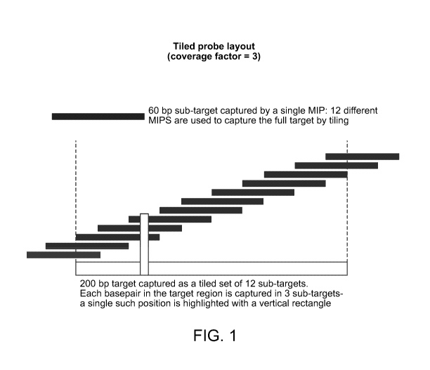
In some embodiments, the 'coverage factor', or number of probes used to
capture a
basepair in a molecule, is an important parameter to specify. Different
numbers of probes per
target are indicated depending on whether one is using the tiling approach
(see, e.g., FIG. 1) or
one of the staggered approaches (see, e.g., FIGs. 2 or 3).
FIG. 1 illustrates a non-limiting embodiment of a tiled probe layout showing
ten captured
sub-targets tiled across a single target. Each position in the target is
covered by three sub-targets
such that MIP performance per base pair is averaged across three probes.
FIG. 2 illustrates a non-limiting embodiment of a staggered probe layout
showing the
targets captured by a set of three MIPs. Each MIP captures the full target,
shown in black, plus
(in some cases) additional extra-target sequence, shown in gray, such that the
targeting arms of
each MIP fall on different sequence. Each position in the target is covered by
three sub-targets
such that MIP performance per basepair is averaged across three probes.
Targeting arms land
immediately adjacent to the black or gray regions shown. It should be
appreciated that in some
embodiments, the targeting arms (not shown) can be designed so that they do
not overlap with
each other.
FIG. 3 illustrates a non-limiting embodiment of an alternating staggered probe
layout
showing the targets captured by a set of three MIPs. Each MIP captures the
full target, shown in
black, plus (in some cases) additional extra-target sequence, shown in gray,
such that the
targeting arms of each MIP fall on different sequence. Each position in the
target is covered by
three sub-targets such that MIP performance per basepair is averaged across
three probes.
Targeting arms land immediately adjacent to the black or gray regions shown.
It should be appreciated that for any of the layouts, the targeting arms on
adjacent tiled or
staggered probes may be designed to either overlap, not overlap, or overlap
for only a subset of
the probes.
In certain embodiments for any of the layouts, a coverage factor of about 3 to
about 10 is
used. However, the methods are not so limited and coverage factors of up to 2,
3, 4, 5, 6, 7, 8, 9,
10, 20 or more may be used. It is to be appreciated that the coverage factor
selected may depend
the probe layout being employed. For example, in the tiling approach, for a
desired coverage
factor, the number of probes per target is typically a function of target
length, sub-target length,
and spacing between adjacent sub-target start locations (step size). For
example, for a desired
coverage factor of 3, a 200 bp target with a start-site separation of 20 bp
and sub-target length of
19
CA 02870702 2014-10-15
WO 2013/158540
PCT/US2013/036575
60 bp may be encompassed with 12 MIPs (FIG. 1). Thus, a specific coverage
factor may be
achieved by varying the number of probes per target nucleic acid and the
length of the molecules
captured. In the staggered approach, a fixed-length target nucleic acid is
captured as several
subregions or as `super-targets', which are molecules comprising the target
nucleic acid and
additional flanking nucleic acids, which may be of varying lengths. For
example, a target of 50
bp can be captured at a coverage factor of 3 with 3 probes in either a
'staggered' (FIG. 2) or
'alternating staggered' configuration (FIG. 3).
The coverage factor will be driven by the extent to which detection bias is
tolerable. In
some cases, where the bias tolerance is small, it may be desirable to target
more subregions of
target nucleic acid with, perhaps, higher coverage factors. In some
embodiments, the coverage
factor is up to 2, 3, 4, 5, 6, 7, 8, 9, 10 or more.
In some embodiments, when a tiled probe layout is used, when the target length
is greater
than 1 bp and when a step size (distance between the 5'-end of a target and
the 5' end of its
adjacent target) is less than the length of a target or subregion thereof, it
is possible to compute
probe number for a particular target based on target length (T), sub target
length (S), and
coverage factor (C), such that probe number = T/(S/C) + (C-1).
In some aspects, the disclosure provides methods to increase the uniformity of
amplification efficiency when multiple molecules are amplified in parallel;
methods to increase
the reproducibility of amplification efficiency; methods to reduce the
contribution of targeting
probe variability to amplification efficiency; methods to reduce the effect on
a given target
nucleic acid of polymorphisms in probe hybridization regions; and/or methods
to simplify
downstream workflows when multiplex amplification by MIPs is used as a
preparative step for
analysis by nucleic acid sequencing.
Polymorphisms in the target nucleic acid under the regions flanking a target
can interfere
with hybridization, polymerase fill-in, and/or ligation. Furthermore, this may
occur for only one
allele, resulting in allelic drop-out, which ultimately decreases downstream
sequencing accuracy.
In some embodiments, using a set of MIPs having multiple hybridization sites
for the capture of
any given target, the probability of loss from polymorphism is substantially
decreased because
not all targeting arms in the set of MIPs will cover the location of the
mutation.
Probes for MIP capture reactions may be synthesized on programmable
microarrays
because of the large number of sequences required. Because of the low
synthesis yields of these
CA 02870702 2014-10-15
WO 2013/158540
PCT/US2013/036575
methods, a subsequent amplification step is required to produce sufficient
probe for the MIP
amplification reaction. The combination of multiplex oligonucleotide synthesis
and pooled
amplification results in uneven synthesis error rates and representational
biases. By synthesizing
multiple probes for each target, variation from these sources may be averaged
out because not all
probes for a given target will have the same error rates and biases.
Applications
Multiplex amplification strategies disclosed herein may be used analytically,
as in
detection of SNPs, or preparatively, often for next-generation sequencing or
other sequencing
techniques. In the preparative setting, the output of an amplification
reaction is generally the
input to a shotgun library protocol, which then becomes the input to the
sequencing platform.
The shotgun library is necessary in part because next-generation sequencing
yields reads
significantly shorter than amplicons such as exons. In addition to the bias-
reduction afforded by
the multi-tiled approach described here, tiling also obviates the need for
shotgun library
preparation. Since the length of the capture molecule can be specified when
the probes, e.g.,
MIPs, are designed, it can be chosen to match the readlength of the sequencer.
In this way, reads
can 'walk' across an exon by virtue of the start position of each capture
molecule in the probe set
for that exon. Reducing analytical errors associated with bias in nucleic acid
preparations:
In some embodiments, aspects of the invention relate to preparative steps in
DNA
sequencing-related technologies that reduce bias and increase the reliability
and accuracy of
downstream quantitative applications.
There are currently many genomics assays that utilize next-generation (polony-
based)
sequencing to generate data, including genome resequencing, RNA-seq for gene
expression,
bisulphite sequencing for methylation, and Immune-seq, among others. In order
to make
quantitative measurements (including genotype calling), these methods utilize
the counts of
sequencing reads of a given genomic locus as a proxy for the representation of
that sequence in
the original sample of nucleic acids. The majority of these techniques require
a preparative step
to construct a high-complexity library of DNA molecules that is representative
of a sample of
interest. This may include chemical or biochemical treatment of the DNA (e.g.,
bisulphite
treatment), capture of a specific subset of the genome (e.g., padlock probe
capture, solution
hybridization), and a variety of amplification techniques (e.g., polymerase
chain reaction, whole
genome amplification, rolling circle amplification).
21
CA 02870702 2014-10-15
WO 2013/158540
PCT/US2013/036575
Systematic and random errors are common problems associated with genome
amplification and sequencing library construction techniques. For example,
genomic sequencing
library may contain an over- or under-representation of particular sequences
from a source
genome as a result of errors (bias) in the library construction process. Such
bias can be
particularly problematic when it results in target sequences from a genome
being absent or
undetectable in the sequencing libraries. For example, an under representation
of particular
allelic sequences (e.g., heterozygotic alleles) from a genome in a sequencing
library can result in
an apparent homozygous representation in a sequencing library. As most
downstream
sequencing library quantification techniques depend on stochastic counting
processes, these
problems have typically been addressed by sampling enough (over-sampling) to
obtain a
minimum number of observations necessary to make statistically significant
decisions.
However, the strategy of oversampling is generally limited to elimination of
low-count Poisson
noise, and the approach wastes resources and increases the expense required to
perform such
experiments. Moreover, oversampling can result in a reduced statistical
confidence in certain
conclusions (e.g., diagnostic calls) based on the data. Accordingly, new
approaches are needed
for overcoming bias in sequencing library preparatory methods.
Aspects of the disclosure are based, in part, on the discovery of methods for
overcoming
problems associated with systematic and random errors (bias) in genome
capture, amplification
and sequencing methods, namely high variability in the capture and
amplification of nucleic
acids and disproportionate representation of heterozygous alleles in
sequencing libraries.
Accordingly, in some embodiments, the disclosure provides methods that reduce
variability in
the capture and amplification of nucleic acids. In other embodiments, the
methods improve
allelic representation in sequencing libraries and, thus, improve variant
detection outcomes. In
certain embodiments, the disclosure provides preparative methods for capturing
target nucleic
acids (e.g., genetic loci) that involve the use of differentiator tag
sequences to uniquely tag
individual nucleic acid molecules. In some embodiments, the differentiator tag
sequence permits
the detection of bias based on the frequency with which pairs of
differentiator tag and target
sequences are observed in a sequencing reaction. In other embodiments, the
methods reduce
errors caused by bias, or the risk of bias, associated with the capture,
amplification and
sequencing of genetic loci, e.g., for diagnostic purposes.
22
CA 02870702 2014-10-15
WO 2013/158540
PCT/US2013/036575
Aspects of the invention relate to associating unique sequence tags (referred
to as
differentiator tag sequences) with individual target molecules that are
independently captured
and/or analyzed (e.g., prior to amplification or other process that may
introduce bias). These
tags are useful to distinguish independent target molecules from each other
thereby allowing an
analysis to be based on a known number of individual target molecules. For
example, if each of
a plurality of target molecule sequences obtained in an assay is associated
with a different
differentiator tag, then the target sequences can be considered to be
independent of each other
and a genotype likelihood can be determined based on this information. In
contrast, if each of
the plurality of target molecule sequences obtained in the assay is associated
with the same
differentiator tag, then they probably all originated from the same target
molecule due to over-
representation (e.g., due to biased amplification) of this target molecule in
the assay. This
provides less information than the situation where each nucleic acid was
associated with a
different differentiator tag. In some embodiments, a threshold number of
independently isolated
molecules (e.g., unique combinations of differentiator tag and target
sequences) is analyzed to
determine the genotype of a subject.
In some embodiments, the invention relates to compositions comprising pools
(libraries)
of preparative nucleic acids that each comprise "differentiator tag sequences"
for detecting and
reducing the effects of bias, and for genotyping target nucleic acid
sequences. As used herein, a
"differentiator tag sequence" is a sequence of a nucleic acid (a preparative
nucleic acid), which
in the context of a plurality of different isolated nucleic acids, identifies
a unique, independently
isolated nucleic acid. Typically, differentiator tag sequences are used to
identify the origin of a
target nucleic acid at one or more stages of a nucleic acid preparative
method. For example, in
the context of a multiplex nucleic acid capture reaction, differentiator tag
sequences provide a
basis for differentiating between multiple independent, target nucleic acid
capture events. Also,
in the context of a multiplex nucleic acid amplification reaction,
differentiator tag sequences
provide a basis for differentiating between multiple independent, primary
amplicons of a target
nucleic acid, for example. Thus, combinations of target nucleic acid and
differentiator tag
sequence (target:differentiator tag sequences) of an isolated nucleic acid of
a preparative method
provide a basis for identifying unique, independently isolated target nucleic
acids.
23
CA 02870702 2014-10-15
WO 2013/158540
PCT/US2013/036575
It will be apparent to the skilled artisan that differentiator tags may be
synthesized using
any one of a number of different methods known in the art. For example,
differentiator tags may
be synthesized by random nucleotide addition.
Differentiator tag sequences are typically of a predefined length, which is
selected to
control the likelihood of producing unique target:differentiator tag sequences
in a preparative
reaction (e.g., amplification-based reaction, a circularization selection-
based reaction, e.g., a MIP
reaction). Differentiator tag sequences may be, up to 5, up to 6, up to 7 up
to 8, up to 9, up to 10,
up to 11, up to 12, up to 13, up to 14, up to 15, up to 16, up to 17, up to
18, up to 19, up to 20, up
to 21, up to 22, up to 23, up to 24, up to 25, or more nucleotides in length.
For purposes of
genotyping, isolated nucleic acids are identified as independently isolated if
they comprise
unique combinations of target nucleic acid and differentiator tag sequences,
and observance of
threshold numbers of unique combinations of target nucleic acid and
differentiator tag sequences
provide a certain statistical confidence in the genotype.
During a library preparation process, each nucleic acid molecule may be tagged
with a
unique differentiator tag sequence in a configuration that permits the
differentiator tag sequence
to be sequenced along with the target nucleic acid sequence of interest (the
nucleic acid sequence
for which the library is being prepared, e.g., a polymorphic sequence). The
incorporation of the
nucleic acid comprising a differentiator tag sequence at a particular step
allows the detection and
correction of biases in subsequent steps of the protocol.
A large library of unique differentiator tag sequences may be created by using
degenerate, random-sequence polynucleotides of defined length. The
differentiator tag
sequences of the polynucleotides may be read at the final stage of the
sequencing. The
observations of the differentiator tag sequences may be used to detect and
correct biases in the
final sequencing read-out of the library. For example, the total possible
number of differentiator
tag sequences, which may be produced, e.g., randomly, is 4N, where N is the
length of the
differentiator tag sequence. Thus, it is to be understood that the length of
the differentiator tag
sequence may be adjusted such that the size of the population of MIPs having
unique
differentiator tag sequences is sufficient to produce a library of MIP capture
products in which
identical independent combinations of target nucleic acid and differentiator
tag sequence are
rare. As used herein combinations of target nucleic acid and differentiator
tag sequences, may
also be referred to as "target:differentiator tag sequences".
24
CA 02870702 2014-10-15
WO 2013/158540
PCT/US2013/036575
In the final readout of a sequencing process, each read may have an additional
unique
differentiator tag sequence. In some embodiments, when differentiator tag
sequences are
distributed randomly in a library, all the unique differentiator tag sequences
will be observed
about an equal number of times. Accordingly, the number of occurrences of a
differentiator tag
sequence may follow a Poisson distribution.
In some embodiments, overrepresentation of target:differentiator tag sequences
in a pool
of preparative nucleic acids (e.g., amplified MIP capture products) is
indicative of bias in the
preparative process (e.g., bias in the amplification process). For example,
target:differentiator
tag sequence combinations that are statistically overrepresented are
indicative of bias in the
protocol at one or more steps between the incorporation of the differentiator
tag sequences into
MIPs and the actual sequencing of the MIP capture products.
The number of reads of a given target:differentiator tag sequence may be
indicative (may
serve as a proxy) of the amount of that target sequence present in the
originating sample. In
some embodiments, the numbers of occurrence of sequences in the originating
sample is the
quantity of interest. For example, using the methods disclosed herein, the
occurrence of
differentiator tag sequences in a pool of MIPs may be predetermined (e.g., may
be the same for
all differentiator tag sequences). Accordingly, changes in the occurrence of
differentiator tag
sequences after amplification and sequencing may be indicative of bias in the
protocol. Bias
may be corrected to provide an accurate representation of the composition of
the original MIP
pool, e.g., for diagnostic purposes.
According to some aspects, a library of preparative nucleic acid molecules
(e.g., MIPs,
each nucleic acid in the library having a unique differentiator tag sequence,
may be constructed
such that the number of nucleic acid molecules in the library is significantly
larger than the
number prospective target nucleic acid molecules to be captured using the
library. This ensures
that products of the preparative methods include only unique
target:differentiator tag sequence;
e.g., in a MIP reaction the capture step would under sample the total
population of unique
differentiator tag sequences in the MIP library. For example, an experiment
utilizing 1 ug of
genomic DNA will contain about ¨150,000 copies of a diploid genome. For a MIP
library, each
MIP in the library comprising a randomly produced 12-mer differentiator tag
sequence (-1.6
million possible unique differentiator tag sequences), there would be more
than 100 unique
CA 02870702 2014-10-15
WO 2013/158540
PCT/US2013/036575
differentiator tag sequences per genomic copy. For a MIP library, each MIP in
the library
comprising a randomly produced 15-mer differentiator tag sequence (-1 billion
possible unique
differentiator tag sequences), there would be more than 7000 unique
differentiator tag sequences
per genomic copy. Therefore, the probability of the same differentiator tag
sequence being
incorporated multiple times is incredibly small. Thus, it is to be appreciated
that the length of
the differentiator tag sequence is to be selected based on the amount of
target sequence in a MIP
capture reaction and the desired probability for having multiple, independent
occurrences of
target:differentiator tag sequence combinations.
The skilled artisan will appreciate that as part of a MIP library preparation
process,
adapters may be ligated onto the ends of the molecules of interest. Adapters
often contain PCR
primer sites (for amplification or emulsion PCR) and/or sequencing primer
sites. In addition,
barcodes may be included, for example, to uniquely identify individual samples
(e.g., patient
samples) that may be mixed together. (See, e.g., USPTO Publication Number US
2007/0020640
Al (McCloskey et al.)
The actual incorporation of the random differentiator tag sequences can be
performed
through various methods known in the art. For example, nucleic acids
comprising differentiator
tag sequences may be incorporated by ligation. This is a flexible method,
because molecules
having differentiator tag sequence can be ligated to any blunt-ended nucleic
acids. The
sequencing primers must be incorporated subsequently such that they sequence
both the
differentiator tag sequence and the target sequence. Alternatively, the
sequencing adaptors can
be synthesized with the random differentiator tag sequences at their 3' end
(as degenerate bases),
so that only one ligation must be performed. Another method is to incorporate
the differentiator
tag sequence into a PCR primer, such that the primer structure is arranged
with the common
adaptor sequence followed by the random differentiator tag sequence followed
by the PCR
priming sequence (in 5' to 3' order). A differentiator tag sequence and
adaptor sequence (which
may contain the sequencing primer site) are incorporated as tags. Another
method to incorporate
the differentiator tag sequences is to synthesize them into a padlock probe
prior to performing a
gene capture reaction. The differentiator tag sequence is incorporated 3' to
the targeting arm but
5' to the amplification primer that will be used downstream in the protocol.
Another method to
incorporate the differentiator tag sequences is as a tag on a gene-specific or
poly-dT reverse-
26
CA 02870702 2014-10-15
WO 2013/158540
PCT/US2013/036575
transcription primer. This allows the differentiator tag sequence to be
incorporated directly at
the cDNA level.
In some embodiments, at the incorporation step, the distribution of
differentiator tag
sequences can be assumed to be uniform. In this case, bias in any part of the
protocol would
change the uniformity of this distribution, which can be observed after
sequencing. This allows
the differentiator tag sequence to be used in any preparative process where
the ultimate output is
sequencing of many molecules in parallel.
Differentiator tag sequences may be incorporated into probes (e.g., MIPs) of a
plurality
when they are synthesized on-chip in parallel, such that degeneracy of the
incorporated
nucleotides is sufficient to ensure near-uniform distribution in the plurality
of probes. It is to be
appreciated that amplification of a pool of unique differentiator tag
sequences may itself
introduce bias in the initial pool. However, in most practical cases, the
scale of synthesis (e.g.,
by column synthesis, chip based synthesis, etc.) is large enough that
amplification of an initial
pool of differentiator tag sequences is not necessary. By avoiding
amplification or selection
steps on the pool of unique differentiator tag sequences, potential bias may
be minimized.
One example of the use of the differentiator tag sequences is in genome re-
sequencing.
Considering that the raw accuracy of most next-generation sequencing
instruments is relatively
low, it is crucial to oversample the genomic loci of interest.
Furthermore, since there are two alleles at every locus, it is important to
sample enough
to ensure that both alleles have been observed a sufficient number of times to
determine with a
sufficient degree of statistical confidence whether the sample is homozygous
or heterozygous.
Indeed, the sequencing is performed to sample the composition of molecules in
the originating
sample. However, after multiple reads have been collected for a given locus,
it is possible that
due to bias (e.g., caused by PCR amplification steps), a large fraction of the
reads are derived
from a single originating molecule. This would skew the population of target
sequences
observed, and would affect the outcome of the genotype call. For example, it
is possible that a
locus that is heterozygous is called as homozygous, because there are only a
few observations of
the second allele out of many observations of that locus. However, if
information is available on
differentiator tag sequences, this situation could be averted, because the
over-represented allele
would be seen to also have an over-represented differentiator tag sequence
(i.e., the sequences
with the overrepresented differentiator tag sequence all originated from the
same single
27
CA 02870702 2014-10-15
WO 2013/158540
PCT/US2013/036575
molecule). Therefore, the sequences and corresponding distribution of
differentiator tag
sequences can be used as an additional input to the genotype-calling algorithm
to significantly
improve the accuracy and confidence of the genotype calls.
Example
Molecular Inversion Probe Capture on Fragmented Genomic DNA
The following example shows a preferred method of practicing the invention.
Genomic DNA was extracted from whole human blood using a Gentra Puregene Blood
Kit and following the Puregene protocol for DNA Purification from Whole Blood
(Qiagen). The
protocol can be scaled (i.e. amount of solution, duration) to accommodate the
desired amount of
whole genomic DNA.
First, a red blood cell Lysis Solution was dispensed into a centrifuge tube,
and 2.0 ml of
human whole blood was added. The sample was mixed by inverting 10 times. Then,
the sample
was incubated at room temperature and inverted at least once during the
incubation for
approximately 5 minutes. After incubation, the sample was centrifuged for a
time period
sufficient to pellet the white blood cells. Once white blood cells pelleted,
supernatant was
discarded by pipetting or pouring while leaving the residual liquid and white
blood cell pellet in
the tube. The remaining residual liquid and white blood cell pellet was
vortexed vigorously to
resuspend the pellet in residual liquid. Cell lysis solution was then added,
and vortexed vigously
for 10 seconds. RNase A solution was added and mixed in by inverting the tube
25 times. The
sample was incubated at 37 C for 30 minutes, vortexing briefly every 15
minutes. Protein
precipitation solution was then added and the sample was vortexed. The sample
was then
centrifuged so that the precipitated proteins formed a tight brown pellet.
Supernatant was poured
into another centrifuge tube containing isopropanol. The tube was inverted
gently until DNA
was visible as threads or a clump. After which, the sample was centrifuged.
The resulting
supernatant was then discarded, and the residual liquid was removed by
pipetting, but leaving the
pellet within in the tube. A 70% ethanol solution was added to the tube to
wash the DNA pellet.
The tube was then centrifuged, after which the supernatant was discarded by
draining the tube,
taking care that the DNA pellet remained. A DNA hydration solution, a Tris-
based buffer at a
pH between 7.5 and 8.0, was then added to the sample, and the sample was then
vortexed to mix.
This provided a sample of isolated whole genomic DNA in DNA hydrating
solution.
28
CA 02870702 2014-10-15
WO 2013/158540
PCT/US2013/036575
In order to fragment and denature the genomic DNA, the isolated whole genomic
DNA
was incubated in the DNA hydrating solution, i.e. the Tris-based buffer at a
pH between 7.5 and
8.0, for 16-24 hours at 65 C.
Once isolated, MIP capture probes were hybridized to the isolated fragmented
genomic
DNA by introducing the fragmented genomic DNA to 1.0 ul capture probe mix (-
2.5 pmol), 2.0
ul 10X Ampligase buffer (Epicentre), and 11 ul dH20. The reaction was
incubated at 60 C for 40
hours. The capture probe mix used for the capture reaction contained MIP
probes having
targeting arm sequences that were different for each capture probe in a set,
while constant region
sequence will be the same for all probes in the set, allowing all captured
targets to be amplified
with a single set of primers. Targeting arm sequences were designed such that
any given pair of
bp sequences is unique in the target genome (to prevent spurious capture of
undesired sites).
Additionally, melting temperatures were matched for all probes in the set such
that hybridization
efficiency was uniform for all probes at a constant temperature (e.g., 60 C).
The reaction was
incubated at various temperatures to promote hybridization.
15
The hybridized probes were converted into covalently-closed circular products
containing
subtargets by preparing a filling-in / ligation reaction mixture, adding the
mixture to the
hybridized probe reaction, and incubating the reaction at 60 C for 10 hours.
The Prepared fill-in
/ ligation reaction mixture included 1.0 ul, 2 mM dNTP mix (Invitrogen), 2.0
ul, 10X Ampligase
buffer (Epicentre), 5 U/ul Taq Stoffel fragment (Applied Biosystems), 5 U/ul
Ampligase
20 (Epicentre), and 4.75 ul dH20.
The circularized probe / subtarget products were then purified from un-reacted
probes
and genomic DNA. An exonuclease reaction mixture was prepared by mixing 21 ul
of the fill-
in/ligation reaction product with 2.0 ul 10X exonuclease I buffer (New England
Biolabs), 2.0 ul
20 U/ul exonuclease I (New England Biolabs), 2.0 ul 100 U/ul exonuclease III
(New England
Biolabs). The reaction was incubated at 37 C for 60 min, then heat-inactivate
by incubating at
80 C for 15 min, thus producing a heat-inactivated exonuclease reaction
mixture. The heat
inactivated exonuclease reaction mixture was then immediately cooled to 4 C
for storage.
Next, the purified circular material was amplified using PCR primers specific
to the
'constant region' of the probes. To perform the amplification, a PCR mixture
was prepared by
mixing the 2.0 ul of the heat-inactivated exonuclease reaction mixture with
5.0 ul 10X
Accuprime reaction buffer (Invitro gen), 1.5 ul 10
uM CP-2-FA (5' -
29
CA 02870702 2014-10-15
WO 2013/158540
PCT/US2013/036575
GCACGATCCGACGGTAGTGT-3') (SEQ ID NO: 1), 1.5 ul 10 uM CP-2-RA (5' -
CCGTAATCGGGAAGCTGAAG-3') (SEQ ID NO: 2), 0.4 ul 25 mM dNTP mix (Invitrogen),
1.5 ul 10X SybrGreen (Invitrogen), 0.4 ul 2.5 U/ul Accuprime Pfx polymerase
(Invitrogen) and
37.7 ul dH20.
The PCR mixture was thermal cycled in real-time thermal cycler according to
the
following protocol, but cycling was stopped before amplification yield
plateaus (generally 8-12
cycles): 1) 95 C for 5 min; 2) 95 C for 30 sec; 3) 58 C for 60 sec; 4) 72 C
for 60 sec; and 5) two
cycles of 95 C for 30 sec.
After PCR, a shotgun next-generation sequencing library was prepared by the
following
steps. The desired amplicon population was purified from non-specific
amplification products
by gel extraction. The amplicons were concatemerized into high-molecular
weight products
suitable for fragmentation. The concatemerized amplicons incubated in a Tris-
buffer at a pH
between 7.5 and 8.0 for 16 hrs at 65 C in order to fragment the amplicons.
Adapters required for
amplification by the sequencing platform were then ligated to the fragmented
amplicons.
Incorporation by Reference
References and citations to other documents, such as patents, patent
applications, patent
publications, journals, books, papers, web contents, have been made throughout
this disclosure.
An such documents are hereby incorporated herein by reference in their
entirety for an purposes.
Equivalents
The invention may be embodied in other specific forms without departing from
the spirit
or essential characteristics thereof. The foregoing embodiments are therefore
to be considered in
an respects illustrative rather than limiting on the invention described
herein.