Note: Descriptions are shown in the official language in which they were submitted.
I
Heart valve prosthesis and a method for manufacturing
the heart valve prosthesis
The present invention relates to a heart valve prosthesis and a method for
manufacturing the heart valve prosthesis.
DEFINITION
In the specification the term "comprising" shall be understood to have a broad
meaning similar to the term "including" and will be understood to imply the
inclusion
of a stated integer or step or group of integers or steps but not the
exclusion of any
other integer or step or group of integers or steps. This definition also
applies to
variations on the term "comprising" such as "comprise" and "comprises".
In the specification the term D-shape shall be understood to mean any shapes
which comprise two parts one of which is bent in about circular manner whereas
the
other part is substantially straight or bent to a less degree on either side
respectively. The straight part might also be bent slightly after
implantation.
In the mitral valve, the anterior leaflet of the valve covers the space
between the
valve commissures (including the trigones) and is in direct fibrous continuity
with the
aortic annulus under the left and non-coronary cusps of the aortic valve,
including
the fibrous trigone between the left and non-coronary cusps of the aortic
valve. Thin
bundles of collagen fibres resembling tendons extend circumferentially from
each
fibrous trigone for a variable distance towards the corresponding side of the
mitral
orifice. The posterior half to two thirds of the annulus supporting the
posterior leaflet
is mainly muscular, with little or no fibrous tissue. This muscle is mainly
positioned
perpendicularly to the annulus, and a less conspicuous group of muscle fibres
is
located parallel to the annulus.
CA 2870791 2017-06-15
1a
The mitral annulus corresponds to the transition between the endocardial layer
of
the left atrium, the valve tissue and the endocardium and myocardium of the
left
ventricle.
The orifice area of the mitral valve at the annulus is approximately 6.5 cm2
in
women and 8 cm2 in men. The circumference is approximately 9 cm in women and
cm in men. Depending on the inotropic state of the heart, the difference
between
the diastolic and systolic size of the annulus varies from 23% to 40%. The
effective
area of the valve orifice is approximately 30% smaller than the size of the
annulus.
CA 2870791 2017-06-15
cp, 02870791 2014-10-17
WO 2013/160439 PCT/EP2013/058708
2
Although solutions have been proposed for the percutaneous tran-
scatheter implantation of artificial aortic valves, the distinc-
tive characteristics of the mitral valve have hitherto prevented
the development of equally effective mitral valve prostheses.
The following considerations must be borne in mind when design-
ing a device for replacing the mitral valve:
Degenerative disease: mitral valve prolapse is the most
common pathological condition found in heart valves, and is pre-
sent in about 2% of the population; 5% of these develop mitral
insufficiency. Mitral prolapse is a degenerative pathology in
which calcification of the annulus is encountered in rare cases.
This is one of the main differences between aortic and mitral
pathologies, since the mitral ring is larger and more elastic,
and these two characteristics make it difficult to anchor a per-
cutaneous device.
Shape of the mitral valve: the annulus and valve are
asymmetric, with a long axis of about 5 cm between the commis-
sures and a short axis of 4 cm in the antero-posterior direction
in systole (generally with a long axis to short axis ratio of
4/4 to 3/4) when the valve is closed, because of the D shape of
the valve. During diastole, the annulus moves outwards with the
posterior wall of the left ventricle, allowing the mitral ori-
fice to become more circular. When degenerative pathological di-
lation of the annulus is present, the shape of the annulus be-
comes more circular throughout the cardiac cycle, principally as
a result of an increase in the posterior part of the annulus.
This leads to a change in the diameters of the mitral annulus.
The short axis (antero-posterior diameter) is elongated, causing
the shape of the valve to become more circular, thereby prevent-
ing perfect coaptation of the two leaflets and aggravating the
mitral insufficiency. Although commercially available mitral
prostheses are circular, in a percutaneous device this may give
CA 02870791 2014-10-17
WO 2013/160439 PCT/EP2013/058708
3
rise to problems of coaptation (intraprosthetic insufficiency)
and/or perivalvular leakage.
= Pressure of the left ventricle: when the left ventricle
contracts, the intraventricular pressure forces the mitral
valve to close, the valve being subjected to the effects
of systolic stress. Systolic stress is significantly
greater than diastolic stress (aortic valve); it is there-
fore important to have perfect coaptation in a device In-
tended for the mitral position.
= Obstruction of the left ventricle outflow tract: when the
mitral valve is surgically replaced the anterior leaflet is
completely excised. One of the major issues related to a
percutaneous implant of a mitral valve prosthesis is that
the anterior leaflet can not be excised. The retaining
leaflet can lead to a potential obstruction of the left
ventricle outflow tract when pushed into the ventricle by
the prosthesis. The bigger the size of the valvular pros-
thesis, the higher is the risk of obstruction as the leaf-
let is pushed further into the ventricle.
= Patients with mitral (annuloplasty) rings: patients who have
already undergone a mitral repair operation may develop re-
current Insufficiency. Circular artificial tricuspid aortic
valves do not operate correctly in patients with D-shaped
mitral rings, since they do not allow perfect coaptation of
the three leaflets; furthermore, owing to the deformation
of the structure, perivalvular leakage occurs, due to the
lack of coupling between the circular and the D shape, as
well as reduced stability of the implant with a risk of mi-
gration of the prosthesis.
For the reasons stated above, there are major constraints on the
use of e.g. aortic percutaneous or sutureless valve implant sys-
4
tems in mitral position. Valve replacement by the percutaneous route or by
minimally invasive surgery is becoming an increasingly common practice for the
aortic and pulmonary valves, but cannot be used as yet for the mitral valve.
As explained above, the mitral annulus differs from the aortic annulus in not
being
circular, especially in a patient with a mitral annuloplasty ring. The
available
transcatheter prostheses are always tricuspid, and if they are not perfectly
circular
after expansion (due to an irregular expansion of the stent) they tend to
reduce the
degree of coaptation of the valve leaflets, resulting in prosthetic
insufficiency. Since
the aortic annulus is practically circular, transcatheter valves almost always
work
well, unless the stent is distorted during implantation owing to the presence
of
dystrophic calcification of the annulus and the valve leaflets. The risk of
insufficiency
in the mitral position, which is not circular, is very considerable, since the
mitral
annulus does not offer any resistance during the expansion of the prosthesis,
thus
leading to over-dilation and consequent loss of coaptation between the three
valve
leaflets.
One object of the present invention is to provide a heart valve prosthesis
which can
at least partially overcome the problems of the prior art described above.
According to the present invention, there is provided an atrio-ventricular
valve
prosthesis comprising a ring-shaped supporting structure to be anchored at the
valve annulus, and a single extended valve leaflet of flexible material
floatingly
supported by said supporting structure, wherein
said supporting structure comprises a support wall portion at which a root end
of the valve leaflet is connected, and a complementary wall portion opposite
to said
support wall portion, which supports a static or quasi-static coaptation
surface
adapted to be sealingly engaged by a free end of the valve leaflet, and
extending in
CA 2870791 2017-06-15
4a
a direction substantially parallel to the movement direction of the free end
of the
valve leaflet at the coaptation surface;
said free end of the valve leaflet is connected to said support wall portion
or
to said complementary wall portion by means of at least one traction member of
flexible material, which is dimensioned to such a length that the movement of
the
free end of the valve leaflet is stopped at said coaptation surface; and
wherein the
supporting structure has a D-shaped cross section in the area where said valve
leaflet is contacting said structure.
Preferred embodiments of the prosthesis are described hereunder.
The aforesaid object is achieved according to the invention with a heart valve
prosthesis of the type defined initially, wherein said supporting structure
comprises
a support wall portion at which a root end of the valve leaflet is connected,
and a
complementary wall portion opposite said support wall portion, which supports
a
static or quasi-static coaptation surface adapted to be sealingly engaged by a
free
end of the valve leaflet, and extending in a direction substantially parallel
to the
/
CA 2870791 2017-06-15
5
direction of movement of the free end of the valve leaflet at the coaptation
surface;
and said free end of the valve leaflet is connected to said support wall
portion or to
said complementary wall portion preferably in their lateral portions by means
of at
least one traction member of flexible material, dimensioned to such a length
that the
movement of the free end of the valve leaflet is stopped at said coaptation
surface.
The invention was developed on the basis of an examination of the prior art in
the
field of heart valve repair.
According to the present invention, there is also provided a prosthesis having
a
support structure, a support wall portion, a complementary wall portion and a
single
extended valve leaflet supported by said support structure, wherein said
complementary wall portion supports a static or quasi-static coaptation
surface
adapted to be sealingly engaged by a free end of the valve leaflet.
As explained by Carpentier, the aim of mitral valve repair is to restore a
good
coaptation surface in order to provide satisfactory mital valve function
(Carpentier
A. Cardiac valve surgery ¨ the "French correction." J Thorac Cardiovasc Surg
1983; 86: 323-37). Conventionally, the repair of a posterior leaflet prolapse
consists
in the resection of the leaflet followed by annuloplasty using a ring, and
this has
been shown to have excellent long-term durability. Typically,
echocardiographic
findings after mitral valve repair show a posterior leaflet with reduced
mobility or no
mobility at all, which bangs vertically from the annulus and forms, as
demonstrated
experimentally and clinically (Cotin, L.H., Couper, G.S., Aranki, S.F., et al.
Long-
term results of mitral valve reconstruction for regurgitation of the
myxomatous mitral
valve. J Thorac Cardiovasc Surg 1994; 107: 143-51), a support against which
the
anterior leaflet bears in apposition. These conclusions were developed further
by
Perier (Perier, P., Hohenberger, W., Lakew, F., Batz, G., Urbanski, P.,
Zacher, M.,
CA 2870791 2017-06-15
CA 02870791 2016-04-11
6
and Diegeler, A. Toward a new paradigm for the reconstruction of posterior
leaflet
prolapse: midterm results of the "respect rather than resect" approach. Ann
Thorac
Surg 2008 Sep; 86(3): 718-25), who deliberately converted the bicuspid mitral
valve
into a monocuspid valve by the apposition of the anterior leaflet on an
extended
coaptation surface formed on the verticalized posterior leaflet.
Preferably, the concept on which the invention is based, therefore, is that of
a single
extended leaflet coapting on a wall which supports an extended, smooth,
regular
and substantially vertical coaptation surface, thus reducing the risk of
intraprosthetic
regurgitation. Additionally, the edge of the leaflet is retained by a traction
member
which simulates a system of chordae tendineae.
Because only one leaflet is present, a larger moving surface is subjected to
the
pressures created by the cardiac cycle. This results in faster opening and
closing of
the valve by comparison with tricuspid valves. Furthermore, using a monocuspid
valve could reduce the incidence of calcification phenomena. Indeed, it is
known
that, in some devices of the tricuspid type, fibrosis of the valve leaflets
occurs,
sometimes with the formation of intrinsic and extrinsic dystrophic
calcification, in the
less mobile part of the leaflet. We may deduce from this that the continual
movement of the single leaflet could contribute to the prevention of fibrosis
and
calcification of the tissue (Gabbay, S., Bort lotti, U., Cipolletti, G.,
Wasserman, F.,
Frater, R.W., and Factor, S.M. The Meadox unicusp pericardial bioprosthetic
heart
valve: new concept. Ann Thorac Surg 1984 Jun; 37(6): 448-56).
The supporting structure preferably has a D-shaped cross section in a plane
perpendicular to the axis of the structure, i.e. the axis corresponding to the
direction
of flow of blood when the supporting structure is implanted.
7
Preferably, the D-shape comprises one part bent in a circular manner and
another
part that is substantially straight or bent to a less degree.
The straight part might also be bent slightly after implantation. With the
bending of
the straight part "in vivo" the native leaflet can be pulled upwards with
respect of the
blood flow. Further, the portion shows better anchoring conditions after the
bending.
The substantially straight part preferably is the support wall portion and the
bent part
preferably is the complementary wall portion comprising the coaptation
surface.
Alternatively and preferably, the straight part is the complementary wall
portion
comprising the coaptation surface and the bent part is the support wall
portion.
Preferably, the cross section of the mitral valve annulus has a D-shape. A
supporting structure with a D-shape therefore provides a better fitting
between the
annulus and the supporting structure as there is contact between the annulus
and
the supporting structure substantially all around the supporting structure.
The better
fitting and contacting between the support structure and the annulus result
e.g. in
better stability of the implant as well as a reduced risk of perivalvular
leakage.
Further, the aortic annulus is not compressed as a result of an implantation
of a
mitral prosthesis, as there is no expansion of the mitral annulus with a D-
shaped
supporting structure, especially no expansion of the mitral annulus in the
direction of
the aortic annulus therefore on the anterior side.
Preferably, the anterior side of the supporting structure is the side which is
arranged
next to the aortic annulus when implanted. The anterior side preferably is the
complementary wall portion. The posterior side is opposite of the anterior
side and
CA 2870791 2017-06-15
CA 02870791 2016-04-11
8
therefore at maximum distance to the aortic annulus when implanted. The
posterior
side preferably is the support wall portion.
The dimensions of the support structure are preferably such that a distance
perpendicular to the anterior-posterior distance is preferably about a factor
from
1.1 -1.3, more preferably about a factor 1.2, longer than the anterior-
posterior
distance. The axial length of the support structure is preferably about 32 mm
and
the thickness of the wall as well as the width of struts is preferably about
500 ,um
(micrometer).
In a preferred variation, the inflow end and the outflow end are flared
outwardly with
respect to the flow direction and the cardiac wall. The inflow end is
preferably flared
about 200 to 40 , more preferably about 30 , with respect to the flow
direction and
the outflow end is preferably flared about 7.5 up to 17.5 , more preferably
about
10 , with respect to the flow direction.
Preferably, in an alternative embodiment, the ends of the prosthesis are
asymmetrically flared such that the anterior side has a different flare than
the
posterior side. The anterior side in the variation preferably is flared at the
outflow
end preferably about 7.5 to 17.5 , more preferably about 10 , with respect to
the
axis and flared at the inflow end preferably about 15 to 25 , more preferably
about
20 , with respect to the axis whereas the posterior side is flared at the
outflow end,
preferably about 7.5 to 17.5 , more preferably about 10 , with respect to the
axis
and at the inflow end about 20 to 40 , more preferably about 30 , with
respect to
the axis. The asymmetric flare might be present in combination with the
various
prosthesis of the invention described herein.
= CA 02870791 2016-04-11
9
Preferably, in a further alternative embodiment, the flares might be provided
as
curvilinear flares. Curvilinear flares mean in that the flares are bent in a
kind of
circular, convex manner with respect to the axis so that the flare bends
outwardly
first and at least slightly inwardly on towards the inflow end or the outflow
end,
respectively, with respect to the axis. The curvilinear flares might be
present in
combination with the various prosthesis of the invention described herein.
The flares provide a force of the support structure on the atrioventricular
junction
and surrounding tissues of the posterior ventricular wall which keeps the
fixation of
the support structure in the annulus. A smaller flare at the outflow end of
the anterior
portion than on the posterior portion helps minimizing the risk of obstructing
the left
outflow tract.
Preferably, in another variation of the prosthesis, the supporting structure
has an
asymmetric arrangement in the axial direction such that one side, preferably
the
anterior side is shorter than an opposite side in axial direction, preferably
the
posterior side.
The asymmetric arrangement is preferably constructed such that the anterior
portion
is as short such that the native leaflet is basically not pushed outwards in
the left
ventricle. In the left ventricle the native leaflet could interfere with the
blood outflow
through the aorta in systole.
Preferably, the native leaflets are not excised when an off-pump procedure is
performed. The native leaflets are rather pushed to the side, therefore out of
the
opening, with the prosthesis such as to not interfere with the blood flow.
However,
with an anterior portion having a certain length, the anterior native leaflet
might be
pushed in the left ventricle. When the heart contracts during systole to push
the
CA 02870791 2016-04-11
blood out of the aorta, the anterior native leaflet might block some of the
blood flow
through the aorta when covering some parts of the aortic annulus. With a
shorter
anterior portion, the anterior leaflet is not pushed into the left ventricle
outflow tract
but rather moves towards the inflow tract.
The asymmetric construction might be part of any prosthesis discussed herein.
It is also possible that the anterior and posterior portions are the same
length.
10
In any prosthesis the valve and the at least one traction member might be
constructed as a single piece, preferably as a single piece made of
pericardium.
The traction member and the valve might be cut out of pericardium as one
piece.
With a construction as one piece, there is no need to attach the traction
member to
the valve leaflets. A construction without an attachment is more stable than a
two
piece construction. Therefore, the risk of a malfunction is reduced.
It is also possible to construct the traction member and the valve as two or
more
pieced arrangements.
Preferably, pericardium has been shown to be persistent and therefore well
suited
for the purpose as valve and traction member. It is also possible to use other
biocompatible materials such as biocompatible plastics or tissues.
The at least one traction member is preferably attached to lateral portions of
the
supporting structure.
11
Preferably, the lateral portions are the two sides of the supporting structure
which
include the utmost ends of the anterior side and the posterior side. The
anterior side
and the posterior side preferably relate to the complementary wall portion and
the
support wall portion. The attachment of the traction member can be anywhere
along
the lateral portions.
When the traction members are arranged at the lateral portions, their
interference
with the movement of the valve is minimized as the valve leaflet preferably
opens in
an anterior posterior direction.
The invention further comprises a delivery device comprising a prosthesis
according
to the invention described herein.
According to the present invention, there is also provided a method for
manufacturing a heart valve prosthesis comprising,
- providing a support structure, said support structure being a D-shaped
support structure with a support wall portion and a complementary wall
portion,
- assembling a valve leaflet of flexible material in the support structure,
wherein the valve leaflet is connected with a root end to the support wall
portion or
the complementary wall portion through stitching, and a free end of the valve
is
attached to the other wall portion, the complementary wall portion or the
support
wall portion through a traction member.
Preferred embodiments of the method are described hereunder.
Further, the invention relates to a method for manufacturing a heart valve
prosthesis, preferably a prosthesis according to the invention described
herein,
comprising the steps of:
CA 2870791 2017-06-15
1 1 a
- providing a support structure, preferably a D-shaped support structure
with
a support wall portion and a complementary wall portion,
- assembling a valve leaflet of flexible material in the support structure,
wherein the valve leaflet is connected with a root end to the support wall or
the
complementary wall, preferably through stitching,
and a free end of the valve is attached to the other wall portion,
complementary wall
portion or support wall portion through a traction member.
CA 2870791 2017-06-15
cp, 02870791 2014-10-17
WO 2013/160439 PCT/EP2013/058708
12
In the method of manufacturing the valve and the traction member
are preferably constructed as a single piece.
Although the characteristics and advantages of the prosthesis
according to the invention are discussed herein with regard to
mitral valve replacement, it will be clear that the inventive
concept can also be applied to valve prostheses intended for the
replacement of other heart valves.
Further characteristics and advantages of the prosthesis accord-
ing to the invention will be made clear by the following de-
tailed description, which refers to the attached drawings, pro-
vided purely by way of non-limiting example, in which:
- Figure la shows a simplified perspective view from
above of a valve prosthesis according to the invention;
- Figure lb shows a simplified schematic view in cross
section (A2-P2 according to Carpentier) of a valve prosthesis
according to the invention;
- Figure 2 is a perspective view from below (showing the
outflow side) of the valve prosthesis of Figure la; for greater
clarity, part of the prosthesis is shown as transparent;
- Figure 3 is a plan view (showing the inflow side) of
the valve prosthesis of Figure la;
Figure 4 is a sectional view of the valve prosthesis,
taken along the line IV-IV in Figure 3;
- Figures 5 to 7 are perspective views from below of dif-
ferent embodiments of the valve prosthesis;
- Figure 8 is a plan view of a further valve prosthesis;
- Figure 9 is a sectional view of the valve prosthesis,
taken along the line IX-IX in Figure 8.
Figure 10a is a perspective view from below (showing
the outflow side) of the valve prosthesis in closed position.
cp, 02870791 2014-10-17
WO 2013/160439 PCT/EP2013/058708
13
- Figure 10b is a perspective view from below (showing
the outflow side) of the valve prosthesis in open position.
- Figure 11a is a side view of the valve prosthesis.
- Figure 11b is a top view (inflow view) of the valve
prosthesis.
With reference to Figs. 1 to 4, a heart valve prosthesis accord-
ing to the invention, shown schematically, is indicated as a
whole by 1. This prosthesis 1 comprises a ring-shaped supporting
structure 3 to be anchored at the valve annulus, which may al-
ready have been repaired with an annuloplasty ring. With respect
to the direction perpendicular to the cross section of the ring,
the supporting structure 3 has an inflow side or end 3b and an
outflow side or end 3a. In this context, the terms "inflow" and
"outflow" refer to the inflow and outflow of the blood into and
from the valve when the prosthesis is in use.
In plan view (Fig. 3), the illustrated valve is D-shaped, making
it suitable for implants in the atrio-ventricular position (mi-
tral or tricuspid). In an alternative embodiment which is not
shown, the valve according to the invention may have a circular
shape suitable for implants in the aortic or pulmonary position.
Preferably, as shown schematically in Figure lb, the supporting
structure 3 comprises a skeleton 3' formed by a valve stent,
which can assume a positioning configuration in which the stent
is folded to allow it to be positioned through a catheter, and
an implant configuration, in which the stent is expanded to be
adapted and anchored to the valve annulus. In order to achieve
this expansion in position, the material of the stent may be a
self-expanding material, for example a shape memory alloy, or a
shaped balloon associated with the positioning system may be
provided, this balloon being inflated to cause the expansion of
cp, 02870791 2014-10-17
WO 2013/160439 PCT/EP2013/058708
14
the stent. The aforesaid expansion may also provide a change in
the shape of the prosthesis, particularly in order to create the
D shape of the mitral or tricuspid valve.
More specifically, the prosthesis according to the positioning
system may be transferred to the implantation site by an on-
pump, sutureless surgical procedure, by an off-pump surgical
procedure with transatrial or transapical access through a mini-
thoracotomy, or, last but not least, by an intervention proce-
dure with percutaneous access.
The release of the prosthesis from the positioning system can
take place in a single action or can be a two step procedure. In
a first step, a portion of the prosthesis (either the inflow or
the outflow portion, depending on the implantation method and
the visualization procedure) is released, so that its vertical
and horizontal positioning can be adjusted. In the second step,
the second portion of the prosthesis is released (complete re-
lease).
Preferably, the valve prosthesis is of the sutureless type; that
is to say, no stitches are required to anchor it to the valve
seat. For this purpose, the supporting structure 3 may be ana-
tomically shaped so as to be anchored securely to the valve an-
nulus, or can be provided with special-purpose formations for
anchoring.
More specifically, the outer portion of the valve stent which
comes into direct contact with the native fibrous annulus may be
slightly concave in order to follow the contour of the annulus
and facilitate anchoring thereto. The structure of the valve
stent in this area may also include anchorages formed by grafts
integrated into the structure itself. Other possibilities to
cp, 02870791 2014-10-17
WO 2013/160439 PCT/EP2013/058708
support the anchor are hooks or struts. This method of anchoring
makes it possible to avoid the progressive dilation of the fi-
brous annulus, thus reducing the risk of perivalvular leakage
and detachment of the prosthesis. The anchoring takes place
jointly with the opening of the atrial and ventricular portions
of the valve stent.
Preferably, the atrial (inflow) portion of the valve stent has a
special collapsible mesh design like the rest of the stent, and
at the end of the opening of the prosthesis it assumes an out-
wardly flared shape such that secure contact can be established
with the atrial (inflow) wall. The purpose of this portion of
the prosthesis is to ensure the positioning of the prosthesis by
means of progressive colonization by fibrous tissue (fibrous
pannus). The profile of the atrial (inflow) portion is higher in
the posterior anatomical portion and lower in the anterior por-
tion, the aim being in the latter case to reduce any possible
interference with the aortic valve. This structure may be made
of metal alloy only, or may be covered with biological or syn-
thetic tissue in order to optimize colonization by the fibrous
tissue.
Preferably, the ventricular (outflow) portion of the valve stent
has a special collapsible mesh design like that of the atrial
(inflow) portion. The profile is markedly asymmetric. The poste-
rior ventricular (outflow) portion has a marked protrusion,
which not only provides contact with the posterior ventricular
(outflow) wall, but also provides a member for anchoring to the
support (chordae) of the anterior monocuspid valve leaflet and
to the chordae of any posterior leaflet, as described below. It
may take the form of a single structure or two or three separate
structures. The anterior ventricular (outflow) part has a low
profile and is given a flared shape to promote the anchoring of
cp, 02870791 2014-10-17
WO 2013/160439 PCT/EP2013/058708
16
the prosthesis to the ventricle without interfering with the mi-
tral-aortic continuity, in order to avoid creating compression
which would lead to conduction disturbances. The anterior ven-
tricular side of the stent in its more distal portion could have
an everting angle close to 180 . Therefore, the stent could he
originally symmetric D shape and after thermal shaping only the
anterior ventricular part could evert becoming asymmetric. This
condition could imply the grabbing of the edge of the native an-
terior leaflet and the native leaflet in the direction of the
atrium, letting the left ventricle outflow tract free. Further,
anchoring of the anterior ventricular portion is improved with
an everted ventricular part. The ventricular (outflow) portion
of the stent may be made of metal alloy only, or may be covered
with biological or synthetic tissue in order to optimize coloni-
zation by the fibrous tissue.
As a general rule, the supporting structure 3 of the valve pros-
thesis may have a coating 3", of pericardium for example, or of
biological tissue in general, or of synthetic tissue, covering
some or all of the supporting structure. The coating 3" is par-
ticularly necessary if the supporting structure is a valve stent
3', in order to provide a seal at the valve annulus to which the
prosthesis is fitted.
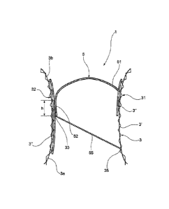
The valve prosthesis further comprises a single valve leaflet 5
of flexible material supported floatingly by the supporting
structure 3. The flexible material of the valve leaflet must
have characteristics meeting the requirements of cyclic fatigue
resistance. The valve leaflet 5 may be made of pericardial tis-
sue, or biological tissue in general, or synthetic tissue. The
pericardial tissue, in addition to the conventional cross-link
tissue fixation, should preferably be subjected to chemical
treatment serving to provide long-term retardation of the dys-
cp, 02870791 2014-10-17
WO 2013/160439 PCT/EP2013/058708
17
trophic calcification of the biological tissue. The valve leaf-
let may be made as a kind of extension of the material which
coats the supporting structure 3 of the prosthesis, or as a sep-
arately produced part which is subsequently anchored to the sup-
port wall portion 31 of the supporting structure 3. In a plan
view (Fig. 3), the surface extension of the valve leaflet 5 in
the example of Figures 1 to 4 is substantially equal to the
cross section of the orifice delimited by the supporting struc-
ture 3.
In relation to this valve leaflet, the supporting structure 3
comprises a support wall portion 31 at which a root end 51 of
the valve leaflet 3 is connected. For this purpose, the outflow
portion 3b of the valve stent has a valve support for the valve
leaflet of the prosthesis. This support is integrated into the
structure of the stent, and is designed to withstand cyclic fa-
tigue stress, to provide adequate support for the valve leaflet,
and to allow the prosthesis to be fully collapsed for insertion
into the positioning system.
The supporting structure 3 further comprises a complementary
wall portion 32 connected to and opposite the support wall por-
tion 31, which supports a coaptation surface 33 (visible in
Figs. lb and 4) adapted to be sealingly engaged by a free end 52
of the valve leaflet 5. In the example shown in Figures 1 to 4,
the coaptation surface 33 is static, in the sense that it is in-
tegral with the supporting structure 3 of the valve prosthesis.
In particular, in the example of Figures 1 to 4, the coaptation
surface 33 is defined by an inner face (that is to say, a face
turned towards the centre of the valve prosthesis) of the com-
plementary wall portion 32 of the supporting structure 3. At the
coaptation surface, the complementary wall portion 32 of the
supporting structure 3 has the coating discussed above.
cp, 02870791 2014-10-17
WO 2013/160439 PCT/EP2013/058708
18
If the valve prosthesis in question is a mitral prosthesis, the
support wall portion 31 of the supporting structure 3 is an an-
terior wall portion of this supporting structure, while the com-
plementary wall portion 32 is a posterior wall portion. The
terms "anterior" and "upper" refer to the positioning of the
valve prosthesis in use, at the mitral annulus.
In use, the valve leaflet 5, being flexible and connected to the
supporting structure 3 by its root end 51, is able to bend with
respect to its root end 51, under the action of the blood pres-
sure present upstream and downstream of the valve prosthesis,
thereby opening or closing the orifice formed by the supporting
structure 3 of the valve prosthesis. In the closed position, the
edge of the free end 52 of the valve leaflet 5 engages the coap-
tation surface 33 positioned on the complementary wall portion
32 of the supporting structure 3.
The coaptation surface 33 extends in a direction substantially
parallel to the direction of movement of the free end 52 of the
valve leaflet 5 at the coaptation surface 33. When the valve
prosthesis is in use, the aforesaid direction is substantially
vertical. Thus it is possible to obtain a large coaptation sur-
face, similar to that achieved with the Perier method for creat-
ing a mitral annuloplasty; the coaptation surface may have an
extension h (see Fig. lb) of at least 5 mm in the direction of
movement of the free end 52 of the valve leaflet. Consequently,
the possibility of regurgitation if the prosthesis becomes de-
formed is reduced.
As can be seen in Figures 2 and 3, the free end 52 of the valve
leaflet 5 is connected to the support wall portion 31 by at
least one traction member 55 of flexible material (in the pre-
cp, 02870791 2014-10-17
WO 2013/160439 PCT/EP2013/058708
19
sent example there are two of these members). The traction mem-
bers 55 simulate the retaining/stopping function of the natural
chordae tendineae, and are therefore dimensioned to such a
length that the movement of the free end 52 of the valve leaflet
is stopped at the coaptation surface 33.
The traction members 55 may be made of the same material as the
valve leaflets 5 or of different material, and may be formed so
as to resemble extensions from the free end 52 of the leaflet,
or as separately made elements which are subsequently fixed to
the free end of the valve leaflet.
The other ends of the traction members 55 are fixed to the sup-
porting structure 3 on the side (the blood inflow side 3a) axi-
ally opposed to the side (the blood outflow side 3b) on which
the valve leaflet is positioned. In order to connect the trac-
tion members to the supporting structure 3 and support them
thereon, it is possible to provide post portions 35 projecting
axially from the supporting structure 3 on the inflow side 3a of
the valve prosthesis.
Preferably, predetermined areas (for example, areas adjacent to
the commissural region) of the supporting structure 3 are pro-
vided with markers made of material opaque to radiation of pre-
determined wavelength, for example a radiopaque material such as
a noble metal, for instance platinum or tantalum. The markers
serve to facilitate the implantation of the prosthesis during a
procedure making use of fluoroscopy, by providing a spatial ref-
erence to the operator, which is to be aligned with an anatomic
reference.
Figure 5 shows another exemplary embodiment of a valve prosthe-
sis according to the invention. Elements corresponding to those
cp, 02870791 2014-10-17
WO 2013/160439 PCT/EP2013/058708
of the preceding embodiment have been given the same reference
numerals; for a detailed explanation of these elements, refer-
ence should be made to the preceding part of the description.
The prosthesis of Figure 5 differs from that of Figures 1 to 4
in that it has only one traction member 55 instead of a pair of
traction members 55. Accordingly, the prosthesis of Figure 5 has
a single post portion 35, to which the end of the traction mem-
ber 55 is connected.
More generally, the number of traction members 55 may vary ac-
cording to circumstances; in an embodiment which is not illus-
trated, there is a plurality of traction members of different
lengths, connected to a plurality of points distributed along
the edge of the free end 52 of the valve leaflet 5. This ar-
rangement enables the stresses acting on the valve leaflet 5 to
be distributed uniformly. In a further embodiment which is not
illustrated, there is a single traction member which forms an
integral extension of the valve leaflet 5, and which therefore
extends along the whole edge of the free end 52 of the valve
leaflet 5. This configuration enables the distribution of
stresses to be improved further.
Figure 6 shows a third exemplary embodiment of a valve prosthe-
sis according to the invention. Elements corresponding to those
of the preceding embodiments have been given the same reference
numerals; for a detailed explanation of these elements, refer-
ence should be made to the preceding part of the description.
The prosthesis of Figure 6 differs from that of Figure 5 in that
the traction member 55 is connected to the complementary wall
portion 32, rather than to the support wall portion, in a posi-
tion which is therefore diametrically opposite that shown in
CA 02870791 2016-04-11
21
Figure 5. Accordingly, the prosthesis of Figure 6 has a post portion 35
positioned on
the complementary wall portion 32, to which the end of the traction member 55
is
connected.
Figure 7 shows a further exemplary embodiment of a valve prosthesis according
to
the invention. Elements corresponding to those of the preceding embodiments
have
been given the same reference numerals; for a detailed explanation of these
elements, reference should be made to the preceding part of the description.
The prosthesis of Figure 7 differs from that of Figure 6 in that it has three
traction
members 55 instead of a single traction member 55. Accordingly, the prosthesis
of
Figure 7 has three support portions 35, to which the ends of the traction
members
55 are respectively connected.
In a variant of the invention shown in Figures 8 and 9, the coaptation surface
33 is
formed by a quasi-static coaptation leaflet 6, positioned on the complementary
wall
32 of the supporting structure 3. For the purposes of the present invention,
the term
"quasi-static" means that the coaptation leaflet 6 has a reduced mobility by
comparison with the valve leaflet 3. The coaptation leaflet 6 comprises a root
end 62
connected to the complementary wall portion 32 of the supporting structure 3.
For
this purpose, the outflow portion 3a of the valve stent has a valve support
for the
coaptation leaflet of the prosthesis. This support is integrated into the
structure of
the stent, and is designed to withstand cyclic fatigue stress, to provide
adequate
support for the coaptation leaflet, and to allow the prosthesis to be fully
collapsed for
insertion into the positioning system.
cp, 02870791 2014-10-17
WO 2013/160439 PCT/EP2013/058708
22
The coaptation leaflet 6 further comprises a free end 61 con-
nected to the complementary wall portion 32 by means of at least
one traction member 65 of flexible material, dimensioned to such
a length that the coaptation leaflet 6 is kept bent towards the
complementary wall portion 32. In order to connect the traction
member 65 to the supporting structure 3 and support it thereon,
it is possible to provide post portions projecting axially from
the supporting structure 3 on the inflow side of the valve pros-
thesis. The number and extension of the traction members 65 of
the coaptation leaflets may vary in a similar way to that de-
scribed above with regard to the traction members 55 of the
valve leaflet 5. In Variation the traction member 65 is arranged
such that the free end 61 is pointing at an outflow end 3a.
The materials from which the coaptation leaflet 6 is made are
the same as those from which the valve leaflet 5 is made.
As can be seen in Figure 8, the surface extension of the valve
leaflet 5 is significantly greater than that of the coaptation
leaflet 6. The anchoring line of the coaptation leaflet in the
wall of the valve stent terminates in continuity with the an-
choring line of the valve leaflet 5, forming two commissures in
the antero-posterior position. The length of the anchoring line
for the anterior leaflet (5) is typically around 40% of the an-
nular circumference while the posterior leaflet (6) is the re-
maining 60%. The depth of the commissure must be at least 5-8
mm, similar to that of the rest of the coaptation surface 33.
As a general rule, the valve leaflet, and the coaptation leaflet
if present, are preferably anchored directly to the valve stent
at the inflow and to the post portions (at the outflow) by the
interposition of biological or synthetic tissue. By means of
this system, the shock of leaflet elongation during the sys-
tole/diastole phases can be absorbed jointly at the coaptation
cp, 02870791 2014-10-17
WO 2013/160439 PCT/EP2013/058708
23
surface of the two leaflets, thus increasing the durability of
the prosthesis over time.
Figure 10a shows a perspective view of the support structure 3
from the outflow end 3a in a closed position. The closed posi-
tion means that the valve leaflet 5 prevents a blood backflow
through the support structure 3. The valve leaflet 5 and two
traction members 55 are formed as a single piece made of peri-
cardium. The leaflet 5 is stitched to the support wall portion
31 along schematically shown stitches. The free end 52 of the
valve leaflet 5 is contacting the complementary wall portion 32
in the closed position. The traction members 55 are sewed at
lateral portions 34 of the support structure 3 along schemati-
cally shown stitches. The traction members 55 prevent further
movement of the valve leaflet 5 in the closed position, thereby
securing a sealed closing.
Figure 10b shows a perspective view of the support structure 3
from the outflow end 3a in an open position. Open position means
that the valve leaflet 5 does basically not interfere with the
blood flow through the support structure 3 from the left atrium
to the left ventricle during diastole. The free end 52 of the
valve leaflet 5 is at distance to the complementary wall portion
32. Two traction members 55 are attached to lateral portions 34
and the free end 52 of the valve leaflet 5.
Figure lla shows a side view of the support structure 3 with the
valve leaflet 5 mounted inside the support structure 3. The
valve leaflet 5 is sewed to the lateral portions 34 by means of
the traction members 55 along the schematically shown stitches.
The inflow end 3b has a flare of 100 with respect to the blood
flow direction. The outflow end 3a has a flare of 300 with re-
spect to the blood flow direction. The axial length dl of the
cp, 02870791 2014-10-17
WO 2013/160439 PCT/EP2013/058708
24
inflow end 3b is shorter than the shortest axial length d2 of
the outflow end 3a. The flare of the outflow end 3a is in a cur-
vilinear manner. There is a outwardly flared portion 61 and a
slightly inwardly bend portion 60, with respect to the blood
flow direction. The flares might also be present in a more cur-
vilinear manner. Further, the flares are asymmetrically ar-
ranged. The anterior portion corresponding to the supplementary
wall portion 31 is flared in an angle al. al is smaller than an
angle a2 present in the flare of the posterior portion corre-
sponding to the complementary wall portion 32.
The support structure comprises multiple struts which form
cells. The struts have a width of 500 um. A wall thickness of
the support structure 3 is also 500 pm. The support structure
has an axial length D of about 32 mm from the inflow end 3b to
the outflow end 3a measured on the support wall portion 31. The
cells on the outflow end 3a can have a larger dimension than the
cells on the inflow end 3b. An anterior portion corresponding to
the supplementary wall portion 31 in figure 11a is shorter in
axial direction than a posterior portion corresponding to the
complementary wall portion 32. The lateral portions 34 connect
the anterior portion and the posterior portion such that the
lateral portions gain constant in length from the anterior por-
tion to the posterior portion. The support structure might be
formed out of Nitinol.
Figure 11b shows a top view of the support structure 3 housing
the valve leaflet 5. The support structure is in a D shape. The
D shape is arranged such that the anterior portion corresponding
to the support wall portion 31 in figure 11b is only slightly
convexly bent and the posterior portion corresponding to the
complementary wall portion 32 is convexly bent with a smaller
radius of curvature. The distance between the lateral portions
CA 02870791 2014-10-17
WO 2013/160439 PCT/EP2013/058708
34 is 1.2 times as big as the distance between the anterior por-
tion and the posterior portion.