Note: Descriptions are shown in the official language in which they were submitted.
1
PRE-LOADED INJECTOR FOR USE WITH INTRAOCULAR LENS
[0001]
BACKGROUND
[0002] The invention relates to a device and a method of injecting a
flexible intraocular
lens which is ready to use, that is, ready to be implanted by injections
through an incision
formed in the wall of a patient's eye.
[00031 Flexible intraocular lenses are useful, for example, in a cataract
operation in order
to restore sight by a surgical procedure, which inserts into the eye such
intraocular lens, which
replaces the natural lens that has become opaque due to the cataract.
[0004] Flexible intraocular lenses are often made of hydrophilic
material(s) such as, for
example, hydrogel, acrygel or acrylic (the latter term deviating from its
normal meaning),
which materials are PMMA (polymethylmethacrylate) and/or I TEMA
(hydroxymethylmethacrylate), hydrated to more than 16%, in particular between
24% and 28%.
U.S. Pat. No. 4,787,904 describes various examples of materials that may be
used to produce
hydrophilic lenses. These lenses need to be kept in a hydrated state for
conservation.
[0005] Flexible intraocular lenses can also be made from silicone
materials, having a
higher refractive index than hydrophilic materials, or hydrophobic acrylic
materials with low
glass transition temperatures. The latter materials are desirable because they
typically have a
high refractive index and lenses made from them unfold more slowly and more
controllably
than silicone lenses. U.S. Pat. No. 7,157,538 describes such a high refractive
index, acrylic
material used for making hydrophobic flexible intraocular lenses.
[0006] Flexible intraocular lenses have the advantage of being able to be
folded, allowing
them to pass through incisions in the eye of small dimensions. However, the
problem arising
with these flexible lenses is precisely that of folding and manipulating them
at the moment of
the surgical act. U.S. Pat. No. 4,787,904 proposes to conserve a hydrophilic
lens in a folded
state in the injection device while being immersed in a conserving solution,
the whole assembly
being contained in a flexible packaging pocket. However, this method may not
be
CA 2871046 2019-07-10
CA 02871046 2014-10-20
WO 2013/159045
PCT/1JS2013/037457
2
used in practice, since a lens which has remained folded for a long period may
retain a shape
memory of the folded state and therefore does not regain its unfolded,
functional shape after
implantation.
[0007] As a result, hydrophilic lenses up to now have been conserved flat in
sterilized rigid
containers of conserving solution. At the moment of the surgical act, the
surgeon removes the
lens using a pincer, folds it (optionally with the aid of a folding device) or
places it in a
folding cartridge or in an injector and injects it into the eye. These
manipulations are
relatively complex and delicate, increasing the risk of contamination and
damage to the lens.
[0008] U.S. Pat. No. 6,386,357 discloses a soft intraocular lens-folding
device comprising
a base member with a tapered slide groove portion, and a movable member
comprising an
elastically bendable pair of legs and a common base connecting the pair of
legs. A soft
intraocular lens is introduced in the lens-receiving portion of the movable
member, the lens
being clamped by wall portions. The lens is folded by moving the movable
member into the
groove portion in the base member, forcing the legs of the movable member to
be drawn near
to one another. This document does not disclose any means for injecting the
folded lens.
[0009] U.S. Patent Publication No. 2005182419 discloses an injector for an
intraocular lens
comprising an injector housing with an intraocular lens disposed in the
housing. The injector
further comprises a lens carrier, which, in response to an actuator, engages
and moves the
lens within a narrowing injection nozzle in order to fold the lens. A plunger
is then used to
advance the folded lens and inject it into a patient's eye. Here, folding and
injection of the
lens cannot be achieved by a single, continuous movement of a plunger, adding
complexity to
the surgical procedure.
[0010] What has been needed, and heretofore unavailable, is an injector
configured to
accept an intraocular lens, the injector also configured to allow the lens and
injector to be
sterilized as one unit. In this manner, the lens is preloaded into the
injector, the injector may
be filled with a suitable fluid, and then subjected to a sterilization
process. The injector
should be able to withstand the sterilization process without leaking any
fluid from a lens
containing portion of the injector, thus ensuring that the lens stays immersed
in the fluid once
the sterilization process is completed and the injector/lens assembly is
packaged and stored.
In this way, the injector/lens assembly is ready for use by a surgeon without
the need to
3
hydrate or rehydrate the intraocular lens, nor load the lens into the
injector, prior to surgery.
The present invention fulfills these, and other needs.
SUMMARY OF THE INVENTION
[0011] In its broadest aspect, the present invention includes an injector
having a lens
compartment configured to hold an intraocular lens and to provide for
injection of the lens into
the eye of a patient. The lens compartment is configured to hold both the lens
and an aqueous
fluid designed to wet the lens in a sealed condition so that the injector,
lens and fluid may be
sterilized, preferably by autoclaving.
[0012] In another aspect, there is described an injector adapted for
folding and injecting
into the eye of a patient a deformable intraocular lens, the injector
comprising: an injection
nozzle assembly; an injector body having a space adapted to for holding the
deformable
intraocular lens in an unfolded configuration, the injector body in
communication with the
injection nozzle assembly; a flange mounted on the injector body at a position
proximal to the
space adapted for holding the deformable intraocular lens and the injection
nozzle assembly,
the flange having a plurality of seal holes disposed adjacent an outer edge of
the flange; a cap
configured to be mounted to the flange and having a plurality of seal posts
configured to
engage the plurality of seal holes in a one-to-one arrangement, the cap having
an interior space
defining a cavity that, when the cap is mounted to the flange, defines a
reservoir for holding a
fluid to bath the injection nozzle assembly, and the space adapted for holding
the deformable
intraocular lens; and a flexible clamp configured to engage the flange and the
cap in such a
manner as to removably fix the cap to the flange in a fluid tight
configuration.
[0013] In an alternative aspect, the cap has a clear portion through which
an intraocular
lens contained therein may be viewed.
[0016] In another aspect, the invention further comprises an intraocular
lens mounted in
the space for holding the unfolded deformable intraocular lens of the injector
in an undeformed
state. In another aspect, an octagonal finger grip is disposed on the injector
body. In still
another alternative aspect, the space for holding the unfolded deformable
intraocular lens is
viewable through a window disposed on the injector body.
CA 2871046 2019-07-10
4
100171 In a further aspect, the invention includes a method for assembling
the injector as
described above, comprising: inserting a plunger into the injector body
through an end piece of
the injector body; inserting a plunger guide within the injector body;
disposing the deformable
intraocular lens in the unfolded configuration within an internal support
cavity of a lens support
within the space adapted for holding the deformable intraocular lens, and
mounting the lens
support on the plunger guide; assembling the injector body and the cap by
aligning the plurality
of seal holes and seal posts in a one to one arrangement; introducing a
sufficient volume of an
aqueous solution though an opening in the reservoir to keep the deformable
intraocular lens
wetted; fixing a clamp onto the flange and the cap to hold the cap onto the
flange in a sealed
relationship; packaging the injector in a sealable foil packaging; and
sterilizing the packaged
injectorIn one alternative aspect, the aqueous solution is a saline solution.
CA 2871046 2019-07-10
CA 02871046 2014-10-20
WO 2013/159045
PCT/1JS2013/037457
[0018] Other features and advantages of the invention will become apparent
from the
following detailed description, taken in conjunction with the accompanying
drawings, which
illustrate, by way of example, the features of the invention.
BRIEF DESCRIPTION OF THE DRAWINGS
[0019] FIG. 1 is a perspective side view of an improved injector having an end
cap, an
injector body, a plunger, according to an embodiment of the invention.
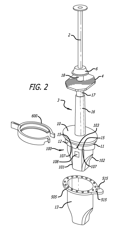
[0020] FIG. 2 is a partial view of the injector of FIG. 1 where the end cap
has been
removed, showing a support guide.
[0021] FIG. 3 is another partial view of the injector where end cap, injector
body, and
support guide have been removed, showing a lens support, a plunger guide and a
plunger.
[0022] FIG. 4 depicts an isolated view of a plunger and a lens support.
[0023] FIG. 5 is an isolated view of a lens support with wedge plate, a pair
of folding
members and an injection nozzle.
[0024] FIG. 6 is another isolated view of the lens support with the pair of
folding members
being pivotally mounted.
[0025] FIG. 7A is a perspective view of the lens support mounted within the
support guide
seen from the plunger side, according to an embodiment of the invention.
[0026] FIG. 7B is a sectional view taken along the line C-C of Fig. 7A.
[0027] FIG. 8 is a sectional view illustrating an intraocular lens disposed
within the lens
support, according to an embodiment of the invention.
[0028] FIG. 9 illustrates the intraocular lens being completely folded within
the lens
support, according to an embodiment of the invention.
[0029] FIG. 10 illustrates a view of the injector of an embodiment of the
invention, viewed
from its proximal end showing details modifications to the proximal end to
enhance retention
of fluid within the injector.
100301 FIG. 11 is a side view of an end cap of embodiment of FIG. 10.
CA 02871046 2014-10-20
WO 2013/159045
PCT/US2013/037457
6
[0031] FIG. 12 is a top view of an embodiment of a clamp used to hold the end
cap of FIG.
11 onto the proximal end of the injector of FIG. 10.
[0032] FIG. 13 is a partial view of the clamp of FIG. 12 taken along the line
D-D showing
the "U shaped" construction of the clamp.
[0033] FIG. 14 is a partial view of the clamp of FIG. 12 taken along the line
E-E showing
the details of a distal end portion of one side of the claim configured to
engage a pin of the
embodiment of FIG. 11.
[0034] FIG. 15 is a top perspective view of an embodiment of an end cap,
looking into the
end cap from its proximal end.
DETAILED DESCRIPTION OF THE PREFERRED EMBODIMENTS
[0035] Referring now to the drawings in detail, in which like reference
numerals indicate
like or corresponding elements among the several figures, there is shown in
FIG. 1 an
embodiment of a preloaded injector. The injector 1 comprises a plunger 2,
extending along a
longitudinal axis corresponding to the injection axis A, within a hollow
cylindrical injector
body 3. In the example of FIGS. 1 and 2, the injector body 3 includes an
octagonal shaped
finger tab 4 which is intended to provide a holding point to facilitate
operation of the plunger
2 during usage of the injector to inject a deformable intraocular lens into an
eye of a patient.
Different configurations of the injector body 3 and finger tab 4 are also
possible as long as
the injector body 3 is provided with means against which the fingers of a user
can bear.
[0036] The injector body 3 is closed at its proximal end by an end piece 6
comprising an
opening 7 in which the plunger 2 is introduced and guided. The end piece 6 has
a sleeve
portion 8 arranged to be fixed by snap-fit into the proximal end of the
injector body 3. A first
tonic joint seal 9 (FIG. 3) is accommodated in the end piece 6 in order to
fluidly seal the end
piece 6 on the injector body 3 and the opening 7 with the plunger 2 passing
through it. The 9
may be formed of any flexible elastomeric material.
[0037] At its distal end, or at the end opposite to the end piece 6, the
injector body 3
comprises an oval-shaped flange portion 10 extending essentially perpendicular
to the
injection axis A. Flange 10 comprises a collar portion 12 (Fig. 2), extending
in the axial
direction from part 10. Other configurations of the flange 10 are also
possible. For example,
CA 02871046 2014-10-20
WO 2013/159045
PCT/1JS2013/037457
7
flange 10 can have a circular, an elliptical or a rectangular shape and can be
supported on the
injector body 3 with support elements (not shown).
[0038] In one embodiment of the invention, the injector body 3 comprises a
first portion 16
having a first internal diameter and extending from the flange 10 to a second
portion 17
having a second internal diameter that is smaller than the first internal
diameter (FIG. 2). The
injector body 3 also comprises a third portion 18, having an internal smaller
than the one of
the second portion 17, and extending between the second portion 17 and the end
piece 6.
[0039] In FIG. 1, the injector 1 comprises an end cap 13 configured to fit
over the collar
portion 12 and engage with flange 10 to hold proximal end of the injector in a
sealed
arrangement, allowing for fluid to be introduced into a cavity of the end cap
13 to form a
fluid reservoir and to maintain the fluid within the reservoir formed by the
cooperation of the
end cap and flange 10. A second toric joint seal 11 (Fig. 2) placed around the
outer wall of
the collar portion 12 insures the fluid tightness between the end cap 13 and
flange 10.
[0040] Referring now to FIGS. 10 and 11, details of an embodiment of the
flange 10 and
end cap 13 are shown that improve the ability to maintain fluid within the
reservoir formed
by flange 10 and end cap 13, even when the injector assembly is sterilized
using an autoclave.
As one skilled in the art will understand, when the injector assembly is steam
sterilized,
pressure may build up within the reservoir that causes fluid to leak from the
reservoir, either
during the sterilization process, or afterwards when the injector is stored.
The inventors have
observed that reservoir integrity and fluid retention may be improved by
incorporating seal
posts 500 (FIG. 11) disposed around a top edge 505 of the cap 13 configured to
be received
by and engage with seal holes or indents 502 disposed on the distal side of
flange 10 (FIG.
10). Seal holes 502 may extend completely through flange 10, or they may be
formed only as
indents of partial holes disposed on the distal side of flange 10, having a
depth sufficient to
receive the seal posts 500 such that the distal side of flange 10 mates with
the top edge of cap
13 to form a fluid seal.
[0041] Referring now to FIG. 12, there is shown a locking clamp 600 configured
to
cooperate with flange 10 and the top edge 505 of cap 13. FIG. 13 is a
sectional view taken
along line D-D of FIG. 12 showing the arrangement of one embodiment of locking
clamp
600. Clamp 600 is formed to have an approximate "C" shape that engages edges
of flange 10
and top edge 505 of the cap 13. To accomplish this, clamp 600 has an upper lip
615 and a
CA 02871046 2014-10-20
WO 2013/159045
PCT/1JS2013/037457
8
lower lip 620 connected by a web 610, forming a "U" shaped channel. The
spacing between
upper lip 615 and lower lip 620 is configured to accept an edge of flange 10
and the top edge
505 of cap 13 between the upper and lower lips.
[0042] Referring again to FIGS. 11 and 12, cap 13 includes a tab 510 that
includes one or
more posts or pins 515 configured to engage ends 625 of clamp 600. In one
embodiment,
each end 625 of clamp 600 is held in place by a post 515 mounted on a top side
of the top
edge 505 of cap 13. Those skilled in the art will understand that other
embodiments are
possible, such as an embodiment where the post 515 is replaced by a tab or
other structure
capable of holding end 625 of clamp 600 in place.
[0043] FIG. 14 is a partial view taken along line E-E of FIG. 12 that shows
how each of the
proximal ends 625 of clamp 600 are configured to engage pins 515 of cap 13. As
shown,
proximal ends 625 are formed in a tab shape that is defined by partially
cutting away lower
edge 620 and a portion of web 610. This construction provides a relief that
allows the end
625 to pass over the top side of flange 10 and engage pin 515. When both ends
625 engage
both pins 515, the clamp is securely held in place, and securely holds flange
10 and cap 13
together. In this manner, the joint between the injector and the cap is made
secure and is
capable of withstanding pressure changes within the cap during sterilization
that could lead to
fluid loss from the reservoir within the cap.
[0044] Clamp 600 may be made of any material that is suitable for use with
the injector
system such that it is able to withstand autoclaving or methods of
sterilization. Clamp 600
must also be sufficiently flexible to allow placement of clamp 600 around the
flange and cap
without breakage. In the embodiment shown in FIG. 12, clamp 600 includes hinge
605
formed between the two arms of the clamp. Hinge 605 allows the arms of the
clamp to be
opened for placement about the edges of the flange and cap. Hinge 605 may be
an actual
hinge arrangement, or the clamp may be formed from a material that can be
repeatedly
articulated, with hinge 605 being formed from a shape that facilitates such
articulation in a
"living hinge" arrangement well known in the art.
[0045] Referring again the FIGS. 1-3, the injector 1 also comprises a lens
compartment
consisting of a support guide 100 and a lens support 200 (Fig. 3). FIG. 2
shows the injector 1
where the end cap 13 has been removed from the injector body 3, showing the
support guide
100 fixed on the flange 10. The support guide 100 is an open hollow structure
having side
CA 02871046 2014-10-20
WO 2013/159045
PCT/1JS2013/037457
9
walls defining a tapered internal shape, a narrower, truncated support guide
distal end 101,
and a wider proximal end 102 having an oval section, or any section conformal
with the
internal periphery of the collar portion 12. The support guide 100 can be
mounted and fixed
on the flange 10 by press-fitting its proximal end 102 within the internal
periphery of the 5
collar portion 12.
[0046] As shown in FIG. 2, the support guide 100 contains a guiding pin 103
fitted in a
corresponding indentation 15 in the collar portion 12, insuring a better
positioning and
fixation of the support guide 100 on the flange 10. Holes 107 are provided in
the support
guide 100 in order to allow for the introduction of a viscoelastic solution
within the lens
support 200 as will be 10 explained below. Holes 107 are accessible through
indentations 15
let into the collar portion 12.
[0047] Support guide 100 also includes an inspection window 108 disposed on a
surface of
support guide 100. Inspection window 108 provides for viewing the positioning
and state of
an intraocular lens 400 disposed within an internal support cavity 208 (FIG.
8) when the
intraocular lens is loaded into the injector.
[0048] In one embodiment of the invention, the injector body 3 is fabricated
in one piece
with an injection plastic molding process. The material used for the injector
body and cap 13
should be sterilizable using various processes, include steam sterilization.
The material used
for cap 13 may be opaque or clear. Alternatively, cap 13 may be formed in such
a manner
that a portion of the cap is opaque and a portion of the cap is clear, forming
a window,
allowing visualization of the portion of the injector and the lens mounted in
the injector, as
well as the level of any fluid within the reservoir formed by flange 10 and
cap 13, that is
placed within cap 13.
[0049] FIG. 3 depicts another partial view of the injector 1 from which the
injector body 3,
the end cap 13, and the support guide 100, have been removed. In this view,
the plunger 2
extending between the end piece 6, with its tonic joint seal 9, and the lens
support 200, placed
underneath the support guide 100. Also visible in Fig. 3 is a plunger guide
300, disposed 20
within the injector body 3 and extending between the internal wall of the
injector body 3 and
the plunger 2. The plunger guide 300 comprises a pair of flexible legs 301 of
hollow semi-
oval shape, the legs 301 being connected on the distal side of the plunger
guide 300, or on the
CA 02871046 2014-10-20
WO 2013/159045
PCT/1JS2013/037457
side of the lens support 200, by a connecting portion 302 integrally formed
with the legs 301.
The legs 301 each 25 comprise a protruding stop piece 303 at their respective
free ends.
[0050] In FIG. 3, the legs 301 are shown in an unstressed open position
allowing the
plunger 2 to move axially within the plunger guide 300. The plunger guide 300
also
comprises two opposite ribs 304, extending along its whole length. The ribs
304 are guided
in corresponding grooves (not shown) provided 30 in the internal wall of the
injector body 3,
when the plunger guide 300 is inserted within the injector body 3, and used to
orient radially
and guide axially the plunger guide 300 within the injector body 3.
[0051] FIG. 4 shows a view of the plunger 2 with the lens support 200 disposed
at the
distal end 22 of the plunger 2. The plunger 2 preferably has an elliptical or
ovoid section but
can have any other suitable section shape such as a circular, square or
rectangular section.
The plunger 2 also comprises clipping means. In the embodiment shown in Fig.
4, the
clipping means are two snap hooks 19 that are oppositely disposed on the
plunger 2, each at a
position corresponding to that one of a stop piece 303 of the plunger guide
300.
[0052] The lens support 200 according to one embodiment of the invention is
represented
in the perspective views of FIGS. 5 and 6. The lens support 200 comprises a
pair of parallel
wedge plates 201 of tapered shape and connected, at their narrow extremity, to
an injection
nozzle 202. The injection nozzle 202 is 15 terminated by a nozzle distal end
203 destined to
be introduced in an incision formed in the wall of a patient's eye during lens
replacement
surgery. The interior of the injection nozzle 202 forms a nozzle canal 204.
The lens support
200 also comprises a folding device for folding the lens 400 in a direction
essentially
perpendicular to the injector axis in response to axial movement of the
plunger 2, as
exemplified by the depictions of FIGS. 8 and 9. In the example of FIGS. 5 and
6, the folding
device is a pair of folding members 205 being fixed by their distal extremity,
which is the
extremity on the side of the injection nozzle 202, to the external wall of the
injection nozzle
202 with a flexible link 206. The folding members 205 comprise a notch 207 at
their distal 25
extremity. The pair of folding members 205 can be pivotally mounted by
abutting their
respective notches 207 against edges of the injection nozzle 202, as shown in
FIG. 6. The
spacing between the two wedge plates 201 allows the folding members 205 to
pivot within
the two plates 201 while being guided laterally by the plates 201. When the
two folding
members 205 are in an open position as shown in FIG. 6, the two wedge plates
201 and the
folding members 205 delimit an internal support cavity 208.
CA 02871046 2014-10-20
WO 2013/159045
PCT/1JS2013/037457
11
[0053] The wedges plates 201 also comprise a tail-shaped part 209, extending
along the
plunger 2 and within the plunger guide 300 as shown in FIG. 3. The internal
surface of the
tail-shaped part 209 forms a groove 210 extending along the injection axis A
on the internal
surface of the wedge plates 201, 5 forming an injection canal that extends the
nozzle canal
204 of the injection nozzle 202. Two ribs 211 extend along the injection axis
A, on the tail-
shaped part 209 and the two opposite external surfaces of the wedge plates 201
of the lens
support 200.
[0054] FIGS. 7A and 7B depict a view of the support guide 100 according to an
embodiment of the invention. In FIG. 7A, the support guide 100 is seen from
the plunger
side, and a section view along the line C-C of Fig. 7A is represented in FIG.
7B. In FIG. 7B,
the lens support 200 is also shown with pivoted folding members 205.
[0055] The support guide 100 comprises two internal lateral sloped ridges 106,
formed
within the internal surface of the support guide 100 and sloping toward one
another from the
support guide proximal end 102 to the support guide distal end 101 of the
support guide 100.
These sloped ridges 106 are destined to cooperate with the folding members 205
as will be
explained below.
[0056] In the example of FIGS. 7A and 7B, the internal surface of the support
guide 100
also comprises two guiding slots 104 extending along both sides of the support
guide 100,
and adapted to guide laterally the movement of the lens support 200 within the
support guide
100 along the injection axis A. The two ribs 211 press against two parallel
guiding faces 105,
extending along the injection axis A and oppositely disposed on the internal
upper and lower
surfaces of the support guide 100, in order to laterally guide the lens
support 200 advancing
within the support guide 100. Alternatively, the two ribs 211 can also press
against two
parallel guide ribs (not shown), extending along the injection axis A and
oppositely disposed
on the internal upper and lower surfaces of the support guide 100.
[0057] Other configurations of the support guide 100 are also possible. For
example, the
guiding slots 104 can be replaced by a pair of ribs in order to guide
laterally the movement of
the lens support 200 within the support guide 100 along the injection axis A.
[0058] The lens injectors of the present invention and their various parts may
fabricated
from different types of plastic materials. For example, the injector body may
be produced
from polycarbonate (PC), polyetherimide (PO) or polysulfone (PSU), the end cap
from PC,
CA 02871046 2014-10-20
WO 2013/159045
PCT/1JS2013/037457
12
PE1 or polyamide (PA), the plunger from PC, PE1 or PSU, the support guide from
PP, PC,
polybutylene-terephtalate (PBT) or polyoxymethylene (PaM), the lens support
from PaM, PP,
BC, PA, PEI or polyethylene-terephtalate (PET), the plunger guide from PA, PBT
or
polypropylene, the plug from silicone or a vulcanized thermoplastic material,
and the tonic
joints from silicone.
[0059] When assembling the injector 1, the end piece 6 and the tonic joint
seal 9 are first
disposed on the proximal end of the plunger 2. Here, the plunger 2 is inserted
into the end
piece 6 through the opening 7. The plunger 2 is then inserted into the
injector body 3. The
two snap hooks 19 of the plunger 2 are arranged such as to be able to pass
through the third
portion 18 of the injector body 3, and abut against the distal end of portion
18 once the hooks
19 have passed this portion 18, preventing the plunger 2 from moving backward.
Preferably,
the end piece 6 is not yet clipped on the proximal end of the injector body 3.
[0060] In a preferred embodiment of the injector of the invention, a flexible
25 plug 20 is
subsequently mounted on the distal end of plunger 22. The plug 20 is
preferably made from a
soft and flexible material, in order to avoid scratching of the lens 400
during the injection
operation. Here, the distal end of the plunger 2 can comprise a forked distal
end 22, as shown
in FM. 4, allowing the flexible plug 20 to extend at least partially in
between the two teeth of
the distal end 22. Other configurations of the distal end 22, that abuts the
plug 20, are also
possible. It is noted that plug 20 may be added to the plunger end 22 at a
later stage, but prior
to the mounting of the lens support 200 on the plunger guide 300.
[0061] The plunger guide 300 is next mounted within the injector body 3. The
two
opposite ribs 304 of the plunger guide 300 are guided within the corresponding
grooves of
the injector body 3 allowing the plunger guide 300 to be introduced into the
desired angular
position within the injector body 3. When the plunger guide 300 reaches its
full rear position,
it is forced into its closed position, the clipping means of the plunger 2,
here the two snap
hooks 19, are able to engage on the distal edge of the stop pieces 303,
reversibly connecting
the plunger guide 300 and the plunger 2.
[0062] The respective internal diameters of the portions 16, 17, 18 are such
as to allow the
plunger guide 300 to be introduced within the first and second portions but
not within the
third portion 18. The plunger guide 300 introduced within the injector body 3
from the flange
side thus abuts against the end of 15 the second portion 17, adjacent to the
third portion
CA 02871046 2014-10-20
WO 2013/159045
PCT/1JS2013/037457
13
18. In this initial position, the plunger guide 300 extends along the first
and second portions
16, 17. The internal diameter of the second portion 17 is such as to force the
two opposite
stop pieces 303 of the legs 301 to come in contact with the two snap hooks 19,
the plunger
guide 300 being thus in a closed position. When, in response to a forward
movement of the
plunger, the plunger guide is advanced out of the second portion 17 and into
the first portion
16, the plunger guide 300 is able to regain its unstressed open position.
[0063] Other configurations of the injector body 3 are also possible, as long
as they
provide a configuration that enables the plunger guide 300 to be either in a
closed position or
in an unstressed open position, depending on the axial position of the plunger
guide 300
within the injector body 3. For example, the injector body 3 can have a
uniform internal
diameter along its whole length but comprise internal ribs distributed around
its internal wall,
the ribs having a height that varies between sections along the injector body
3.
[0064] An intraocular lens 400 is then disposed unfolded between the two wedge
plates
201, within the internal support cavity 208 (FIGS 6 and 8). Preferably, the
lens 400 is
disposed within the internal support cavity 208 with the two haptics 401 of
the lens being
oriented along the injection axis A, as shown in FIG. 8.
[0065] The lens support 200 containing the lens 400 is then mounted on the
plunger guide
300 by inserting the tail-shaped part 209 within the connecting portion 302 of
the plunger
guide 300 (FIGS. 3 and 6). In this position, the two folding members 205 are
prevented from
pivoting on the intraocular lens 400 by abutting against two protrusions 23
located on the
flange 10 of the injector body 3 (FIG. 8). Also shown in FIG. 8 are two
protruding members
21 arranged to maintain the unfolded lens 400 within the lens support 200 in
its unfolded
orientation as described above, until the lens 400 is folded and ejected. The
protruding
members 21 do not prevent the pivoting of the two folding members 205.
[0066] The support guide 100 is then fixed on flange 10 of the injector body 3
and the end
cap 13 is placed over the injector, aligning seal holes 502 (FIG. 10) with
seal posts or pins
500 (FIG. 11) and fastened to flange 10 using clamp 600 (FIG. 12) after
placing the second
tonic joint seal 11 around the external periphery of the collar portion 12
(FIGS. 1 and 2). The
second toric joint seal 11 could also be placed at any other injector assembly
steps, before the
step of mating end cap 13 with the flange 10, described below.
CA 02871046 2014-10-20
WO 2013/159045
PCT/1JS2013/037457
14
[0067] In the case of a flexible hydrophilic intraocular lens, the end cap 13
and the injector
body 3 are filled with an aqueous solution or fluid such as a saline solution,
distilled water, or
any other aqueous solution adequate for keeping the intraocular lens 400 wet.
The aqueous
solution may be introduced through filling openings, in the proximal end of
the injector body
3 by means of a syringe.
[0068] The aqueous solution fills at least partly the volume enclosed by the
end cap 13,
lens support 200 and injector body 3. In the case a flexible hydrophobic
intraocular lens is
used, there is no need for a bathing solution or fluid such as saline and the
step of filling the
injector body 3 and the end cap 13 with an aqueous 30 solution may be omitted.
[0069] When the end cap 13 is fixed on the injector body 3, the lens support
200 abuts
against the end cap 13 and the plunger 2 cannot be depressed.
[0070] In a preferred embodiment of the invention shown in see FIG. 15, end
cap 13
comprises a central hollow tube 24 extending along the injection axis A toward
the injector
body 3. When the end cap 13 is fixed on the injector body 3, the distal end
215 of both
opposite support ribs 211 of the lens support 200 abuts against the proximal
end 25 of the
central tube 24. In this configuration, the plunger 2 cannot be moved backward
due to the
snap hooks 19 abutting against the distal end of portion 18, as described
above.
Consequently, any false manipulation of the plunger 2 prior to the injection
operation is
avoided.
[0071] After fixing the end cap 13, the toric joint seal 9 is placed on a
groove 26 on the
proximal end of the injector body 3 (Fig. 9) and the end piece 6 is clipped on
said proximal
end, making the interior of the injector body sealed. The injector 1 is then
ready to be
packaged into a sealable flexible packaging (not) such as a sleeve, pouch or
blister, or any
other packaging. After the packaging is sealed, the packaged injector 1 is
subjected to
sterilization. A preferred method of sterilization is steam sterilization
(autoclaving). In one
alternative embodiment, the sleeve or pouch may be formed from a suitable foil
material.
[0072] Prior to the injection operation, the injector is separated from its
packaging, clamp
600 is removed and the end cap 13 is separated and removed from the flange 10,
causing the
aqueous solution to drain from the injector body 3 and the lens support 200.
In order to keep
the lens 400 and lens support 200 lubricated during the injection operation, a
viscoelastic
solution such as a solution containing hyaluronic acid, chondroitin sulfate or
a cellulose
CA 02871046 2014-10-20
WO 2013/159045
PCT/1JS2013/037457
derivative such as hydroxypropylmethylcellulose (HPMC) can be introduced
within the
internal support cavity 208 through boles 212 provided in the wedge plates 205
and the
corresponding holes 107 of the support guide 100, for example, by using a
syringe.
Alternatively or in addition, the viscoelastic solution can also be introduced
through the
nozzle distal end 203 of the injection nozzle 202. The holes 107 and 212, and
the nozzle
distal end 203 also increase the fluidic communication within the end cap 13,
facilitating the
penetration of aqueous wetting solution into the lens support 200.
[0073] During an injection operation, the plunger 2 is depressed causing the
plunger guide
300 to move forward over a first distance, advancing the lens support 200
within the support
guide 100 along the injection axis A. During the advance of the lens support
200, the sloped
ridges 106 of the support guide 100 force the pair of folding members 205 to
pivot toward the
injection axis A, drawing them near to one another until they become
essentially parallel to
the injection axis A, transforming the internal support cavity 208 into an
injection canal 213
that extends along the folded folding members 205 and into the nozzle canal
204 of the
injection nozzle 202. The lens support 200 advances in the support guide 100
until it abuts
against the support guide 100 and cannot advance further.
[0074] Alternatively, the advance of the lens support 200 within the support
guide 100, the
folding members 205 of the lens support 200 may interact with the internal
tapered side
walls, forcing the folding members 205 to pivot inward and fold the
intraocular lens in a
direction essentially perpendicular to the injection axis A.
[0075] The above operation causes the intraocular lens 400 to fold, the lens
400 being
folded or rolled in a direction essentially perpendicular to the injection
axis A as shown in
FIG. 7B, when completely folded. Consequently, the folded lens 400 is ready to
be advanced
axially into the nozzle canal 204.
[0076] In one embodiment of the invention, each folding member 205 comprises a
protruding element 214. When the lens support 200 advances within the support
guide 100,
the sloped ridges 106 press against the protruding elements 214, and pivots
the pair of folding
members 205 toward the injection axis A, as described above. The protruding
elements 214
can advantageously enhance the angular distance the folding members 205 will
travel within
the lens support 200 during the forward motion of the lens support within the
support guide
100. Moreover, the use of protruding elements 214 can also reduce the friction
during the
CA 02871046 2014-10-20
WO 2013/159045
PCT/1JS2013/037457
16
advancement of the lens support 200 within the support guide 100, compared to
a contact
made along the whole folding member 205.
[0077] When the plunger 2 has moved over the first distance and the lens
support 200
reached its abutting position within the support guide 100, the plunger guide
300 has moved
completely outside the second portion 17 and extends only within the first
portion 16 of the
injector body 3 and within the support guide 100. It is noted that once
plunger guide 300 has
moved outside of second portion 17, it cannot be returned to its initial
position within portion
17, thereby preventing an unfolding of the folded lens as a consequence of an
accidental
retraction of plunger 2. The diameter of the first portion 16 is large enough
to allow the two
legs 301 of the plunger guide 300 to regain their unstressed position, in
which the two legs
301 are slightly bent apart, enabling the plunger guide 300 to be detached
from the plunger 2,
allowing the plunger 2 to move freely within the plunger guide 300 and advance
within it.
[0078] When operator pressure continues to be applied, the plunger 2 and plug
20 advance
over a second distance and propel the folded lens 400 along the injection
canal 213, and
outside the nozzle distal end 203, enabling the lens 400 to be injected into
the patient's eye
(FIG. 9). The flexible plug 20 is able to follow conformably the varying
dimensions of the
internal support cavity 208 formed by the two folding members 205 and the
nozzle canal 204,
avoiding the necessity of requiring accurate dimensions for the different
parts forming the
compressed support cavity 208 and the nozzle canal 204.
[0079] In an exemplary embodiment of the invention, the lens support 200 is
able to
advance in the support guide 100 over a distance of about 15 mm, this distance
corresponding
to the length of the second portion 17 of the injector body 3. Here, the total
length formed by
the first and second portions 16, 17 corresponds essentially to the length of
the plunger guide
300.
[0080] In one embodiment of the invention, the lens support 200, comprising
the two
wedge plates 201, the injection nozzle 202, the two folding, members 205 and
links 206, is
fabricated in one piece by an injection plastic molding process.
[0081] While several particular forms of the invention have been illustrated
and described,
it will be apparent that various modifications can be made without departing
from the spirit
and scope of the invention.