Note: Descriptions are shown in the official language in which they were submitted.
CA 02871343 2014-13-23
WO 2013/171097
PCT/EP2013/059473
Intracorneal lens
The present invention relates to an intracorneal lens
for the correction of impaired vision, for example
presbyopia.
Intracorneal lenses are used for the correction of
impaired vision. In contrast to contact lenses, which
are placed on the surface of the eye, and to
intraocular lenses, which are implanted in a chamber of
the eye, intracorneal lenses are inserted into a pocket
created in the cornea. Intracorneal lenses differ
significantly from contact lenses or intraocular
lenses, for example in terms of their size, in the
absence of holding elements (haptic elements) required
for intraocular lenses, and in their optical
properties.
Intracorneal lenses are known from the prior art. By
way of example, reference is made to WO 2009/075685, US
5,628,794, US 5,123,921 or EP 1 001 720 Bl.
WO 2009/075685 describes an intracorneal lens having a
central hole. The central hole is concentric to the
optical axis of the lens and is of such a size and
shape that, while the optical properties of the lens
are not impaired, the hole can nevertheless be used at
the same time for more precise positioning of the lens
in the corneal pocket.
However, it has been shown that these lenses do not yet
provide optimal correction of sight in humans.
There was therefore a need for an intracorneal lens
with which it is possible to better restore the visual
acuity of persons with impaired vision, for example
presbyopia.
CA 02871343 2014-10-23
WO 2013/171097 - 2 -
PCT/EP2013/059473
According to the invention, the above object was
achieved by an intracorneal lens comprising a circular
main body with a convex front surface and a concave
rear surface, characterized in that the convex front
surface has a single uniform radius of curvature Rcv
and the concave rear surface has a radius of curvature
Rcci, wherein the radius of curvature Rcci of the
concave rear surface is greater than the average radius
of the cornea by 0.1 mm to 2 mm, preferably by 0.2 to
1.5 mm, particularly preferably by 0.5 to 1 mm.
It has been found, surprisingly, that a pronounced
improvement in the correction of vision by intracorneal
lenses can be achieved if the radius of curvature Rcci
of the concave rear surface of the lens slightly
exceeds the average radius of the cornea. In this way,
on account of its low inherent stiffness, the lens
according to the invention easily conforms to the
cornea and exerts less force on the cornea. At the same
time, however, it was found that the radius of
curvature Rcci of the concave rear surface of the lens
ought only to exceed the average radius of the cornea
by a defined low value, since otherwise there may be
formation of folds in the cornea at the outer edges of
the lens, with associated negative optical effects.
According to the present invention, the radius of
curvature Rcci of the concave rear surface is greater
than the average radius of the cornea by 0.1 mm to 2
mm, preferably by 0.2 to 1.5 mm, particularly
preferably by 0.5 to 1 mm.
It was also found that the provision of lens zones with
different radii of curvature is possible by arranging
the different radii of curvature on the concave rear
surface of the lens, while the convex front surface of
the lens has a single uniform radius of curvature Rcv.
Because of the significant difference in the refractive
CA 02871343 2014-10-23
WO 2013/171097 - 3 -
PCT/EP2013/059473
indices of the cornea and of air, a change in the
curvature of the cornea brought about by different
radii of curvature of the implanted intracorneal lens
has a considerable influence on the optical properties
of the corrected eye on the outside of the cornea
(interface to the environment), whereas an analogous
change in the curvature of the cornea at the interface
to the interior of the eye has an influence that is
less by about a factor of 5 (less difference in the
refractive indices of the cornea and of the vitreous
humor).
As a result of the above measures, much better and more
precise correction of visual acuity can be achieved
with the intracorneal lens according to the invention
than is possible with conventional intracorneal lenses.
With the intracorneal lens according to the invention,
it is possible to correct impaired vision such as
myopia (short-sightedness), hyperopia (long-
sightedness), presbyopia (age-related diminution of the
accommodation of the eye) or a combination of these
sight problems. With the surfaces of the lens suitably
optimized in a manner known to a person skilled in the
art, it is also possible to treat astigmatism with the
aid of the intracorneal lens according to the
invention.
The advantage of correcting these sight problems using
the intracorneal lens according to the invention is
that the correction can be made reversible by removing
the lens from the cornea. Compared to conventional
means of correction such as eyeglasses or contact
lenses, the intracorneal lenses according to the
invention have the advantages of not being visible, of
avoiding wearing problems, of avoiding the misting up
of eyeglasses under certain environmental conditions,
and of avoiding the irritation of the cornea which can
occur when wearing contact lenses.
CA 02871343 2014-13-23
WO 2013/171097 - 4 -
PCT/EP2013/059473
The intracorneal lens according to the invention has a
circular and dome-shaped main body with a diameter in
the range of 2.4 to 4 mm, preferably of 2.7 to 3.5 mm,
particularly preferably of 3 mm.
The intracorneal lens according to the invention is
preferably made of a biocompatible material which
permits the passage of nutrient liquids and other eye
constituents and also permits sufficient gas diffusion
(especially of oxygen). Biocompatible and permeable
materials of this kind are known to a person skilled in
the art. Examples that may be mentioned are silicones,
hydrogels such as Perfilcon A , urethanes or acrylates
such as polymethacrylates. In the preferred embodiment
of an intracorneal lens according to the invention with
a central opening, the passage of liquids and gases
through the lens is additionally facilitated by the
central opening.
An essential aspect of the intracorneal lens according
to the invention concerns the radii of curvature of the
convex front surface and of the concave rear surface.
According to the present invention, all the radii
indicated relate to the lens in the hydrated state,
i.e. the state of the lens when it is implanted in the
corneal pocket.
As has been mentioned above, the convex front surface
of the lens has a single uniform radius of curvature.
In the case of a preferred bifocal lens according to
the invention for the correction of presbyopia, the
radius of curvature of the convex front surface
preferably lies in a range of 7.5 to 8 mm. However,
depending on the application, another radius of
curvature may be chosen, such that the optical
correction afforded by the actual lens according to the
CA 02871343 2014-10-23
WO 2013/171097 - 5 -
PCT/EP2013/059473
invention can be supported by a change in the curvature
of the outer surface of the cornea.
As has been mentioned above, the concave rear surface
of the lens according to the invention has a radius of
curvature Rcci, which slightly exceeds the average
radius of the cornea. The average radius of the cornea
varies slightly from one individual to another and
generally lies in a range of 7.4 mm to 8.1 mm, being
typically about 7.8 mm. The radius of curvature Rcci of
the concave rear surface of the lens according to the
invention exceeds the average radius of the cornea by
0.1 mm to 2 mm, preferably by 0.2 to 1.5 mm,
particularly preferably by 0.5 to 1 mm.
For correction of some sight problems, it is necessary
for the lens according to the invention to be equipped
with several different zones having different optical
power. According to the invention, these zones are
preferably provided by varying the radius of curvature
of the rear surface of the lens, such that the zones
have different thickness and different surface radii on
the reverse. As has already been mentioned above, the
different radii of curvature in the lens according to
the invention are provided on the concave rear surface.
By changing the radius of curvature of the lens
surface, a change is brought about in the curvature of
the cornea, and this change influences the visual
acuity of the person being treated. A change in the
curvature of the outer surface of the cornea has a much
greater influence on visual acuity, since the
refractive index of the cornea differs considerably
from the refractive index of air. By contrast, the
difference in the refractive indices of the cornea and
of the liquid in the interior of the eye is much less.
Consequently, a change in the curvature of the inner
surface of the cornea has an effect on visual acuity
that is less by about a factor of 5 than a
CA 02871343 2014-10-23
WO 2013/171097 - 6 -
PCT/EP2013/059473
corresponding change in the curvature of the outer
surface of the cornea. By maintaining a uniform radius
of curvature of the front convex surface of the lens
and modifying the radius of curvature of the rear
concave surface of the lens, it is possible to achieve
a more precise adjustment of the desired visual acuity.
If the lens according to the invention is made
available with several different zones having different
radii of curvature, it is important that the lens
conforms to the cornea, as has been described above.
The different radii of curvature must therefore be
chosen such that this important aspect of the invention
is achieved. It is preferably achieved by virtue of the
fact that all of the different zones of radii of
curvature are chosen such that each of these radii of
curvature is greater than the average radius of the
cornea by 0.1 mm to 2 mm, preferably by 0.2 to 1.5 mm,
particularly preferably by 0.5 to 1 mm. However, for
certain uses of the lens according to the invention, it
may be sufficient for only the radius of curvature Rcci
of an inner area of the rear concave surface to meet
this requirement, while the radius of curvature Rcco
chosen for outer areas (lying at the lens edge) of the
rear concave surface can be greater. According to the
invention, the radius of curvature Rcco of the concave
rear surface of the lens according to the invention
lies in a range of 8.0 mm to 11.5 mm, preferably of 8.0
mm to 10 mm, particularly preferably of 8.0 to 9 mm.
According to the invention, it is preferable that the
change in the radius of curvature from one zone to the
next zone does not take place abruptly, but instead
across an area of defined size. According to the
invention, the transition from one radius of curvature
to the next radius of curvature preferably takes place
within a portion of the concave rear surface of the
CA 02871343 2014-10-23
. WO 2013/171097 - 7 -
PCT/EP2013/059473
lens, which portion represents a ball segment of 0.1 mm
to 0.5 mm, preferably of 0.2 to 0.3 mm.
According to the invention, the convex front surface of
the lens and the concave rear surface of the lens are
preferably connected to each other via an intermediate
portion, which extends in a circle about the outer edge
of the lens. With the aid of this intermediate portion,
it is possible to maintain optimally the positioning of
the lens inside the cornea and to minimize or avoid any
secondary optical effects at the edge of the lens.
The intermediate portion according to the invention is
characterized in that the radius of curvature of the
front convex surface merges abruptly into a step at a
distance of 0.005 mm to 0.01 mm, preferably of 0.6 mm
to 0.008 mm from the outer edge of the lens, which step
is inclined with respect to the optical axis of the
lens by an angle of 15 to 45 , preferably of 20 to
40 , particularly preferably of 30 . The step has an
edge length of preferably 0.01 mm to 0.015 mm,
particularly preferably 0.01 mm to 0.013 mm.
Preferably, there is no intermediate portion provided
on the rear surface of the lens. The radius of
curvature of the outer zone of the rear surface is
preferably maintained as far as the lens edge.
The intracorneal lens according to the invention has a
dome shape on account of the different radii of
curvature of the front convex surface and of the rear
concave surface. The shape of the lens according to the
invention is preferably such that the center point of
the convex front surface of the lens is at a distance
of 0.1 to 0.2 mm, preferably of 0.13 to 0.17 mm, from
an imaginary straight line connecting the lens edges.
CA 02871343 2014-10-23
WO 2013/171097 - 8 -
PCT/EP2013/059473
According to a preferred embodiment of the present
invention, the intracorneal lens has a central opening,
as is described in WO 2009/075685. Reference is
herewith expressly made to the relevant content of WO
2009/075685.
The central opening serves for optimal positioning of
the lens according to the invention in the corneal
pocket and at the same time facilitates the passage of
nutrient liquids and gases, for example oxygen, through
the lens. The central opening is concentric to the
optical axis of the lens and extends right through the
lens (i.e. it is a hole and not just a depression). The
size of the central opening is chosen such that the
opening is still visible to the surgeon when placing
the lens in the corneal pocket. At the same time,
however, the opening should not be so large as to be
noticeable to the person treated. In other words, the
central opening should not cause optical effects of any
kind. For this reason, in the case of a lens with a
diameter of 3.0 mm, the central opening according to
the invention preferably has a diameter of 0.12 mm to
0.2 mm, preferably of 0.13 mm to 0.17 mm, and
particularly preferably of 0.15 mm. Smaller openings
are unable to meet the demands described above, while
larger openings lead to optical effects that are
noticed by the person treated, which is undesirable
according to the invention. Generally speaking, the
central opening of the lens according to the invention
should only occupy a proportion of the area of the
front surface of less than 1%, preferably of less than
0.5%.
According to the present invention, the central opening
typically has a cylindrical shape. As a result of the
lens thickness at the position of the central opening,
the opening has a length through the lens of 0.01 to
0.1 mm, preferably of 0.015 mm to 0.05 mm, and
CA 02871343 2014-10-23
WO 2013/171097 - 9 -
PCT/EP2013/059473
particularly preferably of 0.02 to 0.03 mm. However,
other shapes of hole can also be chosen, of the kind
described in WO 2009/075685 and incorporated herewith
by reference. Conical openings may be mentioned by way
of example, or openings that have sections of different
diameter.
At the center of the lens (i.e. at its optical axis),
the lens according to the invention preferably has a
thickness of 0.01 to 0.1 mm, preferably of 0.015 mm to
0.05 mm, and particularly preferably of 0.02 to 0.03
mm.
As has been described above, the lens according to the
invention can have different zones with different
optical power. The number of different optical zones is
dependent here on the desired corrective effect that is
intended to be achieved using the lens according to the
invention.
In a preferred embodiment according to the invention,
the intracorneal lens has two optical zones. A first
inner area extends in a circle around the center of the
lens and has no optical power (non-optical area). For
some uses, however, it may be entirely desirable that
the inner area has a slight optical power, for example
of less than 1 diopter, preferably 0.1 to 0.8 diopter,
particularly preferably 0.1 to 0.6 diopter. For other
uses, an optical power of well over 1 diopter may even
be chosen. According to the invention, the inner area
preferably has a diameter of 1.4 mm to 2.4 mm
(depending on the diameter of the lens). With a lens
diameter of 3 mm, the inner area should preferably have
a diameter of 1.5 to 2.0 mm, particularly preferably of
1.8 mm.
A circular second optical area is arranged around the
inner optical area. The second, outer area has an
CA 02871343 2014-13-23
WO 2013/171097 - 10 -
PCT/EP2013/059473
optical power which is dependent on the desired sight
correction that is intended to be achieved using the
lens according to the invention. Generally speaking,
the optical power of the second, outer area is greater
than 1 diopter.
The diopter values indicated above relate to the
optical power that is made available by the lens in the
implanted state, i.e. in the hydrated state, in the
cornea.
According to a further embodiment of the present
invention, the intracorneal lens can also be configured
as a diffractive lens. Diffractive lenses are known
from the prior art. Reference is made by way of example
to WO 2009/075685 or to EP 1 001 720 El. A diffractive
lens is preferably configured in such a way that it has
several circular optical zones that merge into each
other via steps. This is shown, for example, in figures
12 and 13 of WO 2009/075685. Reference is made
expressly to the relevant content of WO 2009/075685.
According to a further embodiment of the present
invention, the intracorneal lens can also be configured
in such a way that, in terms of its optical power, it
has a gradient. This can be achieved, for example, by
the lens being made from different materials with
different refractive indices gradually merging into one
another. Alternatively, the refractive index of a
material can also be gradually changed by suitable
measures such as irradiation with high-energy
electromagnetic radiation.
In light of the different configuration of the front
surface and of the rear surface of the intracorneal
lens according to the invention, it is extremely
important that the intracorneal lens according to the
invention is inserted correctly into the pocket
CA 02871343 2014-13-23
WO 2013/171097 - 11 -
PCT/EP2013/059473
provided for it in the cornea, i.e. with the front
surface of the lens toward the outside of the eye and
with the rear surface of the lens toward the interior
of the eye.
Correct insertion of the lens according to the
invention into a pocket provided for it in the cornea
can particularly preferably be achieved, according to
the invention, using an applicator of the kind
described in WO 2011/069907 Al. Reference is herewith
expressly made to the relevant content of WO
2011/069907 Al.
WO 2011/069907 Al describes an applicator comprising a
grip piece and a pre-load unit. Grip piece and pre-load
unit can be connected to each other, preferably with
the aid of a bayonet connection secure against
rotation. The pre-load unit can first be equipped with
a lens, such as the lens according to the invention,
and stored in a sterile manner in a storage unit. To
insert the lens, the physician removes the pre-load
unit from the storage unit and connects the pre-load
unit to the grip piece. The lens can then be inserted
into the corneal pocket in the manner described in WO
2011/069907 Al.
The present invention thus also relates to a kit
comprising a storage unit and a pre-load unit in the
interior of the storage unit, wherein the storage unit
is made of a watertight material and can be closed in a
watertight manner with a stopper, and the pre-load unit
is fitted with an intracorneal lens according to the
invention.
The pre-load unit is preferably designed for one-off
use. The pre-load unit is equipped with the
intracorneal lens according to the invention and is
stored in a sterile package, from which it is only
CA 02871343 2014-13-23
WO 2013/171097 - 12 -
PCT/EP2013/059473
removed, and connected to the grip piece, directly
before the insertion of the lens into the eye.
To be able to store the pre-load unit in a sterile
state over a long period of time, it is packaged in a
storage unit that protects the pre-load unit from
environmental influences. For this purpose, the
interior of the storage unit is filled with a storage
liquid, which constantly covers at least the lens
located in the chamber of the pre-load unit. This
storage liquid can be water. However, the storage
liquid is preferably physiological saline (NaC1).
The storage unit is made from a material that is
watertight and therefore also allows only very little
water vapor, if any, to pass through. This is important
in order to ensure a sufficient level of storage liquid
within the storage unit during the entire storage
period. If the material were not watertight, the
storage liquid would evaporate over a certain time, and
the lens in the chamber of the pre-load unit would no
longer be stored in a sterile state. Watertight
materials are known to a person skilled in the art.
Examples of such a material are glass or a watertight
plastic. After introduction of the pre-load unit, the
storage unit is closed in a watertight manner by means
of a suitable stopper. This stopper is preferably made
from a watertight plastic.
The top face of the pre-load unit is clearly
identified, for example with a suitable inscription
such as "TOP". Since the pre-load unit is equipped with
the lens according to the invention in such a way that
the convex front surface of the lens points in the
direction of the identified top face of the pre-load
unit, the surgeon has no problem in correctly inserting
the lens into the pocket in the cornea. He simply has
to fit the pre-load unit onto the grip piece, i.e. with
CA 02871343 2014-13-23
= WO 2013/171097
- 13 - PCT/EP2013/059473
=
the identified top face of the pre-load unit facing
upward.
The pre-load unit is described in detail in WO
2011/069907 Al. Reference is herewith expressly made to
the relevant content of WO 2011/069907 Al. The pre-load
unit for inserting lenses into the eye of a human or
animal comprises
i) a housing with means for fastening the unit to a
grip piece, preferably in a manner secure against
rotation,
ii) a lens-receiving part arranged on or in the
housing and comprising a section that protrudes
from the housing and that has preferably exactly
two separate leaf-like units which, at least at
their ends directed away from the housing, are in
releasable contact with each other and there form
a chamber for storing an optical lens,
iii) a slide, which is arranged movably in the interior
of the housing and can be moved between the leaf-
like units of the lens-receiving unit.
According to the present invention, a leaf-like unit is
to be understood as a structural part which is very
thin, similarly to a leaf, and thus has corresponding
flexibility. According to the present invention, the
leaf-like units typically have a thickness of between
0.1 and 0.3 mm and typically have a length of 20-40 mm,
preferably 20 to 30 mm. The material used for the leaf-
like units is a physiologically compatible plastic or a
physiologically compatible metal.
The two leaf-like units preferably each have exactly
one hole, and the holes are arranged such that they lie
directly over each other and form an opening through
the center of the chamber for storing an optical lens.
In this way, a lens located in the chamber can be
positioned particularly easily on the optic axis of the
CA 02871343 2014-10-23
WO 2013/171097 - 14 -
PCT/EP2013/059473
eye. The person inserting the lens sees the lens
through the opening and can align it exactly. These
holes can typically have a diameter of 0.3 to 0.6 mm.
This embodiment is particularly advantageous for
intracorneal lenses with a central hole, as have been
described above.
The leaf-like units of the lens-receiving part, at
their ends directed away from the housing, form a
chamber for storing the lens according to the
invention. The leaf-like units are in loose contact
with each other in the area of the chamber, i.e. they
can be moved apart from each other by application of
force, in order to open the chamber for the purpose of
inserting or removing the lens according to the
invention. The leaf-like units are spread apart from
each other by means of a slide. The chamber is at least
partially bounded by wave-shaped sections, which are
present on both leaf-like units and are arranged such
that the sections of one leaf-like unit mesh with the
corresponding sections of the other leaf-like unit. In
the state of contact between the two leaf-like units,
the lens cannot slip out of the chamber.
With the applicator described in WO 2011/069907 Al, the
lens is not ejected into a corneal pocket, and instead
the applicator is positioned exactly in the corneal
pocket and then, with the lens maintained in position,
the leaf-like parts are drawn back by an exactly
defined distance. For this purpose, the section of the
pre-load unit of the applicator in which the chamber
for storing the lens according to the invention is
formed must be configured in such a way that it can be
inserted into a corresponding corneal pocket. The shape
of the area of the applicator forming this chamber
preferably corresponds substantially to the shape of
the lens to be inserted, or it is even slightly smaller
in width than the corresponding lens. This is possible,
CA 02871343 2014-13-23
WO 2013/171097 - 15 -
PCT/EP2013/059473
for example, by the chamber not having a round shape
and not having the above-described boundaries all
around, i.e. the chamber is laterally open between the
boundaries. The lens located in the chamber can then
protrude from the chamber at some points.
In order to fit the pre-load unit with the lens
according to the invention, the leaf-like units are
spread apart from each other in order to permit access
to the above-described chamber. According to one
embodiment of the present invention, this can be done
by inserting a suitable tool, for example a knife, into
a lateral gap between the leaf-like units. The lens is
then placed in the accessible chamber, preferably under
a microscope. The tool is then removed from the lateral
gap between the leaf-like units, as a result of which
the leaf-like units again come into contact with each
other at their ends and the chamber is closed. For the
loading procedure, it is possible, for example, to
provide a loading station in which the pre-load unit
can be fixed. The loading station has a rotatable tool
(e.g. a knife), which can be rotated into the lateral
gap between the leaf-like units and rotated back out of
said gap.
After it has been removed from the storage unit, the
pre-load unit is fitted onto the grip piece, which is
likewise described in detail in WO 2011/069907 Al.
Reference is herewith expressly made to the relevant
content of WO 2011/069907. The applicator is typically
an elongate tube with a shape that ensures the grip
piece can be easily held in the hand. The grip piece is
designed at one end in such a way that the pre-load
unit and the grip piece can be connected to each other
such that the pre-load unit can be moved inside the
grip piece. For this purpose, the grip piece has a
diameter exceeding the diameter of the part of the pre-
load unit that is to be inserted into the grip piece.
CA 02871343 2014-13-23
,
' WO 2013/171097 - 16 -
PCT/EP2013/059473
=
The grip piece is hollow on the inside, at least at the
end to be connected to the pre-load unit. However, it
is preferably a completely hollow tube.
A slide is arranged movably inside the pre-load unit
and the grip piece. This slide must be able to be moved
forward between the leaf-like units to such an extent
that it comes into contact with the lens located in the
chamber and can fix this lens. The slide preferably
tapers toward the end that is to move between the leaf-
like units. To better fix the lens, the front end of
the slide is correspondingly designed to complement the
lens shape.
The slide and the pre-load unit are moved with the aid
of control elements, which are arranged on the grip
piece. This is described in detail in WO 2011/069907
Al. Reference is herewith expressly made to the
relevant content of WO 2011/069907.
The grip piece of the applicator according to the
invention is preferably intended to be used more than
once. For this purpose, according to a preferred
embodiment of the present invention, the grip piece can
be completely dismantled, such that all the structural
parts can be completely cleaned and sterilized.
However, according to an alternative embodiment of the
present invention, the grip piece can likewise be
provided for one-off use. According to this embodiment,
the grip piece is preferably stored separately until
use, likewise in a storage unit analogous to the above-
described unit. However, according to this embodiment,
it is also conceivable that the pre-load unit and the
grip piece are already stored as a unit until use
(either separately, as described above, or also fixedly
connected to each other), in a storage unit analogous
to the above-described unit. According to this
alternative embodiment, the applicator can therefore be
CA 02871343 2014-13-23
WO 2013/171097 - 17 -
PCT/EP2013/059473
=
constructed differently in that the parts for
connecting grip piece and pre-load unit can be replaced
by a fixed connection of these structural parts.
With the aid of the above-described applicator from WO
2011/069907 Al, the lens according to the invention can
be easily inserted into a pocket in the cornea. The
insertion method comprises the steps of:
a) positioning the above-described applicator in the
correct orientation at the desired location of the
eye, preferably in a pocket in the human cornea,
such that the center of the optical lens contained
in the applicator lies on the optic axis of the
eye;
b) pushing the slide forward by means of a first
control element on the grip piece, until the slide
comes into contact with the lens without moving
the latter, wherein at the same time the leaf-like
units of the lens-receiving part are spread apart
from each other;
c) withdrawing the rest of the pre-load unit by a
defined distance by means of a second control
element on the grip piece, with simultaneous
fixing of the slide, as a result of which the lens
is released from the applicator.
The pre-load unit, preferably stored in a sterile
state, is removed from the storage container and
connected to the grip piece, preferably in a manner
secure against rotation. The applicator thus obtained,
fitted with the lens according to the invention, is
then positioned at the desired location of the eye. For
this purpose, the ends of the leaf-like units of the
pre-load unit which form the chamber, with the lens
according to the invention located therein, are
inserted into a pocket that has been created beforehand
in the cornea of an eye. According to a particularly
preferred embodiment of the present invention, the.pre-
CA 02871343 2014-10-23
' WO 2013/171097 - 18 -
PCT/EP2013/059473
-
load unit has an opening extending through the chamber
with the lens. This allows the person performing the
procedure to align the lens exactly on the optic axis
of the eye while the lens is still located in the
chamber of the applicator. As has been explained above,
it is extremely important that the applicator, and
therefore the lens located in the applicator, is
inserted correctly into the corneal pocket. This is
achieved by clear identification of the top face of the
pre-load unit with the lens placed correctly therein.
The surgeon has to ensure that the applicator correctly
equipped with the lens (i.e. with the front surface of
the lens pointing toward the clearly identified top
face of the pre-load unit) is inserted correctly into
the corneal pocket, i.e. with the identified top face
of the pre-load unit pointing upward (away from the
interior of the eye).
The lens is then fixed inside the applicator by means
of the first control element on the grip piece being
pushed fully forward, as a result of which the slide of
the pre-load unit is moved forward to the maximum
extent, such that it comes into contact with the lens
located in the chamber. This is made possible by the
fact that, as a result of the forward movement of the
slide, the leaf-like units are spread apart from each
other and the interior of the chamber thus becomes
accessible. In the next step, the rest of the pre-load
unit, except for the slide, is drawn back from the eye
by a defined distance with the aid of the second
control element, with the rest of the pre-load unit
being drawn into the grip piece. During this step, the
slide remains fixed and thus holds the lens exactly at
the previously aligned position.
By means of the rest of the pre-load unit being drawn
back, the lens is released from the applicator exactly
in the previously aligned position. The lens is thus
CA 02871343 2014-13-23
'
= WO 2013/171097
- 19 - PCT/EP2013/059473
already placed in the eye as desired. A further
aligning step is no longer required. The applicator is
then removed from the eye and dismantled into its
individual parts. The pre-load unit intended for one-
off use is now preferably disposed of, while the grip
piece is dismantled and cleaned, such that it is
available for the next use with a new pre-load unit.
Methods for generating pockets in the cornea for
implantation of intracorneal lenses are known. For
example, reference is made to specific cutting
instruments (microkeratomes), as are described in EP 1
778 141 Al, and to laser techniques. The creation of
pockets in the cornea with the aid of lasers (such as
pulsed lasers) is described, for example, in US
2003/0014042 or in W02008/072092 Al. Particularly with
the aid of lasers, corneal pockets can be created with
a very precise cutting depth, as a result of which it
is possible to achieve a very exact positioning of the
lenses according to the invention and, by association,
a very precise correction of the corresponding sight
defect.
The present invention is explained in more detail below
with reference to non-limiting drawings and examples.
In the drawings:
Fig. 1 shows a front view of an embodiment of an
intracorneal lens according to the invention,
Fig. 2 shows a side view (not true to scale) of the
intracorneal lens according to the invention
from Fig. 1,
Fig. 3 shows an enlarged view of the detail X of the
intracorneal lens according to the invention
from Fig. 2,
CA 02871343 2014-13-23
WO 2013/171097 - 20 -
PCT/EP2013/059473
Fig. 4 shows an embodiment of the applicator according
to WO 2011/069907 Al for inserting the lens
according to the invention into a corneal
pocket,
Fig. 5 shows an embodiment of the kit according to WO
2011/069907 Al for storing the pre-load unit
equipped with the lens according to the
invention,
Fig. 6 shows a detail view of the function of the
applicator according to WO 2011/069907 Al.
A front view of an embodiment of an intracorneal lens 1
according to the invention is shown in Fig. 1. In this
embodiment, the lens 1 has a diameter of 3.0 mm. The
lens has an outer optical area 2 which, in this
embodiment, has an optical power of 1.25 diopters (in
the cornea). Moreover, the lens 1 has an inner area 3
which, in this embodiment, has a diameter of 1.8 mm and
no optical power or, in other uses, an optical power of
at most 0.5 diopter. The lens I additionally has a
central opening 4. The latter is concentric to the
optical axis of the lens 1 and, in this embodiment, has
a diameter of 0.15 mm.
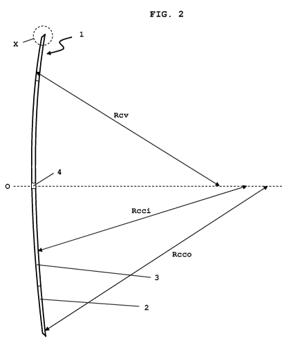
A side view of an embodiment of an intracorneal lens 1
according to the invention is shown in Fig. 2. The
radii of curvature are not shown true to scale in Fig.
2. The lens 1 has a convex front surface with a single
uniform radius of curvature Rcv. The different zones 2
and 3 are identified by different radii of curvature
Rcco and Rcci on the concave rear surface of the lens
1. In the embodiment according to Fig. 2, the radii of
curvature have the following values: Rcv: 7.8 mm; Rcco:
8.858 mm; Rcci: 8.192 mm. The radius of curvature Rcco
merges into the radius of curvature Rcci within a
circle segment of 0.027 mm on the rear surface of the
CA 02871343 2014-13-23
WO 2013/171097 - 21 -
PCT/EP2013/059473
lens 1. Concentrically with respect to the optical axis
0, a central opening 4 is formed in the lens 1 and has
a depth of 0.0259 mm. The convex front surface of the
lens 1 is connected to the concave rear surface of the
lens 1 via an intermediate portion which is located in
the area at the lens edge identified by the circle X in
broken lines.
Fig. 3 shows an enlarged view of the intermediate
portion of the lens 1 according to the invention shown
by the circle X in broken lines in Fig. 2. The front
convex surface of the lens 1 has the radius of
curvature Rcv as far as a point Z on the front surface
of the lens 1. In the embodiment according to Fig. 3,
this point Z is located 0.0078 mm below the edge of the
lens 1. Starting from the point Z, the front surface of
the lens 1 has a straight shape. This straight portion
connects the point Z on the front surface of the lens 1
to the lens edge where the rear surface of the lens 1
with the radius of curvature Rcco has its origin. In
the embodiment according to Fig. 3, the straight
portion between the point Z and the lens edge is
inclined by 30 in relation to the optical axis 0 of
the lens. In the embodiment according to Fig. 3, the
straight portion has a length of 0.012 mm.
Fig. 4 shows an embodiment of the applicator 5 from WO
2011/069907 Al. A pre-load unit P is mounted movably in
the grip piece 11. The pre-load unit P comprises a
housing 6, two leaf-like units 7 and a stop 10, which
limits the movement of the pre-load unit P into the
grip piece 11. In the leaf-like units 7, a continuous
opening 8 is present through the center of the chamber
(not shown here) for receiving a lens. A further hole
9, which makes it easier to equip the applicator 7 with
the lens according to the invention, is additionally
provided. Two control elements 12 and 13 are mounted on
the grip piece 11, by means of which control elements
CA 02871343 2014-13-23
'
= WO 2013/171097
- 22 - PCT/EP2013/059473
12 and 13 the pre-load unit P and a slide (not shown in
Fig. 4) in the interior of the pre-load unit P and of
the grip piece 11 can be moved. A pin, protruding from
the lateral opening 14, is fastened to insert parts
present in the grip piece and fixes these parts in the
grip piece 11. The top face of the pre-load unit P is
clearly marked with the word "TOP".
Fig. 5 shows an embodiment of the kit according to WO
2011/069907 Al, which comprises a storage unit 15. In
the interior of the storage unit 15, the pre-load unit
P is stored in physiological saline. The storage unit
is made of glass. It is closed in a watertight
manner with a stopper 16.
Fig. 6 shows how a lens L is released from the pre-load
unit according to WO 2011/069907 Al. The slide 17 is
pushed forward until it is in contact with the lens L.
The leaf-like units 7 are thereby spread apart from
each other. The leaf-like units 7 can now be drawn back
by a defined distance, while the slide 17 holds the
lens L at the previously oriented site. The lens L is
then released, and the applicator according to the
invention can be removed.
An intracorneal lens according to the invention, of the
kind described in Figures 1 to 3, was implanted in a
group of 28 test subjects. Thereafter, in a standard
sight test, the subjects were able to detect a
resolution of approximately 4 line pairs per mm from a
distance of 25 cm. For comparison purposes using an
intracorneal lens according to WO 2009/075685, it is
known that, with a conventional lens of this kind, it
is possible to achieve only a resolution of
approximately 2 line pairs per mm in the same test.