Note: Descriptions are shown in the official language in which they were submitted.
CA 02871400 2016-06-06
,
DESCRIPTION
BLOOD FLOW IMAGE DIAGNOSING DEVICE AND METHOD
This application claims the benefit of PCT
International Application Number PCT/JP2014/060909 filed on
April 17, 2014 and Japanese Application No. 2013-090562 filed
on April 23, 2013 in Japan.
TECHNICAL FIELD
[0001]
The present invention relates to a blood flow image
diagnosing device and a blood flow image diagnosing method
which apply laser light to a biotissue having blood cells,
and measure and image the speed of blood flow on the basis of
a speckle signal reflected from the biotissue, and which
suppress the influence of the concentration of pigment
deposited in the biotissue on the measured value of the blood
flow.
BACKGROUND ART
[0002]
Heretofore, the present inventors have invented a blood
flow speed measurement apparatus which applies laser light to
a biotissue having blood cells such as the eyeground or the
skin; leads a so-called speckle image (an image of random
1
CA 02871400 2014-10-22
speckle pattern formed as a result of interference of
reflection light from the blood cells) to an image sensor
such as a solid state imaging device (CCD or CMOS);
successively captures and stores a large number of speckle
images at predetermined intervals; selects a predetermined
number of images from the large number of stored images;
calculates a value which reflects the speed of a time course
change in the output of each pixel throughout the images; and
calculates the speed of blood cells (blood flow speed) from
the calculated value. In a blood flow speed measurement
apparatus of such a type, since the value indicating the
output changing speed of each pixel corresponds to the moving
speed of blood cells, the blood flow distribution in the
biotissue can be color-disposed on a monitor screen as a two-
dimensional image (a blood flow map) on the basis of the
calculated value indicating the output changing speed of each
pixel. A blood flow map observed in actuality is composed of
a series of blood flow maps (hereinafter also referred to as
"original maps") calculated at a speed of about 30 frames per
sec, and can be displayed as a motion video. Therefore, the
invented apparatus has been put to practical use as an
apparatus for observing the haemodynamics of the eyeground or
skin (see Patent Documents 1 to 6).
[0003]
Also, the present inventors have proposed a blood flow
speed imaging apparatus (see Patent Document 7). In this
apparatus, a series of blood flow maps obtained through blood
2
CA 02871400 2014-10-22
flow measurement performed for several seconds are used, and
a change in blood flow appearing periodically in synchronism
with the heartbeat is analyzed in various regions within a
field of observation view. A numerical value (i.e., the
degree of distortion) is introduced so as to distinguish
between a region having a sharp rising waveform attributable
to the arterial blood flow and a region having a mildly
rising and falling waveform attributable to the venous blood
flow. Thus, the apparatus can display on the blood flow maps
pulsations caused by the arterial blood flow and pulsations
caused by the venous blood flow.
[0004]
Moreover, the present inventors has proposed the
following method. A new blood flow image diagnosing function
is added to the conventional apparatus, and a function is
added to a computation section so as to separate, from data
of a plurality of blood flow maps over one or more heartbeats,
a blood flow within a surface blood vessel within an
observation region of a biotissue and the background blood
flow therearound. These blood flows are displayed on the ,
blood flow map on a display section in a distinguishable
manner. Various variables which characterize the blood flow
waveforms of the separated regions are defined, and these
variables are compared for clinical diagnosis. In the
following description, an apparatus having such a function
added thereto will be referred to as a "blood flow image
diagnosing device."
3
CA 02871400 2014-10-22
=
PRIOR ART DOCUMENTS
PATENT DOCUMENTS
[0005]
Patent Document 1: Japanese Patent Publication (kokoku) No.
H5-28133
Patent Document 2: Japanese Patent Publication (kokoku) No.
H5-28134
Patent Document 3: Japanese Patent Application Laid-Open
(kokai) No. H4-242628
Patent Document 4: Japanese Patent Application Laid-Open
(kokai) No. H8-112262
Patent Document 5: Japanese Patent Application Laid-Open
(kokai) No. 2003-164431
Patent Document 6: Japanese Patent Application Laid-Open
(kokai) No. 2003-180641
Patent Document 7: NO 2008/69062
SUMMARY OF THE INVENTION
PROBLEMS TO BE SOLVED BY THE INVENTION
[0006]
It was found that in the case where the eyeground blood
flow is measured using a conventional blood flow image
diagnosing device, the measured value of blood flow changes
depending on the light absorptivity of the tissue of the
retina called "pigment epithelium." For example, in the case
of caucasoids (white race), people hardly have that pigment
4
CA 02871400 2014-10-22
unlike people of other races. Therefore, laser light used
for measurement repeats scattering within the eye without
attenuating. In contrast, in the case of colored races such
as the yellow race and the negroid race, people have such
pigment in a high concentration. Therefore, laser light is
absorbed immediately without repeating scattering. This
difference corresponds to the difference between a case where
the interior surface of a camera is not treated to have a
black matted surface and a case where the interior surface of
a camera is treated to have a black matted surface. It is
common knowledge for persons who design optical products that
when such surface treatment is omitted, internal scattering
light fogs a film or an image sensor, and adversely affects
the quality of images (for example, contrast decreases).
[0007]
In the blood flow imaging method developed by the
present inventors, the distribution of blood flow speed is
visualized using the reciprocal of the contrast of a speckle
image which is formed on an image sensor as a result of
interference of laser light scattered by the retina. However,
when the contrast decreases as a result of the above-
mentioned fog caused by light scattering within the eyeball,
the blood flow value is displayed to be rather high (higher
than the actual value). Since people of the white race are
lower in pigment concentration than people of the colored
races as described above, the degree of coherence decreases
as a result of repetition of multiple scattering, and the
CA 02871400 2014-10-22
contrast of a speckle image (interference fringes) lowers,
whereby the blood flow value is displayed to be rather high.
Under the assumption that the size of the eyeball does not
differ greatly among the races, it is considered that the
amount of blood circulating within the eyeball does not
differ greatly among the races. Therefore, it is hard to
accept the measurement result that caucasoid people are high
in blood flow value than people of the colored races, and
some correction is needed.
[0008]
Intraocular blood flows which ophthalmologists consider
important for diagnosis are the blood flows of the arterial
and venous blood vessels on the retina, the tissue blood flow
of the optic papilla, and the blood flow of the choroid. The
arterial and venous blood vessels run through the surface
layer of the retina, and, under this layer, the layer of
visual cells, the layer of pigment epithelium, and the layer
of choroid blood vessels are layered in this order toward the
sclera which is the outmost layer. It is said that although
the arterial and venous blood vessels of the retina extend
through the optic papilla, the pigment epithelium is usually
absent in the optic layer of the optic papilla other than the
blood vessels. Accordingly, in the case of people of the
colored races, the degree of influence of the pigment
epithelium on scattering of laser light differ among regions,
which leads to a complicated result in which the
proportionality constant for equalizing the measured values
6
CA 02871400 2014-10-22
of caucasoid people and those of colored people differs among
regions. In other words, when the eyeground blood flow of a
caucasoid person and that of a colored person are measured,
equalizing the indicated values of the eyeground blood flows
is not a simple task, and it becomes necessary to perform
complicated processing of correcting the indicated values by
applying different proportionality constants for different
regions.
[0009]
A similar problem arises when the skin blood flow is
measured. The concentration of melanin greatly differs among
the human races, and the darker the skin color, the lower the
measured value of blood flow. Therefore, numerical
comparison is difficult. Also, in the case where the color
of the skin has changed due to lesion or a difference in
color arises between an incision formed as a result of an
operation and a region therearound, a numerical difference is
produced as in the case of the above-described retina blood
flow measurement. Therefore, in order to perform comparison
in a standardized state, it is necessary to perform some
correction in accordance with the pigment concentration.
[0010]
An object of the present invention is to solve the
problem of the conventional image analyzing apparatuses for
eyeground blood flow and skin blood flow; i.e., the problem
that the measured value of blood flow is displayed
differently depending on the pigment concentration of a
7
CA 02871400 2014-10-22
4
subject (object under measurement), and to provide means
which allows the measured value of blood flow to be displayed
by a standardized numerical value irrespective of the race of
the subject and allows comparison of measured values among
people of different races.
MEANS FOR SOLVING THE PROBLEMS
[0011]
A blood flow image diagnosing device of the present
invention comprises a laser light irradiation system for
applying laser light to an observation region of a biotissue
having blood cells; a light receiving section having a
plurality of pixels and adapted to detect reflection light
from the observation region of the biotissue; an image
capturing section for successively capturing a plurality of
images on the basis of a signal from the light receiving
section; an image storage section for storing the plurality
of images; a computation section for computing the speed of
blood flow within the biotissue from time course changes of
output signals of the pixels through the plurality of stored
images; and a display section for displaying a two-
dimensional distribution which is the result of the
computation as a blood flow map. The computation section
includes a pigment concentration correction section for
correcting the blood flow map obtained as a result of
computation at the computation section in accordance with the
pigment concentration of the observation region.
8
CA 02871400 2016-06-06
[0012]
The pigment concentration correction section includes a
laser reflectance computation section for detecting the pigment
concentration of the observation region as a laser reflectance;
and a correction coefficient creation section for creating a
correction coefficient, which is used for correction of the
blood flow map, on the basis of the laser reflectance from the
laser reflectance computation section.
[0013]
When a surface under measurement moves during capturing of
images, the blood flow map shifts according. In view of this,
the computation section includes a blood flow analysis section
for performing tracking processing of calculating a shift
amount of the blood flow map and superimposing it while
correcting its movement amount; and a blood flow map correction
section for correcting the blood flow map having undergone the
tracking processing in accordance with the pigment
concentration of the observation region, the correction being
performed on the basis of the correction coefficient output
from the pigment concentration correction section, wherein the
laser reflectance computation section obtains the laser
reflectance on the basis of a laser reflection intensity
obtained from a laser reflection intensity map obtained by
superimposing speckle images from which the blood flow map is
synthesized, and a signal representing the intensity of laser
light radiated from the laser light irradiation system.
[0014]
9
CA 02871400 2014-10-22
The blood flow image diagnosing device further
comprises a storage section for storing a relation between
the laser reflectance and the blood flow value in the
observation region, the relation being obtained in advance
for a plurality of healthy persons, wherein the correction
coefficient creation section creates the correction
coefficient on the basis of the laser reflectance obtained
for a newly measured blood flow map, and the relation between
the laser reflectance and the blood flow value stored in the
storage section. The laser reflectance is calculated on an
observation region-by-observation region basis, and a
different correction coefficient is created for each
observation region. The blood flow of the biotissue is, for
example, the eyeground blood flow or the skin blood flow.
The correction performed in accordance with the pigment
concentration of the skin tissue is composed of first-stage
correction performed for a specific region where a relatively
stable numerical value is obtained, and second-stage
correction of multiplying a value obtained through the first-
stage correction by correction coefficients determined for
regions which differ from one another in terms of the pigment
concentration and the scattering characteristic of the
corneal layer of epidermis.
[0015]
A blood flow image analysis method of the present
invention uses a laser light irradiation system for applying
laser light to an observation region of a biotissue having
CA 02871400 2014-10-22
=
blood cells and a light receiving section having a plurality
of pixels and adapted to detect reflection light from the
observation region of the biotissue. The method comprises
the steps of: successively capturing a plurality of images on
the basis of a signal from the light receiving section and
storing the plurality of images; computing the speed of blood
flow within the biotissue from time course changes of output
signals of the pixels throughout the stored images, and
creating a blood flow map having a two-dimensional
distribution; obtaining, in advance for a plurality of
healthy persons, a relation between the laser reflectance and
the blood flow value in the observation region, and storing
the relation, the relation being used for creation of a
correction coefficient used for correcting the blood flow map
in accordance with the pigment concentration of the
observation region; creating, at the time of new measurement,
the correction coefficient on the basis of a newly obtained
laser reflectance and the stored relation between the laser
reflectance and the blood flow value; and displaying the
blood flow map corrected on the basis of the correction
coefficient.
EFFECT OF THE INVENTION
[0016]
In the case of a conventional blood flow image
diagnosing device, the blood flow measured value greatly
differs between the white race and the colored race. However,
11
CA 02871400 2014-10-22
according to the present invention, it becomes possible to
suppress the influence of the concentration of pigment
deposited in an object under measurement and allow
standardized comparison of blood flow values among human
races.
BRIEF DESCRIPTION OF DRAWINGS
[0017]
FIG. 1(A) is a schematic diagram showing the overall
configuration of a blood flow image diagnosing device
configured on the basis of the present invention, and FIG. 1
(B) is a diagram showing the configuration of a computation
section which is the characteristic of the present invention.
FIG. 2 is a flowchart showing operation of blood flow
image diagnosis when applied to measurement of eyeground
blood flow.
FIG. 3 is an image showing a display example in which
eyeground blood flow maps (original maps) obtained at a speed
of 30 frames per sec (an example for the case where the
subject is the yellow race).
FIG. 4 is an image showing a synthesized blood flow map
obtained by capturing eyeground blood flow maps as shown in
FIG. 3 (120 frames) and superimposing them while correcting
shifts of the eyeground blood flow maps due to eye movement
during fixation (an example for the case where the subject is
the yellow race).
FIG. 5 is an image showing a synthesized blood flow map
12
CA 02871400 2014-10-22
6
Which is similar to that shown in FIG. 4 but was obtained
from a subject of the white race.
FIG. 6 is an image showing a laser reflection intensity
map which is obtained by superimposing speckle images from
which the synthesized blood flow map of FIG. 4 is obtained
(an example for the case where the subject is the yellow
race).
FIG. 7 is an image showing a laser reflection intensity
map which is similar to that shown in FIG. 6 but was obtained
from a subject of the white race.
FIG. 8 is an image showing a laser reflection intensity
map obtained when a monkey was used as a subject.
FIG. 9 is an image showing a synthesized blood flow map
obtained when a monkey was used as a subject.
FIG. 10 is a scatter diagram obtained by obtaining the
averages of MBR values for two regions (choroid and optic
papilla tissue) and plotting them for corresponding laser
reflectances, and regression lines.
FIG. 11 is an image showing an example of correction in
which the correction of the present invention was applied to
the blood flow map of FIG. 4 so as to increase the blood flow
value of people of the yellow race to a value comparable to
that of people of the white race.
MODE FOR CARRYING OUT THE INVENTION
[0018]
The present invention will now be described by way of
13
CA 02871400 2014-10-22
examples. FIG. 1(A) is a schematic diagram showing the
overall configuration of a blood flow image diagnosing device
configured on the basis of the present invention, and FIG.
1(B) is a diagram showing the configuration of a computation
section which is the characteristic of the present invention.
A laser light irradiation system applies laser light, through
a half mirror, to a biotissue (e.g., the eyeground of an eye
to be tested) having blood cells such as eyeground blood flow
or skin blood flow. A light receiving section includes a CCD
(solid state imaging device) having a large number of pixels
on its light receiving surface, a light receiving lens which
focuses laser reflection light on the CCD, an amplification
circuit for amplifying the output of the CCD, etc. The CCD
which is driven on the basis of timing pulses converts an
image of the biotissue formed by the light receiving lens to
an electric signal on the basis of the timing pulses. The
CCD reads out signal charges in a frame storage mode, and
amplifies and outputs them as an image signal.
[0019]
Analog processing such as gain control is performed on
the output image signal, and a resultant analog signal is
converted to a digital signal. On the basis of this digital
signal and the timing pulses, an image capturing section
successively captures a plurality of images at predetermined
intervals (e.g., intervals of 1/30 sec) equal to or greater
than one heartbeat. An image storage section stores data of
the captured images. A computation section computes the
14
CA 02871400 2014-10-22
blood flow speed within the biotissue from the time course
change of output signals of the pixels throughout the
plurality of stored images. The characteristic feature of
the present invention is correcting a blood flow map obtained
as a result of the computation at the computation section in
accordance with the pigment concentration of the observation
region. As will be described in detail later, a laser light
intensity signal is used for this correction. A display
section displays a two-dimensional distribution of
computation results as a blood flow map, and also displays
numerical information which characterizes a blood flow
waveform.
[0020]
The above-described configuration of the blood flow
image diagnosing device is identical to the conventional
configuration disclosed in Patent Document 7, etc. except for
the configuration of the computation section. The
configuration of the computation section which is the feature
of the present invention will be described with reference to
FIG. 1(B). The present invention is characterized by
providing a pigment concentration correction section which
corrects the blood flow map obtained through computation in
accordance with the pigment concentration of the observation
region. Although the image storage section stores eyeground
blood flow maps (original maps) obtained at a speed of, for
example, 30 frames per sec, the quality of the images is not
good enough as will be described in detail later. Therefore,
CA 02871400 2016-06-06
tracking processing is performed in a blood flow analysis
section. In the tracking processing, the amount of shift of
each blood flow map is calculated, and the blood flow map is
superimposed on other blood flow maps by correcting its
movement amount. On the basis of the correction coefficient
output from the pigment concentration correction section, a
blood flow map correction section corrects the blood flow map
having undergone the tracking processing in accordance with
the pigment concentration of the observation region. The
corrected blood flow map is displayed.
[0021]
Since the reflectance (or absorptivity) of laser light
differs among the human races, the pigment concentration is
detected as a laser reflectance (reflection
intensity/incident intensity). For such a purpose, a laser
reflectance computation section obtains the laser reflectance
of the observation region on the basis of a signal
representing the intensity of the applied laser light and the
intensity of a signal reflected from each location within the
observation region and detected by the light receiving
section. In the exemplified method, the intensity of the
detected signal is obtained from a laser reflection intensity
map which is obtained by superimposing speckle images from
which a blood flow map is synthesized. The relation between
the laser reflectance and the blood flow value in the
observation region is obtained for a large number of healthy
persons, and is stored in the apparatus (the storage section).
16
CA 02871400 2014-10-22
A
The correction coefficient creation section creates a
correction coefficient for the blood flow value on the basis
of the laser reflectance obtained for a newly measured blood
flow map and the relation between the laser reflectance and
the blood flow value stored in the storage section.
[0022]
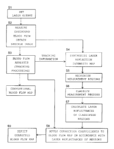
Next, operation of a first embodiment in which the
blood flow image diagnosing device shown in FIG. 1(A) and
1(B) is applied to measurement of the eyeground blood flow
will be described with reference to FIG. 2. FIG. 2 is a
flowchart showing operation of the blood flow image diagnosis
when applied to measurement of the eyeground blood flow. In
step Si shown in FIG. 2, the output of a laser is set, and
laser light is radiated. In step S2, the eyeground blood
flow is measured by the blood flow image diagnosing device.
When the surface of an organism is irradiated with laser
light, scattering light rays interfere with one another and
form a random speckle pattern. In general, this pattern is
called "laser speckle." This speckle image is obtained.
[0023]
FIG. 3 is an image showing a display example in which
eyeground blood flow maps (original maps) obtained at a speed
of 30 frames per sec (an example for the case where the
subject is the yellow race). The eyeground blood flow maps
are stored in the image storage section shown in FIG. 1(A).
As shown in FIG. 3, the blood flow maps are usually displayed
as a motion video (30 fames per sec). However, the image
17
CA 02871400 2014-10-22
quality is not high enough because the degree of graininess
is large. Therefore, tracking processing is performed in
step S3 of FIG. 2. In the tracking processing, each blood
flow map is analyzed, the amount of shift of the map is
calculated, and the blood flow map is superimposed on other
blood flow maps by correcting its movement amount. When the
surface under measurement moves during capturing of an image,
the blood flow map shifts accordingly. The computation
section has a function of performing so-called tracking
processing of calculating the shift amount of each map and
superimposing it while correcting the movement amount.
[0024]
The noise components originally contained in each blood
flow map are averaged by the tracking processing. This
allows the contours of blood vessels to be clearly recognized
as shown in FIG. 4. FIG. 4 is an image showing a synthesized
blood flow map obtained by capturing blood flow maps as shown
in FIG. 3 (120 frames) and superimposing them while
correcting the shifts of the blood flow maps due to eye
movement during fixation (an example for the case where the
subject is the yellow race). In general, such a blood flow
map is displayed in color code. However, here, the blood
flow map is displayed in gray scale, and the higher the
degree of whiteness, the greater the blood flow.
[0025]
Meanwhile, the eyeground blood flow of a caucasoid
person is measured using a conventional blood flow image
18
CA 02871400 2014-10-22
diagnosing device, a synthesized blood flow map of FIG. 5 is
obtained. Although the synthesized blood flow map of FIG. 5
is similar to that of FIG. 4, it shows an example of
measurement values obtained from people of the white race.
FIGS. 4 and 5 correspond to "depiction of a conventional
blood flow map" described in FIG. 2. When FIGS. 4 and 5 are
compared, it is found that the numerical value is displayed
to be considerably high. Under the assumption that the size
of the eyeball does not differ greatly among the races, it is
considered that the amount of blood circulating within the
eyeball does not differ greatly among the races. Therefore,
it is inconvenient that different blood flow values are
displayed even when the difference is the race only, and some
correction is needed. This correction is performed in steps
S4 through S8 as described below. A specific procedure of
the correction will be described for an example case where
the eyeground blood flows of a caucasoid person and a colored
person are measured.
[0026]
In step S4 of FIG. 2, a laser reflection intensity map
is synthesized. FIG. 6 is an image showing a laser
reflection intensity map which is obtained by superimposing
speckle images from which the synthesized blood flow map of
FIG. 4 is obtained (an example for the case where the subject
is the yellow race). FIG. 7 is an image showing a laser
reflection intensity map obtained from a subject of the white
race. Information representing the shift of each blood flow
19
CA 02871400 2014-10-22
map is obtained from the step S3 as tracking information, and
tracking is performed on speckle images, on the basis of
which the synthesized blood flow map is calculated, in order
to average them, a laser reflection intensity map as shown in
FIG. 6 is obtained. FIG. 6 shows an example in which the
subject is the yellow race. From FIG. 6, it is found that
the reflection intensity is high at an optic papilla
surrounded by a circle and is low at the remaining portion.
FIG. 7 shows an example in which the subject is the white
race. From FIG. 7, it is found that little difference is
present between the reflection intensity at the optic papilla
and that at the remaining portion. This shows that the layer
of the pigment epithelium widely spreading at the boundary
between the retina and the choroid is dark in color in the
case of people of the colored races and is almost clear in
the case of people of the white races, and that the
reflectance (or absorptivity) of laser light differs among
the human races.
[0027]
The incident intensity used for calculation of the
laser reflectance (reflection intensity/incident intensity)
(step S7 which will be described below) can be obtained as a
signal representing the intensity of laser light radiated
from the laser light irradiation system. The laser
reflection intensity is obtained as the signal intensity of
light reflected from each location within the observation
region and detected by the light receiving section. For
CA 02871400 2014-10-22
=
example, as exemplified above, it can be obtained from the
laser reflection intensity map. The power of laser light
output from the present blood flow image diagnosing device is
adjusted by setting a laser output value using measurement
software (step S1). In the case of caucasoid people, the
internal scattering is strong, and light returning from the
eyeground to the light receiving section is too strong.
Therefore, the laser output is decreased. In contrast, in
the case of people of the colored races, the light returning
to the light receiving section is weak. Therefore, the laser
output is set to be rather strong. For example, when the
laser output (laser light intensity signal) required to
obtain the map of FIG. 6 is considered 10, the laser output
required to obtain the map of FIG. 7 is only 5. When the
averaged value (reflection intensity) of the laser reflection
intensity map in a certain region other than the optic
papilla was 60 in the case of colored people, the laser
reflectance becomes 6 (= 60/10). Similarly, when the
averaged value (reflection intensity) of the laser reflection
intensity maps in a certain region other than the optic
papilla was 80 in the case of caucasoid people, the laser
reflectance becomes 16 (= 80/5). In other words, the laser
reflectance can be determined from the overall output of
laser light applied to the eyeball and the average value of
the laser reflection intensity map within a region of
interest.
[0028]
21
CA 02871400 2014-10-22
4
In step S5, measurement regions are recognized. For
example, the eyeground blood flow is measured mainly in three
regions of interest; i.e., the choroid, the retina blood
vessel, and the optic papilla tissue. When an eye doctor
diagnoses eye diseases, he or she pays attention mainly to
the blood flow within blood vessels running on the retina,
the tissue blood flow at the optic papilla, and the blood
flow of the choroid, which are considered to closely relate
to angiostenosis, glaucoma, and macular degeneration caused
derivatively by medical diseases such as arteriosclerotic and
diabetes. However, since these blood flows differ in the.
positional relation with the pigment epithelium, they differ
in the degree of influence of the pigment concentration.
Namely, since the retina blood vessel is located closer to
the surface layer than the pigment epithelium layer, when the
pigment concentration is high, the backward scattering light
from a deeper region decreases, and the reflection intensity
of the blood vessel portion also decreases.
[0029]
In step S6, the measurement regions are classified.
Since the pigment concentration and its influence change
among the measurement regions, different correction
coefficients must be prepared for the respective measurement
regions.
[0030]
In step S7, the laser reflectance of each of the
classified regions is calculated. For such calculation, for
22
CA 02871400 2014-10-22
a large number of healthy persons of all human races (white,
yellow, and negroid races), first a blood flow value is
obtained from a synthesized blood flow map having undergone
tracking processing, and a laser reflectance is obtained from
the laser reflection intensity map. The obtained values are
stored. The averaged value of the laser reflectance (=
reflection intensity/incident intensity) of each region is
obtained using the laser reflection intensity map. The
averaged value of the laser reflectance can be considered to
reflect the pigment concentration which affects that region.
Measurement must be performed a large number of times in
order to obtain and store the above-mentioned data. In
actual measurement, the stored data are used for correction.
[0031]
In step S8, a newly measured blood flow map is
corrected. Namely, correction coefficients are applied to
the blood flow map in accordance with the laser reflectances
of the respective regions. For each region, the averaged
laser reflection intensity and the averaged blood flow value
are calculated. This calculation is repeated for all the
stored data of healthy persons, whereby scatter diagrams are
plotted, and regression lines are obtained. FIG. 10 shows
scatter diagrams and regression lines obtained for two
regions (choroid CHR and optic papilla tissue ONH-T). Each
of the scatter diagrams represents the relation between the
averaged blood flow value MBR and the laser reflection
intensity. Although the inclination of the regression line
23
CA 02871400 2014-10-22
changes among the regions, for increase and decrease of the
laser reflectance in each region, it functions as a
correction coefficient. In the example of FIG. 10, data at
the right end of the graph are those of the white race, data
of the colored races are shown on the left side thereof such
that the higher the pigment concentration, the closer to the
left end. The relation between the averaged blood flow value
MBR and the laser reflectance represented by such a
regression line is stored in the image storage section.
[0032]
Next, there will be considered the case where data of a
certain colored race are newly obtained, data of, for example,
the choroid is obtained, is converted to a value of the white
race, and is compared with its standard value. In this case,
the laser reflectance of that region is obtained, and is
divided by the average laser reflectance of the white race
located at the right end of FIG. 10 (the value stored in the
storage section), and a resultant value is used as a
correction coefficient. The averaged blood flow value MBR is
divided by this correction coefficient, whereby a numerical
value corrected for the influence of the pigment can be
obtained and can be compared with that of the white race.
[0033]
In step S9, the corrected blood flow map is depicted.
As can be understood from FIG. 10, the correction
coefficients of the respective regions differ from one
another. However, when the above-described correction is
24
CA 02871400 2014-10-22
=
performed for the respective regions, image comparison of the
blood flow map becomes possible. FIG. 11 shows the result of
correction performed on the blood flow map of the yellow race
shown in FIG. 4. FIG. 11 shows that the corrected numeral
values are comparable to those of the blood flow map of the
white race shown in FIG. 5. Accordingly, even in a country
where people of many races live, there can be determined a
criteria; e.g., a criteria that when the tissue blood flow of
the optic papilla becomes equal to or lower than a
predetermined cutoff value, the risk of glaucoma increases
irrespective of the human race.
[0034]
Next, there will be described operation of a second
embodiment in which the blood flow image diagnosing device
shown in FIG. 1(A) and 1(B) is applied to measurement of the
skin blood flow. Correction similar to that performed for
the eyeground blood flow is necessary for the skin blood flow.
For example, the concentration of melanin of black people is
high, and even near-infrared laser light is easily absorbed.
Therefore, the measured value of the subcutaneous blood flow
is displayed to be rather low as compared with that of people
of the yellow race. It cannot be considered that the flow of
blood flowing through the subcutaneous capillary vessel layer
formed to nourish the skin tissue differs among the human
races. Therefore, it is necessary to introduce a correction
method which allows display of standardized numerical values
irrespective of the human race. Further, it has been found
CA 02871400 2014-10-22
that the color of the palm of the hand is thin in color as
compared with the back of the hand, and the differences among
measured values are small as compared with those of the back
of the hand. Accordingly, the correction coefficient for the
palm of the hand differs from that for the back of the hand,
and it is necessary to apply correction on a region-by-region
basis as in the case of the eyeground.
[0035]
A possible method which realizes this is measuring the
pigment concentration of a skin tissue of a subject (object
under measurement) by using a skin color measurement device
or the like and performing correction. However, this
investigates the reflecting characteristic (or absorbing
characteristic) for visible light, and does not show the
characteristic for a laser wavelength used for measurement.
Accordingly, the most feasible method is directly obtaining,
from a laser scattering signal used for blood flow
measurement, information of the characteristic of reflection
(or the characteristic of absorption) by the pigment of the
subject for the wavelength of the laser signal as in the case
of the eyeground.
[0036]
In the case of the eyeground, the information of the
pigment concentration is contained in the laser reflection
intensity map shown in FIG. 6 or FIG. 7. A value obtained by
dividing the numerical value on this map by the incident
intensity of laser light projected to the eyeground shows the
26
CA 02871400 2014-10-22
4
ratio of reflection by the pigment (reflectance), which is
inverse proportional to absorption. This allows application
of first-stage correction. However, as described above, the
influence of absorption by the pigment changes depending on
the positional relation between a blood vessel or blood
vessel layer to be detected and the pigment layer. Therefore,
it become necessary to perform second-stage correction. In
the second-stage correction, the positional relations are
classified into several groups, regions having the same
positional relation are recognized as the same region, and a
different correction coefficient corresponding to the region
is applied.
[0037]
Although the eyeground blood flow changes slightly
within a day, the blood flows at an approximately constant
rate all times. In contrast, it is known that the skin blood
flow is greatly affected by room temperature, clothing, and
metal condition, and, in particular, its change increases
toward the distal ends of the extremities. When a concept
such as skin perfusion index (SPI) which is a standardized
index in which the influence of melanin concentration is
cancelled is introduced, it is necessary to first find a
reference region which facilitate comparison between
individuals or between human races. According to the results
of a search conducted by us, the value of the skin of the
chest or back whose vibration due to the heartbeat or
breathing is small and which is covered with clothes is
27
CA 02871400 2014-10-22
relatively stable. First, for these portions, the first-
stage correction for melanin concentration is performed using
the above-described laser reflection intensity map. Next, the
second-stage correction is performed. In the second-stage
correction, the value corrected through the first-stage
correction is multiplied by the correction coefficients of
regions (e.g., the palm and back of the hand) which differ in
terms of pigment concentration and the scattering
characteristic of the corneal layer of epidermis. As
described above, in the case of the eyeground and in the case
of the skin, by performing correction in the same procedure,
it becomes possible to display blood flow maps using a
standardized index which allows comparison of blood flow
values between people of different races.
EXAMPLES
[0038]
The index MBR (Mean Blur Rate) that the present
inventor uses for calculation of the blood flow value is
defined by MBR = (contrast of speckles)-2 = (average light
intensity/standard deviation of fluctuation component)2. In
the case of people of the colored race, the MBR value of the
retina blood vessel is displayed to be lower, as compared
with people of the white race, because of the following
reason. The time course change of speckles caused by the
blood flow component of a blood vessel portion of people of
the colored races is the same as that of people of the white
28
CA 02871400 2014-10-22
race. However, backward scattering light becomes weak due to
the pigment component contained in the background tissue, and
the average light intensity (the numerator of the above-
described expression) decreases as compared with caucasoid
people. As a result, the blood flow value (MBR value) is
displayed to be rather low.
[0039]
In an extreme case, as shown in the laser reflection
light distribution of FIG. 8 and the synthesized blood flow
map of FIG. 9, the pigment concentration is very high in
regions other than the optic papilla. It is considered that
no pigment epithelium exists at the optic papilla, and the
backward scattering light is strong. Therefore, the blood
flow value of the retina blood vessel is displayed to be
sufficiently high at the optic papilla; however, when leaving
the optic papilla, the blood flow decreases sharply, and it
becomes almost impossible to recognize as a blood vessel.
[0040]
As shown in the examples of FIGS. 4 and 5, in a region
around the optic papilla, blood flow maps of the retina blood
vessel and the choroid (the blood vessel layer located
underneath the retina) are displayed. Although the former is
displayed as a thin clear line, the image of the latter
becomes blurred because the latter is located on the deeper
side and the information is scattered. Further, as is clear
from FIG. 5, the choroid blood flow value of people of the
white race is displayed to be as high as double that of
29
CA 02871400 2014-10-22
people of the colored race.
[0041]
Meanwhile, in the case of people of the colored races
as well, no pigment epithelium exists at the optic papilla.
Therefore, it was predicted that when the tissue blood flow
at the optic papilla is measured, a value similar to that
obtained for caucasoid people is obtained. However, when a
large number of measurement examples were compared in
actuality, it was found that in the case of the caucasoid
people, the blood flow value is displayed to be rather high
as compared to people of the colored race. Conceivably, this
phenomenon occurred for the following reason. In the case of
caucasoid people, the degree of light absorption by the
pigment epithelium is small. Therefore, laser light repeats
multiple scattering within the eyeball, and scattered light
reaches the optic papilla. As a result, contrast decreases
accordingly, and the MBR value increases slightly.
[0042]
As described above, the influence of the pigment
epithelium on the blood flow index MBR is strongest at the
choroid, is second strongest at the retina blood vessel, and
weakest at the optic papilla tissue. In other words, this
means that when all the values obtained from different
regions are multiplied by the same numerical value, proper
correction cannot be performed, and that the values obtained
from different regions must be multiplied by different
correction coefficients determined for the regions.
CA 02871400 2014-10-22
[0043]
The important point is that even when a subject thinks
that he or she knows his/her race, the actual pigment
concentration does not necessarily correspond thereto. Also,
it is a common knowledge that, for example, people in Asia
have different melanin concentrations state by state or
region by region even though they are of the same colored
race. A method of creating a database of standard values on
a race-by-race basis and performing comparison is widely used
in medical equipment. However, in the case where the
influencing factor varies among people of the same race,
accurate values cannot be obtained unless correction is
performed on the basis of some actually measured values of
the influencing factor obtained from an object under
measurement.
[0044]
Although only some exemplary embodiments of this
invention have been described in detail above, those skilled
in the art will readily appreciated that many modifications
are possible in the exemplary embodiments without materially
departing from the novel teachings and advantages of this
invention. Accordingly, all such modifications are intended
to be included within the scope of this invention.
31